5e6dEndo Dx
46
Transcript of 5e6dEndo Dx
8/12/2019 5e6dEndo Dx
http://slidepdf.com/reader/full/5e6dendo-dx 1/46
8/12/2019 5e6dEndo Dx
http://slidepdf.com/reader/full/5e6dendo-dx 2/46
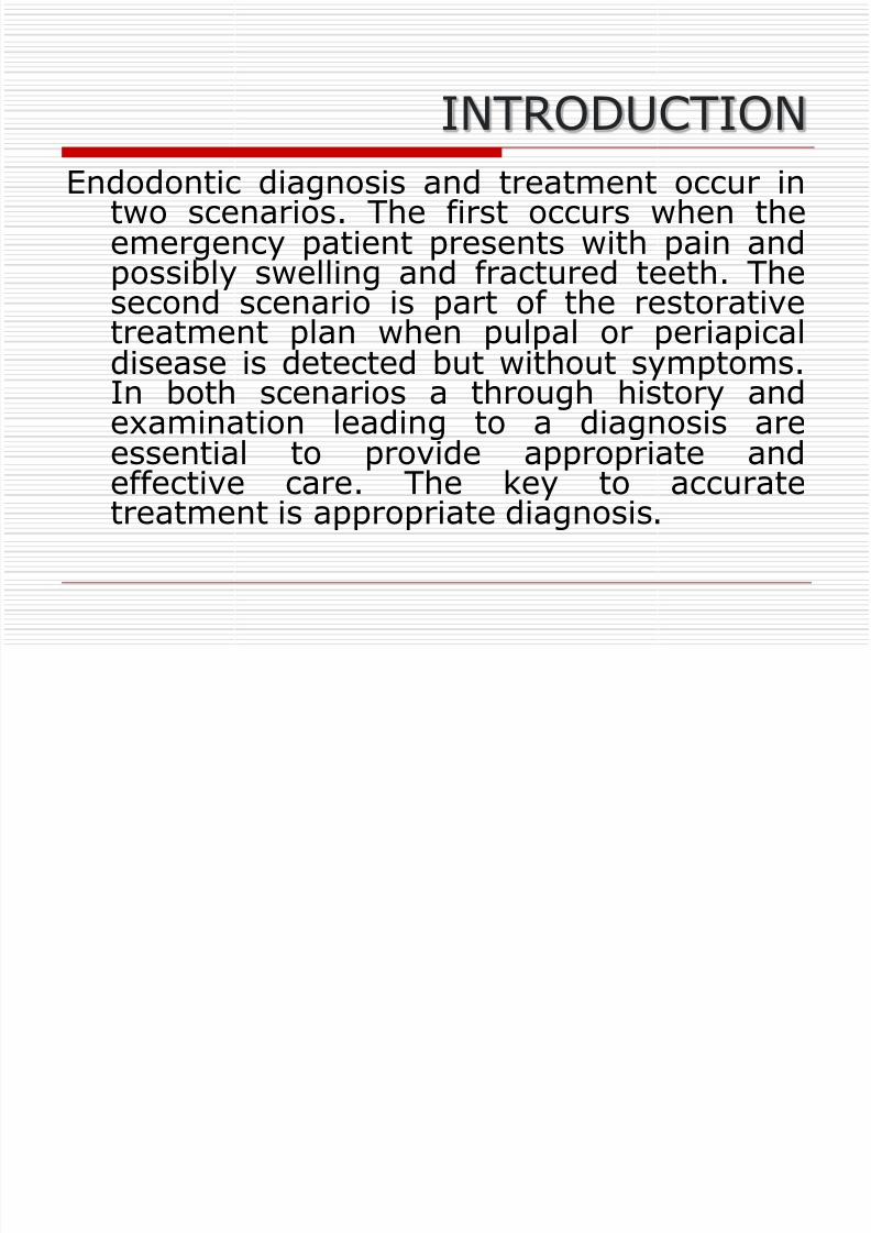
INTRODUCTION
Endodontic diagnosis and treatment occur intwo scenarios. The first occurs when theemergency patient presents with pain andpossibly swelling and fractured teeth. The
second scenario is part of the restorativetreatment plan when pulpal or periapicaldisease is detected but without symptoms.In both scenarios a through history andexamination leading to a diagnosis are
essential to provide appropriate andeffective care. The key to accuratetreatment is appropriate diagnosis.
8/12/2019 5e6dEndo Dx
http://slidepdf.com/reader/full/5e6dendo-dx 3/46
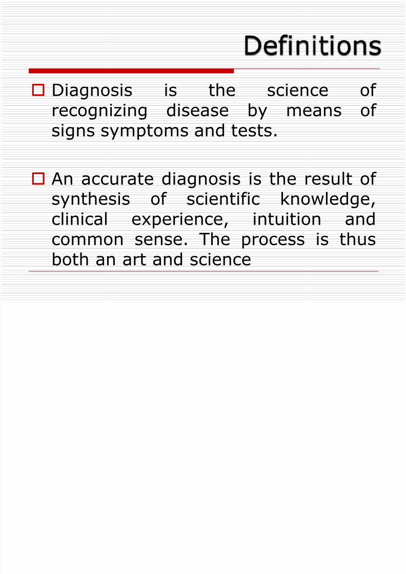
Diagnosis is the science ofrecognizing disease by means ofsigns symptoms and tests.
An accurate diagnosis is the result ofsynthesis of scientific knowledge,clinical experience, intuition andcommon sense. The process is thusboth an art and science
8/12/2019 5e6dEndo Dx
http://slidepdf.com/reader/full/5e6dendo-dx 4/46
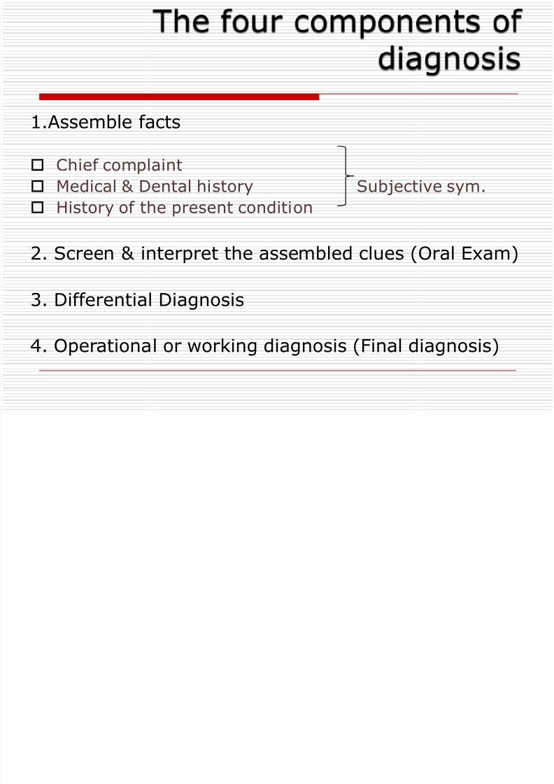
1.Assemble facts
Chief complaint
Medical & Dental history Subjective sym.
History of the present condition
2. Screen & interpret the assembled clues (Oral Exam)
3. Differential Diagnosis
4. Operational or working diagnosis (Final diagnosis)
8/12/2019 5e6dEndo Dx
http://slidepdf.com/reader/full/5e6dendo-dx 5/46
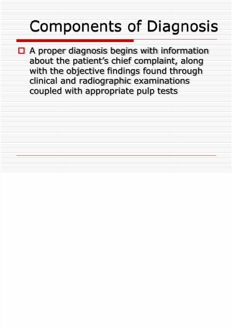
A proper diagnosis begins with informationabout the patient’s chief complaint, alongwith the objective findings found through
clinical and radiographic examinationscoupled with appropriate pulp tests
8/12/2019 5e6dEndo Dx
http://slidepdf.com/reader/full/5e6dendo-dx 6/46
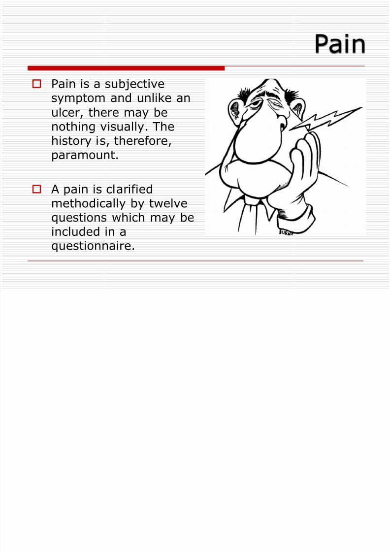
Pain is a subjectivesymptom and unlike an
ulcer, there may benothing visually. The
history is, therefore,paramount.
A pain is clarified
methodically by twelvequestions which may beincluded in aquestionnaire.
8/12/2019 5e6dEndo Dx
http://slidepdf.com/reader/full/5e6dendo-dx 7/46
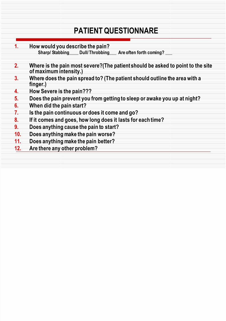
PATIENT QUESTIONNARE
1. How would you describe the pain?Sharp/ Stabbing____ Dull/ Throbbing___ Are often forth coming? ___
2. Where is the pain most severe?(The patient should be asked to point to the siteof maximum intensity.)
3. Where does the pain spread to? (The patient should outline the area with afinger.)
4. How Severe is the pain???
5. Does the pain prevent you from getting to sleep or awake you up at night?
6. When did the pain start?
7. Is the pain continuous or does it come and go?
8. If it comes and goes, how long does it lasts for each time?9. Does anything cause the pain to start?
10. Does anything make the pain worse?
11. Does anything make the pain better?
12. Are there any other problem?
8/12/2019 5e6dEndo Dx
http://slidepdf.com/reader/full/5e6dendo-dx 8/46
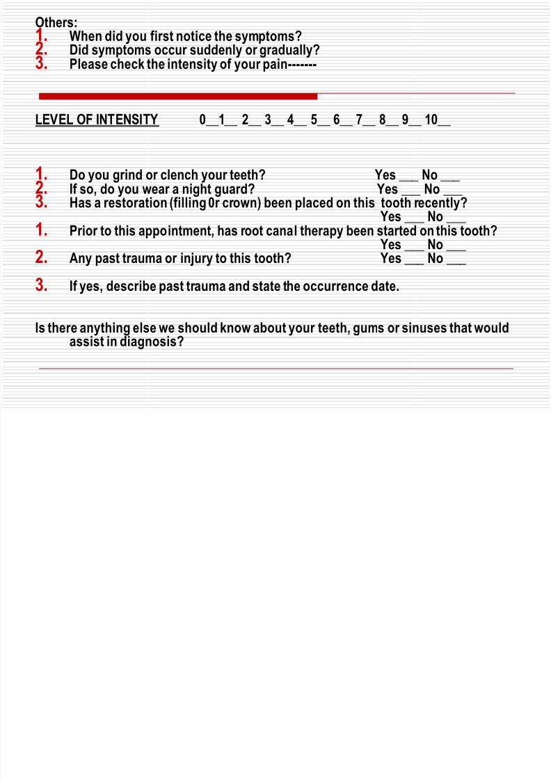
Others:1. When did you first notice the symptoms?2. Did symptoms occur suddenly or gradually?3. Please check the intensity of your pain-------
LEVEL OF INTENSITY 0__1__ 2__ 3__ 4__ 5__ 6__ 7__ 8__ 9__ 10__
1. Do you grind or clench your teeth? Yes ___ No ___
2. If so, do you wear a night guard? Yes ___ No ___3. Has a restoration (filling 0r crown) been placed on this tooth recently? Yes ___ No ___
1. Prior to this appointment, has root canal therapy been started on this tooth? Yes ___ No ___
2. Any past trauma or injury to this tooth? Yes ___ No ___
3. If yes, describe past trauma and state the occurrence date.
Is there anything else we should know about your teeth, gums or sinuses that wouldassist in diagnosis?
8/12/2019 5e6dEndo Dx
http://slidepdf.com/reader/full/5e6dendo-dx 9/46
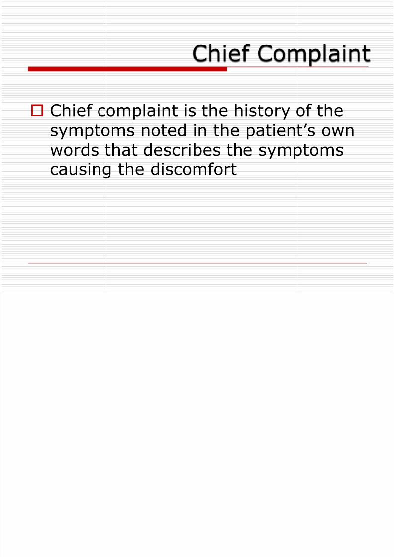
Chief complaint is the history of the
symptoms noted in the patient’s ownwords that describes the symptomscausing the discomfort
8/12/2019 5e6dEndo Dx
http://slidepdf.com/reader/full/5e6dendo-dx 10/46
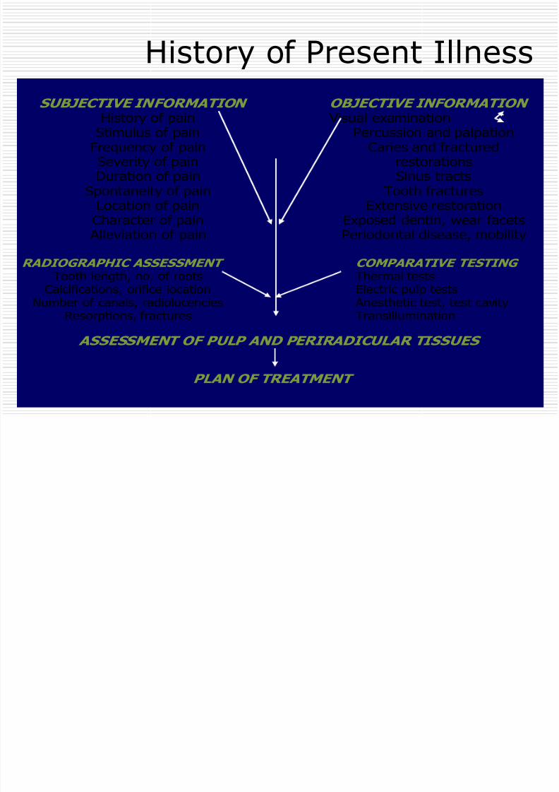
SUBJECTIVE INFORMATION
History of painStimulus of pain
Frequency of painSeverity of painDuration of pain
Spontaneity of painLocation of painCharacter of pain
Alleviation of pain
OBJECTIVE INFORMATION
Visual examinationPercussion and palpation
Caries and fracturedrestorationsSinus tracts
Tooth fracturesExtensive restorationExposed dentin, wear facetsPeriodontal disease, mobility
RADIOGRAPHIC ASSESSMENT
Tooth length, no. of roots
Calcifications, orifice locationNumber of canals, radiolucenciesResorptions, fractures
COMPARATIVE TESTING
Thermal tests
Electric pulp tests Anesthetic test, test cavityTransillumination
ASSESSMENT OF PULP AND PERIRADICULAR TISSUES
PLAN OF TREATMENT
History of Present Illness
8/12/2019 5e6dEndo Dx
http://slidepdf.com/reader/full/5e6dendo-dx 11/46
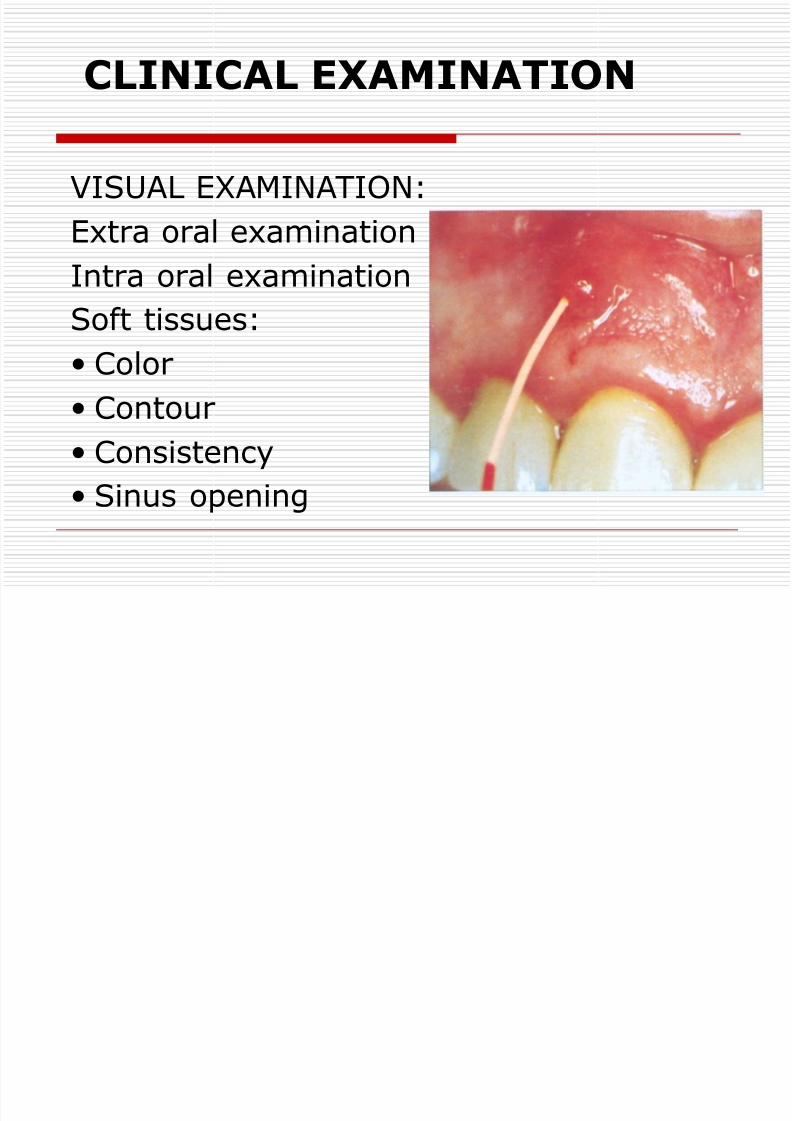
CLINICAL EXAMINATION
VISUAL EXAMINATION:
Extra oral examination
Intra oral examinationSoft tissues:
Color
Contour Consistency
Sinus opening

8/12/2019 5e6dEndo Dx
http://slidepdf.com/reader/full/5e6dendo-dx 12/46

8/12/2019 5e6dEndo Dx
http://slidepdf.com/reader/full/5e6dendo-dx 13/46
8/12/2019 5e6dEndo Dx
http://slidepdf.com/reader/full/5e6dendo-dx 14/46
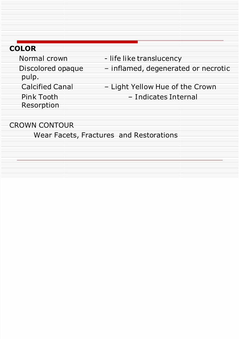
COLOR
Normal crown - life like translucency
Discolored opaque – inflamed, degenerated or necroticpulp.
Calcified Canal – Light Yellow Hue of the Crown
Pink Tooth – Indicates InternalResorption
CROWN CONTOURWear Facets, Fractures and Restorations
8/12/2019 5e6dEndo Dx
http://slidepdf.com/reader/full/5e6dendo-dx 15/46
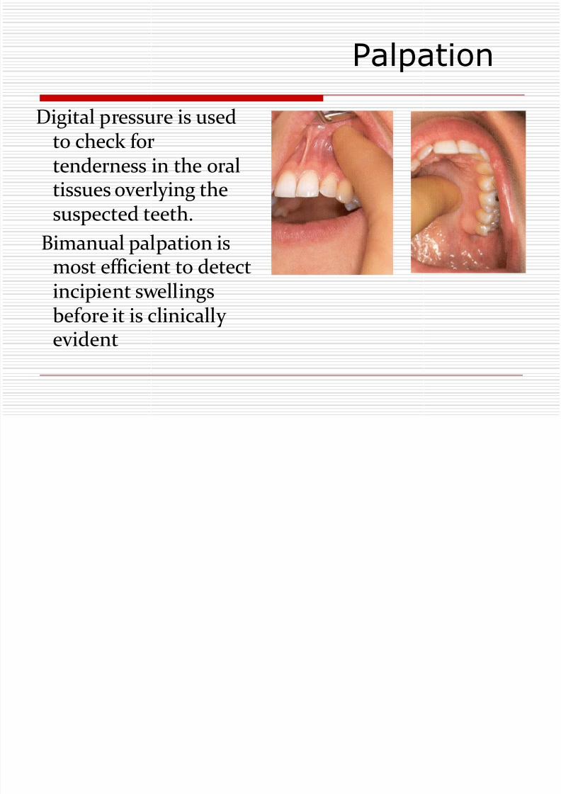
Palpation
Digital pressure is usedto check fortenderness in the oral
tissues overlying thesuspected teeth.
Bimanual palpation ismost efficient to detect
incipient swellingsbefore it is clinicallyevident

8/12/2019 5e6dEndo Dx
http://slidepdf.com/reader/full/5e6dendo-dx 16/46
8/12/2019 5e6dEndo Dx
http://slidepdf.com/reader/full/5e6dendo-dx 17/46
Percussion
Normal resonant sound on percussion indicates good periodontal ligament
Metallic sound on percussion indicates ankylosis.
Response to percussion not only indicates the involvementof the PDL but also the extent of the inflammation.(degreeof response directly proportional to degree ofinflammation).

8/12/2019 5e6dEndo Dx
http://slidepdf.com/reader/full/5e6dendo-dx 18/46
8/12/2019 5e6dEndo Dx
http://slidepdf.com/reader/full/5e6dendo-dx 19/46
Mobility
Tooth mobility provides an indication of the integrity of theattachment apparatus.
Mobility is graded as:
Grade I – Noticeable horizontal movement in its socket.
Grade II – within 1 mm of horizontal movement.
Grade III – Horizontal movement greater than 1 mm and/orvertical depressibility.

8/12/2019 5e6dEndo Dx
http://slidepdf.com/reader/full/5e6dendo-dx 20/46
8/12/2019 5e6dEndo Dx
http://slidepdf.com/reader/full/5e6dendo-dx 21/46
Radiographs are an important andnecessary adjunct in Endodontics.
Accurate radiographic techniques and
proper interpretation are essential forsound diagnosis and treatment. Radiograph techniques of endodontic
importance: Preiapical
BitewingOPG
8/12/2019 5e6dEndo Dx
http://slidepdf.com/reader/full/5e6dendo-dx 22/46
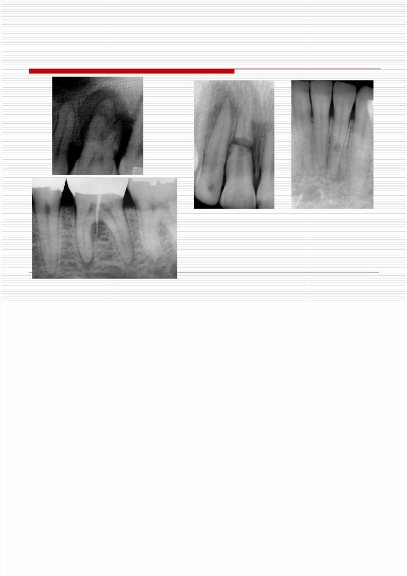
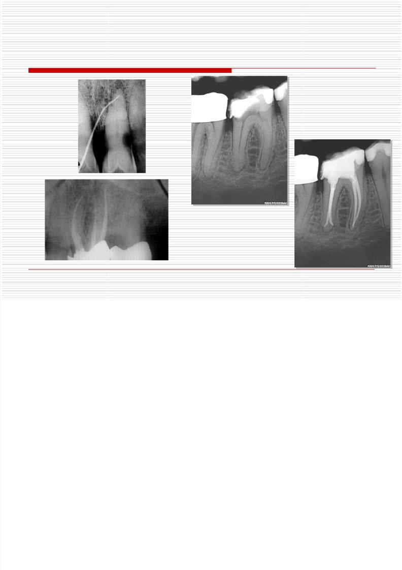
Role of Radiograph in Endodontics:
Endodontic diagnosis. Determination of prognosis of treatment. Disclosing the presence and extent of caries. Checking the thickness of PDL.
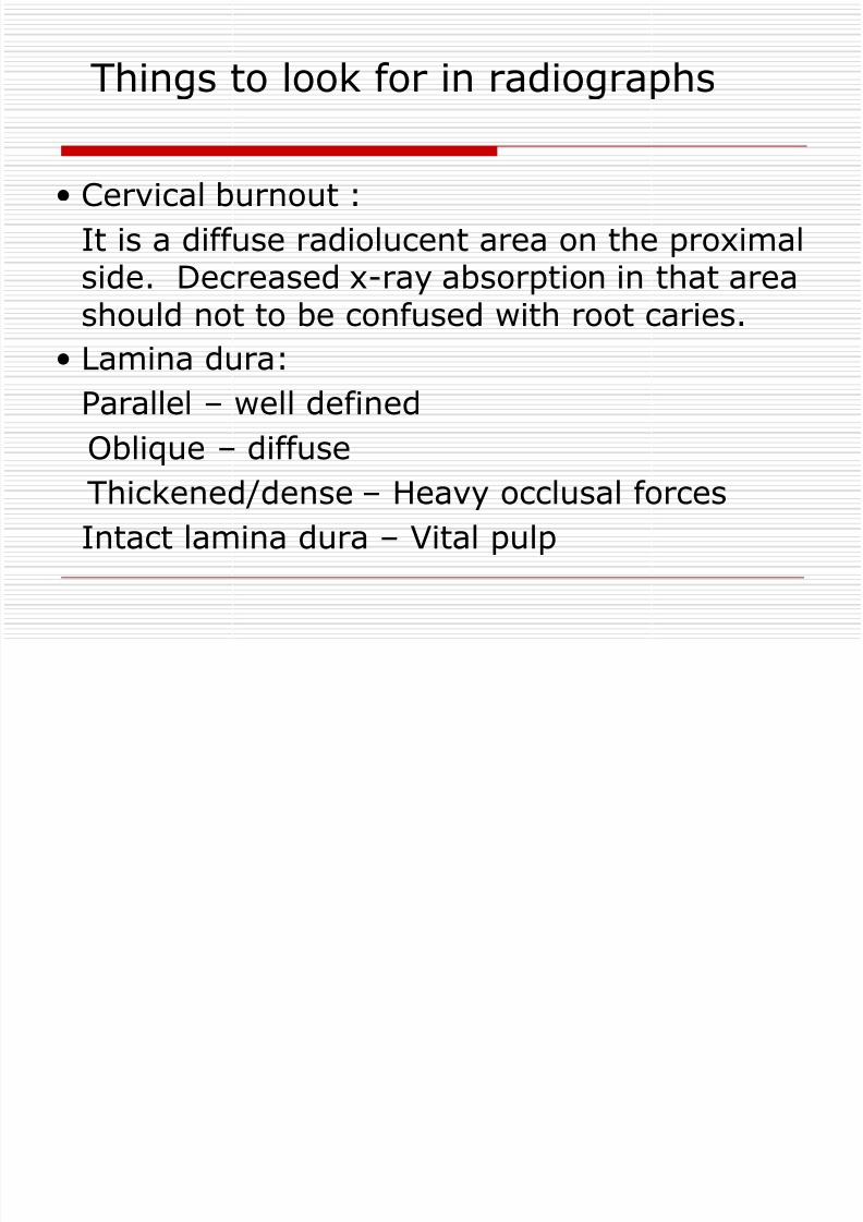
To see the presence or absence of lamina dura. To see the number, shape, length and internalanatomy of root canal.
To check any obstruction present within the root canal. To see the quality of the previous root canal. To see the tooth calcification.
Detrmination of working length and selection of mastercone.
Help to find endodontic errors like, perforation, ledgingand instrument seperation.
To see any resorption associated with tooth.
8/12/2019 5e6dEndo Dx
http://slidepdf.com/reader/full/5e6dendo-dx 23/46
8/12/2019 5e6dEndo Dx
http://slidepdf.com/reader/full/5e6dendo-dx 24/46
8/12/2019 5e6dEndo Dx
http://slidepdf.com/reader/full/5e6dendo-dx 25/46
8/12/2019 5e6dEndo Dx
http://slidepdf.com/reader/full/5e6dendo-dx 26/46
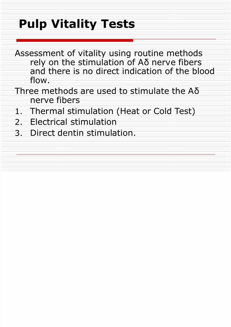
Pulp Vitality Tests
Assessment of vitality using routine methodsrely on the stimulation of Aδ nerve fibersand there is no direct indication of the blood
flow.Three methods are used to stimulate the Aδ
nerve fibers
1. Thermal stimulation (Heat or Cold Test)
2. Electrical stimulation3. Direct dentin stimulation.
8/12/2019 5e6dEndo Dx
http://slidepdf.com/reader/full/5e6dendo-dx 27/46
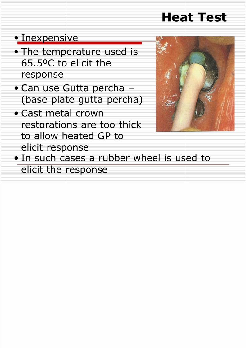
Heat Test
Inexpensive
The temperature used is65.5ºC to elicit theresponse
Can use Gutta percha – (base plate gutta percha)
Cast metal crown
restorations are too thickto allow heated GP toelicit response
In such cases a rubber wheel is used to
elicit the response

8/12/2019 5e6dEndo Dx
http://slidepdf.com/reader/full/5e6dendo-dx 28/46

8/12/2019 5e6dEndo Dx
http://slidepdf.com/reader/full/5e6dendo-dx 29/46
Cold Test
Various materials used for cold
test are Cones of ice – -20ºC
Ethyl chloride spray – -40ºC
Carbon- di- oxide snow–
-70ºC Application of cold for 4
seconds lowers thetemperature to between 26 and
30ºC eliciting pain. Within thepulp temperature is lowered by0.2ºC.

8/12/2019 5e6dEndo Dx
http://slidepdf.com/reader/full/5e6dendo-dx 30/46
8/12/2019 5e6dEndo Dx
http://slidepdf.com/reader/full/5e6dendo-dx 31/46
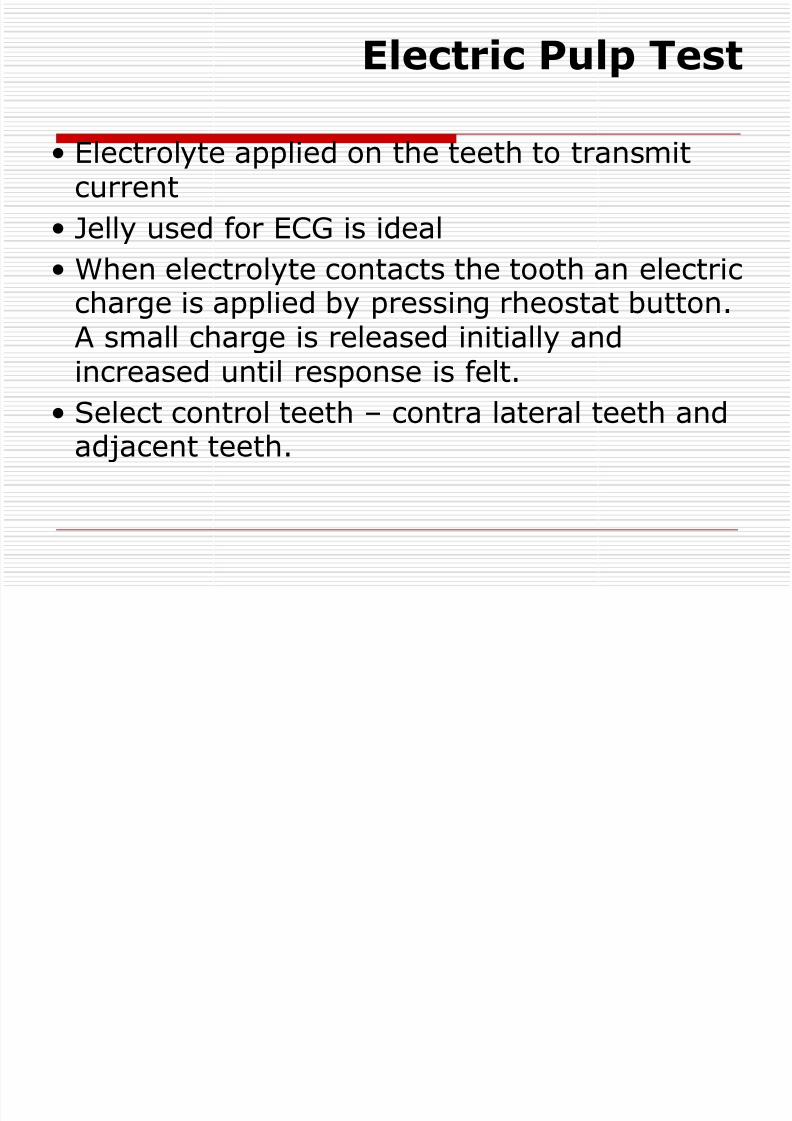
Electric Pulp Test
Electrolyte applied on the teeth to transmitcurrent
Jelly used for ECG is ideal
When electrolyte contacts the tooth an electriccharge is applied by pressing rheostat button.A small charge is released initially andincreased until response is felt.
Select control teeth – contra lateral teeth andadjacent teeth.

8/12/2019 5e6dEndo Dx
http://slidepdf.com/reader/full/5e6dendo-dx 32/46
8/12/2019 5e6dEndo Dx
http://slidepdf.com/reader/full/5e6dendo-dx 33/46
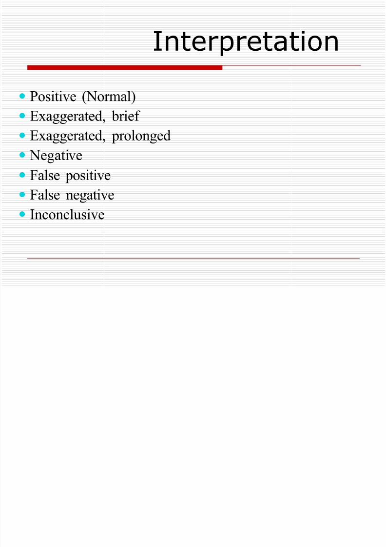
Interpretation
Positive (Normal)
Exaggerated, brief
Exaggerated, prolonged Negative
False positive
False negative
Inconclusive
8/12/2019 5e6dEndo Dx
http://slidepdf.com/reader/full/5e6dendo-dx 34/46
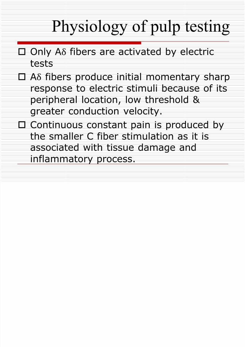
Only Aδ fibers are activated by electric
tests
Aδ fibers produce initial momentary sharp
response to electric stimuli because of itsperipheral location, low threshold &greater conduction velocity.
Continuous constant pain is produced bythe smaller C fiber stimulation as it isassociated with tissue damage andinflammatory process.
Physiology of pulp testing
8/12/2019 5e6dEndo Dx
http://slidepdf.com/reader/full/5e6dendo-dx 35/46
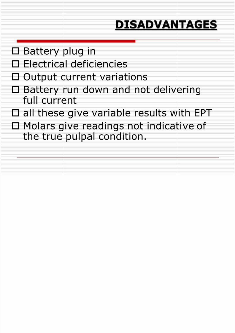
Battery plug in
Electrical deficiencies
Output current variations
Battery run down and not deliveringfull current
all these give variable results with EPT
Molars give readings not indicative ofthe true pulpal condition.
8/12/2019 5e6dEndo Dx
http://slidepdf.com/reader/full/5e6dendo-dx 36/46
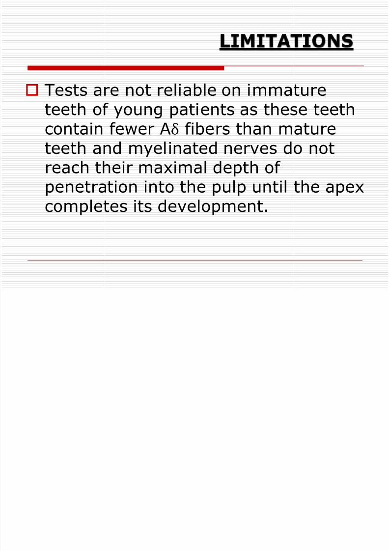
Tests are not reliable on immature
teeth of young patients as these teethcontain fewer Aδ fibers than matureteeth and myelinated nerves do notreach their maximal depth ofpenetration into the pulp until the apex
completes its development.
8/12/2019 5e6dEndo Dx
http://slidepdf.com/reader/full/5e6dendo-dx 37/46
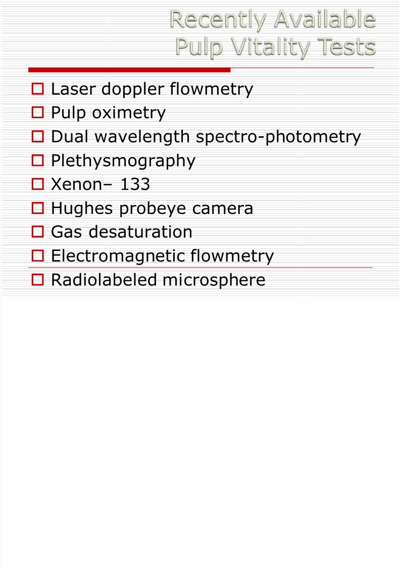
Laser doppler flowmetry
Pulp oximetry
Dual wavelength spectro-photometry
Plethysmography
Xenon– 133
Hughes probeye camera Gas desaturation
Electromagnetic flowmetry
Radiolabeled microsphere
8/12/2019 5e6dEndo Dx
http://slidepdf.com/reader/full/5e6dendo-dx 38/46
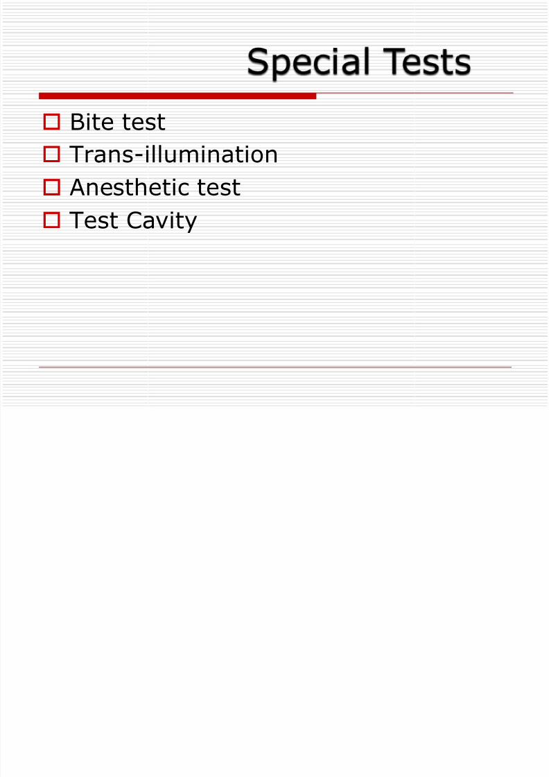
Bite test
Trans-illumination
Anesthetic test
Test Cavity
8/12/2019 5e6dEndo Dx
http://slidepdf.com/reader/full/5e6dendo-dx 39/46
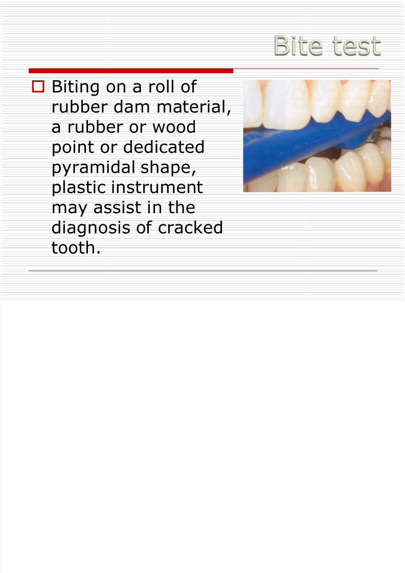
Biting on a roll ofrubber dam material,a rubber or wood
point or dedicatedpyramidal shape,plastic instrument
may assist in thediagnosis of crackedtooth.
8/12/2019 5e6dEndo Dx
http://slidepdf.com/reader/full/5e6dendo-dx 40/46
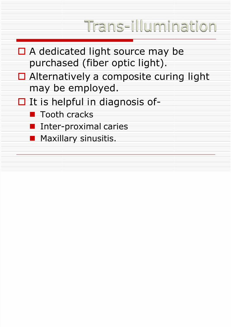
A dedicated light source may bepurchased (fiber optic light).
Alternatively a composite curing lightmay be employed.
It is helpful in diagnosis of-
Tooth cracks
Inter-proximal caries
Maxillary sinusitis.

8/12/2019 5e6dEndo Dx
http://slidepdf.com/reader/full/5e6dendo-dx 41/46
8/12/2019 5e6dEndo Dx
http://slidepdf.com/reader/full/5e6dendo-dx 42/46
Starting from the distal most tooth of thesuspected quadrent, intraligamentary
anesthesia is injected. If pain persists nexttooth is anesthetized (the one mesial to it).In this way the process is continued untilthe affected tooth is identified.
This is a relatively painful process. So it isperformed if all other tests a inconclusive.
8/12/2019 5e6dEndo Dx
http://slidepdf.com/reader/full/5e6dendo-dx 43/46
This method is performed if all othertest are inconclusive. In this method
a cavity is drilled with out applicationof anesthesia and patient is asked ifthere is any sensation of pain.

8/12/2019 5e6dEndo Dx
http://slidepdf.com/reader/full/5e6dendo-dx 44/46
8/12/2019 5e6dEndo Dx
http://slidepdf.com/reader/full/5e6dendo-dx 45/46
Conclusion
A dentist can develop a number ofassets to become a successfuldiagnostician. The most important of
these are knowledge, interest,intuition, curiosity and patience. Thesuccessful diagnostician must also
have acute senses and the necessaryequipment for diagnosis.

8/12/2019 5e6dEndo Dx
http://slidepdf.com/reader/full/5e6dendo-dx 46/46