Localización de Serratia marcescens en perfil ... · Localización de Serratia marcescens en...
11
239 Vet. Méx., 41 (4) 2010 Artículos científicos Localización de Serratia marcescens en perfil bacteriológico y fúngico de la conjuntiva de caballos clínicamente sanos en Monterrey, Nuevo León, México Localization of Serratia marcescens in bacterial and fungal profile of conjunctiva of clinically healthy horses from Monterrey, Nuevo Leon, Mexico Recibido el 25 de enero de 2010 y aceptado el 23 de septiembre de 2010. *Facultad de Medicina Veterinaria y Zootecnia, Universidad Autónoma de Nuevo León, Fco. Villa s/n, Ex-Hacienda El Canadá, General Escobedo, 66050, Nuevo León, Monterrey, México. **Hospital Veterinario La Silla, Antiguo Camino al Diente 3333, La Estanzuela, 64988, Nuevo León, Monterrey, México. ***Facultad de Agronomía, Universidad Autónoma de Nuevo León, Fco. Villa s/n, Ex-Hacienda El Canadá, General Escobedo, 66050, Nuevo León, Monterrey, México. Correspondencia: Dr. Jesús Jaime Hernández Escareño, Tel.: (81) 13 40 43 93, extensión 3615, Correo electrónico: [email protected] Abstract The conjunctival normal microflora, formed by fungi, yeast and bacteria, of 50 clinically healthy horses from Monterrey, Nuevo Leon, was identified using three to 12 year-old horses of different breeds and gender. Samples were taken from the conjunctival sac of both eyes (n =100 eyes) using a cotton swab under sterile conditions. No eye was negative to the presence of the microorganism. There were differences in colony types in each eye of the same animal. One hundred percent were positive to fungal and bacteria colonies in both eyes, while 60% were positive to yeast. In the present study Serratia marcescens, a pathogenic, opportunist and multidrug-resistant organism, is reported for the first time in the conjunctival sac in horses. Serratia marcescens was isolated from 21 clinically healthy horses (42%) with a number of colony forming units (cfu) that ranged from 9 to 42. Key words: SERRATIA MARCESCENS, EYE, HORSES, CONJUNCTIVA, KERATOMYCOSIS. Resumen Se identificó la microflora normal, formada por hongos, levaduras y bacterias, de la conjuntiva en 50 caballos clínicamente sanos, de diferente raza y sexo, de tres a 12 años de edad, del área metropolitana de Monterrey, Nuevo León. Las muestras se tomaron del saco conjuntival de ambos ojos (n = 100 ojos) con un hisopo bajo condiciones estériles. Ningún ojo fue negativo a la presencia de microorganismos. Hubo a menudo diferencias en los tipos de colonias entre los ojos del mismo animal, 100% fueron positivos a la presencia de hongos y bacterias en ambos ojos, y 60% positivos a la presencia de levaduras. Este estudio presenta, por vez primera en conjuntiva de caballos, la presencia de Serratia marcescens, considerada microorganismo patógeno oportunista con multirresistencia a fármacos. En esta investigación, Serratia marcescens se logró aislar de 21 caballos clínicamente sanos (42%) con rango de nueve a 42 unidades formadoras de colonias (ufc). Palabras clave: SERRATIA MARCESCENS, OJO, CABALLOS, CONJUNTIVA, QUERATOMICOSIS. Gustavo Hernández Vidal* Rafael Ramírez Romero* Luís Edgar Rodríguez Tovar* Francisco A. Mora Valdez** Juan Antonio Vidales Contreras*** Jesús Jaime Hernández Escareño*
Transcript of Localización de Serratia marcescens en perfil ... · Localización de Serratia marcescens en...

239Vet. Méx., 41 (4) 2010
Artículos científicosLocalización de Serratia marcescens en perfil
bacteriológico y fúngico de la conjuntiva de caballos clínicamente sanos en Monterrey, Nuevo León, México
Localization of Serratia marcescens in bacterial and fungal profile of conjunctiva of clinically healthy
horses from Monterrey, Nuevo Leon, Mexico
Recibido el 25 de enero de 2010 y aceptado el 23 de septiembre de 2010.*Facultad de Medicina Veterinaria y Zootecnia, Universidad Autónoma de Nuevo León, Fco. Villa s/n, Ex-Hacienda El Canadá, General Escobedo, 66050, Nuevo León, Monterrey, México.**Hospital Veterinario La Silla, Antiguo Camino al Diente 3333, La Estanzuela, 64988, Nuevo León, Monterrey, México.***Facultad de Agronomía, Universidad Autónoma de Nuevo León, Fco. Villa s/n, Ex-Hacienda El Canadá, General Escobedo, 66050, Nuevo León, Monterrey, México.Correspondencia: Dr. Jesús Jaime Hernández Escareño, Tel.: (81) 13 40 43 93, extensión 3615, Correo electrónico: [email protected]
Abstract
The conjunctival normal microflora, formed by fungi, yeast and bacteria, of 50 clinically healthy horses from Monterrey, Nuevo Leon, was identified using three to 12 year-old horses of different breeds and gender. Samples were taken from the conjunctival sac of both eyes (n =100 eyes) using a cotton swab under sterile conditions. No eye was negative to the presence of the microorganism. There were differences in colony types in each eye of the same animal. One hundred percent were positive to fungal and bacteria colonies in both eyes, while 60% were positive to yeast. In the present study Serratia marcescens, a pathogenic, opportunist and multidrug-resistant organism, is reported for the first time in the conjunctival sac in horses. Serratia marcescens was isolated from 21 clinically healthy horses (42%) with a number of colony forming units (cfu) that ranged from 9 to 42.
Key words: Serratia marcescens, EYE, HORSES, CONJUNCTIVA, KERATOMYCOSIS.
Resumen
Se identificó la microflora normal, formada por hongos, levaduras y bacterias, de la conjuntiva en 50 caballos clínicamente sanos, de diferente raza y sexo, de tres a 12 años de edad, del área metropolitana de Monterrey, Nuevo León. Las muestras se tomaron del saco conjuntival de ambos ojos (n = 100 ojos) con un hisopo bajo condiciones estériles. Ningún ojo fue negativo a la presencia de microorganismos. Hubo a menudo diferencias en los tipos de colonias entre los ojos del mismo animal, 100% fueron positivos a la presencia de hongos y bacterias en ambos ojos, y 60% positivos a la presencia de levaduras. Este estudio presenta, por vez primera en conjuntiva de caballos, la presencia de Serratia marcescens, considerada microorganismo patógeno oportunista con multirresistencia a fármacos. En esta investigación, Serratia marcescens se logró aislar de 21 caballos clínicamente sanos (42%) con rango de nueve a 42 unidades formadoras de colonias (ufc).
Palabras clave: Serratia marcescens, OJO, CABALLOS, CONJUNTIVA, QUERATOMICOSIS.
Gustavo Hernández Vidal* Rafael Ramírez Romero* Luís Edgar Rodríguez Tovar*Francisco A. Mora Valdez** Juan Antonio Vidales Contreras***
Jesús Jaime Hernández Escareño*
240
Introduction
It is known that bacterial and fungal microflora of the conjunctiva in healthy animals is controlled by a series of mechanisms that keep populations in balance
and prevent the spread of these potentially pathogenic microorganisms.1 Many studies report the presence of different microflora in healthy conjunctiva and corneas of sheep,2 cows,3.4 pigs,5 birds,6 rabbits,7 goats,8 dogs,9 elephants,10 cats,11 donkeys,12 and horses.13, 14
It is reported that the predominant pathogenic microorganisms in the normal conjunctival flora in horses are Gram-positive.15 However, Gram-negative bacteria are also present as part of the normal conjunctival flora of the horse.15
Some fungi are an inherent part of the normal conjunctival microflora of the horse; however, after a corneal wound, these organisms can act as pathogens. Keratomycosis is fairly common in horses, but their presence is rare in dogs, cats and cows. In horses, keratomycosis is clinically manifested by the appearance of ulcerative keratitis, interstitial keratitis or stromal abscess. The latter occurs as a result of infection by bacteria or fungi in the stroma, due to a corneal defect16 and iris prolapse.
There are reports of the prevalence of fungi isolated from eyes of horses (95%), cows (100%), dogs (22%) and cats (8%). The difference in the percentage may be due to factors such as eye size, degree of exposure or contact with the spores of fungi and bacteria, especially spores and fomites, as well as the large number of microorganisms found in feces in pens, which can infect the eye, causing severe ocular injury.3
The surface of the eye is composed of a series of eye structures that work together and are necessary for good vision. The surface of the epithelial cells of the cornea and conjunctiva, as most of the mucosal surfaces of the body, is protected by a layer composed of branched sugar residues, attached to proteins, collectively called glycocalyx. The main components of the epithelial glycocalyx are mucins or integral glycoproteins. The role of mucin carbohydrates (O-glycans) is not fully understood, but it is suggested that they form a diffusion barrier to extracellular components in a protective way.17
The cornea and conjunctiva have a unique defense system against fungal infections. There are immunological, metabolic and anti-microbial mechanisms, which together with the tissue physical barrier, protect the cornea from keratomycosis.17
Furthermore, the native bacterial flora protects the cornea against fungi by consuming the necessary nutrients for the growth of these organisms, and also by the secretion of some antimicrobial substances with antifungal properties.18
Introducción
Se sabe que la microflora bacteriana y fúngica de la conjuntiva en los animales sanos se controla por una serie de mecanismos que mantienen a
las poblaciones en equilibrio y previenen el contagio de estos microorganismos potencialmente patógenos.1 Muchos trabajos registran presencia de diversa microflora en conjuntiva y córneas sanas en ovejas,2 vacas,3,4 cerdos,5 aves,6 conejos,7 cabras,8 perros,9 elefantes,10 gatos,11 burros12 y caballos.13,14
Se ha informado que los microorganismos no patogénicos predominantes en la flora normal conjuntival en caballos son los Gram positivos.15 Sin embargo, las bacterias Gram negativas también están presentes como parte de la flora normal conjuntival de los equinos.15
Algunos hongos son parte inherente de la microflora conjuntival normal del caballo; no obstante, tras una herida corneal, estos microorganismos pueden actuar como agentes patógenos. La queratomicosis es bastante común en caballos, pero es muy rara su presencia en perros, gatos y vacas. En caballos, la queratomicosis se manifiesta clínicamente por la aparición de queratitis ulcerativa, queratitis intersticial o de absceso estromal. Este último ocurre como resultado de la infección por bacterias u hongos en el estroma, debido a un defecto corneal16 y prolapso del iris.
Existen informes de la prevalencia de hongos aislados en ojos de caballos (95%), vacas (100%), perros (22%) y gatos (8%). La diferencia en el porcentaje puede deberse a factores como tamaño de los ojos, grado de exposición o contacto con las esporas de hongos y bacterias, principalmente esporuladas y fómites, así como a la gran cantidad de microorganismos que se encuentran en las heces en los corrales, los cuales pueden infectar el ojo ocasionando una severa lesión ocular.3
La superficie del ojo está constituida por una serie de estructuras oculares que funcionan de forma conjunta y que son necesarias para tener una buena visión. La superficie de las células epiteliales de la córnea y conjuntiva, al igual que la mayoría de las superficies mucosas del cuerpo humano, se encuentran protegidas por una capa compuesta de residuos de azúcares ramificados, unidos a proteínas, colectivamente llamados glicocálix. Los principales componentes de este glicocálix epitelial son las mucinas o glicoproteínas integrales. No se conoce del todo la función de los carbohidratos de la mucina (O-glicanos), pero se sugiere que forman una barrera de difusión de los componentes extracelulares a manera de protección.17
La córnea y la conjuntiva tienen un sistema de defensa peculiar contra infecciones fúngicas.
241Vet. Méx., 41 (4) 2010
The objective of this research was to identify the normal microflora of fungi, yeasts and bacteria in healthy high-performance horses in the metropolitan area of Monterrey, Nuevo Leon, in order to establish the prevalence and frequency of microorganisms (fungi, yeasts and bacteria) in conjunctiva, and establish risk factors for the development of diseases or eye problems in horses.
Material and methods
This study was conducted in March 2009, at the Laboratorio de Microbiologia, Facultad de Medicina Veterinaria of the Universidad Autonoma de Nuevo Leon. Both eyes of 50 healthy horses (n = 100 eyes) of different breed and sex: 34 females (68%) and 16 males (32%) of three to 12 years of age were evaluated. Samples were taken from high-performance horses, used for show jumping. The animals were kept in stables with concrete floors and sawdust under strict cleaning regimen; the stables were cleaned three times a day. The food given is considered standard for a top athlete horse, consisting of bran, Bermuda grass, Ryegrass and black oat, corn and barley flakes, granules with vitamins, minerals and amino acids. Drinking water was allowed ad libitum.
After ruling out any ocular inflammation or infection, sampling was carried out from the conjunctiva using sterile swabs, taking care not to contaminate the sample by contact with skin. The swabs were placed in Stuart transport medium, identified and transported at 4°C.
In order to isolate aerobic bacteria the following culture mediums were used: blood agar, eosin methylene blue agar (EMB), Mueller Hinton agar and Mac Conkey. For fungi and yeast: potato dextrose agar (PDA), with and without chloramphenicol and cycloheximide (0.5 g / L). The bacterial cultures were incubated at 37°C and checked at 24 and 48 hours. Gram stains were done for morphological identification of these microorganisms. The biochemical tests proposed by Koneman et al.19 were used to characterize the genera of the recovered bacteria.
Incubation for common fungi and yeasts was at 32°C for 7 days, while monitoring for the genus Malassezia was 14 days. Fungi were characterized by direct observation using lactophenol and, when necessary, microcultures were performed on PDA, using identification keys for imperfect fungi according to Barnett and Hunter.20 Yeasts were identified by their macro and micromorphological features, and its physiological characteristics, such as the presence of capsule by negative staining using India ink, production of urease at 25 ° C and germinative test tube.21 22
Existen mecanismos inmunológicos, metabólicos y antimicrobiales, que junto con la barrera física del tejido, protegen a la córnea de queratomicosis.17
Asimismo, la flora bacteriana nativa protege a la córnea contra hongos mediante el consumo de los nutrimentos necesarios para la proliferación de estos microorganismos, y por la secreción de algunos antimicrobianos con propiedades antifúngicas.18
El objetivo de esta investigación fue identificar la microflora normal de hongos, levaduras y bacterias en caballos sanos de alto rendimiento en el área metropolitana de Monterrey, Nuevo León, con la finalidad de establecer la prevalencia de microorganismos (hongos, levaduras y bacterias) y su frecuencia en conjuntiva, y así establecer factores de riesgo para el desarrollo de enfermedades o problemas oculares en equinos.
Material y métodos
El presente estudio se realizó en marzo de 2009, en el Laboratorio de Microbiología de la Facultad de Medicina Veterinaria y Zootecnia de la Universidad Autónoma de Nuevo León. Se evaluaron ambos ojos de 50 caballos sanos (n = 100 ojos) de diferente raza y sexo: 34 hembras (68%) y 16 machos (32%), de tres a 12 años de edad. Las muestras fueron tomadas de caballos de alto rendimiento, cuya actividad deportiva es el salto. Los animales se mantuvieron en cuadras con piso de concreto y cama de aserrín bajo estricto régimen de limpieza, las cuadras fueron aseadas tres veces al día. La alimentación dada es considerada convencional para un caballo atleta de alto rendimiento, que consiste en salvado, pasto bermuda, pasto Raygrass y piensos a base de avena negra, copos de maíz y cebada, gránulos con vitaminas, minerales y aminoácidos. El agua potable se dejó a libre acceso.
Tras descartar cualquier inflamación o infección ocular se procedió a la toma de muestras en conjuntiva, mediante el uso de hisopos estériles, teniendo cuidado de no contaminar la muestra por contacto con la piel. Los hisopos se colocaron en medio de transporte Stuart, se identificaron y trasladaron a una temperatura de 4°C.
Con el fin de aislar bacterias aerobias se utilizaron los siguientes medios de cultivo: agar sangre, agar eosin azul de metileno (EMB), agar Mueller Hinton y Mac Conkey. Para hongos y levaduras: agar papa dextrosa (PDA), con y sin cloranfenicol y cicloheximida (0.5 g/L). Los cultivos bacterianos fueron incubados a 37°C y revisados a las 24 y 48 horas. Para la identificación morfológica de los microorganismos estudiados se realizaron tinciones de Gram. Para la caracterización de género de las bacterias recuperadas se utilizaron las pruebas bioquímicas propuestas por Koneman et al.19
242
Results
It was possible to recover fungi, bacteria and yeast by spreading ocular samples in culture medium, between two to seven days of inoculation. Tables 1a, 1b, 1c and 1d show the total number of colony forming units (cfu) of fungi, yeasts and bacteria found in each eye of the sampled horses.
No eyes were negative for microorganisms. The horse 33 showed the lowest number with 13 cfu isolated from the left eye, and the horse 34 had the highest number with 146 cfu isolated from the left eye. There was no positive correlation between the number of fungi, yeasts and bacteria isolated, breed, sex or age of the animal.
Of the 50 patients examined 100% were positive for the presence of fungi in both eyes. The recovered genera were: Aspergillus spp (45.72% of horses examined), Trichoderma spp (27.28%), Penicillium spp (12.44%), Scopulariospsis spp (5.09%), Chrysosporium spp (2.69%), Fusarium spp (1.94%), Rhizopus spp (1.34%), Geotrichum spp (1.14%), Curvularia spp (1.04%), Alternaria spp (0.89%), Verticillium spp (0.74%) and Cladosporium spp (0.59%) (Table 2). Regarding fungi, the most frequently recovered genus was Aspergillus spp, which included three species: A. flavus (24.28%), A. Niger (12.59%) and A. fumigatus (8.84%) (Table 5); 60% (30/50) of the sampled animals were positive for the presence of yeast, of which 90.27% were of the genus Saccharomyces spp and 9.72%, Pichia spp (Table 3).
Concerning the recovered bacteria, Staphylococcus aureus (35.41%) was the microorganism most often identified, while Bacillus spp was present in 28.35%, Escherichia coli, 21.51%; Serratia marcensces, 13.71%; and Pseudomonas, 1.005% (Table 4). All of the horses were positive to the presence of bacteria in both eyes.
Discussion
This study is the first report on the presence of Serratia marcescens in the normal conjunctival microflora in healthy horses. Although this Gram-negative bacterium is commonly found in natural environments, land, water and raw vegetables, it is considered as a multidrug resistant and opportunistic pathogen, especially in immunosuppressed humans.23 It is recognized as a cause of endophthalmitis24,25 and infectious keratitis in humans.26 It is believed to have the ability to release proteases, which produce loss of proteoglycans in cornea, dispersion of collagen fibers, liquefactive necrosis and corneal perforation.27
Serratia marcescens is rarely found in animals; however, it has been recorded as a cause of nosocomial infections in dogs and cats28 from infected intravenous
La incubación para hongos y levaduras comunes se realizó a 32°C durante siete días, mientras que el seguimiento del género Malassezia fue a 14 días. Se caracterizó a los hongos por observación directa usando lactofenol y, cuando fue necesario, se realizaron microcultivos en PDA, utilizando claves de identificación para hongos imperfectos según Barnett y Hunter.20 Las levaduras fueron identificadas por sus características macro y micromorfológicas, y por sus características fisiológicas, como la presencia de cápsula mediante la tinción negativa, usando tinta china, producción de ureasa a 25°C y la prueba de tubo germinativo.21, 22
Resultados
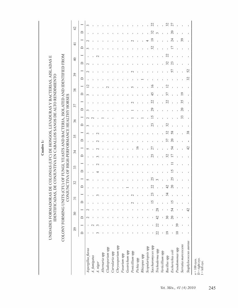
Mediante la siembra de muestras oculares en los medios de cultivo se lograron recuperar hongos, bacterias y levaduras entre los dos y siete días de inoculación. Los Cuadros 1a , 1b, 1c y 1d muestran el número total de unidades formadoras de colonias (ufc) de hongos, levaduras y bacterias encontradas en cada uno de los ojos de los caballos muestreados.
Ningún ojo fue negativo a presencia de microorganismos. El caballo 33 presentó el menor número, con 13 ufc aisladas del ojo izquierdo, y el caballo 34 presentó el mayor número, con 146 ufc aisladas en el ojo izquierdo. No se observó correlación positiva entre el número de hongos, levaduras y bacterias aisladas, la raza, el sexo o la edad del animal.
De los 50 individuos revisados se encontró que 100% fueron positivos a la presencia de hongos en ambos ojos. Los géneros recuperados fueron: Aspergillus spp (45.72 % del total de los caballos examinados), Trichoderma spp (27.28%), Penicillium spp (12.44%), Scopulariospsis spp (5.09%), Chrysosporium spp (2.69%), Fusarium spp (1.94%), Rhizopus spp (1.34%), Geotrichum spp (1.14%), Curvularia spp (1.04%), Alternaria spp (0.89%), Verticillium spp (0.74%) y Cladosporium spp (0.59%) (Cuadro 2). De los hongos, el género recuperado con mayor frecuencia fue Aspergillus spp, del cual se identificaron tres especies: A. flavus (24.28%), A. Niger (12.59%) y A. fumigatus (8.84%) (Cuadro 5). 60% (30/50) de los animales muestreados fueron positivos a la presencia de levaduras, de las cuales 90.27% fueron del género Saccharomyces spp y 9.72%, Pichia spp (Cuadro 3).
Respecto de las bacterias recuperadas, Staphylococcus aureus (35.41%) fue el microorganismo que se identificó con mayor frecuencia, mientras que Bacillus spp se presentó en 28.35%, Escherichia coli, 21.51%, Serratia marcensces, 13.71% y Pseudomonas, 1.005% (Cuadro 4). La totalidad de los caballos revisados fueron positivos a la presencia de bacterias en ambos ojos.

243Vet. Méx., 41 (4) 2010
Cua
dro
1a
UN
IDA
DES
FO
RM
AD
OR
AS
DE
CO
LON
IAS
(UFC
) DE
HO
NG
OS,
LEV
AD
UR
AS
Y B
AC
TER
IAS,
AIS
LAD
AS
E ID
ENTI
FIC
AD
AS,
DE
CO
NJU
NTI
VA E
N C
AB
ALL
OS
SAN
OS
DE
ALT
O R
END
IMIE
NTO
CO
LON
Y F
OR
MIN
G U
NIT
S (C
FU) O
F FU
NG
I, Y
EAST
S A
ND
BA
CTE
RIA
, ISO
LATE
D A
ND
IDEN
TIFI
ED F
RO
M
CO
NJU
NC
TIVA
OF
HIG
H-P
ERFO
RM
AN
CE
HEA
LTH
Y H
OR
SES
12
34
56
78
910
1112
1314
DI
DI
DI
DI
DI
DI
DI
DI
DI
DI
DI
DI
DI
DI
Aspe
rgill
us fl
avus
33
--
--
--
--
2-
--
--
33
-2
66
35
25
47
A. fu
mig
atus
--
--
--
23
--
23
--
--
--
--
33
2-
22
--
A. n
iger
--
--
13
--
--
--
25
21
-3
--
-2
1-
--
-5
Alte
rnar
ia sp
p-
-2
3-
--
--
--
--
--
--
--
--
--
--
--
-C
lado
spor
ium
spp
--
--
--
--
--
--
--
--
--
--
--
--
--
--
Cur
vula
ria
spp
--
--
--
--
--
--
--
--
--
--
--
--
--
--
Chr
ysos
pori
um sp
p-
--
--
-1
2-
-1
1-
-2
3-
-2
2-
--
--
--
-Fu
sari
um sp
p2
--
-1
--
--
--
--
--
--
--
--
--
--
--
-G
eotr
ichu
m sp
p-
--
--
--
--
--
--
--
--
--
--
--
--
--
-Pe
nici
llium
spp
--
22
--
--
32
24
--
--
--
44
--
-2
13
--
Pich
ia sp
p -
--
--
--
--
--
--
--
--
--
--
--
--
--
-Rh
izop
us sp
p-
--
--
--
--
--
--
--
--
--
--
--
-1
--
-Sc
opul
ario
psis
spp
--
--
--
1-
--
--
--
--
--
--
--
--
--
--
Sacc
haro
myc
es sp
p22
15-
-36
26-
2530
-23
3522
--
2325
25-
-15
--
--
23-
-Tr
icho
derm
a sp
p-
--
--
--
--
--
--
--
--
--
--
--
--
--
-Ve
rtic
illiu
m sp
p-
--
--
--
-2
3-
--
--
--
--
--
--
--
--
-Ba
cillu
s spp
32-
2022
--
3232
2132
3332
--
3233
33-
2232
--
32-
22-
3328
Esch
eric
hia
coli
-22
--
-20
--
--
--
-20
--
-20
--
20-
--
-20
--
Pseu
dom
onas
spp
--
--
--
--
--
--
--
--
--
--
--
--
--
--
Serr
atia
mar
cesc
ens
--
--
13-
--
--
20-
--
--
17-
--
-27
--
--
1717
Stap
hylo
cocc
us
aure
us-
52-
--
5454
--
--
-45
54-
--
5433
-54
3926
2754
54-
33
n =
100
eyes
.D
= ri
ght e
ye.
I = le
ft ey
e.

244
Cua
dro
1b
UN
IDA
DES
FO
RM
AD
OR
AS
DE
CO
LON
IAS
(UFC
) DE
HO
NG
OS,
LEV
AD
UR
AS
Y B
AC
TER
IAS,
AIS
LAD
AS
E ID
ENTI
FIC
AD
AS,
DE
CO
NJU
NTI
VA E
N C
AB
ALL
OS
SAN
OS
DE
ALT
O R
END
IMIE
NTO
CO
LON
Y F
OR
MIN
G U
NIT
S (C
FU) O
F FU
NG
I, Y
EAST
S A
ND
BA
CTE
RIA
, ISO
LATE
D A
ND
IDEN
TIFI
ED F
RO
M
CO
NJU
NC
TIVA
OF
HIG
H-P
ERFO
RM
AN
CE
HEA
LTH
Y H
OR
SES
1516
1718
1920
2122
2324
2526
2728
DI
DI
DI
DI
DI
DI
DI
DI
DI
DI
DI
DI
DI
DI
Aspe
rgill
us fl
avus
52
22
-3
--
-3
--
33
--
--
--
--
--
23
53
A. fu
mig
atus
--
--
35
--
--
32
25
--
-3
--
3-
--
--
--
A. n
iger
--
--
--
22
--
--
--
-5
32
3-
2-
-3
--
--
Alte
rnar
ia sp
p-
--
--
--
--
--
--
--
--
--
--
--
--
--
-C
lado
spor
ium
spp
--
--
--
--
--
--
--
--
--
-1
--
--
--
--
Cur
vula
ria
spp
--
--
--
--
--
--
--
--
--
--
--
--
--
--
Chr
ysos
pori
um sp
p-
--
--
--
-2
2-
--
--
--
--
--
--
--
--
-Fu
sari
um sp
p1
3-
--
--
-1
2-
--
-1
1-
--
--
--
--
--
-G
eotr
ichu
m sp
p-
--
--
--
--
--
--
--
--
--
--
--
--
--
-Pe
nici
llium
spp
--
-3
22
--
--
--
--
22
--
-2
--
--
--
32
Pich
ia sp
p -
--
--
--
--
--
-22
19-
--
15-
--
--
--
-32
28Rh
izop
us sp
p-
--
--
--
--
--
--
--
--
--
--
--
--
--
-Sc
opul
ario
psis
spp
11
--
--
--
--
--
--
--
--
--
--
--
--
--
Sacc
haro
myc
es sp
p19
2545
25-
-32
2518
15-
--
2535
2225
1532
22-
--
--
--
-Tr
icho
derm
a sp
p-
--
--
--
--
--
--
--
--
--
--
--
35-
25-
-Ve
rtic
illiu
m sp
p-
--
--
--
--
--
--
--
--
--
--
--
--
--
-Ba
cillu
s spp
-42
--
--
--
3022
32-
--
--
19-
2242
-30
--
3220
2217
Esch
eric
hia
coli
--
--
--
1420
--
--
1920
1633
--
--
23-
--
--
--
Pseu
dom
onas
spp
--
--
--
--
--
--
--
--
--
--
--
--
--
--
Serr
atia
mar
cesc
ens
29-
1727
18-
10-
22-
--
1717
920
1522
--
--
-37
--
--
Stap
hylo
cocc
us a
ureu
s-
--
--
45-
54-
4242
54-
54-
--
3333
-42
3534
--
--
-
n =
100
eyes
.D
= ri
ght e
ye.
I = le
ft ey
e.
245Vet. Méx., 41 (4) 2010
Cua
dro
1c
UN
IDA
DES
FO
RM
AD
OR
AS
DE
CO
LON
IAS
(UFC
) DE
HO
NG
OS,
LEV
AD
UR
AS
Y B
AC
TER
IAS,
AIS
LAD
AS
E ID
ENTI
FIC
AD
AS,
DE
CO
NJU
NTI
VA E
N C
AB
ALL
OS
SAN
OS
DE
ALT
O R
END
IMIE
NTO
CO
LON
Y F
OR
MIN
G U
NIT
S (C
FU) O
F FU
NG
I, Y
EAST
S A
ND
BA
CTE
RIA
, ISO
LATE
D A
ND
IDEN
TIFI
ED F
RO
M
CO
NJU
NC
TIVA
OF
HIG
H-P
ERFO
RM
AN
CE
HEA
LTH
Y H
OR
SES
2930
3132
3334
3536
3738
3940
4142
DI
DI
DI
DI
DI
DI
DI
DI
DI
DI
DI
DI
DI
DI
Aspe
rgill
us fl
avus
--
22
--
-5
--
-5
-3
32
3-
312
-2
2-
32
-3
A. fu
mig
atus
-2
--
--
--
--
--
--
-2
--
--
--
--
--
--
A. n
iger
33
--
-3
--
42
32
22
--
--
--
--
-2
--
--
Alte
rnar
ia sp
p-
--
--
--
--
--
--
--
1-
--
--
--
--
--
-C
lado
spor
ium
spp
--
--
--
--
--
--
--
--
--
-2
--
--
--
--
Cur
vula
ria
spp
--
--
--
--
--
--
--
--
--
--
--
--
--
--
Chr
ysos
pori
um sp
p-
--
--
--
--
--
--
--
--
--
--
--
--
--
Fusa
rium
spp
--
--
--
--
--
--
--
--
--
--
--
--
--
--
Geo
tric
hum
spp
--
--
--
1-
--
--
--
--
--
--
--
--
--
--
Peni
cilli
um sp
p-
--
-2
2-
--
--
--
--
11
2-
5-
2-
--
2-
-Pi
chia
spp
--
--
--
--
--
-16
--
--
--
--
--
--
--
--
Rhiz
opus
spp
--
--
--
--
--
--
--
-1
--
--
1-
--
--
--
Scop
ular
iops
is sp
p-
--
--
--
--
--
--
--
--
--
--
--
--
4-
-Sa
ccha
rom
yces
spp
--
--
1523
2225
--
2527
--
2315
2922
4516
--
--
3219
3222
Tric
hode
rma
spp
2222
4228
--
-3
--
--
--
--
--
--
--
--
--
3-
Vert
icill
ium
spp
--
--
--
--
--
--
--
--
--
--
--
--
--
--
Baci
llus s
pp19
-30
--
3442
--
-32
-37
3232
--
2237
12-
-32
22-
--
32Es
cher
ichi
a co
li-
-20
5415
-20
2515
1117
5420
58-
--
--
--
5723
-17
2420
27Ps
eudo
mon
as sp
p10
39-
--
--
--
--
--
--
--
--
--
--
--
--
-Se
rrat
ia m
arce
scen
s-
--
--
--
--
--
--
--
3320
3519
--
--
--
39-
-
Stap
hylo
cocc
us a
ureu
s-
--
42-
--
--
--
42-
38-
--
--
-32
52-
--
--
-
n =
100
eyes
.D
= ri
ght e
ye.
I = le
ft ey
e.

246
Cuadro 1d
UNIDADES FORMADORAS DE COLONIAS (UFC) DE HONGOS, LEVADURAS Y BACTERIAS, AISLADAS E IDENTIFICADAS, DE CONJUNTIVA EN CABALLOS
SANOS DE ALTO RENDIMIENTO
COLONY FORMING UNITS (CFU) OF FUNGI, YEASTS AND BACTERIA, ISOLATED AND IDENTIFIED FROM CONJUNCTIVA OF HIGH-PERFORMANCE
HEALTHY HORSES
43 44 45 46 47 48 49 50
D I D I D I D I D I D I D I D IAspergillus flavus 1 4 - - 3 - - - - 2 - - - 3 4 3A. fumigatus - 5 - - - 2 - - - - - - - - - -A. niger - - - - - 2 - - - - 4 2 - - 3Alternaria spp - - - - - - - - - - - - - - - -Cladosporium spp - - - 1 - - - - - - - - - - - -Curvularia spp - 3 - - - - - 1 1 1 - - 1 - - -Chrysosporium spp - - - - - - - - - - - - - - - -Fusarium spp - - - - 1 - - - - - - - - - -Geotrichum spp - - - - - - - - - - - - - - - -Penicillium spp - - 3 2 - - - - - 2 2 2 2 4 2 -Pichia spp - - - - - - - - - - - - - - - -Rhizopus spp - - 2 - - - 2 1 - - - 1 - - - -Scopulariopsis spp - 5 - - - - - 22 - - - - - - - -Saccharomyces spp - - 23 35 - - - - - - 25 - - - 25 -Trichoderma spp - - - - - - - - - - 2 - - - - -Verticillium spp - - - - - - - - - - - - - - - -Bacillus spp - 32 - - 22 - - 32 - - - - - - - 27Escherichia coli - - - 49 22 54 - - - 53 - 54 27 45 - -Pseudomonas spp - - - - - - - - - - - - - - - -Serratia marcescens - - - - - - 20 28 - - 22 42 - - 17 22Staphylococcus aureus 32 45 30 48 - 42 - - 42 45 - - 30 32 30 42
n = 100 eyes.D = right eye.I = left eye.
catheters and also in bovine mastitis;29 in horses as the etiologic agent of endocarditis,30 sepsis,31 located in abdominal abscesses,32 in fetal membranes and internal organs of aborted fetuses.33 In this research Serratia marcescens was isolated from 21 healthy horses (42%) ranging from nine to 42 colony forming units (cfu). The genera Staphylococcus and Bacillus spp are the microorganisms most frequently reported as normal flora of the conjunctival sac. Even though these bacteria defend the eye from invasive microorganisms, some of them act as opportunistic pathogens, causing damage to the ocular system.13, 34
Most studies on conjunctival microflora mention the predominance of Gram-positive bacteria, previously identified in parrots, 35 exotic and prey birds, 6,36 dogs,37 cats,38 pigs,5 Asian elephants10 and horses.13 Despite
Discusión
Este estudio registra, por primera vez, la presencia de Serratia marcescens en la microflora normal conjuntival en caballos sanos. Aunque esta bacteria Gram negativa se encuentra comúnmente en ambientes naturales, tierra, agua y vegetales crudos, es considerada patógeno resistente a múltiples fármacos y microorganismo oportunista, especialmente en humanos inmunocomprometidos.23 Es reconocida como agente causal de endoftalmitis24, 25 y queratitis infecciosa26 en humanos. Se cree que tiene la capacidad de liberar proteasas, causantes de pérdida de proteoglicanos en córnea, de dispersión de las fibras de colágeno, necrosis licuefactiva y perforación corneal.27

247Vet. Méx., 41 (4) 2010
this predominance in conjunctiva of both healthy and diseased animals, Gram-negative play an important role in corneal lesions. Pseudomonas spp and some Enterobacteriaceae, such as E. coli, are typically associated with rapid and progressive corneal ulceration.39,
40 In this study these bacteria represent 1.00% and 21.51%, respectively. Aspergillus spp, Penicillium spp, Chrysosporium spp and Fusarium spp fungal genera are usually recovered from the conjunctiva in horses, and are considered as normal in many animal species.13,14,41-43 The results of this study are consistent with previous studies, where the predominant genus was Aspergillus, followed by Trichoderma, Penicillium, Scopulariopsis and Fusarium.
The genus Aspergillus is frequently found as fungal contaminant in different species and is considered among the main etiological agents (secondary infection) present in horse keratomycosis. It was the most abundant and commonly isolated fungus in this work (45.72%), which is consistent with reports by Andrew et al.13 (25.7%), Rosa et al.14 (32.2%), Barsotti et al.42 (33%) and Nardoni et al.12 (33%).
In Monterrey, Mexico, humidity and temperature conditions are suitable for proper sporulation of fungi
Serratia marcescens rara vez se localiza en animales; sin embargo, se tiene registro de ella como agente causal de enfermedades nosocomiales en perros y gatos28 a partir de catéteres intravenosos infectados y de mastitis bovina;29 y en equinos como agente etiológico de endocarditis,30 de sepsis,31 localizado en abscesos abdominales,32 en membranas fetales y órganos internos de fetos abortados.33 En esta investigación la Serratia marcescens se logró aislar de 21 caballos sanos (42%) con rango de nueve a 42 unidades formadoras de colonias (ufc). Los géneros Staphylococcus spp y Bacillus spp son los microorganismos que más se registraron como microflora normal del saco conjuntival. A pesar de que estas bacterias defienden el ojo de microorganismos invasivos, algunas de ellas actúan como patógenos oportunistas, causando daños en el sistema ocular.13,34
La mayoría de los estudios sobre microflora
Cuadro 2
PORCENTAJE DEL TOTAL DE UNIDADES FORMADORAS DE COLONIAS (UFC) DE HONGOS, AISLADOS DE AMBOS OJOS (DERECHO E IZQUIERDO) DE CABALLOS SANOS DE ALTO
RENDIMIENTO
TOTAL PERCENTAGE OF FUNGAL COLONY FORMING UNITS (CFU), ISOLATED FROM BOTH EYES (LEFT AND RIGHT) FROM HIGH-
PERFORMANCE HEALTHY HORSES
Strains Num. cfu %
Aspergillus flavus 162 24.287A. fumigatus 59 8.845A. niger 84 12.593Alternaria spp 6 0.899Cladosporium spp 4 0.599Curvularia spp 7 1.049Chrysosporium spp 18 2.698Fusarium spp 13 1.949Geotrichum spp 1 1.149Penicillium spp 83 12.443Rhizopus spp 9 1.349Scopulariopsis spp 34 5.097Trichoderma spp 182 27.286Verticillium spp 5 0.749
n = 667 cfu.
Cuadro 3
PORCENTAJE TOTAL DE UNIDADES FORMADORAS DE COLONIAS (UFC) DE LEVADURAS, AISLADAS DE AMBOS OJOS (DERECHO E IZQUIERDO) DE
CABALLOS SANOS DE ALTO RENDIMIENTO.
TOTAL PERCENTAGE OF COLONY FORMING UNITS (CFU) OF YEASTS ISOLATED FROM BOTH EYES (RIGHT AND LEFT) OF HIGH-PERFORMANCE
HEALTHY HORSES.
Strains Num. cfu %Saccharomyces spp 1 225 90.272Pichia spp 132 9.727
Cuadro 4
PORCENTAJE DEL TOTAL DE UNIDADES FORMADORAS DE COLONIAS (UFC) DE BACTERIAS, AISLADAS DE AMBOS OJOS (DERECHO E IZQUIERDO) DE CABALLOS SANOS
DE ALTO RENDIMIENTO
TOTAL PERCENTAGE OF BACTERIAL COLONY FORMING UNITS (CFU), ISOLATED FROM BOTH EYES (LEFT AND RIGHT) OF HIGH-PERFORMANCE
HEALTHY HORSES
Strains Num. cfu %
Bacillus spp 1,381 28.351Escherichia coli 1,048 21.515Serratia marcescens 668 13.713Staphylococcus aureus 1,725 35.413Pseudomonas spp 49 1.005
n = 4 871 cfu.

248
conjuntival mencionan la presencia predominante de las bacterias Gram positivas, anteriormente señaladas, en loros,35 aves exóticas y de presa,6,36 perros,37 gatos,38 cerdos,5 elefantes asiáticos10 y caballos.13 Pese a esta predominancia en conjuntiva, tanto en animales sanos como enfermos, las Gram negativas se presentan de manera relevante en lesiones corneales. Pseudomonas spp y algunas enterobacterias como E. coli son asociadas típicamente con una rápida y progresiva ulceración corneal;39, 40 en este estudio dichas bacterias representan 1.00% y 21.51%, respectivamente. Los géneros Aspergillus spp, Penicillium spp, Chrysosporium spp y Fusarium spp son hongos usualmente recuperados de la conjuntiva en caballos, y se consideran normales en muchas especies animales.13,14,41-43 Los resultados del presente estudio coinciden con los de estudios previos, donde el género predominante fue Aspergillus, seguido por Trichoderma, Penicillium, Scopulariopsis y Fusarium.
El género Aspergillus se encuentra de manera frecuente como contaminante fúngico en diferentes especies, y se considera dentro de los principales agentes etiológicos (de infección secundaria) presentes en queratomicosis del caballo; En este trabajo fue el hongo más abundante y comúnmente aislado (45.72%), dato que coincide con los reportes de Andrew et al.13 (25.7%), Rosa et al.14 (32.2%), Barsotti et al.42 (33%) y Nardoni et al.12 (33%).
En Monterrey, México, las condiciones de humedad y temperatura son aptas para una adecuada esporulación de los hongos y su subsecuente diseminación en el medio ambiente, también favorecen la presencia de levaduras. En este contexto, los resultados de estudios similares podrían variar debido a las condiciones sanitarias en la que los animales son mantenidos, las condiciones climáticas y la época del año.
Con base en este estudio, se sugiere ampliar el perfil de exámenes donde se incluya la identificación de Serratia marcescens, organismo con multirresistencia a fármacos, y patógeno importante, virulento y habitual de la conjuntiva normal en equinos.
El conocimiento de los géneros y especies prevalentes son importantes para seleccionar agentes antimicrobianos efectivos, establecer medidas adecuadas de profilaxis, y prescribir tratamiento de enfermedades oculares en caballos.
and its subsequent dissemination in the environment; the presence of yeast is also favored. In this context, the results of similar studies may vary due to health conditions in which animals are kept, weather and season.
Based on this study, it is suggested to increase the test profiles including identification of Serratia marcescens, a microorganism with multiple resistance to drugs and important pathogen, virulent and common in the normal conjunctiva in horses.
Knowledge of the prevalent genera and species are important for selecting effective antimicrobial agents, to establish appropriate prophylaxis measures, and prescribe treatment for eye disease in horses.
Referencias
1.
2.
3.
4.
5.
6.
7.
8.
9.
10.
11.
12.
13.
BURNS RP. Indigenous flora of the lids and conjunctiva. In: TASMAN W, JAEGER EA, editors. Foundations of clinical ophthalmology. Vol 2. Philadelphia:. Lippincott Press, 1993:1-5.SPRADBROW PB. The bacterial flora of the ovine conjunctival sac. Aust Vet J 1968; 44:117-118.SAMUELSON DA, ANDRESEN TL, GWIN RM. Conjunctival fungal flora in horses, cattle, dogs and cats. J Am Vet Med Assoc 1984; 184:1240-1242.WILCOX GE. Bacterial flora of the bovine eye with special reference to the Moraxella and Neisseria. Aust Vet J1970; 46:253-256.DAVISON HJ, ROGERS DP, YEARY TJ, STONE GG, SCHONEWESIS DA, CHENGAPPA MM. Conjunctival microbial flora of clinically normal pigs. Am J Vet Res 1994; 55:949-951.DUPONT C, CARRIER M, HIGGINS R. Bacterial and fungal in healthy eyes of birds of pray. Can Vet J 1994; 34:699-701.COOPER SC, MCLELLAN GJ, REYCROFT AN. Conjunctival flora observed in 70 healthy domestic rabbits. Vet Rec 2001; 149:232-235.MUSHI EZ, BINTA MG, CHAVO RG, DINTWE K. Conjunctival flora of fifty healthy goats in Sebele farm, Gaborone, Botswana. J Anim Vet Adv 2007; 6:1388-1389.KUDIRKIENÈ E, ZILINSKA H, SIUGZDAITE J. Microbial flora of the dog eyes. Lithuanian Vet Acad 2006; 34:18-21.TUNTIVANICH P, SOONTORNIPAVRT K, TUNTIVANICH N, WONGAUMNUAYKUL S, BRIKSAWAN P. Conjunctival microflora in clinically normal Asian elephant in Thailand. Vet Res Comm 2002; 26:251-254.SHEWEN PE, POVERY RC, WILSON MR. A survey of conjunctival flora of clinically normal cats and cats with conjunctivitis. Can Vet J 1980; 21:231-233.NARDONI S, SGORBINI M, BARSOTTI M, CORAZZA G, MANCIATTI F. Conjunctival fungal flora in healthy donkeys. Vet Ophthalmol 2007; 10:207-210.ANDREW SE, NGUYEN A, JONES GL, BROOKS DE. Seasonal effects on the aerobic bacterial and fungal
conjunctival flora of normal thoroughbred brood mares in Florida. Vet Ophthalmol 2003; 6:45-50.ROSA M, CARDOZO LM, PEREIRA JDAS, BROOKS DE, MARTINS ALB, FLORIDO PSS et al. Fungal flora of normal eyes of healthy horses from the state of Rio de Janeiro, Brazil. Vet Ophthalmol 2003; 6:51-55.MOORE C, HELLER N, MAJORS LJ, WHITLEY RD, BURGUÉS EC, WEBER J. Prevalence of ocular microorganisms in hospitalized and stabled horses. Am J Vet Res 1988; 49:773-777.
14.
15.

249Vet. Méx., 41 (4) 2010
GELATT KN. Fundamentos de Oftalmología Veterinaria. Barcelona: MASSON SA, 2003.ARGUESO P. Azúcares: Una capa protectora excepcional de la superficie ocular. Arch Soc Esp Oftalmol 2008; 83:287-290.MARTOS PG, FERNÁNDEZ DEL BARRIO MT, SALIDO FPF. Microbiología Clínica Aplicada. 3a ed. Madrid, España: Ed. Díaz de Santos SA, 1997.KONEMAN EW, ALLEN SD, DOWELL VR, SOMMERS HM. Diagnóstico microbiológico. Madrid, España: Ed. Médica Panamericana SA, 1989.BARNETT H, HUNTER BB. Illustrated genera of imperfect fungi. 3rd ed. Minneapolis Minnesota USA: Burgess Publishing Company, 1972.FISHER F, COOK NB. Fundamental of Diagnostic Mycology. Pennsylvania: WB Saunders Company, 1998.DE HOOG GS, GUARRO J, GENÉ J, FIGUERAS MJ. Atlas of Clinical Fungi. 2nd ed. Centralbureau voor Schimmelcultures, Utrecht, The Netherlands/Universitat Rovira i Virgili, Reus, Spain, 2000.RALLIS E, KARANIKOLA E, PAPADAKIS P. Severe facial infection caused by Serratia marcescens in an immunocompetent soldier. J Am Acad Dermatol 2008; 58:109-110.EQUI RA, GREEN WR. Endogenous Serratia marcescens endophthalmitis with dark hypopyon: a case report and review. Surv Ophthalmol 2001; 46:259-268. SHARMA NS, OOI JL, DOWNIE JA, CORONEO MT. Corneal perforation and intraocular lens prolapsed in Serratia marscescens endophthalmitis. Clin Experiment Ophthalmol 2007; 35:381-382.ALEXANDRAKIS G, ALFONSO EC, MILLAR D. Shifting trends in bacterial keratitis in south Florida and emerging resistance to fluoroquinolones. Ophthalmology 2000; 107:1497-1502.LYERLY D, GRAY L, KREGER A. Characterization of rabbit corneal damage produced by Serratia keratitis and by Serratia protease. Infect Immun 1981; 33:927-932.FOX JM, BEAUCAGE CM, FOLTA CA, TROTON GW. Nosocomial transmission of Serratia marcescens in a veterinary hospital due to contamination by benzalkonium chloride. J Clin Microbiol 1981; 14:157-160.KAMARUDIN MI, FOX LK, GASKINS CT, GAY JM. Environmental reservoirs for Serratia marscescens intramammary infections in dairy cows. J Am Vet Med Assoc 1966; 208:555-558.EWART S, BROWN C, DERKSEN F, KUFUOR-MENSA E. Serratia marcescens endocarditis in a horse. J Am Vet Med Assoc 1992; 200:961-963.
SHAFTOE S. Serratia marscescens septicaemia in a neonatal Arabian foal. Equine Vet J 1984; 16:389-392.RIGG DL, GARTLIN SJ, REINERTSON EL. Marsu-pialization of an abdominal abscess caused by Serratia marscescens in a mare. J Am Vet Med Assoc 1987; 191:222-224.JORES J, BEUTER G, HIRTH-SCHMIDT I, BORSHERS K, PITT TL, LUBKE-BECKER A. Isolation of Serratia marcescens from an equine abortion in Germay. Vet Rec 2004; 154:242-244.GEMENSKY-METZLER AJ, WILKIE DA, KOWALSKI JJ, SCHMALL LM, WILLIS AM, YAMAGATA M. Changes in bacterial and fungal ocular flora of clinically normal horses following experimental application of topical antimicrobial or antimicrobial-corticosteroid ophthalmic preparations. Am J Vet Res 2005; 66:800-811.ZENOBLE RD, GRIFFITH RW, CLUBB SL. Survey of bacteriologic flora of conjunctiva and cornea in healthy psittacine birds. Am J Vet Res 1983; 44:1966-1967.WOLF ED, AMASS K, OLSEN J. Survey of conjunctival flora in the eye of clinically normal captive exotic birds. J Am Med Vet Assoc 1983; 183:1232-1233.MCDONALD PJ, WATSON DJ. Microbial flora of normal canine conjunctivae. J Small Anim Pract 1976; 17:809-812.ESPINOLA MB, LILENBAUM W. Prevalence of bacteria in the conjunctival sac and on the eyelid margin of clinically normal cats. J Small Anim Pract 1996; 37:364-366.BARNETT KC, CRISPIN SM, LAVACH JD, MATTHEWS AG. Equine ophthalmology. 2nd ed. Philadelphia, USA: Elsevier Limited, 2004.SANSON J, FEATHERSTONE H, BARNETT KC. Keratomycosis in six horses in the United Kindgdom. Vet Rec 2005; 156:13-17.MATTHEWS AG. The aethiopatogenesis of infectious keratitis in the horse. Equine Vet J 1994; 26:432-433.BARSOTTI G, SGORBINI M, NARDONI S, CORAZZA M, MANCIANTI F. Occurrence of fungi from conjunctiva of healthy horses in Tuscany, Italy. Vet Res Commun 2006; 30: 903-906.WESTEIN WL, MOORE PA, SANCHEZ S, DIETRICH UM, WOOLEY RE, RITCHIE BW. In vitro efficacy of a buffered chelating solution as an antimicrobial potentiator for antifungal drugs against pathogens obtained from horses with mycotic keratitis. Am J Vet Res 2006; 67:562-568.
31.
32.
33.
34.
35.
36.
37.
38.
39.
40.
41.
42.
43.
16.
17.
18.
19.
20.
21.
22.
23.
24.
25.
26.
27.
28.
29.
30.