N-ACETILGLUTAMATO SINTASA - digital.csic.esdigital.csic.es/bitstream/10261/74367/1/Tesis E...
155
Transcript of N-ACETILGLUTAMATO SINTASA - digital.csic.esdigital.csic.es/bitstream/10261/74367/1/Tesis E...


Departamento de Bioquímica y Biología Molecular
030 G Señalización celular y patologías asociadas
N-ACETILGLUTAMATO SINTASA:
CORRELACIONES ESTRUCTURA-FUNCIÓN
Memoria presentada por
Enea Sancho Vaello
Para optar al grado de Doctor
Director:
Dr. Vicente Rubio Zamora
Codirectora:
Dra. Leonor Fernández-Murga
Laboratorio de Enzimopatología Estructural
Departamento de Genómica y Proteómica
Instituto de Biomedicina de Valencia
Consejo Superior de Investigaciones Científicas
Valencia, a 18 de Enero de 2013
INSTITUTO DE BIOMEDICINA DE VALENCIA


Vicente Rubio Zamora, Doctor en Medicina, Profesor de Investigación del Consejo Superior de Investigaciones CDoctora en Bioquímica, Investigadora Contratada en la Unidad de Investigación del Hospital Dr. Peset, Fundación para la Investigación Sanitaria y Biomédica de la Comunidad Valenciana (FISABIOEstructural de este Instituto CERTIFICAN 1. Que Dª. Enea Sancho Vaello, Licenciada en Biología por la Universidad de Valencia, ha realizado bajo su dirección el trabajo de Tesis Doctoral que lleva por título "Nacetilglutamato sintetasa: correlaciones estructura 2. Que revisado el trabajo, expresadefensa frente al Tribunal correspondiente, ya que consideran que la presentación del mismo reúne los requisitos necesarios para optar al grado de Doctor. 3. Que habiéndose realizado la Memopublicaciones, siendo la doctoranda la primera firmante coautores de dichas publicaciones, declarantrabajo experimental, participde los resultados, extracción de las publicados. 4. Que ninguna de las publicaciones compendiadas en esta Tesis utilizada por otros para la re 5. Que los indicadores de calidad de las publicaciones recogidas en esta tesis son los siguientes:
• FEBS Letters (2 publicaciones) íentre los 290 títulos incluidos en el campoMolecular; y con posición 21 entre los 74 títulos incluidos en el campo de la Biofísica.
• Journal of Bacteriology, con índice de impacto 3,825 y posición 29 entre las 114
revistas incluidas en el campo de la Microbiología Valencia, a 17 de enero de 2013 Fdo. Vicente Rubio Zamora
Vicente Rubio Zamora, Doctor en Medicina, Profesor de Investigación del Consejo Superior de Investigaciones Científicas, y María Leonor Fernández-Murga Chavanne,
Investigadora Contratada en la Unidad de Investigación del Fundación para la Investigación Sanitaria y Biomédica de la
FISABIO), y colaboradora en la Unidad de Enzimopatología Estructural de este Instituto,
1. Que Dª. Enea Sancho Vaello, Licenciada en Biología por la Universidad de Valencia, dirección el trabajo de Tesis Doctoral que lleva por título "N
acetilglutamato sintetasa: correlaciones estructura-función."
el trabajo, expresan su conformidad para que éste sea sometido a defensa frente al Tribunal correspondiente, ya que consideran que la presentación del mismo reúne los requisitos necesarios para optar al grado de Doctor.
3. Que habiéndose realizado la Memoria de Tesis por la modalidad de compendio de siendo la doctoranda la primera firmante y siendo ellos los otros
aciones, declaran que la doctoranda ha sido responsable del trabajo experimental, participando interactivamente con ellos en su diseño, en el análisis
resultados, extracción de las conclusiones, y en la redacción de los trabajos
4. Que ninguna de las publicaciones compendiadas en esta Tesis ha sido ni utilizada por otros para la realización de una tesis doctoral.
5. Que los indicadores de calidad de las publicaciones recogidas en esta tesis son los
FEBS Letters (2 publicaciones) índice de impacto 3,538, con una posición 102 entre los 290 títulos incluidos en el campo de la Bioquímica y Biología Molecular; y con posición 21 entre los 74 títulos incluidos en el campo de la
Journal of Bacteriology, con índice de impacto 3,825 y posición 29 entre las 114 revistas incluidas en el campo de la Microbiología.
encia, a 17 de enero de 2013
Fdo. Vicente Rubio Zamora Mª Leonor Fernández-Murga Chavanne
C/ JAUME ROIG, 11 46010 VALENCIA ESPAÑATEL.: 96 339 17 60
FAX: 96 369 08 00
Vicente Rubio Zamora, Doctor en Medicina, Profesor de Investigación del Consejo urga Chavanne,
Investigadora Contratada en la Unidad de Investigación del Fundación para la Investigación Sanitaria y Biomédica de la
en la Unidad de Enzimopatología
1. Que Dª. Enea Sancho Vaello, Licenciada en Biología por la Universidad de Valencia, dirección el trabajo de Tesis Doctoral que lleva por título "N-
conformidad para que éste sea sometido a defensa frente al Tribunal correspondiente, ya que consideran que la presentación del
compendio de os los otros
que la doctoranda ha sido responsable del os en su diseño, en el análisis
conclusiones, y en la redacción de los trabajos
ha sido ni será
5. Que los indicadores de calidad de las publicaciones recogidas en esta tesis son los
ndice de impacto 3,538, con una posición 102 de la Bioquímica y Biología
Molecular; y con posición 21 entre los 74 títulos incluidos en el campo de la
Journal of Bacteriology, con índice de impacto 3,825 y posición 29 entre las 114
Murga Chavanne


Para la realización de esta Tesis, Enea Sancho Vaello ha disfrutado de una Beca lanzadera del
CIBER de enfermedades raras (CIBERer) y de una Ayuda predoctoral de formación en
investigación en salud del Instituto de Salud Carlos III.
El trabajo se ha enmarcado dentro de los proyectos: “Complejos macromoleculares, proteínas
multifuncionales, pluriempleo y enfermedades raras en la familia aminoácido quinasa”
(BFU2008-05021) financiado por el Ministerio de Ciencia e Innovación, “Bases estructurales
de enfermedades raras metabólicas, endovasculares y de la hemostasia", financiado por el
CIBERER-Instituto de Salud Carlos III y el Ministerio de Sanidad y Consumo y
“Caracterización molecular de la patogénesis de los errores del ciclo de la urea: déficit de la
acetilglutamato sintasa” (AP-082/10) financiado por la Conselleria de Sanitat- Generalitat
Valenciana.
El interés del grupo en la N-acetilglutamato sintasa se relaciona directamente con las
patologías del ciclo de la urea que constituyen una de sus líneas de actuación dentro del
CIBERER-ISCIII.


AGRADECIMIENTOS
Agradecer siempre es difícil, sobre todo, cuando se agradece algo tan inmaterial como el
apoyo. Por eso, es ardua tarea no dejarse a ninguna de las personas que, aunque sólo haya sido a través
de una ligera conversación de máquina de café, me ha dado ánimos para continuar.
Para empezar, quiero agradecer a mi director de tesis, el Prof. Vicente Rubio, por el trozo de
poyata que me cedió durante unos años en los que aprendí mucho tanto científica como humanamente.
He aprendido mucho observando tu actitud hacia la ciencia. Gracias.
A mi codirectora y amiga, la Dra. Leonor Fernández-Murga por todo lo que me ha enseñado
(que ha sido mucho) y por infundirme la idea de “siempre tener un plan B”.
Por supuesto, agradezco a todos mis compañeros y amigos: Fer, Sergio, Mariano, Nadine, Jose
Luis, Juanma, Clara, Alicia y Carles. Os agradezco esas interesantes conversaciones científicas en
Rectorado y esas teorías que de ellas surgieron (tensión-distensión, grifos de leche). Os agradezco
todos esos momentos tanto fuera como dentro del laboratorio. Por supuesto, agradezco a Elena el
dejarme perseguirla al llegar al laboratorio y el enseñarme tanto con su actitud frente a la vida.
Agradezco a los que pasaron por el laboratorio pero conocí menos tiempo de lo que me habría
gustado: Sandra, Santi, Angela I, Angela II, Lorena, Ana, Irene, Jose, Nadia, Josep, Blanca, Belén y
Carmen.
A mis compañeros del IBV, especialmente a mis amigos Marian, Jordi, Paula y Gema, tanto
por la ayuda científica como por los buenos momentos no científicos (que fueron muchos). Agradezco
también al resto del personal del IBV, siempre dispuesto a ayudar: a investigadores, becarios, técnicos,
al personal de limpieza (a Luisa), mantenimiento, administración, seguridad, biblioteca, informática…
A todos mis amigos, desperdigados por el mundo, pero con una intensa conexión mental en un
día como hoy. Agradezco la alegría contagiosa de mi pack.
A mi familia y a Alberto (por esa alegría que despide al pasearse por la vida y por sus consejos
técnicos).
A mi madre por su incondicionalidad, por animarme siempre con sus trasvases de energía y
por confiar en que podía hacerlo. A mi padre porque, a través de los años, sigue dándome fuerza desde
donde quiera que esté.
Y, por supuesto, a Hibai, por prestarme su diván y escuchar todas mis inquietudes, por todas
esas conversaciones divagantes y por darme cuenta día tras día de la suerte que tengo de haberme
cruzado con él.
Esta tesis te la dedico a TI…

ABREVIATURAS NO USUALES
AAK: Dominio aminoácido quinasa
AcCoA: acetil coenzima A
BSA: Albúmina sérica bovina
CPS: Carbamoilfosfato sintetasa
DTNB: Reactivo de Ellman o 5,5'-ditiobis(2-ácido nitrobenzoico)
DTT: Ditiotreitol o 1,4-ditio-D-treitol
EcNAGS: NAGS de Escherichia coli
EDTA: Ácido etilendiaminotetraacético
FPLC: Una modalidad de cromatografía líquida para proteínas, de alta velocidad
Gcn5: Histona acetiltransferasa de levadura
GNAT: Dominio acetiltransferasa (familia de N-acetiltransferasas relacionadas con Gcn5)
HuNAGS: NAGS humana
IPTG: Isopropil-β-D-1-tiogalactopiranósido
MBP: Maltose binding protein o proteína de unión a maltosa
NAG: N-acetilglutamato o N-acetil-L-glutamato
NAGK: N-acetilglutamato quinasa
NAGS: N-acetilglutamato sintasa
NAGSD: Deficiencia en NAGS
NCG: Ácido N-carbamoil-L-glutámico
NgNAGS: NAGS de Neisseria gonorrhoeae
PaAAK: Dominio AAK de la NAGS de Pseudomonas aeruginosa
PaNAGS: NAGS de Pseudomonas aeruginosa
PCR: Reacción en cadena de la polimerasa (Polymerase Chain Reaction)
PDB: Protein data bank (http://www.rcsb.org/pdb/home/home.do)
PFAM: Data base of protein families (http://pfam.sanger.ac.uk)
SDS-PAGE: Electroforesis en gel de poliacrilamida en condiciones desnaturalizantes
SUMO: Small ubiquitin-like modifier
TIGR: The Institute for Genomic Research
(http://cmr.jcvi.org/tigr-scripts/CMR/CmrHomePage.cgi)


Índice General
Introducción general ....................................................................................................... 1
1. Breve contexto histórico ............................................................................................ 3
2. Las funciones biológicas de la NAGS ....................................................................... 4
3. La NAGS es una enzima regulada por arginina ........................................................ 6
4. La NAGS está implicada en patología humana ......................................................... 7
5. La pertenencia de la NAGS a la familia aminoácido quinasa ha propiciado el desarrollo de este trabajo de tesis ............................................................................ 10
6. El investigar la naturaleza de la NAGS humana y de su déficit ha motivado también este trabajo de tesis ........................................................................................ 18
7. La estructura de esta tesis ........................................................................................ 20
Resultados ...................................................................................................................... 23
Capítulo 1 Site-directed mutagenesis studies of acetylglutamate synthase delineate the site for the arginine inhibitor ................................................................ 25
Capítulo 2 Mechanism of arginine regulation of acetylglutamate synthase, the first enzyme of arginine synthesis ......................................................................... 39
Capítulo 3 Functional dissection of N-acetylglutamate sythase (Arg-A) of pseudomonas aeruginosa and restoration of its ancestral N-acetylglutamate kinase activity ............................................................................... 53
Resumen de los resultados ............................................................................................ 89
Discusión general ......................................................................................................... 101
El origen de Arg A ..................................................................................................... 103
Regulación por arginina ............................................................................................. 114
Nota final sobre nuestra contribución al estudio de la NAGS humana .................... 121
Conclusiones ................................................................................................................ 125
Bibliografía correspondiente a la Introducción general, Resumen de los resultados y Discusión general ........................................................ 131


1
Introducción general

2

Introducción general
3
1. Breve contexto histórico
Esta tesis estudia experimentalmente la N-acetil-L-glutamato sintasa (NAGS), enzima
clave para la biosíntesis de arginina y para el control de la misma en casi cualquier especie.
La síntesis de N-acetil-L-glutamato (NAG) a partir de acetil coenzima A (AcCoA) y de L-
glutamato, como un primer paso en la biosíntesis de arginina, se demostró en los años 50 del
siglo pasado en preparaciones crudas de Clostridium kluiveri (1) y de Escherichia coli (2),
estableciéndose muy pronto que esta actividad era inhibida por arginina. En los años 60-70 se
identificaron y localizaron los genes biosintéticos de arginina en el cromosoma de E. coli
incluyendo el gen argA (3), responsable de la biosíntesis de NAG, pero hubo que esperar
hasta los años 70 para obtener las primeras preparaciones homogéneas de NAGSs bacterianas
purificadas (NAGSs a las que llamaremos también ArgA por el nombre de su gen), de E. coli
(4,5) y de Pseudomonas aeruginosa (6,7), en parte por la gran inestabilidad de la enzima. La
secuencia del gen argA de E. coli se determinó en 1987 (8) y fue la base para la identificación
ulterior de dicho gen en muchas otras especies bacterianas y en plantas.
Entre tanto, en los años 50 se estableció que el NAG es un activador esencial de la
carbamil fosfato sintetasa I (CPS I) implicada en la biosíntesis de urea (9). El grupo de
Masamiti Tatibana identificó a comienzos de los años 70, en mitocondria hepática de rata y
ratón, una actividad NAGS responsable de la producción de NAG para la activación de la
CPS I, demostrando la activación de esta NAGS por arginina (10). Este enzima se purificó a
homogeneidad primero a partir de hígado humano, en 1982 (11), y casi a la vez a partir de
hígado de rata (12). La purificación a partir de hígado de rata obligó a tomar extraordinarias
precauciones, como la utilización de tubos siliconados, debido a la inestabilidad del enzima, a
pesar de lo cual sólo se pudieron obtener unos 20 µg de enzima a partir de un kg de hígado de
rata (12). La producción de la NAGS humana dio mejor resultado (11), aunque también en
este caso el enzima era inestable (C. Bachmann, comunicación personal). Hubo que esperar
Introducción general
4
muchos años, hasta 2002, para identificar el gen que codifica la NAGS en ratón y en el ser
humano (13,14). Entre las razones que llevaron a este retraso hay que citar la tardía
determinación de la secuencia del genoma entero de animales y el escaso grado de identidad
de secuencia entre la NAGS animal y las bacterianas, a pesar de que hoy sabemos que son
homólogas en casi toda su extensión (15). La NAGS de E. coli o de P. aeruginosa y la NAGS
de rata o humana han sido durante muchos años las únicas NAGSs caracterizadas. Nuestro
trabajo se ha centrado en ellas, aunque por razones que se exponen al final de la discusión, la
sección de resultados de esta tesis se restringe en su parte experimental (aunque no en su
interés) a la NAGS bacteriana clásica de P. aeruginosa.
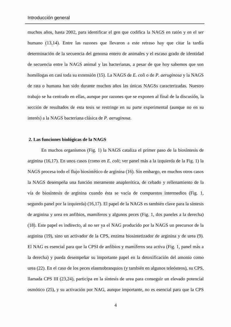
2. Las funciones biológicas de la NAGS
En muchos organismos (Fig. 1) la NAGS cataliza el primer paso de la biosíntesis de
arginina (16,17). En unos casos (como en E. coli; ver panel más a la izquierda de la Fig. 1) la
NAGS procesa todo el flujo biosintético de arginina (16). Sin embargo, en muchos otros casos
la NAGS desempeña una función meramente anaplerótica, de cebado y rellenamiento de la
vía de biosíntesis de arginina cuando ésta se vacía de compuestos intermedios (Fig. 1,
segundo panel por la izquierda) (16,17). El papel de la NAGS es también clave para la síntesis
de arginina y urea en anfibios, mamíferos y algunos peces (Fig. 1, dos paneles a la derecha)
(18). Este papel es indirecto, al no ser ya el NAG producido por la NAGS un precursor de la
arginina (19), sino un activador de la CPS, enzima biosintetizador de arginina y de urea (9).
El NAG es esencial para que la CPSI de anfibios y mamíferos sea activa (Fig. 1, panel más a
la derecha) y pueda desempeñar su importante papel en la detoxificación del amonio como
urea (22). En el caso de los peces elasmobranquios (y también en algunos teleósteos), su CPS,
llamada CPS III (23,24), participa en la síntesis de urea para conseguir un elevado potencial
osmótico (25), y su activación por NAG, aunque importante, no es esencial para que la CPS

Introducción general
5
III sea activa (Fig. 1, segundo panel por la derecha) (23). Esta variedad de papeles de la
NAGS en distintas grupos taxonómicos (18) plantea inmediatamente la cuestión de cómo un
catalizador con una función primigenia biosintética de arginina (función anabólica) derivó
hacia una función de controlador de la fabricación de urea como osmolito en organismos
acuáticos (función ureosmótica) que luego evolucionó a una función detoxificadora de
amonio (función ureotélica) con la colonización del medio terrestre (25).
L-glutamato
NAG
AcCoA
CoA
N-Ac-L-ornitina
L-ornitina
H2O
Acetato
NAG-5-fosfato
ATP
ADP
L-Gln + 2ATP + HCO3
-
Carbamilfosfato
L-citrulina
NAGS
NAGK
Deacilasa
CPS+
-
L-arginina
Lineal
L-glutamato
NAG
AcCoA
CoA
N-Ac-L-ornitina
L-ornitina
L-Glu
NAG-5-fosfato
ATP
ADP
L-Gln + 2ATP + HCO3
-
Carbamilfosfato
L-citrulina
NAGS
NAGK
CPS+
-
L-arginina
Transacetilasa
-
L-glutamato
NAG
AcCoA
CoA
L-Gln + 2ATP + HCO3
-
NAGS
CPS III
+
-
Ureosmótica
L-citrulina
L-arginina
L-ornitinaUrea
L-glutamato
NAG
AcCoA
CoA
NH3+ 2ATP + HCO3
-
NAGS
CPS I
+
Ureotélica
L-citrulina
L-arginina
L-ornitinaUrea
Carbamilfosfato
Carbamilfosfato
+
E. coli plantas
Anaplerótica
P. aeruginosa anfibios mamíferosElasmobranquios
FUNCIÓN BIOSINTÉTICA FUNCIÓN ACTIVADORA UREOGÉNESIS
Figura 1. Papeles principales de la NAGS. Las flechas azules indican activación y las rojas inhibición. En el caso de la CPS I la esencialidad de la activación por NAG se ha reflejado mediante una flecha muy gruesa y un signo positivo grande. Se ha resaltado en fondo amarillo el enzima controlador. Varias fechas superpuestas indican varios pasos. NAGK, NAG quinasa.En los paneles derechos la elipse al pie denota el ciclo de la urea.

Introducción general
6
3. La NAGS es un enzima regulado por arginina
El hecho de que la NAGS inicie la ruta de biosíntesis de arginina o sea un punto
esencial del control de la síntesis de urea obliga a que este enzima esté fuertemente regulado.
Así, la NAGS aúna su función catalítica, que requiere la unión específica de AcCoA y L-
glutamato, con transferencia del grupo acetilo al amino del glutamato, (AcCoA + L-glutamato
→ CoA + N-acetil-L-glutamato), con su control por el producto final de la ruta, la arginina,
con la peculiaridad de que en la mayoría de organismos la arginina inhibe a la NAGS
mientras que en mamíferos la activa (Fig. 1) (4-7, 10-12,27). La inhibición de ArgA por el
producto final de la ruta se corresponde con el proceso usual de retroinhibición de muchas de
las rutas metabólicas bacterianas (16). Es menos comprensible el que la NAGS ureotélica se
active por arginina y en particular el que el grado de activación varíe dependiendo del estado
nutricional, al menos en ratón (28). En un estudio a través de diferentes especies de
vertebrados, el grupo de Tuchman concluyó que la arginina es un inhibidor parcial de la CPS
III, presente en peces, tiene una función neutra en anfibios, pero que se convierte en un
activador de la CPS I en la transición desde la vida marina al entorno terrestre (27). Estos
autores concluyeron que la existencia de un doble ciclo de regulación positiva en el que la
arginina activa a la NAGS y el NAG activa a la CPS I crea un proceso de activación muy
robusto que asegura una respuesta rápida del sistema de detoxificación de amonio, quizá muy
necesaria en mamíferos, para los que dicha detoxificación es vital, mientras que no es crucial
en organismos que pueden detoxificar el amonio mediante dilución en el agua que los rodea,
por lo que en estos últimos no existe tal doble ciclo. Un problema de la activación de la
NAGS ureotélica por la arginina es que las concentraciones de arginina necesarias para la
activación son pequeñas, y que dicha activación debe producirse dentro de la mitocondria, a
donde habría de acceder la arginina (29). Las concentraciones intramitocondriales de arginina

Introducción general
7
podrían ser siempre suficientes para mantener permanentemente activada a la NAGS de
mamífero, lo que justificaría el que dicha enzima sea regulada no por cambios en la
concentración del efector, sino por cambios en la sensibilidad de la NAGS, de naturaleza no
aclarada, pero relacionados con el estado nutricional (28,29). Cualquiera que sea el
significado biológico último del proceso de activación, el hecho de que el efector pueda ser un
inhibidor en unas NAGSs y un activador en otras plantea un problema mecanístico a resolver,
problema que hemos abordado aquí con cierto grado de éxito. La activación por arginina de la
NAGS humana (11) también plantea un posible problema práctico, ya que las mutaciones que
interfieran con dicho proceso de activación podrían disminuir la actividad de la NAGS y
quizá provocar su deficiencia.
4. La NAGS está implicada en patología humana
Un aspecto de interés práctico relacionado con la NAGS es el hecho de que este
enzima está implicado en patologías metabólicas descritas en el ser humano, bien por
fármacos xenobióticos, o incluso por sustancias corporales endógenas tales como ácidos
orgánicos (propiónico, metilmalónico) que parecen inhibir la función NAGS (Tabla 1) (30-
33), bien por carencia o déficit congénito del enzima (34). En el primer caso en general los
mecanismos de la deficiencia adquirida no suelen estar bien establecidos, en parte por la
inexistencia de preparaciones purificadas del enzima humano con las que probar en detalle los
mecanismos mediante los cuales los compuestos inhibidores ejercen su acción negativa. En
cuanto al déficit congénito de NAGS, es el de descripción más reciente entre los que afectan a
enzimas del ciclo de la urea (34), siendo también el más raro (35), a pesar de lo cual se va
acumulando ya un repertorio de mutaciones (36) por cambio de aminoácido observadas en
pacientes (Tabla 2). En uno y otro caso las consecuencias de la deficiencia son las mismas: el
desarrollo de hiperamoniemia, que puede llevar al coma y a la muerte, debida al déficit
secundario de CPS I por falta o por nivel insuficiente del activador NAG de este enzima (35).

Introducción general
8
Tabla 1. Deficiencias secundarias de NAGS
Situación Mecanismo propuesto Referencias Acidemias orgánicas (Acil-CoAs que compiten con acetil-CoA por el centro activo de la NAGS) Propiónica Acumulación de propionil-CoA 30 Metilmalónica Acumulación de propionil-CoA 30 Isovalérica Acumulación de isovaleril-CoA 37 Sustancias administradas Ácido valproico Disminución de AcCoA 31 Acido pent-4-enoico Disminución de AcCoA 32 Experimentales 3-Isobutil-metilxantina Disminución de la afinidad
aparente de la NAGS por el sustrato L-glutamato
38 Xantina Ácido úrico
En el caso del déficit congénito de NAGS, la demostración de la mutación puede no
ser suficiente para establecer su patogenicidad (36), siendo en muchos casos esencial
demostrar de algún modo la responsabilidad de la mutación como causa del déficit. Nuestro
trabajo abre una puerta en ese contexto. Al producir NAGS recombinante en forma abundante
y pura, es posible introducir en el enzima a voluntad las mutaciones identificadas en pacientes
y estudiar experimentalmente los efectos de estas mutaciones sobre la estabilidad y la
funcionalidad de la NAGS. Probamos aquí que el enzima bacteriano puede utilizarse como
modelo de la NAGS humana, al corroborar que enzima humano y bacteriano son homólogos
y presentan el mismo patrón básico de dominios a pesar de que su identidad de secuencia es
limitada. Es más, aunque no se describe aquí en detalle por las razones que se indican en la
última sección de la Discusión, el hecho de que hayamos tenido éxito en expresar in vitro el
enzima humano en forma recombinante y estable nos ha provisto del instrumento más
genuino para estudiar la patogenicidad de las mutaciones clínicas en la NAGS humana.
Introducción general
9
Aunque no investigado aquí, la disponibilidad del enzima humano recombinante debe
permitir también un análisis más detallado de los mecanismos mediante los que se producen
los déficits adquiridos de NAGS.
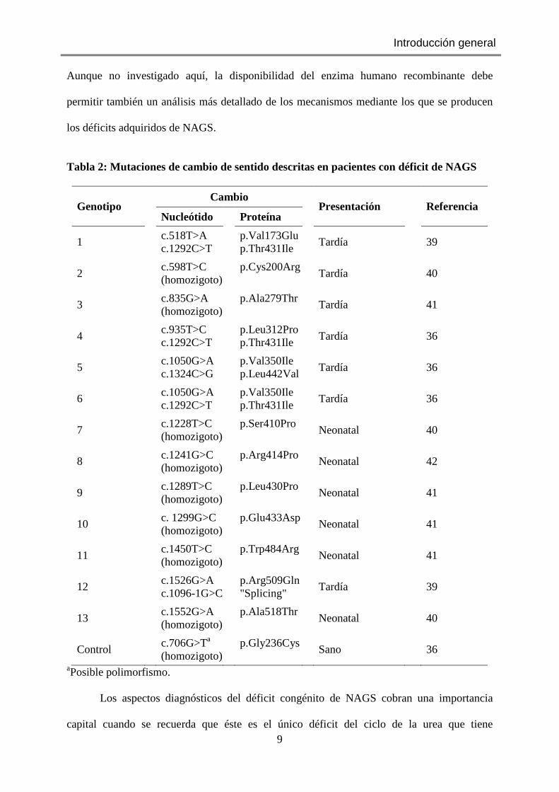
Tabla 2: Mutaciones de cambio de sentido descritas en pacientes con déficit de NAGS
Genotipo Cambio
Presentación
Referencia Nucleótido Proteína
1 c.518T>A
c.1292C>T p.Val173Glu
p.Thr431Ile
Tardía 39
2 c.598T>C
(homozigoto) p.Cys200Arg
Tardía 40
3 c.835G>A
(homozigoto) p.Ala279Thr
Tardía 41
4 c.935T>C
c.1292C>T p.Leu312Pro
p.Thr431Ile
Tardía 36
5 c.1050G>A
c.1324C>G p.Val350Ile
p.Leu442Val
Tardía 36
6 c.1050G>A
c.1292C>T p.Val350Ile
p.Thr431Ile
Tardía 36
7 c.1228T>C
(homozigoto) p.Ser410Pro
Neonatal 40
8 c.1241G>C
(homozigoto) p.Arg414Pro
Neonatal 42
9 c.1289T>C
(homozigoto) p.Leu430Pro
Neonatal 41
10 c. 1299G>C
(homozigoto) p.Glu433Asp
Neonatal 41
11 c.1450T>C
(homozigoto) p.Trp484Arg
Neonatal 41
12 c.1526G>A
c.1096-1G>C p.Arg509Gln
"Splicing"
Tardía 39
13 c.1552G>A
(homozigoto) p.Ala518Thr
Neonatal 40
Control c.706G>Ta
(homozigoto) p.Gly236Cys
Sano 36
aPosible polimorfismo.
Los aspectos diagnósticos del déficit congénito de NAGS cobran una importancia
capital cuando se recuerda que éste es el único déficit del ciclo de la urea que tiene

Introducción general
10
tratamiento sustitutivo, en este caso con un análogo del NAG (N-carbamilglutámico o ácido
carglúmico, llamado también Carbaglu, medicamento huérfano suministrado por la empresa
Orphan Europe) (Fig. 2) que, a diferencia del NAG, es biológicamente estable y absorbible
por vía oral (35-43). Esta excelente posibilidad terapéutica obliga a un diagnóstico temprano,
certero y de alta sensibilidad de los fallos de la NAGS, que hace particularmente deseable
disponer de procedimientos para diferenciar entre mutaciones clínicas y meros polimorfismos.
5. La pertenencia de la NAGS a la familia aminoácido quinasa ha propiciado el
desarrollo de este trabajo de tesis
Nuestro laboratorio identificó en 1999 un nuevo plegamiento sandwich αβ en la
enzima carbamato quinasa (Fig. 3A) (44), y encontró poco después que la NAG quinasa
(NAGK) de E. coli, que cataliza el paso siguiente al de la NAGS en la biosíntesis de arginina
(Fig. 1, panel extremo izquierdo), es homóloga a la carbamato quinasa en toda su longitud y
comparte con ella plegamiento y arquitectura (Fig. 3B) (45,46).
Figura 2. Fórmulas del NAG y de su análogo farmacológico el N-carbamil-L-glutamato o ácido carglúmico, llamado comercialmente Carbaglu). [Tomado de (18)]

Introducción general
11
Estos hallazgos precedieron y basaron la identificación por el Instituto Sanger de una familia
de proteínas a la que se denominó familia aminoácido quinasa (AAK) (PFAM PF00696
http://pfam.sanger.ac.uk). La familia AAK se caracteriza porque las proteínas pertenecientes a
+ Arg
- Arg
A B
C
D E
F
Figura 3. Estructuras de la carbamato quinasa (A), NAGK de
E. coli (B), un dímero de NAGK de P. aeruginosa con las
hélices N-terminales en verde (C) y hexámeros de NAGK de
P. aeruginosa sin arginina (D) y de Thermotoga maritima con
arginina (E). En (F) se esquematiza el efecto de la arginina
sobre el trímero de dímeros de NAGK. Se agradece las
figuras a S. Ramón-Maiques. Se corresponden con las
estructuras del PDB 2WE4 (A), 1GS5 (B), 1UVD (C y D) y
2BTY (E).

Introducción general
12
ella (Fig. 4) poseen al menos un dominio correspondiendo con la secuencia completa de la
carbamato quinasa o de la NAGK de E. coli. Interesantemente, ArgA pertenece a dicha
familia (Fig. 4), identificándose en la misma dos dominios, uno N-terminal, de unos 300
residuos, de carácter aminoácido quinasa (dominio AAK) (Fig. 5A,B), y otro C-terminal, de
unos 140 residuos (Fig. 5B), denominado GNAT, nombre derivado del hecho de que
pertenece a la familia de N-acetiltransferasas relacionadas con Gcn5 (Gcn5 es una
acetiltransferasa de levadura que acetila las histonas) (47). Ambos dominios se encuentran
unidos por un corto conector (Fig. 5B) (48).
N-acetyl-L-glutamyl-5- phosphate
N-acetyl-L-glutamate
N-acetyl-L-glutamate 5-semialdehyde
N-acetyl-
L-ornithine
L-ornithine
Acetylglutamatesynthase
Acetylglutamate
kinase
Acetyl-γ-glutamyl
phosphate
reductase
Acetylornithine-5-
aminotransferase
CoA
acetyl-CoA
H2O CH3COO-
L-glutamate
2-oxoglutarate
NADPH
NADP++ Pi
L-arginine
ATP
ADP
Arginine
deiminaseH2O
NH4+
ATP
ADP
carbamate
Carbamate
kinase
carbamoylphosphate
L-homoserine
ATP
ADPHomoserine kinase
L-homoserine-O-Pi
L-methionine
Pi
Threonine synthase
L-threonine
L-isoleucine
L-aspartate
L-aspartyl-4-phosphate
Aspartokinase
Aspartic semialdehyde
dehydrogenase
L-aspartic-β-semialdehyde
NAD(P)H
NAD(P)+Homoserine dehydrogenase
L-lysine
ATP
ADP
ATP
ADP
L-glutamate
Glutamate 5-kinase
NADPH
NADP++ Pi
L- glutamyl-5-phosphate
L-glutamate 5-semialdehyde
H2O
Pyrroline-5-carboxylate
NADPH
NADP+
L-proline
UMP UDP
UTP, CTP, dTTP
ATP ADP
L-glutamate
L- citrulline
Carbamoyl-phosphate
Pi2ATP, HCO3-L-glutamine
Carbamoylphosphate
synthetase
2ADP, PiL-glutamate
γ-Glutamyl phosphate
reductase
Pyrroline-5-
carboxylate
reductase
Non-enzymatic
NADPH
NADP++Pi
H2O
Ornithine 5-
aminotransferase
Acetyl ornithinase
Transacetylase
Ornithine transcarbamylase
UMP kinase
En realidad, en 2006 nuestro laboratorio había identificado la existencia de dos formas
de NAGK que se diferenciaban por su sensibilidad o insensibilidad a la inhibición "feed-
back" por arginina (46,48). La forma insensible se correspondía con la encontrada en E. coli,
organismo en el que la NAGS es el catalizador por el que pasa todo el flujo biosintético de
Fig. 4. Ubicación de los miembros de la familia aminoácido quinasa (en magenta) en sus rutas metabólicas. Los enzimas se encuadran en fondo coloreado.

Introducción general
13
arginina, flujo lineal, siendo también el punto único de control "feed-back" por arginina (Fig.
1, panel más a la izquierda) (16, 46). En E. coli la acetilornitina producida al final del cuarto
paso de la ruta de síntesis de arginina se convierte en ornitina gracias a su deacilación
hidrolítica catalizada por el producto del gen argE (Fig. 1, panel izquierdo) (16). La ruta
lineal consume una molécula de AcCoA por molécula de arginina producida. En dicha ruta la
NAGK es insensible a la arginina y su estructura es la de un homodímero organizado
esencialmente como el dímero de carbamato quinasa (Fig. 3B) (46).
Sin embargo, en muchos otros organismos tales como P. aeruginosa o las plantas, la
biosíntesis de arginina tiene un carácter cíclico (Fig.1, segundo panel desde la izquierda)
(16,17,48,50). En la ruta cíclica se recicla el grupo acetilo de la acetilornitina mediante su
transferencia al glutamato por una transacetilasa que es el producto del gen argJ (16). Así se
evita el consumo de una molécula de AcCoA por cada molécula de arginina producida. La
génesis de NAG a partir de AcCoA y por tanto la actividad NAGS son todavía necesarias,
pero una vez cebado el sistema con NAG no hace falta más síntesis nueva de NAG,
regenerándose este compuesto a partir de la acetilornitina (16). En este caso ya no pasa por la
NAGS todo el flujo biosintético de arginina, siendo el primer punto obligado de paso en cada
ciclo la NAGK. Por ello la NAGK adquiere propiedades reguladoras, siendo el sujeto de la
inhibición "feed-back" por arginina (16).
Los estudios de nuestro laboratorio (48,51) identificaron una extensión N-terminal de
unos 15-25 aminoácidos (Fig. 5A, subrayado) que es esencial (aunque no suficiente) para que
la NAGK sea inhibible por arginina (51), y los estudios estructurales establecieron que dicha
extensión se pliega como un hélice alfa doblada (Fig. 3C) (48). Así, la estructura básica de la
NAGK inhibible por arginina es la misma que la del dímero de la NAGK de E. coli, excepto
por la presencia extra de la hélice doblada N-terminal, que actúa como un gancho o anzuelo
que sirve para entrelazarse con la hélice correspondiente de un dímero vecino (Fig. 3C). De

Introducción general
14
este modo, tres dímeros se encadenan en un trímero de dímeros con aspecto de anillo
hexamérico (Figs. 3D y 3E) (48).
Fig. 5. Primer modelo estructural de la NAGS bacteriana clásica, basado en la estructura de la NAG quinasa hexamérica. (A) Alineamiento de las secuencias de los dominios aminoácido quinasa de ambas enzimas (ArgA, NAGS; ArgB, NAG quinasa). Se subrayan las secuencias relacionadas con el sitio para arginina. (B) Composición de dominios de la subunidad NAGS. (C) Modelo de arquitectura hexamérica de la NAGS, basada en la de la NAG quinasa sensible a arginina. Nótese que en este modelo se hipotetizaba la unión del acCoA a ambos dominios. Tomada de (48)

Introducción general
15
Un hecho clave en nuestra comprensión de la estructura de la NAGS fue el comprobar
(48) que su dominio AAK va precedido de una extensión que se corresponde con la hélice N-
terminal de la NAGK inhibible por arginina (Fig. 5A, subrayado; aunque se representa la
secuencia de la NAGS de E. coli, ésta presenta elevada identidad con las de P. aeruginosa y
de Neisseria gonorrhoeae). Una inferencia inmediata que fue el objeto del primer modelo
existente de estructura de la NAGS de tipo ArgA, propuesto por nuestro laboratorio antes de
mi incorporación al mismo y que se refleja en la Fig. 5C, fue que la NAGS bacteriana
producida por el gen argA se organiza hexaméricamente como la NAGK inhibible por
arginina (48). En realidad, habría que hablar de hexámero de dominios AAK, ya que la
estructura de la NAGK inhibible por arginina dejaba sin respuesta la cuestión de cuál era el
lugar de los dominios GNAT en la estructura. Por consideraciones derivadas de la aplicación
de la simetría molecular alrededor del eje ternario y de impedimentos estéricos derivados del
tamaño del dominio GNAT y de la escasa longitud del conector que une ambos dominios, era
evidente que los dominios GNAT de subunidades adyacentes debían situarse en caras
opuestas del anillo de dominios AAK (48). Una hipótesis (que luego se ha comprobado
errónea) sobre la composición del centro activo de la NAGS, condujo a nuestro grupo a una
conclusión acertada sobre la ubicación en la estructura del dominio GNAT (48). La hipótesis
se derivaba del hecho de que en la NAGK el dominio AAK une acetilglutamato en su lóbulo
N-terminal y ATP en el lóbulo C-terminal (46,48). Como la NAGS usa un sustrato similar al
NAG, el glutamato, y otro sustrato (el AcCoA) con una parte nucleotídica similar al ATP, se
planteó la posibilidad de que tanto el dominio AAK como el GNAT participaran en la
reacción, el dominio AAK proveyendo los sitios para el glutamato y para la parte similar al
ATP del AcCoA, mientras que el dominio GNAT aportaría el resto del sitio para el AcCoA
así como los grupos catalíticos. La colaboración entre ambos dominios obligaba a la
ubicación del dominio GNAT entre los sitios para el glutamato y para el ATP de un dominio

Introducción general
16
AAK, y esto sólo era posible si el dominio GNAT de una subunidad de un dímero descansara
sobre el dominio AAK de una subunidad del dímero adyacente. Este fue el origen de la
propuesta de arquitectura de NAGS que se presenta en la Fig. 5C (48).
La propuesta de esta arquitectura conllevaba el inmediato entendimiento del
mecanismo de inhibición de la NAGS bacteriana por la arginina. En el caso de la NAGK los
estudios estructurales de mi grupo (48) habían demostrado que la arginina se une en el
dominio C-terminal, junto a la hélice doblada N-terminal, y que dicha unión estabiliza una
forma ampliada del anillo hexamérico en el que ambos dominios de cada subunidad de la
NAGK se distancian, dificultando el cierre del centro activo que hace posible la reacción,
resultando en inhibición por distanciamiento de las moléculas de ambos sustratos (Figs. 3 D-
F). Un mecanismo similar podía ser propuesto para el caso de la NAGS si ésta fuera
hexamérica, si la arginina se uniera a ella como en el caso de la NAGK inhibible por arginina,
y si la arquitectura del centro activo fuera tal como había propuesto el grupo. En la forma con
arginina se alterarían las relaciones entre el dominio GNAT y el dominio AAK sobre el que
descansa, como consecuencia del ensanchamiento del anillo y de la tracción ejercida sobre el
dominio GNAT por el conector que la une covalentemente a su subunidad propia. Aunque sin
corroborarse la complementariedad de ambos dominios en la constitución del centro activo, al
menos en la forma postulada por nuestro grupo, esta hipótesis ha resultado correcta al probar
nuestros resultados (ver capítulo 1 de Resultados) un fuerte papel modulador del dominio
AAK sobre el dominio catalítico GNAT que descansa sobre él.
En este marco conceptual, cuando me incorporé al laboratorio e inicié este trabajo de
tesis carecíamos de información estructural directa de la NAGS, no existía información
alguna sobre el modo de unión de la arginina y de la inhibición por este aminoácido, y
tampoco se habían descrito la producción recombinante y purificación de NAGS codificada
por el gen argA. Por tanto, mi primer objetivo fue producir la NAGS recombinante de P.

Introducción general
17
aeruginosa puesto que gran parte del trabajo clásico sobre NAGS se había realizado con la
enzima obtenida de este microorganismo (6,7). Uno de mis objetivos era utilizarla para
obtener cristales y para determinar la estructura de la misma con y sin arginina. No he
coronado con éxito dicho objetivo, ya que el grupo de Mendel Tuchman se me anticipó al
determinar en 2008 la estructura de la NAGS de tipo ArgA de Neisseria gonorrhoeae (52),
publicando en 2009 la estructura del complejo con arginina (Figs. 6 y 7) (53).
También nos propusimos como objetivo, ese sí culminado con éxito, identificar en el
enzima mediante procedimientos indirectos como mutagénesis dirigida e ingeniería de
proteínas, la ubicación del centro activo, el sitio de unión de la arginina y el mecanismo de
regulación por este inhibidor, habiendo sido la consecución de este objetivo el fuerte de mi
trabajo de tesis.
Las estructuras experimentales publicadas del enzima de N. gonorrhoeae (52,53) han
corroborado el modelo estructural hexamérico propuesto por nuestro laboratorio para la
NAGS (Fig. 5C) (48) y han dado soporte al modelo del laboratorio de regulación por arginina
basado en el ensanchamiento del anillo hexamérico del enzima (ver más adelante). Además,
Figura 6. Hexámero de NAGS de Neisseria gonorrhoeae sin arginina (izquierda) o con arginina (derecha). Tal como se postuló en el modelo de nuestro laboratorio (Fig. 5), se trata de un hexámero (trímero de dímeros; cada dímero en un color) y el dominio acetiltransferasa se apoya sobre el domino aminoácido quinasa de la subunidad del dimero vecino. Nótese como la unión de la arginina ensancha el hexámero. Construido a partir de los archivos del PDB 2R8V (izquierda, sin arginina) y 3D2P (derecha, forma con arginina).

Introducción general
18
como se detalla en la Fig. 7, dichas estructuras (52,53) han demostrado la propuesta del
modelo del laboratorio en la que un dominio GNAT de una subunidad reposa sobre el
dominio AAK de una subunidad de un dímero vecino (48) (ver Discusión general).
Es más, como se demuestra en este trabajo de tesis, con anterioridad a la publicación
de la estructura experimental de la NAGS con arginina ubicamos ya físicamente el sitio para
la arginina en la NAGS de P. aeruginosa y lo inferimos para la NAGS humana, modelizando
dicho sitio a partir del correspondiente de la NAGK (Capítulo 1 de los Resultados), habiendo
corroborado dicho modelo la estructura cristalina del complejo con arginina del enzima de N.
gonorrhoeae (53).
6. El investigar la naturaleza de la NAGS humana y de su déficit ha motivado también
este trabajo de tesis.
Como se ha mencionado anteriormente, durante muchos años las dos únicas formas de
NAGS caracterizadas eran la bacteriana clásica y la mitocondrial de roedores y humanos. La
forma mitocondrial ha sido durante mucho tiempo objeto de controversia, por su
Dominio AAK
Dominio GNAT
Conector
Dímero vecino
Figura 7. Una subunidad de la NAGS de N. gonorrhoeae (en azul) mostrando sus dos dominios y el conector interdominios, para revelar los contactos de ambos dominios con los dominios aminoácido quinasa del dímero vecino (verde y amarillo). Construido a partir del archivo 2R8V
del PDB.

Introducción general
19
inestabilidad, por su escasa identidad de secuencia con las formas bacterianas y por su
activación por arginina (13). La identificación de su gen solamente ha sido posible a partir de
las secuencias de las NAGSs de levadura y de Neurospora crassa (13,54). En estas especies
la producción de NAGS requiere de la presencia de la NAGK (55,56). En levadura la NAGS
no existe como proteína aislada, sino sólo como complejo con la NAGK, que parece actuar
como chaperona de la NAGS, formando con ella un metabolón (56,57). Dada la similitud de
la enzima de mamífero con la NAGS de ascomicetos, es concebible que la inestabilidad de la
primera tenga que ver con posibles interacciones del enzima con otras proteínas presentes en
la mitocondria (aunque no con NAGK, que no existe en animales).
La identificación del gen de ratón y humano para NAGS ha llevado a la expresión
recombinante de estos enzimas en E. coli, particularmente del humano, que han abordado dos
grupos (40,58), incluso con purificación de estos enzimas y caracterización de sus parámetros
cinéticos, actividad, influencia de la eliminación de las etiquetas añadidas para facilitar la
purificación; y expresión de formas con deleciones de porciones N-terminales consideradas
como péptido señal de internalización mitocondrial y porción variable (58). Se han estudiado
así algunas mutaciones clínicas (36,39,40,58), incluídas mutaciones en el sitio de la arginina
(27) predicho por nosotros que han permitido corroborar nuestra predicción (Capítulo 1 de los
resultados). Sin embargo en esos trabajos de otros grupos no se detalla el rendimiento de
proteína purificada ni su estabilidad, siendo la impresión obtenida en presentaciones en
congresos a los que hemos asistido que uno y otra son escasas.
Por ello decidimos intentar la producción recombinante de la enzima humana con
etiquetas diferentes a las descritas con la finalidad de juzgar por nosotros mismos sobre la
factibilidad de un buen rendimiento y sobre la estabilidad del producto purificado. La segunda
parte del proyecto con el enzima humano consideraba la posibilidad de producir cristales del
mismo si la abundancia y la pureza del producto lo permitía, con la finalidad de estudio
estructural. Razonamos que aún si no se tenía éxito en la producción de cristales que

Introducción general
20
difractaran, sería posible realizar estudios de disección similares a los realizados aquí
(Capítulo 3 de los Resultados) con el enzima bacteriano. Como nuestro grupo mantiene
colaboraciones con grupos clínicos sobre errores congénitos del ciclo de la urea, realizando
con frecuencia estudios de estructura-función, la producción propia de NAGS humana
recombinante, nos podría situar en buena posición para la realización de estudios
experimentales de estructura-función para las mutaciones clínicas que se vayan identificando
en pacientes.
Aunque no se describe aquí en detalle por razones que se expresan al final de la
Discusión General, he tenido éxito en la producción recombinante y estable del enzima
humano como proteína de fusión con la proteína de unión a la maltosa (MBP), habiendo
disecado parcialmente los dominios componentes, con caracterización funcional del dominio
GNAT, y habiendo comprobado el valor de este sistema para la investigación de la
patogenicidad de mutaciones clínicas descritas en el déficit de NAGS humana.
7. La estructura de esta tesis
El formato de esta tesis es por compendio de publicaciones, con tres capítulos de
Resultados que se corresponden cada uno con una publicación. De acuerdo con lo establecido
para este formato de tesis, he hecho preceder dichos capítulos de esta Introducción General
para el conjunto del trabajo, siguiéndolos un Resumen de los Resultados y una Discusión
General, así como un capítulo de Conclusiones. Aunque cada capítulo de resultados se
acompaña de su propia bibliografía, al final de la tesis se incluye una sección bibliográfica
que recoge las referencias para la Introducción General, el Resumen de Resultados y la
Discusión General.
Como puede verse, las publicaciones cuentan con sólo tres autores, siendo yo en todos
los casos la primera autora y siendo los otros dos autores mis Directores de Tesis. Estas
publicaciones son las siguientes:

Introducción general
21
1. Título: Site-directed mutagenesis studies of acetylglutamate synthase delineate the site for
the arginine inhibitor.
Autores: Enea Sancho-Vaello, M. Leonor Fernández-Murga y Vicente Rubio
Referencia: FEBS Letters (2008), Vol. 582, Págs. 1081-1086
2. Título: Mechanism of arginine regulation of acetylglutamate synthase, the first enzyme of
arginine synthesis
Autores: Enea Sancho-Vaello, M. Leonor Fernández-Murga y Vicente Rubio
Referencia: FEBS Letters (2009), Vol. 583, Págs. 202-206
3. Título: Functional dissection of N-acetylglutamate synthase (ArgA) of Pseudomonas
aeruginosa and restoration of its ancestral N-acetylglutamate kinase activity
Autores: Enea Sancho-Vaello, María L. Fernández-Murga y Vicente Rubio
Referencia: J Bacteriol.(2012), Vol. 194, Págs. 2791-2801.

Introducción general
22

23
Resultados

24

25
Resultados: Capítulo 1
Site-directed mutagenesis studies of acetylglutamate
synthase delineate the site for the arginine inhibitor
Enea Sancho-Vaello, M. Leonor Fernández-Murga y Vicente Rubio
Trabajo publicado en
FEBS Letters (2008), Vol. 582, Págs. 1081-1086

26

FEBS Lett. (2008) 582, 1081-1086
27
Site-directed mutagenesis studies of acetylglutamate synthase
delineate the site for the arginine inhibitor
Enea Sancho-Vaello†, M. Leonor Fernández-Murga† and Vicente Rubio
Instituto de Biomedicina de Valencia (IBV-CSIC) and Centro de Investigación Biomédica en
Red de Enfermedades Raras (CIBERER-ISCIII). Jaime Roig 11, 46010-Valencia. Spain
Received 3 January 2008; revised 8 February 2008; accepted 24 February 2008
Available online 3 March 2008
† E.S-V. and M.L.F-M. contributed equally to this work
ABSTRACT
N-Acetyl-L-glutamate synthase (NAGS), the first enzyme of bacterial/plant arginine
biosynthesis and an essential activator of the urea cycle in animals, is, respectively, arginine-
inhibited and activated. Site-directed mutagenesis of recombinant Pseudomonas aeruginosa
NAGS (PaNAGS) delineates the arginine site in the PaNAGS acetylglutamate kinase-like
domain, and, by extension, in human NAGS. Key residues for glutamate binding are
identified in the acetyltransferase domain. However, the acetylglutamate kinase-like domain
may modulate glutamate binding, since one mutation affecting this domain increases the Km
for glutamate. The effects on PaNAGS of two mutations found in human NAGS deficiency
support the similarity of bacterial and human NAGSs despite their low sequence identity.
Keywords: N-Acetyl-L-glutamate synthase, arginine biosynthesis, feed-back inhibition, glutamate binding, urea cycle errors, NAGS deficiency. Abbreviations: AAK, amino acid kinase; GNAT, GCN5-related N-acetyltransferase; NAG, N-acetyl-L-glutamate; NAGK, N-acetyl-L-glutamate kinase; NAGS, N-acetyl-L-glutamate synthase; Pa, Pseudomonas aeuginosa; Ec, Escherichia coli; Ng, Neisseria gonorrhoeae; Hu, human.

Capítulo 1
28
INTRODUCTION
In bacteria and in plants, N-acetyl-L-glutamate synthase (NAGS) uses AcCoA to N-
acetylate glutamate in the first step of arginine synthesis. This enzyme is feed-back inhibited
by arginine (1). However, in animals, NAGS is activated by arginine, and its only role is to
make N-acetyl-L-glutamate (NAG) for activation of the urea cycle enzyme carbamoyl
phosphate synthetase (2). Consequently, human NAGS (HuNAGS) deficiency is an inborn
error of the urea cycle that causes hyperammonaemia (3).
Classical bacterial NAGS was reported in Escherichia coli (EcNAGS) to be a homohexamer
(4). Each subunit is a ~430-residue chain. It has (Fig. 1A-C; http://www.expasy.org) a 285-
residue N-terminal domain (called here the amino acid kinase (AAK) domain) resembling the
arginine-inhibitable enzyme of the AAK family, N-acetyl-L-glutamate kinase (NAGK) (5-7).
It also has a C-terminal domain resembling GNAT-type acetyltransferases (8,9) (called here
the GNAT domain). Structure determination allowed characterization of the arginine and the
substrate sites of NAGK (6,7), rendering possible to define arginine binding signatures that
were recognized also in the AAK domain of bacterial NAGS (7). We use now site-directed
mutagenesis of recombinant Pseudomonas aeruginosa NAGS (PaNAGS) to confirm the
functionality of these sequences in arginine binding, corroborating the similarity of the
arginine sites of NAGK and NAGS (Fig. 1D). On the basis of this information we infer the
localization of the arginine site of HuNAGS. Our mutagenesis studies also reveal a key role of
the GNAT domain in binding glutamate. By showing that the introduction in PaNAGS of two
clinical HuNAGS mutations cause deleterious effects, we support the similarity of PaNAGS
and HuNAGS despite their low sequence identity. Our results are discussed in the light of
crystal structures of Neisseria gonorrhoeae NAGS (NgNAGS) that were reported after this
paper was submitted (10).

FEBS Lett. (2008) 582, 1081-1086
29
GNAT DOMAINC HuSSAT 4 VRIREAKEGDCGDILRLIRELAEFEKLSDQVKISEEALRADGFGDNPFYHC 54 PaNAGS 286 EQLREAGIEDVGGLIELIRPLEEQGILVRRSRE-----------------V 319 HuNAGS 378 LRVRSLDKLDQGRLVDLVN--ASFGKKLRDDYL------------------ 408 HuSSAT LVAEILPAPGKLLGPCVVGYGIYYFIYSTWKGRTIYLEDIYVMPEYRGQGIG 106 PaNAGS LEREIEQFSIVEREGLIIACAALYPIADSEAG---ELACLAVNPEYRHGGRG 368 HuNAGS ASLRPRLHSIYVSEGYNAAAILTMEPVLGGTP---YLDKFVVSSSRQGQGSG 457 HuSSAT SKIIKKVAEVALDKGC-----------------SQFRLAVLDWNQRAMDLYK 141 PaNAGS DELLERIEERARGLGLK----------------TLFVLTTRTAHWFRERGFQ 404 HuNAGS QMLWECLRRDLQTLFWRSRVTNPINPWYFKHSDGSFSNKQWIFFWFGLADIR 510 HuSSAT ALGAQDLTEAEGWHFFCFQGEATRKLAGK 170 PaNAGS PSSVERLP-AARASLYNFQRNSQVFEKSL 432 HuNAGS DS-YE-LVNHAKGLPDSFHKPASDPGS 535
V
285 432
GNAT/acetyltransferaseAmino acid kinase1
Conserved domainMitochimport
1
Variable
49 94 534
1 301
Amino acid kinase
NAGENZYME
PaNAGK substrate inhibits
PaNAGS product inhibits
ARGININE
HuNAGS product activates
A
B AMINO ACID KINASE (AAK) DOMAIN
PaNAGK MTLSRDDAAQVAKVLSEALPYIRRFV---GKTLVIKYGGNAMES 41 PaNAGS -------------------------------MPDYVNWLRHASPYINSHR---DRTFVVMLPGEGVEH 34 HuNAGS 50 STAWSQPQPPPEEYAGADDVSQSPVAEEPSWVPSPRPPVPHESPEPPSGRSLVQRDIQAFLNQCGASP 117 PaNAGK EELKAGFARDVVLMKAVGIN---PVVVHGGGPQIGDLLKRLSIESHFID--GMRVTDAATMDVVEMVL 104 PaNAGS PNFGN-IVHDLVLLHSLGAR---LVLVHGSRPQIEARLAARGLAPRYHR--DLRVTDAPTLECVIDAV 96 HuNAGS GEARH-WLTQFQTCHHSADKPFAVIEVDEEVLKCQQGVSSLAFALAFLQRMDMKPLVVLGLPAPTAPS 184 PaNAGK GGQVNKDIVNLINRHGGSAIGLTGKDAELIRAKKL--TVTRQTPEMTKPEIIDIGHVGEVTGVNVGLL 170 PaNAGS GSLR----IAIEARLSMDMAASPMQGARLRVAGGN--LVTAR--PIGVVEGVDYHHTGEVRRIDRKGI 156 HuNAGS GCLS----FWEAKAQLAKSCKVLVDALRHNAAAAVPFFGGGS--VLRAAEPAPHASYGGIVSVETDLL 246 PaNAGK NMLVKGDFIPVIAPIGVGSNGESYNINADLVAGKVAEALKAEKLMLLTNIAGLMDKQGQVLTGLSTEQ 238 PaNAGS GRLLDERSIVLLSPLGYSPTGEIFNLACEDVAMRAAIDLEAEKLILYGAEQGLLDASGKLVRELRPQQ 224 HuNAGS QWCLESGSIPILCPIGETAARRSVLLDSLEVTASLAKALRPTKIIFLNNTGGLRDSSHKVLSNVN--- 311 PaNAGK VNELIADGTIYGGMLPKIRCALEAVQGGVTS----AHIIDGRVPNAVLLEIFTDSGVGTLISNRKRH 301 PaNAGS VPAHLQR-LGNSYQAELLDAAAQACRAGVKR----SHIVSYTEDGALLSELFTRTGNGTLVA-QEQF 285 HuNAGS LPADLDL-VCNAEWVSTKERQQMRLIVDVLSRLPHHSSAVITAASTLLTELFSNKGSGTLFKNAERM 377
ααααN
ααααA ααααB ααααC
ααααC
ααααD ααααE
ααααF ααααG ααααH
ββββ1
ββββ2 ββββ3 ββββ4
ββββ5 ββββ6 ββββ7
ββββ8 ββββ9 ββββ10 ββββ11 ββββ12 ββββ13 ββββ14
ββββ15 ββββ16
C
D
K199
Y14
E269
G275
Arginine
helix-H
N-helix
β16
Fig. 1. Domain composition, sequence alignments and the arginine site of NAGS. (A) Schematic domain organization of PaNAGS, compared with PaNAGK and with HuNAGS. The regions of HuNAGS are those defined in [2]. (B) and (C) Alignment of PaNAGS with HuNAGS and with either PaNAGK or the human thialysine Nε-acetyltransferase (HuSSAT). Identical residues are shadowed. The regions corresponding to the signature sequences for arginine identified in NAGK and NAGS [7] are boxed. PaNAGS residues mutated here are encircled, and those corresponding to EcNAGS mutated residues in feed-back resistant enzyme forms [18] are underlined. Clinical mutations are indicated by the amino acid change encircled below the HuNAGS sequence. Shown as lines (helices) and arrows (β strands) above the AAK domain sequences of PaNAGK and PaNAGS are the secondary structure elements (named according to [7]) of the PaNAGK [7] and NgNAGS [10] structures (the NgNAGS structure can be used for PaNAGS given their nearly identical length and important sequence identity/similarity, estimated as 39.4 %/60.1% [10]; all the residues mutated here are conserved in NgNAGS except L353, which is replaced by I). (D) The proposed arginine site of PaNAGS. The NgNAGS crystal structure is illustrated ([10]; Protein Databank file 2r8v; http://www.rcsb.org/pdb), but the numbering of the residues for which the side-chains are shown (which are those mutated here; G275 is highlighted as a black sphere) is that for PaNAGS. Secondary structure elements are named as in PaNAGK. Arginine is shown in dark grey and is placed in the orientation observed in the arginine site of NAGK [7]. Dotted lines mark polar interactions between enzyme residues and the inhibitor.

Capítulo 1
30
MATERIALS AND METHODS
Gene PA5204 (http://www.TIGR.org), PCR-amplified from P.aeruginosa PAO1
genomic DNA, was cloned into the NdeI and XhoI sites of pET-22b and subjected to site-
directed mutagenesis with the Quickchange kit (from Stratagene) (11). Mutations K199A,
V358A and G368A were prepared by the overlapping extension method (12). Primers used
are given in Table 1. PaNAGS was expressed in transformed BL21(DE3) cells (from
Novagene) grown to OD600 ≈ 0.5. After standing 45 min at 4ºC, 2% (v/v) ethanol and 0.02
mM isopropyl-β-δ-thiogalactoside were added and the culture was continued overnight at
15ºC. All enzyme forms contained the GSLEH6 tail and were purified from centrifuged cell
sonicates in buffer A (20 mM Na phosphate pH 8/1 mM dithiothreitol/0.5 M NaCl/20 mM
imidazole), using 0.1-ml His-SpinTrap centrifugal columns (GE Healthcare) and 0.5 M
imidazole-containing buffer A for elution.
Enzyme activity assays were carried out at least in duplicate and were based on
measurement at 412 nm of glutamate-dependent CoA release using 5,5-dithio-bis(2-
nitrobenzoic acid) (9) [ε412 nm, 13.6×10³ M-1 cm-1 (13)]. Fresh enzyme was incubated at 37ºC
in 20 µl of 0.2 M Tris-HCl pH 9/1 mM AcCoA/20 mM (or, when indicated, 0.1 M) L-
glutamate/10 mM MgCl2 and, when used, the indicated arginine concentrations. After 10 min,
0.8 ml 0.2 mM 5,5-dithio-bis(2-nitrobenzoic acid) in 0.1 M Na phosphate pH 7 was added
and OD412 was determined. Color production was linear with time for >10 min, and was
corrected for 3.7% spontaneous hydrolysis of AcCoA, determined in enzyme-free reactions.
Enzyme concentration was adjusted to prevent >30% consumption of the less abundant
substrate. When AcCoA or glutamate were varied (respective variation ranges, 0.1-4 and 0.5-
20 mM, unless indicated), glutamate and AcCoA were fixed, respectively, at 20 mM (or,
when indicated, at 0.1 M) and 1 mM. Program GraphPadPrism (GraphPad Software, San
Diego) was used to fit the results to hyperbolic kinetics, or, in the presence of arginine, to

FEBS Lett. (2008) 582, 1081-1086
31
sigmoidal kinetics. For arginine inhibition, the data were fitted to the following expression:
v = A + B × (1− [Arg]N/ (IN0.5 + [Arg]N)); v and A are activities at a given [Arg] and at
[Arg]=∞; B, activity fall caused by [Arg]=∞; I0.5, half-inhibitory [Arg]; N, Hill coefficient.
Protein was determined by the Bradford assay (14) using a commercial reagent (from Bio
Rad) and bovine serum albumin as standard.
Table 1. Synthetic oligonucleotides used
Primer Mutation Direction Sequencea
1b WT Forward 5´GCTTTCCCCCATATGCCCGACTACG 3´
2c WT Reverse 5´GGACGCCTCGAGGGATCCCAGGCTCTTCTCG 3´
3 Y14A Forward 5´CACGCTTCGCCCGCCATCAACTCGCACCGG 3´
4 Y14A Reverse 5´GTGCGAGTTGATGGCGGGCGAAGCGTGACG 3´
5 G146C Forward 5´CTATCACCATACCTGTGAGGTCCGTCGCATCG 3´
6 G146C Reverse 5´GCGACGGACCTCACAGGTATGGTGATAGTC 3´
7 K199A Forward 5´GGAAGCGGAAGCCCTGATTCTCTACGG 3´
8 K199A Reverse 5´GTAGAGAATCAGGGCTTCCGCTTCCAGG 3´
9 E269A Forward 5´CTGCTCAGCGCACTATTCACCCGCACC 3´
10 E269A Reverse 5´GCGGGTGAATAGTGCGCTGAGCAGCG 3´
11 G275A Forward 5´CACCCGCACCGCCAACGGCACCCTGGTC 3´
12 G275A Reverse 5´CAGGGTGCCGTTGGCGGTGCGGGTGAATAG 3´
13 C339A Forward 5´GAAGGGCTGATCATCGCCGCCGCCGCGCTCTACCCG 3´
14 C339A Reverse 5´GTAGAGCGCGGCGGCGGCGATGATCAGCCCTTCG 3´
15 E352A Forward 5´CCGAGGCGGGCGCGCTGGCCTGTCTGGCG 3´
16 E352A Reverse 5´CAGACAGGCCAGCGCGCCCGCCTCGGAATC 3´
17 E352D Forward 5´CCGAGGCGGGCGATCTGGCCTGTCTGGCG 3´
18 E352D Reverse 5´CAGACAGGCCAGATCGCCCGCCTCGGAATC3´
19 L353V Forward 5´CGGGCGAGGTGGCCTGTCTGGCGGTCAAC 3´
20 L353V Reverse 5´GACCGCCAGACAGGCCACCTCGCCCGCC 3´
21 V358A Forward 5´CTGTCTGGCGGCCAACCCGGAGTAC 3´
22 V358A Reverse 5´CTCCGGGTTGGCCGCCAGACAGGC 3´
23 G368A Forward 5´CGGCGGGCGTGCAGACGAACTGCTGGAGC 3´
24 G368A Reverse 5´CCAGCAGTTCGTCTGCACGCCCGCCGTG 3´ aMutated bases are underlined. The portions of primers 1 and 2 shown in italic lettering correspond to the beginning and to the end of the open reading frame. bIncludes an NdeI site at the initiator ATG (in bold type). cIncludes a XhoI site (in bold type) and abolishes termination, to allow incorporation of the tail GSLEH6.

Capítulo 1
32
RESULTS
3.1. Production and properties of recombinant PaNAGS
Wild-type and mutant PaNAGS forms were expressed abundantly and were purified to
essential homogeneity (Fig. 2). Protein mobility in SDS-PAGE (Fig. 2) agreed with sequence-
deduced masses (wild-type PaNAGS, 49,068 Da). As expected, the wild-type protein
catalyzed (Vmax ≥ 24 U mg-1 at 37ºC) L-glutamate-dependent, but not L-aspartate- or L-
glutamine-dependent (using 20 mM of these amino acids) CoA release from AcCoA. The
concentration-dependency of the activity was hyperbolic for both substrates (Fig. 3A and
Table 2). As reported earlier (15), arginine inhibed PaNAGS with sigmoidal kinetics (Fig. 3C
and Table 2) and it rendered sigmoidal the dependency of the activity on the concentration of
glutamate (Fig. 3A, inset). The Hill coefficient values of ~2 (Table 2) for arginine and for
glutamate (in the presence of 0.5 mM arginine) suggests that ≥ 2 arginine and glutamate sites
exist per enzyme molecule. PaNAGS was eluted in fast (<1 h) gel filtration assays through
Superdex 200 (11) at 23ºC (in 50 mM Tris-HCl, pH 7.5/0.15 M NaCl), as a symmetrical peak,
irrespective of enzyme concentration or of the presence or absence of 10 mM arginine. The
mass estimates (range 280-296 kDa) indicate that PaNAGS is a non-dissociating hexamer.
Fig. 2. Coomassie-stained SDS-PAGE (15% polyacrylamide gel) of purified wild-type (WT) and mutant forms of PaNAGS. Low-load tracks, shown for E352A (central gel) and for wild-type (gel on the right), were used for polypeptide mass estimation by comparison with the mobility of molecular mass markers (St). Markers masses are given at the sides in kDa.
FEBS Lett. (2008) 582, 1081-1086
33
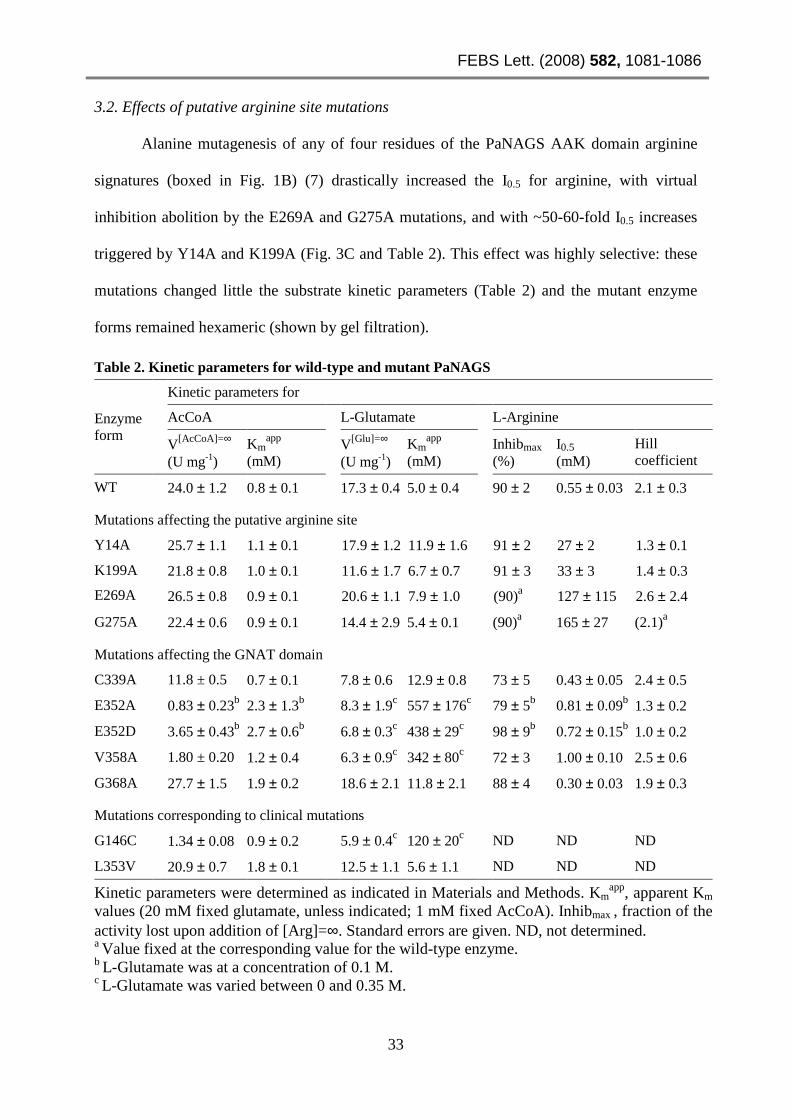
3.2. Effects of putative arginine site mutations
Alanine mutagenesis of any of four residues of the PaNAGS AAK domain arginine
signatures (boxed in Fig. 1B) (7) drastically increased the I0.5 for arginine, with virtual
inhibition abolition by the E269A and G275A mutations, and with ~50-60-fold I0.5 increases
triggered by Y14A and K199A (Fig. 3C and Table 2). This effect was highly selective: these
mutations changed little the substrate kinetic parameters (Table 2) and the mutant enzyme
forms remained hexameric (shown by gel filtration).
Table 2. Kinetic parameters for wild-type and mutant PaNAGS
Enzyme form
Kinetic parameters for
AcCoA L-Glutamate L-Arginine
V[AcCoA]=∞ (U mg-1)
Kmapp
(mM) V[Glu]=∞
(U mg-1) Km
app (mM)
Inhibmax (%)
I0.5 (mM)
Hill coefficient
WT 24.0 ± 1.2 0.8 ± 0.1 17.3 ± 0.4 5.0 ± 0.4 90 ± 2 0.55 ± 0.03 2.1 ± 0.3
Mutations affecting the putative arginine site
Y14A 25.7 ± 1.1 1.1 ± 0.1 17.9 ± 1.2 11.9 ± 1.6 91 ± 2 27 ± 2 1.3 ± 0.1
K199A 21.8 ± 0.8 1.0 ± 0.1 11.6 ± 1.7 6.7 ± 0.7 91 ± 3 33 ± 3 1.4 ± 0.3
E269A 26.5 ± 0.8 0.9 ± 0.1 20.6 ± 1.1 7.9 ± 1.0 (90)a 127 ± 115 2.6 ± 2.4
G275A 22.4 ± 0.6 0.9 ± 0.1 14.4 ± 2.9 5.4 ± 0.1 (90)a 165 ± 27 (2.1)a
Mutations affecting the GNAT domain
C339A 11.8 ± 0.5 0.7 ± 0.1 7.8 ± 0.6 12.9 ± 0.8 73 ± 5 0.43 ± 0.05 2.4 ± 0.5
E352A 0.83 ± 0.23b 2.3 ± 1.3b 8.3 ± 1.9c 557 ± 176c 79 ± 5b 0.81 ± 0.09b 1.3 ± 0.2
E352D 3.65 ± 0.43b 2.7 ± 0.6b 6.8 ± 0.3c 438 ± 29c 98 ± 9b 0.72 ± 0.15b 1.0 ± 0.2
V358A 1.80 ± 0.20 1.2 ± 0.4 6.3 ± 0.9c 342 ± 80c 72 ± 3 1.00 ± 0.10 2.5 ± 0.6
G368A 27.7 ± 1.5 1.9 ± 0.2 18.6 ± 2.1 11.8 ± 2.1 88 ± 4 0.30 ± 0.03 1.9 ± 0.3
Mutations corresponding to clinical mutations
G146C 1.34 ± 0.08 0.9 ± 0.2 5.9 ± 0.4c 120 ± 20c ND ND ND
L353V 20.9 ± 0.7 1.8 ± 0.1 12.5 ± 1.1 5.6 ± 1.1 ND ND ND
Kinetic parameters were determined as indicated in Materials and Methods. Kmapp, apparent Km
values (20 mM fixed glutamate, unless indicated; 1 mM fixed AcCoA). Inhibmax , fraction of the activity lost upon addition of [Arg]=∞. Standard errors are given. ND, not determined. a Value fixed at the corresponding value for the wild-type enzyme. b L-Glutamate was at a concentration of 0.1 M. c L-Glutamate was varied between 0 and 0.35 M.

Capítulo 1
34
3.3. Effects of GNAT domain mutations
GNAT domain mutations
affected little arginine inhibition, but
some of them greatly influenced
substrate kinetics (Table 2). C339 and
E352 (Fig. 1C), which are conserved in
bacterial NAGSs, appeared candidates
for being acceptors of, respectively, the
acetyl group of AcCoA or the proton of
the glutamate amino group (this last
role was suggested by that of E173 of
yeast histone acetyltransferase [16]).
These roles were excluded by the
relatively modest magnitude of the
decreases in the apparent Vmax
estimated for the C339A and E352A/D
mutations (Fig 3A and B and Table 2).
However, E352 appears to impair very
importantly glutamate binding, since
the E352A/D mutations dramatically
increased KmGlu (Fig. 3B and Table 2).
In addition, these mutations increased
~3-fold (Table 2) KmAcCoA. Mutation to
Fig. 3. Influence of the mutations on the dependency of enzyme activity (v) on the concentration of glutamate (A, B), or of arginine (C). The inset in (A) illustrates the effect of adding 0.5 mM arginine on the kinetics for glutamate.
FEBS Lett. (2008) 582, 1081-1086
35
alanine of the invariant putative AcCoA-binding residues (17) V358 and G368 modestly
increased KmAcCoA, but the V358A mutation, similarly to the E352A/D mutations, drastically
increased KmGlu (Fig. 3B and Table 2) and decreased modestly (~3-fold) Vmax (Table 2).
3.4. The effects of clinical mutations highlights the similarity of PaNAGS and HuNAGS
The mutation L442V was found in a late-onset patient and decreased 50% the activity
of in vitro expressed HuNAGS (3). We introduced the corresponding mutation L353V in
PaNAGS (mapping in the GNAT domain) and observed merely a ~2.3-fold increase in
KmAcCoA (Table 2), justifying the fact that the deficiency was partial. In contrast, the G236C
clinical mutation, for which the effects remain uncharacterized in HuNAGS, when replicated
in PaNAGS (G146C mutation, mapping in the AAK domain) caused a large increase (~24-
fold; Table 2 and Fig. 3B) in KmGlu and a ~3-fold decrease in Vmax (Table 2). Thus, this
mutation certainly appears disease-causing, and should be associated with a severe phenotype.
These observations support the similarity of HuNAGS and PaNAGS despite their limited
sequence similarity, illustrating the value of PaNAGS as a model for testing the pathogenicity
of mutations identified in HuNAGS deficiency.
DISCUSSION
Our results with GNAT domain mutations, although not very disruptive for catalysis
or for AcCoA binding, point to the involvement of this domain in both functions, since some
of them trigger substantial (although modest) increases in KmAcCoA and/or decreases in Vmax.
Further, the mutations affecting E352 and V358 reveal a crucial role of the GNAT domain in
glutamate binding. These GNAT domain functions agree with the finding in the crystal
structures of NgNAGS (10) of GNAT domain-bound AcCoA or CoA and acetylglutamate.
The important hampering of glutamate binding by the E352A/D and V358A mutations is
consistent with the observations that in the NgNAGS structure the corresponding residues
E353 and V359, respectively, fix or are only 3-2 residues downstream from the

Capítulo 1
36
acetylglutamate-interacting residues R416 or C356-L357. The increase in KmGlu and decrease
in Vmax triggered by the AAK domain mutation G146C appear indirect effects, since in the
NgNAGS crystal structure (10) G148 (corresponding to G146 of PaNAGS) is at the base of a
protruding superficial stem loop (residues 134-145, NgNAGS numbering) of the AAK
domain that interacts with two GNAT domain loops (residues 392-394 and 425-427) that
participate in the acetylglutamate site. Thus, the AAK domain might modulate the GNAT
domain functions, and this superficial AAK domain stem loop might be involved in this
modulation. In agreement with a role of the AAK domain in increasing the affinity for
glutamate, the lack of the AAK domain in the NAGS of Mycobacterium tuberculosis is
associated with an extremely low affinity of this enzyme for glutamate (9).
The conservation in NAGK and NAGS of the arginine site residues mutated here (Fig.
1B), and the selective effects of these mutations on arginine inhibition, strongly support the
similarity of the arginine sites of NAGK and NAGS, a similarity that is supported also by the
presence in NAGS (predicted for PaNAGS, data not shown; experimentally demonstrated in
[10] for NgNAGS) of the N- and H-helices and the αH-β16 loop (Fig. 1B) that are key
elements of the arginine site of NAGK (7). EcNAGS mutations affecting H15, Y19, S54, R58
and G287 (corresponding to respective PaNAGS residues H10, Y14, S49, R53 and G277),
found (18) in feed-back resistant forms of EcNAGS, also support the arginine site proposed
here (Fig. 1D): H10 and Y14 belong to the N-helix part of the site, and G277 to the αH-β16
loop, whereas S49 and R53 belong to the αA-β2 connection (Fig. 1B) that interacts with the
N-helix at its entry into the enzyme body (7,10), conceivably hampering arginine inhibition
by altering the N-helix position. The arginine site can be inferred in HuNAGS from the
alignment with NAGK (Fig. 1B; the sites of arginine signatures are boxed), with conservation
of all the arginine site residues mutated here except Y14, and of two (three including
conservative replacement) of the mutation-harboring residues identified in feed-back resistant

FEBS Lett. (2008) 582, 1081-1086
37
EcNAGS (18). Further, the clinical mutation V350I of HuNAGS abolished arginine activation
(3), as expected, since it falls in the more C-terminal arginine signature (Fig. 1B).
Nevertheless, an important difference between HuNAGS and PaNAGS that might be related
with the different effects of arginine on these enzymes is the lack of α-helical character, given
the presence of eight proline residues (Fig. 1B), in the N-helix-corresponding region of
HuNAGS.
ACKNOWLEDGEMENTS
Supported by grant BFU2004-05159 and by an Intramural Project of CIBERER-
ISCIII. E.S-V. was a fellow-in-training of CIBERER and M.L.F-M. had a postdoctoral I3P
contract of CSIC. We thank Elena Cogollos and Nadine Gougeard (from CIBERER) for
technical help.
REFERENCES
(1) Cunin, R., Glansdorff, N., Piérard, A. and Stalon, V. (1986) Biosynthesis and metabolism of arginine in bacteria. Microbiol. Rev. 50, 314-352.
(2) Caldovic, L. and Tuchman, M.(2003) N-acetylglutamate and its changing role through evolution. Biochem. J. 372, 279-290.
(3) Caldovic, L., Morizono, H. and Tuchman, M. (2007) Mutations and polymorphisms in the human N-acetylglutamate synthase (NAGS) gene. Hum. Mutat. 28, 754-759.
(4) Marvil, D.K. and Leisinger, T. (1977) N-Acetylglutamate Synthase of Escherichia coli: purification, characterization, and molecular properties. J. Biol. Chem. 252, 3295- 3303.
(5) Gessert, S.F., Kim, J.H., Nargang, F.E. and Weiss, R.L. (1994) A polyprotein precursor of two mitochondrial enzymes in Neurospora crassa. Gene structure and precursor processing. J. Biol. Chem. 269, 8189-8203.
(6) Ramón-Maiques, S., Marina, A., Gil-Ortiz, F., Fita, I. and Rubio, V. (2002) Structure of acetylglutamate kinase, a key enzyme for arginine biosynthesis and a prototype for the amino acid kinase enzyme family, during catalysis. Structure 10, 329-342.
(7) Ramón-Maiques, S., Fernández-Murga, M.L., Gil-Ortiz, F., Vagin, A., Fita, I. and Rubio, V. (2006) Structural bases of feed-back control of arginine biosynthesis, revealed by the structures of two hexameric N-acetylglutamate kinases, from Thermotoga maritima and Pseudomonas aeruginosa. J. Mol. Biol. 356, 695-713.
Capítulo 1
38
(8) Neuwald, A.F. and Landsman, D. (1997) GCN5-related histone N-acetyltransferases belong to a diverse superfamily that includes the yeast SPT10 protein.Trends Biochem. Sci. 22, 154-155.
(9) Errey, J.C. and Blanchard, J.S. (2005) Functional characterization of a novel ArgA from Mycobacterium tuberculosis. J. Bacteriol. 187, 3039-3044.
(10) Shi, D., Sagar, V., Jin, Z., Yu, X., Caldovic, L., Morizono, H., Allewell, N.M. and Tuchman M. (2008) The crystal structure of N-acetyl-L-glutamate synthase from Neisseria gonorrhoeae provides insights into mechanisms of catalysis and regulation. J. Biol Chem. in press.
(11) Marco-Marín, C., Ramón-Maiques, S., Tavárez, S. and Rubio, V. (2003) Site-directed mutagenesis of Escherichia coli acetylglutamate kinase and aspartokinase III probes the catalytic and substrate-binding mechanisms of these amino acid kinase family enzymes and allows three-dimensional modelling of aspartokinase. J. Mol. Biol. 334, 459-476.
(12) Pérez-Arellano, I., Rubio, V. and Cervera, J. (2006) Mapping active site residues in glutamate-5-kinase. The substrate glutamate and the feed-back inhibitor proline bind at overlapping sites. FEBS Lett. 580, 6247-6253.
(13) Ellman, G.L. (1959) Tissue sulfhydryl groups. Arch Biochem Biophys. 82, 70-77.
(14) Bradford, M. M. (1976) A rapid and sensitive method for the quantitation of microgram quantities of protein utilizing the principle of protein-dye binding. Anal. Biochem.72, 248-254.
(15) Haas, D., Kurer, V. and Leisinger, T. (1972) N-acetylglutamate synthetase of Pseudomonas aeruginosa. An assay in vitro and feedback inhibition by arginine. Eur. J. Biochem. 31, 290-295.
(16) Langer, M.R., Tanner, K.G. and Denu, J.M. (2001) Mutational analysis of conserved residues in the GCN5 family of histone acetyltransferases. J. Biol. Chem. 276, 31321-31331.
(17) Han, B.W., Bingman, C.A., Wesenberg, G.E. and Phillips G.N. Jr. (2006) Crystal structure of Homo sapiens thialysine Nɛ-acetyltransferase (HsSSAT2) in complex with acetyl coenzyme A. Proteins 64, 288-293.
(18) Rajagopal, B.S., DePonte, J. 3rd, Tuchman, M. and Malamy, M.H. (1998) Use of inducible feedback-resistant N-acetylglutamate synthetase (argA) genes for enhanced arginine biosynthesis by genetically engineered Escherichia coli K-12 strains. Appl. Environ. Microbiol. 64, 1805-1811.

39
Resultados: Capítulo 2
Mechanism of arginine regulation of acetylglutamate
synthase, the first enzyme of arginine synthesis
Enea Sancho-Vaello, María L. Fernández-Murga y Vicente Rubio
Trabajo publicado en
FEBS Letters (2009), Vol. 583, Págs. 202-206

40

FEBS Lett. (2009) 583, 202-206
41
Mechanism of arginine regulation of acetylglutamate
synthase, the first enzyme of arginine synthesis Enea Sancho-Vaello1, María L. Fernández-Murga1 and Vicente Rubio
Instituto de Biomedicina de Valencia (IBV-CSIC) and Centro de Investigación Biomédica en
Red de Enfermedades Raras (CIBERER-ISCIII), Jaime Roig 11, 46010 Valencia, Spain
Received 22 November 2008; accepted 1 December 2008;
Available online 10 December 2008 1 Contributed equally to this work.
ABSTRACT
N-acetyl-L-glutamate synthase (NAGS), the first enzyme of arginine biosynthesis in
bacteria/plants and an essential urea cycle activator in animals, is, respectively, arginine-
inhibited and activated. Arginine binds to the hexameric ring-forming amino acid kinase
(AAK) domain of NAGS. We show that arginine inhibits Pseudomonas aeruginosa NAGS by
altering the functions of the distant, substrate binding/catalytic GCN5-related N-
acetyltransferase (GNAT) domain, increasing KmGlu, decreasing Vmax and triggering substrate
inhibition by AcCoA. These effects involve centrally the interdomain linker, since we show
that linker elongation or two-residue linker shortening hampers and mimics, respectively,
arginine inhibition. We propose a regulatory mechanism in which arginine triggers the
expansion of the hexameric NAGS ring, altering AAK-GNAT domain interactions, and the
modulation by these interactions of GNAT domain functions, explaining arginine regulation.
Keywords: N-acetyl-L-glutamate synthase; Arginine biosynthesis; Feed-back inhibition; Glutamate binding; Urea cycle errors; NAGS deficiency. Abbreviations: AAK, amino acid kinase; GNAT, GCN5-related N-acetyltransferase; NAG, N-acetyl-L-glutamate; NAGK, N-acetyl-L-glutamate kinase; NAGS, N-acetyl-L-glutamate synthase; Pa, Pseudomonas aeruginosa; Ng, Neisseria gonorrhoeae; WT, wild-type; +A, +2A, −Q and −EQ, mutants with the respective linker sequences 282QEQAFEQ, 282QEAQAFEQ, 282QEFEQ and 282QFEQ.

Capítulo 2
42
1. INTRODUCTION In microorganisms and plants, N-acetyl-L-glutamate synthase (NAGS) catalyzes the
first arginine biosynthesis step and is feed-back inhibited by arginine (1). In contrast, in
mammals, NAGS is activated by arginine (2,3) and its product, N-acetyl-L-glutamate (NAG),
is the essential activator of the first urea cycle enzyme, carbamoyl phosphate synthetase I
(CPSI). Thus, NAGS deficiency is an inborn error causing clinical hyperammonaemia (4).
Bacterial NAGS gene sequences (Fig. 1A; http://www.expasy.org) revealed a two-
domain organization of the NAGS polypeptide, with ∼280- and ~150-residue N- and C-
terminal domains belonging, respectively, to the amino acid kinase (AAK) and GCN5-related
N-acetyltransferase (GNAT)-acetyltransferase families. Since the AAK domain particularly
resembles the hexameric, arginine-inhibitable acetylglutamate kinase (NAGK) (5), and since
Escherichia coli NAGS was proven hexameric (6), bacterial NAGS was proposed (5) to be, as
NAGK, a hexameric ring of AAK domains, formed by linking three AAK domain dimers
through their interlaced N-terminal α helices. The GNAT domain is linked to the AAK
domain through a connector, and was proposed (5) to rest on the AAK domain of an adjacent
dimer (Fig. 1B). The recent determination of the crystal structure of Neisseria gonorrhoeae
NAGS (NgNAGS) (Fig. 1C) (7) confirmed this architecture and revealed the binding of
AcCoA and NAG in the GNAT domain, a domain shown by site-directed mutagenesis studies
(8) to be responsible for substrate binding and catalysis.
Using sequence comparisons and site-directed mutagenesis we localized in
Pseudomonas aeruginosa NAGS (PaNAGS) the arginine site in the AAK domain, in the same
place as in NAGK, next to the interdimeric boundary and the N-terminal α-helix (Fig. 1B)
(8). We clarify here, using mutant forms of PaNAGS in which the interdomain linker (Fig.
1E) was elongated or shortened, how the binding of the effector at its remote site (Fig. 1B)
can influence the activity of the GNAT domain. Our data support the same basic mode of

FEBS Lett. (2009) 583, 202-206
43
regulation by arginine as in NAGK (5): the triggering by the effector of hexameric ring
GNATAAKAAK
GNAT
AAK domain
Arg
GNAT
domain
:
substra
te bind
ing
andcat
alysis
Arg
Arg
GNAT domain:
substratebinding
andcatalysis
GNAT domain:
substra
tebinding
andcatalysis
AAK domain
AAK domain*
G148
Acetyl transferase domain
283KEAFVS linker
(B)
(C)
(E)linker
NgNAGS 276GIGTSIAKEAFVSIRQA
PaNAGS 275GNGTLVAQEQFEQLREA WT linker: 282QEQFEQ
Mutants:−−−−Q 282QE-FEQ −−−−EQ 282Q--FEQ
+A 282QEQAFEQ
+2A 282QEAQAFEQ
StWT +A +2A−−−−EQ−−−−Q(F)
Activity(U mg-1)
24.0±±±± 2.7
79.4±±±± 5.6
114.9±±±± 5.9
48.8±±±± 3.5
43.6±±±± 3.9
Acetyl transferase (GNAT) domain
1 281 288 432substratebinding
& catalysis
Amino acid kinase (AAK) domain
(A)G146
+Arg
−−−−Arg
(D)
widening and flattening (Fig. 1D). This widening would be expected to displace the GNAT
domains from their normal resting places, because of pulling mediated by the interdomain
Fig. 1. Bacterial NAGS and its engineered forms used here. (A) Domain composition and (B) schematic architecture of bacterial NAGS showing the gross locations of the arginine site (8) and of G146 (an AAK domain residue that, when mutated, affected KmGlu and Vmax (8)) to highlight its relative locations with respect to the GNAT domain. (C) Detail of the NgNAGS structure (7) showing the GNAT-AAK domain relations. Domains from different subunits are in different colors. Residue G148 (corresponding to G146 of PaNAGS), is highlighted with a sphere and labelled. (D) Proposed ring expansion upon arginine binding. The arrows indicate the movements leading to ring enlargement and flattening. Note the corresponding displacement of the GNAT domains. For clarity only the three GNAT domains that are closer to the viewer are represented. (E) Interdomain linker identification in PaNAGS from its alignment with the NgNAGS linker, and mutations engineered into the PaNAGS linker. (F) Coomassie-stained SDS-PAGE of the WT and mutant forms (St, molecular weight standards), and, at the bottom, activity of each enzyme form in the standard assay (mean ±SD).

Capítulo 2
44
linker (Fig. 1C and D). In agreement with this expectation, we find that linker elongation
hampers arginine inhibition and that two-residue linker shortening mimics the effects of
arginine, which we also characterize here. These studies provide further (8) support for a key
role of the AAK domain in modulating GNAT domain functions. Since we find that one-
residue linker shortening can increase NAGS activity and that one-residue linker lengthening
can render arginine an activator, a similar mechanism may underlie arginine activation of
mammalian NAGS (2,3).
2. MATERIALS AND METHODS
PaNAGS containing the GSLEH6 C-terminal tail was purified as described (8).
PaNAGS with the mutant linker sequences 282QEQAFEQ, 282QEAQAFEQ (inserted alanines
are underlined), 282QEFEQ and 282QFEQ (Fig. 1E; abbreviated +A, +2A, −Q and −EQ,
respectively) were prepared by the overlapping extension method using a commercial kit
(Quickchange, from Stratagene), the pET-22b plasmid carrying the wild-type (WT) gene as
template, and appropriate mutagenic forward and reverse oligonucleotides (primer sequences
will be provided on request). Mutant purifications were as for WT enzyme (8), using the
storage and dilution solutions reported in (6).
NAGS activity was assayed colorimetrically with Ellman's reagent, as CoA release
(9), as previously reported (8), except for the use of 30 mM glutamate and 4 mM AcCoA and
for the omission of MgCl2. When the concentration of AcCoA was varied, assay volume was
increased as required, from the standard 0.02 ml, up to 0.3 ml, for higher sensitivity. Blanks
without enzyme were run in parallel and were subtracted. Color production was linear in all
assays for at least 10 min. One enzyme unit produces 1 µmol CoA min-1. Results of at least
duplicate assays were fitted with GraphPadPrism (GraphPad Software, San Diego) to either
hyperbolic kinetics or to substrate inhibition kinetics (10) (v = V × [S] / (KmS+ [S] + [S]2 / Ki);

FEBS Lett. (2009) 583, 202-206
45
S, substrate, v and V, activities at a given [S] and at [S]=∞; KmS, apparent Km for S; Ki,
substrate inhibition constant). Protein was determined by the Bradford assay (11) using
bovine serum albumin as standard.
3. RESULTS
3.1 Preparation and activity of the mutants
We identified from the sequence (Fig. 1E) the PaNAGS interdomain linker, and we
prepared mutants (see sequences in Fig. 1E) with 1 and 2 extra alanines (+A, +2A) or lacking
one (−Q) or two (−EQ) residues. These mutants were easily purified to homogeneity from E.
coli BL21 (DE3) cells (Fig. 1F) and were active and stable, although their specific activities
were (Fig. 1F) ∼38%, ∼45% and ∼70% lower for +A, +2A and −EQ, respectively and ∼45%
higher for −Q, relative to WT NAGS.
Table 1. Influence of linker elongation or shortening on the kinetic parameters for NAGS
activity in the absence and in the presence of arginine
NAGS form
[Arg]
Kinetic parameters for AcCoA Kinetic parameters for glutamate
KmApp V[AcCoA]=∞ K i Km
App V[Glu]=∞ K i
(mM) (mM) (U mg-1) (mM) (mM) (U mg-1) (mM)
WT − 0.09 ±0.01 80 ±1 No inhib. 5.2 ±1.4 136 ±20 72 ±36
2 0.05 ±0.02 15 ±2 2.4 ±0.6 85 ±19 22 ±3 No inhib.
+ A − 0.10 ±0.02 45 ±1 No inhib. 2.1 ±0.2 80 ±3 60 ±7
2 0.14 ±0.02 58±1 No inhib. 2.2 ±0.6 79 ±7 78 ±19
+ 2A − 0.07 ±0.02 42 ±1 No inhib. 2.6 ±1.0 70 ±11 67 ±37
2 0.13 ±0.03 50 ±3 15.1 ±5.8 4.9 ±2.0 60 ±10 152 ±89
− Q − 0.24 ±0.04 175 ±11 7.9 ±1.7 20 ±1 192 ±4 No inhib.
2 0.15 ±0.04 13 ±2 1.6 ±0.4 106 ±36 36 ±8 No inhib.
− EQ − 0.26 ±0.11 56 ±10 3.0 ±1.2 100 ±5 90 ±3 No inhib.
2 0.22 ±0.05 18.2 ±1.7 10.1 ±4.4 107 ±62 57 ±21 No inhib.
Kinetic parameters were determined as indicated in Section 2, and correspond to the data in Fig. 2. Km
App, apparent Km values (30 mM fixed glutamate; 4 mM fixed AcCoA). K i, inhibition constant in the substrate inhibition equation. Standard errors are given.

Capítulo 2
46
3.2 Linker elongation mutations decrease Vmax and hamper arginine inhibition.
The lower activity of the +A and +2A mutants resulted from Vmax decrease (Table 1),
but in all other respects these mutants presented similar kinetics as WT NAGS, with
hyperbolic plots for AcCoA and with substrate inhibition at high glutamate concentrations
(Figs. 2A and B and Table 1).
Fig. 2. Dependency of enzyme activity on the concentrations of the substrates for the different enzyme forms in the absence (A, B and inset in B) and in the presence (C, D) of 2 mM arginine. When fixed, AcCoA and glutamate were at respective concentrations of 4 mM and 30 mM. The lines correspond either to hyperbolic kinetics or to hyperbolic kinetics with substrate inhibition (see Table 1 for the values of the individual constants). Enzyme concentrations were,
for variable glutamate, in the respective absence and presence of arginine, 0.35-1.6 and 1.1-5.5 µg ml-1 (the concentrations differed for different enzyme forms); and, for varied AcCoA, depending in addition on the concentration
of this substrate, in the respective absence and presence of arginine, 0.04-0.61 and 0.14-1.2 µg ml-1. For all enzyme
forms the specific activity of the enzyme was independent of protein concentration (tested in the 0.04-0.61 µg ml-1 range, at 1 mM AcCoA and 30 mM glutamate, in the absence of arginine).

FEBS Lett. (2009) 583, 202-206
47
As expected for arginine inhibition resulting from hexameric ring widening (Fig. 1D),
linker lengthening dramatically hampered arginine inhibition. Whereas WT NAGS was
virtually completely inhibited by 2 mM arginine, none of these mutants was inhibited
substantially by concentrations of arginine below 5 mM (Fig. 3).
Interestingly, low arginine concentrations activated, although little, mutant +A (Fig. 3;
significant differences with respect to absence of arginine are illustrated with asterisks). An
activation too small to be statistically significant was also observed with the +2A mutant. The
activation reflects the binding to these mutants of arginine, as expected given the intactness of
the arginine site. The arginine activation appears to result from a decrease in substrate
inhibition by glutamate triggered by 2 mM arginine (Fig. 2C and D; Table 1); otherwise, 2
mM arginine affected little the substrate kinetics of these two mutants, triggering only on the
+2A mutant some substrate inhibition by AcCoA (an inhibitory effect that was
counterbalanced by the decreased substrate inhibition by glutamate). In contrast, 2 mM
arginine had dramatic effects on WT NAGS (Fig. 2C and D; Table 1), increasing ∼16-fold
apparent KmGlu, causing substrate inhibition by AcCoA (Ki=2.4 mM) and reducing ∼60% Vmax
(estimated by calculating the expected Vmax if arginine changed only the Km and Ki values,
Table 1).
Fig. 3. Influence of arginine concentration on enzyme activity of the WT and the mutant enzyme forms. Results are expressed as a percentage of the activity of the same enzyme form in the absence of arginine. The standard assay containing 4 mM AcCoA and 30 mM glutamate was used (see Section 2). When L-arginine was replaced by D-arginine there was no inhibition (data not shown). Asterisks indicate statistically significant differences for mutant +A, for the different arginine concentrations, relative to the activity in the absence of arginine (**P<0.01, and *P<0.05; n = 9-12), using ANOVA, followed by the Bonferroni test for individual points. Similar statistical analysis for mutant +2A (n = 10-14) did not yield significant differences.

Capítulo 2
48
3.3. Effects of linker shortening
As expected for the hexameric ring expansion model of arginine inhibition, the −EQ
mutant caused on the enzyme some effects that were similar to those caused by arginine (Fig.
2A and B; Table 1): it decreased enzyme activity, increasing the KmGlu and inducing substrate
inhibition by AcCoA. Even 1-residue shortening (-Q mutant) increased KmGlu and triggered
AcCoA inhibition, (Fig. 2A and B; Table 1). However, the −Q mutant was more active in the
standard assay (Fig. 1F) and exhibited a higher Vmax than WT NAGS (Fig. 2A and B; Table
1).
Both deletion mutants were sensitive to arginine (Fig. 3) at similar concentration
ranges than the WT enzyme. The arginine inhibition of the −EQ mutant appeared incomplete,
but this was a relative effect of the poor activity of this mutants, since its absolute activity at
infinite arginine concentration was estimated to be only marginally higher (∼2-fold higher)
than for WT NAGS. The kinetic factors responsible for arginine inhibition with these two
mutants were grossly similar as for WT NAGS (Fig. 2C and D; Table 1).
4. DISCUSSION
The high similarity in architecture (5,7) and arginine site (5,8) between NAGS and
NAGK strongly suggest that, as with NAGK (5), arginine triggers the expansion and
flattening of the hexameric enzyme ring (Fig. 1D). Since in NAGS the substrates bind and
catalysis proceeds in a separately folded GNAT domain (7,8), arginine-triggered overall ring
changes must affect GNAT domain functions by altering the interactions of this domain with
the AAK domain. These interactions were previously proven to be important (8). Ring
expansion should drag the GNAT domain out of its normal position on the AAK domain (Fig.
1D). Therefore, linker lengthening should alleviate this drag and should hamper arginine

FEBS Lett. (2009) 583, 202-206
49
inhibition, whereas linker shortening should mimic arginine in dragging out of position the
GNAT domain. These two predictions have been fulfilled in our experiments, which thus
provide support for this mechanism of regulation, in addition to revealing a key role of the
interdomain linker in the intramolecular transduction of the arginine signal.
Arginine is found here to alter NAGS activity by a multiplicity of effects. It increases
KmGlu, it elicits and prevents substrate inhibition by, respectively, AcCoA and by glutamate,
and it decreases Vmax. The NgNAGS structure ([7] and Fig. 1C) provides clues on how small
changes in GNAT-AAK domain interactions can trigger these effects. This is particularly
clear concerning glutamate, since its γ-COO- is bound to Arg425 and its binding pocket wall
includes residues 390-392, two elements that are adjacent to residues (Thr393, Asn394,
Asn426 and His428) which anchor the GNAT domain on the AAK domain. Similarly, it is
conceivable that arginine, by altering the position of the GNAT domain, might make extra
room between the surfaces of the AAK and GNAT domains that sandwich the
phophoadenosine moiety of the AcCoA molecule (7), possibly allowing aberrant AcCoA
conformations that result in non-productive complexes and in substrate inhibition by AcCoA.
Finally, the influence of arginine on the Vmax can be explained if kcat is determined by
conformational changes associated with the opening and closing of the active centre (a likely
possibility, in view of the deepness of the acetyl group transfer site [7]). Given the intimate
contact between both domains, it is conceivable that small changes in the interactions may
hamper or facilitate active site opening/closing in the GNAT domain.
Our demonstration that the Vmax can be augmented by one-residue shortening (−Q
mutant), most likely because of altered interdomain interactions, and our observation that
arginine can become an activator (although modest) by simply adding one residue (+A) to the
linker, may be relevant for understanding arginine activation of NAGS in land vertebrates
(2,3,12). In fact, even in the case of mammalian NAGS, arginine has a dual role, being an

Capítulo 2
50
activator at low concentration, and inhibiting the enzyme at higher concentration (2). Our
results suggest that not much change appears necessary in NAGS for shifting the effects of
arginine from inhibition to activation, as observed in the transition from marine life forms to
land tetrapods.
ACKNOWLEDGEMENTS
Grant BFU2004-05159 and CIBERER-ISCIII Intramural Project. E.S.-V. was a Health
Ministry fellow and M.L.F.-M. a postdoctoral I3P of CSIC. We thank Nadine Gougeard
(CIBERER) for technical help.
REFERENCES
(1) Cunin, R., Glansdorff, N., Pierard, A. and Stalon, V. (1986) Biosynthesis and metabolism of arginine in bacteria. Microbiol. Rev. 50, 314–352.
(2) Tatibana, M., Shigesada, K. and Mori, M. (1976) Acetylglutamate synthase. In The Urea Cycle (Grisolía, S., Báguena, R. and Mayor, F., Eds) pp. 95-105, John Wiley & Sons, New York.
(3) Caldovic, L., López, G.Y., Haskins, N., Panglao, M., Shi, D., Morizono, H., and Tuchman, M. (2006) Biochemical properties of recombinant human and mouse N-acetylglutamate synthase. Mol. Genet. Metab. 87, 226-232.
(4) Caldovic, L. and Tuchman, M. (2003) N-acetylglutamate and its changing role through evolution. Biochem. J. 372, 279–290.
(5) Ramón-Maiques, S., Fernández-Murga, M.L., Gil-Ortiz, F.,Vagin, A., Fita, I. and Rubio, V. (2006) Structural bases of feed-back control of arginine biosynthesis, revealed by the structures of two hexameric N-acetylglutamate kinases, from Thermotoga maritima and Pseudomonas aeruginosa. J. Mol. Biol. 356, 695–713.
(6) Marvil, D.K. and Leisinger, T. (1977) N-acetylglutamate synthase of Escherichia coli: purification, characterization, and molecular properties. J. Biol. Chem. 252, 3295–3303.
(7) Shi, D., Sagar, V., Jin, Z., Yu, X., Caldovic, L., Morizono, H., Allewell, N.M. and Tuchman, M. (2008) The crystal structure of N-acetyl-L-glutamate synthase from Neisseria gonorrhoeae provides insights into mechanisms of catalysis and regulation. J. Biol. Chem. 283, 7176-7184.

FEBS Lett. (2009) 583, 202-206
51
(8) Sancho-Vaello, E., Fernández-Murga, M.L. and Rubio, V. (2008) Site-directed mutagenesis studies of acetylglutamate synthase delineate the site for the arginine inhibitor. FEBS Lett. 582, 1081-1086.
(9) Errey, J.C. and Blanchard, J.S. (2005) Functional characterization of a novel ArgA from Mycobacterium tuberculosis. J. Bacteriol. 187, 3039–3044.
(10) Dixon, M. and Webb, E.C. (1979) Enzymes, 3rd. edn., Longman Group Ltd., London.
(11) Bradford, M.M. (1976) A rapid and sensitive method for the quantitation of microgram quantities of protein utilizing the principle of protein-dye binding. Anal. Biochem. 72, 248–254.
(12) Haskins, N., Panglao, M., Qu, Q., Majumdar, H., Cabrera-Luque, J., Morizono, H., Tuchman, M. and Caldovic, L.(2008) Inversion of allosteric effect of arginine on N-acetylglutamate synthase, a molecular marker for evolution of tetrapods. BMC Biochem. 9, 24.

Capítulo 2
52

53
Resultados: Capítulo 3 Functional dissection of N-acetylglutamate synthase (ArgA) of Pseudomonas aeruginosa and restoration
of its ancestral N-acetylglutamate kinase activity
Enea Sancho-Vaello, María L. Fernández-Murga y Vicente Rubio
Págs. 2791-2801

54
Errata: En la figura 2C, la numeración de la NAGS humana (HuNAGS) debería ser 366 en
lugar de 266.

J Bacteriol. (2012) 194, 2791-2801
55
Functional dissection of N-acetylglutamate synthase (ArgA) of
Pseudomonas aeruginosa and restoration of its ancestral N-
acetylglutamate kinase activity
Enea Sancho-Vaello†, María L. Fernández-Murga† and Vicente Rubio
Instituto de Biomedicina de Valencia (IBV-CSIC) and Centro de Investigación Biomédica en
Red de Enfermedades Raras (CIBERER-ISCIII), C/ Jaime Roig 11, 46010 Valencia, Spain
Received 27 January 2012; accepted 12 March 2012
Published ahead of print 23 March 2012
† E.S.-V. and M.L.F.-M. contributed equally to this work.
ABSTRACT
In many microorganisms, the first step of arginine biosynthesis is catalyzed by the
classical N-acetylglutamate synthase (NAGS), an enzyme composed of N-terminal amino
acid kinase (AAK) and C-terminal histone acetyltransferase (GNAT) domains that bind the
feed-back inhibitor arginine and the substrates, respectively. In NAGS, three AAK domain
dimers are interlinked by their N-terminal helices, conforming a hexameric ring, whereas each
GNAT domain sits on the AAK domain of an adjacent dimer. The arginine inhibition of
Pseudomonas aeruginosa NAGS was strongly hampered, abolished, or even reverted to
modest activation by changes in the length/sequence of the short linker connecting both
Capítulo 3
56
domains, supporting a crucial role of this linker in arginine regulation. Linker cleavage or
recombinant domain production allowed the isolation of each NAGS domain. The AAK
domain was hexameric and inactive, whereas the GNAT domain was monomeric/dimeric and
catalytically active although with ∼50-fold-increased and ∼3-fold-decreased Kmglutamate and kcat
values, respectively, with arginine not influencing its activity. The deletion of N-terminal
residues 1 to 12 dissociated NAGS into active dimers, catalyzing the reaction with substrate
kinetics and arginine insensitivity identical to those for the GNAT domain. Therefore, the
interaction between the AAK and GNAT domains from different dimers modulates GNAT
domain activity, whereas the hexameric architecture appears to be essential for arginine
inhibition. We proved the closeness of the AAK domains of NAGS and N-acetylglutamate
kinase (NAGK), the enzyme that catalyzes the next arginine biosynthesis step, shedding light
on the origin of classical NAGS, by showing that a double mutation (M26K/L240K) in the
isolated NAGS AAK domain elicited NAGK activity.
J Bacteriol. (2012) 194, 2791-2801
57
INTRODUCTION
In most microorganisms N-acetyl-L-glutamate synthase (NAGS), the enzyme encoded
by argA, catalyzes the first step of arginine biosynthesis (Fig. 1A), producing N-acetyl-L-
glutamate (NAG) from glutamate and acetyl coenzyme A (acetyl-CoA) and being feed-back
inhibited by arginine (Fig. 1A) (8,10). Plants also use NAGS for making arginine (35).
Although NAG is not a precursor of arginine in animals (2), NAGS is present in animals that
make urea (8), since NAG is an essential activator of the urea cycle enzyme carbamoyl
phosphate synthetase I (31) (Fig. 1B). Consequently, human NAGS deficiency is an inborn
error of the urea cycle that causes clinical hyperammonaemia (7).
Although the key role and widespread distribution of NAGS in all domains of life
warrant studies of this enzyme, most detailed data concern the bacterial forms of this enzyme
(17,23,27,32,33), including the crystal structure of NAGS from Neisseria gonorrhoeae
(NgNAGS) in the substrate-bound and arginine-bound forms (24,34). NgNAGS can be
considered a typical example of classical bacterial NAGSs, as defined by the early-studied
Escherichia coli and Pseudomonas aeruginosa enzymes (17,23,32,33). These classical
bacterial forms, encoded by argA (Fig.2A), consist of a single polypeptide with a mass of ∼50
kDa that is composed of an N-terminal ∼260-residue domain and a C-terminal ∼150-residue
domain, belonging to the amino acid kinase (AAK) and the histone acetyltransferase (GNAT)
families, respectively (32). The NgNAGS structure proved that the GNAT domain binds both
substrates (34), whereas the AAK domain was shown to host the site for the feed-back
inhibitor arginine (24,34). As predicted from the homology of classical bacterial NAGS with
arginine-sensitive NAG kinase (NAGK) (28), the NgNAGS structure is nucleated by a ring-
like hexameric trimer of dimers of AAK domains that closely resembles the NAGK hexamer
(34) (Fig. 1A, and see Fig. S1 in the supplemental material). NAGK catalyzes the next step in
the route of arginine synthesis in many microbes and in plants (10,35) (Fig. 1A) and is

Capítulo 3
58
considered a paradigm for the AAK domain family (28,29). In both NAGS and arginine-
sensitive NAGK, a kinked α helix emerging at the N-terminal end of each AAK domain is
interlaced with the corresponding N-terminal helix of an adjacent dimer, linking the three
AAK domain dimers to form the hexamer (28,34) (see Fig. S1 in the supplemental material).
L-glutamate
N-acetylglutamyl-
5- phosphate
CoA
ATP
ADP
N-acetylglutamate
L-ornithine
L-citrulline
A)Acetyl-CoA
carbamyl-Pi
Pi
OTC
ααααN-acetylornithine
CPS2ATPHCO3
-
glutamine 2ADP, Piglutamate
+
-
-
L-glutamate
CoA
N-acetylglutamate
L-ornithine
L-citrulline
B)Acetyl-CoA
carbamyl-Pi
Pi
CPS I2ATPHCO3
-
NH3 2ADP, Pi
+
+
essentialactivation
urea
ARG1
H2O
UREA CYCLE
NAGK
NAGS
NAGS
OTC
L-glutamateAcOrn/Glu
acetyltransferase
L-arginine
L-arginine
In bacterial NAGS and arginine-sensitive NAGK, the feed-back inhibitor arginine sits
in the AAK domain (24,28). The arginine sites flank the junctions between the AAK dimers,
next to the kinked N-terminal helices, with participation in each arginine site of the short N-
Fig.1. Roles of N-acetyl-L-glutamate synthase (NAGS) and of its homologous enzyme N-acetyl-L-glutamate kinase (NAGK) in arginine biosynthesis. Two arrows in succession indicate the existence of two steps that are not detailed. Double and triple green arrows and green-encircled plus signs denote activation, whereas double red arrows and red-encircled minus signs denote inhibition. OTC, ornithine transcarbamylase. ARG1, arginase 1. (A) Schematic representation of the arginine biosynthetic pathway of P. aeruginosa and of many other bacteria and plants. The dotted arrows for the NAGS reaction indicate an anaplerotic role of NAGS in those organisms like P. aeruginosa in which the N-acetyl group is recycled by transacetylation from acetylornithine to glutamate (10). However, some organisms like E. coli deacetylate acetylornithine hydrolytically, and in these cases NAGS makes one NAG molecule per arginine molecule synthesized (10). The structures of the NAGS from N. gonorrhoeae (34) (PDB accession number 2R8V) and of NAGK from P. aeruginosa (28) (PDB accession number 2BUF) are shown next to the steps catalyzed by them to grossly illustrate their structural similarity. They are viewed along their 3-fold axes, with each dimer colored differently and with both subunits of each dimer in different hues. NAGK and the AAK domain of NAGS are shown in a cartoon representation. In NAGS, to avoid occluding the view of the AAK domains, the GNAT domains are shown in a surface semitransparent representation, and those in the background are fainter. (B) Arginine and urea biosynthesis in urea-making terrestrial animals such as humans. Animals do not make ornithine through N-acetylated intermediates, and they lack NAGK and other enzymes of the route except NAGS (2). The large green triple arrow stresses the essentiality of the activation of carbamoyl phosphate synthetase (CPS I) by NAG.

J Bacteriol. (2012) 194, 2791-2801
59
terminal helix segment following the kink. By binding at its site, arginine widens the
hexameric ring of AAK domains (24,28). In NAGK, this binding causes inhibition because it
favors an open conformation of the AAK domain, where catalysis takes place, distorting and
widening the active center (28). However, in NAGS both substrates bind in the GNAT
domain (34), and this inhibitory mechanism cannot apply. The globular GNAT domain of
NgNAGS, connected to its cognate AAK domain by a 5-residue linker (Fig. 2B and C), sits
on the AAK domain of an adjacent dimer (34) (Fig. 1A, and see Fig. S1A in the supplemental
material). Judging from site-directed mutagenesis studies (32), the AAK domain may play a
regulatory role in NAGS activity. In agreement with this view, the NgNAGS crystal
structures revealed that arginine dramatically changes the spacial relations between the AAK
and GNAT domains (24). Thus, in the arginine-bound form of NgNAGS, the GNAT domain
interacts with the AAK domain of its own subunit and experiences a 109º rotation around its
linker (Fig. 2B), drastically altering its interactions with the AAK domain on which it lies
(24). Our prior studies (33) of the effects of linker shortening or lengthening by up to 2
residues on P. aeruginosa NAGS (PaNAGS) kinetic parameters support the importance of
this rotation around the linker. NAGS inhibition by arginine involves a decrease in the Vmax as
well as an increase in the apparent Km for glutamate (33). The latter effect fits the observation
that in the arginine-bound form of NgNAGS, two loops of the glutamate site become
disordered (24). It also fits the finding that two-residue linker shortening, expected to drag
away the GNAT domain from its normal position, mimics arginine in increasing the Km for
glutamate (33).
In the present work, we subject some key functional inferences that were based on the
NgNAGS structure as well as our previous proposal (33) that the interdomain linker plays a
paramount role in NAGS functionality to experimental corroboration by using PaNAGS. We
engineered this linker, cleaved it, isolated and studied the properties of the individual domains

Capítulo 3
60
and also produced recombinantly the two domains in the isolated form (Fig. 2C), showing that
the GNAT domain alone can catalyze the reaction but exhibits a low affinity for
Fig. 2. Engineered and mutant forms of PaNAGS. (A) Schematic representation of the PaNAGS polypeptide, illustrating the constructions prepared in the present work, giving the abbreviated denominations used here for each construction. The figures above the wild-type form correspond to the first and last residues of each domain. (B) Detail of the interdomain linker (in string representation and yellow) of a subunit of NgNAGS in the arginine-free form (left) (PDB accesion number 2R8V) (34) and the arginine-bound form (right) (PDB accesion number 3D2P) (24), showing the AAK and GNAT domains that are connected by this linker in a semitransparent surface representation and in blue and brown-orange, respectively. The side chain of the linker residue F286 (corresponding to F285 of PaNAGS) is shown in a stick representation, with its Van der Waals suface shown as yellow dots. (C) Linker mutants, shown below the alignment of the region encompassing the last
and first β strands (shown as arrows) of the AAK and GNAT domains, respectively, of NgNAGS and the corresponding sequences of human NAGS (HuNAGS) and PaNAGS. Amino acid identities and conservative replacements are highlighted in black and grey backgrounds, respectively, with lettering in white. All the mutant sequences replaced the EQF sequence of the wild-type form. In the PSsite mutant, the cleavage by PreScission protease should take place immediately before the two underlined residues. (D) Coomassie-stained SDS-PAGE gel of the purified wild-type enzyme, of the
rAAK and rGNAT domains, and of the ∆1-12 engineered form (see panel A for the composition of these forms) as well as of the gel-filtration-separated AAK and GNAT domains prepared by the PreScission cleavage of the PSsite form. St, molecular mass standard markers, with masses (in kilodaltons) given on the side.

J Bacteriol. (2012) 194, 2791-2801
61
glutamate and that this isolated domain is insensitive to arginine. We also prove that the AAK
domain is closely related to NAGK by restoring some NAGK activity by mutating two
residues of this domain. The importance of the hexameric organization is demonstrated by
rendering the enzyme dimeric by the deletion of the N-terminal helix up to the expected site
for its kink (Fig. 2C). Our findings dissect functionally classical bacterial NAGS and help
generate an integrated picture that may contribute to an understanding of mammalian NAGS,
which was reported to have the same domain organization as that of the bacterial enzyme
(27,32). In fact, by engineering the linker of PaNAGS, we managed to convert arginine from
a potent inhibitor to a modest activator, rendering the effect of arginine on enzyme activity
reminiscent of the more potent activation triggered by arginine on the NAGSs of terrestrial
animals, including humans (3, 6,36).
MATERIALS AND METHODS
Preparation of cloned DNA sequences encoding wild-type and engineered PaNAGS
forms. We previously reported the cloning in pET22b (from Novagene) of argA (PA5204
gene) (http://cmr.jcvi.org/tigr-scripts/CMR/CmrHomePage.cgi) from P.aeruginosa encoding
wild-type (WT) NAGS with a GSLEH6 tail (Fig. 2A, top) (32). This plasmid is called here
pETNAGS. Single and double point mutations (Fig. 2C) at adjacent positions in the interdomain
linker were introduced by site-directed mutagenesis into pETNAGS, using the QuickChange kit
(from Stratagene) and utilizing appropriate mutagenic forward and reverse oligonucleotides
(see Table S1 in the supplemental material). The same approach was used to produce the
isolated recombinant AAK (rAAK) domain (Fig. 2A) by replacing codon 287 of argA with a
stop codon. Two further rounds of site-directed mutagenesis on the latter plasmid introduced
the M26K/L240K double mutation into rAAK. The preparation of PaNAGS having
283EAQAF replacing 283EQF was reported previously (33). Plasmid pET22b carrying this

Capítulo 3
62
mutated argA form was used for the preparation of other engineered enzyme forms having a
linker of increased length (Fig. 2C). Thus, a single round of site-directed mutagenesis on this
plasmid allowed the change of the mutant sequence from 283EAQAF to 283EAQGP. The
resulting mutant plasmid was mutated further to the sequence 283LFQGP (PScore form), and
this last plasmid was used for the insertion of three extra residues "en bloc" in a single
mutagenic round to give the linker sequence 283LEVLFQGP (PSsite mutant), which was
engineered to host the entire PreScission protease (an engineered derivative of human
rhinovirus 3C protease [9], provided by GE Healthcare) cleavage site. All these changes were
carried out with the QuickChange system as described above, using the primers indicated in
Table S1 in the supplemental material.
The DNA sequence encoding the GNAT domain was cloned into pET26b (from
Novagene) after the PCR amplification of codons 287 to 432 of argA from pETNAGS using a
forward primer (see Table S1 in the supplemental material) that introduced an initial ATG
codon as part of an NdeI site and a reverse primer that introduced an XhoI site (the same
primer [32] used for the cloning of argA into pETNAGS, called "GNAT reverse" in Table S1 in
the supplemental material), allowing directional insertion by ligation into the corresponding
sites of pET26b digested with these enzymes. BL21 (DE3) cells (from Novagene)
transformed with this plasmid failed to express the GNAT domain. This plasmid was used as
a template for the PCR amplification of the GNAT domain-encoding sequence in a way
permitting its fusion to the C terminus of SUMO (the protein chimera encoded by this
construction is called here rGNAT) (Fig. 2A). The primers used for this amplification (see
Table S1 in the supplemental material) restored the stop codon at its normal site (thus, the C-
terminal His6 tag was eliminated, although the SUMO moiety carries one such tag) and
introduced BsaI and BamHI sites before and after the coding sequence, respectively, allowing
directional insertion into the corresponding sites of plasmid pSUMO (from LifeSensors Inc).

J Bacteriol. (2012) 194, 2791-2801
63
The same strategy used for the cloning of the GNAT domain into pET26b was used
for the pET22b cloning of argA carrying a deletion of codons 1 to 12 (called here ∆1-12)
(Fig. 2A). For this purpose, in the PCR amplification step, we used a forward primer
corresponding to nucleotides 20 to 51 of the coding sequence in which 4 nucleotides were
changed to include an NdeI site (see Table S1 in the supplemental material). In this way, the
engineered gene encodes residues 13 to 432 of PaNAGS preceded by a methionine and
followed by the C-terminal GSLEH6 tail.
PCR amplification was carried out with a high-fidelity thermostable DNA polymerase
(Deep-Vent; New England Biolabs). The correctness of all the constructs and mutants
prepared here was confirmed by DNA sequencing.
Expression and purification of the protein constructs. We produced wild-type PaNAGS
and its mutant forms, including ∆1-12, as reported previously (32) for the wild enzyme. The
procedure used included growing transformed BL21(DE3) cells at 37ºC to an optical density
at 600 nm (OD600) of ~0.5 in Luria-Bertani medium containing 0.1 mg/ml ampicillin and then
keeping the culture standing for 45 min in ice, followed by the addition of 2% (vol/vol)
ethanol and 0.02 mM isopropyl-β-δ-thiogalactoside (IPTG) and the continuing of the culture
with aeration overnight at 15ºC. However, for the production of rGNAT and rAAK, either the
wild-type or with the M26K L240K double mutation, induction was done for 3 h at 37ºC with
1mM IPTG.
Cells were harvested by centrifugation, and subsequent steps were performed at 4ºC.
The cells were disrupted by sonication in a solution containing 20 mM Na phosphate (pH 8),
1 mM dithiothreitol (DTT), 0.5 M NaCl, and 20 mM imidazole. Insoluble material was
centrifuged away. There was an abundant production of the recombinant proteins in soluble
form, allowing purification to essential homogeneity (Fig. 2D; point and linker mutants are

Capítulo 3
64
not shown) by Ni-affinity chromatography. His-Spin Trap centrifugal columns (GE
Healthcare) were used when small amounts of protein were required (32). For larger amounts,
the cell pellet from a 0.5-liter culture was suspended in 15 ml of sonication buffer, and the
centrifuged sonicate was applied onto a 1-ml His Trap-HP column mounted onto an Äkta fast
protein liquid chromatography (FPLC) system (both from GE Healthcare), eluting the His6-
tagged protein with a 30-ml linear gradient of 20 mM to 500 mM imidazole-containing
buffer. The different proteins were placed into storage buffer (10 mM sodium phosphate [pH
7.0], 15% [vol/vol] glycerol, 1 mM EDTA, 1 mM DTT, 20 mM NaCl and 10 mM NAG) (23)
by either centrifugal desalting through PD SpinTrap G-25 columns (from GE Healthcare) or,
for larger volumes, repeated cycles of centrifugal ultrafiltration (Amicon Ultra with a 10,000-
molecular-weight [10K] cutoff; Millipore) and dilution using the same buffer.
For the purification of the rAAK domain and its double mutant, which had no His6 tag,
0.5-liter cultures were used, the sonication buffer used contained 15 ml of 20 mM Na
phosphate (pH 8)-1mM DTT, and the postcentrifugal supernatant was subjected to sequential
precipitations with ammonium sulfate at 30% and 60% saturations, dissolving the final
precipitate in 15 ml of sonication buffer, followed by desalting by repeated centrifugal
ultrafiltration as described above by using the same buffer. The protein solution was then
applied onto a 1-ml HiTrap Q HP column (GE Healthcare) mounted onto an Äkta FPLC
system equilibrated with sonication buffer, followed by washing and by elution of essentially
pure rAAK (Fig. 2D) with a 35-ml linear gradient of 0 to 1 M NaCl in the same buffer. The
protein was concentrated to 25 mg/ml by centrifugal ultrafiltration.
Cleavage with PreScission protease and separation of the two domains. PreScission
protease (from GE Healthcare) and the wild-type or the engineered PScore or PSsite forms of
PaNAGS (Fig. 2C), at the indicated concentrations, were incubated for 5 h at 15ºC in a

J Bacteriol. (2012) 194, 2791-2801
65
solution containing 50 mM Tris-HCl (pH 7.1), 0.15 M NaCl, 1 mM EDTA, and 1 mM DTT,
monitoring the cleavage by SDS-PAGE. At the end of the incubation, either the mixture was
centrifuged through a His Spin Trap column or, for the separation of the two domains, 0.3 ml
of the digestion mixture or of an equivalent nondigested mixture was applied onto a Superdex
200HR (10/300) column (GE Healthcare) mounted onto an Äkta fast protein liquid
chromatography system. The column was equilibrated and run (flow rate, 0.50 ml/min) with a
solution containing 50mM Tris-HCl (pH 8.5)-0.1 M NaCl at 4ºC, monitoring the absorbance
of the effluent at 280 nm and collecting 0.5- to 1-ml fractions over ice. The fractions
corresponding to the AAK domain peak or to the GNAT domain peak from two identical
chromatographic experiments were pooled together and concentrated by centrifugal
ultrafiltration to ~1 mg protein/ml.
Enzyme activity assays. NAGS activity was determined colorimetrically with Ellman´s
reagent as CoA release (12) at 37ºC, as previously reported (32), using a solution of 0.2 M
Tris-HCl (pH 9) containing, in the standard assay mixture, 30 mM L-glutamate (sodium salt),
4 mM acetyl-CoA, and, when used, the indicated concentrations of arginine hydrochloride.
For estimations of the kinetic parameters for the substrates, the acetyl-CoA concentration was
fixed at 4 mM while the glutamate concentration was varied, or the glutamate concentration
was fixed at 30 mM (except where indicated) and the acetyl-CoA concentration was varied.
For assays, the enzyme was appropriately diluted in storage buffer lacking both NAG and
NaCl and supplemented with 30 mg/ml bovine serum albumin (albumin concentration in the
assays, 1.5 to 3 mg/ml). Reactions were carried out at least in duplicate, and blanks in which
the enzyme was replaced by dilution solution were run in parallel and were subtracted. One
enzyme unit produces 1 µmol CoA min-1. Results (means ± standard errors [SE]) were fitted

Capítulo 3
66
with GraphPadPrism (GraphPad Software, San Diego, CA) to either hyperbolic kinetics or
substrate inhibition kinetics as reported previously (33).
NAGK activity was determined at 37ºC and pH 7.5 as previously described (13), using
a hydroxylamine-containing colorimetric assay mixture described previously by Haas and
Leisinger (18). One enzyme unit is the amount of enzyme that generates 1 µmol of product in
1 min.
Other techniques. SDS-PAGE (20) was carried out with 15% polyacrylamide gels. The
protein concentration was determined by a Bradford assay (4), using bovine serum albumin as
the standard. The structures of E. coli NAGK (Protein Databank [PDB] accession number
1GS5) (29) and of the AAK domain of NgNAGS (PDB accession number 2R8V) (34) were
superposed with the program Coot (Crystallographic Object-Oriented Toolkit) (11), using
default parameters. Figures representing protein structures were generated by using PyMOL
(http://www.pymol.org/).

J Bacteriol. (2012) 194, 2791-2801
67
RESULTS AND DISCUSSION
Influence of the interdomain linker on the arginine modulation of NAGS activity
To ascribe functions to the GNAT domain, we engineered the interdomain linker (Fig.
2A and C, and see Materials and Methods) to introduce a cleavage site for the highly specific
PreScission protease by replacing the EQF sequence within the linker connecting both
domains with LEVLFQGP (Fig. 2A and C). The enzyme with this replacement, called here
the PSsite form, was expressed and purified similarly to the recombinant wild-type enzyme
(not shown). This form was catalytically active, with kinetic parameters not too different from
those of wild-type PaNAGS (Table 1), but it failed to be inhibited by arginine, whereas the
wild-type enzyme was nearly totally inhibited by 2 mM arginine (Fig. 3A and Table 1). This
observation confirms our previous conclusion (33) that the interdomain linker is highly
important for the arginine modulation of NAGS activity.
Surprisingly, when we engineered the linker sequence to replace the wild-type EQF
linker sequence with LFQGP (called here the PScore form) and, particularly, with EAQGP
(Fig. 2C), a form prepared as an intermediate in the stepwise engineering of the linker from
the wild-type to the PSsite form, the enzyme was modestly but significantly activated by
arginine rather than being inhibited (Fig. 3A and Table 1). This effect, which is somewhat
reminiscent of the activation of the NAGSs from terrestrial animals by arginine (6,19),
suggests that the linker sequence might determine whether arginine is an inhibitor or an
activator. The importance not only of the linker length (33) but also of the linker sequence in
determining the effect of arginine is illustrated by the observation that the replacement of
EAQGP with EAQAF in the modified linker (Fig. 2C) resulted in enzyme inhibition by
arginine although with less potency and at higher arginine concentrations than in the case of
the wild-type sequence, EQF (Fig. 3A and Table 1). Since all these linker changes reduced the
Vmax somewhat (Table1), the linker also influences the efficiency of the enzyme as a catalyst
in the absence of arginine.

Capítulo 3
68
We attempted to replicate, without success, the arginine activation of the human
enzyme by replacing the Q and F of the PaNAGS wild-type linker sequence (EQF) with R
and M, respectively, the two residues found at these positions in human NAGS (Fig. 2C).
This change prevented enzyme inhibition by arginine, with perhaps a slight trend towards
activation (Fig. 3B and Table 1). When enzyme forms with only one or the other of these
mutations were studied, it was found that the EQF-to-EQM change (the changed residue is
underlined) virtually abolished arginine inhibition although without any evidence of arginine-
triggered activation, whereas the EQF-to-ERF change failed to abolish inhibition by arginine,
although the arginine concentration needed for inhibition was increased (Fig. 3B). In contrast,
the replacement of the E residue, which is common to human and bacterial NAGSs, with A
Table 1. Influence of linker changes on PaNAGS kinetics
Linker changea
Mean apparent kinetic parameter value ± SE forb:
Acetyl-CoA L-Glutamate activity20mM Arg
100 × activityno Arg
(%)
V[AcCoA]=∞ (U mg-1)
Km (µM)
V[Glu]= ∞ (U mg-1)
Km (mM)
K I (mM)
None (EQF) (WT) 80 ± 1 92 ± 9 136 ± 20 5.2 ± 1.4 72 ± 36 10.2 ± 3.6c
LEVLFQGP (PSsite) 55 ± 1 85 ± 5 83 ± 8 10.8 ± 1.6 95 ± 36 105.1 ± 0.6
LFQGP (PScore)
49 ± 2 63 ± 14 85 ± 5 2.7 ± 0.4 28 ± 4 124.5 ± 2.8
EAQGP 47 ± 1 25 ± 2 67 ± 5 4.9 ± 0.8 69 ± 13 156.3 ± 4.4
EAQAF 42 ± 1 68 ± 15 70 ± 11 2.6 ± 1.0 67 ± 37 58.0 ± 3.8
ERM 21 ± 1 25 ± 5 53 ± 4 5.5 ± 0.6 20 ± 2 114.0 ± 1.1
EQM 84 ± 2 31 ± 4 125 ± 5 4.3 ± 0.4 58 ± 7 94.0 ± 0.6
ERF 54 ± 1 35 ± 4 84 ± 3 2.7 ± 0.3 30 ± 3 5.6 ± 0.3
AQF 63 ± 1 35 ± 4 82 ± 5 7.7 ± 1.0 150 ± 30 9.4 ± 0.5 a Changes as defined in the legend of Fig. 2C. bSee Materials and Methods for details. c [Arg] equals 10 mM

J Bacteriol. (2012) 194, 2791-2801
69
(change of EQF to AQF) (Fig. 2C) did not alter the sensitivity of the enzyme to
arginine (Fig. 3B and Table 1). These results clearly show that the phenylalanine found in the
bacterial linker is essential for arginine inhibition. The crystal structures of NgNAGS (24,34)
with and without arginine (Fig. 2B) support the importance of this phenylalanine, which
would act as an end lever, stabilizing the orientations of the GNAT domain relative to the
AAK domain across the linker in the arginine-free and arginine-bound enzyme
conformations. Thus, in the arginine-free form, the benzene ring of phenylalanine makes
extensive and close contacts with the GNAT domain (Fig. 2B, left), whereas in the arginine-
bound form, it makes extensive contacts with the AAK domain (Fig. 2B, right). Our
observation that the F285M mutation, although abolishing arginine inhibition, had virtually
Fig. 3. Influence of linker mutations on the effect of arginine on PaNAGS activity (expressed as a percentage of that for each form in the absence of arginine). See Fig. 2C for the key to each mutant form. Mutant forms having (A) or not having (B) a lengthened linker are compared with the wild-type form (EQF). The activation of the EAQGP and LFQGP forms by arginine is statistically significant for the points marked with double stars (p<0.001) or single stars (p<0.01) (tested by analysis of variance [ANOVA], followed by the Bonferroni test for individual points; n= 4 to 8). The insets to the right of panel A show the glutamate concentration dependency of the velocity for the form with the EAQGP linker sequence (top inset) and for the wild-type enzyme (bottom inset). The curves drawn in these insets are those for hyperbolic kinetics with substrate inhibition for KmGlu, KIGlu and Vmax, as follows: 4.9 mM, 69 mM, and 67 U/mg, respectively, for EAQGP without arginine; 37 mM, 103 mM, and 136 U/mg, respectively, for EAQGP with 30 mM arginine; 5.2 mM, 72 mM, and 136 U/mg, respectively, for EQF (wild-type) without arginine; and 85
mM, ∞ (no substrate inhibition), and 22 U/mg, respectively, for EQF with 2 mM arginine.

Capítulo 3
70
no effect on the kinetic parameters of the enzyme in the absence of arginine (Table 1) strongly
supports the view that the methionine cannot fulfill this lever function, failing to stabilize the
arginine-bound conformation. In any case, our results show that the mere replacement of this
phenylalanine or of the Q preceding it by its human counterparts is not enough for rendering
arginine an activator.
We examined the reasons for the activation of the enzyme with the EAQGP linker
sequence by arginine. In the presence of 30 mM arginine, the glutamate dependency of the
activity (Fig. 3A, top inset) revealed that only when glutamate concentrations exceeded 20
mM was the activity higher with arginine than without arginine. Below this glutamate
concentration, arginine was an inhibitor, revealing that arginine can be an inhibitor or an
activator depending on the glutamate concentration. The effective glutamate concentration at
which the enzyme reaches its experimental activity maximum is higher with arginine than
without arginine, and substrate inhibition also appears to occur at increased glutamate
concentrations when arginine is present (Fig. 3A, top inset). These changes are qualitatively
similar to those triggered by arginine on the kinetics of wild-type PaNAGS (Fig. 3A, bottom
inset) (33), but their magnitude was smaller with the EAQGP form than with the wild-type
enzyme. However, in the case of the modified enzyme, arginine did not appear to trigger a
reduction in the apparent Vmax for glutamate, whereas kinetic results with the wild-type
enzyme were consistent with a nearly 7-fold reduction in the Vmax triggered by as little as 2
mM arginine (Fig. 3A, insets) (33). This different effect on the Vmax appears to be the main
reason for the observed activation of the enzyme with the modified linker by arginine.
Influence of linker cleavage on activity and arginine inhibition
PreScission protease cleaved the PSsite enzyme form at its interdomain linker, but it
failed to cleave wild-type PaNAGS (Fig. 4A) or the PScore form (not shown), as shown by

J Bacteriol. (2012) 194, 2791-2801
71
SDS-PAGE, which revealed bands with the expected masses for the AAK (31 kDa) and
GNAT (18 kDa) domains in the digestion of the PSsite form. Linker cleavage did not lead to
the immediate dissociation of the two domains, since only a small fraction (~25%) (Fig. 4B)
of the cleaved AAK domain was not retained by centrifugation through an Ni-affinity column,
whereas the majority was retained (Fig. 4B), being eluted together with the His6-tag-
containing GNAT domain by the application to the centrifugal column of 0.5 M imidazole.
Linker cleavage did not inactivate the enzyme, but it caused a very large increase in
the concentrations of glutamate required for activity (Fig.5A, diamonds). Given the lack of an
Fig. 4. Linker protease cleavage of the linker and size-exclusion chromatography of the digested enzyme and of the recombinant AAK domain. (A) SDS-PAGE analysis (Coomassie staining) revealing that wild-type PaNAGS (WT) is not cleaved, whereas the enzyme engineered to include the PreScission cleavage site (PSsite) (Fig. 2A and C) in the linker is cleaved. The PaNAGS and PreScission concentrations were 0.5 mg/ml and 167 U/ml, respectively. Note that a large excess of protease was added and that the polypeptide mass of this protease is very close to that of PaNAGS. St, protein markers of the indicated masses. (B) Ninety-five microliters of a digestion mixture of the PSsite form of PaNAGS (1 mg/ml) with 30 U/ml of PreScission protease was centrifuged through a 0.1-ml His Spin Trap column, followed by a 0.2-ml wash with 2 mM Na phosphate (pH 8) -1 mM DTT-0.5 M NaCl-20 mM imidazole and elution with two lots of the same buffer supplemented with 0.5 M imidazole, collecting separately each eluate from the sample, the washing, and the two-lot elutions. The figure shows the result of SDS-PAGE analysis of the various fractions. AAK and GNAT denote the bands corresponding to these individual domains. (C) Size-exclusion chromatography of cleaved (continuous line) or noncleaved (broken line) PaNAGS and of recombinant AAK domain (dotted line). For details, see Materials and Methods. A total of 0.3 mg of each protein was injected. The digestion is that shown in panel B. The upper line is the semilogarithmic plot of the masses of marker proteins (closed circles) versus their elution volumes. The open symbols correspond to the protein peaks below them for the following sequence-deduced masses: 294.4 kDa for the whole enzyme, assuming it is hexameric (�); 189.1 kDa for AAK domain, either recombinat or produced by cleavage, assuming that it is hexameric (�); and 18.1 kDa or 36.2 kDa for the GNAT
domain, assuming that it is monomeric (∆) or dimeric (∇), respectively. The following protein standards were used: thyroglobulin (669 kDa) (not shown), ferritin (440 kDa); Thermotoga maritima acetylglutamate kinase (182 kDa) (28); E. coli UMP kinase (165 kDa) (5); bovine serum albumin (66.4 kDa); carbonic anhydrase (29 kDa), and RNAase (13.7 kDa).

Capítulo 3
72
immediate dissociation of the two domains, this strongly suggests that the physical continuity
across the linker is crucial to endow the enzyme with its normal affinity for glutamate.
Similarly to the noncleaved form of the PSsite form, the cleaved form remained insensitive to
arginine (data not shown), suggesting that the physical continuity between both domains
provided by the linker is a requisite for the arginine modulation of enzyme activity.
Gel filtration separates the AAK and GNAT domains of the cleaved enzyme
The application of the PreScission-cleaved PSsite enzyme form (Fig. 2A) onto a gel
filtration column (Fig. 4C) resulted in the separation of the AAK and GNAT domains as
individual peaks (Fig. 2D and 4C). We also succeeded in producing the AAK domain
recombinantly by replacing codon 287 of the pET22b-encoded PaNAGS with a stop codon
(Q287X mutation) (Fig. 2A and D). The AAK domain generated by PreScission cleavage or
produced recombinantly was eluted identically from the column, at a volume corresponding
to hexamers (Fig. 4C). Similarly, as reported previously (23,32), the noncleaved enzyme was
also eluted as expected for a hexamer (Fig. 4C). These findings are in agreement with
observations made for the crystal structure of NgNAGS (24,34) (Fig. 1A, and see Fig. S1A in
the supplemental material) of a hexameric enzyme architecture nucleated by a hexameric ring
of AAK domains that closely resembles the hexameric ring of AAK domains forming the
structure of arginine-sensitive bacterial NAGK (28) (Fig. 1A, and see Fig. S1B in the
supplemental material). In contrast to the hexameric oligomerization of the enzyme and the
AAK domain, the GNAT domain produced by PreScission cleavage appeared late in the
effluent from the gel filtration column, at a position intermediate between those expected for
monomers and dimers (Fig. 4C) although somewhat closer to that of the monomer (mass
estimate by interpolation of the calibration line of 25.6 kDa, corresponding to 1.4 protomers).

J Bacteriol. (2012) 194, 2791-2801
73
A monomeric architecture would agree with the lack of interactions between GNAT domains
in the crystal structure of the NgNAGS hexamer (24,34).
Fig. 5. Dependency of NAGS activity on the concentration of both substrates (A and B) and of arginine (C) for the WT, the PSsite form (either noncleaved or PreScission protease cleaved), and the ∆1-12 form of PaNAGS and of the GNAT domain either isolated after cleavage (GNAT cleaved) or produced recombinantly (rGNAT). When the concentration of acetyl-CoA was varied, the concentration of glutamate was fixed at 100 mM, except for the WT and the noncleaved PSsite forms, with which it was kept at 30 mM. When the concentration of glutamate was varied, the concentration of acetyl-CoA was fixed at 4 mM. To allow meaningful comparison of the activities of various enzyme forms having different masses, velocities are given as turnover numbers per polypeptide chain (units are s-1). A similar concentration dependency of the Pssite-cleaved form, the ∆1-12 form, and the isolated GNAT domains is evident for each substrate. (A and B) Therefore, single curves were fitted for the results for all these forms for glutamate (A) and for acetyl-CoA (B). The curve for glutamate corresponds to hyperbolic kinetics with substrate inhibition and with apparent values of KmGlu, KIGlu, and kcat at infinite
glutamate, of 240 ± 45 mM, 1254 ± 480 mM, and
66 ± 8 s-1, respectively The curve for acetyl-CoA (B) is a hyperbola, with apparent values of Kmacetyl-CoA
and kcat at infinite acetyl-CoA of 190 ± 30 µM and
20.0 ± 0.7 s-1, respectively. The KmGlu, KIGlu, and Kmacetyl-CoA values for the WT and for the noncleaved PSsite forms of the enzyme are those in Table 1,
and the apparent kcat values are 111 ± 16 s-1 for the
WT and 69 ± 7 s-1 for the noncleaved PSsite form
(see inset in panel A) for infinite glutamate and 65 ±
1 s-1 for the WT and 46 ± 1 s-1 for the PSsite form for infinite acetyl-CoA (see panel B). (C) Influence of arginine concentration on enzyme activity. Results are expressed as a percentage of the activity of the same enzyme form in the absence of arginine. A single line corresponding to no inhibition has been fitted to the results for all forms except the wild-type enzyme. Substrate concentrations in these assays mixtures were 4 mM acetyl-CoA and either 30 mM glutamate for the WT and noncleaved PSsite forms or 100 mM glutamate for all other forms.
Capítulo 3
74
The isolated GNAT domain catalyzes the NAGS reaction and is insensitive to arginine.
In agreement with the observation made for the NgNAGS structure that the sites for
both substrates are located in the GNAT domain (34), only this domain exhibited NAGS
activity (Fig. 5A and B, inverted closed triangles). However, the concentrations of glutamate
required for activity with the isolated GNAT domain were much higher than those for the
noncleaved PSsite form (~25-fold higher) (Fig 5A, compare main panel and inset), in
agreement with the results obtained (see above) with the cleaved PSsite form in which the two
domains had not been separated (Fig. 5A, diamonds). In addition, as expected from the
binding of arginine in the AAK domain of PaNAGS (32) and NgNAGS (24), the isolated
GNAT domain was insensitive to arginine (Fig. 5C). Identical results were obtained, within
experimental error (Fig 5, open circles), for the substrate dependency of the activity and the
lack of arginine sensitivity with the recombinant GNAT domain which we finally succeeded
in producing as a chimera with SUMO (Fig. 2A and D).
The deletion of the N-terminal helix renders PaNAGS dimeric
The present results indicate that the AAK domain, when connected covalently to the
GNAT domain by the normal version or by mutated versions of the interdomain linker
(including 5-residue-longer versions as in the engineered linker of the PSsite form), triggers a
very important increase in the apparent affinity of the GNAT domain for glutamate. The
structure of NgNAGS (34) reveals that each GNAT domain is connected to two or even three
(in the arginine bound-form [24]) AAK domains: one belonging to the same subunit, to which
the GNAT domain is covalently linked, and the others from an adjacent dimer on which the
GNAT domain lies in the hexameric structure. Therefore, we sought to clarify whether it was
the cognate AAK domain or the ones from an adjacent dimer that are responsible for the
triggering of the increased affinity of the GNAT domain for glutamate.

J Bacteriol. (2012) 194, 2791-2801
75
We decided to dissociate the PaNAGS hexamer to dimers by deleting the N-terminal
helix, which, by interlacing with the corresponding N-terminal helix from another dimer,
links the three dimers into the hexamer (34) (see Fig. S1A in the supplemental material). In
this way, the connection of each GNAT domain with its cognate AAK domain of the same
subunit would be preserved, while the interaction with the AAK domains from another dimer
would be lost if the hexamer was dissociated to its three composing dimers. We actually
deleted only residues 1 to 12 (Fig. 2A), which should correspond to the larger and more N-
terminal portion of the N-terminal helix, down to the kink before the second portion of the
helix (34), which is shorter and is involved in arginine binding (24,33), thus expectedly
preserving the arginine binding site. The resulting protein (called the ∆1-12 form) was
expressed as the wild-type enzyme, it was soluble and was purified easily (Fig. 2D), and it
behaved in gel filtration experiments as dimers (Fig. 6), as expected, although with some
skew toward larger elution volumes, which may indicate coexistence with a small fraction of
monomers. Thus, these results reveal hexamer dissociation and, therefore, the abolition of
interdimeric contacts. The peak of the dissociated enzyme exhibited enzyme activity (Fig. 6)
with a constant ratio, within experimental errors, of activity versus the protein concentration
(monitored as the OD280 [Fig. 6] and confirmed by SDS-PAGE [not shown]), indicating that
the whole peak consists of a single species in terms of specific activity. The substrate kinetic
parameters for this enzyme form (Fig. 5A and B, open squares) appeared to be identical,
within experimental errors, to those of the isolated recombinant or PreScission-cleaved
GNAT domain when velocities were expressed as the turnover number per polypeptide chain.
Thus, single concentration-activity curves for glutamate and acetyl-CoA were fitted to the
pooled values for these enzyme forms (Fig. 5A and B). The curve for glutamate, adjusted to
hyperbolic kinetics with substrate inhibition, corresponds to ~50-fold increase and ~2-fold
decrease in the Km and kcat, respectively, relative to the values for the wild-type enzyme (Fig.
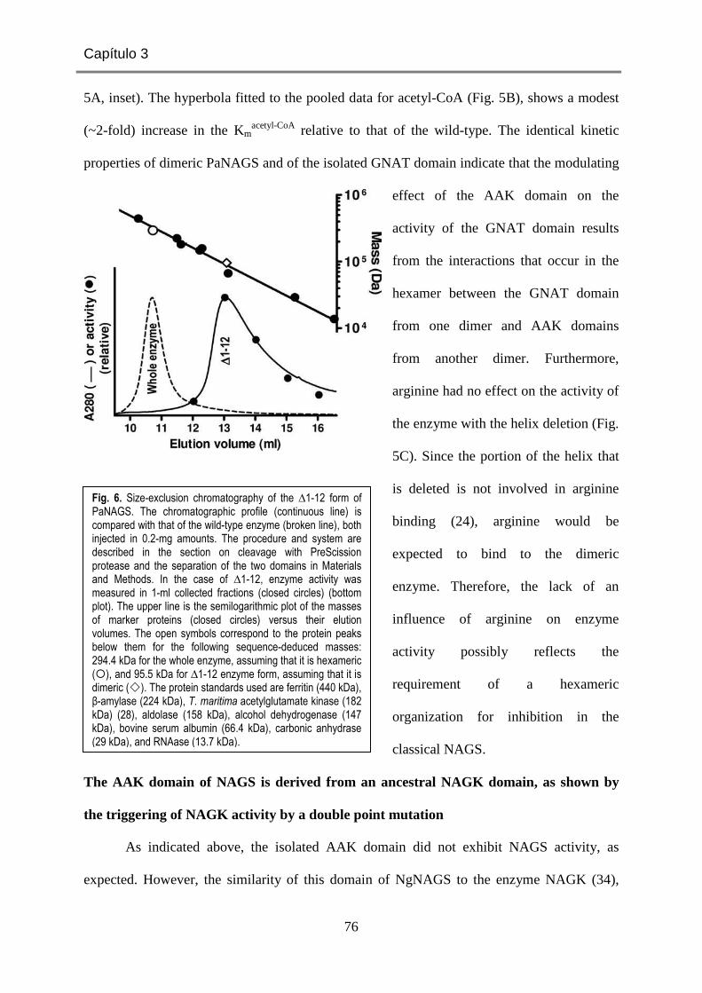
Capítulo 3
76
5A, inset). The hyperbola fitted to the pooled data for acetyl-CoA (Fig. 5B), shows a modest
(~2-fold) increase in the Kmacetyl-CoA relative to that of the wild-type. The identical kinetic
properties of dimeric PaNAGS and of the isolated GNAT domain indicate that the modulating
effect of the AAK domain on the
activity of the GNAT domain results
from the interactions that occur in the
hexamer between the GNAT domain
from one dimer and AAK domains
from another dimer. Furthermore,
arginine had no effect on the activity of
the enzyme with the helix deletion (Fig.
5C). Since the portion of the helix that
is deleted is not involved in arginine
binding (24), arginine would be
expected to bind to the dimeric
enzyme. Therefore, the lack of an
influence of arginine on enzyme
activity possibly reflects the
requirement of a hexameric
organization for inhibition in the
classical NAGS.
The AAK domain of NAGS is derived from an ancestral NAGK domain, as shown by
the triggering of NAGK activity by a double point mutation
As indicated above, the isolated AAK domain did not exhibit NAGS activity, as
expected. However, the similarity of this domain of NgNAGS to the enzyme NAGK (34),
Fig. 6. Size-exclusion chromatography of the ∆1-12 form of PaNAGS. The chromatographic profile (continuous line) is compared with that of the wild-type enzyme (broken line), both injected in 0.2-mg amounts. The procedure and system are described in the section on cleavage with PreScission protease and the separation of the two domains in Materials and Methods. In the case of ∆1-12, enzyme activity was measured in 1-ml collected fractions (closed circles) (bottom plot). The upper line is the semilogarithmic plot of the masses of marker proteins (closed circles) versus their elution volumes. The open symbols correspond to the protein peaks below them for the following sequence-deduced masses: 294.4 kDa for the whole enzyme, assuming that it is hexameric (�), and 95.5 kDa for ∆1-12 enzyme form, assuming that it is dimeric (�). The protein standards used are ferritin (440 kDa), β-amylase (224 kDa), T. maritima acetylglutamate kinase (182 kDa) (28), aldolase (158 kDa), alcohol dehydrogenase (147 kDa), bovine serum albumin (66.4 kDa), carbonic anhydrase (29 kDa), and RNAase (13.7 kDa).

J Bacteriol. (2012) 194, 2791-2801
77
with the preservation of the active center crevice (Fig. 7A), led us to investigate whether this
domain had any NAGK activity. In fact, a bifunctional NAGS/NAGK was identified
previously in Xanthomonas campestris (27), although its NAGK activity was quite low when
compared with its own NAGS activity or with the NAGK activities of classical bacterial
NAGKs (13,22).
We failed to detect any NAGK activity with PaNAGS, even at very high enzyme
concentrations (up to 0.16 mg/ml in the assay mixture; at this concentration, the X. campestris
enzyme would have consumed all the NAG present in the assay mixture). This lack of NAGK
activity might have been expected, since P. aeruginosa has a separate gene (argB) that
encodes a highly active arginine-sensitive NAGK (28). Similarly, we failed to detect any
NAGK activity with the isolated recombinant AAK domain of PaNAGS, even at
concentrations of 0.3 mg/ml in the assay mixture (Fig.7B and C; closed circles), excluding
that the lack of NAGK activity of the complete enzyme was due to some inhibitory effect of
the GNAT domain on that activity.
However, when we mutated two residues of the putative NAGK active center of the
AAK domain of PaNAGS to lysine (M26K/L240K double mutation), rAAK became active as
an NAGK. These residues were chosen because they are the counterparts of two invariant
NAGK active center lysines in PaNAGS (K8 and K217 of E. coli NAGK) (Fig. 7A) that play
key catalytic roles in acetylglutamate phosphorylation (16,29). Whereas no activity was
observed when only one of the two mutations was introduced into rAAK (results not shown),
the double mutant exhibited clear although low NAGK activity (Fig. 7B and C). The activity
depended hyperbolically on the ATP concentration (Fig. 7B) and also presented a nonlinear
dependency on the concentration of NAG (Fig. 7C), although the apparent affinity for this
substrate was too low to approach saturation. The apparent KmATP of the double mutant (1.6 ±
0.2 mM) was in the range typical for a bacterial NAGK in the same type of hydroxylamine-

Capítulo 3
78
based assay (13,18). In contrast, the KmNAG could not be estimated accurately, given its high
value, since the range of NAG concentrations used did not reach the levels needed for
saturation. In any case, from the data shown in Fig. 7C, it appears evident that the KmNAG and
the activity at saturation of this substrate are >0.225 M and >0.5 U/mg, respectively. Actually,
the best hyperbolic fit for the NAG data gives apparent KmNAG and Vmax values of ~0.8 M and
~1 U/mg, respectively. The Vmax value is not too far from the activity exhibited by the
Fig. 7. The AAK domain of NAGS is an ancestral NAGK. (A) Stereo view of the superimposition of the structure of the active center of E. coli NAGK (in green) bound to NAG and the ATP inert analog AMPPNP (PDB accession number 1GS5) (29) with the corresponding region of NgNAGS (in orange-brown) (PDB accession number 2R8V). The main chain of the structural elements (labeled) that make the site are shown. A few important residues are illustrated in the same color as the main chain (except the N atoms of the side chains, which are in blue) and are labeled. NAG and AMPPNP are shown in a stick representation, with C, P, O and N atoms shown in yellow, green, red and blue, respectively. (B and C) Dependency of the activity of the M26K/L240K double mutant (squares) of rAAK on ATP and NAG concentrations. The circle illustrates the lack of activity of the wild-type enzyme (0.3 mg/ml) assayed under the same conditions at concentrations of 20 mM ATP and 100 mM NAG. The hyperbola fitted to the points for variable ATP yields KmATP and V[ATP] = ∞ values of 1.6 ± 0.2 mM and 0.122 ± 0.003 U/mg, respectively. The nearly linear NAG concentration dependency of the activity indicates that the KmNAG and the V[NAG] = ∞ values exceed 0.225 M and 0.5 U/mg, respectively. The curve fitted over the points would correspond to the hyperbola for KmNAG and V[NAG] = ∞ values of
0.83 M and 1.1 U/mg, respectively.

J Bacteriol. (2012) 194, 2791-2801
79
bifunctional X. campestris enzyme (27). However, the KmNAG is much higher than that for
genuine NAGKs (13,18), possibly reflecting a tendency toward a closed conformation of the
NAG site of the AAK domain of NAGS, as revealed by the lowered position of the lid for this
site (the β3-β4 hairpin) (Fig. 7A) in the NgNAGS structure (24) despite the emptiness of this
site, whereas in NAGK, this site closes down only when NAG is bound (15).
How and why did two-domain NAGSs emerge?
The closeness of the AAK domains of NAGS and NAGK goes beyond the overall
domain fold, extending to the similarity of the NAGK active center crevice (Fig. 7A) and
even to the conservation of a key NAG-binding arginine (R66 of E. coli NAGK, R85 of
NgNAGS [Fig. 7A], and R82 of PaNAGS) (29,34). Indeed, although with a low affinity, the
AAK domain of NAGS binds NAG, as attested by the NAGK activity of its M26K/L240K
double mutant despite the fact that these mutations do not affect NAG site residues. An
invariant aspartate (D162 in E. coli NAGK), which in NAGKs has a key active center-
organizing role by coordinating the two catalytic lysines and by binding MgATP (16,29), is
also preserved in many NAGSs as a glutamate (E185 of PaNAGS) although not in NgNAGS
(34). The NAGK activity of the M26K/L240K double mutant indicates that classical NAGS
lost its ancestral NAGK activity primarily because of the mutation of these two catalytic
lysines (16,22,29). Indeed, as expected from its NAGK activity, the bifunctional
NAGS/NAGK of X. campestris has these two lysines preserved (27).
Since Mycobacterium tuberculosis NAGS is merely a GNAT domain and yet it is
feed-back inhibited by arginine (12), the AAK-GNAT domain organization of the classical
bacterial NAGS is clearly unessential for arginine-regulated NAG synthesis from acetyl-CoA
and glutamate. A previously reported proposal by Xu et al. (37) that the classical two-domain
NAGSs arose from the fusion of a NAGS similar to that found in M. tuberculosis and an
arginine-sensitive NAGK is supported very strongly by our finding that a mere two-residue

Capítulo 3
80
mutation in the AAK domain of PaNAGS can render this domain an NAGK. This proposal is
also supported by the closeness of the structures of NAGK (28,29) and the AAK domain of
NgNAGS (34) (Fig. 7A), and by the discovery of bifunctional NAGS/NAGK having the same
domain organization as that of PaNAGS (27). A question remaining to be answered
concerning the evolutionary process from single-domain to two-domain NAGS is whether the
arginine sensitivity of M. tuberculosis NAGS (12) was a late evolutionary acquisition or
whether this trait of the GNAT component was originally present and has been lost over the
course of the evolution of two-domain NAGSs.
A major advantage of associating AAK and GNAT domains into a two-domain NAGS
that has become patent in our studies is that because of the important modulating role of the
AAK domain on the activity of the GNAT domain, the Km for glutamate is brought down
from the very high values for the M. tuberculosis enzyme (12) to the millimolar range that is
characteristic of the canonical microbial enzymes (17,23,24,27,32). This value for these
enzymes is closer to the range of glutamate concentrations present in bacteria (see, for
example, reference 25), thus increasing the efficiency of the enzyme in the catalysis of the
reaction in vivo. Another potential reason for the shift from a single-domain to a two-domain
NAGS may be related to NAG channelling between NAGS and NAGK, since such
channelling would prevent unwanted NAG hydrolysis by aminoacylases such as those
prevalent in animals (30) or existing (although nowadays in the periplasmic space) in P.
aeruginosa (14). This channelling requires the direct interaction of the GNAT domain of one
subunit with the AAK domain of another subunit, as observed for hexameric NAGS.
Reminiscent of such channelling might be the association in Saccharomyces cerevisiae of
NAGS and NAGK, both having the two-domain composition of PaNAGS (27), to form a
metabolon when NAG is produced by NAGS (1,26). It would be important to clarify whether
such channelling occurs in this metabolon and/or in bifunctional NAGS/NAGK.

J Bacteriol. (2012) 194, 2791-2801
81
A third potential advantage of the two-domain organization of NAGS is the possibility
of easily modulating the arginine regulation of NAGS activity. In two-domain NAGS,
arginine regulation results from primary arginine-triggered changes in the architecture of the
AAK domain hexamer (24). These changes are similar to those observed previously for
hexameric NAGK upon arginine binding to this enzyme (28). Since with the latter enzyme, a
signalling protein, PII, was shown previously to modulate the sensitivity of the enzyme to
arginine inhibition (21), it cannot be excluded that analogous regulatory mechanisms may
exist for modulating the arginine sensitivity of canonical hexameric NAGSs. Furthermore, the
key role of the interdomain linker as a mediator of the effects of arginine that has been
revealed by our previous (33) and present studies provides the basis for adapting arginine
regulation by changes merely in the 5-residue linker sequence. This endows AAK-GNAT
domain NAGSs with an enormous potential for adaptation to the specific physiological needs
of different organisms. The best example of this ability to adapt is provided by the change in
the effect of arginine on NAGS from inhibition to activation with the shift of animals from
marine life to terrestrial ureotelism (19), a change that we have partially reproduced by linker
manipulation in the present studies.
ACKNOWLEDGEMENTS
This work was supported by grants BFU2008-05021 of the Spanish Ministry of
Science (MEC and MICINN) and Prometeo/2009/051 of the Valencian Government. E.S.-V.
and M.L.F.-M. were supported by contracts from the Instituto de Salud Carlos III and the
JAE-DOC Program of the Consejo Superior de Investigaciones Científicas. We thank N.
Gougeard (IBV-CIBERER) and the late E. Cogollos (IBV-CSIC) for technical assistance and
F. Gil-Ortiz (IBV-CSIC) for help with Fig. 2B and 7A.

Capítulo 3
82
REFERENCES
1. Abadjieva A, Pauwels K, Hilven P, Crabeel M. 2001. A new yeast metabolon involving at least the two first enzymes of arginine biosynthesis: acetylglutamate synthase activity requires complex formation with acetylglutamate kinase. J. Biol. Chem. 276: 42869-42880.
2. Alonso E, Rubio V. 1989. Participation of ornithine aminotransferase in the synthesis and catabolism of ornithine in mice. Studies using gabaculine and arginine deprivation. Biochem. J. 259:131-138.
3. Bachmann C, Krähenbühl S, Colombo JP. 1982. Purification and properties of acetyl-CoA:L-glutamate N-acetyltransferase from human liver. Biochem. J. 205:123-127.
4. Bradford MM. 1976. A rapid and sensitive method for the quantitation of microgram quantities of protein utilizing the principle of protein-dye binding. Anal. Biochem. 72:248–254.
5. Briozzo P, Evrin C, Meyer P, Assairi L, Joly N, Barzu O, Gilles AM. 2005. Structure of Escherichia coli UMP kinase differs from that of other nucleoside monophosphate kinases and sheds new light on enzyme regulation. J. Biol. Chem. 280:25533-25540.
6. Caldovic L, Lopez GY, Haskins N, Panglao M, Shi D, Morizono H, Tuchman M. 2006. Biochemical properties of recombinant human and mouse N-acetylglutamate synthase. Mol. Genet. Metab. 87:226-232.
7. Caldovic L, Morizono H, Tuchman M. 2007. Mutations and polymorphisms in the human N-acetylglutamate synthase (NAGS) gene. Hum. Mutat. 28:754-759.
8. Caldovic L, Tuchman M. 2003. N-acetylglutamate and its changing role through evolution. Biochem. J. 372:279-290.
9. Cordingley MG, Callahan PL, Sardana VV, Garsky VM, Colonno RJ. 1990. Substrate requirements of human rhinovirus 3C protease for peptide cleavage in vitro. J. Biol. Chem. 265:9062-9065.
10. Cunin R, Glansdorff N, Pierard A, Stalon V. 1986. Biosynthesis and metabolism of arginine in bacteria. Microbiol. Rev. 50:314–352.
11. Emsley P, Cowtan K. 2004. Coot: model-building tools for molecular graphics. Acta Crystallogr. D Biol. Crystallogr. 60:2126-2132.
12. Errey JC, Blanchard JS. 2005. Functional characterization of a novel ArgA from Mycobacterium tuberculosis. J. Bacteriol. 187:3039–3044.
13. Fernández-Murga ML, Gil-Ortiz F, Llácer JL, Rub io V. 2004. Arginine biosynthesis in Thermotoga maritima: characterization of the arginine-sensitive N-acetyl-L-glutamate kinase. J. Bacteriol. 186:6142-6149.
14. Früh H, Leisinger T. 1981. Properties and localization of N-acetylglutamate deacetylase from Pseudomonas aeruginosa. J. Gen. Microbiol. 125:1-10.

J Bacteriol. (2012) 194, 2791-2801
83
15. Gil-Ortiz F, Ramón-Maiques S, Fernández-Murga ML, Fita I, Rubio V. 2010. Two crystal structures of Escherichia coli N-acetyl-L-glutamate kinase demonstrate the cycling between open and closed conformations. J. Mol. Biol. 399:476-490.
16. Gil-Ortiz F, Ramón-Maiques S, Fita I, Rubio V. 2003. The course of phosphorus in the reaction of N-acetyl-L-glutamate kinase, determined from the structures of crystalline complexes, including a complex with an AlF4
- transition state mimic. J. Mol. Biol. 331:231-244.
17. Haas D, Kurer V, Leisinger T. 1972. N-acetylglutamate synthetase of Pseudomonas aeruginosa. An assay in vitro and feed-back inhibition by arginine. Eur. J. Biochem. 31:290-295.
18. Haas D, Leisinger T. 1975. N-acetylglutamate 5-phosphotransferase of Pseudomonas aeruginosa. Catalytic and regulatory properties. Eur. J. Biochem. 52:377-393.
19. Haskins N, Panglao M, Qu Q, Majumdar H, Cabrera-Luque J, Morizono H, Tuchman M, Caldovic L. 2008. Inversion of allosteric effect of arginine on N-acetylglutamate synthase, a molecular marker for evolution of tetrapods. BMC Biochem. 9:24.
20. Laemmli, UK. 1970. Cleavage of structural proteins during the assembly of the head of bacteriophage T4. Nature. 227:680-685.
21. Llácer JL, Contreras A, Forchhammer K, Marco-Marín C, Gil-Ortiz F, Maldonado R, Fita I, Rubio V. 2007. The crystal structure of the complex of PII and acetylglutamate kinase reveals how PII controls the storage of nitrogen as arginine. Proc. Natl. Acad. Sci. U S A 104:17644-17649.
22. Marco-Marín C, Ramón-Maiques S, Tavárez S, Rubio V. 2003. Site-directed mutagenesis of Escherichia coli acetylglutamate kinase and aspartokinase III probes the catalytic and substrate-binding mechanisms of these amino acid kinase family enzymes and allows three-dimensional modelling of aspartokinase. J. Mol. Biol. 334:459-476.
23. Marvil DK, Leisinger T. 1977. N-acetylglutamate synthase of Escherichia coli: purification, characterization, and molecular properties. J. Biol. Chem. 252:3295-3303.
24. Min L, Jin Z, Caldovic L, Morizono H, Allewell NM, Tuchman M, Shi D. 2009. Mechanism of allosteric inhibition of N-acetyl-L-glutamate synthase by L-arginine. J. Biol. Chem. 284:4873-4880.
25. Ogahara T, Ohno M, Takayama M, Igarashi K, Kobayashi H. 1995. Accumulation of glutamate by osmotically stressed Escherichia coli is dependent on pH. J. Bacteriol. 177:5987-5990.
26. Pauwels K, Abadjieva A, Hilven P, Stankiewicz A, Crabeel M. 2003. The N-acetylglutamate synthase/N-acetylglutamate kinase metabolon of Saccharomyces cerevisiae allows co-ordinated feed-back regulation of the first two steps in arginine biosynthesis. Eur. J. Biochem. 270:1014-1024.

Capítulo 3
84
27. Qu Q, Morizono H, Shi D, Tuchman M, Caldovic L. 2007. A novel bifunctional N-acetylglutamate synthase-kinase from Xanthomonas campestris that is closely related to mammalian N-acetylglutamate synthase. BMC Biochem. 8:4.
28. Ramón-Maiques S, Fernández-Murga ML, Gil-Ortiz F, Vagin A, Fita I, Rubio V. 2006. Structural bases of feed-back control of arginine biosynthesis, revealed by the structures of two hexameric N-acetylglutamate kinases, from Thermotoga maritima and Pseudomonas aeruginosa. J. Mol. Biol. 356:695–713.
29. Ramón-Maiques S, Marina A, Gil-Ortiz F, Fita I, Rubio V. 2002. Structure of acetylglutamate kinase, a key enzyme for arginine biosynthesis and a prototype for the amino acid kinase enzyme family, during catalysis. Structure. 10:329-342.
30. Reglero A, Rivas J, Mendelson J, Wallace R, Grisolia S. 1977. Deacylation and transacetylation of acetyl glutamate and acetyl ornithine in rat liver. FEBS. Lett. 81:13-17.
31. Rubio V, Ramponi G, Grisolia S. 1981. Carbamoyl phosphate synthetase I of human liver. Purification, some properties and immunological cross-reactivity with the rat liver enzyme. Biochim. Biophys. Acta. 659:150-160.
32. Sancho-Vaello E, Fernández-Murga ML, Rubio V. 2008. Site-directed mutagenesis studies of acetylglutamate synthase delineate the site for the arginine inhibitor. FEBS Lett. 582:1081-1086.
33. Sancho-Vaello E, Fernández-Murga ML, Rubio V. 2009. Mechanism of arginine regulation of acetylglutamate synthase, the first enzyme of arginine synthesis. FEBS Lett. 583:202-206.
34. Shi D, Sagar V, Jin Z, Yu X, Caldovic L, Morizono H, Allewell NM, Tuchman M. 2008. The crystal structure of N-acetyl-L-glutamate synthase from Neisseria gonorrhoeae provides insights into mechanisms of catalysis and regulation. J. Biol. Chem. 283:7176-7184.
35. Slocum RD. 2005. Genes, enzymes and regulation of arginine biosynthesis in plants. Plant. Physiol. Biochem. 43:729-745.
36. Sonoda T, Tatibana M. 1983. Purification of N-acetyl-L-glutamate synthetase from rat liver mitochondria and substrate and activator specificity of the enzyme. J. Biol. Chem. 258:9839-9844.
37. Xu Y, Glansdorff N, Labedan B. 2006. Bioinformatic analysis of an unusual gene-enzyme relationship in the arginine biosynthetic pathway among marine gamma proteobacteria: implications concerning the formation of N-acetylated intermediates in prokaryotes. BMC Genomics. 7:4.

J Bacteriol. (2012) 194, 2791-2801
85
SUPPORTING INFORMATION (CAPÍTULO 3)
Functional dissection of N-acetylglutamate synthase (ArgA) of
Pseudomonas aeruginosa and restoration of its ancestral N-
acetylglutamate kinase activity
Enea Sancho-Vaello, María L. Fernández-Murga and Vicente Rubio
Instituto de Biomedicina de Valencia (IBV-CSIC) and Centro de Investigación Biomédica en Red
de Enfermedades Raras (CIBERER-ISCIII), C/ Jaime Roig 11, 46010 Valencia, Spain
Contains:
Supplementary Figure S1
Supplementary Table S1

Capítulo 3
86
Supplementary Table S1. Synthetic oligonucleotides used in this work. Mutations are underlined. The ATG for the initial Met is in bold-type. Restriction sites are highlighted in grey background. Italic lettering marks the beginning or the end of the open reading frame.
Direction Sequence
Whole domains
GNAT Forward 5´GCCCAGGAGCAGCATATGCAGTTGCGCGAGGCG 3´
Reverse 5´GGACGCCTCGAGGGATCCCAGGCTCTTCTCG 3´
rGNAT Forward 5´GTTTAACTTTAAGAAGGGTCTCTAGGTATGCAGTTGCGCGAGGCG3´
Reverse 5´GTGGTGCTCGAGGGATCCTTACAGGCTCTTCTCGAATACC 3´
rAAK Forward 5´GCAGTTCGAGTAGTTGCGCGAGGCGGGC 3´
Reverse 5´CGCCTCGCGCAACTACTCGAACTGCTC 3´
∆1-12a Forward 5´GGTTACGTCACCATATGCCCTACATCAACTCG 3´
Linker changesb
ERM Forward 5´GGTCGCCCAGGAGCGGATGGAGCAGTTGCG 3´
Reverse 5´CAACTGCTCCATCCGCTCCTGGGCGACCAG 3´
AQF Forward 5´CTGGTCGCCCAGGCACAGTTCGAGCAGTTG 3´
Reverse 5´CTGCTCGAACTGTGCCTGGGCGACCAGGG 3´
EQM Forward 5´GCCCAGGAGCAGATGGAGCAGTTGCGCGAG 3´
Reverse 5´GCGCAACTGCTCCATCTGCTCCTGGGCGAC 3´
ERF Forward 5´GCCCAGGAGAGGTTCGAGCAGTTGCGC 3´
Reverse 5´CAACTGCTCGAACCTCTCCTGGGCGAC 3´
EAQGP Forward 5´CAGGAGGCACAGGGTCCCGAGCAGTTGCGCGAG 3´
Reverse 5´GCGCAACTGCTCGGGACCCTGTGCCTCCTGGGC 3´
LFQGP Forward 5´CTGGTCGCCCAGCTGTTTCAGGGTCCCGAGCAGTTG 3´
Reverse 5´CTGCTCGGGACCCTGAAACAGCTGGGCGACCAGGG 3´
LEVLFQGP Forward 5´GCACCCTGGTCGCCCAGCTGGAAGTTCTGTTTCAGGGTCCCGAGC3´
Reverse 5´CGGGACCCTGAAACAGAACTTCCAGCTGGGCGACCAGGGTGCCG3´
rAAK point mutations
L240K Forward 5´GCTACCAGGCCGAGAAGCTGGACGCCGCG 3´
Reverse 5´GGCGTCCAGCTTCTCGGCCTGGTAGCTG 3´
M26K Forward 5´CACCTTCGTGGTCAAGCTCCCCGGCGAAGGG 3´
Reverse 5´CTTCGCCGGGGAGCTTGACCACGAAGGTGCG 3´
a This forward oligonucleotide was combined with the GNAT domain reverse oligonucleotide b Changes as defined in Fig. 2B.

J Bacteriol. (2012) 194, 2791-2801
87
(A)
(B)
Supplementary Figure S1. The structures of (A) the NAGS from N. gonorrhoeae (PDB file 2R8V) and (B) of NAGK from P. aeruginosa (PDB file 2BUF) to illustrate grossly their structural similarity. They are viewed along their threefold axes, with each dimer colored differently and both subunits of each dimer in different color hues. AAK and GNAT domains are shown, respectively, in cartoon and surface representation. In the case of NAGS, to avoid occluding the view of the AAK domains, the GNAT domains are semi-transparent, and those in the background are fainter.

Capítulo 3
88

89
Resumen de los resultados

90

Resumen de los resultados
91
Para aumentar el conocimiento estructural y funcional sobre la NAGS, hemos
realizado estudios centrados en el enzima bacteriano clásico de P. aeruginosa (PaNAGS), que
han generado tres publicaciones que constitutyen los tres capítulos de Resultados que
resumimos aquí individualmente, condensando los resultados de cada capítulo por separado.
Para realizar estos estudios hemos utilizado extensamente técnicas de clonación y
expresión heteróloga de genes; de mutagénesis dirigida e ingeniería de proteínas; de manejo,
purificación y análisis de proteínas; ensayos enzimáticos y estudios cinéticos; y tecnologías de
cristalización en gota colgante mediante equilibrio de vapor, si bien los cristales obtenidos
aún no han dado sus frutos.
Además de los resultados resumidos aquí, hemos realizado mucho trabajo con la
NAGS humana, trabajo todavía en curso, por lo que sólo daremos noticia breve del mismo en
un apartado apropiado de la Discusión.
Capítulo 1: Site-directed mutagenesis studies of acetylglutamate synthase delineate the
site for the arginine inhibitor. Enea Sancho-Vaello, M. Leonor Fernández-Murga y Vicente
Rubio. Trabajo publicado en FEBS Letters (2008) 582: 1081-1086.
La hipótesis principal de este trabajo era que el sitio para la arginina de la NAGS
bacteriana clásica (ArgA) era el predicho por nuestro grupo por comparación con la NAGK
sensible a arginina (48). Para corroborar esta hipótesis, que estaba basada en el alineamiento
de los dominios AAK de NAGS y NAGK, se eligió la estrategia de mutar residuos clave del
sitio potencial para arginina inferido a partir de la comparación de dichas secuencias
alineadas.
Para poder abordar los experimentos de mutagénesis dirigida fue preciso en primer
lugar clonar el gen argA que codifica para la NAGS de P. aeruginosa [identificado en la base
de datos TIGR (http://cmr.jcvi.org/tigr-scripts/CMR/CmrHomePage.cgi) como PA5204])

Resumen de los resultados
92
mediante una metodología de PCR, para luego ligarlo en un plásmido de expresión de tipo
pET22b para expresarlo en forma recombinante en E. coli, modificando el enzima mediante la
adición C-terminal de la etiqueta de polihistidinas GSLEHHHHHH para facilitar su
purificación. Fue difícil la expresión en forma soluble de la proteína en células de E. coli
BL21. Requirió el uso de estrés por frío, etanol, concentraciones bajas del inductor IPTG y
expresión prolongada a 15ºC. Con ello conseguimos la expresión abundante, soluble y
reproducible de la PaNAGS.
El ensayo de actividad utilizado se basó en el de Errey y Blanchard (59) que explota
la detección colorimétrica del grupo tiol del producto CoA mediante la utilización de reactivo
de Ellman (60). Mediante este ensayo se pudo probar que el enzima presentaba la actividad y
especifidad esperadas, que la cinética para ambos sustratos a las concentraciones utilizadas
era hiperbólica, y que la arginina inhibía virtualmente toda la actividad con un patrón
sigmoide de dependencia de decrecimiento de actividad frente a la concentración creciente de
arginina. Aunque no estudiado en detalle, la arginina influyó negativamente sobre la cinética
para el glutamato.
De acuerdo con la predicción realizada de una arquitectura hexamérica similar a la de
la NAGK (48), los resultados de gel filtración dieron para la NAGS la masa esperada para un
hexámero tanto en ausencia como en presencia de 10 mM arginina. A partir del alineamiento
con la NAGK de P. aeruginosa se predijo el sitio de la arginina de la PaNAGS. Las
mutaciones a alanina de cuatro residuos pertenecientes al sitio predicho, Tyr14 de la hélice N-
terminal que predijimos interacciona con el Cα de la arginina, Lys199, que predijimos
interacciona con el α-carboxilato, y Glu269 y Gly275, de la zona propuesta como de
interacción con la región guanidinio de la arginina, tuvieron efectos drásticos sobre el proceso
de inhibición, sin afectar gravemente los parámetros cinéticos para los sustratos en ausencia
de arginina. En particular, las mutaciones de los dos residuos que rodean a la porción

Resumen de los resultados
93
guanidinio de la arginina abolieron la inhibición, mientras que los residuos de interacción con
la porción del carbono y carboxilato α disminuyeron casi dos órdenes de magnitud la afinidad
aparente para el efector. Estos resultados nos permitieron confirmar que la forma de unión de
la arginina era la misma en NAGK y en NAGS, es decir, que tanto las hélices H y N como el
lazo αH-β16 estaban conservados y eran elementos cruciales para la unión del efector
alostérico arginina. Concretamente, en el sitio para la arginina de PaNAGS, Lys199 y Glu269
serían residuos de interacción directa con la arginina mientras que Gly275 conferiría la
flexibilidad necesaria para la formación del sitio de unión para el efector. En la hélice N
terminal, Tyr14 actuaría como una tapa cerrando el sitio.
El disponer de enzima recombinante susceptible de ser mutado a voluntad nos llevó
también a tratar de establecer de forma concluyente el papel del dominio GNAT mediante
mutagénesis dirigida de residuos potencialmente implicados en la unión de sustratos o en la
catálisis. Recuérdese que cuando se realizaron estos experimentos todavía no se disponía de
ninguna estructura de ArgA. Los resultados obtenidos mutando el Glu352 y la Val358
elevaron muy fuertemente el Km aparente para glutamato, estableciendo claramente el papel
del dominio GNAT en la unión de este sustrato. Como estas y otras mutaciones de dicho
dominio produjeron descensos moderados en la Vmax e incrementos sustanciales aunque
modestos en el Km aparente para AcCoA, todos los resultados para los mutantes del dominio
GNAT están de acuerdo con la implicación de este dominio en la unión de ambos sustratos y
en la catálisis, lo que está plenamente de acuerdo con los resultados estructurales posteriores
(52,61).
Todavía explotamos la PaNAGS recombinante con una finalidad aplicada, relativa al
déficit humano de NAGS. Aunque el grado de identidad de secuencia entre las NAGSs
bacteriana clásica y humana no es alto, es posible alinear sus secuencias con un grado
relativamente alto de seguridad, habiendo procedido a introducir en PaNAGS dos mutaciones

Resumen de los resultados
94
clínicas que fueron identificadas en la NAGS humana y que afectan a residuos conservados en
el alineamiento entre ambas NAGSs. Una de estas mutaciones (Leu353Val, correspondiendo
a la mutación humana Leu442Val) tuvo un efecto modesto que está de acuerdo con la forma
tardía de deficiencia a la que se encontró asociada (36). La otra mutación, sin embargo, de
efectos no caracterizados en humanos, Gly236Cys (36), cuyo correlato bacteriano es la
mutación Gly146Cys en el dominio AAK, produjo un fuerte aumento en el Km para glutamato
así como una disminución a 1/3 de la Vmax del enzima. Hemos interpretado estos resultados
para el mutante Gly146Cys como indicativos de que el dominio AAK es un modulador claro
de la actividad de la enzima, actividad de la que es responsable en forma exclusiva el dominio
GNAT. Estudios de disección del enzima en sus dominios constituyentes detallados en el
Capítulo 3 de los resultados han confirmado plenamente esta propuesta.
La correlación de resultados clínicos y mutaciones experimentales en la NAGS
bacteriana nos ha incitado a intentar identificar en el enzima humano el sitio para arginina,
habiendo descrito en este trabajo tal identificación, con conservación de tres de los cuatro
residuos que hemos mutado aquí en dicho sitio (la Tyr14 de PaNAGS no está conservada en
la NAGS humana). Concluimos que el sitio de unión de la arginina en la NAGS humana
debía ser el mismo que en la NAGK y NAGS bacteriana clásica, a pesar de que en la enzima
bacteriana la arginina actuaba como inhibidor mientras que en la enzima humana actuaba
como activador. De hecho, una forma mutante clínica (V350I, NAGS humana) del sitio
propuesto para la arginina parece abolir la activación por este aminoácido (36).
Capítulo 2. Mechanism of arginine regulation of acetylglutamate synthase, the first
enzyme of arginine synthesis. Enea Sancho-Vaello, M. Leonor Fernández-Murga y Vicente
Rubio. Trabajo publicado en FEBS Letters (2009) 583: 202-206.

Resumen de los resultados
95
Tras demostrar que la arginina se une en el dominio AAK de un modo similar a como
lo hace en la NAGK, y dado que todo indica (52, 53, 61, 62) que el dominio GNAT es el que
ejerce la catálisis en exclusiva, mientras que el dominio AAK parece tener una acción
moduladora de la actividad, cabía preguntarse cómo la arginina, uniéndose a un sitio en el
dominio AAK, lejos del dominio GNAT (distancia > 25Å) puede modificar la actividad del
enzima y por ende del dominio GNAT.
Propusimos que, en consonancia con el mecanismo de inhibición por la arginina de la
NAGK (48), el anillo de dominios AAK sufriría una expansión al unirse la arginina a la
NAGS. Ello produciría tracción sobre el dominio GNAT a través del conector que lo une al
dominio AAK de su propia subunidad. Dicha tracción resultaría en un movimiento que
modificaría la relación entre el dominio GNAT y el dominio AAK de la subunidad del dímero
vecino sobre el que reposa. Este cambio en las relaciones entre ambos dominios alteraría la
modulación de la actividad del dominio. Si este mecanismo fuera correcto, el conector
interdominios debería desempeñar un papel clave, y habría que esperar que cambios en la
longitud de dicho conector tuvieran un efecto importante sobre dicha modulación de la
actividad y sobre la inhibición por arginina.
Para someter a prueba esta hipótesis elongamos el conector mediante la adición de uno
o dos residuos de alanina en la zona central del mismo (secuencia natural en P. aeruginosa,
QEQFEQ; formas elongadas, QEQAFEQ y QEAQAFEQ), o lo acortamos mediante la
eliminación de uno o dos residuos (QEFEQ y QFEQ), determinando las consecuencias de
estos cambios sobre la actividad, parámetros cinéticos e inhibición por arginina. Las
elongaciones tuvieron un efecto modesto sobre la actividad o sobre los parámetros cinéticos
del enzima en ausencia de arginina, aunque disminuyeron sustancialmente la Vmax aparente.
Sin embargo, tal como cabía esperar de la presencia de un conector más largo, que debería
dificultar la correcta tracción sobre el dominio GNAT cuando el anillo de dominios AAK se
expandiera por la unión de la arginina, la inhibición por la arginina se vio abolida, con

Resumen de los resultados
96
evidencias estadísticas de que en realidad su efecto revertía a una ligera activación. Un
análisis detallado reveló que esta falta de efecto de la arginina se traducía en ausencia
significativa de impacto del efector sobre los parámetros cinéticos para los sustratos, a
diferencia de lo que sucedía con el enzima intacto, en el que la arginina producía, por
ejemplo, una importante disminución en la afinidad aparente por el glutamato.
En cambio, las mutaciones que acortaron el conector inhibieron constitutivamente al
enzima sin necesidad de añadir arginina, como consecuencia principalmente de la elevación
del Km para glutamato pero también induciendo, igualmente que la arginina, inhibición por el
sustrato acetil-CoA, eliminación de la inhibición por alta concentración del sustrato glutamato
y decrecimiento en Vmax.
Así, mediante mutantes de elongación y acortamiento del conector interdominios,
mostramos que dicho conector es una pieza clave en el mecanismo de modulación por
arginina, sugiriendo incluso que cambios en la relación entre el dominio GNAT y el dominio
AAK puedan ser responsables no sólo de la inhibición de la forma bacteriana de NAGS, sino
incluso de la activación de las formas de mamíferos, incluida la humana.
Capítulo 3. Functional dissection of N-acetylglutamate synthase (ArgA) of Pseudomonas
aeruginosa and restoration of its ancestral N-acetylglutamate kinase activity. Enea
Sancho-Vaello, María L. Fernández-Murga y Vicente Rubio. Trabajo publicado en Journal of
Bacteriology (2012) 194: 2791-2801.
Este capítulo refleja nuestros estudios sobre la separación de ambos dominios de la
PaNAGS, en los que examinamos la forma oligomérica y función de cada dominio. Para ello
se intentó su expresión independiente, con éxito inicial sólo para el dominio AAK, pudiendo
finalmente producir el dominio GNAT fusionado en su extremo N-terminal a SUMO (de Life
Sensors Inc.). Antes de tener éxito con la expresión recombinante de este último dominio,

Resumen de los resultados
97
intentamos otra estrategia basada en la introducción en el conector interdominios de un sitio
de corte para la proteasa comercial PreScission (GE Healthcare), que es altamente específica.
La introducción del sitio de corte, que suponía un cambio de secuencia y un alargamiento del
conector, se realizó en varios pasos, aprovechando las construcciones para profundizar más
allá de nuestro análisis previo (63) de los efectos de los cambios en el conector sobre la
actividad del enzima y sobre la influencia de la arginina.
Un hallazgo de particular relevancia en este contexto fue que el cambio de la
secuencia natural del conector, EQF, a la secuencia elongada y distinta LFQGP, y sobre todo
a la forma EAQGP, convirtió la arginina de potente inhibidor a modesto pero sustancial
activador, reproduciendo de este modo el cambio a activación por arginina que se asocia en la
NAGS al paso al ureotelismo terrestre (27). Intentamos emular la secuencia del conector de la
forma humana, sustituyendo la secuencia de la bacteriana, EQF, por la secuencia ERM. Este
cambio abolió la inhibición por arginina y, de hecho dio resultados que aunque no siendo
significativos estadísticamente, revelan una tendencia a la activación. Estos resultados y otros
de cambio aminoácido a aminoácido revelan que no sólo la longitud del conector es
importante para determinar los efectos de la arginina, sino también su secuencia, hecho éste
que hemos interpretado principalmente en base a la presencia de una fenilalanina en uno de
los residuos del conector bacteriano.
Al analizar el por qué la mutante con la secuencia del conector EAQGP se activaba
por la arginina, se evidenció que un factor clave era la concentración de glutamato, siendo la
arginina a baja concentración de glutamato un inhibidor y a partir de 20 mM glutamato un
activador para dicho mutante, indicando que la arginina podía ejercer un efecto activador o
inhibidor dependiendo de la concentración de glutamato.
La obtención de los dominios aislados también nos proporcionó información acerca
del enzima. El hecho de que el dominio GNAT aislado tras el corte con PreScission o
producido en forma recombinante como fusión SUMO-GNAT, fuera activo y exhibiera la

Resumen de los resultados
98
misma actividad enzimática por subunidad confirmó que el dominio GNAT por sí mismo es
catalítico, sin requerir de otros elementos para unir los sustratos y catalizar la reacción. Sin
embargo, la afinidad aparente para el glutamato fue muy baja e idéntica para cualquiera de
estas formas de dominio GNAT aislado, o incluso cuando se había cortado el conector pero
no se había separado del dominio AAK, lo que confirma (62) que el dominio AAK en la
subunidad completa aumenta fuertemente la afinidad aparente del enzima por el glutamato e
incluso aumenta algo la kcat. Además, tanto el dominio GNAT aislado por gel filtración tras el
corte con PreScission como el recombinante fusionado a SUMO fueron insensibles a la
inhibición por arginina, de acuerdo con lo esperado si la arginina se une al dominio AAK,
como indican nuestros resultados previos (62) y los hallazgos cristalográficos con el enzima
de N. gonorrhoeae (53).
El dominio AAK aislado fue inactivo y se eluyó de una columna de gel filtración
como un hexámero estable, lo que prueba que son las interacciones entre dímeros de dominios
AAKs las que determinan la formación del anillo hexamérico en la NAGS. Por el contrario, el
dominio GNAT se comportó como un monómero o dímero (la resolución de masas de la
columna utilizada no permitió diferenciar claramente entre ambos).
La importante influencia del dominio AAK sobre la actividad del dominio GNAT
podría deberse bien a interacciones del dominio GNAT con el dominio AAK de la misma
subunidad o a interacciones con el dominio AAK del dímero vecino. Ambos tipos de
interacciones se han demostrado en estudios cristalográficos con la NAGS de N. gonorrhoeae
(52,53). Para diferenciar entre ambas posibilidades delecionamos la porción primera de la
hélice N-terminal, disociando así el enzima a dímeros (corroborado por gel filtración). Estos
dímeros fueron activos, pero su actividad (expresada como número de recambio) por
subunidad y dependencia de la concentración del glutamato fueron idénticas a las del dominio
GNAT aislado, habiendo perdido también la capacidad de ser inhibidas por arginina a pesar
Resumen de los resultados
99
de que su sitio para arginina debió de permanecer intacto. Por tanto, la activación del dominio
GNAT por el dominio AAK se debe a las interacciones entre uno y otro dominio de dímeros
distintos, y la inhibición por arginina requiere de la preservación de la estructura hexamérica.
Una cuestión que está por resolver es de dónde surgió el dominio AAK de la NAGS.
Demostramos en este capítulo que su origen está directamente relacionado con la enzima
NAGK, ya que la introducción de dos mutaciones en este dominio de la NAGS se ha
acompañado de la aparición de actividad NAGK catalizada por este dominio, con un
razonable Km para ATP aunque con un Km muy elevado para glutamato. Estos resultados
apoyan un modelo de evolución de NAGS en el que ArgA surgió por fusión de un dominio
NAGK y de otro dominio de tipo GNAT, siendo originalmente una forma bifuncional
NAGS/NAGK quizá similar en su bifuncionalidad a la encontrada recientemente en
Xanthomonas campestris (15).

100

101
Discusión general

102

Discusión general
103
El origen de ArgA
El panel izquierdo de la Fig. 1 de esta sección [correspondiente a la portada del
número del J. Bacteriol. en el que se publicó el Capítulo 3 de los resultados (64)] representa
una superposición de la estructura de la NAGS de N. gonorrhoeae (forma coloreada) con la
de la NAGK de P. aeruginosa (forma en cintas de color gris), ambas sin arginina. La
estructura experimental de la NAGS confirma la corrección del modelo propuesto
previamente por nuestro laboratorio para este enzima (panel derecho) sobre la base de la
estructura de la NAGK sensible a arginina (48), en el que el dominio GNAT de cada
subunidad (panel izquierdo: representación de superficies; panel derecho: en gris claro) se
apoya sobre el dominio AAK de la subunidad equivalente por simetría ternaria en el dímero
adyacente en sentido horario (sentido definido mirando al hexámero en la Fig. 1). De otro
Figura 1. Izquierda. Imagen similar a la de la portada del número 11 del vol. 194 del J Bacteriol. Superposición de las estructuras sin arginina de la NAGK de P. aeruginosa (PDB 2BUF; cintas grises) (48) y de la NAGS de Neisseria gonorrhoeae (PDB 2R8V; cada subunidad coloreada en un tono diferente; dominios AAK y GNAT ilustrados respectivamente en representaciones de cintas o como superficies semitransparentes) (52). Se agradece la ayuda de Clara Marco-Marín y Sergio de Cima. Derecha. Propuesta realizada por el grupo sobre la arquitectura de la NAGS bacteriana basada en la de la NAGK bacteriana sensible a la arginina (48).

Discusión general
104
lado, la superposición de las estructuras de los dominios AAKs de NAGS (panel izquierdo,
cintas coloreadas) y NAGK (panel izquierdo, cintas en gris) es excelente (48). También lo es
la superposición equivalente de las estructura con arginina de la NAGS (53) y de la NAGK de
Thermotoga marítima (48), lo que prueba que la arginina produce los mismos cambios en los
anillos de dominios AAK de NAGS y NAGK.
La ubicación del sitio para la arginina en uno y otro enzima es también idéntica
(Fig.2; la arginina se representa en esferas). La comparación de la estructura propuesta del
sitio de la arginina de la NAGS hecha por nosotros en el Capítulo 1 sobre la base del sitio
correspondiente de la NAGK (62), con la estructura del sitio para la arginina observada
posteriormente en la estructura cristalina de la NAGS de N. gonorrhoeae (Fig. 3, paneles
izquierdo y central) (53) revela tanto la corrección de nuestra propuesta como la similitud de
ambos sitios, similitud que corrobora la superposición en detalle de la estructura de dicho sitio
en la NAGS de N. gonorrhoeae (53) y la NAGK de T. marítima (48).
Figura 2. Superposición de las estructuras con arginina (en representación en esferas) de la NAGK de T. maritima. (PDB 2BTY; cintas grises) (48) y de la NAGS de Neisseria gonorrhoeae (PDB 3D2P; cada subunidad coloreada en un tono diferente; dominios AAK y GNAT ilustrados respectivamente en representaciones de cintas o como superficies semitransparentes) (53). Se agradece la ayuda de Sergio de Cima.

Discusión general
105
Como se mostró en el Capítulo 3 de los resultados (64), también el centro activo genuino de la
NAGK se superpone muy bien con el putativo e inactivo encontrado en el dominio AAK de
la NAGS de N. gonorrhoeae, revelando una buena conservación de dicho sitio, excepto por la
existencia de algunas mutaciones puntuales inactivantes, mutaciones que una vez restauradas
a su secuencia canónica de sitio NAGK han resultado en la puesta en marcha de actividad
NAGK por el dominio AAK de la NAGS (Capítulo 3 de los resultados). Todos estos datos
hacen inescapable concluir que la NAGS de tipo ArgA es una NAGK hexamérica sensible a
la arginina a la que se le ha añadido en el extremo C-terminal de cada cadena polipeptídica un
dominio de tipo GNAT.
Parece improbable que una maquinaria resultante de la fusión de dos dominios sea la
maquinaria primigenia de síntesis de NAG. A este respecto hay que recordar que no todos los
organismos utilizan una ruta de síntesis de arginina basada en la utilización de compuestos N-
acetilados. Alonso y Rubio (19) probaron que los mamíferos utilizan para fabricar arginina
una ruta que es común con la de la síntesis de la prolina, en la que el glutamato no se N-
acetila (Fig. 4). Los pasos de esa vía son muy similares a los equivalentes de la ruta de
biosíntesis de arginina con compuestos acetilados (a la que llamaremos "clásica"), difiriendo
en la presencia o no del grupo N-acetilo. A la NAG quinasa (argB), NAGP-reductasa (argC),
K199
Y14
E269
G275
Arginine
helix-H
N-helix
β16
K201
E270
G276
Y17Arginine
N-helix
ββββ16
Figura 3. Sitio para arginina en la NAGS bacteriana. Izquierda, estructura del sitio propuesta en el capítulo 1 de resultados (la numeración de residuos corresponde a la NAGS de P. aeruginosa). Centro, sitio observado experimentalmente en la NAGS de N. gonorrhoeae (PDB 3D2P) mediante cristalografía de rayos X (53). Derecha, superposición de las estructuras cristalográficas del sitio para arginina en NAGK de T.
maritima (en verde; PDB 2BTY) (48) y en NAGS de N. gonorrhoeae (en naranja, PDB 3D2P) (53).
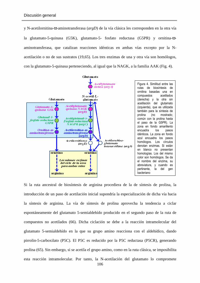
Discusión general
106
y N-acetilornitina-ω-aminotransferasa (argD) de la vía clásica les corresponden en la otra vía
la glutamato-5-quinasa (G5K), glutamato-5- fosfato reductasa (G5PR) y ornitina-ω-
aminotransferasa, que catalizan reacciones idénticas en ambas vías excepto por la N-
acetilación o no de sus sustratos (19,65). Los tres enzimas de una y otra vía son homólogos,
con la glutamato-5-quinasa perteneciendo, al igual que la NAGK, a la familia AAK (Fig. 4).
Si la ruta ancestral de biosíntesis de arginina procediera de la de síntesis de prolina, la
introducción de un paso de acetilación inicial supondría la especialización de dicha vía hacia
la síntesis de arginina. La vía de síntesis de prolina aprovecha la tendencia a ciclar
espontáneamente del glutamato 5-semialdehído producido en el segundo paso de la ruta de
compuestos no acetilados (66). Dicha ciclación se debe a la reacción intramolecular del
glutamato 5-semialdehído en la que su grupo amino reacciona con el aldehídico, dando
pirrolin-5-carboxilato (P5C). El P5C es reducido por la P5C reductasa (P5CR), generando
prolina (65). Sin embargo, si se acetila el grupo amino, como en la ruta clásica, se imposibilita
esta reacción intramolecular. Por tanto, la N-acetilación del glutamato lo compromete
Figura 4. Similitud entre las rutas de biosíntesis de ornitina basadas una en compuestos acetilados (derecha) y la otra sin acetilación del glutamato (izquierda), que es utilizada también para la síntesis de prolina (no mostrado; común con la prolina hasta el paso de la G5PR). La zona en fondo amarillento encuadra los pasos idénticos. La zona en fondo azul encuadra los pasos homólogos. Los círculos denotan enzimas. Si están en blanco no presentan homologías. Los del mismo color son homólogos. Se da el nombre del enzima, su abreviatura, y cuando es pertinente, la del gen bacteriano

Discusión general
107
enteramente con la síntesis de arginina (65). Para aquellos organismos capaces de sintetizar
arginina de novo y que tienen una gran necesidad biosintética de aminoácidos (como las
plantas o los microorganismos autótrofos o con muy pocos requisitos nutricionales) debe ser
esencial discriminar entre fabricar prolina y fabricar arginina, pues las disponibilidades de uno
y otro aminoácido pueden ser muy diferentes. Además, muchos microorganismos y plantas
sintetizan y acumulan prolina como mecanismo de defensa frente al estrés hídrico o salino
(67), mientras que en condiciones de abundancia de nitrógeno los organismos fotosintéticos
sintetizan muy activamente arginina, acumulándola en proteínas o polímeros ricos en arginina
(68). Por tanto, posiblemente representaría una notable ventaja la separación de las vías de
síntesis de arginina y de prolina mediante el uso de intermedios N-acetilados en la ruta de la
biosíntesis de la arginina.
Para convertir una ruta de síntesis de arginina/prolina en una de síntesis exclusiva de
arginina habría que incorporar una maquinaria para la acetilación del glutamato y otra para la
desacetilación de la ornitina o de un intermedio posterior a la ornitina. Con respecto al paso
de acetilación del glutamato, parece poco probable que la ArgA canónica se generara e
incorporara como el acetilador primigenio, dada su complejidad estructural. Está de acuerdo
con ello el que ninguna arquea contenga un gen para ArgA canónica [(69); extendido por
nosotros a 49 genomas de arquea encontrados en la base de datos TIGR en enero de 2013] a
pesar de que en muchas arqueas se encuentran genes obvios para los demás pasos de la
síntesis de arginina (no mostrado). Del mismo modo, en algunas eubacterias sintetizadoras de
arginina falta argA (69) a pesar de que sintetizan arginina y de que, como encontró nuestro
grupo en T. maritima (70), poseeen una NAGK altamente específica e inhibible por arginina
(69). Todo ello está de acuerdo con que la ArgA canónica bacteriana sea un producto tardío,
posterior al establecimiento de la vía N-acetilada, habiendo surgido posiblemente como una
adaptación o mejora de dicha vía.
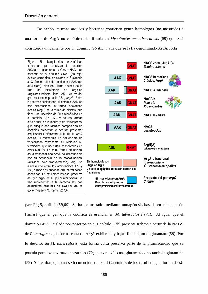
Discusión general
108
De hecho, muchas arqueas y bacterias contienen genes homólogos (no mostrado) a
una forma de ArgA no canónica identificada en Mycobacterium tuberculosis (59) que está
constituida únicamente por un dominio GNAT, y a la que se la ha denominado ArgA corta
(ver Fig.5, arriba) (59,69). Se ha demostrado mediante mutagénesis basada en el trasposón
Himar1 que el gen que la codifica es esencial en M. tuberculosis (71). Al igual que el
dominio GNAT aislado por nosotros en el Capítulo 3 del presente trabajo a partir de la NAGS
de P. aeruginosa, la forma corta de ArgA exhibe muy baja afinidad por el glutamato (59). Por
lo descrito en M. tuberculosis, esta forma corta preserva parte de la promiscuidad que se
postula para los enzimas ancestrales (72), pues no sólo usa glutamato sino también glutamina
(59). Sin embargo, como se ha mencionado en el Capítulo 3 de los resultados, la forma de M.
NAGS bacteriana Clásica, ArgA
AAK GNAT
ASLArgH(A)vibriones marinos
Producto del gen argOC.jejuni
Sin homología con ArgA.
Posible homología con
estreptotricina acetiltransferasa
NAGS corta, ArgA(S) M.tuberculosisGNAT
GNAT
NAGS/KM.marisX.campestris
NAGS A. thaliana
NAGS levaduraGNAT
GNAT
AAK
AAK
AAK
GNAT
AAK GNAT NAGSvertebrados
ArgJ bifuncionalT. NeapolitanaG. stearothermophilus
Sin homología con
ArgA or ArgO
Un sólo polipéptido autoescindido en dos
fragmentos
Figura 5. Maquinarias enzimáticas conocidas que catalizan la reacción AcCoa + L-glutamato → CoA + NAG. Las basadas en el dominio GNAT (en rojo) existen como dominio aislado, o fusionado al C-término bien de un dominio AAK (en azul claro), bien del último enzima de la ruta de biosíntesis de arginina (argininosuccinato liasa, ASL; en verde; gen bacteriano para la ASL, argH). Entre las formas fusionadas al dominio AAK se han diferenciado la forma bacteriana clásica (ArgA) de la forma de plantas, que tiene una inserción de 80 aminoácidos en el dominio AAK (17), y de las formas bifuncional, de levadura y de vertebrados, que aunque con idéntica composición de dominios presentan o podrían presentar arquitecturas diferentes a la de la ArgA clásica. El rectángulo lila del enzima de vertebrados representa 45 residuos N-terminales que no están conservados en otras NAGSs. En rosa, forma bifuncional de la transacetilasa ArgJ, no diferenciable por su secuencia de la monofuncional (actividad sólo transacetilasa). ArgJ se autoescinde entre los aminoácidos 179 y 180, dando dos cadenas que permanecen asociadas. En azul claro intenso, producto del gen argO de C. jejuni (ver texto). Se han representdo a la derecha las dos estructuras descritas de NAGSs, de N.
gonorrhoeae y M. maris (52,73).

Discusión general
109
tuberculosis presenta un signo claro de que ya es una forma evolucionada y restringida a la
síntesis de arginina, pues exhibe potente inhibición por arginina (59), una función reguladora
que debió incorporarse posteriormente al desarrollo de la actividad catalítica.
En resumen, puede concebirse la posibilidad de que la ruta clásica de síntesis de
arginina haya derivado de la de biosíntesis de prolina, reclutando un dominio GNAT con
capacidad acetiltransferasa que usa AcCoA como sustrato, así como una actividad
aminoacilasa capaz de deacilar la acetilornitina. En este modelo los enzimas de la ruta
ancestrales de síntesis de prolina deberían ser también promiscuas, al menos en cuanto a la
discriminación entre entre formas acetiladas y no acetiladas del glutamato y de los demás
compuestos intermedios.
Sin embargo, es posible que esta visión sea simplista. De la comparación de parálogos
entre las rutas de síntesis de arginina, de prolina y de las dos rutas de biosíntesis de lisina
(Fig.6) es posible proponer con Fondi et al. (74) una vía unificada ancestral compartida en la
Glutamate
proB
proA
proC
ornithineproline
Figura 6. Rutas existentes de síntesis de prolina/ornitina, lisina y arginina. Relaciones evolutivas entre sus genes. Los genes que comparten color y nivel son homólogos. Los genes en blanco no tienen homólogos en las rutas metabólicas mostradas (modificado de [74]).

Discusión general
110
que los mismos enzimas catalizan diferentes pasos de cada vía. Estas cuatro vías tienen en
común la presencia consecutiva y constante de los parálogos de argB y argC [argB y argC
codifican (16) la NAGK y la NAG-5-fosfato reductasa, abreviada NAGPR]. Todas ellas
conservan también el parálogo de argD (que codifica la transaminasa) aunque, en el caso de
la ruta de la aspartoquinasa de la biosíntesis de la lisina (la que parte del aspartato; vía más a
la derecha en la Fig. 6), tras la inserción de varios pasos. Está también presente el parálogo de
argE (acetilornitinasa) en todas estas vías, excepción hecha de la vía de síntesis de prolina.
Dos de estas cuatro vías se alimentan con aminoácidos acetilados (Fig. 6), pero el enzima
acetilador difiere en ellas e incluso difiere en distintos organismos para la biosíntesis de
arginina. Parece lógico concluir que el tronco correlativo y común formado por argB y argC
es la base ancestral de una ruta promiscua original. El carácter no parálogo del gen que acetila
el aminoácido en las dos vías que usan sustratos acetilados apoya la visión de que la
acetilación ha sido un proceso sobreañadido a una ruta más antigua sin sustratos acetilados.
Apoya la idea de que la acetilación es un hecho tardío para el que se ha echado mano
de las maquinarias acetiladoras disponibles, la observación de que no fue la incorporación del
dominio GNAT la única forma de introducir la acetilación del glutamato a esta ruta ancestral
(Fig. 5). En Thermotoga neapolitana y Geobacillus stearothermophilus (75) que son
organismos con una ruta cíclica de biosíntesis de la arginina (ver Fig. 1 de la introducción), y
aparentemente también en Thermotoga maritima (70), la acetilornitina/glutamato
acetiltransferasa (ArgJ) tiene carácter bifuncional, teniendo también actividad NAGS, aunque
no ejercida por un dominio GNAT reconocible (75). Como no se encuentran en esos
organismos genes para las formas canónica o corta de ArgA, parece claro que esa actividad de
ArgJ es responsable en ellos de la biosíntesis anaplerótica de NAG. El origen de esta
actividad NAGS en ArgJ es oscuro. También se desconoce el grado de difusión de la forma
bifuncional de ArgJ entre las distintas especies, al no existir secuencias-firma características

Discusión general
111
que permitan establecer a partir de la secuencia de argJ si este gen codifica para una forma
mono o bifuncional de transacetilasa (69). Sólo la última de estas formas tendría la capacidad
de satisfacer las necesidades de la anaplerosis para acetilglutamato. Hay que señalar que, a
juzgar por la composición del grupo de genes de biosíntesis de arginina en M. tuberculosis
(76) y por la estructura de su NAGK (depositada en el PDB con el identificador 2AP9; sin
publicación asociada), que es hexamérica y tiene en cada subunidad el característico sitio para
arginina, la ruta de síntesis de arginina es cíclica en este microorganismo, e incluye el gen
argJ, que codifica para una acetiltransferasa posiblemente monofuncional, de acuerdo con la
esencialidad de ArgA corta en ese microorganismo (71). El hecho de que en las dos rutas de
biosíntesis de lisina (Fig. 6) existan parálogos de argE en el mismo nivel (o uno no muy
distante del mismo, Fig. 6) que argE de la ruta de síntesis de arginina, hace suponer que la
primera forma de la ruta para intermedios N-acetilados incluía la hidrólisis del grupo acetilo
de la acetilornitina. Por tanto, cabe suponer que la transacetilasa es una incorporación
posterior a la de la actividad hidrolítica de acetilornitina, y por tanto que la bifuncionalidad de
esta transacetilasa fue todavía un desarrollo ulterior que permitió la eliminación de un gen
para ArgA corta.
Un posible ejemplo adicional de utilización para la síntesis de arginina de otras
maquinarias catalíticas sin aparente relación con el dominio GNAT lo representa la proteína
codificada por el gen argO de Campylobacter jejuni (Fig.5) (77). Esta proteína de 146
aminoácidos no exhibe identidad de secuencia significativa con el dominio GNAT ni con la
NAGS bacteriana clásica. Presenta similaridad de secuencia (aunque baja) con la
estreptomicina acetiltransferasa, y complementa cepas argA-, aunque nuestra información
sobre ella es muy escasa.
Las similitudes cinéticas entre la ArgA corta y el dominio GNAT que hemos aislado
en nuestros estudios estarían de acuerdo con que el dominio GNAT se hubiera fusionado a la
NAGK ya especializada en la síntesis de arginina (69). Parece claro que la primera forma de

Discusión general
112
ArgA clásica tendría tanto actividad NAGS como NAGK, es decir, sería bifuncional, pues no
sólo se han identificado formas bifuncionales de NAGS/NAGK en Xhantomonas campestris
(15) y Maricaulis maris (15,73) sino que la arquitectura de dominios de la NAGK de levadura
incluye dominios AAK y GNAT, este último inactivo (Fig 7) (78). Además, nosotros hemos
recuperado aquí actividad NAGK en la ArgA clásica con muy poco esfuerzo mutacional
(Capítulo 3).
En este contexto, resultan sorprendentes las diferencias de arquitectura entre, de un
lado, la forma bifuncional de M. maris (73) y pseudobifuncional de NAGK de levadura (78),
ambas tetraméricas y planas (Fig. 7), y de otro lado la forma hexamérica en anillo (Figs 1 y 2)
(52) que muy probablemente es característica de todas las ArgAs canónicas bacterianas. El
hecho de que, en contraposición a la mayoría de bacterias, E.coli presente una forma de
NAGK dimérica e insensible a arginina (46), similar al dímero de carbamato quinasa (otro
enzima de la familia AAK que es dimérico y muy antiguo y que está presente en arqueas y en
eubacterias) (44), plantea la cuestión no resuelta de cómo adquirió E. coli la NAGS de tipo
ArgA clásica manteniendo una NAGK dimérica y supuestamente antigua.
Figura 7. Estructuras de la NAGS/NAGK bifuncional de Maricaulis maris (izquierda) (PDB 3S6H) (73) y de la NAGK de Sacharomyces cerevisiae (derecha) (PDB 4AB7) (78), para subrayar su extraordinaria similitud. Ambas son tetrámeros organizados como dímeros de dímeros. Cada subunidad se representa en un color. El dominio AAK se ubica alrededor del orificio central, mientras que los dominios GNAT forman dímeros a los lados derecho e izquierdo. En el caso del enzima de levadura, su dominio GNAT es inactivo, y carece de actividad NAGS.
Discusión general
113
Sea cual sea el mecanismo de difusión de unas u otras formas de estos enzimas, no hay
duda de que la NAGS bidominio ha tenido un éxito notable, pues la estructura de tipo ArgA
se difundió ampliamente en eubacterias y es aparentemente la única organización existente
para la síntesis de NAG en eucariotas: la NAGS de plantas es de tipo ArgA, aunque con una
inserción de 80 residuos (17) y los resultados presentados aquí así como los resultados de
otros (15) indican que la NAGS de los animales ureosmóticos y ureotélicos (incluido el ser
humano) tiene una estructura de dominios similar a la de ArgA, con extensión N-terminal,
dominio AAK donde se ubica el sitio para la arginina y dominio C-terminal de tipo GNAT.
Como ya se ha dicho, las levaduras y Neurospora crassa tienen una forma de NAGS que
también responde a este tipo general de patrón de dominios (Fig.5) (56,57,79). Ello no obsta
para que las formas eucarióticas puedan adoptar quizá arquitecturas oligoméricas distintas del
hexámero de ArgA bacteriano, como sin duda es el caso para la NAGS de levadura, que
participa en un metabolón con la NAGK, el enzima siguiente en la ruta de biosíntesis de
arginina (56, 57).
La fusión del dominio GNAT a la NAGK no es la única forma existente en que el
dominio GNAT se ha fusionado a otro enzima. Se ha identificado también una forma en que
el dominio GNAT se fusiona al extremo C-terminal de la argininosuccinato liasa (o ArgH, por
el nombre de su gen bacteriano) (Fig.5) el enzima que cataliza el último paso de la biosíntesis
de arginina (80,81). Aunque este tipo de organización está mal conocido y es poco frecuente
al menos como forma exclusiva (en la mayoría de las especies en que se ha encontrado
coexiste con ArgA canónica [81]), no hay duda que la porción GNAT unida a ArgH es capaz
de complementar mutantes Arg- de E. coli, y por tanto de que es funcional en la síntesis de
NAG (80).
En última instancia, la considerable difusión de la NAGS constituida por la fusión del
dominio AAK y del GNAT debe reflejar su éxito funcional, éxito que quizá radique, como ya
se ha comentado en el Capítulo 3 de los Resultados, en el aumento en la afinidad por el

Discusión general
114
glutamato, y probablemente también en la versatilidad que proporciona la forma bidominios a
la regulación por arginina (ver sección siguiente).
Regulación por arginina
La regulación por arginina es una característica constante de las NAGSs de tipo ArgA.
La forma corta de M. tuberculosis (59) plantea un problema conceptual importante, ya que
dicha retroinhibición no puede ejercerse por el mismo mecanismo que en la ArgA bidominio,
pues le falta el dominio AAK al que se une la arginina en la NAGS bidominio. Dicha
inhibición debe ejercerse en la ArgA corta exclusivamente a través del dominio GNAT. Sin
embargo, hemos probado aquí que no se observa tal inhibición con el dominio GNAT aislado
de la ArgA clásica (ver Capítulo 3 de Resultados). Es concebible que en la evolución hacia las
formas modernas de enzimas sintetizadoras de NAG se diera una situación de encrucijada
evolutiva en la que un camino llevara a incorporar al dominio GNAT ancestral no inhibible
por arginina una maquinaria ya existente que ejerciera el control por arginina, mientras que en
otro camino el dominio biosintético evolucionó sin incorporación de otro dominio, habiendo
de desarrollar por sí mismo un mecanismo propio de retroinhibición por producto final que
probablemente difiere fuertemente del descrito aquí para ArgA clásica. Alternativamente,
cabe la posibilidad de que la inhibición por arginina se desarrollara muy tempranamente en
las ArgA cortas, y que luego, tras la fusión con la NAGK, se perdiera esta capacidad de
regulación directa al incorporarse la posibilidad de inhibición por el sitio para arginina de la
NAGK. Carecemos de información sobre cómo se incorporó el sitio para arginina en la
NAGK. Sin embargo, el hecho de que en arqueas la NAGK sea probablemente dimérica y no
inhibible por arginina (datos no mostrados: la secuencia de ArgB de arqueas no revela una
extensión N-terminal) hace altamente plausible la idea de que la ArgA corta que parece estar
presente en esta gran división de los seres vivos sea inhibida generalmente por arginina
(aunque también se da argJ en algunas arqueas, la cual podría ser bifuncional [69]).

Discusión general
115
Independientemente de cómo tenga lugar la inhibición por arginina de la forma corta
de ArgA (Fig.5), los datos publicados para este enzima de M. tuberculosis indican que dicha
inhibición es potente y sucede a bajas concentraciones del inhibidor (59). Reiteraremos aquí
que la ArgA canónica ofrece grandes ventajas en cuanto al proceso de inhibición. Como
prueban nuestros resultados, pequeños cambios en el conector interdominios pueden hacer
variar de forma dramática el grado de inhibición alcanzable o los rangos de concentraciones
de arginina a que tiene lugar la inhibición, lo que confiere a ArgA un gran potencial
adaptativo al entorno concreto en el que el microorganismo se desenvuelve normalmente.
Sólo existe información estructural directa sobre el sitio para la arginina de una
NAGS, la de N. gonorrhoeae (52). Se ha inferido la ubicación del sitio para arginina en la
forma bifuncional de M.maris a partir de la observación de la estructura cristalina de la misma
(73). Así, se ha concluido que la arginina se uniría en el dominio AAK, en idéntica ubicación
que el sitio de la arginina de la NAGS de N. gonorrhoeae, a pesar de que el enzima de N.
gonorrhoeae es un anillo hexamérico y el bifuncional es un tetrámero limitado lateralmente
por dímeros de dominios GNAT (73). Está de acuerdo con dicha ubicación la identificación
por nuestro grupo del sitio para arginina en la estructura cristalina de la NAGK
pseudobifuncional de levadura (78), que tiene virtualmente idéntica estructura que la NAGS
bifuncional de M. maris (Fig. 7). El sitio está localizado exactamente donde le habría
correspondido estar en la NAGS canónica, en el dominio AAK. Aunque desconocemos la
estructura de la NAGS animal, nuestros estudios de comparación de secuencias establecieron
la ubicación del sitio para la arginina en el enzima humano en idéntica localización en el
dominio AAK (Capítulo 1 de los Resultados). Este sitio incluye la secuencia consenso de
unión de arginina E-L-F-(T/S)-x-x-G-x-G-T, correspondiente a la hélice H del plegamiento
AAK (48). Con posterioridad, estudios de mutagénesis dirigida con la NAGS recombinante
de ratón confirmaron experimentalmente dicha ubicación del sitio (27). Todas estas
evidencias nos llevan a concluir que excepto en la forma de ArgA corta, en que la arginina se
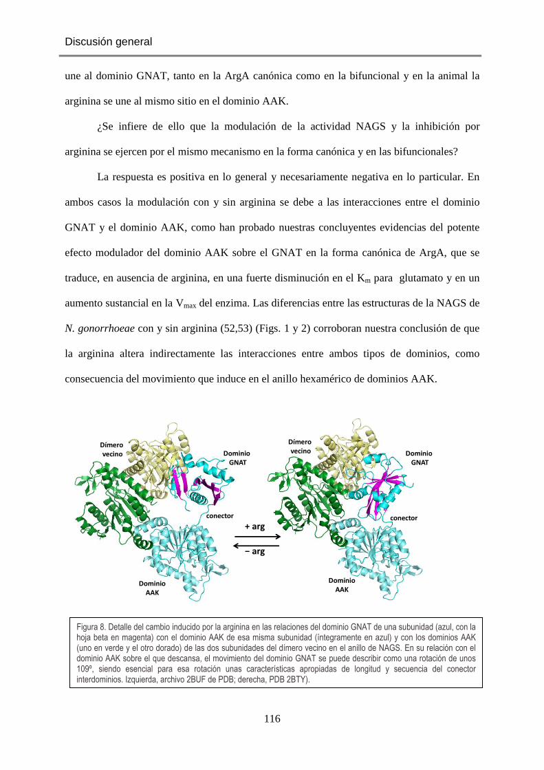
Discusión general
116
une al dominio GNAT, tanto en la ArgA canónica como en la bifuncional y en la animal la
arginina se une al mismo sitio en el dominio AAK.
¿Se infiere de ello que la modulación de la actividad NAGS y la inhibición por
arginina se ejercen por el mismo mecanismo en la forma canónica y en las bifuncionales?
La respuesta es positiva en lo general y necesariamente negativa en lo particular. En
ambos casos la modulación con y sin arginina se debe a las interacciones entre el dominio
GNAT y el dominio AAK, como han probado nuestras concluyentes evidencias del potente
efecto modulador del dominio AAK sobre el GNAT en la forma canónica de ArgA, que se
traduce, en ausencia de arginina, en una fuerte disminución en el Km para glutamato y en un
aumento sustancial en la Vmax del enzima. Las diferencias entre las estructuras de la NAGS de
N. gonorrhoeae con y sin arginina (52,53) (Figs. 1 y 2) corroboran nuestra conclusión de que
la arginina altera indirectamente las interacciones entre ambos tipos de dominios, como
consecuencia del movimiento que induce en el anillo hexamérico de dominios AAK.
Dímero vecino Dominio
GNAT
DominioAAK
conector
Dímero vecino Dominio
GNAT
DominioAAK
conector+ arg
− arg
Figura 8. Detalle del cambio inducido por la arginina en las relaciones del dominio GNAT de una subunidad (azul, con la hoja beta en magenta) con el dominio AAK de esa misma subunidad (íntegramente en azul) y con los dominios AAK (uno en verde y el otro dorado) de las dos subunidades del dímero vecino en el anillo de NAGS. En su relación con el dominio AAK sobre el que descansa, el movimiento del dominio GNAT se puede describir como una rotación de unos 109º, siendo esencial para esa rotación unas características apropiadas de longitud y secuencia del conector interdominios. Izquierda, archivo 2BUF de PDB; derecha, PDB 2BTY).

Discusión general
117
La figura 8 ilustra en más detalle el cambio con la unión de la arginina de la posición y
las relaciones de un dominio GNAT con respecto al dominio AAK de su misma subunidad y a
los dominios AAK del dímero vecino. Se ha señalado (53) que estos cambios hacen que en
presencia de arginina se pierdan las interacciones entre ambos dominios que estabilizan el
centro activo particularmente en el sitio para glutamato (52), aumentando considerablemente
el desorden en varios lazos del centro activo (Fig.8) (53).
Para la NAGS bifuncional no tenemos datos con y sin arginina, por lo que hemos de
inferirlos de las estructuras cristalinas de nuestro laboratorio con la NAGK pseudobifuncional
de levadura (78), un enzima que es muy similar estructuralmente a la NAGS bifuncional
(Fig.7). La estructura de estas enzimas, un tetrámero plano de dominios AAK que configuran
un dímero de dímeros y que está flanqueado por dos dímeros de dominios GNAT (Fig. 7),
impone estreses estructurales, con tendencia a la separación de los dos lóbulos de los
dominios AAK. La arginina, al tirar del dominio C-terminal hacia la hélice que conecta los
dos dímeros que conforman el tetrámero, favorece esta apertura, que es de similar naturaleza
al movimiento del lóbulo C-terminal observado en las NAGKs canónicas y que obedece
también a similares mecanismos. Aunque el complejo con arginina de esta NAGK sólo se ha
observado en la forma truncada sin dominio GNAT, el movimiento de apertura del lóbulo C-
terminal está presente en tres de cuatro subunidades del tetrámero de la forma de NAGK
completa (78). Dicha apertura se acompaña de cambios en la relación y en los contactos entre
los dominios GNAT y AAK (Fig. 9).

Discusión general
118
Por tanto, aún cuando no disponemos de datos experimentales para afirmarlo, es
probable también que en el caso de las formas bifuncionales los cambios en las interacciones
entre los dominios AAK y GNAT reulten en cambios en la actividad y propiedades cinéticas
de la reacción NAGS catalizada por el dominio GNAT, actuando la arginina por cambiar
dichas interacciones al favorecer el predominio de las formas abiertas del dominio AAK. La
posibilidad alternativa de que en los enzimas bifuncionales existan sitios independientes para
arginina en los dominios GNAT y AAK parecía apoyada por la observación con la NAGS/K
de X. campestris de que las concentraciones de arginina requeridas para inhibir la actividad
NAGS son considerablemente menores que las necesarias para inhibir la actividad NAGK
(15). Sin embargo, mutaciones en el sitio de la arginina en el dominio AAK de la NAGS
bifuncional de X. campestris abolieron la inhibición por arginina de la actividad NAGS (27),
Figura 9. Relaciones entre el dominio GNAT (verde) y el lóbulo C-terminal del dominio AAK (en rojo burdeos) en las formas cerrada y abierta de la subunidad de la NAGK de levadura (que es estructuralmente muy similar a la NAGS bifuncional). La arginina favorece la forma abierta. Nótese la diferente relación del dominio GNAT con respecto al AAK
en una y otra forma (PDB 4AB7)
Forma cerrada Forma abierta(favorecida por la arginina)
GNAT GNAT
AAK AAK

Discusión general
119
por lo que el sitio canónico para la arginina en el dominio AAK es responsable de la
inhibición de la actividad catalizada por el dominio GNAT.
Si la modulación de la actividad y los efectos de la arginina obedecen en el enzima
canónico y en el bifuncional al mismo mecanismo general, es decir, son reflejo de
interacciones entre los dos dominios distintos que son en un caso positivas (sin arginina) y en
otro (con arginina) menos positivas, el mecanismo específico operativo con el enzima clásico
y con la forma bifuncional no puede ser el mismo. Las interacciones importantes tienen lugar
entre dominios de dímeros distintos en las formas canónicas de ArgA, como han probado
nuestros experimentos de disociación por eliminación de parte de la hélice N-terminal. Sin
embargo, dada la arquitectura de la forma bifuncional, en ella las interacciones han de
mediarse con el dominio AAK de la misma subunidad, por la sencialla razón de que
virtualmente no existen interacciones entre dominio GNAT de un dímero y dominio AAK del
otro dímero (78). Aunque los análisis sobre el mecanismo de inhibición por arginina de las
dos NAGSs bifuncionales (de X. campestris y de M. maris) que han sido estudiadas
experimentalmente son más bien escasos (15,27,73), se ha postulado que la modulación se
ejercería en la enzima bifuncional a través del sitio de unión del AcCoA (73), cuya estructura
se vería influenciada por los contactos con el dominio AAK, dificultando la presencia de la
arginina la unión del AcCoA. Por el contrario, los efectos en el caso de la NAGS hexamérica
parecen depender principalmente de alteraciones en el sitio para glutamato.
Terminaremos esta sección de la discusión con una reflexión sobre cómo la arginina
puede causar activación en lugar de inhibición, tal como se ha observado con la enzima de los
mamíferos incluido el ser humano (10-12,58). Este aspecto no puede ser respondido
plenamente sin tener la seguridad de cuál es la estructura de la NAGS animal. Cuando se
llegó a la convicción de que el enzima animal presenta el mismo patrón bidominio que el
humano, se manejaron modelos de la NAGS humana de carácter hexamérico, similares a los
de la NAGS de N. gonorrhoeae (36). Luego, con la demostración de la arquitectura

Discusión general
120
tetramérica de la forma bifuncional, y dado que la identidad de secuencia de esta forma con
la forma humana es superior a la de las NAGSs bacterianas clasicas, se ha propuesto para la
forma humana una arquitectura tetramérica basada en la de la forma de M. maris (73). Sin
embargo, el hecho de que la similitud de secuencia sea también mayor con la forma de
levadura que con las formas bacterianas clásicas (13,41), combinado con el reconocimiento
general de que la forma aislada de mamífero es fastidiosa e inestable y tiende a agregar, hace
concebible que, como en el caso de la NAGS de levadura, que requiere de la enzima NAGK
para formar un complejo y ser estable y activa (56,57,78), la forma animal de NAGS podría
tener que asociarse íntimamente a otra proteína [no la NAGK, no presente en animales (19)]
para ser plenamente estable, lo que impdediría establecer con seguridad cual es su
arquitectura y por tanto su mecanismo específico de activación. Aún con esta incertidumbre,
nuestros experimentos con la forma hexamérica, en la que cambios en la longitud y la
secuencia del conector interdominios han resultado en cierto grado de activación, sugieren un
mecanismo posible para tal activación. La activación sería posible si las interacciones
originales entre el dominio GNAT y su dominio AAK modulador (del dímero vecino en las
formas hexaméricas, de la misma subunidad en las tetraméricas) son subóptimas para la
activación del domino GNAT en ausencia de arginina. Así, si los cambios conformacionales
inducidos por la arginina llevan al dominio GNAT a una nueva relación con dicho dominio
AAKcon interacciones más favorables para la estabilización del centro activo, habrá
activación. De hecho, en su publicación sobre la estructura de la NAGS/K de M. maris (73) el
grupo de Tuchman propone que un residuo del conector, correspondiente a la alanina 274 del
enzima humano, determina el carácter activador o no de la arginina, al permitir un
movimiento u otro de un dominio respecto al otro. Esta propuesta coincide con nuestra
conclusión del Capítulo 3, que establece que la secuencia del conector tiene un papel clave en
la determinación del efecto de la arginina. En todo caso habrá que esperar a la determinación

Discusión general
121
de la estructura del enzima humano (o de otra NAGS animal) con y sin arginina para aclarar
de forma definitiva cómo ejerce la arginina su acción activadora.
Nota final sobre nuestra contribución al estudio de la NAGS humana
Con la finalidad última de determinar la estructura del enzima humano y de aclarar su
regulación, así como de disponer de un instrumento propio para establecer experimentalmente
los efectos de las mutaciones clínicas sobre la actividad de la NAGS human, hemos tenido
éxito en la preparación de una forma recombinante expresada en E. coli de la enzima humana
gracias a su fusión N-terminal con la proteína de unión a la maltosa (MBP). Por razones de
extensión, porque dicho material aún no ha sido publicado, estando ahora en fase de análisis y
preparación para publicación, y por mi necesidad de poner un límite temporal a la terminación
de esta memoria de tesis y a la obtención del grado de doctor, no ha sido incluido dicho
trabajo en esa memoria de tesis. Nuestro sistema de expresión permite producir esta proteína
en abundancia y en forma estable (Fig. 10), lo que no parece ser el caso con los sistemas de
expresión recombinante de NAGS de mamífero o humana descritos hasta ahora por otros dos
grupos (40,58). También he producido aisladamente con alto rendimiento (Fig. 10) el dominio
GNAT sin el dominio AAK, fusionando dicho dominio GNAT al C-término de SUMO
(SUMO-HuGNAT). Con finalidad comparativa con la NAGS de P. aeruginosa he preparado
también el dominio GNAT fusionado al extremo C-terminal del dominio AAK de la NAGS
de P. aeruginosa (PaNAAK-HuGNAT) si bien en ese caso se ha obtenido un rendimiento de
proteína purificada y soluble mucho menor. En todos los casos la proteína de fusión contiene
una etiqueta HHHHHH, precediendo N-terminalmente a MBP y SUMO, y siendo C-terminal
en la quimera PaNAAK-HuGNAT (Fig. 10).

Discusión general
122
Región variable
dominioGNATC
onector
Región conservada
Péptido señal
50 95 372 379 5341
HuAAK HuGNAT
dominioAAK
FORMA HUMANA SILVESTRE (HuNAGS)
MBP-HuNAGS
PreScission
6HisMBP
42.5kDa huAAK HuGNAT
25013010070
55
35
kDa
25
StMBP-
HuNAGS
PaAAK-HuGNAT
6His
PaAAK HuGNATkDa
66
45
36
2924
20
14
St
PaAAK-
HuGNAT
SUMO-HuGNAT
6His
SUMO HuGNAT
kDa
66
4536
2924
20
14
StSUMO-
HuGNAT
Cuando se escindió el conector con la MBP usando proteasa PreScission, los valores
de Km para glutamato y para AcCoa fueron del mismo orden de los descritos para la forma
purificada de rata (12) (Km para AcCoA y para L-glutamato, 1.1±0.2 mM y 4.5±1.1 mM,
respectivamente; a comparar con los correspondientes valores para la NAGS obtenida de rata,
de 0.76 y 3.4 mM [12]). En la forma no escindida los respectivos valores fueron
aproximadamente 2 y 5 veces más altos. La actividad específica fue también del mismo orden
de magnitud que la descrita para el enzima de hígado de rata (en presencia de 1 mM arginina,
18.7 µmol/min/mg para el enzima de hígado de rata [12] y 44 µmol/min/mg para la humana
recombinante tras escisión). Dicha actividad específica debe ser para el enzima recombinante
Figura 10. Estructura de dominios de la NAGS humana (HuNAGS; arriba) y de las construcciones preparadas y expresadas aquí, mostrando a la izquierda la forma purificada de cada construcción expresada en E. coli (SDS-PAGE y tinción de azul Coomassie). Se ha usado la definición de las regiones del péptido señal, de la región variable y constante que se da en (14), y la definición del conector dada en el Capítulo 3, Fig. 2C de esta tesis. 6His- etiqueta con un motivo HHHHHH. MBP, proteína de unión a la maltosa. El prefijo Hu indica la enzima humana o sus dominios. PaAAK, dominio AAK de la NAGS de P. aeruginosa. St, proteínas patrón de masas conocidas (se dan a la izquierda de cada gel, en kDa).

Discusión general
123
aún mayor, del orden de 90 µmol/min/mg, ya que tras la escisión no retiramos la MBP, que
así contribuye aproximadamente el 50% de la proteína, pero no es activa. Esta mayor
actividad específica probablemente refleja las mayores concentraciones de los sustratos en
nuestro ensayo (100 mM glutamato y 6 mM AcCoA) que en los ensayos descritos con el
enzima de hígado de rata (1 mM glutamato y 0.5 mM AcCoA). En esta forma escindida el Ka
para la arginina fue del orden de 0.05 mM, alcanzándose una activación máxima de unas 4.5
veces la actividad sin arginina, lo mismo que se había descrito para la NAGS de hígado de
rata (12).
En resumen, los resultados indican que, si se escinde el conector con el MBP, el
enzima se comporta como el enzima de rata natural y probablemente como el humano.
Desgraciadamente, hasta ahora hemos fracasado en separar el dominio MBP escindido, ya
que este dominio y la NAGS se eluyen juntas de las columnas de Ni con alto imidazol a pesar
de que sólo el dominio MBP contiene la cola de poli-His (resultados no mostrados). Esta
coelución parece indicar la tendencia de la NAGS humana a interaccionar con otras proteínas,
o al menos con MBP. Tampoco hemos tenido éxito en obtener cristales que difracten los
rayos X a resolución razonable, ni con el enzima unido a MBP, ni con las otras construcciones
mostradas en la Fig. 10.
La construcción constituida por el dominio GNAT unido a SUMO también catalizó la
reacción con muy buena actividad, que fue aproximadamente del doble (estimada por
dominio GNAT) que en el enzima entero fusionado a MBP, en ausencia de arginina. La
arginina careció de efecto sobre la actividad del dominio GNAT unido a SUMO, lo que
confirma la importancia del dominio AAK para la activación. Si la actividad de dicho
dominio se compara con la del enzima entero a saturación de arginina, ésta es, por dominio
GNAT, aproximadamente del doble que la del dominio GNAT unido a SUMO (velocidad
estimada a saturación de ambos sustratos). De otro lado, el dominio GNAT unido a SUMO
Discusión general
124
exhibió una fuerte elevación en el Km para glutamato (45 mM). Por tanto, la presencia del
dominio AAK aumentó la Vmax al doble, y más de 10 veces la afinidad por el glutamato, al
menos en la forma más activa del enzima, que es la forma con arginina. Estos hallazgos son
cualitativamente similares a los del Capitulo 3 de la sección de Resultados con el enzima de
P. aeruginosa, pues con éste la forma más activa es la sin arginina, y dicha forma exhibe el
doble de actividad y un Km para glutamato mucho menor que el dominio GNAT aislado, el
cual es también insensible a la arginina. Hay que resaltar que, aunque fue menos activa, la
construcción quimérica de los enzimas de P. aeruginosa y humano (Fig. 10) exhibió un valor
de Km para glutamato similar al del enzima completo, sugiriendo un efecto reminiscente del
que tiene con su dominio GNAT nativo (el de la misma especie). Contrasta este hallazgo, sin
embargo, con el hecho de que en esta quimera no se observó activación ni inhibición de la
actividad por arginina.
Está ahora en proceso la introducción en la proteína de fusión a MBP de las
mutaciones clínicas que se ubican en el dominio AAK y que habían sido descritas en
pacientes con déficit de NAGS. Por las realizadas hasta el momento, parece que esas
mutaciones no inactivan enteramente el enzima, explicado las presentaciones tardías
consistentes con deficiencia parcial observadas en la mayoría de los pacientes que las portan.
De otro lado, hemos introducido en el dominio GNAT unido a SUMO algunas mutaciones
clínicas que afectan a este dominio, con una mayor frecuencia de hallazgo de mutaciones
inactivantes. Quedan por analizar los efectos cinéticos detallados de estas mutaciones.
Confiamos en que trabajo futuro del laboratorio puedan culminar los estudios con el
enzima humano obteniendo cristales que permitan determinar la estructura de la forma
humana, y también introduciendo mutaciones en el conector interdominios y aclarando el
papel de dicho conector, en el enzima humano, para la activación por la arginina de este
enzima.

125
Conclusiones

126

Conclusiones
127
1. En la N-acetilglutamato sintasa (acetilglutamato sintasa) bacteriana clásica y, por
extensión, en la acetilglutamato sintasa humana, el sitio de unión del efector arginina
se asienta en el dominio aminoácido quinasa de estos enzimas, en la misma
localización que el sitio para arginina en el enzima parálogo N-acetilglutamato quinasa
(acetilglutamato quinasa). Dicho sitio se forma entre las hélices N-terminal y H y el
lazo αH-β16, como se ha corroborado luego por otros al determinar la estructura de la
acetilglutamato sintasa de Neisseria gonorrhoeae.
2. El dominio acetiltransferasa (GNAT) de la acetilglutamato sintasa cataliza por sí solo
la reacción (acetil coenzima A + L-glutamato → coenzima A + N-acetil-L-glutamato),
y por tanto une ambos sustratos y contiene íntegra la maquinaria catalítica. Sin
embargo, su afinidad aparente por el sustrato glutamato y su velocidad a saturación de
ambos sustratos son respectivamente muy inferior y aproximadamente la mitad que las
del enzima completo.
3. La arginina no modula la actividad del dominio acetiltransferasa aislado, como cabe
esperar si no se une al mismo.
4. El dominio aminoácido quinasa ejerce un notable efecto modulador de la actividad del
dominio acetiltransferasa, modulación que hemos abolido mediante mutación de tan
solo un residuo del dominio aminoácido quinasa. Por tanto, este dominio, que también
media la inhibición por la arginina, es responsable de la mayor actividad enzimática
(por molécula) y del gran aumento en la afinidad aparente por el glutamato del enzima
completo, comparado con la actividad y la afinidad del dominio acetiltranferasa
aislado.
5. El dominio aminoácido quinasa aislado de la acetilglutamato sintasa de P. aeruginosa
carece de actividad acetilglutamato sintasa o acetilglutamato quinasa. Forma
hexámeros, siendo por tanto responsable de la arquitectura hexamérica de la
acetilglutamato sintasa bacteriana clásica.

Conclusiones
128
6. Los residuos 1-12 de la acetilglutamato sintasa de P. aeruginosa son clave para la
arquitectura hexamérica, pues su eliminación conduce a que esta enzima disocie a una
forma dimérica.
7. La arquitectura hexamérica es esencial para que el dominio aminoácido quinasa y la
arginina modulen la actividad del dominio acetiltransferasa, ya que la forma dimérica
generada por corte de los residuos 1-12 exhibe igual cinética de sustratos e igual falta
de inhibición por arginina que el dominio acetiltransferasa aislado.
8. La acetilglutamato sintasa bacteriana clásica procede de una acetilglutamato quinasa
ancestral, ya que la mutación de tan sólo dos residuos del dominio aminoácido quinasa
aislado de la acetilglutamato sintasa de P. aeruginosa hicieron que este dominio
adquiriera actividad acetilglutamato quinasa, sugiriendo fuertemente que este enzima
era en el pasado bifuncional, con actividades acetilglutamato sintasa y acetilglutamato
quinasa.
9. El conector interdominios tiene un papel clave en la modulación de la actividad del
dominio acetiltransferasa por el dominio aminoácido quinasa y por la arginina, como
demuestran nuestros experimentos de elongación y acortamiento y de cambio de
secuencia de este conector.
10. El hecho de que para las acetilglutamato sintasas de mamíferos la arginina sea un
activador y para las bacterianas un inhibidor no supone un cambio drástico de
mecanismo regulador, pues hemos conseguido mediante cambios en el conector
interdominios de la acetilglutamato sintasa de P.aeruginosa que la arginina pase de ser
un inhibidor a un activador. Además, en la forma modificada en que se convirtió en
activador, la arginina fue un inhibidor a concentraciones bajas de glutamato.
11. El éxito evolutivo de la forma de la acetilglutamato sintasa compuesta por dominios
aminoácido quinasa y acetiltransferasa probablemente se debe a la facilidad de

Conclusiones
129
modulación de la actividad del dominio acetiltransferasa y de su sensibilidad a la
arginina mediante cambios mínimos en el conector interdominios que, así, ha debido
representar un elemento clave en la evolución de las acetilglutamato sintasas.
12. La acetilglutamato sintasa recombinante de P. aeruginosa constituye un modelo
experimental apropiado para examinar el impacto funcional de las mutaciones clínicas
en el déficit humano de acetilglutamato sintasa, particularmente si las mutaciones
afectan a residuos conservados en los enzimas bacteriano y humano. Sin embargo,
nuestra preparación de la acetilgutamato sintasa humana recombinante fusionada a la
proteína de unión a la maltosa, de la que damos aquí noticia preliminar, abre la puerta
a determinar el impacto de las mutaciones clínicas en la forma más genuina posible,
ya que el enzima recombinante se produce puro, estable y en cantidad abundante, y
exhibe una cinética normal para sus sustratos y para la arginina si se escinde el
conector que lo enlaza con la proteína de unión a la maltosa.

130
131
Bibliografía correspondiente a la Introducción general, Resumen de los
resultados y Discusión general

132

133
Bibliografía correspondiente a la Introducción general, Resumen de los resultados y Discusión general
1. Stadtman ER, Katz J, Barker HA. 1952. Cyanide-induced acetylation of amino
acids by enzymes of Clostridium kluyveri. J. Biol. Chem.195:779-785.
2. Maas WK, Novelli GD, Lipmann F. 1953. Acetylation of glutamic acid by extracts
of Escherichia coli. Proc. Natl. Acad. Sci. U. S. A. 39:1004-1008.
3. Bachmann B, Low KB, Taylor AL. 1976. Recalibrated linkage map of Escherichia
coli K-12. Bacteriol. Rev. 40:116-167.
4. Leisinger T, Haas D. 1975. N-Acetylglutamate synthase of Escherichia coli
regulation of synthesis and activity by arginine. J. Biol. Chem. 250:1690-1693.
5. Marvil DK, Leisinger T. 1977. N-acetylglutamate synthase of Escherichia coli:
purification, characterization, and molecular properties. J Biol Chem. 252: 3295-3303.
6. Haas D, Kurer V, Leisinger T. 1972. N-acetylglutamate synthetase of Pseudomonas
aeruginosa. An assay in vitro and feedback inhibition by arginine. Eur. J. Biochem.
31:290-295.
7. Haas D, Leisinger T. 1974. Multiple control of N-acetylglutamate synthetase from
Pseudomonas aeruginosa: synergistic inhibition by acetylglutamate and polyamines.
Biochem. Biophys. Res. Commun. 60:42-47.
8. Brown K, Finch PW, Hickson ID, Emmerson PT. 1987. Complete nucleotide
sequence of the Escherichia coli argA gene. Nucleic Acids Res. 15:10586.
9. Hall LM, Metzenberg RL, Cohen PP. 1958. Isolation and characterization of a
naturally occurring cofactor of carbamyl phosphate biosynthesis. J. Biol. Chem.
230:1013-1021.
10. Shigesada K, Tatibana M. 1971.Enzymatic synthesis of acetylglutamate by
mammalian liver preparations and its stimulation by arginine. Biochem. Biophys. Res.
Commun. 44:1117-1124.

134
11. Bachmann C, Krähenbühl S, Colombo JP. 1982. Purification and properties of
acetyl-CoA: L-glutamate N-acetyltransferase from human liver. Biochem. J. 205:123-
127.
12. Sonoda T, Tatibana M. 1983. Purification of N-acetyl-L-glutamate synthetase from
rat liver mitochondria and substrate and activator specificity of the enzyme. J. Biol.
Chem. 258:9839-9844.
13. Caldovic L, Morizono H, Yu X, Thompson M, Shi D, Gallegos R, Allewell NM,
Malamy MH, Tuchman M. 2002. Identification, cloning and expression of the
mouse N-acetylglutamate synthase gene. Biochem. J. 364:825-831.
14. Caldovic L, Morizono H, Gracia Panglao M, Gallegos R, Yu X, Shi D, Malamy
MH, Allewell NM, Tuchman M. 2002. Cloning and expression of the human N-
acetylglutamate synthase gene. Biochem. Biophys. Res. Commun. 299:581-586.
15. Qu Q, Morizono H, Shi D, Tuchman M, Caldovic L.2007. A novel bifunctional N-
acetylglutamate synthase-kinase from Xanthomonas campestris that is closely related
to mammalian N-acetylglutamate synthase. BMC Biochem. 8:4
16. Cunin R, Glansdorff N, Pierard A, Stalon V. 1986. Biosynthesis and metabolism of
arginine in bacteria. Microbiol. Rev. 50:314–352.
17. Slocum RD. 2005. Genes, enzymes and regulation of arginine biosynthesis in plants.
Plant. Physiol. Biochem. 43:729-745.
18. Caldovic L, Tuchman M. 2003. N-acetylglutamate and its changing role through
evolution. Biochem. J. 372:279-290.
19. Alonso E, Rubio V. 1989. Participation of ornithine aminotransferase in the synthesis
and catabolism of ornithine in mice. Studies using gabaculine and arginine
deprivation. Biochem J. 259:131-138.

135
20. Britton HG, Rubio V, Grisolia S. 1981. Synthesis of carbamoyl phosphate by
carbamoylphosphate synthetase I in the absence of acetylglutamate. Activation of the
enzyme by cryoprotectants. Biochem Biophys Res Commun. 99:1131-1137.
21. Rubio V, Ramponi G, Grisolia S. 1981. Carbamoyl phosphate synthetase I of human
liver. Purification, some properties and immunological cross-reactivity with the rat
liver enzyme. Biochim Biophys Acta. 659:150-160.
22. Ratner S.1973. Enzymes of arginine and urea synthesis. Adv Enzymol Relat Areas
Mol Biol. 39:1-90.
23. Anderson PM.1981. Purification and properties of the glutamine- and N-acetyl-L-
glutamate-dependent carbamoyl phosphate synthetase from liver ofSqualus acanthias.
J Biol Chem. 256:12228-12238.
24. Casey CA, Anderson PM. 1983. Glutamine- and N-acetyl-L-glutamate-dependent
carbamoyl phosphate synthetase from Micropterus salmoides. Purification, properties,
and inhibition by glutamine analogs. J. Biol. Chem. 258: 8723-8732.
25. Mommsen TP, Walsh PJ. 1989. Evolution of urea synthesis in vertebrates: the
piscine connection. Science. 243:72-75.
26. Anderson PM.1980. Glutamine- and N-acetylglutamate-dependent carbamoyl
phosphatesynthetase in elasmobranchs. Science. 208:291-293.
27. Haskins N, Panglao M, Qu Q, Majumdar H, Cabrera-Luque J, Morizono H,
Tuchman M, Caldovic L. 2008. Inversion of allosteric effect of arginine on N-
acetylglutamate synthase, a molecular marker for evolution of tetrapods. BMC
Biochem. 9: 24.
28. Kawamoto S, Ishida H, Mori M, Tatibana M. 1982. Regulation of N-
acetylglutamate synthetase in mouse liver. Postprandial changes in sensitivity to
activation by arginine. Eur J Biochem. 123:637-641.

136
29. Tatibana M, Shigesada K, Mori M. 1976. Acetylglutamate synthetase. En
The Urea Cycle (Grisolia S, Baguena R, Mayor F, editores), pp. 95-105
and 143-145 (coloquio). John Wiley & Sons, New York.
30. Coude FX, Sweetman L, Nyhan WL. 1979. Inhibition by propionyl-coenzyme A of
N-acetylglutamate synthetase in rat liver mitochondria. A possible explanation for
hyperammonemia in propionic and methylmalonic acidemia. J. Clin. Invest. 64:1544-
1551.
31. Coude FX, Grimber G, Parvy P, Rabier D,Petit F. 1983. Inhibition of ureagenesis
by valproate in rat hepatocytes. Role of N-acetylglutamate and acetyl-CoA. Biochem.
J. 216:233-236.
32. Coude FX, Grimber G. 1984. Inhibition of urea synthesis by pent-4-enoic acid:
potentiation by ammonia. Biochem. Biophys. Res. Commun. 118:47-52.
33. Jones S, Reed CA, Vijay S, Walter JH, Morris AA. 2008. N-carbamylglutamate for
neonatal hyperammonaemia in propionic acidaemia. J Inherit Metab Dis. 31 Suppl
2:S219-222.
34. Bachmann C, Krähenbühl S, Colombo JP, Schubiger G, Jaggi KH, Tönz O.
1981.N-acetylglutamate synthetase deficiency: a disorder of ammonia detoxication. N.
Engl. J. Med. 304:543.
35. Häberle J, Boddaert N, Burlina A, Chakrapani A, Dixon M, Huemer M, Karall
D, Martinelli D, Crespo PS, Santer R, Servais A, Valayannopoulos V, Lindner M,
Rubio V, Dionisi-Vici C. 2012. Suggested guidelines for the diagnosis and
management of urea cycle disorders. Orphanet J Rare Dis. 29; 7-32.
36. Caldovic L, Morizono H, Tuchman M. 2007. Mutations and polymorphisms in the
human N-acetylglutamate synthase (NAGS) gene. Hum. Mutat. 28:754-759.

137
37. Coudé FX, Grimber G, Parvy P, Pham Dinh D, Bardet J, Saudubray JM. 1983.
Characterization of enzymatic deficiencies of branched chain amino-acid catabolism
in human fibroblasts by genetic complementation. Biochem. Biophys. Res. Commun.
114:175-182.
38. Nissim I, Horyn O, Nissim I, Daikhin Y, Caldovic L, Barcelona B, Cervera J,
Tuchman M, Yudkoff M. 2011. Down-regulation of hepatic urea synthesis by
oxypurines: xanthine and uric acid inhibit N-acetylglutamate synthase. J Biol Chem.
286:22055-22068.
39. Caldovic L, Morizono H, Panglao MG, Lopez GY, Shi D, Summar ML, Tuchman
M. 2005. Late onset N-acetylglutamate synthase deficiency caused by hypomorphic
alleles. Hum. Mutat. 25:293-298.
40. Schmidt E, Nuoffer JM, Häberle J, Pauli S, Guffon N, Vianey-Saban C,
Wermuth B, Koch HG. 2005. Identification of novel mutations of the human N-
acetylglutamate synthase gene and their functional investigation by expression studies.
Biochim. Biophys. Acta. 1740:54-59.
41. Häberle J, Schmidt E, Pauli S, Kreuder JG, Plecko B, Galler A, Wermuth B,
Harms E, Koch HG. 2003. Mutation analysis in patients with N-acetylglutamate
synthase deficiency. Hum. Mutat. 21:593-597.
42. Nordenström A, Halldin M, Hallberg B, Alm J. 2007. A trial with N-
carbamylglutamate may not detect all patients with NAGS deficiency and neonatal
onset. J Inherit Metab Dis. 30:400
43. Rubio V, Grisolía S. 1981. Treating urea cycle defects. Nature.292:496
44. Marina A, Alzari PM, Bravo J, Uriarte M, Barcelona B, Fita I, Rubio V. 1999.
Carbamate kinase: New structural machinery for making carbamoyl phosphate, the
common precursor of pyrimidines and arginine. Protein Sci. 8:934-940.

138
45. Marina A, Uriarte M, Barcelona B, Fresquet V, Cervera J, Rubio V.1998.
Carbamate kinase from Enterococcus faecalis and Enterococcus faecium--cloning of
the genes, studies on the enzyme expressed in Escherichia coli, and sequence
similarity withN-acetyl-L-glutamate kinase. Eur J Biochem. 253:280-291.
46. Ramón-Maiques S, Marina A, Gil-Ortiz F, Fita I, Rubio V. 2002. Structure of
acetylglutamate kinase, a key enzyme for arginine biosynthesis and a prototype for the
amino acid kinase enzyme family, during catalysis. Structure. 10:329-342.
47. Dyda F, Klein DC, Hickman AB. 2000.GCN5-related N-acetyltransferases: a
structural overview. Annu. Rev. Biophys. Biomol. Struct. 29:81-103.
48. Ramón-Maiques S, Fernández-Murga ML, Gil-Ortiz F, Vagin A, Fita I, Rubio V.
2006. Structural bases of feed-back control of arginine biosynthesis, revealed by the
structures of two hexameric N-acetylglutamate kinases, from Thermotoga maritime
and Pseudomonas aeruginosa. J. Mol. Biol. 356:695–713.
49. Llácer JL, Contreras A, Forchhammer K, Marco-Marín C, Gil-Ortiz F,
Maldonado R, Fita I, Rubio V. 2007. The crystal structure of the complex of PII
andacetylglutamate kinase reveals how PII controls the storage of nitrogen asarginine.
Proc Natl Acad Sci U S A. 104:17644-17649.
50. Mizuno Y, Moorhead GB, Ng KK. 2007. Structural basis for the regulation of N-
acetylglutamate kinase by PII in Arabidopsis thaliana. J Biol Chem. 282:35733-
35740.
51. Fernández-Murga ML, Rubio V. 2008. Basis of arginine sensitivity of microbial N-
acetyl-L-glutamate kinases: mutagenesis and protein engineering study with the
Pseudomonas aeruginosa and Escherichia coli enzymes. J Bacteriol.190: 3018-3025.
52. Shi D, Sagar V, Jin Z, Yu X, Caldovic L, Morizono H, Allewell NM, Tuchman M.
2008. The crystal structure of N-acetyl-L-glutamate synthase from Neisseria
139
gonorrhoeae provides insights into mechanisms of catalysis and regulation. J Biol
Chem.283: 7176-7184.
53. Min L, Jin Z, Caldovic L, Morizono H, Allewell NM, Tuchman M, Shi D. 2009.
Mechanism of allosteric inhibition of N-acetyl-L-glutamate synthase by L-arginine. J
Biol Chem. 284: 4873-4880.
54. Häberle J, Schmidt E, Pauli S, Kreuder JG, Plecko B, Galler A, Wermuth B,
Harms E, Koch HG. 2003. Mutation analysis in patients with N-acetylglutamate
synthase deficiency. Hum Mutat. 21:593-597.
55. Kim JH, Weiss RL. 1995. Genetic analysis of interactions between arg-14 and arg-6
products in Neurospora crassa. Mol. Cells 5:461-466.
56. Abadjieva A, Pauwels K, Hilven P, Crabeel M. 2001. A new yeast metabolon
involving at least the two first enzymes of arginine biosynthesis: acetylglutamate
synthase activity requires complex formation with acetylglutamate kinase. J Biol
Chem. 276: 42869-42880.
57. Pauwels K, Abadjieva A, Hilven P, Stankiewicz A, Crabeel M. 2003. The N-
acetylglutamate synthase/N-acetylglutamate kinase metabolon of Saccharomyces
cerevisiae allows co-ordinated feedback regulation of the first two steps in arginine
biosynthesis. Eur J Biochem. 270:1014-1024.
58. Caldovic L, Lopez GY, Haskins N, Panglao M, Shi D, Morizono H, Tuchman M.
2006. Biochemical properties of recombinant human and mouse N-acetylglutamate
synthase. Mol Genet Metab. 87:226-232.
59. Errey JC, Blanchard JS. 2005. Functional characterization of a novel ArgA from
Mycobacterium tuberculosis. J. Bacteriol. 187:3039-3044.
60. Ellman GL . 1959. Tissue sulfhydryl groups. Arch Biochem Biophys. 82:70-77.

140
61. Zhao G, Allewell NM, Tuchman M, Shi D.2013. Structure of the complex of
Neisseria gonorrhoeae N-acetyl-l-glutamate synthase with a bound bisubstrate
analog.Biochem Biophys Res Commun. en prensa.
62. Sancho-Vaello E, Fernández-Murga ML, Rubio V. 2008. Site-directed mutagenesis
studies of acetylglutamate synthase delineate the site for the arginineinhibitor. FEBS
Lett. 582:1081-1086.
63. Sancho-Vaello E, Fernández-Murga ML, Rubio V.2009. Mechanism of arginine
regulationof acetylglutamate synthase, the first enzyme of arginine synthesis. FEBS
Lett.583:202-206.
64. Sancho-Vaello E, Fernández-Murga ML, Rubio V. 2012. Functional dissection of
N-acetylglutamate synthase (ArgA) of Pseudomonas aeruginosa and restoration of its
ancestral N-acetylglutamate kinase activity. J Bacteriol. 194:2791-2801.
65. Bender DA.2012. Amino Acid Metabolism. 3ª Edición. Capítulo 5. Amino acids
synthesized from glutamate: glutamine, proline, ornithine, citrulline and arginine, pgs.
157-226. John Wiley & Sons Ltd.
66. Vogel HJ, David BD. 1952.Glutamic γ-semialdehyde and ∆1-pyrroline-5-carboxylic
acid, intermediates in the biosynthesis of proline. J Am Chem Soc. 74:109-112.
67. Pérez-Arellano I, Carmona-Alvarez F, Martínez AI, Rodríguez-Díaz J, Cervera
J. 2010. Pyrroline-5-carboxylate synthase and proline biosynthesis: from
osmotolerance to rare metabolic disease. Protein Sci. 19:372-382..
68. Llácer JL, Fita I, Rubio V. 2008. Arginine and nitrogen storage. Curr Opin Struct
Biol. 18:673-681.
69. Xu Y, Labedan B, Glansdorff N. 2007. Surprising arginine biosynthesis: a
reappraisal of the enzymology and evolution of the pathway in microorganisms.
Microbiol. Mol. Biol. 71:36-47.

141
70. Fernández-Murga ML, Gil-Ortiz F, Llácer JL, Rubio V . 2004. Arginine
biosynthesis in Thermotoga maritima: characterization of the arginine-sensitive N-
acetyl-L-glutamate kinase. J Bacteriol. 186:6142-6149.
71. Sassetti CM, Boyd DH, Rubin EJ. 2003. Genes required for mycobacterial growth
defined by high density mutagenesis. Mol. Microbiol.48:77-84.
72. Jensen RA.1976. Enzyme recruitment in evolution of new function. Annu
RevMicrobiol. 30:409-425.
73. Shi D, Li Y, Cabrera-Luque J, Jin Z, Yu X, Zhao G, Haskins N, Allewell NM,
Tuchman M.2011. A novel N-acetylglutamate synthase architecture revealed by the
crystal structure of the bifunctional enzyme from Maricaulis maris. PLoS
One.6:e28825.
74. Fondi M, Brilli M, Emiliani G, Paffetti D, Fani R. 2007. The primordial
metabolism: an ancestral interconnection between leucine, arginine, and lysine
biosynthesis. BMC Evol Biol.7 Suppl 2:S3.
75. Marc F, Weigel P, Legrain C, Almeras Y, Santrot M, Glansdorff N, Sakanyan V.
2000. Characterization and kinetic mechanism of mono- and bifunctional ornithine
acetyltransferases from thermophilic microorganisms. Eur J Biochem. 267:5217-5226.
76. Gordhan BG, Smith DA, Alderton H, McAdam RA, Bancroft GJ, Mizrahi V.
2002. Construction and phenotypic characterization of an auxotrophic mutant of
Mycobacterium tuberculosis defective in L-arginine biosynthesis. Infect
Immun.70:3080-3084.
77. Hani EK, Ng D, Chan VL. 1999. Arginine biosynthesis in Campylobacter jejuni
TGH9011: determination of the argCOBD cluster. Can J Microbiol. 45: 959-969.
78. De Cima S, Gil-Ortiz F, Crabeel M, Fita I, Rubio V.2012. Insight on an arginine
synthesis metabolon from the tetrameric structure of yeast acetylglutamate kinase.
PLoS One. 7:e34734.

142
79. Hinde RW, Jacobson JA, Weiss RL, Davis RH. 1986. N-acetyl-L-glutamate
synthase of Neurospora crassa. Characteristics, localization, regulation, and genetic
control. J Biol Chem. 261: 5848-5852.
80. Xu Y, Liang Z, Legrain C, Rüger HJ, Glansdorff N.2000. Evolution of arginine
biosynthesis in the bacterial domain: novel gene-enzyme relationships from
psychrophilic Moritella strains (Vibrionaceae) and evolutionary significance of N-
alpha-acetyl ornithinase. J Bacteriol. 182:1609-1615.
81. Xu Y, Glansdorff N, Labedan B. 2006. Bioinformatic analysis of an unusual gene
enzyme relationship in the arginine biosynthetic pathway among marine gamma
proteobacteria: implications concerning the formation of N-acetylated intermediates in
prokaryotes. BMC Genomics. 7: 4.