EVALUACIÓN DE LA RESISTENCIA ADHESIVA MEDIANTE LA PRUEBA ... · DESALOJO EN POSTES DE FIBRA DE...
26
296 Revista Facultad de Odontología Universidad de Antioquia - Vol. 27 N. o 2 - Primer semestre, 2016 ABSTRACT. Introduction: endodontically treated teeth usually need to be rehabilitated with posts that normally undergo a restoration. The material replacing lost dentin must guarantee appropriate clinical performance (post, cement, or rehabilitator) and closely integrate to dentin, forming a single unit. The goal of this article is to determine which cementation protocol for fiber glass posts shows the best adhesive strength in the presence of the push-out test. Method: a sample of 60 teeth were divided into two groups and subdivided into two subgroups, performing four cuts with an IsoMet ® 1000 Precision machine (Buehler) and a diamond disc (Isocut Wafering Blade-CBN HC) measuring 7 inches in diameter and 0.03 inches thick, obtaining three root disks: one of the cervical area, one of the middle zone, and another of the apical area. The groups were sorted out as follows: Group 1: 30 teeth filled with epoxy resin cement (Top Seal). Sub-groups 1.1 (15 teeth) and 2.1 (15 teeth), which were treated with Condac 37% acid phosphoric, 2% chlorhexidine, Duolink cement, and prefabricated post. Group 2: 30 teeth filled with zinc oxide eugenol cement (Grossman). Sub-groups 1.2 (15 teeth) and 2.2 (15 teeth), treated with 32% Uni-etch acid, Duolink cement, and prefabricated post. All samples were subjected to the push-out test using a universal machine (Instron, model: ELS-5, made in China, with 1 to 600 Kn load capacity). Samples were photographed with a digital camera AxioCam ERc5s ® Zeiss, stereo-microscope Stemi 2000-CG ® , in order to carry out an observational analysis of the results according to failure type. Results: failure types: cohesive to dentin (CD), adhesive to post (AP), adhesive to dentine (AD). Most frequent failures: Group 1, subgroup 1.1: middle zone (CD 80%). Subgroup 1.2: middle zone (AD 66.7%). Group 2, subgroup 2.1: apical area (AD 73.3%). Subgroup 2.2: apical area (AD 86.7%). Conclusions: there were no statistically significant differences between the Grossman and the Top Seal cements, but there was less adhesive strength with the Grossman cement, and lower resistance with the Uni-etch phosphoric acid and no chlorhexidine, compared to phosphoric acid plus chlorhexidine. Key words: cementation, adhesion, dentin, post. Moreno-Preciado J, Vivas-Moncayo JC, Campo-Gómez IC, Garzón-Rayo H. Evaluation of push-out bond strength in fiberglass posts cemented in natural teeth using different cementation protocols. Rev Fac Odontol Univ Antioq 2016; 27 (2): 296-321. DOI: http://dx.doi.org/10.17533/udea.rfo.v27n2a4 RECIBIDO: JUNIO 09/2015 - ACEPTADO: NOVIEMBRE 24/2015 1 Odontóloga, Universidad Santiago de Cali. Rehabilitadora oral, Universidad del Valle. 2 Odontólogo, Universidad Santiago de Cali. Rehabilitador oral, Universidad del Valle. 3 Odontóloga, Universidad Santiago de Cali. Rehabilitadora oral, Universidad del Valle. 4 Odontólogo, Universidad del Valle. Rehabilitador oral, Universidad Militar Nueva Granada. Director del posgrado de Rehabilitación Oral, Universidad del Valle. EVALUACIÓN DE LA RESISTENCIA ADHESIVA MEDIANTE LA PRUEBA DE DESALOJO EN POSTES DE FIBRA DE VIDRIO CEMENTADOS EN DIENTES NATURALES USANDO DIFERENTES PROTOCOLOS DE CEMENTACIÓN EVALUATION OF PUSH-OUT BOND STRENGTH IN FIBERGLASS POSTS CEMENTED IN NATURAL TEETH USING DIFFERENT CEMENTATION PROTOCOLS JULIANA MORENO PRECIADO 1 , JUAN CARLOS VIVAS MONCAYO 2 , ISABEL CRISTINA CAMPO GÓMEZ 3 , HERNEY GARZÓN RAYO 4 RESUMEN. Introducción: con frecuencia, los dientes con endodoncia requieren ser rehabilitados con postes sobre los que se efectúa una restauración. El material que reemplace a la dentina debe garantizar un adecuado rendimiento clínico (poste, cemento o reconstructor) y constituir una unidad que se integre a la dentina, formando un complejo único. El objetivo de este artículo es determinar cuál protocolo de cementación de postes de fibra de vidrio presenta mejor resistencia adhesiva ante la prueba de desalojo (conocida en inglés como push-out test). Método: a una muestra de 60 dientes, divididos en dos grupos y subdivididos en dos subgrupos, se les realizaron cuatro cortes con el equipo IsoMet ® 1000 Precision (Buehler) y con un disco de diamante (Isocut Wafering Blade-CBN HC) de 7 pulgadas de diámetro y 0,03 pulgadas de espesor, de modo que se obtuvieron tres discos radiculares: uno de la zona cervical, otro de la zona media y otro de la zona apical. Los grupos se dividieron así: Grupo 1: 30 dientes obturados con cemento de resina epóxica (Top Seal). Subgrupos 1.1 (15 dientes) y 2.1 (15 dientes), en los que se usó ácido fosfórico Condac 37%, clorhexidina 2%, cemento Duolink y poste prefabricado. Grupo 2: 30 dientes obturados con cemento Óxido de Zinc Eugenol (Grossman). Subgrupos 1.2 (15 dientes) y 2.2 (15 dientes), en los que se usó ácido Uni-etch 32%, cemento Duolink y poste prefabricado. A todas las muestras se les aplicó la prueba de desalojo por medio de la máquina universal (Instron, Modelo: ELS-5, made in China, con capacidad de carga de 1 a 600Kn). A los especímenes se les tomaron fotografías con una cámara digital AxioCam ERc5s ® Zeiss, del estereomicroscopio Stemi 2000-CG ® , para luego llevar a cabo un análisis observacional de los resultados de acuerdo al tipo de fallas presentadas. Resultados: tipos de falla: cohesiva a dentina (CD), adhesiva al poste (AP), adhesiva a dentina (AD). Mayor frecuencia de las fallas: Grupo 1, subgrupo 1.1: zona media (CD 80%). Subgrupo 1.2: zona media (AD 66,7%). Grupo 2, subgrupo 2.1: zona apical (AD 73,3%). Subgrupo 2.2: zona apical (AD 86,7%). Conclusiones: no se presentaron diferencias estadísticamente significativas entre los cementos obturadores Grossman y Top Seal, pero se presentó menor resistencia adhesiva cuando se utilizó cemento obturador Grossman, y menor resistencia cuando se usó ácido fosfórico Uni-etch sin clorhexidina, en comparación con el ácido fosfórico con clorhexidina. Palabras clave: cementación, adhesión, dentina, poste. Moreno-Preciado J, Vivas-Moncayo JC, Campo-Gómez IC, Garzón-Rayo H. Evaluación de la resistencia adhesiva mediante la prueba de desalojo en postes de fibra de vidrio cementados en dientes naturales usando diferentes protocolos de cementación. Rev Fac Odontol Univ Antioq 2016; 27(2): 296-321. DOI: http://dx.doi.org/10.17533/udea.rfo.v27n2a4 1 DMD, Universidad Santiago de Cali. Oral rehabilitator, Universidad del Valle. 2 DMD, Universidad Santiago de Cali. Oral rehabilitator, Universidad del Valle. 3 DMD, Universidad Santiago de Cali. Oral rehabilitator, Universidad del Valle. 4 DMD, Universidad del Valle. Oral rehabilitator, Universidad Militar Nueva Granada. Head of the Graduate Program in Oral Rehabilitation, Universidad del Valle. SUBMITTED: JUNE 09/2015 - ACCEPTED: NOVEMBER 24/2015
-
Upload
nguyenkhue -
Category
Documents
-
view
214 -
download
0
Transcript of EVALUACIÓN DE LA RESISTENCIA ADHESIVA MEDIANTE LA PRUEBA ... · DESALOJO EN POSTES DE FIBRA DE...

296 Revista Facultad de Odontología Universidad de Antioquia - Vol. 27 N.o 2 - Primer semestre, 2016
ABSTRACT. Introduction: endodontically treated teeth usually need to be rehabilitated with posts that normally undergo a restoration. The material replacing lost dentin must guarantee appropriate clinical performance (post, cement, or rehabilitator) and closely integrate to dentin, forming a single unit. The goal of this article is to determine which cementation protocol for fiber glass posts shows the best adhesive strength in the presence of the push-out test. Method: a sample of 60 teeth were divided into two groups and subdivided into two subgroups, performing four cuts with an IsoMet® 1000 Precision machine (Buehler) and a diamond disc (Isocut Wafering Blade-CBN HC) measuring 7 inches in diameter and 0.03 inches thick, obtaining three root disks: one of the cervical area, one of the middle zone, and another of the apical area. The groups were sorted out as follows: Group 1: 30 teeth filled with epoxy resin cement (Top Seal). Sub-groups 1.1 (15 teeth) and 2.1 (15 teeth), which were treated with Condac 37% acid phosphoric, 2% chlorhexidine, Duolink cement, and prefabricated post. Group 2: 30 teeth filled with zinc oxide eugenol cement (Grossman). Sub-groups 1.2 (15 teeth) and 2.2 (15 teeth), treated with 32% Uni-etch acid, Duolink cement, and prefabricated post. All samples were subjected to the push-out test using a universal machine (Instron, model: ELS-5, made in China, with 1 to 600 Kn load capacity). Samples were photographed with a digital camera AxioCam ERc5s® Zeiss, stereo-microscope Stemi 2000-CG®, in order to carry out an observational analysis of the results according to failure type. Results: failure types: cohesive to dentin (CD), adhesive to post (AP), adhesive to dentine (AD). Most frequent failures: Group 1, subgroup 1.1: middle zone (CD 80%). Subgroup 1.2: middle zone (AD 66.7%). Group 2, subgroup 2.1: apical area (AD 73.3%). Subgroup 2.2: apical area (AD 86.7%). Conclusions: there were no statistically significant differences between the Grossman and the Top Seal cements, but there was less adhesive strength with the Grossman cement, and lower resistance with the Uni-etch phosphoric acid and no chlorhexidine, compared to phosphoric acid plus chlorhexidine. Key words: cementation, adhesion, dentin, post. Moreno-Preciado J, Vivas-Moncayo JC, Campo-Gómez IC, Garzón-Rayo H. Evaluation of push-out bond strength in fiberglass posts cemented in natural teeth using different cementation protocols. Rev Fac Odontol Univ Antioq 2016; 27 (2): 296-321. DOI: http://dx.doi.org/10.17533/udea.rfo.v27n2a4
RECIBIDO: JUNIO 09/2015 - ACEPTADO: NOVIEMBRE 24/2015
1 Odontóloga, Universidad Santiago de Cali. Rehabilitadora oral, Universidad del Valle.
2 Odontólogo, Universidad Santiago de Cali. Rehabilitador oral, Universidad del Valle.
3 Odontóloga, Universidad Santiago de Cali. Rehabilitadora oral, Universidad del Valle.
4 Odontólogo, Universidad del Valle. Rehabilitador oral, Universidad Militar Nueva Granada. Director del posgrado de Rehabilitación Oral, Universidad del Valle.
EVALUACIÓN DE LA RESISTENCIA ADHESIVA MEDIANTE LA PRUEBA DE DESALOJO EN POSTES DE FIBRA DE VIDRIO CEMENTADOS EN DIENTES
NATURALES USANDO DIFERENTES PROTOCOLOS DE CEMENTACIÓN
EVALUATION OF PUSH-OUT BOND STRENGTH IN FIBERGLASS POSTS CEMENTED IN NATURAL TEETH USING DIFFERENT CEMENTATION
PROTOCOLS
JULIANA MORENO PRECIADO1, JUAN CARLOS VIVAS MONCAYO2, ISABEL CRISTINA CAMPO GÓMEZ3, HERNEY GARZÓN RAYO4
RESUMEN. Introducción: con frecuencia, los dientes con endodoncia requieren ser rehabilitados con postes sobre los que se efectúa una restauración. El material que reemplace a la dentina debe garantizar un adecuado rendimiento clínico (poste, cemento o reconstructor) y constituir una unidad que se integre a la dentina, formando un complejo único. El objetivo de este artículo es determinar cuál protocolo de cementación de postes de fibra de vidrio presenta mejor resistencia adhesiva ante la prueba de desalojo (conocida en inglés como push-out test). Método: a una muestra de 60 dientes, divididos en dos grupos y subdivididos en dos subgrupos, se les realizaron cuatro cortes con el equipo IsoMet® 1000 Precision (Buehler) y con un disco de diamante (Isocut Wafering Blade-CBN HC) de 7 pulgadas de diámetro y 0,03 pulgadas de espesor, de modo que se obtuvieron tres discos radiculares: uno de la zona cervical, otro de la zona media y otro de la zona apical. Los grupos se dividieron así: Grupo 1: 30 dientes obturados con cemento de resina epóxica (Top Seal). Subgrupos 1.1 (15 dientes) y 2.1 (15 dientes), en los que se usó ácido fosfórico Condac 37%, clorhexidina 2%, cemento Duolink y poste prefabricado. Grupo 2: 30 dientes obturados con cemento Óxido de Zinc Eugenol (Grossman). Subgrupos 1.2 (15 dientes) y 2.2 (15 dientes), en los que se usó ácido Uni-etch 32%, cemento Duolink y poste prefabricado. A todas las muestras se les aplicó la prueba de desalojo por medio de la máquina universal (Instron, Modelo: ELS-5, made in China, con capacidad de carga de 1 a 600Kn). A los especímenes se les tomaron fotografías con una cámara digital AxioCam ERc5s® Zeiss, del estereomicroscopio Stemi 2000-CG®, para luego llevar a cabo un análisis observacional de los resultados de acuerdo al tipo de fallas presentadas. Resultados: tipos de falla: cohesiva a dentina (CD), adhesiva al poste (AP), adhesiva a dentina (AD). Mayor frecuencia de las fallas: Grupo 1, subgrupo 1.1: zona media (CD 80%). Subgrupo 1.2: zona media (AD 66,7%). Grupo 2, subgrupo 2.1: zona apical (AD 73,3%). Subgrupo 2.2: zona apical (AD 86,7%). Conclusiones: no se presentaron diferencias estadísticamente significativas entre los cementos obturadores Grossman y Top Seal, pero se presentó menor resistencia adhesiva cuando se utilizó cemento obturador Grossman, y menor resistencia cuando se usó ácido fosfórico Uni-etch sin clorhexidina, en comparación con el ácido fosfórico con clorhexidina.Palabras clave: cementación, adhesión, dentina, poste.Moreno-Preciado J, Vivas-Moncayo JC, Campo-Gómez IC, Garzón-Rayo H. Evaluación de la resistencia adhesiva mediante la prueba de desalojo en postes de fibra de vidrio cementados en dientes naturales usando diferentes protocolos de cementación. Rev Fac Odontol Univ Antioq 2016; 27(2): 296-321. DOI: http://dx.doi.org/10.17533/udea.rfo.v27n2a4
1 DMD, Universidad Santiago de Cali. Oral rehabilitator, Universidad del Valle.
2 DMD, Universidad Santiago de Cali. Oral rehabilitator, Universidad del Valle.
3 DMD, Universidad Santiago de Cali. Oral rehabilitator, Universidad del Valle.
4 DMD, Universidad del Valle. Oral rehabilitator, Universidad Militar Nueva Granada. Head of the Graduate Program in Oral Rehabilitation, Universidad del Valle.
SUBMITTED: JUNE 09/2015 - ACCEPTED: NOVEMBER 24/2015
297
EVALUATION OF PUSH-OUT BOND STRENGTH IN FIBERGLASS POSTS CEMENTED IN NATURAL TEETH USING DIFFERENT CEMENTATION PROTOCOLS
Revista Facultad de Odontología Universidad de Antioquia - Vol. 27 N.o 2 - Primer semestre, 2016
INTRODUCCIÓN
Los dientes tratados con endodoncia y que han sufrido pérdida de estructura coronal requieren ser rehabilitados con el uso de poste y muñón. El poste se cementa en el canal radicular preparado y con un sellado apical de 4 a 5 mm. Para que un tratamiento con postes prefabrica-dos sea efectivo, es importante tener en cuenta el tipo de cemento utilizado y la técnica de cementación. Las propiedades mecánicas dependen del comportamiento de todo el conjunto; por lo tanto, es ideal un cemento con módulo de elasticidad inferior al del resto de los compo-nentes.1
Existen dos tipos de postes: prefabricados y con núcleo colado, que por mucho tiempo ha sido considerado el método más confiable para reconstruir la estructura dental. Los postes prefabricados se recomiendan por-que brindan un tratamiento rápido, de fácil manipulación, económico y poco invasivo, a diferencia de los postes colados.2 Entre sus ventajas se encuentran la simplici-dad de la técnica y la fabricación del muñón en una sola sesión.
Desde la introducción comercial de los postes reforza-dos con fibra, los estudios han demostrado que ha ha-bido éxito en el comportamiento clínico de los sistemas compuestos por un adhesivo, un cemento de resina y un poste, debido a sus buenos valores de retención y a su comportamiento de bajo estrés mecánico, dado que los postes reforzados con fibra de vidrio tienen un módulo de elasticidad de 18 a 22 Gpa, que es similar al de la dentina (que es de 18 Gpa), lo que permite producir un campo de estrés similar al de los dientes naturales, redu-ciendo así el riesgo de fracturas radiculares.3
Para la cementación de postes prefabricados en fibra de vidrio a la dentina del canal radicular se han propuesto diferentes tipos de agentes cementantes y sus corres-pondientes sistemas adhesivos. Estos materiales se pueden dividir en tres clases: adhesivos, autograbadores y adhesivos con sistema de grabado ácido. La adhesión a la dentina del canal radicular puede verse afectada por la falta de visión directa del operador a la hora de
INTRODUCTION
Endodontically treated teeth that have suffered coronal structure loss need to be rehabilitated with a post and a stump. The post is cemented in the prepared root canal with an apical seal of 4 to 5 mm. For a treatment with prefabricated posts to be effective, it is important to take into account the type of cement and the cementing technique to be used. The mechanical properties depend on the behavior of the whole; a cement with an elastic modulus lower than the rest of the components is therefore ideal.1
There are two types of posts: prefabricated posts and cast posts, which has long been considered the most reliable method to reconstruct tooth structure. Prefabricated posts are recommended because they provide quick treatment, easy handling, and are economical and minimally invasive, unlike cast posts.2 Their advantages include simplicity of technique and manufacturing of the stump in a single session.
Since the commercial introduction of fiber-reinforced posts, studies have shown the clinical success of systems including an adhesive, a resin cement, and a post, due to their good retention values and their behavior under mechanical stress, as fiber-reinforced posts have a modulus of elasticity of 18 to 22 Gpa, which is similar to that of dentin (18 Gpa), allowing the production of a stress field similar to that of natural tooth, thus reducing the risk of root fractures.3
Different types of cementing agents and their corresponding adhesive systems have been proposed for cementing prefabricated fiberglass posts to root canal dentin. These materials can be divided into three classes: adhesives, self-etching, and adhesives with an acid etching system. Adhesion to root canal dentin may be affected by the operator’s lack of direct vision when

298
EVALUACION DE LA RESISTENCIA ADHESIVA MEDIANTE LA PRUEBA DE DESALOJO EN POSTES DE FIBRA DE VIDRIO CEMENTADOS EN DIENTES NATURALES USANDO DIFERENTES PROTOCOLOS DE CEMENTACION
Revista Facultad de Odontología Universidad de Antioquia - Vol. 27 N.o 2 - Primer semestre, 2016
aplicar protocolos de cementación, y por la dificultad en el control de la humedad. Se ha demostrado que el con-trol de la humedad después de la aplicación y remoción de ácido ortofosfórico, así como la infiltración incomple-ta de la resina dentro de la dentina, afectan significativa-mente las fuerzas de adhesión.4
La influencia de la humectabilidad de la dentina en la eficiencia de la adhesión se ha demostrado en los ad-hesivos de grabado total. Los adhesivos autograbadores (de un solo frasco) se introdujeron para superar la sen-sibilidad de la técnica al agua, presentada durante la in-filtración del adhesivo en la dentina grabada. La principal diferencia entre los adhesivos autograbadores y los de grabado total es la acidez, ya que el ácido fosfórico utili-zado en el grabado total es químicamente más agresivo comparado con el autograbador.5, 6
La literatura reporta diversos estudios sobre la utiliza-ción de postes y cementos, en sus diferentes presen-taciones y polimerizaciones, evaluando la resistencia al desalojo. Los reportes de Bitter y colaboradores en 2006 indican que la desmineralización de la dentina radicular con ácido fosfórico al 37% o el uso de sistemas adhesi-vos autograbadores no revelan influencia significativa en la resistencia de adhesión de la dentina; en su estudio, los autores utilizaron postes FRC Postec® y seis agen-tes cementantes, entre ellos RelyX Unicem®. Además, observaron que el espesor de la capa híbrida no tiene influencia en la capacidad adhesiva de los sistemas au-toadhesivos-autograbadores.4
En 2008, Wang y colaboradores investigaron la resis-tencia de adhesión ante fuerzas de desalojo de postes de fibra reforzados y cementados con dos sistemas ad-hesivos, entre ellos RelyX Unicem®, aplicando el test en cuatro sitios de la raíz. Los resultados mostraron que los postes de fibra de cuarzo Aestheti-Plus Bisco®, cemen-tados con grabado ácido y sistema adhesivo, tuvieron significativamente mayor retención del poste que los postes de fibra de carbono cementados con un sistema autograbador autoadhesivo. La región coronal del canal radicular fue más retentiva que la región apical.7
implementing cementation protocols, and because of the difficulty in humidity control. It has been shown that humidity control after the application and removal of orthophosphoric acid, as well as the incomplete penetration of resin in dentin, significantly affect bond strength.4
The influence of dentin humidity on adhesion has been demonstrated in total-etch adhesives. Self-etching adhesives (of a single dose) were introduced to overcome the issue of sensitivity of the water-based technique during penetration of adhesive in etched dentin. The main difference between self-etching adhesives and total-etch adhesives is acidity, because the phosphoric acid used in the total-etch system is chemically more aggressive compared to that of the self-etching system.5, 6
The literature reports several studies on the use of posts and cements and their different presentations and polymerizations, evaluating resistance to push-out. The reports by Bitter et al in 2006 indicate that demineralization of radicular dentin with 37% phosphoric acid or the use of self-etching adhesive systems do not show a significant influence on dentin bond strength; these authors used FRC Postec® posts and six cementing agents, one of them being RelyX Unicem®. In addition, they observed that thickness of the hybrid layer has no influence on the adhesive ability of self-adhesive self-etching systems.4
In 2008, Wang et al assessed bond strength in the presence of push-out forces in fiber posts strengthened and cemented with two adhesive systems, including RelyX Unicem®, and applying the test on four root sites. Their findings showed that the Aestheti-Plus Bisco® quartz fiber posts cemented with acid-etching and an adhesive system had significantly increased retention of the post than carbon fiber posts cemented with a self-adhesive self-etching system. The coronal area of root canal was more retentive than the apical area.7

299
EVALUATION OF PUSH-OUT BOND STRENGTH IN FIBERGLASS POSTS CEMENTED IN NATURAL TEETH USING DIFFERENT CEMENTATION PROTOCOLS
Revista Facultad de Odontología Universidad de Antioquia - Vol. 27 N.o 2 - Primer semestre, 2016
Por todo lo anterior, la aparición de los nuevos sistemas de cementos autograbadores y autoadhesivos ha gene-rado la disminución de los pasos clínicos en los pro-cedimientos restaurativos con postes prefabricados en dientes tratados endodónticamente. Esto con el fin de buscar una mayor rapidez y eficacia en el momento de la cementación de los postes prefabricados.8
Finalmente, el material que reemplace la dentina perdida debe garantizar un adecuado rendimiento clínico; es así como el material (poste prefabricado, cemento o recons-tructor) debe constituir una unidad que se integre a la dentina, formando un complejo único.9
El propósito de este artículo es dar a conocer los re-sultados de dos protocolos de cementación de postes de fibra de vidrio, con el fin de identificar cuál presenta mejor resistencia adhesiva ante la prueba de desalojo.
MATERIALES Y MÉTODOS
El presente es un estudio observacional seudoexperi-mental in vitro, de corte transversal, que pretende deter-minar la resistencia adhesiva de las interfaces en postes prefabricados de fibra de vidrio, cementados mediante un agente cementante en premolares humanos, some-tidos a efectos de envejecimiento por almacenamiento a temperatura constante (27 0C) durante 30 días en una máquina HigroTerm. La expresión para el tamaño de muestra en cada uno de los grupos se estableció con la ayuda de la siguiente fórmula:
El coeficiente de variación se fijó en un 20%, indicando una variación leve, y la media global se estableció en 25%.
z alfa z beta CV% Diferenciaporcentual Tamaño por grupo
1,96 1,28 0,2 0,25 13,436928
All this suggests that the emergence of new systems of self-etching cements and adhesives has reduced the number of clinical steps in restorative procedures with prefabricated posts in endodontically treated teeth. It all seeks greater speed and efficiency at the time of cementing prefabricated posts.8
Finally, the material replacing lost dentin must guarantee appropriate clinical performance; therefore, the material (prefabricated post, cement, or rehabilitator) should closely integrate to dentin, forming a single unit.9
The purpose of this article is to present the results of two cementation protocols in fiberglass posts, in order to identify which shows the best adhesive strength in the presence of the push-out test.
MATERIALS AND METHODS
This is an observational, pseudo-experimental, cross-sectional in vitro study seeking to determine the adhesive strength of interfaces in prefabricated fiberglass posts cemented with a luting agent in human premolars subjected to aging effects by storing them at a constant temperature (27 0C) for 30 days in a HigroTerm machine. The size of samples in each group was found with the following formula:
The coefficient of variation was set at 20%, indicating a slight variation, and the global average was set at 25%.
Alpha Z Beta Z CV % Percentage difference Size per group
1.96 1.28 0.2 0.25 13,436928

300
EVALUACION DE LA RESISTENCIA ADHESIVA MEDIANTE LA PRUEBA DE DESALOJO EN POSTES DE FIBRA DE VIDRIO CEMENTADOS EN DIENTES NATURALES USANDO DIFERENTES PROTOCOLOS DE CEMENTACION
Revista Facultad de Odontología Universidad de Antioquia - Vol. 27 N.o 2 - Primer semestre, 2016
Estos resultados indicaron que por cada grupo se de-ben tener 14 muestras; finalmente se determinó que ha-bría15 muestras por cada subgrupo.
Los datos obtenidos se ingresaron en una plantilla de Ex-cel® y se procesaron en el software SPSS® versión 15. Se compararon los promedios de la resistencia adhesiva y de la deformación entre los grupos, mediante el uso de la prueba ANOVA o su respectiva prueba no paramé-trica (Kolmogorov-Smirnov), previa verificación de las condiciones de normalidad. Una p < 0,05 se consideró estadísticamente significativa.
El diseño de este estudio cumple con lo estipulado por la Declaración de Helsinki10 para la investigación en hu-manos; de la misma forma, cumple con lo estipulado en la Resolución 008430 del Ministerio de la Protección Social.11
Se escogió una muestra de 60 premolares unirradicu-lares humanos extraídos por motivos ortodónticos, con edades comprendidas entre los 18 y 30 años (previo aval del Comité Institucional de Revisión de Ética Huma-na de la Facultad de Salud de la Universidad del Valle) y que cumpliesen los criterios de inclusión. La muestra se dividió en dos grupos de forma aleatoria antes de reali-zar las endodoncias; cada grupo se subdividió en dos subgrupos de 15 dientes y a cada diente se le realizaron cuatro cortes, para obtener tres discos radiculares de zona cervical, media y apical, los cuales fueron analiza-dos (tabla 1).
These results showed that each group must have 14 samples; it was finally decided to include 15 samples in each subgroup.
The obtained data were entered in an Excel® spreadsheet and processed in version 15 of the SPSS® software. The averages of adhesive strength and deformation were compared among groups by means of ANOVA test or its respective non-parametric test (Kolmogorov-Smirnov), upon verification of the conditions of normality. P < 0.05 was considered statistically significant.
The design of this study meets the Declaration of Helsinki10 standards for research in humans; similarly, it complies with the provisions in Resolution 008430 of Colombia’s Ministry of Social Protection.11
The sample included 60 single-root human premolars extracted for orthodontic reasons, aged 18 to 30 years (prior approval of the Committee for Institutional Review and Human Ethics of Universidad del Valle School of Health) and meeting the inclusion criteria. The samples were randomly sorted out into two groups before root canal treatment; each group was subdivided into two subgroups of 15 teeth and each tooth was cut in four parts, getting three radicular discs of the cervical, middle, and apical areas, which were later analyzed (table 1).
Tabla 1. Clasificación de la muestra según protocolo de cementación
Epóxica Top Seal® Dentsply® Cemento de Grosman®
Núcleo Endodoncia, ácido fosfórico, clorhexidina y cementación del poste
Endodoncia, Uni-etch y cementación del poste
Endodoncia, ácido fosfórico, clorhexidina y cementación del poste
Endodoncia, Uni-etch y cementación del poste
Fibra de vidrio Parapost Taper Lux (Coltene) 15 dientes 15 dientes 15 dientes 15 dientes
Table 1. Classification of the sample according to cementation protocol
Epoxy Top Seal® Dentsply® Grossman® cement
NucleusEndodontics, phosphoric acid,
chlorhexidine and cementation of post
Endodontics, Uni-etch and cementation of
post
Endodontics, phosphoric acid, chlorhexidine and cementation of
post
Endodontics, Uni-etch and cementation of
postFiberglass Parapost Taper Lux (Coltene) 15 teeth 15 teeth 15 teeth 15 teeth
301
EVALUATION OF PUSH-OUT BOND STRENGTH IN FIBERGLASS POSTS CEMENTED IN NATURAL TEETH USING DIFFERENT CEMENTATION PROTOCOLS
Revista Facultad de Odontología Universidad de Antioquia - Vol. 27 N.o 2 - Primer semestre, 2016
La corona de cada diente se eliminó dos milímetros por encima de la unión amelocementaria, utilizando un disco de diamante (Isocut Wafering Blade-CBN HC, de 7 pul-gadas de diámetro y 0,03 pulgadas de espesor), bajo enfriamiento con abundante agua. Las raíces se prepa-raron endodónticamente, instrumentando a una longitud de trabajo de 1 mm del ápice radiográfico, con una lima apical principal estandarizada #35. Todos los conductos fueron instrumentados por el mismo operador; la técnica utilizada fue Step Back® (stainless-steel K-files®) y fre-sas Gates Glidden drills® (tamaño 2-4 Union Broach®). La irrigación se realizó con hipoclorito de sodio al 5,25% después de cada cambio de lima, posteriormente se en-juagó con agua destilada, se secó con puntas de papel (Dentsply-Maillefer®) y se obturó con conos de gutaper-cha (Dentsply-Maillefer®) y cemento endodóntico a base de resina epóxica Top Seal® Dentsply® para el grupo 1 y cemento de Grossman® para el grupo 2, utilizando una técnica de condensación lateral. Luego se efectuó el se-llado de la parte cervical de manera temporal con mota de algodón y coltosol (Coltene®) y los dientes fueron al-macenados en una humedad del 100% en recipientes oscuros por 8 días a temperatura ambiente.
Grupo 1: a 30 dientes se les realizó tratamiento endodónti-co y obturación del mismo con cemento a base de resina epóxica Top Seal® Dentsply® por el experto en endodoncia.
Subgrupo 1.1: a 15 dientes se les aplicó ácido fosfórico condac al 37% en gel FGM®, lavado y secado con co-nos de papel, gluconato de clorhexidina Consepsis al 2% Ultradent®, secado con conos de papel y cementación del poste con Parapost Taper Lux Coltene®, de acuerdo con las instrucciones del fabricante del cemento Duo-link universal TM BISCO® (tabla 2).
Subgrupo 1.2: a 15 dientes se les aplicó ácido de Unietch BISCO® al 32% gel, lavado y secado con conos de papel, y cementación del poste con Parapost Taper Lux Coltene®, de acuerdo con las instrucciones del fabricante del cemento Duo-link universal TM BISCO® (tabla 2).
The crown of each tooth was cut two millimeters above the cemento-enamel junction using a diamond disc (Isocut Wafering Blade-CBN HC, of 7 inches in diameter and 0.03 inches thick), under cooling with abundant water. The roots were endodontically prepared, instrumenting at a working length of 1 mm from the radiographic apex with an apical standardized file #35. All the conducts were instrumented by the same operator using the technique of Step Back® (stainless-steel K-files®) and burs of Gates Glidden drills® (size 2-4 Union Broach®). Irrigation was performed with 5.25% sodium hypochlorite after changing each file, rinsing with distilled water, wiping with paper tips (Dentsply-Maillefer®) and filling with gutta-percha cones (Dentsply-Maillefer®) plus Top Seal® Dentsply® epoxy resin-based root canal sealer for Group 1 and Grossman® cement for Group 2, using a lateral condensation technique. Then the cervical portion was temporarily sealed with a cotton wad and coltosol (Coltene®), and the teeth were stored in 100% humidity in dark containers for 8 days to room temperature.
Group 1: 30 teeth received endodontic treatment and filling with Top Seal® Dentsply® epoxy resin-based root canal sealer by an endodontics expert.
Subgroup 1.1: 15 teeth were applied Condac FGM® 37% phosphoric acid in gel, rinsing and wiping with paper cones, Consepsis Ultradent® 2% chlorhexidine gluconate, wiping with paper cones and cementing the post with Parapost Taper Lux Coltene®, following instructions of the manufacturer of Duo-link universal TM BISCO® cement (table 2).
Subgroup 1.2: 15 teeth were applied Unietch BISCO® 32% acid in gel, rinsing and wiping with paper cones, and cementing the post with Parapost Taper Lux Coltene®, following instructions of the manufacturer of Duo-link universal TM BISCO® cement (table 2).

302
EVALUACION DE LA RESISTENCIA ADHESIVA MEDIANTE LA PRUEBA DE DESALOJO EN POSTES DE FIBRA DE VIDRIO CEMENTADOS EN DIENTES NATURALES USANDO DIFERENTES PROTOCOLOS DE CEMENTACION
Revista Facultad de Odontología Universidad de Antioquia - Vol. 27 N.o 2 - Primer semestre, 2016
Grupo 2: a 30 dientes se les realizó tratamiento endodóntico y obturación del mismo con cemento de Grossman® por el experto en endodoncia.
Subgrupo 2.1: a 15 dientes se les aplicó ácido fosfórico condac al 37% en gel FGM®, lavado y secado con conos de papel, gluconato de clorhexidina Consepsis al 2% Ul-tradent®, secado con conos de papel y cementación del poste con Parapost Taper Lux Coltene®, de acuerdo con las instrucciones del fabricante del cemento Duo-link universal TM BISCO® (tabla 2).
Subgrupo 2.2: a 15 dientes se les aplicó ácido de Unietch BISCO® al 32% gel, lavado y secado con conos de papel, y cementación del poste con Parapost Taper Lux Coltene®, de acuerdo con las instrucciones del fabricante del cemento Duo-link universal TM BISCO® (tabla 2).
Una vez finalizadas las endodoncias, se realizó la des-obturación del conducto radicular a una profundidad de 9 mm con respecto a la unión amelocementaria, dejando como mínimo un sellado apical de 4 a 5 mm de guta-percha en el conducto. Los conductos fueron ampliados con el sistema indicado para el tipo de núcleo a utilizar, luego se lavó con agua estéril y se secó con puntas de papel (Dentsply-Maillefer®).
Los dientes se separaron en los dos grupos y se llevó a cabo el protocolo de cementación para cada subgrupo explicado anteriormente (figura 1).
Los especímenes se sometieron a envejecimiento por al-macenamiento a temperatura constante (27 0C) durante 30 días en una máquina HigroTerm, el cual es un proce-dimiento de inmersión en agua destilada para valorar el comportamiento de materiales de resina, ya que la pre-sencia de agua es crucial para su deterioro y su efecto es muy pronunciado cuando se utilizan sistemas adhe-sivos. Se realizaron los cortes con el equipo Isomet® 1000 Precision (Buehler) y con un disco de diamante (Isocut Wafering Blade-CBN HC, de 7 pulgadas de diá-metro y 0,03 pulgadas de espesor), bajo enfriamiento, para producir cuatro cortes de dos milímetros de espe-sor poste/dentina (cervical, medio y apical), generando así tres discos radiculares de muestra.
Group 2: 30 teeth received endodontic treatment and filling with Grossman® cement by an endodontics expert.
Subgroup 2.1: 15 teeth were applied Condac FGM® 37% phosphoric acid in gel, rinsing and wiping with paper cones, Consepsis Ultradent® 2% chlorhexidine gluconate, wiping with paper cones and cementing the post with Parapost Taper Lux Coltene®, following instructions of the manufacturer of Duo-link universal TM BISCO® cement (table 2).
Subgroup 2.2: 15 teeth were applied Unietch BISCO® 32% acid in gel, rinsing and wiping with paper cones, and cementing the post with Parapost Taper Lux Coltene®, following instructions of the manufacturer of Duo-link universal TM BISCO® cement (table 2).
Once the endodontic treatments were completed, the root canals were unfilled at a depth of 9 mm with respect to the cemento-enamel junction, leaving at least an apical seal of 4 to 5 mm of gutta-percha into the canal. The canals were widened with the right system for the type of nucleus to use, and then they were rinsed with sterile water and wiped with paper tips (Dentsply-Maillefer®).
The teeth were sorted out into two groups and the cementation protocol for each subgroup was carried out as previously described (figure 1).
The samples were subjected to aging by storing them at a constant temperature (27 0C) for 30 days in a HigroTerm machine, a procedure consisting on immersing the samples in distilled water to assess the behavior of resin materials, since the presence of water is crucial to its deterioration and its effect is noticeable when used in adhesive systems. The cuts were performed with an Isomet® 1000 Precision (Buehler) machine and a diamond disc (Isocut Wafering Blade-CBN HC, of 7 inches in diameter and 0.03 inches thick) under cooling, in order to produce four cuts of a post/dentin thickness of two millimeters (cervical, middle, and apical), thus producing three radicular discs per sample.

303
EVALUATION OF PUSH-OUT BOND STRENGTH IN FIBERGLASS POSTS CEMENTED IN NATURAL TEETH USING DIFFERENT CEMENTATION PROTOCOLS
Revista Facultad de Odontología Universidad de Antioquia - Vol. 27 N.o 2 - Primer semestre, 2016
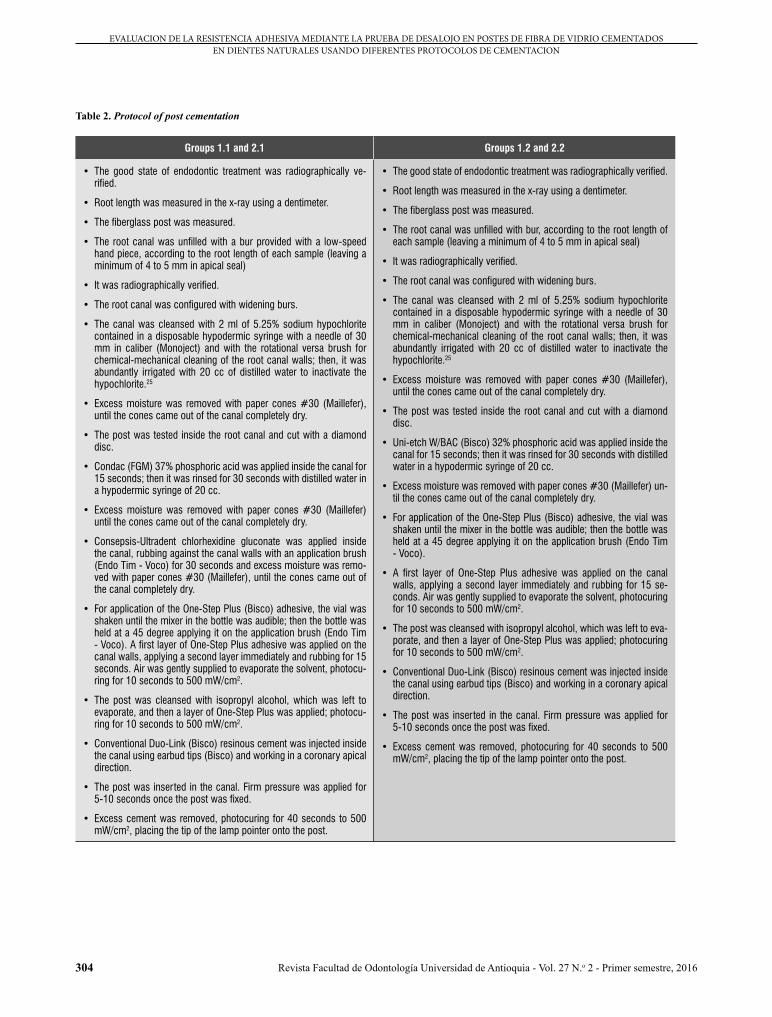
Tabla 2. Protocolo de cementación del poste
Grupos 1.1 y 2.1 Grupos 1.2 y 2.2
• Se verificó radiográficamente el buen estado de la endodoncia.
• Se midió la longitud radicular en la radiografía, con la ayuda de un dentímetro.
• Se midió el poste de fibra de vidrio.
• Se desobturó el conducto con fresa desobturadora con pieza de mano de baja velocidad, de acuerdo con la longitud radicular de cada mues-tra (dejando como mínimo de 4 a 5 mm de sellado apical)
• Se verificó radiográficamente.
• Se realizó la conformación del conducto con las fresas ensanchado-ras.
• Se limpió el conducto con 2 ml de hipoclorito de sodio al 5,25% dis-pensado en una jeringa hipodérmica desechable con aguja de 30 mm de calibre (Monoject), y con el cepillo de conducto rotacional versa brush, para obtener una limpieza química-mecánica de las paredes del conducto; luego, de manera profusa se irrigó con 20 cc de agua destilada, para inactivar el hipoclorito.25
• Se retiraron los excesos de humedad con conos de papel #30 (Maille-fer), hasta que los conos salieron del conducto completamente secos.
• Se probó el poste dentro del conducto radicular y se cortó con un disco de diamante.
• Dentro del conducto se aplicó ácido fosfórico Condac (FGM) al 37% por 15 segundos; posteriormente se lavó durante 30 segundos con agua destilada en una jeringa hipodérmica de 20 cc.
• Se retiraron los excesos de humedad con conos de papel #30 (Maille-fer), hasta que los conos salieron del conducto completamente secos.
• Se aplicó gluconato de clorhexidina (Consepsis - Ultradent) dentro del conducto, se frotó contra las paredes del conducto con la ayuda de un pincel de aplicación endodóntico (Endo Tim - Voco) durante 30 segundos y se retiraron los excesos de humedad con conos de papel #30 (Maillefer), hasta que los conos salieron del conducto completa-mente secos.
• Para la aplicación del adhesivo One-Step Plus (Bisco), se agitó el fras-co hasta que el elemento mezclador del frasco fue audible, luego se sostuvo la botella en un ángulo de 45 grados y se dispensó sobre el pincel de aplicación endodóntico (Endo Tim - Voco), Se aplicó una primera capa de adhesivo One-Step Plus sobre las paredes del con-ducto, inmediatamente se aplicó una segunda capa y se frotó durante 15 segundos. Se aireó suavemente hasta evaporar el solvente y se fotopolimerizó por 10 segundos a 500 mW/cm2.
• El poste se limpió con alcohol isopropílico, se dejó evaporar el alcohol y luego se le aplicó una capa de One-Step Plus; se fotocuró por 10 segundos a 500 mW/cm2.
• Dentro del conducto se inyectó cemento resinoso dual convencional Duo-Link (Bisco) utilizando puntas intracanal (Bisco) y trabajando en dirección apical coronaria.
• Se colocó el poste dentro del conducto. Se mantuvo presión firme durante 5-10 segundos una vez que el poste estuvo asentado.
• Se removieron los excesos del cemento y se fotocuró por 40 segundos a 500 mW/cm2, colocando la punta del puntero de la lámpara sobre el poste.
• Se verificó radiográficamente el buen estado de la endodoncia.
• Se midió la longitud radicular en la radiografía, con la ayuda de un dentímetro.
• Se midió el poste de fibra de vidrio.
• Se desobturó el conducto con fresa desobturadora de acuerdo con la longitud radicular de cada muestra (dejando como mínimo de 4 a 5 mm de sellado apical).
• Se verificó radiográficamente.
• Se realizó la conformación del conducto con las fresas ensanchado-ras.
• Se limpió el conducto con 2 ml de hipoclorito de sodio al 5,25%, dis-pensado en una jeringa hipodérmica desechable con aguja de 30 mm de calibre (Monoject), y con el cepillo de conducto rotacional versa brush, para obtener una limpieza qíumica-mecánica de las paredes del conducto; luego, de manera profusa se irrigó con 20 cc de agua destilada, para inactivar el hipoclorito.25
• Se retiraron los excesos de humedad con conos de papel #30 (Maille-fer), hasta que los conos salieron del conducto completamente secos.
• Se probó el poste dentro del conducto radicular y se cortó con un disco de diamante.
• Dentro del conducto se aplicó ácido fosfórico Uni-etch W/BAC (Bisco) al 32% por 15 segundos; posteriormente se lavó durante 30 segundos con agua destilada en una jeringa hipodérmica de 20 cc.
• Se retiraron los excesos de humedad con conos de papel #30 (Maille-fer), hasta que los conos salieron del conducto completamente secos.
• Para la aplicación del adhesivo One-Step Plus (Bisco), se agitó el fras-co hasta que el elemento mezclador del frasco fue audible; luego se sostuvo la botella en un ángulo de 45 grados y se dispensó sobre el pincel de aplicación endodóntico (Endo Tim - Voco).
• Se aplicó una primera capa de adhesivo One-Step Plus sobre las pa-redes del conducto, inmediatamente se aplicó una segunda capa y se frotó durante 15 segundos. Se aireó suavemente hasta evaporar el solvente y se fotopolimerizó por 10 segundos a 500 mW/cm2.
• El poste se limpió con alcohol isopropílico, se dejó evaporar el alcohol y luego se le aplicó una capa de One-Step Plus; se fotocuró por 10 segundos a 500 mW/cm2.
• Dentro del conducto se inyectó cemento resinoso dual convencional Duo-Link (Bisco) utilizando puntas intracanal (Bisco) y trabajando en dirección apical coronaria.
• Se colocó el poste dentro del conducto. Se mantuvo presión firme durante 5-10 segundos una vez que el poste estuvo asentado.
• Se removieron los excesos del cemento y se fotocuró por 40 segundos a 500 mW/cm2, colocando la punta del puntero de la lámpara sobre el poste.
304
EVALUACION DE LA RESISTENCIA ADHESIVA MEDIANTE LA PRUEBA DE DESALOJO EN POSTES DE FIBRA DE VIDRIO CEMENTADOS EN DIENTES NATURALES USANDO DIFERENTES PROTOCOLOS DE CEMENTACION
Revista Facultad de Odontología Universidad de Antioquia - Vol. 27 N.o 2 - Primer semestre, 2016
Table 2. Protocol of post cementation
Groups 1.1 and 2.1 Groups 1.2 and 2.2
• The good state of endodontic treatment was radiographically ve-rified.
• Root length was measured in the x-ray using a dentimeter.
• The fiberglass post was measured.
• The root canal was unfilled with a bur provided with a low-speed hand piece, according to the root length of each sample (leaving a minimum of 4 to 5 mm in apical seal)
• It was radiographically verified.
• The root canal was configured with widening burs.
• The canal was cleansed with 2 ml of 5.25% sodium hypochlorite contained in a disposable hypodermic syringe with a needle of 30 mm in caliber (Monoject) and with the rotational versa brush for chemical-mechanical cleaning of the root canal walls; then, it was abundantly irrigated with 20 cc of distilled water to inactivate the hypochlorite.25
• Excess moisture was removed with paper cones #30 (Maillefer), until the cones came out of the canal completely dry.
• The post was tested inside the root canal and cut with a diamond disc.
• Condac (FGM) 37% phosphoric acid was applied inside the canal for 15 seconds; then it was rinsed for 30 seconds with distilled water in a hypodermic syringe of 20 cc.
• Excess moisture was removed with paper cones #30 (Maillefer) until the cones came out of the canal completely dry.
• Consepsis-Ultradent chlorhexidine gluconate was applied inside the canal, rubbing against the canal walls with an application brush (Endo Tim - Voco) for 30 seconds and excess moisture was remo-ved with paper cones #30 (Maillefer), until the cones came out of the canal completely dry.
• For application of the One-Step Plus (Bisco) adhesive, the vial was shaken until the mixer in the bottle was audible; then the bottle was held at a 45 degree applying it on the application brush (Endo Tim - Voco). A first layer of One-Step Plus adhesive was applied on the canal walls, applying a second layer immediately and rubbing for 15 seconds. Air was gently supplied to evaporate the solvent, photocu-ring for 10 seconds to 500 mW/cm2.
• The post was cleansed with isopropyl alcohol, which was left to evaporate, and then a layer of One-Step Plus was applied; photocu-ring for 10 seconds to 500 mW/cm2.
• Conventional Duo-Link (Bisco) resinous cement was injected inside the canal using earbud tips (Bisco) and working in a coronary apical direction.
• The post was inserted in the canal. Firm pressure was applied for 5-10 seconds once the post was fixed.
• Excess cement was removed, photocuring for 40 seconds to 500 mW/cm2, placing the tip of the lamp pointer onto the post.
• The good state of endodontic treatment was radiographically verified.
• Root length was measured in the x-ray using a dentimeter.
• The fiberglass post was measured.
• The root canal was unfilled with bur, according to the root length of each sample (leaving a minimum of 4 to 5 mm in apical seal)
• It was radiographically verified.
• The root canal was configured with widening burs.
• The canal was cleansed with 2 ml of 5.25% sodium hypochlorite contained in a disposable hypodermic syringe with a needle of 30 mm in caliber (Monoject) and with the rotational versa brush for chemical-mechanical cleaning of the root canal walls; then, it was abundantly irrigated with 20 cc of distilled water to inactivate the hypochlorite.25
• Excess moisture was removed with paper cones #30 (Maillefer), until the cones came out of the canal completely dry.
• The post was tested inside the root canal and cut with a diamond disc.
• Uni-etch W/BAC (Bisco) 32% phosphoric acid was applied inside the canal for 15 seconds; then it was rinsed for 30 seconds with distilled water in a hypodermic syringe of 20 cc.
• Excess moisture was removed with paper cones #30 (Maillefer) un-til the cones came out of the canal completely dry.
• For application of the One-Step Plus (Bisco) adhesive, the vial was shaken until the mixer in the bottle was audible; then the bottle was held at a 45 degree applying it on the application brush (Endo Tim - Voco).
• A first layer of One-Step Plus adhesive was applied on the canal walls, applying a second layer immediately and rubbing for 15 se-conds. Air was gently supplied to evaporate the solvent, photocuring for 10 seconds to 500 mW/cm2.
• The post was cleansed with isopropyl alcohol, which was left to eva-porate, and then a layer of One-Step Plus was applied; photocuring for 10 seconds to 500 mW/cm2.
• Conventional Duo-Link (Bisco) resinous cement was injected inside the canal using earbud tips (Bisco) and working in a coronary apical direction.
• The post was inserted in the canal. Firm pressure was applied for 5-10 seconds once the post was fixed.
• Excess cement was removed, photocuring for 40 seconds to 500 mW/cm2, placing the tip of the lamp pointer onto the post.

305
EVALUATION OF PUSH-OUT BOND STRENGTH IN FIBERGLASS POSTS CEMENTED IN NATURAL TEETH USING DIFFERENT CEMENTATION PROTOCOLS
Revista Facultad de Odontología Universidad de Antioquia - Vol. 27 N.o 2 - Primer semestre, 2016
Figure 1. Cementation protocol
Figura 1. Protocolo de cementación
Para determinar la resistencia adhesiva entre el material cementante y la dentina radicular, se llevó a cabo la prueba denominada test de desalojo, por medio de la máquina universal (Instron, Model: ELS-5 made in China, con capacidad de carga de 1 a 600 Kn), valorando la resistencia adhesiva entre las interfaces de los materiales y el diente. En esta prueba, cada muestra se unió a la base de la máquina universal con un
In order to determine bonding strength between cement and root dentin, the so-called push-out test was conducted using the universal machine (Instron, Model: ELS-5 made in China, with 1 to 600 Kn load capacity), and assessing adhesive strength between the interfaces of materials and teeth. For this test, each sample was attached to the base of the universal machine with a
306
EVALUACION DE LA RESISTENCIA ADHESIVA MEDIANTE LA PRUEBA DE DESALOJO EN POSTES DE FIBRA DE VIDRIO CEMENTADOS EN DIENTES NATURALES USANDO DIFERENTES PROTOCOLOS DE CEMENTACION
Revista Facultad de Odontología Universidad de Antioquia - Vol. 27 N.o 2 - Primer semestre, 2016
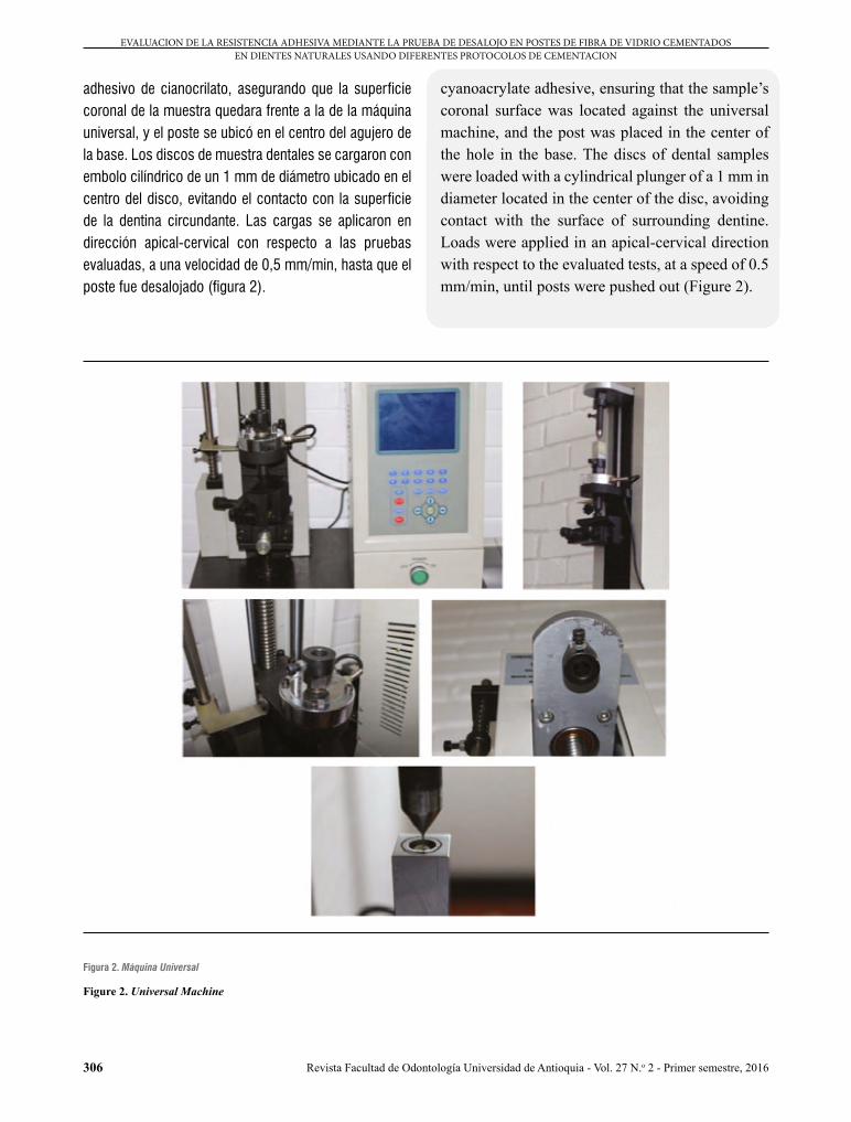
adhesivo de cianocrilato, asegurando que la superficie coronal de la muestra quedara frente a la de la máquina universal, y el poste se ubicó en el centro del agujero de la base. Los discos de muestra dentales se cargaron con embolo cilíndrico de un 1 mm de diámetro ubicado en el centro del disco, evitando el contacto con la superficie de la dentina circundante. Las cargas se aplicaron en dirección apical-cervical con respecto a las pruebas evaluadas, a una velocidad de 0,5 mm/min, hasta que el poste fue desalojado (figura 2).
cyanoacrylate adhesive, ensuring that the sample’s coronal surface was located against the universal machine, and the post was placed in the center of the hole in the base. The discs of dental samples were loaded with a cylindrical plunger of a 1 mm in diameter located in the center of the disc, avoiding contact with the surface of surrounding dentine. Loads were applied in an apical-cervical direction with respect to the evaluated tests, at a speed of 0.5 mm/min, until posts were pushed out (Figure 2).
Figura 2. Máquina Universal
Figure 2. Universal Machine

307
EVALUATION OF PUSH-OUT BOND STRENGTH IN FIBERGLASS POSTS CEMENTED IN NATURAL TEETH USING DIFFERENT CEMENTATION PROTOCOLS
Revista Facultad de Odontología Universidad de Antioquia - Vol. 27 N.o 2 - Primer semestre, 2016
A los especímenes se les tomó una fotografía con una cámara digital AxioCam ERc5s® Zeiss del estereomi-croscopio Stemi 2000-CG®, para realizar un análisis ob-servacional de los resultados de acuerdo con el tipo de fallas presentadas.
RESULTADOS
Para el estudio se tomó como criterio de decisión el nivel de significancia en α (alfa) = 0,05 (5%); no hubo porcentaje de confiabilidad por que no se usaron intervalos de confianza sino prueba de hipótesis.
Los tipos de fallas encontrados en el estudio fueron: adhesiva a la dentina(AD), adhesiva al poste (AP), cohesiva a la dentina (CD) y cohesiva al poste (CP). Los porcentajes de falla se describen en la tabla 3 y en la figura 3.
Tabla 3. Tipos de fallas
Frecuencia Porcentaje Porcentaje válido
Porcentaje acumulado
Válid
os
Adhesiva a la dentina 94 52,2 52,2 52,2
Adhesiva al poste 12 6,7 6,7 58,9
Cohesiva a la dentina 73 40,6 40,6 99,4
Cohesiva al poste 1 0,6 0,6 100,0
Total 180,0 100,0 100,0
Samples were photographed with an AxioCam ERc5s® Zeiss digital camera of the Stemi 2000-CG® stereomicroscope in order to conduct an observational analysis of the results according to failure type.
RESULTS
In this study, a significance level in α (alfa) 0.05 (5%) was taken as a decision criterion; there was no percentage of reliability because no intervals of confidence were used, statistical hypothesis testing was used instead.
The types of failure found in the study were: adhesive to dentine (AD), adhesive to post (AP), cohesive to dentine (CD), and cohesive to post (CP). The failure percentages are described in table 3 and in figure 3.
Table 3. Failure types
Frequency Percentage Valid percentage
Cumulative percentage
Valid
Adhesive to tooth
94 52.2 52.2 52.2
Adhesive to post
12 6.7 6.7 58.9
Cohesive to dentin
73 40.6 40.6 99.4
Cohesive to post
1 0.6 0.6 100.0
Total 180,0 100.0 100.0
Adhesiva a dentina Adhesiva al poste Cohesiva a dentina Cohesiva al poste Adhesiva a dentina Adhesiva al poste Cohesiva a dentina Cohesiva al poste Adhesiva a dentina Adhesiva al poste
Adhesiva a dentina Adhesiva al poste Cohesiva a dentina Cohesiva al poste Adhesiva a dentina Adhesiva al poste Cohesiva a dentina Cohesiva al poste Cohesiva a dentina Cohesiva al poste
Figura 3. Tipos de fallas
Adhesiva a dentina Adhesiva al poste Cohesiva a dentina Cohesiva al poste Adhesiva a dentina Adhesiva al poste Cohesiva a dentina Cohesiva al poste Adhesive to dentine Adhesive to post
Adhesiva a dentina Adhesiva al poste Cohesiva a dentina Cohesiva al poste Adhesiva a dentina Adhesiva al poste Cohesiva a dentina Cohesiva al poste Cohesive to dentin Cohesive to post
Figure 3. Failures types

308
EVALUACION DE LA RESISTENCIA ADHESIVA MEDIANTE LA PRUEBA DE DESALOJO EN POSTES DE FIBRA DE VIDRIO CEMENTADOS EN DIENTES NATURALES USANDO DIFERENTES PROTOCOLOS DE CEMENTACION
Revista Facultad de Odontología Universidad de Antioquia - Vol. 27 N.o 2 - Primer semestre, 2016
Las medias y las desviaciones estándar de la resistencia adhesiva (push out) del grupo 1, comparando los sub-grupos 1.1 y 1.2 y las zonas coronal (C), media (M) y apical (A), se presentan en la tabla 4.
Tabla 4. Descriptivos del grupo 1, comparando subgrupos y zonas
Subgrupo Zona N Mínimo Máximo Media Desv. Tip.
Ácido fosfórico y clorhexidina
CoronalResistencia (Mpa) 15 2,22 45,20 16,5187 10,86077
N (Válido) según lista 15
MediaResistencia (Mpa) 15 2,85 18,19 10,9027 5,15855
N (Válido) según lista 15
ApicalResistencia (Mpa) 15 1,72 20,54 6,7467 5,10709
N (Válido) según lista 15
Uni-etch
CoronalResistencia (Mpa) 15 1,20 15,84 9,3307 4,90852
N (Válido) según lista 15
MediaResistencia (Mpa) 15 1,36 16,88 5,6567 5,06505
N (Válido) según lista 15
ApicalResistencia (Mpa) 15 1,25 10,19 4,2127 3,33927
N (Válido) según lista 15
*MPa: Megapascales *N: Newton *Desv. Tip: Desviación típica
The means and standard deviations of Group 1 adhesive strength (push out) comparing subgroups 1.1 and 1.2, as well the coronal (C), middle (M), and apical (A) areas, are shown in table 4.
Table 4. Group 1 descriptors comparing subgroups and areas
Subgroup Area N Minimum Maximum Mean SD
Phosphoric acid plus chlorhexidine
CoronalResistance (Mpa) 15 2.22 45.20 16.5187 10.86077
N (valid) according to list 15
MiddleResistance (Mpa) 15 2.85 18.19 10.9027 5.15855
N (valid) according to list 15
ApicalResistance (Mpa) 15 1.72 20.54 6.7467 5.10709
N (valid) according to list 15
Uni-etch
CoronalResistance (Mpa) 15 1.20 15.84 9.3307 4.90852
N (valid) according to list 15
MiddleResistance (Mpa) 15 1.36 16.88 5.6567 5.06505
N (valid) according to list 15
ApicalResistance (Mpa) 15 1.25 10.19 4.2127 3.33927
N (valid) according to list 15
* MPa: Megapascal * N: Newton * SD: Standard deviation
Las medias y las desviaciones estándar de la resistencia adhesiva (push out) del grupo 2, comparando los sub-grupos 2.1 y 2.2 y las zonas coronal (C), media (M) y apical (A), se presentan en la tabla 5.
The means and standard deviations of Group 2 adhesive strength (push out) comparing subgroups 2.1 and 2.2, as well the coronal (C), middle (M), and apical (A) areas, are shown in table 5.

309
EVALUATION OF PUSH-OUT BOND STRENGTH IN FIBERGLASS POSTS CEMENTED IN NATURAL TEETH USING DIFFERENT CEMENTATION PROTOCOLS
Revista Facultad de Odontología Universidad de Antioquia - Vol. 27 N.o 2 - Primer semestre, 2016
Tabla 5. Descriptivos del grupo 2, comparando subgrupos y zonas
Subgrupo Zona N Mínimo Máximo Media Desv. Tip.
Ácido fosfórico y clorhexidina
CoronalResistencia (Mpa) 15 0,61 14,23 6,9673 5,26677
N (Válido) según lista 15
MediaResistencia (Mpa) 15 0,17 11,49 4,1020 3,84004
N (Válido) según lista 15
ApicalResistencia (Mpa) 15 0,19 13,98 3,3767 3,79702
N (Válido) según lista 15
Unietch
CoronalResistencia (Mpa) 15 0,35 19,13 10,6213 5,71357
N (Válido) según lista 15
MediaResistencia (Mpa) 15 0,26 9,70 2,9100 3,18632
N (Válido) según lista 15
ApicalResistencia (Mpa) 15 0,06 6,98 1,7253 2,00712
N (Válido) según lista 15
*MPa: Megapascales *N: Newton *Desv. Tip: Desviación típica
Table 5. Group 2 descriptors comparing subgroups and areas
Subgroup Area N Minimum Maximum Mean SD
Phosphoric acid plus chlorhexi-dine
CoronalResistance (Mpa) 15 0.61 14.23 6.9673 5.26677
N (valid) according to list 15
MiddleResistance (Mpa) 15 0.17 11.49 4.1020 3.84004
N (valid) according to list 15
ApicalResistance (Mpa) 15 0.19 13.98 3.3767 3.79702
N (valid) according to list 15
Unietch
CoronalResistance (Mpa) 15 0.35 19.13 10.6213 5.71357
N (valid) according to list 15
MiddleResistance (Mpa) 15 0.26 9.70 2.9100 3.18632
N (valid) according to list 15
ApicalResistance (Mpa) 15 0.06 6.98 1.7253 2.00712
N (valid) according to list 15
* MPa: Megapascal * N: Newton * SD: Standard deviation
El test de Kolmogorov-Smirnov confirmó que hubo nor-malidad en el grupo 1, subgrupo 1.1, y en el grupo 2, subgrupos 2.1 y 2.2 (p = 0,383, p = 0,086, p = 0,099, respectivamente), y anormalidad en el grupo 1, subgru-po 1.2 (p = 0,048). El test Anova mostró que hay dife-rencia significativa en la resistencia adhesiva en el grupo 1, subgrupos 1.1 y 1.2, y en el grupo 2, subgrupo 2.2 (p < 0,05) y no mostró diferencia significativa en el gru-po 2, subgrupo 2.1 (p > 0,05) (tablas 6 y 7).
The Kolmogorov-Smirnov test confirmed that there was normality in Group 1, subgroup 1.1 and in Group 2, subgroups 2.1 and 2.2 (p = 0.383, p = 0.086, p = 0.099, respectively), and non-normality in Group 1, subgroup 1.2 (p = 0,048). The Anova test showed significant difference in adhesive strength in Group 1, subgroups 1.1 and 1.2, and in Group 2, subgroup 2.2 (p < 0.05) and no significant difference in Group 2, subgroup 2.1 (p > 0.05) (tables 6 and 7).

310
EVALUACION DE LA RESISTENCIA ADHESIVA MEDIANTE LA PRUEBA DE DESALOJO EN POSTES DE FIBRA DE VIDRIO CEMENTADOS EN DIENTES NATURALES USANDO DIFERENTES PROTOCOLOS DE CEMENTACION
Revista Facultad de Odontología Universidad de Antioquia - Vol. 27 N.o 2 - Primer semestre, 2016
Tabla 6. Grupo 1 ANOVA – Resistencia MPa
Subgrupo Suma de Cuadrados gl Media cuadrática F Sig.
Ácido fosfórico y clorhexidina
Intergrupos 721,519 2 360,759 6,342 0,004
Intragrupos 2389,091 42 56,883
Total 3110,610 44
Unietch
Intergrupos 280,887 2 104,443 5,145 0,010
Intragrupos 852,585 42 20,300
Total 1061,472 44
*MPa: Megapascales
Table 6. Group 1 ANOVA - Strength in MPa
Subgroup Sum of squares GL Root mean square F Sig.
Phosphoric acid plus chlorhexidine
Intergroups 721.519 2 360.759 6.342 0.004
Intragroups 2389.091 42 56.883
Total 3110.610 44
Unietch
Intergroups 280.887 2 104.443 5.145 0.010
Intragroups 852.585 42 20.300
Total 1061.472 44
* MPa: Megapascal
Tabla 7. Grupo 2 ANOVA – Resistencia Mpa
Subgrupo Suma de Cuadrados gl Media cuadrática F Sig.
Ácido fosfórico y clorhexidina
Intergrupos 108,146 2 54,073 2,851 0,69
Intragrupos 796,629 42 18,967
Total 904,774 44
Unietch
Intergrupos 700,035 2 350,017 22,425 0,000
Intragrupos 655,564 42 15,609
Total 1355,599 44
*MPa: Megapascales
Table 7. Group 2 ANOVA - Strength in MPa
Subgroup Sum of squares GL Root mean square F Sig.
Phosphoric acid plus chlorhexidine
Intergroups 108.146 2 54.073 2.851 0.69
Intragroups 796.629 42 18.967
Total 904.774 44
Unietch
Intergroups 700.035 2 350.017 22.425 0.000
Intragroups 655.564 42 15.609
Total 1355.599 44
* MPa: Megapascal
311
EVALUATION OF PUSH-OUT BOND STRENGTH IN FIBERGLASS POSTS CEMENTED IN NATURAL TEETH USING DIFFERENT CEMENTATION PROTOCOLS
Revista Facultad de Odontología Universidad de Antioquia - Vol. 27 N.o 2 - Primer semestre, 2016
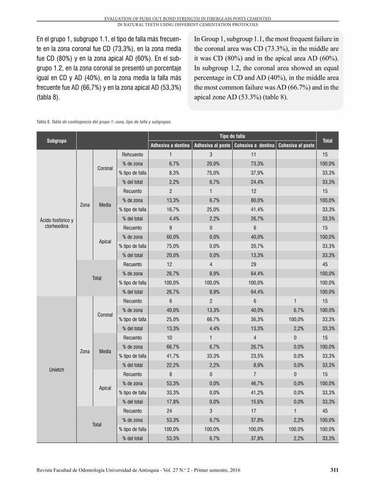
In Group 1, subgroup 1.1, the most frequent failure in the coronal area was CD (73.3%), in the middle are it was CD (80%) and in the apical area AD (60%). In subgroup 1.2, the coronal area showed an equal percentage in CD and AD (40%), in the middle area the most common failure was AD (66.7%) and in the apical zone AD (53.3%) (table 8).
En el grupo 1, subgrupo 1.1, el tipo de falla más frecuen-te en la zona coronal fue CD (73,3%), en la zona media fue CD (80%) y en la zona apical AD (60%). En el sub-grupo 1.2, en la zona coronal se presentó un porcentaje igual en CD y AD (40%), en la zona media la falla más frecuente fue AD (66,7%) y en la zona apical AD (53,3%) (tabla 8).
Tabla 8. Tabla de contingencia del grupo 1: zona, tipo de falla y subgrupos
SubgrupoTipo de falla
TotalAdhesiva a dentina Adhesiva al poste Cohesiva a dentina Cohesiva al poste
Ácido fosfórico y clorhexidina
Zona
Coronal
Rehcuento 1 3 11 15
% de zona 6,7% 20,0% 73,3% 100,0%
% tipo de falla 8,3% 75,0% 37,9% 33,3%
% del total 2,2% 6,7% 24,4% 33,3%
Media
Recuento 2 1 12 15
% de zona 13,3% 6,7% 80,0% 100,0%
% tipo de falla 16,7% 25,0% 41,4% 33,3%
% del total 4,4% 2,2% 26,7% 33,3%
Apical
Recuento 9 0 6 15
% de zona 60,0% 0,0% 40,0% 100,0%
% tipo de falla 75,0% 0,0% 20,7% 33,3%
% del total 20,0% 0,0% 13,3% 33,3%
Total
Recuento 12 4 29 45
% de zona 26,7% 8,9% 64,4% 100,0%
% tipo de falla 100,0% 100,0% 100,0% 100,0%
% del total 26,7% 8,9% 64,4% 100,0%
Unietch
Zona
Coronal
Recuento 6 2 6 1 15
% de zona 40,0% 13,3% 40,0% 6,7% 100,0%
% tipo de falla 25,0% 66,7% 36,3% 100,0% 33,3%
% del total 13,3% 4,4% 13,3% 2,2% 33,3%
Media
Recuento 10 1 4 0 15
% de zona 66,7% 6,7% 26,7% 0,0% 100,0%
% tipo de falla 41,7% 33,3% 23,5% 0,0% 33,3%
% del total 22,2% 2,2% 8,9% 0,0% 33,3%
Apical
Recuento 8 0 7 0 15
% de zona 53,3% 0,0% 46,7% 0,0% 100,0%
% tipo de falla 33,3% 0,0% 41,2% 0,0% 33,3%
% del total 17,8% 0,0% 15,6% 0,0% 33,3%
Total
Recuento 24 3 17 1 45
% de zona 53,3% 6,7% 37,8% 2,2% 100,0%
% tipo de falla 100,0% 100,0% 100,0% 100,0% 100,0%
% del total 53,3% 6,7% 37,8% 2,2% 33,3%
312
EVALUACION DE LA RESISTENCIA ADHESIVA MEDIANTE LA PRUEBA DE DESALOJO EN POSTES DE FIBRA DE VIDRIO CEMENTADOS EN DIENTES NATURALES USANDO DIFERENTES PROTOCOLOS DE CEMENTACION
Revista Facultad de Odontología Universidad de Antioquia - Vol. 27 N.o 2 - Primer semestre, 2016
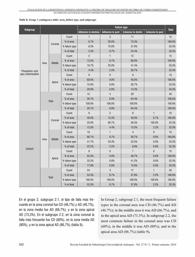
Table 8. Group 1 contingency table: area, failure type, and subgroups
SubgroupFailure type
TotalAdhesive to dentine Adhesive to post Cohesive to dentin Cohesive to post
Phosphoric acid plus chlorhexidine
Area
Coronal
Count 1 3 11 15
% of area 6.7% 20.0% 73.3% 100.0%
% failure type 8.3% 75.0% 37.9% 33.3%
% of total 2.2% 6.7% 24.4% 33.3%
Middle
Count 2 1 12 15
% of area 13.3% 6.7% 80.0% 100.0%
% failure type 16.7% 25.0% 41.4% 33.3%
% of total 4.4% 2.2% 26.7% 33.3%
Apical
Count 9 0 6 15
% of area 60.0% 0.0% 40.0% 100.0%
% failure type 75.0% 0.0% 20.7% 33.3%
% of total 20.0% 0.0% 13.3% 33.3%
Total
Count 12 4 29 45
% of area 26.7% 8.9% 64.4% 100.0%
% failure type 100.0% 100.0% 100.0% 100.0%
% of total 26.7% 8.9% 64.4% 100.0%
Unietch
Area
Coronal
Count 6 2 6 1 15
% of area 40.0% 13.3% 40.0% 6.7% 100.0%
% failure type 25.0% 66.7% 36.3% 100.0% 33.3%
% of total 13.3% 4.4% 13.3% 2.2% 33.3%
Middle
Count 10 1 4 0 15
% of area 66.7% 6.7% 26.7% 0.0% 100.0%
% failure type 41.7% 33.3% 23.5% 0.0% 33.3%
% of total 22.2% 2.2% 8.9% 0.0% 33.3%
Apical
Count 8 0 7 0 15
% of area 53.3% 0.0% 46.7% 0.0% 100.0%
% failure type 33.3% 0.0% 41.2% 0.0% 33.3%
% of total 17.8% 0.0% 15.6% 0.0% 33.3%
Total
Count 24 3 17 1 45
% of area 53.3% 6.7% 37.8% 2.2% 100.0%
% failure type 100.0% 100.0% 100.0% 100.0% 100.0%
% of total 53.3% 6.7% 37.8% 2.2% 33.3%
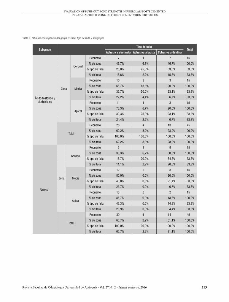
In Group 2, subgroup 2.1, the most frequent failure types in the coronal area was CD (46.7%) and AD (46.7%); in the middle area it was AD (66.7%), and in the apical area AD (73.3%). In subgroup 2.2, the most common failure in the coronal area was CD (60%), in the middle it was AD (80%), and in the apical area AD (86.7%) (table 9).
En el grupo 2, subgrupo 2.1, el tipo de falla más fre-cuente en la zona coronal fue CD (46,7%) y AD (46,7%), en la zona media fue AD (66,7%), y en la zona apical AD (73,3%). En el subgrupo 2.2, en la zona coronal la falla más frecuente fue CD (60%), en la zona media AD (80%), y en la zona apical AD (86,7%) (tabla 9).
313
EVALUATION OF PUSH-OUT BOND STRENGTH IN FIBERGLASS POSTS CEMENTED IN NATURAL TEETH USING DIFFERENT CEMENTATION PROTOCOLS
Revista Facultad de Odontología Universidad de Antioquia - Vol. 27 N.o 2 - Primer semestre, 2016
Tabla 9. Tabla de contingencia del grupo 2: zona, tipo de falla y subgrupos
SubgrupoTipo de falla
TotalAdhesiv a dentinata Adhesiva al poste Cohesiva a dentina
Ácido fosfórico y clorhexidina
Zona
Coronal
Recuento 7 1 7 15
% de zona 46,7% 6,7% 46,7% 100,0%
% tipo de falla 25,0% 25,0% 53,8% 33,3%
% del total 15,6% 2,2% 15,6% 33,3%
Media
Recuento 10 2 3 15
% de zona 66,7% 13,3% 20,0% 100,0%
% tipo de falla 35,7% 50,0% 23,1% 33,3%
% del total 22,2% 4,4% 6,7% 33,3%
Apical
Recuento 11 1 3 15
% de zona 73,3% 6,7% 20,0% 100,0%
% tipo de falla 39,3% 25,0% 23,1% 33,3%
% del total 24,4% 2,2% 6,7% 33,3%
Total
Recuento 28 4 13 45
% de zona 62,2% 8,9% 28,9% 100,0%
% tipo de falla 100,0% 100,0% 100,0% 100,0%
% del total 62,2% 8,9% 28,9% 100,0%
Unietch
Zona
Coronal
Recuento 5 1 9 15
% de zona 33,3% 6,7% 60,0% 100,0%
% tipo de falla 16,7% 100,0% 64,3% 33,3%
% del total 11,1% 2,2% 20,0% 33,3%
Media
Recuento 12 0 3 15
% de zona 80,0% 0,0% 20,0% 100,0%
% tipo de falla 40,0% 0,0% 21,4% 33,3%
% del total 26,7% 0,0% 6,7% 33,3%
Apical
Recuento 13 0 2 15
% de zona 86,7% 0,0% 13,3% 100,0%
% tipo de falla 43,3% 0,0% 14,3% 33,3%
% del total 28,9% 0,0% 4,4% 33,3%
Total
Recuento 30 1 14 45
% de zona 66,7% 2,2% 31,1% 100,0%
% tipo de falla 100,0% 100,0% 100,0% 100,0%
% del total 66,7% 2,2% 31,1% 100,0%

314
EVALUACION DE LA RESISTENCIA ADHESIVA MEDIANTE LA PRUEBA DE DESALOJO EN POSTES DE FIBRA DE VIDRIO CEMENTADOS EN DIENTES NATURALES USANDO DIFERENTES PROTOCOLOS DE CEMENTACION
Revista Facultad de Odontología Universidad de Antioquia - Vol. 27 N.o 2 - Primer semestre, 2016
Table 9. Group 2 contingency table: area, failure type, and subgroups
SubgroupFailure type
TotalAdhesive to dentin Adhesive to post Cohesive to dentin
Phosphoric acid plus chlorhexidine
Area
Coronal
Count 7 1 7 15
% of area 46.7% 6.7% 46.7% 100.0%
% failure type 25.0% 25.0% 53.8% 33.3%
% of total 15.6% 2.2% 15.6% 33.3%
Middle
Count 10 2 3 15
% of area 66.7% 13.3% 20.0% 100.0%
% failure type 35.7% 50,0% 23.1% 33.3%
% of total 22.2% 4.4% 6.7% 33.3%
Apical
Count 11 1 3 15
% of area 73.3% 6.7% 20.0% 100.0%
% failure type 39.3% 25.0% 23.1% 33.3%
% of total 24.4% 2.2% 6.7% 33.3%
Total
Count 28 4 13 45
% of area 62.2% 8.9% 28.9% 100.0%
% failure type 100.0% 100.0% 100.0% 100.0%
% of total 62.2% 8.9% 28.9% 100.0%
Unietch
Area
Coronal
Count 5 1 9 15
% of area 33.3% 6.7% 60.0% 100.0%
% failure type 16.7% 100.0% 64.3% 33.3%
% of total 11.1% 2.2% 20.0% 33.3%
Middle
Count 12 0 3 15
% of area 80.0% 0.0% 20.0% 100.0%
% failure type 40.0% 0.0% 21.4% 33.3%
% of total 26.7% 0.0% 6.7% 33.3%
Apical
Count 13 0 2 15
% of area 86.7% 0.0% 13.3% 100.0%
% failure type 43.3% 0.0% 14.3% 33.3%
% of total 28.9% 0.0% 4.4% 33.3%
Total
Count 30 1 14 45
% of area 66.7% 2.2% 31.1% 100.0%
% failure type 100.0% 100.0% 100.0% 100.0%
% of total 66.7% 2.2% 31.1% 100.0%
DISCUSIÓN
En este estudio se evaluó la resistencia adhesiva de pos-tes de fibra de vidrio cementados en dientes humanos premolares unirradiculares, indicados para exodoncia por motivos ortodónticos, usando diferentes protocolos de cementación y con dos materiales obturadores diferentes,
DISCUSSION
This study evaluated the adhesive strength of fiberglass posts cemented in single-root human premolars extracted for orthodontic reasons, using different cementing protocols and two different filling materials,
315
EVALUATION OF PUSH-OUT BOND STRENGTH IN FIBERGLASS POSTS CEMENTED IN NATURAL TEETH USING DIFFERENT CEMENTATION PROTOCOLS
Revista Facultad de Odontología Universidad de Antioquia - Vol. 27 N.o 2 - Primer semestre, 2016
y se analizaron tres zonas radiculares específicas (coro-nal, media y apical). El primer protocolo consistía en el manejo de ácido fosfórico más clorhexidina (subgrupos 1.1 y 2.1) y el segundo utilizando Unietch (subgrupos 1.2 y 2.2), el cual es un ácido fosfórico que contiene cloruro de benzalconio, un agente antimicrobiano que, según la evidencia científica, tiene la misma función de la clor-hexidina.12 Una vez cementados los postes, las muestras se sometieron a envejecimiento por almacenamiento a temperatura constante (27 oC) durante 30 días; este mé-todo se realizó con el fin de valorar el rendimiento clínico de los materiales en el tiempo, simulando la degradación de la restauración.13-15 Otro de los métodos más utilizados es el envejecimiento por termociclado, mediante el cual se simulan los cambios térmicos de la cavidad oral. Recientemente, en la convención de Charlotte de 2014, Kwon, Burgess y Beck señalaron, como una de sus con-clusiones, que 10.000 ciclos no presentan diferencia significativa con el método de almacenamiento realizado durante 24 horas.15 La literatura recomienda la estanda-rización de los protocolos en tiempo para determinar el número de ciclos. Sin embargo, existe una hipótesis se-gún la cual se podían presentar entre 20 y 50 ciclos por día, y por ende se habla de unos 10.000 ciclos por año.
Con el fin de evaluar la resistencia adhesiva, se utilizó la prueba de desalojo (push-out test), basados en que esta prueba proporciona datos sobre áreas adhesivas más pequeñas, uniforme distribución de estrés en la inter-face adhesiva, menor pérdida de especímenes durante el experimento, bajos valores de desviación estándar y facilidad en la ejecución.16,17
Inicialmente, se planteó que no existirían diferencias significativas entre los protocolos utilizados, pero se encontraron variaciones en la resistencia adhesiva de-pendiendo del material utilizado en la obturación del con-ducto radicular, el protocolo de cementación y la zona del diente a evaluar.
Una vez aplicadas las pruebas físicas, los resultados arrojaron datos según los cuales la máxima resis-tencia adhesiva se presentó en la zona coronal de los dientes obturados con cemento Top Seal (grupo 1)
and analyzing three specific root areas (coronal, middle, and apical). The first protocol was phosphoric acid plus chlorhexidine (subgroups 1.1 and 2.1) and the second protocol used Unietch (subgroup 1.2 and 2.2), which is a phosphoric acid containing benzalkonium chloride, an antimicrobial agent that according to scientific evidence has the same function as chlorhexidine.12 Once posts had been cemented, the samples were subjected to aging by storing them at constant temperature (27 °C) for 30 days. This method was used in order to assess the clinical performance of materials over time, simulating the degradation of restorations.13-15 Another commonly used method is aging by thermocycling, which simulates thermal changes in the oral cavity. Recently, in the 2014 Charlotte Convention, Kwon, Burgess and Beck pointed out as one of their conclusions that 10,000 cycles do not show significant difference with the storage method for 24 hours.15 The literature recommends standardizing the protocols in terms of time to determine the number of cycles. However, one hypothesis states that there could be 20 to 50 cycles per day, and therefore there would be about 10,000 cycles per year.
In order to evaluate adhesive strength, the push-out test was used as it provides data on the smallest adhesive areas, uniform distribution of adhesive interface stress, fewer sample loss during experiments, low standard deviation values, and is easy to perform.16,17
The initial assumption was that there would be no significant differences between the protocols used, but variations were found in terms of adhesive strength depending on the material used to fill the root canals, the cementing protocol, and the tooth area under evaluation.
Once the physical tests were completed, the results yielded data suggesting that the maximum adhesive strength happened in the coronal area of teeth filled with the Top Seal cement (Group 1)

316
EVALUACION DE LA RESISTENCIA ADHESIVA MEDIANTE LA PRUEBA DE DESALOJO EN POSTES DE FIBRA DE VIDRIO CEMENTADOS EN DIENTES NATURALES USANDO DIFERENTES PROTOCOLOS DE CEMENTACION
Revista Facultad de Odontología Universidad de Antioquia - Vol. 27 N.o 2 - Primer semestre, 2016
y en los cuales para la cementación del poste se utilizó el protocolo con ácido fosfórico, clorhexidina y cemento resinoso dual (Duolink) (subgrupo 1.1), con un valor de 45,2 Mpa. En el caso de los dientes tratados con Unietch (subgrupos 1.2 y 2.2), los valores máximos encontrados fueron de 19,13 MPa en aquellos donde se usó cemento obturador Grossman (grupo 2) y de 15,84 Mpa en los que se usó cemento obturador Top Seal (grupo 1). A pe-sar de que los valores con el protocolo de Unietch fueron más bajos que los encontrados con los del grupo en don-de se usó ácido fosfórico y clorhexidina (subgrupo 1.1), estos valores siguen siendo más altos que los obtenidos por Pereira y colaboradores en el año 2013, quienes en-contraron valores máximos de 11,5 Mpa para Duolink.17 Los autores manejaron un protocolo de cementación di-ferente, ya que no utilizaron versa brush unido al uso del hipoclorito, lo cual confiere o da una limpieza química y mecánica. El versa brush es un instrumento rotatorio utilizado junto con la pieza de baja velocidad con el fin de optimizar la limpieza del conducto radicular y remover los contaminantes que quedan después de la desobtura-ción del conducto radicular (componentes del cemento sellador y el barrido dentinario). Además, no manejaron clorhexidina después de la aplicación del ácido fosfórico, y tampoco tuvieron en cuenta el cemento obturador, que en el presente estudio mostró relevancia a la hora de evaluar la resistencia adhesiva.
En 2013, Saraiva y colaboradores investigaron el efecto del grabado con ácido fosfórico y el pretratamiento de la dentina con hipoclorito de sodio sobre la resistencia adhesiva mediante la prueba de push-out entre poste y dentina del canal, utilizando un cemento resinoso dual (Variolink II-Ivoclar/Vivadent). Ellos encontraron que el pretratamiento con hipoclorito de sodio no presentó di-ferencias estadísticamente significativas en los grupos evaluados; sin embargo, cuando el ácido fosfórico fue aplicado durante 60 s en la porción apical sin hipoclorito de sodio (como pretratamiento), las fuerzas adhesivas fueron superiores. En el presente estudio, también se uti-lizó un cemento resinoso dual (Duolink-Bisco), el hipo-clorito de sodio se usó como pretratamiento en ambos grupos, y el ácido desmineralizante durante 15 segundos.
where the posts were cemented with phosphoric acid, chlorhexidine, and dual resinous cement (Duolink) (subgroup 1.1), with a score of 45.2 Mpa. In the case of teeth treated with Unietch (subgroups 1.2 and 2.2), the maximum values were 19.13 MPa in those treated with the Grossman filling cement (Group 2) and 15.84 Mpa in those treated with the Top Seal cement (Group 1). While the scores of the Unietch protocol were lower than those of the group treated with phosphoric acid plus chlorhexidine (subgroup 1.1), these scores are still higher than those obtained in 2013by Pereira et al, who found peak scores of 11.5 Mpa for Duolink.17 These authors used a different cementing protocol, since they did not use versa brush together plus hypochlorite, which provides chemical and mechanical cleaning. The versa brush is a rotary instrument used along with low-speed piece to optimize the cleaning of the root canal and to remove the contaminants left by unfilling the root canal (components of the sealing cement and the smear layer). In addition, they did not use chlorhexidine after applying phosphoric acid, and did not take into account the filling cement, which in the present study showed relevance in assessing adhesive strength.
In 2013, Saraiva et al analyzed the effect of etching with phosphoric acid and pretreating dentin with sodium hypochlorite on adhesive strength by means of the push-out between post and canal dentin, using a dual resinous cement (Variolink II-Ivoclar/Vivadent). They found out that pretreating with sodium hypochlorite did not show statistically significant differences in the evaluated groups; however, when phosphoric acid was applied for 60 s in the apical portion without sodium hypochlorite (as pretreatment), the adhesive forces were higher. The present study also used a dual resinous cement (Duolink-Bisco), sodium hypochlorite was used as pretreatment in both groups, and demineralizing acid was used for 15 seconds.
317
EVALUATION OF PUSH-OUT BOND STRENGTH IN FIBERGLASS POSTS CEMENTED IN NATURAL TEETH USING DIFFERENT CEMENTATION PROTOCOLS
Revista Facultad de Odontología Universidad de Antioquia - Vol. 27 N.o 2 - Primer semestre, 2016
Cuando Saraiva y colaboradores evaluaron las muestras utilizando ácido fosfórico durante 15 segundos, obtuvie-ron valores de resistencia adhesiva máxima en apical de 9,6 Mpa utilizando pretratamiento con hipoclorito y en este estudio se obtuvo un valor máximo de 20,54 Mpa en la zona apical utilizando el mismo protocolo; el uso del versa brush puede indicar la diferencia en los resul-tados.18
En 2013, Shiratori y colaboradores evaluaron la resisten-cia adhesiva de tres cementos autoadhesivos utilizando diferentes métodos para llevar el cemento al interior del conducto radicular. Ellos encontraron valores de resis-tencia máximos de 13,65 Mpa en el grupo en que utiliza-ron léntulo para introducir el material al conducto, y con-cluyeron que la técnica utilizada para llevar el cemento parece influir en la resistencia adhesiva cuando se quiera cementar postes intrarradiculares; en otro grupo realiza-ron un protocolo en el cual se utilizaron puntas intracon-ducto para dispensar el cemento; en la zona apical, que es la de más difícil acceso debido a la reducción del diá-metro del conducto, se obtuvo una resistencia adhesiva máxima de 20,54 Mpa, resultando mayor a la obtenida en los cementos autoadhesivos en general, por lo cual el uso de puntas intraconducto posiblemente influyó para obtener valores de resistencia adhesiva más elevados en zonas críticas como la apical.19
Un factor importante en los protocolos del presente es-tudio fue la utilización de clorhexidina, que hoy en día es usada en adhesión dental como un inhibidor de proteasas. La literatura reporta que la dentina desmineralizada con-tiene metaloproteinasas (MMP) y catepsinas, las cuales pueden degradar lentamente las fibras colágenas.12 Las MMP son una clase de endopeptidasas dependientes de zinc y de calcio, capaces de degradar todos los compo-nentes de la matriz extracelular. La actividad colagenolí-tica y gelatinolítica puede ser superada por inhibidores de proteasas, indicando que la inhibición de MMP quizás preserve la integridad de la capa híbrida. De hecho, la aplicación de la clorhexidina en vivo mejora la integri-dad de la capa híbrida. Cuando se aplica ácido fosfórico sin la posterior aplicación de clorhexidina, esto no inhi-be la actividad colagenolítica de la dentina mineralizada,
When Saraiva et al analyzed their samples using phosphoric acid for 15 seconds, they found out maximum adhesive strength scores of 9.6 Mpa on apical by pretreating with hypochlorite, while the present study observed a maximum score of 20.54 Mpa in the apical zone using the same protocol; the use of versa brush explain the difference in results.18
In 2013, Shiratori et al evaluated the adhesive strength of three self-adhesive cements using different methods to bring the cement to the interior of the root canal. They found maximum resistance values of 13.65 Mpa in the group that used lentulo to insert the material into the canal, concluding that the technique used to carry the cement seems to influence adhesive strength when cementing intraradicular posts; the protocol in another group used intracanal tips to apply the cement. In the apical area, which is the hardest to reach due to a reduction in canal diameter, they observed a maximum adhesive strength of 20.54 Mpa, being higher than that obtained in self-adhesive cements in general, so the use of intracanal tips possibly influenced the higher adhesive strength values in critical areas such as the apical.19
An important factor in the protocols of the present study was the use of chlorhexidine, which is currently used in dental adhesion as a protease inhibitor. The literature reports that demineralized dentin contains matrix metalloproteinases (MMP) and cathepsins, which can slowly degrade collagen fibers.12 MMP are a type of calcium and zinc-dependent endopeptidases, capable of degrading all components of the extracellular matrix. Collagenolytic and gelatinolytic activity can be controlled by protease inhibitors, indicating that MMP inhibition may preserve integrity of the hybrid layer. In fact, applying chlorhexidine in vivo improves integrity of the hybrid layer. When phosphoric acid is applied without no subsequent application of chlorhexidine, this does not inhibit the collagenolytic activity mineralized of mineralized dentin,
318
EVALUACION DE LA RESISTENCIA ADHESIVA MEDIANTE LA PRUEBA DE DESALOJO EN POSTES DE FIBRA DE VIDRIO CEMENTADOS EN DIENTES NATURALES USANDO DIFERENTES PROTOCOLOS DE CEMENTACION
Revista Facultad de Odontología Universidad de Antioquia - Vol. 27 N.o 2 - Primer semestre, 2016
mientras que el uso de clorhexidina después de grabado ácido, incluso en concentraciones muy bajas, inhibe fuer-temente la actividad. Los resultados del presente estudio demostraron en general valores superiores en resistencia adhesiva en las muestras en que se utilizó el protocolo con clorhexidina, comparado con el que utilizó solamente el Unietch (cloruro de benzalconio), en los tres tercios de los grupos 1 y 2 (cemento Top Seal y Grossman respectiva-mente), lo que es congruente con lo que reporta la litera-tura acerca de la prevención de la degradación de la capa hibrida, mejorando así la adhesión en dentina.12
Se han conducido diversos estudios sobre resistencia ad-hesiva en postes de fibra de vidrio cementados con ionó-meros vítreos convencionales y modificados con resina (cementantes). En 2014, Pereira y colaboradores encon-traron valores comprendidos entre 0,5 Mpa y 19,6 Mpa, en donde observaron que el nivel del poste (cervical, medio o coronal) no influía en la resistencia adhesiva de los postes a la dentina de la raíz. En el presente estudio se encontraron valores entre 0,6 Mpa y 45,20 Mpa, pero, a diferencia del estudio de Pereira y colaboradores, sí se encontraron diferencias entre las zonas evaluadas. Esto demuestra que los cementos resinosos necesitan una interface de unión confiable, la cual es difícil de obte-ner, especialmente en la zona apical donde generalmente solo queda espacio para el poste. La ausencia de capa híbrida en la interface adhesiva puede contribuir a estas diferencias cuando los postes de fibra son cementados con cementos de resina.19-21
Los resultados del presente estudio mostraron los niveles más bajos de resistencia adhesiva a nivel del tercio api-cal. Es posible que esto se deba a restos de gutapercha y material sellador de endodoncia que puedan permanecer en la zona apical después de la preparación, como lo reportan Bolhuis y colaboradores en su estudio.22
En 2002, Hagge y colaboradores evaluaron el efecto de tres cementos selladores del conducto radicular sobre la retención de los postes de fibra de vidrio cementa-dos con cementos resinosos. Los autores utilizaron ce-mentos a base de resina epóxica, eugenol e hidróxido de calcio, y descubrieron que la formulación química
while using chlorhexidine after acid etching, even in very low concentrations, strongly inhibits this activity. The results of the present study generally showed higher adhesive strength scores in protocols with chlorhexidine, compared with those that only used Unietch (benzalkonium chloride) in the three thirds of groups 1 and 2 (Top Seal and Grossman respectively), which is consistent with literature reports about the prevention of degradation of the hybrid layer, thus improving the adhesion to dentin.12
There are various studies on adhesive strength in fiberglass posts cemented with conventional glass ionomers and modified with resin (cementitious). In 2014, Pereira et al found values ranging from 0.5 Mpa to 19.6 Mpa, observing that area were the post is located (cervical, middle, or coronal) does not influence adhesive strength of posts to root dentin. The present study yielded values ranging from 0.6 Mpa to 45.20 Mpa, but unlike the study by Pereira et al, there were differences between the assessed areas. This shows that resin cements need a reliable bonding interface—something that is hart to achieve, especially in the apical area, where hardly there is room for the post—. The absence of hybrid layer at the adhesive interface may contribute to these differences when fiber posts are cemented with resin cements.19-21
The findings of the present study showed that the lowest adhesive strength levels are at the apical third. This may be due to the residues of gutta-percha and endodontic sealer material that may remain in the apical area after preparation, as reported by Bolhuis et al. 22
In 2002, Hagge et al evaluated the effect of three cements to seal the root canal on the retention of fiberglass posts cemented with resin cements. The authors used epoxy resin-based cements, eugenol, and calcium hydroxide, finding out that the chemical formulation
319
EVALUATION OF PUSH-OUT BOND STRENGTH IN FIBERGLASS POSTS CEMENTED IN NATURAL TEETH USING DIFFERENT CEMENTATION PROTOCOLS
Revista Facultad de Odontología Universidad de Antioquia - Vol. 27 N.o 2 - Primer semestre, 2016
of sealing cements did not significantly affect the posts retention values. Our study assessed epoxy resin-based cements (Top Seal) and zinc oxide-based cements (Grossman), finding out the lowest values in the group treated with Grossman cement, but these were not statistically significant, agreeing with the findings by Hagge et al.23
One of the limitations of our study is that it is an in vitro research imitating oral conditions. Cement type and the different protocols for cementation of fiberglass posts require clinical studies in greater periods of time in order to evaluate their behavior and to have more reliable results.
CONCLUSIONS
There were no statistically significant differences in using the Top Seal and Grossman sealing cements; however, the group with the lowest adhesive strength scores was that using a zinc oxide-based cement (Grossman) and in the apical areas of the groups —although they remained higher than those found in other related studies—.
Using chlorhexidine as part of the cementation protocol cementing showed higher strength scores compared to the group where it was not used; these results were statistically significant only in the group using the Top Seal cement (epoxy resin) as a sealer.
By comparing the samples using phosphoric acid and chlorhexidine versus Unietch, the lower values were found when using only Unietch only.
CONFLICTS OF INTEREST
The authors declare that they do not have any conflict of interest.
de los cementos selladores no afectaba significativa-mente los valores de retención de los postes. En nuestro estudio se evaluaron cementos a base de resina epóxica (Top Seal) y óxido de zinc (Grossman) y se encontraron menores valores en el grupo donde su utilizó el cemento Grossman, pero estos no fueron estadísticamente signi-ficativos, lo cual coincide con los resultados del artículo de Hagge y colaboradores.23
Una de las limitaciones del presente estudio es que se trata de una investigación in vitro, en la que se igualan las condiciones orales. El tipo de cemento utilizado y los diferentes protocolos de cementación de postes de fi-bra de vidrio requieren estudios clínicos investigativos a mayor tiempo para evaluar su comportamiento y tener resultados más confiables.
CONCLUSIONES
No se presentaron diferencias estadísticamente signifi-cativas al utilizar los cementos selladores Grossman y Top Seal; sin embargo, el grupo que mostró menores valores de resistencia adhesiva fue aquel en el que se utilizó el cemento a base de óxido de zinc (Grossman) y en la zonas apicales de los grupos —aunque siguieron siendo valores más altos que los encontrados en otras investigaciones relacionadas—.
El uso de clorhexidina dentro del protocolo de cementa-ción mostró mayores valores de resistencia comparado con el grupo donde no se utilizó, estos resultados fueron estadísticamente significativos solo en el grupo que se utilizó cemento Top Seal (resina epóxica) como sellador.
Comparando las muestras que utilizaron ácido fosfórico y clorhexidina vs Unietch, se encontraron menores resul-tados cuando se utilizó solo uni-etch.
CONFLICTOS DE INTERÉS
Los autores manifiestan no tener ningún conflicto de in-terés.

320
EVALUACION DE LA RESISTENCIA ADHESIVA MEDIANTE LA PRUEBA DE DESALOJO EN POSTES DE FIBRA DE VIDRIO CEMENTADOS EN DIENTES NATURALES USANDO DIFERENTES PROTOCOLOS DE CEMENTACION
Revista Facultad de Odontología Universidad de Antioquia - Vol. 27 N.o 2 - Primer semestre, 2016
CORRESPONDING AUTHOR
Juliana Moreno Preciado(+ 57) 316 461 58 [email protected] Carrera 82 # 5-69Cali, Colombia
CORRESPONDENCIA
Juliana Moreno Preciado(+57) 316 461 58 [email protected] 82 # 5-69Cali, Colombia
REFERENCIAS / REFERENCES
1. Scotti R, Ferrari M. Pernos de fibra de vidrio: bases teóricas y aplicaciones clínicas. Barcelona: Masson, 2004.
2. Kogan EF, Gad ZF. Estudio comparativo de la adaptación de tres sistemas prefabricados de postes endodónticos a la preparación del conducto. Rev ADM 2004; LXI(3): 102-108.
3. D’Arcangelo C, Cinelli M, De Angelis F, D’Amario M. The effect of resin cement film thickness on the pullout strength of a fiber-reinforced post system. J Prosthet Dent 2007; 98(3): 193-198.
4. Bitter K, Meyer-Lueckel H, Priehn K, Kanjuparambil JP, Neumann K, Kielbassa AM. Effects of luting agent and thermocycling on bond strengths to root canal dentine. Int Endod J 2006; 39(10): 809-818.
5. Foxton RM, Nakajima M, Tagami J, Miura H. Adhesion to root canal dentin using one two-step adhesives with dual-cure composite core materials. J Oral Rehabil 2005; 32(2): 97-104.
6. Aguilar-Mendoza JA, Rosales-Leal JI, Rodríguez-Valverde MA, González-López S, Cabrerizo-Vílchez MA. Wettability and bonding of self-etching dental adhesives. Influence of the smear layer. Dent Mater 2008; 24(7): 994-1000.
7. Wang VJ, Chen YM, Yip KH, Smales RJ, Meng QF, Chen L. Effect of two fiber post types and two luting cement systems on regional post retention using the push-out test. Dental Mater 2008; 24(3): 372-377.
8. Erdemir U, Mumcu E, Topcu FT, Yildiz E, Yamanel K, Akyol M. Micro push-out bond strengths of 2 fiber post types luted using different adhesive strategies. Oral Surg Oral Med Oral Pathol Oral Radiol Endod 2010; 110(4): 534-544.
9. Vera RJ, Dib KA, Polanco-Stephané H, Franco-Romero G, Betancourt-Lozano E, Valois-Rodríguez Q. Comparación entre dos cementos selladores con y sin eugenol sobre la retención de postes de fibra de vidrio cementados con resina dual Variolink II. Oral 2004; 5(17): 249-253.
10. Asociación Médica Mundial. Principios éticos para las investigaciones médicas en seres humanos. Declaración de Helsinki. Finlandia: Asociación Médica Mundial, 1964.
11. Resolución 008430 del 4 de octubre de 1993. Resolución por la cual se establecen las normas científicas, técnicas y administrativas para la investigación en salud. Ministerio de la Protección Social.
12. Pashley D, Tay FR, Breschi L, Tjäderhane L, Carvalho RM, Carrilho M et al. State of the art etch-and-rinse adhesives. Dent Mater 2011; 27(1): 1-16.
13. Amaral F, Colucci V, Palma RG, Corona SA. Assessment of in vitro methods used to promote adhesive interface degradation: a clinical review. J Esthet Restor Dent 2007; 19(6): 340-353; discussion 354.
14. Ali-Saghiri M, Asatourian A., Garcia F. The impact of thermocycling process on the dislodgement force of different endodontic cements. Biomed Res Int [Internet] 2013; 317185.
15. Kwon S, Burgess J, Cakir-Ustun D, Beck P, Kalavacharla VK, Ramp LC Shear bond strength universal bonding agents to enamel and dentin. En: 43rd Annual Meeting & Exhibition of the AADR; Charlotte, USA 2014 Mar 19-22. Charlotte: AADR; 2014.
16. Da Rosa RA, Bergoli CD, Kaizer OB, Valandro LF. Influence of cement thickness and mechanical cycling on the push-out bond strength between posts and root dentin. Gen Dent 2011; 59(4): e156-161.
http://www.ncbi.nlm.nih.gov/pubmed/?term=Carvalho%20RM%5BAuthor%5D&cauthor=true&cauthor_uid=21112620
321
EVALUATION OF PUSH-OUT BOND STRENGTH IN FIBERGLASS POSTS CEMENTED IN NATURAL TEETH USING DIFFERENT CEMENTATION PROTOCOLS
Revista Facultad de Odontología Universidad de Antioquia - Vol. 27 N.o 2 - Primer semestre, 2016
17. Pereira JR, Lins-do-Valle A, Ghizoni JS, Lorenzoni FC, Ramos MB, Dos-Reis-Só MV. Push-out bond strengths of different dental cements used to cement glass fiber posts. J Prosthet Dent 2013; 110(2): 134-140.
18. Saraiva L, Rodrigues T, Costa L, Correr L, Muniz L, Mathias P. Effect of different adhesion strategies on fiber post cementation: push-out test and scanning electron microscopy analysis. Contemp Clin Dent 2013; 4(4): 443-447.
19. Shiratori FK, Valle AL, Pegoraro TA, Carvalho RM, Pereira JR. Influence of technique and manipulation on self-adhesive resin cements used to cement intraradicular posts. J Prosthet Dent 2013; 110(1): 56-60.
20. Pereira J, Rosa RA, Só MV, Afonso D, Kuga MC, Honório HM et al. Push-out bond strength of fiber posts
to root dentin using glass ionomer and resin modified glass ionomer cements, J Appl Oral Sci 2014; 22(5): 390-396.
21. Perdigão J, Dentin bonding-variables related to the clinical situation and the substrate treatment. Dent Mater. 2010; 26(2): e24-37.
22. Bolhuis P, de-Gee A, Feilzer A. The influence of fatigue loading on the quality of cement layer and retention strength of carbon fiber post-resin composite core restorations. Oper Dent 2005; 30(2): 220-227.
23. Hagge M, Wong RD, Lindemuth JS. Effect of three root canal sealers on the retentive strength of endodontic posts luted with a resin cement. Int Endod J 2002; 35(4):372-378.