Universidad Miguel Hernández - UMH
166
Universidad Miguel Hernández Escuela Politécnica Superior de Orihuela Departamento de Tecnología Agroalimentaria CARACTERIZACIÓN DE ACEITES ESENCIALES DE PLANTAS AROMÁTICAS MEDITERRÁNEAS Y SU APLICACIÓN A FILMS DE QUITOSANO PARA LA CONSERVACIÓN DE PRODUCTOS CÁRNICOS Memoria realizada para optar al titulo de Doctor, presentada por Yolanda Ruiz Navajas Orihuela 2014
Transcript of Universidad Miguel Hernández - UMH

Universidad Miguel Hernández Escuela Politécnica Superior de Orihuela
Departamento de Tecnología Agroalimentaria
CARACTERIZACIÓN DE ACEITES ESENCIALES DE PLANTAS
AROMÁTICAS MEDITERRÁNEAS Y SU APLICACIÓN A FILMS DE QUITOSANO
PARA LA CONSERVACIÓN DE PRODUCTOS CÁRNICOS
Memoria realizada para optar al titulo de Doctor, presentada por
Yolanda Ruiz Navajas
Orihuela 2014
Universidad Miguel Hernández Escuela Politécnica Superior de Orihuela
Departamento de Tecnología Agroalimentaria
CARACTERIZACIÓN DE ACEITES ESENCIALES DE PLANTAS
AROMÁTICAS MEDITERRÁNEAS Y SU APLICACIÓN A FILMS DE QUITOSANO
PARA LA CONSERVACIÓN DE PRODUCTOS CÁRNICOS
TESIS DOCTORAL
Presentada por:
Yolanda Ruiz Navajas
Directores:
Juana Fernández López
Manuel Viuda Martos
Esther Sendra Nadal

Universidad Miguel Hernández Escuela Politécnica Superior de Orihuela
Departamento de Tecnología Agroalimentaria
CARACTERIZACIÓN DE ACEITES ESENCIALES DE PLANTAS
AROMÁTICAS MEDITERRÁNEAS Y SU APLICACIÓN A FILMS DE QUITOSANO
PARA LA CONSERVACIÓN DE PRODUCTOS CÁRNICOS
Dr. Juana Fernández López
Dr. Manuel Viuda Martos
Dr. Esther Sendra Nadal
Yolanda Ruiz Navajas

D. José Ramón Díaz Sánchez, Dr. Ingeniero Agrónomo y Catedrático de
Escuela Universitaria del Departamento de Tecnología Agroalimentaria de la
Universidad Miguel Hernández,
CERTIFICA:
Que la Tesis Doctoral Titulada “Caracterización de aceites esenciales
de plantas aromáticas mediterráneas y su aplicación a films de quitosano
para la conservación de productos cárnicos” de la que es autora la
Licenciada en Ciencia y Tecnología de los Alimentos Yolanda Ruiz Navajas
ha sido realizada bajo la dirección de la Dra. Juana Fernández López, el Dr.
Manuel Viuda Martos y la Dra. Esther Sendra Nadal la cual considero conforme
en cuanto a forma y contenido para que sea presentada para su
correspondiente exposición publica.
Y para que conste a los efectos oportunos firmo el presente certificado
en Orihuela a veintinueve de Enero de dos mil catorce.
Fdo.: Dr. José Ramón Díaz Sánchez
Dña. Juana Fernández López, Dra. Veterinaria y Profesor Titular de
Universidad, del Departamento de Tecnología Agroalimentaria de la
Universidad Miguel Hernández.
D. Manuel Viuda Martos, Dr. por la Universidad Miguel Hernández e
Investigador Científico del Departamento de Tecnología Agroalimentaria de la
Universidad Miguel Hernández.
Dña. Esther Sendra Nadal, Dra. Veterinaria y Profesor Titular de Universidad,
del Departamento de Tecnología Agroalimentaria de la Universidad Miguel
Hernández.
CERTIFICAN:
Que la Tesis Doctoral Titulada “Caracterización de aceites esenciales
de plantas aromáticas mediterráneas y su aplicación a films de quitosano
para la conservación de productos cárnicos” llevada a cabo por la
Licenciada en Ciencia y Tecnología de los Alimentos Yolanda Ruiz Navajas
ha sido realizada bajo nuestra dirección y autorizamos a que sea presentada
para optar a la obtención del grado de Doctor por la Universidad Miguel
Hernández.
Y para que conste a los efectos oportunos se firma el presente
certificado en Orihuela a veintinueve de enero de dos mil catorce.
Fdo.: Dr. Juana Fernández López Fdo.: Dr. Manuel Viuda Martos
Fdo.: Dr. Esther Sendra Nadal

Esta Tesis Doctoral ha sido realizada dentro del programa de Becas Pre-
doctorales de la Fundación CajaMurcia.
AGRADECIMIENTOS
Me gustaría que estas líneas sirvieran para expresar mi más profundo y sincero
agradecimiento a todas aquellas personas que con su ayuda han colaborado en la
realización de esta Tesis Doctoral.
En primer lugar, agradecer a la Fundación Caja Murcia la concesión de la beca pre-
doctoral que me ha permitido realizar esta Tesis Doctoral.
En especial, quiero dar mi más sincero agradecimiento a mis directores de Tesis, la
Doctora Juana Fernández López, al Doctor Manuel Viuda Martos y a la Doctora Esther
Sendra Nadal por su apoyo y confianza, he llegado a realizar una de mis grandes metas,
lo cual constituye la herencia más valiosa que pudiera recibir.
También quiero expresar mi agradecimiento a todos los miembros que forman parte del
grupo de investigación IPOA, en especial a su director José Ángel Pérez Álvarez, por su
ayuda, apoyo e interés.
El más especial lo guardo para mis padres, Manuel y Mariana Carmen “por ser mi mejor
amiga, mi aliada y mi ejemplo”, hicieron todo en la vida para que yo pudiera lograr mis
sueños, por motivarme y darme la mano cuando sentía que el camino se terminaba, mi
agradecimiento con todo mi cariño y mi amor.
A mi marido, Paco, gracias por tu infinita paciencia, por tu tierna compañía y tú
inagotable apoyo. Gracias por compartir mi vida y mis logros, por creer en mi más que
nadie, Te quiero.
Finalmente, quiero dedicar unas líneas de agradecimiento a mis hermanos, hermanas,
sobrinos, sobrinas, cuñados, suegros etc.… a mi gente y amigos/as de toda la vida de
Elda, que me han estado apoyando hasta el último momento y sé que siempre estarán
ahí. Cada uno formáis un trozo de puzle en mi vida.
A Emma, te espero con paciencia y amor. La niña de mis ojos.

A mis Padres

Es absolutamente imposible
demostrarlo todo.
Aristóteles
(384 a. C.-322 a. C)

INDICE
I
INDICE GENERAL Página
1.- ESTRUCTURA DE LA TESIS 1
2.- INTRODUCCION 3
2.1.-Hierbas aromáticas 3
2.2.-Aceites esenciales de hierbas aromáticas 5
2.2.1.-Propiedades antimicrobianas de los aceites esenciales 6
2.2.2.- Propiedades Antioxidantes de los aceites esenciales 9
2.3.-Recubrimientos comestibles 11
2.3.1.- Quitosano 12
2.3.2.-Films de Quitosano 13
2.3.3.- Films de quitosano adicionados con Aceites esenciales de
hierbas aromáticas 14
2.3.3.1.-Propiedades antimicrobianas de los films de quitosano
adicionados con aceites esenciales de hierbas aromáticas
15
2.3.3.2.-Propiedades antioxidantes de los films de quitosano
adicionados con aceites esenciales de hierbas aromáticas
16
2.4.- Aplicación de los films de quitosano adicionados con aceites
esenciales de hierbas aromáticas, en alimentos
17
3.- OBJETIVOS 19
3.1.- Objetivos generales 19
3.2.- Objetivos particulares 19
4.- MATERIALES Y METODOS 20
4.1.- Material vegetal 20
4.2.- Extracción de los aceites esenciales 21
4.3.- Composición química de los aceites esenciales de especias 21
4.4.- Actividad antibacteriana de los aceites esenciales 21
4.5.- Actividad antifúngica de los aceites esenciales 22
4.6.- Actividad antioxidante de los aceites esenciales 22
4.7.- Elaboración de films de quitosano con aceites esenciales 23
4.8.- Determinación de la actividad antibacteriana de los films de
quitosano 24
4.9.- Determinación de la actividad antioxidante de los films de
quitosano 24
4.10.- Aplicación de los films en un producto cárnico cocido tipo
Jamón York 24

II
4.11.- Efecto de los films de quitosano con aceites esenciales sobre las
características y vida útil de un producto cárnico cocido tipo Jamón
York.
24
4.11.1.- Determinaciones físico-químicas 24
4.11.1.1.- Color 25
4.11.1.2.- pH 25
4.11.2.- Oxidación lipídica 25
4.11.3.- Análisis microbiológico 25
4.11.4.- Determinaciones del contenido en fenoles totales de los films 26
4.12.- Metodología estadística. 26
5.- RESULTADOS Y DISCUSION 27
5.1.- Composición química de los aceites esenciales sometidos a
estudio 27
5.2.- Actividad antibacteriana de los aceites esenciales sometidos a
estudio 28
5.3.- Actividad antifúngica de los aceites esenciales sometidos a
estudio 32
5.4.- Actividad antioxidante de los aceites esenciales sometidos a
estudio
35
5.5.- Actividad antibacteriana de los films de quitosano adicionados
con los aceites esenciales sometidos a estudio
41
5.6.- Actividad antioxidante de films de quitosano adicionados con los
aceites esenciales sometidos a estudio 44
5.7.- Aplicación de los films de quitosano adicionados con AEs de T.
moroderi, T. piperella sobre un producto cárnico cocido. 51
5.7.1.- Oxidación lipídica 51
5.7.2.- pH 52
5.7.3.- Análisis microbiológico 53
5.7.4.- Liberación de compuestos bioactivos del film al producto
cárnico 56
5.7.5.- Color 57
6.- CONCLUSIONES 60
7.- BIBLIOGRAFIA 62
8.-ARTICULOS PUBLICADOS 73
9.-COMUNICACIONES A CONGRESOS

III
INDICE TABLAS Página
Tabla 1. Principales hierbas aromáticas y especias procedentes de la Cuenca
Mediterránea
4
Tabla 2. Principales aplicaciones del quitosano 13
Tabla 3. Concentraciones y aceites esenciales empleados en la elaboración
de films de quitosano.
23
Tabla 4. Principales componentes químicos así como su porcentaje relativo
con respecto al total e índice de Kovats de los distintos aceites esenciales
objeto de estudio.
28
Tabla 5. Valores de inhibición del crecimiento bacteriano obtenidos para los
distintos aceites esenciales
29
Tabla 6. Efecto del volumen adicionado de los distintos aceites esenciales
objeto de estudio sobre la inhibición del crecimiento bacteriano.
30
Tabla 7. Porcentaje de inhibición del crecimiento del micelio (ICM)
provocado por los aceites esenciales de T. moroderi, T. piperella, S.
angustifolia y S. chamaecyparissus, frente a diversos mohos.
33
Tabla 8. Valores de Concentración Mínima de Inhibición (CMI) provocado
por los AEs de T. moroderi, T. piperella, S. angustifolia and S.
chamaecyparissus frente a diversas levaduras.
34
Tabla 9. Relación entre el contenido en fenoles totales y actividad
antioxidante.
40
Tabla 10. Actividad antibacteriana de los films de quitosano adicionados
con aceites esenciales de T. moroderi y T. piperella a distintas
concentraciones frente a diversas bacterias.
42
Tabla 11. Actividad antibacteriana de los films de quitosano adicionados
con aceites esenciales de S. angustifolia y S. chamaecyparissus a distintas
concentraciones, frente a diversas bacterias.
43
Tabla 12. Valores de las coordenadas de color (L*, a* y b*) de las muestra
de jamón cocido, almacenadas a 4ºC durante 21 días, en las que se
emplearon, como separadores de lonchas, films de quitosano o films de
quitosano adicionados con aceites esenciales de T. moroderi y T. piperella
58

IV
INDICE FIGURAS Página
Figura 1. Mecanismos de acción propuestos para los aceites esenciales y sus
componentes frente a las células microbianas.
8
Figura 2. Estructura química del quitosano (A) y de la quitina (B). 12
Figura 3. Evolución de la concentración de compuestos activos añadidos
directamente sobre la superficie del sistema alimentario o a través de una
película comestible.
15
Figura 4. Plantas utilizadas para la obtención de los aceites esenciales
analizados. A: Thymus moroderi; B: Thymus piperella; C: Sideritis
angustifolia; D: Santolina chamaecyparissus
20
Figura 5. Contenido en fenoles totales de los AEs obtenidos de T. moroderi,
T. piperella, S. angustifolia y S. chamaecyparissus.
36
Figura 6. Secuestro de los radicales DPPH por la acción de los aceites
esenciales (a diferentes concentraciones) de T. moroderi, T. piperella, S.
angustifolia y S. chamaecyparissus.
37
Figura 7. Capacidad quelante del ion ferroso (Fe+2
) de los aceites esenciales
(a diferentes concentraciones) de T. moroderi, T. piperella, S. angustifolia y
S. chamaecyparissus.
38
Figura 8. Capacidad de reducción del ion férrico (Fe+3
) debido a la acción
de los AEs obtenidos de T. moroderi, T. piperella, S. angustifolia y S.
chamaecyparissus.
39
Figura 9. Capacidad de inhibición de la formación de las especies reactivas
del ácido tiobarbitúrico (TBARs) debido a la acción de los aceites esenciales
de T. moroderi, T. piperella, S. angustifolia y S. chamaecyparissus.
39
Figura 10. Contenido en fenoles totales (mg equivalentes de ácido gálico/g
de film) de los films de quitosano adicionados, a distintas concentraciones,
con AEs obtenidos de T. moroderi (TMAE) y T. piperella (TPAE).
44
Figura 11. Contenido en Fenoles Totales (FT) de los films de quitosano
adicionados, a distintas concentraciones, con AEs obtenidos de S.
angustifolia (SAAE) y S. chamaecyparissus (SCAE).
45
Figura 12. Secuestro de los radicales DPPH de los films de quitosano (CH)
adicionados con aceites esenciales de T. moroderi (TMAE) o T. piperella
(TPAE), a diferentes concentraciones.
47
Figura 13. Secuestro de los radicales DPPH por acción de los films de
quitosano adicionados con aceites esenciales de S. angustifolia (SAAE) y S.
chamaecyparissus (SCAE), a diferentes concentraciones
47
Figura 14. Capacidad de reducción del ion férrico (Fe+3
) debido a la acción
de los films de quitosano adicionados con aceites esenciales obtenidos de T.
moroderi y T. piperella, a diferentes concentraciones.
48
Figura 15. Capacidad de reducción del ion férrico (Fe+3
) de los films de
quitosano adicionados con aceites esenciales de S. angustifolia y S.
chamaecyparissus, a diferentes concentraciones.
49
V
Figura 16. Capacidad quelante del ion ferroso (Fe+2
) de los films de
quitosano adicionados con aceites esenciales de T. moroderi y T. piperella, a
diferentes concentraciones.
50
Figura 17. Evolución de la oxidación lipídica de las muestra de jamón
cocido en las que se emplearon como separadores entre lonchas films de
quitosano adicionados con aceites esenciales de T. moroderi y T. piperella.
51
Figura 18. Evolución del pH de las muestra de jamón cocido en las que se
emplearon, como separadores entre lonchas, films de quitosano adicionados
con aceites esenciales de T. moroderi y T. piperella.
53
Figura 19. Evolución de los recuentos de bacterias aerobias mesófilas de las
muestra de jamón cocido en las que se emplearon, como separadores entre
lonchas, films de quitosano adicionados con aceites esenciales de T.
moroderi y T. piperella.
55
Figura 20. Evolución de los recuentos de bacterias ácido lácticas de las
muestra de jamón cocido en las que se emplearon, como separadores de
lonchas, films de quitosano adicionados con aceites esenciales de T.
moroderi y T. piperella.
56
Figura 21. Liberación de los compuestos bioactivos desde los films de
quitosano adicionados con aceites esenciales de T. moroderi y T. piperella
(que se emplearon como separadores de lonchas) a las muestra de jamón
cocido, durante su almacenamiento a 4ºC durante 21 días.
57

ESTRUCTURA
DE LA TESIS

Estructura de la Tesis Doctoral
1
1.- ESTRUCTURA DE LA TESIS
Para la realización de la presente Tesis Doctoral se ha seguido una metodología
basada en la publicación de un compendio de artículos, tanto de investigación como
bibliográficos.
La estructura de esta Tesis Doctoral consta de una breve introducción (capítulo
segundo) en la que se incluye una revisión bibliográfica sobre la composición y
propiedades, tanto tecnológicas como funcionales, de las especias y los aceites
esenciales y su posible uso en la industria de alimentos. También incluye una revisión
sobre el quitosano y su potencial uso en la elaboración de films para la aplicación en la
conservación de alimentos.
En el capítulo tercero se describen los objetivos propuestos y en el capítulo
cuarto, se presenta un resumen de los materiales y métodos utilizados para poder
entender los distintos procesos de elaboración que se han llevado a cabo y las
determinaciones analíticas practicadas.
En el capítulo quinto se recoge un resumen global de los resultados más
relevantes obtenidos en los diferentes estudios realizados.
Seguidamente, en el capítulo sexto se recogen las conclusiones de todos los
estudios que forman parte de la presente memoria de Tesis Doctoral, mientras que el
capítulo séptimo corresponde a la bibliografía consultada.
En el último capítulo (octavo) de la presente Tesis Doctoral se incluyen las
publicaciones que componen la base de la misma. El primer grupo de publicaciones se
centra en la caracterización química y en la determinación de las propiedades
antimicrobianas y antioxidantes de los aceites esenciales de hierbas aromáticas objeto
de estudio. Forman parte de este primer grupo 3 publicaciones: la primera, en la revista
Food Control donde se identifica la composición química, mediante cromatografía de
gases y espectrometría de masas (GC/MS), y las propiedades antibacterianas de dos de
los aceites esenciales, Thymus moroderi y Thymus piperella, objeto de estudio; la
segunda, en la revista Journal of Food Safety, donde se identifica la composición
química, mediante GC/MS, y las propiedades antibacterianas de los otros dos aceites
esenciales, Sideritis angustifolia y Santolina chamaecyparissus, sometidos a estudio; la
Estructura de la Tesis Doctoral
2
tercera y última publicada en la revista Journal of Food Protection sobre la actividad
antifúngica (mohos y levaduras) y antioxidante de los cuatro aceites esenciales
sometidos a estudio.
El segundo grupo de publicaciones incluye 2 artículos donde se recogen los
resultados de la aplicación de estos aceites esenciales a films de quitosano. La primera
publicación, realizada en la revista Food Control, se centra en determinar las
propiedades antioxidantes y antibacterianas de los films de quitosano adicionados con
aceites esenciales de T. moroderi y T. piperella. La segunda publicación se refiere al
efecto de dichos films de quitosano sobre la vida útil de un producto cárnico cocido.
Esta última publicación está en proceso de revisión en la revista Meat Science.
De forma complementaria a estas publicaciones, se presentan una serie de
comunicaciones sobre resultados de esta Tesis Doctoral, presentados a Congresos
Internacionales donde se examinan las propiedades antioxidantes y antimicrobianas de
los aceites esenciales analizados y de los films de quitosano adicionados con dichos
aceites esenciales.

INTRODUCCION

Introducción
3
2.- INTRODUCCION
2.1.- Hierbas aromáticas
Las hierbas aromáticas y las especias son una parte muy importante de la
nutrición humana y su empleo se da en todas las culturas del mundo. La literatura
describe cómo son utilizadas para impartir sabor y reducir la necesidad del empleo de
sal y condimentos grasos, para mejorar la digestión, y proporcionar al organismo una
carga de antioxidantes adicionales que pueden prevenir la aparición de alteraciones
fisiológicas y metabólicas (Pérez-Alvarez et al., 2002).
Gran parte de las especias y, en menor medida, las hierbas aromáticas tienen su
origen en países orientales, mientras otras han sido introducidas en Europa tras el
descubrimiento del Nuevo Mundo. La Cuenca Mediterránea también ha aportado un
buen número de hierbas aromáticas y especias, como el cilantro, romero, tomillo o el
orégano (Díaz-Maroto et al., 2002) los cuales han sido utilizados desde la antigüedad
por civilizaciones como la Egipcia o la Romana. Las principales hierbas aromáticas y
especias procedentes de la Cuenca Mediterránea pertenecen, fundamentalmente, a dos
familias, la familia Lamiaceae y la familia Apiaceae (Tabla 1).
Uno de los géneros más importantes de hierbas aromáticas presentes en la
Cuenca Mediterránea es el género Thymus, perteneciente a la familia Laminaceae, del
que se conocen más de 215 especies distintas. Son plantas que se adaptan muy bien a
climas calurosos y secos, propios de la Cuenca Mediterránea, habiéndose extendido por
la zonas áridas de la Península Ibérica (Horwath et al., 2008). Esta hierba aromática se
emplea como condimento culinario, además de ser ampliamente utilizada en la medicina
popular por su acción estimulante sobre todas las funciones del organismo (Viuda-
Martos et al., 2011a). Como se ha mencionado, es una planta de crecimiento silvestre en
la Cuenca Mediterránea, aunque también puede ser cultivada, existiendo distintos
ecotipos los cuales difieren en sus características morfológicas y en la composición de
los aceites esenciales que producen (Corticchiato et al., 1998; Tedone et al., 2001),
aunque todos están caracterizados por un fuerte y penetrante olor y un sabor balsámico
y especiado muy pronunciado.
Introducción
4
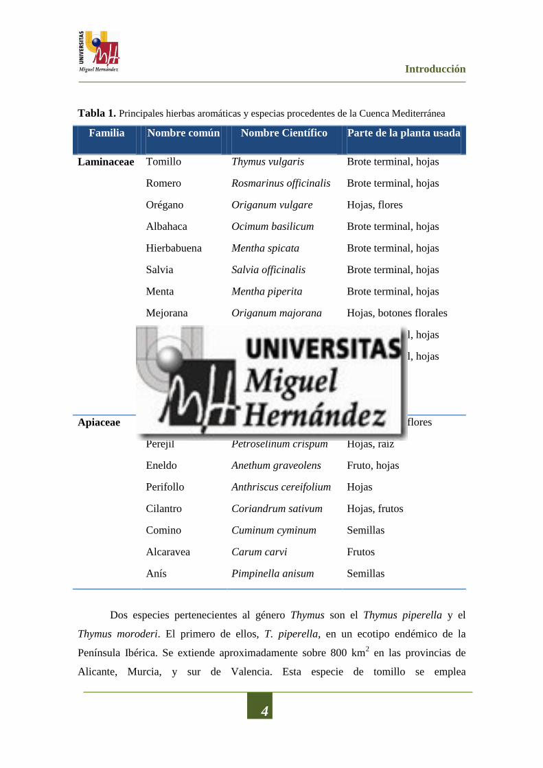
Tabla 1. Principales hierbas aromáticas y especias procedentes de la Cuenca Mediterránea
Familia Nombre común Nombre Científico Parte de la planta usada
Laminaceae Tomillo Thymus vulgaris Brote terminal, hojas
Romero Rosmarinus officinalis Brote terminal, hojas
Orégano Origanum vulgare Hojas, flores
Albahaca Ocimum basilicum Brote terminal, hojas
Hierbabuena Mentha spicata Brote terminal, hojas
Salvia Salvia officinalis Brote terminal, hojas
Menta Mentha piperita Brote terminal, hojas
Mejorana Origanum majorana Hojas, botones florales
Melisa Melissa officinalis Brote terminal, hojas
Ajedrea Satureja hortensis Brote terminal, hojas
Hisopo Hyssopus officinalis Hojas, flores
Lavanda Lavandula angustifolia Hojas, flores
Apiaceae Hinojo Foeniculum vulgare Hojas, tallos, flores
Perejil Petroselinum crispum Hojas, raíz
Eneldo Anethum graveolens Fruto, hojas
Perifollo Anthriscus cereifolium Hojas
Cilantro Coriandrum sativum Hojas, frutos
Comino Cuminum cyminum Semillas
Alcaravea Carum carvi Frutos
Anís Pimpinella anisum Semillas
Dos especies pertenecientes al género Thymus son el Thymus piperella y el
Thymus moroderi. El primero de ellos, T. piperella, en un ecotipo endémico de la
Península Ibérica. Se extiende aproximadamente sobre 800 km2 en las provincias de
Alicante, Murcia, y sur de Valencia. Esta especie de tomillo se emplea

Introducción
5
fundamentalmente como especia para condimentar diversos platos tradicionales. El
segundo, T. moroderi, está estrechamente relacionado con el tomillo común (Thymus
vulgaris L.), también se trata de un ecotipo endémico del sureste de la Península Ibérica,
encontrándose únicamente en las provincias de Alicante y Murcia. Esta planta se utiliza
sobre todo para la elaboración de bebidas espirituosas además de como especia en la
condimentación de algunos platos tradicionales.
Otros géneros, también muy comunes en la Cuenca Mediterránea son, el género
Santolina y el género Sideritis. El género Santolina, perteneciente a la familia
Asteraceae, está representado por más de 10 especies, las cuales están ampliamente
distribuidas por todo el Mediterráneo (Derbesy et al., 1989), siendo las especies más
representativas: Santolina viridis W. (presente en el sur de Francia y norte de España),
Santolina pectinata Lag. (Presente en la Península Ibérica) y Santolina
chamaecyparissus L. (crece en forma silvestre en toda la Cuenca Mediterránea). Esta
última es un arbusto perenne, nativo de la zona oeste y central del Mediterráneo, que
crece de forma silvestre en España, Italia, Túnez y Marruecos. A menudo se cultiva
como planta ornamental debido a sus tallos lanudos y a sus flores amarillas, aunque se
ha utilizado en la medicina popular debido a sus propiedades analgésicas,
antiinflamatorias, antisépticas, antiespasmódicas, bactericidas, fungicidas y para el
tratamiento de diferentes dermatitis (El-Sahhar et al., 2011).
El género Sideritis, que pertenecen a la familia Lamiaceae, subfamilia
Lamioideae, comprende al menos 150 especies. Las plantas de este género,
principalmente Sideritis angustifolia, han sido ampliamente utilizadas en la medicina
popular, a modo de infusión, como antiinflamatorios, antiulcerosos, antimicrobianos,
astringentes, para el tratamiento contra la gripe y como agentes estimulantes
circulatorios (Senatore, 2000).
Todas las plantas anteriormente mencionadas tienen en común, sobre todo las
dos especies de tomillos, la producción de altas concentraciones de aceite esencial.
2.2.- Aceites esenciales de hierbas aromáticas
En general, los aceites esenciales (AEs) son productos formados por la mezcla
de numerosas sustancias, con una composición química bastante compleja, que se

Introducción
6
obtienen a partir del metabolismo secundario de las plantas. Son lípidos simples, sin
ácidos grasos, y están compuestos por sustancias volátiles a diferencia de los aceites
fijos que contienen ácidos grasos como componentes estructurales fundamentales y que
no son volátiles (Burt, 2004). En términos generales los AEs están compuestos por más
de setenta componentes, fundamentalmente: terpenos, monoterpenos y sesquiterpenos,
hidrocarburos, alcoholes, cetonas, etc. Éstos pueden ser acíclicos, monocíclicos,
bicíclicos o tricíclicos (Russo et al., 1998).
Los componentes mayoritarios pueden constituir por encima del 85% de la
composición del AE, mientras que otros componentes se encuentran en forma de trazas
(Bauer et al., 2001). Es muy importante el papel que juegan estos compuestos
minoritarios, ya que existen evidencias de que estos componentes contribuyen, de
manera significativa, a las propiedades funcionales que el AE pueda presentar, debido al
posible sinergismo entre distintos componentes (Burt, 2004). Las hierbas aromáticas y
especias sintetizan y acumulan estos AEs en estructuras glandulares, las cuales pueden
estar distribuidas por toda la parte aérea de la planta, aunque esta distribución no es
uniforme, ya que generalmente se suelen encontrar en las hojas y en la flores (Faleiro et
al., 2002).
Los AEs presentan múltiples propiedades, entre las que destacan sus propiedades
antioxidantes y sus propiedades antimicrobianas.
2.2.1.- Propiedades Antimicrobianas de los aceites esenciales
Están ampliamente documentadas en la literatura científica las propiedades
antimicrobianas de los AEs de hierbas aromáticas y especias, tanto frente a bacterias
como frente a mohos y/o levaduras. Sin embargo, no se han encontrado referencias
sobre la actividad antimicrobiana de los AEs obtenidos de las dos especies de tomillo
(T. piperella y T. moroderi) analizadas en este trabajo de investigación. Así como,
tampoco se ha encontrado información de la actividad antimicrobiana de los AEs de las
especies del género Sideritis y del género Santolina analizadas. No obstante, la
actividad antimicrobiana de los AEs obtenidos de plantas del género Thymus está
ampliamente establecida.

Introducción
7
De Martino et al. (2009) analizaron la actividad antibacteriana de los AEs
obtenidos de Thymus longicaulis y Thymus pulegioides recolectados en Italia, frente a
una serie de bacterias, tanto Gram-negativas como Gram-positivas, como son:
Sthaphylococcus aureus, Streptococcus faecalis, Bacillus subtilis, Bacillus cereus,
Proteus mirabilis, Escherichia coli, Salmonella typhi Ty2 and Pseudomonas
aeruginosa. Se obtuvieron halos de inhibición comprendidos entre 9 y 18 mm para T.
longicaulis y halos comprendidos entre 11 y 20 mm para T. pulegioides. En un estudio
similar, Pinto et al. (2006), determinó la actividad antifúngica del AE obtenido a partir
de T. pulegioides recolectado en Portugal, consiguiendo unos valores de concentración
mínima inhibitoria (CMI) de entre 0,16 y 0,32 µL/mL frente a dermófitos y diversas
cepas de Aspergillus. Gonçalves et al. (2010) evaluaron la actividad antifúngica de
cuatro AEs de Thymus zygis subsp. sylvestris cultivados en Portugal, frente a dos
hongos como son Aspergillus niger y Aspergillus fumigatus, obteniendo valores de CMI
comprendidos entre 0,16 y 1,25 µL/mL. Zouari et al. (2011) analizaron la actividad
antimicrobiana del AE de Thymus algeriensis Boiss cultivado en Túnez, frente a seis
bacterias (E. coli, P. aeruginosa, B. cereus, Klebsiella pneumoniae, Salmonella
typhimurium y Enterococcus faecalis) y dos hongos (Fusarium solani y Aspergillus
niger). Los halos de inhibición obtenidos y los valores de CMI obtenidos estaban en un
rango de 13,5-64 mm y 1-6 µL/mL, respectivamente. Viuda-Martos et al. (2011b)
analizaron la actividad antibacteriana del AE de Thymus vulgaris cultivado en Egipto,
frente a diversas bacterias relacionadas con el deterioro de alimentos como son Listeria
innocua, Serratia marcescens y Pseudomona fluorescens, obteniendo halos de
inhibición, con un volumen de 40 µL, de 41,00; 20,25 y 23,50 mm, respectivamente.
Como se ha descrito para el género Thymus, la actividad antibacteriana del
género Sideritis ha sido, relativamente, analizada. Kiliç et al. (2003) describieron que
los AEs de Sideritis athoa, Sideritis trojana, Sideritis dichotoma, Sideritis spilyea y
Sideritis argyrea, eran activos frente a E. coli, S. aureus, P. aeruginosa, K. pneumoniae
y E. faecalis. Basile et al. (2006) analizaron el AE de Sideritis italic (Miller) frente a 9
cepas bacterianas, tanto Gram-negativas como Gram-positivas, encontrando unos
valores de CMI comprendidos entre 3,9 y 250 μg/mL. Köse et al. (2010) analizaron la
actividad antibacteriana de los AEs de dos variedades de Sideritis erythrantha

Introducción
8
endémicas de Turquía. Estos autores concluyeron que el AE de S. erythrantha var.
cedretorum era efectivo como antibiótico frente a Staphylococcus aureus meticilina-
resistente, E. faecalis vancomicin-resistente, Haemophilus influenza ampicilina-
resistente y E. faecalis vancomicina-sensitivo. Por otro lado, el AE de S. erythrantha
var. erythrantha fue también activo frente E. faecalis vancomicina-resistente y H.
influenza ampicilina-resistente.
Diversos trabajos han tratado de describir los posibles modos de acción de los
constituyentes de los AEs (Davidson y Naidu, 2000; Davidson, 2001), sin embargo, el
mecanismo concreto todavía no ha sido completamente dilucidado (Lambert et al.,
2001). Teniendo en cuenta el gran número de componentes químicos presentes en los
AEs, es muy probable que su actividad antimicrobiana no se deba a un único
mecanismo específico, sino a la acción conjunta de diferentes mecanismos que actúan
sobre distintos “Targets” de la célula (Skandamis et al., 2001; Carson et al., 2002). La
Figura 1 muestra los distintos mecanismos de acción propuestos a este efecto.
Figura 1. Mecanismos de acción propuestos para los aceites esenciales y sus componentes
frente a las células microbianas. Fuente: Raybaudi-Massilia et al. (2009).
Introducción
9
Generalmente, la composición, la estructura así como los grupos funcionales de
los componentes que integran el AE desempeñan un papel muy importante a la hora de
determinar su actividad antimicrobiana.
Los compuestos con grupos fenólicos tipo timol o carvacrol son los principales
responsables de las propiedades antimicrobianas, aunque también pueden existir otros
componentes, presentes en los AEs que pueden presentan las mismas propiedades
antimicrobianas y que son del tipo aldehídos, cetonas, alcoholes, etc. Tratar de
identificar cada uno de los compuestos químicos del AE y determinar cuál es el posible
responsable del efecto antimicrobiano es una tarea muy complicada y difícil de abordar.
Por esta razón es muy importante conocer la composición química que presenta el AE
objeto de estudio para así intentar predecir sus propiedades antimicrobianas.
2.2.2.- Propiedades Antioxidantes de los aceites esenciales
La actividad antioxidante de las hierbas aromáticas, de las especias, de los AEs y
de sus componentes, ha sido objeto de múltiples estudios (Mata et al., 2007; Milan et
al., 2008; Bozin et al., 2008; Alves-Silva et al., 2013), aunque gran parte de estos
ensayos se hayan realizado in vitro. Como sucedía en el caso de las propiedades
antimicrobianas, no existen estudios donde se determine la actividad antioxidante de los
AEs obtenidos de las dos especies de tomillo (T. piperella y T. moroderi) analizadas en
este trabajo de investigación. Así como, tampoco se han encontrado referencias sobre la
actividad antioxidantes de AEs de las especies del género Sideritis y del género
Santolina utilizadas en este trabajo. Sin embargo, la actividad antioxidante de los AEs
obtenidos de plantas del género Thymus ha sido ampliamente referenciada. Tepe et al.
(2005) compararon el potencial antioxidante, utilizando el ensayo del secuestro del
radical 2,2’-difenil-1-picrilhidrazil (DPPH), de los AEs obtenidos de dos especies de
tomillos, como son, Thymus sipyleus subsp. sipyleus var. sipyleus y Thymus sipyleus
subsp. sipyleus var. Rosulans. Estos AEs presentaron un valor de IC50 (concentración de
AE que inhibe el 50% del radical DPPH) de 0,22 y 2,67 mg/mL, respectivamente.
Viuda-Martos et al. (2010) determinaron la actividad antioxidante del AE de Thymus
vulgaris cultivado en Egipto, empleando distintos ensayos; con el ensayo del DPPH se
obtuvo un valor de IC50 de 4,50 mg/mL; con el análisis de las especies reactivas del
ácido tiobarbitúrico (TBARs) se obtuvo un valor de IC50 de 4,09 mg/mL; y con el

Introducción
10
ensayo de la actividad quelante de metales se obtuvo un valor de IC50 de 0,27 mg/mL.
De igual modo, no existe ninguna referencia científica de la actividad
antioxidante del AE de S. angustifolia. Sin embargo, la actividad antioxidante de los
AEs obtenidos de otras especies del género Sideritis ha sido ampliamente estudiada.
Köse et al. (2010) analizaron la actividad antioxidante de los AEs de Sideritis
erythrantha var. erythrantha y S. erythrantha var. cedretorum. Estos autores
concluyeron que dichos AEs presentaron una baja capacidad de secuestro de radicales
libres (% de inhibición de 5,12 y 4,62, respectivamente, a concentraciones de 2,0
mg/mL). Basile et al. (2006) estudiaron la actividad antioxidante de los AEs de Sideritis
itálica, obtenidos de las hojas o de las cabezas florales, mostrando una actividad
antioxidante, expresada como equivalentes de ácido ascórbico, de 4,29 y 1,98 mg/mL,
respectivamente.
No se han encontrado referencias bibliográficas sobre la actividad antioxidante
de AEs obtenidos a partir de plantas del género Santolina.
El mecanismo de acción que provoca esta actividad antioxidante no está todavía
esclarecido. Existen diversos mecanismos de acción para dicha capacidad antioxidante:
el secuestro de radicales libres, la donación de hidrógenos, la quelación de iones
metálicos o incluso pueden actuar como sustrato de radicales como el superóxido o el
hidroxil (Al-Mamary et al., 2002). Como ocurría en el caso de la actividad
antimicrobiana, la actividad antioxidante de los aceites esenciales está relacionada con
la presencia y concentración de distintos componentes en dicho aceite esencial y
fundamentalmente aquellos que presentan grupos fenólicos en su estructura.
La determinación de la capacidad antioxidante que las hierbas aromáticas y las
especias, así como sus derivados (fundamentalmente los AEs), ejercen en los alimentos,
es un tema que actualmente está teniendo un importante auge, tanto por parte de los
investigadores como por parte de la industria agroalimentaria. No hay que olvidar que la
oxidación lipídica constituye una de las principales causas de deterioro de los alimentos
y se produce, tanto durante el almacenamiento de materias primas, como durante su
procesado o incluso durante el almacenamiento de los productos finales (Tepe et al.,
2005). Dicha oxidación conlleva una pérdida significativa del valor nutricional del
alimento, ya que existe una pérdida de vitaminas y ácidos grasos esenciales, además de
Introducción
11
una pérdida de calidad sensorial, ya que se producen cambios en el color, la textura y el
sabor, lo que da como resultado una disminución de la vida útil del alimento y el
consiguiente rechazo por parte del consumidor (Fernández-López et al., 2007).
Uno de los principales problemas que se plantean cuando se recurre a la
aplicación directa de los AEs para que ejerzan, en los alimentos a los que son añadidos,
efectos antioxidantes o antimicrobianos, reside en que para la obtención de dichos
efectos inhibitorios se necesitan elevadas concentraciones, lo cual puede alterar las
características sensoriales de los productos sobre los que son añadidos. Esta limitación
puede superarse, potencialmente, a través del control de su liberación desde una matriz
polimérica adecuada, diseñada como un film o recubrimiento comestible.
2.3.- Recubrimientos comestibles
Actualmente, hay un creciente interés por el desarrollo de materiales que puedan
mejorar la vida útil de los alimentos y también, por la seguridad microbiológica que se
les otorga. Los recubrimientos, películas o films comestibles formados por polímeros
biodegradables, son uno de estos materiales que ha logrado mayor interés y por lo tanto
se le atribuye un alto potencial comercial.
Una película o film comestible se puede definir como una capa fina y continua
de material comestible, que se dispone sobre una superficie alimentaria para mejorar la
calidad y aumentar la vida útil del alimento (Fernández-Pan y Maté-Caballero, 2011).
En la formulación de films debe presentarse al menos un componente capaz de formar
una matriz estructural estable, como los hidrocoloides clasificados en proteínas
(colágeno, gelatina, zeina, gluten de trigo, proteína de soja, proteínas lácteas, etc.)
(Morillon et al., 2002) o carbohidratos (derivados de celulosa, almidones, extractos de
algas, pectinas, gomas o quitosano) (Ponce et al., 2008), los cuales se caracterizan por
formar films con propiedades de barrera al oxígeno, aromas y lípidos, presentan
biocompatibilidad, además de mejorar la apariencia estética del alimento (Han, 2000;
Kalemba y Kunicka, 2003). Las características de este tipo de materiales están afectadas
por diversos parámetros como son la formulación, la tecnología de formación de la
película, las características del solvente y los aditivos (Gocho et al., 2000). Como se ha
mencionado, son muchas las sustancias que son capaces de formar una matriz estable;

Introducción
12
una de ellas, la cual presenta unas magníficas propiedades formadoras de dichas
películas, además de mostrar ciertas propiedades antioxidantes y antimicrobianas por sí
misma, es el quitosano (Sayas-Barberá et al., 2011).
2.3.1.- Quitosano
El quitosano, poli-β-(1,4)-D-glucosamina-N-acetil-D-glucosamina, (Figura 2A)
se obtiene por desacetilación de la quitina, poli-β-(1,4)-N-acetil-D-glucosamina, (Figura
2B), la cual constituye el segundo polisacárido natural, más abundante, después de la
celulosa. La quitina es uno de los componentes principales de las paredes celulares de
los hongos, y del exoesqueleto de crustáceos e insectos. Es altamente insoluble en agua
y solventes orgánicos, lo cual, restringe sus aplicaciones (Rinaudo, 2006).
Figura 2. Estructura química del quitosano (A) y de la quitina (B).
Sin embargo, el quitosano es un compuesto que exhibe unas características
fisicoquímicas de notable interés, como son, una elevada proporción de grupos amino
libres, mayor solubilidad comparada con la quitina, biocompatibilidad y
biodegradabilidad, lo cual hace que presente múltiples aplicaciones en medicina,
industria cosmética, agricultura, biotecnología, industria alimentaria, industria papelera
y en el tratamiento de aguas (Tabla 2).
El quitosano se define en términos del grado de acetilación, peso molecular,
viscosidad y solubilidad. La importancia de este biopolímero, para la industria de
alimentos, está fundamentalmente, en su propiedad para formar films (Fernández-Saiz
et al., 2009).
A B
Introducción
13
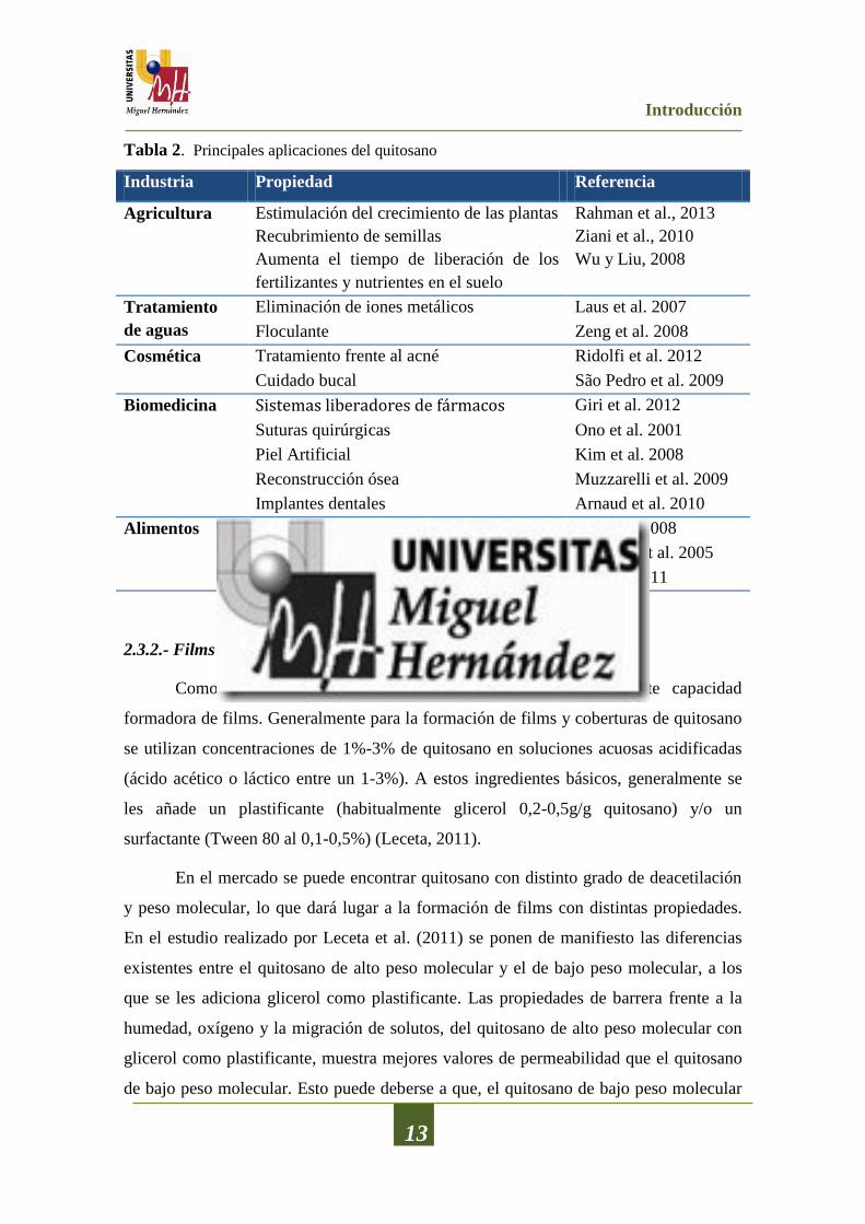
Tabla 2. Principales aplicaciones del quitosano
Industria Propiedad Referencia
Agricultura Estimulación del crecimiento de las plantas Rahman et al., 2013
Recubrimiento de semillas Ziani et al., 2010
Aumenta el tiempo de liberación de los
fertilizantes y nutrientes en el suelo
Wu y Liu, 2008
Tratamiento
de aguas
Eliminación de iones metálicos Laus et al. 2007
Floculante Zeng et al. 2008
Cosmética Tratamiento frente al acné Ridolfi et al. 2012
Cuidado bucal São Pedro et al. 2009
Biomedicina Sistemas liberadores de fármacos Giri et al. 2012
Suturas quirúrgicas Ono et al. 2001
Piel Artificial Kim et al. 2008
Reconstrucción ósea Muzzarelli et al. 2009
Implantes dentales Arnaud et al. 2010
Alimentos Reducción del colesterol Liu et al. 2008
Estabilizante de salsas Laplante et al. 2005
Conservante Li et al. 2011
2.3.2.- Films de Quitosano
Como ya se ha comentado, el quitosano tiene una excelente capacidad
formadora de films. Generalmente para la formación de films y coberturas de quitosano
se utilizan concentraciones de 1%-3% de quitosano en soluciones acuosas acidificadas
(ácido acético o láctico entre un 1-3%). A estos ingredientes básicos, generalmente se
les añade un plastificante (habitualmente glicerol 0,2-0,5g/g quitosano) y/o un
surfactante (Tween 80 al 0,1-0,5%) (Leceta, 2011).
En el mercado se puede encontrar quitosano con distinto grado de deacetilación
y peso molecular, lo que dará lugar a la formación de films con distintas propiedades.
En el estudio realizado por Leceta et al. (2011) se ponen de manifiesto las diferencias
existentes entre el quitosano de alto peso molecular y el de bajo peso molecular, a los
que se les adiciona glicerol como plastificante. Las propiedades de barrera frente a la
humedad, oxígeno y la migración de solutos, del quitosano de alto peso molecular con
glicerol como plastificante, muestra mejores valores de permeabilidad que el quitosano
de bajo peso molecular. Esto puede deberse a que, el quitosano de bajo peso molecular
Introducción
14
interacciona en menor medida con el glicerol empleando como plastificante,
permitiendo que las moléculas de agua puedan escapar más fácilmente fuera del film.
Respecto a las propiedades mecánicas, también se ha observado que la
resistencia a la tracción se ve aumentada y la rotura por alargamiento disminuida,
cuando el quitosano tiene un peso molecular mayor, lo que puede atribuirse a la
formación de una red más compacta en el quitosano; sin embargo, para la deformación
por punción no se observaronn diferencias significativas en cuanto al peso molecular del
quitosano, aunque sí se muestran con el contenido en glicerol de las muestras (Vargas et
al., 2009; Sánchez-González et al., 2010; Leceta et al., 2011).
Aparte de actuar como barreras selectivas frente a la humedad, gases y la
migración de solutos, estos films podrían actuar como vehiculizantes de una gran
cantidad de componentes funcionales (Ojagh et al., 2010). Estos componentes
funcionales incluyen agentes antioxidantes, agentes antimicrobianos, saborizantes,
colorantes, etc., los cuales mejorarían la funcionalidad de los materiales de envasado
debido a la “aparición” de nuevas funciones (Salmieri y Lacroix, 2006).
Uno de estos componentes que pueden ser adicionados a los films de quitosano
para mejorar sus propiedades funcionales, son los aceites esenciales obtenidos de
hierbas aromáticas y/o especias.
2.3.3.- Films de quitosano adicionados con aceites esenciales de hierbas aromáticas
Los films de quitosano adicionados con aceites esenciales de hierbas aromáticas
o especias, además de exhibir determinadas propiedades de barrera (oxigeno, lípidos,
aromas, humedad) presentan unas excelentes propiedades antioxidantes y/o
antimicrobianas provocadas por la presencia de los AEs. Debido a todas estas
propiedades, este tipo de films se convierten en una seria alternativa para lograr el
objetivo de aumentar la vida útil y mantener la calidad del producto en el cual son
utilizados.
Los films que incorporan AEs, los cuales presentan propiedades antioxidantes
y/o antimicrobianas, permiten que la migración de sus componentes desde el
recubrimiento a la superficie del alimento sea realice lentamente, permitiendo controlar
esta migración (Figura 3) tal y como describe Fernández-Pan et al. (2010).

Introducción
15
Figura 3. Evolución de la concentración de compuestos activos añadidos directamente sobre
la superficie del sistema alimentario o a través de una película comestible.
El único inconveniente de la aplicación de este tipo de films adicionados con
AEs de hierbas aromáticas sobre el alimento es que, cualquier pequeña modificación en
los atributos organolépticos sea percibida o detectada por parte de los consumidores. Es
por ello que se debe tener una gran certeza de que las características finales del producto
varían lo menos posible y de que no son percibidas por el consumidor.
2.3.3.1.- Propiedades antimicrobianas de los films de quitosano adicionados con
aceites esenciales de hierbas aromáticas
Una de las tecnologías emergentes más novedosas, que pueden aplicarse en la
conservación de los alimentos, es el empleo de films o recubrimientos comestibles
activos que pueden funcionar como matrices portadoras de agentes antimicrobianos
(Martin-Belloso et al., 2009). Durante la última década, la investigación sobre los films
de quitosano como portadores de AEs con propiedades antimicrobianas se ha
incrementado notablemente (Altiok et al., 2010; Avila-Sosa et al., 2012; Kurek et al.,
2013; Leceta et al., 2013). Moradi et al. (2011) analizó la actividad antibacteriana frente
a Listeria monocytogenes de films de quitosano adicionados con AE de Zataria
multiflora Boiss, obteniendo unos halos de inhibición de 92 y 162 mm2 para
Introducción
16
concentraciones de 5 y 10 mg/g de film. Zivanovic et al. (2005) desarrollaron films de
quitosano enriquecidos con AEs de anís, albahaca, cilantro y orégano, y los evaluaron in
vitro frente a los patógenos L. monocytogenes y E. coli O157:H7. Los films más activos
resultaron ser los que incorporaban AE de orégano, seguidos por los de AE de cilantro,
albahaca y finalmente anís. Altiok et al. (2010) desarrollaron films de quitosano
adicionados con distintas concentraciones de AE de T. vulgaris, analizando
posteriormente su actividad antibacteriana frente a distintas bacterias como E. coli, K.
pneumoniae, P. aeruginosa y S. aureus. Estos autores encontraron que todas las
bacterias fueron sensibles a los films desarrollados pero solo a la máxima concentración
(1,2% (v/v)) obteniendo halos de inhibición comprendidos entre 16 y 19 mm. En otro
estudio, Sánchez-González et al. (2011) analizaron la actividad antimicrobiana de films
de quitosano preparados con diferentes concentraciones de AE de bergamota, AE de
limón o AE de árbol del té. Estos autores concluyeron que los films analizados
mostraron un efecto bacteriostático frente a diversas bacterias como E. coli, L.
monocytogenes y S. aureus.
La efectividad antimicrobiana de los films de quitosano adicionados con AEs
reside en la lenta migración de sus agentes activos hacia la superficie del producto que
recubren, ayudando al mantenimiento de altas concentraciones de ingrediente activo
donde son necesarias (Kristo et al., 2008).
2.3.3.2.- Propiedades antioxidantes de los films de quitosano adicionados con aceites
esenciales de hierbas aromáticas
Otra de las propiedades que presentan los films de quitosano adicionados con
AEs de hierbas aromáticas o especias y que puede ser muy importante en la industria
agroalimentaria es su actividad antioxidante. Sin embargo, existen pocos estudios donde
se determine la actividad antioxidante de los films de quitosano adicionados con AEs
(Altiok et al., 2010; Moradi et al., 2012).
Esta actividad antioxidante podría estar relacionada con la presencia, en los
films de quitosano, de compuestos bioactivos tales como compuestos fenólicos o ácidos
terpénicos procedentes de los AEs. Así, los compuestos fenólicos y terpenoides
presente en la composición química de los AEs están estrechamente relacionados
Introducción
17
con las propiedades antioxidantes, fundamentalmente debido a sus propiedades redox
ejercidas siguiendo distintos mecanismos, como el secuestro de radicales libres, la
donación de átomos de hidrogeno y/o quelación de metales de transición (Liyana-
Pathirana y Shahidi, 2006).
2.4.- Aplicación de los films de quitosano adicionados con aceites esenciales de
hierbas aromáticas, en alimentos.
Tras la comprobación de la actividad antimicrobiana y antioxidante de los films
de quitosano adicionados con AEs en ensayos de laboratorio, una variedad de ellos se
han aplicado en sistemas alimentarios, fundamentalmente de origen vegetal. En el
desarrollo de películas y recubrimientos comestibles antimicrobianos dirigidos al
mantenimiento de la calidad y aumento de la vida útil de los productos cárnicos, las
principales dianas microbianas empleadas, por su poder alterante, pertenecen a
Pseudomonas spp. y Listeria spp. ya que son los responsables directos más comunes del
daño producido en productos cárnicos frescos y cocidos, respectivamente, almacenados
a bajas temperaturas (Fernández-Pan y Maté-Caballero, 2011). El grupo de bacterias
ácido-lácticas también son una diana fundamental puesto que se reconocen como la
microflora resistente y predominante en los productos envasados a vacío en condiciones
de refrigeración (Emiroğlu et al., 2010).
Así, recientemente, Beverlya et al. (2008) analizaron la actividad antibacteriana
frente a L. monocytogenes, de películas comestibles de quitosano en carne asada
“Ready-to-eat”. Estos autores informaron que tras catorce días de almacenamiento a 4
ºC las muestras recubiertas con los films de quitosano presentaban un reducción en los
recuentos de L. monocytogenes de 2-3 UFC/g. Zivanovic et al. (2005) estudiaron el
efecto antibacteriano frente a L. monocytogenes de films de quitosano y films de
quitosano adicionado con distintos aceites esenciales (anís, albahaca, cilantro y orégano)
empleados como separadores entre lonchas de mortadela. Las películas de quitosano
adicionadas con AEs resultaron más eficaces que las películas formadas solo por el
quitosano, ya que las primeras provocaron una reducción en los recuentos de L.
monocytogenes de 3,6-4 ciclos logarítmicos mientras que las de segundas provocaron
una reducción en los recuentos de 2 ciclos logarítmicos. Khanjari et al. (2013) llevaron a
cabo un estudio donde se analizó el efecto combinado de film de quitosano con aceite

Introducción
18
esencial de orégano, aplicado sobre filetes de pechuga de pollo, inoculados con L.
monocytogenes mediante inmersión. Estos autores indican que tras catorce días de
almacenamiento a 4 ºC las muestras no tratadas alcanzaron unos recuentos de aerobios
totales de 7 log UFC/g (recuentos en los que se detecta alteración) en los primeros 6
días, mientras que en las muestras tratadas con quitosano y aceite esencial de orégano
nunca alcanzaron los recuentes de 7 log UFC/g. Del mismo modo, en las muestras
inoculadas con L. monocytogenes y tratadas con los films de quitosano más AE de
orégano se produjo la inhibición completa de esta bacteria tras dos días de
almacenamiento. Siripatrawan y Noipha (2012) analizaron el efecto, sobre la extensión
de la vida útil de salchichas elaboradas con carne de cerdo, de films de quitosano a los
que se incorporó extractos de té verde que eran empleados como envoltorios de dichas
salchichas. Estos autores informaron que la incorporación del film de quitosano con el
té verde mejoró las propiedades antimicrobianas de la película, obteniendo recuentos
para aerobios totales, levaduras y mohos, inferiores en las salchichas tratadas que en las
muestras control. Moradi et al. (2012) estudiaron la eficacia, frente a bacterias ácido-
lácticas, aerobios totales y L. monocytogenes, de los films de quitosano que contenían
AE de Zataria multiflora Boiss (ZEO) y extracto de semilla de uva (GSE) empleados
como recubrimientos en mortadela. En este caso, el crecimiento de L. monocytogenes
fue inhibido significativamente por los films que contenían ZEO-GSE. Así mismo, las
bacterias aerobias mesófilas y las bacterias lácticas fueron los grupos más sensibles y
resistentes a la acción de los films, con reducciones en los recuentos de 0,1-1,1 y 0,1-0,7
ciclos logarítmicos, respectivamente.

OBJETIVOS

Objetivos
19
3.- OBJETIVOS
3.1- Objetivos generales
El objetivo general del estudio es evaluar el potencial tecnológico de los aceites
esenciales obtenidos de diferentes hierbas aromáticas endémicas del sureste de la
península Ibérica, y su aplicación en films de quitosano para aumentar la vida útil de un
sistema modelo cárnico cocido.
Para alcanzar este objetivo general, se plantearon los siguientes objetivos
particulares.
3.2.- Objetivos particulares
Caracterizar químicamente los aceites esenciales obtenidos de diferentes hierbas
aromáticas endémicas del sureste de la península Ibérica.
Determinar la actividad antibacteriana de estos aceites esenciales frente a diversas
cepas bacterianas relacionadas con la degradación de alimentos o indicadoras de la
presencia de patógenos.
Determinar la capacidad de inhibición del crecimiento fúngico, tanto frente a
mohos como a levaduras.
Conocer la capacidad antioxidante de los aceites esenciales mediante la utilización
de diferentes técnicas analíticas.
Aplicar los aceites esenciales obtenidos de las hierbas aromáticas sometidas a
estudio en films de quitosano.
Determinar la capacidad antioxidante de los films de quitosano adicionados con los
aceites esenciales sometidos a estudio.
Determinar la actividad antibacteriana de los films de quitosano adicionados con
los aceites esenciales sometidos a estudio.
Evaluar la vida útil de un producto cárnico cocido loncheado en el que se han
utilizado, como separadores de lonchas, films de quitosano adicionados con los
aceites esenciales de estudio.

MATERIALES
Y
METODOS
Materiales y Métodos
20
4.- MATERIALES Y METODOS
En este apartado se recoge un resumen de los materiales utilizados, de los
procesos de elaboración aplicados y de los análisis efectuados. La información completa
de toda esta metodología se ha desarrollado en los respectivos artículos publicados en
revistas internacionales y que se adjuntan a esta memoria.
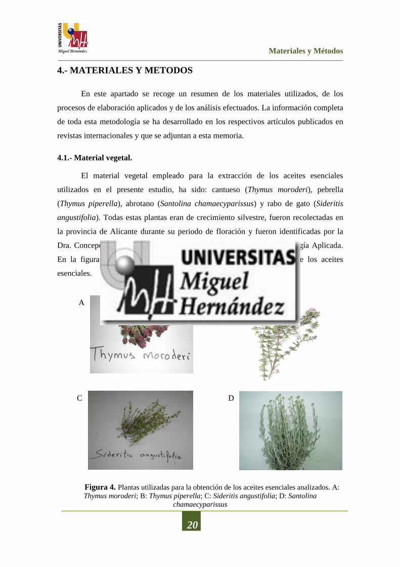
4.1.- Material vegetal.
El material vegetal empleado para la extracción de los aceites esenciales
utilizados en el presente estudio, ha sido: cantueso (Thymus moroderi), pebrella
(Thymus piperella), abrotano (Santolina chamaecyparissus) y rabo de gato (Sideritis
angustifolia). Todas estas plantas eran de crecimiento silvestre, fueron recolectadas en
la provincia de Alicante durante su periodo de floración y fueron identificadas por la
Dra. Concepción Obón del Área de Botánica del Departamento de Biología Aplicada.
En la figura 4 se muestran las plantas utilizadas para la obtención de los aceites
esenciales.
Figura 4. Plantas utilizadas para la obtención de los aceites esenciales analizados. A:
Thymus moroderi; B: Thymus piperella; C: Sideritis angustifolia; D: Santolina
chamaecyparissus
A B
C D

Materiales y Métodos
21
4.2.- Extracción de los aceites esenciales
Los aceites esenciales (AE) de T. moroderi, T. piperella, S. chamaecyparissus y
S. angustifolia se extrajeron de la planta completa (tallos, hojas y flores) mediante
hidrodestilación, utilizando un equipo tipo Clevenger, durante 3 horas. La capa oleosa
obtenida en la parte superior del destilado acuoso se separó y se secó con sulfato de
sodio anhidro (0,5 g). Los AE así extraídos se guardaron a 4 ºC en viales de vidrio
opacos y sellados herméticamente hasta su posterior análisis. Los rendimientos
obtenidos para los distintos AEs fueron T. moroderi 1,90%, T. piperella 1,66%, S.
chamaecyparissus 1% y S. angustifolia 0,90%.
4.3.- Composición química de los aceites esenciales de especias
La determinación de la composición de química de los distintos aceites
esenciales estudiados se realizó mediante cromatografía de gases/espectrometría de
masas (CG/MS) en un cromatógrafo de gases Shimadzu GC-17A (Shimadzu
Corporation, Tokio, Japón), acoplado a un detector selectivo de masas Shimadzu
GCMS-QP5050A (Shimadzu Corporation) equipado con una columna TRACSIL Meta
X5 (Teknokroma S. Coop. C. Ltd, Barcelona, España), 30 m x 0.25 mm i.d., y un
espesor de relleno de 0.25 µm. Las Tª del inyector y del detector fueron de 250 y 300
ºC, respectivamente. La Tª del horno se mantuvo a 40 ºC durante 5 min
incrementándose gradualmente hasta los 200 ºC en un ratio de 3 ºC/min manteniéndose
a esta Tª durante 1 min. Transcurrido este tiempo se volvió a aumentar la Tª hasta los
280 ºC en un ratio de 15 ºC/min manteniéndose a 280 ºC durante 10 min. Como gas
portador se utilizó Helio con un flujo de 1 mL/min. La identificación de los compuestos
se realizó mediante comparación de los tiempos de retención y el espectro de masas con
los de los estándares previamente inyectados, así como, con la biblioteca Wiley 229 del
sistema, los valores del índice de Kovats y valores presentes en la literatura científica.
4.4.- Actividad antibacteriana de los aceites esenciales
Para la determinación de la actividad antibacteriana se seleccionaron cepas
bacterianas relacionadas con el deterioro de alimentos en refrigeración y cepas
indicadoras de cepas patógenas: Listeria innocua CECT 910, Serratia marcenscens
CECT 854, Pseudomonas fragi CECT 446, Pseudomonas fluorescens CECT 844,

Materiales y Métodos
22
Aeromonas hydrophila CECT 5734, Shewanella putrefaciens CECT 5346,
Achromobacter denitrificans CECT 449, Enterobacter amnigenus CECT 4078,
Enterobacter gergoviae CECT 587, Alcaligenes faecalis CECT 145 y Leuconostoc
carnosum CECT 4024. Todas ellas fueron adquiridas en la Colección Española de
Cultivos Tipo de la Universidad de Valencia (España). Para la determinación de la
actividad antibacteriana se empleó el método de difusión de disco en agar, siguiendo las
recomendaciones de Tepe et al. (2005). También se determinó el efecto de la
concentración siguiendo el método descrito por Viuda-Martos et al. (2005).
4.5.- Actividad antifúngica de los aceites esenciales
La actividad antifúngica de los aceites esenciales se ensayó frente a seis cepas de
hongos, seleccionadas por su relación con el deterioro de alimentos, como son:
Aspergillus niger CECT 2091, Aspergillus flavus CECT 2685, Mucor racemosus CECT
2670, Mucor circinelloides CECT 20765, Penicillium chrysogenum CECT 2784 y
Alternaria alternata CECT 20560. Esta actividad antifúngica también se ensayó frente a
seis levaduras: Yarrowia lipolytica CECT 1240, Saccharomyces cerevisiae CECT 1383,
Candida zeylanoides CECT 10048, Debaryomyces hansenii CEPT 11369, Rhodotorula
mucilaginosa CECT 10011, Pichia carsonii CECT 10227. Todas las cepas se
adquirieron en la Colección Española de Cultivos Tipo de la Universidad de Valencia
(España). Para analizar el efecto inhibidor de los AE frente a los hongos se empleó el
método de dilución en agar, siguiendo las recomendaciones de Fraternale et al. (2003).
Las concentraciones analizadas fueron 0,2; 0,1; 0,05 y 0,025 mL/100 mL. Para la
determinación de la actividad antifúngica frente a las levaduras se empleó el método
colorimétrico de microdilución propuesto por Abate et al. (1998). Las concentraciones
analizadas estaban comprendidas entre 0,04 y 40 µL/mL.
4.6.- Actividad antioxidante de los aceites esenciales
La determinación de la actividad antioxidante in vitro se realizó siguiendo cuatro
métodos analíticos diferentes: (i) secuestro del radical 2,2’-difenil-1-picrilhidrazil
(DPPH), siguiendo las recomendaciones de Brand-Williams et al. (1995); (ii) poder
antioxidante de la reducción de hierro (FRAP), según el método descrito por Oyaizu
(1986); (iii) actividad antioxidante de las especies reactivas del ácido tiobarbitúrico

Materiales y Métodos
23
(TBARS), siguiendo las indicaciones de Daker et al. (2008); y (iv) capacidad quelante
del ion ferroso (FIC), según el método utilizado por Carter (1971). También se
determinó el contenido en fenoles totales utilizando el reactivo Folin-Ciocalteu’s,
siguiendo las recomendaciones de Singleton y Rossi (1965).
4.7.- Elaboración de films de quitosano con aceites esenciales
Para la elaboración de los films de quitosano adicionados con los aceites
esenciales se siguió la metodología descrita por Ojagh et al. (2010). Se utilizó quitosano
de alto peso molecular con un grado de deacetilacion de 75-85% (Sigma-Aldrich
Chemical Co., Steinheim, Alemania) al 2% en una disolución de ácido láctico al 1%,
utilizando Tween 80 como emulsionante al 0,2% y glicerol (0,75 mL/g quitosano) como
plastificante. Se utilizaron, como moldes, placas de Petri estériles de 6 cm de diámetro.
Se dosificaron 7 g de dilución a cada placa y se llevaron a deshidratar a 37ºC durante 48
h en estufa de convección. Transcurrido ese tiempo se almacenaron a 25 ºC y una
humedad relativa de 51% hasta su uso. Los diferentes AEs se adicionaron a los films de
quitosano a diferentes concentraciones (Tabla 3).
Tabla 3. Concentraciones y aceites esenciales empleados en la elaboración de films de
quitosano.
Formulación
T.
piperella
(%)
T.
moroderi
(%)
S.
chamaecyparissus
(%)
S.
angustifolia
(%)
CH 0 0 0 0
CH+0,5TP 0,5 0 0 0
CH+1TP 1 0 0 0
CH+2TP 2 0 0 0
CH+0,5TM 0 0,5 0 0
CH+1TM 0 1 0 0
CH+2TM 0 2 0 0
CH+0,5SC 0 0 0,5 0
CH+1SC 0 0 1 0
CH+2SC 0 0 2 0
CH+0,5SA 0 0 0 0,5
CH+1SA 0 0 0 1
CH+2SA 0 0 0 2 CH: quitosano; CH+0,5TP: quitosano más 0,5% de AE de T. piperella; CH+1TP: quitosano más 1% de AE de T.
piperella; CH+2TP: quitosano más 2% de AE de T. piperella; CH+0,5TM: quitosano más 0,5% de AE de T.
moroderi; CH+1TM: quitosano más 1% de AE de T. moroderi; CH+2TM: quitosano más 2% de AE de T. moroderi;
CH+0,5SC: quitosano más 0,5% de AE de S. chamaecyparissus; CH+1SC: quitosano más 1% de AE de S.
chamaecyparissus; CH+2SC: quitosano más 2% de AE de S. chamaecyparissus; CH+0,5SA: quitosano más 0,5% de
AE de S. angustifolia; CH+1SA: quitosano más 1% de AE de S. angustifolia; CH+2SA: quitosano más 2% de AE de
S. angustifolia.

Materiales y Métodos
24
4.8.- Determinación de la actividad antibacteriana de los films de quitosano
Para la determinación de la actividad antibacteriana de los films de quitosano
adicionados con los AE de T. moroderi, T. piperella, S. chamaecyparissus y S.
angustifolia se siguió la metodología descrita en el apartado 4.4
4.9.- Determinación de la actividad antioxidante de los films de quitosano
Para la determinación de la actividad antioxidante de los films de quitosano
adicionados con los AE de T. moroderi, T. piperella, S. chamaecyparissus y S.
angustifolia se siguió la metodología descrita en el apartado 4.6
4.10.- Aplicación de los films en un producto cárnico tipo Jamón cocido
El Jamón Cocido (JC) fue adquirido directamente de un supermercado y
transportado inmediatamente, en condiciones de refrigeración, al laboratorio del grupo
de Industrialización de Productos de Origen Animal (IPOA) localizado en el
Departamento de Tecnología Agroalimentaria de la Escuela Politécnica Superior de
Orihuela. Posteriormente se procedió a lonchear el producto, obteniendo lonchas con
un espesor de 3 mm y un peso aproximado de 12,5 g. Los films elaborados con las
distintas formulaciones se colocaron como separadores entre dos lonchas de JC y
fueron introducidas en bolsas estériles de polietileno y poliamida laminada con las
siguientes características: permeabilidad al vapor de agua a 23 ºC 1,1 g/m2/24 h;
permeabilidad al nitrógeno a 23 ºC 10 cm3/m
2/24 h; permeabilidad al dióxido de
carbono a 23º C 140 cm3/m
2/24 h y permeabilidad al oxígeno a 23 ºC 30 cm
3/m
2/24 h
(Fibran, Girona, España). De igual modo se envasaron dos lonchas sin ningún tipo de
película, que actuarían como muestras control.
Las bolsas se sellaron y fueron almacenadas a temperatura de refrigeración 4±1
ºC durante 21 días. El análisis de las muestras para cada tratamiento se realizó a los 0,
7, 14, y 21 días (tiempo de almacenamiento) y dichos análisis se llevaron a cabo el
mismo día del muestreo.
4.11.- Efecto de los films de quitosano con aceites esenciales sobre las
características y vida útil de un producto cárnico tipo Jamón cocido.
4.11.1.- Determinaciones Físico-Químicas.

Materiales y Métodos
25
4.11.1.1.- Color.
Las determinaciones de color se efectuaron de acuerdo a las normas de la
Asociación Americana de la Carne (Hunt et al., 1991). Dichas determinaciones se
realizaron mediante un colorímetro Minolta CM-2600 (Minolta Camera Co., Osaka,
Japón) con iluminante D65, y el observador 10º. En todas las determinaciones de color
se interpusieron cristales de baja reflectancia Minolta CR-A51/1829-752 (Minolta Co.,
Osaka, Japón) entre las muestras y el equipo (Hunt et al., 1991).
4.11.1.2.- pH.
El pH de cada una de las unidades de muestras se determinó bajo las directrices
del Ministerio de Agricultura Pesca y Alimentación (1994), mediante disolución acuosa
en una proporción 1:10 de producto y agua destilada. Las lecturas de pH se efectuaron
con un equipo Crison modelo 507 (Crison, Barcelona, España).
4.11.2.- Oxidación lipídica
La determinación de la oxidación lipídica se realizó mediante el test del ácido 2-
tiobarbiturico siguiendo las directrices descritas por Buege y Aust (1978). Los
resultados se expresaron como mg malonaldehido/kg de muestra.
4.11.3.- Análisis microbiológico.
Para el recuento microbiológico, se tomaron 25 g de muestra que se
homogeneizaron con 225 mL de agua de peptona al 1,5% en un Stomacher 400
(Colworth, Londres, Reino Unido) durante 2 minutos. Los medios de cultivo utilizados
y las condiciones de incubación se detallan a continuación:
Recuento total de aerobios mesófilos en agar de recuento en placa (PCA) a 35 ºC
durante 48 horas.
Recuento total de bacterias ácido lácticas en una doble capa de MRS (Man,
Rogosa and Sharpe Agar) a 35 ºC durante 48 horas.
Recuento total de enterobacterias en Agar Glucosa Bilis Rojo Violeta (VRBG) a
35 ºC durante 48 horas.
Recuento total de mohos y levaduras en Agar Rosa de Bengala con
cloramfenicol a 26 ºC durante 5 días.
Materiales y Métodos
26
4.11.4.- Determinaciones del contenido en fenoles totales de los films
Para la evaluación de la liberación de compuestos bioactivos de los films durante
el almacenamiento refrigerado de las muestras se procedió de la siguiente manera: en el
momento del muestreo se recuperó el film, se pesó y se realizó una extracción con 5 mL
de metanol en un baño de ultrasonidos durante 1 hora. Posteriormente se centrifugaron
las muestras y con el sobrenadante obtenido se procedió a la determinación del
contenido en fenoles totales utilizando el reactivo Folin-Ciocalteu’s siguiendo las
recomendaciones de Singleton y Rossi (1965).
4.12. Metodología estadística
La metodología estadística se diseñó para cada uno de los estudios a analizar. La
totalidad de los análisis se realizaron mediante el paquete estadístico Statgraphics Plus
para Windows versión 5.1 (Statical Graphics Corp., Rockville, USA) utilizando el
programa Analisis of Variance.
Para la determinación de las media y la desviación estándar se siguieron
métodos estadísticos convencionales. El análisis estadístico empleado en cada ensayo
fue la aplicación de un análisis de la varianza (ANOVA) de uno, dos o tres factores,
dependiendo del ensayo realizado.
Para estudiar entre qué variables de los factores principales las diferencias
fueron estadísticamente significativas se realizaron contrastes entre las medias,
aplicando el test de Tukey siguiendo las recomendaciones de Afifi y Azen (1979).

RESULTADOS
Resultados y Discusión
27
5.- RESULTADOS Y DISCUSION
Este capítulo recoge los principales resultados y una breve discusión de los
diferentes trabajos realizados. Las versiones completas de los mismos se encuentran en
los correspondientes artículos publicados o en proceso de revisión en revistas
internacionales incluidas en el Journal Citations Reports y se adjuntan al final de esta
memoria (capítulo octavo).
5.1.- Composición química de los aceites esenciales sometidos a estudio
El objetivo de estos trabajos, publicados en las revistas “Food Control” (2012,
27:294-299) y “Food Safety” (2012, 32:426-434), fue la determinación de la
composición química de los aceites esenciales (AE) objeto de estudio Thymus moroderi,
Thymus piperella, Santolina angustifolia y Sideritis chamaecyparissus mediante el
empleo de cromatografía de gases acoplado a espectrometría de masas.
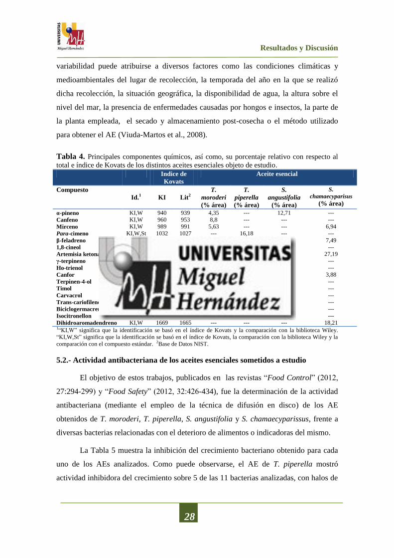
En la Tabla 4 se muestran los principales componentes químicos, así como su
porcentaje relativo con respecto al total y el índice de Kovats, de los distintos aceites
esenciales objeto de estudio.
En el caso del AE obtenido de T. moroderi se identificaron 51 compuestos
distintos, representando el 92% de los componentes del aceite. Los compuestos
principales fueron canfor (26,74%), 1,8-cineol (24,99%) y mirceno (5,63%). Para el
AE de T. piperella se identificaron 48 compuestos, representando el 90,5% de los
componentes del aceite. Los componentes mayoritarios identificados en este AE fueron
el carvacrol (31,92%) el para-cimeno (16,18%) y el γ-terpineno (10,11%). En el análisis
del AE de S. chamaecyparissus, se identificaron 58 componentes representando el
90,1% del total de componentes, siendo el componente principal la artemisa-ketona
(27,19%) seguido por dihidro-aromadendreno (18,21%) y β-felandreno (7,49%). Para el
AE de S. angustifolia se identificaron 77 compuestos, los cuales representaban el 94,6%
del total de componentes del aceite. El componente mayoritario fue el α-pineno
(12,71%). Otros componentes importantes fueron el β-felandreno (11,97%) y el 1,8-
cineol (7,41%).
Existe una gran variabilidad de composición entre los AE pertenecientes al
mismo género y especie, tal y como se muestra en la literatura científica. Esta
Resultados y Discusión
28
variabilidad puede atribuirse a diversos factores como las condiciones climáticas y
medioambientales del lugar de recolección, la temporada del año en la que se realizó
dicha recolección, la situación geográfica, la disponibilidad de agua, la altura sobre el
nivel del mar, la presencia de enfermedades causadas por hongos e insectos, la parte de
la planta empleada, el secado y almacenamiento post-cosecha o el método utilizado
para obtener el AE (Viuda-Martos et al., 2008).
Tabla 4. Principales componentes químicos, así como, su porcentaje relativo con respecto al
total e índice de Kovats de los distintos aceites esenciales objeto de estudio. Indice de
Kovats
Aceite esencial
Compuesto
Id.1 KI Lit
2 T.
moroderi
(% área)
T.
piperella
(% área)
S.
angustifolia
(% área)
S.
chamaecyparisus
(% área)
α-pineno KI,W 940 939 4,35 --- 12,71 ---
Canfeno KI,W 960 953 8,8 --- --- ---
Mirceno KI,W 989 991 5,63 --- --- 6,94
Para-cimeno KI,W,St 1032 1027 --- 16,18 --- ---
β-feladreno KI,W 1033 1031 --- --- 11,97 7,49
1,8-cineol KI,W,St 1042 1039 24,99 --- 7,41 ---
Artemisia ketona KI,W 1061 1062 --- --- --- 27,19
γ-terpineno KI,W 1064 1062 --- 10,11 --- ---
Ho-trienol KI,W 1101 1101 --- --- 5,33 ---
Canfor KI,W 1163 1143 26,74 --- --- 3,88
Terpinen-4-ol KI,W 1191 1189 2,36 7,29 2,46 ---
Timol KI,W,St 1294 1290 --- 5,19 --- ---
Carvacrol KI,W,St 1307 1299 --- 31,92 --- ---
Trans-cariofileno KI,W 1436 1430 --- 6,09 6,33 ---
Biciclogermacreno KI,W 1511 1494 1,86 --- 5,11 ---
Isocitronellon KI,W 1565 1563 --- --- 5,83 ---
Dihidroaromadendreno KI,W 1669 1665 --- --- --- 18,21 1“KI,W” significa que la identificación se basó en el índice de Kovats y la comparación con la biblioteca Wiley.
“KI,W,St” significa que la identificación se basó en el índice de Kovats, la comparación con la biblioteca Wiley y la
comparación con el compuesto estándar. 2Base de Datos NIST.
5.2.- Actividad antibacteriana de los aceites esenciales sometidos a estudio
El objetivo de estos trabajos, publicados en las revistas “Food Control” (2012,
27:294-299) y “Food Safety” (2012, 32:426-434), fue la determinación de la actividad
antibacteriana (mediante el empleo de la técnica de difusión en disco) de los AE
obtenidos de T. moroderi, T. piperella, S. angustifolia y S. chamaecyparissus, frente a
diversas bacterias relacionadas con el deterioro de alimentos o indicadoras del mismo.
La Tabla 5 muestra la inhibición del crecimiento bacteriano obtenido para cada
uno de los AEs analizados. Como puede observarse, el AE de T. piperella mostró
actividad inhibidora del crecimiento sobre 5 de las 11 bacterias analizadas, con halos de

Resultados y Discusión
29
inhibición comprendidos entre 16,00 mm para A. denitrificans y 45,00 mm para A.
hydrophila.
Tabla 5. Valores de inhibición del crecimiento bacteriano obtenidos para los distintos aceites
esenciales (40µL) impregnados en un disco de 9 mm de diámetro
Aceite esencial
Bacteria T.
moroderi
T.
piperella
S.
chamaecyparissus
S.
angustifolia
S. marcescens 23,90±0,10a¥Ж
24,50±0,70a 13,50±0,07
b 14,80±0,04
c
P. fragi 9,00±0,00a
9,00±0,00a 9,00±0,00
a 9,00±0,00
a
P. fluorescens 9,00±0,00a
9,00±0,00a 9,00±0,00
a 9,00±0,00
a
A. hydrophila 20,50±0,70a 45,50±3,50
b 16,50±0,07
c 11,00±0,00
d
S. putrefaciens 9,00±0,00a
9,00±0,00a 9,00±0,00
a 9,00±0,00
a
E. gergoviae 9,00±0,00a
9,00±0,00a 9,00±0,00
a 9,00±0,00
a
E. amnigenus 9,00±0,00a
9,00±0,00a 9,00±0,00
a 9,00±0,00
a
A. faecalis 24,00±1,40a 45,50±0,70
b 16,50±0,07
c 18,00±0,70
d
A. denitrificans 9,00±0,00a
16,00±0,00b
11,50±0,07c 12,50±0,07
d
L. carnosum 9,00±0,00a
9,00±0,00a 9,00±0,00
a 9,00±0,00
a
L. innocua 15,50±0,70a 27,30±1,20
b 12,70±0,02
c 12,10±0,01
d
¥Para una misma cepa. Valores seguidos de la misma letra dentro de la misma fila no presentan diferencias
estadísticamente significativas (p>0,05) de acuerdo con el Test de Rangos Múltiples de Tukey. Ж
Diámetro (media y desviación) del halo de inhibición (mm) incluyendo el diámetro del disco de 9 mm
Hay que destacar que el AE de T. piperella fue el que presentó un mayor halo de
inhibición bacteriana para todas aquellas cepas bacterianas que resultaron sensibles a
dichos AEs. En el caso del AE de T. moroderi, presentó actividad antibacteriana sobre 4
de las 11 cepas analizadas, con halos de inhibición que variaban entre los 15 mm para L.
innocua y 24 mm para A. faecalis.
Cuando se analizó la actividad antibacteriana del AE de S. chamaecyparissus,
este mostró, al igual que ocurría con el AE de T. piperella, acción inhibitoria del
desarrollo sobre 5 de las 11 bacterias analizadas, con halos de inhibición comprendidos
entre los 11,50 mm para A. denitrificans y 16,50 mm para A. hydrophila y A. faecalis.
El AE de S. angustifolia, también presentó actividad inhibitoria del desarrollo
bacteriano sobre 5 de las 11 bacterias analizadas, con halos de inhibición que oscilaban
entre los 11 mm para A. hydrophila y los 18 mm para A. faecalis.

Resultados y Discusión
30
Es importante resaltar que ninguno de los aceites esenciales analizados presentó
actividad antibacteriana frente a las siguientes cepas: P. fragi, P. fluorescens, S.
putrefaciens, E. gergoviae, E. amnigenus y L. carnosum.
El efecto de la aplicación de diferentes volúmenes de AE (40, 20, 10, 4 y 2 µL),
sobre su actividad antibacteriana, se puede observar en la Tabla 6. Dicha determinación
solo se realizó sobre aquellas cepas bacterianas que presentaron inhibición a la máxima
concentración ensayada, en este caso 40 µL (ver Tabla 5).
Tabla 6. Efecto del volumen adicionado de los distintos aceites esenciales objeto de estudio
sobre la inhibición del crecimiento bacteriano. Bacteria
Aceite esencial Volumen
(µL)
S.
marcescens
A.
hydrophila
A.
faecalis
A.
denitrificans
L.
innocua
T.
moroderi
2 9,00±0,00aЖ
9,00±0,00a 9,00±0,00
a 9,00±0,00
a 9,00±0,00
a
4 9,00±0,00a 9,00±0,00
a 9,00±0,00
a 9,00±0,00
a 9,00±0,00
a
10 11,30± 0,10a
12,50±0,70a
11,50±0,70a
9,00±0,00a 10,90±0,10
a
20 14,90± 0,10b
16,00±1,40b
13,50±0,00b
9,00±0,00a 12,20±0,10
b
40 23,90±0,10c 20,50±0,70
c 24,00±1,40
c 9,00±0,00
a 15,50±0,70
c
T.
piperella
2 13,80±0,40a
32,00±0,00a
21,50±0,70a
11,00±0,00a
15,80±0,30a
4 15,70±0,60b
35,50±2,10b
27,50±2,10b
12,00±0,00b
17,80±0,40b
10 19,60±0,60c
38,50±2,10b
30,05±2,10c
13,00±0,00c
21,50±0,70c
20 22,80±0,70d
43,00±2,80c 35,00±0,70
d 14,50±0,70
d 23,30±1,10
d
40 24,50±0,70e 45,50±3,50
c 45,50±0,70
e 16,00±0,00
e 27,30±1,20
e
S.
chamaecyparissus
2 9,00±0,00a 9,00±0,00
a 9,00±0,00
a 9,00±0,00
a 9,00±0,00
a
4 9,00±0,00a 9,00±0,00
a 9,00±0,00
a 9,00±0,00
a 9,00±0,00
a
10 9,00±0,00a 9,00±0,00
a 11,50±0,70
b 9,00±0,00
a 9,00±0,00
a
20 9,00±0,00a 10,00±0,00
b 14,00±0,00
b 9,00±0,00
a 9,00±0,00
a
40 13,50±0,07b
16,50±0,07c 16,50±0,07
d 11,50±0,07
b 12,70±0,02
b
S.
angustifolia
2 9,00±0,00a 9,00±0,00
a 9,00±0,00
a 9,00±0,00
a 9,00±0,00
a
4 9,00±0,00a 9,00±0,00
a 9,00±0,00
a 9,00±0,00
a 9,00±0,00
a
10 9,00±0,00a 9,00±0,00
a 9,00±0,00
a 9,00±0,00
a 9,00±0,00
a
20 9,00±0,00a 9,00±0,00
a 9,00±0,00
a 9,00±0,00
a 9,00±0,00
a
40 14,80±0,04b
11,00±0,00b
18,00±0,00b 12,50±0,07
b 12,10±0,01
b
¥Para un mismo AE. Valores seguidos de la misma letra dentro de la misma columna no presentan diferencias
estadísticamente significativas (p>0,05) de acuerdo con el Test de Rangos Múltiples de Tukey. Ж
Diámetro (media y desviación estándar) del halo de inhibición (mm) incluyendo el diámetro del disco de 9 mm.

Resultados y Discusión
31
En el caso del AE de T. moroderi, no se observó ningún efecto antibacteriano de
los discos impregnados con 4 y 2 µL, sobre ninguna de las cinco bacterias analizadas.
Sin embargo, para los discos impregnados con 40, 20 y 10 µL si se apreció un efecto
inhibidor del desarrollo de dichas bacterias, produciéndose dicho efecto inhibidor de
forma concentración dependiente (p<0,05). Por lo que respecta al AE de T. piperella, a
todas las concentraciones ensayadas, se obtuvieron halos de inhibición en las cepas
analizadas (S. marcescens, A. denitrificans, A. faecalis, A. hydrophila y L. innocua).
Apreciándose, además, diferencias estadísticamente significativas (p<0,05) para todos
los volúmenes empleados, así como para las cepas. Para el AE de S. chamaecyparissus
no hubo efecto inhibitorio del desarrollo de las bacterias seleccionadas con los discos
impregnados con 10, 4 y 2 µL, excepto en el caso de A. faecalis que si se observó un
ligero halo cuando se empleó un volumen de 10 µL. Para S. marcescens, A.
denitrificans y L. innocua, no se apreció ninguna actividad antibacteriana en ninguno de
los discos impregnados con 20, 10, 4 y 2 µL. A. hydrophila mostró halos de inhibición
en los discos impregnados con 40 y 20 µL apreciándose diferencias significativas entre
ellos (p<0,05). En el caso del AE de S. angustifolia solo se obtuvieron halos de
inhibición cuando se ensayó el volumen máximo (40 µL). Los volúmenes de 20, 10, 5 y
2 µL no tuvieron ningún efecto sobre las bacterias seleccionadas.
Aunque está ampliamente demostrado que los AEs presentan una amplia
actividad antibacteriana, la razón por la que ésta se produce aún no está bien
establecida. Dicha actividad podría estar provocada por los componentes principales de
los AEs, o podría deberse a un efecto sinérgico entre compuestos que se hallan en
menor proporción, o a sinergias entre los compuestos principales y los de menor
concentración, tal y como establece Carović-Stanko et al. (2010). Del mismo modo que
no se conoce qué compuesto provoca la actividad antibacteriana, tampoco se conoce el
mecanismo por el cual ésta se produce. En la literatura científica existen muchas teorías
que tratan de explicarlo, entre ellas están: la rotura de la bicapa lipídica de la membrana
celular de la bacteria, la interrupción de los sistemas enzimáticos, la coagulación del
citoplasma celular, ataques sobre el material genético o daños sobre las proteínas de
membrana que permiten el flujo de electrones al exterior (Oussalah et al., 2006; Viuda-
Martos et al., 2011b; Xing et al., 2012). Los resultados obtenidos muestran que las
características de pared de las bacterias (ser Gram-positivas o Gram-negativas) no han
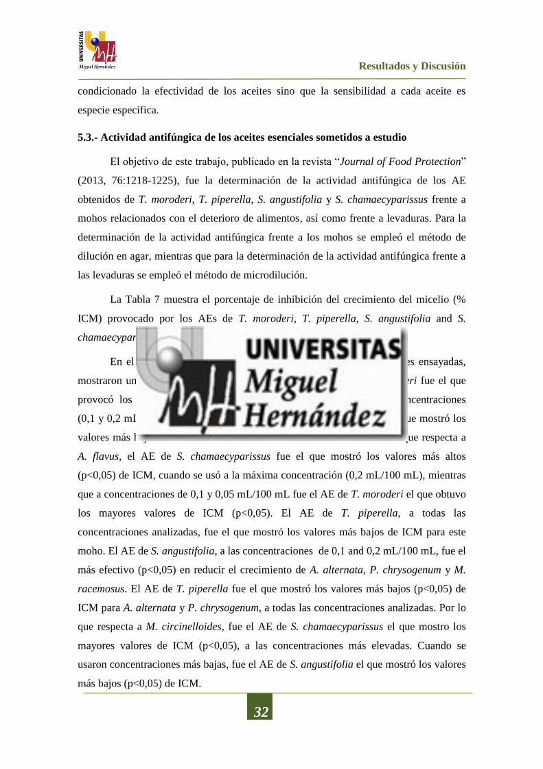
Resultados y Discusión
32
condicionado la efectividad de los aceites sino que la sensibilidad a cada aceite es
especie específica.
5.3.- Actividad antifúngica de los aceites esenciales sometidos a estudio
El objetivo de este trabajo, publicado en la revista “Journal of Food Protection”
(2013, 76:1218-1225), fue la determinación de la actividad antifúngica de los AE
obtenidos de T. moroderi, T. piperella, S. angustifolia y S. chamaecyparissus frente a
mohos relacionados con el deterioro de alimentos, así como frente a levaduras. Para la
determinación de la actividad antifúngica frente a los mohos se empleó el método de
dilución en agar, mientras que para la determinación de la actividad antifúngica frente a
las levaduras se empleó el método de microdilución.
La Tabla 7 muestra el porcentaje de inhibición del crecimiento del micelio (%
ICM) provocado por los AEs de T. moroderi, T. piperella, S. angustifolia and S.
chamaecyparissus frente a diversos mohos.
En el caso de A. niger, todos los AEs, a todas las concentraciones ensayadas,
mostraron un reducción del crecimiento del micelio. El AE de T. moroderi fue el que
provocó los mayores valores de ICM (p<0,05) cuando se aplicó a altas concentraciones
(0,1 y 0,2 mL/100 mL). Por el contrario, el AE de S. angustifolia fue el que mostró los
valores más bajos de ICM a todas las concentraciones analizadas. En lo que respecta a
A. flavus, el AE de S. chamaecyparissus fue el que mostró los valores más altos
(p<0,05) de ICM, cuando se usó a la máxima concentración (0,2 mL/100 mL), mientras
que a concentraciones de 0,1 y 0,05 mL/100 mL fue el AE de T. moroderi el que obtuvo
los mayores valores de ICM (p<0,05). El AE de T. piperella, a todas las
concentraciones analizadas, fue el que mostró los valores más bajos de ICM para este
moho. El AE de S. angustifolia, a las concentraciones de 0,1 and 0,2 mL/100 mL, fue el
más efectivo (p<0,05) en reducir el crecimiento de A. alternata, P. chrysogenum y M.
racemosus. El AE de T. piperella fue el que mostró los valores más bajos (p<0,05) de
ICM para A. alternata y P. chrysogenum, a todas las concentraciones analizadas. Por lo
que respecta a M. circinelloides, fue el AE de S. chamaecyparissus el que mostro los
mayores valores de ICM (p<0,05), a las concentraciones más elevadas. Cuando se
usaron concentraciones más bajas, fue el AE de S. angustifolia el que mostró los valores
más bajos (p<0,05) de ICM.

Resultados y Discusión
33
Tabla 7. Porcentaje de inhibición del crecimiento del micelio (ICM) provocado por los aceites
esenciales de T. moroderi, T. piperella, S. angustifolia y S. chamaecyparissus, frente a diversos
mohos.
% Inhibición del crecimiento del micelio (ICM)
Mohos Cψ T.
moroderi
T.
piperella
S.
angustifolia
S.
chamaecyparissus
A. niger 0,025 6,42±0,12aA
8,44±0,02bA
4,06±0,15cA
6,68±0,12aA
0,05 13,73±0,24aB
14,33±0,01bB
12,91±0,09cB
14,50±0,06dB
0,1 29,01±0,15aC
26,79±0,12bC
22,10±0,11cC
22,24±0,07cC
0,2 52,54±0,23aD
44,94±0,05bD
33,74±0,13cD
42,82±0,12dD
A. flavus 0,025 10,10±0,11aA
3,94±0,07bA
8,43±0,08cA
7,06±0,11dA
0,05 19,89±0,23aB
10,76±0,14bB
13,61±0,10cB
13,58±0,08cB
0,1 26,79±0,10aC
16,21±0,09bC
21,88±0,07cC
23,26±0,14dC
0,2 34,84±0,11aD
28,22±0,10bD
29,54±0,09cD
41,23±0,09dD
A. alternata 0,025 12,83±0,06aA
0,00±0,00bA
14,47±0,01cA
14,48±0,06cA
0,05 24,57±0,12aB
15,80±0,06bB
33,24±0,12cB
23,96±0,07dB
0,1 39,27±0,14aC
20,64±0,08bC
50,29±0,12cC
32,83±0,11dC
0,2 54,69±0,06aD
29,43±0,09bD
55,70±0,09cD
43,96±0,14dD
P. chrysogenum 0,025 13,84±0,05aA
0,00±0,00bA
10,51±0,12cA
1,17±0,11dA
0,05 30,47±0,09aB
5,56±0,11bB
21,69±0,14cB
7,24±0,15dB
0,1 51,57±0,11aC
8,33±0,05bC
60,56±0,05cC
38,13±0,06dC
0,2 63,94±0,15aD
28,33±0,13bD
79,81±0,04cD
53,94±0,01dD
M. circinelloides 0,025 4,92±0,06aA
7,01±0,03bA
0,00±0,00cA
1,60±0,20dA
0,05 13,98±0,07aB
9,23±0,04bB
1,12±0,01cB
6,79±0,12dB
0,1 19,01±0,09aC
29,14±0,14bC
26,88±0,14cC
33,59±0,09dC
0,2 22,57±0,13aD
44,57±0,07bD
42,81±0,12cD
45,60±0,14dD
M. racemosus 0,025 1,46±0,04aA
3,58±0,04bA
0,00±0,00cA
0,00±0,00cA
0,05 17,54±0,12aB
5,58±0,14bB
12,61±0,08cB
0,00±0,00dA
0,1 30,29±0,08aC
18,28±0,14bC
49,60±0,07cC
14,93±0,18dB
0,2 40,02±0,09aD
38,63±0,17bD
62,24±0,11cD
29,06±0,15dC
ψC: Concentración (mL/100 mL)
Para un mismo moho y una misma concentración: valores seguidos por la misma letra minúscula dentro de la misma
fila no son significativamente diferentes (p>0,05) de acuerdo con el Test de Rangos Múltiples de Tukey.
Para un mismo moho y un mismo AE: valores seguidos por la misma letra mayúscula dentro de la misma columna no
son significativamente diferentes (p>0,05) de acuerdo con el Test de Rangos Múltiples de Tukey.

Resultados y Discusión
34
El método de microdilución se empleó para determinar la actividad antifúngica
de los AEs frente a diversas levaduras, y los resultados se muestran en la Tabla 8. Todos
los AEs analizados presentaron efecto inhibidor del desarrollo de todas las cepas de
levadura ensayadas. Fue el AE de S. angustifolia el que mostró la menor concentración
mínima de inhibición (CMI) sobre las levaduras R. mucilaginosa, S. cerevisiae, D.
hansenii y P. carsoni. Por otro lado el AE de T. moroderi fue el más eficaz frente a Y.
lipolytica con un valor de CMI de 0,25 mL/100 mL. Sin embargo, este AE presentó los
valores más altos de CMI para C. zeylanoides y S. cerevisiae.
Aligianis et al. (2001) propusieron una clasificación de la actividad
antimicrobiana para productos vegetales, basada en los resultados de la CMI:
compuestos vegetales con CMI ≤ 0,05 mL/100 ml, se clasificarían como inhibidores
fuertes; compuestos vegetales con CMI entre 0,06 y 0,15 mL/100 mL, inhibidores
moderados; compuestos vegetales con CMI ≥ 0,16 mL/100 mL, inhibidores débiles. Por
lo tanto, basado en los resultados de la CMI de los ensayos de microdilución, los AEs
analizados mostraron un pobre potencial como compuestos “antilevaduras”, ya que
todas las cepas de levaduras analizadas presentaron un valor de CMI > a 0,16 mL/100
mL.
Tabla 8. Valores de Concentración Mínima de Inhibición (CMI) provocado por los AEs de T.
moroderi, T. piperella, S. angustifolia y S. chamaecyparissus frente a diversas levaduras
Levadura
Aceite esencial (μL/mL)
T.
moroderi
T.
piperella
S.
angustifolia
S.
chamaecyparissus
Y. lipolytica 2,5 5 5 5
C. zeylanoides 5 2,5 2,5 2,5
P. carsonni 5 5 2.5 5
S. cerevisiae 10 5 2,5 5
D. hansenii 2,5 2,5 1,25 2,5
R. mucilaginosa 5 5 2,5 5
Al igual que sucede con el mecanismo por el cual se produce la actividad
antibacteriana, el mecanismo por el cual se produce la inhibición del crecimiento de los
mohos y levaduras por la acción de los AEs tampoco es conocido con total detalle. Son

Resultados y Discusión
35
varias las publicaciones científicas que aportan datos para tratar de explicar dicho
comportamiento. Así, Lucini et al. (2006) indicaron que la inhibición del crecimiento
del micelio es causada por los monoterpenos presentes en los aceites esenciales. Estos
componentes podrían aumentar la concentración de peróxidos lipídicos tales como
radicales hidroxilo, alcoxilo y alkoperoxilo y así provocar la muerte celular. Para
Sharma y Tripathi (2006), los AEs actuaría sobre las hifas del micelio, provocando la
salida de los componentes del citoplasma, la pérdida de rigidez y la integridad de la
pared celular de la hifa, lo que provocaría su colapso y como consecuencia la muerte del
micelio.
5.4.- Actividad antioxidante de los aceites esenciales sometidos a estudio
El objetivo de este trabajo, publicado en la revista “Journal of Food Protection”
(2013, 76:1218-1225), fue la evaluación de la actividad antioxidante de los AE
obtenidos de T. moroderi, T. piperella, S. angustifolia y S. chamaecyparissus, así como
su relación con el contenido en fenoles. La determinación de la actividad antioxidante in
vitro se realizó siguiendo cuatro diferentes métodos analíticos, como son: el método del
secuestro del radical 2,2’-difenil-1-picrilhidrazil (DPPH), el poder antioxidante de la
reducción de hierro (FRAP), la determinación de la actividad antioxidante de las
especies reactivas del ácido tiobarbitúrico (TBARS) y la capacidad quelante del ion
ferroso (FIC).
La Figura 5 muestra el contenido en fenoles totales (FT) de los AEs obtenidos de
T. moroderi, T. piperella, S. angustifolia y S. chamaecyparissus. El AE de T. piperella
mostró los valores más elevados (p<0,05) de FT (57,43 mg equivalentes del ácido
gálico (AGE)/g) de todas las muestras analizadas. Los AEs de S. chamaecyparissus y S.
angustifolia mostraron un menor contenido en FT (21,59 y 38,48 mg de AGE/g,
respectivamente), mientras que el AE de T. moroderi presentó los valores más bajos
(9,33 mg de AGE/g).
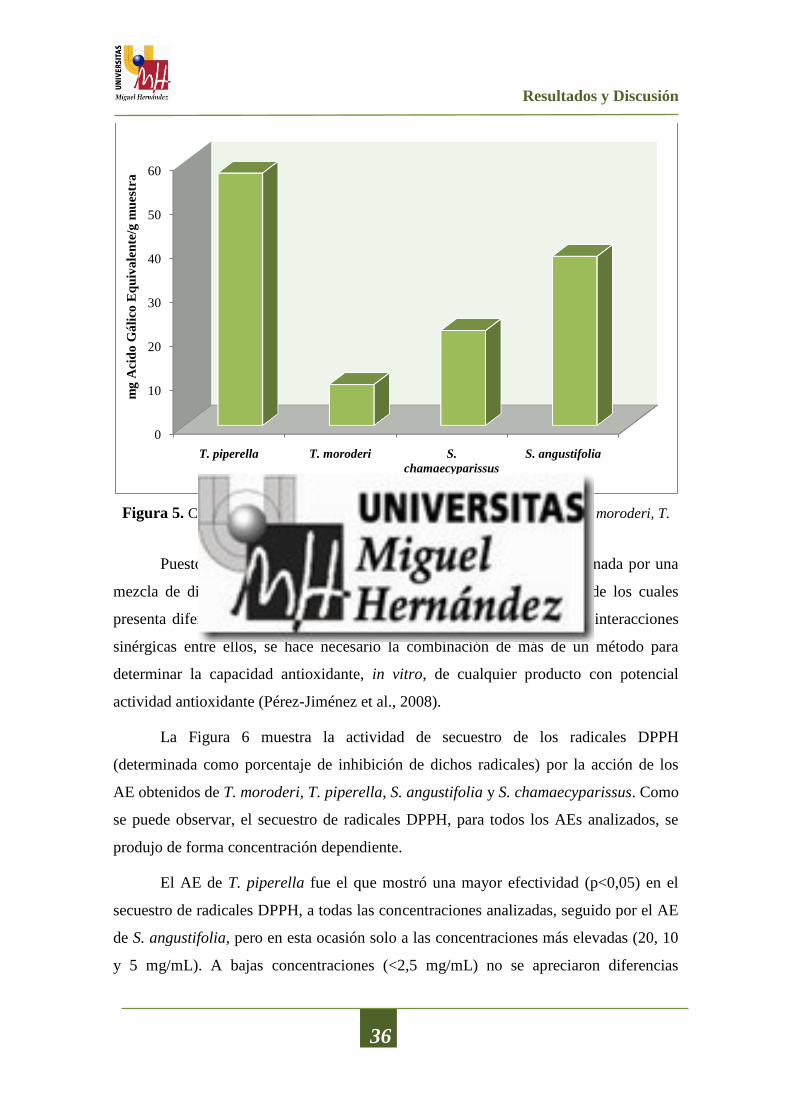
Resultados y Discusión
36
Figura 5. Contenido en fenoles totales de los aceites esenciales obtenidos de T. moroderi, T.
piperella, S. angustifolia y S. chamaecyparissus.
Puesto que la actividad antioxidante en los alimentos está determinada por una
mezcla de diferentes compuestos con actividad antioxidante, cada uno de los cuales
presenta diferentes mecanismos de acción, e incluso se pueden producir interacciones
sinérgicas entre ellos, se hace necesario la combinación de más de un método para
determinar la capacidad antioxidante, in vitro, de cualquier producto con potencial
actividad antioxidante (Pérez-Jiménez et al., 2008).
La Figura 6 muestra la actividad de secuestro de los radicales DPPH
(determinada como porcentaje de inhibición de dichos radicales) por la acción de los
AE obtenidos de T. moroderi, T. piperella, S. angustifolia y S. chamaecyparissus. Como
se puede observar, el secuestro de radicales DPPH, para todos los AEs analizados, se
produjo de forma concentración dependiente.
El AE de T. piperella fue el que mostró una mayor efectividad (p<0,05) en el
secuestro de radicales DPPH, a todas las concentraciones analizadas, seguido por el AE
de S. angustifolia, pero en esta ocasión solo a las concentraciones más elevadas (20, 10
y 5 mg/mL). A bajas concentraciones (<2,5 mg/mL) no se apreciaron diferencias
0
10
20
30
40
50
60
T. piperella T. moroderi S.
chamaecyparissus
S. angustifolia
mg
Aci
do
Gá
lico
Eq
uiv
ale
nte
/g m
ues
tra

Resultados y Discusión
37
estadísticamente significativas (p>0,05) para los AEs de T. moroderi, S.
chamaecyparissus y S. angustifolia.
Figura 6. Secuestro de los radicales DPPH por la acción de los aceites esenciales (a diferentes
concentraciones) de T. moroderi, T. piperella, S. angustifolia y S. chamaecyparissus.
Los valores de IC50 (concentración de AE que inhibe el 50% del radical DPPH)
de los diferentes AEs siguieron el orden siguiente: AE de T. piperella (9,30 mg/mL) <
AE de S. angustifolia (39,58 mg/mL) < AE de S. chamaecyparissus (75,45 mg/mL) <
AE de T. moroderi (90,01 mg/mL).
La capacidad quelante del ion ferroso (Fe+2
) de los diferentes AEs obtenidos de
T. moroderi, T. piperella, S. angustifolia y S. chamaecyparissus, se muestra en la Figura
7. El análisis de esta propiedad mostró que todos los AEs analizados fueron capaces de
quelar el ion ferroso y dicha acción se produjo de forma dependiente de la
concentración. En las concentraciones más elevadas (10 y 20 mg/mL) fueron los AEs
de S. chamaecyparissus y S. angustifolia los que mostraron una mayor (p<0,05)
capacidad quelante, no existiendo diferencias estadísticamente significativas entre ellos.
El AE de T. piperella, a todas las concentraciones estudiadas, mostró los valores más
bajos (p<0,05) en la quelación del ion ferroso.
0
10
20
30
40
50
60
70
80
90
0 2,5 5 7,5 10 12,5 15 17,5 20
% I
nh
ibic
ión
Concentración (mg/mL)
S. chamaecyparissusS. angustifoliaT. piperellaT. moroderi
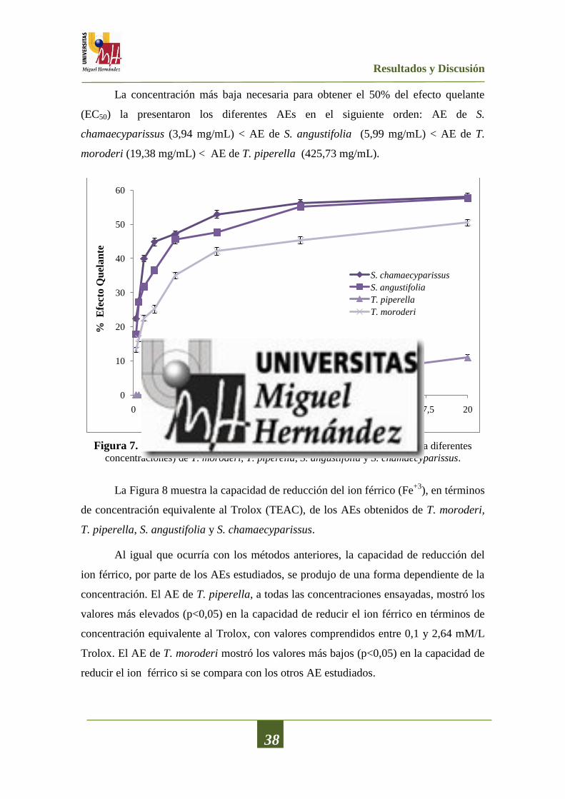
Resultados y Discusión
38
La concentración más baja necesaria para obtener el 50% del efecto quelante
(EC50) la presentaron los diferentes AEs en el siguiente orden: AE de S.
chamaecyparissus (3,94 mg/mL) < AE de S. angustifolia (5,99 mg/mL) < AE de T.
moroderi (19,38 mg/mL) < AE de T. piperella (425,73 mg/mL).
Figura 7. Capacidad quelante del ion ferroso (Fe+2
) de los aceites esenciales (a diferentes
concentraciones) de T. moroderi, T. piperella, S. angustifolia y S. chamaecyparissus.
La Figura 8 muestra la capacidad de reducción del ion férrico (Fe+3
), en términos
de concentración equivalente al Trolox (TEAC), de los AEs obtenidos de T. moroderi,
T. piperella, S. angustifolia y S. chamaecyparissus.
Al igual que ocurría con los métodos anteriores, la capacidad de reducción del
ion férrico, por parte de los AEs estudiados, se produjo de una forma dependiente de la
concentración. El AE de T. piperella, a todas las concentraciones ensayadas, mostró los
valores más elevados (p<0,05) en la capacidad de reducir el ion férrico en términos de
concentración equivalente al Trolox, con valores comprendidos entre 0,1 y 2,64 mM/L
Trolox. El AE de T. moroderi mostró los valores más bajos (p<0,05) en la capacidad de
reducir el ion férrico si se compara con los otros AE estudiados.
0
10
20
30
40
50
60
0 2,5 5 7,5 10 12,5 15 17,5 20
% E
fect
o Q
uel
an
te
Concentración mg/mL
S. chamaecyparissus
S. angustifolia
T. piperella
T. moroderi

Resultados y Discusión
39
Figura 8. Capacidad de reducción del ion férrico (Fe+3
) debido a la acción de los AEs
obtenidos de T. moroderi, T. piperella, S. angustifolia y S. chamaecyparissus.
La Figura 9 muestra la inhibición de la formación de especies reactivas del ácido
tiobarbiturico (TBARs), el cual es un metabolito secundario de la oxidación lipídica.
Todos los AEs analizados presentaron un alto porcentaje de inhibición de las TBARs y
dicha inhibición se produjo de forma dependiente de la concentración.
Figura 9. Capacidad de inhibición de la formación de las especies reactivas del ácido
tiobarbitúrico (TBARs) debido a la acción de los aceites esenciales de T. moroderi, T. piperella,
S. angustifolia y S. chamaecyparissus.
0,00
0,25
0,50
0,75
1,00
1,25
1,50
1,75
2,00
2,25
2,50
2,75
3,00
0,16 0,31 0,63 1,25 2,5 5 10 20
TE
AC
(m
M/L
Tro
lox)
Concentración (mg/mL)
T. moroderi
S. chamaecyparissus
S. angustifolia
T. piperella
0
10
20
30
40
50
60
70
0 2,5 5 7,5 10 12,5 15 17,5 20
% I
nh
ibic
ión
Concentración (mg/mL)
S. chamaecyparissus
S. angustifolia
T. piperella
T. moroderi
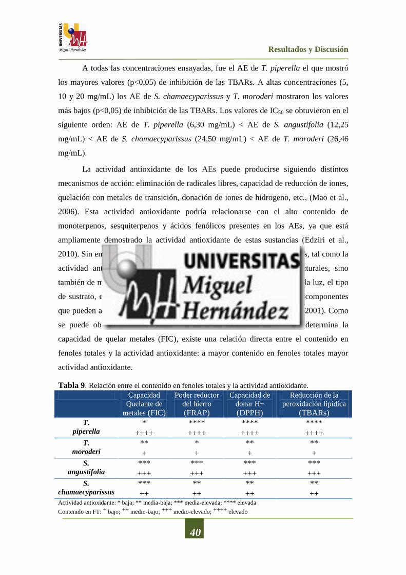
Resultados y Discusión
40
A todas las concentraciones ensayadas, fue el AE de T. piperella el que mostró
los mayores valores (p<0,05) de inhibición de las TBARs. A altas concentraciones (5,
10 y 20 mg/mL) los AE de S. chamaecyparissus y T. moroderi mostraron los valores
más bajos (p<0,05) de inhibición de las TBARs. Los valores de IC50 se obtuvieron en el
siguiente orden: AE de T. piperella (6,30 mg/mL) < AE de S. angustifolia (12,25
mg/mL) < AE de S. chamaecyparissus (24,50 mg/mL) < AE de T. moroderi (26,46
mg/mL).
La actividad antioxidante de los AEs puede producirse siguiendo distintos
mecanismos de acción: eliminación de radicales libres, capacidad de reducción de iones,
quelación con metales de transición, donación de iones de hidrogeno, etc., (Mao et al.,
2006). Esta actividad antioxidante podría relacionarse con el alto contenido de
monoterpenos, sesquiterpenos y ácidos fenólicos presentes en los AEs, ya que está
ampliamente demostrado la actividad antioxidante de estas sustancias (Edziri et al.,
2010). Sin embargo, debe tenerse en cuenta que las propiedades de los AEs, tal como la
actividad antioxidante, dependen no sólo de sus características estructurales, sino
también de muchos otros factores como la concentración, la temperatura, la luz, el tipo
de sustrato, el estado físico del sistema así como, la presencia de micro-componentes
que pueden actuar como agentes pro-oxidantes o sinérgicos (Yanishlieva, 2001). Como
se puede observar en la Tabla 9, excepto para el método donde se determina la
capacidad de quelar metales (FIC), existe una relación directa entre el contenido en
fenoles totales y la actividad antioxidante: a mayor contenido en fenoles totales mayor
actividad antioxidante.
Tabla 9. Relación entre el contenido en fenoles totales y la actividad antioxidante.
Capacidad
Quelante de
metales (FIC)
Poder reductor
del hierro (FRAP)
Capacidad de
donar H+ (DPPH)
Reducción de la
peroxidación lipídica (TBARs)
T.
piperella * **** **** ****
++++ ++++ ++++ ++++
T.
moroderi ** * ** **
+ + + +
S.
angustifolia *** *** *** ***
+++ +++ +++ +++
S.
chamaecyparissus *** ** ** **
++ ++ ++ ++ Actividad antioxidante: * baja; ** media-baja; *** media-elevada; **** elevada
Contenido en FT: + bajo; ++ medio-bajo; +++ medio-elevado; ++++ elevado
Resultados y Discusión
41
5.5.- Actividad antibacteriana de los films de quitosano adicionados con los aceites
esenciales sometidos a estudio
El objetivo de estos trabajos, publicados en la revista “Food Control” (2013,
30:386-392) y en los Proceedings de EUCHIS 2013 - International Conference of the
European Chitin Society, fue la determinación de la actividad antibacteriana de films de
quitosano a los que se adicionó diferentes concentraciones de los AEs obtenidos de T.
moroderi, T. piperella, S. angustifolia y S. chamaecyparissus. Dicha actividad se
determinó mediante la técnica de difusión en disco frente a diversas bacterias
relacionadas con el deterioro de alimentos o indicadoras de la presencia de patógenos.
En la Tabla 10 se muestra la actividad antibacteriana de los films de quitosano
(CH) adicionados con AEs de T. moroderi (CH+TMAE) y T. piperella (CH+TPAE) a
distintas concentraciones (0,5; 1 y 2%) frente a diversas bacterias. Los films formados
únicamente por quitosano (CH) no presentaron ningún tipo de inhibición frente a
ninguna de las bacterias analizadas. Estos resultados están en concordancia con los
expuestos por Ojagh et al. (2010) y Wang et al. (2011) quienes informaron de que no
existía ninguna inhibición significativa de los films de quitosano puro frente a diversas
bacterias, y lo explicaron por el hecho de que el film de quitosano no difunde sobre los
medios de cultivo formados por agar. En el presente estudio se puso de manifiesto que
los films de quitosano depositados sobre los medios de cultivo no sufrían alteraciones, y
por tanto el quitosano no se solubilizaba sobre el medio, no pudiendo de este modo
ejercer efecto inhibitorio alguno fuera de la zona de contacto con el medio.
En lo que respecta a los films CH+TPAE, se puede observar que, a todas las
concentraciones ensayadas, mostraron un efecto inhibidor sobre 3 de las 5 bacterias
analizadas, produciéndose dicho efecto de una forma concentración dependiente. La
bacteria más sensible a estos films fue A. denitrificans presentando halos de inhibición
comprendidos entre 20 y 30 mm, no apreciándose diferencias (p<0,05) entre los
tomillos ensayados. Para las bacterias S. marcescens y A. faecalis, los films CH+TPAE
no mostraron ninguna actividad antibacteriana cuando se usó el AE a la concentración
más baja (0,5%).
En el caso de los films CH+TMAE no se obtuvo ningún tipo de actividad
antibacteriana, a ninguna de las concentraciones analizadas, frente a S. marcescens. Del
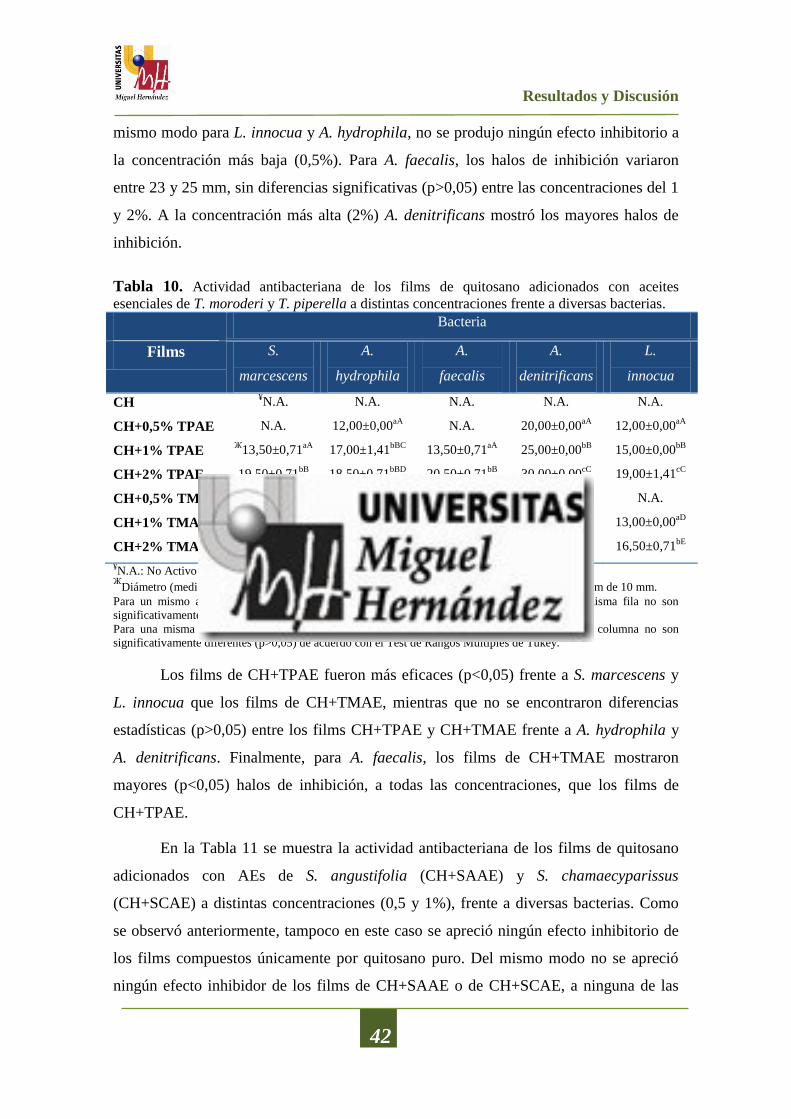
Resultados y Discusión
42
mismo modo para L. innocua y A. hydrophila, no se produjo ningún efecto inhibitorio a
la concentración más baja (0,5%). Para A. faecalis, los halos de inhibición variaron
entre 23 y 25 mm, sin diferencias significativas (p>0,05) entre las concentraciones del 1
y 2%. A la concentración más alta (2%) A. denitrificans mostró los mayores halos de
inhibición.
Tabla 10. Actividad antibacteriana de los films de quitosano adicionados con aceites
esenciales de T. moroderi y T. piperella a distintas concentraciones frente a diversas bacterias.
Bacteria
Films S.
marcescens
A.
hydrophila
A.
faecalis
A.
denitrificans
L.
innocua
CH ¥N.A. N.A. N.A. N.A. N.A.
CH+0,5% TPAE N.A. 12,00±0,00aA
N.A. 20,00±0,00aA
12,00±0,00aA
CH+1% TPAE Ж
13,50±0,71aA
17,00±1,41bBC
13,50±0,71aA
25,00±0,00bB
15,00±0,00bB
CH+2% TPAE 19,50±0,71bB
18,50±0,71bBD
20,50±0,71bB
30,00±0,00cC
19,00±1,41cC
CH+0,5% TMAE N.A. N.A. 23,00±0,00aC
20,00±0,00aA
N.A.
CH+1% TMAE N.A. 16,50±0,71aC
24,50±0,71bD
24,00±1,41bB
13,00±0,00aD
CH+2% TMAE N.A. 19,50±0,71bD
25,00±0,00bD
29,50±0,71cC
16,50±0,71bE
¥N.A.: No Activo
ЖDiámetro (media y desviación estándar) del halo de inhibición (mm) incluyendo el diámetro del film de 10 mm.
Para un mismo aceite esencial, valores seguidos por la misma letra minúscula dentro de la misma fila no son
significativamente diferentes (p>0,05) de acuerdo con el Test de Rangos Múltiples de Tukey.
Para una misma bacteria, valores seguidos por la misma letra mayúscula dentro de la misma columna no son
significativamente diferentes (p>0,05) de acuerdo con el Test de Rangos Múltiples de Tukey.
Los films de CH+TPAE fueron más eficaces (p<0,05) frente a S. marcescens y
L. innocua que los films de CH+TMAE, mientras que no se encontraron diferencias
estadísticas (p>0,05) entre los films CH+TPAE y CH+TMAE frente a A. hydrophila y
A. denitrificans. Finalmente, para A. faecalis, los films de CH+TMAE mostraron
mayores (p<0,05) halos de inhibición, a todas las concentraciones, que los films de
CH+TPAE.
En la Tabla 11 se muestra la actividad antibacteriana de los films de quitosano
adicionados con AEs de S. angustifolia (CH+SAAE) y S. chamaecyparissus
(CH+SCAE) a distintas concentraciones (0,5 y 1%), frente a diversas bacterias. Como
se observó anteriormente, tampoco en este caso se apreció ningún efecto inhibitorio de
los films compuestos únicamente por quitosano puro. Del mismo modo no se apreció
ningún efecto inhibidor de los films de CH+SAAE o de CH+SCAE, a ninguna de las
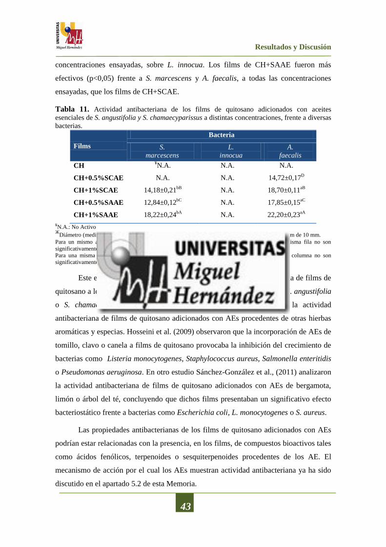
Resultados y Discusión
43
concentraciones ensayadas, sobre L. innocua. Los films de CH+SAAE fueron más
efectivos (p<0,05) frente a S. marcescens y A. faecalis, a todas las concentraciones
ensayadas, que los films de CH+SCAE.
Tabla 11. Actividad antibacteriana de los films de quitosano adicionados con aceites
esenciales de S. angustifolia y S. chamaecyparissus a distintas concentraciones, frente a diversas
bacterias.
Films
Bacteria
S.
marcescens
L.
innocua
A.
faecalis
CH ¥N.A. N.A. N.A.
CH+0.5%SCAE N.A.
N.A.
14,72±0,17D
CH+1%SCAE 14,18±0,21bB
N.A.
18,70±0,11aB
CH+0.5%SAAE 12,84±0,12bC
N.A.
17,85±0,15aC
CH+1%SAAE 18,22±0,24bA
N.A.
22,20±0,23aA
¥N.A.: No Activo
ЖDiámetro (media y desviación estándar) del halo de inhibición (mm) incluyendo el diámetro del film de 10 mm.
Para un mismo aceite esencial, valores seguidos por la misma letra minúscula dentro de la misma fila no son
significativamente diferentes (p>0,05) de acuerdo con el Test de Rangos Múltiples de Tukey.
Para una misma bacteria, valores seguidos por la misma letra mayúscula dentro de la misma columna no son
significativamente diferentes (p>0,05) de acuerdo con el Test de Rangos Múltiples de Tukey.
Este es el primer estudio donde se analiza la actividad antibacteriana de films de
quitosano a los que se han incorporado AEs de T. moroderi, T. piperella, S. angustifolia
o S. chamaecyparissus. Sin embargo, sí que hay referencias sobre la actividad
antibacteriana de films de quitosano adicionados con AEs procedentes de otras hierbas
aromáticas y especias. Hosseini et al. (2009) observaron que la incorporación de AEs de
tomillo, clavo o canela a films de quitosano provocaba la inhibición del crecimiento de
bacterias como Listeria monocytogenes, Staphylococcus aureus, Salmonella enteritidis
o Pseudomonas aeruginosa. En otro estudio Sánchez-González et al., (2011) analizaron
la actividad antibacteriana de films de quitosano adicionados con AEs de bergamota,
limón o árbol del té, concluyendo que dichos films presentaban un significativo efecto
bacteriostático frente a bacterias como Escherichia coli, L. monocytogenes o S. aureus.
Las propiedades antibacterianas de los films de quitosano adicionados con AEs
podrían estar relacionadas con la presencia, en los films, de compuestos bioactivos tales
como ácidos fenólicos, terpenoides o sesquiterpenoides procedentes de los AE. El
mecanismo de acción por el cual los AEs muestran actividad antibacteriana ya ha sido
discutido en el apartado 5.2 de esta Memoria.
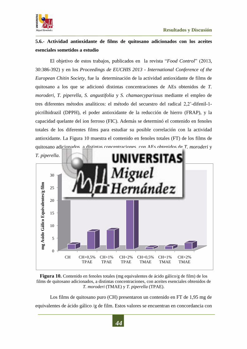
Resultados y Discusión
44
5.6.- Actividad antioxidante de films de quitosano adicionados con los aceites
esenciales sometidos a estudio
El objetivo de estos trabajos, publicados en la revista “Food Control” (2013,
30:386-392) y en los Proceedings de EUCHIS 2013 - International Conference of the
European Chitin Society, fue la determinación de la actividad antioxidante de films de
quitosano a los que se adicionó distintas concentraciones de AEs obtenidos de T.
moroderi, T. piperella, S. angustifolia y S. chamaecyparissus mediante el empleo de
tres diferentes métodos analíticos: el método del secuestro del radical 2,2’-difenil-1-
picrilhidrazil (DPPH), el poder antioxidante de la reducción de hierro (FRAP), y la
capacidad quelante del ion ferroso (FIC). Además se determinó el contenido en fenoles
totales de los diferentes films para estudiar su posible correlación con la actividad
antioxidante. La Figura 10 muestra el contenido en fenoles totales (FT) de los films de
quitosano adicionados, a distintas concentraciones, con AEs obtenidos de T. moroderi y
T. piperella.
Figura 10. Contenido en fenoles totales (mg equivalentes de ácido gálico/g de film) de los
films de quitosano adicionados, a distintas concentraciones, con aceites esenciales obtenidos de
T. moroderi (TMAE) y T. piperella (TPAE).
Los films de quitosano puro (CH) presentaron un contenido en FT de 1,95 mg de
equivalentes de ácido gálico /g de film. Estos valores se encuentran en concordancia con
0
5
10
15
20
25
30
CH CH+0,5%
TPAE
CH+1%
TPAE
CH+2%
TPAE
CH+0,5%
TMAE
CH+1%
TMAE
CH+2%
TMAE
mg A
sid
o G
áli
co E
qu
ivale
nte
s/g f
ilm

Resultados y Discusión
45
los obtenidos por Siripatrawan y Harte (2010) y Moradi et al. (2012). Dichos autores lo
atribuyen a la formación de cromógenos producida, posiblemente, por la reacción del
reactivo Folin Ciocalteu con sustancias reductoras de origen no fenólico y que se
podrían detectar espectrofotométricamente. Tanto para los films de CH+TPAE como
para los films de CH+TMAE, la incorporación del AE provocó un aumento en el
contenido de FT y esto ocurrió de una forma dependiente de la concentración. Cabe
destacar que los films de CH+TPAE mostraron un contenido en FT, a todas las
concentraciones analizadas, superior (p<0,05) que los films de CH+TMAE. Como se
vió en el apartado 5.4, el AE de T. piperella, presentaba un mayor contenido en fenoles
totales que el AE de T. moroderi.
La Figura 11 muestra el contenido en fenoles totales de los films de quitosano
adicionados, a distintas concentraciones, con AEs de S. angustifolia y S.
chamaecyparissus.
Figura 11. Contenido en Fenoles Totales (FT) de los films de quitosano adicionados, a
distintas concentraciones, con aceites esenciales obtenidos de S. angustifolia (SAAE) y S.
chamaecyparissus (SCAE).
0
0,1
0,2
0,3
0,4
0,5
0,6
0,7
0,8
0,9
CH CH+0,5%
SAAECH+1%
SAAECH+0,5%
SCAECH+1%
SCAE
mg A
cid
o G
áli
co E
qu
ivale
nte
s/g

Resultados y Discusión
46
Los films elaborados con quitosano puro (CH) mostraron un contenido de FT de
aproximadamente 0,26 mg de equivalentes del ácido gálico/g de film. Como se ha
mencionado anteriormente, este hecho estaría explicado por la producción de
cromógenos. Después de la incorporación de los AE, como era de esperar, el contenido
en FT de los films de CH aumentó. Dicho aumento se produjo de forma dependiente de
la concentración (p<0,05) para ambos AEs. Los films de CH+SCAE mostraron un
contenido en FT, a todas las concentraciones analizadas, superior (p<0,05) que los films
de CH+SAAE. Es importante destacar que, pese a que el contenido en FT del AE de S.
angustifolia es mayor que el del AE de S. chamaecyparissus en los films elaborados con
estos aceites ocurre al contrario. Los films elaborados con AE de S. chamaecyparissus
presentan mayor contenido en FT que los films adicionados con AE de S. angustifolia.
Esto podría deberse a que se haya producido una mayor evaporación de este tipo de
compuestos durante la elaboración del film.
Para determinar la actividad antioxidante, in vitro, de los films de quitosano
adicionados, a distintas concentraciones, con AE de T. moroderi, T. piperella, S.
angustifolia y S. chamaecyparissus se emplearon tres métodos distintos, como son:
DPPH, FRAP y FIC.
La Figura 12 muestra la actividad de secuestro de los radicales DPPH por la
acción de los films de quitosano adicionados con AEs obtenidos de T. moroderi o T.
piperella.
Los films de quitosano puro sin la adición de ningún tipo de AE mostraron una
leve actividad secuestrante de radicales DPPH (3 μg equivalentes del trolox (TE)/g
film). Como era de esperar, la adición de AE a los films provocó un aumento
significativo en la capacidad de secuestro de radicales libres DPPH, produciéndose esta
capacidad secuestrante de forma dependiente de la concentración. Los films de
CH+TPAE mostraron unos valores comprendidos entre 690 y 1090 μg TE/g film
(p<0,05). En el caso de los films de CH+TMAE, los valores de secuestro de radicales
libres oscilaron entre 420 y 630 μg TE/g film, produciéndose también la actividad
antioxidante de forma dependiente de la concentración.

Resultados y Discusión
47
Figura 12. Secuestro de los radicales DPPH de los films de quitosano (CH) adicionados con
aceites esenciales de T. moroderi (TMAE) o T. piperella (TPAE), a diferentes concentraciones.
La Figura 13 muestra la actividad de secuestro de los radicales DPPH por la
acción de los films de quitosano adicionados con AEs obtenidos de S. angustifolia y S.
chamaecyparissus.
Figura 13. Secuestro de los radicales DPPH por acción de los films de quitosano adicionados
con aceites esenciales de S. angustifolia (SAAE) y S. chamaecyparissus (SCAE), a diferentes
concentraciones
0
200
400
600
800
1000
1200
CH CH+0,5%
TPAE
CH+1%
TPAE
CH+2%
TPAE
CH+0,5%
TMAE
CH+1%
TMAE
CH+2%
TMAE
µg
Tro
lox E
qu
iva
len
tes/
g f
ilm
0
50
100
150
200
250
CH CH+0,5%SCAE CH+1%SCAE CH+0,5%SAAE CH+1%SAAE
µg
Tro
lox E
qu
iva
len
tes/
g f
ilm

Resultados y Discusión
48
El film de quitosano puro mostró un ligera actividad secuestrante de radicales
DPPH, muy similar a la reportada anteriormente. En el caso de los films CH+SCAE, los
valores de actividad secuestrante de radicales DPPH fueron 0,33 y 0,72 mM TE/g film
(p<0,05), para las concentraciones de 0,5 y 1%, respectivamente. Los films CH+SAAE,
a todas las concentraciones ensayadas, mostraron una actividad secuestrante de
radicales DPPH superior (p<0,05) que los films de CH+SCAE, obteniendo unos valores
de 0,57 y 0,85 mM TE/g film para las concentraciones de 0,5 y 1%, respectivamente.
La Figura 14 muestra la capacidad de reducción del ion férrico (Fe+3
) de los
films de quitosano adicionados con AEs obtenidos de T. moroderi y T. piperella.
Figura 14. Capacidad de reducción del ion férrico (Fe+3
) debido a la acción de los films de
quitosano adicionados con aceites esenciales obtenidos de T. moroderi, T. piperella, a diferentes
concentraciones.
Los films de CH puro mostraron una leve capacidad de reducción del ion Fe+3
(0,21 mg TE/g film). El análisis de los films CH+TPAE mostró un amplio rango de
actividad reductora con valores comprendidos entre 2,79 y 10,09 mg TE/g film
(p<0,05). Como en el caso del ensayo DPPH, la actividad reductora del ion Fe+3
sucede
de una forma dependiente de la concentración. Con respecto a los films CH+TMAE, a
bajas concentraciones (0,5 y 1%) no se apreció ningún tipo de actividad reductora,
0
2
4
6
8
10
12
CH CH+0,5%
TPAE
CH+1%
TPAE
CH+2%
TPAE
CH+0,5%
TMAE
CH+1%
TMAE
CH+2%
TMAE
mg T
rolo
x E
qu
ivale
nte
s/g f
ilm

Resultados y Discusión
49
mientras que a la concentración más elevada (2%) mostró una leve capacidad de
reducción (0,67 mg TE/g film). A todas las concentraciones ensayadas, los films
CH+TPAE mostraron una actividad de reducción del ion Fe+3
superior (p<0,05) a la de
los films CH+TMAE.
La Figura 15 muestra la capacidad de reducción del ion férrico (Fe+3
) de los
films de quitosano adicionados con AEs obtenidos de S. angustifolia y S.
chamaecyparissus. Como se aprecia en dicha figura la capacidad de reducción del ion
F+3
aumentó por la incorporación de los AE de S. angustifolia y S. chamaecyparissus, a
los films de CH y esta reducción se produjo de forma dependiente de la concentración.
En el caso de los films CH+SCAE los valores de reducción del ion Fe+3
(p<0,05)
fueron de 1,89 y 3,05 mM TE/g film, para las concentraciones de 0,5 y 1%,
respectivamente. Los films CH+SAAE mostraron una mayor actividad de reducción del
ion Fe+3
(p<0,05), a todas las concentraciones ensayadas, que los films CH+SCAE,
obteniendo unos valores de 3,09 y 4,23 mM TE/g film para las concentraciones de 0,5 y
1%, respectivamente.
Figura 15. Capacidad de reducción del ion férrico (Fe+3
) de los films de quitosano adicionados
con aceites esenciales de S. angustifolia y S. chamaecyparissus, a diferentes concentraciones.
0
0,2
0,4
0,6
0,8
1
1,2
CH CH+0,5%
SAAE CH+1% SAAE CH+0,5%
SCAE CH+1% SCAE
mM
Tro
lox E
qu
ivale
nte
s/g f
ilm

Resultados y Discusión
50
La Figura 16 muestra la capacidad quelante del ion ferroso (Fe+2
) de los films de
quitosano adicionados con AEs obtenidos de T. moroderi, T. piperella.
Como se aprecia en la Figura 15 los films CH+TPAE, a todas las
concentraciones analizadas, fueron capaces de quelar el ion ferroso y esto ocurrió de
una forma dependiente de la concentración (p<0,05). Del mismo modo, los films
CH+TMAE mostraron una alta capacidad para quelar dicho ion ferroso y como en el
caso de los films CH+TPAE, esto ocurrió de una forma dependiente de la concentración
(p<0,05).
A todas las concentraciones analizadas (excepto al 0,5%) los films CH+TMAE
fueron mejores agentes quelantes del ion ferroso (p<0,05) que los films CH+TPAE. En
términos generales se puede decir que los films de CH adicionados con AE de T.
moroderi y T. piperella mostraron un considerable capacidad quelante de metales.
Figura 16. Capacidad quelante del ion ferroso (Fe+2
) de los films de quitosano adicionados con
aceites esenciales de T. moroderi, T. piperella, a diferentes concentraciones.
0
5
10
15
20
25
30
35
40
45
CH CH+0,5%
TPAE
CH+1%
TPAE
CH+2%
TPAE
CH+0,5%
TMAE
CH+1%
TMAE
CH+2%
TMAE
Act
ivid
ad
Qu
elan
te (
% d
e in
hib
ició
n)
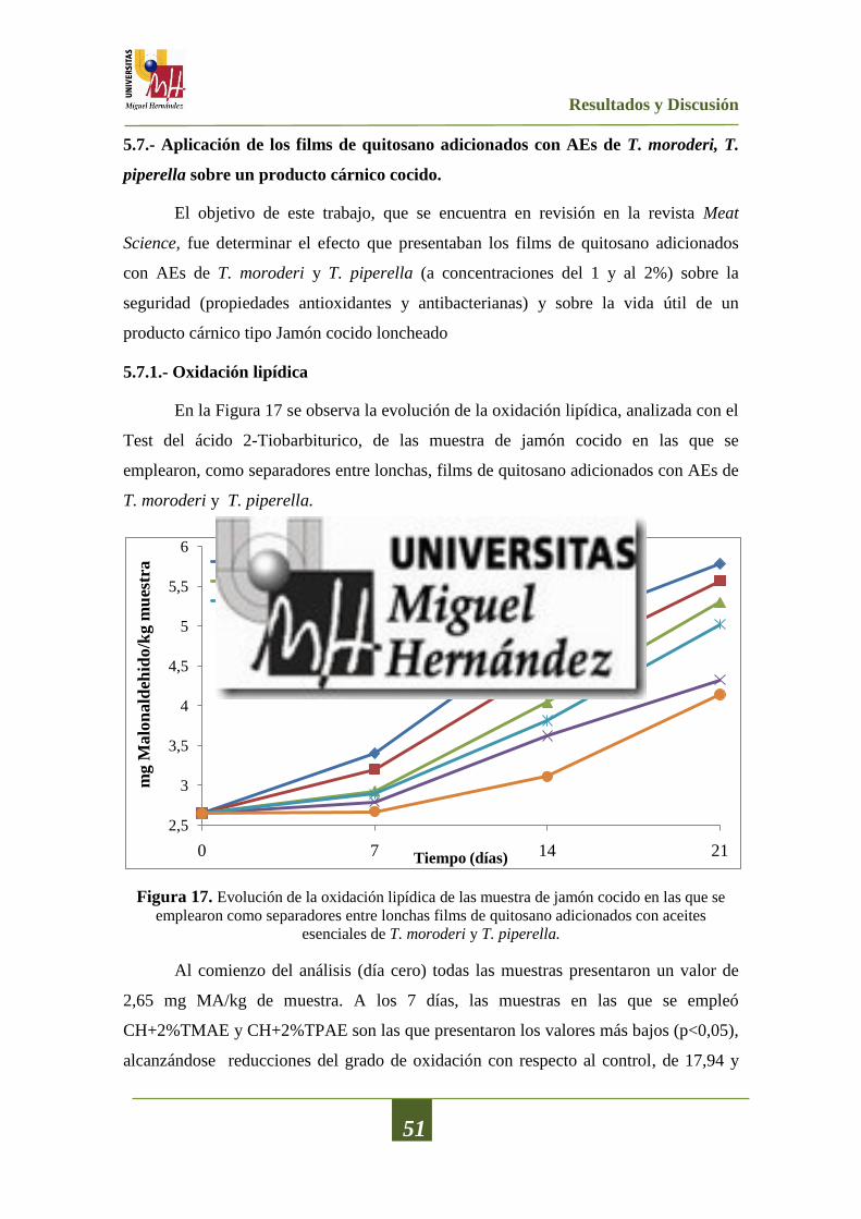
Resultados y Discusión
51
5.7.- Aplicación de los films de quitosano adicionados con AEs de T. moroderi, T.
piperella sobre un producto cárnico cocido.
El objetivo de este trabajo, que se encuentra en revisión en la revista Meat
Science, fue determinar el efecto que presentaban los films de quitosano adicionados
con AEs de T. moroderi y T. piperella (a concentraciones del 1 y al 2%) sobre la
seguridad (propiedades antioxidantes y antibacterianas) y sobre la vida útil de un
producto cárnico tipo Jamón cocido loncheado
5.7.1.- Oxidación lipídica
En la Figura 17 se observa la evolución de la oxidación lipídica, analizada con el
Test del ácido 2-Tiobarbiturico, de las muestra de jamón cocido en las que se
emplearon, como separadores entre lonchas, films de quitosano adicionados con AEs de
T. moroderi y T. piperella.
Figura 17. Evolución de la oxidación lipídica de las muestra de jamón cocido en las que se
emplearon como separadores entre lonchas films de quitosano adicionados con aceites
esenciales de T. moroderi y T. piperella.
Al comienzo del análisis (día cero) todas las muestras presentaron un valor de
2,65 mg MA/kg de muestra. A los 7 días, las muestras en las que se empleó
CH+2%TMAE y CH+2%TPAE son las que presentaron los valores más bajos (p<0,05),
alcanzándose reducciones del grado de oxidación con respecto al control, de 17,94 y
2,5
3
3,5
4
4,5
5
5,5
6
0 7 14 21
mg M
alo
nald
ehid
o/k
g m
ues
tra
Tiempo (días)
Control CH
CH+1% TMAE CH+2%TMAE
CH+1% TPEAE CH+2% TPAE

Resultados y Discusión
52
21,47%, respectivamente, existiendo diferencias significativas (p<0,05) entre ellas. Para
este mismo día la muestra en la que solo se empleó quitosano el grado de reducción de
la oxidación lipídica fue solo del 5,88%. Entre las muestras en las que se empleó
CH+1%TMAE y CH+1%TPAE no se apreciaban diferencias (p>0,05).
A los 14 días, nuevamente las muestras en las que se empleó CH+2%TMAE y
CH+2%TPAE son las que presentaronn los valores más bajos (p<0,05), con reducciones
del grado de oxidación con respecto al control de 26,86 y 37,17%, respectivamente. Al
final del ensayo (21 días), la muestra control mostraba unos valores de 5,78 mg MA/kg
de muestra, siendo las muestras en las que se empleó CH+2%TMAE y CH+2%TPAE
las que presentaron los valores más bajos (p<0,05), con reducciones del grado de
oxidación respecto al control de 25,25 y 28,37% respectivamente, y con diferencias
estadísticamente significativas (p<0,05) ente ellas.
El descenso en el grado de oxidación puede deberse, probablemente, a una
liberación muy pronunciada, que se produce fundamentalmente durante los primeros
días de almacenamiento, de los compuestos bioactivos presentes en los films de
quitosano y que proceden de los AEs, tal y como proponen Oussalah et al. (2007). Para
Moradi et al. (2011), la absorción, por parte del film, de agua presente en el producto
cárnico, puede causar que éste se hinche y que las tasas de migración cambien
continuamente debido a un proceso de relajación, dependiente del tiempo, provocado
por el propio hinchamiento.
5.7.2.- pH
En la Figura 18 se observa la evolución del pH de las muestra de jamón cocido
en las que se emplearon, como separadores de lonchas, films de quitosano adicionados
con AEs de T. moroderi y T. piperella.
En la muestra control (no contenía ningún tipo de separador) se produjo un leve
descenso del valor del pH con el paso del tiempo, partiendo de valores medios de 6,24 y
alcanzando un valor final 6,12 (p<0,05). En las muestras, en las que se emplearon los
films de quitosano y los films de quitosano adicionado con AEs de T. moroderi y T.
piperella se produjo un descenso más pronunciado de este parámetro.

Resultados y Discusión
53
Figura 18. Evolución del pH de las muestra de jamón cocido en las que se emplearon, como
separadores entre lonchas, films de quitosano adicionados con aceites esenciales de T. moroderi
y T. piperella.
A los 7 días de almacenamiento, la muestra en la que se empleó CH+2%TMAE
mostró el valor más bajo (p<0,05) de pH, con un reducción, con respecto al control, de
aproximadamente 0,6 unidades. Al final del ensayo (día 21), como se ha mencionado
anteriormente, la muestra control presentó el valor más elevado de pH (6,12) seguida
por la muestra (p<0,05) en la que se empleó CH+2%TPAE (pH 5,81). Para el resto de
las muestras, con valores de pH comprendidos entre 5,63 y 5,68, no se apreciaron
diferencias estadísticamente significativas (p>0,05) entre ellas; aunque sí existían entre
éstas y el control (p<0,05) y entre éstas y la muestra (p<0,05) con CH+2%TPAE. Como
veremos a continuación, este descenso de pH se explicaría por el aumento en las
poblaciones de bacterias ácido lácticas que se observó en dichas muestras.
5.7.3.- Análisis microbiológico
En las Figura 19 y 20 se observa la evolución de los recuentos de bacterias
aerobias mesófilas y bacterias acido lácticas, respectivamente, de las muestra de jamón
cocido, almacenadas a 4ºC durante 21 días, en las que se emplearon, como separadores
entre lonchas, films de quitosano o films de quitosano adicionados con AEs de T.
moroderi y T. piperella.
5,6
5,7
5,8
5,9
6
6,1
6,2
6,3
0 7 14 21
pH
Tiempo (días)
Control CH
CH+1% TMAE CH+2%TMAE
CH+1% TPEAE CH+2% TPAE
Resultados y Discusión
54
Las poblaciones de bacterias aerobias mesófilas y de bacterias ácido lácticas
aumentaron gradualmente durante el almacenamiento a 4ºC, aunque la liberación de los
compuestos bioactivos presentes en los films de quitosano influenció de forma
significativa el grado en el que estos microorganismos se desarrollaron.
Cabe destacar que no se apreció crecimiento ni de enterobacterias ni de mohos y
levaduras en ninguna de las muestras tratadas con films de quitosano adicionados con
AEs de T. moroderi y T. piperella. Como se ha discutido anteriormente, los mecanismos
de acción por los cuales estos compuestos bioactivos, fundamentalmente compuestos
fenólicos con grupos hidroxilo, podrían ejercer ejercen cierto efecto antibacteriano
serian (i) interfiriendo con los fosfolípidos presentes en la bicapa lipídica de la
membrana celular, lo cual provoca un aumento en la permeabilidad de esta y una
pérdida de los constituyentes celulares (ii) alternado sistemas enzimáticos y/o (iii)
inactivando o destruyendo material genético (Corbo et al., 2008).
En cuanto a los recuentos de bacterias aerobias mesófilas (Figura 19), se puede
observar como a los 7 días de almacenamiento, las muestras en las que se empleó
CH+2%TMAE y CH+2%TPAE presentaron los valores más bajos (p<0,05) de
crecimiento microbiano, con reducciones de los recuentos, para estas bacterias, de 2,57
y 2,61 ciclos logarítmicos, con respecto al control, no apreciándose diferencias (p>0,05)
entre ambas. Las muestras en las que solo se empleó quitosano presentaron una
reducción (p<0,05) de los recuentos de 0,62 ciclos logarítmicos respecto al control,
evidenciando un cierta actividad antibacteriana del quitosano.
Al final del almacenamiento, día 21, las muestras en las que se empleó
CH+2%TPAE presentaron los recuentos más bajos (p<0,05), alcanzándose una
reducción de 0,87 ciclos logarítmicos respecto al control. Entre las muestras en las que
se empleó CH+1%TMAE, CH+2%TMAE y CH+1%TPAE no se apreciaron diferencias
significativas (p>0,05) aunque sí entre éstas y el control (p<0,05), con reducciones de
los recuentos de 0,34; 0,53 y 0,37 ciclos logarítmicos, respectivamente.

Resultados y Discusión
55
Figura 19. Evolución de los recuentos de bacterias aerobias mesófilas de las muestra de jamón
cocido en las que se emplearon, como separadores entre lonchas, films de quitosano adicionados
con aceites esenciales de T. moroderi y T. piperella.
En el caso de las bacterias ácido lácticas (Figura 20), a los 7 días de
almacenamiento, las muestras en las que se empleó CH+2%TPAE mostraron los
recuentos más bajos (p<0,05), con una reducción, respecto al control, de 2,16 ciclos
logarítmicos.
Entre las muestras en las que se empleó CH+1%TMAE, CH+2%TMAE y
CH+1%TPAE no se apreciaron diferencias significativas (p>0,05) aunque sí entre éstas
y el control (p<0,05) con reducciones de los recuentos de 1,89; 1,98 y 1,93 ciclos
logarítmicos, respectivamente. Al final del ensayo, no se apreciaron diferencias
(p>0,05) entre el control y el grupo en el que se emplearon solo films de quitosano. De
nuevo, el empleo de CH+2%TPAE dio lugar a los recuentos más bajos (p<0,05) seguido
por las muestras con CH+2%TMAE, alcanzándose reducciones con respecto al control
de 1,14 y 0,84 ciclos logarítmicos, respectivamente. Entre las muestras en las que se
empleó CH+1%TMAE y CH+1%TPAE no se apreciaron diferencias significativas
(p>0,05) aunque sí (p<0,05) de ellas frente al control.
1
1,5
2
2,5
3
3,5
4
4,5
5
0 7 14 21
Ba
cter
ias
aer
ob
ias
mes
ófi
las
(lo
g U
FC
/g)
Tiempo (días)
Control CH
CH+1% TMAE CH+2%TMAE
CH+1% TPEAE CH+2% TPAE

Resultados y Discusión
56
Figura 20. Evolución de los recuentos de bacterias ácido lácticas de las muestra de jamón
cocido en las que se emplearon, como separadores de lonchas, films de quitosano adicionados
con aceites esenciales de T. moroderi y T. piperella.
5.7.4.- Liberación de compuestos bioactivos del film al producto cárnico.
La Figura 21 muestra una estimación de la liberación de los compuestos
bioactivos desde los films de quitosano adicionados con AEs de T. moroderi y T.
piperella (que se emplearon como separadores de lonchas) a las muestra de jamón
cocido, durante su almacenamiento a 4ºC durante 21 días.
Entre los días 0 y 7, para todas las muestras estudiadas (CH+1%TMAE,
CH+2%TMAE, CH+1%TPAE y CH+2%TPAE), se produjo una liberación muy
pronunciada de compuestos bioactivos desde el film al producto cárnico, siendo esta
liberación más pronunciada en las muestras que contenían mayor concentración de AE
(2%); se liberaron 7,27 y 8,34 mg AGE/g film para las muestras CH+2%TMAE y
CH+2%TPAE, respectivamente, mientras que para las muestras CH+1%TMAE y
CH+1% TPAE la liberación fue de 0,87 y 1,46 mg AGE/g film. Sin embargo, a partir
del día 7, la liberación de los compuestos bioactivos fue más gradual, hasta llegar al
final del ensayo (día 21), con valores comprendidos entre 0,13 y 0,49 mg AGE/g film
para todas las muestras, a excepción de la muestra de CH+2%TPAE.
1
1,5
2
2,5
3
3,5
4
4,5
5
5,5
0 7 14 21
Ba
cter
ias
áci
do
lá
ctic
as
(lo
g U
FC
/g)
Tiempo (días)
Control CH
CH+1% TMAE CH+2%TMAE
CH+1% TPEAE CH+2% TPAE
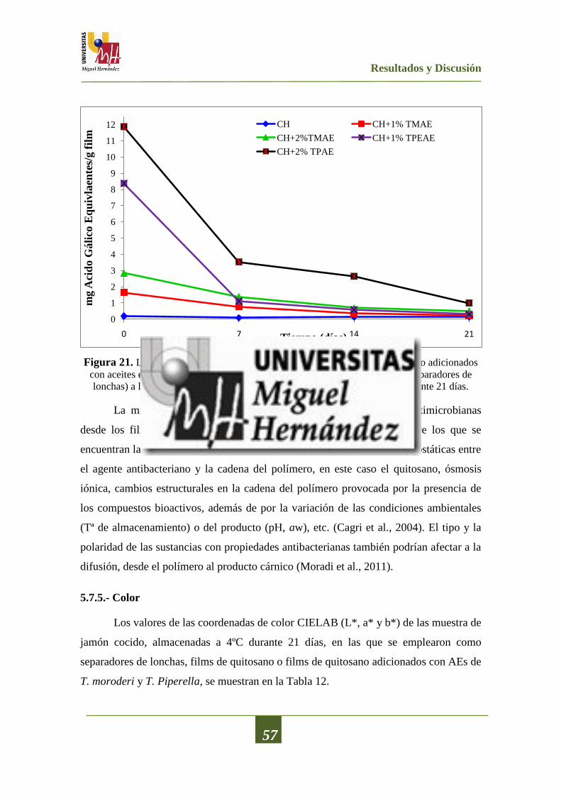
Resultados y Discusión
57
Figura 21. Liberación de los compuestos bioactivos desde los films de quitosano adicionados
con aceites esenciales de T. moroderi y T. piperella (que se emplearon como separadores de
lonchas) a las muestra de jamón cocido, durante su almacenamiento a 4ºC durante 21 días.
La migración de los compuestos bioactivos con propiedades antimicrobianas
desde los films al producto cárnico, depende de diversos factores, entre los que se
encuentran la composición de dicho producto cárnico, interacciones electrostáticas entre
el agente antibacteriano y la cadena del polímero, en este caso el quitosano, ósmosis
iónica, cambios estructurales en la cadena del polímero provocada por la presencia de
los compuestos bioactivos, además de por la variación de las condiciones ambientales
(Tª de almacenamiento) o del producto (pH, aw), etc. (Cagri et al., 2004). El tipo y la
polaridad de las sustancias con propiedades antibacterianas también podrían afectar a la
difusión, desde el polímero al producto cárnico (Moradi et al., 2011).
5.7.5.- Color
Los valores de las coordenadas de color CIELAB (L*, a* y b*) de las muestra de
jamón cocido, almacenadas a 4ºC durante 21 días, en las que se emplearon como
separadores de lonchas, films de quitosano o films de quitosano adicionados con AEs de
T. moroderi y T. Piperella, se muestran en la Tabla 12.
0
1
2
3
4
5
6
7
8
9
10
11
12
0 7 14 21
mg A
cid
o G
áli
co E
qu
ivla
ente
s/g f
ilm
Tiempo (días)
CH CH+1% TMAE
CH+2%TMAE CH+1% TPEAE
CH+2% TPAE

Resultados y Discusión
58
En referencia a la coordenada de luminosidad L*, en las medidas realizadas
durante los días 0 y 7, no se apreciaronn diferencias estadísticamente significativas
(p>0,05) entre las muestras control y aquellas en la que se utilizaron films de quitosano
o films de quitosano adicionado con AE de T. piperella y T. moroderi.
Tabla 12. Valores de las coordenadas de color (L*, a* y b*) de las muestra de jamón cocido,
almacenadas a 4ºC durante 21 días, en las que se emplearon, como separadores de lonchas,
films de quitosano o films de quitosano adicionados con aceites esenciales de T. moroderi y T.
piperella
Muestra Tiempo (Días)
0 7 14 21
L* Control 60,43±0,53aA
57,92±0,66bA
55,76±0,18cA
58,95±0,56bA
CH 60,43±0,53aA
60,62±1,93aA
59,65±1,79aB
59,53±0,64aA
CH+1%TMAE 60,43±0,53aA
60,62±1,17aA
58,28±0,76bB
59,47±0,58aA
CH+2%TMAE 60,43±0,53aA
57,18±2,38aA
59,80±1,03aB
59,53±1,09aA
CH+1%TPAE 60,43±0,53aA
60,72±2,95abcA
58,86±0,67bB
62,99±0,39cB
CH+2%TPAE 60,43±0,53aA
58,18±1,03bA
59,45±0,59abB
59,66±1,09abA
a* Control 10,08±0,43aA
9,58±1,66abA
10,15±0,56aA
8,89±0,68bA
CH 10,08±0,43aA
9,19±2,40abA
9,52±0,14aA
8,54±0,37bA
CH+1%TMAE 10,08±0,43aA
8,99±2,89abA
10,25±0,54aA
8,91±0,31bA
CH+2%TMAE 10,08±0,43aA
10,18±1,14aA
9,68±0,23aA
8,77±0,19bA
CH+1%TPAE 10,08±0,43aA
9,24±1,93aA
9,66±0,23aA
5,06±0,30bB
CH+2%TPAE 10,08±0,43aA
8,99±3,25abA
8,71±0,96abA
8,43±0,99bA
b* Control 8,37±0,11aA
8,74±1,39aA
8,81±0,46aA
8,17±0,66aA
CH 8,37±0,11aA
7,78±2,93aA
8,69±0,35aA
7,90±0,75aA
CH+1%TMAE 8,37±0,11aA
8,54±2,29aA
8,09±0,47aA
7,98±0,40aA
CH+2%TMAE 8,37±0,11aA
9,74±2,44abA
8,15±0,48aA
10,05±0,82bB
CH+1%TPAE 8,37±0,11aA
8,19±1,31abA
7,72±2,36abA
11,37±0,93cB
CH+2%TPAE 8,37±0,11aA
12,26±2,08bB
8,36±0,22aA
7,37±0,63cA
Para una misma coordenada, valores seguidos por la misma letra minúscula dentro de la misma fila no son
significativamente diferentes (p>0,05) de acuerdo con el Test de Rangos Múltiples de Tukey.
Para una misma coordenada,, valores seguidos por la misma letra mayúscula dentro de la misma columna no son
significativamente diferentes (p>0,05) de acuerdo con el Test de Rangos Múltiples de Tukey.
A los 14 días, la muestra control es la que presentaba los valores más bajos de
L* (p<0,05), no apreciándose diferencias significativas entre las muestras en las que se
empleó quitosano o quitosano adicionado con AE de T. piperella y T. moroderi. Sin
embargo, al final del ensayo (día 21) todas las muestras mostraban unos valores de
luminosidad muy similares, no existiendo diferencias estadísticamente significativas
(p>0.05) entre ellas, a excepción de la muestra CH+1%TPAE que mostraba valores de
L* (p<0,05) superiores al resto. El tiempo de almacenamiento afectó significativamente
(p<0,05) a la L* de la muestra control, provocando un leve descenso con respecto a la

Resultados y Discusión
59
medida inicial (tiempo 0). También afectó a la muestra CH+1%TPAE, pero en sentido
contrario, pues mostraba valores superiores de L* al final, que los obtenidos al inicio
del ensayo. En el resto de las muestras, el tiempo de almacenamiento, no provocó
(p>0,05) ningún efecto sobre esta coordenada (L*). Diversos autores indican que los
valores de luminosidad (L*) en la carne y los productos cárnicos está relacionada con el
contenido en agua de la superficie del producto, los intercambios de vapor de agua entre
el medio y el producto o las modificaciones que se producen en los diferentes estados de
los hemopigmentos del producto (Fernández-López, et al., 2000).
En cuanto a la coordenada rojo-verde (a*), el empleo de los films de quitosano
no provocó ningún cambio significativo (p<0,05) con respecto al control, a excepción
de la muestra CH+1%TPAE que mostró los valores más bajos de a* (p>0,05) al final
del ensayo. Sin embargo, el tiempo de almacenamiento sí influyó de forma significativa
sobre esta coordenada. Al final del ensayo, todas las muestras mostraban unos valores
de a* inferiores (p<0,05) a los obtenidos al inicio del estudio. Este descenso en los
valores de la coordenada rojo-verde podrían estar relacionados con procesos oxidativos,
tal y como reportaron Fernández-López et al. (2006b). Estos autores indicaron que el
descenso en la coordenada a* está relacionado con la oxidación de los lípidos y de los
hemopigmentos, y como puede verse en la Figura 17 la oxidación aumentó a lo largo
del almacenamiento.
Para la coordenada amarillo-verde (b*), la utilización de los films de quitosano
no tubo ningún efecto significativo (p>0,05) para esta coordenada, a excepción de las
muestras CH+2%TPAE y CH+1%TMAE donde se produjo un aumento de b*, no
apreciándose diferencias significativas entre ellas (p<0,05). De igual modo, el tiempo de
almacenamiento tampoco provocó cambios (p>0,05) en esta coordenada (b*) para las
muestras control, las muestras en las que se empleó quitosano (CH) o las muestras en la
que se empleó CH+1%TMAE. En contraste, las muestras en las que se utilizó
CH+1%TPAE, CH+2%TPAE y CH+2%TMAE sí se vieron afectadas
significativamente (p<0,05) por el tiempo de almacenamiento. El comportamiento de la
coordenada b* depende en gran medida de la matriz del alimento. Según Cofrades et al.
(2004) cambios en el pH, actividad de agua y grado de oxidación del producto,
influirán, de forma significativa, sobre esta coordenada.

CONCLUSIONES

Conclusiones
60
6.-CONCLUSIONES
1. Los aceites esenciales obtenidos de las cuatro hierbas aromáticas analizadas
(Thymus piperella, Thymus moroderi, Sideritis angustifolia y Santolina
chamaecyparissus) muestran una gran variedad de compuestos en su composición
variando éstos, desde los 48 hasta los 77. Entre los distintos grupos funcionales
podemos encontrar fundamentalmente terpenos (dihidro-aromadendreno) fenoles
monoterpenicos (carvacrol) y éteres monoterpenicos (1,8-cineol).
2. Los aceites esenciales ensayados muestran una moderada actividad antibacteriana,
siendo efectivos, al máximo volumen ensayado (40 µL), sobre 5 de las 11 bacterias
analizadas. Para estos volúmenes, el aceite esencial de Thymus piperella presenta el
mayor efecto antibacteriano, mientras que el aceite esencial de Santolina
chamaecyparissus es el menos efectivo.
3. Cuando se analiza el efecto de la concentracion, solo el aceite esencial de Thymus
piperella, muestra efecto inhibidor del crecimiento bacteriano, sobre las cepas
sensibles, a todos las concentraciones analizadas. A bajas concentraciones (4 y 2
µL) los aceites esenciales de Thymus moroderi, Sideritis angustifolia y Santolina
chamaecyparissus no ejercen ningún efecto inhibidor del desarrollo bacteriano.
4. Todos los aceites esenciales analizados, a todas las concentraciones ensayadas,
presentan actividad antifúngica, tanto frente a hongos como frente a levaduras. El
aceite esencial de Sideritis angustifolia, a la máxima concentración ensayada,
presenta los mayores valores de inhibición del crecimiento del micelio frente a tres
de los seis hongos analizados, seguido por el aceite esencial de Santolina
chamaecyparissus en dos de los seis hongos analizados. En el caso de las levaduras,
el aceite esencial de Sideritis angustifolia mostró la menor concentración mínima
de inhibición sobre 4 de las 6 levaduras analizadas.
5. Todos los aceites esenciales analizados presentan un alto contenido en fenoles
totales, así como una importante actividad antioxidante, con todos los métodos

Conclusiones
61
utilizados. El aceite esencial de Thymus piperella muestra los mayores valores de
actividad antioxidante en tres de los cuatro métodos empleados (capacidad de donar
H+, capacidad de reducir el ion férrico y la capacidad de inhibición de la
peroxidación lipídica); sin embargo, muestra los valores más bajos en la capacidad
de quelar metales.
6. Para todos los aceites esenciales analizados y todos los métodos antioxidantes
ensayados, excepto la capacidad quelante de metales, hay una importante
correlación entre el contenido en fenoles totales y la actividad antioxidante.
7. La adición de los aceites esenciales sometidos a estudio a films de quitosano,
aumenta sus propiedades antibacterianas. Los films de quitosano adicionados con
aceite esencial de Thymus piperella son los que muestran una mayor actividad
antibacteriana.
8. La adición de los aceites esenciales sometidos a estudio a los films de quitosano,
mejora, considerablemente, sus propiedades antioxidantes. Nuevamente los films
de quitosano a los que se adicionó aceite esencial de Thymus piperella son los que
muestran una mayor actividad antioxidante (en los tres métodos ensayados).
9. La utilización de los films de quitosano adicionados con aceite esencial de Thymus
piperella o Thymus moroderi como separadores de lonchas de un producto cárnico
cocido tipo jamón cocido, reduce los valores de oxidación lipídica de dicho
producto. Este efecto es más acentuado en las muestras en las que se utiliza el
aceite esencial de Thymus piperella.
10. El empleo de films de quitosano adicionados con aceite esencial de Thymus
piperella o Thymus moroderi, como separadores de lonchas de un producto cárnico
cocido tipo jamón cocido, reduce los recuentos de bacterias aerobias mesófilas y
bacterias ácido lácticas en el producto cárnico. Tampoco se observó en ningún caso
crecimiento de mohos, levaduras y enterobacterias.

BIBLIOGRAFIA

Bibliografía
62
7.-BIBLIOGRAFÍA
Abate, G., Mshana R.N., Miörner H. (1998). Evaluation of a colorimetric assay based
on 3-(4,5-dimethylthiazol-2-yl)-2,5-diphenyl tetrazolium bromide (MTT) for rapid
detection of rimampicin resistance in Mycobacterium tuberculosis. International
Journal of Tuberculosis and Lung Disease, 2, 1011-1016.
Afifi, A.A., & Azen, S.P. (1979). Statistical analysis. A computer oriented approach.
London: Academic.
Aligianis, N., Kalpoutzakis, E., Mitaku, S., Chinou, I.B. (2001). Composition and
antimicrobial activity of the essential oil from Origanum species. Journal of
Agricultural and Food Chemistry, 49, 4168-4170.
Al-Mamary, M., Al-Meeri, A., Al-Habori, M. (2002). Antioxidant activities and total
phenolics of different types of honey. Nutrition Research, 22, 1041-1047.
Altiok, D., Altiok, E. Tihminlioglu, F. (2010). Physical, antibacterial and antioxidant
properties of chitosan films incorporated with thyme oil for potential wound healing
applications. Journal of Materials Science: Materials in Medicine, 21, 2227-2236.
Alves-Silva, J.M., Dias dos Santos, S.M., Pintado, M.E., Pérez-Álvarez,
J.A.,Fernández-López, J., Viuda-Martos, M. (2013). Chemical composition and in
vitro antimicrobial, antifungal and antioxidant properties of essential oils obtained
from some herbs widely used in Portugal. Food Control, 32,371-378.
Arnaud, T.M.S., de Barros Neto, B. Diniz, F.B. (2010).Chitosan effect on dental enamel
de-remineralization: An in vitro evaluation. Journal of Dentistry, 38, 848-852.
Avila-Sosa, R., Palou, E., Jiménez-Munguía, M.T., Nevárez-Moorillón, G.V., Navarro
Cruz, A.R. López-Malo, A. (2012). Antifungal activity by vapor contact of essential
oils added to amaranth, chitosan, or starch edible films. International Journal of
Food Microbiology, 153, 66-72.
Basile, A., Senatore, F., Gargano, R., Sorbo, S., Del Pezzo, M., Lavitola, A., Ritieni, A.,
Bruno, M., Spatuzzi, D., Rigano, D., Vuotto, M.L. (2006). Antibacterial and
antioxidant activities in Sideritis italica (Miller) Greuter et Burdet essential oils.
Journal of Ethnopharmacology, 107, 240-248.
Bauer, K., Garbe, D., Surburg, H. (2001). Common fragrance and flavour materials:
Preparation, properties and uses. Wiley-VCH, Weinheim. 293.

Bibliografía
63
Beverlya, R.L., Janes, M.E., Prinyawiwatkul, W., No, H.K. (2008). Edible chitosan
films on ready-to-eat roast beef for the control of Listeria monocytogenes. Food
Microbiology, 25, 534-537.
Bozin, B., Mimica-Dukic, N., Samojlik, I., Goran, A., Igic, R. (2008). Phenolics as
antioxidants in garlic (Allium sativum L., Alliaceae). Food Chemistry, 111(4), 925-
929.
Brand-Williams, W., Cuvelier, M.E., Berset, C. (1995). Use of a free radical method to
evaluate antioxidant activity. LWT-Food Science and Technology, 28, 25-30.
Buege, J.A., Aust, S.D. (1978). Microsomal lipid peroxidation. Methods in Enzymology,
52, 302-304.
Burt, S. (2004). Essentials oils: their antibacterial properties and potential applications
in foods: a review. International Journal of Food Microbiology, 94, 223-253.
Cagri, A., Ustunol, Z., Ryser, E.T. (2004). Antimicrobial edible films and coatings.
Journal of Food Protection, 67, 833-848.
Carović-Stanko, K., Orlić, S., Politeo, O., Strikić, F., Kolak, I., Milos, M., Satovic, Z.
(2010). Composition and antibacterial activities of essential oils of seven Ocimum
taxa. Food Chemistry, 119, 196-201.
Carson, C.F., Hammer, K.A., Riley, T.V. (2002). Mechanism of action of tea tree oil on
S. aureus determined by time-kill, lysis, leakage and salt tolerance assays and
electron microscopy. Antimicrobial Agents and Chemotherapy, 46(6), 1914-1920.
Carter, P. (1971). Spectrophotometric determination of serum iron at the submicrogram
level with a new reagent (ferrozine). Anales of Biochemistry, 40, 450-458.
Cofrades, S., Serrano, A., Ayo, J., Solas, M.T., Carballo, J., Jiménez-Colmenero, F.
(2004). Restructured beef with different proportions of walnut as affected by meat
particle size. European Food Research and Technology, 218(3), 230-236.
Corbo, M.R., Speranza, B., Filippone, A., Granatiero, S., Conte, A., Sinigagalia, M., et
al. (2008). Study on the synergic effect of natural compounds on the microbial
quality decay of packed fish hamburger. Intenational Journal of Food Microbiology,
127, 261-267.
Corticchiato, M., Tomi, F., Bernardin, A.F., & Casanova, J. (1998). Composition and
infraspecific variability of essential oil from Thymus herba barona Lois. Biochemical
Systematics and Ecology, 26, 915-932.
Bibliografía
64
Daker, M., Abdullah, N., Vikineswary, S., Goh, P.C., Kuppusamy, U.R. (2008).
Antioxidant from maize and maize fermented by Marasmiellus sp. As stabiliser of
lipid-rich foods. Food Chem. 107, 1092-1098.
Davidson, P.M., Naidu, A.S. (2000). Phytophenols. In: Natural Food Antimicrobial
Systems. pp. 265-294. Naidu A.S. ed. CRC Press. Boca Raton, Florida.
Davidson, P.M. (2001). Chemical preservatives and naturally antimicrobial compounds.
In: Food Microbiology. pp. 593-628. Davidson, P.M., Beuchat, L.R., Montville T.J.
eds. Fundamentals and Frontiers, 2nd
ed. ASM Press. Washington, USA.
Derbesy, M., Touche, J. Zola, A. (1989). The essential oil of Santolina
chamaecyparissus L. Journal of Essential Oil Research, 1, 269-275.
De Martino, L., Bruno, M., Formisano, C., De Feo, V., Napolitano, F., Rosselli, S.,
Senatore, F. (2009). Chemical composition and antimicrobial activity of the essential
oils from two species of Thymus growing wild in southern Italy. Molecules, 14,
4614-4624.
Díaz-Maroto, M.C., Pérez Coello, M.S., González-Viñas, M.A., Cabezudo, M.D.
(2002). Flavour components of Mediterranean spices. Research Advances in Food
Science, 31, 101-120.
Edziri, H., Mastouri, M., Cheraif, I., Aouni, M. (2010). Chemical composition and
antibacterial, antifungal and antioxidant activities of the flower oil of Retama
raetam (Forssk.) Webb from Tunisia. Natural Products Research, 24, 789-796.
El-Sahhar, K.F., Nassar, D.M. Farag, H.M. (2011). Morphological and anatomical
studies of Santolina chamaecyparissus L. (Asteraceae). II. Anatomical characteristics
and volatile oil. Research Journal of Agricultural and Biolocal Science, 76, 413-422.
Emiroğlu, Z.K., Yemiş, G.P., Coşkun, B.K., Candoğan, K. (2010). Antimicrobial
activity of soy edible films incorporated with thyme and oregano essential oils on
fresh ground beef patties. Meat Science, 86, 283-288.
Faleiro, M.L., Miguel, M.G., Ladeiro, F., Venancio, F., Tavares, R., Brito, J.C.,
Figueiredo, A. C., Barroso, J.G., Pedro, L.G. (2002). Antimicrobial activity of
essential oils isolated from Portuguese endemic spices of Thymus. Letters in Applied
Microbiology, 36, 35-40.
Fernández- López, J., Pérez-Alvarez J.A., Aranda-Catalá, V. (2000). Effect of mincing
degree on color properties in pork meat. Research and Application, 25, 376-380.

Bibliografía
65
Fernández-López, J., Sayas-Barberá, M. E., Sendra, E., Pérez-Alvarez, J.A. (2006).
Shelflife of ostrich (Struthio camelus) liver stores under different packaging
conditions. Journal of Food Protection, 69(8), 1920-1927.
Fernández-López, J., Viuda-Martos, M., Sendra, E., Sayas-Barberá, E., Navarro, C.,
Pérez-Alvarez, J.A. (2007). Orange fibre as potential functional ingredient for
drycured sausages. European Food Research and Technology, 226(1-2), 1-6.
Fernández-Pan, I., Armendariz, A., Mate, J.I. (2010). Aumento de la vida útil de
pechuga de pollo mediante el empleo de recubrimientos comestibles activos. Actas
del VI Congreso español de ingeniería de alimentos. ISBN: 978-84-7359-654-1.
Fernández-Pan, I., Maté-Caballero, J.G. (2011). Recubrimientos comestibles
antimicrobianos para el aumento de la seguridad y vida comercial de productos
cárnicos. Eurocarne, 197, 46-55.
Fernández-Saiz, P., Lagarón, J.M., Ocio, M.J. (2009). Optimization of the film-forming
and storage conditions of chitosan as an antimicrobial agent. Journal of Agricultural
and Food Chemistry, 57, 3298-3307.
Fraternale, D., Giamperi, L., Ricci, D. (2003). Chemical composition and antifungal
activity of essential oil obtained from in vitro plants of Thymus mastichina L.
Journal of Essential Oil Research, 15, 278-281.
Giri, T.K., Thakur, A., Ajazuddin, A.A., Badwaik, H., Tripathi, D.K. (2012). Modified
chitosan hydrogels as drug delivery and tissue engineering systems: present status
and applications. Acta Pharmaceutica Sinica B, 2, 439-449.
Gocho, H., Shimizub, H., Taniokac, A., Chouc, T.J., Nakajimad, T. (2000). Effect of
polymer chain end on sorption isotherm of water by chitosan, Carbohydrate
Polymers, 41, 87-90.
Gonçalves, M.J., Cruz, M.T., Cavaleiro, C., Lopes, M.C., Salgueiro, L. (2010).
Chemical, antifungal and cytotoxic evaluation of the essential oil of Thymus zygis
subsp. Sylvestris. Industrial Crops and Products, 32, 70-75.
Han, J.H., (2000). Antimicrobial Food Packaging. Food Technology, 54, 55-65.
Horwath, A.B., Grayer, R. J., Keith-Lucas, D.M., Simmonds, M.S.J. (2008). Chemical
characterisation of wild populations of Thymus from different climatic regions in
southeast Spain. Biochemical Systematics and Ecology, 36(2), 117-133.
Bibliografía
66
Hosseini, M.H., Razavi, S.H., Mousavi, M.A. (2009). Antimicrobial, physical and
mechanical properties of chitosan based films incorporated with thyme, clove and
cinnamon essential oils. Journal of Food Processing and Preservation, 33, 727-743.
Hunt, M.C., Acton, J.C., Benedict, R.C., Calkins, C.R., Cornforth, D.P., Jeremiah, L. E.,
et al. (1991). Guidelines for meat color evaluation (pp. 1-12). Chicago, USA:
American Meat Science Association and National Live Stock and Meat Board.
Kalemba, D., Kunicka, A. (2003). Antibacterial and antifungal properties of essential
oils. Current Medical Chemistry, 10, 813-829.
Khanjari, A., Karabagias, I.K., Kontominas, M.G. (2013). Combined effect of N,O-
carboxymethyl chitosan and oregano essential oil to extend shelf life and control
Listeria monocytogenes in raw chicken meat fillets. LWT-Food Science and
Technology, 53, 94-99.
Kiliç, T., Yildiz, Y.K., Goren, A.C., Tumen, G., Topcu, G. (2003). Phytochemical
analysis of some Sideritis species of Turkey. Chemistry of Natural Compounds,
39(5), 453-456.
Kim, I.Y., Seo, S.J., Moon, H.S., Yoo, M.K., Park, I.Y., Kim, B.C., Cho, C.S. (2008).
Chitosan and its derivatives for tissue engineering applications. Biotechnology
Advances, 26, 1-21.
Köse, E.O., Deniz, I., Sarıkürkçü, C., Aktaş, O., Yavuz, M. (2010). Chemical
composition, antimicrobial and antioxidant activities of the essential oils of Sideritis
erythrantha Boiss. and Heldr. (var. erythrantha and var. cedretorum PH Davis)
endemic in Turkey. Food and Chemical Toxicology, 48, 2960-2965.
Kristo, E., Koutsoumanis, K.P., Biliaderis, C.G. (2008). Thermal, mechanical and water
vapor barrier properties of sodium caseinate films containing antimicrobials and their
inhibitory action on Listeria monocytogenes. Food Hydrocolloids, 22, 373-386.
Kurek, M., Moundanga, S., Favier, C., Galić, K., Debeaufortm, F. (2013).
Antimicrobial efficiency of carvacrol vapour related to mass partition coefficient
when incorporated in chitosan based films aimed for active packaging. Food Control,
32, 168-175.
Lambert, R.J.W., Skandamis, P.N., Coote, P., Nychas, G.J.E. (2001). A study of the
minimum inhibitory concentration and mode of action of oregano essential oil,
thymol and carvacrol. Journal of Applied Microbiology, 91, 453-462.
Bibliografía
67
Laplante, S., Turgeon, S.L., Paquin, P. (2005). Effect of pH, ionic strength, and
composition on emulsion stabilizing properties of chitosan in a model system
containing whey protein isolate. Food Hydrocolloids, 19, 721-729.
Laus, R. Geremias, R., Vasconcelos, H.L., Laranjeira, M.C.M., Fávere, V.T. (2007).
Reduction of acidity and removal of metal ions from coal mining effluents using
chitosan microspheres. Journal of Hazardous Materials, 149, 471-474.
Leceta, I., Guerrero, P., Ibarburu, I., Dueñas, M.T., de la Caba, K. (2013).
Characterization and antimicrobial analysis of chitosan-based films. Journal of Food
Engineering, 116, 889-899.
Li, X., Shi, X., Wang, M., Du, Y. (2011). Xylan chitosan conjugate - A potential food
preservative. Food Chemistry, 126, 520-525.
Liyana-Pathirana, C.M., Shahidi, F. (2006). Antioxidant properties of commercial soft
and hard winter wheats (Triticum aestivum L.) and their milling fractions. Journal of
the Science and Food Agricultural, 86, 477-485.
Liu, J., Zhang, J., Xia, W. (2008). Hypocholesterolaemic effects of different chitosan
samples in vitro and in vivo. Food Chemistry, 107, 419-425.
Lucini, E.I., Zunino, M.P., López, M.L., Zygadlo, J.A. (2006). Effect of monoterpenes
on lipid composition and sclerotial development of Sclerotium cepivorum Berk.
Journal of Phytopathology, 154, 441-446.
Mata, A.T., Proença, C., Ferreira, A. R., Serralheiro, M.L.M., Nogueira, J.M.F., Araújo,
M.E.M. (2007). Antioxidant and antiacetylcholinesterase activities of five plants
used as Portuguese food spices. Food Chemistry, 103(3), 778-786.
Mao, L.C., Pan, X., Que, F., Fang, X.H. (2006). Antioxidant properties of water and
ethanol extracts from hot air-dried and freezedried daylily flowers. European Food
Research and Technology, 222, 236-241.
Martin-Belloso, O., Rojas-Grau, M.A., Soliva-Fortuny, R. (2009). Delivery of flavor
and activeingredients using edible films and coatings. Edible films and coatings for
food applications. M. E. Embuscado and K. C. Huber. New York, Springer: 295-314.
Milan, K.S.M., Dholakia, H., Tiku, P.K., Vishveshwaraiah, P. (2008). Enhancement of
digestive enzymatic activity by cumin (Cuminum cyminum L.) and role of spent
cumin as a bionutrient. Food Chemistry, 110(3), 678-683.
Bibliografía
68
Moradi, M., Tajik, H., Rohani, S.M.R., Oromiehie, A.R. (2011). Effectiveness of
Zataria multiflora Boiss essential oil and grape seed extract impregnated chitosan
film on ready-to-eat mortadella-type sausages during refrigerated storage. Journal of
the Science and Food Agricultural, 91, 2850-2857.
Moradi, M., Tajik, H., Rohani, S.M.R., Oromiehie, A.R., Malekinejad, H., Aliakbarlu,
J., Hadian, M. (2012). Characterization of antioxidant chitosan film incorporated
with Zataria multiflora Boiss essential oil and grape seed extract. LWT-Food Science
and Technology, 46, 477-484.
Morillon, V., Debeaufort, F., Bond, G., Capelle, M., & Volley, A. (2002). Factors
affecting the moisture permeability of lipid - based edible fi lms: A Review. Critical
Reviews in Food Science and Nutrition, 42, 67-89.
Muzzarelli, R.A.A. (2009). Chitins and chitosans for the repair of wounded skin, nerve,
cartilage and bone. Carbohydrate Polymers, 76, 167-182.
Ojagh, S.M., Rezaei, M., Razavi, S.H., Hosseini, S.M.H. (2010). Development and
evaluation of a novel biodegradable film made from chitosan and cinnamon essential
oil with low affinity toward water. Food Chemistry, 122, 161-166.
Ono, K., Ishihara, M., Ozeki, Y., Deguchi, H., Sato, M., Saito, Y., et al. (2011).
Experimental evaluation of photocrosslinkable chitosan as a biologic adhesive with
surgical applications. Surgery, 130, 844-850.
Oussalah, M., Caillet, S., Saucier, L., Lacroix, M. (2006). Antimicrobial effects of
selected plant essential oils on the growth of a Pseudomonas putida strain isolated
from meat. Meat Science, 73, 236-244.
Oussalah, M., Caillet, S., Salmieri, S., Saucier, L., Lacroix, M. (2007). Antimicrobial
effects of alginate-based films containing essential oils on Listeria monocytogenes
and Salmonella typhimurium present in bologna and ham. Journal of Food
Protection, 70, 901-908.
Oyaizu, M. (1986). Studies on products of browning reaction: antioxidative activity of
products of browning reaction prepared from glucosamine. Japan Journal of
Nutrition, 44, 307-315.
Pérez-Alvarez, J.A., Fernández-López, J., Sayas-Barberá, E. (2002). En: Fundamentos
Tecnológicos y Nutritivos de la Dieta Mediterranea. Pérez-Alvarez, J.A., Sayas-
Barberá, E., y Fernández-López, J.; Eds.; U. Miguel Hernández, Elche, pp 103-119.

Bibliografía
69
Pérez-Jiménez, J., Arranz, S., Tabernero, M., Díaz-Rubio, E., Serrano, J., Goñi, I., et al.
(2008). Updated methodology to determine antioxidant capacity in plant foods, oils
and beverages: Extraction, measurement and expression of results. Food Research
International, 41, 274-285.
Pinto, E., Pina-Vaz, C., Salgueiro, L., Gonçalves, M. J., Costa-de-Oliveira, S.,
Cavaleiro, C., Palmeira, A., Rodrigues, A., Martinez-Oliveira, J. (2006). Antifungal
activity of the essential oil of Thymus pulegioides on Candida, Aspergillus and
dermatophyte species. Journal of Medical Microbiology, 55, 1367-1373.
Ponce, A.G., Roura S.I., del Valle C.E., Moreira M.R. (2008). Antimicrobial and
antioxidant activities of edible coatings enriched with natural plant extracts: in vitro
and in vivo studies. Postharvest Biology and Technology, (49), 294-300.
Rahman, M.M. Kabir, S., Rashid, T. U., Nesa, B., Nasrin, R., Haque, P., Khan, M.A.
(2013). Effect of γ-irradiation on the thermomechanical and morphological properties
of chitosan obtained from prawn shell: Evaluation of potential for irradiated chitosan
as plant growth stimulator for Malabar spinach. Radiation Physics and Chemistry,
82, 112-118.
Raybaudi-Massilia, R.M., Mosqueda-Melgar, J., Soliva-Fortuny, R., Martín-Belloso, O.
(2009). Control of pathogenic and spoilage microorganisms in fresh-cut fruits and
fruit juices by traditional and alternative natural antimicrobials. Comprehensive
Reviews in Food Science and Food Safety, 8, 157-180.
Ridolfi, D.M., Marcato, P.D., Justo, G.Z., Cordi, L., Machado, D., Durán, N. (2012).
Chitosan-solid lipid nanoparticles as carriers for topical delivery of tretinoin.
Colloids Surf B Biointerfaces, 93, 36-40.
Rinaudo, M. (2006). Chitin and chitosan: Properties and applications. Progress in
Polymere Science, 31, 603-632.
Russo, M., Gallet, G.C., Bocchini, P., Carnacini, A. (1998). Essential oil chemical
composition of wild populations of Italian oregano spice (Oringanum vulgare ssp.
hirtum (Link) Ietswaart: Preliminary evaluation of their use in chemotaxonomy by
cluster analysis: 1. Inflorescences. Journal of Agricultural and Food Chemistry, 46,
3741-3746.
Bibliografía
70
Salmieri, S., Lacroix, M. (2006). Physicochemical properties of alginate/
polycaprolactone-based films containing essential oils. Journal of Agricultural and
Food Chemistry, 54, 10205-10214.
Sánchez-González, L., González-Martínez, C., Chiralt, A., Cháfer, M. (2010). Physical
and antimicrobial properties of chitosan–tea tree essential oil composite films.
Journal of Food Engineering, 98, 443-452.
Sánchez-González, L., Cháfer, M., Hernández, M., Chiralt, A., González-Martínez, C.
(2011). Antimicrobial activity of polysaccharide films containing essential oils. Food
Control, 22, 1302-1310.
São Pedro, A., Cabral-Albuquerque, E., Ferreira, Sarmento, B. (2009). Chitosan: An
option for development of essential oil delivery systems for oral cavity care?.
Carbohydrate Polymers, 76, 501-508.
Sayas-Barberá, E., Quesada, J., Sánchez-Zapata, E., Viuda-Martos, M., Fernández-
López, J., Pérez-Alvarez, J.A., Sendra, E. (2011). Effect of the molecular weight and
concentration of chitosan in pork model burgers. Meat Science, 88, 740-749.
Sharma, N., Tripathi, A. (2006). Effects of Citrus sinensis (L.) Osbeck epicarp essential
oil on growth and morphogenesis of Aspergillus niger (L.) Van Tieghem.
Microbiological Research, 163, 337-344.
Singleton, V.L., Rossi, J.A. (1965). Colorimetry of total phenolics with
phosphomolybdic phosphotungstic acid reagents. American Journal of Enology and
Viticulture, 16, 144-158.
Siripatrawan, U., Harte, B.R. (2010). Physical properties and antioxidant activity of an
active film from chitosan incorporated with green tea extract. Food Hydrocolloid, 24,
770-775.
Siripatrawan, U., Noipha, S. (2012). Active film from chitosan incorporating green tea
extract for shelf-life extension of pork sausages. Food Hydrocolloids, 27, 102-108.
Skandamis, P., Koutsoumanis, K., Fasseas, K., Nychas, G.J.E. (2001). Inhibition of
oregano essential oil and EDTA on Escherichia coli O157:H7. Italian Journal of
Food Science, 13(1), 65-75.
Tedone, L., D’andrea, L., Marzi, V. (2001). Caratterizzazione e valutazione agronomica
di specie e popolazioni di timo (Thymus spp.) nell’area mediterranea. In: Atti del VI
Convegno delle Biodiversità, Valenzano, 6 settembre. pp. 1134-1142.

Bibliografía
71
Tepe, B., Daferera, D., Sokmen, A., Sokmen, M., & Polissiou, M. (2005). Antimicrobial
and antioxidant activities of the essential oils and various extracts of Salvia
tomentosa Miller (Lamiaceae). Food Chemistry, 90, 333-340.
Vargas, M., Albors, A., Chiralt, A., González-Martínez, C. (2009). Characterization of
chitosan-oleic acid composite films. Food Hydrocolloids, 23, 536-547.
Viuda-Martos, M., Fernández-López, J., Pérez-Álvarez, J.A. (2005). Método de
concentración mínima de inhibición. Technical report. Projects Recoveg and
Compost Management. Rehobot, Israel, 19-21 February.
Viuda-Martos, M., Ruiz-Navajas, Y., Fernández-López, J., Perez-Álvarez, J.A., (2008),
Antibacterial activity of lemon (Citrus lemon L.), mandarin (Citrus reticulata L.),
grapefruit (Citrus paradisi L.) and orange (Citrus sinensis L.) essential oils. Journal
of Food Safety, 28, 567-576.
Viuda-Martos, M., El-Gendy, G.S.A., Sendra, E., Fernández-López, J., Razik, K.A.A.,
Omer, E.A., Pérez-Alvarez, J.A. (2010). Chemical composition and antioxidant and
anti-listeria activities of essential oils obtained from some Egyptian plants. Journal
of Agricultural and Food Chemistry, 58, 9063-9070.
Viuda-Martos, M., Ruiz-Navajas, Y., Fernández-López, J., Pérez-Álvarez, J.A. (2011a).
Spices as functional foods. Critical Review in Food Science and Nutrition, 51(1), 13-
28.
Viuda-Martos, M., Mohamady, M.A., Fernández-López, J., Abd ElRazik, K.A., Omer,
E.A., Pérez-Álvarez, J.A., Sendra, E. (2011b). In vitro antioxidant and antibacterial
activities of essential oils obtained from Egyptian aromatic plants. Food Control, 22,
1715-1722.
Wang, L., Liu, F., Jiang, Y., Chai, Z., Li, P., Cheng, Y., Jing, H., Leng, X. (2011).
Synergistic antimicrobial activities of natural essential oils with chitosan films.
Journal of Agricultural and Food Chemistry, 59, 12411-12419.
Wu, L., Liu, M. (2008). Preparation and properties of chitosan-coated NPK compound
fertilizer with controlled-release and water-retention. Carbohydrate Polymers, 72,
240-247.
Xing, Y., Xu, Q., Li, X., Che, Z., Yun, J. (2011). Antifungal activities of clove against
Rhizopus nigricans, Aspergillus flavus and Penicillium citrinum in vitro and in
wounded fruit test. Journal of Food Safety, 32, 84-93.
Bibliografía
72
Yanishlieva, N.V. (2001). Inhibiting oxidation. In: Pokorny, J., Yanishlieva, N.,
Gordon, M. (Eds.), Antioxidants in food: Practical applications. Woodhead
Publishing Ltd. Cambridge, pp. 22-69.
Zeng, D., Wu, J., Kennedy, J.F. (2008). Application of a chitosan flocculant to water
treatment. Carbohydrate Polymers, 71, 135-139.
Ziani, K., Ursúa, B., Maté, J.I. (2010). Application of bioactive coatings based on
chitosan for artichoke seed protection. Crop Protection, 29, 853-859.
Zivanovic, S., S. Chi y A. F. Draughon. 2005. Antimicrobial activity of chitosan films
enriched with essential oils. Journal of Food Science, 70, 45-51.
Zouari, N., Fakhfakh, N., Zouari, S., Bougatef, A., Karray, A., Neffati, M., Ayadi, M.A.
(2011). Chemical composition, angiotensin I-converting enzyme inhibitory,
antioxidant and antimicrobial activities of essential oil of Tunisian Thymus
algeriensis Boiss. et Reut. (Lamiaceae). Food and Bioproducts Processing, 89, 257-
265.

PUBLICACIONES
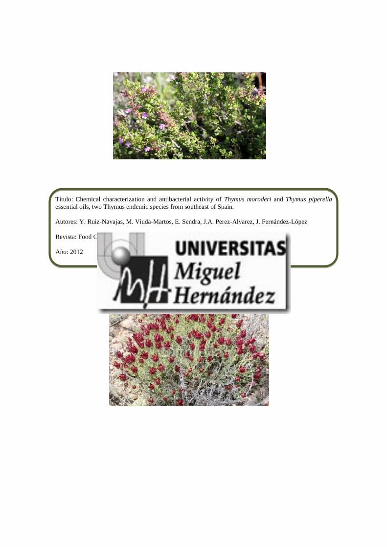
Título: Chemical characterization and antibacterial activity of Thymus moroderi and Thymus piperella
essential oils, two Thymus endemic species from southeast of Spain.
Autores: Y. Ruiz-Navajas, M. Viuda-Martos, E. Sendra, J.A. Perez-Alvarez, J. Fernández-López
Revista: Food Control, 27, 294-299
Año: 2012

at SciVerse ScienceDirect
Food Control 27 (2012) 294e299
Contents lists available
Food Control
journal homepage: www.elsevier .com/locate/ foodcont
Chemical characterization and antibacterial activity of Thymus moroderi andThymus piperella essential oils, two Thymus endemic species from southeastof Spain
Y. Ruiz-Navajas, M. Viuda-Martos, E. Sendra, J.A. Perez-Alvarez, J. Fernández-López*
IPOA Research Group (UMH-1 and REVIV-Generalitat Valenciana), AgroFood Technology Departament, Escuela Politecnica Superior de Orihuela, Univ. Miguel Hernandez, Crta,Beniel km. 3.2, E-03312 Orihuela Alicante, Spain
a r t i c l e i n f o
Article history:Received 12 January 2012Received in revised form29 March 2012Accepted 3 April 2012
Keywords:ThymusChemical compositionGCeMSAntibacterialEssential oils
* Corresponding author. Tel.: þ34 966749784.E-mail address: [email protected] (J. Fernández
0956-7135/$ e see front matter � 2012 Elsevier Ltd.doi:10.1016/j.foodcont.2012.04.005
a b s t r a c t
The aim of this work was to determine the following characteristics of the essentials oils (EOs) obtainedfrom two Thymus endemic species such as Thymus moroderi and Thymus piperella: (i) their chemicalcomposition and (ii) their effect on the growth of several bacteria related to food spoilage, such as Listeriainnocua, Serratia marcenscens, Pseudomonas fragi, Pseudomonas fluorescens, Aeromonas hydrophila, She-wanella putrefaciens, Achromobacter denitrificans, Enterobacter amnigenus, Enterobacter gergoviae, Alcali-genes faecalis and Leuconostoc carnosum. The EOs were chemically analysed and identified by GC and GCeMS, while the agar disc diffusion method was used to determine their antibacterial activities. Theconcentration effect was also determined.
The major constituents of T. moroderi EO were camphor (26.74%), 1,8-cineol (24.99%) and myrcene(5.63%). In the T. piperella EO the main constituents were carvacrol (31.92%), para-cymene (16.18%) and g-terpinene (10.11%). T. piperella EO was much more effective than T. moroderi EO, and had a much greatereffect on the Gram-positive bacteria than on the Gram-negative bacteria.
� 2012 Elsevier Ltd. All rights reserved.
1. Introduction
Thymus L. (Labiatae) consists of about 215 species of herbaceousperennials and sub-shrubs (Nickavar, Mojab, & Dolat-Abadi, 2005).Species of Thymus arewell adapted to the hot and dry climate of theMediterranean region and are widespread in the arid parts of theIberian Peninsula (Horwath, Grayer, Keith-Lucas, & Simmonds,2008). “Thyme”, with a long list of pharmacological and aromaticproperties, is commonly used in the herbalist sector. Thymus is usedas spice in several foods. In addition, it is one of the most widelyused genuses in folk medicine, where it is popular for its stimula-tory action on all organism functions (Viuda-Martos, Ruiz-Navajas,Fernández-López, & Pérez-Álvarez, 2011) and also for the antimi-crobial activity of its essential oil. Growing wild in the Mediterra-nean environment, there are several ecotypes of thyme, whichdiffer in their morphological characteristics (Tedone, D’andrea, &Marzi, 2001) and in the composition of their essential oils(Corticchiato, Tomi, Bernardin, & Casanova, 1998), although all arecharacterized by a strong and penetrating odour and sometimesa very pronounced balsamic and spicy flavour. Two species found in
-López).
All rights reserved.
the Thymus genus are Thymus piperella and Thymus moroderi. Thefirst of these is an endemic eastern Iberian taxon, extending overapproximately 800 km2 of the province of Valencia. T. moroderi,which is closely related to common thyme (Thymus vulgaris L.), isalso endemic to southeast of Spain. The genus Thymus is charac-terized by its high content of essential oils (EOs), which leads to itbeing regarded as an aromatic herb with potential health benefits.
In general terms, EOs are bioactive compounds formed ofa mixture of many substances; they have a complicated chemicalcomposition and are obtained from the secondary metabolism ofplants (Delamare, Moschen-Pistorello, Artico, Atti-Serafini, &Echeverrigaray, 2007). Theyare simple lipidswithout fatty acids andare composed of volatile substances, most of which derived fromthree main classes of compound: terpenoids, phenylpropanoids/benzenoids and fatty acid derivates (Vainstein, Lewinsohn,Pichersky, & Weiss, 2001). Among the numerous properties ofthese EOs are their antioxidant, digestion-stimulating, hypolipi-demic, antiinflammatory, anticarcinogenic and antibacterial activi-ties (Viuda-Martos et al., 2011).
The antimicrobial substances used in the food industry includechemical compounds (added or naturally present in foods) for twomain reasons: (i) to control natural deterioration processes(food preservation) and (ii) to prevent or limit the growth of
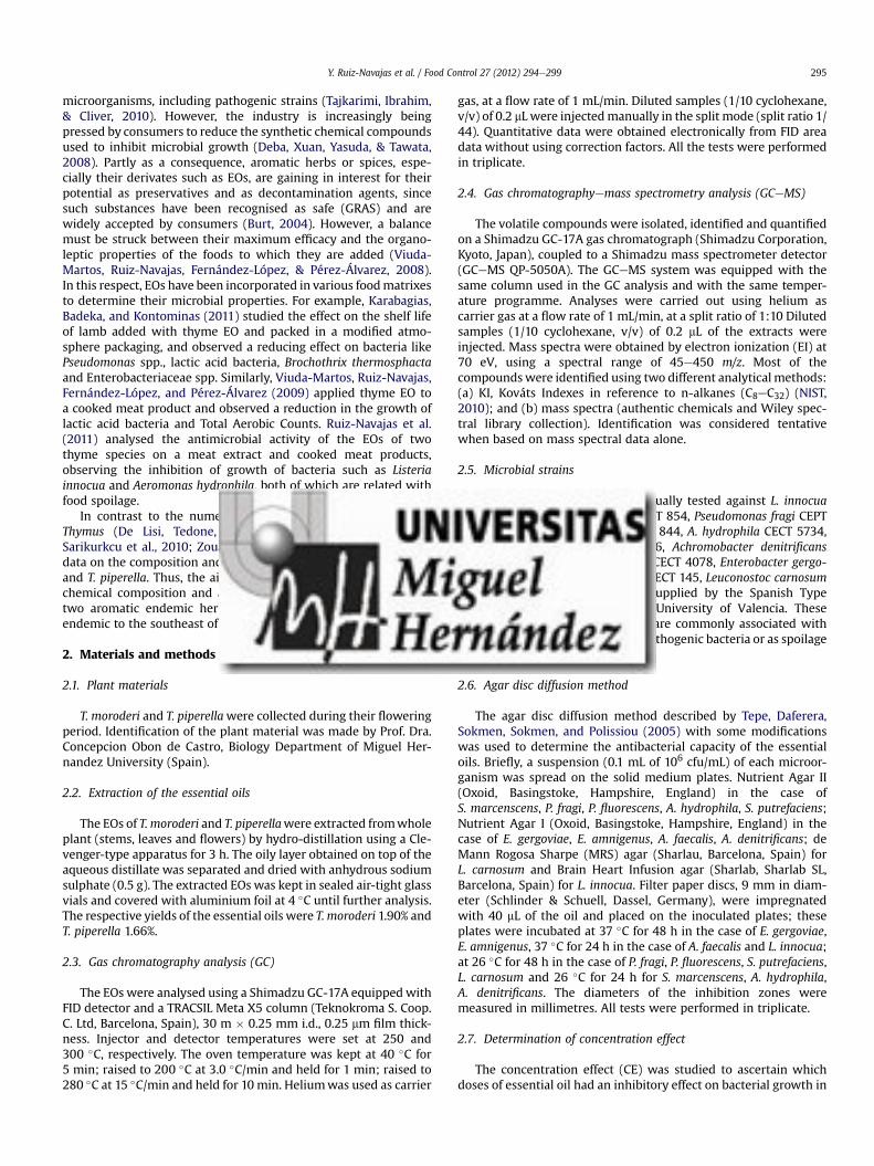
Y. Ruiz-Navajas et al. / Food Control 27 (2012) 294e299 295
microorganisms, including pathogenic strains (Tajkarimi, Ibrahim,& Cliver, 2010). However, the industry is increasingly beingpressed by consumers to reduce the synthetic chemical compoundsused to inhibit microbial growth (Deba, Xuan, Yasuda, & Tawata,2008). Partly as a consequence, aromatic herbs or spices, espe-cially their derivates such as EOs, are gaining in interest for theirpotential as preservatives and as decontamination agents, sincesuch substances have been recognised as safe (GRAS) and arewidely accepted by consumers (Burt, 2004). However, a balancemust be struck between their maximum efficacy and the organo-leptic properties of the foods to which they are added (Viuda-Martos, Ruiz-Navajas, Fernández-López, & Pérez-Álvarez, 2008).In this respect, EOs have been incorporated in various foodmatrixesto determine their microbial properties. For example, Karabagias,Badeka, and Kontominas (2011) studied the effect on the shelf lifeof lamb added with thyme EO and packed in a modified atmo-sphere packaging, and observed a reducing effect on bacteria likePseudomonas spp., lactic acid bacteria, Brochothrix thermosphactaand Enterobacteriaceae spp. Similarly, Viuda-Martos, Ruiz-Navajas,Fernández-López, and Pérez-Álvarez (2009) applied thyme EO toa cooked meat product and observed a reduction in the growth oflactic acid bacteria and Total Aerobic Counts. Ruiz-Navajas et al.(2011) analysed the antimicrobial activity of the EOs of twothyme species on a meat extract and cooked meat products,observing the inhibition of growth of bacteria such as Listeriainnocua and Aeromonas hydrophila, both of which are related withfood spoilage.
In contrast to the numerous studies on the essential oils ofThymus (De Lisi, Tedone, Montesano, Sarli, & Negro, 2011;Sarikurkcu et al., 2010; Zouari et al., 2011) there are only limiteddata on the composition and antimicrobial activities of T. moroderiand T. piperella. Thus, the aim of this study was to determine thechemical composition and antibacterial properties of the EOs oftwo aromatic endemic herbs, T. moroderi and T. piperella, bothendemic to the southeast of Spain.
2. Materials and methods
2.1. Plant materials
T. moroderi and T. piperellawere collected during their floweringperiod. Identification of the plant material was made by Prof. Dra.Concepcion Obon de Castro, Biology Department of Miguel Her-nandez University (Spain).
2.2. Extraction of the essential oils
The EOs of T. moroderi and T. piperellawere extracted fromwholeplant (stems, leaves and flowers) by hydro-distillation using a Cle-venger-type apparatus for 3 h. The oily layer obtained on top of theaqueous distillate was separated and dried with anhydrous sodiumsulphate (0.5 g). The extracted EOs was kept in sealed air-tight glassvials and covered with aluminium foil at 4 �C until further analysis.The respective yields of the essential oils were T. moroderi 1.90% andT. piperella 1.66%.
2.3. Gas chromatography analysis (GC)
The EOs were analysed using a Shimadzu GC-17A equipped withFID detector and a TRACSIL Meta X5 column (Teknokroma S. Coop.C. Ltd, Barcelona, Spain), 30 m � 0.25 mm i.d., 0.25 mm film thick-ness. Injector and detector temperatures were set at 250 and300 �C, respectively. The oven temperature was kept at 40 �C for5 min; raised to 200 �C at 3.0 �C/min and held for 1 min; raised to280 �C at 15 �C/min and held for 10 min. Heliumwas used as carrier
gas, at a flow rate of 1 mL/min. Diluted samples (1/10 cyclohexane,v/v) of 0.2 mL were injectedmanually in the split mode (split ratio 1/44). Quantitative data were obtained electronically from FID areadata without using correction factors. All the tests were performedin triplicate.
2.4. Gas chromatographyemass spectrometry analysis (GCeMS)
The volatile compounds were isolated, identified and quantifiedon a Shimadzu GC-17A gas chromatograph (Shimadzu Corporation,Kyoto, Japan), coupled to a Shimadzu mass spectrometer detector(GCeMS QP-5050A). The GCeMS system was equipped with thesame column used in the GC analysis and with the same temper-ature programme. Analyses were carried out using helium ascarrier gas at a flow rate of 1 mL/min, at a split ratio of 1:10 Dilutedsamples (1/10 cyclohexane, v/v) of 0.2 mL of the extracts wereinjected. Mass spectra were obtained by electron ionization (EI) at70 eV, using a spectral range of 45e450 m/z. Most of thecompoundswere identified using two different analytical methods:(a) KI, Kováts Indexes in reference to n-alkanes (C8eC32) (NIST,2010); and (b) mass spectra (authentic chemicals and Wiley spec-tral library collection). Identification was considered tentativewhen based on mass spectral data alone.
2.5. Microbial strains
The essentials oils were individually tested against L. innocuaCECT 910, Serratia marcenscens CECT 854, Pseudomonas fragi CEPT446, Pseudomonas fluorescens CECT 844, A. hydrophila CECT 5734,Shewanella putrefaciens CECT 5346, Achromobacter denitrificansCECT 449, Enterobacter amnigenus CECT 4078, Enterobacter gergo-viae CECT 587, Alcaligenes faecalis CECT 145, Leuconostoc carnosumCECT 4024. These species were supplied by the Spanish TypeCulture Collection (CECT) of the University of Valencia. Thesebacteria were chosen due to they are commonly associated withrefrigerated foods: as indicator of pathogenic bacteria or as spoilagebacteria.
2.6. Agar disc diffusion method
The agar disc diffusion method described by Tepe, Daferera,Sokmen, Sokmen, and Polissiou (2005) with some modificationswas used to determine the antibacterial capacity of the essentialoils. Briefly, a suspension (0.1 mL of 106 cfu/mL) of each microor-ganism was spread on the solid medium plates. Nutrient Agar II(Oxoid, Basingstoke, Hampshire, England) in the case ofS. marcenscens, P. fragi, P. fluorescens, A. hydrophila, S. putrefaciens;Nutrient Agar I (Oxoid, Basingstoke, Hampshire, England) in thecase of E. gergoviae, E. amnigenus, A. faecalis, A. denitrificans; deMann Rogosa Sharpe (MRS) agar (Sharlau, Barcelona, Spain) forL. carnosum and Brain Heart Infusion agar (Sharlab, Sharlab SL,Barcelona, Spain) for L. innocua. Filter paper discs, 9 mm in diam-eter (Schlinder & Schuell, Dassel, Germany), were impregnatedwith 40 mL of the oil and placed on the inoculated plates; theseplates were incubated at 37 �C for 48 h in the case of E. gergoviae,E. amnigenus, 37 �C for 24 h in the case of A. faecalis and L. innocua;at 26 �C for 48 h in the case of P. fragi, P. fluorescens, S. putrefaciens,L. carnosum and 26 �C for 24 h for S. marcenscens, A. hydrophila,A. denitrificans. The diameters of the inhibition zones weremeasured in millimetres. All tests were performed in triplicate.
2.7. Determination of concentration effect
The concentration effect (CE) was studied to ascertain whichdoses of essential oil had an inhibitory effect on bacterial growth in

Y. Ruiz-Navajas et al. / Food Control 27 (2012) 294e299296
the disc diffusion assay. The culture techniques used were thosedescribed in the previous paragraph (Agar disc diffusion method),but adding 40, 20, 10, 4 and 2 mL of essential oil which meant dosesof 100%, 50%, 25%, 10% and 5% of the initial volume (Viuda-Martos,Fernández-López, & Pérez-Álvarez, 2005). All tests were performedin triplicate.
2.8. Statistical analysis
Conventional statistical methods were used to calculate meansand standard deviations of three simultaneous assays carried outwith the different methods. Statistical analysis (ANOVA) wasapplied to the data to determine differences (P < 0.05). To discoverwhether there were significant differences between the levels ofthemain factor, contrasts betweenmeans weremade using a Tukeytest (Afifi & Azen, 1979). For the antibacterial activity, an ANOVAwas applied, with the following factors: doses (five levels; 40, 20,10, 4 and 2 mL) for each essential oil. The statistical analyses weremade using Statgraphics 5.1 for Windows.
3. Results and discussions
3.1. Chemical composition
The chemical composition of T. moroderi and T. piperella essen-tial oil was investigated using both GC and GCeMS techniques. Thepercentages and the retention indexes of the main oil componentsare listed in Table 1 in the order of their elution on the TRACSILMeta X5 column. In the case of T. moroderi essential oil, 51compounds were identified as representing 92.1% of the oil. Themain components were camphor (26.74%), 1.8-cineol (24.99%),myrcene (5.63%) and a-pinene (4.35%). Forty-eight compoundswere identified for T. piperella, representing 90.5% of the essentialoil. The major component was carvacrol (31.92%) followed by para-cymene (16.18%), g-terpinene (10.11%) and, to a lesser extent, 4-
Table 1Constituents of T. moroderi and T. piperella and their relative percentages of totalchromatogram area, Kováts index and retention time.
Compounds Ida Kováts indexes Essential oils
KI Litb T. moroderi(% area)
T. piperella(% area)
Alpha pinene KI,W 940 939 4.35 e
Camphene KI,W 960 953 8.8 e
Sabinene KI,W 979 976 1.14 e
Myrcene KI,W 989 991 5.63 e
Alpha-terpinene KI,W 1023 1018 e 2.69Para-cymene KI,W,St 1032 1027 e 16.18I-Limonene KI,W,St 1035 1036 1.93 1.541,8-Cineole KI,W,St 1042 1039 24.99 e
Gamma, terpinene KI,W 1064 1062 e 10.11l-Linalool KI,W 1102 1098 1.31 2.84Cis-sabinene hydrate KI,W 1111 1097 e 2.02Camphor KI,W 1163 1143 26.74 e
1-Borneol KI,W 1186 1165 3.62 e
Terpineol-4 KI,W 1192 1189 2.36 7.29Alpha-terpineol KI,W 1205 1207 2.62 1.14Thymol KI,W,St 1294 1290 e 5.19Carvacrol KI,W,St 1307 1299 e 31.92Beta-elemene KI,W 1399 1393 1.06 e
Trans-caryophyllene KI,W 1436 1430 e 6.09Bicyclogermacrene KI,W 1511 1494 1.86 e
Viridiflorol KI,W 1595 1590 2.08 e
Caryophyllene oxide KI,W 1602 1606 e 1.58
a “KI,W” means that identification was based on Kováts indexes and comparisonwith Wiley library. “KI” compounds tentatively identified based on Kováts indexes,“W” compounds tentatively identified (only Wiley library was used foridentification).
b NIST database.
terpineol (7.29%). Marti et al. (2007) reported that the majorconstituents of T. piperella essential oil were para-cymene (31.70%),carvacrol (14.00%) and g-terpinene (11.30%), while Boira andBlanquer (1998) investigated the essential oil composition ofthree subspecies of T. piperella, reporting that there are three che-motypes: chemotype A composed of para-cymene, carvacrol and g-terpinnene, chemotype B composed of para-cymene and thymol,and chemotype C composed of para-cymene and carvacrol. The EOof T. piperella analysed in this study may be include in chemotype C.
This is the first report to analyse the chemical composition ofT. moroderi and no information on its composition has beenreported previously. However, the chemical composition of the EOobtained from plants of genus Thymus has been widely investi-gated. Thus, Bagamboula, Uyttendaele, and Debevere (2003)reported that no major differences were observed betweencommercial thyme essential oil and the essential oil obtained bysteam distillation of finely ground dried thyme leaves, and onlylinalool and g-terpinenewere found in higher concentrations in thecommercial essential oil. In another study the same authors(Bagamboula, Uyttendaele, & Debevere, 2004) investigated the EOof thyme, identifying 28 compounds, which represented 92.6% ofthe total oil, the major constituents being thymol (32.23%), g-ter-pinene (21.19%) and para-cymene (20.27%). Napoli, Curcuruto, andRuberto (2010) reported on the composition of the essential oilsof the thirty samples of thyme collected in Italy. In total, 46components were fully identified, covering more than 96% of thetotal composition. Pinto et al. (2006) analysed the composition ofthe EO of Thymus pulegioides from Portugal. In this case, the oil wascharacterized by high amounts of thymol (26.0%), and carvacrol(21.0%) and its biogenetic precursors, g-terpinene (8.8%) and para-cymene (7.8%).
The compositional data shows that carvacrol is by far the maincompound in almost all samples. It is accepted that the terpenesthymol, para-cymene and carvacrol are the major volatile compo-nents of thyme. Some studies have reported that thyme essential oilpossesses a high level of the phenolic precursors, para-cymene andg-terpinene, probably due to the early flowering time of the plant(Saez, 1998). In other published results, Lee, Umano, Shibamoto,and Lee (2005) identified 43 monoterpenes, 16 sesquiterpenes, 14aromatic compounds, 7 alcohols, 3 aldehydes, 4 ketones and esters,and 3 acids in a thyme extract. Phenolic compounds were thearoma principles in this chemotype of thyme. Quantitatively, themost important compounds were thymol (72%) and isothymol(carvacrol) (5.7%), which constituted almost three-quarters of thetotal volatiles quantified. This great variability and diversityobserved in the chemical composition of the essential oils ofThymus species and subspecies can be attributed to climatic andsoil variations, the stage of the vegetative cycle, seasonal variations,etc. (Cosentino et al., 1999). Tzakou, Verykokidou, Rousis, andChinou (1998) reported that in some cases two different varietiesmay provide the same essential oil yield and quality, even thoughthe plants are morphologically different.
3.2. Antibacterial activity
The antibacterial activity of the essential oils of T. moroderi andT. piperella was assayed in vitro following the diffusion in agar discmethod using eleven bacteria associated with food spoilage. Table 2shows the microbial growth inhibition achieved by each of the EOsassayed. As can be seen, T. piperella essential oil had an inhibitoryeffect on 5 of the 11 bacteria assayed, with inhibition halos thatranged from 16.00 mm for A. denitrificans to 45.00 mm forA. hydrophila, and no antibacterial activity against P. fragi,P. fluorescens, S. putrefaciens, E. gergoviae, Enterobacter anmigenusand L. carnosum.

Table
2Antimicrobial
activity
ofT.morod
erian
dT.Pipe
rella
essential
oils
usingdiscdiffusion
method
.
Essential
oils
Diameter
(mea
nan
dSD
)of
inhibitionzo
ne(m
m)includingdiscdiameter
of9mm
S.marcenscens
P.frag
iP.
fluo
rescen
sA.h
ydroph
ilaS.
putrefaciens
E.gergov
iae
E.am
nigenu
sA.fae
calis
A.d
enitrificans
L.carnosum
L.inno
cua
T.morod
eri
23.90�
0.10
aN.A.
N.A.
20.50�
0.70
aN.A.
N.A.
N.A.
24.00�
1.40
aN.A.
N.A.
15.50�
0.70
a
T.pipe
rella
24.50�
0.70
aN.A.
N.A.
45.50�
3.50
bN.A.
N.A.
N.A.
45.50�
0.70
b16
.00�
0.00
N.A.
27.30�
1.20
b
Forthesameba
cteria,v
alues
follo
wed
bydifferentletterswithin
thesameco
lumnaresign
ificantlydifferent(P
<0.05
)acco
rdingto
Turkey
’smultiple
range
test.
N.A.,non
-active.
Y. Ruiz-Navajas et al. / Food Control 27 (2012) 294e299 297
The T. moroderi essential oil had an inhibitory effect on 4 of thebacteria, with inhibition halos from 15 mm (L. innocua) to24.00 mm (A. faecalis). This EO showed no activity against P. fragi,P. fluorescens, S. putrefaciens, E. gergoviae, E. anmigenus,A. denitrificans and L. carnosum. This is the first study to analyse theantibacterial activity of the EOs from T. moroderi and T. piperella.However, as in the case of the chemical composition, the antibac-terial activity of several other thyme varieties has been studied.
De Martino et al. (2009) investigated the antibacterial activity ofessential oils of the aerial parts of Thymus longicaulis and T. pule-gioides collected in Italy. At a concentration of 10 mg/mL both EOsshowed substantial antibacterial activity against Gram-positive andGram-negative bacteria such as Staphylococcus aureus, Strepto-coccus faecalis, B. subtilis, Bacillus cereus, Proteus mirabilis, Enter-obacter coli, Salmonella typhi Ty2 and Pseudomonas aeruginosa, withinhibition zones that ranged between 9 and 18mm for T. longicaulisand between 11 and 20 mm for T. pulegioides. Cetin, Cakmakci, andCakmakci (2011) investigated the antibacterial activity of Thymussipyleus subsp. sipyleus var. rosulans EO from Turkey. The EOshowed the highest inhibitory effects on Pseudomonas pseu-doalkaligenes (59 mm) and S. aureus (56 mm), followed byB. subtilis, P. aeruginosa, S. pyogenes, and Pseudomonas vulgaris(nearly to the same extent) and a lower inhibitory effect on Enter-obacter cloacae. Viuda-Martos et al. (2008) analysed the antimi-crobial activity of the essential oil from T. vulgaris cultivated inSpain at a volume of 40 mL, and obtained inhibition zones of 21.60,28.57, 53.85, 21.61, 24.05 and 23.64 mm for Staphylococcus xylosus,Staphylococcus carnosus, Lactobacillus sakei, Lactobacillus curvatus,E. gergoviae and Enterobacter Amnigenus, respectively.
The antimicrobial activity of the EOs from aromatic herbs andspices results from a small number of terpenes and phenoliccompounds (Dorman & Deans, 2000). As mentioned above, theprincipal components of T. piperella EO are carvacrol and para-cymene, and those of T. moroderi EO are 1,8-cineole and camphor.Bagamboula et al. (2003) mentioned that carvacrol at concentra-tions of 0.5% and 1% shows antibacterial activity against Shigellasonnei and Shigella flexneri. Similarly, Rattachaikunsopo andPhumkhachrom (2010) suggested that the antimicrobial effect ofT. moroderi EO may be partly associated with the presence ofcamphor and 1,8-cineole since both compounds have been docu-mented as having antibacterial activity (Mourey & Canillac, 2002;Viljoen et al., 2003).
In the present study, it was observed that T. moroderi EO con-tained neither carvacrol nor para-cymene, the major componentsbeing 1,8-cineole and camphor. It has been shown that carvacrol isa stronger inhibitor than camphor and 1,8-cineole, which mayexplain the lower antimicrobial activity of T. moroderi EO comparedwith T. piperella EO.
Considering the large number of different groups of chemicalcompounds present in EOs, it is most likely that their antibacterialactivity is not attributable to one specific mechanism but that thereare several targets in the cell (Burt, 2004). Different modes of actionhave been suggested: for example, EOs may act by causing struc-tural and functional damage to the bacterial cell membrane (Goniet al., 2009). The leakage of ions and other cell contents may alsooccur (Carson, Hammer, & Riley, 2002). Davidson (2001) reportedthat the exact causeeeffect relation for the mode of action ofphenolic compounds, such as thymol, eugenol and carvacrol, hasnot been determined, but that they may inactivate essentialenzymes, react with the cell membrane or disturb genetic material.
The apparent antimicrobial efficacy of plant-origin antimicro-bials depends on factors such as the method of extracting EOs fromplant material, the volume of inoculum, growth phase, the culturemedium used, and intrinsic or extrinsic properties of the food suchas pH, fat, protein, water content, antioxidants, preservatives,

Table
3Antimicrobial
activity
ofT.morod
eria
ndT.Pipe
rella
essential
oils
usingdiscdiffusion
method
.
Essential
oils
Doses
(mL)
Diameter
(mea
nan
dSD
)of
inhibitionzo
ne(m
m)includingdiscdiameter
of9mm
S.marcenscens
P.frag
iP.
fluo
rescen
sA.h
ydroph
ilaS.
putrefaciens
E.gergov
iae
E.am
nigenu
sA.fae
calis
A.d
enitrificans
L.carnosum
L.inno
cua
T.morod
eri
2N.A.
N.A.
N.A.
N.A.
N.A.
N.A.
N.A.
N.A.
N.A.
N.A.
N.A.
4N.A.
N.A.
N.A.
N.A.
N.A.
N.A.
N.A.
N.A.
N.A.
N.A.
N.A.
1011
.30�
0.10
aN.A.
N.A.
12.50�
0.70
aN.A.
N.A.
N.A.
11.50�
0.70
aN.A.
N.A.
10.90�
0.10
a
2014
.90�
0.10
bN.A.
N.A.
16.00�
1.40
bN.A.
N.A.
N.A.
13.50�
0.00
bN.A.
N.A.
12.20�
0.10
b
4023
.90�
0.10
cN.A.
N.A.
20.50�
0.70
cN.A.
N.A.
N.A.
24.00�
1.40
cN.A.
N.A.
15.50�
0.70
c
T.Pipe
rella
213
.80�
0.40
aN.A.
N.A.
32.00�
0.00
aN.A.
N.A.
N.A.
21.50�
0.70
a11
.00�
0.00
aN.A.
15.80�
0.30
a
415
.70�
0.60
bN.A.
N.A.
35.50�
2.10
bN.A.
N.A.
N.A.
27.50�
2.10
b12
.00�
0.00
bN.A.
17.80�
0.40
b
1019
.60�
0.60
cN.A.
N.A.
38.50�
2.10
bN.A.
N.A.
N.A.
30.05�
2.10
c13
.00�
0.00
cN.A.
21.50�
0.70
c
2022
.80�
0.70
dN.A.
N.A.
43.00�
2.80
cN.A.
N.A.
N.A.
35.00�
0.70
d14
.50�
0.70
dN.A.
23.30�
1.10
d
4024
.50�
0.70
eN.A.
N.A
45.50�
3.50
cN.A
N.A.
N.A.
45.50�
0.70
e16
.00�
0.00
eN.A.
27.30�
1.20
e
Forthesameessential
oil,va
lues
follo
wed
bydifferentletterswithin
thesameco
lumnaresign
ificantlydifferent(P
<0.05
)acco
rdingto
Tuke
y’smultiple
range
test.
N.A.,non
-active.
Y. Ruiz-Navajas et al. / Food Control 27 (2012) 294e299298
incubation time/temperature, packaging procedure, and the phys-ical structure of food (Brandi, Amagliani, Schiavano, De Santi, &Sisti, 2006; Burt, 2004; Lis-Balchin, Steyrl, & Krenn, 2003; Lopez-Malo, Palou, & Alzamora, 2005).
The type of bacterium also has an influence on the effectivenessof EOs. Generally, Gram-negative bacteria are less sensitive to theantimicrobials (Gilles, Zhao, An, & Agboola, 2010). The difference insusceptibility could be due to the lipopolysaccharide outermembrane of this group, which restricts diffusion of hydrophobiccompounds. However, this does not mean that Gram-positivebacteria are always more susceptible (Burt, 2004). Gram-negativebacteria are usually more resistant to the plant-origin antimicro-bials (which may even have no effect), than Gram-positive bacteria(Rameshkumar, George, & Shiburaj, 2007; Stefanello et al., 2008).On the other hand, Sokmen et al. (2004) affirmed that the essentialoils of spices showno selectivity as regards the cell walls of bacteria.
3.3. Concentration effect
Table 3 shows the concentration effect (CE) of the EOs fromT. moroderi and T. piperella on the eleven bacterial strains studied. Inthe case of T.moroderiEO, no antibacterial effectwas observed at anyconcentration on S. putrefaciens, P. fragi, P. fluorescens, E. gergoviae,E. amnigenus, L. carnosum and A. denitrificans. Neither 4 nor 2 mL hadany effect on discs impregnated with S. marcenscens but concen-trations of 40, 20 and 10 mL did have an effect, with statisticallysignificant differences between them (P < 0.05). The same behav-iour was seen with A. hydrophila, A. faecalis and L. innocua.
As regards T. piperella EO, all the concentrations assayedproduced inhibition halos with S. marcenscens, A. denitrificans,A. faecalis, A. hydrophila and L. innocua. There were statisticallysignificant differences for the concentrations and strains (P < 0.05)except for A. hydrophila, when no differences (P < 0.05) existedbetween the discs impregnated with 10 and 4 mL or between 40 and20 mL, as occurred with T. moroderi EO. The EO from T. piperellawasnot active at any concentration against S. putrefaciens, P. fragi,P. fluorescens, E. gergoviae, E. amnigenus and L. innocua. Of note wasthe fact that T. piperella EO, when it had an effect, produced greaterinhibition halos than T. moroderi EO.
The antibacterial activity of both EOs was dose-dependent.However, it should be borne in mind that when these substancesare used as antibacterials in food their efficiency diminishes whenthey are added to complex matrixes and their organoleptic impactcould be noticeable. There are also questions of safety and potentialtoxicity that should not be forgotten.
4. Conclusions
The essential oils from T. moroderi and T. piperella are a source ofimportant bioactive compounds with antibacterial propertieswhich merit further study for possible application in the foodindustry, where they may be considered as natural preservatives toreplace the synthetic preservatives of which consumers areincreasingly distrustful. However, the use of these essential oilsmay be limited by changes to the organoleptic properties of foodsthat contain them and so the most suitable essential oil must bechosen for each type of food. In-depth studies are required, sincethe absence of norms to test and guarantee the quality of essentialoils is a major problem faced by the food industry when they areused as a basic ingredient.
Acknowledgements
We are grateful to the Caja Murcia for the research scholarshipawarded to one of the authors (Yolanda Ruiz Navajas).

Y. Ruiz-Navajas et al. / Food Control 27 (2012) 294e299 299
References
Afifi, A. A., & Azen, S. P. (1979). Statistical analysis. A computer assisted approach (2nded.). London: Academic Press Inc.
Bagamboula, C. F., Uyttendaele, M., & Debevere, J. (2003). Antimicrobial effect ofspices and herbs towards Shigella sonnei and S. flexneri. Journal of Food Protec-tion, 66(4), 668e673.
Bagamboula, C. F., Uyttendaele, M., & Debevere, J. (2004). Inhibitory effect of thymeand basil essential oils, carvacrol, thymol, estragol, linalool and p-cymenetowards Shigella sonnei and S. flexneri. Food Microbiology, 21, 33e42.
Boira, H., & Blanquer, A. (1998). Environmental factors affecting chemical variabilityof essential oils in Thymus piperella L. Biochemical Systematics and Ecology, 26,811e822.
Brandi, G., Amagliani, G., Schiavano, G. F., De Santi, M., & Sisti, M. (2006). Activity ofBrassica oleracea leaf juice on food borne pathogenic bacteria. Journal of FoodProtection, 69(9), 2274e2279.
Burt, S. (2004). Essential oils: their antibacterial properties and potentialapplications in foods e a review. International Journal of Food Microbiology,94(3), 223e253.
Carson, C. F., Hammer, K. A., & Riley, T. V. (2002). Mechanism of action of Melaleucaalternifolia (tea tree) oil on Staphylococcus aureus determined by time-kill, lysis,leakage and salt tolerance assays and electron microscopy. Antimicrobial Agentsand Chemotherapy, 46, 1914e1920.
Cetin, B., Cakmakci, S., & Cakmakci, R. (2011). The investigation of antimicrobialactivity of thyme and oregano essential oils. Turkish Journal of Agriculture andForestry, 35, 145e154.
Corticchiato, M., Tomi, F., Bernardin, A. F., & Casanova, J. (1998). Composition andinfraspecific variability of essential oil from Thymus herba barona Lois.Biochemical Systematics and Ecology, 26, 915e932.
Cosentino, S., Tuberoso, C. I. G., Pisano, B., Satta, M., Arzedi, E., & Palmas, F. (1999).In-vitro antimicrobial activity and chemical composition of Sardinian Thymusessential oils. Letter in Applied Microbiology, 29, 130e135.
Davidson, P. M. (2001). Chemical preservatives and naturally antimicrobialcompounds. In M. P. Beuchat, & L. R. Montville (Eds.), Food microbiology (pp.593e628). Washington DC: ASM Press.
De Lisi, A., Tedone, L., Montesano, V., Sarli, G., & Negro, D. (2011). Chemical char-acterisation of Thymus populations belonging from Southern Italy. FoodChemistry, 125(4), 1284e1286.
De Martino, L., Bruno, M., Formisano, C., De Feo, V., Napolitano, F., Rosselli, S., et al.(2009). Chemical composition and antimicrobial activityof the essential oils fromtwo species of Thymus growing wild in southern Italy.Molecules, 14, 4614e4624.
Deba, F., Xuan, T. D., Yasuda, M., & Tawata, S. (2008). Chemical composition andantioxidant, antibacterial and antifungal activities of the essential oils fromBidens pilosa Linn. var. Radiata. Food Control, 19, 346e352.
Delamare, A. P. L., Moschen-Pistorello, I. T., Artico, L., Atti-Serafini, L., &Echeverrigaray, S. (2007). Antibacterial activity of the essential oils of Salviaofficinalis L. and Salvia triloba L. cultivated in South Brazil. Food Chemistry,100(2), 603e608.
Dorman, H. J. D., & Deans, S. G. (2000). Antimicrobial agents from plants: antibac-terial activity of plant volatile oils. Journal of Ethnopharmacology, 70, 343e349.
Gilles, M., Zhao, J., An, M., & Agboola, S. (2010). Chemical composition and anti-microbial properties of essential oils of three Australian Eucalyptus species.Food Chemistry, 119(2), 731e737.
Goni, P., Lopez, P., Sanchez, C., Gomez-Lus, R., Becerril, R., & Nerin, C. (2009).Antimicrobial activity in the vapour phase of a combination of cinnamon andclove essential oils. Food Chemistry, 116, 982e989.
Horwath, A. B., Grayer, R. J., Keith-Lucas, D. M., & Simmonds, M. S. J. (2008).Chemical characterisation of wild populations of Thymus from different climaticregions in southeast Spain. Biochemical Systematics and Ecology, 36(2), 117e133.
Karabagias, I., Badeka, A., & Kontominas, M. G. (2011). Shelf life extension of lambmeat using thyme or oregano essential oils and modified atmospherepackaging. Food Chemistry and Technology, 88(1), 109e116.
Lee, S. J., Umano, K., Shibamoto, T., & Lee, K. G. (2005). Identification of volatilecomponents in basil (Ocimum basilicum L.) and thyme leaves (Thymus vulgarisL.) and their antioxidant properties. Food Chemistry, 91, 131e137.
Lis-Balchin, M., Steyrl, H., & Krenn, E. (2003). The comparative effect of novelPelargonium essential oils and their corresponding hydrosols as antimicrobialagents in a model food system. Phytotherapy Research, 17, 60e65.
Lopez-Malo, A., Palou, E., & Alzamora, S. M. (2005). Naturally occurring compoundsplant sources. In P. M. Davidson, J. N. Sofos, & A. L. Branen (Eds.), Antimicrobialsin food (pp. 429e446). Boca Raton: CRC Press.
Martí, D., Pérez-Gracia, M., Blanquer, A., Villagrasa, V., Sanahuja, M. A., & Moreno, L.(2007). Thymus piperella (L.) essential oil: an alternative in the treatment ofdiarrhoea. Flavour and Fragrance Journal, 22, 201e205.
Mourey, A., & Canillac, N. (2002). Anti-Listeria monocytogenes activity of essentialoils components of conifers. Food Control, 13, 289e292.
Napoli, E. M., Curcuruto, G., & Ruberto, G. (2010). Screening of the essential oilcomposition of wild Sicilian thyme. Biochemical Systematics and Ecology, 38(4),816e822.
National Institute of Standards and Technology (Nist): http://webbook.nist.gov/chemistry/name-ser.html. Accessed 10. 2011.
Nickavar, B., Mojab, F., & Dolat-Abadi, R. (2005). Analysis of the essential oils of twoThymus species from Iran. Food Chemistry, 90, 609e611.
Pinto, E., Pina-Vaz, C., Salgueiro, L., Gonçalves, M. J., Costa-de-Oliveira, S.,Cavaleiro, C., et al. (2006). Antifungal activity of the essential oil of Thymuspulegioides on Candida, Aspergillus and dermatophyte species. Journal of MedicalMicrobiology, 55, 1367e1373.
Rameshkumar, K. B., George, V., & Shiburaj, S. (2007). Chemical constituents andantibacterial activity of the leaf oil of Cinnamomum chemungianum Mohan etHenry. Journal of Essential Oil Research, 19, 98e100.
Rattanachaikunsopon, P., & Phumkhachorn, P. (2010). Assessment of factors influ-encing antimicrobial activity of carvacrol and cymene against Vibrio cholerae infood. Journal of Bioscience and Bioengineering, 110(5), 614e619.
Ruiz-Navajas, Y., Viuda-Martos, M., Sánchez-Zapata, E., Sendra, E., Sayas-Barbera,E., Fernandez-Lopez, J., et al. (2011). Antibacterial activity of essential oils oftwo Thymus species in meat homogenates. In: Proceeding of the American MeatScience Association’s reciprocal meat conference. Manhattan, Kansas (USA).p. 89.
Saez, F. J. (1998). Variability in essential oils from populations of Thymus hyemalisLange in southeastern Spain. Journal of Herbs, Spices & Medicinal Plants, 5,65e76.
Sarikurkcu, C., Ozer, M. S., Eskici, M., Tepe, B., Can, S., & Mete, E. (2010). Essential oilcomposition and antioxidant activity of Thymus longicaulis C. Presl subsp.longicaulis var. longicaulis. Food and Chemical Toxicology, 48(7), 1801e1805.
Sokmen, A., Gulluce, M., Akpulat, H. A., Daferera, D., Tepe, B., Polissiou, M., et al.(2004). The in vitro antimicrobial and antioxidant activities of the essential oilsand methanol extracts of endemic Thymus spathulifolius. Food Control, 15,627e634.
Stefanello, M. E. A., Cervi, A. C., Ito, I. Y., Salvador, M. J., Wisniewski, A., Jr., &Simionatto, E. L. (2008). Chemical composition and antimicrobial activity ofessential oils of Eugenia chlorophylla (Myrtaceae). Journal of Essential OilResearch, 20(1), 75e78.
Tajkarimi, M. M., Ibrahim, S. A., & Cliver, D. O. (2010). Antimicrobial herb and spicecompounds in food. Food Control, 21, 1199e1218.
Tedone, L., D’andrea, L., & Marzi, V. (2001). Caratterizzazione biomorfologica evalutazione agronomica di specie e popolazioni di timo (Thymus spp.) nell’areamediterranea. In: Atti del VI Convegno delle Biodiversità, Valenzano, 6 settembre.pp. 1134e1142.
Tepe, B., Daferera, D., Sokmen, A., Sokmen, M., & Polissiou, M. (2005). Antimicrobialand antioxidant activities of the essential oils and various extracts of Salviatomentosa Miller (Lamiaceae). Food Chemistry, 90, 333e340.
Tzakou, O., Verykokidou, E., Rousis, V., & Chinou, I. (1998). Chemical compositionand antibacterial properties of Thymus longicaulis subsp. chaoubardii oils: threechemotypes in the same population. Journal of Essential Oil Research, 10, 97e99.
Vainstein, A., Lewinsohn, E., Pichersky, E., & Weiss, D. (2001). Floral fragrance: newinroads into an old commodity. Plant Physiology, 127, 1383e1389.
Viljoen, A., Vuuren, S. V., Ernst, E., Klepser, M., Demirci, B., Baser, H., et al. (2003).Osmitopsis asteriscoides (Asteraceae)-the antimicrobial and essential oilcomposition of a Cape-Dutch remedy. Journal of Ethnopharmacology, 88,137e143.
Viuda-Martos, M., Fernández-López, J., & Pérez-Álvarez, J. A. (2005). Método deconcentración mínima de inhibición. Technical report. Rehovot, Israel: ProjectsRecoveg and Compost Management. 19e21 February.
Viuda-Martos, M., Ruiz-Navajas, Y., Fernández-López, J., & Pérez-Álvarez, J. A.(2008). Antibacterial activity of different essential oils obtained from spiceswidely used in Mediterranean diet. International Journal of Food Science andTechnology, 43, 526e531.
Viuda-Martos, M., Ruiz-Navajas, Y., Fernández-López, J., & Pérez-Álvarez, J. A.(2009). Effect of adding citrus waste water, thyme and oregano essential oil onthe chemical, physical and sensory characteristics of a bologna sausage.Innovative Food Science and Emerging Technologies, 10, 655e660.
Viuda-Martos, M., Ruiz-Navajas, Y., Fernández-López, J., & Pérez-Álvarez, J. A. (2011).Spices as functional foods. Critical Review in Food Science and Nutrition, 51(1),13e28.
Zouari, N., Fakhfakh, N., Zouari, S., Bougatef, A., Karray, A., Neffati, M., et al. (2011).Chemical composition, angiotensin I-converting enzyme inhibitory, antioxidantand antimicrobial activities of essential oil of Tunisian Thymus algeriensis Boiss.et Reut. (Lamiaceae). Food and Bioproducts Processing, 89(4), 257e265.
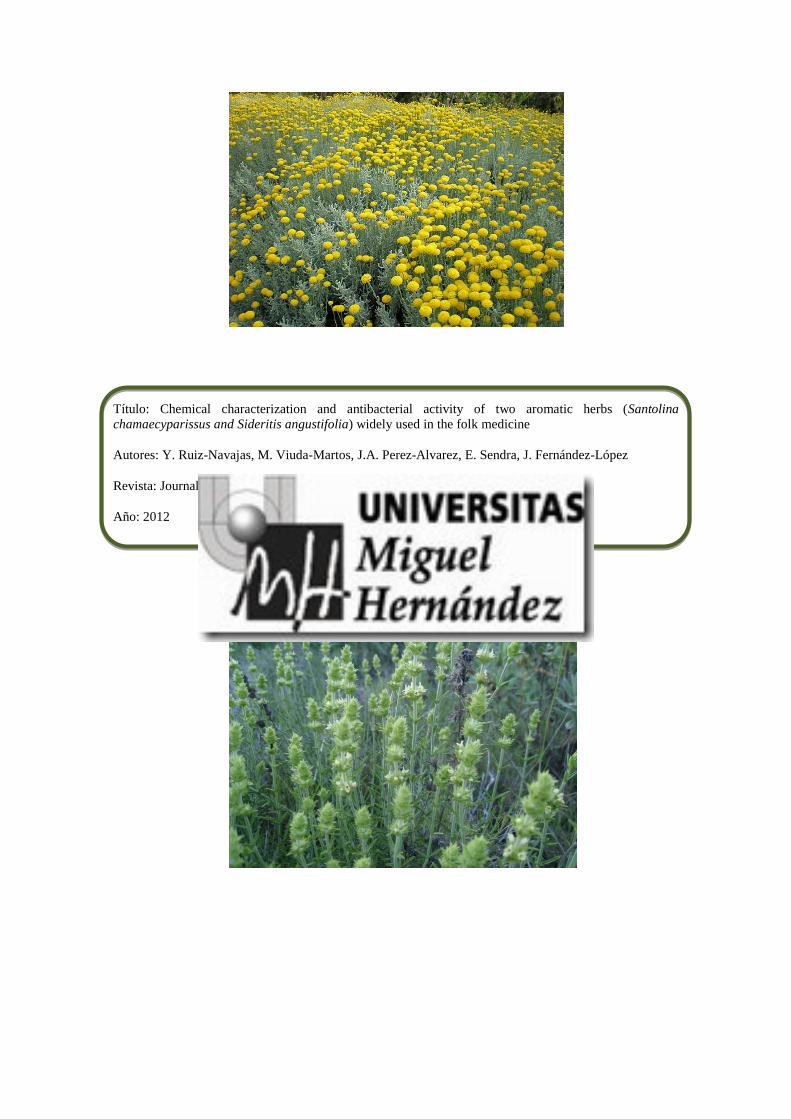
Título: Chemical characterization and antibacterial activity of two aromatic herbs (Santolina
chamaecyparissus and Sideritis angustifolia) widely used in the folk medicine
Autores: Y. Ruiz-Navajas, M. Viuda-Martos, J.A. Perez-Alvarez, E. Sendra, J. Fernández-López
Revista: Journal of Food Safety, 32, 426-434
Año: 2012

CHEMICAL CHARACTERIZATION AND ANTIBACTERIALACTIVITY OF TWO AROMATIC HERBS (SANTOLINACHAMAECYPARISSUS AND SIDERITIS ANGUSTIFOLIA) WIDELYUSED IN THE FOLK MEDICINEYOLANDA RUIZ-NAVAJAS, MANUEL VIUDA-MARTOS, JOSE ANGEL PEREZ-ALVAREZ, ESTHER SENDRA andJUANA FERNÁNDEZ-LÓPEZ1
IPOA Research Group (UMH-1 and REVIV-Generalitat Valenciana), AgroFood Technology Department, Escuela Politecnica Superior de Orihuela,Miguel Hernandez University, Crta, Beniel km. 3, 2, E-03312 Orihuela, Alicante, Spain
1Corresponding author. TEL: +34 966749784;FAX: +34 966749677; EMAIL:[email protected]
Received for Publication June 7, 2012Accepted for Publication August 28, 2012
doi: 10.1111/jfs.12001
ABSTRACT
The aim of this work was characterize the essentials oils (EOs) obtained from San-tolina chamaecyparissus and Sideritis angustifolia determining (1) its chemicalcomposition and (2) its effectiveness on the growth of some bacteria related tofood spoilage, such as Listeria innocua, Serratia marcescens, Pseudomonas fragi,Pseudomonas fluorescens, Aeromonas hydrophila, Shewanella putrefaciens, Alcali-genes faecalis, Achromobacter denitrificans, Enterobacter amnigenus, Enterobactergergoviae and Leuconostoc carnosum. The EOs were chemically analyzed and iden-tified by gas chromatography and gas chromatography/mass spectrometry, whilethe agar disc diffusion method was used to determine their antibacterial activities.The concentration effect was also determined. In S. chamaecyparissus, the majorconstituents were artemisia ketone (27.19%), dihydroaromadendrene (18.21%)and b-phellandrene (7.49%), while in S. angustifolia EO, the major constituent isa-pinene (12.71%). Another important compound was b-phellandrene (11.97%).The agar disc diffusion method indicated that S. chamaecyparissus EO showed thehighest antibacterial activity against the L. innocua and A. hydrophila, with inhibi-tion zones of 12.70 and 16.50 mm, respectively. S. angustifolia EO showed thehighest antibacterial activity against A. faecalis, S. marcescens and A. denitrificans.S. chamaecyparissus and S. angustifolia EOs can be used as antibacterial agents.
PRACTICAL APPLICATIONS
The research on plant-based natural bioactive compounds to control the food-borne and spoilage pathogens has attained special interest in the recent years. Theuse of essential oils of Santolina chamaecyparissus and Sideritis angustifolia asantibacterial agents will be suitable for applications on the food industry asnatural preservatives or flavoring to control foodborne pathogens. Anotherimportant reason for their suitability is their natural origin, which consumersfind comforting.
INTRODUCTION
Many of the valuable organoleptic and sensory properties offood products diminish with time. This may be attributedto a large number of factors, including the action of oxygen,light and temperature that provoke the oxidation process.
However, the most limiting factor of food shelf life is thegrowth of microorganisms, whether they are molds, bacte-ria or yeasts (Viuda-Martos et al. 2008). In addition, foodprocessors, food safety researchers and regulatory agencieshave been increasingly concerned with the growing numberof foodborne illness outbreaks caused by some pathogens
bs_bs_banner
Journal of Food Safety ISSN 1745-4565
426 Journal of Food Safety 32 (2012) 426–434 © 2012 Wiley Periodicals, Inc.

and/or their enterotoxins (Oroojalian et al. 2010). Atpresent, therefore, a wide variety of chemical preservativesare used throughout the food industry to prevent thegrowth of food-spoiling bacteria (Davidson 2001).
During the recent decades and mostly as a result of theirdiversity, versatility and safety in comparison with the syn-thetic materials, natural products of plant, animal andmicroorganism origins have attained special interest. Theexploration of naturally occurring antimicrobial for foodpreservation receives increasing attention owing to the con-sumer awareness of natural food products and a growingconcern of microbial resistance toward conventional preser-vatives (Sharififar et al. 2007).
Many spices and aromatic plants that contain essentialoils (EOs) are within the major plant categories in theMediterranean region. These plants have the ability to syn-thesize, accumulate and emit volatile compounds that mayact as aroma and flavor molecules (Hassiotis and Lazari2010). These mixtures of volatile compounds (mainlymono- and sesquiterpenoids, benzoids, phenylpropanoids,etc.) exert different biological actions on humans, animalsand other plants. The antimicrobial activity of EOs was rec-ognized long ago; these components of spices and aromaticherbs are gaining interest for their potential as preservativesand as decontamination agents, since such substances havebeen generally recognized as safe (GRAS) and are widelyaccepted by consumers (Burt 2004). Thus, the ability ofplant EOs to protect foods against pathogenic and spoilagemicroorganisms has been reported by several researchers(Viuda-Martos et al. 2009, 2011; Ahmadi et al. 2010; Tenoreet al. 2011). However, in order to achieve effective antimi-crobial activity in direct food applications, high concentra-tions of EOs are generally needed, which might impactinappropriate flavors and odors of the product (Seydim andSarikus 2006). The specific advantage of EOs appears to bein synergistic effects of their compounds, as evidenced ingreater activity when applied as natural EOs compared withreported effects of the individual active substances(Zivanovic et al. 2005).
The genus Santolina, belonging to the family Asteraceae,is represented by more than 10 species widely distributed inthe Mediterranean area (Derbesy et al. 1989). Among Santo-lina species, Santolina viridis W. (south of France and northof Spain), Santolina pectinata Lag. (Iberian Peninsula) andSantolina chamaecyparissus L. (which grows wild around theMediterranean basin) are the most widespread. S. chamae-cyparissus is a hardy aromatic, dwarf evergreen shrub nativeto the West and Central Mediterranean area (growing wild,e.g., in Spain, Italy, Tunisia and Morocco, and being natural-ized in parts of Britain). It is often grown in gardens for itsattractive wooly silver-gay leaves born on woody stems andfor its yellow flowers. S. chamaecyparissus is widely used inMediterranean folk medicine. The flowers are used for their
analgesic, anti-inflamatory, antiseptic, antispasmodic, bacte-ricidal, fungicidal, digestive and vulnerary properties, andare used in phytotherapy for different kinds of dermatitis(El-Sahhar et al. 2011).
The genus Sideritis, belonging to the family Lamiaceae,subfamily Lamioideae, comprises at least 150 species. Plantsof this genus, mainly Sideritis angustifolia, have been widelyused in folk medicine as anti-inflammatory, anti-ulcer, cyto-static, antimicrobial, vulnerary, astringent, flu vaccine andstimulant circulatory agents (Palomino et al. 1996). Further-more, EOs from Sideritis species have been used for manytherapeutic purposes, such as lung disinfectants, diuretics,stomachics and neurorelaxings (Senatore 2000).
To our best knowledge, no comprehensive and compara-tive work has been carried out on the chemical compositionas well as antimicrobial activity with respect to S. chamaecy-parissus and S. angustifolia EOs. Therefore, the aims of thiswork were to (1) determine the chemical composition and(2) the effectiveness of the essentials oils obtained fromS. chamaecyparissus and S. angustifolia on the growth ofsome bacteria related to food spoilage to determine if theseEOs could be used as natural food preservatives.
MATERIALS AND METHODS
Plant Materials
S. chamaecyparissus and S. angustifolia were collectedduring their flowering period. Identification of the plantmaterial was made by Prof. Dr. Concepcion Obon deCastro, Applied Biology Department of Miguel HernandezUniversity (Spain).
Extraction of the EOs
The EOs of S. chamaecyparissus and S. angustifolia wereextracted from whole plant (stems, leaves and flowers) byhydro-distillation using a Clevenger-type apparatus for 3 h.The oily layer obtained on top of the aqueous distillate wasseparated and dried with anhydrous sodium sulfate (0.5 g).The extracted EOs were kept in sealed air-tight glass vialsand covered with aluminum foil at 4C until further analysis.The respective yields of the EOs were 1.00 � 0.05%(S. chamaecyparissus) and 0.90 � 0.08% (S. angustifolia).
Gas Chromatography (GC) Analysis
The EOs were analyzed using a Shimadzu GC-17A (Shi-madzu Corporation, Kyoto, Japan) equipped with flameionization detector (FID) and a TRACSIL Meta X5 column(Teknokroma S. Coop. C. Ltd., Barcelona, Spain), 60 m ¥0.25 mm i.d., 0.25-mm film thickness. Injector and detectortemperatures were set at 250 and 300C, respectively. The
Y. RUIZ-NAVAJAS ET AL. ANTIBACTERIAL ACTIVITY OF AROMATIC HERBS
427Journal of Food Safety 32 (2012) 426–434 © 2012 Wiley Periodicals, Inc.

oven temperature was kept at 40C for 5 min, raised to 200Cat 3.0C/min and held for 1 min, raised to 280C at 15C/minand held for 10 min. Helium was used as the carrier gas, at aflow rate of 1 mL/min. Diluted samples (1/10 cyclohexane,v/v) of 0.2 mL were injected manually in the split mode(split ratio 1/44). Quantitative data were obtained electroni-cally from FID area data without using correction factors.All tests were performed in triplicate.
Gas Chromatography/Mass Spectrometry(GC-MS) Analysis
The volatile compounds were isolated, identified andquantified on the Shimadzu GC-17A gas chromatograph(Shimadzu Corporation), coupled to a Shimadzu massspectrometer detector (GC-MS QP-5050A). The GC-MSsystem was equipped with the same column used in the GCanalysis and with the same temperature program. Analyseswere carried out using helium as the carrier gas at a flowrate of 1 mL/min, at a split ratio of 1:10. Diluted samples(1/10 cyclohexane, v/v) of 0.2 mL of the extracts wereinjected. Mass spectra were obtained by electron ionizationat 70 eV using a spectral range of 45–450 m/z. Most of thecompounds were identified using two different analyticalmethods: (1) Kováts indices (KI) in reference to n-alkanes(C8–C32) (NIST 2010) and (2) mass spectra (authenticchemicals and Wiley spectral library collection). Identifica-tion was considered tentative when based on mass spectraldata alone.
Microbial Strains
The essentials oils were individually tested against Listeriainnocua (CECT 910), Serratia marcescens (CECT 854),Pseudomonas fragi (CEPT 446), Pseudomonas fluorescens(CECT 844), Aeromonas hydrophila (CECT 5734),Shewanella putrefaciens (CECT 5346), Achromobacter deni-trificans (CECT 449), Enterobacter amnigenus (CECT 4078),Enterobacter gergoviae (CECT 587), Alcaligenes faecalis(CECT 145) and Leuconostoc carnosum (CECT 4024). Thesespecies were supplied by the Spanish Type Culture Collec-tion (CECT) of the University of Valencia. These bacteriawere chosen based on their activity as spoilage bacteria fromrefrigerated foods (mainly meat and meat products), or asindicators or models of food pathogenic bacteria.
Agar Disc Diffusion Method
The agar disc diffusion method described by Tepe et al.(2005), with some modifications, was used to determine theantibacterial capacity of the EOs. Briefly, a suspension(0.1 mL of 106 cfu/mL) of each microorganism was spreadon the solid medium plates: nutrient agar II (Oxoid, Basing-
stoke, Hampshire, England) in the case of S. marcescens,P. fragi, P. fluorescens, A. hydrophila and S. putrefaciens;nutrient agar I (Oxoid) in the case of E. gergoviae, E. amni-genus, A. faecalis and A. denitrificans; de Mann RogosaSharpe agar (Sharlau, Barcelona, Spain) for L. carnosum andbrain–heart infusion agar (Sharlab, Barcelona, Spain) forL. innocua. Filter paper discs, 9 mm in diameter (Schlinder& Schuell, Dassel, Germany), were impregnated with 40 mLof the oil and placed on the inoculated plates; these plateswere incubated at 37C for 48 h in the case of E. gergoviaeand E. amnigenus; at 37C for 24 h in the case of A. faecalisand L. innocua; at 26C for 48 h in the case of P. fragi, P. fluo-rescens, S. putrefaciens and L. carnosum; and at 26C for 24 hfor S. marcescens, A. hydrophila and A. denitrificans. Thediameters of the inhibition zones were measured in milli-meters. All tests were performed in triplicate.
Determination of Concentration Effect (CE)
The CE was studied to ascertain which concentrations ofEO had an inhibitory effect on the bacterial growth in thedisc diffusion assay. The culture techniques used were thosedescribed in the previous paragraph (agar disc diffusionmethod), but adding 40, 20, 10, 4 and 2 mL of EO, whichmeant concentrations of 100, 50, 25, 10 and 5% of theinitial volume (Viuda-Martos et al. 2005). All tests were per-formed in triplicate.
Statistical Analysis
Conventional statistical methods were used to calculatemeans and standard deviations of three simultaneous assayscarried out with the different methods. Statistical analysis ofvariance (ANOVA) was applied to the data to determine dif-ferences (P < 0.05). To discover whether there were signifi-cant differences between the levels of the main factor,contrasts between means were made using a Tukey’s test(Afifi and Azen 1979). For the antibacterial activity, anANOVA was applied, with the following factors: concentra-tion (five levels: 40, 20, 10, 4 and 2 mL) for each EO. The sta-tistical analyses were made using Statgraphics 5.1 forWindows (Statistical Graphics Corp., Rockville, MD).
RESULTS AND DISCUSSION
Chemical Composition
The chemical composition of S. chamaecyparissus and S. an-gustifolia EOs was investigated using both GC and GC-MStechniques (Table 1). In the case of S. chamaecyparissus EO,58 compounds were identified as representing 90.1% of theoil. The main components were artemisia ketone (27.19%),dihydroaromadendrene (18.21%), b-phellandrene (7.49%)
ANTIBACTERIAL ACTIVITY OF AROMATIC HERBS Y. RUIZ-NAVAJAS ET AL.
428 Journal of Food Safety 32 (2012) 426–434 © 2012 Wiley Periodicals, Inc.

and myrcene (6.94%). These results were very similar tothat reported by Demirci et al. (2000). They found that themain components identified in the EO from plants gatheredin Turkey included artemisia ketone (38.1%), camphor(11.7%), b-phellandrene (9.2%), a-bisabolol (6.6%),myrcene (4.3%), yomogi alcohol (1.5%) and artemisiaalcohol (1.5%). In the same way, Pérez-Alonso and Velasco-Negueruela (1992) analyzed samples from S. chamaecyparis-sus subspecies, both wild (Spanish insular and peninsular)and cultivated (Spanish and British). They found that culti-vated plants showed a preponderance of artemisia ketone(27.8–35.6%) and d-cadinol (4.8–23.6%), whereas camphor(42.9%) and cubenol (17.3%) were predominated in theinsular plant-derived oils. Haggag et al. (2000) mentionedthat the volatile oil of S. chamaecyparissus fresh arial partswas found to contain 74 identified components: artemisiaketone (35.49%) was the major one. Grosso et al. (2009)investigated the EO obtained from S. chamaecyparissus L.flower heads and reported that the main components were1,8-cineole (25–30%), camphor (7–9%), borneol (7–8%)and terpinen-4-ol (6–7%). Villar et al. (1986) analyzed theEO of S. chamaecyparissus ssp. squarrosa from Valencia(Spain). They showed that the main components werecamphor (25%), alloaromadendrene (19%), p-cymene +1,8-cineole (10%) and a-muurolene (7%). Ahuja et al.(2005) analyzed the EO isolated from foliage S. chamaecy-
parissus. They found that the major constituents weremyrcene, limonene, (E)-linalool, (Z)-ocimene and caryo-phyllene oxide.
As regards S. angustifolia, 77 compounds were indenti-fied, representing the 94.6% of the oil. The major constitu-ent was a-pinene (12.71%). Other important compoundswere b-phellandrene (11.97%), 1,8-cineole (7.41%) andtrans-caryophyllene (6.33%). This is the first report toanalyze the chemical composition of S. angustifolia and noinformation on its composition has been reported previ-ously. However, the chemical composition of the EOobtained from plants of genus Sideritis has been widelyinvestigated. Thus, Formisano et al. (2010) analyzed thechemical composition of the EOs of Sideritis italica flowersand leaves. These authors identified 43 compounds in theEO from flowers and 29 in the EO from leaves. Kaur-15-ene(20.0%) was recognized as the main constituent of the EOfrom flowers, together with b-cubebene (12.1%), b-pinene(8.5%), (Z)-nuciferol (6.5%) and tricyclene (4.5%). In theEO from leaves, p-methoxyacetophenone (26.0%) prevailed,as did hexadecanoic acid (21.3%), followed by nonanal(4.8%), a-bisabolol (4.5%) and b-cubebene (4.7%).Aboutabl et al. (2009) investigated the aerial parts of Sideri-tis taurica Stephan ex Willd grown in Egypt. These authorsreported that monoterpene hydrocarbons were mainlyb-pinene (17.7%) and a-pinene (12.8%). The main sesquit-
TABLE 1. THE MAIN CONSTITUENTS OF SIDERITIS ANGUSTIFOLIA AND SANTOLINA CHAMAECYPARISSUS ESSENTIAL OILS AND THEIR RELATIVEPERCENTAGES OF TOTAL CHROMATOGRAM AREA, KOVÁTS INDEX AND RETENTION TIME
Compounds ID*
Kováts indexes Essential oils
KI Lit†S. angustifolia(% area)
S. chamaecyparissus(% area)
a-Pinene KI,W 940 939 12.71 –Sabinene KI,W 982 976 – 2.4Myrcene KI,W 992 991 – 6.94b-Phellandrene KI,W 1033 1031 11.97 7.491,8-Cineole KI,W 1041 1039 7.41 –Artemisia ketone KI,W 1061 1062 – 27.19Hotrienol KI,W 1101 1101 5.33 –Camphor KI,W 1145 1143 – 3.88Terpinen-4-ol KI,W 1191 1189 2.46 –trans-Caryophyllene KI,W 1435 1430 6.33 –a-Zingiberene KI,W 1493 1495 – 2.29a-Curcumene KI,W 1499 1498 – 2.74Bicyclogermacrene KI,W 1501 1494 5.11 –d-Cadinene KI,W 1530 1534 3.45 –Isocitronellen KI,W 1565 1563 5.83 –Spathulenol KI,W 1595 1586 – 2.27a-Cadinol KI,W 1663 1653 3.88 –Dihydroaromadendrene KI,W 1669 1665 – 18.21
* “KI, W” means that identification was based on Kováts indexes and comparison with Wiley library. “KI” compounds tentatively identified basedon Kováts indexes. “W” compounds tentatively identified (only Wiley library was used for identification).† NIST database.
Y. RUIZ-NAVAJAS ET AL. ANTIBACTERIAL ACTIVITY OF AROMATIC HERBS
429Journal of Food Safety 32 (2012) 426–434 © 2012 Wiley Periodicals, Inc.

erpene hydrocarbons were aromadendrene (3.5%) anda-cubebene (2.2%). The main oxygenated monoterpeneswere a-terpineol (2%) and trans-pinocarveol (1.4%),while the major oxygenated sesquiterpenes were a-cadinol(12.2%), curcumenol (3.5%) and a-bisabolol (7.4 and2.9%).
This great variability and diversity observed in the chemi-cal composition of the EOs of the same aromatic herbspecies and subspecies can be attributed to several factors,including local climatic and environmental conditions,season, geographical location, geology, postharvest dryingand storage, availability of water, height above sea level,presence of fungal diseases and insects, part of the plant andthe method used to obtain the EO (Viuda-Martos et al.2007, 2008).
Antibacterial Activity
The in vitro antibacterial activities of S. chamaecyparissusand S. angustifolia EOs against 11 bacteria associated withfood spoilage and their activity potentials was quantitativelyassessed, following the agar disc diffusion method, for thepresence or absence of inhibition zones (Table 2).
As can be seen, S. chamaecyparissus EO had an inhibitoryeffect on 5 of the 11 bacteria assayed, with inhibition halosthat ranged from 11.50 mm for A. denitrificans to 16.50 mmfor A. hydrophila and A. faecalis. However, no antibacterialactivity was found against P. fragi, P. fluorescens, S. putrefa-ciens, E. gergoviae, E. anmigenus and L. carnosum. In thesame way as the S. chamaecyparissus, S. angustifolia EO hadan inhibitory effect on five of the bacteria, with inhibitionhalos from 11 mm (A. hydrophila) to 18.00 mm (A. faeca-lis). This EO showed no antibacterial activity againstP. fragi, P. fluorescens, S. putrefaciens, E. gergoviae, E. anmige-nus and L. carnosum, the same as reported for S. chamaecy-parissus. The agar disc diffusion method indicated thatS. chamaecyparissus EO showed the highest (P < 0.05) anti-bacterial activity against A. faecalis, A. hydrophila andS. marcescens, while S. angustifolia EO showed the highestantibacterial (P < 0.05) activity against A. faecalis, S. marce-scens and A. denitrificans.
This is the first study to analyze the antibacterial activityof the EOs from S. chamaecyparissus and S. angustifolia.There are no studies where the antibacterial activity of EOsobtained from plants of Santolina genus has been deter-mined. However, as in the case of the chemical composi-tion, the antibacterial activity of several other Sideritis hasbeen studied. Thus, Kiliç et al. (2003) reported that Sideritisathoa, Sideritis trojana, Sideritis dichotoma, Sideritis spilyeaand Sideritis argyrea EOs were active against Escherichiacoli, Staphylococcus aureus, Pseudomonas aeruginosa, Kleb-siella pneumoniae and Enterococcus faecalis. Basile et al.(2006) investigated the antibacterial activity of Sideritis TA
BLE
2.A
NTI
MIC
ROBI
AL
AC
TIV
ITY
OF
SAN
TOLI
NA
CH
AM
AEC
YPA
RISS
US
AN
DSI
DER
ITIS
AN
GU
STIF
OLI
AES
SEN
TIA
LO
ILS
USI
NG
DIS
CD
IFFU
SIO
NM
ETH
OD
Esse
ntia
loils
Dia
met
er(m
ean
and
SD)
ofin
hibi
tion
zone
(mm
)in
clud
ing
disc
diam
eter
of9
mm
Serr
atia
mar
cesc
ens
Pseu
dom
onas
frag
iPs
eudo
mon
asflu
ores
cens
Aer
omon
ashy
drop
hila
Shew
anel
lapu
tref
acie
nsEn
tero
bact
erge
rgov
iae
Ente
roba
cter
amni
genu
sA
lcal
igen
esfa
ecal
isA
chro
mob
acte
rde
nitr
ifica
nsLe
ucon
osto
cca
rnos
umLi
ster
iain
nocu
a
S.ch
amae
cypa
rissu
s13
.50
�0.
07a
NA
NA
16.5
0�
0.07
aN
AN
AN
A16
.50
�0.
07a
11.5
0�
0.07
aN
A12
.70
�0.
02a
S.an
gust
ifolia
14.8
0�
0.04
bN
AN
A11
.00
�0.
00b
NA
NA
NA
18.0
0�
0.70
b12
.50
�0.
07b
NA
12.1
0�
0.01
b
For
the
sam
eba
cter
ia,
valu
esfo
llow
edby
diff
eren
tle
tter
sw
ithin
the
sam
eco
lum
nar
esi
gnifi
cant
lydi
ffer
ent
(P<
0.05
)ac
cord
ing
toTu
key’
sm
ultip
lera
nge
test
.N
A,
non-
activ
e;SD
,st
anda
rdde
viat
ion.
ANTIBACTERIAL ACTIVITY OF AROMATIC HERBS Y. RUIZ-NAVAJAS ET AL.
430 Journal of Food Safety 32 (2012) 426–434 © 2012 Wiley Periodicals, Inc.

italic (Miller) EO against nine gram-positive and gram-negative bacterial strains. These authors found that at con-centrations between 3.9 and 250 mg/mL, the EOs showeda significant antibacterial effect against P. aeruginosa(minimum inhibitory concentrations [MICs] = 3.9 and7.8 mg/mL for flower heads and leaves, respectively), Proteusmirabilis (MICs = 15.6 and 7.8 mg/mL), Salmonella typhi(MIC = 7.8 mg/mL) and Proteus vulgaris (MIC = 15.6 mg/mL). Kirimer et al. (2008) tested the antimicrobial activityof Sideritis perfoliata and Sideritis trojana EOs againstE. coli, methicillin-resistant S. aureus, Enterobacter aero-genes, Salmonella typhimurium, Bacillus cereus, Staphylococ-cus epidermidis and Candida albicans. These authorsindicated that E. coli, methicillin-resistant S. aureus, E. aero-genes, B. cereus and C. albicans were inhibited by S. trojanaEO with MIC values between 125 and 250 mg/mL, but theoil showed strong inhibitory effects against S. epidermidiswith a MIC value of 62.5 mg/mL. S. perfoliata EO, on theother hand, was less active against the test microorganismsexcept for C. albicans. Köse et al. (2010) analyzed the anti-bacterial activity of two varieties of Sideritis erythranthaendemic in Turkey. They showed that S. erythrantha var.cedretorum EO was as effective as antibiotic againstmethicillin-resistant Staphylococcus aureus, vancomycin-resistant E. faecalis, ampicillin-resistant Haemophilus influ-enzae and vancomycin-sensitive E. faecalis. Similarly,S. erythrantha var. erythrantha EO was also as effective asantibiotic against E. faecalis and ampicillin-resistant H.influenzae.
The composition, structure as well as functional groupsof the EO play an important role in determining their anti-microbial activity (Holly and Patel 2005). Considering thelarge number of different groups of chemical compoundspresent in EOs, it is most likely that their antibacterial activ-ity is not attributable to one specific mechanism, but thatthere are several targets in the cell (Burt 2004). Differentmodes of action have been suggested. Thus, the EOs affectmicrobial cells by various antimicrobial mechanisms,including attacking the phospholipid bilayer of the cellmembrane, disrupting enzyme systems, compromising thegenetic material of bacteria and forming fatty acid hydrop-eroxidase caused by oxygenation of unsaturated fatty acids(Burt et al. 2007; Arques et al. 2008). The leakage of ionsand other cell contents may also occur (Carson et al. 2002).Thus, the EO components can provoke an increase in K+
and often cytoplasmic content effluxes from cells inresponse to antimicrobial challenge. These effects maydevelop as result of (1) membrane depolarization by alteredion transport or through changes in membrane structure;(2) inhibition of energy (ATP) generation by interferencewith glucose uptake; or (3) inhibition of enzymes involvedin oxidative or substrate level phosphorylation (Skocibusicet al. 2006). In addition, the EOs can coagulate the cyto-
plasm and damage lipids and proteins. Their mechanism ofaction would be similar to other phenolics, i.e., the distur-bance of the proton motive force, electron flow, active trans-port and coagulation of cell contents (Burt 2004).
The apparent antimicrobial efficacy of plant-origin anti-microbials depends on factors such as the method ofextracting EOs from plant material, the volume of inocu-lum, growth phase, the culture medium used, and intrinsicor extrinsic properties of the food such as pH, fat, protein,water content, antioxidants, preservatives, incubation time/temperature, packaging procedure and the physical struc-ture of food (Burt 2004; Lopez-Malo et al. 2005; Brandiet al. 2006).
The type of bacterium also has an influence on the effec-tiveness of EOs. Generally, thanks to their extra protectiveouter membrane, gram-negative bacteria are usually consid-erably more resistant to antibacterial agents than theirgram-positive counterparts (Saier 2009). This outer mem-brane may restrict the diffusion of hydrophobic compoundsthrough its lipopolysaccharide covering. Gram-positive bac-teria allow direct contact of the EOs’ hydrophobic constitu-ents with the phospholipid bilayer of the cell membrane,where they bring about their effect, either causing orincreasing ion permeability and leakage of vital intracellularconstituents, or impairment of bacterial enzyme systems(Sandri et al. 2007). However, this does not mean thatgram-positive bacteria are always more susceptible (Burt2004). In this way, Sokmen et al. (2004) affirmed that theEOs of spices show no selectivity as regards the cell walls ofbacteria. In this regard, the EOs obtained from S. chamaecy-parissus and S. angustifolia showed similar antibacterialactivity in disc diffusion test on gram-positive and gram-negative bacteria.
Concentration Effect
Table 3 shows the CE of the EOs from S. chamaecyparissusand S. angustifolia on the 11 bacterial strains studied. Noneof the two EOs showed an inhibitory capacity at all the con-centrations; as can be seen, the inhibitory effect increasedwith increasing concentrations. In other words, their inhibi-tory effect was related to the concentration used. In the caseof S. chamaecyparissus EO, no antibacterial effect wasobserved at any concentration on S. putrefaciens, P. fragi,P. fluorescens, E. gergoviae, E. amnigenus and L. carnosum.Neither of discs impregnated with 4 nor 2 mL had any effectagainst A. faecalis, but concentrations of 40, 20 and 10 mLhad an inhibitory effect, with statistically significant differ-ences between them (P < 0.05). For S. marcescens, A. denitri-ficans and L. innocua, no antibacterial effect was observed atany concentration, except 40 mL. A. hydrophila showed anti-bacterial activity at concentrations of 40 and 20 mL, withsignificant differences between them (P < 0.05).
Y. RUIZ-NAVAJAS ET AL. ANTIBACTERIAL ACTIVITY OF AROMATIC HERBS
431Journal of Food Safety 32 (2012) 426–434 © 2012 Wiley Periodicals, Inc.
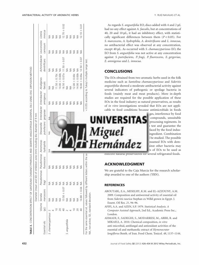
As regards S. angustifolia EO, discs added with 4 and 2 mLhad no any effect against A. faecalis, but at concentrations of40, 20 and 10 mL, it had an inhibitory effect, with statisti-cally significant differences between them (P < 0.05). ForS. marcescens, A. hydrophila, A. denitrificans and L. innocua,no antibacterial effect was observed at any concentration,except 40 mL. As occurred with S. chamaecyparissus EO, theEO from S. angustifolia was not active at any concentrationagainst S. putrefaciens, P. fragi, P. fluorescens, E. gergoviae,E. amnigenus and L. innocua.
CONCLUSIONS
The EOs obtained from two aromatic herbs used in the folkmedicine such as Santolina chamaecyparissus and Sideritisangustifolia showed a moderate antibacterial activity againstseveral indicators of pathogenic or spoilage bacteria infoods (mainly meat and meat products). More in-depthstudies are required for the possible application of theseEOs in the food industry as natural preservatives, as resultsof in vitro investigations revealed that EOs are not appli-cable to food conditions because antimicrobials in foodsmay be hampered by effective dosages, interference by foodconstituents or other food-grade compounds, unsuitablewater activity, incompatible pH or processing regiments. Inaddition, the absence of norms to test and guarantee thequality of EOs is a major problem faced by the food indus-try when they are used as a basic ingredient. Combinationof EOs from different plants should be studied. The possiblecombination with other widely consumed EOs with dem-onstrated antibacterial activity against other bacteria mayallow the development of mixtures of EOs to be used aseffective natural preservatives for several refrigerated foods.
ACKNOWLEDGMENT
We are grateful to the Caja Murcia for the research scholar-ship awarded to one of the authors (YRN).
REFERENCES
ABOUTABL, E.A., MESELHY, K.M. and EL-AZZOUNY, A.M.2009. Composition and antiwormal activity of essential oilfrom Sideritis taurica Stephan ex Willd grown in Egypt. J.Essent. Oil Res. 21, 94–96.
AFIFI, A.A. and AZEN, S.P. 1979. Statistical Analysis. AComputer Assisted Approach, 2nd Ed., Academic Press Inc.,London.
AHMADI, F., SADEGHI, S., MODARRESI, M., ABIRI, R. andMIKAELI, A. 2010. Chemical composition, in vitroanti-microbial, antifungal and antioxidant activities of theessential oil and methanolic extract of Hymenocraterlongiflorus Benth. of Iran. Food Chem. Toxicol. 48, 1137–1144.TA
BLE
3.C
ON
CEN
TRA
TIO
NEF
FEC
TO
FSA
NTO
LIN
AC
HA
MA
ECY
PARI
SSU
SA
ND
SID
ERIT
ISA
NG
UST
IFO
LIA
ESSE
NTI
AL
OIL
SU
SIN
GD
ISC
DIF
FUSI
ON
MET
HO
D
Esse
ntia
loils
Volu
me
(mL)
Dia
met
er(m
ean
and
SD)
ofin
hibi
tion
zone
(mm
)in
clud
ing
disc
diam
eter
of9
mm
Serr
atia
mar
cesc
ens
Pseu
dom
onas
frag
iPs
eudo
mon
asflu
ores
cens
Aer
omon
ashy
drop
hila
Shew
anel
lapu
tref
acie
nsEn
tero
bact
erge
rgov
iae
Ente
roba
cter
amni
genu
sA
lcal
igen
esfa
ecal
isA
chro
mob
acte
rde
nitr
ifica
nsLe
ucon
osto
cca
rnos
umLi
ster
iain
nocu
a
S.ch
amae
cypa
rissu
s2
NA
NA
NA
NA
NA
NA
NA
NA
NA
NA
NA
4N
AN
AN
AN
AN
AN
AN
AN
AN
AN
AN
A10
NA
NA
NA
NA
NA
NA
NA
11.5
0�
0.70
cN
AN
AN
A20
NA
NA
NA
10.0
0�
0.00
bN
AN
A.
NA
14.0
0�
0.00
bN
AN
AN
A40
13.5
0�
0.07
NA
NA
16.5
0�
0.07
aN
AN
AN
A16
.50
�0.
07a
11.5
0�
0.07
NA
12.7
0�
0.02
S.an
gust
ifolia
2N
AN
AN
AN
AN
AN
AN
AN
AN
AN
AN
A4
NA
NA
NA
NA
NA
NA
NA
NA
NA
NA
NA
10N
AN
AN
AN
AN
AN
AN
A12
.00
�0.
00c
NA
NA
NA
20N
AN
AN
AN
AN
AN
AN
A14
.00
�0.
00b
NA
NA
NA
4014
.80
�0.
04N
AN
A11
.00
�0.
00N
AN
AN
A18
.00
�0.
00a
12.5
0�
0.07
NA
12.1
0�
0.01
For
the
sam
ees
sent
ialo
il,va
lues
follo
wed
bydi
ffer
ent
lett
ers
with
inth
esa
me
colu
mn
are
sign
ifica
ntly
diff
eren
t(P
<0.
05)
acco
rdin
gto
Tuke
y’s
mul
tiple
rang
ete
st.
NA
,no
n-ac
tive;
SD,
stan
dard
devi
atio
n.
ANTIBACTERIAL ACTIVITY OF AROMATIC HERBS Y. RUIZ-NAVAJAS ET AL.
432 Journal of Food Safety 32 (2012) 426–434 © 2012 Wiley Periodicals, Inc.

AHUJA, A., BAKSHI, S.K., SHARMA, S.K., THAPPA, R.K.,AGARWAL, S.G., KICHLU, S.K., PAUL, R. and KAUL, M.R.2005. Production of volatile terpenes by proliferating shootsand micropropagated plants of Santolina chamaecyparissus L.(cotton lavender). Flavour Fragrance J. 20(4), 403–406.
ARQUES, J.L., RODRIGUEZ, E., NUÑEZ, M. and MEDINA, M.2008. Inactivation of gram-negative pathogens in refrigeratedmilk by reuterin in combination with nisin or thelactoperoxidase system. Eur. Food Res. Technol. 227(1),77–82.
BASILE, A., SENATORE, F., GARGANO, R., SORBO, S., DELPEZZO, M., LAVITOLA, A., RITIENI, A., BRUNO, M.,SPATUZZI, D., RIGANO, D. ET AL. 2006. Antibacterial andantioxidant activities in Sideritis italica (Miller) Greuter etBurdet essential oils. J. Ethnopharmacol. 107, 240–248.
BRANDI, G., AMAGLIANI, G., SCHIAVANO, G.F., DE SANTI,M. and SISTI, M. 2006. Activity of Brassica oleracea leaf juiceon food borne pathogenic bacteria. J. Food Prot. 69(9),2274–2279.
BURT, S. 2004. Essential oils: Their antibacterial properties andpotential applications in foods – a review. Int. J. FoodMicrobiol. 94(3), 223–253.
BURT, S.A., DER ZEE, R.V., KOETS, A.P., DE GRAAFF, A.M.,VAN KNAPEN, F., GAASTRA, W., HAAGSMAN, H.P. andVELDHUIZEN, E. 2007. Carvacrol induces heat shock protein60 and inhibits synthesis of flagellin in Escherichia coliO157:H7. Appl. Environ. Microbiol. 73, 4484–4490.
CARSON, C.F., HAMMER, K.A. and RILEY, T.V. 2002.Mechanism of action of Melaleuca alternifolia (tea tree) oil onStaphylococcus aureus determined by time-kill, lysis, leakageand salt tolerance assays and electron microscopy.Antimicrob. Agents Chemother. 46, 1914–1920.
DAVIDSON, P.M. 2001. Chemical preservatives and naturallyantimicrobial compounds. In Food Microbiology:Fundamentals and Frontiers, 2nd Ed (M.P. Beuchat and L.R.Montville, eds.) pp. 593–628, ASM Press, Washington, DC.
DEMIRCI, B., ÖZEK, T. and BASER, K.H.C. 2000. Chemicalcomposition of Santolina chamaecyparissus L. essential oil.J. Essent. Oil Res. 12, 625–627.
DERBESY, M., TOUCHE, J. and ZOLA, A. 1989. The essentialoil of Santolina chamaecyparissus L. J. Essent. Oil Res. 1,269–275.
EL-SAHHAR, K.F., NASSAR, D.M. and FARAG, H.M. 2011.Morphological and anatomical studies of Santolinachamaecyparissus L. (Asteraceae). II. Anatomicalcharacteristics and volatile oil. Res. J. Agric. Biol. Sci. 7(6),413–422.
FORMISANO, C., RIGANO, D., SENATORE, F., TENORE, G.C.,BRUNO, M. and PIOZZI, F. 2010. Volatile compounds offlowers and leaves of Sideritis italica (Miller) Greuter etBurdet (Lamiaceae), a plant used as mountain tea. Nat. Prod.Res. 24(7), 640–646.
GROSSO, C., FIGUEIREDO, A.C., BURILLO, J., MAINAR,A.M., URIETA, J.S., BARROSO, J.G., COELHO, J.A. andPALAVRA, A.M. 2009. Supercritical fluid extraction of the
volatile oil from Santolina chamaecyparissus. J. Sep. Sci.32(18), 3215–3222.
HAGGAG, M.Y., EL-TANTAWY, M.E., AHMED, F.I. andSHAMS, M.M. 2000. Study of chemical composition,antimicrobial and anthelmintic activity of theessential oils of Artemisia vulgaris L. and Santolinachamaecyparissus L. cultivated in Egypt. Quality Controlof Drugs, Vaccines and Natural Products 5th. ScientificConference, National Organization for Drug Controland Research, Meridien Hotel-Cairo, 1–3 April 2000.No. 54.
HASSIOTIS, C.N. and LAZARI, D.M. 2010. Decompositionprocess in the Mediterranean region. Chemical compoundsand essential oil degradation from Myrtus communis. Int.Biodet. Biodeg. 64, 356–362.
HOLLY, R.A. and PATEL, D. 2005. Improvement in shelf lifeand safety of perishable foods by plant essential oils andsmoke antimicrobials. Food Chem. 22, 273–292.
KILIÇ, T., YILDIZ, Y.K., GOREN, A.C., TUMEN, G. andTOPCU, G. 2003. Phytochemical analysis of someSideritis species of Turkey. Chem. Nat. Comp. 39(5),453–456.
KIRIMER, N., DEMIRCI, B., ISCAN, G., BASER, K.H.C. andDUMAN, H. 2008. Composition of the essential oils of twoSideritis species from Turkey and antimicrobial activity.Chem. Nat. Comp. 44, 121–123.
KÖSE, E.O., DENIZ, I., SARIKÜRKÇÜ, C., AKTAS, O. andYAVUZ, M. 2010. Chemical composition, antimicrobial andantioxidant activities of the essential oils of Sideritiserythrantha Boiss. and Heldr. (var. erythrantha and var.cedretorum PH Davis) endemic in Turkey. Food Chem. 48,2960–2965.
LOPEZ-MALO, A., PALOU, E. and ALZAMORA, S.M.2005. Naturally occurring compounds plant sources.In Antimicrobials in Food (P.M. Davidson, J.N. Sofosand A.L. Branen, eds.) pp. 429–446, CRC Press, BocaRaton, FL.
NATIONAL INSTITUTE OF STANDARDS ANDTECHNOLOGY (NIST). 2010. http://webbook.nist.gov/chemistry/name-ser.html (accessed October, 2011).
OROOJALIAN, F., KASRA-KERMANSHAHI, R., AZIZI, M. andBASSAMI, M.R. 2010. Phytochemical composition of theessential oils from three Apiaceae species and theirantibacterial effects on food-borne pathogens. Food Chem.120, 765–770.
PALOMINO, O.M., GÓMEZ-SERRANILLOS, P., CARRETERO,E. and VILLAR, A. 1996. High performance liquidchromatography of flavonoids from Sideritis species.J. Chromatogr. A 731, 103–108.
PÉREZ-ALONSO, M.J. and VELASCO-NEGUERUELA, A. 1992.Essential oil components of Santolina chamaecyparissus L.Flavour Fragrance J. 7, 37–42.
SAIER, M.H. Jr. 2009. Cell membrane, prokaryotic. InEncyclopedia of Microbiology, 3rd Ed., (M. Schaechter, ed.)pp. 341–356, Academic Press, Boston, MA.
Y. RUIZ-NAVAJAS ET AL. ANTIBACTERIAL ACTIVITY OF AROMATIC HERBS
433Journal of Food Safety 32 (2012) 426–434 © 2012 Wiley Periodicals, Inc.

SANDRI, I.G., ZACARIA, J., FRACARO, F., DELAMARE, A.P.L.and ECHEVERRIGARAY, S. 2007. Antimicrobial activity ofthe essential oils of Brazilian species of the genus Culinaagainst foodborne pathogens and spoiling bacteria. FoodChem. 103, 823–828.
SENATORE, F. 2000. Oli essenziali. In Origine Estrazione edAnalisi Chimica (Edizioni Mediche Scientifiche Internazionali,ed.) pp. 125–131, EMSI, Rome.
SEYDIM, A.C. and SARIKUS, G. 2006. Antimicrobial activity ofwhey protein based edible films incorporated with oregano,rosemary and garlic essential oils. Food Res. Int. 39, 639–644.
SHARIFIFAR, F., MOSHAFI, M.H., MANSOURI, S.H.,KHODASHENAS, M. and KHOSHNOODI, M. 2007. In vitroevaluation of antibacterial and antioxidant activities of theessential oil and methanol extract of endemic Zatariamultiflora Boiss. Food Control 18, 800–805.
SKOCIBUSIC, M., BEZIC, N. and DUNKIC, V. 2006.Phytochemical composition and antimicrobial activities of theessential oils from Satureja subspicata Vis. growing in Croatia.Food Chem. 96, 20–28.
SOKMEN, A., GULLUCE, M., AKPULAT, H.A., DAFERERA, D.,TEPE, B., POLISSIOU, M., SOKMEN, M. and SAHIN, F.2004. The in vitro antimicrobial and antioxidant activities ofthe essential oils and methanol extracts of endemic Thymusspathulifolius. Food Control 15, 627–634.
TENORE, G.C., CIAMPAGLIA, R., ARNOLD, N.A., PIOZZI, F.,NAPOLITANO, F., RIGANO, D. and SENATORE, F. 2011.Antimicrobial and antioxidant properties of the essential oilof Salvia lanigera from Cyprus. Food Chem. Toxicol. 49,238–243.
TEPE, B., DAFERERA, D., SOKMEN, A., SOKMEN, M. andPOLISSIOU, M. 2005. Antimicrobial and antioxidantactivities of the essential oils and various extracts of Salviatomentosa Miller (Lamiaceae). Food Chem. 90, 333–340.
VILLAR, A., GINER, R.M. and RIOS, J.L. 1986. Chemicalcomposition of Santolina chamaecyparissus ssp. squarrosaessential oil. J. Nat. Prod. 49(6), 1143–1144.
VIUDA-MARTOS, M., FERNÁNDEZ-LÓPEZ, J. andPÉREZ-ÁLVAREZ, J.A. 2005. Método de concentraciónmínima de inhibición. Technical Report. Project Recoveryand Compost Management. Rehobot, Israel, 19–21February.
VIUDA-MARTOS, M., RUIZ-NAVAJAS, Y.,FERNÁNDEZ-LÓPEZ, J. and PEREZ-ÁLVAREZ, J.A. 2007.Chemical composition of the essential oils obtained fromsome spices widely used in Mediterranean region. Acta Chim.Slov. 54, 921–926.
VIUDA-MARTOS, M., RUIZ-NAVAJAS, Y.,FERNÁNDEZ-LÓPEZ, J. and PEREZ-ÁLVAREZ, J.A. 2008.Antibacterial activity of lemon (Citrus lemon L.), mandarin(Citrus reticulata L.), grapefruit (Citrus paradisi L.) andorange (Citrus sinensis L.) essential oils. J. Food Saf. 28,567–576.
VIUDA-MARTOS, M., RUIZ-NAVAJAS, Y.,FERNÁNDEZ-LÓPEZ, J. and PÉREZ-ÁLVAREZ, J.A. 2009.Effect of adding citrus waste water, thyme and oreganoessential oil on the chemical, physical and sensorycharacteristics of a bologna sausage. Inno. Food Sci. Emerg.Technol. 10, 655–660.
VIUDA-MARTOS, M., MOHAMADY, M.A.,FERNÁNDEZ-LÓPEZ, J., ABD ELRAZIK, K.A., OMER, E.A.,PÉREZ-ALVAREZ, J.A. and SENDRA, E. 2011. In vitroantioxidant and antibacterial activities of essentials oilsobtained from Egyptian aromatic plants. Food Control 22,1715–1722.
ZIVANOVIC, S., CHI, S. and DRAUGHON, A.F. 2005.Antimicrobial activity of chitosan films enriched withessential oils. J. Food Sci. 70(1), 45–51.
ANTIBACTERIAL ACTIVITY OF AROMATIC HERBS Y. RUIZ-NAVAJAS ET AL.
434 Journal of Food Safety 32 (2012) 426–434 © 2012 Wiley Periodicals, Inc.

Título: In vitro antioxidant and antifungal properties of essential oils obtained from aromatic herbs
endemic to the southeast of Spain
Autores: Y. Ruiz-Navajas, M. Viuda-Martos, E. Sendra, J.A. Perez-Alvarez, J. Fernández-López
Revista: Journal of Food Protection, 76, 1218-1225
Año: 2013

In Vitro Antioxidant and Antifungal Properties of Essential OilsObtained from Aromatic Herbs Endemic to the Southeast of Spain
YOLANDA RUIZ-NAVAJAS, MANUEL VIUDA-MARTOS, ESTHER SENDRA, JOSE A. PEREZ-ALVAREZ,AND JUANA FERNANDEZ-LOPEZ*
IPOA Research Group (UMH-1 and REVIV-Generalitat Valenciana), AgroFood Technology Department, Escuela Politecnica Superior de Orihuela,
Univ. Miguel Hernandez, Crta, Beniel km. 3, 2, E-03312 Orihuela Alicante, Spain
MS 12-554: Received 14 December 2012/Accepted 13 March 2013
ABSTRACT
The aim of this work was to determine (i) the antioxidant capacity of Thymus moroderi, Thymus piperella, Santolinachamaecyparissus, and Sideritis angustifolia essential oils (EOs) by means of four different antioxidant tests (the 2,29-diphenyl-1-
picrylhydrazyl radical scavenging method, the ferrous ion–chelating ability assay, the ferric reducing antioxidant power test, and
the thiobarbituric acid reactive species test) and (ii) the antifungal activity against molds and yeast of these EOs by the agar
dilution and the microdilution methods. T. piperella EO showed the lowest 50% inhibitory concentration by the 2,29-diphenyl-1-
picrylhydrazyl radical scavenging method (9.30 mg/ml) and by the thiobarbituric acid reactive species test (6.30 mg/ml) and the
highest value by the ferric reducing antioxidant power test (2.64 Trolox equivalent antioxidant capacity), while S.chamaecyparissus showed the lowest 50% inhibitory concentration in the ferrous ion–chelating ability assay (3.94 mg/ml).
All EOs had a substantial inhibitory effect on all assayed yeast strains. S. angustifolia EO had the lowest MICs (2.5 ml/ml) for the
yeasts Saccharomyces cerevisiae, Debaryomyces hansenii, Rhodotorula mucilaginosa, and Pichia carsonii. As regards antifungal
activity, S. angustifolia EO at high concentrations was the most effective EO in reducing the growth of Alternaria alternata,Penicillium chrysogenum, and Mucor racemosus, while S. chamaecyparissus was the best inhibitor of the molds Aspergillusflavus and Mucor circinelloides. The results obtained in this study suggest the possibility of using these essential oils as natural
antioxidant food preservatives.
Lipid oxidation and microbiological degradation oc-
curring in food products are the major concerns in food
technology.
They are responsible for rancid odors and flavors of the
products, with a consequent decrease in nutritional quality
and safety caused by the formation of secondary, potentially
toxic compounds (49). In addition, illnesses caused by
consumption of foods contaminated with pathogens have a
great economic and public health impact worldwide (18).In the food industry, the rate of auto-oxidation is
reduced by freezing, refrigeration, packaging under inert
gas, and vacuum packaging (10). When these methods are
neither economic nor practical from nutritional and
technological points of view, it is highly desirable to
control oxidation by addition of antioxidants, inhibitory
substances that do not reduce food quality (20). As regards
the control of microorganisms, a range of different chemical
and synthetic compounds have been used as antibacterial
and antifungal agents to inhibit microbial food spoilage.
Antimicrobial substances used in the food industry include
chemical substances (added or already present naturally in
foods), which are used in the food industry for two main
reasons: (i) to control natural spoilage processes (food
preservation) and (ii) to prevent or control the growth of
microorganisms, including pathogenic microorganisms
(food safety) (39). Yeasts, one group of microorganisms,
are best known for their positive contributions to society,
particularly through their activities in food fermentation.
Yeasts also cause spoilage, although this effect is limited to
a narrow range of products, especially those of low pH or
high sugar concentration, where competition from bacterial
growth is restricted (16). The role played by molds in food
spoilage not only involves mycotoxin production but also
has very important adulterating effects on sensorial
properties (15).At present, consumer demands for less use of synthetic
food preservatives have increased throughout the world. So,
the replacement of traditional food preservation agents and
synthetic antioxidants such as butylated hydroxyanisole and
butylated hydroxytoluene (BHT) by natural vegetables and
plants materials have caused great interest in nutrition
research (3).Herbs and spices and their derivatives such as essential
oils (EOs) have been known since ancient times and used in
the preparation of foodstuffs to enhance their flavor and
organoleptic properties. Nowadays, they have great potential
in a growing nutrition industry because many plant-derived* Author for correspondence. Tel: z34 966749784; Fax: z34 966749677;
E-mail: [email protected].
1218
Journal of Food Protection, Vol. 76, No. 7, 2013, Pages 1218–1225doi:10.4315/0362-028X.JFP-12-554Copyright G, International Association for Food Protection

phytochemical preparations possess dual functionality in
preventing lipid oxidation and microbial spoilage, the main
causes of food quality deterioration (33). Thus, there are
numerous studies in which the antioxidant and antimicrobial
activities of herbs, spices, and especially the EOs have been
determined (3, 43, 45–47, 50).The genus Thymus is a member of the Lamiaceae
family. It is characterized by a high content of bioactive
compounds such as phenolic acids, flavonoids, and terpenes
present in EOs, which leads to its being regarded as an
aromatic herb with potential health benefits. Two species
found in the genus Thymus are Thymus piperella and
Thymus moroderi, both endemic to the southeast of Spain
(30). The genus Santolina, family Asteraceae, is widely
distributed in the Mediterranean area. Santolina chamaecy-parissus is widely used in Mediterranean folk medicine; the
flowers are used for their analgesic, anti-inflammatory,
antiseptic, and antispasmodic properties (13). The genus
Sideritis, family Lamiaceae, subfamily Lamioideae, com-
prises at least 150 species. Plants of this genus, mainly
Sideritis angustifolia, have been widely used in folk
medicine as anti-inflammatory, antiulcer, cytostatic, and
stimulant circulatory agents (28).According to our best knowledge, no report of the
antifungal and antioxidant activities of T. moroderi, T.piperella, S. chamaecyparissus, and S. angustifolia EOs
exists. Thus, the aim of this work was to determine (i) the
antioxidant capacity of T. moroderi, T. piperella, S.chamaecyparissus, and S. angustifolia EOs by means of
four different antioxidant tests (the 2,29-diphenyl-1-picryl-
hydrazyl radical scavenging method [DPPH], the ferrous
ion–chelating ability assay [FIC], the ferric reducing
antioxidant power [FRAP] test, and the thiobarbituric acid
reactive species test [TBARs]) and (ii) the antifungal
activity (against several molds and yeasts) of these EOs
by the agar dilution method and the microdilution method,
to investigate whether these EOs could be used as natural
food ingredients and represent a useful alternative for the
food industry to reduce the quantity of synthetic additives
used in their attempt to satisfy the demands of consumers.
MATERIALS AND METHODS
Plant materials. T. moroderi, T. piperella, S. chamaecypar-issus, and S. angustifolia were collected in the province of
Valencia (southeast of Spain) during their flowering period.
Identification of the plant material was made by Prof. Dr.
Concepcion Obon de Castro, Applied Biology Department of
Miguel Hernandez University (Spain).
Extraction of the EOs. The EOs of T. moroderi, T. piperella,S. chamaecyparissus, and S. angustifolia were extracted from 500 g
of whole plant (stems, leaves, and flowers) by hydrodistillation
using a Clevenger-type apparatus for 3 h. The respective yields of
the EOs were as follows: T. moroderi, 1.90%; T. piperella, 1.66%;
S. chamaecyparissus, 1.00%; and S. angustifolia, 0.90%. The oily
layer obtained on top of the aqueous distillate was separated and
dried with 0.5 g of anhydrous sodium sulfate (Panreac Quimica,
Barcelona, Spain). The extracted EOs were kept in sealed air-tight
glass vials and covered with aluminum foil at 4uC until further
analysis. There were three replicates for each EO.
TPC. The total phenol content (TPC) was determined using
the Folin-Ciocalteu’s reagent (35). The results were expressed in
milligrams of gallic acid equivalents (GAE) per gram of EO as the
mean of three replicates.
Antioxidant activity assays. (i) Determination of antiox-idant activity using the DPPH radical scavenging method. The
antioxidant activities of different concentrations (0.15 to 20 mg/ml)
of EO samples were measured in terms of hydrogen-donating or
radical-scavenging ability, using the stable radical DPPH (8). BHT
(0.02 to 2.5 mg/ml) was used as a positive control. The results were
expressed as the mean percent inhibition of three replicates.
(ii) FRAP. The FRAP of different concentrations (0.15 to
20 mg/ml) of EO samples was determined by using the potassium
ferricyanide–ferric chloride method (27). The FRAP of the samples
was estimated in terms of Trolox equivalent antioxidant capacity
(TEAC) in millimolar Trolox per liter as the mean of three
replicates.
(iii) TBARS. The method of Daker et al. (10) was used to
determine the TBARS, a secondary product of lipid peroxidation.
To measure the percentage of lipid peroxidation inhibition,
different concentrations (0.15 to 20 mg/ml) of EO samples were
used. BHT (0.002 to 0.31 mg/ml) was used as a positive control.
The results were expressed as the mean percent inhibition of three
replicates.
(iv) FIC assay. The FIC of different concentrations (0.15 to
20 mg/ml) of EO samples was measured by inhibiting the
formation of Fe2z–ferrozine complex after treatment of test
material with Fe2z, following the method of Carter (9). EDTA
(0.01 to 1.25 mg/ml) was used as a positive control. The results
were expressed as the mean percent chelating effect of three
replicates.
Microbial strains. The EOs were individually tested against
yeasts (Yarrowia lipolytica CECT 1240, Saccharomyces cerevisiaeCECT 1383, Candida zeylanoides CECT 10048, Debaryomyceshansenii CEPT 11369, Rhodotorula mucilaginosa CECT 10011,
and Pichia carsonii CECT 10227) and molds (Aspergillus nigerCECT 2091, Aspergillus flavus CECT 2685, Mucor racemosusCECT 2670, Mucor circinelloides CECT 20765, Penicilliumchrysogenum CECT 2784, and Alternaria alternata CECT
20560). These species were supplied by the Spanish Type Culture
Collection of the University of Valencia.
Antifungal activity assays. (i) Microdilution method. The
antifungal activity against yeast species was determined based on a
colorimetric broth microdilution method proposed by Abate et al.
(1), with some modification. The yeast strains Y. lipolytica, S.cerevisiae, C. zeylanoides, R. mucilaginosa, D. hansenii, and P.carsonii were cultured 24 h at 26uC in Litmus Milk (LM) broth and
diluted in sterile LM broth to a final level of 106 CFU/ml (adjusted
to a turbidity of 0.5 McFarland standard). EOs were dissolved in
dimethyl sulfoxide to reach a final concentration of 40 ml/ml. Serial
twofold dilutions were made in a concentration ranging from 0.04
to 40 ml/ml in sterile test tubes containing Mueller-Hinton broth.
The 96-well microplates (Iwaki, Japan) were prepared by
dispensing 90 ml of Mueller-Hinton broth and 10 ml of the yeast
inocula into each well. A 100-ml aliquot from each EO initially
prepared was added into the first wells. Then, 100 ml from their
serial dilutions was transferred into 11 consecutive wells. The last
row, containing 190 ml of Mueller-Hinton broth without the
compound and 10 ml of the inoculum, was used as negative control.
J. Food Prot., Vol. 76, No. 7 ANTIOXIDANT AND ANTIFUNGAL PROPERTIES OF ESSENTIAL OILS FROM SPAIN 1219

The final volume in each well was 200 ml. Contents of each well
were mixed on a plate shaker at 150 rpm for 2 min prior to
incubation for 24 h at 26uC. After incubation, 25 ml of 3-(4,5-
dimethylthiazol-2-yl)-2,5-diphenyl tetrazolium bromide (MTT)
(Sigma Life Science), dissolved in dimethyl sulfoxide (0.8 mg/
ml) was added to each of the wells and incubated for 1 h in order to
allow the viable microorganisms to metabolize the yellow MTT
dye into formazan (purple crystals). The MIC was considered as
the concentration of the first well that did not undergo color change
(from yellow to purple) and was confirmed by plating 10-ml
samples from clear wells onto Mueller-Hinton agar medium. The
procedure was repeated three times for each microorganism.
(ii) Agar dilution method. The food spoilage molds were
tested by the agar dilution method (17) with some modifications.
The EOs tested were added to the culture medium (potato dextrose
agar, Oxoid, Basingstoke, Hampshire, England) at a temperature of
40 to 45uC, and the mixtures were then poured into petri dishes
(9-cm diameter). Concentrations of 0.2, 0.1, 0.05, and 0.025 ml/
100 ml of medium were tested for T. moroderi, T. piperella, S.chamaecyparissus, and S. angustifolia EOs. The molds were
inoculated as soon as the medium had solidified. A disc (9 mm in
diameter, Schlinder & Schuell, Dassel, Germany) of mycelium
material, taken from the edge of 5-day-old fungi cultures, was
placed at the center of each petri dish. The petri dish with the
inoculum was then incubated at 26uC. The incubation was stopped
when the mycelium mass of control petri dishes had almost filled
the petri dish (6 days). The diameter of the growth mass was
determined by averaging the radial growth of the mycelial mass in
two orthogonal directions. The efficacy of treatment was evaluated
each day during 9 days by measuring the diameter of the fungus
colony. The values were expressed in diameter millimeters per day.
All tests were performed in triplicate.
The antifungal activity was calculated in terms of percentage
of mycelial inhibition growth (MIG), using the following equation:
%MIG ~ [(DC 2 DT)/DC] | 100, where DC is the average
diameter of the mycelial colonies of the control on day 8 and DT is
the average diameter of the mycelial colonies of treated sets on day
6. Each test was carried out in triplicate.
Statistical analysis. Conventional statistical methods were
used to calculate means and standard deviations of three
simultaneous assays carried out with the different methods. Data
collected for antioxidant properties were analyzed by two-way
analysis of variance to test the effects of two fixed factors:
concentration (levels, 0.15 to 20 mg/ml) and EO (levels for T.piperella, T. moroderi, S. angustifolia, and S. chamaecyparissus).
Data collected for antifungal properties were analyzed by two-way
analysis of variance to test the effects of two fixed factors:
concentration (levels, 0.025, 0.05, 0.1, and 0.2 ml/100 ml) and EO
(levels for T. piperella, T. moroderi, S. angustifolia, and S.chamaecyparissus). The Tukey post hoc test was applied for
comparisons of means; differences were considered significant at Pvalues of ,0.05. Statistical analysis and comparisons among
means were carried out using the statistical package Statgraphics
5.1 for Windows.
RESULTS AND DISCUSSION
The chemical composition of the essential oils used in
this work was previously determined by Ruiz-Navajas et al.
(30, 31). In the T. moroderi EO, the main components were
camphor (26.74%), 1,8-cineol (24.99%), myrcene (5.63%),
and a-pinene (4.35%). In T. piperella EO, the predominant
compounds were carvacrol (31.92%), para-cymene
(16.18%), c-terpinene (10.11%), and a-terpineol (7.29%). In
S. chamaecyparissus, the major constituents were artemisia
ketone (27.19%), dihydroaromadendrene (18.21%), and b-
phellandrene (7.49%), while in S. angustifolia EO the major
constituent was a-pinene (12.71%). Other important com-
pounds in S. angustifolia EO were b-phellandrene (11.97%),
1,8-cineol (7.41%), and trans-caryophyllene (6.33%).
TPCs. The TPCs of T. moroderi, T. piperella, S.chamaecyparissus, and S. angustifolia EOs are presented in
Table 1. T. piperella EO showed the highest content of total
phenols (57.43 mg of GAE/g). S. chamaecyparissus and S.angustifolia EOs were seen to be a less rich source of total
phenols (21.59 and 38.48 mg of GAE/g, respectively), while
T. moroderi EO showed the lowest amount of total phenols.
The concentration and type of phenolic substances in a plant
depend on several factors, such as differences in varieties,
ripeness, season, environmental factors, soil type and
climate, genetic factors, and processing and extraction
methods. The TPC could be used as an important indicator
of the antibacterial and antioxidant capacities, since
phenolic compounds are considered to have a high redox
potential allowing them to act as radical scavengers or
hydrogen donors (44).
Antioxidant activity. The antioxidant potential of
different plant extracts and pure compounds can be
measured using numerous in vitro assays. Each of these
tests is based on one feature of the antioxidant activity, such
as the ability to scavenge free radicals, the inhibition of lipid
peroxidation, or chelation of the transition metal ions (33).Thus, the antioxidant activity assessment may require a
combination of different methods because there are
substantial differences in sample preparation, extraction of
antioxidants, the selection of endpoints, and the way in
which the results are expressed, even for the same method
(47). In this study, four different methods were used to
determine the antioxidant activities of EOs.
The DPPH radical is one of the most commonly used
substrates for fast evaluation of antioxidant activity because
of its stability (in radical form) and the simplicity of the
assay. Figure 1 shows the DPPH radical-scavenging activity
of T. moroderi, T. piperella, S. chamaecyparissus, and S.angustifolia. It can be seen that EOs present different
scavenging capacities. A concentration-dependent scaveng-
ing activity was found for all the EOs studied. T. piperellaEO showed the highest (P , 0.05) DPPH radical-
scavenging activity at all concentrations assayed, followed
TABLE 1. Total phenol content (TPC) of T. moroderi, T.
piperella, S. chamaecyparissus, and S. angustifolia EOs
EO from: TPC (mg of GAE/g of EO)
T. piperella 57.43 ¡ 0.38
T. moroderi 9.33 ¡ 0.53
S. chamaecyparissus 21.59 ¡ 0.39
S. angustifolia 38.48 ¡ 0.39
1220 RUIZ-NAVAJAS ET AL. J. Food Prot., Vol. 76, No. 7
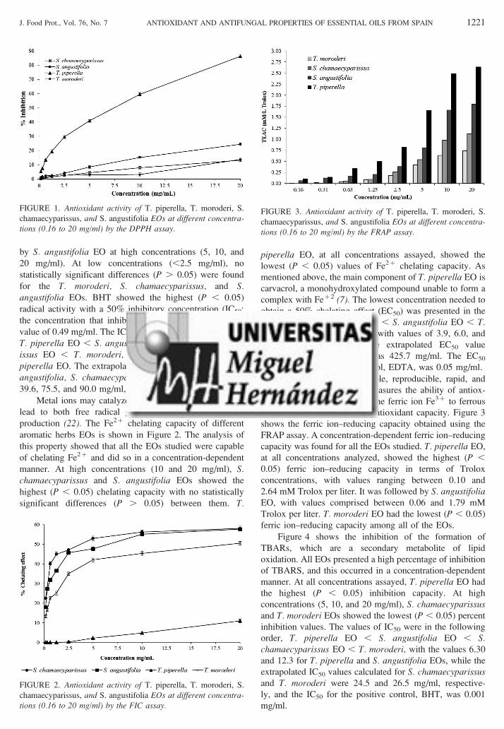
by S. angustifolia EO at high concentrations (5, 10, and
20 mg/ml). At low concentrations (,2.5 mg/ml), no
statistically significant differences (P . 0.05) were found
for the T. moroderi, S. chamaecyparissus, and S.angustifolia EOs. BHT showed the highest (P , 0.05)
radical activity with a 50% inhibitory concentration (IC50;
the concentration that inhibits 50% of the DPPH radical)
value of 0.49 mg/ml. The IC50s were in the following order:
T. piperella EO , S. angustifolia EO , S. chamaecypar-issus EO , T. moroderi, with a value of 9.30 for T.piperella EO. The extrapolated IC50 values obtained for S.angustifolia, S. chamaecyparissus, and T. moroderi were
39.6, 75.5, and 90.0 mg/ml, respectively.
Metal ions may catalyze lipid peroxidation, which can
lead to both free radical generation and lipid peroxide
production (22). The Fe2z chelating capacity of different
aromatic herbs EOs is shown in Figure 2. The analysis of
this property showed that all the EOs studied were capable
of chelating Fe2z and did so in a concentration-dependent
manner. At high concentrations (10 and 20 mg/ml), S.chamaecyparissus and S. angustifolia EOs showed the
highest (P , 0.05) chelating capacity with no statistically
significant differences (P . 0.05) between them. T.
piperella EO, at all concentrations assayed, showed the
lowest (P , 0.05) values of Fe2z chelating capacity. As
mentioned above, the main component of T. piperella EO is
carvacrol, a monohydroxylated compound unable to form a
complex with Fez2 (7). The lowest concentration needed to
obtain a 50% chelating effect (EC50) was presented in the
order S. chamaecyparissus EO , S. angustifolia EO , T.moroderi , T. piperella EO, with values of 3.9, 6.0, and
19.4, respectively, while the extrapolated EC50 value
calculated for T. piperella was 425.7 mg/ml. The EC50
obtained for the positive control, EDTA, was 0.05 mg/ml.
The FRAP test is a simple, reproducible, rapid, and
inexpensive procedure that measures the ability of antiox-
idative compounds to reduce the ferric ion Fe3z to ferrous
Fe2z, as a measure of total antioxidant capacity. Figure 3
shows the ferric ion–reducing capacity obtained using the
FRAP assay. A concentration-dependent ferric ion–reducing
capacity was found for all the EOs studied. T. piperella EO,
at all concentrations analyzed, showed the highest (P ,
0.05) ferric ion–reducing capacity in terms of Trolox
concentrations, with values ranging between 0.10 and
2.64 mM Trolox per liter. It was followed by S. angustifoliaEO, with values comprised between 0.06 and 1.79 mM
Trolox per liter. T. moroderi EO had the lowest (P , 0.05)
ferric ion–reducing capacity among all of the EOs.
Figure 4 shows the inhibition of the formation of
TBARs, which are a secondary metabolite of lipid
oxidation. All EOs presented a high percentage of inhibition
of TBARS, and this occurred in a concentration-dependent
manner. At all concentrations assayed, T. piperella EO had
the highest (P , 0.05) inhibition capacity. At high
concentrations (5, 10, and 20 mg/ml), S. chamaecyparissusand T. moroderi EOs showed the lowest (P , 0.05) percent
inhibition values. The values of IC50 were in the following
order, T. piperella EO , S. angustifolia EO , S.chamaecyparissus EO , T. moroderi, with the values 6.30
and 12.3 for T. piperella and S. angustifolia EOs, while the
extrapolated IC50 values calculated for S. chamaecyparissusand T. moroderi were 24.5 and 26.5 mg/ml, respective-
ly, and the IC50 for the positive control, BHT, was 0.001
mg/ml.
FIGURE 1. Antioxidant activity of T. piperella, T. moroderi, S.
chamaecyparissus, and S. angustifolia EOs at different concentra-tions (0.16 to 20 mg/ml) by the DPPH assay.
FIGURE 2. Antioxidant activity of T. piperella, T. moroderi, S.
chamaecyparissus, and S. angustifolia EOs at different concentra-tions (0.16 to 20 mg/ml) by the FIC assay.
FIGURE 3. Antioxidant activity of T. piperella, T. moroderi, S.
chamaecyparissus, and S. angustifolia EOs at different concentra-tions (0.16 to 20 mg/ml) by the FRAP assay.
J. Food Prot., Vol. 76, No. 7 ANTIOXIDANT AND ANTIFUNGAL PROPERTIES OF ESSENTIAL OILS FROM SPAIN 1221
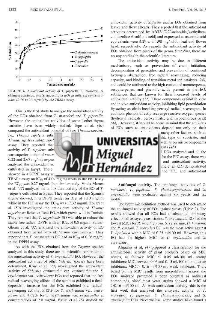
This is the first study to analyze the antioxidant activity
of the EOs obtained from T. moroderi and T. piperella.
However, the antioxidant activities of several other thyme
varieties have been widely studied. Tepe et al. (40)compared the antioxidant potential of two Thymus species,
i.e., Thymus sipyleus subsp. sipyleus var. sipyleus and
Thymus sipyleus subsp. sipyleus var. rosulans, by the DPPH
assay. They reported that the free radical–scavenging
activity of T. sipyleus subsp. sipyleus var. rosulans EO
was superior to that of var. sipyleus EO, with IC50 values of
0.22 and 2.67 mg/ml, respectively. Viuda-Martos et al. (43)analyzed the antioxidant activity of Thymus vulgaris EO
cultivated in Egypt. These authors reported that this EO
showed in a DPPH assay an IC50 of 4.50 mg/ml, and in a
TBARs assay an IC50 of 4.09 mg/ml while in the FIC assay
the EC50 was 0.27 mg/ml. In a similar study, Viuda-Martos
et al. (47) analyzed the antioxidant activity of the EO of T.vulgaris cultivated in Spain. They reported that the Spanish
thyme showed, in a DPPH assay, an IC50 of 1.10 mg/ml,
while in the FIC assay the EC50 was 17.32 mg/ml. Zouari et
al. (50) investigated the antioxidant activity of Thymusalgeriensis Boiss. et Reut EO, which grows wild in Tunisia.
They reported that T. algeriensis EO was able to reduce the
stable free radical DPPH with an IC50 of 0.8 mg/ml. Safaei-
Ghomi et al. (32) analyzed the antioxidant activity of EO
obtained from aerial parts of Thymus caramanicus. They
reported that T. caramanicus EO had an IC50 of 0.26 mg/ml
in the DPPH assay.
As with the EOs obtained from the Thymus species
analyzed in this study, there are no scientific reports about
the antioxidant activity of S. angustifolia EO. However, the
antioxidant activities of other Sideritis species have been
determined. Kose et al. (23) investigated the antioxidant
activity of Sideritis erythrantha var. erythrantha and S.erythrantha var. cedretorum EOs and reported that the free
radical–scavenging effects of the samples exhibited a dose-
dependent increase but the EOs exhibited low radical-
scavenging activity, 5.12% for S. erythrantha var. cedre-torum and 4.62% for S. erythrantha var. erythrantha at
concentrations of 2.0 mg/ml. Basile et al. (6) studied the
antioxidant activity of Sideritis italica EOs obtained from
leaves and flower heads. They reported that the antioxidant
activities determined by ABTS [2,29-azino-bis(3-ethylben-
zothiazoline-6-sulfonic acid] and expressed as ascorbic acid
equivalents were 4.29 and 1.98 mg/ml for leaf and flower
head, respectively. As regards the antioxidant activity of
EOs obtained from plants of the genus Santolina, there are
no any studies in the scientific literature.
The antioxidant activity may be due to different
mechanisms, such as prevention of chain initiation,
decomposition of peroxides, and prevention of continued
hydrogen abstraction, free radical scavenging, reducing
capacity, and binding of transition metal ion catalysts (24),and could be attributed to the high content of monoterpenes,
sesquiterpenes, and phenolic acids present in the EO,
substances that are known for their increased levels of
antioxidant activity (12). These compounds exhibit in vitro
and in vivo antioxidant activity, inhibiting lipid peroxidation
by acting as chain-breaking peroxyl radical scavengers. In
addition, phenols directly scavenge reactive oxygen species
(hydroxyl radicals, peroxynitrite, and hypochlorous acid)
(44). However, it should be borne in mind that the activities
of EOs such as antioxidants depend not only on their
structural features but also on many other factors, such as
concentration, temperature, light, type of substrate, and
physical state of the system, as well as on microcomponents
acting as pro-oxidants or synergists (48).It is interesting that for all EOs analyzed and all the
antioxidant assays used, except for the FIC assay, there was
a correlation between TPC and antioxidant activity.
Djeridane et al. (11) and Katalinic et al. (21) demonstrated
a linear correlation between the TPC and antioxidant
capacity.
Antifungal activity. The antifungal activities of T.moroderi, T. piperella, S. chamaecyparissus, and S.angustifolia were evaluated against six yeasts and six
molds.
The broth microdilution method was used to determine
the antifungal activity of EOs against yeasts (Table 2). The
results showed that all EOs had a substantial inhibitory
effect on all assayed yeast strains. S. angustifolia EO had the
lowest MICs for R. mucilaginosa, S. cerevisiae, D. hansenii,and P. carsoni. T. moroderi EO was the most active against
Y. lipolytica with a MIC of 0.25 ml/100 ml. However, this
EO had the highest MIC for C. zeylanoides and S.cerevisiae.
Aligianis et al. (4) proposed a classification for the
antimicrobial activity of plant products based on MIC
results, as follows: MIC # 0.05 ml/100 ml, strong
inhibitors; MIC between 0.06 and 0.15 ml/100 ml, moderate
inhibitors; MIC . 0.16 ml/100 ml, weak inhibitors. Thus,
based on the MIC results from microdilution assays, the
EOs analyzed presented a poor potential as antiyeast
compounds, since most yeast strains showed a MIC of
.0.16 ml/100 ml. As with antioxidant activity, this is the
first work that analyzed the antiyeast activity of T.moroderi, T. piperella, S. chamaecyparissus, and S.angustifolia EOs. Nevertheless, some studies have found a
FIGURE 4. Antioxidant activity of T. piperella, T. moroderi, S.
chamaecyparissus, and S. angustifolia EOs at different concentra-tions (0.16 to 20 mg/ml) by the TBARs assay.
1222 RUIZ-NAVAJAS ET AL. J. Food Prot., Vol. 76, No. 7

strong antiyeast effect of EOs obtained from aromatic herbs
or spices (5, 29, 37, 41). Thus, Pinto et al. (29) reported that
Salvia officinalis EO had MICs of 0.25 ml/100 ml against
Candida albicans, Candida krusei, Candida tropicalis, and
Candida parapsilosis. Souza et al. (37) showed that
Origanum vulgare EO was effective in inhibiting the
growth of C. albicans, C. krusei, C. tropicalis, Pichiaminuscula, Pichia ohmeri, Rhodotorula rubra, and S.cerevisiae, with MICs between 2 and 0.06 ml/100 ml.
Tserennadmid et al. (41) analyzed the antiyeast activities of
clary sage, juniper, lemon, and marjoram EOs against
Geotrichum candidum, Pichia anomala, S. cerevisiae, and
Schizosaccharomyces pombe. These authors reported that
the most sensitive yeast was S. pombe (0.006 to 0.0125 ml/
100 ml) while G. candidum proved to be the most
insensitive (MICs, 0.05 to 0.2 ml/100 ml). For antifungal
activity against molds, the agar dilution method was used,
and the results are summarized in Table 3. In the case of A.niger, all EOs at all concentrations assayed showed
reduction in mycelial growth. T. moroderi EO produced
the greatest (P , 0.05) MIG values, 29.01 and 52.54%, at
concentrations of 0.1 and 0.2 ml/100 ml, respectively,
followed by T. piperella EO with MIG values of 26.79 and
44.94%, respectively, at the same concentrations. At all
concentrations, S. angustifolia EO caused the lowest MIG
with values ranging between 6.06 and 33.74%. In the case
of A. flavus, S. chamaecyparissus EO showed the highest (P, 0.05) MIG values (41.23%) at maximum concentration
(0.2 ml/100 ml), while at concentrations of 0.05 and 0.1 ml/
100 ml it was T. moroderi EO that showed the highest (P ,
TABLE 2. MICs of T. moroderi, T. piperella, S. chamaecyparissus, and S. angustifolia EOs against several yeast species
Yeast
Concn (ml/100 ml) of EO from:
T. moroderi T. piperella S. angustifolia S. chamaecyparissus
Y. lipolytica 0.25 0.50 0.50 0.50
C. zeylanoides 0.50 0.25 0.25 0.25
P. carsonii 0.50 0.50 0.25 0.50
S. cerevisiae 1.00 0.50 0.25 0.50
D. hansenii 0.25 0.25 0.12 0.25
R. mucilaginosa 0.50 0.50 0.25 0.50
TABLE 3. Percent mycelial inhibition growth (MIG) of T. moroderi, T. piperella, S. angustifolia, and S. chamaecyparissus EOs againstseveral molds by the agar dilution methoda
Mold EO concn (ml/100 ml)
%MIG
T. moroderi T. piperella S. angustifolia S. chamaecyparissus
A. niger 0.025 6.42 ¡ 0.12 a A 8.44 ¡ 0.02 b A 4.06 ¡ 0.15 c A 6.68 ¡ 0.12 a A
0.05 13.73 ¡ 0.24 a B 14.33 ¡ 0.01 b B 12.91 ¡ 0.09 c B 14.50 ¡ 0.06 d B
0.1 29.01 ¡ 0.15 a C 26.79 ¡ 0.12 b C 22.10 ¡ 0.11 c C 22.24 ¡ 0.07 c C
0.2 52.54 ¡ 0.23 a D 44.94 ¡ 0.05 b D 33.74 ¡ 0.13 c D 42.82 ¡ 0.12 d D
A. flavus 0.025 10.10 ¡ 0.11 a A 3.94 ¡ 0.07 b A 8.43 ¡ 0.08 c A 7.06 ¡ 0.11 d A
0.05 19.89 ¡ 0.23 a B 10.76 ¡ 0.14 b B 13.61 ¡ 0.10 c B 13.58 ¡ 0.08 c B
0.1 26.79 ¡ 0.10 a C 16.21 ¡ 0.09 b C 21.88 ¡ 0.07 c C 23.26 ¡ 0.14 d C
0.2 34.84 ¡ 0.11 a D 28.22 ¡ 0.10 b D 29.54 ¡ 0.09 c D 41.23 ¡ 0.09 d D
A. alternata 0.025 12.83 ¡ 0.06 a A 0.00 ¡ 0.00 b A 14.47 ¡ 0.01 c A 14.48 ¡ 0.06 c A
0.05 24.57 ¡ 0.12 a B 15.80 ¡ 0.06 b B 33.24 ¡ 0.12 c B 23.96 ¡ 0.07 d B
0.1 39.27 ¡ 0.14 a C 20.64 ¡ 0.08 b C 50.29 ¡ 0.12 c C 32.83 ¡ 0.11 d C
0.2 54.69 ¡ 0.06 a D 29.43 ¡ 0.09 b D 55.70 ¡ 0.09 c D 43.96 ¡ 0.14 d D
P. chrysogenum 0.025 13.84 ¡ 0.05 a A 0.00 ¡ 0.00 b A 10.51 ¡ 0.12 c A 1.17 ¡ 0.11 d A
0.05 30.47 ¡ 0.09 a B 5.56 ¡ 0.11 b B 21.69 ¡ 0.14 c B 7.24 ¡ 0.15 d B
0.1 51.57 ¡ 0.11 a C 8.33 ¡ 0.05 b C 60.56 ¡ 0.05 c C 38.13 ¡ 0.06 d C
0.2 63.94 ¡ 0.15 a D 28.33 ¡ 0.13 b D 79.81 ¡ 0.04 c D 53.94 ¡ 0.01 d D
M. circinelloides 0.025 4.92 ¡ 0.06 a A 7.01 ¡ 0.03 b A 0.00 ¡ 0.00 c A 1.60 ¡ 0.20 d A
0.05 13.98 ¡ 0.07 a B 9.23 ¡ 0.04 b B 1.12 ¡ 0.01 c B 6.79 ¡ 0.12 d B
0.1 19.01 ¡ 0.09 a C 29.14 ¡ 0.14 b C 26.88 ¡ 0.14 c C 33.59 ¡ 0.09 d C
0.2 22.57 ¡ 0.13 a D 44.57 ¡ 0.07 b D 42.81 ¡ 0.12 c D 45.60 ¡ 0.14 d D
M. racemosus 0.025 1.46 ¡ 0.04 a A 3.58 ¡ 0.04 b A 0.00 ¡ 0.00 c A 0.00 ¡ 0.00 c A
0.05 17.54 ¡ 0.12 a B 5.58 ¡ 0.14 b B 12.61 ¡ 0.08 c B 0.00 ¡ 0.00 d A
0.1 30.29 ¡ 0.08 a C 18.28 ¡ 0.14 b C 49.60 ¡ 0.07 c C 14.93 ¡ 0.18 d B
0.2 40.02 ¡ 0.09 a D 38.63 ¡ 0.17 b D 62.24 ¡ 0.11 c D 29.06 ¡ 0.15 d C
a For the same mold and at the same concentration, values followed by the same lowercase letter within the same row are not significantly
different (P . 0.05) according to Tukey’s multiple range test. For the same mold and the same EO, values followed by the same
uppercase letter within the same column are not significantly different (P . 0.05) according to Tukey’s multiple range test.
J. Food Prot., Vol. 76, No. 7 ANTIOXIDANT AND ANTIFUNGAL PROPERTIES OF ESSENTIAL OILS FROM SPAIN 1223

0.05) MIG values (19.89 and 26.79%, respectively). T.piperella at all concentrations showed the lowest (P , 0.05)
MIG values. S. angustifolia EO, at high concentrations (0.1
and 0.2 ml/100 ml) was the most effective EO (P , 0.05) in
reducing the growth of A. alternata, P. chrysogenum, and
M. racemosus with MIG values of 50.29 and 55.70%, 60.56
and 79.81%, and 49.60 and 62.24%, respectively. For these
molds, the next most (P , 0.05) effective EO at high
concentrations was that of T. moroderi with MIG values of
39.27 and 54.69%, 51.57 and 63.94%, and 30.29 and
40.02% for A. alternata, P. chrysogenum, and M.racemosus, respectively, at high concentrations. For A.alternata and P. chrysogenum, the T. piperella EO at all
concentrations showed the lowest (P , 0.05) MIG values.
Insofar as M. circinelloides is concerned, S. chamaecypar-issus EO showed the highest (P , 0.05) MIG values (33.59
and 45.60%) at high concentrations (0.1 and 0.2 ml/100 ml,
respectively). This was followed by T. piperella EO with
slightly lower reductions (29.14 and 44.57% at the same
concentrations). S. angustifolia showed the lowest (P ,
0.05) MIG values at low concentrations. The antifungal
activity of aromatic herbs and spices EOs against molds has
been widely demonstrated (19, 25, 36, 45). Indeed, Viuda-
Martos et al. (45) demonstrated that thyme, oregano, or
clove EOs at concentrations of 0.04 ml/100 ml provoked a
total inhibition of growth of A. niger and A. flavus. Sokmen
et al. (36) demonstrated the capacity of thyme EO at 10 ml
to inhibit the growth of molds such as Alternaria spp., A.flavus, Fusarium spp., and Penicillium spp. Goncalves et al.
(19) reported that Thymus zygis subsp. sylvestris EO had
MICs between 0.016 and 0.12 ml/100 ml against A. niger,A. flavus, and Aspergillus fumigatus.
The mechanisms behind the antifungal activity of EOs
against molds and yeasts are not fully understood because
these substances are a mixture of several compounds that do
not act on specific targets in the fungal cells. However, there
are many studies that try to explain them.
As regards yeasts, Uribe et al. (42) reported that
terpenes present in EOs inhibited the respiratory activity in
intact yeast cells and also in isolated mitochondria, while
Adegoke et al. (2) reported that EOs caused cell membrane
degradation and loss of cytoplasmic components in the
yeast C. tropicalis. Ferguson and von Borstel (14) reported
that the cytotoxicity of the EOs is accompanied by the
induction of cytoplasmic ‘‘petite’’ mutations, indicating
mitochondrial damage and impairment of oxidative me-
tabolism. In the case of the molds, Soylu et al. (38) found
that monoterpenes present in the EOs cause alterations in
the hyphal morphology and hyphal aggregates, resulting in
reduced hyphal diameters and lyses of hyphal wall
interacting with the cell membrane of the pathogen. For
Sharma and Tripathi (34), the EOs would act on the hyphae
of the mycelium, provoking the exit of components from
the cytoplasm and the loss of rigidity and integrity of the
hyphal cell wall, resulting in its collapse and death of the
mycelium. Omidbeygi et al. (26) have suggested that
antimicrobial components of the EOs cross the molds’ cell
membrane, interacting with the enzymes and proteins of
the membrane, thus producing a flux of protons toward the
cell exterior that induces changes in the cells and,
ultimately, their death.
The results obtained in this study suggest the possibility
of using the essential oils of T. moroderi, T. piperella, S.chamaecyparissus, and S. angustifolia as natural antioxidant
food preservatives, emerging from a growing tendency to
replace synthetic preservatives by natural ones. In addition,
these EOs may have a great potential to be used as safe and
ecofriendly antifungal ingredients compounds in the food
industry. However, in general, the levels of EOs and their
compounds necessary to inhibit microbial growth are higher
in foods than in culture media. This is due to interactions
between phenolic compounds and the food matrix and
should be considered for commercial applications. In this
respect, studying the results obtained with native species
may be of great interest.
ACKNOWLEDGMENT
We are grateful to Caja Murcia for the research scholarship awarded
to one of the authors (Y.R.-N.).
REFERENCES
1. Abate, G., R. N. Mshana, and H. Miorner. 1998. Evaluation of a
colorimetric assay based on 3-(4,5-dimethylthiazol-2-yl)-2,5-diphenyl
tetrazolium bromide (MTT) for rapid detection of rifampicin
resistance in Mycobacterium tuberculosis. Int. J. Tuberc. Lung Dis.
2:1011–1016.
2. Adegoke, G. O., H. Iwahashi, Y. Komatsu, K. Obuchi, and Y.
Iwahashi. 2000. Inhibition of food spoilage yeasts and aflatoxigenic
moulds by monoterpenes of the spice Aframomum danielii. Flavour
Fragr. J. 15:147–150.
3. Ahmadi, F., S. Sadeghi, M. Modarresi, R. Abiri, and A. Mikaeli.
2010. Chemical composition, in vitro anti-microbial, antifungal and
antioxidant activities of the essential oil and methanolic extract of
Hymenocrater longiflorus Benth., of Iran. Food Chem. Toxicol. 48:
1137–1144.
4. Aligianis, N., E. Kalpoutzakis, S. Mitaku, and I. B. Chinou. 2001.
Composition and antimicrobial activity of the essential oil from
Origanum species. J. Agric. Food Chem. 49:4168–4170.
5. Bakkali, F., S. Averbeck, D. Averbeck, A. Zhiri, and M. Idaomar.
2005. Cytotoxicity and gene induction by some essential oils in the
yeast Saccharomyces cerevisiae. Mutat. Res. 585:1–13.
6. Basile, A., F. Senatore, R. Gargano, S. Sorbo, M. D. Pezzo, A.
Lavitola, A. Ritieni, M. Bruno, D. Spatuzzi, D. Rigano, and M. L.
Vuotto. 2006. Antibacterial and antioxidant activities in Sideritis
italica (Miller) Greuter et Burdet essential oils. J. Ethnopharmacol.
107:240–248.
7. Bounatirou, S., S. Smiti, M. G. Miguel, L. Faleiro, M. N. Rejeb, M.
Neffati, M. M. Costa, A. C. Figueriredo, J. G. Barroso, and L. G.
Pedro. 2007. Chemical composition, antioxidant and antibacterial
activities of the essential oils isolated from Tunisian Thymus capitatus
Hoff. et Link. Food Chem. 105:146–155.
8. Brand-Williams, W., M. E. Cuvelier, and C. Berset. 1995. Use of a
free radical method to evaluate antioxidant activity. LWT-Food Sci.
Technol. 28:25–30.
9. Carter, P. 1971. Spectrophotometric determination of serum iron at
the submicrogram level with a new reagent (ferrozine). Anal.Biochem. 40:450–458.
10. Daker, M., N. Abdullah, S. Vikineswary, P. C. Goh, and U. R.
Kuppusamy. 2008. Antioxidant from maize and maize fermented by
Marasmiellus sp. as stabiliser of lipid-rich foods. Food Chem. 107:
1092–1098.
11. Djeridane, A., M. Yousfi, B. Nadjemi, D. Boutassouna, P. Stocker,
and N. Vidal. 2006. Antioxidant activity of some Algerian medicinal
plant extracts containing phenolic compounds. Food Chem. 97:654–
660.
1224 RUIZ-NAVAJAS ET AL. J. Food Prot., Vol. 76, No. 7

12. Edziri, H., M. Mastouri, I. Cheraif, and M. Aouni. 2010. Chemical
composition and antibacterial, antifungal and antioxidant activities of
the flower oil of Retama raetam (Forssk.) Webb from Tunisia. Nat.
Prod. Res. 24:789–796.
13. El-Sahhar, K. F., D. M. Nassar, and H. M. Farag. 2011.
Morphological and anatomical studies of Santolina chamaecypar-
issus L. (Asteraceae): anatomical characteristics and volatile oil. Res.J. Agric. Biol. Sci. 7:413–422.
14. Ferguson, L. R., and R. C. von Borstel. 1992. Induction of the
cytoplasmic ‘petite’ mutation by chemical and physical agents in
Saccharomyces cerevisiae. Mutat. Res. 265:103–148.
15. Filtenborg, O., J. C. Frisvad, and U. Thrane. 1996. Moulds in food
spoilage. Int. J. Food Microbiol. 33:85–102.
16. Fleet, G. H. 2003. Yeast interactions and wine flavour. Int. J. Food
Microbiol. 86:11–22.
17. Fraternale, D., L. Giamperi, and D. Ricci. 2003. Chemical
composition and antifungal activity of essential oil obtained from in
vitro plants of Thymus mastichina L. J. Essent. Oil Res. 15:278–281.
18. Gandhi, M., and M. L. Chikindas. 2007. Listeria: a foodborne
pathogen that knows how to survive. Int. J. Food Microbiol. 113:1–
15.
19. Goncalves, M. J., M. T. Cruz, C. Cavaleiro, M. C. Lopes, and L.
Salgueiro. 2010. Chemical, antifungal and cytotoxic evaluation of the
essential oil of Thymus zygis subsp. sylvestris. Ind. Crops Prod. 32:
70–75.
20. Gramza, A., S. Khokhar, S. Yoko, A. Gliszczynska-Swiglo, M. Hes,
and J. Korczak. 2006. Antioxidant activity of tea extracts in lipids and
correlation with polyphenol content. Eur. J. Lipid Sci. Technol. 108:
351–362.
21. Katalinic, V., M. Milos, and M. Jukic. 2006. Screening of 70 medical
plant extracts for antioxidant capacity and total phenols. Food Chem.
94:550–557.
22. Koksal, E., I. Gulcin, S. Beyza, O. Sarikaya, and E. Bursal. 2009. Invitro antioxidant activity of silymarin. J. Enzyme Inhib. Med. Chem.
24:395–405.
23. Kose, E. O., I. G. Deniz, G. Sarıkurkcu, O. Aktas, and M. Yavuz.
2010. Chemical composition, antimicrobial and antioxidant activities
of the essential oils of Sideritis erythrantha Boiss. and Heldr. (var.
erythrantha and var. cedretorum P.H. Davis) endemic in Turkey.
Food Chem. Toxicol. 48:2960–2965.
24. Mao, L. C., X. Pan, F. Que, and X. H. Fang. 2006. Antioxidant
properties of water and ethanol extracts from hot air-dried and freeze-
dried daylily flowers. Eur. Food Res. Technol. 222:236–241.
25. Nguefack, J., O. Tamgue, J. B. Lekagne Dongmo, C. D. Dakole, V.
Leth, H. F. Vismer, P. H. Amvam Zollo, and A. E. Nkengfack. 2012.
Synergistic action between fractions of essential oils from Cymbo-
pogon citratus, Ocimum gratissimum and Thymus vulgaris against
Penicillium expansum. Food Control 23:377–383.
26. Omidbeygi, M., M. Barzegar, Z. Hamidi, and H. Naghdibadi. 2007.
Antifungal activity of thyme, summer savory and clove essential oils
against Aspergillus flavus in liquid medium and tomato paste. Food
Control 18:1518–1523.
27. Oyaizu, M. 1986. Studies on products of browning reaction:
antioxidative activity of products of browning reaction prepared
from glucosamine. Jpn. J. Nutr. 44:307–315.
28. Palomino, O. M., P. Gomez-Serranillos, E. Carretero, and A. Villar.
1996. High performance liquid chromatography of flavonoids from
Sideritis species. J. Chromatogr. A 731:103–108.
29. Pinto, E., L. Ribeiro Salgueiro, C. Cavaleiro, A. Palmeira, and M. J.
Goncalves. 2007. In vitro susceptibility of some species of yeasts and
filamentous fungi to essential oils of Salvia officinalis. Ind. Crops
Prod. 26:135–141.
30. Ruiz-Navajas, Y., M. Viuda-Martos, E. Sendra, J. A. Perez-Alvarez,
and J. Fernandez-Lopez. 2012. Chemical characterization and
antibacterial activity of Thymus moroderi and Thymus piperella
essential oils, two Thymus endemic species from southeast of Spain.
Food Control 27:294–299.
31. Ruiz-Navajas, Y., M. Viuda-Martos, E. Sendra, J. A. Perez-Alvarez,
and J. Fernandez-Lopez. 2012. Chemical characterization and
antibacterial activity of two aromatic herbs (Santolina chamaecypar-
issus and Sideritis angustifolia) widely used in the folk medicine. J.
Food Saf. 32:426–434.
32. Safaei-Ghomi, J., A. H. Ebrahimabadi, Z. Djafari-Bidgoli, and H.
Batooli. 2009. GC/MS analysis and in vitro antioxidant activity of
essential oil and methanol extracts of Thymus caramanicus Jalas and
its main constituent carvacrol. Food Chem. 115:1524–1528.
33. Samojlik, I., N. Lakic, N. Mimica-Dukic, K. Dakovic-Svajcer, and B.
Bozin. 2010. Antioxidant and hepatoprotective potential of essential
oils of coriander (Coriandrum sativum L.) and caraway (Carum carvi
L.) (Apiaceae). J. Agric. Food Chem. 58:8848–8853.
34. Sharma, N., and A. Tripathi. 2008. Effects of Citrus sinensis (L.)
Osbeck epicarp essential oil on growth and morphogenesis of
Aspergillus niger (L.) Van Tieghem. Microbiol. Res. 163:337–344.
35. Singleton, V. L., and J. A. Rossi. 1965. Colorimetry of total phenolics
with phosphomolybdic phosphotungstic acid reagents. Am. J. Enol.Vitic. 16:144–158.
36. Sokmen, A., M. Gulluce, H. A. Akpulat, D. Daferera, B. Tepe, M.
Polissiou, M. Sokmen, and F. Sahin. 2004. The in vitro antimicrobial
and antioxidant activities of the essential oils and methanol extracts of
endemic Thymus spathulifolius. Food Control 15:627–634.
37. Souza, E. L., T. L. M. Stamford, E. O. Lima, and V. N. Trajano.
2007. Effectiveness of Origanum vulgare L. essential oil to inhibit the
growth of food spoiling yeasts. Food Control 18:409–413.
38. Soylu, S., H. Yigitbas, E. M. Soylu, and S. Kurt. 2007. Antifungal
effects of essential oils from oregano and fennel on Sclerotinia
sclerotiorum. J. Appl. Microbiol. 103:1021–1030.
39. Tajkarimi, M. M., S. A. Ibrahim, and D. O. Cliver. 2010.
Antimicrobial herb and spice compounds in food. Food Control 21:
1199–1218.
40. Tepe, B., M. Sokmen, H. A. Akpulat, D. Daferera, M. Polissiou, and
A. Sokmen. 2005. Antioxidative activity of the essential oils of
Thymus sipyleus subsp. sipyleus var. sipyleus and Thymus sipyleus
subsp. sipyleus var. rosulans. J. Food Eng. 66:447–454.
41. Tserennadmid, R., M. Tako, L. Galgoczy, T. Papp, M. Pesti, C.
Vagvolgyi, K. Almassy, and J. Krisch. 2011. Anti yeast activities of
some essential oils in growth medium, fruit juices and milk. Int. J.
Food Microbiol. 144:480–486.
42. Uribe, S., J. Ramirez, and A. Pena. 1985. Effects of b-pinene on yeast
membrane function. J. Bacteriol. 161:1195–1200.
43. Viuda-Martos, M., G. S. A. El-Gendy, E. Sendra, J. Fernandez-
Lopez, K. A. A. Razik, E. A. Omer, and J. A. Perez-Alvarez. 2010.
Chemical composition and antioxidant and anti-listeria activities of
essential oils obtained from some Egyptian plants. J. Agric. Food
Chem. 58:9063–9070.
44. Viuda-Martos, M., M. A. Mohamady, J. Fernandez-Lopez, K. A. Abd
ElRazik, E. A. Omer, J. A. Perez-Alvarez, and E. Sendra. 2011. In
vitro antioxidant and antibacterial activities of essential oils obtained
from Egyptian aromatic plants. Food Control 22:1715–1722.
45. Viuda-Martos, M., Y. Ruiz-Navajas, J. Fernandez-Lopez, and J. A.
Perez-Alvarez. 2007. Antifungal activities of thyme, clove and
oregano essential oils. J. Food Saf. 27:91–101.
46. Viuda-Martos, M., Y. Ruiz-Navajas, J. Fernandez-Lopez, and J. A.
Perez-Alvarez. 2008. Antibacterial activity of different essential oils
obtained from spices widely used in Mediterranean diet. Int. J. Food
Sci. Technol. 43:526–531.
47. Viuda-Martos, M., Y. Ruiz-Navajas, E. Sanchez-Zapata, J. Fernandez-
Lopez, and J. A. Perez-Alvarez. 2010. Antioxidant activity of essential
oils of five spice plants widely used in a Mediterranean diet. Flavour
Fragr. J. 25:13–19.
48. Yanishlieva, N. V. 2001. Inhibiting oxidation, p. 22–69. In J.
Pokorny, N. Yanishlieva, and M. Gordon (ed.), Antioxidants in food:
practical applications. Woodhead Publishing Ltd., Cambridge.
49. Yanishlieva, N. V., E. Marinova, and J. Pokorny. 2006. Natural
antioxidants from herbs and spices. Eur. J. Lipid Sci. Technol. 108:
776–793.
50. Zouari, N., N. Fakhfakh, S. Zouari, A. Bougatef, A. Karray, M.
Neffati, and M. A. Ayadi. 2011. Chemical composition, angiotensin
I-converting enzyme inhibitory, antioxidant and antimicrobial
activities of essential oil of Tunisian Thymus algeriensis Boiss. et
Reut. (Lamiaceae). Food Bioprod. Process. 89:257–265.
J. Food Prot., Vol. 76, No. 7 ANTIOXIDANT AND ANTIFUNGAL PROPERTIES OF ESSENTIAL OILS FROM SPAIN 1225
Título: In vitro antibacterial and antioxidant properties of chitosan edible films incorporated with Thymus
moroderi or Thymus piperella essential oils
Autores: Y. Ruiz-Navajas, M. Viuda-Martos, E. Sendra, J.A. Perez-Alvarez, J. Fernández-López
Revista: Food Control 30, 386-392
Año: 2013

at SciVerse ScienceDirect
Food Control 30 (2013) 386e392
Contents lists available
Food Control
journal homepage: www.elsevier .com/locate/ foodcont
In vitro antibacterial and antioxidant properties of chitosan edible filmsincorporated with Thymus moroderi or Thymus piperella essential oils
Y. Ruiz-Navajas, M. Viuda-Martos, E. Sendra, J.A. Perez-Alvarez, J. Fernández-López*
IPOA Research Group (UMH-1 and REVIV-Generalitat Valenciana), AgroFood Technology Departament, Escuela Politecnica Superior de Orihuela, Miguel Hernandez University,Crta. Beniel km. 3, 2, E-03312 Orihuela, Alicante, Spain
a r t i c l e i n f o
Article history:Received 20 April 2012Received in revised form25 July 2012Accepted 31 July 2012
Keywords:ThymusAntioxidantAntibacterialChitosanFilmsEssential oils
* Corresponding author. AgroFood Technology DepUniversity, Orihuela, Alicante, Spain. Tel.: þ34 966749
E-mail address: [email protected] (J. Fernández
0956-7135/$ e see front matter � 2012 Elsevier Ltd.http://dx.doi.org/10.1016/j.foodcont.2012.07.052
a b s t r a c t
The aim of this work was to evaluate chitosan edible films incorporated with the EOs of two aromaticherbs, Thymus moroderi and Thymus piperella for (i) the growth inhibition of some bacterial strains (ii)their total phenolic content (TPC), and (iii) their antioxidant activity by means of three different anti-oxidant tests to define if the chitosan edible films incorporated with these EOs could be used as naturalactive films for food use. The agar disc diffusion method was used to determine the antibacterialactivities of chitosan edible films. For the antioxidant activity, three different analytical assays were used(DPPH, FRAP and FIC). The chitosan films containing T. piperella EO (CH þ TPEO) were more effective(p < 0.05) against Serratia marcenscens and Listeria innocua than chitosan films containing T. moroderi EO(CH þ TMEO), while no statistically differences were found (p > 0.05) between CH þ TPEO andCH þ TMEO against Aeromonas hydrophila and Achromobacter denitrificans. The CH þ TMEO films showedlower (p < 0.05) antioxidant activity, at all concentrations and with all methods assayed, thanCH þ TPEO. The antioxidant activity occurred in a concentration dependent manner.
The results showed that chitosan edible films incorporated with T. piperella and T. moroderi EOs couldbe used as active films due to its excellent antibacterial and antioxidant activities.
� 2012 Elsevier Ltd. All rights reserved.
1. Introduction
At present there is an interest in biodegradable edible packagingdue to the excellent biodegradability, biocompatibility, edibilityand their potential applications. These filmsmay operate as carriersof many functional ingredients. Such ingredients may includeantioxidants, antimicrobial agents, flavours, spices and colourantswhich improve the functionality of the packaging materials byadding novel or extra functions (Salmieri & Lacroix, 2006).
In particular, chitosan, poly-b-(1/4) N-acetyl-D-glucosamine, isobtained by deacetylation of chitin, a biopolymer that is abundantin a variety of crustacean shells, such as crab shells, crawfish shellsand shrimp shells (Kim et al., 2011). Chitosan has a great potentialfor a wide range of food applications due to its biodegradability,biocompatibility, antimicrobial and antioxidant activities, non-toxicity and film-forming capacity (Tharanathan & Kittur, 2003).Chitosan films have selective permeability to gases (CO2 and O2)and good mechanical properties. However, due to their hydrophilic
artment, Miguel Hernández784; fax: þ34 966749677.-López).
All rights reserved.
nature, they are poor barriers to moisture, which limits their uses(Ojagh, Rezaei, Razavi, & Hosseini, 2010). This is an importantdrawback since an effective control of moisture transfer is a desir-able property for most foods. Usually, hydrophobic compounds,such as lipids, are incorporated into this type of hydrophilichydrocolloid films to improve their water barrier properties. Onepossibility is the use of essential oils (EOs). Several studies haveshown that incorporation of EOs into chitosan films or coatingsmaynot only enhance the film’s antimicrobial and antioxidant proper-ties but also reduce water vapour permeability and down lipidoxidation of the product on which the film is applied (Kanatt,Chander, & Sharma, 2008) mainly meat and meat products. Onthe other hand, the purpose of incorporating antimicrobialcompounds into an edible film instead of applying them directlyonto themeat surface by spraying or dippingwas to extend deliveryof the antimicrobials during meat storage rather than deliveringthem in a single massive dose (Ouattara et al., 2000). Thus, the useof edible chitosan films in meat and meat products have beencomprehensively explained (Ouattara et al., 2000; Park, Marsh, &Dawson, 2010; Petrou, Tsiraki, Giatrakou, & Savvaidis, 2012).
EOs extracted from plants or spices are rich sources of biologi-cally active compounds such as terpenoids and phenolic acids. Ithas been long recognized that some of the EOs have antimicrobial

Y. Ruiz-Navajas et al. / Food Control 30 (2013) 386e392 387
and antioxidant properties (Viuda-Martos, El Gendy, et al., 2010;Viuda-Martos, Ruiz-Navajas, Fernández-López, & Pérez-Álvarez,2008; Viuda-Martos, Ruiz-Navajas, Sánchez-Zapata, Fernández-López, & Pérez-Alvarez, 2010). Although most of the EOs are clas-sified as Generally Recognized as Safe (GRAS) their use as foodpreservatives is often limited due to flavouring considerations sinceeffective antimicrobial doses may exceed organoleptically accept-able levels (Viuda-Martos et al., 2008). To avoid this problem analternative is the incorporation of EOs within edible films. Ediblefilms can reduce the diffusion of antimicrobial compounds into theproduct since the EO forms part of the chemical structure of thefilm and interacts with the polymer and the plasticizer. Antimi-crobial compounds release from the edible films depends on manyfactors, including electrostatic interactions between the antimi-crobial agent and the polymer chains, osmosis, structural changesinduced by the presence of antimicrobial, and environmentalconditions (Avila-Sosa et al., 2012). Compared with direct applica-tion, smaller amounts of antimicrobial agents would be neededwhen edible films are used as carriers in order to achieve a specificfood shelf life due to a gradual release on food surfaces (Ponce,Roura, del Valle, & Moreira, 2008).
Within a great variety of spices, Thymus species are consideredto be one of the most active. Thymus L. (Labiatae) consists of about215 species of herbaceous perennials and sub-shrubs (Nickavar,Mojab, & Dolat-Abadi, 2005). Growing wild in the Mediterraneanenvironment, there are several ecotypes of thyme, which differ intheir morphological characteristics (Tedone, D’andrea, & Marzi,2001). Two species found in the Thymus genus are Thymus piper-ella and Thymus moroderi. The first of these is an endemic easternIberian taxon, extending over approximately 800 km2 of theprovince of Valencia. T. moroderi, which is closely related tocommon thyme (Thymus vulgaris L.), is also endemic to south-eastern Spain.
The aim of this work was to evaluate chitosan edible filmsincorporated with the EOs of two aromatic herbs, T. moroderi andT. piperella for (i) the inhibition growth of some bacterial strains (ii)their total phenolic content (TPC), and (iii) their antioxidant activityby means of three different antioxidant tests to define if the chi-tosan edible films incorporated with these EOs could be used asnatural active films for food use mainly in meat and meat products.
2. Materials and methods
2.1. Extraction of the essential oils
T. moroderi (TM) and T. piperella (TP) were collected during theirflowering period. The EOs of TM and TP were extracted fromwholeplant (stems, leaves and flowers) by hydro-distillation using a Cle-venger-type apparatus for 3 h. The oily layer obtained on top of theaqueous distillate was separated and dried with 0.5 g of anhydroussodium sulphate (Panreac Química, Barcelona, Spain). The extrac-ted EOs was kept in sealed air-tight glass vials and covered withaluminium foil at 4 �C until further analysis.
2.2. Preparation of edible films
Chitosan-based film was prepared by dissolving chitosan (highmolecular weight 75e85% deacetylated, SigmaeAldrich ChemicalCo., Steinheim, Germany) in a lactic acid aqueous solution (1% v/v)(SigmaeAldrich Chemical Co., Steinheim, Germany) at a concen-tration of 2% (w/v) while stirring on a magnetic stirrer/hot plate,following the indications of Ojagh et al. (2010) with some modifi-cations. The chitosan solution was stirred at room temperatureuntil it was completely dissolved (24 h). The resultant chitosansolution was filtered through a Whatman No. 3 filter paper to
remove any undissolved particles. After filtration the solution wasreturned to the magnetic stirrer/hot plate and glycerol (PanreacQuimica, Barcelona, Spain) was added to a level of 0.75 mL/g chi-tosan as a plasticizer. The plasticizer wasmixed into the solution for15 min. Then, Tween 80 (Panreac, Quimica, Barcelona, Spain)at level of 0.2% (v/v) of EO, was added as an emulsifier toassist EO dispersion in film forming solutions. After 15 min ofstirring, T. moroderi (TMEO) or T. piperella (TPEO) EOs were added tochitosan solution to reach a final concentration of 0.5%, 1%, and 2%(v/v).
Both chitosan TMEO (CH þ TMEO) and chitosan TPEO(CH þ TPEO) mixtures were emulsified at room temperature usinga rotor-stator homogenizer (Ultraturrax DI 25, Janke & Kunkel,Staufen, Germany) at 20,000 rpm for 3 min. These emulsions weredegasified at room temperature using an ultrasonic water bath(Selecta S.A. Barcelona, Spain), without temperature control, during2 h. The film forming solutions (15 g) were casted into 90 mm innerdiameter sterile Petri dishes (0.23 g/cm2) covers and then dried for48 h at 37 �C. Dried films were peeled and stored in a desiccator at25 �C and 51% relative humidity until evaluation. Saturatedmagnesium nitrate (Panreac Quimica, Barcelona, Spain) solutionwas used to meet required relative humidity.
2.3. Microbial strains
The chitosan films incorporate with TPEO and TMEO wereindividually tested against Listeria innocua CECT 910, Serratia mar-censcens CECT 854, Aeromonas hydrophila CECT 5734, Achromo-bacter denitrificans CECT 449, and Alcaligenes faecalis CECT 145.These species were supplied by the Spanish Type Culture Collection(CECT) of the University of Valencia. These bacteria were chosenbased on they are commonly isolated from refrigerated foods(mainlymeat andmeat products), or as indicators ormodels of foodpathogenic bacteria.
2.4. Agar disc diffusion method
The agar disc diffusion method described by Tepe, Daferera,Sokmen, Sokmen, and Polissiou (2005) with some modificationswas used to determine the antibacterial capacity of chitosan ediblefilms incorporated with TMEO or TPEO. Briefly, a suspension(0.1 mL of 106 cfu/mL) of each microorganism was spread on thesolid medium plates. Nutrient Agar II (Oxoid, Basingstoke, Hamp-shire, England) in the case of S. marcenscens and A. hydrophila;Nutrient Agar I (Oxoid, Basingstoke, Hampshire, England) in thecase of A. faecalis and A. denitrificans; and Brain Heart Infusion agar(Sharlab, Barcelona, Spain) for L. innocua. CHþ TMEO or CHþ TPEOedible films discs, 10 mm in diameter, were aseptically obtainedand placed on the inoculated plates; these plates were incubated at37 �C for 24 h in the case of A. faecalis and L. innocua; at 26 �C for24 h for S. marcenscens, A. hydrophila and A. denitrificans. Thediameters of the inhibition zones were measured inmillimetres. Alltests were performed in triplicate.
2.5. Total phenol content
The total phenol content (TPC) was determined using theFolineCiocalteu reagent (Singleton & Rossi, 1965). Every sample(25 mg) of each film was dissolved in 3 mL of methanol and thena volume of 0.3 mL was introduced into the test tubes followed by2.5 mL of Folin Ciocalteu’s reagent (SigmaeAldrich Chemical Co.,Steinheim, Germany) (diluted 10 times with water) and 2 mL ofsodium carbonate (Panreac Quimica, Barcelona, Spain) (7.5% w/v).The tubes were vortex-mixed, covered with parafilm and incubatedat 50 �C for 5 min. Absorption at 760 nmwas measured with an HP

Y. Ruiz-Navajas et al. / Food Control 30 (2013) 386e392388
8451 spectrophotometer (HewlettePackard, Cambridge, UK) andcompared to a gallic acid calibration curve. The results wereexpressed in mg gallic acid equivalents (GAE)/g of films, as mean ofthree replicates.
2.6. Antioxidant activity
2.6.1. DPPH (2,20-diphenyl-1-picrylhydrazyl) radical scavengingassay
The efficacy of the films to scavenge 2,20-diphenyl-1-picrylhydrazyl (DPPH) (SigmaeAldrich Chemical Co., Steinheim,Germany) radicals was determined using spectrophotometrymethod on basis of bleaching of the bluish-red or purple colour ofDPPH solution as a reagent, according to Siripatrawan and Harte(2010) with minor changes. Every sample (25 mg) of each filmwas dissolved in 3 mL of methanol, and then 2.8 mL of film extractsolution were mixed with 0.2 mL of 1 mM methanolic solutions ofDPPH. The mixtures were well shaken in a Vortex (2500 rpm) for1 min and then placed in a dark room. The decrease in absorbanceat 517 nm was determined with an HP 8451 spectrophotometer(Hewlett Packard, Cambridge, UK) after 1 h for all samples. Resultsare expressed in mg Trolox equivalent/g film as mean of threereplicates.
2.6.2. Ferric reducing antioxidant powerThe ferric reducing antioxidant power (FRAP) of the different
films samples was determined by using the potassium ferricyanide-ferric chloridemethod (Oyaizu,1986). Every sample (25mg) of eachfilm was dissolved in 3 mL of methanol and then 1 mL of differentdilutions was added to 2.5 mL phosphate buffer (0.2 M, pH 6.6) and2.5 mL potassium ferricyanide (Panreac Quimica, Barcelona, Spain)(1%). The mixtures were incubated at 50 �C for 20 min, after which2.5 mL trichloroacetic acid (Panreac Quimica, Barcelona, Spain)(10%) were added. An aliquot of the mixture (2.5 mL) was taken andmixed with 2.5 mL water and 0.5 mL 0.1% FeCl3$6H2O (PanreacQuimica, Barcelona, Spain). The absorbance at 700 nm wasmeasured after allowing the solution to stand for 30 min. Resultswere expressed in mg Trolox equivalent/g film as mean of threereplicates.
2.6.3. Ferrous ion-chelating ability assayFerrous ions (Fe2þ) chelating activity (FIC) was measured by
inhibiting the formation of Fe2þeferrozine complex after treatmentof test material with Fe2þ, following the method of Carter (1971)with some modifications. Every sample (25 mg) of each film wasdissolved in 3 mL of methanol and then 1 mL of different dilutionswas mixed with 0.1 mL FeCl2$4H2O (Panreac Quimica, Barcelona,Spain) (2 mM) and 3.7 mL of methanol. After 5 min incubation, thereaction was initiated by the addition of 0.2 mL of ferrozine
Table 1Antibacterial effect of chitosan edible films, incorporated with different concentrations ofby disc diffusion method.
Films Diameter (mean and SD) of inhibition zone (mm) including
Serratia marcenscens Aeromonas hydrophila
CH N.A. N.A.CH þ 0.5% TPEO N.A. 12.00 � 0.00aA
CH þ 1% TPEO 13.50 � 0.71aA 17.00 � 1.41bBC
CH þ 2% TPEO 19.50 � B0.71b 18.50 � 0.71bBD
CH þ 0.5% TMEO N.A. N.A.CH þ 1% TMEO N.A. 16.50 � 0.71aC
CH þ 2% TMEO N.A. 19.50 � 0.71bD
N. A.: Non active.For a same essential oil, values followed by the same lower case letter are not significanFor a same bacteria, values followed by the same upper case letter are not significantly
(Panreac Quimica, Barcelona, Spain) (5 mM). The mixture wasshaken vigorously and after a further 10 min incubation period, theabsorbance of the solution was measured spectrophotometricallyat 562 nm. The inhibition percentage of ferrozineeFe2þ complexformation was calculated by using the following equation:
Chelating effectð%Þ ¼ ½ðAC � ASÞ=AC� � 100;
where AC ¼ absorbance of control sample; (The control containsFeCl2 and ferrozine, complex formation molecules) andAS ¼ absorbance of a tested sample.
2.7. Statistical analysis
Conventional statistical methods were used to calculate meansand standard deviations of three simultaneous assays carried outwith the different methods. Data collected for antioxidant andantibacterial properties were analysed by two-way analysis ofvariance to test the effects of two fixed factors: concentration(levels: 0.5, 1 and 2%) and EO (levels: T. piperella, T. moroderi). TheTukey post hoc test was applied for comparisons of means, differ-ences were considered significant at p > 0.05. Statistical analysisand comparisons among means were carried out using the statis-tical package Statgraphics 5.1 for Windows.
3. Results and discussions
The chemical composition of the essential oils used in this workwas previously determined by Ruiz-Navajas, Viuda-Martos, Sendra,Pérez-Álvarez, and Fernández-López (2012). In the TMEO the maincomponents were camphor (26.74%), 1.8-cineol (24.99%), myrcene(5.63%) and a-pinene (4.35%) while in TPEO the predominantcompounds were carvacrol (31.92%), para-cymene (16.18%), g-ter-pinene (10.11%) and a-terpineol (7.29%).
3.1. Antibacterial activity
The antibacterial effects of chitosan edible films incorporatedwith the EOs of two aromatic herbs, T. moroderi and T. piperella atdifferent concentrations, against five Gram-positive or Gram-negative bacteria are shown in Table 1.
Films containing only chitosan were not effective against any ofthe five tested bacteria. These results are in concordance withOjagh et al. (2010), Zivanovic, Chi, and Draughon (2005) and Wanget al. (2011) who reported that no significant inhibition zone wasobserved for the pure chitosan film against Escherichia coli andStaphylococcus aureus. This effect of chitosan may be related to thefact that chitosan does not diffuse through the adjacent agar mediain the agar diffusion test method, so that only organisms in direct
T. moroderi and T. piperella EOs, against five Gram-positive or Gram-negative bacteria,
film (10 mm)
Alcaligenes faecalis Achromobacter denitrificans Listeria innocua
N.A. N.A. N.A.N.A. 20.00 � 0.00aA 12.00 � 0.00aA
13.50 � 0.71aA 25.00 � 0.00bB 15.00 � 0.00bB
20.50 � 0.71bB 30.00 � 0.00cC 19.00 � 1.41cC
23.00 � 0.00aC 20.00 � 0.00aA N.A.24.50 � 0.71bD 24.00 � 1.41bB 13.00 � 0.00aD
25.00 � 0.00bD 29.50 � 0.71cC 16.50 � 0.71bE
tly different (p > 0.05) according to Tukey’s Multiple Range Test.different (p > 0.05) according to Tukey’s Multiple Range Test.
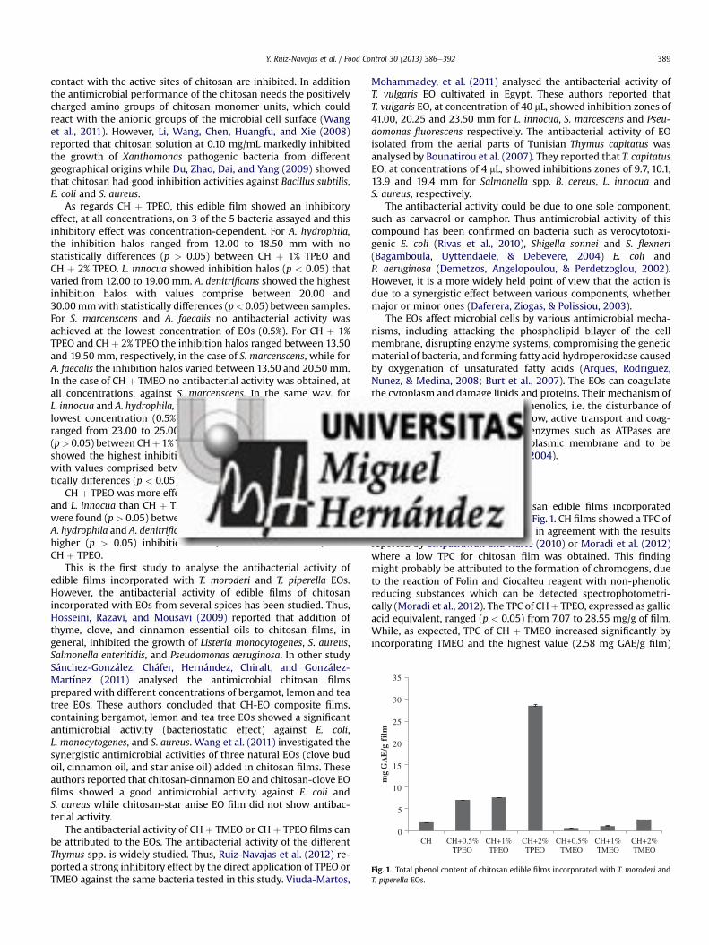
0
5
10
15
20
25
30
35
CH CH+0.5% TPEO
CH+1% TPEO
CH+2% TPEO
CH+0.5% TMEO
CH+1% TMEO
CH+2% TMEO
mg
GA
E/g
fil
m
Fig. 1. Total phenol content of chitosan edible films incorporated with T. moroderi andT. piperella EOs.
Y. Ruiz-Navajas et al. / Food Control 30 (2013) 386e392 389
contact with the active sites of chitosan are inhibited. In additionthe antimicrobial performance of the chitosan needs the positivelycharged amino groups of chitosan monomer units, which couldreact with the anionic groups of the microbial cell surface (Wanget al., 2011). However, Li, Wang, Chen, Huangfu, and Xie (2008)reported that chitosan solution at 0.10 mg/mL markedly inhibitedthe growth of Xanthomonas pathogenic bacteria from differentgeographical origins while Du, Zhao, Dai, and Yang (2009) showedthat chitosan had good inhibition activities against Bacillus subtilis,E. coli and S. aureus.
As regards CH þ TPEO, this edible film showed an inhibitoryeffect, at all concentrations, on 3 of the 5 bacteria assayed and thisinhibitory effect was concentration-dependent. For A. hydrophila,the inhibition halos ranged from 12.00 to 18.50 mm with nostatistically differences (p > 0.05) between CH þ 1% TPEO andCH þ 2% TPEO. L. innocua showed inhibition halos (p < 0.05) thatvaried from 12.00 to 19.00 mm. A. denitrificans showed the highestinhibition halos with values comprise between 20.00 and30.00mmwith statistically differences (p< 0.05) between samples.For S. marcenscens and A. faecalis no antibacterial activity wasachieved at the lowest concentration of EOs (0.5%). For CH þ 1%TPEO and CH þ 2% TPEO the inhibition halos ranged between 13.50and 19.50 mm, respectively, in the case of S. marcenscens, while forA. faecalis the inhibition halos varied between 13.50 and 20.50 mm.In the case of CH þ TMEO no antibacterial activity was obtained, atall concentrations, against S. marcenscens. In the same way, forL. innocua and A. hydrophila, no inhibitory effect was obtained at thelowest concentration (0.5%). For A. faecalis, the inhibition halosranged from 23.00 to 25.00 mm with no statistically differences(p> 0.05) between CHþ 1% TPEO and CHþ 2% TPEO. A. denitrificansshowed the highest inhibition halos, at the higher concentration,with values comprised between 20.00 and 29.50 mm with statis-tically differences (p < 0.05) between samples.
CH þ TPEO was more effective (p < 0.05) against S. marcenscensand L. innocua than CH þ TMEO, while no statistically differenceswere found (p > 0.05) between CH þ TPEO and CH þ TMEO againstA. hydrophila and A. denitrificans. For A. faecalis, CHþ TMEO showedhigher (p > 0.05) inhibition halos, at all concentrations, thanCH þ TPEO.
This is the first study to analyse the antibacterial activity ofedible films incorporated with T. moroderi and T. piperella EOs.However, the antibacterial activity of edible films of chitosanincorporated with EOs from several spices has been studied. Thus,Hosseini, Razavi, and Mousavi (2009) reported that addition ofthyme, clove, and cinnamon essential oils to chitosan films, ingeneral, inhibited the growth of Listeria monocytogenes, S. aureus,Salmonella enteritidis, and Pseudomonas aeruginosa. In other studySánchez-González, Cháfer, Hernández, Chiralt, and González-Martínez (2011) analysed the antimicrobial chitosan filmsprepared with different concentrations of bergamot, lemon and teatree EOs. These authors concluded that CH-EO composite films,containing bergamot, lemon and tea tree EOs showed a significantantimicrobial activity (bacteriostatic effect) against E. coli,L. monocytogenes, and S. aureus. Wang et al. (2011) investigated thesynergistic antimicrobial activities of three natural EOs (clove budoil, cinnamon oil, and star anise oil) added in chitosan films. Theseauthors reported that chitosan-cinnamon EO and chitosan-clove EOfilms showed a good antimicrobial activity against E. coli andS. aureus while chitosan-star anise EO film did not show antibac-terial activity.
The antibacterial activity of CH þ TMEO or CH þ TPEO films canbe attributed to the EOs. The antibacterial activity of the differentThymus spp. is widely studied. Thus, Ruiz-Navajas et al. (2012) re-ported a strong inhibitory effect by the direct application of TPEO orTMEO against the same bacteria tested in this study. Viuda-Martos,
Mohammadey, et al. (2011) analysed the antibacterial activity ofT. vulgaris EO cultivated in Egypt. These authors reported thatT. vulgaris EO, at concentration of 40 mL, showed inhibition zones of41.00, 20.25 and 23.50 mm for L. innocua, S. marcescens and Pseu-domonas fluorescens respectively. The antibacterial activity of EOisolated from the aerial parts of Tunisian Thymus capitatus wasanalysed by Bounatirou et al. (2007). They reported that T. capitatusEO, at concentrations of 4 mL, showed inhibitions zones of 9.7, 10.1,13.9 and 19.4 mm for Salmonella spp. B. cereus, L. innocua andS. aureus, respectively.
The antibacterial activity could be due to one sole component,such as carvacrol or camphor. Thus antimicrobial activity of thiscompound has been confirmed on bacteria such as verocytotoxi-genic E. coli (Rivas et al., 2010), Shigella sonnei and S. flexneri(Bagamboula, Uyttendaele, & Debevere, 2004) E. coli andP. aeruginosa (Demetzos, Angelopoulou, & Perdetzoglou, 2002).However, it is a more widely held point of view that the action isdue to a synergistic effect between various components, whethermajor or minor ones (Daferera, Ziogas, & Polissiou, 2003).
The EOs affect microbial cells by various antimicrobial mecha-nisms, including attacking the phospholipid bilayer of the cellmembrane, disrupting enzyme systems, compromising the geneticmaterial of bacteria, and forming fatty acid hydroperoxidase causedby oxygenation of unsaturated fatty acids (Arques, Rodriguez,Nunez, & Medina, 2008; Burt et al., 2007). The EOs can coagulatethe cytoplasm and damage lipids and proteins. Their mechanism ofaction would be similar to other phenolics, i.e. the disturbance ofthe proton motive force, electron flow, active transport and coag-ulation of cell contents. Instead, enzymes such as ATPases areknown to be located in the cytoplasmic membrane and to bebordered by lipid molecules (Burt, 2004).
3.2. Total phenol content
Total phenolic content of chitosan edible films incorporatedwith TPEO and TMEOwas shown in Fig. 1. CH films showed a TPC of1.95 mg GAE/g film. This result was in agreement with the resultsreported by Siripatrawan and Harte (2010) or Moradi et al. (2012)where a low TPC for chitosan film was obtained. This findingmight probably be attributed to the formation of chromogens, dueto the reaction of Folin and Ciocalteu reagent with non-phenolicreducing substances which can be detected spectrophotometri-cally (Moradi et al., 2012). The TPC of CHþ TPEO, expressed as gallicacid equivalent, ranged (p < 0.05) from 7.07 to 28.55 mg/g of film.While, as expected, TPC of CH þ TMEO increased significantly byincorporating TMEO and the highest value (2.58 mg GAE/g film)

Y. Ruiz-Navajas et al. / Food Control 30 (2013) 386e392390
was for the film formulated with 2% TMEO. The CH films made withTPEO showed higher TFC values (p < 0.05), at all concentrations,than the CH films elaborated with TMEO.
The phenolic content can be used as an important indicator ofantioxidant capacity which can be used as a preliminary screen forany product when intended as a natural source of antioxidants infunctional foods (Viuda-Martos, Ruiz-Navajas, et al., 2011). Thus,the polyphenolic compounds are very important plant constitu-ents, by virtue of their antioxidant activity by chelatingredox-active metal ions, inactivating lipid free radical chains andpreventing hydroperoxide conversion into reactive oxyradicals(Cabral de Oliveira et al., 2009).
45
50
3.3. Antioxidant activity
Since the antioxidant capacity of food is determined bya mixture of different antioxidants with different action mecha-nisms, among which synergistic interactions, it is necessary tocombine more than one method in order to determine in vitro, theantioxidant capacity of foodstuffs (Pérez-Jiménez et al., 2008).
Therefore the antioxidant activities of the edible films ofCH þ TPEO and CH þ TMEO were investigated using DPPH radical-scavenging assay, FIC assay as well as FRAP assay.
DPPH is a compound that consists of a nitrogen free radicalwhich is easily quenched by proton radical scavenger of hydrogendonating antioxidant and subsequently transformed into a non-radical form (DPPH-H) (Prior, Wu, & Schaich, 2005). CH films(Table 2) showed a slight scavenging activity ct activity (0.003 mgTrolox Equivalent (TE)/g film). The scavenging mechanism of chi-tosan is related to the fact that free radical can react with theresidual free amino (NH2) groups to form stable macromoleculeradicals, and the NH2 groups can form ammonium (NH3
þ) groups byabsorbing a hydrogen ion from the solution (Yen, Yang, & Mau,2008). The scavenging capacities of the CH þ TPEO films (Table 2)significantly increased (p < 0.05) with increasing TPEO concen-tration. The results showed that DPPH scavenging activity of thefilms varied (p < 0.05) from 0.69 to 1.09 mg TE/g film. In the case ofCH þ TMEO films, the antioxidant activity values ranged between0.42 and 0.63 mg TE/g film, and occur in a concentration-dependent manner. The CH þ TMEO films showed lower(p < 0.05) antioxidant activity, at all concentrations assayed, thanCH þ TPEO.
The FRAP assay is commonly used to study the antioxidantcapacity of plant materials. The antioxidant capacity of films isdetermined by the ability of the antioxidants in these extracts toreduce ferric to ferrous iron in FRAP reagent. CH film showeda slight ability to reduce ferric to ferrous iron (0.21 mg TE/g film).The analysed CH þ TPEO showed a wide range of antioxidant
Table 2Antioxidant effect of chitosan edible films incorporated with different concentra-tions of T. moroderi and T. piperella EOs by means of two different antioxidant test(DPPH and FRAP assays).
DPPH (mg TE/g) FRAC (mg TE/g)
CH 0.003 � 0.00aA 0.21 � 0.00aA
CH þ 0.5% TPEO 0.69 � 0.00aB 2.79 � 0.13aB
CH þ 1% TPEO 1.03 � 0.01bC 5.42 � 0.07bC
CH þ 2% TPEO 1.07 � 0.01cD 10.09 � 0.13cD
CH þ 0.5% TMEO 0.42 � 0.00aE N.A.CH þ 1% TMEO 0.54 � 0.00bF N.A.CH þ 2% TMEO 0.63 � 0.00cG 0.67 � 0.09aE
For a same essential oil, values followed by the same lower case letter are notsignificantly different (p > 0.05) according to Tukey’s Multiple Range Test.For a same antioxidant assay, values followed by the same upper case letter are notsignificantly different (p > 0.05) according to Tukey’s Multiple Range Test.
capacities, with values (p < 0.05) between 2.79 and 10.09 mg TE/gof film. As in the case of DPPH the antioxidant capacity occur ina concentration dependent manner. As regards CH þ TMEO film, atlow concentrations (0.5 and 1%) no antioxidant activity was found.CH þ 2% TMEO showed a low ability to reduce ferric to ferrous iron(0.67 mg TE/g film). The CH þ TPEO films showed higher (p < 0.05)ability to reduce ferric to ferrous ion activity, at all concentrationsassayed, than CH þ TMEO.
FIC assay measures how effective the compounds in it cancompete with ferrozine for ferrous ion. By forming a stable iron (II)chelate, an extract with high chelating power reduces the freeferrous ion concentration thus decreasing the extent of Fentonreaction (Halliwell & Gutteridge, 1990). Analysis of metal ion-chelating properties (Fig. 2) showed that CH þ TPEO films studiedwere capable of chelating iron (II) and did so in a concentration-dependent manner (p < 0.05). In the same way, CH þ TMEOshowed a high ability to chelating iron (II) and as in the case ofCH þ TPEO its occur in a concentration-dependent manner(p < 0.05). At all concentrations (except 0.5%) CH þ TMEO filmswere better chelators of iron (II) (p< 0.05) than CHþ TPEO films. Ingeneral terms it can be said that edible chitosan films showed highchelating activity, which is of great significance because thechelation of transition metals is of great potential interest in thefood industry (Viuda-Martos, Ruiz-Navajas, et al., 2010).
Few studies have examined the antioxidant activity of chitosanedible films incorporated with spices EOs (Altiok, Altiok, &Tihminlioglu, 2010; Moradi et al., 2012). This antioxidant activitycould be related with the presence, in the edible films, of bioactivecompounds such as phenolics acids of terpenoids coming from theessential oils. Thus, the phenolic and terpenoids compoundspresent in the chemical composition of EOs are closely associatedwith their antioxidant function mainly due to their redox proper-ties exerted by various possible mechanisms: free-radical scav-enging activity, hydrogen donors, transition-metal-chelatingactivity, and/or singlet-oxygen-quenching capacity (Liyana-Pathirana & Shahidi, 2006).
The antioxidant activity of EOs obtained from plants belonged togenus Thymus is widely demonstrated (Sarikurkcu et al., 2010;Viuda-Martos, El Gendy, et al., 2010; Zouari et al., 2011). Theactivities of EOs such as antioxidants depend on several structuralfeatures of the molecules and are primarily attributed to the highreactivity of hydroxyl groups substituents (�Cí�z et al., 2010), but alsoon many other factors, such as concentration, temperature, light,type of substrate, physical state of the system, as well as on micro-
0
5
10
15
20
25
30
35
40
CH CH+0.5%TPEO
CH+1%TPEO
CH+2%TPEO
CH+0.5%TMEO
CH+1%TMEO
CH+2%TMEO
Che
lati
ng a
ctiv
ity
(% in
hibi
tion
)
Fig. 2. Chelating activity of chitosan edible films incorporated with the EOs, evaluatedby means of FIC assay, of T. moroderi and T. piperella at different concentrations.

Y. Ruiz-Navajas et al. / Food Control 30 (2013) 386e392 391
components acting as pro-oxidants or synergists may influence theantioxidant activity (Yanishlieva-Maslarova, 2001).
In order to infer a major contributing effect of the total phenoliccontent (TPC) to the total antioxidant activities of methanolicextracts of edible films of CH þ TPEO and CH þ TMEO, linearcorrelation studies were performed with the three antioxidantassays (DPPH, FRAC and FIC). Thus, a significant correlation wasobserved between TPC of CH edible films incorporated with TPEOand TMEO and the DPPH radical-scavenging activity (R2¼ 0.928). Inthe same way, for FRAP assay, there was again a high correlationbetween this assay and the TPC of the CH edible films (R2 ¼ 0.914).However, a low correlation was obtained between FIC assay andTPC of the CH edible films added with EOs (R2 ¼ 0.051). A positiveand significant correlation has also been obtained between theTPC and antioxidant activity by other authors (Baltru�saityte,Venskutonis, & Ceksteryte, 2007; Contreras-Calderón, Calderón-Jaimes, Guerra-Hernández, & García-Villanova, 2011; Ruiz-Navajaset al., 2011).
4. Conclusions
The incorporation of T. piperella and T. moroderi essential oils tochitosan edible films may have supplementary applications in foodpackaging. Thus, the results showed that chitosan edible filmsincorporated with these essential oils could be used as active filmsdue to its excellent in vitro antibacterial and antioxidant activities.However, further studies are necessary to consider, the chitosanedible film, an additional stress factor for preserving food products,assuring its quality as well as a prolonged shelf-life.
Acknowledgements
We are grateful to Caja Murcia for the research scholarshipawarded to one of the authors (YRN).
References
Altiok, D., Altiok, E., & Tihminlioglu, F. (2010). Physical, antibacterial and antioxidantproperties of chitosan films incorporated with thyme oil for potential woundhealing applications. Journal of Materials Science: Materials in Medicine, 21,2227e2236.
Arques, J. L., Rodriguez, E., Nunez, M., & Medina, M. (2008). Inactivation of Gram-negative pathogens in refrigerated milk by reuterin in combination with nisinor the lactoperoxidase system. European Food Research and Technology, 227(1),77e82.
Avila-Sosa, R., Palou, E., Jiménez-Munguía, M. T., Nevárez-Moorillón, G. V., Navarro-Cruz, A. R., & López-Malo, A. (2012). Antifungal activity by vapor contact ofessential oils added to amaranth, chitosan, or starch edible films. InternationalJournal of Food Microbiology, 153, 66e72.
Bagamboula, C. F., Uyttendaele, M., & Debevere, J. (2004). Inhibitory effect of thymeand basil essential oils, carvacrol, thymol, estragol, linalool and p-cymenetowards Shigella sonnei and S. flexneri. Food Microbiology, 21, 33e42.
Baltru�saityte, V., Venskutonis, P. R., & Ceksteryte, V. (2007). Radical scavengingactivity of different floral origin honey and beebread phenolic extracts. FoodChemistry, 101, 502e514.
Bounatirou, S., Smiti, S., Miguel, M. G., Faleiro, L., Rejeb, M. N., Neffati, M., et al.(2007). Chemical composition, antioxidant and antibacterial activities of theessential oils isolated from Tunisian Thymus capitatus Hoff. et Link. FoodChemistry, 105, 146e155.
Burt, S. (2004). Essential oils: their antibacterial properties and potential applica-tions in food: a review. International Journal of Food Microbiology, 94, 223e253.
Burt, S. A., Der Zee, R. V., Koets, A. P., De Graaff, A. M., Van Knapen, F., Gaastra, W.,et al. (2007). Carvacrol induces heat shock protein 60 and inhibits synthesis offlagellin in Escherichia coli O157:H7. Applied and Environmental Microbiology, 73,4484e4490.
Cabral de Oliveira, A., Barros Valentim, I., Silva, C. A., Bechara, E. J. H., Paes deBarros, M., Mano, C. M., et al. (2009). Total phenolic content and free radicalscavenging activities of methanolic extract powders of tropical fruit residues.Food Chemistry, 115, 469e475.
Carter, P. (1971). Spectrophotometric determination of serum iron at the submi-crogram level with a new reagent (ferrozine). Analytical Biochemistry, 40,450e458.
�Cí�z, M., �Cí�zová, H., Denev, P., Kratchanova, M., Slavov, A., & Lojek, A. (2010). Differentmethods for control and comparison of the antioxidant properties of vegeta-bles. Food Control, 21, 518e523.
Contreras-Calderón, J., Calderón-Jaimes, L., Guerra-Hernández, E., & García-Villanova, B. (2011). Antioxidant capacity, phenolic content and vitamin C inpulp, peel and seed from 24 exotic fruits from Colombia. Food Research Inter-national, 44, 2047e2053.
Daferera, D. J., Ziogas, B. N., & Polissiou, M. G. (2003). The effectiveness of plantessential oils on the growth of Botrytis cinerea, Fusarium sp. and Clavibactermichiganensis subsp. Michiganensis. Crop Protection, 22, 39e44.
Demetzos, C., Angelopoulou, D., & Perdetzoglou, D. (2002). A comparative study ofthe essential oils of Cistus salviifolius in several populations of Crete (Greece).Biochemical Systematics and Ecology, 30, 651e665.
Du, Y., Zhao, Y., Dai, H., & Yang, B. (2009). Preparation of water-soluble chitosanfrom shrimp shell and its antibacterial activity. Innovative Food Science &Emerging Technologies, 10, 103e107.
Halliwell, B., & Gutteridge, J. M. C. (1990). Role of free radicals and catalytic metalions in human disease: an overview. Methods in Enzymology, 186, 1e85.
Hosseini, M. H., Razavi, S. H., & Mousavi, M. A. (2009). Antimicrobial, physical andmechanical properties of chitosan based films incorporated with thyme, cloveand cinnamon essential oils. Journal of Food Processing and Preservation, 33,727e743.
Kanatt, S. R., Chander, R., & Sharma, A. (2008). Chitosan and mint mixture: a newpreservative for meat and meat products. Food Chemistry, 107, 845e852.
Kim, K. W., Min, B. J., Kim, Y. T., Kimmel, R. M., Cooksey, K., & Park, S. I. (2011).Antimicrobial activity against foodborne pathogens of chitosan biopolymerfilms of different molecular weights. LWT-Food Science and Technology, 44,565e569.
Li, B., Wang, X., Chen, R., Huangfu, W., & Xie, G. (2008). Antibacterial activity ofchitosan solution against Xanthomonas pathogenic bacteria isolated fromEuphorbia pulcherrima. Carbohydrate Polymers, 72, 287e292.
Liyana-Pathirana, C. M., & Shahidi, F. (2006). Antioxidant properties of commercialsoft and hard winter wheats (Triticum aestivum L.) and their milling fractions.Journal of the Science and Food Agricultural, 86, 477e485.
Moradi, M., Tajik, H., Rohani, S. M. R., Oromiehie, A. R., Malekinejad, H., Aliakbarlu, J.,et al. (2012). Characterization of antioxidant chitosan film incorporated withZataria multiflora Boiss essential oil and grape seed extract. LWT-Food Scienceand Technology, 46, 477e484.
Nickavar, B., Mojab, F., & Dolat-Abadi, R. (2005). Analysis of the essential oils of twoThymus species from Iran. Food Chemistry, 90, 609e611.
Ojagh, S. M., Rezaei, M., Razavi, S. H., & Hosseini, S. M. H. (2010). Development andevaluation of a novel biodegradable film made from chitosan and cinnamonessential oil with low affinity toward water. Food Chemistry, 122, 161e166.
Ouattara, B., Ronald, E., Simard, R. E., Piette, G., Begin, A., & Holley, R. A. (2000).Inhibition of surface spoilage bacteria in processed meats by application ofantimicrobial films prepared with chitosan. International Journal of FoodMicrobiology, 62, 139e148.
Oyaizu, M. (1986). Studies on products of browning reaction: antioxidative activityof products of browning reaction prepared from glucosamine. Japan Journal ofNutrition, 44, 307e315.
Park, S., Marsh, K. S., & Dawson, P. (2010). Application of chitosan-incorporatedLDPE film to sliced fresh red meats for shelf life extension. Meat Science, 85,493e499.
Pérez-Jiménez, J., Arranz, S., Tabernero, M., Díaz-Rubio, E., Serrano, J., Goñi, I., et al.(2008). Updated methodology to determine antioxidant capacity in plant foods,oils and beverages: extraction, measurement and expression of results. FoodResearch International, 41(3), 274e285.
Petrou, S., Tsiraki, M., Giatrakou, V., & Savvaidis, I. N. (2012). Chitosan dipping ororegano oil treatments, singly or combined on modified atmosphere packagedchicken breast meat. International Journal of Food Microbiology, 156, 264e271.
Ponce, A. G., Roura, S. I., del Valle, C. E., & Moreira, M. R. (2008). Antimicrobial andantioxidant activities of edible coatings enriched with natural plant extracts:in vitro and in vivo studies. Postharvest Biology and Technology, 49, 294e300.
Prior, R. L., Wu, X., & Schaich, K. (2005). Standardized methods for the determi-nation of antioxidant capacity in phenolics in foods and dietary supplements.Journal of Agricultural and Food Chemistry, 53, 4290e4302.
Rivas, L., McDonnell, M. J., Burgess, M. C., O’Brien, M., Navarro-Villa, A., Fanning, S.,et al. (2010). Inhibition of verocytotoxigenic Escherichia coli in model broth andrumen systems by carvacrol and thymol. International Journal of Food Microbi-ology, 139, 70e78.
Ruiz-Navajas, Y., Viuda-Martos, M., Fernández-López, J., Zaldivar-Cruz, J. M., Kuri, V.,& Pérez-Álvarez, J. A. (2011). Antioxidant activity of artisanal honey fromTabasco, Mexico. International Journal of Food Properties, 14, 459e470.
Ruiz-Navajas, Y., Viuda-Martos, M., Sendra, E., Pérez-Álvarez, J. A., & Fernández-López, J. (2012). Chemical characterization and antibacterial activity of Thymusmoroderi and Thymus piperella essential oils, two Thymus endemic species fromsoutheast of Spain. Food Control, 27(2), 294e299.
Salmieri, S., & Lacroix, M. (2006). Physicochemical properties of alginate/polycaprolactone-based films containing essential oils. Journal of Agriculturaland Food Chemistry, 54, 10205e10214.
Sánchez-González, L., Cháfer, M., Hernández, M., Chiralt, A., & González-Martínez, C.(2011). Antimicrobial activity of polysaccharide films containing essential oils.Food Control, 22, 1302e1310.
Sarikurkcu, C., Ozer, M. S., Eskici, M., Tepe, B., Can, S., & Mete, E. (2010).Essential oil composition and antioxidant activity of Thymus longicaulis C.

Y. Ruiz-Navajas et al. / Food Control 30 (2013) 386e392392
Presl subsp. Longicaulis var. longicaulis. Food and Chemical Toxicology, 48,1801e1805.
Singleton, V. L., & Rossi, J. A. (1965). Colorimetry of total phenolics with phospho-molybdic phosphotungstic acid reagents. American Journal of Enology and Viti-culture, 16, 144e158.
Siripatrawan, U., & Harte, B. R. (2010). Physical properties and antioxidant activity ofan active film from chitosan incorporated with green tea extract. Food Hydro-colloids, 24, 770e775.
Tedone, L., D’andrea, L., & Marzi, V. (2001). Caratterizzazione biomorfologica evalutazione agronomica di specie e popolazioni di timo (Thymus spp.) nell’areamediterranea. In Atti del VI Convegno delle Biodiversità, Valenzano, 6 settembre(pp. 1134e1142).
Tepe, B., Daferera, D., Sokmen, A., Sokmen, M., & Polissiou, M. (2005). Antimicrobialand antioxidant activities of the essential oils and various extracts of Salviatomentosa Miller (Lamiaceae). Food Chemistry, 90, 333e340.
Tharanathan, R. N., & Kittur, F. (2003). Chitin e the undisputed biomolecule of greatpotential. Critical Reviews in Food Science, 43(1), 61e87.
Viuda-Martos, M., El Gendy, N. G. S., Sendra, E., Fernández-López, J., El-Razik, K. A. A.,El-Sayed, A., et al. (2010). Chemical composition and antioxidant and anti-listeriaactivities of essential oils obtained from some Egyptian plants. Journal of Agri-cultural and Food Chemistry, 58, 9063e9070.
Viuda-Martos, M., Mohammadey, M., Fernández-López, J., El-Razik, K. A. A.,Omer, E. A., Pérez-Alvarez, J. A., et al. (2011). In vitro antioxidant and antibac-terial activities of essentials oils obtained from Egyptian aromatic plants. FoodControl, 22, 1715e1722.
Viuda-Martos, M., Ruiz-Navajas, Y., Fernández-López, J., & Pérez-Álvarez, J. A.(2008). Antibacterial activity of different essential oils obtained from spiceswidely used in Mediterranean diet. International Journal of Food Science andTechnology, 43, 526e531.
Viuda-Martos, M., Ruiz-Navajas, Y., Fernández-López, J., Sendra, E., Sayas-Barberá, E., & Pérez-Álvarez, J. A. (2011). Antioxidant properties of pomegranate(Punica granatum L.) bagasses obtained as co-product in the juice extraction.Food Research International, 44, 1217e1223.
Viuda-Martos, M., Ruiz-Navajas, Y., Sánchez-Zapata, E., Fernández-López, J., & Pérez-Alvarez, J. A. (2010). Antioxidant activity of essential oils of five spice plantswidely used in Mediterranean diet. Flavour and Fragrance Journal, 25, 13e19.
Wang, L., Liu, F., Jiang, Y., Chai, Z., Li, P., Cheng, Y., et al. (2011). Synergistic antimi-crobial activities of natural essential oils with chitosan films. Journal of Agri-cultural and Food Chemistry, 59, 12411e12419.
Yanishlieva-Maslarova, N. V. (2001). Inhibiting oxidation. In J. Pokorny,N. Yanishlieva, & M. Gordon (Eds.), Antioxidants in food: Practical applications(pp. 22e69). Cambridge, U.K.: Woodhead Publishing Ltd.
Yen, M., Yang, J., & Mau, J. (2008). Antioxidant properties of chitosan from crabshells. Carbohydrate Polymers, 74, 840e844.
Zivanovic, S., Chi, S., & Draughon, A. F. (2005). Antimicrobial activity of chitosanfilms enriched with essential oils. Journal of Food Science, 70(1), 45e51.
Zouari, N., Fakhfakh, N., Zouari, S., Bougatef, A., Karray, A., Neffati, M., et al. (2011).Chemical composition, angiotensin I-converting enzyme inhibitory, antioxidantand antimicrobial activities of essential oil of Tunisian Thymus algeriensis Boiss.et Reut. (Lamiaceae). Food and Bioproducts Processing, 89, 257e265.
Título: Effect of chitosan edible films added with Thymus moroderi and Thymus piperella essential oil on
shelf-life of cooked cured ham
Autores: Y. Ruiz-Navajas, M. Viuda-Martos, X. Barber, E. Sendra, J.A. Pérez-Alvarez, J. Fernández-
López
Revista: Meat Science
Año: 2014

Elsevier Editorial System(tm) for Meat Science Manuscript Draft Manuscript Number: Title: Effect of chitosan edible films added with Thymus moroderi and Thymus piperella essential oil on shelf-life of cooked cured ham Article Type: Research Article Keywords: Essential oils; chitosan; films; Shelf-life; Cooked cured ham Corresponding Author: Dr. Juana Fernandez-Lopez, PhD Corresponding Author's Institution: Universidad Miguel Hernández First Author: Yolanda Ruíz-Navajas Order of Authors: Yolanda Ruíz-Navajas; Manuel Viuda-Martos; Xavier Barber; Esther Sendra; José Angel Pérez-Alvarez; Juana Fernandez-Lopez, PhD Abstract: The aim of this work was to develop chitosan edible films added with essential oils obtained from two Thymus species, Thymus moroderi (TMEO) and Thymus piperella (TPEO) to determine their application for enhancing safety (antioxidant and antibacterial properties) and shelf-life of cooked cured ham (CCH) storage at 4 ºC during 21 days. Addition of TMEO and TPEO into chitosan films decreased the aerobic mesophilic bacteria (AMB) and lactic acid bacteria (LAB) counts in coated cooked cured ham samples as compared with uncoated samples. Both AMB and LAB showed the lowest counts in CCH samples coated with chitosan films added with TPEO at 2%. As regards lipid oxidation, the CCH samples coated with chitosan films added with TMEO or TPEO had lower degrees of lipid oxidation than uncoated control samples. Again, chitosan films added with TPEO at 2% showed the lowest values. The addition of TPEO or TMEO in chitosan films used as coated in CCH improved their shelf life Suggested Reviewers: Marcelo Raul Rosmini Universidad Católica de Córdoba, Argentine [email protected] Clemencia Chaves-López Universitat di Teramo, Italy [email protected]

1
Effect of chitosan edible films added with Thymus moroderi and Thymus piperella 1
essential oil on shelf-life of cooked cured ham 2
3
4
Y. Ruiz-Navajas1, M. Viuda-Martos
1, X. Barber
2, E. Sendra
1, J.A. Perez-Alvarez
1, J. 5
Fernández-López1
6
7
8
9
1IPOA Research Group (UMH-1 and REVIV-Generalitat Valenciana), AgroFood 10
Technology Department, Escuela Politecnica Superior de Orihuela, Miguel Hernandez 11
University, Alicante, (Spain). 12
13
2Applied statistical Unit, Operations Research Center, Miguel Hernandez University, 14
Elche, Alicante, Spain 15
16
17
18
19
20
Corresponding author: Dr. Juana Fernández-López. AgroFood Technology Department. 21
Miguel Hernández University. Ctra. Beniel Km 3,2. E-03312. Orihuela, Alicante 22
(Spain). E-mail: [email protected] Tel.: +34 966749784; Fax: +34 966749677. 23
24
25

2
Abstract 26
The aim of this work was to develop chitosan edible films added with essential oils 27
obtained from two Thymus species, Thymus moroderi (TMEO) and Thymus piperella 28
(TPEO) to determine their application for enhancing safety (antioxidant and 29
antibacterial properties) and shelf-life of cooked cured ham (CCH) storage at 4 ºC 30
during 21 days. 31
Addition of TMEO and TPEO into chitosan films decreased the aerobic mesophilic 32
bacteria (AMB) and lactic acid bacteria (LAB) counts in coated cooked cured ham 33
samples as compared with uncoated samples. Both AMB and LAB showed the lowest 34
counts in CCH samples coated with chitosan films added with TPEO at 2%. As regards 35
lipid oxidation, the CCH samples coated with chitosan films added with TMEO or 36
TPEO had lower degrees of lipid oxidation than uncoated control samples. Again, 37
chitosan films added with TPEO at 2% showed the lowest values. 38
The addition of TPEO or TMEO in chitosan films used as coated in CCH improved 39
their shelf life 40
41
42
43
Keywords: Essential oils, chitosan, films, Shelf-life, Cooked cured ham 44
45
46
47
48
49
50
3
1. Introduction 51
At the present there is great variety of ready-to-eat (RTE) foods including pre-packed 52
and precooked meats, in individual or family-size packages, with very different 53
presentations. However, the transformation into RTE food of meat product such as 54
cooked ham, involves additional manipulation such as cutting, slicing, dicing and 55
packaging aimed at facilitating its consumption at home (Gil-Díaz, Santos-Delgado, 56
Rubio-Barroso & Polo-Díez, 2009). This may contribute considerably to the 57
contamination of RTE meat products with pathogenic bacteria. For this reason there is 58
a need for continuous supervision of RTE food products to ensure safety to consumers 59
in the industrial countries. The increased demands by consumers for better quality and 60
improved freshness of RTE food products have given rise to the development and 61
implementation of edible films (Beverlya, Janes, Prinyawiwatkula & No, 2008). 62
Edible films and coatings are thin layers of edible material formed or placed on or 63
between foods or food components (Bravin, Peressini & Sensidoni, 2006) which can 64
play an important role on their preservation, distribution and marketing of food products 65
(Falguera, Quintero, Jiménez, Muñoz & Ibarz, 2011). They are prepared from 66
biopolymers; the major constituents of these are polysaccharides, proteins and lipids. 67
In particular, chitosan has a great potential for a wide range of food applications due 68
to its biodegradability, biocompatibility, antimicrobial and antioxidant activities, 69
nontoxicity and film-forming capacity (Tharanathan & Kittur, 2003). The use of edible 70
films helps to maintain product quality, enhance sensory properties, improve product 71
safety, and increase the shelf life of various RTE food products (Beverlya et al., 2008). 72
Moreover, they can act as carriers of active substances, such as antioxidant, 73
antimicrobial or flavoring compounds, resulting in shelf-life extension and safety 74
improvement of the food product. One of these active substances is the essential oils 75

4
(EO). The ability of EOs to protect foods against pathogenic and spoilage 76
microorganisms as well as the oxidation have been reported by several researchers 77
(Ruiz-Navajas, Viuda-Martos, Sendra, Perez-Alvarez & Fernández-López, 2013; Alves-78
Silva et al. 2013). In order to achieve effective antimicrobial activity in direct food 79
applications, high concentrations of essential oils are generally needed, which might 80
impact inappropriate flavors and odors in the product (Seydim & Sarikus, 2006). To 81
avoid this problem, the EOs could be incorporated into bioactive film coatings which 82
would allow us to fix and retain the compound on the product surface, thus increasing 83
its effectiveness (Sánchez-González, González-Martínez, Chiralt & Cháfer, 2010). In 84
these coatings, the major compounds are biodegradable polymers and a relatively 85
reduced amount of EOs can be used. Consequently, the application costs of essential 86
oils and/or other problems, such as the intense aroma and potential toxicity, could be 87
minimized (Sánchez-González et al. 2010). 88
Several studies have shown that incorporation of EOs into chitosan films or coatings 89
may not only enhance the film’s antimicrobial and antioxidant properties but also 90
reduce water vapor permeability and down lipid oxidation of the product on which the 91
film is applied (Kanatt, Chander & Sharma, 2008) mainly meat and meat products. 92
There are several scientific works where the edible films elaborated with isolate whey 93
protein, isolate soy protein, alginate and so on and incorporated with essential oils have 94
been used to prevent the lipid oxidation and microbial growth in meat and meat product 95
(Zinoviadou, Koutsoumanis & Biliaderis, 2009; Emiroğlu, Yemiş, Coşkun & 96
Candoğan, 2010; Juck, Neetoo & Chen, 2010). However, the applications of chitosan 97
edible films added with EOs are lesser analyze. 98
The aim of this work was to develop chitosan edible film added with EOs obtained 99
from two Thymus species, Thymus moroderi and Thymus piperella to determine their 100
5
application for enhancing safety (antioxidant and antibacterial properties) and shelf-life 101
of cooked cured ham storage at 4 ºC during 21 days. 102
103
2. Materials and Methods 104
2.1. Extraction of the essential oils 105
Thymus moroderi (TM) and Thymus piperella (TP) were collected during their 106
flowering period and the identification of the plant material was made by Prof Dra. 107
Concepcion Obon de Castro, Biology Department of Miguel Hernandez University 108
(Spain). The EOs of TM and TP were extracted from whole plant (stems, leaves and 109
flowers) by hydro-distillation using a Clevenger-type apparatus for 3 h. The oily layer 110
obtained on top of the aqueous distillate was separated and dried with 0.5 g of 111
anhydrous sodium sulphate (Panreac Química, Barcelona, Spain). The extracted EOs 112
was kept in sealed air-tight glass vials and covered with aluminium foil at 4 ºC until 113
further analysis. 114
2.2. Preparation of edible films 115
Chitosan-based film was prepared by dissolving chitosan (high molecular weight 75-116
85% deacetylated, Sigma-Aldrich Chemical Co., Steinheim, Germany) in a lactic acid 117
aqueous solution (1% v/v) (Sigma-Aldrich Chemical Co., Steinheim, Germany) at a 118
concentration of 2% (w/v) while stirring on a magnetic stirrer/hot plate, following the 119
indications of Ojagh, Rezaei, Razavi and Hosseini (2010) with some modifications. 120
The chitosan solution was stirred at room temperature until it was completely 121
dissolved (24 h). The resultant chitosan solution was filtered through a Whatman No. 3 122
filter paper to remove any undissolved particles. After filtration the solution was 123
returned to the magnetic stirrer/hot plate and glycerol (Panreac Quimica, Barcelona, 124
Spain) was added to a level of 0.75 mL/g chitosan as a plasticizer. The plasticizer was 125

6
mixed into the solution for 15 min. Then, Tween 80 (Panreac, Quimica, Barcelona, 126
Spain) at level of 0.2% (v/v) of EO, was added as an emulsifier to assist EO dispersion 127
in film forming solutions. After 15 min of stirring, the following five solutions were 128
prepared by adding T. moroderi (TMEO) or T. piperella (TPEO) EOs into chitosan 129
solution: (i) chitosan without TMEO and TPEO; (ii) chitosan with 1% TMEO; (iii) 130
chitosan with 2% TMEO; (iv) chitosan with 1% TPEO; (v) chitosan with 2% TPEO. 131
Both chitosan TMEO (CH+TMEO) and chitosan TPEO (CH+TPEO) mixtures were 132
emulsified at room temperature using a rotor-stator homogenizer (Ultraturrax DI 25, 133
Janke & Kunkel, Staufen, Germany) at 20,000 rpm for 3 min. These emulsions were 134
degasified at room temperature using an ultrasonic water bath (Selecta S.A. Barcelona, 135
Spain), without temperature control, during 2 h. The film forming solutions (7 g) were 136
casted into 60 mm inner diameter sterile Petri dishes (0.25 g/cm2) covers and then dried 137
for 48 h at 37 ºC. Dried films were peeled and stored in a desiccator at 25 ºC and 51% 138
relative humidity until evaluation. Saturated magnesium nitrate (Panreac Quimica, 139
Barcelona, Spain) solution was used to meet required relative humidity. 140
2.3 Film application on cooked cured ham 141
Cooked cured hams (CCH) were purchased directly from a local producer and 142
transported immediately to the laboratory under refrigerated conditions and then they 143
were sliced (3 mm thick and 12.5 g weight). Each film with different formulations was 144
placed between two slices and was placed in a sterile bags made of polyethylene and 145
polyamide laminate of 1.1 g/m2/24 h water vapor permeability at 23 ºC 10 cm
3/m
2/24 h, 146
nitrogen permeability at 23 ºC, 140 cm3/m
2/24 h carbon dioxide permeability at 23 ºC, 147
and 30 cm3/m
2/24 h oxygen permeability at 23 ºC (Fibran, Girona, Spain). Slices 148
without any chitosan film were also prepared as control CMH samples. The bags were 149
7
heat-sealed and stored at 4±1 ºC. The packs were stored for 21 d. Samples from each 150
treatment were taken at 0, 7, 14, and 21 d (storage time) and analyzed on the same day. 151
2.4. Physico-chemical analysis 152
The CIEL*a*b* color space was studied following the procedure of Cassens et al. 153
(1995). The following color coordinates were determined: lightness (L*), redness (a*, ± 154
red-green), and yellowness (b*, ± yellow-blue). Color determinations were made, at 155
12±2 ºC by means of a Minolta CM-2600D (Minolta Camera Co., Osaka, Japan) 156
spectrophotometer with illuminant D65, 10º observer angle, 11 mm aperture for 157
illumination and 8 mm for measurement. American Meat Science Association 158
guidelines for color measurements were followed and spectrally pure glass (CRA51, 159
Minolta Co., Osaka, Japan) was put between the samples and the equipment (AMSA, 160
2012). 161
The pH was measured by blending a 5 g sample with 50 mL deionized water for 2 162
min The pH of the resultant suspension was measured with a Crison pH meter (Model 163
507, Crison, Barcelona, Spain) equipped with a Crison combination electrode (Cat. nº. 164
52, Crison, Barcelona, Spain). 165
2.5. Lipid oxidation 166
Lipid oxidation was assessed in triplicate by the 2-thiobarbituric acid (TBA) test 167
following the recommendations of Buege and Aust (1978). TBARS values were 168
calculated from a standard curve of malonaldehyde (MAD) and expressed as mg 169
MAD/kg sample. 170
2.6. Microbiological analysis 171
At each time (0, 7, 14, and 21 d), films were removed from the middle of two slices 172
with a forceps and then the slices (two slices from the same treatment, weighing 25 g) 173
were homogenized with sterile 1.5% peptone water (225 mL) in a Stomacher 400 174

8
(Colworth, London, UK) for 2 min. Total viable counts were determined on Plate Count 175
Agar, Enterobacteriaceae using Violet Red Bile Glucose Agar (VRBGA) and lactic 176
acid bacteria (LAB) were counted on double layer MRS Agar at pH 5.6. In all cases, 177
plates were incubated at 37 ºC for 48 h. molds and yeasts were determined on Rose 178
Bengal plates with chloramphenicol incubated at 28 ºC for 5 days. All results are 179
reported as log10 colony forming per gram (CFU). 180
2.7. Release of phenolic compounds from film into cooked cured hams 181
On each sampling day (0, 7, 14, and 21 d), films were removed from the middle of 182
two slices and total phenolic content (TPC) of films was determined with Folin-183
Ciocalteu reagent (Singleton & Rossi, 1965). Every sample of each film was extracted 184
with 5 mL of methanol using an ultrasonic water bath (Selecta S.A. Barcelona, Spain) 185
without temperature control during 2 h. Then, the mixtures were centrifuged at 3600 g 186
for 15 min at 4 °C. Then a volume of 0.3 mL of supernatant was introduced into the test 187
tubes followed by 2.5 mL of Folin Ciocalteu’s reagent (SigmaeAldrich Chemical Co., 188
Steinheim, Germany) (diluted 10 times with water) and 2 mL of sodium carbonate 189
(Panreac Quimica, Barcelona, Spain) (7.5% w/v). 190
The tubes were vortex-mixed, covered with parafilm and incubated at 50 ºC for 5 191
min. Absorbance at 760 nm was measured with an HP 8451 spectrophotometer (Hewlett 192
Packard, Cambridge, UK) and compared to a gallic acid calibration curve. The results 193
were expressed in mg gallic acid equivalents (GAE)/g of films, as mean of three 194
replicates. 195
2.8. Statistical assay 196
Statistical analysis and comparisons among means were carried out using the 197
statistical package SPSS 19.0 (SPSS Inc., Chicago, IL.). All the data collected for pH, 198
color, lipid oxidation, microbiological analysis and release of phenolic compounds were 199
9
analyzed by two-way analysis of variance (ANOVA) to test the effects of two fixed 200
factors: film samples (levels: control, CH, CH+TPEO 1%, CH+TPEO 2%, CH+TMEO 201
1% and CH+TMEO 2%) and time (levels: 0, 7, 14 and 21 days). Tukey's post hoc test 202
was applied for comparisons of means; differences were considered significant at 203
p<0.05. Generalized Additive Models were used in order to estimate the relationship 204
between variables with different links and transformations of the variables following the 205
recommendations of Wood (2006). 206
207
3. Results and discussion 208
The chemical composition of the essential oils used in this work was previously 209
determined by Ruiz-Navajas, Viuda-Martos, Sendra, Perez-Alvarez and Fernández-210
López (2012). In the Thymus moroderi (TMEO) the main components were camphor 211
(26.74%), 1.8-cineol (24.99%), myrcene (5.63%) and α-pinene (4.35%) while in 212
Thymus piperella (TPEO) the predominant compounds were carvacrol (31.92%), para-213
cymene (16.18%), γ-terpinene (10.11%) and α-terpineol (7.29%). 214
3.1. Physico-chemical analysis 215
Figure 1 showed the effect of chitosan films formulated with TMEO and TPEO 216
essential oils on pH values in cooked cured ham storage at 4 ºC during 21 days. In all 217
uncoated and coated samples, the pH values decreased with the storage time. In 218
uncoated CCH samples there was a slight fell (p<0.05) of pH values (6.24 at day 0 to 219
6.12 at day 21). Nevertheless, in coated CHH samples the decrease in pH values was 220
most accentuated. At the end of storage time all coated CHH samples showed pH values 221
comprised between 5.63 and 5.68 with no statistical differences (p>0.05) between them, 222
except for the sample coated with CH+TPEO2% that showed higher (p<0.05) values 223
(5.81). This decreased in pH values of coated CHH samples could be attributed to the 224

10
release of lactic acid present in chitosan films into meat product. Additionally, the 225
gradual growth of lactic bacteria in the CHH samples, which could have generate lactic 226
acid, which would lead to the gradual decrease of pH. 227
As regards to color parameters, Table 1 shows the effect of chitosan films formulated 228
with TMEO and TPEO essential oils on lightness (L*), redness (a*) and yellowness 229
(b*) coordinates in cooked cured ham storage at 4 ºC during 21 days. For lightness (L*), 230
at the end of storage time, the type of film used had no significant (p>0.05) effect on 231
this coordinate except for the sample coated with CH+TPEO 1% that showed higher 232
(p<0.05) values. In the same way, storage time had no significant (p>0.05) effect in the 233
samples coated with CH, CH+TMEO and CH+TPEO, except for the sample coated with 234
CH+TPEO 1%. The absence of any modification in this parameter may have been due 235
to the protective effect of the packing film against oxygen. However, the storage time 236
had a significant (p<0.05) effect in the uncoated CCH sample. In these samples, L* 237
values decreased from 60.43 at the outset to 58.95 at the end of the experiment. Some 238
authors reported that L* values in meat and meat products are related to (i) surface 239
water, (ii) water vapor exchanges between the products and the environment and (iii) 240
modifications of the different states of hemopigments (Fernández-López, Pérez-Alvarez 241
& Aranda-Catalá, 2000). 242
As regards redness (a*) coordinate, as occur with lightness, at the end of storage 243
time, there no were differences (p>0.05) between uncoated and coated CCH samples, 244
except for CH+TPEO 1% that showed lowest (p<0.05) values. The storage time, 245
however, had an effect (p<0.05), and the redness values fell in both uncoated and coated 246
CCH samples. This coordinate is affected by the structural integrity of the food, the 247
pigment content and disposition (water or lipid-soluble) and surface water availability 248
(Fernández-López, Sayas-Barberá, Navarro, Sendra & Pérez-Álvarez, 2005). Moreover, 249
11
decreases in redness have been related to oxidation of lipids and hemopigments 250
(Fernandez-Lopez, Sayas-Barberá, Sendra & Pérez-Alvarez, 2006). 251
For yellowness (b*) coordinate, at day 21, there no were differences (p>0.05) 252
between uncoated CCH samples and samples coated with CH or CH+1%TMEO. 253
Similarly, storage time had no effect on either of these samples. On the other hand, the 254
CCH samples coated with CH+2%TMEO, CH+1%TPEO and CH+2%TPEO were 255
affected (p<0.05) by type of films used and storage time. The behavior of b* depends to 256
a great extent on the food matrix, and it is recognized that changes (pH, oxidation 257
extent, water activity, etc.) in the matrix have the greatest influence on this coordinate in 258
many foods (Cofrades et al., 2004). 259
3.2. Lipid oxidation 260
There are several scientific works that shown the beneficial effects on lipid oxidation 261
of chitosan when it is added to meat and meat products as ingredient (Georgantelis, 262
Blekas, Katikou, Ambrosiadis & Fletouris, 2007; Sayas-Barberá et al. 2011). However, 263
to our knowledge, few studies have been conducted to analysis the antioxidant effect of 264
chitosan films or chitosan films added with EO on meat and meat products is limited. 265
Figure 2 shows the effect of chitosan films formulated with Thymus TMEO and 266
TPEO essential oils on TBA values in cooked cured ham storage at 4 ºC during 21 days. 267
At day 0, there was no significant difference (p>0.05) in TBA values between uncoated 268
and coated CCH samples. On day 7, the CCH samples coated with CH+TPEO 2% and 269
CH+TMEO 2% showed the lowest (p<0.05) oxidation degree with TBA values of 2.79 270
and 2.67 mg MA/kg sample, respectively. The CCH sample coated with chitosan film 271
without EOs added showed a slight reduction in oxidation degree (p<0.05) with regards 272
to uncoated sample. The antioxidant ability of CH films is thought to be due to 273
chelation of free ion which is released from hemoproteins of meat product during 274

12
storage (Shahidi, Arachchi & Jeon, 1999). At day 14, again the CCH samples in which 275
CH+TPEO 2% and CH+TMEO 2% was used showed the lowest TBA values (p<0.05) 276
with reductions in the oxidation degree when compared with control of 26.86 and 277
37.17% respectively with statistically differences (p<0.05) between them. At the end of 278
experiment (day 21) the CCH sample coated with CH+TPEO 2 % showed the lowest 279
(p<0.05) TBA values with a reduction in the oxidation degree regarding to uncoated 280
CCH sample of 28.37% followed by the CCH sample coated with CH+TMEO 2% 281
(p<0.05) with a reduction in the oxidation degree regarding to uncoated CCH sample of 282
25.25%. These results were in agreement with Moradi, Tajik, Rohani and Oromiehie 283
(2012) who reported that a cooked meat product coated with chitosan films containing 284
Zataria multiflora Boiss EO had lower degrees of lipid oxidation than uncoated control 285
sample. In the same way, Suman et al. (2010) showed that coating ground beef patties 286
with chitosan reduced TBARS values and improved the surface red color of patties as 287
compared to uncoated samples. Chitosan film incorporating green tea extract was 288
analyzed as active packaging for shelf life extension of pork sausages by Siripatrawan 289
and Noipha (2012). These authors reported that during the storage time (20 days), 290
significantly higher TBA values were evident in control samples than those coated with 291
chitosan film or chitosan film incorporating green tea extract. 292
The decrease in the lipid oxidation of samples coated with CH, CH+TPEO or 293
CH+TMEO may be, probably, due to a sharp release of active compounds from those 294
films during first days of storage as mentioned Moradi et al. (2012). These results 295
showed that the incorporation of EOs in the films improved the protection of the meat 296
samples against lipid oxidation. The antioxidant activity of EOs obtained from plants 297
belonged to genus Thymus is widely demonstrated (Viuda-Martos et al., 2010a; Zouari 298
et al., 2011). The antioxidant activity of EOs obtained from plants belonging to genus 299

13
Thymus can be credited to the presence of its major phenolic compounds, particularly 300
thymol and carvacrol, and their recognized impact on lipid oxidation. The antioxidant 301
activity of phenolic compounds is related to the hydroxyl groups linked to the aromatic 302
ring, which are capable of donating hydrogen atoms with electrons and stabilizing free 303
radicals (Dorman, Peltoketo, Hiltunen & Tikkanen, 2003; Yanishlieva, Marinova & 304
Pokorny, 2006). 305
Moreover, the storage conditions (4 ºC and protected from light) as well as the low 306
oxygen permeability characteristics of chitosan films may contributed to the inhibition 307
of lipid oxidation. 308
3.3. Microbial counts 309
Effect of chitosan films formulated with TMEO and TPEO essential oils on Aerobic 310
Mesophilic Bacteria (AMB) and Lactic Acid Bacteria (LAB) in cooked cured ham 311
storage at 4 ºC during 21 days are given in Figures 3 and 4. Microbial counts (aerobic 312
mesophilic bacteria and lactic acid bacteria) were (p<0.05) affected by storage time and 313
the application of chitosan edible films added with TMEO or TPEO. In our experiment, 314
no moulds and yeast were found in any of the treatments regardless of time of storage, 315
probably due to the aseptic slicing process, together with the presence of the sodium 316
chloride in the products (Viuda-Martos, Ruiz-Navajas, Fernandez-Lopez & Perez-317
Alvarez, 2010b). In the same way, no enterobacteria were found. Ouattara et al. (2000) 318
reported that indigenous Enterobacteriaceae in meat products (bologna, beef pastrami 319
and cooked ham) were inhibited by the use of antimicrobial films, containing chitosan 320
and acetic acid. 321
As regards to aerobic mesophilic bacteria (Figure 3), the log CFU/g of the uncoated 322
and those samples coated with chitosan added with TMEO and TPEO showed no 323
significant difference (p>0.05) at day 0. However, on day 7, the CCH samples coated 324
14
with CH+TMEO or CH+TPEO at 2% showed the lowest counts (p<0.05) with 2.57 and 325
2.61 log cycle reduction, respectively, with regard to uncoated sample. CCH sample 326
coated with CH showed a 0.62 log reduction relating to uncoated sample demonstrating 327
a certain antibacterial activity of chitosan. The antimicrobial effect of chitosan is 328
thought to be related to electrostatic interaction between a positive charge on the NH+3
329
group of glucosamine monomer in chitosan molecules and negative charge of microbial 330
cell membrane that leads to the leakage of intracellular constituents (Dutta, Tripathi, 331
Mehrotra & Dutta, 2009). At day 14, CCH samples coated with CH+TMEO 2%, 332
CH+TPEO 1% and CH+TPEO 2% showed the lowest counts (p<0.05) with no 333
statistically differences between them (p>0.05). At the end of experiment (day 21) the 334
CCH sample coated with CH+TPEO 2 % showed the lowest counts (p<0.05) in aerobic 335
mesophilic bacteria with a reduction of 0.87 log cycles regarding to uncoated CCH 336
sample. CCH samples coated with CH+TMEO 1%, CH+TMEO 2% and CH+TPEO 1% 337
showed a 0.34, 0.53 and 0.37 log cycle reduction, respectively, in aerobic mesophilic 338
bacteria counts when compared to the control, with no statistically differences between 339
them (p>0.05). 340
In the case of Lactic acid bacteria (Figure 4), as occur with mesophilic bacteria, at 341
day 0 no statistically differences were found (p>0.05) between uncoated and coated 342
CCH samples. On day 7, the CCH sample coated with CH+TPEO 2% showed the 343
lowest (p<0.05) counts with 2.16 log cycle reduction with regard to uncoated CCH 344
sample. In CCH samples coated with CH+TMEO 1%, CH+TMEO 2% and CH+TPEO 345
1% no significant difference between them (p>0.05) were found. The reduction of the 346
counts, when compared with the uncoated CCH sample, was 1.89, 1.91 and 1.93 log 347
cycles, respectively. The same behavior was observed at day 14. At the end of the 348
analysis, day 21, no statistically differences (p>0.05) were found between uncoated 349

15
CCH sample and the coated CCH sample with chitosan. Again, the CCH samples 350
coated with CH+TPEO 2% showed the lowest (p<0.05) counts for lactic acid bacteria 351
followed by the coated CCH sample in which CH+TMEO 2% was employed, with a 352
reduction in counts, when compared with the uncoated CCH sample of 1.14 and 0.84 353
log cycles. In coated CCH samples in which CH+TMEO 1% and CH+TP 1% was used 354
no significant differences (p>0.05) between them were found, However, significant 355
differences (p<0.05) were observed between these samples and uncoated CCH sample. 356
The use of chitosan edible films or chitosan edible films added with essential oils in 357
meat, meat product and RTE meat products helps to maintain product quality, improve 358
product safety, and increase the shelf life. The purpose of incorporating antimicrobial 359
compounds into an edible film instead of applying them directly onto the meat surface 360
by spraying or dipping was to extend delivery of the antimicrobials during meat storage 361
rather than delivering them in a single massive dose (Ouattara et al., 2000). The 362
scientific literature describes how chitosan film added or not with essential oils 363
improves the shelf-life of RTE meat products. Therefore, Giatrakou, Ntzimani, 364
Zwietering and Savvaidis (2010) informed that a 5-day microbiological shelf-life 365
extension was obtained for a poultry product (ready to cook chicken-pepper kebab) 366
treated with either thyme oil (0.2% v/w) or chitosan (1.5% w/v). The chitosan films and 367
chitosan-oregano EO films were applied on inoculated bologna samples and stored 5 d 368
at 10 ºC were analyzed by Zivanovic, Chi and Draughon (2005). They reported that 369
pure chitosan films reduced Listeria monocytogenes by 2 log cycles, whereas the films 370
with 1% and 2% oregano EO decreased the numbers of L. monocytogenes by 3.6 to 4 371
log cycles, respectively, and Escherichia coli by 3 log cycles. Siripatrawan and Noipha 372
(2012) analyzed chitosan films incorporating green tea for improve the shelf life 373
extension of pork sausages. These authors reported that incorporation of chitosan film 374
16
with green tea enhanced the antimicrobial properties of the film, as total aerobic counts, 375
yeasts, and molds in the sausages wrapped with chitosan-tea film were lower than those 376
wrapped with chitosan film. Moradi et al. (2012) studied the effectiveness of chitosan 377
films containing Z. multiflora Boiss essential oil (ZEO) and grape seed extract (GSE) on 378
microbial (lactic acid bacteria, aerobic mesophiles and inoculated L. monocytogenes) 379
characteristics of mortadella sausage. They informed that the growth of L. 380
monocytogenes was significantly inhibited by ZEO-GSE containing films especially 381
during storage of the sausages for 6 days. Aerobic mesophiles and lactic acid bacteria 382
were the most sensitive and resistant groups to films by 0.1-1.1 and 0.1-0.7 log cycles 383
reduction, respectively. Zinoviadou et al. (2009) used antimicrobial films that were 384
prepared by incorporating different levels of oregano EO into whey protein isolate 385
films. These authors observed that Total viable count and Pseudomonas were 386
significantly reduced while the growth of lactic acid bacteria was completely inhibited. 387
The antibacterial activity of chitosan films incorporated with EOs can be attributed to 388
the EOs. The antibacterial activity could be due to one sole component, such as 389
carvacrol, camphor or thymol. However, it is a more widely held point of view that the 390
action is due to a synergistic effect between various components, whether major or 391
minor ones (Daferera, Ziogas & Polissiou, 2003). The EOs affect microbial cells by 392
various antimicrobial mechanisms, including attacking the phospholipid bilayer of the 393
cell membrane, disrupting enzyme systems, compromising the genetic material of 394
bacteria, and forming fatty acid hydroperoxidase caused by oxygenation of unsaturated 395
fatty acids (Burt et al. 2007; Arques, Rodriguez, Nuñez & Medina, 2008). 396
3.4 Release of phenolic compounds from the film into cooked cured hams 397
Figure 5 shows the release of phenolic compounds from chitosan films formulated 398
with TMEO and TPEO essential oils into cooked cured ham storage at 4 ºC during 21 399

17
days. In the first seven days of storage, a pronounced release of phenolic compounds 400
from the film to the meat product shown in all samples analyzed. This release was 401
higher in the CH films added with 2% TMEO or TPEO (7.27 and 8.34 mg GAE/g film, 402
respectively) than CH films added with 1% TMEO or TPEO (0.87 and 1.46 mg GAE/g 403
film, respectively). In this way, Contini (2013) informed that antioxidant components 404
are released from the coating films into the meat as soon as the meat comes in contact 405
with the films. From day 7 until the end of storage time (21 days) the release of 406
phenolic compounds is more gradual, where all samples, except for CH+TPEO2%, had 407
values comprised between 0.13 and 0.49 mg GAE/g film. For Cagri, Ustunol and Ryser 408
(2004) the migration of bioactive compounds from edible film is dependent on many 409
factors, including food composition, electrostatic interactions between the bioactive 410
compounds and polymer chains, ionic osmosis and structural changes induced by the 411
presence of antimicrobial and environmental conditions (pH, aw and storage 412
temperature) 413
3.5 Correlation for different variables. 414
To identify variables with a great correlation, an 8x8 matrix was constructed in 415
which the TPC, lipid oxidation (MA), pH, aerobic mesophilic bacteria (AMB), lactic 416
acid bacteria (LAB), lightness (L*), redness (a*) and yellowness (b*) were included 417
(Figure 6), where the matrix is depicted with squares for greater clarity. Each square has 418
a different color, indicating the correlation value of the two associated variables. The 419
red color indicates maximum positive correlation and the blue maximum negative 420
correlation. This figure illustrates how AMB and LAB show a greater degree of positive 421
correlation with the lipid oxidation with values of 0.93 and 0.94 respectively in the 422
different samples assayed. Lipid oxidation also shows a considerable negative 423
correlation with the other variables, such as TPC, pH and color coordinate redness with 424

18
values of -0.45, -0.44 and -0.44. These results indicated that higher values of TPC, pH 425
and redness lower values of lipid oxidation. As regards color coordinates (L*, a* and 426
b*) there was a lower correlation between them with values close to zero. 427
428
4. Conclusions 429
The results of this study clearly demonstrated that addition of Thymus moroderi and 430
Thymus piperella essential oil in active chitosan films used as coated in cooked cured 431
ham improve their shelf life, due to a decreased in Aerobic Mesophilic Bacteria, Lactic 432
Acid Bacteria and lipid oxidation compared to uncoated control samples. This increase 433
in shelf life can be attributed to the protective effects of bioactive compounds release 434
from chitosan films into cooked cured ham. 435
436
5. Acknowledged 437
The authors would like to thank the project CYTED-IBEROFUN (Codec: 438
110AC0386) and CajaMurcia for supporting the doctoral grant of one of the authors. 439
440
6. References 441
Alves-Silva, J. M., Dias dos Santos, S. M., Pintado, M. E., Pérez-Álvarez, J. A., 442
Fernández-López, J., & Viuda-Martos, M. (2013). Chemical composition and in vitro 443
antimicrobial, antifungal and antioxidant properties of essential oils obtained from 444
some herbs widely used in Portugal. Food Control, 32, 371-378. 445
AMSA (2012). Meat color measurement guidelines. Champaign, Illinois USA: 446
American Meat Science Association. 447

19
Arques, J. L., Rodriguez, E., Nunez, M., & Medina, M. (2008). Inactivation of Gram-448
negative pathogens in refrigerated milk by reuterin in combination with nisin or the 449
lactoperoxidase system. European Food Research and Technology, 227(1), 77-82. 450
Beverlya, R. L., Janes, M. E., Prinyawiwatkula, W., & No, H. K. (2008) Edible chitosan 451
films on ready-to-eat roast beef for the control of Listeria monocytogenes. Food 452
Microbiology, 25, 534-537. 453
Bravin, B., Peressini, D., & Sensidoni, A. (2006). Development and application of 454
polysaccharide-lipid edible coating to extend shelf-life of dry bakery products. 455
Journal of Food Engineering, 76, 280-290. 456
Buege, J. A., & Aust, S. D. (1978). Microsomal lipid peroxidation. Methods in 457
Enzymology, 52, 302-304. 458
Burt, S. A., Der Zee, R. V., Koets, A. P., De Graaff, A. M., Van Knapen, F., Gaastra, 459
W., Haagsman, H. P., & Veldhuizen, E. J. (2007). Carvacrol induces heat shock 460
protein 60 and inhibits synthesis of flagellin in Escherichia coli O157:H7. Applied 461
and Environmental Microbiology, 73, 4484-4490. 462
Cagri, A., Ustunol, Z., & Ryser, E. T. (2004). Antimicrobial edible films and coatings. 463
Journal of Food Protection, 67, 833-848. 464
Cassens, R.G., Demeyer, D., Eilelemboom, G., Honikel, K.O., Johansson, G., Nielsen, 465
T., Renerre, M., RIichardson, I., & Sakata, R. (1995). Recommendations of reference 466
methods for assessment of meat color. In proceesing 41st International Congress of 467
Meat Science and Technology (pp. 86:410-86:411). San Antonio, Texas. USA. 468
Cofrades, S., Serrano, A., Ayo, J., Solas, M. T., Carballo, J., & Jiménez-Colmenero, F. 469
(2004). Restructured beef with different proportions of walnut as affected by meat 470
particle size. European Food Research and Technology, 218, 230-236. 471

20
Contini, C. (2013). Characterisation of citrus extract-based active packaging to reduce 472
lipid oxidation in cooked meats. (PhD Thesis). Ireland: School of Agriculture and 473
Food Science, University College Dublin. 474
Daferera, D. J., Ziogas, B. N., & Polissiou, M. G. (2003). The effectiveness of plant 475
essential oils on the growth of Botrytis cinerea, Fusarium sp. and Clavibacter 476
michiganensis subsp. Michiganensis. Crop Protection, 22, 39-44. 477
Dorman, H. J. D., Peltoketo, A., Hiltunen, R., & Tikkanen, M. J. (2003). 478
Characterisation of the antioxidant properties of de-odourised aqueous extracts from 479
selected Lamiaceae herbs. Food Chemistry, 83, 255-262. 480
Dutta, P. K., Tripathi, S., Mehrotra, G. K., & Dutta, J. (2009). Perspectives for chitosan 481
based antimicrobial films in food applications. Food Chemistry, 114, 1173-1182. 482
Emiroğlu, Z. K., Yemiş, G. P., Coşkun, B. K., & Candoğan, K. (2010). Antimicrobial 483
activity of soy edible films incorporated with thyme and oregano essential oils on 484
fresh ground beef patties. Meat Science, 86, 283-288. 485
Falguera, V., Quintero, J. P., Jiménez, A., Muñoz, J. A., & Ibarz, A. (2011). Edible 486
films and coatings: Structures, active functions and trends in their use. Trends in 487
Food Science & Technology, 22, 292-303. 488
Fernández-López, J., Pérez-Alvarez J. A., & Aranda-Catalá, V. (2000). Effect of 489
mincing degree on color properties in pork meat. Research and Application, 25, 376-490
380. 491
Fernández-López, J., Sayas-Barberá, M. E., Navarro, C., Sendra, E., & Pérez-Álvarez, J. 492
A. (2005). Antioxidant and antibacterial activities of natural extracts: Application on 493
cooked meat balls. Meat Science, 69, 371-380. 494

21
Fernández-López, J., Sayas-Barberá, M. E., Sendra E., & Pérez-Alvarez J. A. (2006). 495
Shelf life of ostrich (Struthio camelus) liver stores under different packaging 496
conditions. Journal of food protection 69(8), 1920-1927. 497
Georgantelis, D., Blekas, G., Katikou, P., Ambrosiadis, I., & Fletouris, D. J. (2007). 498
Effect of rosemary extract, chitosan and α-tocopherol on lipid oxidation and color 499
stability during frozen storage of beef burgers. Meat Science, 75, 256-264. 500
Giatrakou, V., Ntzimani, A., Zwietering, M., & Savvaidis, I. N. (2010). Combined 501
chitosanthyme treatments with modified atmosphere packaging on a Greek Ready-to- 502
Cook (RTC) poultry product. Journal of Food Protection 73, 663-669. 503
Gil-Díaz, M., Santos-Delgado, M. J., Rubio-Barroso, S., & Polo-Díez, L. M. (2009). 504
Free D-amino acids determination in ready-to-eat cooked ham irradiated with 505
electron-beam by indirect chiral HPLC. Meat Science, 82, 24-29. 506
Juck, G., Neetoo, H., & Chen, H. (2010). Application of an active alginate coating to 507
control the growth of Listeria monocytogenes on poached and deli turkey products. 508
International Journal of Food Microbiology, 142, 302-308 509
Kanatt, S. R., Chander, R., & Sharma, A. (2008). Chitosan and mint mixture: a new 510
preservative for meat and meat products. Food Chemistry, 107, 845-852. 511
Moradi, M., Tajik, H., Rohani, S. M. R., & Oromiehie, A. R. (2011). Effectiveness of 512
Zataria multiflora Boiss essential oil and grape seed extract impregnated chitosan 513
film on ready-to-eat mortadella-type sausages during refrigerated storage. Journal of 514
the Science and Food Agricultural, 91, 2850-2857. 515
Ojagh, S. M., Rezaei, M., Razavi, S. H., & Hosseini, S. M. H. (2010). Development and 516
evaluation of a novel biodegradable film made from chitosan and cinnamon essential 517
oil with low affinity toward water. Food Chemistry, 122, 161-166. 518

22
Ouattara, B., Ronald, E., Simard, R. E., Piette, G., Begin, A., & Holley, R. A. (2000). 519
Inhibition of surface spoilage bacteria in processed meats by application of 520
antimicrobial films prepared with chitosan. International Journal of Food 521
Microbiology, 62, 139-148. 522
Ruiz-Navajas, Y., Viuda-Martos, M., Sendra, E., Pérez-Álvarez, J. A., & Fernández-523
López, J. (2012). Chemical characterization and antibacterial activity of Thymus 524
moroderi and Thymus piperella essential oils, two Thymus endemic species from 525
southeast of Spain. Food Control, 27, 294-299. 526
Ruiz-Navajas, Y., Viuda-Martos, M., Sendra, E., Perez-Alvarez, J. A., & Fernández-527
López, J. (2013). In Vitro antioxidant and antifungal properties of essential oils 528
obtained from aromatic herbs endemic to the southeast of Spain. Journal of Food 529
Protection, 76, 1218-1225. 530
Sánchez-González, L., González-Martínez, C., Chiralt, A., & Cháfer, M. (2010). 531
Physical and antimicrobial properties of chitosan-tea tree essential oil composite 532
films. Journal of Food Engineering, 98, 443-452. 533
Sayas-Barberá, E., Quesada, J., Sánchez-Zapata, E., Viuda-Martos, M., Fernández-534
López, J., Pérez-Alvarez, J. A., & Sendra, E. (2011). Effect of the molecular weight 535
and concentration of chitosan in pork model burgers. Meat Science, 88, 740-749. 536
Seydim, A. C., & Sarikus, G. (2006). Antimicrobial activity of whey protein based 537
edible films incorporated with oregano, rosemary and garlic essential oils. Food 538
Research International, 39, 639-644. 539
Shahidi, F., Arachchi, J. K. V., & Jeon, Y. J. (1999). Food applications of chitin and 540
chitosans. Trends in Food Science & Technology, 10, 37-51. 541

23
Singleton, V. L., & Rossi, J. A. (1965). Colorimetry of total phenolics with 542
phosphomolybdicphosphotungstic acid reagents. American Journal of Enology and 543
Viticulture, 16, 144-158. 544
Siripatrawan, U., & Noipha, S. (2012). Active film from chitosan incorporating green 545
tea extract for shelf-life extension of pork sausages. Food Hydrocolloids, 27, 102-546
108. 547
Suman, S. P., Mancini, R. A., Joseph, P., Ramanathan, R., Konda, M. K. R., Dady, G., 548
& Yin, S. (2010). Packaging-specific influence of chitosan on color stability and 549
lipid oxidation in refrigerated ground beef. Meat Science, 86, 994-998. 550
Tharanathan, R. N., & Kittur, F. (2003). Chitin e the undisputed biomolecule of great 551
potential. Critical Reviews in Food Science and Nutrition, 43(1), 61-87. 552
Viuda-Martos, M., El Gendy, N. G. S., Sendra, E., Fernández-López-J., El-Razik K. A. 553
A., El-Sayed, A., & Perez-Alvarez, J. A. (2010a). Chemical composition and 554
antioxidant and anti-listeria activities of essential oils obtained from some Egyptian 555
plants. Journal of Agricultural and Food Chemistry, 58, 9063-9070. 556
Viuda-Martos, M., Ruiz-Navajas, Y., Fernández-López, J., & Pérez-Álvarez, J. A. 557
(2010b). Effect of added citrus fibre and spice essential oils on quality characteristics 558
and shelf-life of mortadella. Meat Science, 85, 568-576. 559
Wood, S. N. (2006) Generalized Additive Models: An Introduction with R. Chapman 560
and Hall/CRC. Boca Raton, Florida, USA. 561
Yanishlieva, N. V., Marinova, E., & Pokorny, J. (2006). Natural antioxidants from 562
herbs and spices. European Journal of Lipid Science and Technology, 108, 776-793. 563
Zivanovic, S., Chi, S., & Draughon, A. F. (2005). Antimicrobial activity of chitosan 564
films enriched with essential oils. Journal of Food Science, 70, 45-51. 565
24
Zinoviadou, K. G., Koutsoumanis, K. P., & Biliaderis, C. G. (2009). Physico-chemical 566
properties of whey protein isolate films containing oregano oil and their 567
antimicrobial action against spoilage flora of fresh beef. Meat Science, 82, 338-345. 568
Zouari, N., Fakhfakh, N., Zouari, S., Bougatef, A., Karray, A., Neffati, M., & Ayadi, M. 569
A. (2011). Chemical composition, angiotensin I-converting enzyme inhibitory, 570
antioxidant and antimicrobial activities of essential oil of Tunisian Thymus algeriensis 571
Boiss. et Reut. (Lamiaceae). Food and Bioproducts Processing, 89, 257-265. 572
573
574
575
576
577
578
579
580
581
582
583
584
585
586
587
588
589
590

25
FIGURE CAPTIONS 591
Figure 1. Effect of chitosan films formulated with Thymus moroderi (TMEO) and 592
Thymus piperella (TPEO) essential oils on pH values in cooked cured ham storage at 4 593
ºC during 21 days. 594
Figure 2. Effect of chitosan films formulated with Thymus moroderi (TMEO) and 595
Thymus piperella (TPEO) essential oils on TBA values in cooked cured ham storage at 596
4 ºC during 21 days. 597
Figure 3. Effect of chitosan films formulated with Thymus moroderi (TMEO) and 598
Thymus piperella (TPEO) essential oils on Aerobic Mesophilic Bacteria (AMB) in 599
cooked cured ham storage at 4 ºC during 21 days. 600
Figure 4. Effect of chitosan films formulated with Thymus moroderi (TMEO) and 601
Thymus piperella (TPEO) essential oils on Lactic Acid Bacteria (LAB) in cooked cured 602
ham storage at 4 ºC during 21 days. 603
Figure 5. Release of phenolic compounds from chitosan films formulated with Thymus 604
moroderi (TMEO) and Thymus piperella (TPEO) essential oils into cooked cured ham 605
storage at 4 ºC during 21 days. 606
Figure 6. Correlation matrix for different variables. 607

Highlights
Chitosan films incorporated with Thymus moroderi or Thymus piperella EOs
were analyzed.
These films decrease aerobic and lactic acid bacteria counts in cooked cured
ham (CCH).
Lipid oxidation degree decrease in CCH samples where these films were used.
Application of these films extend the shelf life of CCH
Highlights (for review)

Figure 1.
5.6
5.7
5.8
5.9
6
6.1
6.2
6.3
0 7 14 21
pH
Time (days)
Control CH
CH+TMEO 1% CH+TMEO 2%
CH+TPEO 1% CH+TPEO 2%
Figure 1

Figure 2. Effect of chitosan films formulated with Thymus moroderi (TMEO) and
Thymus piperella (TPEO) essential oils on TBA values in cooked cured ham storage at
4 ◦C during 21 days.
2.5
3
3.5
4
4.5
5
5.5
6
0 7 14 21
mg
Malo
nald
eh
yd
e/k
g s
am
ple
Time (Days)
Control CH
CH+TMEO 1% CH+TMEO 2%
CH+TPEO 1% CH+TPEO 2%
Figure 2
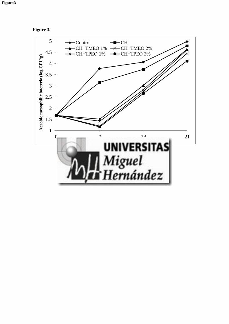
Figure 3.
1
1.5
2
2.5
3
3.5
4
4.5
5
0 7 14 21
Aer
ob
ic m
esop
hil
ic b
act
eria
(lo
g C
FU
/g)
Time (Days)
Control CHCH+TMEO 1% CH+TMEO 2%CH+TPEO 1% CH+TPEO 2%
Figure3

Figure 4.
1
1.5
2
2.5
3
3.5
4
4.5
5
5.5
0 7 14 21
Lact
ic a
cid
bact
eria
(lo
g C
FU
/g)
Time (Days)
Control CH
CH+TMEO 1% CH+TMEO 2%
CH+TPEO 1% CH+TPEO 2%
Figure 4

Figure 5.
0
1
2
3
4
5
6
7
8
9
10
11
12
0 7 14 21
mg G
all
ic A
cid
Eq
uiv
ale
nt/
g f
ilm
Time (Days)
CH
CH+TMEO 1%
CH+TMEO 2%
CH+TPEO 1%
CH+TPEO 2%
Figure 5

Figure 6.
Figure 6

COMUNICACIONES
A CONGRESOS