


Transferencia de Genes a Células Animales en Cultivo 2011-3.pdf · Transferencia de Genes a...
107
Unidad de Transferencia Gen Unidad de Transferencia Gen é é tica tica Instituto de Oncolog Instituto de Oncolog í í a a “Á “Á ngel H. ngel H. Roffo Roffo ” ” Universidad de Buenos Aires Universidad de Buenos Aires Transferencia de Genes a Transferencia de Genes a C C é é lulas Animales en Cultivo lulas Animales en Cultivo Conceptos y T Conceptos y T é é cnicas de Biotecnolog cnicas de Biotecnolog í í a I a I 2011 2011 – – 2do cuatrimestre 2do cuatrimestre FBMC FBMC - - FCEN FCEN - - UBA UBA
Transcript of Transferencia de Genes a Células Animales en Cultivo 2011-3.pdf · Transferencia de Genes a...

Unidad de Transferencia GenUnidad de Transferencia GenééticaticaInstituto de OncologInstituto de Oncologíía a “Á“Ángel H. ngel H. RoffoRoffo””
Universidad de Buenos AiresUniversidad de Buenos Aires
Transferencia de Genes aTransferencia de Genes aCCéélulas Animales en Cultivolulas Animales en Cultivo
Conceptos y TConceptos y Téécnicas de Biotecnologcnicas de Biotecnologíía I a I 2011 2011 –– 2do cuatrimestre2do cuatrimestre
FBMCFBMC--FCENFCEN--UBAUBA
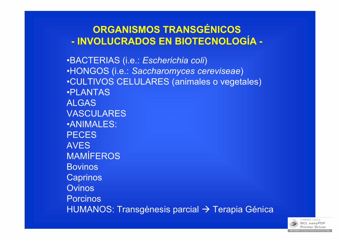
•BACTERIAS (i.e.: Escherichia coli)•HONGOS (i.e.: Saccharomyces cereviseae)•CULTIVOS CELULARES (animales o vegetales)•PLANTASALGASVASCULARES•ANIMALES:PECESAVESMAMÍFEROSBovinosCaprinosOvinosPorcinosHUMANOS: Transgénesis parcial Terapia Génica
ORGANISMOS TRANSGÉNICOS- INVOLUCRADOS EN BIOTECNOLOGÍA -
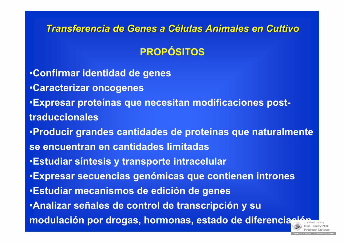
Transferencia de Genes a CTransferencia de Genes a Céélulas Animales en Cultivolulas Animales en Cultivo
PROPÓSITOS
•Confirmar identidad de genes
•Caracterizar oncogenes
•Expresar proteínas que necesitan modificaciones post-
traduccionales
•Producir grandes cantidades de proteínas que naturalmente
se encuentran en cantidades limitadas
•Estudiar síntesis y transporte intracelular
•Expresar secuencias genómicas que contienen intrones
•Estudiar mecanismos de edición de genes
•Analizar señales de control de transcripción y su
modulación por drogas, hormonas, estado de diferenciación
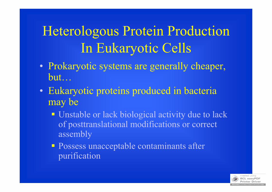
Heterologous Protein ProductionIn Eukaryotic Cells
• Prokaryotic systems are generally cheaper, but…
• Eukaryotic proteins produced in bacteria may be Unstable or lack biological activity due to lack
of posttranslational modifications or correct assembly Possess unacceptable contaminants after
purification
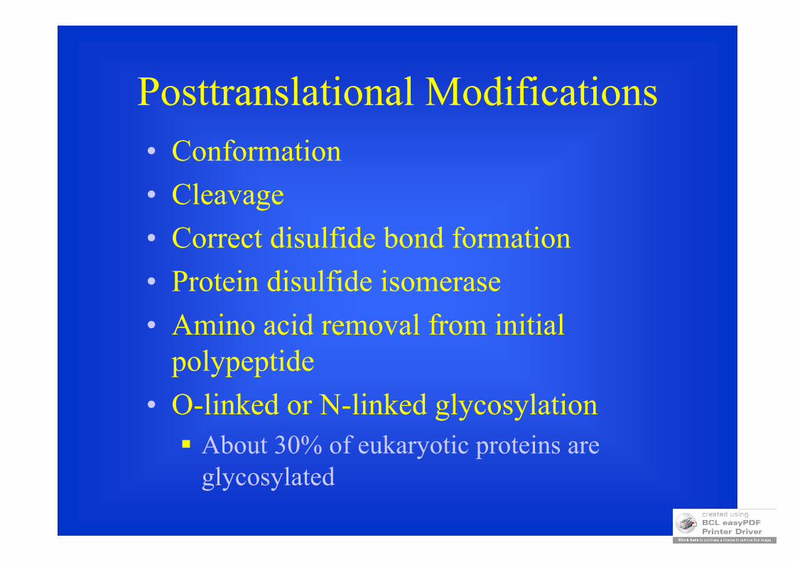
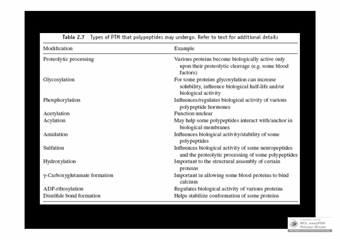
Posttranslational Modifications• Conformation
• Cleavage
• Correct disulfide bond formation
• Protein disulfide isomerase
• Amino acid removal from initial polypeptide
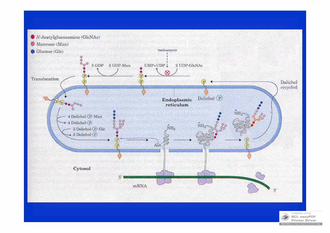
• O-linked or N-linked glycosylation About 30% of eukaryotic proteins are
glycosylated
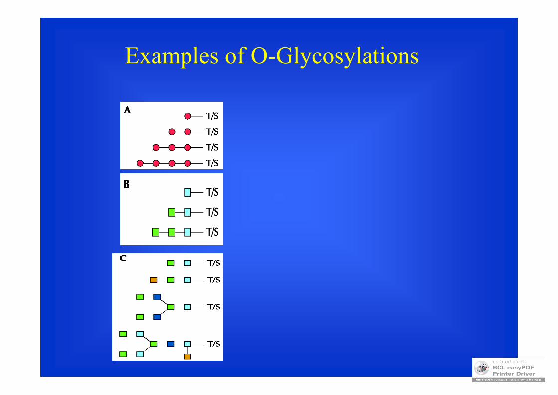
Examples of O-Glycosylations
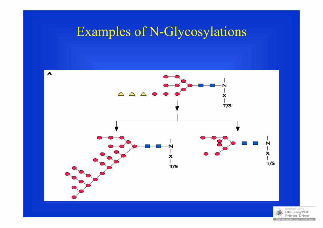
Examples of N-Glycosylations
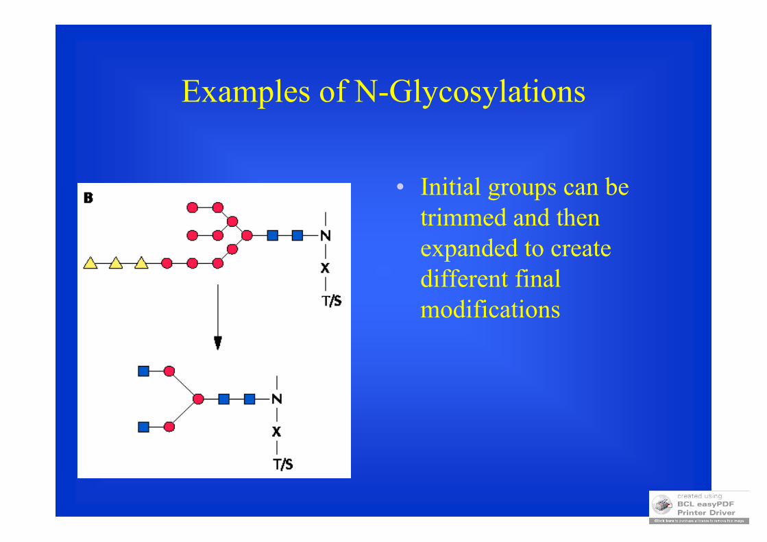
Examples of N-Glycosylations
• Initial groups can be trimmed and then expanded to create different final modifications
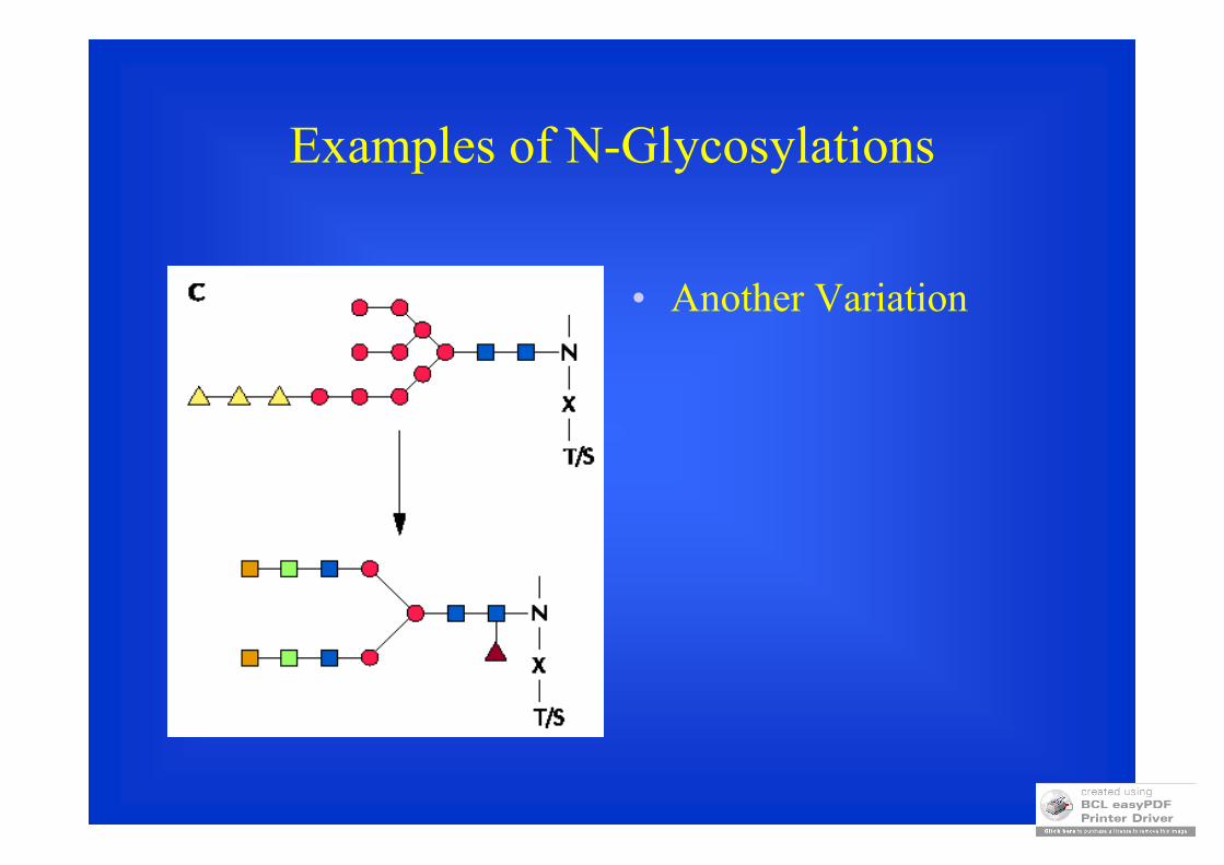
Examples of N-Glycosylations
• Another Variation
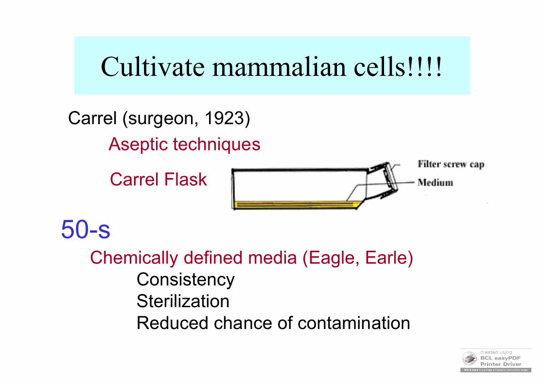
Cultivate mammalian cells!!!!
Carrel (surgeon, 1923)
Aseptic techniques
Carrel Flask
Chemically defined media (Eagle, Earle)ConsistencySterilizationReduced chance of contamination
50-s
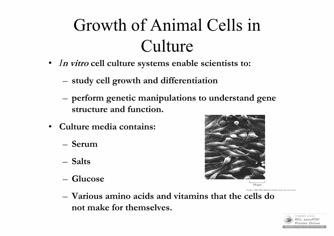
Growth of Animal Cells inCulture
• In vitro cell culture systems enable scientists to:
– study cell growth and differentiation
– perform genetic manipulations to understand gene structure and function.
• Culture media contains:
– Serum
– Salts
– Glucose
– Various amino acids and vitamins that the cells do not make for themselves.
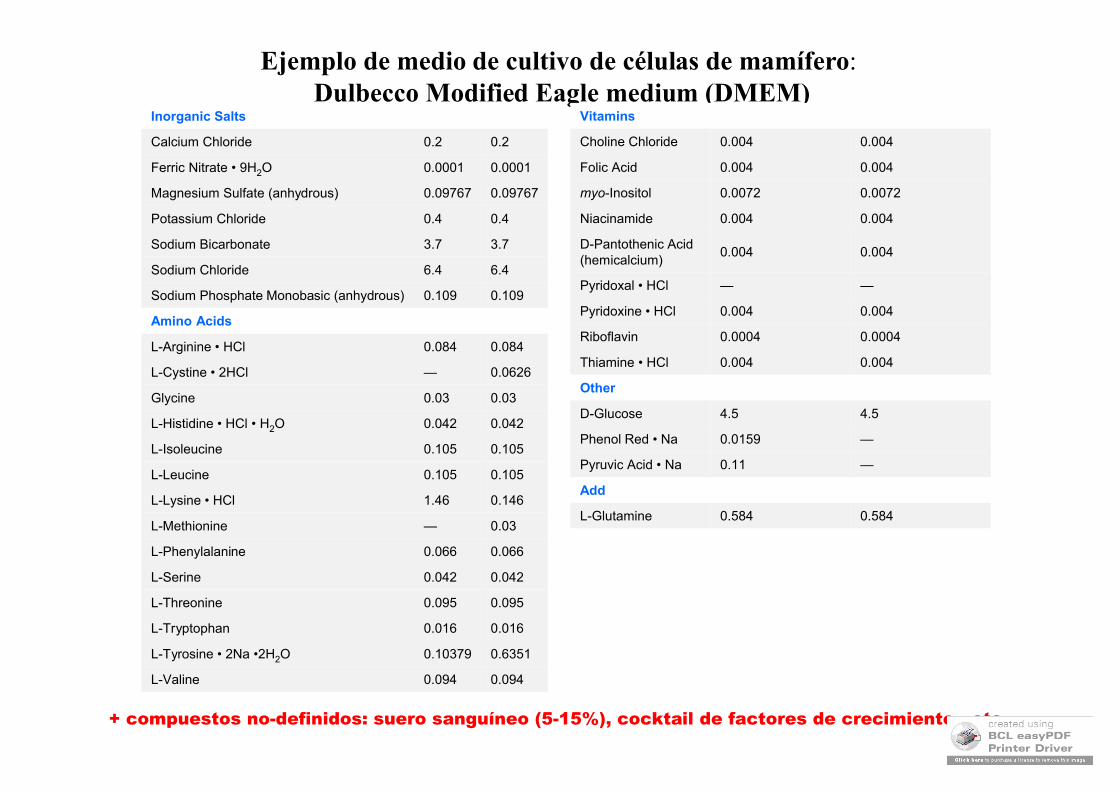
+ compuestos no-definidos: suero sanguíneo (5-15%), cocktail de factores de crecimiento, etc.
0.0940.094L-Valine
0.63510.10379L-Tyrosine • 2Na •2H2O
0.0160.016L-Tryptophan
0.0950.095L-Threonine
0.0420.042L-Serine
0.0660.066L-Phenylalanine
0.03—L-Methionine
0.1461.46L-Lysine • HCl
0.1050.105L-Leucine
0.1050.105L-Isoleucine
0.0420.042L-Histidine • HCl • H2O
0.030.03Glycine
0.0626—L-Cystine • 2HCl
0.0840.084L-Arginine • HCl
Amino Acids
0.1090.109Sodium Phosphate Monobasic (anhydrous)
6.46.4Sodium Chloride
3.73.7Sodium Bicarbonate
0.40.4Potassium Chloride
0.097670.09767Magnesium Sulfate (anhydrous)
0.00010.0001Ferric Nitrate • 9H2O
0.20.2Calcium Chloride
Inorganic Salts
Ejemplo de medio de cultivo de células de mamífero:Dulbecco Modified Eagle medium (DMEM)
0.5840.584L-Glutamine
Add
—0.11Pyruvic Acid • Na
—0.0159Phenol Red • Na
4.54.5D-Glucose
Other
0.0040.004Thiamine • HCl
0.00040.0004Riboflavin
0.0040.004Pyridoxine • HCl
——Pyridoxal • HCl
0.0040.004D-Pantothenic Acid(hemicalcium)
0.0040.004Niacinamide
0.00720.0072myo-Inositol
0.0040.004Folic Acid
0.0040.004Choline Chloride
Vitamins
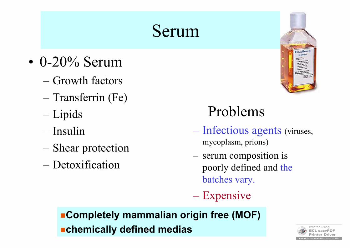
Serum
• 0-20% Serum– Growth factors
– Transferrin (Fe)
– Lipids
– Insulin
– Shear protection
– Detoxification
Problems– Infectious agents (viruses,
mycoplasm, prions)
– serum composition is poorly defined and the batches vary.
– Expensive
Completely mammalian origin free (MOF)
chemically defined medias
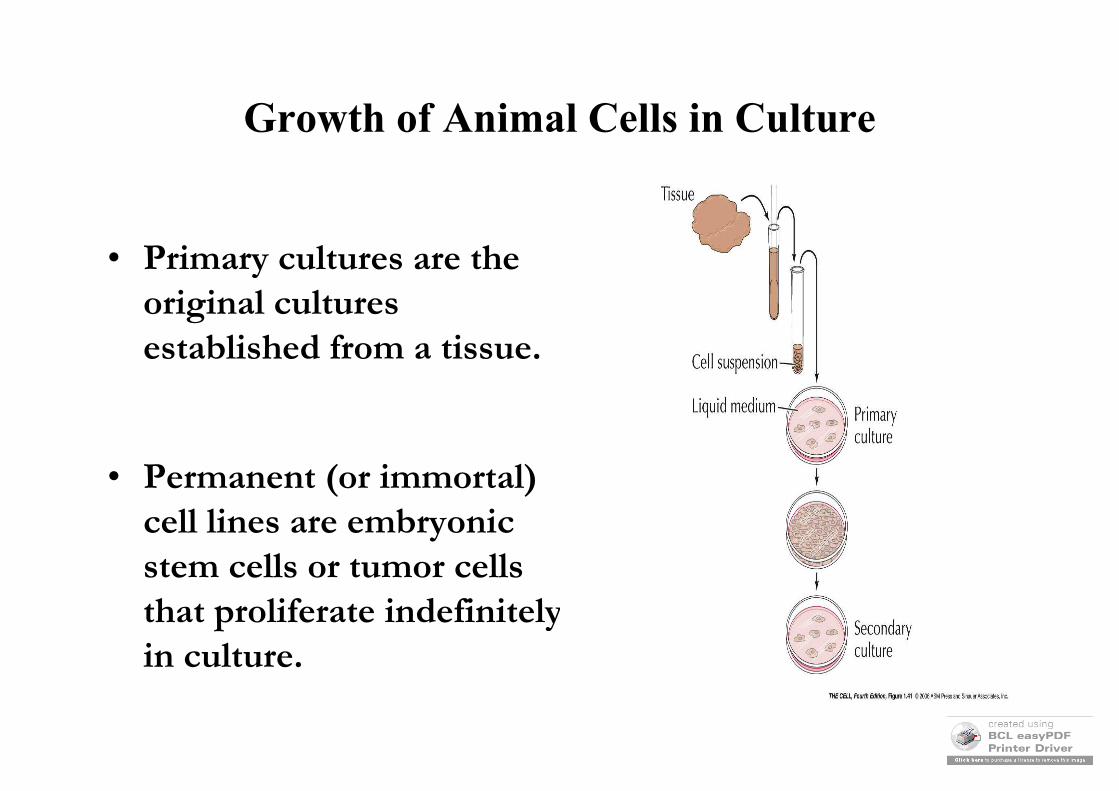
Growth of Animal Cells in Culture
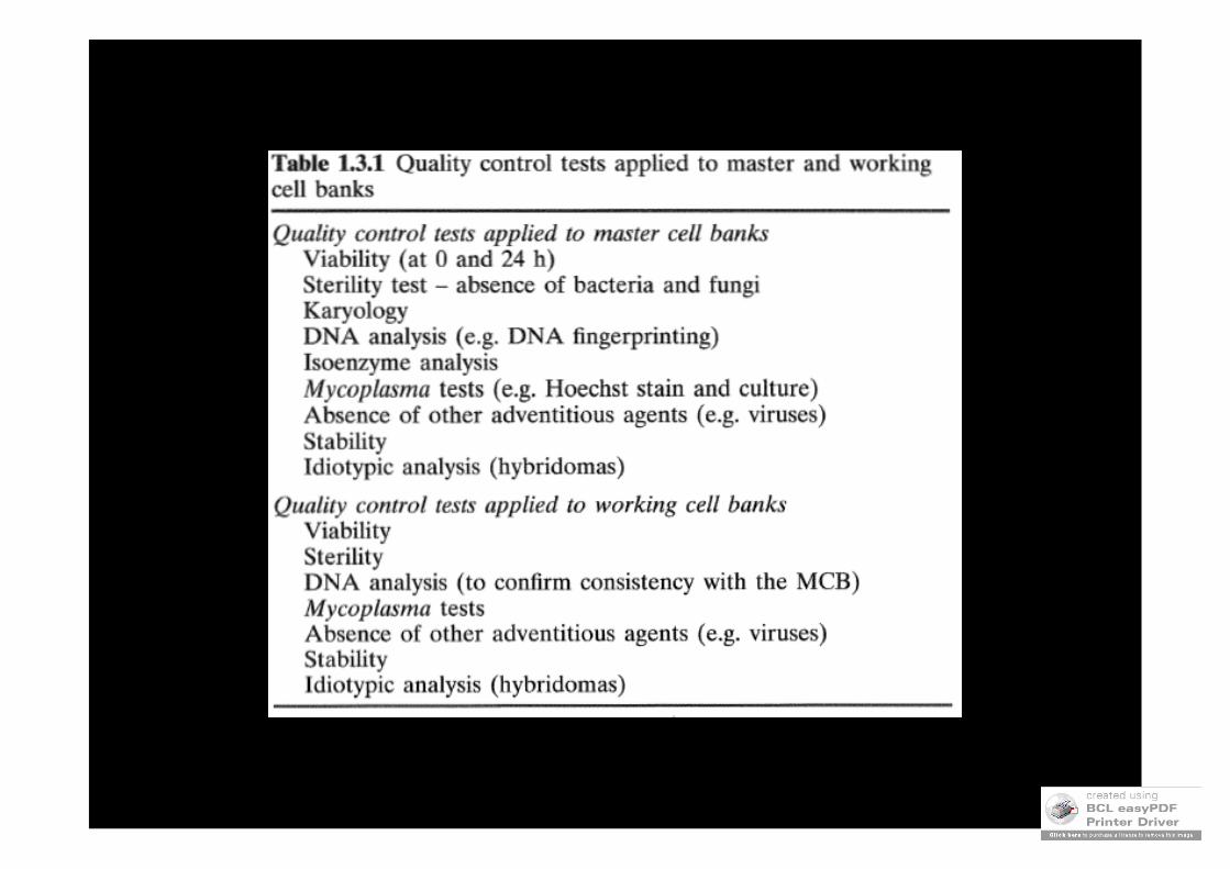
• Primary cultures are the original cultures established from a tissue.
• Permanent (or immortal) cell lines are embryonic stem cells or tumor cells that proliferate indefinitely in culture.
0.0
0.0
0.0
0.0
0.1
1.0
10.0
0 50 100Time (h)
Cx
(g/l
)
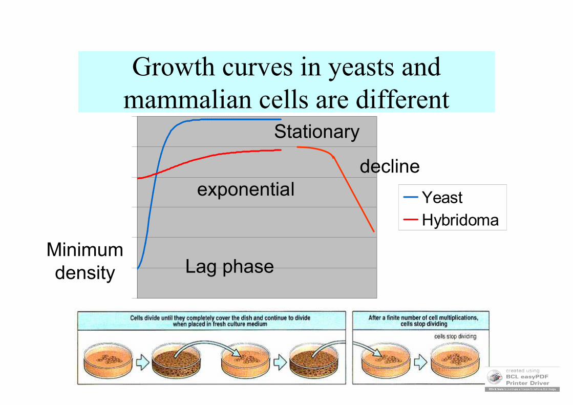
YeastHybridoma
Growth curves in yeasts andmammalian cells are different
Minimumdensity Lag phase
exponential
Stationary
decline
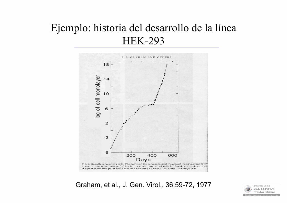
Ejemplo: historia del desarrollo de la línea HEK-293
Graham, et al., J. Gen. Virol., 36:59-72, 1977
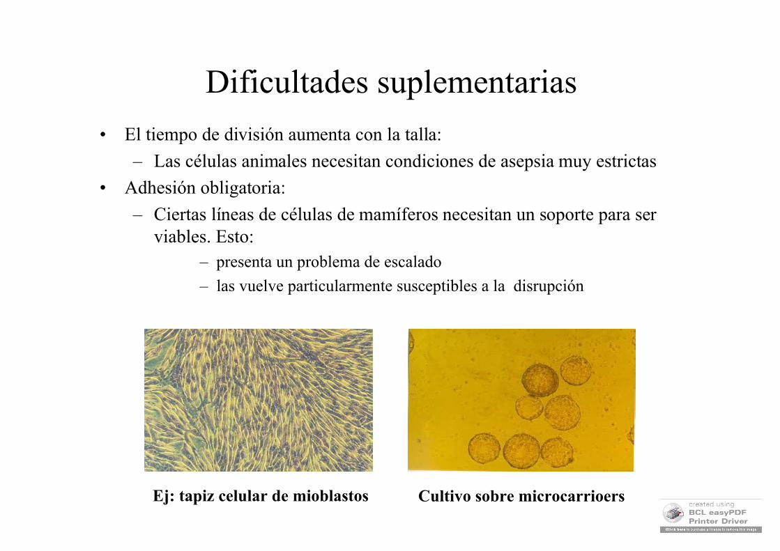
Dificultades suplementarias
• El tiempo de división aumenta con la talla:
– Las células animales necesitan condiciones de asepsia muy estrictas
• Adhesión obligatoria:
– Ciertas líneas de células de mamíferos necesitan un soporte para ser viables. Esto:
– presenta un problema de escalado
– las vuelve particularmente susceptibles a la disrupción
Ej: tapiz celular de mioblastos Cultivo sobre microcarrioers
MODALIDAD
•Transitoria: s/ Integración: Expresión 12-72 horas
•Permanente: c/ Integración: Expresión 1-3 semanas
PARÁMETROS A CONSIDERAR
•Tipos celulares disponibles
•Expresión transitoria o permanente (estable)
•Elementos de control de expresión adecuados
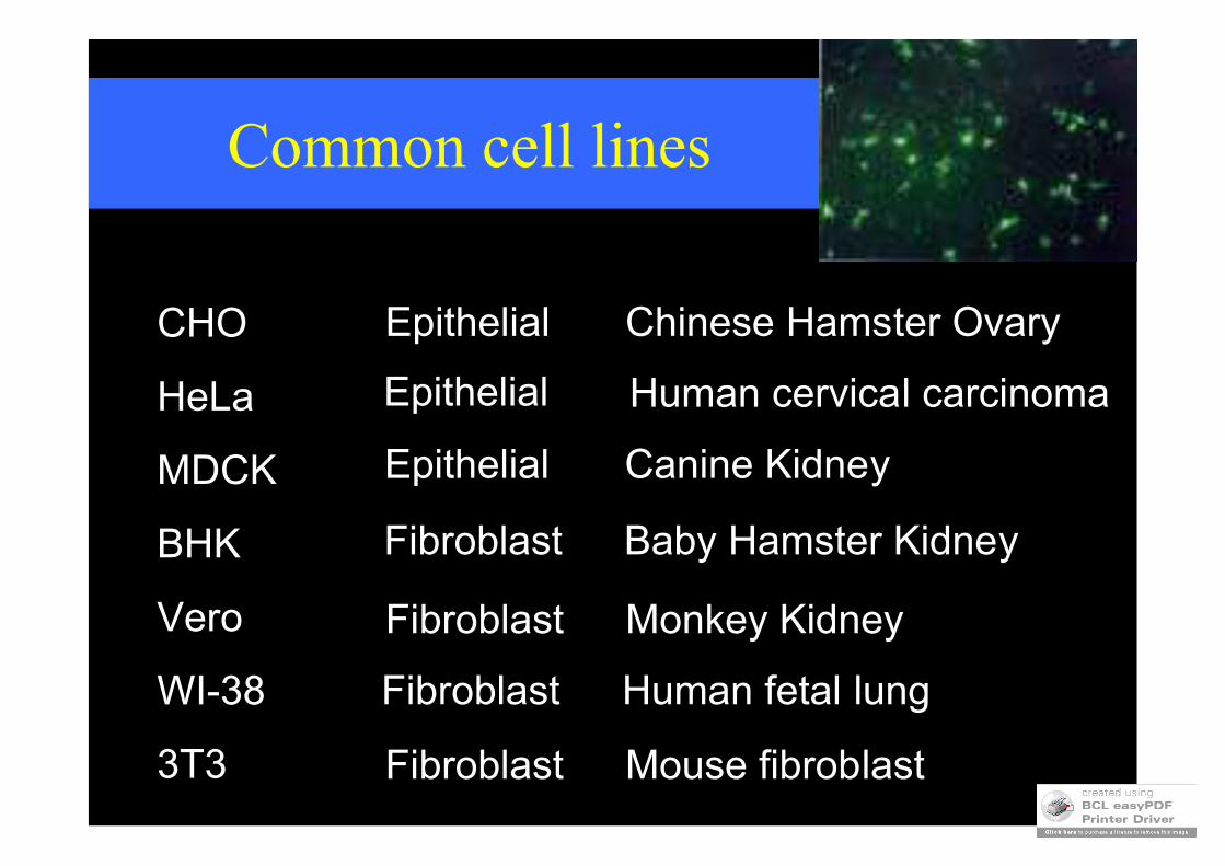
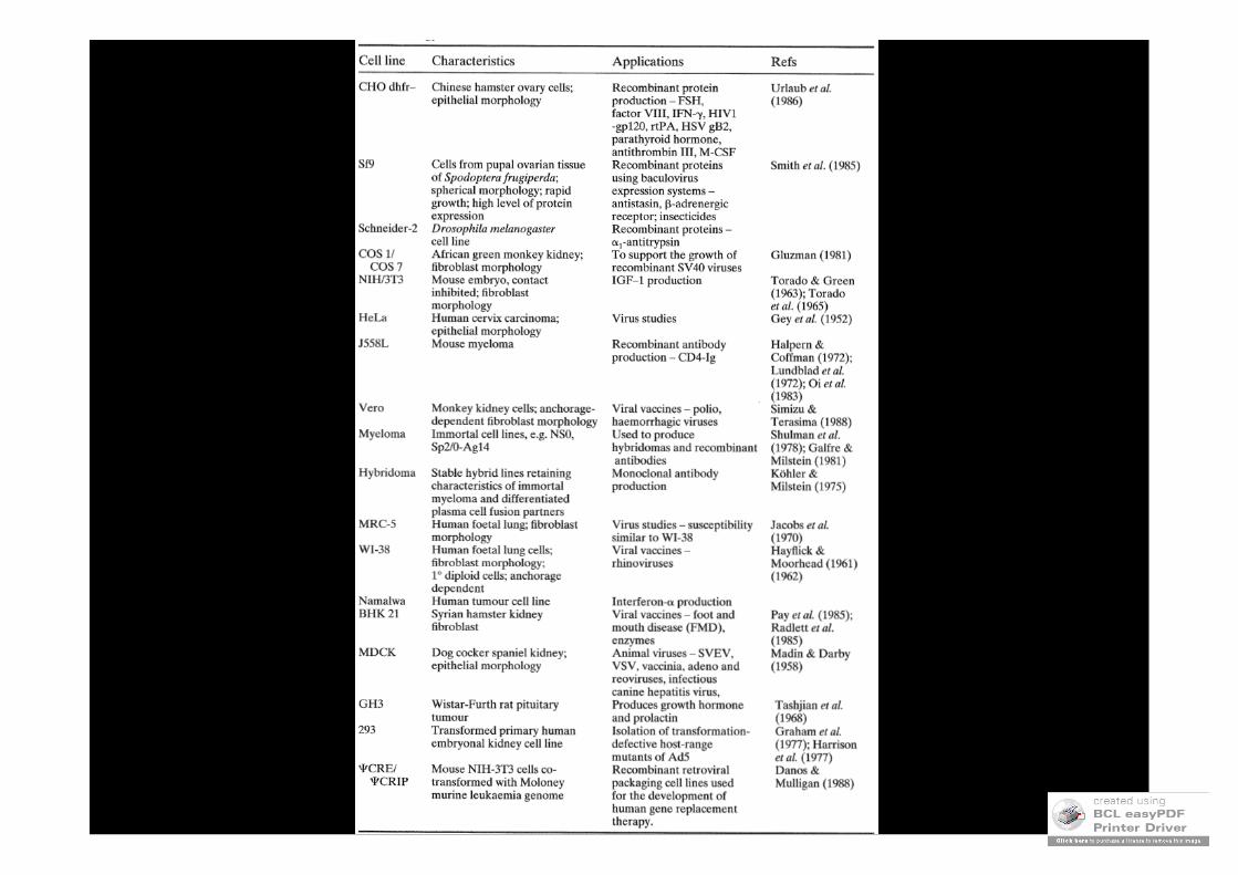
Canine KidneyEpithelial
Common cell lines
CHO
HeLa
MDCK
BHK
Vero
WI-38
3T3
Chinese Hamster OvaryEpithelial
Mouse fibroblastFibroblast
Monkey KidneyFibroblast
Epithelial Human cervical carcinoma
Human fetal lungFibroblast
Baby Hamster KidneyFibroblast
MARCADORES FENOTMARCADORES FENOTÍÍPICOSPICOS
••Crecimiento en agarCrecimiento en agar••Crecimiento con bajo sueroCrecimiento con bajo suero••Cambios morfolCambios morfolóógicosgicos••Rescate de la muerte de cultivos celulares primariosRescate de la muerte de cultivos celulares primarios
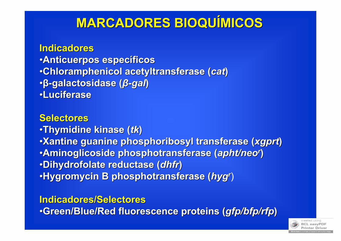
MARCADORES BIOQUMARCADORES BIOQUÍÍMICOSMICOS
IndicadoresIndicadores••Anticuerpos especAnticuerpos especííficosficos••ChloramphenicolChloramphenicol acetyltransferaseacetyltransferase ((catcat))••ββ--galactosidasegalactosidase ((ββ--galgal))••LuciferaseLuciferase
SelectoresSelectores••ThymidineThymidine kinasekinase ((tktk))••XantineXantine guanineguanine phosphoribosylphosphoribosyl transferasetransferase ((xgprtxgprt))••AminoglicosideAminoglicoside phosphotransferasephosphotransferase ((aphtapht//neoneorr))••DihydrofolateDihydrofolate reductasereductase ((dhfrdhfr))••HygromycinHygromycin B B phosphotransferasephosphotransferase ((hyghygrr)
Indicadores/SelectoresIndicadores/Selectores••GreenGreen/Blue/Red /Blue/Red fluorescencefluorescence proteinsproteins ((gfpgfp//bfpbfp//rfprfp) )
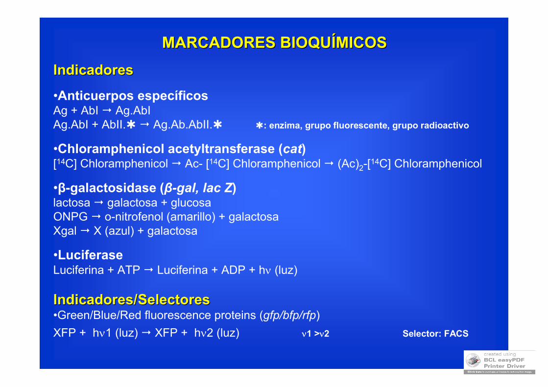
MARCADORES BIOQUMARCADORES BIOQUÍÍMICOSMICOS
IndicadoresIndicadores
•Anticuerpos específicosAg + AbI Ag.AbIAg.AbI + AbII. Ag.Ab.AbII. : enzima, grupo fluorescente, grupo radioactivo
•Chloramphenicol acetyltransferase (cat)[14C] Chloramphenicol Ac- [14C] Chloramphenicol (Ac)2-[
14C] Chloramphenicol
•β-galactosidase (β-gal, lac Z)lactosa galactosa + glucosaONPG o-nitrofenol (amarillo) + galactosaXgal X (azul) + galactosa
•LuciferaseLuciferina + ATP Luciferina + ADP + h (luz)
Indicadores/SelectoresIndicadores/Selectores•Green/Blue/Red fluorescence proteins (gfp/bfp/rfp)
XFP + h1 (luz) XFP + h2 (luz) 1 >2 Selector: FACS
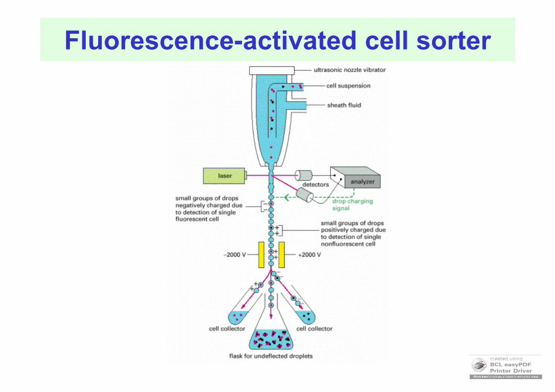
Fluorescence-activated cell sorter

Reporter gene systems1. chloramphenicol acetyl transferase (CAT)
CAT is a bacterial enzyme that catalyzes the transfer of acetyl groups from acetyl-coenzyme A to the antibiotic chloramphenicol.(chloramphenicol deactivation)
For mammalian cells it is laborous and expensive.
Extract protein and measure activity…
thin-layer chromatographic sheetChloramphenicol
is radiolabelled
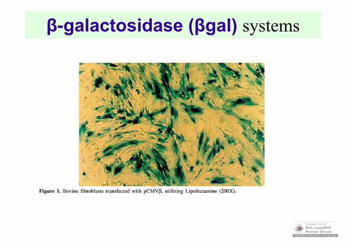
β-galactosidase (βgal) systems
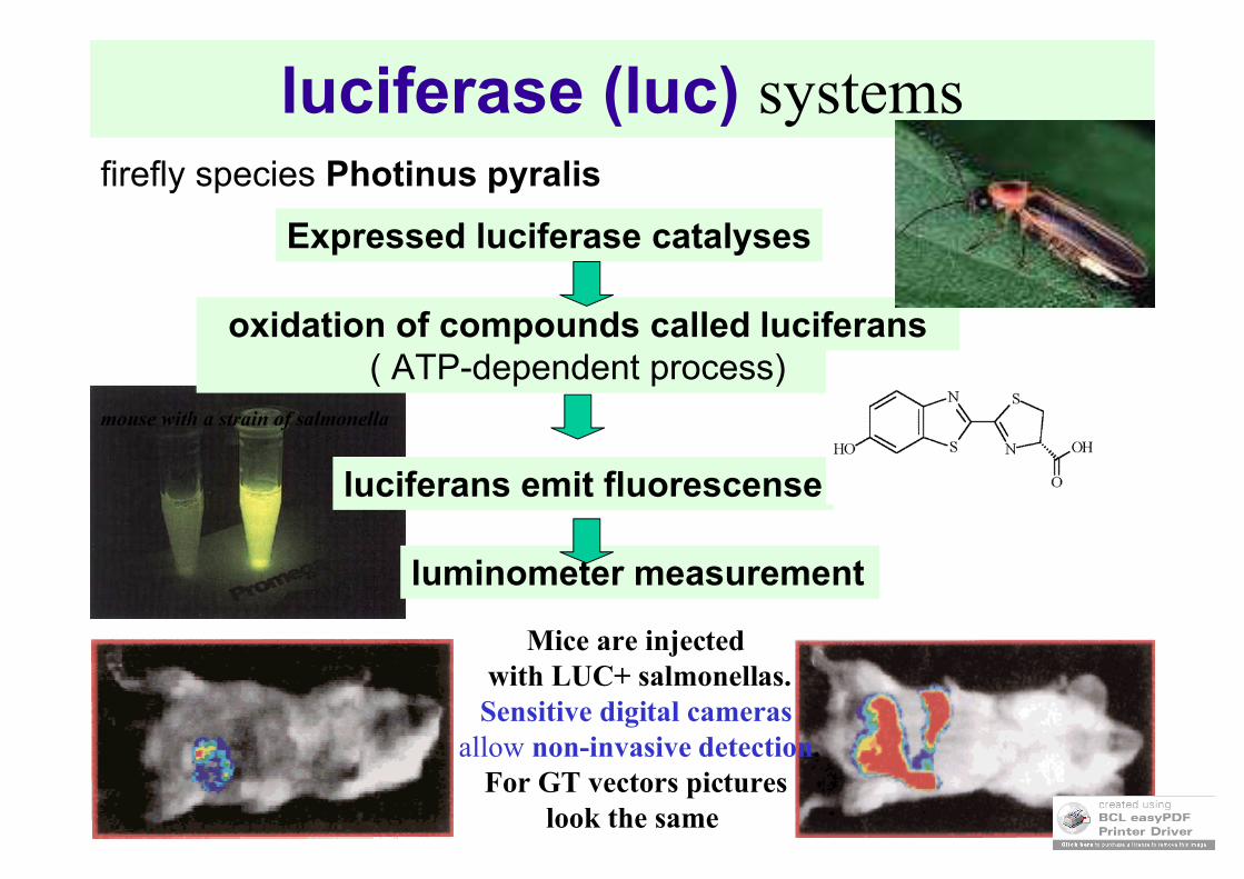
luciferase (luc) systemsfirefly species Photinus pyralis
oxidation of compounds called luciferans( ATP-dependent process)
luciferans emit fluorescense
Expressed luciferase catalyses
mouse with a strain of salmonella
Mice are injected with LUC+ salmonellas.
Sensitive digital camerasallow non-invasive detection.
For GT vectors pictures look the same
luminometer measurement
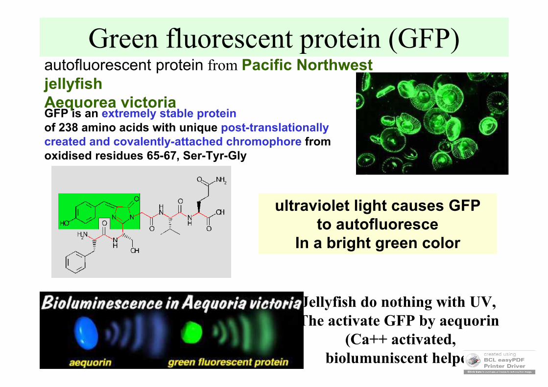
Green fluorescent protein (GFP)autofluorescent protein from Pacific Northwest jellyfish Aequorea victoriaGFP is an extremely stable proteinof 238 amino acids with unique post-translationallycreated and covalently-attached chromophore from oxidised residues 65-67, Ser-Tyr-Gly
ultraviolet light causes GFP to autofluoresce
In a bright green color
Jellyfish do nothing with UV, The activate GFP by aequorin
(Ca++ activated, biolumuniscent helper)
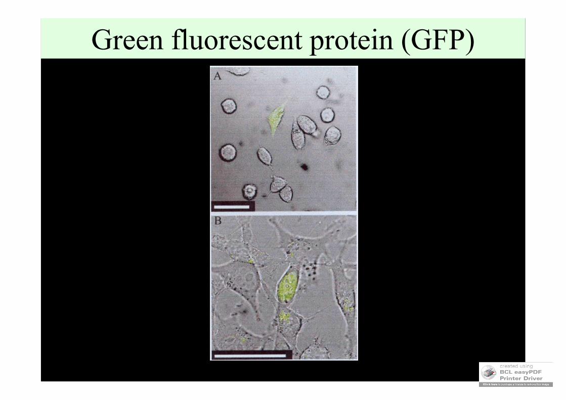
Green fluorescent protein (GFP)
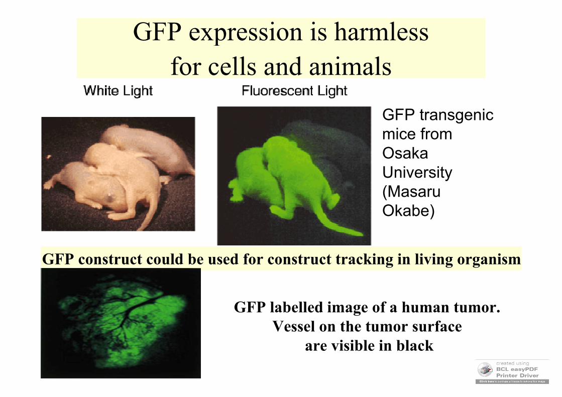
GFP expression is harmlessfor cells and animals
GFP transgenic mice from Osaka University (Masaru Okabe)
GFP construct could be used for construct tracking in living organism
GFP labelled image of a human tumor. Vessel on the tumor surface
are visible in black
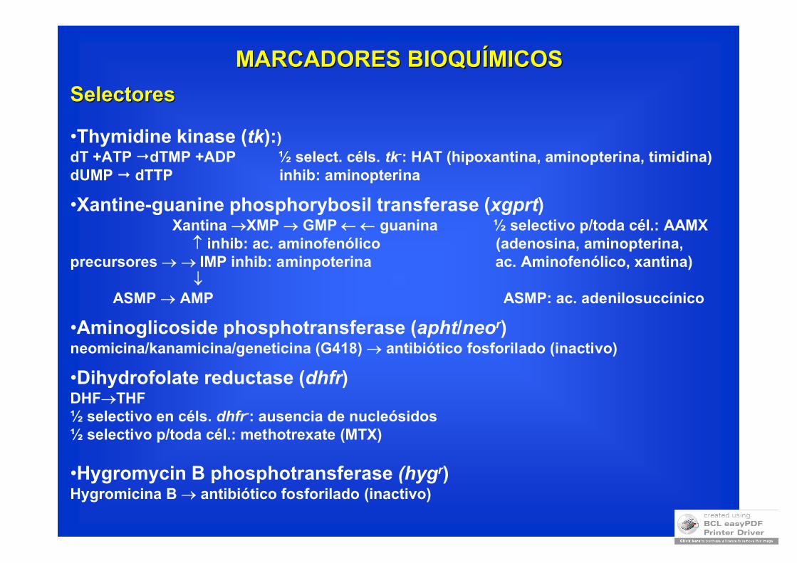
MARCADORES BIOQUMARCADORES BIOQUÍÍMICOSMICOS
SelectoresSelectores
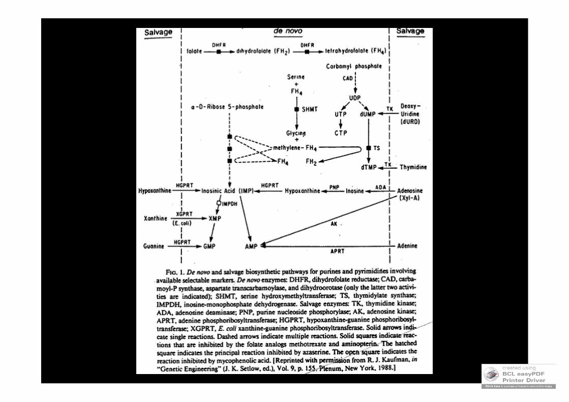
•Thymidine kinase (tk):)dT +ATP dTMP +ADP ½ select. céls. tk-: HAT (hipoxantina, aminopterina, timidina)dUMP dTTP inhib: aminopterina
•Xantine-guanine phosphorybosil transferase (xgprt)Xantina XMP GMP guanina ½ selectivo p/toda cél.: AAMX
inhib: ac. aminofenólico (adenosina, aminopterina,precursores IMP inhib: aminpoterina ac. Aminofenólico, xantina)
ASMP AMP ASMP: ac. adenilosuccínico
•Aminoglicoside phosphotransferase (apht/neor)neomicina/kanamicina/geneticina (G418) antibiótico fosforilado (inactivo)
•Dihydrofolate reductase (dhfr)DHFTHF½ selectivo en céls. dhfr-: ausencia de nucleósidos½ selectivo p/toda cél.: methotrexate (MTX)
•Hygromycin B phosphotransferase (hygr)Hygromicina B antibiótico fosforilado (inactivo)
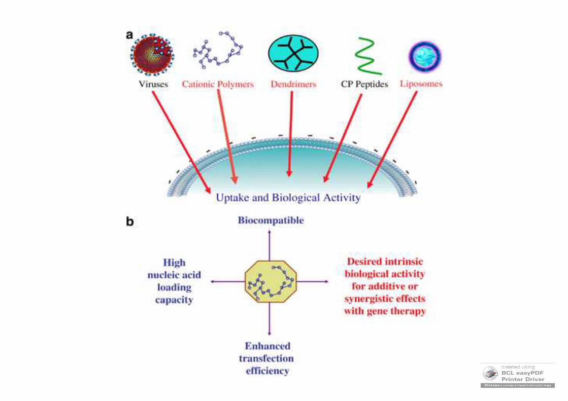
• Vectores virales
• Vectores no virales
TRANSFERENCIA GENTRANSFERENCIA GENÉÉTICA:TICA:METODOLOGIAMETODOLOGIA
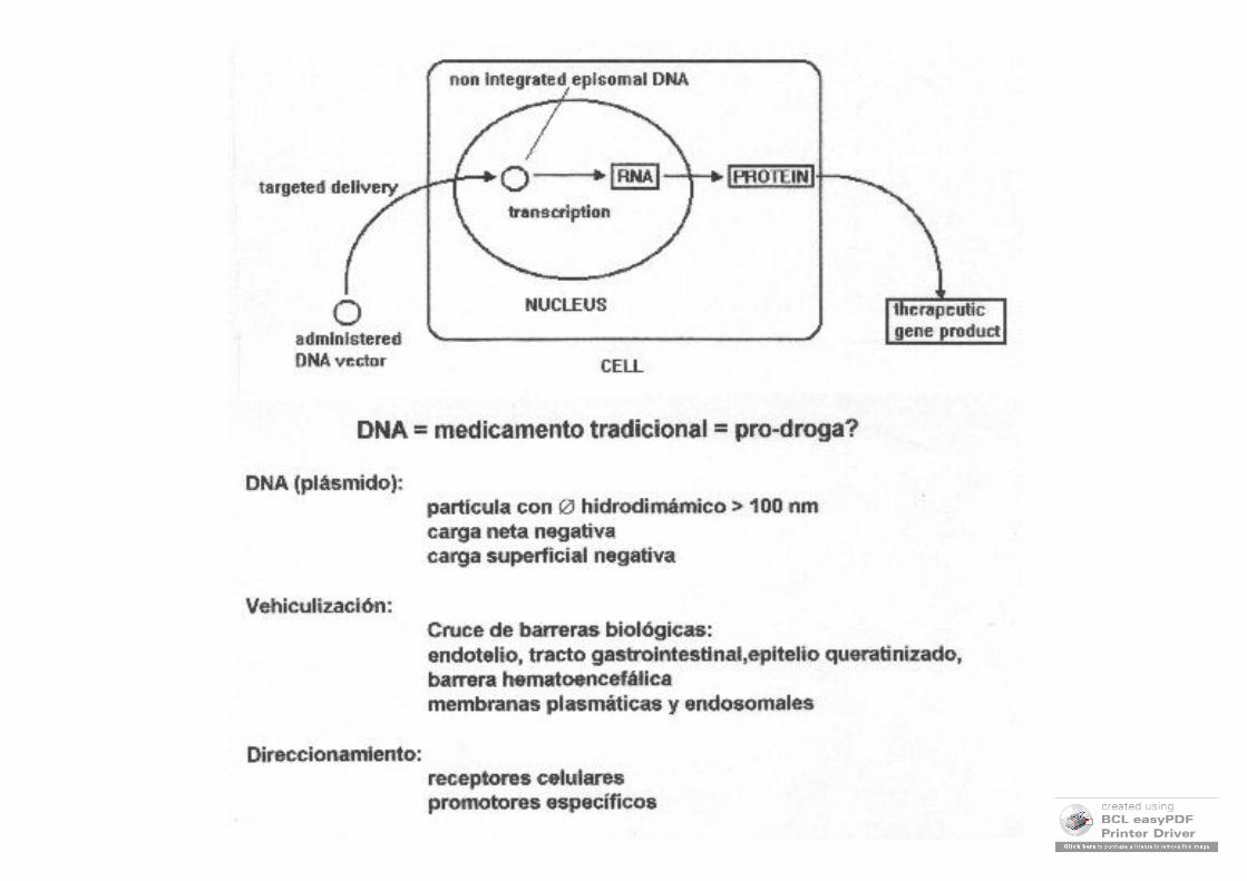
Direccionamiento de vectores
• Via de administración
• Receptores celulares
• Promotores específicos de tejido
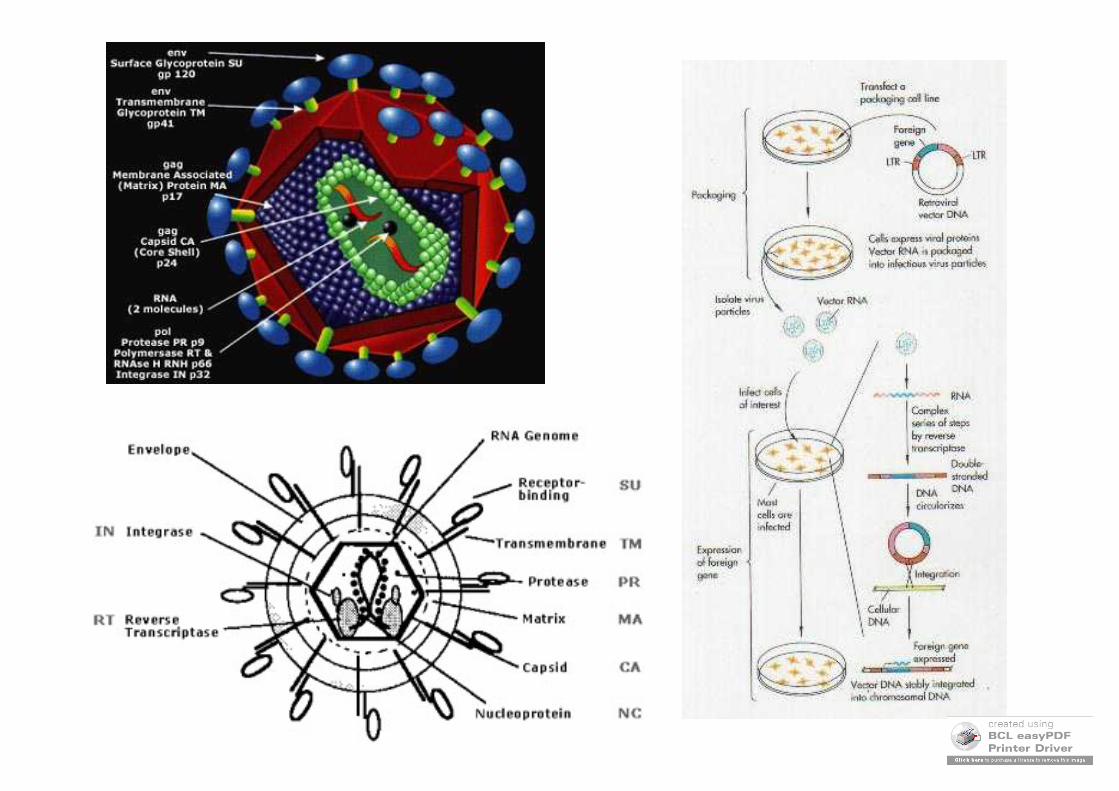
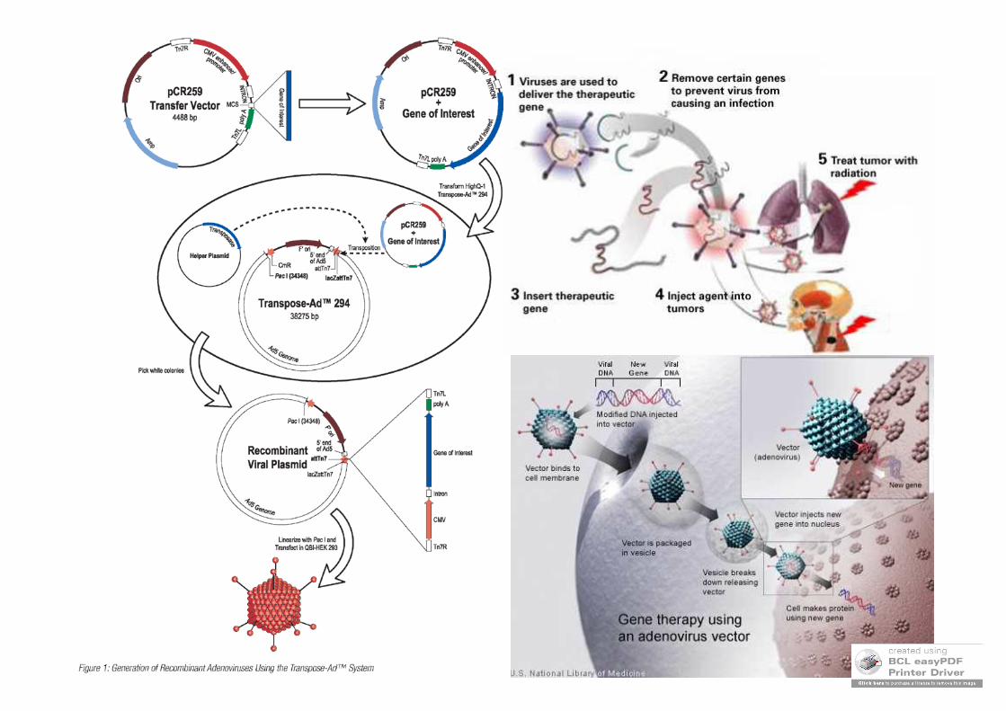
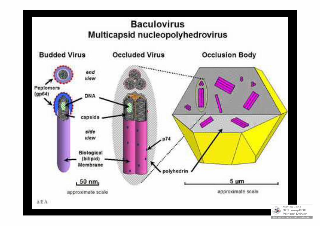
•• Vectores ViralesVectores Virales
Virus naturales (silvestres):•Crecen en todas las células•Infectan todas las células•Son patogénicos
Virus terapeúticos (recombinantes):•Sólo crecen en células empaquetadoras•Infectan todas las células•No son patogénicos
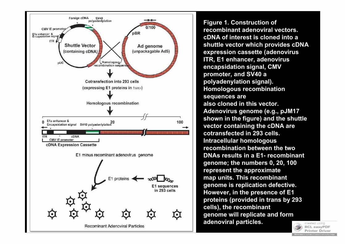
Figure 1. Construction of recombinant adenoviral vectors. cDNA of interest is cloned into a shuttle vector which provides cDNAexpression cassette (adenovirusITR, E1 enhancer, adenovirus encapsidation signal, CMV promoter, and SV40 a polyadenylation signal). Homologous recombination sequences arealso cloned in this vector. Adenovirus genome (e.g., pJM17 shown in the figure) and the shuttle vector containing the cDNA are cotransfected in 293 cells.Intracellular homologous recombination between the two DNAs results in a E1- recombinant genome; the numbers 0, 20, 100 represent the approximatemap units. This recombinant genome is replication defective. However, in the presence of E1 proteins (provided in trans by 293 cells), the recombinantgenome will replicate and form adenoviral particles.
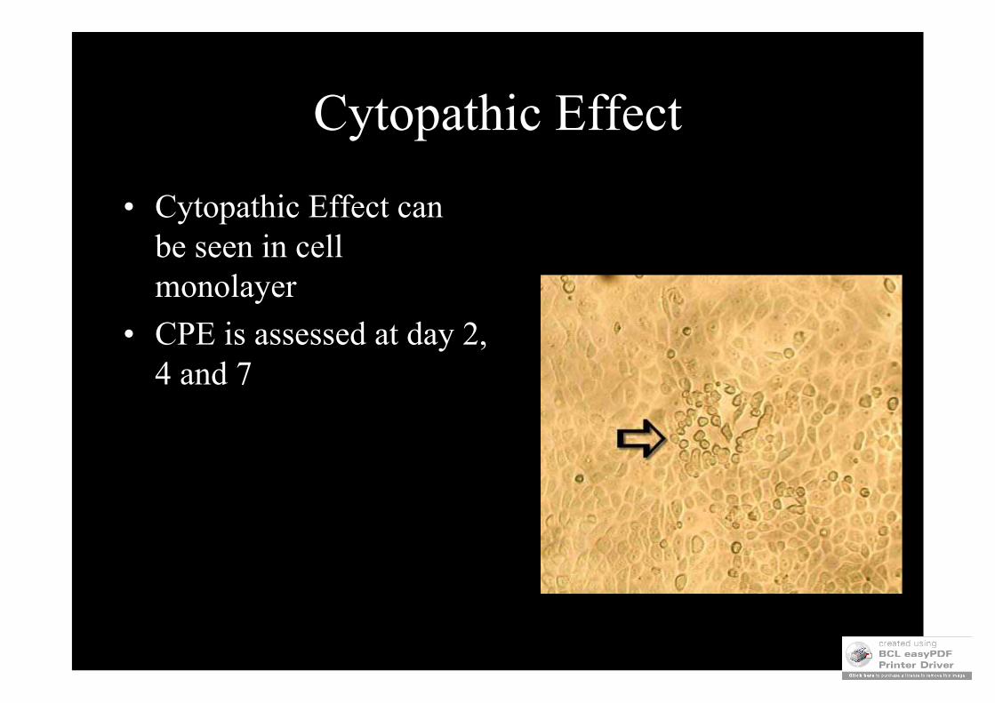
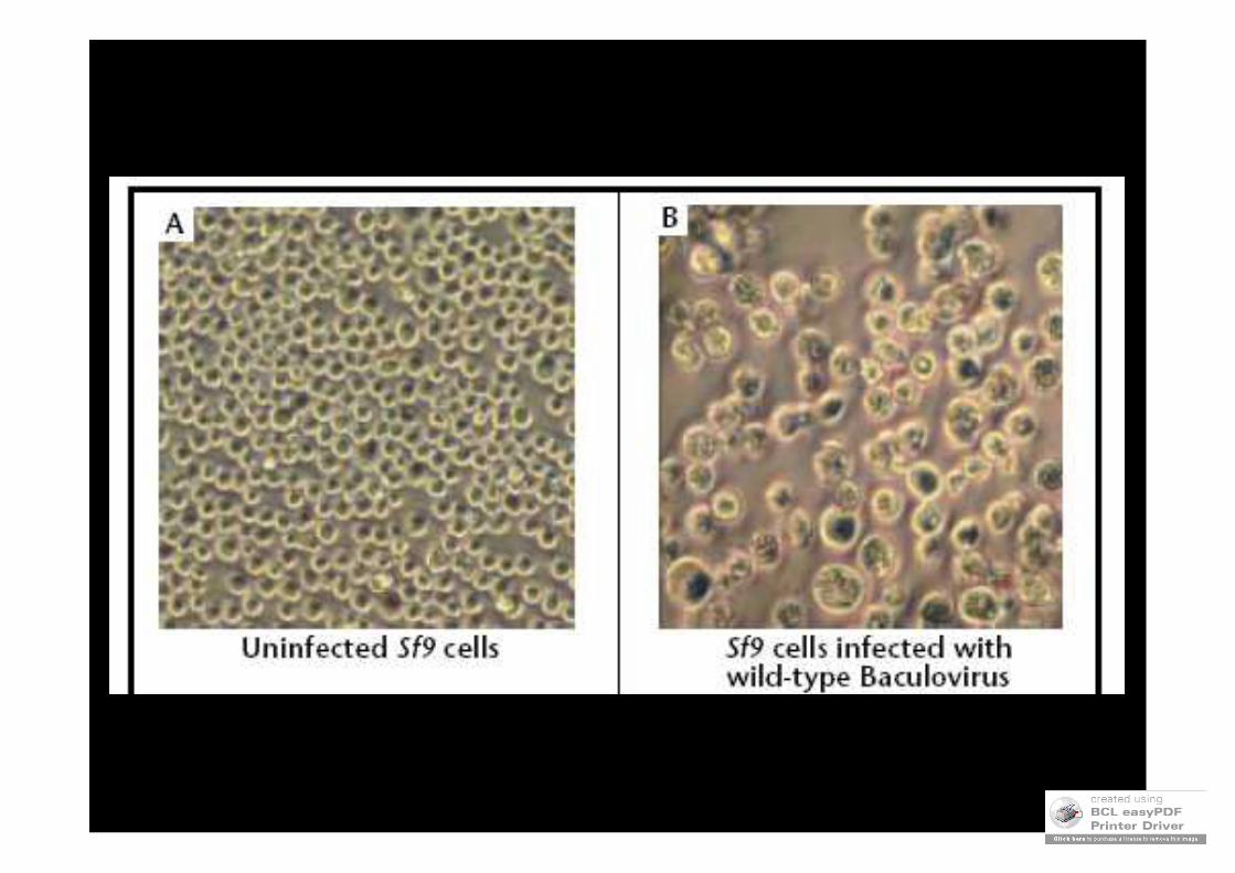
Cytopathic Effect
• Cytopathic Effect can be seen in cell monolayer
• CPE is assessed at day 2, 4 and 7
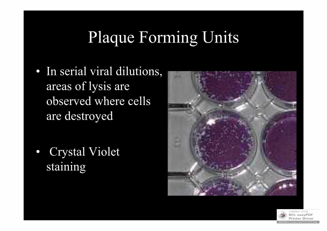
Plaque Forming Units
• In serial viral dilutions, areas of lysis are observed where cells are destroyed
• Crystal Violet staining

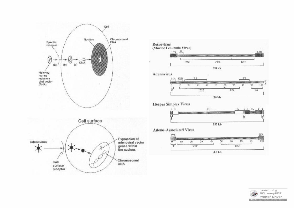
VECTORES VIRALES
Retrovirus Adenovirus Herpes virus Virus Adenoasociados
Tamaño máximo del gen terapeútico
8 kb 35 kb 30 kb 4,8 kb
Células objetivo Sólo en división activa
En división activa o sin
división
En división activa o sin
división
En división o quizás sin división
Administración Ex vivo o in situ
Ex vivo o in situ
Ex vivo o in situ
Ex vivo o in situ
Expresión del gen terapeútico
Estable Transitoria Transitoria Posiblemente estable
Indice de expresión del gen terapeútico
Moderado Elevado Moderado Moderado
Riesgos Posible integración mutagénica
Reacciones inflamatorias/inmunitarias
Posible integración mutagénica
Posible integración mutagénica
Inmunidad preexistente en el hospedador
No Sí Sí Sí
Recombinación con el hospedador
Improbable Posible Posible Improbable
Recombinación con el virus parental
Imposible Posible Posible Posible
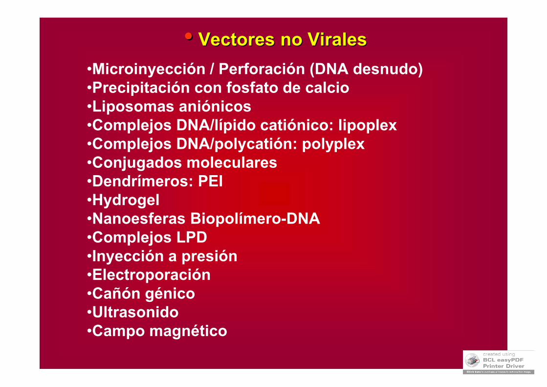
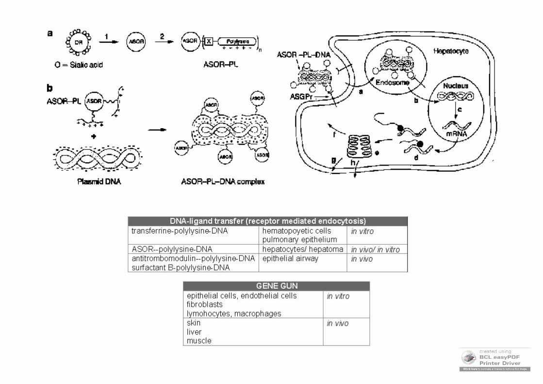
•• VectoresVectores no no ViralesVirales
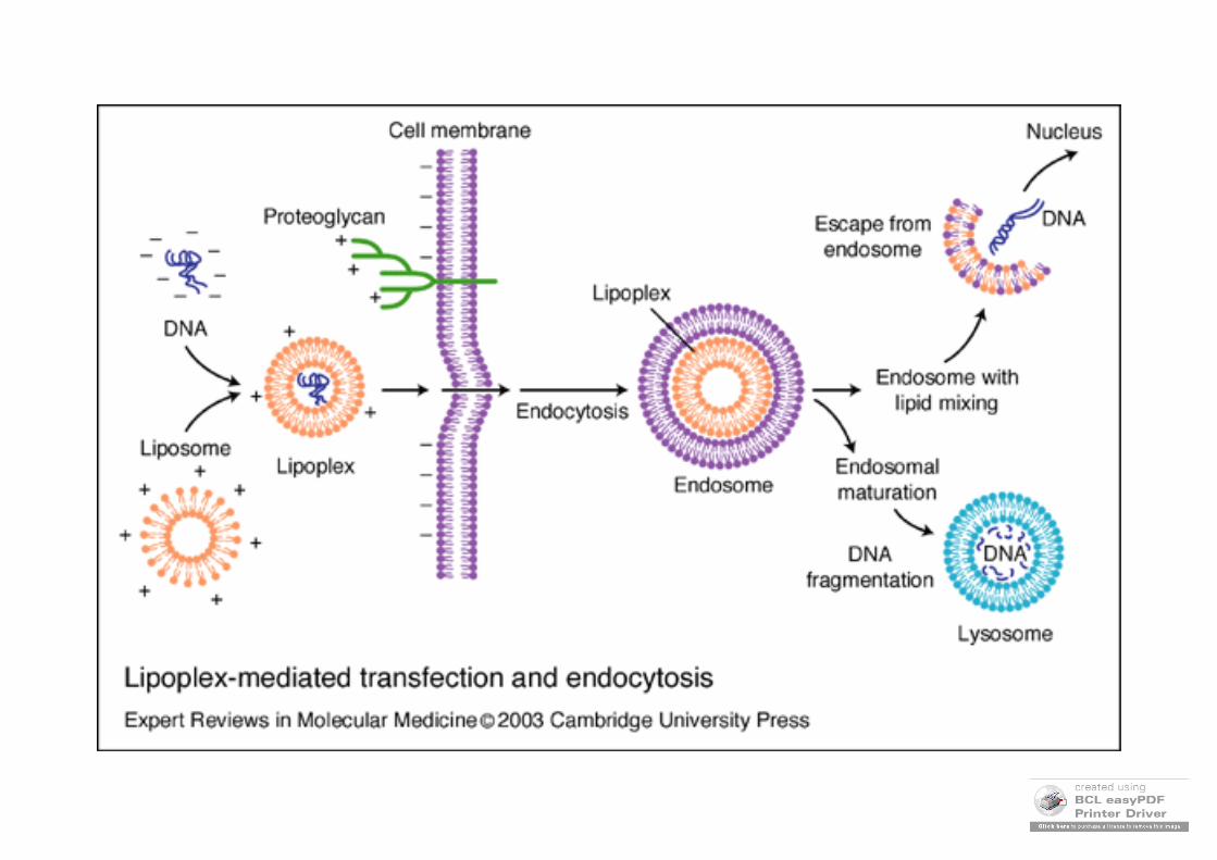
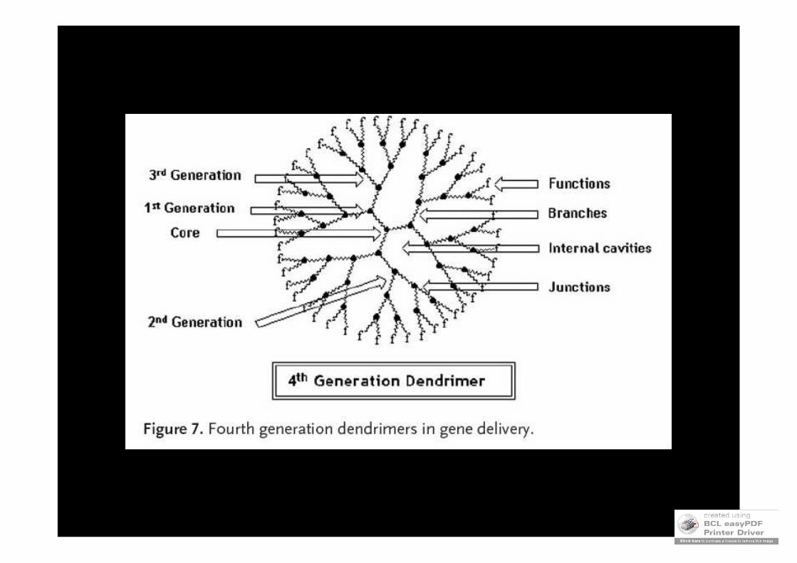
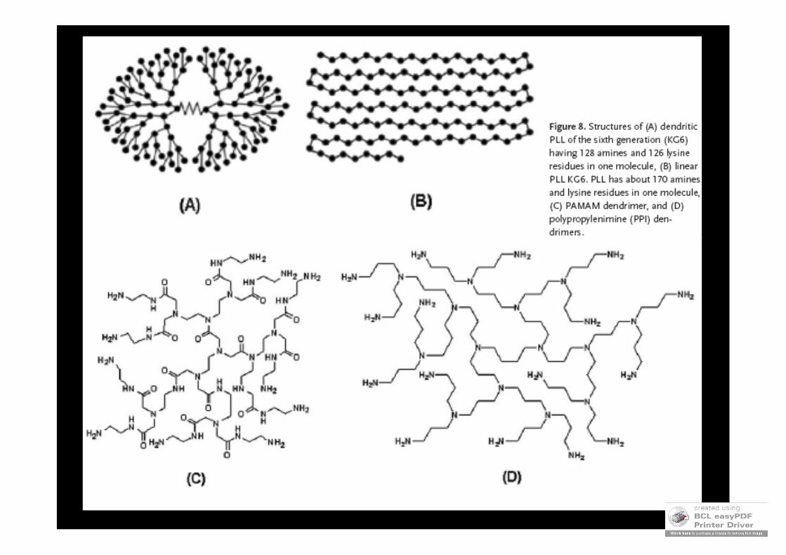
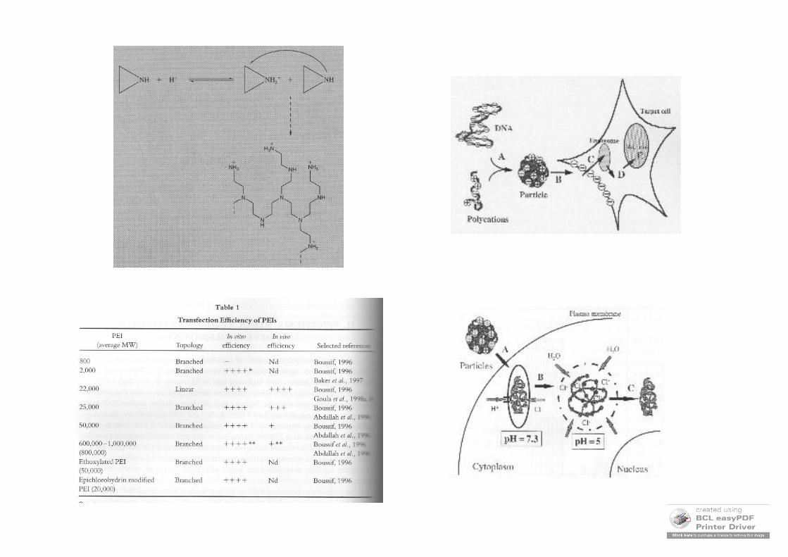
•Microinyección / Perforación (DNA desnudo)•Precipitación con fosfato de calcio•Liposomas aniónicos•Complejos DNA/lípido catiónico: lipoplex•Complejos DNA/polycatión: polyplex•Conjugados moleculares•Dendrímeros: PEI•Hydrogel•Nanoesferas Biopolímero-DNA •Complejos LPD•Inyección a presión•Electroporación•Cañón génico•Ultrasonido•Campo magnético
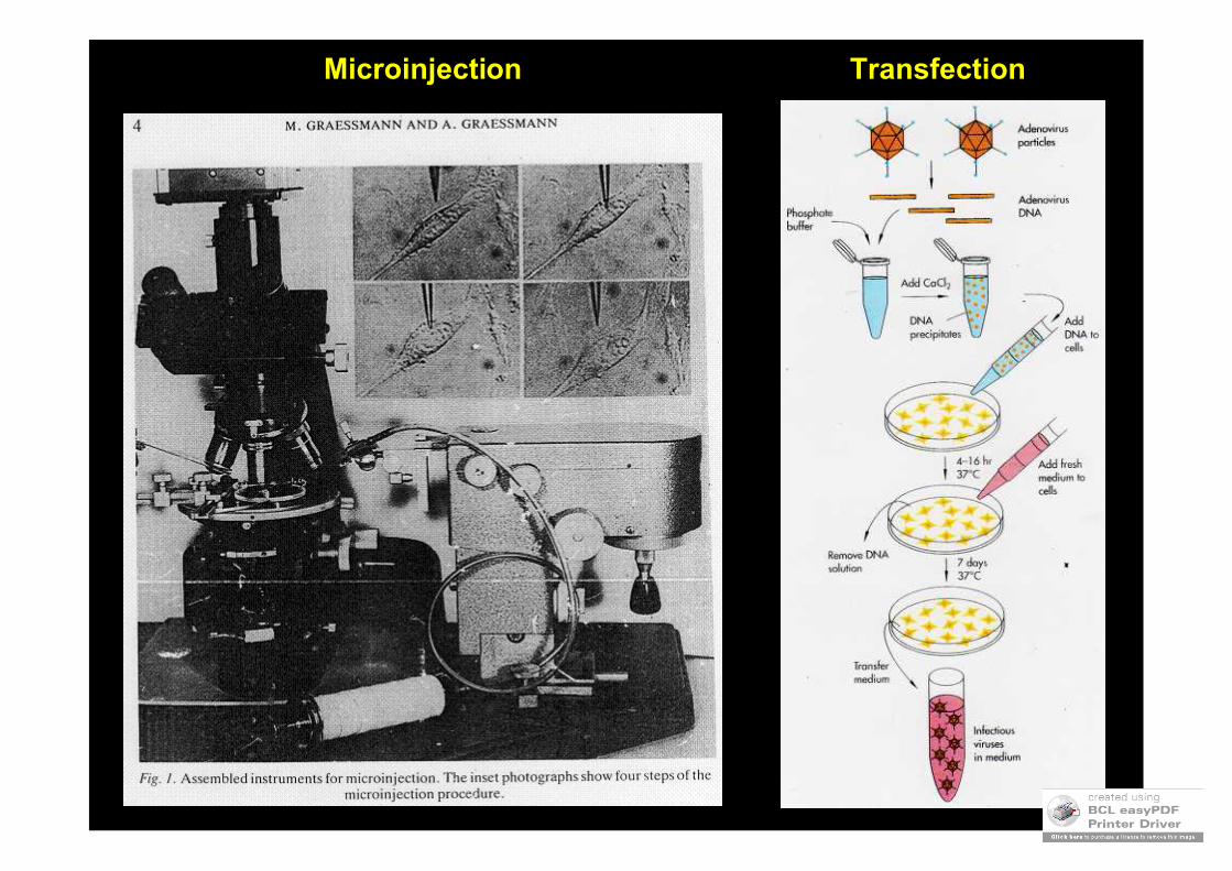
TransfectionMicroinjection
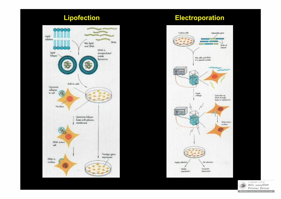
Lipofection Electroporation
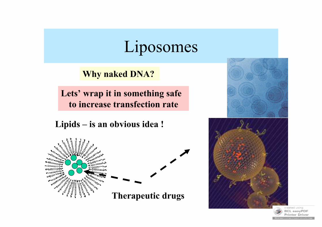
Liposomes
Why naked DNA?
Lets’ wrap it in something safe to increase transfection rate
Therapeutic drugs
Lipids – is an obvious idea !
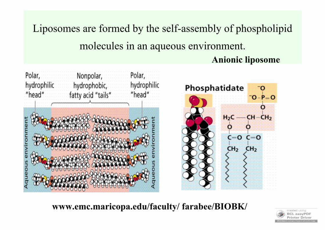
Liposomes are formed by the self-assembly of phospholipid
molecules in an aqueous environment.
www.emc.maricopa.edu/faculty/ farabee/BIOBK/
Anionic liposome

Cationic liposomes
positively charged lipid dropletscan interact with negatively charged DNA
to wrap it up and deliver to cells
Positively charged lipid heads
Lipofectin, lipofectamine, lipofectase….
Inside liposomes DNA is resistant to degradation
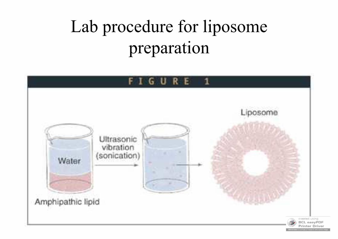
Lab procedure for liposomepreparation
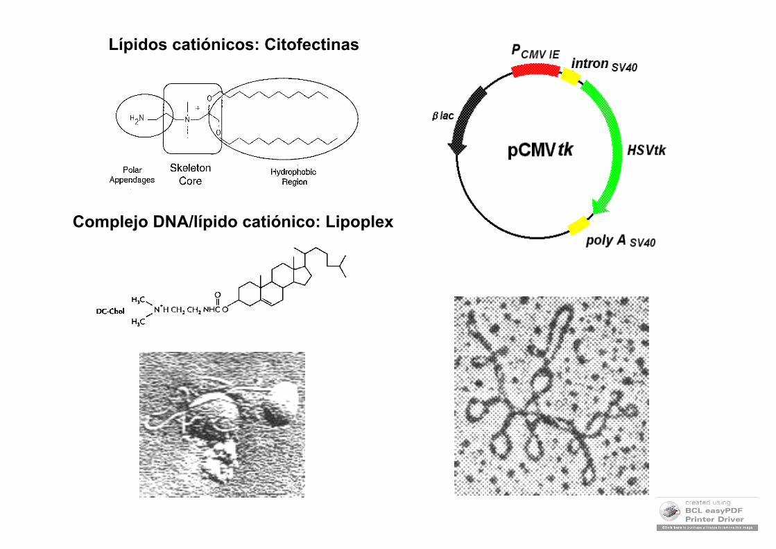
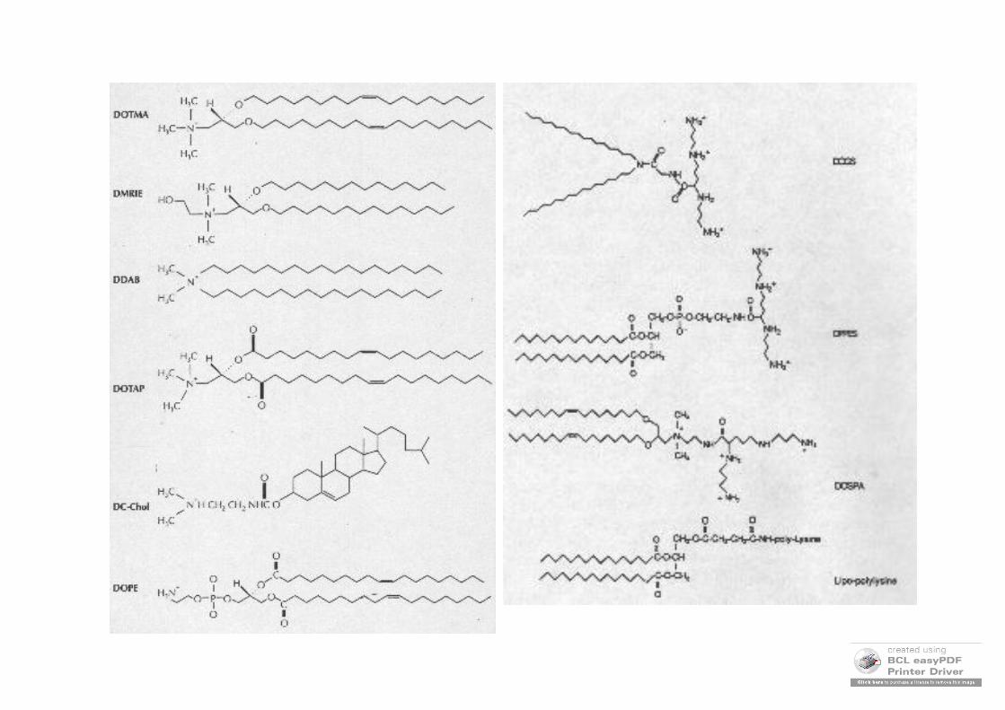
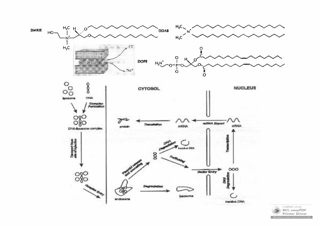
Lípidos catiónicos: Citofectinas
Complejo DNA/lípido catiónico: Lipoplex
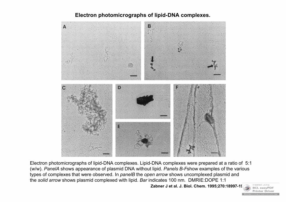
Electron photomicrographs of lipid-DNA complexes.
Zabner J et al. J. Biol. Chem. 1995;270:18997-19007
Electron photomicrographs of lipid-DNA complexes. Lipid-DNA complexes were prepared at a ratio of 5:1 (w/w). PanelA shows appearance of plasmid DNA without lipid. Panels B-Fshow examples of the various types of complexes that were observed. In panelB the open arrow shows uncomplexed plasmid and the solid arrow shows plasmid complexed with lipid. Bar indicates 100 nm. DMRIE:DOPE 1:1
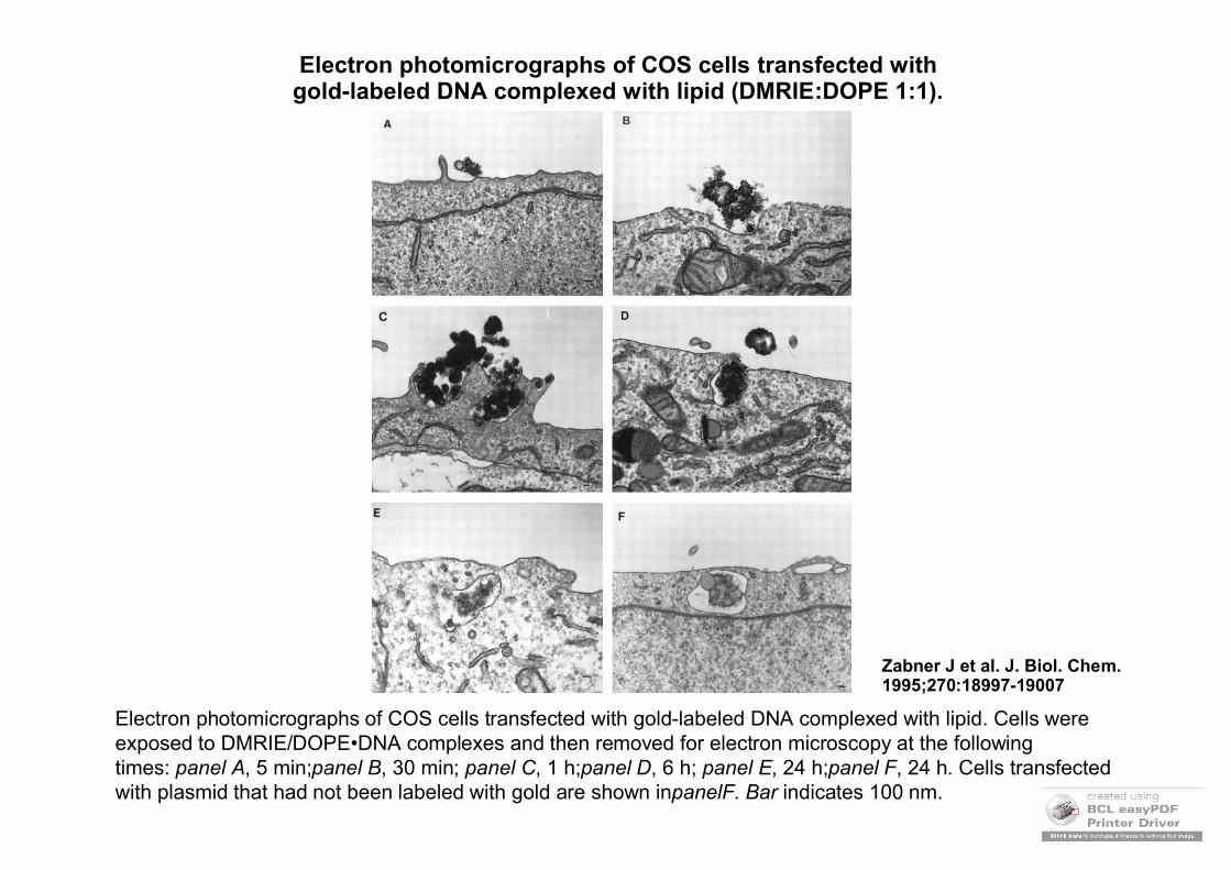
Electron photomicrographs of COS cells transfected with gold-labeled DNA complexed with lipid (DMRIE:DOPE 1:1).
Zabner J et al. J. Biol. Chem. 1995;270:18997-19007
Electron photomicrographs of COS cells transfected with gold-labeled DNA complexed with lipid. Cells were exposed to DMRIE/DOPE•DNA complexes and then removed for electron microscopy at the following times: panel A, 5 min;panel B, 30 min; panel C, 1 h;panel D, 6 h; panel E, 24 h;panel F, 24 h. Cells transfectedwith plasmid that had not been labeled with gold are shown inpanelF. Bar indicates 100 nm.
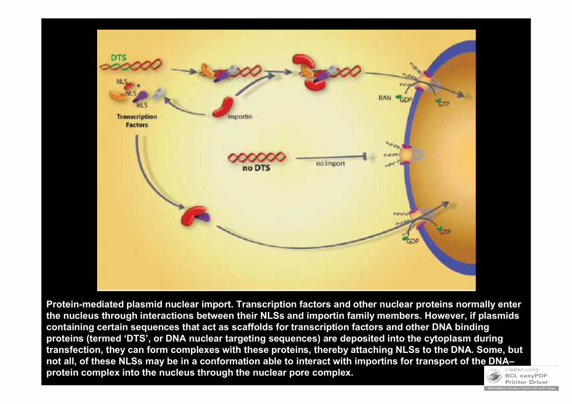
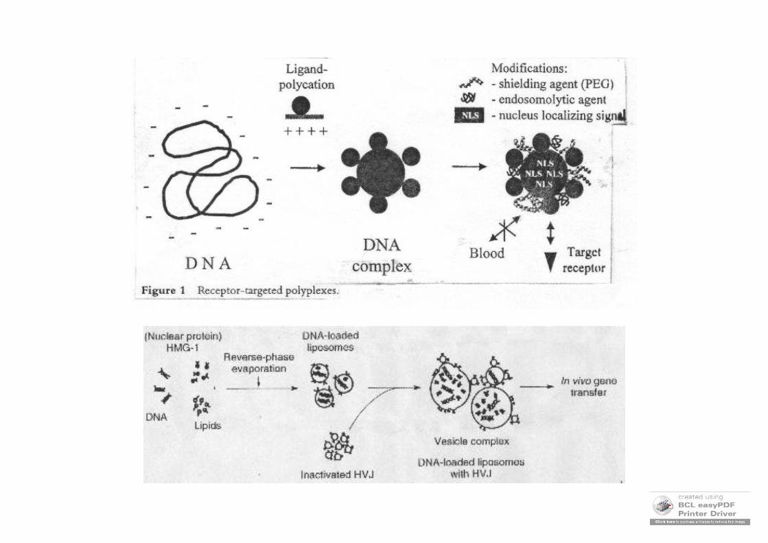
Protein-mediated plasmid nuclear import. Transcription factors and other nuclear proteins normally enter the nucleus through interactions between their NLSs and importin family members. However, if plasmids containing certain sequences that act as scaffolds for transcription factors and other DNA binding proteins (termed ‘DTS’, or DNA nuclear targeting sequences) are deposited into the cytoplasm during transfection, they can form complexes with these proteins, thereby attaching NLSs to the DNA. Some, but not all, of these NLSs may be in a conformation able to interact with importins for transport of the DNA–protein complex into the nucleus through the nuclear pore complex.

Methods to enhance plasmid nuclear import. A number of different approaches have been developed to promote recognition of plasmids by importin family members to increase nuclear import. These include peptide-nucleic acid clamp-conjugated NLS peptides bound to DNA, sequence-specific DNA binding proteins bound to DNA, NLS peptides covalently attached to DNA and NLS peptides electrostatically bound to DNA.
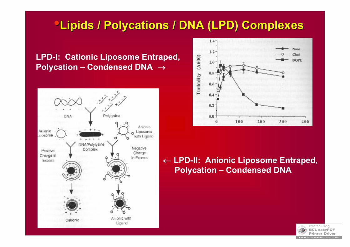
••Lipids / Lipids / PolycationsPolycations / DNA (LPD) Complexes/ DNA (LPD) Complexes
LPD-I: Cationic Liposome Entraped,Polycation – Condensed DNA
LPD-II: Anionic Liposome Entraped,Polycation – Condensed DNA

How to make the gene expressed in the target cell?
Four basic types of expression vectors :
1. Minimal promoters used to study gene regulatory elements such as enhancer elements (in the lab studies).
2. Constitutive promoters used to direct expression of gene productsto produce enough target protein.
3. Cell-specific promoters used to specify expression to target cells(tissue-specific promoters in case of GT)
4. Regulated promoters used to control the on/off expressionof cloned genes.
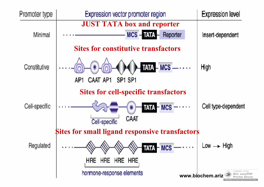
JUST TATA box and reporter
Sites for constitutive transfactors
Sites for cell-specific transfactors
Sites for small ligand responsive transfactors
www.biochem.arizona.edu
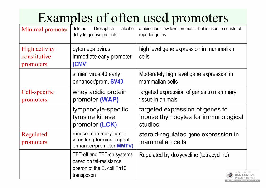
Examples of often used promoters
Moderately high level gene expression in mammalian cells
simian virus 40 early enhancer/prom. SV40
Regulated by doxycycline (tetracycline)TET-off and TET-on systems based on tet-resistance operon of the E. coli Tn10 transposon
steroid-regulated gene expression in mammalian cells
mouse mammary tumor virus long terminal repeat enhancer/promoter MMTV)
Regulated promoters
targeted expression of genes to mouse thymocytes for immunological studies
lymphocyte-specific tyrosine kinasepromoter (LCK)
targeted expression of genes to mammary tissue in animals
whey acidic protein promoter (WAP)
Cell-specific promoters
high level gene expression in mammalian cells
cytomegalovirus immediate early promoter (CMV)
High activityconstitutivepromoters
a ubiquitous low level promoter that is used to construct reporter genes
deleted Drosophila alcohol dehydrogenase promoter
Minimal promoter
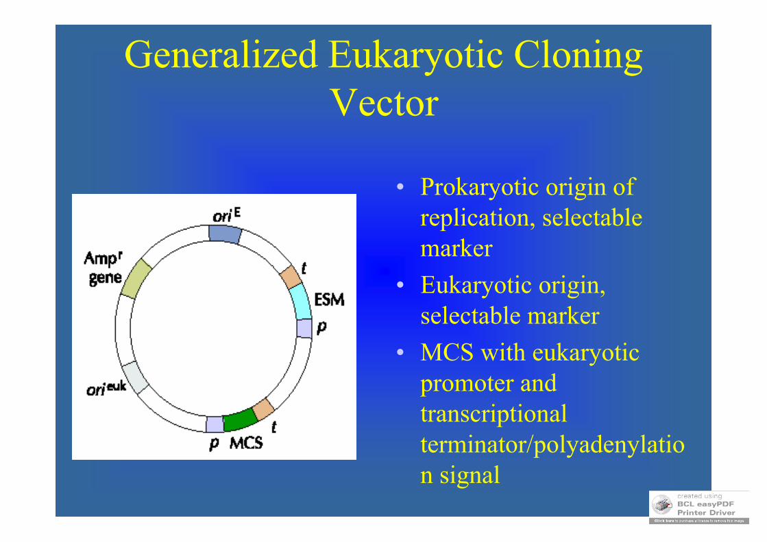
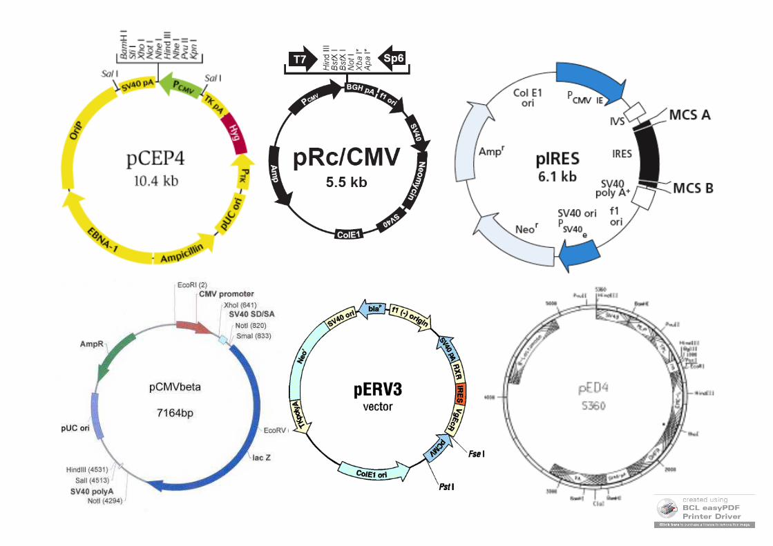
Generalized Eukaryotic CloningVector
• Prokaryotic origin of replication, selectable marker
• Eukaryotic origin, selectable marker
• MCS with eukaryotic promoter and transcriptional terminator/polyadenylation signal
Mammalian Systems
• Sometimes insect cells simply don’t carry out proper/necessary glycosylations
• Other processing may also not occur
• Mammalian cell systems are more expensive by may be required for active product
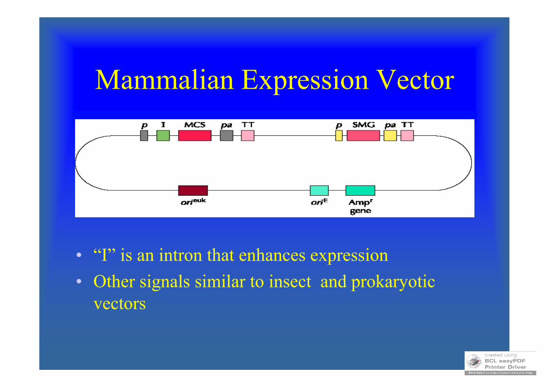
Mammalian Expression Vector
• “I” is an intron that enhances expression
• Other signals similar to insect and prokaryotic vectors

Translation Control Elements
• K - Kozak Sequence (equiv. To rbs)
• S - for secretion signal peptide
• T – tag peptide for purification
• P – proteolytic cleavage sequence
• SC – stop codon for translation
• 3’UTR – proper sequences for efficient translation and mRNA stability (e.g. polyadenylation sequence)
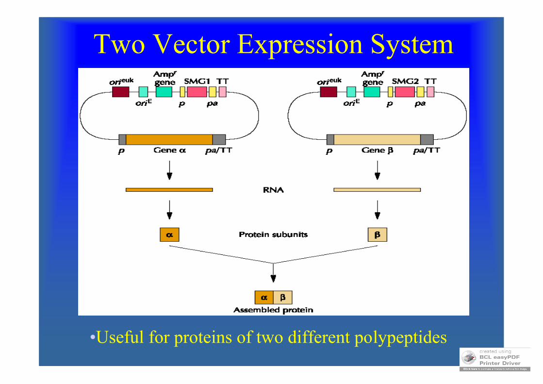
Two Vector Expression System
•Useful for proteins of two different polypeptides
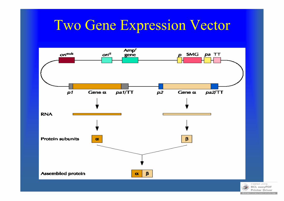
Two Gene Expression Vector
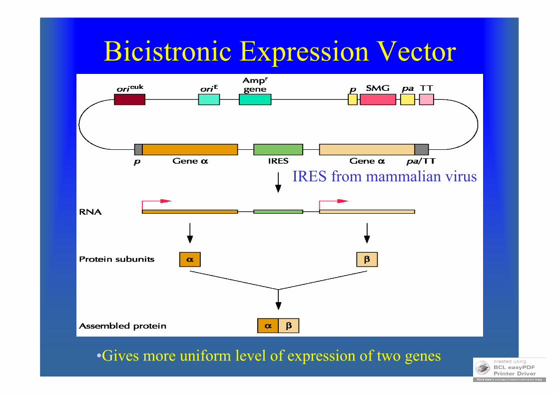
Bicistronic Expression Vector
IRES from mammalian virus
•Gives more uniform level of expression of two genes
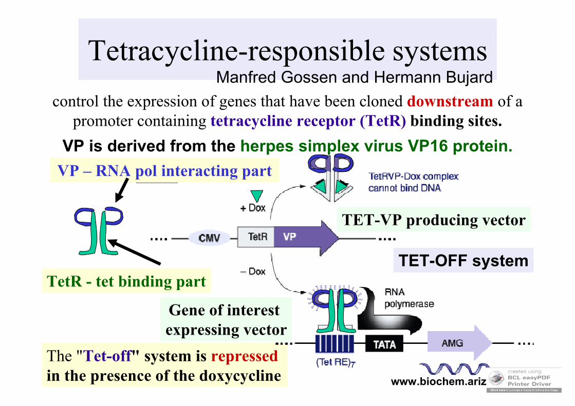
Tetracycline-responsible systemsManfred Gossen and Hermann Bujard
www.biochem.arizona.edu
control the expression of genes that have been cloned downstream of a promoter containing tetracycline receptor (TetR) binding sites.
The "Tet-off" system is repressedin the presence of the doxycycline
VP is derived from the herpes simplex virus VP16 protein.
TET-VP producing vector
Gene of interest expressing vector
VP – RNA pol interacting part
TetR - tet binding partTET-OFF system
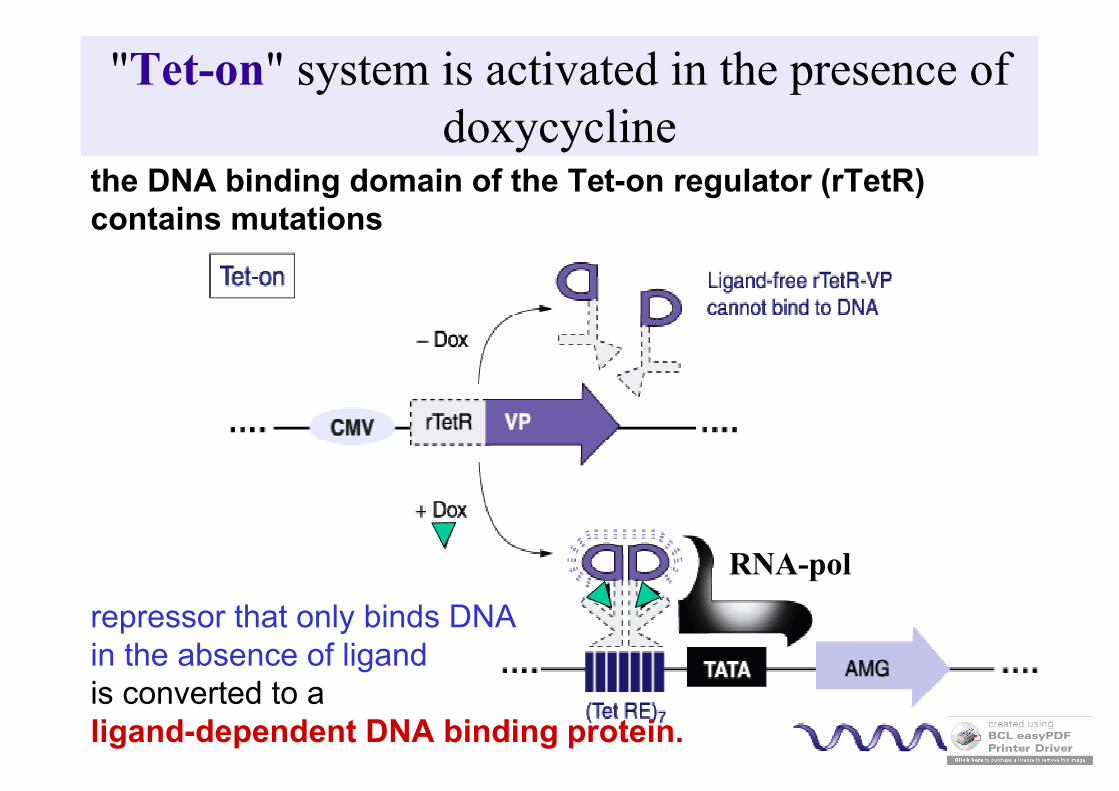
"Tet-on" system is activated in the presence of doxycycline
the DNA binding domain of the Tet-on regulator (rTetR) contains mutations
repressor that only binds DNA in the absence of ligandis converted to a ligand-dependent DNA binding protein.
RNA-pol
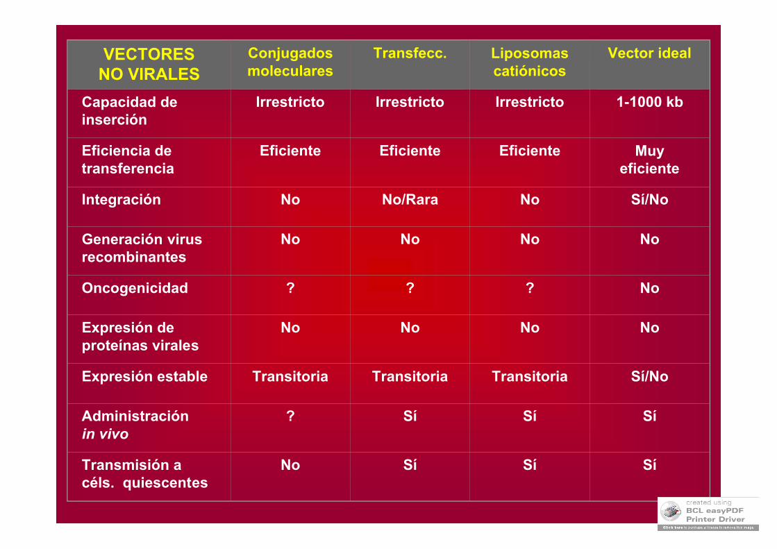
VECTORESNO VIRALES
Conjugadosmoleculares
Transfecc. Liposomascatiónicos
Vector ideal
Capacidad de inserción
Irrestricto Irrestricto Irrestricto 1-1000 kb
Eficiencia detransferencia
Eficiente Eficiente Eficiente Muy eficiente
Integración No No/Rara No Sí/No
Generación virus recombinantes
No No No No
Oncogenicidad ? ? ? No
Expresión deproteínas virales
No No No No
Expresión estable Transitoria Transitoria Transitoria Sí/No
Administraciónin vivo
? Sí Sí Sí
Transmisión acéls. quiescentes
No Sí Sí Sí
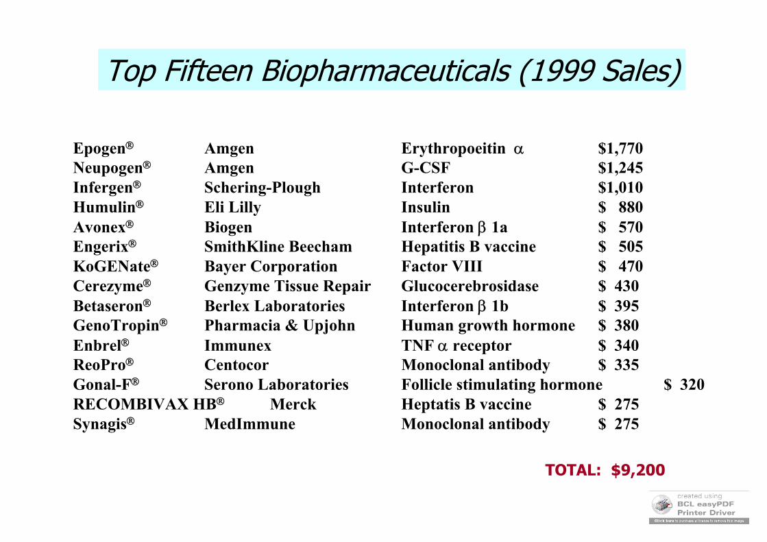
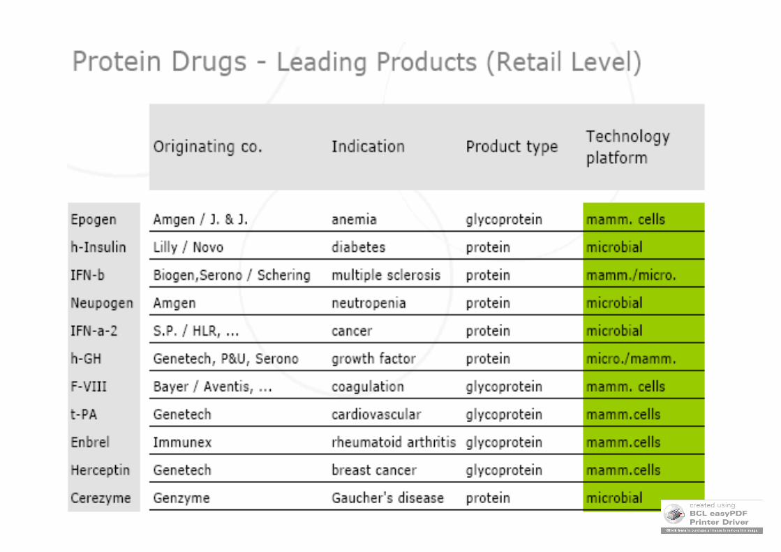
Top Fifteen Biopharmaceuticals (1999 Sales)
Epogen Amgen Erythropoeitin $1,770Neupogen Amgen G-CSF $1,245Infergen Schering-Plough Interferon $1,010 Humulin Eli Lilly Insulin $ 880Avonex Biogen Interferon 1a $ 570Engerix SmithKline Beecham Hepatitis B vaccine $ 505KoGENate Bayer Corporation Factor VIII $ 470Cerezyme Genzyme Tissue Repair Glucocerebrosidase $ 430Betaseron Berlex Laboratories Interferon 1b $ 395GenoTropin Pharmacia & Upjohn Human growth hormone $ 380Enbrel Immunex TNF receptor $ 340ReoPro Centocor Monoclonal antibody $ 335Gonal-F Serono Laboratories Follicle stimulating hormone $ 320RECOMBIVAX HB Merck Heptatis B vaccine $ 275Synagis MedImmune Monoclonal antibody $ 275
TOTAL: $9,200
http://www.lonza.com/group/en/news/downloads/speeches.Par.0028.File2.tmp/part2.pdf
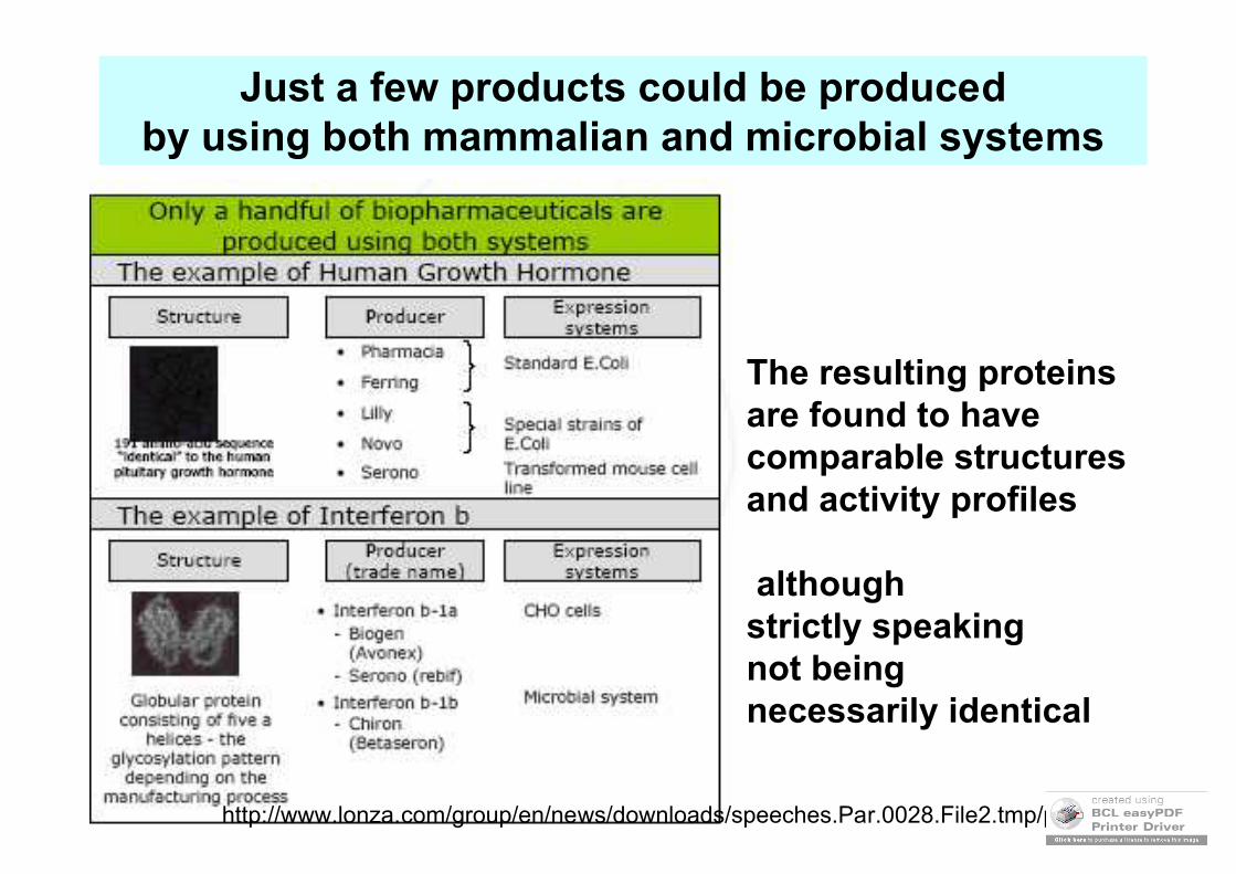
The resulting proteins are found to have comparable structures and activity profiles
although strictly speakingnot being necessarily identical
Just a few products could be produced by using both mammalian and microbial systems

Mammalian system demands on thegrow
http://www.lonza.com/group/en/news/downloads/speeches.Par.0028.File2.tmp/part2.pdf