Mechanisms involved in the · Daniela Del Valle Diaz Lucena . Agustina García Sánchez, Ph.D....
120
ADVERTIMENT. Lʼaccés als continguts dʼaquesta tesi doctoral i la seva utilització ha de respectar els drets de la persona autora. Pot ser utilitzada per a consulta o estudi personal, així com en activitats o materials dʼinvestigació i docència en els termes establerts a lʼart. 32 del Text Refós de la Llei de Propietat Intel·lectual (RDL 1/1996). Per altres utilitzacions es requereix lʼautorització prèvia i expressa de la persona autora. En qualsevol cas, en la utilització dels seus continguts caldrà indicar de forma clara el nom i cognoms de la persona autora i el títol de la tesi doctoral. No sʼautoritza la seva reproducció o altres formes dʼexplotació efectuades amb finalitats de lucre ni la seva comunicació pública des dʼun lloc aliè al servei TDX. Tampoc sʼautoritza la presentació del seu contingut en una finestra o marc aliè a TDX (framing). Aquesta reserva de drets afecta tant als continguts de la tesi com als seus resums i índexs. ADVERTENCIA. El acceso a los contenidos de esta tesis doctoral y su utilización debe respetar los derechos de la persona autora. Puede ser utilizada para consulta o estudio personal, así como en actividades o materiales de investigación y docencia en los términos establecidos en el art. 32 del Texto Refundido de la Ley de Propiedad Intelectual (RDL 1/1996). Para otros usos se requiere la autorización previa y expresa de la persona autora. En cualquier caso, en la utilización de sus contenidos se deberá indicar de forma clara el nombre y apellidos de la persona autora y el título de la tesis doctoral. No se autoriza su reproducción u otras formas de explotación efectuadas con fines lucrativos ni su comunicación pública desde un sitio ajeno al servicio TDR. Tampoco se autoriza la presentación de su contenido en una ventana o marco ajeno a TDR (framing). Esta reserva de derechos afecta tanto al contenido de la tesis como a sus resúmenes e índices. WARNING. The access to the contents of this doctoral thesis and its use must respect the rights of the author. It can be used for reference or private study, as well as research and learning activities or materials in the terms established by the 32nd article of the Spanish Consolidated Copyright Act (RDL 1/1996). Express and previous authorization of the author is required for any other uses. In any case, when using its content, full name of the author and title of the thesis must be clearly indicated. Reproduction or other forms of for profit use or public communication from outside TDX service is not allowed. Presentation of its content in a window or frame external to TDX (framing) is not authorized either. These rights affect both the content of the thesis and its abstracts and indexes.
Transcript of Mechanisms involved in the · Daniela Del Valle Diaz Lucena . Agustina García Sánchez, Ph.D....

ADVERTIMENT. Lʼaccés als continguts dʼaquesta tesi doctoral i la seva utilització ha de respectar els drets de lapersona autora. Pot ser utilitzada per a consulta o estudi personal, així com en activitats o materials dʼinvestigació idocència en els termes establerts a lʼart. 32 del Text Refós de la Llei de Propietat Intel·lectual (RDL 1/1996). Per altresutilitzacions es requereix lʼautorització prèvia i expressa de la persona autora. En qualsevol cas, en la utilització delsseus continguts caldrà indicar de forma clara el nom i cognoms de la persona autora i el títol de la tesi doctoral. Nosʼautoritza la seva reproducció o altres formes dʼexplotació efectuades amb finalitats de lucre ni la seva comunicaciópública des dʼun lloc aliè al servei TDX. Tampoc sʼautoritza la presentació del seu contingut en una finestra o marc alièa TDX (framing). Aquesta reserva de drets afecta tant als continguts de la tesi com als seus resums i índexs.
ADVERTENCIA. El acceso a los contenidos de esta tesis doctoral y su utilización debe respetar los derechos de lapersona autora. Puede ser utilizada para consulta o estudio personal, así como en actividades o materiales deinvestigación y docencia en los términos establecidos en el art. 32 del Texto Refundido de la Ley de PropiedadIntelectual (RDL 1/1996). Para otros usos se requiere la autorización previa y expresa de la persona autora. Encualquier caso, en la utilización de sus contenidos se deberá indicar de forma clara el nombre y apellidos de la personaautora y el título de la tesis doctoral. No se autoriza su reproducción u otras formas de explotación efectuadas con fineslucrativos ni su comunicación pública desde un sitio ajeno al servicio TDR. Tampoco se autoriza la presentación desu contenido en una ventana o marco ajeno a TDR (framing). Esta reserva de derechos afecta tanto al contenido dela tesis como a sus resúmenes e índices.
WARNING. The access to the contents of this doctoral thesis and its use must respect the rights of the author. It canbe used for reference or private study, as well as research and learning activities or materials in the terms establishedby the 32nd article of the Spanish Consolidated Copyright Act (RDL 1/1996). Express and previous authorization of theauthor is required for any other uses. In any case, when using its content, full name of the author and title of the thesismust be clearly indicated. Reproduction or other forms of for profit use or public communication from outside TDXservice is not allowed. Presentation of its content in a window or frame external to TDX (framing) is not authorized either.These rights affect both the content of the thesis and its abstracts and indexes.

Mechanisms involved in the
remyelinating effect of sildenafil.
Daniela Del Valle Diaz Lucena
Doctoral program in Biochemistry, Molecular Biology and Biomedicine.
Department of Biochemistry and Molecular Biology of UAB .
Institute of Biotechnology and Biomedicine V. Villar Palasí.
Bellaterra, September 2016


Mechanisms involved in the remyelinating
effect of sildenafil.
Daniela Del Valle Diaz Lucena
Doctoral program in Biochemistry, Molecular Biology and Biomedicine.
Department of Biochemistry and Molecular Biology.
Institute of Biotechnology and Biomedicine V. Villar Palasí.
Directors:
Agustina García Sánchez, Ph.D.
Institute of Institute of Biotechnology and Biomedicine, Universitat Autònoma de
Barcelona (UAB). Department of Biochemistry and Molecular Biology, UAB.
Paula Pifarré, Ph.D.
Center for Genomic Regulation (CRG), Parc de Recerca Biomédica de Barcelona.
Daniela Del Valle Diaz Lucena
Agustina García Sánchez, Ph.D. Paula Pifarré, Ph.D.
Bellaterra, September 2016


Aquesta tesi va ser desenvolupada amb el suport de la Secretaria d’Universitats i Recerca del Departament d’Economia i Coneixement de la Generalitat de Catalunya i del Fons Social Europeu
This thesis has been developed with the support of the Secretary of Universities and Research of the Ministry of Economy and Knowledge of the Generalitat of Catalonia and the European Social Fund
Esta tesis fue desarrollada con el apoyo de la Secretaría de Universidades e Investigación del Departamento de Economía y Conocimiento de la Generalidad de Cataluña y del Fondo Social Europeo


1
CONTENTS

2

3
CONTENTS
ABSTRACT_______________________________________________________5
ABREVIATIONS__________________________________________________9
INTRODUCTION_________________________________________________13
1. Multiple Sclerosis __________________________________________15 2. Experimental Autoimmune Encephalomyelitis_____________________15 3. Neuroinflammation________________________________________17
3.1. Immunity in the Central Nervous System_____________________17 4. Cyclic GMP______________________________________________23 5. The NO-cGMP pathway_____________________________________23
5.1. Nitric oxide synthases__________________________________24 5.2 . Guanylyl cyclises_____________________________________25
5.3 . cGMP Targets__________________________________________27
5.4. cGMP inactivation_____________________________________29 5.5. cGMP and remyelination________________________________30
AIMS______________________________________________________33
MATERIALS AND METHODS______________________________________37
RESULTS_____________________________________________________55
DISCUSSION_________________________________________________79
CONCLUSIONS_______________________________________________99
REFERENCES_________________________________________________103

4

5
ABSTRACT

6

7
ABSTRACT
Multiple Sclerosis (MS) is a chronic autoimmune demyelinating disease of the central
nervous system characterized by a coordinated inflammatory attack on the myelin sheaths
with ensuing damage to the underlying axons. Using myelin oligodendrocyte glycoprotein
(MOG)-induced chronic experimental autoimmune encephalomyelitis (EAE) as a MS model,
it has been previously demonstrated that daily administration of the PDE-5 inhibitor
sildenafil starting at peak disease rapidly ameliorates clinical symptoms whereas
administration at the onset of symptoms prevents disease progression. These beneficial
effects involved down-regulation of adaptive and innate immune responses and protection
of axons and oligodendrocytes and promotion of remyelination. The aim of this work was
confirm the remyelinating potential of sildenafil treatment and investigate mechanisms
involved in this effect in CNS cells. Results show that sildenafil induces remyelination in EAE
mice even when the administration of the drug starts during the chronic stage of the
disease. Sildenafil also stimulates remyelination in cerebellar organotypic cultures
demyelinated with lysophosphatidylcholine and this effect is prevented by inhibitors of nitric
oxide-dependent guanylyl cyclase (NO-GC), NO synthase type 2 (NOS-2) and cGMP-
dependent protein kinase (PKG), indicating the involvement of the endogenous NO-cGMP-
PKG pathway. Maturation of oligodendrocytes as a potential mechanism implicated in the
remyelinating effect of sildenafil was investigated by immunostaining for transcription
factors involved in different stages of oligodendrocyte development. Results in the EAE
model show that sildenafil treatment increases oligodendrocyte precursor cells (OPCs;
Nkx2.2+ cells) and promotes the final stage of oligodendrocyte maturation (olig2-/MBP+
cells). These later result was confirmed in LPC-demyelinated cerebellar slices treated with
cGMP increasing compounds. This work also shows that expression of the neurotrophic
factor CNTF, that has been implicated in oligodendrocyte maturation, is increased in
astroytes of sildenafil-treated EAE mice spinal cord, as well as in cerebellar slice cultures.
Results also show that cGMP-increasing treatments alter expression of inflammatory
phenotype markers (COX-2 and Arg-1) in microglia in demyelinated slice cultures. The
potential of cGMP-increasing treatments for regulating the inflammatory phenotype of
monocytes was confirmed in bone marrow derived macrophages (BMDM). In these cells
sildenafil treatment induces arginase activity and potentiates the effect of IL-4 suggesting
the promotion of an M2 phenotype. Analysis by flow cytometry of BMDM confirmed that
cGMP augments the number of cells expressing an M2 phenotype marker (CD206). This work
further demonstrates that sildenafil significantly increases the myelin phagocytic capacity of
microglia/macrophages in EAE mice and in BMDM. Taken together these data suggest that
promotion of oligodendrocyte maturation, growth factor expression, modulation of the
inflammatory process and clearance of myelin debris may be relevant mechanisms involved
in sildenafil enhancement of remyelination in demyelinated tissue.

8

9
ABREVIATIONS

10

11
ABREVIATIONS
Abreviation Name
Abreviation Name
8BrcGMP 8-Bromoguanosine 3′,5′-cyclic monophosphate sodium salt
EAE
Experimental Autoimmune Encephalomyelitis
8pCPTcGMP
8-(4-Chlorophenylthio)-guanosine 3 ,̈́5 -̈́cyclic monophosphate sodium salt
EDTA TRIS buffer solution
ANP Atrial natriuretic peptide
FBS Fetal Bovine Serum
BAY41 BAY 41-2272
GC Guanylyl cyclase enzymes
BBB Blood brain barrier
GFAP Glial fibrillary acidic protein
BDNF Brain derived neurotrophic factor
Glucose Glucose
bFGF Basic fibroblast growth factor
HBSS Hank’s buffered salt solution
BME Basal medium containing Earle’s salt
HS Heat Inactivated horse serum
BNP Brain natriuretic peptide
Iba-1 ionized calcium-binding adaptor protein-1
BSA Neonatal Goat serum
IFNγ Recombinant Mouse Interferon γ
cGMP Cyclic Guanosine Monophospate
IGF-1 Insuline-like growth factor
CNP C-type natriuretic peptide
IL-4 Recombinant Murine Interleukin-4
CNPase Cyclic Nucleotide Phosphodiesterase
INFγ Interferon gamma
CNS Central nervous system
IS Immune system
CNTF Ciliary neurotrophic factor
L-Glutamine
L-Glutamine 200mM
CPB Cytometry permeabilization buffer
L-NNA L-NNA
dbcGMP dibutyryl-cGMP
LPC L-α-Lysophosphatidylcholine from egg yolk
Dil
1,1”-diotadecyl-3,3,3’,3’-tetramethyl-lindocarbocyanide perchlorate
LPS Lipopolysaccharide
DMEM Dubelco's Modified Eagle medium GlutaMax
LPS Lipopolysaccharides from Salmonella enterica

12
Abreviation Name
Abreviation Name
MAG Myelin Associated Glycoprotein
PDE5 Phosphodiesterase type 5
MBP Myelin Basic Protein
PDEs Phosphodiesterases
M-CSF Recombinant Macrophage Colony Stimulating Factor
PDGFαR Platlet Derived Growth Factor α Receptor
MHC Major histocompatibility complex
Pen/Strep Penicillin-Streptomycin
MOG Myelin Oligodendrocyte Glycoprotein
PFA Paraformaldehyde
MS Multiple Sclerosis
pGC Particulate guanylyl cyclases
NGF Nerve growth factor
PKG cGMP dependent protein kinases
NGS Neonatal Goat serum
PLP Proteolipid Protein
NMDA N-methyl-d-aspartate
PP-MS Primary Progressive Multiple Sclerosis
NMDA N-methyl-D-Aspartate
RI Remyelination index
NO Nitric oxide
RIPA RIPA Lysis Buffer
NO-GC NO-sensitive guanylyl cyclase
ROCK Rho-Associated-Kinase
NOS Nitric oxide synthases
ROS Reactive oxygen species
NOS-1 Neuronal NOS
Rp-8pCPT-cGMP
8-[(4-Chlorophenyl)thio]-guanosine-cyclic 3',5' monophosphate
NOS-2 Inducible NOS
RR-MS Relapsing-Remitting Multiple Sclerosis
NOS-3 Endothelial NOS
s.c sub-cutaneous
NP Natriuretic peptide
SC Spinal cord
NPR Natriuretic peptide receptor
Sil Sildenafil
ODQ ODQ
TGF-β Transforming growth factor beta
Ols Oligodendrocytes
TNFα Tumor necrosis factor alpha
ON overnight
Tris Phosphate buffer saline
OPCs Oligodendroglial precursor cells
U0126
1,4-Diamino-2,3-dicyano-1,4-bis[2-aminophenylthio]butadiene
ORO Oil Red-O
WB Western Blot
PBS Bovine serum albumim
Ym-1 Chitinase-like 3

13
INTRODUCTION

14

15
INTRODUCTION
1. Multiple Sclerosis
Multiple Sclerosis (MS) is a chronic, inflammatory and demyelinating
disease of the CNS (Herz et al. 2010). MS etiology and the initiation of
autoimmunity that comes with it, are still unknown (Nave 2010). Symptoms of
MS include limb weakness, numbness, blurred or double vision and ataxia.
Some advanced cases present cognitive impairments and memory loss (Hauser
and Oksenberg 2006). In MS the immune system reacts against central nervous
system (CNS) myelin components, initiating a detrimental inflammatory
cascade that leads to demyelination and axonal and neuronal
degeneration which is the principal anatomical correlate of progressive clinical
deterioration. Axon protection can be achieved directly by intervention on the
mechanisms by which axons are injured or degenerate and can also be
achieved by immune-modulatory therapies and by promotion of remyelination.
MS research has mainly focused on the immunological aspects of the disease
and has translated into the development of highly effective immune-
modulatory therapies to control the initial relapsing-remitting phase of MS,
however the secondary progressive phase, in which there is continual atrophy
of demyelinated axons, remains largely untreatable. (Garg and Smith 2015,
Nave 2010, Peterson and Fujinami 2007).
2. Experimental Autoimmune Encephalomyelitis
Experimental autoimmune encephalomyelitis (EAE) has been extensively
used as an animal model of MS since it shares with the human autoimmune
disease the presence of inflammatory infiltrates in the CNS parenchyma,
demyelination and axonal loss, predominantly in the spinal cord, and paralysis
(Friese et al. 2006). Like MS, EAE seems to be initiated by myelin antigen-

16
specific CD4+ T-lymphocyte infiltration into the CNS. CD4+ cells together with
infiltrated macrophages, dendritic cells, and resident microglia constitute the
ultimate effector cells of neuroinflammation, progression of demyelination,
and axonal damage (Bynoe et al. 2007, Friese et al. 2006). In contrast,
accumulating evidence indicates that local astroglial activation is
neuroprotective in EAE. Using transgenic mice with targeted ablation of
proliferating scar-forming astrocytes it has been shown that severity of EAE is
enhanced and that leukocyte and macrophage entry into the CNS parenchyma
is significantly increased (Voskuhl et al. 2009). Recent evidence additionally
indicates that demyelination and oligodendrocyte degeneration follows
inflammation-induced astrocyte dysfunction (Sharma et al. 2010). High levels of
inflammatory mediators are secreted by infiltrating immune cells and resident
CNS cells, which is characteristic of the inflammatory environment during
disease (Raivich and Banati 2004).
EAE can be classified as passive (when induced by introducing MBP-
reactive T-cells into the animal) or active (when induced by immunization with
CNS tissue or myelin peptides). Active EAE can also be divided into two
different types according to the mouse strain and peptide used to induce it. In
the SJL/J mouse strain active induction with PLP develops a relapsing–remitting
form, while in the C57BL/6 mouse, immunization with MOG35–55 in CFA induces
a chronic-sustained form of EAE (Constantinescu et al. 2011, McCarthy et al.
2012). Symptoms are characterized by ascending hind limb paralysis that is
associated with inflammation and demyelination of axonal tracks.

17
3. Neuroinflammation
3.1. Immunity in the Central Nervous System
The CNS has been classically considered an immunologically “privileged”
organ because it was thought to be isolated from the immune system (IS) and
excluded from its surveillance. Nowadays, it is well accepted that the “immune
privilege” of the CNS is an active process, the immune response is present but
restricted and involves a closely regulated inter-communication between CNS
resident cells and the IS which allows control of immune-mediated
inflammation and related secretion of potentially damaging molecules that
could have devastating consequences in this essential organ with limited
capacity for regeneration (Nakamizo et al. 2003).
The Blood Brain Barrier
The most important feature in CNS immune privilege is the presence of
the Blood-Brain Barrier (BBB), a structure formed by blood vessel endothelial
cells joined by tight junctions, the basal lamina in which pericytes are
embedded and the end-feet of astrocytes, in conjunction with intra- and
extracellular enzymes that represent a metabolic barrier. The BBB exerts bi-
directional control over the transcellular passage of substances such as
regulatory proteins, nutrients and electrolytes, maintaining the optimal ionic
composition for axonal transmission and synaptic signaling. The BBB also avoids
the entry of potentially harmful or toxic substances and supervises the
infiltration of IS cells by expression of adhesion molecules in endothelial cells.
However, in neuropathological conditions activating signals are produced by
resident (glial) cells that can facilitate inflammation or promote recovery, but
uncontrolled neuroinflammation can induce secondary injury (Abbott et al.
2010).

18
In addition to the BBB, other factors also contribute to restricting the
immune response in the CNS, like the absence of a conventional lymphatic
system and the failure of antigen presenting cells (APCs) to migrate to lymph
nodes along perivascular lymphatic drainage pathways in the healthy brain,
although they might do so in the inflamed CNS. In addition, the highly regulated
entrance of immune cells by the presence of immune-inhibitory factors
constitutively produced by neurons, and the very low expression of activating
and co-stimulatory molecules, are also important factors that contribute to
minimize inflammatory responses in the CNS (Griffiths et al. 2009, Weller et al.
2010). Nevertheless, immunological reactions do occur in the CNS in response
to infections and in immune-mediated disorders such as multiple sclerosis
(MS).
Resident immune effector cells in the CNS
The functions of immune surveillance and differentiation between “self”
and “non-self” antigens in non-CNS tissue, provided by neutrophils, dendritic
cells, macrophages and natural killer cells in the periphery, are in the CNS
attributed to resident glial cells, astrocytes and microglia (Veerhuis et al. 2011).
After a CNS injury, glial cells show phenotypic changes referred to as reactive
gliosis, one of the most important features in neuroinflammation (Aloisi 2001,
Dong and Benveniste 2001). Glial activation plays a crucial role in acute and
chronic inflammatory responses and reactive glia has been found in brains after
traumatic injury, ischemia, infections, neuropathic pain, seizures, autoimmune
inflammatory diseases, glaucoma, leukodystrophies, edema, psychiatric
disorders, brain tumors and also in neurodegenerative diseases (Ghafouri et al.
2006, Kim Y. S. and Joh 2006, Sofroniew and Vinters 2010, Streit et al. 2005,
Wyss-Coray 2006)

19
Astrocytes
Astrocytes are the glial cells responsible of the CNS structure support,
regulate blood flow, form and maintain the BBB and also mediate its
permeability. Astrocytes regulate transport of ions, nutrients and toxins. These
cells also regulate synapses and nerve impulse transmission (Ludwin et al.
2016). After a brain injury, astrocytes are recruited to the lesion site and help
forming a glial scar that promotes healing after the lesion (Lee et al. 2015).
Astrocytes express neurotrophic and growth factors such as brain derived
neurotrophic factor (BDNF), ciliary neurotrofic factor (CNTF), transforming
growth factor beta (TGF-β) and insulin-like growth factor (IGF-1). Astrocytes are
also able to express pro and anti-inflammatory cytokines (Ludwin et al. 2016,
Peterson and Fujinami 2007).
Microglia
Microglial cells are considered the resident macrophages of the CNS,
descendants of the monocytic lineage invade the central nervous system early
during ontogenesis (Fedoroff and Hao 1991, Hailer 2008). In resting conditions,
microglial cells present a ramified morphology with an elaborate tertiary and
quaternary branch structure. Their prolongations cover a space of 30-50 µm
not overlapping each other, expanding throughout the CNS (Raivich et al.
1999). Although in their apparent resting state, microglial cells are greatly
active, continually surveying their microenvironment with extremely motile
processes and protrusions poised to rapidly respond to environmental changes
(Nimmerjahn et al. 2005). CNS injury provokes immediate and focal activation
of microglia, which is thought to occur before astrocyte activation, switching
their behavior from resting to defending cells in the injured site, with a
different response depending on the stimulation provided (Town et al. 2005).

20
Reactive microglial cells proliferate, retract its ramifications and acquire
amoeboid morphology, extend new pseudopodia that enable active migration
and phagocytic capacity for removing dead cells and cell debris. These
characteristics together with their antigen presenting capacity in MHC-II, make
them the antigen-presenting cells of the CNS initiating the adaptive immune
response (Fig.2) (Aloisi 2001, Hailer 2008, Kim S. U. and de Vellis 2005).
Microglia/macrophage activation phenotype
Activation of microglia in response to brain injury involves a continuum
spectrum of phenotypes that share characteristics with their homologue IS
cells, the macrophages. The reactive microglia/macrophage phenotype
oscillates between two end-points depending on the stimulatory environment,
one corresponds to the adaptive activation with release of pro-inflammatory
cytokines that is associated with capacity for stimulation of Th1 and Th2
subpopulations (M1 or classically activated phenotype), and the other to innate
activation with a phagocytic phenotype and anti-inflammatory cytokine
secretion (M2 o alternativelly activated). In vitro, the M1 phenotype is induced
in the presence of pro-inflammatory cytokines and LPS and the M2 phenotype
in the presence of anti-inflammatory cytokines such as IL-4. Recent studies
have identified M2 phenotype subtypes that have been classified according to
specific protein expression and possible functions. M2a has been associated
with regeneration and repair, M2b is considered immunoregulatory and M2c
immunosuppressive (Chhor et al. 2013, Miron and Franklin 2014, Mosser and
Edwards 2008, Ransohoff and Perry 2009). Microglia/macrophage phenotypes
can be recognized by changes in the expression of specific molecules. NOS-2,
TNFα, COX-2, and IL-1β are characteristic molecules of a M1 phenotype while
arginase-1, CD206, Ym-1 and FIZZ-1 are considered M2-phenotype markers

21
(Chhor et al. 2013, Fernando et al. 2014, Gensel and Zhang 2015). However,
recent studies in vitro and in vivo have revealed that reactive
microglia/macrophages are able to co-express M1 and M2 markers. In vivo
experiments have shown that treatent of mice with LPS increases pro-
inflammatory cytokines including IL-1β, but also IL-4R and further treatment
with IL-4 induced the expression of Arg-1 as well as IL-1β, and this unique
phenotype was able to promote neurite growth and spinal cord injury recovery
(Fenn et al. 2014). Furthermore, macrophages treated with IL-4 in vitro, were
shown to produce IL-6 while co-expressing the M2 characteristic molecule
CD206, and the co-expression of IL-6 did not affect immunosuppressive
properties of the M2 phenotype macrophages (Casella et al. 2016). Other
studies aimed at characterizing specific molecules to delineate and identify
microglia /macrophage phenotypes have also shown co-expression of the anti-
and pro-inflammatory phenotype markers (Chhor et al. 2013, Gensel and Zhang
2015).
Myelination/Remyelination
Myelination is an ordered and rapid process the uninjured, healthy adult
brain. New myelin is continually generated along with new oligodendrocytes in
active myelination, which perform myelin maintenance with subtle remodeling
of specific areas (Bercury and Macklin 2015).
Demyelination is the pathological process where myelin sheaths are lost
from around axons impairing nerve impulse conduction (Franklin and Ffrench-
Constant 2008). There are two major causes of primary demyelination in the
CNS, the first is due to genetic abnormalities that affect glia, such as
leukodystrophies, and the second is due to inflammatory damage to myelin
and oligodendrocytes, which can be observed in neurological diseases such as

22
MS and other neuroinflammatory disorders (Franklin and Ffrench-Constant
2008).
Remyelination is the process responsible of restoring entired myelin
sheaths around demyelinated axons, reinstating saltatory conduction and
ameliorating functional deficits (Franklin and Ffrench-Constant 2008). Extensive
data indicate that, after demyelination, remyelinated internodes are thinner
and shorter than, but after late time of recovery remyelinated fibers may have
similar internode length and thickness to developmentally myelinated axons
(Bercury and Macklin 2015).
Oligodendrocytes
Oligodendrocytes (OLs) are the CNS glial cells responsible for the production
of the myelin sheath that surrounds axons isolating the electrical impulse in
order to gain speed and efficiency (Nave 2010). OLs can have over 50
extensions reaching axons, and can myelinate them simultaneously (Copray et
al. 2006, Liu et al. 2007, Nave 2010). OLs can interact with surrounding cells
that influence OLs migration, differentiation and myelination (Kimelberg 2010,
Li et al. 2016, Talbott et al. 2005). OL proliferation and differentiation is
regulated by cytokines and growth factors (Nave 2010), such as BDNF (De Santi
et al. 2009, Weishaupt et al. 2012) and CNTF (Stankoff et al. 2002). OLs express
characteristic transcription factors that allow determination of their
differentiation or maturation stage, such as Nkx.2, an early marker of OL
precursor differentiation, and Olig1 and Olig2, expressed during early to late
differentiation stages. Mature OLs express myelin basic protein (MBP),
proteolipid protein (PLP), myelin oligodendrocyte glycoprotein (MOG), myelin
associated glycoprotein (MAG) and 2’,3’-cyclic nucleotide 3’-phosphodiesterase
(CNPase) among others (Copray et al. 2006, Liu et al. 2007, Meffre et al. 2015,
Miron et al. 2010, Nishiyama et al. 2009, Zhang H. et al. 2011).

23
4. Cyclic GMP
Cyclic guanosine monophospate (cGMP) is a second messenger in signal
transduction. It is generated by guanylyl cyclase enzymes (GC) that catalyze the
conversion of GTP into cGMP. There are two main classes of GCs: nitric oxide-
sensitive guanylyl cyclase (NO-GC) that is mainly soluble, and particulate
guanylyl cyclases (pGC) represented by the natriuretic peptide (NP)-receptor
(NPR) group (Tsai and Kass 2009) (Fig 1).
Figure 1: Schematic representation of cyclic GMP synthesizing pathways. GC activation
mechanisms. GC-A, GC-B and GC are membrane bound GC and NO-GC is cytoplasmic.
Scheme from Duman and Nestler, Guanylyl Ciclase, 1999 (Duman RS 1999).
5. The NO – cGMP pathway
Nitric oxide (NO) is a small, gaseous, highly diffusible, and reactive
molecule with a short half-life, rapidly oxidized to the stable and inactive end-
products, nitrite and nitrate (Zhang J. and Snyder 1995). In the CNS, NO has
been associated with the modulation of synaptic plasticity, brain development,
visual and sensory processing, neuro-endocrine secretion and cerebral blood
flow (Garthwaite J. 2000, Guix et al. 2005). It can act as a

24
neurotransmitter/neuromodulator or as an inflammatory response mediator,
low concentrations of NO mediate physiological signaling and can also be
neuroprotective and it has shown to be essential for synaptic plasticity, control
of cerebral blood flow, neurogenesis and synaptogenesis (Manucha 2016,
Zhang J. and Snyder 1995). However, in high concentrations is a
neuropathological agent responsible for excitotoxic cell death and
neuroinflammatory cell damage in many neurological disorders (Duncan and
Heales 2005, Manucha 2016, Murphy 2000) .
5.1. Nitric oxide synthases
Nitric oxide synthases (NOS) are a group of enzymes that produce NO by
oxidation of one of the guanidine nitrogens of L-arginine, using O2 and NADPH
as co-substrates and forming L-citrulline as product, a reaction that requires O2
and NADPH as co-substrates (Zhang J. and Snyder 1995). There are three
isoforms of NOS, two of them NOS-1 and NOS-3 are expressed constitutively
while NOS-2 is an inducible enzyme. NOS-1 is found in the nervous system
(primarily in neurons and astrocytes) and NOS-3 is expressed in vascular
endothelial cells. Both, NOS-1 and NOS-3, are calcium-calmodulin-dependent
and produce nanomolar concentrations of NO in response to transient
elevations in intracellular calcium. In neuronal populations this generally occurs
by N-methyl-D-aspartate (NMDA) type glutamate receptor stimulation that
allows calcium entry (Maarsingh et al. 2009, Manucha 2016). NOS-2 is inducible
at the transcriptional level and its activity is calcium-independent since it has
tightly-bound calmodulin. NOS-2 produces higher and longer-lasting amounts
of NO (micromolar concentrations) after induction by inflammatory
compounds such as LPS, pro-inflammatory cytokines (IL-1β, TNF-α and IFN-γ) or
Aβ peptides, that can cause neurotoxicity. In peripheral tissues this enzyme is
mainly, but not exclusively, expressed in macrophages. In the CNS, NOS-2 is

25
expressed in microglia and astroglia, and its generation of NO has been
implicated in the pathogenesis of various insults as well as in neurological
disorders (Brown and Neher 2010, Murphy 2000, Steinert et al. 2011). Up-
regulation of iNOS has been implicated in tissue damage in MS and EAE (Raivich
and Banati 2004). However, iNOS-deficient mice develop more severe EAE
(Willenborg et al. 2007), suggesting that NO may be neuroprotective.
5.2 . Guanylyl cyclises
NO-sensitive guanylyl cyclases
NO-GC is recognized as the major receptor for NO, and mediates
numerous of its physiological functions. NO-GC was initially thought to be
entirely cytosolic and was thus named “soluble GC”, however it has also been
found associated to membranes (Zabel et al. 2002). NO-GC is composed of an α
and a β subunit and both subunits are required for catalytic activity (Koesling et
al. 2004). Each subunit has a regulatory domain which contains a prosthetic
heme group that is the NO-binding site, a catalytic domain that shares
sequence homology with the corresponding domains in pGC and adenylyl
cyclases and a central domain which allows subunit dimerization (Foster et al.
1999) (Fig. 4).
In contrast to the ubiquitous NO formation in CNS parenchymal cells,
NO-dependent cGMP synthesis by activation of NO-GC appears to occur mainly
in neurons and astrocytes. Not much is known about the regulation and
function of cGMP formation in astrocytes during neuroinflammation, when an
excess of NO production occurs (Baltrons et al. 2008). Inflammatory
compounds known to induce astroglial reactivity and NOS-2 expression, such as
LPS, Aβs or IL-1β have been shown to down-regulate NO-GC at the protein and
mRNA level in rat brain astroglial cultures and after intracerebral

26
administration in adult rat brain (Baltrons and Garcia 1999, Baltrons et al. 2002,
Pedraza et al. 2003). In addition, decreased astroglial expression of the NO-GCβ
subunit was observed in post-mortem brains of Alzheimer’s disease (AD), MS
and Creutzfeld-Jacob disease patients, suggesting that an impairment of the
astroglial NO/cGMP system may bear some relation to neuronal dysfunction
(Baltrons et al. 2004).
Si pones esta figura que musi
Figure 2: Schematic representation of NO-dependent cGMP formation, cGMP targets and
site of action of sildenafil in the pathway

27
Natriuretic peptide receptor- guanylyl ciclases
Particulate guanylyl cyclases (pGC) are membrane bound with
extracellular receptors (Zabel et al. 2002). The pGC has three isoforms, two of
them are membrane NPR that have GC activity: NPR-A or GC-A that binds atrial
(ANP) and brain natriuretic peptide (BNP), NPR-B or GC-B binds C-type
natriuretic peptide (CNP) and is expressed in neuronal. The third isotype of GC
is GC-C binds the endogenous peptide guanylin and can be activated by a
bacterial enterotoxin and localized in the intestine (Fig 3) (Duman RS 1999).
Functional natriuretic peptide receptors are mainly expressed un neuron;
however they can also be expressed in astroglia and microglia regulating
important physiological responses (Prado et al. 2010).
5.3 cGMP targets
Downstream targets of cGMP are cyclic nucleotide regulated ion channels,
PKGs and cGMP-regulated PDEs (Fig 2).
Cyclic nucleotide regulated ion channels.
Cyclic nucleotide regulated ion channels are non-selective cation
channels activated by cGMP and/or cAMP binding and have calcium
permeability under physiological conditions (Bradley et al. 2005). Cyclic
nucleotide regulated ion channels form heterotetrameric complexes consisting
of two or three different types of subunits (Kaupp and Seifert 2002). In the CNS,
this channels have been implicated in the regulation of synaptic plasticity,
these channels are widely expressed in central and peripheral neurons, they
control a variety of fundamental processes including signal transduction in
retinal photoreceptors and in olfactory neurons (Biel et al. 1996, Bradley et al.
2005).

28
PKGs.
PKGs are serine/threonine-kinases that are activated by cGMP binding.
Two subtypes of PKG exists PKGI and PKGII, both homodimers that regulate
multiple signaling pathways, phosphorylating ion channels, G proteins and
phosphorylating downstream signaling pathways. PKGI can be found as a
soluble protein and has two isoforms PKGIα and PKGIβ, both of them expressed
predominantly in the CNS, especially in cerebellum and hippocampus. PKGII is
membrane-bound and can also be found in CNS. PKGI/II mediates most of the
cGMP effects, regulating multiple signaling pathways by phosphorylation of ion
channels, G proteins and associated regulators and cytoskeleton-associated
proteins, among others. (Hofmann et al. 2009, Tsai and Kass 2009). PKG is the
main target of cGMP, its binding to PKG regulates transcription factors such as
CREB, NF-κB and c-Fos (Vollmar 2005); more over PKG activation increases
ErkI/II, Akt y GSK3β phosphorylation, among other molecules (Das et al. 2008).
Erk and Akt pathways have been related with oligodendrocyte protection and
differentiation (Guardiola-Diaz et al. 2012).
cGMP-regulated PDEs.
cGMP selectively regulates the activity of PDE2,PDE3 and PDE5 by
binding to their regulatory domains. In PDE5, cGMP binding into its regulatory
domain induces cGMP hydrolysis into GTP. In the case of PDE2 that has high
affinity for cAMP and low affinity for cGMP, the binding of cGMP to the
regulatory site provokes a conformational change which increases its enzymatic
activity towards cAMP. PDE3 also has affinity for both, cAMP and cGMP, with a
higher hydrolysis rate towards cAMP; however, cGMP binding to the catalytic
site and inhibits cAMP breackdown, thereby regulating cAMP and cGMP
signaling (Francis et al. 2011, Tsai and Kass 2009).

29
5.4. cGMP inactivation
Cyclic GMP PDEs
PDEs are the enzymes responsible for the hydrolysis of the
phosphodiester bond of cyclic nucleotides such as cAMP and cGMP, controlling
the duration and reach of the cyclic nucleotide signaling. Eleven different
families of PDEs have been identified (PDE1 to PDE11), with different
expression, substrate specificity, regulation or sensitivity to inhibitors. PDEs can
be divided in three groups depending on the specificity for the nucleotide.
PDEs specific for cGMP are:PDE5, PDE6 and PDE9. cGMP-specific PDE5 has 3
isoforms (PDE5A1-3) widely distributed and having no difference in activity
between them (Tsai and Kass 2009).
PDE inhibitors were initially developed as potential therapeutic tools,
recently PDE inhibitors are being used according to their specificity, to treat
different disorders and potentiate the effect of the cyclic nucleotide signaling
(Bell and Palmer 2011, Hertz and Beavo 2011). PDE5 inhibitors were initially
developed because of the important role of this PDE in controlling cGMP levels
in vascular smooth muscle and its effects on vascular tone. Three PDE5
inhibitors (vardenafil, tadalafil y sildenafil) are used worldwide for the
treatment of different pathologies. Sildenafil (Viagra) is currently approved for
the treatment of erectile dysfunction and pulmonary arterial hypertension (Bell
and Palmer 2011, Tsai and Kass 2009).
Numerous studies in animal models have shown beneficial effects of PDE5
inhibition in the CNS. In focal brain injury, PDE5 inhibition promotes astrogliosis
and angiogenesis, decreases microglia/macrophage activation and oxidative
stress contributing with neuroprotection (Pifarre et al. 2010). Moreover, it has
been described that sildenafil treatment ameliorates clinical symptoms of EAE,
30
an in vivo model that mimics neuropathological and neuroinflammatory
features of MS, inducing almost full recovery after 8 days of sildenafil
treatment starting at peak of disease, effect associated with an increase in
myelin staining and reduced axonal damage (Pifarre et al. 2011). In addition,
sildenafil treatment after cuprizone induced demyelination in vivo, showed
protective effects in cerebellum by preserving myelin and axons ultra-structure
and reducing pro-inflammatory cytokines IFN-γ, TNF-α, IL-1β, IL-2 and COX-2
(Nunes et al. 2012). Furthermore, starting treatment with sildenafil at the onset
on EAE prevents disease progression, showed oligodendrocyte protection
against apoptotic death, reduced microglia/macrophage reactivity while
increased the expression of Ym-1 anti-inflammatory microglia/macrophage
phenotype (M2) protein, increased in BDNF expression in splenocytes
accompanied by an increase immune-reactivity of this neurotrophic factor in
the SC of sildenafil treated mice; the augment of BDNF expression in SC of
sildenafil treated mice was associated with increased expression in infiltrating
leukocytes and axons and decreased pro-inflammatory cytokine expression in
splenocytes, modulating adaptative immune response (Pifarre et al. 2014).
5.5. cGMP and remyelination.
cGMP and oligodendrocytes
Studies in immature rat brain slices have shown that increasing NO levels
with a NO donor combined with a PDE inhibitor generated an increase in cGMP
accumulation in oligodendrocytes (Tanaka et al. 1997). Furthermore, increasing
cGMP levels by inhibiting PDE5 with sildenafil have been reported to increase
neural stem cells and their neuronal and oligodendrocyte progeny in the mouse
ischemic brain model (Bibollet-Bahena and Almazan 2009). Additionally, it has
been reported that low NO concentrations or a cGMP analogue protect

31
differentiated murine oligodendrocytes from caspase-mediated death via PKG
(Benjamins and Nedelkoska 2007). Protection of mature and immature
oligodendrocytes has been also reported in sildenafil-treated EAE mice (Pifarré
et al. 2014) and very recent studies have shown that stimulation of the NO-
cGMP pathway promotes oligodendrocyte morphological development in vitro
(Garthwaite G. et al. 2015).
As above mentioned, sildenafil treatment after cuprizone induced
demyelination preserved myelin and axon integrity (Nunes et al. 2012, Raposo
et al. 2014) and previous studies developed in the laboratory where this thesis
was developed have shown in significant amelioration of EAE clinical symptoms
after sildenafil treatment, suggesting a possible role of sildenafil in
remyelination due to an increase in myelin stainings the protection exerted
against apoptotic death of oligodendrocytes, the decrease of reactive gliosis
and increase in neurotrophic factor BDNF (Pifarre et al. 2011, Pifarre et al.
2014).

32

33
AIMS

34

35
AIMS
Studies conducted in the laboratory where this thesis was developed
showed that in a model of multiple sclerosis (MS), MOG induced experimental
autoimmune encephalomyelitis (EAE), that the administration of the PDE5
inhibitor sildenafil in the acute phase of the disease rapidly ameliorates clinical
symptoms, prevents axonal, decreased CD3+-leukocyte infiltration and
microglial/macrophage activation in the spinal cord and suggests to restore
myelin content (Pifarre et al. 2011). Additionally, early administration of
sildenafil have shown to delay EAE progression, preserves axons and myelin
sheath integrity, prevents the death of oligodendrocytes and enhances
expression of BDNF neurotrofic factor in chronic EAE, it was also observed
reduced microglia/macrophage reactivity while increased the expression of Ym-
1 anti-inflammatory microglia/macrophage phenotype (M2) protein and
modulating adaptative immune response (Pifarre et al. 2014).
1. General aim.
The general aim of this thesis was to study mechanisms involved in the
remyelinating effect of sildenafil in CNS cells.
2. Specific aims.
1. To confirm the remyelinating potential of sildenafil in the spinal cord of EAE
mice and in demyelinated cerebellar organotypic cultures
2. To investigate the implication of the NO-cGMP-PKG pathway in the
remyelinating effect of sildenafil
3. To investigate if sildenafil induces CNTF in CNS cells

36
4. To investigate if sildenafil induces changes in inflammatory phenotype of
microglia in demyelinated slice cultures and in bone marrow-derived
macrophages (BMDM).
5. To investigate if sildenafil increases myelin phagocytosis in spinal cord of EAE
mice and in BMDM

37
MATERIALS AND METHODS

38
MATERIALS AND METHODS
1. MATERIALS
1.1. Reagents
Table 1: Tissue Culture Material.
Product Abbreviation Supplier Dubelco's Modified Eagle medium GlutaMax DMEM Gibco
Fetal Bovine Serum FBS Gibco
Basal medium containing Earle’s salt BME Gibco, Invitrogen
Hank’s buffered salt solution HBSS Gibco, Invitrogen
Heat Inactivated horse serum HS Gibco, Invitrogen
Glucose Glucose Sigma-Aldrich
L-Glutamine 200mM L-Glutamine Sigma-Aldrich
Penicillin-Streptomycin Pen/Strep Sigma-Aldrich
Recombinant Macrophage Colony Stimulating Factor
M-CSF Immunotools
Cell Culture Inserts Millipore
Table 2: Specific Reagents
Product Abbreviation Use Supplier
cGMP analogues
8-Bromoguanosine 3′,5′-cyclic
monophosphate sodium salt
8BrcGMP Cell-permeable hydrolysis
resistant cGMP analog. Activates PKG.
Sigma-Aldrich
8-(4-Chlorophenylthio)-guanosine 3 ,̈́5 -̈́cyclic
monophosphate sodium salt 8pCPTcGMP
Membrane-permeable analog of cGMP, selective
activator of PKG. Sigma-Aldrich
cGMP increasing agents
N-methyl-D-Aspartate NMDA NMDA receptor agonist Sigma-Aldrich
BAY 41-2272 BAY41 NO-GC Activator TOCRIS

39
Sildenafil Sil PDE-5 Inhibtor Sigma-Aldrich
cGMP production inhibitors
1H-[1,2,4]Oxadiazolo[4,3-a]quinoxalin-1-one
ODQ Selective inhibitor of NO-GC Sigma-Aldrich
Nω-Nitro-L-arginine L-NNA Inhibitor of NOS Sigma-Aldrich
Signaling pathways inhibitors
8-[(4-Chlorophenyl)thio]-guanosine-cyclic 3',5'
monophosphate
Rp-8pCPT-cGMP
PKG inhibitor Biolog
Rapamycin
mTOR inhibitor Calbiochem
1,4-Diamino-2,3-dicyano-1,4-bis[2-
aminophenylthio]butadiene U0126
Selective non-competitive inhibitor of the MAP kinase
kinase pathway. Inhibits MEK 1/2
Calbiochem
Inflammatory stimuli
Recombinant Murine Interleukin-4
IL-4 Induces M2 phenotype Immunotools
Recombinant Mouse Interferon γ
IFNγ Induces M1 phenotype Millipore
Lipopolysaccharides from Salmonella enterica
LPS Induces M1 phenotype Sigma-Aldrich
Other specific reagents
L-α-Lysophosphatidylcholine from egg yolk
LPC Demyelinates axonal fibers Sigma-Aldrich
1,1”-diotadecyl-3,3,3’,3’-tetramethyl-lindocarbocyanide
perchlorate Dil Lipophilic membrane stain Sigma-Aldrich
Oil Red-O ORO Fat-soluble dye for lipid
staining Sigma
FluoPrep Mounting medium Biomeireux
S.A.
RIPA Lysis Buffer
RIPA
Lysis of cells to perform analysis on proteins.
Millipore
Anti phosphatases cocktail
Improves the yields of intact proteins inhibiting enzymes that modify proteins in cell
extracts
Sigma-Aldrich

40
Anti proteases Inhibits degradation of
proteins by endogenous proteases
Roche
Ethylenediaminetetraacetic acid
EDTA Ligand and chelating agent. Preventing cells aggregates
and detaching adherent cells
Fluka
TRIS buffer solution Tris Buffer use in protein and
nucleic acid extraction and purification.
Sigma
Phosphate buffer saline PBS Gibco
Bovine serum albumin BSA Blocking non-specific
antibody binding Sigma
Neonatal Goat serum NGS Blocking non-specific
secondary antibody binding Gibco
TruStain FcX anti–mouse CD16/32
Blocking Fc receptors in cell
membranes Biolegend
1.2. Antibodies.
Table 3: Primary Antibodies
Antibodies Origin Supplier and Reference anti-MBP Rat Abcam ab7349
anti- CNPase Mouse Abcam Ab6319
anti-Caspr Rabbit Abcam ab34151
anti-Olig2 Rabbit Millipore ab9610
anti-NF-200 Mouse Sigma-Aldrich N0142
anti- βActin Mouse Sigma-Aldrich A5316
anti-GAPDH Mouse Ambion AM4300
anti-NOS-2 Mouse BD 610329
Primary antibodies coupled to fluorescent dyes
Alexa Fluor 488-anti-CD206 Rat Biolegend 141710 (Rat IgG2a,k)
Alexa Fluor 488-Rat IgG2a,k Isotype Rat Biolegend 400525 (Rat IgG2a,k)

41
Table 4: Secondary antibodies
Antibody Made in Supplier
Alexa coupled to anti-IgG
Alexa Fluor anti Rat 488 Goat Invitrogen
Alexa Fluor anti Mouse 568 Goat Invitrogen
Alexa Fluor anti Rabbit 488 Goat Invitrogen
Alexa Fluor anti Rabbit 568 Goat Invitrogen
Horseradish peroxidase (HRP) conjugated anti-IgG
anti -Rat-HRP Goat Thermo Fisher
anti -Mouse-HRP Goat Sigma
2. EAE
Two-month old female C57BL/6 mice (Charles River) were housed in the
animal facility of Universitat Autonoma de Barcelona (UAB) under constant
temperature and provided food and water ad libitum. Chronic Experimental
Autoimmune Encephalomyelitis (EAE) was induced by immunization with
MOG35-55 peptide (Scientific Technical Service, Universitat Pompeu Fabra
Barcelona, Spain) as a model of MS. Mice were injected subcutaneously (sc)
into the hind flanks with an emulsion of 100 µl MOG35–55 (3 mg/ml) and 100 µl
Complete Freund’s Adjuvant (CFA) supplemented with 4 mg/ml Mycobacterium
tuberculosis H37RA (Difco). Additionally, animals received an intraperitoneal
injection of 500 ng pertussis toxin that was repeated 2 days after
immunization. Control mice were immunized only with CFA supplemented with
mycobacterium and compared with Naïve group (Pifarre et al. 2011).

42
2.1. EAE clinical score evaluation
Mice were weighted daily and clinically evaluated for EAE-ch progression
according to the following scores: 0 no disease sings, 0.5 partial loss of tail
tonus, 1 loss of tail tonus, 2 moderate hind limb paraparesis, 2.5 severe hind
limb paraparesis, 3 partial hind limb paralysis, 3.5 hind limb paralysis, 4
tetraplegy and 5 death.
2.2. Treatments and Sacrifice
2.2.1. Acute phase treatment
Immunized animals were randomly divided into two groups before being
treated by injection (s.c) with vehicle (water) or sildenafil (10 mg/kg) once a
day starting near peak of the disease (16 days post-immunization (dpi) score
close to 2). Mice were sacrificed under pentobarbital anesthesia after 15 days
of treatment (dot), spinal cords (SC) were collected (Pifarre et al. 2014).
2.2.2. Chronic phase treatment
Once immunized, the animals were randomly divided into two groups
before starting treatments. Clinical symptoms peaked at 18 dpi and stabilized
thereafter at a mean score of 2. Mice were treated by injection (s.c) with
vehicle (water) or sildenafil (10mg/kg) once a day starting at 41 dpi. After 17
days of treatment, mice were sacrificed 2 h after the last administration under
pentobarbital anesthesia and SC were removed.
Experiments were approved by the UAB Animal and Human
Experimentation Ethics Committee.

43
3. Culture procedures.
3.1. Cerebellar Organotypic Cultures
Organotypic slice cultures were stablished from cerebellum of 7-day-old
C57BL/6 mice (Harlan). Mice were killed by decapitation and cerebella were
dissected. Sagital 300 µm cerebellum slices were obtained by a McIlwain Tissue
Chopper (Mickle Laboratory). Three or four slices per well were seeded on Cell
Culture Inserts in six-well plates and incubated at 37 ºC with 5 % CO2 in
medium containing 50 % BME, 25% HBSS, 25% HS, 5 mg/ml glucose, 0.25 mM
L-glutamine and 25 ug/ml penicillin/streptomycin. Medium was changed every
2–3 days. After 7 days in vitro (DIV), the slices were demyelinated using L-α-
lysophosphatidylcholine (LPC) from egg yolk (0.5 mg/ml) for 14 h. After that
time medium was replaced with LPC-free medium (Birgbauer et al. 2004).
Figure 3: Scheme of mouse organotypic cerebellar culture. Treatment time
points.
Treatments with specific activators or inhibitors of cGMP mediated
pathways and kinases (Fig 2) were started at 9 DIV (Fig 1), and medium were
replaced every 2-3 days until 19 DIV. Inhibitors of the NO-cGMP pathway were
added 24h before (8 DIV) (Fig 1).

44
Figure 4: Treatments with specific activators or inhibitors of cGMP mediated
pathways
3.2. Bone Marrow derived Macrophage Culture
Bone marrow derived macrophage cultures were obtained as previously
describe by Classen, et al., 2009 (Classen et al. 2009) with some modifications.
Briefly C57BL/6 naïve 8 to 12 week-old mice were used. Mice were euthanized
by decapitation and hind legs were removed. Tibia and femur were exposed
and epiphyses were cut and the medullary cavity of the bones was left visible.
A 25G needle was introduced in the cavity, and the content was washed off
with DMEM medium. Bone marrow was seeded in 140 mm plates in
DMEM+20%FBS+1%peniciline/streptomycin containing 10 ng/ml M-CSF and

45
incubated at 37 ºC at 5% CO2. After 4 days, a boost of 5 ng/ml M-CSF was
added.
Cells were harvested at day 7 and isolated BMDM were seeded in
DMEM+10%FBS+1%peniciline/streptomycin. For arginase activity and nitrite
concentration 96-well plates were used, containing 2x10⁵ cells/well. For
contrast phase examination 35 mm plates were used, containing 1.5x10⁶
cells/well. For mRNA and flow cytometry studies 6-well plates were used with a
density of 1.5x10⁶ cells/well. Myelin phagocytosis experiments were carried out
in 24-well plates with 3x10⁵ cells/well. After treatments, all culture media were
collected and stored at -80ºC until use.
4. Myelin phagocytosis.
4.1. Myelin Isolation and labelling.
Myelin isolation was performed as previously describe by Larocca et al.,
2007 with some modifications (Larocca and Norton 2007). Briefly, two C57BL/6
mice were euthanized by decapitation. Brains and SC were collected, weighted
and homogenized in 0.3 M sucrose containing anti-protease (1:25 dilution) to
achieve to a final concentration of 5 % w/v, with a Potter-Elvehjem
homogenizer mechanically driven at 800 rpm (5 strokes). The homogenate was
added into a tube containing sucrose 0.83 M (carefully through the tube wall)
to achieve an equal volume and generate a two-phase discontinuous sucrose
gradient. The suspension was centrifuged at 75.000xg for 30 min at 4 ºC.
Myelin was isolated from the interface. Ten milliliters of Tris-HCl 1 M was
added to the myelin extract that was homogenized in Potter at 800 rpm. The
suspension was brought to a final volume of 114 ml with Tris-HCl 1 M. After
centrifugation the pellet was resuspended in Tris-HCl 1 M. The procedure was
repeated twice times with a first centrifugation at 75.000xg and a second at

46
12.000xg for 15 min at 4 ºC. The pellet was resuspended in MilliQ water and
lyophilized in SPD Speed Vac (Thermo). Lyophilized myelin was weighted and
stored at -20 ºC until use (Larocca and Norton 2007).
For myelin labeling, lyophilized myelin was resuspended in MiliQ water
to a final concentration of 1 mg/ml and one volume of 1,1'-Dioctadecyl-
3,3,3',3'-Tetramethylindocarbocyanine perchlorate (Dil) was added to reach a
final concentration of 12 µg/ml. After incubating at 37 ºC for 30 min, the
myelin-Dil suspension was centrifuged at 24.000xg for 20 min and 4 ºC.
Supernatant was discarded and labeled myelin was further resuspended in
sterile PBS to a final concentration of 1mg/ml and stored at -20ºC (Hendriks et
al. 2008, van der Laan et al. 1996).
4.2. Myelin phagocytosis assay.
Myelin phagocytosis assay was performed as previously described (van
der Laan et al. 1996). BMDM seeded in 24-well plates and stimulated for 24h,
were incubated with Dil-labeled myelin (25 µg/ml) for 1.5 h at 37ºC. Cells were
detached by adding PBS/5 mM EDTA for 15 min at 37ºC and cell fluorescence
was determined by flow cytometry using a FACScalibur cytometer. Intensity
and percentage of positive cells were used to quatify Dil-labeled myelin uptake
after treatments (Hendriks et al. 2008).
5. Nitrite concentration determination
NO production was measured as nitrite using the Griess reagent (Classen
et al. 2009) 24h after BMDM stimulation. Briefly, the volume of 100 µl culture
supernatant was mixed with 100 µl 1% sulfanilamide 0.1% N-(1-naphthyl)
ethylenediamine dihydrochloride and 2.5 % H3PO4. Absorbance was measured
at 540 nm in a multilabel plate reader VICTOR 3 (PerkinElmer).

47
6. Arginase activity assay.
Arginase activity was measured in as previously described (Classen et al.
2009). Briefly, cells were lysed with 100 µl 0.1% Triton X-100. After 15 min on a
shaker, 100 µl 50 mM Tris-HCl and 10 µl 100 mM MnCl2 were added. The
enzyme activativation was performed by heating for 7 min at 56 ºC. The lysate
was incubated with 100 µl 0.5 M L-arginine (pH 9.7) at 37ºC for 60 min.
Reaction was stopped with 900 µl 96% H2SO4/85% H3PO4/H2O2 (1:3:7). Urea
concentration was measured by adding 40 µl α-isonitrosopropiophenone
(dissolved in100% ethanol) followed by heating at 95C for 30 min and read at
540 nm in a multilabel plate reader VICTOR 3 (PerkinElmer).
7. Determination of mRNA levels by q-PCR.
7.1. RNA extraction
RNA was extracted using Maxwell® RSC simply RNA Tissue Kit Promega
following the manufacturer’s instructions. In organotypic cerebellar cultures,
RNA was extracted from 4 slices per condition, 24h after treatments. In BMDM
cultures, RNA extraction was performed 6 h after treatments.
7.2. RNA Reverse Transcription
RNA was reverse-transcribed using i-Script de Bio-Rad following the
manufacturer’s protocol. RNA was quantified using NanoDrop 1000 Thermo
Scientific. Reverse transcription of 500 ng RNA from BMDM and 100 ng RNA
from organotypic slices was performed. The reaction was done 5 min at 25 ºC,
30 min at 42ºC and finally samples were heated at 95 ºC for 5 min using MJ
Research PTC-150 MiniCycler.

48
7.3. q-PCR
The cDNA obtained was appropriately diluted to 100ng/ml cDNA, and
qPCR was performed using i-Taq de Bio-Rad. Master-mix for q-PCR contained 5
µl Syber green, 0.3 µl of each forward and reverse primers and 1.9 µl nuclease-
free water, for a total of 7.5 µl of master-mix per reaction well. The primers
used can be found in table 5. For amplification Bio-Rad CFX96 Touch™ Real-
Time PCR Detection System was used. Further analysis was performed by Bio-
Rad CFX Manager™ 3.1 Software. HPRT was chosen as reference gene. Relative
gene expression was assessed using the ΔΔCq Method (Livak and Schmittgen
2001).
Table 5: Primer Sequences
Primers Sequence 5’-3’ Size NCBI Ref
HPRT-Fw GATTAGCGATGATGAACCAGGTT 150bp NM_013556.2
HPRT-Rv CCTCCCATCTCCTTCATGACA
MBP-Fw CACACACGAGAACTACCCA 115bp NM_001025258.2
MBP-Rv GGTGTTCGAGGTGTCACAA
PLP-FW AGCAAAGTCAGCCGCAAAAC 121bp NM_001290562.1
PLP-Rv CCAGGGAAGCAAAGGGGG
CNTF-Fw TCGTTCAGACCTGACTGCTC 115bp NM_170786.2
CNTF-Rv ACTCCAGCGATCAGTGCTTG
BDNF-Fw ATCCACTGAGCAAAGCCGAAC 198bp NM_007540.4
BDNF-Rv GCCTTCATGCAACCGAAGTAT
Arg 1-Fw TTGCGAGACGTAGACCCTGG 160bp NM_007482.3
Arg 1-Rv CAAAGCTCAGGTGAATCGGC
CD206-Fw CTTCGGGCCTTTGGAATAAT 149bp NM_008625.2

49
CD206-Rv TAGAAGAGCCCTTGGGTTGA
NOS-2-Fw CTGCATGGACCAGTAATAAGGCAAAC 231bp NM_001313922.1
NOS-2-Rv CAGACAGCTTCTGGTCGATGTCATGA
COX-2-Fw TCATTCACCAGACAGATTGCT 137bp NM_011198.4
COX-2-Fw AAGCGTTTGCGGTACTCATT
IL-10-Fw GCTCTTGCACTACCAAAGCC 112bp NM_010548.2
IL-10-Rv CTGCTGATCCTCATGCCAGT
IL-6-Fw GCTTAATTACACATGTTCTCTGGGAAA 93bp NM_031168.2
IL-6-Rv CAAGTGCATCATCGTTGTTCATAC
IL-1 β-Fw CCTGGGCTGTCCTGATGAGAG 131bp NM_008361.4
IL-1β-Rv TCCACGGGAAAGACACAGGTA
TNFα-Fw CTTCTCATTCCTGCTTGTG 198bp NM_013693.3
TNFα-Rv ACTTGGTGGTTTGCTACG
8. Analysis of BMDM phenotype by Flow Cytometry.
After stimulation, BMDM were washed with medium and detached using
cold PBS Ca++ and Mg++ free, scraping softly. Cells were centrifuged at 500xg for
5 min at 4 ºC and distributed in tubes at a concentration of 5x10⁵ cells per tube.
For cellular membrane marker (CD206) staining, cells were centrifuged
and pellets were resuspended in 100 µl of TruStain FcX anti–mouse CD16/32
diluted 1:100 v/v in DMEM 1% FBS and incubated for 15 min at 4 ºC, in order to
block non-specific binding to Fc receptors. Cells were washed in DMEM+1%FBS
and centrifuged at 500xg for 5 min, at 4 ºC. Pellets were resuspended in
fluorescence-labelled antibody (table 3) at the proper dilution and incubated
for 1 h at 4 ºC in the darkness. After washing, pellets were fixed in 1%

50
Paraformaldehyde (PFA) for 30 min at room temperature (RT). Cells were
washed and pellets resuspended in PBS.
For intracellular marker (NOS-2) staining, cell pellets were fixed in 1%
PFA 30min at RT prior to staining. Cells were washed in PBS and centrifuged at
500g for 5 min, at 4ºC. Fc blocking and permeabilization were performed at the
same time with TruStain FcX anti–mouse CD16/32 diluted 1:100 v/v in
cytometry permeabilization buffer (CPB) prepared with PBS, 0.5% Tween-20
and 10% NGS, 100 µl of blocking-permeabilization solution was added and
incubated 1 h at RT. Cells were washed by adding CPB and centrifuged at 500xg
for 5 min, at 4ºC. Pellets were resuspended in 100 µl primary antibody (table 3)
proper diluted in CPB and incubated for 2 h at RT. As secondary antibody
control, the primary antibody was replaced by CPB. Cells were washed and
pellets were resuspended in 100 µl of secondary Alexa-coupled antibody
diluted 1:400 v/v in CPB (table 4) and incubated for 1 h at RT in the darkness.
After washing, cells were resuspended in PBS and were analysed using a BD
FACScanto (10,000 events per sample).
9. Stainings
9.1. Immunofluorescence staining.
Cervical–thoracic regions of the SC were fixed in 4% PFA overnight at
4 °C, cryopreserved by immersion in 30% sucrose in PBS and fresh-frozen
cryostat-cut into 16 μm-thick sections (Microtome Cryostat Thermoshandon).
Immunofluorescence staining (IF) was performed in all sections. After blocking
with PBS, 3% BSA, 10% NGS, 0.2 M Glycine for 2 h, sections were incubated
overnight (ON), at 4 °C with primary antibodies (table 3). Tissue was washed
and incubated with the appropriated secondary Alexa-conjugated antibodies

51
(table 4) diluted 1:1000 v/v. Nuclei were stained with DAPI (0.25 μg/ml).
Control sections were incubated in the absence of primary antibodies.
Cerebellar organotypic slices were fixed with 4% PFA for 60 min at RT.
Slices were rinsed 10 min in PBS and further blocked with PBS, 10% NGS, 0.5%
Triton X-100 for 2 h. Afterwards slices were incubated with primary antibody
(table 3) dilution overnight (ON), at 4ºC. Slices were washed twice prior to
incubation with the appropriate secondary Alexa-conjugated antibody (table 4)
diluted 1:1000 v/v in PBS+10%NGS+0.3%Triton X-100. Slices were washed twice
with PBS for 10 min, and incubated with Dapi for 10 min, washed and mounted
with FluoPrep.
9.2. Oil-Red-O Staining.
SC longitudinal sections were washed in PBS. ORO stainig was performed,
slightly modified, as previously described (Koopman et al. 2001). Brieffly, 0.5mg
ORO was dissolved in 100ml of 60% triethyl-phospate, creating a stock solution.
To stain SC sections a working solution (6 stock solution : 4 deionized water)
was used. Sections were rinsed in PBS and then incubated 1h at RT in de ORO
working solution. Afterwards sections were rinsed in deionized water three
times and 10 min in tap water. Sections were flat embedded in 10% glycerol
containing Dapi.
10. Determination of protein levels by Western Blot.
10.1 Protein collection
SC from EAE mice were homogenized in in 50 mM Tris–HCl and 1 mM
EDTA, pH 7.4 containing RIPA with antiprotease and antiphosphatases cocktail
at a 10% w/v proportion in a glass–teflon Potter-Elvehjeim homogenizer

52
mechanically with 20 strokes at 800 rpm as previously described (Pifarre et al.
2011).
Cerebellar slices, 4 per condition, were homogenized in 300 µl of the
previous mentioned homogenizing solution, by pipetting up and down 10
times. Homogenates were centrifuged at 700xg and supernatants were aliquot
and frozen at -80 ºC until use.
Protein levels in all samples were measured by Pierce BCA Protein Assay
Kit (Thermo Scientific), following manufacturer’s instructions.
10.2. Western Blot assay
Samples from SC or organotypic cultures homogenates, containing 20 µg
and 30 µg of protein respectively, were subjected to Novex NuPAGE® 4-12%
Bis-Tris Midi Gels Invitrogen electrophoresis carried out with the XCell
SureLock™ Electrophoresis Cell. Transference was carried out using iBlot™ Gel
Transfer Device with the iBLot® Gel Transfer Stacks PVDF membrane, following
manufacturer’s instructions, during 7 minutes. The protein transference was
confirmed by Ponceau staining. To avoid non-specific binding, membranes
were blocked with 5% w/v powder milk in PBS ON at 4 ºC. Membranes were
incubated ON at 4ºC with the appropriated primary antibody (table 3). After
washingwith PBS, 0,05% Tween-20, membranes were incubated for 1 hour with
secondary HRP-conjugated antibodies (table 4). Detection was performed with
Millipore Luminata Forte Western HRP substrate. Results were analyzed with
QuantityOne software.

53
11. Cytokine Detection
Mouse TNFα, IL-1β and IL-10 cytokines were detected using Luminex
technology. Briefly, MILLIPLEX® MAP Mouse Cytokine/Chemokine Magnetic 96-
well plate Bead Panel Millipore (MCYTOMAG-70K) assay was used, following
the manufacturer’s instructions and with program Milliplex Analyte version
5.1.0.0.

54

55
RESULTS

56

57
RESULTS
1. Sildenafil administration to animals with EAE ameliorates symptoms and
restores myelin protein levels.
Using MOG-induced EAE as a model of MS, the laboratory were this
thesis was developed has previously shown that daily treatment with sildenafil
at peak disease rapidly ameliorates clinical symptoms and neuropathology
(Pifarre et al. 2011). An increase in LFB staining of myelin after 8 days of
treatment suggested that sildenafil was promoting remyelination.
Furthermore, administration of sildenafil from the onset of EAE symptoms
when the immune response prevails, prevented disease progression (Pifarre et
al. 2011, Pifarre et al. 2014). Ultrastructural analysis of spinal cord evidenced
that sildenafil treatment was preserving axons and myelin and increasing the
number of remyelinating axons. In order to confirm the remyelinating potential
of sildenafil in EAE we have analyzed the effect of the drug on myelin proteins
levels in the spinal cord of EAE animals treated from peak disease o and during
the chronic phase of the disease. As shown in Fig 1A, C57BL/6 mice immunized
with MOG35–55 developed clinical symptoms of EAE around 8 days post-
immunization (dpi). Symptoms rapidly increased up to day 16 (clinical score
around 2.5; moderate hind limb paraparesis) and slightly increased thereafter
until the last day examined (30 dpi). Treatments with vehicle (water) or
sildenafil (10mg/kg) started at 16 dpi (black arrow). A rapid amelioration was
observed in the sildenafil-treated group (n=17) compared with the vehicle-
treated group (n=11). The clinical score stabilized close to 1 (loss of tail tonus)
after 5 days of treatment and no further improvement was observed even
when the treatment was extended for 10 more days (Fig 1A). Immune-staining
for the major myelin protein MBP was performed in SC sections at the end of
the treatment. As can be observed in the representative images shown in Fig

58
1B, MBP staining was stronger in sildenafil-treated mice. Quantification of
staining intensity showed than sildenafil treatment significantly increased MBP
immune-reactivity respect to the vehicle-treated group.
Figure 1: Sildenafil administration to animals with EAE ameliorates symptoms and restores
myelin protein levels. Clinical evolution of MOG-induced EAE in mice treated with vehicle
(VH; water) or sildenafil (Sil; 10 mg/kg) daily starting at peak disease (16 dpi, arrow in A) or
during the chronic phase (41 dpi, arrow in C). Sildenafil significantly ameliorates EAE severity
with both treatments. Values are means±SEM (n = 11 Vh and 17 Sil, acute phase; 8 Vh-8 Sil,
chronic phase). Two-way ANOVA reveals statistically significant differences between vehicle-
and sildenafil-treated animals with both treatments protocols (***p < 0.001); Bonferroni´s
post hoc analysis reveals significant differences after 5 days of treatment (dot) from peak
disease (*p < 0.05; **p < 0.01; ***p < 0.001). (B) MBP immune-staining in the SC of EAE mice
treated with sildenafil from peak disease is significantly increased after 15 dot. Values are
means±SEM (n=3-5 mice per group). Student’s t-Test **p<0.01 vs vehicle-treated; (D) MBP
protein levels analyzed by WB in SC of EAE mice treated during the chronic phase. In EAE
vehicle-treated animals, MBP levels decrease respect to naïve group and recover after
sildenafil treatment. Values are means±SEM (n=6), one-way ANOVA followed by Bonferroni’s
post-hoc (*p<0.05).

59
In a parallel experiment (Fig 1C), treatment was initiated during the
chronic phase of the disease (41 dpi, arrow). Ten days after the initiation of
sildenafil-treatment the clinical score was significantly decreased from values
around 2 to values close to 1, and stabilized at that level until the animals were
sacrificed (58 dpi). WB analysis of MBP protein in SC homogenates (Fig 1D)
showed that levels in EAE vehicle-treated animals were 40% lower than in naïve
animals and that sildenafil treatment restored MBP levels. Taken together
these results show that sildenafil stimulates myelin protein recovery when
administered both in the acute phase of the disease and when the disease has
become chronic.
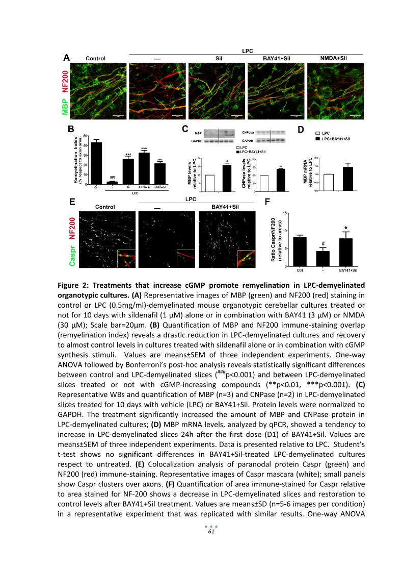
2. Sildenafil promotes remyelination in LPC-demyelinated mouse cerebellar
organotypic cultures.
To investigate if the remyelinating effect of sildenafil observed in EAE
mice involved effects only in CNS cells or also required peripheral immune-
regulatory actions, we used the in vitro model of mouse cerebellar organotypic
cultures (ORG) demyelinated with LPC (Zhang H. et al. 2011). Cultures were
established from cerebella of 7-day-old C57BL/6 mice as described in Methods
(section 3.1). After 7 days in vitro (DIV), cerebellar slices were exposed to LPC
(0.5 mg/ml) for 14-17h, a treatment that causes a dramatic demyelination
without axonal or cell death (Birgbauer et al. 2004). Twenty-four hours after
removing LPC, cultures were treated with four doses of sildenafil (1 µM) alone
or in combination with the cGMP-increasing agents BAY 41-2272 (BAY41; 3
µM), a NO-GC activator, or the glutamate receptor agonist NMDA (30 µM), as
indicated in Methods (section 3.1). At 19 DIV, cerebellar slices were harvested
and double immune fluorescence-stained for neurofilament (NF200, red) and
myelin (MBP, green) and analyzed by confocal microscopy and Image-J

60
software. Quantification of the area stained for MBP relative to the area
stained for NF200 gave a remyelination index (RI). As shown in Fig 2A, LPC-
treated slices show a significant reduction of MBP staining compared to control
slices, whereas in slices exposed to cGMP-increasing compounds the amount of
MBP surrounding neurofilaments notably increased. Quantification of the RI
shows that the remyelination induced by sildenafil alone is similar to that
attained in combination with BAY41 (BAY41+Sil) or NMDA (NMDA+Sil) (Fig 2B).
Remyelination by BAY41 alone was 40% lower than that induced by BAY41+Sil
(not shown). To confirm the remyelinating effect of cGMP-increasing
treatments in cerebellar slices, we analyzed by WB the levels of MBP, a major
myelin protein, and of 2',3'-cyclic-nucleotide 3'-phosphodiesterase (CNPase), a
minor CNS myelin-protein that is only expressed in CNS oligodendrocytes.
Results show that BAY41+Sil treatment significantly increased both MBP and
CNPase (Fig 2C) in LPC-demyelinated cultures. We further analyzed mRNA
expression of MBP by qPCR and showed that BAY41+Sil tendency to induced
MBP gene expression (Fig 2D).
The presence of contactin-associated protein (Caspr) clusters in the
paranodes delimiting myelin sheaths correlates with a proper remyelination of
the axons (Fancy et al. 2011, Meffre et al. 2015). To investigate the
remyelination status in sildenafil-treated cerebellar slices, double immune-
staining for Caspr and NF200 was performed. As shown in Fig 2E, Caspr clusters
associated with axons were drastically reduced in LPC-demyelinated cultures.
Treatment with BAY41+Sil largely restored Caspr staining levels and distribution
(Fig 2E,F).
61
Figure 2: Treatments that increase cGMP promote remyelination in LPC-demyelinated organotypic cultures. (A) Representative images of MBP (green) and NF200 (red) staining in control or LPC (0.5mg/ml)-demyelinated mouse organotypic cerebellar cultures treated or not for 10 days with sildenafil (1 µM) alone or in combination with BAY41 (3 µM) or NMDA (30 µM); Scale bar=20µm. (B) Quantification of MBP and NF200 immune-staining overlap (remyelination index) reveals a drastic reduction in LPC-demyelinated cultures and recovery to almost control levels in cultures treated with sildenafil alone or in combination with cGMP synthesis stimuli. Values are means±SEM of three independent experiments. One-way ANOVA followed by Bonferroni’s post-hoc analysis reveals statistically significant differences between control and LPC-demyelinated slices (###p<0.001) and between LPC-demyelinated slices treated or not with cGMP-increasing compounds (**p<0.01, ***p<0.001). (C) Representative WBs and quantification of MBP (n=3) and CNPase (n=2) in LPC-demyelinated slices treated for 10 days with vehicle (LPC) or BAY41+Sil. Protein levels were normalized to GAPDH. The treatment significantly increased the amount of MBP and CNPase protein in LPC-demyelinated cultures; (D) MBP mRNA levels, analyzed by qPCR, showed a tendency to increase in LPC-demyelinated slices 24h after the first dose (D1) of BAY41+Sil. Values are means±SEM of three independent experiments. Data is presented relative to LPC. Student’s t-test shows no significant differences in BAY41+Sil-treated LPC-demyelinated cultures respect to untreated. (E) Colocalization analysis of paranodal protein Caspr (green) and NF200 (red) immune-staining. Representative images of Caspr mascara (white); small panels show Caspr clusters over axons. (F) Quantification of area immune-stained for Caspr relative to area stained for NF-200 shows a decrease in LPC-demyelinated slices and restoration to control levels after BAY41+Sil treatment. Values are means±SD (n=5-6 images per condition) in a representative experiment that was replicated with similar results. One-way ANOVA

62
followed by Bonferroni’s post-hoc analysis reveals statistically significant differences between vehicle- and BAY41+sildenafil-treated slices (*p<0.05 vs LPC; #p<0.05 vs control).
3. Mechanisms implicated in the remyelinating effect of sildenafil
3.1. Involvement of the NO-cGMP-PKG pathway in the remyelinating effect of
sildenafil in LPC-demyelinated cerebellar cultures
The observation that treatment of LPC-demyelinated cerebellar slices
with sildenafil alone produced a similar remyelinating effect than its
combination with BAY41 or NMDA, both of which increase cGMP production
via NO-GC, suggested that activation of this enzyme was involved in the
sildenafil effect. That this was in fact the case was demonstrated by the
observation that sildenafil-induced remyelination was prevented in the
presence of the NO-GC inhibitor ODQ (10 µM; Fig 3A). Moreover, ODQ
treatment decreased the amount of myelin associated with axons in control
cultures, suggesting that NO-GC activity may be also implicated in the
myelination process that occurs in the immature cerebellar slices during culture
(Fig 3A). To investigate if induction of NOS-2 in LPC-demyelinated slices was
responsible for the generation of the NO that stimulates NO-GC activity we first
examined the effect of L-NNA, a predominant NOS-2 inhibitor, in sildenafil-
induced remyelination. As shown in Fig 3B, L-NNA prevented the sildenafil
effect. To further examine if NOS-2 was actually induced in the LPC-
demyelinated slices we performed immune-fluorescence staining for NOS-2
(green) in cerebellar cultures exposed for 24h to LPC, or to LPS (1 µg/ml) as a
positive control. Fig 3C shows that while only a few cells were stained for NOS-
2 in control cultures, numerous cells were NOS-2+ in cultures exposed to LPC or
LPS.

63
Cyclic GMP-dependent kinase (PKG) is the main target of cGMP. To
examine if PKG was involved in sildenafil-induced remyelination we examined
this effect in the presence or absence of the PKG inhibitor Rp-8pCPTcGMP (10
µM). Results showed that the PKG inhibitor prevented the sildenafil effect (Fig
3D) implicating the kinase in the NO-cGMP signaling cascade.
Figure 3: The remyelinating
effect of sildenafil in LPC-
demyelinated cultures is
prevented by inhibitors of NO-
GC, NOS-2 and PKG activities.
The remyelination index pre-
and post-LPC was quantified in
cerebellar slices treated 10 DIV
post-LPC with sildenafil (1 µM)
in the presence of: (A) NO-GC
inhibitor ODQ (10 µM), n=2;
(B) NOS-2 inhibitor L-NNA (100
µM), n=3. Values are
means±SEM. Two-way ANOVA
followed by Bonferroni’s post-
hoc analysis reveals
statistically significant
differences in the absence or
presence of ODQ in controls
(#p<0.05) and in sildenafil-
treated (***P<0.001) slices. (C)
NOS-2 immune-staining
(green) reveals similar levels of
induction in LPC-demyelinated
slices and in 24h LPS-treated
slices. Cell nuclei stained with
DAPI (blue). (D) PKG inhibitor
Rp-8pCPT-cGMP (10 µM).
Representative figure
replicated with similar results.
Values are means±SD (n=5-6
images per condition).

64
3.2. Increasing cGMP promotes oligodendrocyte differentiation in the spinal
cord of EAE mice and in LPC-demyelinated cerebellar cultures
Previous results have shown that treatment with sildenafil to MOG-
immunized mice at the initiation of EAE symptoms prevents disease
progression, preserves axons and myelin and protects immature and mature
OLs (Pifarre et al. 2014). We have now examined if sildenafil affects OL
maturation in the spinal cord of EAE animals treated with sildenafil for 15 days
at peak disease by immune-staining for transcription factors involved in early
(Nkx2.2) and late stages of OL differentiation (Olig2) and for the mature OL
marker MBP. As shown in Fig 4A, staining intensity of Nkx2.2+-cells (green) is
increased after sildenafil treatment. Quantification of bright Nkx2.2 cells (high
expression) confirms a significant increase respect to vehicle-treated animals
(Fig 4A). Double immune-staining for olig2 (red) and MBP (green) shows that
after 15 days of treatment from peak disease olig2+/MBP+-cells (yellow
arrows) and olig2-/MBP+-cells (green arrows) significantly increase in both
vehicle-treated and sildenafil-treated animals (Fig 4B). In both cases the
number of positive cells was higher in sildenafil-treated animal compared to
vehicle-treated animals. The percentage of Olig2-/MBP+ cells respect to total
MBP+ cells was significantly increased in thoracic SC sections after 15 days of
sildenafil treatment respect to vehicle (Fig 4B, right panel). A significant
increase in the number olig2-/MBP+cells relative to total MBP+cells was also
observed in LPC-demyelinated cerebellar cultures after 10 days of treatment
with BAY41+Sil or NMDA+Sil (Fig 4C). Taken together these results indicate that
stimulation of OL maturation may be an important mechanism contributing to
the remyelinating effect of sildeanfil.

65
Figure 4: Increasing cGMP promotes oligodendrocyte differentiation in the spinal cord of
EAE mice and in LPC-demyelinated cerebellar cultures. (A) Representative images of
immune-staining for Nkx2.2, a transcription factor involved in early stages of OL
differentiation, in the SC of EAE mice treated with vehicle or sildenafil for 15 days starting at
peak disease. Quantification of bright Nkx2.2 cells (high expression) shows a significant
increase after sildenafil treatment. Values are mean±SEM (n=3-6 mice per group) analyzed
by Student’s t-test (*p<0.05); (B) Immune-staining for MBP (green) and Olig2 (red), a
transcription factor expressed by differentiating oligodendrocyte lineage cells, images taken
at 63X (left panel). Quantification of the number of MPB+/Olig2+ (yellow arrow) and
MPB+/Olig2- (green arrow) is shown in the center panel. The percentage of Olig2-/MBP+
cells respect to total MBP+ cells in thoracic SC sections is shown in the right panel. Values are
mean±SEM (n=3-5 mice per group) and were analyzed by two-way ANOVA followed by
Bonferroni’s post-hoc test (*p<0.05, **p<0.01). (C) MBP and Olig2 staining in LPC-
demyelinated cerebellar cultures treated or not with Sil (1 µM)+BAY41 (3 µM) or NMDA (30
µM). Open arrow heads: Olig2+/MBP- (immature OLs; open arrow heads); Olig2+/MBP+
(early mature OLs; arrow heads); Olig2-/MBP+ (mature myelinating OLs; arrow). Scale bar=

66
20µm. The percentage of Olig2-/MBP+ respect to total MBP+ cells is significantly increased
by cGMP-increasing treatments (right graph). Values are mean±SEM of two independent
experiments, analyzed by one-way ANOVA followed by Bonferroni’s post hoc (*p<0.05
respect to LPC; ##p<0.01 respect to control).
3.3. Sildenafil increases CNTF expression in spinal cord of EAE mice and in
cerebellar organotypic cultures.
Increased expression of trophic factors is associated with
neuroprotective effects, oligodendrocyte support and proliferation and axon
remyelination (De Santi et al. 2009, Weishaupt et al. 2012). CNTF has a
recognized influence on OL differentation (Stankoff et al. 2002). Moreover, it
has been shown that this growth factor acts as a quemoatractant controling OL
progenitor migration during (Vernerey et al. 2013). Interestingly, recent in vitro
studies demonstrated that the NO-cGMP-protein kinase G pathway up-
regulates CNTF expression in astrocytes and that astrocyte-derived CNTF
enhances oligodendrocyte (OL) differentiation (Paintlia et al. 2013). In this
work, using double immune-staining for CNTF and the astrocyte protein GFAP,
we have observed a strong CNTF-staining in highly reactive astrocytes in the
spinal cord of EAE mice (Fig 5A). We further show that treatment with sildenafil
for 15 days at peak disease strongly enhances CNTF immune-staining compared
to vehicle-treated animals (Fig 5B). In agreement with these results, we have
also observed a significant increase in CNTF mRNA expression in LPC-
demyelinated cerebellar slices 24h after the first dose (D1) of Sil (1µM)+ BAY41
(3 µM), but not after the third dose, suggesting that CNTF induction may be is
an early event contributing to the remyelinating effect of cGMP-increasing
compounds (Fig 5C).

67
Figure 5: Sildenafil treatment increases CNTF expression in the spinal cord of EAE mice and
in LPC-demyelinated cerebellar cultures. (A) Colocalization of GFAP (red) and CNTF (green)
immune-staining in coronal sections of cervical spinal cord from naïve and EAE animals
showing CNTF increased expression in reactive astrocytes; (B) Treatment with sildenafil for
15 days at peak EAE further increases CTNF levels when compared with vehicle-treated EAE
animals Values are mean±SEM (n=5 per group); (C) CNTF mRNA levels assessed by qPCR in
LPC-demyelinated cerebellar cultures 24h after the first (D1) and third (D3) doses of BAY41
(3 µM)+Sil (1 µM), showing a significant increase of CNTF mRNA expression after the first
dose. Values are mean±SEM of three independent experiments. Data shown is relative to
LPC. Two-way ANOVA followed by Bonferroni’s post-hoc analysis reveals statistically
significant differences respect to LPC(**p<0.01).

68
3.4. Treatments that increase cGMP induce changes in the inflammatory
microglial phenotype in LPC-demyelinated cerebellar cultures.
It has been reported that in the cuprizone-induced demyelinization
mouse model that treatment with sildenafil reduces expression of the
inflammatory cytokines IL-1β and TNFα and increases the expression of the
anti-inflammatory cytokine IL-10 in cerebella (Nunes et al. 2015). Additionally it
has been shown that treatment with sildenafil to EAE mice for 7 days starting at
disease onset induced a decrease in the release of the pro-inflammatory
cytokines IL-2, IFNγ and TNFα in splenocytes. Furthermore, RNA microarray
analysis of spinal cord and immune-staining revealed that sildenafil up-
regulates YM-1 (Pifarré et al. 2014), a marker of the alternative
macrophage/microglial M2 phenotype that has neuroprotective and
regenerative properties. To gain insight into the possible implication of a
microglial phenotype change in the remyelinating effect of cGMP-increasing
compounds, we have examined by qPCR mRNA expression of M1 (NOS-2 and
COX-2) and M2 phenotype markers (Arg-1 and CD206) and of pro-inflammatory
(IL-1β, TNFα, IL-6) and anti-inflammatory (IL-10) cytokines in LPS-demyelinated
cerebellar slices 24h after the first and third doses of Sil (1 µM)+ BAY41 (3 µM).
Results showed that mRNA expression of phenotype marker molecules in LPC-
demyelinated slices do not significantly change between dose one (D1) and
three (D3) of vehicle (Fig 6). In BAY41+Sil-treated slices there was a tendency to
increase in Arg1 and COX-2 mRNA, but not in CD206 or NOS-2 mRNA after D1
and no change was observed after D3, except for a tendency to decrease in
NOS-2 expression (Fig 6).

69
Figure 6: Effect of sildenafil treatment on microglia inflammatory phenotype markers in
LPC-demyelinated cerebellar cultures. mRNA levels of M1 (NOS-2 and COX-2) and M2 (Arg-1
and CD206) phenotypic markers determined by qPCR in LPC-demyelinated slices treated or
not with BAY41(3µM)+Sil(1µM). mRNA was collected 24h after first (D1) and third (D3) dose.
Values are mean fold change±SEM of three independent experiments. Two-way ANOVA
followed by Bonferroni’s post-hoc analysis.
Regarding cytokines (Fig 7), again no change was observed in any of the
cytokines from D1 to D3 in vehicle-treated slices. BAY41+Sil treatment induced
a significant increase in IL-10 and IL-6 after D1, the larger increase
corresponding to IL-6 (2-fold). After D3, no significant changes were observed
in any of the cytokines, although a tendency to decrease was apparent in IL-1β,
TNFα and IL-10. Taken together these results suggest that a M2-like phenotype
is induced after the first dose of cGMP stimuli (increases in Arg1 and IL-10) that
also shows expression of IL-6 and COX-2.

70
Figure 7: Sildenafil treatment induces changes in cytokine expression in LPC-demyelinated
cerebellar cultures. mRNA levels of inflammatory cytokines assessed by qPCR in LPC-
demyelinated cerebellar cultures 24h after the first (D1) and third (D3) doses of BAY41
(3µM)+Sil (1µM), showing a significant increase in IL-6 and IL-10 cytokines after the first
dose. IL-6, IL-1β and TNFα are values are mean fold change±SEM of three independent
experiments. IL-10 is a representative figure replicated with similar results; values are mean
fold change±SD of triplicates. Two-way ANOVA followed by Bonferroni’s post hoc analysis
reveals statistically significant differences respect to LPC (*p<0.05, **p<0.01).
3.5 Treatments that increase cGMP induce phenotype changes in BMDM
LPS-demyelinated cerebellar cultures are a complex system to study
inflammatory phenotypic changes induced in microglia and the results
presented above on the effect of treatment with cGMP-increasing stimuli are
not easy to interpret. In order to directly examine the potential of cGMP-
increasing compounds to modulate the inflammatory phenotype in monocytes,
we used as model mice bone marrow derived macrophage (BMDM) primary
cultures. These cells are readily polarized to a M1 phenotype by incubation

71
with LPS (10-100 ng/ml)+IFNγ (20 ng/ml) and to a M2 phenotype by incubation
with IL-4 (10-20 ng/ml) (Classen et al. 2009). These conditions were used as
controls of phenotypic changes
Fig 8: Sildenafil induces a morphological change in BMDM from C57Bl/6 mice. Treatment of
BMDM primary cultures with sildenafil for 24 h induced process elongation (arrow) and
lamellipodia (arrow head) formation in a concentration-dependent manner (30-100µM). This
effect was prevented by the specific protein kinase G (PKG) inhibitor Rp-8pCPTcGMP (1µM)
added 30 min before (lower panel, left). The morphological change induced by sildenafil was
similar but not identical to that produced by IL-4 (20 ng/ml), an inducer of the M2 phenotype
(upper and middle panels). Moreover, sildenafil potentiated the IL-4 effect (lower panel,
right).
As shown in Fig 8, when BMDM were treated with sildenafil (30-100 µM)
for 24h the morphology of cells changed from a more rounded/amoeboid type
to an elongated shape similar to that acquired when BMDM are treated with IL-
4 (Durafourt et al. 2012, McWhorter et al. 2013, Vereyken et al. 2011).
Moreover, the combination of IL-4 (20 ng/ml) with sildenafil (30 µM) produced

72
an even more elongated shape (Fig 8). When sildenafil (100 µM) was combined
with the PKG inhibitor Rp-8pCPTcGMP (1 µM) cell elongation was prevented.
Since sildenafil treatment induced a morphological change similar to that
induced by IL-4 we further investigated if sildenafil could promote a shift from
a M1 to a M2 phenotype in BMDM by analyzing nitrite concentration in the
media as an index of NOS-2 induction (M1 effect) and arginase activity as an
index of Arg1 gene expression that is increased by M2 stimuli (Classen et al.
2009). Cultures were treated with sildenafil alone (1-100 µM) or in
combination (added 60 min before) with to LPS+IFNγ or IL-4. After 24h, nitrite
concentration in the media and arginase activity in cell extracts were
measured. No changes were observed in the concentration of nitrites in the
media of cells treated with sildenafil or IL-4 alone or in combination (Fig 9A). As
expected, LPS+IFNγ induced a large increase in nitrite concentration that was
not significantly affected by the presence of sildenafil, although at the highest
concentration (100 µM) used there was a tendency to decrease the LPS effect
(Fig 9A). In contrast, arginase activity was increased not only by the M2
stimulus IL-4, but also by LPS+IFNγ (Fig 9B). Sildenafil at the highest
concentrations used (30-100 µM) also induced a small non-significant increase
in arginase activity in control cells and a small potentiation of the effect of
LPS+IFNγ. However, when combined with IL-4 sildenafil produced a large
potentiation of the cytokine effect (Fig 9B).

73
Figure 9: Sildenafil alters the inflammatory phenotype of mouse BMDM. (A-B) Primary
cultures of BMDM were incubated for 24h with LPS (100 ng/ml)+INFγ (20 ng/ml) to induce a
M1 phenotype, with IL-4 (20 ng/ml) to induce a M2 phenotype, or with sildenafil (1-100 µM)
alone or in combination with the inflammatory stimuli (added 30 min before); (A) Nitrite
accumulation in the media was only induced by LPS+INFγ and was not significantly affected
by combination with sildenafil; (B) Arginase activity was slightly increased in cells exposed to
LPS+INFγ and IL-4. Sildenafil (30-100 µM) alone was little effective but potentiated the effect
of the inflammatory stimuli, in particular that of IL-4. Values are mean±SEM of three
independent experiments. Two-way ANOVA followed by Bonferroni’s post-hoc analysis
revealed statistically significant differences respect to untreated cells (*p<0.05;***p<0.001);
(C-D) FACS analysis of the percentage of BMDM expressing the M1 phenotype marker NOS-2
(n=2) or the M2 phenotype marker CD206 (n=3). BMDM were left untreated (control) or
treated for 48h with LPS (10 ng/ml)+IFNγ (20 ng/ml), IL-4 (10 ng/ml) or the cGMP analogue
8pCPTcGMP (50 µM) alone or in combination with the inflammatory stimuli, added 30 min
before; (C) The % of cells expressing NOS-2 was notably increased in LPS+IFNγ-treated cells
and this was not affected by the cGMP analogue. (D) The % of cells expressing CD206 was
increased by IL-4. The cGMP analogue had a small effect and notably potentiated the effect
of IL-4. Values are means±SEM analyzed by two-way ANOVA followed by Bonferroni’s post-
hoc analysis revealed statistically significant differences (***p<0.001) respect to cells not
exposed to the cGMP analogue.

74
These results indicate that sildenafil can promote a M2 phenotype but
not properly induce a shift M1 to M2. In order to confirm these results we
analyzed by flow cytometry the percentage of cells expressing NOS-2 or the M2
marker CD206 in BMDM treated with the cGMP analogue 8pCPTcGMP (50 µM)
alone or in combination (added 30 min before) with of LPS (10 ng/ml)+IFNγ (20
ng/ml) or IL-4 (10 ng/ml) for 48h. As shown in Fig 9C, more than 80% percent
of the cells expressed NOS-2 in LPS+IFNγ-treated BMDM whereas the number
of cells expressing this M1 marker was minimal in control cells or in cells
treated with the cGMP analogue or IL-4, alone or in combination. In agreement
with the nitrite data, 8pCPTcGMP did not affect the percentage of cells
expressing NOS-2 expression in LPS+IFNγ-treated BMDM (Fig 9C). As expected,
the number of cells expressing the M2 marker CD206 was increased in BMDM
treated with IL-4. The cGMP analogue alone also increased the number of
CD206-expressing cells to a small extend and significantly potentiated the
effect of IL-4, doubling the number of CD206+ cells, in agreement with the
sildenafil effect on arginase activity (Fig 9D). In contrast, no change in the
number of CD206+ cells was observed in LPS+IFNγ-treated BMDM (Fig 9D).
Furthermore, we analyzed the effect of the cGMP analogue on levels of the
pro-inflammatory cytokine (IL-1β) and the anti-inflammatory cytokine (IL-10) in
the media of BMDM 18h after treatment with the same stimuli using Luminex
technology. Surprisingly, results showed a large increase in both cytokines in
the media of LPS+IFNγ-treated but not in IL-4-treated BMDM (Fig 10). The
cGMP analogue alone induced a small increase in IL-10 release in control cells
and a larger increase in IL-4-treated cells and significantly inhibited the increase
in both cytokines induced by LPS+INFγ but that of IL-1β to a much larger extend
(Fig 10). These results support the contention that in BMDM cGMP promotes
an anti-inflammatory phenotype.

75
Figure 10: Increasing cGMP induces changes in pro-and anti-inflammatory cytokine release
from BMDM. Cytokine was determined in BMDM media by Luminex technology. Cells were
left untreated (control) or treated for 18h with LPS (10 ng/ml)+IFNγ (20 ng/ml), IL-4 (10
ng/ml) or 8pCPTcGMP (50 µM) alone or in combination with the inflammatory stimuli, added
30min before. Levels of both the anti-inflammatory cytokine IL-10 and the pro-inflammatory
cytokine IL-1β were increased by LPS+INFγ but were not affected by IL-4. The cGMP
analogue induced a small but significant decrease in IL-10 levels and a larger decrease in IL-
1β in LPS+INFγ-treated cells. However, in cells treated with IL-4 the analogue significantly
increased IL-10 release. Results are representative of two independent experiments; values
are means±SD, of two independent experiments. Two-way ANOVA followed by Bonferroni’s
post-hoc analysis revealed statistically significant differences (*p<0.05, ***p<0.001) respect
to cells not exposed to the cGMP analogue.
3.6. Sildenafil increases myelin phagocytosis in microglia/macrophages in
spinal cord of EAE animals and in BMDM
It has been reported that in demyelinating disorders, such as MS, myelin
debris interferes with the remyelination process (Kotter et al. 2006). Myelin
clearance correlates with increased remyelination and differentiation of OPCs
(Kotter et al. 2006, Miron and Franklin 2014). Furthermore, recent reports
indicate that M2 cells have higher myelin phagocytic activity than M1 cells
(Durafourt et al. 2012) and that M2 cell polarization is essential for efficient
remyelination (Miron et al. 2013). To investigate if sildenafil treatment affects
myelin debris phagocytosis in EAE mice we performed Oil-red O (ORO) staining
in longitudinal SC sections. Results showed that 15 days of sildenafil (10mg/kg)

76
treatment starting after disease onset increased the amount of ORO-loaded
vesicles in infiltrates (Fig 11 A-D). Quantification of the amount of ORO-loaded
vesicles relative to infiltrated area in infiltrates of different sizes showed a
significant increase in sildenafil-treated mice compared to vehicle-treated
controls (Fig 11E). To investigate if this lipid-loaded vesicles stained by ORO
were inside M2 cells, SC sections were double-stained for the M2-phenotype
marker YM-1 and with ORO and were visualized by confocal microscopy.
Confocal image analysis followed by 3D-reconstruction of images showed ORO
in vesicles inside Ym-1 possitive macrophage/microgial cells (Fig 11F).
To investigate if sildenafil has a direct effect on the myelin phagocytic
capacity of macrophages, a myelin phagocytosis assay was performed on
mouse BMDM. Cells were treated with the cGMP analogue 8Br-cGMP (500 µM)
or, sildenafil (30-100 µM) alone or in combination with IL-4 (20 ng/ml). After
24h, cells were incubated with Dil-labelled myelin (25 µg/ml) for 90 min and
myelin uptake was analyzed by flow cytometry. IL-4, the cGMP analogue and
sildenafil significantly alone increased myelin phagocytosis (Fig 11G). The effect
of cGMP-increasing stimuli alone was of higher magnitude than that induced by
IL-4 and when added together their effects were a little less than additive.

77
Fig 11. Sildenafil treatment enhances myelin phagocytosis in macrophages/microglia in the
spinal cord of EAE animals and in mouse BMDM cultures. (A-D) Phagocytosis of myelin in
the spinal cord of EAE animals evidenced by vesicular Oil-red O (ORO) staining.
Representative images of ORO staining in infiltrates (cell nuclei stained with DAPI) of
different sizes in animals treated with vehicle (A,C) or sildenafil (B, D) for 7 or 15 days (dot)
starting at onset of disease; (E) Quantification reveals that sildenafil significantly enhances
ORO staining after 15 dot. Values are means±SEM (n=4-6). Two-way ANOVA followed by
Bonferroni’s post-hoc analysis reveals statistically significant differences respect to EAE
vehicle-treated animals after 15 dot (*p<0.05). (F) 3D rendering of macrophage/microgial
cells positive for the M2-phenotype marker YM-1 (green) containing ORO in vesicles (red) in
the spinal cord of sildenafil-treated EAE animals; Cell nuclei are shown in blue. (G) BMDM
were treated for 24h with the cGMP analogue 8Br-cGMP (500 µM) or sildenafil (30 µM-100
µM) alone or in combination with IL-4 (20 ng/ml) and were exposed to Dil-labeled myelin for
90min. Myelin phagocytosis was analyzed by flow-cytometry. Values are means±SEM of
three independent experiments. Two-way ANOVA followed by Bonferroni’s post-hoc analysis
reveals statistically significant differences respect to untreated (*p<0.05, **p<0.01,
***p<0.001) and respect to untreated control (## p<0.01).

78

79
DISCUSSION

80

81
DISCUSSION
In MS, inflammation, demyelination and axonal damage are important
hallmarks of the pathology (Henderson et al. 2009, Lassmann 2014, Peterson
and Fujinami 2007). This inflammatory and neurodegenerative nature of MS
generates the necessity of the development of treatments that combine
immune-modulatory and neuroprotective effects (Aharoni 2010).
In MS models, it has been observed that sildenafil improve clinical
symptoms and disease progression. In EAE mice treated for 8 days with
sildenafil starting at the acute phase (peak) of the disease, showed significantly
amelioration of clinical symptoms associated with an increase in myelin
staining and decreased of axonal loss, suggesting that sildenafil may be
promoting remyelination and axonal protection; moreover, it was shown that 3
days after sildenafil treatment reduced the amount of infiltrating cells,
increased the number of Foxp3+ lymphocyte T regs and decreased
microglia/macrophage activation (Pifarre et al. 2011). Sildenafil effects on
remyelination were further studied by curpizone demyelination model,
showing as well a significant amelioration of clinical symptoms associated with
a protective effect on myelin structure, reduction of reactive gliosis (microglia
and astrocytes) and decreased expression of pro-inflammatory cytokines
(Nunes et al. 2012). Furthermore, treatment with sildenafil starting at the onset
of EAE has shown to prevent the disease progression, to maintain myelin and
axon structure, decrease microglial activation, down-regulate the innate and
adaptative responses decreasing pro-inflammatory cytokine production and
proliferation in splenocyte, and has shown to upregulate the alternative

82
microglia/macrophage activation marker Ym-1 associated with
neuroregenerative effects (Pifarre et al. 2014).
The results obtained in this thesis showed that sildenafil treatment in de
acute phase indeed ameliorates clinical symptoms decreasing clinical score
close to 1, and that extending the treatment from 8 to 15 days does not show
further improvement. After 15 days of treatment increase MBP staining was
observed, suggesting remyelination enhancement as seen previously (Pifarre et
al. 2011). The results obtained also showed that when sildenafil treatment
starts at the chronic phase of EAE stills promotes significant amelioration of the
symptoms. Further analysis demonstrated that sildenafil promotes
remyelination increasing MBP levels after 15 dot starting at chronic phase. This
result has clinical significance since it shows that sildenafil exerts remyelinating
and neuroprotective effects once EAE becomes chronic, when more infiltrating
cells, more lesions and an increased grade of demyelination can be found
(Berard et al. 2010).
Taking in account these results and with the aim to study mechanisms
involved in sildenafil effects on CNS cells, mouse cerebellar organotypic
cultures were established. EAE is induced by an auto-immune reaction against
myelin peptide that promotes an acute attack by peripheral immune cells that
infiltrate the CNS which is followed by demyelination (Constantinescu et al.
2011, McCarthy et al. 2012). On the other hand, cerebellar organotypic cultures
demyelinated with lysophosphatidylcholine (LPC) have proven to be an
excellent model to study demyelination-remyelination process without axonal
death and excluding the influence of the immune system. This model is
accessible to experimental perturbation, which allows testing the effect of
different stimuli on the mechanisms that might be implied in the processes that

83
wants to be studied (Birgbauer et al. 2004, Zhang H. et al. 2011). After LPC
demyelination, microglia activation can be observed, myelin debris is
generated as a consequence of demyelination and naked axons sometimes
suffer damage which is evident by the presence of axonal bulbs, Caspr proteins
are no longer visible, OPCs are present in the slices and after demyelination
OPCs proliferation is induced (Zhang H. et al. 2011).
The results obtained in this work showed almost complete demyelination
of axons after LPC treatment; however after 10 days of treatment with
sildenafil myelin sheath reappeared increasing the remyelination index, along
with Caspr clusters. Combination of sildenafil and BAY41, a direct activator of
NO-GC, revealed a significant increase in myelin protein levels; however no
significantly effects where observed in MBP mRNA expression; however, no
further increase in remyelination index were observed compared with sildenafil
alone. Similar effects on the remyelination index were observed after NMDA
treatment in combination with sildenafil. NMDA is a glutamate analogue that
activates this neurotransmitter receptors (NMDA-R), in cerebellum present
primarily in granular neurons and can be also found in astrocytes and
oligodendrocytes. Activation of NMDA-R allows calcium to entry into the cell
and the activation of the constitutive form of NOS (NOS-1) and further NO
production (Maarsingh et al. 2009, Manucha 2016, Verkhratsky and Kirchhoff
2007). This NO can diffuse across the plasmatic membrane generating cGMP
not only in NMDA-R positive cells but also in neighbor cells.
One possible molecular-signaling pathway involved in the increase in
remyelination by sildenafil is the NO-cGMP-PKG pathway. In the CNS, NO has
been associated with the modulation of synaptic plasticity, brain development,
visual and sensory processing, neuro-endocrine secretion and cerebral blood

84
flow (Garthwaite J. 2000, Guix et al. 2005). Constitutive NOS (NOS-1 and NOS-3)
produce small amounts of NO under physiological conditions (Maarsingh et al.
2009, Manucha 2016). On the other hand, NO has been also described as a
molecule important in the inflammation modulation, considered as a
neuropathological agent responsible for excitotoxic cell death and
neuroinflammatory cell damage in many neurological disorders (Duncan and
Heales 2005, Murphy 2000). Moreover, one of NO targets is the NO-GC, its
binding induces NO-GC activation and further cGMP production. NO-cGMP
pathway impairment may be implied in neurological dysfunctions, decreased
astroglial expression of the NO-GC β subunit was observed in astrocytes of
post-mortem brains of different neurodegenerative diseases’ patients (Baltrons
et al. 2004). We have observed that effect of sildenafil on remyelination index
was inhibited by NO-GC inhibitor (ODQ) and NOS inhibitor (L-NNA) treatments.
Similar effects were found when PKG was inhibited with Rp-8pCPTcGMP. On
the other hand, immune stainings revealed an increase in NOS-2 after 24h of
LPC treatment in organotypic cultures. As said before NOS-2 is induced under
pro-inflammatory conditions, LPC demyelination in organotypic cultures leave
axons exposed, myelin debris and microglia activation, characteristics of
neuroinflammation (Zhang H. et al. 2011). NOS-2 produces higher and longer-
lasting amounts of NO after induction and can be expressed primarily in
microglia and astrocytes cells (Brown and Neher 2010, García 2004, Maarsingh
et al. 2009, Murphy 2000) suggesting that LPC treatment may be responsible
of cGMP and NO increase, which can be in part promoting sildenafil effects.
Furthermore, PKG targets important pathways involved in remyelination and
neuroprotection, such as MEK/ERK and Akt/MTOR pathways (Dai et al. 2014,
Guardiola-Diaz et al. 2012). Taking together, these results demonstrate that
sildenafil treatment increases production of myelin by actions exerted directly

85
on CNS cells, restoring myelin sheath and mature internodes which is
associated with functional remyelination (Meffre et al. 2015, Zhang H. et al.
2011) suggesting that NO-cGMP-PKG pathway activation is required to
promote sildenafil effects.
In relapsing-remiting MS demyelinating episodes are characterized by
the death of OLs followed by new OLs generation and migration, but with the
recurrence of this episodes decreases their capacity of generation, which in
consequence affects remyelination (Copray et al. 2006, Lassmann 2014);
however, previous results have shown that sildenafil protects OLs from
apoptotic death in EAE model (Pifarre et al. 2014). Taking previous and new
results into account, in this work we study the effect of sildenafil in OLs
maturation in organotypic cerebellar cultures. OLs express a series of specific
molecules, which allows determining their differentiation or maturation stage.
Neuron-Glial precursor cells (NG2+) are observed in cerebellar organotypic
cultures (Zhang H. et al. 2011). OPCs can also be found in this model,
expressing molecules such as: transcriptional factor Nkx2.2 and Platlet Derived
Growth Factor α Receptor (PDGFαR) among others. Transcriptional factors
Olig1 and Olig2 are express during early to late differentiation stages of
immature OLs. More mature OLs express Myelin Basic Protein (MBP),
Proteolipid Protein (PLP), Myelin Oligodendrocyte Glycoprotein (MOG), Myelin
Associated Glycoprotein (MAG) and Cyclic Nucleotide Phosphodiesterase
(CNPase) among others (Fig 12) (Copray et al. 2006, Liu et al. 2007, Meffre et
al. 2015, Miron et al. 2010, Nishiyama et al. 2009, Zhang H. et al. 2011).

86
Figure 12: Olygodendrocyte maturation markers expression. Figure modified from Akiko
Nishiyama, Mila Komitova, Ryusuke Suzuki & Xiaoqin Zhu; Polydendrocytes (NG2 cells):
multifunctional cells with lineage plasticity. Nature Reviews Neuroscience 10, 9-22 (January
2009). Molecules and protein expression of oligodendroglial cells throughout development
(Nishiyama et al. 2009).
Our results showed that sildenafil treatment significantly increased the
expression of OPCs transcription factor involved in early stage of OL
differentiation (Nkx2.2+) and showed a tendency increased the number of
mature OLs (Olig2+MBP+) as well as the number late stage mature OLs (Olig2-
MBP+) in SC of EAE mice. Moreover, when ventral and dorsal thoracic sections
of EAE mice SC where analyzed, an increase number of late stage mature OLs
(Olig2-MBP+) relative to the total MBP+ mature OLs was found in sildenafil
treated group. This result was further confirmed in organotypic cultures, where
increased number of late stage mature oligodendrocytes relative to the total of
mature oligodendrocytes was found with sildenafil treatment combined with
BAY41 and with NMDA. Taking together this results demonstrate that cGMP
increased by sildenafil is promoting maturation of OLs. In agreement with these
results, other studies have demonstrated that an increase in intracellular cGMP
derived from NO-GC in OLs promotes their maturation and arborization,
extending the reach of each cell in cerebellar slices, the source of NO in this
model was partially attributed to an increase production of NO in neighbor
granular neurons via NMDA-R activation (Garthwaite G. et al. 2015); however

87
recent studies have shown that OLs can also express NMDA receptors specially
in their processes (Salter and Fern 2005); therefore cGMP levels in OLs could be
increased by actions of NMDA-R agonists. Similar to these results, in an
ischemic mouse model sildenafil has proven to increase nestin lineage OPCs
and mature OLs (CNPAse+ and CC1+) in corpus callosum and striatum of
treated mice (Zhang R. L. et al. 2012). Transduction signals of cGMP increase
primarily occur via PKG, in OLs this kinase may be targeting different pathways
implicated in OLs maturation and protection. Some of these pathways as above
mention could be MEK/ERK and Aky/mTOR, different studies in vitro have
involved them with growth and differentiation of oligodendrocytes (Bibollet-
Bahena and Almazan 2009, Kumar et al. 2013). Furthermore, recent in vitro
studies have shown that ERK I/II activation is required for the progression of
oligodendrocyte early progenitors to the development of immature
oligodendrocytes, but does not affect the transition from immature to mature
oligodendrocytes; however, mTOR activation is not required for the
development of immature oligodendrocytes, but it shows to be essential in the
transition from immature to mature myelinating oligodendrocytes (Guardiola-
Diaz et al. 2012). Additionally, important cross-talk between Akt/mTOR and
MEK/ERK pathway has been studied, demonstrating that both pathways
regulate MBP mRNA and protein expression and that Akt/mTOR inhibition
increase ERK I/II phosphorylation in vitro (Dai et al. 2014)

88
Figure 13: Sildenafil possible mechanism pathway
Therefore, sildenafil not only protects OLs from apoptotic death, but this
work shows that it promotes OLs maturation into mature myelinating OLs.
Other mechanism possibly implicated in sildenafil ameliorating and
remyelinating effects was the generation of trophic factors. Previous studies
have demonstrated that sildenafil increases BDNF expression in splenocytes of
EAE mice after sildenafil treatment and in SC sections, denoting that the
increased BDNF expression in SC was mainly generated by infiltrating
leukocytes and in axons (Pifarre et al. 2014). Another important neurotrophic
factor associated with remyelination and regeneration process is CNTF. The
results of this work showed that sildenafil treatment to EAE-mice increased
CNTF expression in the SC, and was observed primarily by astrocytes.
Furthermore cGMP increase also induced CNTF gene expression in cerebellar
organotypic cultures. The CNTF produced in our model could be enhancing
sildenafil effects on OLs, it has been recognized influence of CTNF on OL

89
differentiation (Stankoff et al. 2002) and it has been recently shown that this
growth factor acts as a quemoatractant controlling OL progenitor migration
during remyelination (Vernerey et al. 2013). In agreement with the results
shown in this work, it has been recently demonstrated in vitro that the NO-
cGMP-protein kinase G pathway up-regulates CNTF expression in astrocytes
and that astrocyte-derived CNTF enhances OLs differentiation (Paintlia et al.
2013).. Moreover, studies have demonstrated that CNTF plays an important
role in myelin protein expression and in oligodendrocyte protection against
apoptotic death (Modi et al. 2013); therefore sildenafil effect on
oligodendrocyte protection against apoptotic death observed in previously the
EAE model (Pifarre et al. 2014) may be partially exerted through sildenafil
increase expression of CNTF. Moreover, treatment with exogenous CNTF
injection (s.c.) in the cuprizone-demyelinating mouse model, increased the
myelin protein MOG expression after demyelination, thus suggesting that CNTF
may be potentiating remyelination (Salehi et al. 2013). Treating EAE mice with
mesenchymal cells overexpressing CNTF induced functional recovery of the
disease, reduced demyelination, increased NG2+ cells (OLs precursors),
decreased pro-inflammatory cytokines (TNFα and IFNγ) and increased anti-
inflammatory cytokine IL-10 (Lu et al. 2009). This evidence suggests that
sildenafil enhances the production not only of BDNF but also CNTF, and this
increased expression of neurotrophic factors may develop important
mechanisms and actions in the CNS after demyelinating and inflammatory
lesions, promoting oligodedroglial protection and differentiation.
In EAE model it was previously described that sildenafil treatment
significantly decreased microglial/macrophage activation and showed down
regulation of adaptative immune response by decreasing pro-inflammatory
cytokines (IL-2, INFγ and TNFα) release by splenocytes re-stimulated with MOG,

90
it was also described that sildenafil increased alternative activation
microglia/macrophage marker Ym-1 (Pifarre et al. 2014); these results suggest
that sildenafil besides having remyelinating and neuroprotective effects, it can
also be promoting anti-inflammatory effects in the IS and CNS and may be
promoting M2 microglia/macrophage phenotype. In order to study sildenafil
effects on CNS inflammatory microglial phenotype, cytokines and
microglia/macrophage phenotypic markers were determined.
The M1 phenotype has been related to release of pro-inflammatory
cytokines, cytotoxicity, radical oxygen species (ROS) release, auto antigen
presentation to T cells and therefore contributing with disease progression as
has been observed Alzehimer’s disease, MS and stroke studies (Chhor et al.
2013, Rawji et al. 2016). The M2 anti-inflammatory phenotype has been related
with the production of anti-inflammatory cytokines and is associated with
regeneration properties and wound healing (Gensel and Zhang 2015, Ransohoff
and Perry 2009). Furthermore, the M2 phenotype resolution and regenerative
properties have been related with inducing OPCs differentiation, remyelination
and promoting myelin clearance (Durafourt et al. 2012, Kotter et al. 2006,
Miron et al. 2013, Wang et al. 2015, Yu et al. 2015).
Our results showed that BAY41+Sil treatment in LPC demyelinated
cerebellar cultures induced tendency to an early increased in Arg-1 (M2
phenotype marker) and COX-2 (M1 phenotype marker), no changes were found
in CD206 (M2 phenotype marker) expression and a late decrease tendency in
NOS-2 (M1 phenotype marker) expression was observed.
Increased arginase activity has been associated with alternatively
activated microglia/macrophages (M2) (Gensel and Zhang 2015, Ransohoff and
Perry 2009) which converts L-arginine into ornithine, a precursor of proline and

91
polyamines. Polyamines have been related with oxidative stress amelioration
and cellular proliferation (Guasco Herrera et al. 2014). Curiously, in our results
the treatment induced a tendency to increase M1 marker, COX-2
simultaneously with the increase Arg-1; COX-2 increase has usually been
associated to pro-inflammatory process (Deininger and Schluesener 1999,
Minghetti 2004). However recent studies propose anti-inflammatory properties
of COX-2, demonstrating the presence of a group of lipids in postmortem brain
samples of Alzehimer’s disease patients, with pro-resolving and anti-
inflammatory properties called pro-resolving lipid mediators (SPMs)
synthesized by 5-lipoxygenase (LOX) and COX; this SPMs have shown to
improve neuronal survival and Aβ2 phagocitosys in vitro (Zhu et al. 2016).
Therefore, the early increase in COX-2 observed in the cerebellar organotypic
cultures after sildenafil treatment could be promoting the production of SPMs.
Moreover, COX-2 increase has been associated with M2b/c (immune-
regulatory/immune-suppressor) phenotype associated with the production of
anti-inflammatory prostaglandins (Chhor et al. 2013, Ransohoff and Perry 2009,
Wu et al. 2013), and wound healing after spinal cord injury (Gensel and Zhang
2015). On the other hand, within sildenafil treatment doses tendency to
decrease M1 phenotype was observed, suggesting that PDE5 inhibitor may be
promoting healing and regeneration of tissue by enhancing a M2-like
phenotype and diminishing the pro-inflammatory phenotype.
In order to confirm the inflammatory effect of sildenafil in CNS, cytokine
expression was determined in organotypic cerebellar cultures. Results showed
that BAY41+Sil treatment significantly increase IL-10 and IL-6 24h after first
dose, no changes were found in IL-1β and TNFα, pro-inflammatory cytokines,
expression.

92
IL-10 is a known anti-inglammatory cytokine, its functions are exerted
primarly on dendirtic cells and macrophages, IL-10 inhibits major
histocompatibility complex class II expression and inhibits the production of
proinflammatory cytokines and mediators (IL-1, IL-6, IL-12, TNFα) (Mosser and
Zhang 2008). In agreement with previous results in EAE model (Pifarre et al.
2014), recent studies have shown that the increased expression of IL-10 and
promotion of M2 microglia phenopype after estrogen treatment promotes
neuroprotection preventing EAE progression (Benedek et al. 2016). Moreover,
it has been shown that IL-10 increase expression resulted in attenuated
inflammatory response, decreased microglia activation and less
neurodegeneration in EAE model (Mayo et al. 2016). On the other hand, IL-6
was classically conceived as a pro-inflammatory cytokine; recently, more
studies come to light showing its anti-inflammatory properties. In agreement
with our results, studies performed by using interleukin-6 (IL-6) fused to its
soluble receptor (sIL-6R) protein have shown that IL-6 increase in vivo has
effects on myelination, inducing OPCs to differentiate into mature
oligodendrocytes and promoting their survival (Haggiag et al. 2001, Valerio et
al. 2002). Moreover, increase immune-regulatory response, by increasing IL-6
and IL-10 expression, has been associated with myelin repair in chronic phase
of EAE in IL-4Rα-/- mice (Gaupp et al. 2008). Even though previous studies have
shown that sildenafil treatment reduced pro-inflammatory cytokines
expression in mouse cerebella of cuprizone-demyelinated model (Nunes et al.
2012) and in splenocytes of EAE mice (Pifarre et al. 2014), in our results
BAY41+Sil treatment in cerebellar slices did not show significant changes in
pro-inflammatory cytokines expression. In the case of TNFα excessive
generation in brain has been involved in the pathogenesis of stroke, traumatic
brain injury and Alzehimer’s disease (Clark and Vissel 2016) and IL-1β is a

93
known pro-inflammatory cytokine, the release of this cytokine has been
associated with cytotoxicity (Rawji et al. 2016).
Taking these results together suggests that sildenafil is promoting a
mixed microglia phenotype, since an early increase in the expression of Arg-1,
COX-2 was observed accompanied by an increase expression of IL-10 and IL-6;
these features are recently being associated with M2b/c microglia/macrophage
phenotype which shows immune-regulatory and immune-suppressor
properties by promoting inflammatory resolution, tissue repair, production of
anti-inflammatory and prostaglandins (Colton 2009, Chhor et al. 2013, Gensel
and Zhang 2015, Ransohoff and Perry 2009, Wu et al. 2013) and even more,
this phenotype has been associated with OPCs differentiation (Miron et al.
2013). Recent studies have indentified another mixed microglia/macrophage
phenotype not classically M1 nor alternatively activated M2 cells; they were
called “resolution-phase” macrophages (rM), since this particular phenotype
has been observed in macrophages isolated from the resolving phase of acute
inflammation. rM showed to increase M2 marker CD206 and anti-inflammatory
molecules (IL-10 and PGD2) expression, along with M1 markers including NOS-
2, COX-2 (Bystrom et al. 2008). Moreover, new evidence has emerge showing
that after spinal cord injury, exogenous IL-4 treatment increase the population
of M2 phenotype, but it also appeared another population of cells suggesting
to be infiltrating macrophages with a mixed phenotype co-expressing M1
markers (NOS-2, COX-2, LOX, CD16/32) and M2 markers (Arg-1, CD206) as well
as anti-inflammatory cytokines IL-10 and TGFβ1 and were associated with rM,
known to be important in the resolving of inflammation (Francos-Quijorna et
al. 2016). Even though the phenotype markers and cytokines expressed after
BAY41+Sil treatment in our cerebellar slices is very similar to those observed in
the rM, our treatment in this model despite increasing Arg-1 failed in

94
promoting CD206; moreover no expression of NOS-2 or any other pro-
inflammatory molecule besides COX-2 was observed. Therefore it is not clear
yet if sildenafil is promoting rM unique phenotype in CNS.
Mouse cerebellar organotypic cultures are complex models to study
inflammatory phenotype changes; this can result as a consequence of the
presence of the other cells that are part of the CNS, such as astrocytes that
could also be contributing in the production of cytokines and other
inflammatory molecules (like NOS-2). In order to study more specifically
sildenafil effects on microglia/macrophage phenotype BMDM cultures were
established. The study of these cells grants clinical significance since EAE model
as well as in MS peripheral immune cell infiltrates can be found (Berard et al.
2010, Henderson et al. 2009).
BMDM cultures were established as previously described (Classen et al.
2009) and were polarized into M1 phenotype with LPS+INFγ or into M2
phenotype with IL-4; a un-polarized BMDM population was used as control.
Results showed nitrite accumulation only in M1 BMDM and that sildenafil
treatment showed a tendency to decrease this accumulation. Additionally NOS-
2 positive cell number significantly increased in LPS+INFγ treated BMDM;
however treatment with cGMP analogue did not decreased the number NOS-2
positive cell population. On the other hand, the three populations of BMDM
showed arginase activity; nevertheless, sildenafil treatment showed a tendency
to increase arginase activity in control BMDM, being significant in M1 BMDM at
the highest concentration of sildenafil and significantly doubled arginase
activity in M2 BMDM. Moreover, CD206 positive cell number increased after IL-
4 treatment; cGMP analogue induced an increase in CD206 positive population

95
in control BMDM and further potentiated the number of CD206 positive cells in
IL-4 treated BMDM.
In order confirm the effect of cGMP increase on inflammatory
phenotypes; IL-10 and IL-1β release were determined in the culture media of
BMDM 18h after treatments. Results showed that LPS+INFγ treatment
significantly increased IL-10 release in BMDM, these results agree with those of
Howes, et al. that demonstrated that C57BL/6 BMDM produce higher levels of
IL-10 in response to bacterial products such as LPS (Howes et al. 2016), similar
IL-10 increase has been observed in primary microglia after LPS treatment
(Chhor et al. 2013); nevertheless, cGMP analogue treatment significantly
reduced IL-10 production in LPS+INFγ treated BMDM. Interestingly, IL-10
production was not observed in control or IL-4 stimulated BMDM; however,
cGMP analogue increased IL-10 production in both populations, being
significant in IL-4 BMDM. On the other hand, there was only IL-1β release on
behave of LPS+INFγ BMDM and treatment with cGMP analogue significantly
decreased the production of IL-1β in this M1 population. In agreement with this
results, inhibiting PDE activity in human monocytes decreased the release of IL-
1β and this effect was mimic by cGMP analogues and NO donor suggesting NO-
GC activation is implied in the effect (von Bulow et al. 2005). Taking these
results together suggest that increase cGMP levels promoted by sildenafil
affect inflammatory outcomes, do not significantly reverse M1 phenotype but
it does promote M2 phenotype and anti-inflammatory mediators.
One of the important actions of microglia/macrophage in
neurodegenerative diseases is their phagocytic activity of cellular and myelin
debris produced in the lesions (Ransohoff and Perry 2009). In
neurodegenerative diseases, such as MS, olygodendrocyte differentiation is not

96
observed in demyelinated lesions, and it has been proved that the presence of
myelin debris can impair remyelination by inhibition of OPCs differentiation
(Kotter et al. 2006). For this reason myelin clearance by phagocytic cells has
gained relevance. Studies have shown that M2 macrophages phagocyte greater
amount of myelin than M1 (Durafourt et al. 2012). Myelin phagocytosis was
assessed in EAE model through ORO staining, making evident that the lipid
vesicles around infiltrates increase in EAE mice treated with sildenafil at the
onset of the disease; furthermore, it was observed that most of the vesicles
where inside Ym-1 stained M2 microglia/macrophages. To confirm that
sildenafil was promoting myelin phagocytosis in M2 cells, control and M2
BMDM where treated with sildenafil and a cGMP analogue for 24h before
being exposed to purified myelin. Flow cytometry analysis revealed that
sildenafil treatment increased myelin phagocytosis in IL-4 stimulated BMDM
compared with controls and LPS treated BMDM (data not shown); moreover
sildenafil significantly increases myelin pahocytosis in control and M2 BMDM,
this effect was mimicked by cGMP analogue. This increase in myelin clearance
is another mechanism promoted by sildenafil essential for remyelination to
take place.
The results observed in organotypic cerebellar and BMDM cultures did
not showed a clear pro or anti-inflammatory phenotype induction. These
results showed that sildenafil can promote inflammatory changes through
cytokine expression and could be generating a mixed microglia/macrophage
phenotype. Nowadays increasing evidence of mixed or overlapping
microglia/macrophage phenotype can be found, and has been associated with
the recovery after damage. IL-4 treatment of macrophages in vitro produced IL-
6 and co-expressed CD206 (M2 marker) without affecting immunosuppressive
properties of M2 phenotype macrophages (Casella et al. 2016). Moreover, the

97
expression of Arg-1 as well as IL-1β was associated with neurite growth
promotion and spinal cord injury recovery (Fenn et al. 2014). IL-4 gene therapy
to EAE mice decreased demyelination and axonal loss showing an increase in
the expression of CD206 in microglia/macrophages, but also an increase IL-6
(Casella et al. 2016). Even more, it has comes to light evidence that a pro-
inflammatory environment at the beginning of the lesion followed by anti-
inflammatory environment with increase of pro-resolving and tissue
regenerating molecules is required for an effective resolution and repair after
CNS injury (Rawji et al. 2016) and that the whole spectrum of
microglia/macrophage phenotypes are required in the resolution of CNS injury
(Gensel and Zhang 2015, Rawji et al. 2016).
Indeed classifying microglia/macrophages as classically (M1) or
alternatively-activated (M2) in ex-vivo (such as organotypic cerebellar cultures)
or in vivo models is a complex matter. Some authors are starting to consider as
either of both phenotypes, since this nomenclature was generated from
monocyte-derived macrophages treated in vitro using defined cytokine stimuli
and controlled environment, but after inflammatory stimuli in vivo
microglia/macrophage plasticity promotes the generation of a broad spectrum
of slightly different phenotypes that can be required for the resolution and
healing process to take place (Gensel and Zhang 2015, Stables et al. 2011).
Therefore, it cannot be said that sildenafil treatment is inducing a shift into M1
or M2 phenotype, but it is affecting immune response in CNS and in IS,
generating anti-inflammatory mediators that promotes resolution of
inflammatory process. More importantly sildenafil treatment increased myelin
phagocytosis in non-polarized BMDM suggesting that this restoring and
essential process can be promoted despite microglia/macrophage phenotype.

98

99
CONCLUSIONS

100

101
CONCLUSIONS
1. Sildenafil treatment ameliorates clinical symptoms in mouse chronic EAE
model when administrated in the acute and in chronic phase.
2. Sindenafil treatment promotes remyelination of axons and mature-
functional internodes increasing myelin protein levels.
3. The NO-cGMP-PKG pathway is required in sildenafil-induced
remyelination.
4. Some of the mechanisms underliying sildenafil-induced remyelination
and neuroprotection are associated with:
Maturation of oligodendocytes
Increased CNTF expression in CNS
Modulation of inflammatory process
Increased myelin phagocytic capacity

102

103
REFERENCES

104

105
REFERENCES
Abbott NJ, Patabendige AA, Dolman DE, Yusof SR, Begley DJ. 2010. Structure and function of the blood-brain barrier. Neurobiol Dis 37: 13-25.
Aharoni R. 2010. Immunomodulatory drug treatment in multiple sclerosis. Expert Rev Neurother 10: 1423-1436.
Aloisi F. 2001. Immune function of microglia. Glia 36: 165-179. Baltrons MA, Garcia A. 1999. Nitric oxide-independent down-regulation of soluble guanylyl cyclase
by bacterial endotoxin in astroglial cells. J Neurochem 73: 2149-2157. Baltrons MA, Pedraza CE, Heneka MT, Garcia A. 2002. Beta-amyloid peptides decrease soluble
guanylyl cyclase expression in astroglial cells. Neurobiol Dis 10: 139-149. Baltrons MA, Pifarre P, Ferrer I, Carot JM, Garcia A. 2004. Reduced expression of NO-sensitive
guanylyl cyclase in reactive astrocytes of Alzheimer disease, Creutzfeldt-Jakob disease, and multiple sclerosis brains. Neurobiol Dis 17: 462-472.
Baltrons MA, Pifarre P, Berciano MT, Lafarga M, Garcia A. 2008. LPS-induced down-regulation of NO-sensitive guanylyl cyclase in astrocytes occurs by proteasomal degradation in clastosomes. Mol Cell Neurosci 37: 494-506.
Bell AS, Palmer MJ. 2011. Novel phosphodiesterase type 5 modulators: a patent survey (2008 - 2010). Expert Opin Ther Pat 21: 1631-1641.
Benedek G, Zhang J, Bodhankar S, Nguyen H, Kent G, Jordan K, Manning D, Vandenbark AA, Offner H. 2016. Estrogen induces multiple regulatory B cell subtypes and promotes M2 microglia and neuroprotection during experimental autoimmune encephalomyelitis. J Neuroimmunol 293: 45-53.
Benjamins JA, Nedelkoska L. 2007. Cyclic GMP-dependent pathways protect differentiated oligodendrocytes from multiple types of injury. Neurochem Res 32: 321-329.
Berard JL, Wolak K, Fournier S, David S. 2010. Characterization of relapsing-remitting and chronic forms of experimental autoimmune encephalomyelitis in C57BL/6 mice. Glia 58: 434-445.
Bercury KK, Macklin WB. 2015. Dynamics and mechanisms of CNS myelination. Dev Cell 32: 447-458.
Bibollet-Bahena O, Almazan G. 2009. IGF-1-stimulated protein synthesis in oligodendrocyte progenitors requires PI3K/mTOR/Akt and MEK/ERK pathways. J Neurochem 109: 1440-1451.
Biel M, Zong X, Hofmann F. 1996. Cyclic nucleotide-gated cation channels molecular diversity, structure, and cellular functions. Trends Cardiovasc Med 6: 274-280.
Birgbauer E, Rao TS, Webb M. 2004. Lysolecithin induces demyelination in vitro in a cerebellar slice culture system. J Neurosci Res 78: 157-166.
Bradley J, Reisert J, Frings S. 2005. Regulation of cyclic nucleotide-gated channels. Curr Opin Neurobiol 15: 343-349.
Brown GC, Neher JJ. 2010. Inflammatory neurodegeneration and mechanisms of microglial killing of neurons. Mol Neurobiol 41: 242-247.
Bynoe MS, Bonorino P, Viret C. 2007. Control of experimental autoimmune encephalomyelitis by CD4+ suppressor T cells: peripheral versus in situ immunoregulation. J Neuroimmunol 191: 61-69.
Bystrom J, Evans I, Newson J, Stables M, Toor I, van Rooijen N, Crawford M, Colville-Nash P, Farrow S, Gilroy DW. 2008. Resolution-phase macrophages possess a unique inflammatory phenotype that is controlled by cAMP. Blood 112: 4117-4127.
Casella G, Garzetti L, Gatta AT, Finardi A, Maiorino C, Ruffini F, Martino G, Muzio L, Furlan R. 2016. IL4 induces IL6-producing M2 macrophages associated to inhibition of neuroinflammation in vitro and in vivo. J Neuroinflammation 13: 139.
Clark IA, Vissel B. 2016. Excess cerebral TNF causing glutamate excitotoxicity rationalizes treatment of neurodegenerative diseases and neurogenic pain by anti-TNF agents. J Neuroinflammation 13: 236.

106
Classen A, Lloberas J, Celada A. 2009. Macrophage activation: classical versus alternative. Methods Mol Biol 531: 29-43.
Colton CA. 2009. Heterogeneity of microglial activation in the innate immune response in the brain. J Neuroimmune Pharmacol 4: 399-418.
Constantinescu CS, Farooqi N, O'Brien K, Gran B. 2011. Experimental autoimmune encephalomyelitis (EAE) as a model for multiple sclerosis (MS). Br J Pharmacol 164: 1079-1106.
Copray S, Balasubramaniyan V, Levenga J, de Bruijn J, Liem R, Boddeke E. 2006. Olig2 overexpression induces the in vitro differentiation of neural stem cells into mature oligodendrocytes. Stem Cells 24: 1001-1010.
Chhor V, et al. 2013. Characterization of phenotype markers and neuronotoxic potential of polarised primary microglia in vitro. Brain Behav Immun 32: 70-85.
Dai J, Bercury KK, Macklin WB. 2014. Interaction of mTOR and Erk1/2 signaling to regulate oligodendrocyte differentiation. Glia 62: 2096-2109.
Das A, Xi L, Kukreja RC. 2008. Protein kinase G-dependent cardioprotective mechanism of phosphodiesterase-5 inhibition involves phosphorylation of ERK and GSK3beta. J Biol Chem 283: 29572-29585.
De Santi L, Annunziata P, Sessa E, Bramanti P. 2009. Brain-derived neurotrophic factor and TrkB receptor in experimental autoimmune encephalomyelitis and multiple sclerosis. J Neurol Sci 287: 17-26.
Deininger MH, Schluesener HJ. 1999. Cyclooxygenases-1 and -2 are differentially localized to microglia and endothelium in rat EAE and glioma. J Neuroimmunol 95: 202-208.
Dong Y, Benveniste EN. 2001. Immune function of astrocytes. Glia 36: 180-190. Duman RS NE. 1999. Guanylyl Cyclase in Siegel GJ AB, Albers RW, et al, ed. Basic Neurochemistry:
Molecular, Cellular and Medical Aspects. Philadelphia: Lippincott-Raven. Duncan AJ, Heales SJ. 2005. Nitric oxide and neurological disorders. Mol Aspects Med 26: 67-96. Durafourt BA, Moore CS, Zammit DA, Johnson TA, Zaguia F, Guiot MC, Bar-Or A, Antel JP. 2012.
Comparison of polarization properties of human adult microglia and blood-derived macrophages. Glia 60: 717-727.
Fancy SP, et al. 2011. Axin2 as regulatory and therapeutic target in newborn brain injury and remyelination. Nat Neurosci 14: 1009-1016.
Fedoroff S, Hao C. 1991. Origin of microglia and their regulation by astroglia. Adv Exp Med Biol 296: 135-142.
Fenn AM, Hall JC, Gensel JC, Popovich PG, Godbout JP. 2014. IL-4 signaling drives a unique arginase+/IL-1beta+ microglia phenotype and recruits macrophages to the inflammatory CNS: consequences of age-related deficits in IL-4Ralpha after traumatic spinal cord injury. J Neurosci 34: 8904-8917.
Fernando MR, Reyes JL, Iannuzzi J, Leung G, McKay DM. 2014. The pro-inflammatory cytokine, interleukin-6, enhances the polarization of alternatively activated macrophages. PLoS One 9: e94188.
Foster DC, Wedel BJ, Robinson SW, Garbers DL. 1999. Mechanisms of regulation and functions of guanylyl cyclases. Rev Physiol Biochem Pharmacol 135: 1-39.
Francis SH, Blount MA, Corbin JD. 2011. Mammalian cyclic nucleotide phosphodiesterases: molecular mechanisms and physiological functions. Physiol Rev 91: 651-690.
Francos-Quijorna I, Amo-Aparicio J, Martinez-Muriana A, Lopez-Vales R. 2016. IL-4 drives microglia and macrophages toward a phenotype conducive for tissue repair and functional recovery after spinal cord injury. Glia.
Franklin RJ, Ffrench-Constant C. 2008. Remyelination in the CNS: from biology to therapy. Nat Rev Neurosci 9: 839-855.
Friese MA, Montalban X, Willcox N, Bell JI, Martin R, Fugger L. 2006. The value of animal models for drug development in multiple sclerosis. Brain 129: 1940-1952.

107
García A, Baltrons, M.A. 2004. The nitric oxide/cyclic GMP pathway in CNS glial cells. Advances in Molecular and Cell Biology 31: 575-593.
Garg N, Smith TW. 2015. An update on immunopathogenesis, diagnosis, and treatment of multiple sclerosis. Brain Behav 5: e00362.
Garthwaite G, Hampden-Smith K, Wilson GW, Goodwin DA, Garthwaite J. 2015. Nitric oxide targets oligodendrocytes and promotes their morphological differentiation. Glia 63: 383-399.
Garthwaite J. 2000. The phisiologycal roles of nitric oxide in the central nervous system. Nitric Oxide: 259-275.
Gaupp S, Cannella B, Raine CS. 2008. Amelioration of experimental autoimmune encephalomyelitis in IL-4Ralpha-/- mice implicates compensatory up-regulation of Th2-type cytokines. Am J Pathol 173: 119-129.
Gensel JC, Zhang B. 2015. Macrophage activation and its role in repair and pathology after spinal cord injury. Brain Res 1619: 1-11.
Ghafouri M, Amini S, Khalili K, Sawaya BE. 2006. HIV-1 associated dementia: symptoms and causes. Retrovirology 3: 28.
Griffiths MR, Gasque P, Neal JW. 2009. The multiple roles of the innate immune system in the regulation of apoptosis and inflammation in the brain. J Neuropathol Exp Neurol 68: 217-226.
Guardiola-Diaz HM, Ishii A, Bansal R. 2012. Erk1/2 MAPK and mTOR signaling sequentially regulates progression through distinct stages of oligodendrocyte differentiation. Glia 60: 476-486.
Guasco Herrera C, Chávez Servín JL, Ferriz Martínez RA, de la Torre Carbot K, Elton Puente E, García Gasca T. 2014. Poliaminas: pequeños gigantes de la regulación metabólica. REB. Revista de educación bioquímica 33: 51-57.
Guix FX, Uribesalgo I, Coma M, Munoz FJ. 2005. The physiology and pathophysiology of nitric oxide in the brain. Prog Neurobiol 76: 126-152.
Haggiag S, Zhang PL, Slutzky G, Shinder V, Kumar A, Chebath J, Revel M. 2001. Stimulation of myelin gene expression in vitro and of sciatic nerve remyelination by interleukin-6 receptor-interleukin-6 chimera. J Neurosci Res 64: 564-574.
Hailer NP. 2008. Immunosuppression after traumatic or ischemic CNS damage: it is neuroprotective and illuminates the role of microglial cells. Prog Neurobiol 84: 211-233.
Hauser SL, Oksenberg JR. 2006. The neurobiology of multiple sclerosis: genes, inflammation, and neurodegeneration. Neuron 52: 61-76.
Henderson AP, Barnett MH, Parratt JD, Prineas JW. 2009. Multiple sclerosis: distribution of inflammatory cells in newly forming lesions. Ann Neurol 66: 739-753.
Hendriks JJ, Slaets H, Carmans S, de Vries HE, Dijkstra CD, Stinissen P, Hellings N. 2008. Leukemia inhibitory factor modulates production of inflammatory mediators and myelin phagocytosis by macrophages. J Neuroimmunol 204: 52-57.
Hertz AL, Beavo JA. 2011. Cyclic nucleotides and phosphodiesterases in monocytic differentiation. Handb Exp Pharmacol: 365-390.
Herz J, Zipp F, Siffrin V. 2010. Neurodegeneration in autoimmune CNS inflammation. Exp Neurol 225: 9-17.
Hofmann F, Bernhard D, Lukowski R, Weinmeister P. 2009. cGMP regulated protein kinases (cGK). Handb Exp Pharmacol: 137-162.
Howes A, et al. 2016. Differential Production of Type I IFN Determines the Reciprocal Levels of IL-10 and Proinflammatory Cytokines Produced by C57BL/6 and BALB/c Macrophages. J Immunol 197: 2838-2853.
Kaupp UB, Seifert R. 2002. Cyclic nucleotide-gated ion channels. Physiol Rev 82: 769-824. Kim SU, de Vellis J. 2005. Microglia in health and disease. J Neurosci Res 81: 302-313. Kim YS, Joh TH. 2006. Microglia, major player in the brain inflammation: their roles in the
pathogenesis of Parkinson's disease. Exp Mol Med 38: 333-347.

108
Kimelberg HK. 2010. Functions of mature mammalian astrocytes: a current view. Neuroscientist 16: 79-106.
Koesling D, Russwurm M, Mergia E, Mullershausen F, Friebe A. 2004. Nitric oxide-sensitive guanylyl cyclase: structure and regulation. Neurochem Int 45: 813-819.
Koopman R, Schaart G, Hesselink MK. 2001. Optimisation of oil red O staining permits combination with immunofluorescence and automated quantification of lipids. Histochem Cell Biol 116: 63-68.
Kotter MR, Li WW, Zhao C, Franklin RJ. 2006. Myelin impairs CNS remyelination by inhibiting oligodendrocyte precursor cell differentiation. J Neurosci 26: 328-332.
Kumar S, Patel R, Moore S, Crawford DK, Suwanna N, Mangiardi M, Tiwari-Woodruff SK. 2013. Estrogen receptor beta ligand therapy activates PI3K/Akt/mTOR signaling in oligodendrocytes and promotes remyelination in a mouse model of multiple sclerosis. Neurobiol Dis 56: 131-144.
Larocca JN, Norton WT. 2007. Isolation of myelin. Curr Protoc Cell Biol Chapter 3: Unit3 25. Lassmann H. 2014. Mechanisms of white matter damage in multiple sclerosis. Glia 62: 1816-1830. Lee HH, Park SC, Choe IS, Kim Y, Ha YS. 2015. Time Course and Characteristics of Astrocyte
Activation in the Rat Brain after Injury. Korean J Neurotrauma 11: 44-51. Li J, Zhang L, Chu Y, Namaka M, Deng B, Kong J, Bi X. 2016. Astrocytes in Oligodendrocyte Lineage
Development and White Matter Pathology. Front Cell Neurosci 10: 119. Liu Z, Hu X, Cai J, Liu B, Peng X, Wegner M, Qiu M. 2007. Induction of oligodendrocyte
differentiation by Olig2 and Sox10: evidence for reciprocal interactions and dosage-dependent mechanisms. Dev Biol 302: 683-693.
Livak KJ, Schmittgen TD. 2001. Analysis of relative gene expression data using real-time quantitative PCR and the 2(-Delta Delta C(T)) Method. Methods 25: 402-408.
Lu Z, Hu X, Zhu C, Wang D, Zheng X, Liu Q. 2009. Overexpression of CNTF in Mesenchymal Stem Cells reduces demyelination and induces clinical recovery in experimental autoimmune encephalomyelitis mice. J Neuroimmunol 206: 58-69.
Ludwin SK, Rao VT, Moore CS, Antel JP. 2016. Astrocytes in multiple sclerosis. Mult Scler. Maarsingh H, Zaagsma J, Meurs H. 2009. Arginase: a key enzyme in the pathophysiology of allergic
asthma opening novel therapeutic perspectives. Br J Pharmacol 158: 652-664. Manucha W. 2016. Mitochondrial dysfunction associated with nitric oxide pathways in glutamate
neurotoxicity. Clin Investig Arterioscler. Mayo L, et al. 2016. IL-10-dependent Tr1 cells attenuate astrocyte activation and ameliorate
chronic central nervous system inflammation. Brain 139: 1939-1957. McCarthy DP, Richards MH, Miller SD. 2012. Mouse models of multiple sclerosis: experimental
autoimmune encephalomyelitis and Theiler's virus-induced demyelinating disease. Methods Mol Biol 900: 381-401.
McWhorter FY, Wang T, Nguyen P, Chung T, Liu WF. 2013. Modulation of macrophage phenotype by cell shape. Proc Natl Acad Sci U S A 110: 17253-17258.
Meffre D, et al. 2015. Liver X receptors alpha and beta promote myelination and remyelination in the cerebellum. Proc Natl Acad Sci U S A 112: 7587-7592.
Minghetti L. 2004. Cyclooxygenase-2 (COX-2) in inflammatory and degenerative brain diseases. J Neuropathol Exp Neurol 63: 901-910.
Miron VE, Franklin RJ. 2014. Macrophages and CNS remyelination. J Neurochem. Miron VE, Ludwin SK, Darlington PJ, Jarjour AA, Soliven B, Kennedy TE, Antel JP. 2010. Fingolimod
(FTY720) enhances remyelination following demyelination of organotypic cerebellar slices. Am J Pathol 176: 2682-2694.
Miron VE, et al. 2013. M2 microglia and macrophages drive oligodendrocyte differentiation during CNS remyelination. Nat Neurosci 16: 1211-1218.
Modi KK, Sendtner M, Pahan K. 2013. Up-regulation of ciliary neurotrophic factor in astrocytes by aspirin: implications for remyelination in multiple sclerosis. J Biol Chem 288: 18533-18545.

109
Mosser DM, Edwards JP. 2008. Exploring the full spectrum of macrophage activation. Nat Rev Immunol 8: 958-969.
Mosser DM, Zhang X. 2008. Interleukin-10: new perspectives on an old cytokine. Immunol Rev 226: 205-218.
Murphy S. 2000. Production of nitric oxide by glial cells: regulation and potential roles in the CNS. Glia 29: 1-13.
Nakamizo T, et al. 2003. Phosphodiesterase inhibitors are neuroprotective to cultured spinal motor neurons. J Neurosci Res 71: 485-495.
Nave KA. 2010. Myelination and support of axonal integrity by glia. Nature 468: 244-252. Nimmerjahn A, Kirchhoff F, Helmchen F. 2005. Resting microglial cells are highly dynamic
surveillants of brain parenchyma in vivo. Science 308: 1314-1318. Nishiyama A, Komitova M, Suzuki R, Zhu X. 2009. Polydendrocytes (NG2 cells): multifunctional cells
with lineage plasticity. Nat Rev Neurosci 10: 9-22. Nunes AK, Raposo C, Luna RL, Cruz-Hofling MA, Peixoto CA. 2012. Sildenafil (Viagra(R)) down
regulates cytokines and prevents demyelination in a cuprizone-induced MS mouse model. Cytokine 60: 540-551.
Nunes AK, Raposo C, Rocha SW, Barbosa KP, Luna RL, da Cruz-Hofling MA, Peixoto CA. 2015. Involvement of AMPK, IKbetaalpha-NFkappaB and eNOS in the sildenafil anti-inflammatory mechanism in a demyelination model. Brain Res 1627: 119-133.
Paintlia MK, Paintlia AS, Singh AK, Singh I. 2013. S-nitrosoglutathione induces ciliary neurotrophic factor expression in astrocytes, which has implications to protect the central nervous system under pathological conditions. J Biol Chem 288: 3831-3843.
Pedraza CE, Baltrons MA, Heneka MT, Garcia A. 2003. Interleukin-1 beta and lipopolysaccharide decrease soluble guanylyl cyclase in brain cells: NO-independent destabilization of protein and NO-dependent decrease of mRNA. J Neuroimmunol 144: 80-90.
Peterson LK, Fujinami RS. 2007. Inflammation, demyelination, neurodegeneration and neuroprotection in the pathogenesis of multiple sclerosis. J Neuroimmunol 184: 37-44.
Pifarre P, Prado J, Giralt M, Molinero A, Hidalgo J, Garcia A. 2010. Cyclic GMP phosphodiesterase inhibition alters the glial inflammatory response, reduces oxidative stress and cell death and increases angiogenesis following focal brain injury. J Neurochem 112: 807-817.
Pifarre P, Prado J, Baltrons MA, Giralt M, Gabarro P, Feinstein DL, Hidalgo J, Garcia A. 2011. Sildenafil (Viagra) ameliorates clinical symptoms and neuropathology in a mouse model of multiple sclerosis. Acta Neuropathol 121: 499-508.
Pifarre P, Gutierrez-Mecinas M, Prado J, Usero L, Roura-Mir C, Giralt M, Hidalgo J, Garcia A. 2014. Phosphodiesterase 5 inhibition at disease onset prevents experimental autoimmune encephalomyelitis progression through immunoregulatory and neuroprotective actions. Exp Neurol 251: 58-71.
Prado J, Baltrons MA, Pifarre P, Garcia A. 2010. Glial cells as sources and targets of natriuretic peptides. Neurochem Int 57: 367-374.
Raivich G, Banati R. 2004. Brain microglia and blood-derived macrophages: molecular profiles and functional roles in multiple sclerosis and animal models of autoimmune demyelinating disease. Brain Res Brain Res Rev 46: 261-281.
Raivich G, Jones LL, Werner A, Bluthmann H, Doetschmann T, Kreutzberg GW. 1999. Molecular signals for glial activation: pro- and anti-inflammatory cytokines in the injured brain. Acta Neurochir Suppl 73: 21-30.
Ransohoff RM, Perry VH. 2009. Microglial physiology: unique stimuli, specialized responses. Annu Rev Immunol 27: 119-145.
Raposo C, Luna RL, Nunes AK, Thome R, Peixoto CA. 2014. Role of iNOS-NO-cGMP signaling in modulation of inflammatory and myelination processes. Brain Res Bull 104: 60-73.
Rawji KS, Mishra MK, Yong VW. 2016. Regenerative Capacity of Macrophages for Remyelination. Front Cell Dev Biol 4: 47.

110
Salehi Z, Hadiyan SP, Navidi R. 2013. Ciliary neurotrophic factor role in myelin oligodendrocyte glycoprotein expression in Cuprizone-induced multiple sclerosis mice. Cell Mol Neurobiol 33: 531-535.
Salter MG, Fern R. 2005. NMDA receptors are expressed in developing oligodendrocyte processes and mediate injury. Nature 438: 1167-1171.
Sharma R, Fischer MT, Bauer J, Felts PA, Smith KJ, Misu T, Fujihara K, Bradl M, Lassmann H. 2010. Inflammation induced by innate immunity in the central nervous system leads to primary astrocyte dysfunction followed by demyelination. Acta Neuropathol 120: 223-236.
Sofroniew MV, Vinters HV. 2010. Astrocytes: biology and pathology. Acta Neuropathol 119: 7-35. Stables MJ, Shah S, Camon EB, Lovering RC, Newson J, Bystrom J, Farrow S, Gilroy DW. 2011.
Transcriptomic analyses of murine resolution-phase macrophages. Blood 118: e192-208. Stankoff B, Aigrot MS, Noel F, Wattilliaux A, Zalc B, Lubetzki C. 2002. Ciliary neurotrophic factor
(CNTF) enhances myelin formation: a novel role for CNTF and CNTF-related molecules. J Neurosci 22: 9221-9227.
Steinert JR, Robinson SW, Tong H, Haustein MD, Kopp-Scheinpflug C, Forsythe ID. 2011. Nitric oxide is an activity-dependent regulator of target neuron intrinsic excitability. Neuron 71: 291-305.
Streit WJ, Conde JR, Fendrick SE, Flanary BE, Mariani CL. 2005. Role of microglia in the central nervous system's immune response. Neurol Res 27: 685-691.
Talbott JF, Loy DN, Liu Y, Qiu MS, Bunge MB, Rao MS, Whittemore SR. 2005. Endogenous Nkx2.2+/Olig2+ oligodendrocyte precursor cells fail to remyelinate the demyelinated adult rat spinal cord in the absence of astrocytes. Exp Neurol 192: 11-24.
Tanaka J, Markerink-van Ittersum M, Steinbusch HW, De Vente J. 1997. Nitric oxide-mediated cGMP synthesis in oligodendrocytes in the developing rat brain. Glia 19: 286-297.
Town T, Nikolic V, Tan J. 2005. The microglial "activation" continuum: from innate to adaptive responses. J Neuroinflammation 2: 24.
Tsai EJ, Kass DA. 2009. Cyclic GMP signaling in cardiovascular pathophysiology and therapeutics. Pharmacol Ther 122: 216-238.
Valerio A, Ferrario M, Dreano M, Garotta G, Spano P, Pizzi M. 2002. Soluble interleukin-6 (IL-6) receptor/IL-6 fusion protein enhances in vitro differentiation of purified rat oligodendroglial lineage cells. Mol Cell Neurosci 21: 602-615.
van der Laan LJ, Ruuls SR, Weber KS, Lodder IJ, Dopp EA, Dijkstra CD. 1996. Macrophage phagocytosis of myelin in vitro determined by flow cytometry: phagocytosis is mediated by CR3 and induces production of tumor necrosis factor-alpha and nitric oxide. J Neuroimmunol 70: 145-152.
Veerhuis R, Nielsen HM, Tenner AJ. 2011. Complement in the brain. Mol Immunol 48: 1592-1603. Vereyken EJ, Heijnen PD, Baron W, de Vries EH, Dijkstra CD, Teunissen CE. 2011. Classically and
alternatively activated bone marrow derived macrophages differ in cytoskeletal functions and migration towards specific CNS cell types. J Neuroinflammation 8: 58.
Verkhratsky A, Kirchhoff F. 2007. NMDA Receptors in glia. Neuroscientist 13: 28-37. Vernerey J, Macchi M, Magalon K, Cayre M, Durbec P. 2013. Ciliary neurotrophic factor controls
progenitor migration during remyelination in the adult rodent brain. J Neurosci 33: 3240-3250.
Vollmar AM. 2005. The role of atrial natriuretic peptide in the immune system. Peptides 26: 1086-1094.
von Bulow V, Rink L, Haase H. 2005. Zinc-mediated inhibition of cyclic nucleotide phosphodiesterase activity and expression suppresses TNF-alpha and IL-1 beta production in monocytes by elevation of guanosine 3',5'-cyclic monophosphate. J Immunol 175: 4697-4705.
Voskuhl RR, Peterson RS, Song B, Ao Y, Morales LB, Tiwari-Woodruff S, Sofroniew MV. 2009. Reactive astrocytes form scar-like perivascular barriers to leukocytes during adaptive immune inflammation of the CNS. J Neurosci 29: 11511-11522.

111
Wang G, et al. 2015. HDAC inhibition prevents white matter injury by modulating microglia/macrophage polarization through the GSK3beta/PTEN/Akt axis. Proc Natl Acad Sci U S A 112: 2853-2858.
Weishaupt N, Blesch A, Fouad K. 2012. BDNF: the career of a multifaceted neurotrophin in spinal cord injury. Exp Neurol 238: 254-264.
Weller RO, Galea I, Carare RO, Minagar A. 2010. Pathophysiology of the lymphatic drainage of the central nervous system: Implications for pathogenesis and therapy of multiple sclerosis. Pathophysiology 17: 295-306.
Willenborg DO, Staykova M, Fordham S, O'Brien N, Linares D. 2007. The contribution of nitric oxide and interferon gamma to the regulation of the neuro-inflammation in experimental autoimmune encephalomyelitis. J Neuroimmunol 191: 16-25.
Wu D, Zheng S, Li W, Yang L, Liu Y, Zheng X, Yang Y, Wang Q, Smith FG, Jin S. 2013. Novel biphasic role of resolvin D1 on expression of cyclooxygenase-2 in lipopolysaccharide-stimulated lung fibroblasts is partly through PI3K/AKT and ERK2 pathways. Mediators Inflamm 2013: 964012.
Wyss-Coray T. 2006. Inflammation in Alzheimer disease: driving force, bystander or beneficial response? Nat Med 12: 1005-1015.
Yu Z, et al. 2015. MSX3 Switches Microglia Polarization and Protects from Inflammation-Induced Demyelination. J Neurosci 35: 6350-6365.
Zabel U, et al. 2002. Calcium-dependent membrane association sensitizes soluble guanylyl cyclase to nitric oxide. Nat Cell Biol 4: 307-311.
Zhang H, Jarjour AA, Boyd A, Williams A. 2011. Central nervous system remyelination in culture--a tool for multiple sclerosis research. Exp Neurol 230: 138-148.
Zhang J, Snyder SH. 1995. Nitric oxide in the nervous system. Annu Rev Pharmacol Toxicol 35: 213-233.
Zhang RL, Chopp M, Roberts C, Wei M, Wang X, Liu X, Lu M, Zhang ZG. 2012. Sildenafil enhances neurogenesis and oligodendrogenesis in ischemic brain of middle-aged mouse. PLoS One 7: e48141.
Zhu M, Wang X, Hjorth E, Colas RA, Schroeder L, Granholm AC, Serhan CN, Schultzberg M. 2016. Pro-Resolving Lipid Mediators Improve Neuronal Survival and Increase Abeta42 Phagocytosis. Mol Neurobiol 53: 2733-2749.