III Simposio Biopsia líquida - tactics. · Bienvenidos al III Simposio Biopsia Líquida: El camino...
36
III Simposio Biopsia líquida el camino a la oncología de precisión www.simposiobiopsialiquida.com #SimposioBiopsiaLiquida Coordinación científica Scientific coordination: Dr. Rafael López López Complejo Hospitalario Universitario de Santiago Sede Venue: San Francisco Hotel Monumento Campillo de San Francisco, 3 Santiago de Compostela Organizado por Organized by: 25 - 27 de enero 2018 · JANUARY 25 TH - 27 th · Santiago de Compostela
Transcript of III Simposio Biopsia líquida - tactics. · Bienvenidos al III Simposio Biopsia Líquida: El camino...

III SimposioBiopsia líquidael camino a la oncología de precisión
www.simposiobiopsialiquida.com#SimposioBiopsiaLiquida
Coordinación científi ca
Scientifi c coordination:
Dr. Rafael López López
Complejo Hospitalario Universitario de Santiago
Sede
Venue:
San Francisco Hotel Monumento
Campillo de San Francisco, 3Santiago de Compostela
Organizado por
Organized by:
25 - 27 de enero 2018 · JANUARY 25TH - 27th · Santiago de Compostela
Bienvenidos al III Simposio Biopsia Líquida: El
camino a la oncología de precisión.
Tras el éxito de las dos primeras ediciones, donde superamos gratamente las expectativas, os traemos el tercer simposio, con las mismas ganas de aportar y compartir el conocimiento más reciente sobre las diferentes posibilidades de la biopsia líquida. Como hemos hecho anteriormente, abordaremos la temática desde perspectivas tan atractivas como la utilidad clínica, los progresos en el conocimiento biológico y los continuos avances tecnológicos, todo ello en un escenario de formación, discusión y planteamiento de retos futuros.
Como organizador del simposio, quiero presenta-ros el nuevo programa científi co, en el que con-tamos con un grupo de destacados expertos para liderar las diferentes sesiones y ponencias, en las que trataremos los diferentes aspectos de la biop-sia líquida y las diferentes patologías oncológicas.
El San Francisco Hotel Monumento, situado en el corazón de Santiago, nos acogerá para poder celebrar el simposio en un entorno sereno y agradable.
Por todo esto, esperamos que disfrutes del simposio y sea una fuente de enriquecimiento
profesional en el camino de la oncología de
precisión.
¡Bienvenidos a Santiago!
Welcome to the III Simposio Biopsia Líquida: El
camino a la oncología de precisión.
After the success of the fi rst editions, which pleasantly surpassed our expectations, we bring you this third Symposium, with the same desire to offer and share the most recent knowledge about the different possibilities of liquid biopsy. As done before, we will approach the topic from interesting perspectives such as clinical utility, progress made in biological knowledge and the continuous technological advances, all of it in a scenario of training, discussion and addressing future challenges.
As organiser of the Symposium, I would like to introduce you to the scientifi c programme, bringing together a group of prominent experts who will lead a range of sessions and talks, during which we will address specifi c aspects of liquid biopsy and the different oncological pathologies.
The San Francisco Hotel Monument, located in the centre of Santiago, will welcome us with a relaxing and pleasant environment to hold the Symposium.
Therefore, we hope you will enjoy the event and will regard it as a source of professional
enrichment on the road to precision oncology.
Welcome to Santiago!
Dr. Rafael López López
Servicio de Oncología Médica,Complejo Hospitalario Universitario de Santiago
III Simposio Biopsia líquidael camino a la oncología de precisión
2
3
COMITÉ CIENTÍFICO
SCIENTIFIC COMMITTEE
Dr. Rafael López López
Jefe de Servicio de Oncología MédicaOncometComplejo Hospitalario Universitario de Santiago
Dra. Laura Muinelo
Unidad de Análisis de Biopsia LíquidaOncometComplejo Hospitalario Universitario de Santiago
Dra. Clotilde Costa
Unidad mixta ROCHE CHUSOncometComplejo Hospitalario Universitario de Santiago
Dr. Miguel Abal
Laboratorio de Oncología TraslacionalOncometComplejo Hospitalario Universitario de Santiago
Dr. Roberto Piñeiro
Unidad mixta ROCHE CHUSOncometComplejo Hospitalario Universitario de Santiago
Dra. Ana Belén Dávila
Unidad mixta ROCHE CHUSOncometComplejo Hospitalario Universitario de Santiago
Dr. Juan Cueva
Servicio de Oncología MédicaOncometComplejo Hospitalario Universitario de Santiago
Dra. Teresa Curiel
Servicio de Oncología MédicaOncometComplejo Hospitalario Universitario de Santiago
III Simposio Biopsia líquidael camino a la oncología de precisión
4
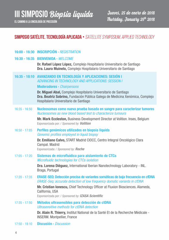
SIMPOSIO SATÉLITE. TECNOLOGÍA APLICADA • SATELLITE SYMPOSIUM. APPLIED TECHNOLOGY
16:00 - 16:30 INSCRIPCIÓN · REGISTRATION
16:30 - 16:35 BIENVENIDA · WELCOMEDr. Rafael López López, Complejo Hospitalario Universitario de SantiagoDra. Laura Muinelo, Complejo Hospitalario Universitario de Santiago
16:35 - 18:10 AVANZANDO EN TECNOLOGÍA Y APLICACIONES: SESIÓN I
ADVANCING IN TECHNOLOGY AND APPLICATIONS: SESSION I Moderadores · ChairpersonsDr. Miguel Abal, Complejo Hospitalario Universitario de SantiagoDra. Beatriz Sobrino, Fundación Pública Galega de Medicina Xenómica, Complejo
Hospitalario Universitario de Santiago
16:35 - 16:50 Nucleosomas como nueva prueba basada en sangre para caracterizar tumores
Nucleosomes as new blood based test to characterize tumoursMr. Mark Eccleston, Business Development Director at Volition. Inses, BelgiumEsponsorizada por: / Sponsored by: Volition
16:50 - 17:05 Perfi les genómicos utilizados en biopsia líquida
Genomic profi les employed in liquid biopsyDr. Emiliano Calvo, START Madrid CIOCC, Centro Integral Oncológico Clara
Campal. MadridEsponsorizada: / Sponsored by: Roche
17:05 - 17:20 Sistemas de microfl uídica para aislamiento de CTCs
Microfl uidic technologies for CTCs isolationDra. Lorena Diéguez, International Iberian Nanotechnology Laboratory - INL.
Braga, Portugal
17:20 - 17:35 ERASE-SEQ: Detección precisa de variantes somáticas de baja frecuencia en cfDNA
ERASE-Seq: accurate detection of low frequency domatic variants in cfDNAMr. Cristian Ionescu, Chief Technology Offi cer at Fluxion Biosciences. Alameda,
California, USAEsponsorizada por: / Sponsored by: IZASA Scientifi c
17:35 - 17:50 Métodos ultrasensibles para detección de ctDNA
Ultrasensitive methods for ctDNA detection Dr. Alain R. Thierry, Institut National de la Santé Et de la Recherche Médicale - INSERM. Montpellier, France
17:50 - 18:10 Discusión · Discussion
Jueves, 25 de enero de 2018 Thursday, January 25th 2018

5
PROGRAMA científicoscientific PROGRAM
18:10 - 18:20 Compra pública innovadora aplicada a oncología: Plan de innovación código 100
Public procurement of innovation applied to oncology: Código 100 innovation plan
Dr. Luis León, Axencia de Coñecemento en Saúde-ACIS. Santiago de Compostela
18:20 - 18:40 PAUSA CAFÉ · COFFEE BREAK
18:40 - 20:00 AVANZANDO EN TECNOLOGÍA Y APLICACIONES: SESIÓN II
ADVANCING IN TECHNOLOGY AND APPLICATIONS: SESSION IIModeradores · ChairpersonsDr. Roberto Piñeiro, Unidad mixta ROCHE CHUS. Complejo Hospitalario
niversitario de Santiago Dra. Silvia Calabuig-Fariñas, Hospital General Universitario de Valencia
18:40 - 18:55 Aplicaciones de Droplet-Based digital PCR
Applications of Droplet-Based digital PCRDra. Valérie Taly, Université Paris Descartes. Paris, France
18:55 - 19:10 La experiencia de la biopsia líquida de OncoBEAM en la práctica clínica del
mundo real
The OncoBEAM liquid biopsy experience in real-world clinical practiceMr. Frederick S. Jones, Director Medical Affairs Liaison at Sysmex-Inostics.
Mundelein, Illinois, USA
Esponsorizada por: / Sponsored by: Sysmex
19:10 - 19:25 Enriquecimiento CTC con PARSORTIX™: clinical applications
CTC enrichment with PARSORTIX™: clinical applications Dra. Eva Obermayr, Medical University Vienna. Austria Esponsorizada por: / Sponsored by: Parsortix
19:25 - 19:40 Detección de mutaciones ctRAS-BRAF con la plataforma Idylla™ en tan solo
horas . Las ventajas de Idylla™ en la práctica clínica
RAS-BRAF mutation detection on the Idylla™ System in just 2 hours. How Idylla™ can bring real value into clinical practice Mrs. Marijke Van der Auwera, Product Manager at Biocartis. Mechelen, Belgium Esponsorizada por: / Sponsored by: Biocartis
19:40 - 20:00 Discusión · Discussion
Jueves, 25 de enero de 2018 Thursday, January 25th 2018

III Simposio Biopsia líquidael camino a la oncología de precisión
6
09:30 - 10:35 BIOPSIA LÍQUIDA: DÓNDE ESTAMOS Y HACIA DÓNDE VAMOS
LIQUID BIOPSY: WHERE WE STAND AND WHERE WE GO
Moderadores · ChairpersonsDr. Eduardo Díaz-Rubio, Hospital Clínico San Carlos. MadridDra. Teresa Curiel, Complejo Hospitalario Universitario de Santiago
09:30 - 09:45 ¿Qué hemos aprendido hasta hoy?
What have we learned so far?
Dr. Luis Marques da Costa, Hospital de Santa Maria. Lisboa, Portugal
09:45 - 10:00 Situación actual en biopsia líquida
Current situation of liquid biopsy
Dr. Carlos Camps, Hospital General Universitario de Valencia
10:00 - 10:15 Generación de modelos predictivos
Generation of predictive models
Dr. David Tamborero, Institute for Research and Biomedicine - IRB. Barcelona
10:15 - 10:35 Discusión · Discussion
10:35 - 11:00 INAUGURACIÓN OFICIAL · OFFICIAL OPENING
11:00 - 11:45 ENTENDIENDO LA DISEMINACIÓN
UNDERSTANDING DISSEMINATION
Moderadores · Chairpersons
Dr. Jorge Barbazán, Institute Curie. Paris, FranceDr. Urbano Anido, Complejo Hospitalario Universitario de Santiago
11:00 - 11:15 Diseminación temprana en cáncer de próstata
Early dissemination in prostate cancer
Dr. Miodrag Guzvic, University of Regensburg. Germany
11:15 - 11:30 Vesículas extracelulares en biopsia líquida y cáncer
Extracelluar vesicles in liquid biopsy and cancer
Dr. Guido Jenster, Erasmus University Medical Center Rotterdam. Netherlands
11:30 - 11:45 Discusión · Discussion
Viernes, 26 de enero de 2018 Fr iday, January 26th 2018
7
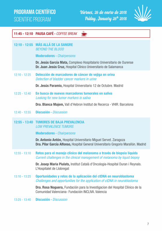
11:45 - 12:10 PAUSA CAFÉ · COFFEE BREAK
12:10 - 12:55 MÁS ALLÁ DE LA SANGRE
BEYOND THE BLOOD
Moderadores · Chairpersons
Dr. Jesús García Mata, Complexo Hospitalario Universitario de OurenseDr. Juan Jesús Cruz, Hospital Clínico Universitario de Salamanca
12:10 - 12:25 Detección de marcadores de cáncer de vejiga en orina
Detection of bladder cancer markers in urine
Dr. Jesús Paramio, Hospital Universitario 12 de Octubre. Madrid
12:25 - 12:40 En busca de nuevos marcadores tumorales en saliva
Looking for new tumor markers in saliva
Dra. Blanca Majem, Vall d’Hebron Institut de Recerca - VHIR. Barcelona
12:40 - 12:55 Discusión · Discussion
12:55 - 13:40 TUMORES DE BAJA PREVALENCIA
LOW PREVALENCE TUMORS
Moderadores · Chairpersons
Dr. Antonio Antón, Hospital Universitario Miguel Servet. ZaragozaDra. Pilar García Alfonso, Hospital General Universitario Gregorio Marañón. Madrid
12:55 - 13:10 Retos para el manejo clínico del melanoma a través de biopsia líquida Current challenges in the clinical management of melanoma by liquid biopsy
Dr. Josep Maria Piulats, Institut Català d’Oncologia-Hospital Duran i Reynals. L’Hospitalet de Llobregat
13:10 - 13:25 Oportunidades y retos de la aplicación del ctDNA en neuroblastoma
Challenges and opportunities for the application of ctDNA in neuroblastoma
Dra. Rosa Noguera, Fundación para la Investigacion del Hospital Clínico de la Comunidad Valenciana- Fundación INCLIVA. Valencia
13:25 - 13:40 Discusión · Discussion
PROGRAMA científicoscientific PROGRAM
Viernes, 26 de enero de 2018 Fr iday, January 26th 2018
III Simposio Biopsia líquidael camino a la oncología de precisión
8
13:40 - 15:00 ALMUERZO Y VISITA PÓSTERES · LUNCH & POSTER VIEWING
15:00 - 15:45 INMUNO BIOPSIA LÍQUIDA
IMMUNE LIQUID BIOPSY
Moderadores · Chairpersons
Dr. Alfredo Carrato, Hospital Universitario Ramón y Cajal. MadridDr. Ignacio Chacón, Hospital Virgen de la Salud. Toledo
15:00 - 15:15 Nuevas técnicas de seguimiento de respuesta a inmuno terapia por biopsia
líquida
New techniques for monitoring the response to immunotherapy by liquid biopsy
Dr. Eduardo Sotomayor, George Washington University Hospital. Washington DC, USA
15:15 - 15:30 Contribución de la biopsia líquida al avance de la inmunoterapia
Contribution of the liquid biopsy to the immunotherapy advance
Dr. Jesús García-Foncillas, Hospital Universitario Fundación Jiménez Díaz. Madrid
15:30 - 15:45 Discusión · Discussion
15:45 - 16:50 CÁNCER COLORRECTAL
COLORECTAL CANCER
Moderadores · Chairpersons
Dr. Juan de la Cámara, Complejo Hospitalario Universitario de FerrolDra. Sonia Candamio, Complejo Hospitalario Universitario de Santiago
15:45 - 16:00 Potencial y limitaciones de la técnica de secuenciación de biopsia líquida en
pacientes con carcinoma de colon
Potential and limitations of sequencing techniques of liquid biopsy in colon carcinoma patients
Dr. Nicola Normanno, Istituto Nazionale dei Tumori. Napoli, Italia
Viernes, 26 de enero de 2018 Fr iday, January 26th 2018
9
16:00 - 16:15 Del laboratorio a la clínica: análisis preliminar del estudio VISNU
From the lab to the clinic: preliminary analysis of the VISNU study
Dr. Javier Sastre, Hospital Clínico San Carlos. Madrid
16:15 - 16:30 Potencial clínico de ARNs no codifi cantes como biomarcadores de biopsia
líquida en cánceres gastrointestinales
Clinical potential of noncoding RNAs as liquid biopsy biomarkers in gastrointestinal cancers
Dr. Ajay Goel, Baylor Scott & White Research Institute and Charles A. Sammons Cancer Center. Dallas, Texas, USA
16:30 - 16:50 Discusión · Discussion
16:50 - 17:20 PAUSA CAFÉ · COFFEE BREAK
17:20 -18:40 CÁNCER DE MAMA
BREAST CANCER
Moderadores · Chairpersons
Dr. Vicente Guillem, Insituto Valenciano de Oncología - IVO. ValenciaDr. Juan Cueva, Complejo Hospitalario Universitario de Santiago
17:20 - 17:35 Desafío para la aplicación clínica de biopsia líquida en cáncer de mama
Challenges to overcome the applicability of liquid biopsy in breast cancer
Dr. Isaac García-Murillas, Breast Cancer Now Research Center. The Institute of Cancer Research. London, United Kingdom
17:35 - 17:50 CTCs vs ctDNA en el manejo del cáncer de mama metastásico
CTCs vs ctDNA in the clinical management of the metastatic breast cancer
Dra. Evi Lianidou, Analysis of Circulating Tumore Cells - ACTC Lab, University of Athens. Grece
17:50 -18:05 Modelo de grupos cooperativos para la aplicación de biopsia líquida
Model of cooperative groups for the liquid biopsy application
Dr. Miguel Martín, Hospital General Universitario Gregorio Marañón. Madrid
PROGRAMA científicoscientific PROGRAM
Viernes, 26 de enero de 2018 Fr iday, January 26th 2018

III Simposio Biopsia líquidael camino a la oncología de precisión
10
18:05 - 18:20 Generación de CDX a partir de una paciente con cáncer de mama
metastásico triple negativo
Establishment of a CDX from a triple-negative metastatic breast cancer patient
Dra. Clotilde Costa, Unidad mixta ROCHE CHUS. Complejo Hospitalario Universitario de Santiago
18:20 - 18:40 Discusión · Discussion
18:40 - 19:40 COMUNICACIONES ORALES
ORAL COMMUNICATIONS
Moderadores · Chairpersons
Dra. Ana Belén Dávila, Unidad Mixta ROCHE CHUS, Complejo Hospitalario Universitario de Santiago
Dr. Juan Ruiz, Complejo Hospitalario Universitario de Santiago
18:40 - 18:55 La caracterización molecular de CTCs de pacientes con NSCLC avanzada es
una estrategia valiosa para predecir la respuesta de quimioterapia de
primera línea
Molecular characterization of CTCs from patients with advanced NSCLC is a valuable strategy to predict fi rst line chemotherapy response
Dra. Silvia Calabuig-Fariñas, Hospital General Universitario de Valencia
18:55 - 19:10 Análisis de células tumorales circulantes (CTCs) a nivel de célula única para
inferir cambios genéticos y de fenotipo en respuesta a presiones terapéuticas
en cáncer de vías biliares
Analysis of single Circulating Tumor Cells (CTCs) to infer phenotype and genome changes in response to therapeutic pressures in biliary tract cancer
Dra. Carolina Reduzzi, Fondazione IRCCS Istituto Nazionale dei Tumori. Milano, Italy
19:10 - 19:25 Niveles de ctDNA por ddPCR en cáncer de pulmón de células no pequeñas:
implementación en la práctica clínica
ctDNA levels by ddPCR in non-small-cell lung cancer: implementation in clinical practice
Dr. Aitor Azkárate, Hospital Universitari Son Espases. Mallorca
Viernes, 26 de enero de 2018 Fr iday, January 26th 2018
11
19:25 - 19:40 El ctDNA refl eja la heterogeneidad intrapaciente en pacientes con cáncer
colorrectal metastásico (mCRC) que progresan a FOLFIRI+ Panitumumab Circulating tumor (ct)DNA captures intrapatient heterogeneity in metastatic colorectal (mCRC) patients progressing to FOLFIRI+panitumumab Dra. Joana Vidal, Hospital del Mar-Parc de Salut Mar. Barcelona
19:40 - 20.40 CÓCTEL Y VISITA PÓSTERES · COCKTAIL & POSTER VIEWING
PROGRAMA científicoscientific PROGRAM
Viernes, 26 de enero de 2018 Fr iday, January 26th 2018
III Simposio Biopsia líquidael camino a la oncología de precisión
12
09.00 - 10:05 CÁNCER DE PULMÓN
LUNG CANCER
Moderadores · Chairpersons
Dr. Sergio Vázquez, Hospital Universitario Lucus Augusti. Lugo Dr. Jorge García González, Complejo Hospitalario Universitario de Santiago
09:00 - 09:15 Búsqueda de marcadores tempranos en sangre
Searching for early markers in blood
Dra. Eloisa Jantus, Hospital General Universitario de Valencia
09:15 - 09:30 Uso de técnicas de secuenciación de nueva generación en ctDNA para
caracterizar tumores de pulmón
Application of next generation sequencing on ctDNA for lung cancer characterization
Dr. José Luis Costa, Institute of Molecular Pathology and Immunology of the University of Porto - IPATIMUP. Porto, Portugal
Esponsorizada por: / Sponsored by: Thermofi sher
09:30 - 09:45 ¿Podemos predecir la recurrencia del cáncer de pulmón monitorizando el
ctDNA?
Can we predict recurrence in lung cancer by ctDNA monitoring?
Dr. Luis Paz-Ares, Hospital Universitario 12 de Octubre. Madrid
09:45 - 10:05 Discusión · Discussion
10:05 - 11:10 CÁNCER DE PRÓSTATA
PROSTATE CANCER
Moderadores · Chairpersons
Dr. David Olmos, Centro Nacional de Investigaciones Oncológicas-CNIO. Madrid Dr. Juan Antonio Virizuela, Hospital Universitario Virgen de la Macarena. Sevilla
10: 05 - 10:20 AR-V7. Signifi cado clínico en pacientes con cáncer de próstata metastásico
AR-V7. Clinical signifi cance in metastatic prostate cancer patients
Dr. Emmanuel Antonarakis, Sidney Kimmel Comprehensive Cancer Center at Johns Hopkins. Baltimore, Maryland, USA
Sábado, 27 de enero de 2018 Saturday, January 27th 2018
13
10:20 - 10:35 ctDNA como guía para el tratamiento de cáncer de próstata con inhibidores
de PARP
ctDNA as a guide for the treatment of prostate cancer with PARP inhibitors
Dr. Joaquín Mateo, Vall d’Hebron Institut d’Oncologia - VHIO. Barcelona
10:35 - 10:50 Ensayos clínicos activos para validar el uso clínico de biopsias líquidas en
cáncer de próstata
Ongoing clinical trials for the validation of liquid biopsy in prostate cancer
Dr. José Ángel Arranz, Hospital General Universitario Gregorio Marañón. Madrid
10:50 - 11:10 Discusión · Discussion
11:10 - 11:40 PAUSA CAFÉ · COFFEE BREAK
11:40 - 12:45 BIOPSIA LÍQUIDA Y HETEROGENEIDAD TUMORAL
LIQUID BIOPSY AND TUMOR HETEROGENEITY
Moderadores · Chairpersons Dr. Ángel Díaz-Lagares, Complejo Hospitalario Universitario de Santiago
Dra. Elena Brozos, Complejo Hospitalario Universitario de Santiago
11:40 - 11:55 Análisis evolutivo de genomas de células individuales
Evolutionary analysis of single-cell genomes
Dr. David Posada, Universidad de Vigo
11:55 - 12:10 Factores clave para la supervivencia celular durante la diseminación
sanguínea
Key factors in cell survival during blood-borne dissemination
Dr. Nicola Aceto, University of Basel. Switzerland
12:10 - 12:25 Manejo de la heterogeneidad tumoral en clínica
Clinical management of tumor heterogeneity
Dr. Emilio Alba, Hospital Universitario Virgen de la Victoria. Málaga
12:25 - 12:45 Discusión · Discussion
PROGRAMA científicoscientific PROGRAM
Sábado, 27 de enero de 2018 Saturday, January 27th 2018
III Simposio Biopsia líquidael camino a la oncología de precisión
14
12:45 - 13:45 MESA DE DEBATE: RIVALES O COMPLEMENTARIOS EN BUSCA DE UNA
MEDICINA DE PRECISIÓN?
ROUND TABLE: RIVAL OR COMPLEMENTARY IN SEARCH OF A PRECISION MEDICINE?
Moderadores · Chairpersons
Dra. Laura Muinelo, Complejo Hospitalario Universitario de Santiago Dr. Luis León, Axencia de Coñecemento en Saúde-ACIS. Santiago de Compostela
12:45 - 13:05 Ronda 1: Biopsia de tejido versus biopsia líquida
Round 1: Tissue biopsy versus liquid biopsy Dra. Mar Varela, Institut d’Investigació Biomèdica de Bellvitge-IDIBELL, Barcelona Dr. César A. Rodríguez, Hospital Clínico Universitario, Salamanca
13:05 - 13:25 Ronda 2: ctDNA vs CTCs
Round 2: ctDNA vs CTCs
Dra. Joana Vidal, Hospital del Mar-Parc de Salut Mar. Barcelona Dr. Roberto Piñeiro, Unidad mixta ROCHE CHUS. Complejo Hospitalario Universitario de Santiago
13:25 - 13:45 Discusión · Discussion
13:45 - 14:00 CLAUSURA Y ENTREGA DE PREMIOS DEL CONCURSO DE PÓSTERES
CLOSING AND POSTER COMPETITION AWARDS CEREMONY
Sábado, 27 de enero de 2018 Saturday, January 27th 2018
15
III SimposioBiopsia líquidael camino a la oncología de precisión
ABSTRACTS
16
ABSTRACTS
1· EVALUATING THE METASTATIC POTENTIAL OF BREAST CANCER CTC-CLUSTERS BY IN VITRO AND IN VIVO ASSAYS.
Authors: Inés Martínez Pena1*, Pablo Hurtado Blanco1*, Carmen Abuín Redondo1, Laura Sánchez Piñón2, Rafael López López3, Roberto Piñeiro Cid1‡.
1Roche-CHUS Joint Unit, University Hospital of Santiago (CHUS). 2Genetics Department, Veterinary Faculty, University of Santiago de Compostela (USC). 3Translational Medical Oncology Group, Health Research Institute of Santiago (IDIS), University Hospital of Santiago (CHUS).
* These authors has contributed equally. ‡ Tutor.
Introduction
Breast cancer (BC) is among the major tumour types diagnosed annually. In fact, BC is the most frequent tumour in female population. The majority of deaths associated with cancer are caused by the complex process of metastasis, in which new tumour foci are established in regions far from the primary tumour. Circulating tumour cells (CTCs), which are those cells released into the blood stream from the primary tumour, have the potential to seed metastatic lesions. CTCs can travel either as single cells or in groups called CTC-clusters. CTC-clusters are believed to have a greater metastatic capacity than single CTCs and their presence in the blood of BC patients is associated with a worse prognosis, indicated by a decrease in metastasis-free survival and the formation of new metastatic foci. Therefore, these CTCs-clusters could be useful to study the prognosis and progression in BC. However, little knowledge is yet available about what characteristics make them more metastatic than single CTCs, and overall, what is their contribution to metastasis.
Objective
The objective of our research is to better understand the biology of CTC-clusters. For this reason, we seek to functionally characterise CTC-clusters in order to identify differential properties compared to single CTCs, and to understand their contribution to metastasis.
Materials and methods
We use an in vitro model of CTCs and clusters generated from human breast cancer cell lines. Cells are spiked into blood from healthy donors, purifi ed and allowed to aggregate in culture. Once aggregates are formed, they are mechanically dissociated to generate either a single or cluster cell suspension. The performance of both populations is compared in different in vitro assays of metastasis. Moreover, we assess their in vivo metastatic potential by using a xenograft model in zebrafi sh embryos (Danio rerio) in which fl uorescently labelled cells are injected into the circulation of the fi sh (Duct of Cuvier) allowing tracking tumour spread and metastasis.
Results
Initial in vitro results are showing that our model of clusters have different properties typically associated with malignance, such as higher migration, invasion and tumourigenic capacity, when compared to individual cells. On the other hand, zebrafi sh xenograft experiments are showing that CTCs and CTC-clusters can survive in the circulation of the fi sh and effi ciently disseminate and seed metastases. However, when analysing tumour volumes, no signifi cant differences have been observed between both populations.
Conclusions
Preliminary differences between single CTCs and CTC-clusters have been observed in our in vitro functional assays. These differential properties could partially be the underlying causes that could explain the differential contribution of the clusters to the metastasis. Furthermore, we are using a novel in vivo approach to study the biology of CTCs, zebrafi sh xenografts. So far, our data show the ability of circulating tumour cells to disseminate and form metastases, although under these experimental conditions we have not detected signifi cant differences between single cells and CTC-cluster populations. Therefore, our CTC-cluster model is a promising model to study the biology of these groups of cells and their contribution to metastasis.
17
ABSTRACTS
2· ISOLATION AND MOLECULAR CHARACTERIZATION OF A NEGATIVE ENRICHED CTCS POPULATION IN METASTATIC
BREAST AND PROSTATE CANCER PATIENTS BEFORE AND AFTER TREATMENT
Thais Pereira-Veiga1*, Mónica Martínez-Fernández2,3,4, Carmen Abuin1, Rafael López-López1,4,5, Laura Muinelo-Romay4,5 and Clotilde Costa1,4
1. Roche-Chus Joint Unit. University Hospital of Santiago. Travesía da Choupana s/n 15706 Santiago de Compostela, Spain.2. Molecular Oncology Unit. CIEMAT. Avda Complutense 40 28040 Madrid, Spain.3. Molecular and Celular Oncology Unit. Universitary Hospital 12 Octubre. Avda Córdoba s/n 28041Madrid, Spain.4. CIBERONC, Centro de Investigación Biomédica en Red Cáncer, Madrid, Spain.5. Liquid Biopsy Analysis Unit, Oncomet, Health Research Institute of Santiago (IDIS), Complexo Hospitalario Universitario de Santiago de Compostela (SERGAS); Trav. Choupana s/n, Santiago de Compostela 15706, Spain.
Introduction:
Breast and prostate cancer are the two most prevalent invasive cancers in women and men, respectively. Being hormone-dependent cancers, the emergence of hormonal resistance is common. Tumour recurrence, resistance to therapy and metastasis formation are the leading causes of death in these patients, being CTCs principal actors for tumour dissemination in both cancers.Detection of CTCs with CellSearch is based on the expression of EpCAM and CK8, 18 and 19. Therefore, the detection of CTCs that undergo a downregulation expression of epithelial markers, like more mesenchymal populations due to the EMT process, is limited using this strategy.So far, few studies have investigated CTCs expression profi le changes with the treatment but, to our knowledge, none of them have studied a negative enrichment approach in order to cover all the tumoral heterogeneity.
Objective:
The aim of the present study was to characterize a non-labelled CTCs population and to provide new insights into the dynamic evolution that occur during treatment and progression.
Methods:
CTCs from 11 metastatic breast (mBC) and 10 prostate (mPC) cancer patients were enriched before and after one cycle of treatment, and at recurrence, using CellSearch (Menarini-Silicon Biosystems) and RosetteSep (STEMCELL Technologies). Gene expression analysis was performed with a customized panel of 36 genes. Peripheral blood from 12 healthy donors was used to normalize the obtained data. In a number of samples cytospin and immunostaining was performed to assess the absence of CD45 expression in the isolated population.
Results:
Eleven out of 21 patients (52%) were positive for CTCs by CellSearch at baseline. Epithelial markers were associated with CTCs positivity by CellSearch in all of the mBC and mPC samples. Overexpression of both epithelial (EpCAM, CDH1 and KRT19) and mesenchymal markers (SNAI1) were found when comparing patient samples with healthy donors at baseline, and an increase of mesenchymal markers (SNAI1) was identifi ed after treatment.
Conclusion:
Negative enrichment allows the isolation of a heterogeneous CTCs population, overcoming the absence of a universal marker. This approach may help better monitoring the evolution of the disease.
18
ABSTRACTS
3· ASSESSMENT OF QMSP SENSITIVITY FOR THE DETECTION OF IGFBP-3 PROMOTER METHYLATION IN PLASMA FROM
PATIENTS WITH ADVANCED STAGE NON-SMALL-CELL LUNG CANCER.
Olga Pernía1,2*, Rocío Rosas1,2, Julia Jiménez1,2, Isabel Esteban3,Ana M. Rodríguez García 3, Patricia Cruz-Castellanos4, Darío Sánchez-Cabrero4, Javier de Castro2,4 and Inmaculada Ibáñez1,2
1. Cancer Epigenetics Laboratory, INGEMM, La Paz University Hospital, Madrid, Spain;2. Biomarkers and Experimental Therapeutics in Cancer, IdiPAZ, Madrid, Spain;3. Department of Pathology, La Paz University Hospital, Madrid, Spain;4. Department of Oncology, La Paz University Hospital, Madrid, Spain.* Olga Pernía Arias. Cancer Epigenetics Laboratory, INGEMM, Biomarkers and Experimental Therapeutics in Cancer, IdiPAZ, Paseo de la Castellana 261, Madrid, 28046. Introduction:
Cancer is the main cause of morbidity and mortality, with 14 million new cases and 8 million deaths related 2012. Non-small cell lung cancer (NSCLC) represents the 80-85% of the lung cancers. Approximately 80% of the patients are diagnosed in advanced stages with a very poor prognosis. Cisplatin-based chemotherapy is the fi rst line theraphy for NSCLC; however, it also induces de novo DNA-hypermethylation, a process that may be involved in the development of drug-resistant phenotypes by inactivating genes required for drug-cytotoxicity. We have previously reported that IGFBP-3 promoter hypermethylation results in a reduction of tumor cell sensitivity to cisplatin in advance-stages NSCLC. Liquid biopsy is a useful tool for the analysis of tumors with limited-sample size as advanced stages of NSCLC. DNA methylation detection has been shown affordable in plasma sources, showing concordance between ctDNA and corresponding tumor tissue DNA. Knowing the IGFBP-3 methylation status in ctDNA may provide robust information associated with prognosis and treatment response for the clinical management of NSCLC.
Objective:
Assessment the sensitivity of qMSP to detect the methylation status of the IGFBP-3 gene promoter in ctDNA from NSCLC patients with advanced stages and correlate with the methylation levels in paired tumor tissue.
Methodology:
53 FFPE/plasma paired samples from stages III-IV NSCLC patients have been collected in the tumor bank at Hospital La Paz, following the ethical and confi dentiality issues. Blood samples were processed within the fi rst 60 min from the extraction and plasma stored at -80C. Tumor samples from FFPE or cytologies were reviewed by an expert pathologist to confi rm the diagnosis and guarantee at least 90% tumor content. DNA and ctDNA was isolated and bisulfi te modifi ed following routine methodology. Methylation status of the IGFBP3-promoter was measured by qMSP methodology which presents a 1:10000 sensitivity, obtaining quantitative methylation levels (Cmeth = 100/ [1+2(CTCG – CTTG) ]).
Results:
We analyzed IGFBP-3 methylation in 52 matched tumor/ctDNA samples. Four samples were undetermined. 65% (31 out of 48 determined) were unmethylated in the tumor sample, and 35% (17 out of 48) were methylated. Those results are in accordance with previous cohorts analyzed. When comparing DNA methylation from tumor and ctDNA, 69% (33 out of 48) of the patients presented the same methylation pattern. We found 90% correlation between T/ctDNA in unmethylated tumor samples, but the power to fi nd methylated ctDNA when the tumor DNA was methylated decrease to 29%. It was consider methylated when the % of methylation was >30%
Conclusion:
Firstly, we have found the expected methylation ratios when analyzing DNA from tumor tissue. Secondly, DNA methylation status was determined in 48 out of 52 ctDNA samples (92% sensitivity). Thirdly, the presence of IGFBP-3 methylation in ctDNA from plasma by using qMSP was found in 29% of advanced-stages NSCLC patients harbouring a methylated tumor. Although it is necessary to improve the detection ratio at ctDNA, these results indicate that the epigenetic marker IGFBP-3, able to predict the response to therapy or monitor the progression in NSCLC patients, could be potentially used in liquid biopsy.
19
ABSTRACTS
4· NANOSYSTEMS FOR CIRCULATING TUMOUR CELLS CULTURE
Carmona-Ule, Nuria Nano-Oncology Laboratory of the Roche-Chus Joint Unit. Hospital Gil Casares. Travesía da Choupana, 0, 15706 Santiago de Compostela, La CoruñaAbuín-Redondo, C.; de la Fuente, M.; López-López, R. and Dávila-Ibañez, A.B.
Introduction:
The study of Circulating Tumour Cells (CTCs) have gained attention during the last years due to their applicability in Liquid Biopsy related with the detection and progression of cancer metastasis. Some of the potential clinical applications for CTC analysis include monitoring, prognosis and the selection of the most appropriate therapy. However, it is still a challenge to translate these studies to the clinic because of the low effi ciency of the isolation methods and the diffi culty of CTC culture.Nanotechnology is a tool that offer great advantages in nanomedicine, particularly nanoemulsions (NEs) which can interact with the cellular machinery by entering the cells through different cellular pathways, as opposed to many common small drugs or free substances that simply diffuse according to their solubility.
Objectives:
The main objective is to formulate NEs from phospholipids involved in cell migration, invasion, angiogenesis and cancer cell proliferation to prove their activity as pro-proliferative nanosystems in metastatic breast cancer cell lines. Then, experimental conditions will be modifi ed (NE concentrations, cell lines, incubation times, cell seeding concentration, etc) to mimic the CTCs culture conditions, since the fi nal goal will be to use these NE as proliferative nanosystems to stimulate the proliferation of CTCs. The achievement of CTCs expansion will allow to analyze their molecular pattern to get more information about these cells and achieve more precise diagnostic tools by testing personalized therapies in vitro.
Methods:
The NEs were prepared by the previously reported low-energy emulsifi cation oil in water method and were physically characterized. Mean average size, polidispersion index (PI) and Zeta potential (ZP) were determined by ZetaSizer Nano. The activity of the NEs was evaluated in vitro by cell viability assays (alamarBlue®). Moreover, uptake of the NEs by breast cancer cell lines was proved using confocal laser scanning microscopy.
Results:
NEs using several phospholipids and fatty acids were successfully formulated in a wide range of sizes. Their chemico-physical characterization was done showing adequate colloidal stability in different media and with time. The cell viability assays to test the activity of the nanoemulsions related to their pro-proliferative activity showed a concentration and cell line dependence. Cellular uptake of the nanoemulsions was confi rmed by confocal microscopy images.
Conclusions:
1. We have formulated coloidally stable NEs with different biocompatible components. Additionally, we were able to modify their sizes openning their applicability for different areas of nanomedicine.2. It was proved that NEs increased the cell viability of breast cancer lines.3. Preliminary experiments with CTCs isolated from metastatic patients showed that NEs provide advantages in the maintenance of CTCs culture over time.
20
ABSTRACTS
5· ONCODYNAMICS LIQUID BIOBANKING: A HOLISTIC PERSPECTIVE TOWARDS PREDICTIVE MEDICINE IN METASTATIC
CANCER
Authors: Patrícia Corredeira1, Patrícia Martins2, Pedro Andrade2, André Mansinho3, Arlindo Ferreira 1,3, Sandra Casimiro1, Julie Ribot1, Bruno Silva-Santos1, Lorena Dieguez4, Luis Costa1,3
Affi liation:1: Instituto de Medicina Molecular – João Lobo Antunes, Faculdade de Medicina, Universidade de Lisboa, Av Prof. Egas Moniz, 1649-028 Lisbon 2: Centro de Investigação Clínica, Hospital Santa Maria, Lisbon3: Oncology Division, Hospital de Santa Maria, Centro Hospitalar Lisboa Norte, Lisbon4: International Iberian Nanotechnology Laboratory, Braga
Introduction:
Clinical outcomes in oncology result from complex interactions between the tumour, host microenvironment and immune response. Therefore, to fully understand such complexity, it is essential to integrate the dynamics of clonal evolution with each one of these players during disease progression.Major technological advances in the past years have led to important discoveries about tumor genetics that translate into new targetable therapies. A major breakthrough has been the recent success of immunotherapy. But although is consensual that clonal evolution of the tumor is a major driver of therapy resistance, the interplay between clonal evolution and host immune response was never addressed.The Oncodynamics Biobank is a prospective collection of liquid biopsies, designed to characterize both the host immune response and tumor clonal evolution during disease progression under therapy, offering new insights of Precision Medicine in cancer.
Objectives:
Oncodynamics Biobank focuses on de novo stage IV breast, prostate, colorectal, melanoma, gastric and glioblastoma cancer patients. It was planned to generate genetic and biological information along with clinical data, and provide a repository of samples obtained from liquid biopsies, including: quantifi cation and characterization of circulating tumor cells (CTCs); DNA and RNA from CTCs; circulating tumor DNA (ctDNA); exosomes; immune profi le.
Methodology:
Oncodynamics Biobank includes patients diagnosed at Oncology Department of Hospital de Santa Maria, Lisbon, with de novo stage IV breast, prostate, colorectal, melanoma, gastric and glioblastoma cancer, that referred at the Multidisciplinary meetings. Liquid biopsies (whole blood and urine) are collected at baseline (before starting systemic treatment) and at each tumour objective evaluation (every 12-16 weeks), until two consecutive progression episodes. Electronic Case report forms include demographic and real time clinical data.CTCs are isolated in a microfl uidic device by cell size and morphology (PCT/EP2016/078406, patent pending), and stained in situ with antibodies against pan-cytokeratin (pan-CK to stain Epithelial CTCs), vimentin (to stain mesenchymal CTCs), CD45 (leukocyte common antigen) and DAPI (for nucleus). Each cell is imaged in a fl uorescence microscope for further analysis. Extraction of DNA and RNA from CTCs is being optimized.Immune profi le is assessed by Flow Cytometry analysis of different populations, namely: monocytes, myeloid dendritic cells, plasmocytoid dendritic cells, basophils, memory B cells, naïve B cells, monocytic myeloid-derived suppressor cells, polymorphonuclear myeloid-derived suppressor cells, natural killer T cells, effector T cells, regulatory T cells, αβ T cells and γδ T cells (γδ1 and γδ2).Serum and plasma is collected and stored for posterior ctDNA and exosome analysis. Urine aliquots and buffy coat are stored at -80ºC. Genomic DNA is extracted with NZY Blood gDNA Isolation kit (Nzytech).
21
ABSTRACTS
Results:
Since April 2017 the Oncodynamics Biobank has included 28 patients from three different pathologies (breast cancer n=15; colorectal cancer n=9; prostate cancer n=4), with baseline and respective follow-up collections. The majority of patients presented with bone metastases (100% of prostate cancer patients and 39% of breast cancer patients), liver metastases (83% of colorectal cancer patients and 8% of breast cancer patients) and lung metastases (66% of colorectal cancer patients and 15% of breast cancer patients). The prospective collection of biological samples includes ~700 aliquots (plasma, serum, buffy coat and urine).Preliminary analysis suggest that decrease of CTCs during follow-up is accompanied by a signifi cant clinical improvement. Evolution in the immune profi le in association with therapy and clinical outcome will be reported.
Conclusion:
The Oncodynamics Biobank will allow us to generate data to characterize both the host immune response and tumour clonal evolution during disease progression under therapy. The enrolment rate and preliminary data show that such a comprehensive collection of data, together with a valuable biological collection, is a major resource for Oncobiology-driven research.
6· EXPLORING LIQUID BIOPSY AND GLYCOSYLATION FOR EARLY DETECTION OF BLADDER CANCER
AuthorsSandra Carvalho1, Manuel Neves1,2, Catarina Abreu1, Dylan Ferreira2, Luis Lima2, Avelino Fraga3, José Alexandre Ferreira2, Lorena Diéguez1, Marta I Oliveira1
1 INL- International Iberian Nanotechnology Laboratory, Braga, Portugal2 Portuguese Institute of Oncology, Porto, Portugal3 Centro Hospitalar do Porto – Hospital de Santo António
Bladder cancer (BC) is the most common malignancy of the urinary tract, being associated with substantial morbidity and mortality. Despite the great advances in diagnosis, prognosis and therapy monitoring, BC has one of the highest recurrence rates and progression from non-muscle to muscle invasive BC commonly leads to metastasis [1]. Liquid biopsy has recently been exploited as a promising non-invasive tool towards the molecular profi ling of circulating tumour cells (CTC) and/or circulating molecules in various biological fl uids, envisaging early detection and monitoring of cancer [2]. In BC, identifi cation of circulating molecular biomarkers will greatly improve and guide the clinical management of patients.
One emerging classes of tumour biomarkers are cancer-associated glycans, particularly sialyl Tn (STn), expressed at the membrane of glycoproteins or secreted into the bloodstream. In fact, STn expression has been directly associated with cancer progression and metastatic potential of neoplastic cells [3]. Recent studies in BC have shown overexpression of STn antigens in primary tumours, lymph nodes and distant metastasis, which correlated with decreased overall survival [4]. However, the sialylation signature of tumour cells in biological fl uids and the precise role of STn glycans in BC dissemination and metastasis remains to be determined.
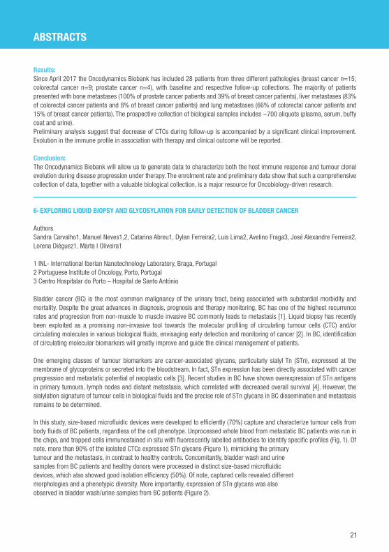
In this study, size-based microfl uidic devices were developed to effi ciently (70%) capture and characterize tumour cells from body fl uids of BC patients, regardless of the cell phenotype. Unprocessed whole blood from metastatic BC patients was run in the chips, and trapped cells immunostained in situ with fl uorescently labelled antibodies to identify specifi c profi les (Fig. 1). Ofnote, more than 90% of the isolated CTCs expressed STn glycans (Figure 1), mimicking the primarytumour and the metastasis, in contrast to healthy controls. Concomitantly, bladder wash and urinesamples from BC patients and healthy donors were processed in distinct size-based microfl uidicdevices, which also showed good isolation effi ciency (50%). Of note, captured cells revealed differentmorphologies and a phenotypic diversity. More importantly, expression of STn glycans was alsoobserved in bladder wash/urine samples from BC patients (Figure 2).

22
ABSTRACTS
Our preliminary fi ndings demonstrate that besides the primary bladder tumour and metastaticlesions, tumour-associated cells in circulation also express STn, confi rming its malignant nature, andhighlighting its potential as a novel BC biomarker. Further studies are ongoing to elucidate themolecular nature and the clinical relevance of STn positive cells in BC patients.
Additional tables and fi gures:
Figure 2. Photomicrograph of cells trapped inside the device from BC patients and stained for DAPI(blue) and STn (orange).
References:
1. Sanli, O., et al., Bladder cancer. Nature Reviews Disease Primers, 2017. 3: p. 17022.2. Heitzer, E., et al., The potential of liquid biopsies for the early detection of cancer. npj Precision Oncology, 2017. 1(1): p. 36.3. Azevedo, R., et al., Over forty years of bladder cancer glycobiology: Where do glycans stand facing precision oncology? Oncotarget, 2017. 8(53): p. 91734-91764.4. Lima, L., et al., Sialyl-Tn identifi es muscle-invasive bladder cancer basal and luminal subtypes facing decreased survival, being expressed by circulating tumor cells and metastases. Urologic Oncology: Seminars and Original Investigations, 2017. 35(12): p. 675.e1-675.e8.
Figure 1. Photomicrograph of cancer cells trapped inside the device from whole blood of metastaticBC patients and stained for DAPI (blue), cytokeratin (green), and STn (orange).
23
ABSTRACTS
7· COMPARATIVE GENOMIC HYBRIDIZATION IN PLASMA CIRCULATING TUMOR DNA FOR THE MONITORING OF MELANOMA
PATIENTS
Main author:Yolanda Ruano Domínguez, Instituto de Investigación Hospital 12 de Octubre.
Other authors:Leyla Blanco, Instituto de Investigación Hospital 12 de Octubre José Antonio López Martín, Servicio de Oncología, Hospital 12 de Octubre Rocío Cubo, Instituto de Investigación Hospital 12 de Octubre Erica Riveiro-Falkenbach, Instituto de Investigación Hospital 12 de Octubre José Luis Rodríguez Peralto, Servicio de Anatomía Patológica, Hospital 12 de Octubre.
Introduction:
Melanoma is a cutaneous cancer with an increasing worldwide prevalence and high mortality due to unresectable or metastatic stages. The analysis of cell-free circulating tumor DNA (ctDNA) from blood (liquid biopsies) appears to be a promising noninvasive, repeatable, and systemic sampling tool for detecting and monitoring melanoma. Mutations in BRAF are present in approximately 50% of melanoma cases and they are proposed as a useful blood-based biomarker for the clinical monitoring of melanoma patients. However, an equivalent biomarker for wild type BRAF melanoma has not yet been fully developed.
Objectives:
Melanoma shows a recurrent pattern of chromosomal aberrations that are distinct from benign melanocytic lesions and are independent of mutational status profi le. Hence, we propose the analysis of DNA copy number changes in ctDNA for monitoring patients with metastatic melanoma using comparative genomic hybridization (CGH).
Methods:
We performed CGH analysis by using the OncoScan kit (ThermoFisher Scientifi c) in ctDNA from liquid biopsies and paraffi n-embedded tissues from solid biopsies at diagnosis of metastatic melanoma patients. The BRAF status was determined by quantitive PCR analysis by Idylla (Biocartis) in plasma and Cobas (Roche) in tissue.
Results:
The CGH analysis in the plasma from a BRAF V600E metastatic melanoma patient revealed chromosome gains at 5p,7q and 21, and chromosome losses at 4q, 6q, 7p, 10, 12p and 14. The chromosome gains at 5p and 7q, as well as chromosome lost at 4q were present in the tissue sample of the patient at diagnosis. The CGH analysis in the plasma from a BRAF wild-type melanoma patient with metastasis in several organsrevealed chromosome gains at 3q and 8q and a chromosome lost at 8p. The 8q gain and 8p loss were detected in the tissue sample at diagnosis in addition to another chromosome alterations such as 1q gain, 9p, 10, 11q, 14, 16 and 20 losses and MET amplifi cation at 7q.
Conclusions:
The CGH analysis by using OncoScan kit allowed us to detect chromosome alterations in the plasma from metastatic melanoma patients independently of BRAF status. In addition, most of the genetic aberrations detected in plasma were present in their respective tumor tissues at diagnosis. Therefore, CGH analysis would be a suitable technique for the analysis of ctDNA and monitoring metastatic melanoma patients.

24
ABSTRACTS
8· DEVELOPMENT OF NEW, FAST AND ECONOMIC PROTOCOLS FOR THE ELIMINATION OF HIGH ABUNDANCE PROTEINS
IN PLASMA SAMPLES
María del Pilar Chantada-Vázquez 1*, Ceres Fernández-Rozadilla2, MVÁlvarez-Sánchez3, Ángel Carracedo-Álvarez4, Susana B. Bravo5, María García-Vence5, Cristina Núñez1, Sergio Vázquez Estévez6, Marta Covela Rúa6, Natalia Fernández Núñez6
1Laboratory of Nanoproteomic, Research Unit, Hospital Universitario Lucus Augusti (HULA), Servizo Galego de Saúde (SERGAS), 27002 Lugo (Spain)2 Cancer Genomics and Transcriptomics Group. Fundación Pública Galega de Medicina Xenómica. Instituto de Investigación de Santiago (IDIS). Choupana s/n, 15706 Santiago de Compostela (Spain)3 Digestive System Service. Hospital University Complex of Pontevedra. EOXI Pontevedra e o Salnés. Institute of Health Research Galicia Sur (IISGS)4Fundación Pública Galega de Medicina Xenómica. Instituto de Investigación de Santiago (IDIS). Choupana s/n, 15706 Santiago de Compostela (Spain)5 Proteomic Service. Instituto de Investigación Sanitaria de Santiago (IDIS). Choupana s/n, 15706 Santiago de Compostela (Spain)6Oncology Division, Hospital Universitario Lucus Augusti (HULA), Servizo Galego de Saúde(SERGAS), 27002 Lugo (Spain)
Introduction:
The analysis of human body fl uids is one of the most important approaches to the diagnosis of disease and following therapeutic interventions. Human body fl uids carry information about the status of the organism that may help in the recognition of phycological misbalances when overt pathological symptoms are not yet present.Human plasma proteins originate from a variety of tissue and blood cells as a result of secretion or leakage. Numerous biomedical studies have demonstrated that plasma protein levels refl ect human physiological or pathological states and can be used for disease diagnosis and prognosis. Sample preparation and handling is a critical for plasma proteome analysis. Other critical issue is the complexity of the proteome. Plasma contains a huge number of proteins differing by the extraordinary dynamic range of at least 9-10 orders of magnitude. How to globally quantify the proteins in free, bound, or modifi ed forms remains a critical challenge.Some of the plasma highly abundant proteins including: albumins, immunoglobulins (IgG), alpha-1-antitrypsin (A1AT), fi brinogen, and haptoglobin (HG). Depletion of these highly abundant proteins is often desired prior to proteome analysis. Immunodepletion methodsremove a good/variable portion, but not all of the highly abundant proteins; however, more problematic is that these methods may also remove other proteins by ``nonspecifi c´´ binding. A possible approach to address this problem is to disrupt the union between a low molecular weight (LMW) proteins and carrier proteins as albumin/IgG.For instance, denaturing conditions by adding acetonitrile (ACN) may disrupt this union, result in proteins detected increased when compared to native conditions.In addition, rather than depletion of highly abundant proteins, it would be promising to develop methods such as functionalized nanoparticles to enrich the low abundant proteins or reduce the protein concentration range.
Objectives:
The objectives of this work are to develop a rapid, cheap and effective protocol for the elimination of the majority proteins of plasma samples. In our case we development a centrifugal strategy using Amicon® with different NMWL to enrichment the serum samples in low abundant proteins. Thus, this methodology can be implemented in proteomic platforms which the number of plasma samples is high in recent years.
25
ABSTRACTS
Methodology:
Two different protocols for the removal of high abundance proteins have been tested. One of them is to pass the plasma sample through an affi nity column using the manufacture protocol.The other protocol is to mix the sample with a special denaturing buffer (200mM NaCl / 10% AcN in 200 nM AmBic) and pass the sample through Amicon®(Millipore) with decreased NMWL. In both protocols the proteins are identifi ed with a Triple-TOF 6600 (AbSciex).
Results:
With both protocols the number of proteins identifi ed with 1% of error is very high. It has even been possible to compare the different fractions obtained in the case of the size separation method with Amicons®. Both are quickly methods, have a relative low cost and are easy to implement in proteomic platforms in which the entry of complex biological samples such as serum increases every day.
Findings
Two valid protocols have been obtained, which provide a good pretreatment of plasma samples in order to make a proteomic analysis and give good results.
9· CTDNA LEVELS BY DDPCR IN NON-SMALL-CELL LUNG CANCER: IMPLEMENTATION IN CLINICAL PRACTICE.
Main author:Aitor Azkárate Martínez.Oncología Médica. Hospital Universitari Son Espases.
Other authors:Mónica Enver. Advanced Therapies and Biomarkers in Clinical Oncology Group. IdISBa.Margarida Mut. Oncología Médica. Hospital Universitari Son Espases.Esther Martinez-Font. Advanced Therapies and Biomarkers in Clinical Oncology Group. IdISBa.Magda Cordeiro. Oncología Médica. Hospital Universitari Son Espases.
Tutor:Antonia Obrador-Hevia. Advanced Therapies and Biomarkers in Clinical Oncology Group. IdIS-Ba.Josefa Terrasa Pons. Oncología Médica. Hospital Universitari Son Espases.
Introduction:
Lung cancer is the second most frequent tumor and the leading cause of death by cancer. About 15-20% of non-small-cell lung cancer (NSCLC) presents with driver mutation in the EGFR domain. At progressive disease, about 60% of these patients will harbour T790M mutation. In the last years, liquid biopsy has emerged as a promising new tool to complement tumor biop-sies data. Nowadays digital PCR may overcome real-time PCR which is the gold standard in this setting. Liquid biopsy has shown to be useful in early diagnosis, monitoring responses, early detection of progressive disease and as prognostic and predictive tool.
Objectives:
In this fi rst analysis of our data we aim to calculate how many days in advance progressive disease can be detected by droplet digital PCR (ddPCR) compared to conventional follow-up by CT scan. We also wanted to prove whether or not tumor burden is related to the amount of circulating tumor DNA (ctDNA) detected by ddPCR; and how a greater amount of T790M cop-ies/mL may have infl uence on progression free survival (PFS) during the fi rst line treatment.

26
ABSTRACTS
Methods:
Blood samples were collected from 18 patients diagnosed with advanced or metastatic NSCLC patients who carried an activating EGFR mutation treated with fi rst or second generation TKI and T790M mutation detection was carried out as described below. Blood samples were ob-tained every 3 months.Plasma cfDNA was extracted with the Qiagen Circulating Nucleic Acids kit and quantifi ed by QuantiFluor® dsDNA System. Finally, the mutational study of EGFR was carried out by digital droplet PCR (ddPCR) with the QX200 Droplet Digital PCR System with specifi c probes and pri-mers. Statistical analysis was performed with IBM SPPS Statistics 22 software.CT-scan was performed according to the criteria of the attending physician. The clinical re-sponse was evaluated according to RECIST criteria v1.1.
Results:
Data of serial blood samples from 3 out of 18 patients was fully available at the time of this study and ctDNA was assessed to evaluate the advance in diagnosis of progressive disease by ddPCR ddPCR in comparison to imaging tests. Using ddPCR, we could detect progressive disease from 71 to 200 days before imaging (image 1). We detected a trend that correlates tumour burden with ctDNA levels by ddPCR with a R2 of 0.565 (p=0.015) (image 2). Finally, patients harbouring T790M mutation had a trend to have worse PFS comparing with those who didn’t express this mutation (image 3), although these fi ndings weren’t statistically signifi cant.
Conclusions:
In this fi rst analysis of our data, we could detect progressive disease by ddPCR in advance of imaging tests in the same way other works have done recently (Provencio et al) and correlated with tumor burden. Those fi ndings set the bases to question whether T790M positive patients by dPCR should start second line treatment or should wait until progressive disease by CT-scan is detected. Clinical trials should be designed to answer this question.
Image 1. Number of days since the earliest identifi cation of sensitizing and T790M mutations in blood and assessment of disease progression by CT-scan.
Image 2. Correlation between tumor burden and copies/mL of ctDNA by ddPCR.
Image 3. PFS by number of copies/mL.
27
ABSTRACTS
10· CIRCULATING TUMOR (CT)DNA CAPTURES INTRAPATIENT HETEROGENEITY IN METASTATIC COLORECTAL (MCRC)
PATIENTS PROGRESSING TO FOLFIRI+PANITUMUMAB
J. Vidal1, J.M. Viéitez2, D. Paez3, C. Santos4, D. Azuara5, A. Dalmeses6, B. Bellosillo6, R. Salazar7, E. Aranda Aguilar8, C. Montagut1 en representación del Grupo de Tratamiento de los Tumores Digestivos (TTD)
1. Oncología Médica, Hospital del Mar, Barcelona2. Oncología Médica, Hospital Universitario Central de Asturias, Oviedo3. Oncología Médica, Hospital de la Santa Creu i Sant Pau, Barcelona4. Oncología Médica, Institut Catalá d’Oncologia Hospital Duran i Reynals, Barcelona5. Laboratorio Investigación Traslacional, Institut Catalá d’Oncologia Hospital Duran i Reynals-IDIBELL, Barcelona6. Molecular Biology Laboratory H. del Mar, Barcelona. Spain7. Oncología Médica, Institut Catalá d’Oncologia Hospital Duran i Reynals-IDIBELL., CIBERONC, Barcelona8. Oncología Médica, University Hospital Reina Sofi a. CIBERONC Instituto de Salud Carlos III, Cordoba
Background:
CRC cells evade EGFR blockade by several mechanisms of acquired resistance, mainly mutations in RAS, EGFR ECD, HER2 and MET. ctDNA is shed into the bloodstream by tumor cells and can be effectively used to track tumor heterogeneity and to evaluate the acquisition of molecular alterations at tumor progression.
Objective:
to analyze the acquisition of gene mutations in plasma ctDNA from patients with mCRC progressing to antiEGFR treatment and to correlate with laterality of primary tumor.
Methods:
We included mCRC patients treated within a phase II study: FOLFIRI + panitumumab in irinotecan-refractory mCRC. Plasma samples were collected at baseline and at the end of treatment and ctDNA was analyzed with the Oncomine colon ctDNA Assay. The resulting library was sequenced in the Ion PGM NGS System and analysed with the Torrent Suite Software. The detectable cutoff mutant allele fraction (MAF) was 0.1%. Subclonal mutations were defi ned as mutations with MAF ≤ 20% of the greatest somatic MAF in the sample.
Results:
Clinical characteristics of 16 patients were 69% male; median age 61.5 years; 75% left vs 25% right colon. ctDNA from all 16 patients was analyzed al progression. At least one mutation was detected in 94% of patients (15/16); median mutations per sample was 2.5 (range 1-13). The frequency of detected mutations was: 13 TP53, 3 APC, 1 CTNNB1, 15 KRAS, 8 NRAS, 7 EGFR, 4 BRAF, 4 PIK3CA, 4 MAP2K1, 1 GNAS, 1 SMAD4. While TP53, APC, PIK3CA and BRAF were most likely to be clonal, EGFR, RAS (KRAS+NRAS) were generally subclonal and all MAP2K1 and SMAD4 mutations were subclonal. All EGFR ECD mutations emerged in the left colon and co-existed with RAS mutations plus at least one additional acquired mutation (median 6, range3-11). In the right colon, acquired mutations were detected at a low frequency (median 2.5; range 1-6). RAS/BRAF mutations emerged in 100% and 66% of right and left colon respectively being always clonal in right colon whereas only in 50% of cases were clonal in left colon. In 10 out of 16 patients plasma baseline was available. At least one mutation was detected in 80% of patients (8/10); median mutations per sample was lower compared with plasma at progression: 1.8 (range 1 -4). All APC and TP53 mutations were present baseline. 12/15 (80%) of KRAS mutations and all NRAS, MAP2K1 and EGFR mutations were not detected baseline, and acquired during treatment. As a novelty, the SMAD4 mutation was not detected baseline and emerged after treatment with a 5.25% MAF.
Best response was: PR 8 patients, SD 6 patients and PD 2. In both patients with PD all mutations detected in plasma at progression were already detected in pre-treatment sample, with no new acquired mutations detected at progression (TP53 p.R175H + KRAS p.A146T and TP53 p.[V274F;.R273L;G245D] + PIK3CA p.H1047R respectively).
Conclusions:
ctDNA analysis captured intrapatient heterogeneity which developed as a result of EGFR inhibition. All EGFR ECD mutations emerged in the left colon and always co-existed with several other mechanisms of acquired resistance, refl ecting genomic complexity.
28
ABSTRACTS
11· ANALYSIS OF SINGLE CIRCULATING TUMOR CELLS (CTCS) TO INFER PHENOTYPE AND GENOME CHANGES IN
RESPONSE TO THERAPEUTIC PRESSURES IN BILIARY TRACT CANCER
C. Reduzzi1, L. Celio2, M. Vismara1, M. Silvestri1, R. Motta1, P. Miodini1, A. Martinetti2, F. De Braud2, M.G. Daidone1 and V. Cappelletti1.
1Department of Experimental Oncology and Molecular Medicine,2Department of Medical Oncology,Fondazione IRCCS Istituto Nazionale dei Tumori, Milano, Italy.
Introduction:
Biliary tract cancer (BTC) is a rare disease with poor prognosis and limited therapeutic options. Access to tissue biopsies for genotype analysis is often impossible hampering this way the use of targeted therapies. Circulating tumor cells (CTCs) could function as surrogate of tumor material and could also allow monitoring the disease in response to treatment. However, when using conventional methods based on the expression of epithelial markers, CTC are detected only in a low proportion of BTC patients.
Objectives:
Being aware of the limitations in conventional CTC detection, we aimed at the development of a novel and unbiased CTC detection and characterization approach, allowing the analysis of different subpopulation of CTC at single-cell level. The specifi c objective was to obtain information regarding i) presence, ii) peculiar phenotype and iii) molecular profi le of CTCs which could inform on treatment options thus giving prognostic and predictive advice.
Methodology:
Ten mL of peripheral whole blood were processed for CTC enrichment by the Parsortix which selects cells based on size and deformability. CTCs were than identifi ed and recovered with the DEPArray by positive selection for epithelial markers (eCTCs) and negative selection based on lack of epithelial and leukocyte markers (non conventional CTCs, ncCTC). After whole genome amplifi cation, single CTCs were used for molecular characterization (copy-number alterations (CNA) and mutations) at single-cell level.
Results:
Fourty-one blood samples were longitudinally collected from 16 patients and analyzed. Overall, 36/41 samples were CTC-positive, with ncCTCs being more frequent than eCTCs (71% vs. 39% of samples respectively). At baseline all patients presented at least 1 CTC, with the two CTC populations being equally represented. During treatment we observed a switch towards the non-conventional phenotype and a decrease in global CTC positivity (100% at baseline vs 80% during treatment), which could be linked to resistance or response to therapy, respectively. Overall, CTC-status correlated with clinical outcome of patients. From a biological point of view, molecular characterization of different CTCs from the same samples showed a high intra-patient heterogeneity both at CNA and mutation level. CNA data were used both to estimate chromosomal instability of CTCs in relation to response to treatment and to identify recurring alterations across patients and alterations arising during disease progression in single patients.
Findings:
The developed protocol allows the isolation of CTC in 100% of patients and their molecular characterization at single-cell level, supporting their value as valuable surrogate when tissue biopsies are not available.CTC phenotype seems to mirror (sometimes anticipate) clinical outcome of patients. A combined characterization at phenotype and genotype (mutations, CNA and chromosomal instability) level can be used to monitor the emergence of CTC subclones with peculiar phenotypic-genotypic patterns, related to response/resistance to treatment thus improving clinical management in BTC patients.
29
ABSTRACTS
12· FAST AND EFFICIENT ISOLATION OF CIRCULATING TUMOR CELLS FROM UNPROCESSED WHOLE BLOOD USING A
MICROFLUIDIC CELL FILTER
Thais Pereira-Veiga1, Silvina Ribeiro-Samy2, Marta I Oliveira2, Laura Muinelo-Romay3,4, Rafael López-López1,3,4, Lorena Diéguez2 and Clotilde Costa1,3
1. Roche-Chus Joint Unit. University Hospital of Santiago. Travesía da Choupana s/n 15706 Santiago de Compostela, Spain.2. INL - International Iberian Nanotechnology Laboratory, Av. Mestre José Veiga s/n, 4715-330 Braga, Portugal.3. CIBERONC, Centro de Investigación Biomédica en Red Cáncer, Madrid, Spain.4. Liquid Biopsy Analysis Unit, Oncomet, Health Research Institute of Santiago (IDIS), Complexo Hospitalario Universitario de Santiago de Compostela (SERGAS); Trav. Choupana s/n, Santiago de Compostela 15706, Spain.
Introduction:
Metastatic disease is directly responsible for most of cancer-related deaths worldwide. Circulating tumour cells (CTCs) shedding from the primary tumour have the ability to invade other organs, causing metastasis. Therefore, the study of CTCs might provide continuous and real-time valuable information for the clinical management of cancer patients1.However, these cancer cells are extremely rare – presenting ratios as low as 1 to 10 CTCs per a billion blood cells –, which makes their isolation a very hard task2. Unravelling the phenotypic and molecular profi le of CTCs can provide key information about the biology of tumour cells and highly contribute to personalized therapy. Isolation and characterization of these exceptionally scarce entities is still a challenge, mainly due to CTCs heterogeneity.
Objective:
To overcome this limitation, the CTC+ chip, a microfl uidic cell fi lter for label-free isolation of CTCs based on size and deformability, was developed and its performance was compared to the CellSearch® system.Methods:We have developed and fabricated, by Silicon Deep Reactive Ion Etching, the CTC+ chip: a size-based rare cell capture device, which comprises several isolation areas containing size exclusion fi lters.Effi ciency of the device was measured with spiked samples using SW480 colon cancer cell line. Blood samples from nine advanced colorectal cancer patients were analysed in parallel with the CTC+ chip and with CellSearch as the gold standard technique. Following isolation, cells retained in the CTC+ chip were immunostained with fl uorescently labelled antibodies to identify specifi c phenotypes and molecular characterization was performed by droplet digital PCR (ddPCR).
Results:
Spiking experiments with cultured tumour cells run through the CTC+ chip demonstrated a high capture effi ciency, 70% in average.The CTC+ chip was able to rapidly (i.e., less than one hour) process whole blood samples from a set of metastatic colorectal cancer patients and capture more CTCs than the current gold standard CellSearch® in all individuals analysed with high effi ciency and purity. To check the tumoral origin of the CTCs we performed ddPCR, with APC mutations being detected in CTC+ chip-isolated CTCs, confi rming the versatility of the technology for downstream applications.Since the isolation effi ciency of the CTC+ chip is higher, and considering the established cut off for bad prognosis in colorectal cancer used by the CellSearch® technology (≥ 3 CTCs/7.5 ml of whole blood), by enumerating the CTCs isolated in the CTC+ chip, it was possible to stratify patients with different prognosis.
Conclusion:
The CTC+ chip allows the fast and effi cient isolation of unlabelled CTCs. Furthermore, after CTCs isolation with this device, downstream analysis can be performed to achieve prognosis utility.
1. Gorges, T.M., Kuske, A., Röck, K., Mauermann, O., Müller, V., Peine, S., Verpoort, K., Novosadova, V., Kubista, M., Riethdorf, S. and Pantel, K. Accession of Tumor Heterogeneity by Multiplex Transcriptome Profi ling of Single Circulating Tumor Cells. Clin Chem. 2016 Nov; 62(11):1504-1515.2. Yu, M., Stott, S., Toner, M., Maheswaran, S. and Haber, D.A. Circulating tumor cells: approaches to isolation and characterization. J Cell Biol. 2011 Feb; 192(3):373-82
30
ABSTRACTS
13· CLINICAL UTILITY OF LIQUID BIOPSY IN ADVANCED LUNG CANCER.
Main author:Name: Dra. Ana Reguera AriasServicio de Anatomía Patológica, Complexo Hospitalario Universitario A Coruña (CHUAC)
Other authors:Hermida Romero T, Escalante Pérez M, López Solache L, Álvarez Martínez M, Antón Aparicio L*, Concha López A.Servicio de Anatomía Patológica*Servicio de Oncología MédicaComplexo Hospitalario Universitario A Coruña (CHUAC)
Tutor:Dr. Ángel Concha López, Jefe de Servicio de Anatomía Patológica, Complexo Hospitalario Universitario. A Coruña (CHUAC)
Introduction:
Liquid biopsy is a non-invasive technique that allows circulating biomarkers to be identifi ed in peripheral blood and recognize the genetic profi le of tumours and metastasis in order to offer the patient targeted therapy.It also allows to monitorize treatment response and to detect possible resistance to it.
Objective:
To determine through liquid biopsy the T790M resistance mutation to tyrosine kinase inhibitors in lung cancer patients in advanced stages (III- IV) carrying epidermal growth factor receptor (EGFR) mutations.
Methodology:
The patient cohort was formed by 53 patients with an average age of 61 years old (38-86), 32 women and 21 males. Out of the 53, 35 presented the EFGR gene mutation in the tissue, determined by a commercial Real-Time PCR kit, being the most common mutation EGFR Exon 19 deletion (57% of the total).A liquid biopsy has been carried out in those patients with a clinical or radiological progression and with a clinical suspicion to tyrosine kinase inhibitors resistance or with not enough tissue avaliable to determine the mutational profi le. The liquid biopsy was carried out extracting of 10cc of plasma to determine the circulating ctDNA.
Results:
Out of the 35 patients with an EGFR gene mutation, it has been determined that the T790M resistance mutation was present in 14 cases. 11 of them were detected in the fi rst blood extraction and the other 3 in thesecond extration, that took place a few days later. In addition and due to clinical insistence, there were 3 other cases in which the T790M was eventually determined through a surgical biopsy after several negative results of the liquid biopsy.Out of the 18 remaining cases, in which the mutational profi le was unknown, the EGFR gene mutation was determined in three of them after the fi rst liquid biopsy.
Conclusions:
Liquid biopsy is an extremely helpful tool to determine the EGFR T790M secondary mutation, especially in cases in which the obtention of a tissue sample is diffi cult, although surgical biopsies are sometimes needed when the clinical and radiological results indicate a progression of the illness
31
ABSTRACTS
14· MOLECULAR CHARACTERIZATION OF CTCS FROM PATIENTS WITH ADVANCED NSCLC IS A VALUABLE STRATEGY TO
PREDICT FIRST LINE CHEMOTHERAPY RESPONSE
Silvia Calabuig-Fariñas1,2,3,#, Laura Muinelo Romay2,4,#, , Alicia Abalo2,4, Héctor Amado Labrador1 , Ramón Lago2,4, Marais Mosqueda1, Carmela Rodríguez4,5, Eva Escorihuela2, Jorge García4,5, Eloisa Jantus-Lewintre 1,2,6, Rafael López2,4,5*, Carlos Camps 2,7,8,*
1 Molecular Oncology Laboratory, Fundación Investigación, Hospital General Universitario de Valencia, Valencia, Spain 2 CIBERONC 3 Depatment of Pathology, Universitat de València, Valencia, Spain 4 Liquid Biopsy Analysis Unit, Oncomet, Health Research Institute of Santiago (IDIS), Complexo Hospitalario Universitario de Santiago de Compostela (SERGAS); Santiago de Compostela, Spain. 5 Medical Oncology Department, Health Research Institute of Santiago (IDIS), Complexo Hospitalario Universitario de Santiago de Compostela (SERGAS); Santiago de Compostela, Spain. 6 Department of Biotechnology, Universitat Politècnica de València, Valencia, Spain 7 Department of Medical Oncology, Hospital General Universitario de Valencia, Valencia, Spain 8 Department of Medicine, Universitat de València, Valencia, Spain # co-fi rst authors *equal contribution
Introduction:
Non-small cell lung carcinoma (NSCLC) represents the 85% of lung cancer cases. At diagnosis, approximately 70% of patients present advanced disease. Although targeted therapies have improved their prognosis, there is still a clear need of more specifi c tumour biomarkers that allow a better treatment selection and monitoring. Circulating Tumour Cells (CTCs) represent a principal component of the dissemination process. Importantly, CTCs evaluation allows readily “liquid biopsy” meanwhile access to tissue specimens is often insuffi cient and fails to refl ect tumour dynamics, heterogeneity or even drug sensitivity.
Objective:
The present study was conducted to validate the characterization CTCs from patients with advanced NSCLC as a valuable tool for anticipating the disease evolution and the therapy response. Materials and Methods: 78 patients with advanced NSCLC were enrolled in the study at Hospital General Universitario de Valencia and Complexo Hospitalario de Santiago de Compostela. EpCAM positive CTCs from these patients were isolated and analysed using bothCellSearch technology and a qRT-PCR based approach at baseline and within treatment. For the gene expression analysis we selected a panel of genes with a relevant role for NSCLC aggressiveness and the resistance to platinum-based treatments.
Results:
From all patients included in the study 46% were positive for CTCs using CellSearch system at baseline, showing only 12% of patients ≥ 5CTCs. In accordance with previous studies in NSCLC, patients with ≥ 5CTCs showed poor PFS (3,2 vs 10,5 months, p=0,015) and OS (3,3 vs 13,9 months, p<0,001) rates compared with those patients with CTCs <5. In addition patients with high CTCs levels during treatment also had a more aggressive disease evolution in terms of PFS and OS. Importantly, from the analyzed gene panel in the CTC-enriched fraction we found low ERCC1 levels as a powerful marker to discriminate patients with a good response to chemotherapy from those that progressed during treatment administration (11,6 vs 4,86 months, respectively, p=0,001). In addition, patients with high ERCC1 expression in the CTCs population at baseline showed also lower OS (7,1 vs 14,6 months respectively, p<0,001).
Conclusions:
We demonstrated that gene expression analyses in CTCs represents an adequate strategy to identify biomarkers with prognostic value and potential applicability in the management of NSCLC patients.

32
ABSTRACTS
15· LIQUID BIOPSIES ALLOW EARLY DETECTION IN EGFR RESISTANCE
Silvia Calabuig-Fariñas1,2,3, Eloísa Jantus-Lewintre1,2,4, Amaya Fernández Díaz5, Albert Junquero5, Ana Blasco2,5, Cristóbal Aguilar-Gallardo1,2, Eva Escorihuela1,2, Marais Mosqueda1, Francisco Aparisi5, Carlos Camps 1,2,5,6
1Molecular Oncology Laboratory, Fundación Investigación Hospital General Universitario de Valencia; 2Centro de Investigación Biomédica en Red de Cáncer, 2Department of Pathology, Universitat de València; 4Department of Biotechnology, Universidad Politécnica de Valencia; 5Department of Medical Oncology, Consorcio Hospital General Universitario de Valencia; 6 Department of Medicine, Universitat de València.
Introduction:
Liquid biopsies appear a reliable alternative to conventional biopsies that can provide both useful precise molecular data to improve the clinical management of lung cancer patients and invasive way to monitor tumor behaviour. These advances are supported by important biotechnological developments in the fi elds of circulating tumor cells (CTCs) and circulating tumor DNA (ctDNA). An analysis of CTCs and ctDNA may be useful in treatment selection, for response monitoring, and for studying resistance mechanisms.
Methods:
Review and report a case of a female patient with lung adenocarcinoma EGFR exon 19 deletion with non-invasive mutational monitoring. Blood samples werecollected at diagnosis and repeated sampling was performed during follow-up and at progression in Streck cell-free DNA BCT® or EDTA tubes. Circulating cell-free DNA genotyping was performed with the OncoBEAM™ EGFR kit. The results were compared to those obtained from the DNA extracted from tissue at diagnosis and at progression.
Results:
Case report of a 73 year old female nonsmoker patient, with a clinical 4-month evolution of cough, dyspnea and weight loss. A thoraco-abdominal CT scan showed a 65 mm mass in the right upper lobe, pleural effusion, bilateral multiple pulmonary micronodules and metastatic bone lesions (iliac, sacro, lumbar spine). Bronchofi broscopy and a histopathological exam showed adenocarcinoma, with an EGFR mutation (exon 19 deletion). Lung adenocarcinoma stage IV (EGFR exon 19 deletion) diagnosis was made and the patient started treatment with erlotinib 150 mg/day. The EGFR mutation was confi rmed on cfDNA at diagnosis, and monitoring was performed with early undetectable blood mutations after EGFR-TKI predicted a good clinical response. Subsequent imaging revealed that the lesions in her chest and bone had stabilized or reduced in size in the following for next months. At 10 months, of the analized blood-based EGFR mutation status assessment revealed a T790M resistance mutation, however therapy with erlotinib continued. Thoracic and bone progression was observed at 17 months, and new tissue biopsy positive results for the T790M mutation were confi rmed. The patient was started on osimertinib and remained stable for 8 months until progression was exhibited in TACs. According to a liquid biopsy, exon 19 deletion reappeared, which suggested that osimertinib had failed. The patient died 3 months later from disease complications. When we compare our methodology with the conventional approach to detect mutations in liquid biopsy, we confi rm anticipated detections. Our results confi rm that early detected mutations can provide relevant outcome information.
Conclusions:
The purpose of this case report is to confi rm the clinical utility of ctDNA to detect epidermal growth factor receptor mutations by BEAMING methodology, which is feasible and effective monitoring treatment response to EGFR TKIs, and to detect the underlying mechanism of resistance. This work was supported by Astra Zeneca (ISSRES0110), the RD12/0036/0025 ISCIII and grants from the Fondo Europeo de Desarrollo Regional (FEDER).. Keywords: Circulating free DNA, EGFR mutation, NSCLC

33
ABSTRACTS
III SimposioBiopsia líquidael camino a la oncología de precisión
INFORMACIÓN GENERALGENERAL INFORMATION
III Simposio Biopsia líquidael camino a la oncología de precisión
34
SEDE Y ALOJAMIENTO · VENUE & ACCOMMODATION
San Francisco Hotel Monumento
Campillo de San Francisco, 3Santiago de Compostela • A Coruña (España)www.sanfranciscohm.com
Alojamiento: tarifa especial para asistentes al simposioAccommodation: special rate available for the symposium attendees
Reservas: contacte vía e-mail a [email protected]: please contact [email protected]
IDIOMAS · LANGUAGES
Idiomas principales: inglés y español. Traducción simultánea disponibleMain languages: English and Spanish. Simultaneous translation available
CONCURSO DE PÓSTERES · POSTER COMPETITION
Los pósteres permanecerán expuestos durante los tres días del simposio. El jurado, compuesto por 5 expertos, seleccionará de todos los trabajos presentados en formato póster, dos casos ganadores y 4 casos que serán presentados en formato de comunicación oral durante el simposio (éstos casos quedarían excluidos de los premios). El último día (sábado 27 de enero) se entregarán los premios a los dos ganadores. Habrá un primer premio por valor de 500€ y un segundo premio por valor de 300€.
Puede consultar las bases del concurso en la web del simposio: www.simposiobiopsialiquida.com
The posters will be on display during the three days of the symposium.From all the work submitted in poster format, a panel comprised of 5 experts will select two winning cases and 4 cases to be presented orally during the symposium (these cases will be excluded from the prizes). The awards will be presented the last day of the symposium (Saturday 27th January): €500 for the fi rst prize and €300 for the second.
Please check the terms and conditions of the competition at the symposium website: www.simposiobiopsialiquida.com
35
INFORMACIÓN GENERAL · GENERAL INFORMATION
CERTIFICADO DE ASISTENCIA · CERTIFICATE OF ATTENDANCE
Si desea recibir un certifi cado de asistencia y/o de presentación de poster deberá solicitarlo durante el evento rellenando el formulario que encontrará en la secretaría técnica.
If you want to receive the certifi cate of attendance you must fi ll the form that can be found at the technical secretary during the event.
INSCRIPCIONES · REGISTRATION
Formalice su inscripción en: www.simposiobiopsialiquida.comLa cuota de inscripción incluye: acceso a todas las sesiones científi cas, certifi cado de asistencia, cafés entre sesiones, almuerzos y cóctel.Es imprescindible ir acreditado durante todo el evento. Por favor asegúrese de llevar siempre su acreditación visible.
Please make your registration at: www.simposiobiopsialiquida.comThe registration fee covers: access to all the scientifi c sessions, certifi cate of attendance, coffee breaks, lunches and cocktail.Only proper accredited attendants will be able to enter the venue; please, make sure your lanyard is visible all the time.
PÁGINA WEB · WEBSITE
En la web del simposio puede consultar toda la información sobre el mismo: programa científi co, ponentes, inscripciones, sede y alojamiento.
In the website you can fi nd all the information about the event, such as: scientifi c programme, speakers, registration, venue and accommodation.
www.simposiobiopsialiquida.com
#SimposioBiopsiaLíquida

36
ABSTRACTS CON EL AUSPICIO DE · UNDER THE AUSPICE OF
PATROCINADORES ORO · GOLD SPONSORS
PATROCINADORES PLATA · SILVER SPONSORS
PATROCINADORES BRONCE · BRONZE SPONSORS
COLABORADORES · COLABORATORS
Passatge Batlló 15, 08036 Barcelona +34 93 451 17 24 [email protected] www.tacticsmd.net
SECRETARÍA TÉCNICA
®
TECHNICAL SECRETARY