Biopsia líquida: ¿el nuevo fotomatón? · Hospital Clínico Universitario e Instituto de...
39
. Dr. Rafael López López Oncología Médica Traslacional Hospital Clínico Universitario e Instituto de Investigación Sanitaria Santiago de Compostela Biopsia líquida: ¿el nuevo fotomatón? “LA FORMA DE LO QUE VENDRÁ”
-
Upload
nguyenkhue -
Category
Documents
-
view
216 -
download
0
Transcript of Biopsia líquida: ¿el nuevo fotomatón? · Hospital Clínico Universitario e Instituto de...
.
Dr. Rafael López López
Oncología Médica Traslacional
Hospital Clínico Universitario e Instituto de Investigación Sanitaria
Santiago de Compostela
Biopsia líquida:
¿el nuevo fotomatón?
“LA FORMA DE LO QUE VENDRÁ”
.

.
1943 2017

.
1943 2017

.
1943 2017
Oncología Clínica Oncología de precisión

.
15:56
The cancer genome
Stratton M, Campbell P & Futreal A. Nature 2009;458: doi:10.1038/nature07943
RL: Fotomatón_Toledo_2017
.
15:56
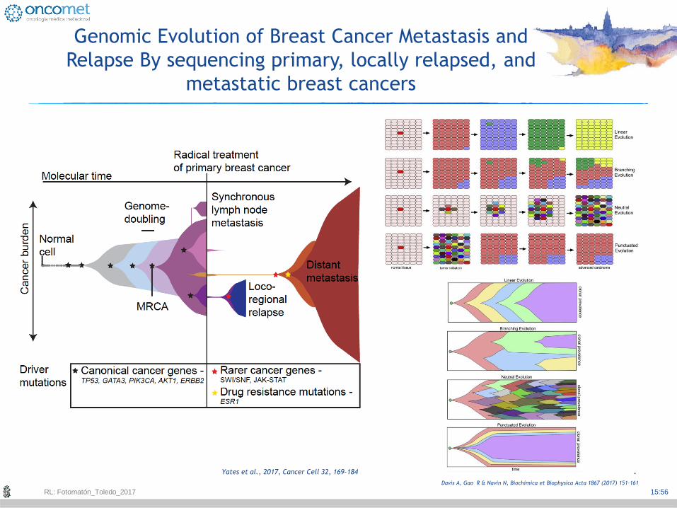
Genomic Evolution of Breast Cancer Metastasis and
Relapse By sequencing primary, locally relapsed, and
metastatic breast cancers
Yates et al., 2017, Cancer Cell 32, 169–184
RL: Fotomatón_Toledo_2017
Davis A, Gao R & Navin N, Biochimica et Biophysica Acta 1867 (2017) 151–161

.
15:56
Genomic Evolution of Breast Cancer Metastasis and
Relapse By sequencing primary, locally relapsed, and
metastatic breast cancers
Yates et al., 2017, Cancer Cell 32, 169–184
RL: Fotomatón_Toledo_2017

.
15:56
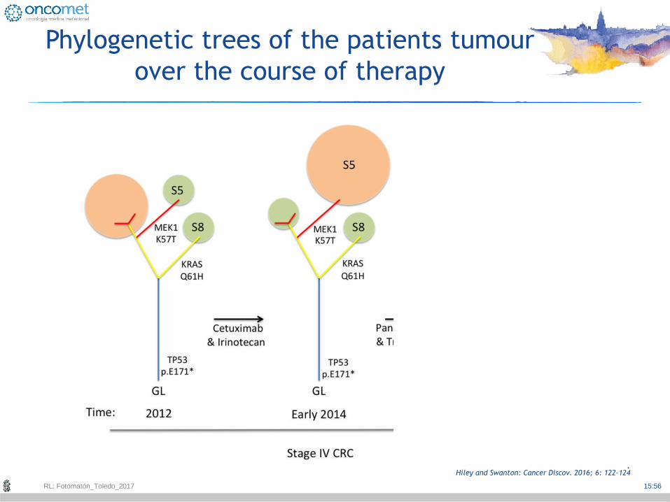
Phylogenetic trees of the patients tumour
over the course of therapy
Hiley and Swanton: Cancer Discov. 2016; 6: 122–124
RL: Fotomatón_Toledo_2017
.
15:56
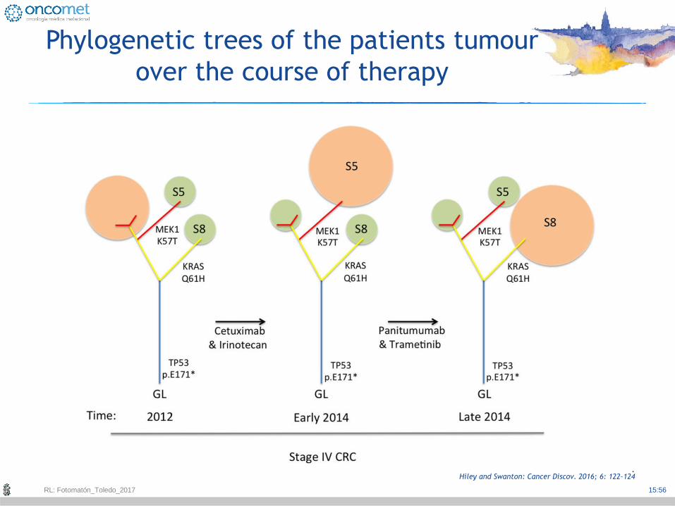
Phylogenetic trees of the patients tumour
over the course of therapy
Hiley and Swanton: Cancer Discov. 2016; 6: 122–124
RL: Fotomatón_Toledo_2017
.
15:56
Phylogenetic trees of the patients tumour
over the course of therapy
Hiley and Swanton: Cancer Discov. 2016; 6: 122–124
RL: Fotomatón_Toledo_2017
.
15:56
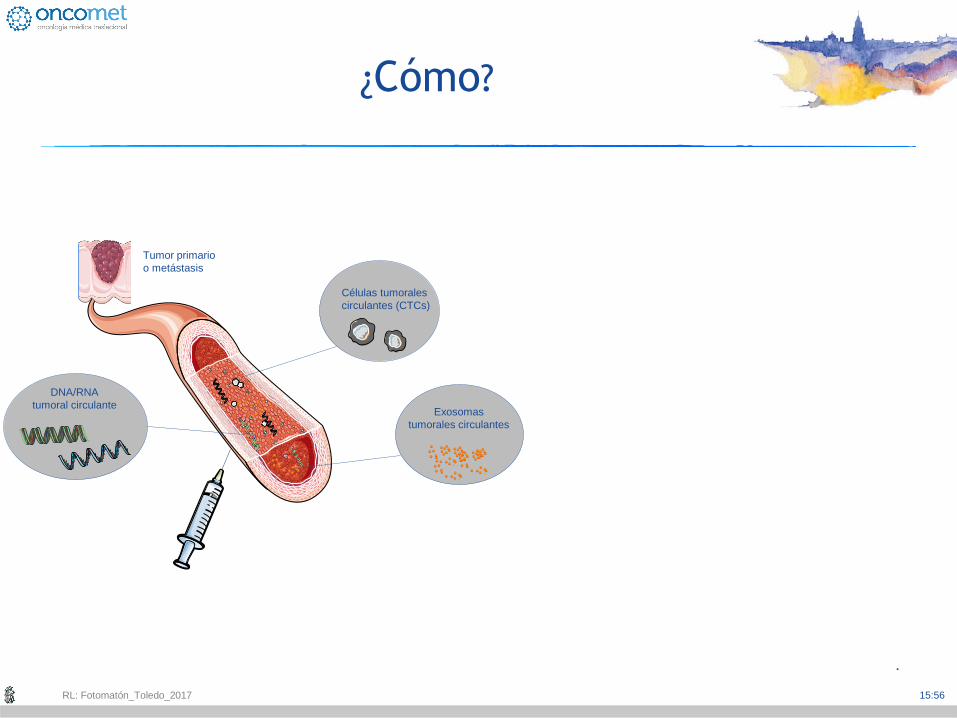
¿Cómo?
Células tumorales
circulantes (CTCs)
Tumor primario
o metástasis
DNA/RNA
tumoral circulante Exosomas
tumorales circulantes
RL: Fotomatón_Toledo_2017

.
15:56
¿Cómo?
Células tumorales
circulantes (CTCs)
Tumor primario
o metástasis
DNA/RNA
tumoral circulante Exosomas
tumorales circulantes
RL: Fotomatón_Toledo_2017

.
15:56
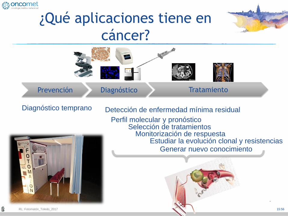
Prevención Diagnóstico Tratamiento
¿Qué aplicaciones tiene en
cáncer?
RL: Fotomatón_Toledo_2017

.
15:56
Prevención Diagnóstico Tratamiento
Generar nuevo conocimiento
Perfil molecular y pronóstico
Detección de enfermedad mínima residual Diagnóstico temprano
Selección de tratamientos
Monitorización de respuesta Estudiar la evolución clonal y resistencias
¿Qué aplicaciones tiene en
cáncer?
RL: Fotomatón_Toledo_2017
.
15:56
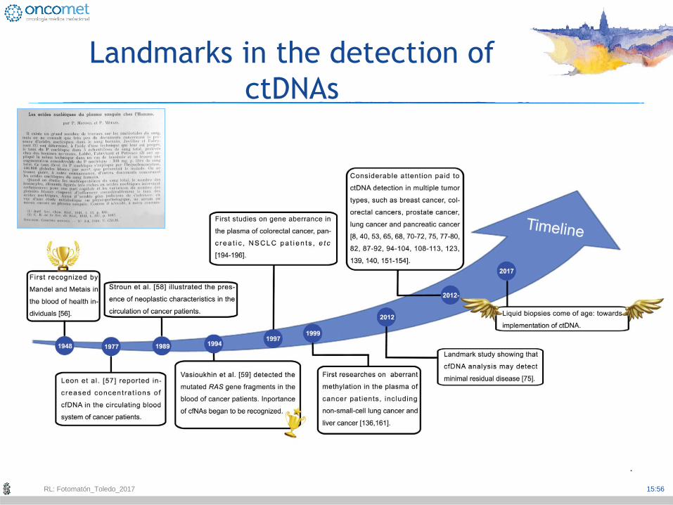
Landmarks in the detection of
ctDNAs
RL: Fotomatón_Toledo_2017
.
15:56
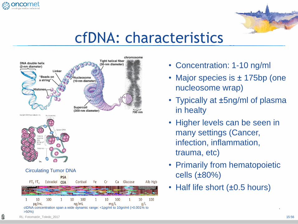
cfDNA: characteristics
• Concentration: 1-10 ng/ml
• Major species is ± 175bp (one
nucleosome wrap)
• Typically at ±5ng/ml of plasma
in healty
• Higher levels can be seen in
many settings (Cancer,
infection, inflammation,
trauma, etc)
• Primarily from hematopoietic
cells (±80%)
• Half life short (±0.5 hours)
Circulating Tumor DNA
ctDNA concentration span a wide dynamic range: <1pg/ml to 10gn/ml (>0.001% to
>50%)
RL: Fotomatón_Toledo_2017
.
15:56
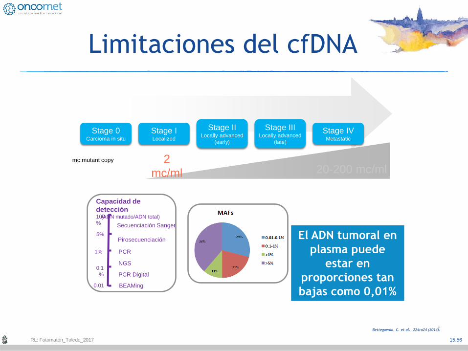
Limitaciones del cfDNA
Bettegowda, C. et al., 224ra24 (2014).
Stage 0 Carcioma in situ
Stage I Localized
Stage II Locally advanced
(early)
Stage III Locally advanced
(late)
Stage IV Metastatic
2
mc/ml 20-200 mc/ml mc:mutant copy
100
%
5%
1%
0.1
%
0.01
Capacidad de
detección (ADN mutado/ADN total)
Secuenciación Sanger
Pirosecuenciación
PCR
PCR Digital
NGS
BEAMing
El ADN tumoral en
plasma puede
estar en
proporciones tan
bajas como 0,01%
RL: Fotomatón_Toledo_2017
.
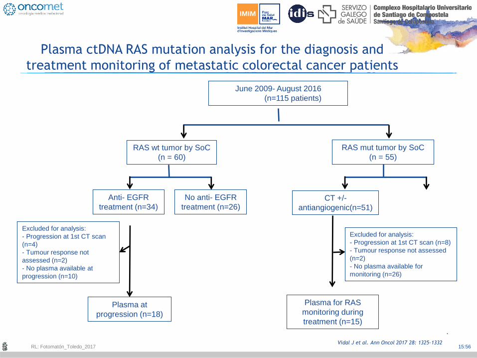
15:56 Vidal J et al. Ann Oncol 2017 28: 1325–1332
Plasma ctDNA RAS mutation analysis for the diagnosis and
treatment monitoring of metastatic colorectal cancer patients
June 2009- August 2016
(n=115 patients)
RAS wt tumor by SoC
(n = 60)
RAS mut tumor by SoC
(n = 55)
No anti- EGFR
treatment (n=26)
Anti- EGFR
treatment (n=34)
Plasma at
progression (n=18)
Excluded for analysis:
- Progression at 1st CT scan
(n=4)
- Tumour response not
assessed (n=2)
- No plasma available at
progression (n=10)
CT +/-
antiangiogenic(n=51)
Plasma for RAS
monitoring during
treatment (n=15)
Excluded for analysis:
- Progression at 1st CT scan (n=8)
- Tumour response not assessed
(n=2)
- No plasma available for
monitoring (n=26)
RL: Fotomatón_Toledo_2017
.
15:56
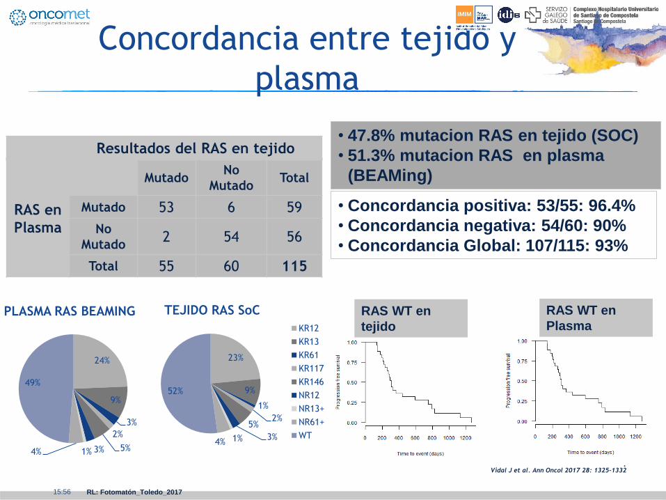
Vidal J et al. Ann Oncol 2017 28: 1325–1332
RL: Fotomatón_Toledo_2017
Concordancia entre tejido y
plasma
• 47.8% mutacion RAS en tejido (SOC)
• 51.3% mutacion RAS en plasma
(BEAMing)
Resultados del RAS en tejido
RAS en
Plasma
Mutado No
Mutado Total
Mutado 53 6 59
No
Mutado 2 54 56
Total 55 60 115
24%
9%
3%
2%
5% 3% 1% 4%
49%
PLASMA RAS BEAMING
23%
9%
1%
2% 5%
3% 1% 4%
52%
TEJIDO RAS SoC
KR12
KR13
KR61
KR117
KR146
NR12
NR13+
NR61+
WT
• Concordancia positiva: 53/55: 96.4%
• Concordancia negativa: 54/60: 90%
• Concordancia Global: 107/115: 93%
RAS WT en
tejido
RAS WT en
Plasma

.
15:56
Vidal J et al. Ann Oncol 2017 28: 1325–1332
RL: Fotomatón_Toledo_2017
Valor pronóstico de los niveles
basales de MAF
p=0.044
p=0.038
MAFs
levels
n Median OS
MAF <1% 8 43,6 (28,8-58,4)
MAF ≥1% 14 19,7 (6,8-32,5)
n Median PFS
MAF <1% 8 17,6 (7,5-27,6)
MAF ≥1% 14 7,2 (4,5-9,9)
.
15:56
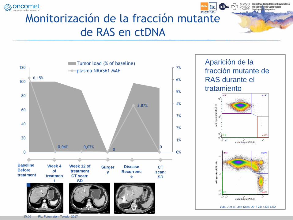
Vidal J et al. Ann Oncol 2017 28: 1325–1332
RL: Fotomatón_Toledo_2017
Monitorización de la fracción mutante
de RAS en ctDNA
6,15%
0,04% 0,07% 0
3,87%
0
0%
1%
2%
3%
4%
5%
6%
7%
0
20
40
60
80
100
120Tumor load (% of baseline)
plasma NRAS61 MAF
Baseline
Before
treatment
Week 4
of
treatmen
t
Week 12 of
treatment
CT scan:
SD
Surger
y
Disease
Recurrenc
e
CT
scan:
SD
Aparición de la
fracción mutante de
RAS durante el
tratamiento

.
15:56
Retratamiento con Gefitinib cuando
pierde la T790M: a propósito de un caso
Chic N. Reguart N, JTO 2017
RL: Fotomatón_Toledo_2017
Months of treatment
Biopsy 3
Biopsy 2
Biopsy 1
EGFR Exon 19 Del+ EGFR Exon 20 T790M-
EGFR Exon 19 Del+ EGFR Exon 20 T790M+
EGFR Exon 19 Del+ EGFR Exon 20 C797S+ EGFR Exon 20 T790M-
Afatinib 250 mg/QD
Osimertinib 80 mg/QD
Gefitinib 250 mg/QD
Pre-treatment Biopsy Genotyping Treatment Response
1B
1C
1A
Afatinib Osimertinib Gefitinib
0 43 45 47 49 50
Courtesy N. Reguart

.
15:56
Monitoring clonal evolution
using liquid biopsies
Siravegna G et al. Nat Rev Clin Oncol. 2017;14:531-548
RL: Fotomatón_Toledo_2017

.
15:56
Monitoring CAD-ALK rearrangement
Siravegna G Ann Oncol 2017;28: 1302–1308
RL: Fotomatón_Toledo_2017
.
15:56
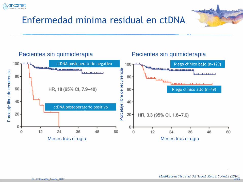
Enfermedad mínima residual en ctDNA
Pacientes sin quimioterapia Pacientes sin quimioterapia
Po
rceta
je lib
re d
e r
ecu
rre
ncia
Meses tras cirugía
Po
rceta
je lib
re d
e r
ecu
rre
ncia
Meses tras cirugía
ctDNA postoperatorio negativo
(n=164)
ctDNA postoperatorio positivo
(n=14)
Riego clínico alto (n=49)
Riego clínico bajo (n=129)
RL: Fotomatón_Toledo_2017

.
15:56
cfDNA and ctDNA in healthy
individuals and patients with cancer
Phallen et al., Sci. Transl. Med. 9, eaan2415 (2017)
RL: Fotomatón_Toledo_2017
.
15:56
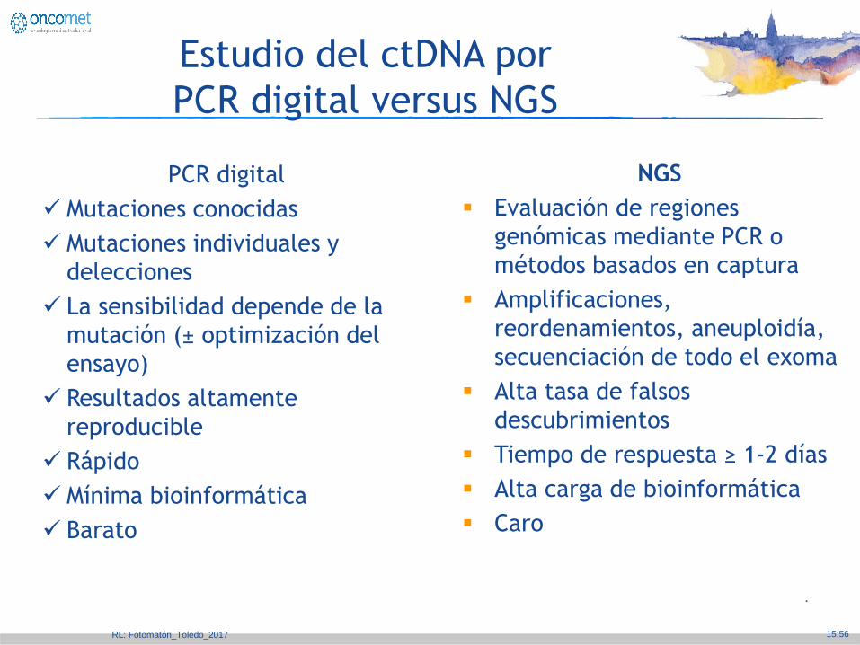
Estudio del ctDNA por
PCR digital versus NGS
RL: Fotomatón_Toledo_2017
PCR digital
Mutaciones conocidas
Mutaciones individuales y
delecciones
La sensibilidad depende de la
mutación (± optimización del
ensayo)
Resultados altamente
reproducible
Rápido
Mínima bioinformática
Barato
NGS
Evaluación de regiones
genómicas mediante PCR o
métodos basados en captura
Amplificaciones,
reordenamientos, aneuploidía,
secuenciación de todo el exoma
Alta tasa de falsos
descubrimientos
Tiempo de respuesta ≥ 1-2 días
Alta carga de bioinformática
Caro

.
15:56
Paneles Comerciales
RL: Fotomatón_Toledo_2017
.
15:56
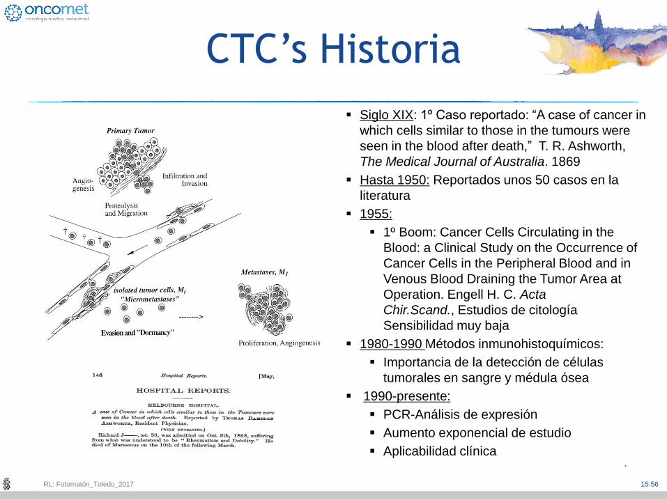
Siglo XIX: 1º Caso reportado: “A case of cancer in
which cells similar to those in the tumours were
seen in the blood after death,” T. R. Ashworth,
The Medical Journal of Australia. 1869
Hasta 1950: Reportados unos 50 casos en la
literatura
1955:
1º Boom: Cancer Cells Circulating in the
Blood: a Clinical Study on the Occurrence of
Cancer Cells in the Peripheral Blood and in
Venous Blood Draining the Tumor Area at
Operation. Engell H. C. Acta
Chir.Scand., Estudios de citología
Sensibilidad muy baja
1980-1990 Métodos inmunohistoquímicos:
Importancia de la detección de células
tumorales en sangre y médula ósea
1990-presente:
PCR-Análisis de expresión
Aumento exponencial de estudio
Aplicabilidad clínica
CTC’s Historia
RL: Fotomatón_Toledo_2017

.
vs
Monitorerización de la respuesta
al tratamiento
CTC
1st CT
15:5
6
RL: F
oto
mató
n_Tole
do_2017

.
vs
Los marcadores de CTC pueden predecir la respuesta del tratamiento con mayor precisión y antes que la
TAC
Monitorerización de la respuesta
al tratamiento
CTC
1st CT
15:5
6
RL: F
oto
mató
n_Tole
do_2017

.
Enriched CTCs fraction
4 months 5 months
#MCHUS20 TNBC Cellsearch: 1938 CTCs
24,6 mm
20,6 mm
Ki67
Molecular
characterization
2 months
15 mm
10 mm
Las CTCs de una paciente metastásico con cáncer de mama TN son tumorigénicas
Unidad Mixta Roche-Chus Clotilde Costa Thais Pereira
15:5
6
RL: F
oto
mató
n_Tole
do_2017

.
15:56
RNA-seq ANALYSIS: vs control
FAM46B
COL7A1
PDLIM4
ACTG2
KRT5
PI3K3A1
SATB1-
As1
P <0,05
Inflammation
Integrin
signalling
RL: Fotomatón_Toledo_2017

.
15:56
¿Qué técnica (s) utilizaremos en el futuro?
Según la situación clínica, disponibilidad, financiación y sobre todo
….
RL: Fotomatón_Toledo_2017
Diagnóstico Tratamiento
Conocimiento
Habilidades
Competencias
.
15:56
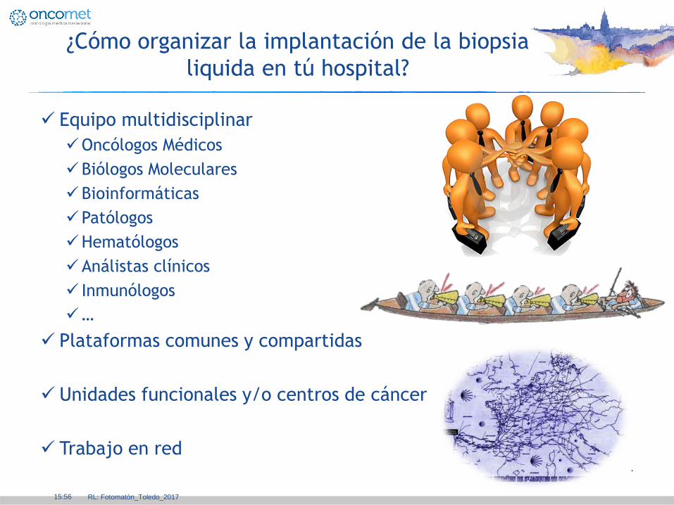
¿Cómo organizar la implantación de la biopsia
liquida en tú hospital?
Equipo multidisciplinar
Oncólogos Médicos
Biólogos Moleculares
Bioinformáticas
Patólogos
Hematólogos
Análistas clínicos
Inmunólogos
…
Plataformas comunes y compartidas
Unidades funcionales y/o centros de cáncer
Trabajo en red
RL: Fotomatón_Toledo_2017

.
15:56
Conclusiones
El desarrollo pleno de la biopsia líquida contribuirá
a aplicar una oncología de precisión
La biopsia líquida emerge como técnica
fundamental para monitorizar y tratar al paciente
con cáncer
La biopsia líquida incrementará el conocimiento
biológico y el desarrollo tecnológico en cáncer
La biopsia líquida producirá un cambio en el
paradigma del manejo del cáncer por el Oncólogo
Médico
RL: Fotomatón_Toledo_2017