FACULTAD DE CIENCIAS DE LA SALUD ESCUELA DE...
59
Asociación entre síndrome metabólico y enfermedad nodular tiroidea en el Hospital Nacional Edgardo Rebagliati Martins en el año 2014 Item Type info:eu-repo/semantics/bachelorThesis Authors Cornejo Champin, Raisa Amelia; Silva Caso, Wilmer Gianfranco; Soria Montoya, Andrea Citation 1. Cornejo Champin RA, Silva Caso WG, Soria Montoya A. Asociación entre síndrome metabólico y enfermedad nodular tiroidea en el Hospital Nacional Edgardo Rebagliati Martins en el año 2014 [Internet]. [Lima, Perú]: Universidad Peruana de Ciencias Aplicadas (UPC); 2015 [cited 2017 Nov 18]. Available from: http://repositorioacademico.upc.edu.pe/upc/ handle/10757/621623 Publisher Universidad Peruana de Ciencias Aplicadas (UPC) Rights info:eu-repo/semantics/openAccess Download date 01/07/2018 10:17:18 Item License http://creativecommons.org/licenses/by-nc-nd/4.0/ Link to Item http://hdl.handle.net/10757/621623
Transcript of FACULTAD DE CIENCIAS DE LA SALUD ESCUELA DE...

Asociación entre síndrome metabólico yenfermedad nodular tiroidea en el Hospital
Nacional Edgardo Rebagliati Martins en el año 2014
Item Type info:eu-repo/semantics/bachelorThesis
Authors Cornejo Champin, Raisa Amelia; Silva Caso, Wilmer Gianfranco;Soria Montoya, Andrea
Citation 1. Cornejo Champin RA, Silva Caso WG, Soria Montoya A.Asociación entre síndrome metabólico y enfermedad nodulartiroidea en el Hospital Nacional Edgardo Rebagliati Martinsen el año 2014 [Internet]. [Lima, Perú]: Universidad Peruanade Ciencias Aplicadas (UPC); 2015 [cited 2017 Nov 18].Available from: http://repositorioacademico.upc.edu.pe/upc/handle/10757/621623
Publisher Universidad Peruana de Ciencias Aplicadas (UPC)
Rights info:eu-repo/semantics/openAccess
Download date 01/07/2018 10:17:18
Item License http://creativecommons.org/licenses/by-nc-nd/4.0/
Link to Item http://hdl.handle.net/10757/621623

1
FACULTAD DE CIENCIAS DE LA SALUD
ESCUELA DE MEDICINA
Asociación entre síndrome metabólico y enfermedad nodular tiroidea en el
Hospital Nacional Edgardo Rebagliati Martins en el año 2014.
INVESTIGACIÓN ORIGINAL
Para optar por el título profesional de:
MÉDICO CIRUJANO
AUTORES:
Raisa Amelia Cornejo Champin
Wilmer Gianfranco Silva Caso
Andrea Soria Montoya
ASESOR DE TESIS:
Alejandro Piscoya Rivera
Calificación: Notable
Lunes 02 de Febrero del 2015
Lima, Perú

2
Dedicatoria:
A nuestras familias,
por su apoyo incondicional

3
Agradecimientos
A nuestros maestros,
quienes nos guiaron
y en especial al Dr. Antonio Bernabé Ortiz
y a la Dra Susana Tara Britto,
por su colaboración en este proyecto.

4
Tabla de contenidos
I. Portada…………………………………………………………………………1
II. Dedicatoria……………………………………………………………………..2
III. Agradecimientos………………………………………………………………3
IV. Índice……………………………………………………………………..……4
V. Filiación……………………………………………………………………..…5
VI. Artículo científico (Versión en español)
1. Resumen/
Abstract……………………………………………………….……….6
2. Introducción……..………………………………………....….……10
3. Métodos………………………………………………………..……12
4. Resultados……………………………………………..……...……18
5. Discusión…………………………………….…………...…………20
6. Conclusiones……………………………...…………...……...……23
7. Referencias
bibliográficas……………………………………….……….....……..24
8. Figura……………………………………………………………….27
9. Tablas…………………………………………….………..………...28
VII. Artículo científico (Versión en inglés)……………………………………33
VIII. Revista donde se envió el
articulo…………………………..………………….……..………………..56
IX. Proceso de
revisión……………………………………….…………………..………….57

5
X. Estado de la
publicación………………………………………………………….……….58

6
III.Filiación
Asociación entre síndrome metabólico y enfermedad nodular tiroidea en
el Hospital Nacional Edgardo Rebagliati Martins en el año 2014.
Raisa C. Champin1, Wilmer Silva Caso1, Andrea Soria-Montoya1, A
Piscoya1,2
1. Escuela de Medicina, Universidad Peruana de Ciencias Aplicadas,
Lima, Perú.
2. Servicio de gastroenterología, Hospital Guillermo Kaelin de la Fuente,
Essalud, Lima, Perú
Correspondencia
Raisa Cornejo Champin
Dirección postal: Jr Domingo Ponte 1183 Interior B, Magdalena del Mar, Lima-
Perú.
E-mail: [email protected]
Teléfono: 973823970
Wilmer Silva Caso
Dirección postal: Calle Doña Delmira 365, Santiago de Surco, Lima-Peru .
E-mail: [email protected]
Andrea Soria Montoya
Dirección postal: Calle 28 231 Urbanización Corpac, San Isidro, Lima-Perú.
E-mail: [email protected]

7
IV.Artículo científico
Asociación entre síndrome metabólico y enfermedad nodular tiroidea en
el Hospital Nacional Edgardo Rebagliati Martins en el año 2014.
RESUMEN
Introducción: Pocos son los estudios que analizan la relación entre el
síndrome metabólico y la enfermedad nodular tiroidea, tema en el que existe
un vacío de conocimiento. El objetivo de este estudio es determinar la
asociación entre síndrome metabólico y enfermedad nodular tiroidea en un
hospital de Lima, Perú. Materiales y métodos: Estudio longitudinal,
prospectivo, analítico, observacional de casos y controles, realizado en el
Hospital Nacional Edgardo Rebagliati Martins en Lima - Perú. Un total de 182
pacientes se separaron como casos a los pacientes en los que se encontrara
por lo menos un nódulo tiroideo detectado por ultrasonografía mayor a 3 mm
(n=91) y como controles a los pacientes en los cuales se excluyera la
presencia del nódulo de las características descritas por la misma técnica
diagnostica (n=91). Se evaluaron el nivel y la fuerza de asociación entre la
presencia de síndrome metabólico y cada uno de sus componentes por
separado con la presencia de enfermedad nodular tiroidea. Resultados: El
análisis bivariado muestra asociación significativa entre la presencia de nódulo
tiroideo y síndrome metabólico con un OR de 2.56 (IC: 95% 1.41 a 4.66, p <
0.05). Se evidenció que los niveles de HDL bajo y la glicemia basal alterada
se encuentran asociadas significativamente con la presencia de nódulo
tiroideo, independientemente de la presencia de síndrome metabólico con OR
de 2.81 ( IC: 95% 1.54 a 5.12, p<0.05) y 2.05 (IC:95% 1.10 a 3.78, p<0.05)
respectivamente. El análisis multivariado mantuvo la asociación entre nódulo
tiroideo y el síndrome metabólico con un OR de 2.96 (IC: 95% 1,47 a 5,95 ,
p<0.05), así mismo con niveles de HDL bajo con un OR de 2.77 (IC:95 % 1,44
a 5,3, p<0.05) y con la glicemia basal alterada con un OR de 2,23 (IC:95%

8
1,14 a 4,34, p<0,05). Conclusiones: El Síndrome metabólico incrementa el
riesgo de padecer enfermedad nodular tiroidea, específicamente la
disminución de valores de HDL y la glicemia basal alterada fueron los factores
en los que halló mayor asociación.
Palabras clave: Nódulo Tiroideo, Síndrome X Metabólico, Resistencia a la
Insulina. (Fuente: DECS BIREME)

9
Association between metabolic syndrome and thyroid nodular disease in
the National Hospital Edgardo Rebagliati Martins in 2014
ABSTRACT
Introduction: Few studies analyses the relation between metabolic syndrome
and thyroid nodular disease, subject in which there is a knowledge gap. The
object of this study is to determinate the association between metabolic
syndrome and thyroid nodular disease in a hospital in Lima, Peru. Materials
and methods: A longitudinal, prospective, analytic, observational, case - control
study, was performed “Hospital Nacional Edgardo Rebagliati Martins” in Lima-
Peru. A total of 182 patients were separated as cases in which at least find a
thyroid nodule detected by ultrasonography greater than 3 mm ( n = 91) and
controls as patients in whom the presence of the node with the characteristics
described was excluded by the same technique (n=91). The level and strength
of association was evaluated between the presence of metabolic syndrome and
each of its components by itself with the presence of thyroid nodular was
evaluated. Results: Bivariate analysis shows significant association between
the presence of thyroid nodule and metabolic syndrome with an OR of 2.56
(IC:95% 1.41 to 4.66, p < 0.05). Low levels of HDL and impaired fasting
glucose are significant associated with the presence of thyroid nodule,
independent of the presence of metabolic syndrome, with an OR of 2.81
(IC:95% 1.54 to 5.12, p<0.05) and 2.05 (IC: 95% 1.10 to 3.78, p<0.05)
respectively. The multivariate analysis maintained the association between
thyroid nodule and metabolic syndrome with an OR of 2.96 (IC: 95% 1,47 to
5,95 , p<0.05); like was the low levels of HDL with an OR of 2.77 ( IC: 95% 1,44
to 5,3, p<0.05) and with impaired fasting glucose with an OR of 2,23 ( IC 95%
1,14 to 4,34, p<0,05).Conclusions: Metabolic syndrome increases de risk of
having thyroid nodule disease. Low HDL levels and impaired fasting glucose
were the factors with more association.

10
Key words: Thyroid Nodule, Metabolic Syndrome X, Insulin Resistance.
(MeSH)
11
INTRODUCCIÓN
El síndrome metabólico es una agrupación de alteraciones metabólicas y
obesidad abdominal, asociado a un incremento en el riesgo de padecer
diabetes, enfermedad cardiovascular y mortalidad prematura (1,2). Los
factores involucrados, definidos por Alberti et all.(3), son la obesidad
abdominal, la hipertensión arterial, los niveles elevados de triglicéridos séricos,
los bajos niveles de lipoproteínas de alta densidad (HDL) y la hiperglicemia en
ayunas (1,3,4). Cada uno de estos componentes constituye un criterio para su
diagnóstico (3,4,5).Se describe una elevada prevalencia mundial de este
síndrome, estimándose que entre el 20 y 25% de la población adulta lo
padece(1). Sin embargo, la prevalencia varía según el país de procedencia y
los criterios diagnósticos que se usen para su detección (6). En la población
peruana, la prevalencia es de 32.8% según los criterios establecidos por la
Federación Internacional de Diabetes (IFD). (7)
El síndrome metabólico está fuertemente asociado con la resistencia a la
insulina. Desde hace dos décadas se ha investigado y descrito que la
resistencia a la insulina podría constituir el factor central para su desarrollo de
este síndrome(5,8). Es importante señalar que ante la presencia de tejidos
resistentes a la acción de la insulina, en asociación al síndrome metabólico, el
páncreas endocrino aumenta la producción de dicha hormona, generando un
estado de hiperinsulinemia en el organismo. La insulina posee la facultad de
actuar como factor de proliferación de células tiroideas, hecho que ha sido
demostrado previamente en cultivos celulares(9), lo cual conllevaría a un
incremento del volumen de la glándula tiroides y, por lo tanto, a la enfermedad
nodular tiroidea.
Se sabe que los receptores de los factores de crecimiento insulínicos 1 y 2
(IGF-1, IGF-2) se encuentran altamente expresados en líneas celulares
tiroideas cancerígenas, los cuales son activados de manera
autocrina/paracrina por los IGF producidos localmente, actuando como
importantes factores mitogénicos y anti-apoptóticos (9,10,11).

12
Lo expuesto anteriormente indica que el desarrollo del síndrome metabólico
podría traer como consecuencia el desarrollo de nódulos tiroideos, los cuales
se presentan en un amplio espectro clínico que incluye a los nódulos pequeños
asintomáticos, en los cuales la mayor preocupación es la exclusión de
neoplasia maligna, hasta los nódulos grandes con porciones intratorácicas que
causan síntomas compresivos.(12)La prevalencia de la enfermedad nodular
tiroidea en los Estados Unidos es de 7% si estos son detectados por palpación
bimanual de la glándula tiroides y asciende al 50% si son detectados por
ultrasonografía(12). Debido a la diferencia entre ambas técnicas, es de gran
importancia la detección temprana de estos nódulos, la cual podría ser
favorecida con el screening ecográfico de los pacientes con factores de riesgo
para su desarrollo, ya que el 5% de los nódulos tiroideos detectados son
carcinomas malignos. (12)
Pocos son los estudios que analizan la relación entre dicho síndrome y la
enfermedad nodular tiroidea (2); sin embargo en ellos se ha logrado determinar
asociación entre ambas entidades.
Debido a que el síndrome metabólico es una entidad de fácil diagnóstico con
criterios establecidos para su identificación (3), la presencia de asociación
entre este síndrome y la enfermedad nodular tiroidea generaría una sospecha
clínica de la presencia de esta última al haber sido detectado dicho síndrome
en la consulta diaria del profesional de la salud.
13
MATERIALES Y MÉTODOS
DISEÑO DE ESTUDIO Y DEFINICIÓN DE CASOS Y CONTROLES
Estudio longitudinal, prospectivo, analítico, observacional de casos y controles,
realizado en el Hospital Nacional Edgardo Rebagliati Martins, establecimiento
de referencia nacional nivel IV de atención en la red asistencial Essalud (13)
en Lima-Perú, durante el año 2014. Todos los pacientes evaluados fueron
pobladores de zonas con adecuados niveles de iodo (más de 100µg/l de Iodo
urinario). Se definió como caso a todos los pacientes eutiroideos (definidos con
valores de TSH de 0.4 a 4 mg/dl y de T4 libre de 0.9 a 1.8 mg/dl) mayores de
18 años, los cuales contaban con una ecografía tiroidea que mostraba la
presencia de por lo menos un nódulo tiroideo mayor de 3 milímetros. Se tomó
como controles a todos aquellos pacientes eutiroideos mayores de 18 años de
edad, en los que se hubiera descartado la presencia de nódulo tiroideo por
ultrasonografía. Se excluyeron a los pacientes que presentaran alguna
condición que pudiera causar secundariamente la aparición de nódulos
tiroideos o la medición errónea de alguno de los criterios diagnósticos del
síndrome metabólico, tales como el diagnóstico de cáncer metastásico de la
glándula tiroides, antecedente de enfermedad tiroidea no nodular, cirugía
bariátrica previa, ascitis, pacientes postrados, pacientes embarazadas o en
lactancia, pacientes que presentaron los criterios de síndrome metabólico con
una diferencia de tres meses o más en su registro y falta de al menos uno de
los siguientes datos en la historia clínica: triglicéridos, colesterol HDL y glucosa
en ayunas.
TAMAÑO MUESTRAL
Para el cálculo del tamaño de muestra se usó EPIDAT 4.0. Asumiendo un nivel
de confianza del 95%, un poder del 80% y una proporción de presencia de
síndrome metabólico en la población que presenta nódulo tiroideo de
78.6%,(2) con lo cual se requeriría 91 casos y 91 controles para encontrar un
OR de 2.5 o más. (2) Debido a los criterios de inclusión y exclusión planteados

14
se evaluaron 475 pacientes para poder alcanzar el tamaño muestral propuesto
(Figura 1).
MEDICIÓN DEL DESENLACE
Para la búsqueda de la presencia de nódulos tiroideos en ambos grupos de
pacientes se utilizó un transductor lineal de 7.5 MHz., dicha búsqueda fue
realizada por dos expertos en el campo de la radiología diagnostica. Para
definir la presencia de nódulo se consideró como tal a todas las lesiones o
aumento focal de volumen o consistencia, localizado dentro de la tiroides y que
se distingue del resto del parénquima con un diámetro mayor de 3 mm. (14)Se
procedió a clasificar los mismos mediante la escala de TIRADS modificada por
Russ (15), la cual agrupa a los nódulos tiroideos en seis tipos (TIRADS I, II, III,
IV, V y VI) según sus características ultrasonográficas.
DEFINICIÓN DE VARIABLES
Se buscó como variable de asociación la presencia de síndrome metabólico
definido por los criterios diagnósticos planteados por Alberti y col (3) (Tabla 1).
Se utilizó esta definición debido a que estos criterios son producto de un
consenso entre diversas organizaciones como la Federación Internacional de
Diabetes, la Asociación Americana del Corazón, la Asociación Internacional de
Estudio de la Obesidad, entre otras; son empleados en la mayoría de estudios
realizados para demostrar la asociación planteada en el presente estudio y
consideran variaciones étnicas para la obesidad abdominal. Para obtener
dichos datos se realizaron las medidas respectivas y se revisaron las historias
clínicas de los casos y los controles de donde se obtuvieron los datos
laboratoriales como niveles de triglicéridos, colesterol HDL y glucosa sérica,
los cuales no tuvieron un tiempo mayor de tres meses de diferencia con
respecto a la medición del outcome; y datos clínicos como uso de tratamiento
antihipertensivo, antidiabético o con agentes hipolipemiantes. Además se
obtuvieron datos demográficos como sexo, edad y lugar de procedencia.

15
MEDICIÓN DE CO-VARIABLES
Para medir la presión arterial se utilizó un esfigmomanómetro de mercurio
calibrado. Se realización la medición con el paciente sentado (ambos pies en
contacto con el suelo) y en reposo por un tiempo mínimo de cinco minutos.
Ambos brazos estuvieron por encima del nivel del corazón y se tomóla presión
en el brazo derecho, con ayuda de un estetoscopio. Se infló el manguito a 30
mmHg más después de dejar de escuchar las palpitaciones y se desinfló a una
velocidad aproximada de 2 mmHg por segundo. Se consideró el valor de
presión arterial sistólica al nivel que se escuchó el primer ruido de Korotkoff
y en el nivel en el que desapareció se registró la presión arterial diastólica. El
paciente no tomó bebidas con cafeína, realizó ejercicio ni fumó en un lapso de
30 minutos antes de la toma de presión arterial. Se consideró aumento de la
presión arterial si la presión arterial sistólica fue ≥140 y / o la presión arterial
diastólica ≥90 mm Hg, o auto-reporte de diagnóstico médico y uso de
medicación antihipertensiva (16,17).
El perímetro abdominal fue medido siguiendo las indicaciones de la
Organización mundial de la Salud (OMS), con una cinta métrica no elástica, a
la altura del punto medio entre la última costilla palpable y la cresta iliaca. El
paciente fue ubicado en bipedestación, con los pies juntos y los brazos sueltos
a los lados. La medición se hizo paralela al suelo, sin comprimir estructuras
abdominales y al final de la espiración (18).Se consideró la obesidad
abdominal si el perímetro abdominal fue ≥90 cm (hombres) o ≥80 cm (mujeres)
(1,3)
La medición de los criterios laboratoriales se realizaron en los laboratorios del
hospital, para la medición de los niveles séricos de glucosa se utilizó el kit
comercial de laboratorio Wiener (Glicemia enzimática AA liquida), para la
medición de niveles séricos de HDL y triglicérido se usó el kit comercial del
mismo laboratorio (Colesterol enzimático AA liquida). El aumento en los niveles
de triglicéridos séricos fue considerado cuando se encontró niveles ≥150 mg /
dl o si se encontraba actualmente en tratamiento farmacológico para la
elevación de los triglicéridos; (3) los niveles bajos de HDL-colesterol fueron
16
considerados como criterio diagnóstico cuando se encontró valores <40 mg / dl
en varones y <50 mg / dl en mujeres o si hubo uso de terapia con
medicamentos para el colesterol HDL bajo. Por último, se consideró el criterio
de glicemia basal alterada cuando se encontró glucosa en ayunas ≥100 mg /
dL o uso actual de medicamentos antidiabéticos (agentes orales o insulina).
Se utilizó la metodología estandarizada de Lohmann y col., citada en un
informe de expertos de la OMS, para medir la talla y el peso. Para la medición
de la talla se utilizó un tallímetro de madera, validado, perpendicular al suelo,
dividido en centímetros, con una cabecera deslizable paralela al suelo. El
paciente se colocó con los brazos sueltos a los lados y los pies descalzos y
juntos y con los talones en contacto con la tabla, así como nalgas, espalda y
cabeza. En el momento de la medición el paciente debió mirar a un punto fijo,
y posicionar la cabeza procurando que la línea de visión forme un ángulo de 90
grados con el cuerpo. Estando el paciente en posición correcta se deslizó la
cabecera hasta presionar el cabello del paciente contra su cabeza(19). Para
registrar el peso se utilizó una balanza digital calibrada. El paciente de pie,
descalzo al centro de la plataforma, con el peso distribuido equitativamente en
ambos pies, sin utilizar ningún apoyo y con los brazos sueltos a ambos lados.
Se realizaron las aproximaciones correspondientes según la ropa que el
paciente utilizó. (19)
El Índice de Masa Corporal (IMC) se calculó como la división del peso de la
persona entre la talla elevada al cuadrado. Según la OMS, un IMC de 18,5 a
24,9 kg/m2 define normopeso, uno de 25 a 29,9 kg/m2 define sobrepeso y
mayor o igual a 30 obesidad. (19)
ANÁLISIS ESTADÍSTICO
Se elaboró una base de datos con la información obtenida, debidamente
codificada en el programa Microsoft EXCEL con doble digitación y posterior
17
control de calidad. Posteriormente esta base de datos se transfirió al programa
STATA versión 11.2 para el análisis estadístico.
Se utilizó medias y desviaciones estándar para la descripción de variables
numéricas (edad, talla, peso, perímetro abdominal, niveles de TSH, niveles de
T4, niveles de HDL, niveles de triglicéridos, presión sistólica y diastólica) y para
las variables categóricas se utilizaron frecuencias absolutas y relativas
(género, IMC y antecedentes de diabetes, hipertensión arterial, dislipidemia de
triglicéridos y de HDL).
Se utilizó la prueba de Shapiro Wilk para comprobar la normalidad de las
variables numéricas. Para comparar las variables numéricas entre los dos
grupos, en caso tuvieran una distribución normal, se utilizó la prueba de T
Student. En caso estas no tuvieran una distribución normal se utilizó la prueba
de Suma de rangos de Wilcoxon. Para la comparación de las características
de la población entre los casos y controles se utilizó Chi cuadrado para las
variables categóricas. Finalmente se utilizó la regresión logística para calcular
los Odds ratio(OR) e intervalos de confianza al 95% ajustando a potenciales
confusores como edad, sexo y estado nutricional según el índice de masa
corporal (IMC).
ASPECTOS ÉTICOS
El presente estudio fue evaluado y aprobado por los comités de ética de la
Universidad Peruana de Ciencias Aplicadas (UPC) y del Hospital Nacional
Edgardo Rebagliati Martins.
Se aplicó un consentimiento informado a todos los pacientes que participaron
en el estudio antes del inicio de cualquier actividad de la investigación, en el
cual se brindaba información clara sobre lo que se realizaría y se garantizaba
la absoluta confidencialidad.
No se colectaron identificadores personales en la base de datos para
garantizar el anonimato y confidencialidad de los datos. Se colectó el número

18
de historia clínica y nombre del participante solo para fines de obtención de
información de la misma, pero estos no fueron digitados en la base de datos.

19
RESULTADOS
CARACTERÍSTICAS DE LA POBLACIÓN
El estudio incluyó a un total de 182 pacientes, de los cuales 91 presentaron al
menos un nódulo tiroideo y 91 fueron el grupo de control (tabla 2). Del grupo
de los pacientes con nódulo tiroideo el 91,2% (n=83) fueron de sexo femenino,
mientras que en el grupo control se encontraron 82,4%. La mediana de la edad
en el grupo control fue de 51 (Rango intercuartílico o RI: 27) y en el grupo de
casos fue de 50 (RI: 21). El estado nutricional de mayor prevalencia en ambos
grupos fue de sobrepeso, siendo este el 40% de los controles y el 34% de los
casos, seguido de bajo peso, obesidad y normo peso para ambos grupos. En
el grupo de casos la mediana del tamaño de nódulos tiroideos fue de 12,6 (RI:
11,9).
Se encontró diferencia significativa entre los valores de HDL en mujeres en el
grupo de casos con respecto al grupo de control (p<0.05). No se hallaron
variaciones significativas para el IMC, talla, peso, perímetro abdominal,
glucosa, triglicéridos presión sistólica y diastólica para ambos grupos.
CARACTERIZACIÓN DEL SÍNDROME METABÓLICO EN LA POBLACIÓN
La presencia de síndrome metabólico se presentó en el 59,34% (n=54) de los
pacientes con nódulo tiroideo, mientras que en el grupo de los controles dicha
entidad se encontró en el 36,26% (n=33) de los pacientes (p<0.005) (tabla 3).
Se encontró que los cinco componentes de síndrome metabólico se hallan
presentes con mayor frecuencia en el grupo de pacientes con nódulo tiroideo
siendo asíla obesidad abdominal (76,92%) el componente más frecuente en
dicho grupo, seguido el nivel de HDL bajo(63.74%),presión arterial elevada
(58.24%), nivel de triglicéridos elevados (48.35%) y glicemia basal aumentada
(45.05%); mientras que en el grupo control se encontró que el factor más
frecuente fue obesidad abdominal (76,92%) seguido por presión arterial
elevada (43.96%), nivel de triglicéridos elevados (42.86%), niveles bajos de
HDL (38.46%) y glicemia basal alterada (28.57%). Sin embargo, solo se halló

20
diferencia significativa en los valores de HDL bajos y de glicemia basal entre
ambos grupos.
ANÁLISIS BIVARIADO Y MULTIVARIADO
En el análisis bivariado se evidencia asociación significativa entre la presencia
de nódulo tiroideo y síndrome metabólico con un OR de 2,56(IC:95% 1.41 a
4,66, p < 0.05. Además se evidenció que, específicamente, los niveles de HDL
bajo y la glicemia basal alterada se encuentran asociadas significativamente
con la presencia de nódulo tiroideo con un OR de 2.81 (IC: 95% 1.54 a 5.12,
p<0.05) y 2.05 (IC: 95% 1.10 a 3.78, p<0.05) respectivamente.
En el análisis multivariado se mantuvo la asociación entre la presencia de
nódulo tiroideo con el síndrome metabólico con un OR de 2,96 (IC: 95% 1,47
a 5,95 , p<0.05), con niveles de HDL bajo con un OR de 2.77 ( IC: 95% 1,44 a
5,3, p<0.05) y con la glicemia basal alterada con un OR de 2,23 ( IC:95% 1,14
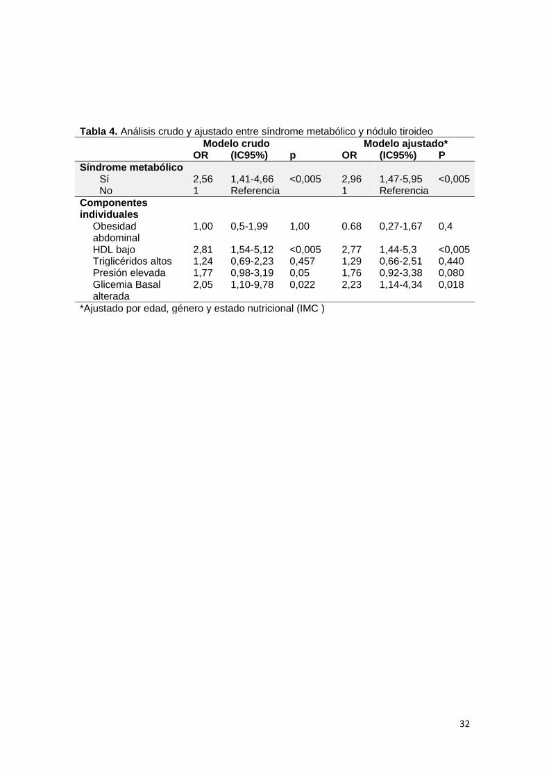
a 4,34, p<0,05). (tabla 4)
Considerando al síndrome metabólico como una variable dicotómica se evaluó
la asociación con el número de nódulos tiroideos presentes en el paciente,
como resultado no se encuentra asociación al realizar el análisis.

21
DISCUSIÓN
En el presente estudio se encontró que la frecuencia de presencia de síndrome
metabólico fue mayor en pacientes con enfermedad nodular tiroidea, siendo
aproximadamente tres veces más probable que en ausencia de esta. Además,
al analizar de manera aislada cada componente del síndrome, se halló
asociación independiente en dos criterios: la presencia de bajos niveles de
HDL séricos, encontrándose un riesgo de casi tres veces mayor de presentar
bajos niveles de HDL séricos en el grupo de pacientes con nódulos tiroideos, y
la glicemia basal alterada, en donde se demostró dos veces más probabilidad
de presentar niveles alterados de glicemia en el grupo de pacientes con
enfermedad nodular tiroidea.
En la actualidad, no son muchas las investigaciones realizadas para demostrar
la asociación planteada, entre ellas se encuentra la investigación realizada por
Ayrtuck y colaboradores (2), quienes realizaron un estudio en una zona de
deficiencia leve a moderada de iodo, de tipo caso-control, en el cual incluyeron
a 539 pacientes y tomaron como variable dependiente al síndrome metabólico
(278 pacientes en el grupo de casos y 261 en el grupo de control). En los
resultados de este se demostró que los pacientes que padecían de síndrome
metabólico presentaban un porcentaje mayor de pacientes con nódulos
tiroideos con respecto al grupo control (50,4 vs 14,6%, p<0.0001) y además de
un mayor volumen tiroideo (17,5 ±5,5 vs 12,2 ± 4,2 ml, p<0,0001). Además,
demostraron la asociación entre la formación de nódulos tiroideos y la
presencia de resistencia a la insulina obteniendo un OR de 3,2 con un intervalo
de confianza al 95% para dicha asociación. Otra investigación en la cual se
demuestra la asociación planteada en el presente trabajo de investigación es
la realizada por Yin y col
(20), quienes en el año 2014 realizan un estudio en China, de tipo cohorte, en
el cual incluyeron a un total de 1061 pacientes a quienes siguieron por tres
años. En sus resultados los investigadores describen que la prevalencia de
nódulo tiroideo fue mayor en el grupo de pacientes con síndrome metabólico
(x2=69,63, p<0,001), además, al realizar el análisis de cada componente del

22
síndrome con la presencia de nódulos tiroideos, obtienen como resultado que
la obesidad abdominal y los valores séricos de triglicéridos incrementados se
encontraban asociados al desarrollo de nódulos tiroideos con RR de 1,434 y
de 1,001 respectivamente, difiriendo con nuestros resultados en los cuales
encontramos que los componentes asociados son los valores de HDL sérico
bajos y la glicemia basal alterada. Ambas investigaciones, al igual que el
presente estudio, concluyen que existe asociación entre el síndrome
metabólico y la presencia de nódulos tiroideos, reforzando la evidencia
científica de dicha asociación.
Otras investigaciones buscan demostrar asociaciones relacionadas al tema
como la asociación entre la resistencia a la insulina y la aparición de nódulos
tiroideos, tomando en cuenta de que esta es la base fisiopatológica del
síndrome metabólico es clara la importancia de estos estudios; entre ellos se
encuentran las investigaciones realizadas por Rezzonico y col. (9), en donde
se describe que los pacientes con resistencia a la insulina presentaron mayor
porcentaje de enfermedad nodular tiroidea y mayor volumen tiroideo con
respecto a los pacientes que no presentaron la resistencia a la insulina,
concluyendo que a mayores niveles de insulina circulante se produce un
aumento de proliferación tiroidea traducida clínicamente como la formación de
nódulos tiroideos y que además dichos nódulos tiroideos son de mayor tamaño
a los que se presentan en pacientes sin resistencia a dicha hormona. Yasar y
col. (11) realizaron un estudio de tipo caso control con 146 pacientes (63
casos y 83) controles para investigar dicha asociación, obtuvieron como
resultados que el índice HOMA tomado como medida para determinar la
resistencia a la insulina, fue significativamente mayor en el grupo de pacientes
con nódulos tiroideos (15.87%) que en el grupo control (10.84%).
Otra asociación de interés demostrada en la literatura es la de resistencia a la
insulina con el desarrollo de cáncer de tiroides, investigada por Rezzonico y col
(21), quienes en el 2009 realizan una investigación con 20 pacientes con
cáncer de tiroides diferenciado (CTD) y 20 pacientes en el grupo de control,
obtuvieron como resultados que el 50% de pacientes con CTD presentaron

23
resistencia a la insulina mientras que en el grupo control solo el 10% con una
diferencia estadísticamente significativa. Además, describen que en el grupo
de pacientes con CDT la resistencia a la insulina estaba presente en el 56,3%
de pacientes con cáncer papilar de tiroides y en el 25% en el cáncer folicular
de tiroides, de estos resultados podemos inferir que la resistencia a la insulina
además de factor de riesgo para la génesis de nódulos tiroideos, podría
constituir un importante indicador de la presencia de células neoplásicas en los
nódulos tiroideos secundarios a esta.
Al realizar el análisis de cada componente del síndrome metabólico de manera
independiente con la presencia de enfermedad nodular tiroidea en el presente
estudio, se demostró que existe asociación entre la disminución de los niveles
séricos de HDL y la aparición de nódulos tiroideos. Esto puede deberse a que
la disminución de los niveles séricos de HDL es uno de los componentes
encontrado con más prevalencia en la población de mujeres peruanas que
sufre de este síndrome, presentándose del 71.2% (7) al 86.8 % (22). Además,
se ha demostrado en múltiples estudios, la resistencia a la insulina se
encuentra asociada a la disminución de niveles de HDL (1,3,4,5,23). Si bien
aún no se ha establecido una relación causal de tal fenómeno, se sabe que la
resistencia a esta hormona genera un aumento en el catabolismo de la
apolipoproteina A1 (24,25,26,27), componente principal del HDL, lo cual
conlleva su disminución. Otro componente del síndrome metabólico asociado a
la aparición de nódulos en la tiroides es la glicemia alterada en ayunas, se
constituye como uno de los factores de mayor impacto en la aparición de
nódulos tiroideos según algunos estudios (2). Como se sabe, la presencia del
síndrome metabólico establece un estado de hiperinsulinismo en el organismo
donde los niveles circulantes más altos de insulina causan aumento de la
proliferación de células tiroideas. Pimenta y cols. describen que la insulina y
las hormonas tiroideas están íntimamente involucradas en el metabolismo
celular y por lo tanto el exceso o déficit de cualquiera de estas hormonas dan
lugar a la alteración funcional de la otra. Esta registrada la interrelación
fisiológica y bioquímica entre los niveles de insulina y la influencia tanto de la
insulina y yodotironinas sobre el metabolismo de carbohidratos y proteínas.

24
Las manifestaciones clínicas son el aumento del volumen de la tiroides y la
formación de nódulos acompañado de niveles alterados de glucosa sérica. (9).
Las asociaciones demostradas en el presente estudio permiten generar mayor
conciencia sobre el riesgo de desarrollo de enfermedad nodular tiroidea en los
pacientes que padecen de síndrome metabólico y promover mayor énfasis en
el control y manejo de los componentes en los que se han demostrado mayor
asociación para evitar la aparición de dichos nódulos. Incluso existen estudios
en donde se ha evidenciado que el uso de metformina en pacientes con
nódulos tiroideos pequeños y resistencia a la insulina, reduce
significativamente el tamaño de los mismos.(28)
En nuestro estudio se encontraron las siguientes limitaciones: la
ultrasonnografía, prueba diagnóstica para detección de nódulo tiroideo, a pesar
de ser la más sensible es operador dependiente suponiendo un sesgo de
medición (por lo cual se limitó a sólo dos operadores y se utilizó una escala
validada); la población de estudio se encontraba en un ambiente hospitalario y
fue elegida de manera no aleatoria, por lo que los resultados de este estudio
pueden no ser extrapolables a la población en general.
Del presente estudio se puede concluir que la presencia de enfermedad
nodular tiroidea está asociada al riesgo incrementado de padecer síndrome
metabólico, específicamente la disminución de valores de HDL y la glicemia
basal alterada fueron los factores en los que halló mayor asociación.
Se recomienda la realización de estudios prospectivos en el futuro,
aleatorizados y con mayor población para poder analizar la relación entre el
número de nódulos y el síndrome metabólico.
25
REFERENCIAS BIBLIOGRÁFICAS
1) Alberti G, Zimmet P, Shaw J, Grundy S. The IDF consensus worldwide
definition of the metabolic syndrome. IDF 2006:4-19.
2) Ayturk S, Gursoy A, Kut A, Anil C, Nar A, Bascil N. Metabolic syndrome
and its components are associated with increased thyroid volume and
nodule prevalence in a mild-to-moderate iodine-deficient area .Eur J
Endocrinol 2009;161:599-605.
3) Alberti K, Eckel R, Grundy S, Zimmet P, Cleeman J. Harmonizing the
metabolic syndrome: a joint interim statement of the International
Diabetes Federation Task Force on Epidemiology and Prevention;
National Heart, Lung, and Blood Institute; American Heart Association;
World Heart Federation; International Atherosclerosis Society; and
International Association for the Study of Obesity. Circulation
2009;120:1640-1645.
4) Grundy S, Brewer H, Cleeman J, Smith S, Lenfant C. Definition of
Metabolic Syndrome: Report of the National Heart, Lung, and Blood
Institute/American Heart Association Conference on Scientific Issues
Related to Definition. Circulation 2004;109:433-438.
5) Grundy S. Metabolic syndrome pandemic. Arterioscler Thromb Vasc Biol
2008; 28:629-636.
6) ZimmetP, Magliano D, Matsuzawa Y, Alberti G, Shaw J. The metabolic
syndrome: A Global Public Health Problem and A New Definition. J
Atheroscler Thromb 2005; 12:295-300.
7) Bernabe-Ortiz A, Pastorius-Benziger C, Gilman R, Smeeth L. Sex
Differences in Risk Factors for cardiovascular disease: The PERU
MIGRANT Study. PLoS ONE 2012 Abril 5;7(4):1-6.
8) Haas J, Biddinger S. Dissecting the role of insulin resistance in the
metabolic syndrome. Curr Opin Lipidol. 2009;20(3):206-210.
9) Rezzonico J, Rezzonico M, Pusiol E, Pitoia F, Niepomniszcze H.
Introducing the Thyroid Gland as Another Victim of the Insulin
Resistance Syndrome. Thyroid 2008;18(4):461-464.

26
10) Frittitta L, Sciacca L, Catalfamo R, Ippolito A, Gangemi P, Pezzino V,
Filetti S, Vigneri R. Functional insulin receptors are overexpressed in
thyroid tumors: is this an early event in thyroid tumorigenesis? Cancer.
1999 ;85(2):492-8.
11) Yasar H, Ertuğrul O, Ertuğrul B, Ertuğrul D, Sahin M. Insulin Resistance
in Nodular Thyroid Disease. Endocr Res 2011;36(4):167-174.
12) Hegedüs L. The Thyroid Nodule. N Engl J Med 2004; 351:1764-71.
13) Gerencia Central de Prestaciones de Salud. Lineamientos de
Programación de Prestaciones de Salud - 2014 2013 20-12-2013.
14) Moscoso R, Hernández S, Ochoa C, Rodríguez S, Torres P. Diagnóstico
y tratamiento del nódulo tiroideo: posición de la sociedad mexicana de
nutrición y endocrinología.
15) Moifo B, Oben Takoeta E, Tambe J, Blanc F, Gonsu Fotsin J. Reliability
of Thyroid Imaging Reporting and Data System (TIRADS) Classification
in Differentiating Benign from Malignant Thyroid Nodules. Open Journal
of Radiology. 2013; 103-107.
16) Chobanian AV, Bakris GL, Black HR, Cushman WC, Green LA, et al.
(2003) Seventh report of the Joint National Committee on Prevention,
Detection, Evaluation, and Treatment of High Blood Pressure.
Hypertension 42: 1206–1252.
17) Mancia G, De Backer G, Dominiczak A, Cifkova R, Fagard R, et al.
(2007) 2007 Guidelines for the management of arterial hypertension:
The Task Force for the Management of Arterial Hypertension of the
European Society of Hypertension (ESH) and of the European Society of
Cardiology (ESC). Eur Heart J 28: 1462–1536.
18) World Health Organization (WHO): Waist circumference and waist-hip
ratio: report of a WHO expert consultation. Geneva, 8-11 December
2008. Geneva, Switzerland: WHO; 2011.
19) Bastemir M, Akin F, Alkis E &Kaptanoglu B. Obesity is associated with
increased serum TSH level, independent of thyroid function. Swiss
Medical Weekly 2007 137 431–434.
27
20) Yin J, Wang C, Shao Q, Qu D, Song Z, Shan P, et al. Relationship
between the Prevalence of Thyroid Nodules and Metabolic Syndrome in
the Iodine-Adequate Area of Hangzhou, China: A Cross-Sectional and
Cohort Study Int J Endocrinol 2014 17 Ago 2014:1-8.
21) Rezzónico J, Rezzónico M, Pusiol E, Niepomniszcze H. Increased
prevalence of insulin resistance in patients with differentiated thyroid
carcinoma. Metab Syndr Relat Disord 2009;7(4):375-380.
22) Pajuelo J, Sánchez J. El síndrome metabólico en adultos, en el Perú.
An Fac Med Lima. 2007; 68: 38-46.Organización Mundial de la Salud
(OMS). El estado físico: uso e interpretación de la antropometría. Serie
de Informes Técnicos Nº 854. Ginebra: Publicación de la OMS, 1995.
23) Mottillo S, Filion K, Genest J, Joseph L, Pilote L, Poirier P, et al. The
Metabolic Syndrome and Cardiovascular Risk. JACC 2010;56(14):1113-
1132.
24) Robins SJ, Rubins HB, Faas FH, Schaefer EJ, Elam MB, Anderson JW,
Collins D on behalf off the VA-HIT study group. Insulin resistance and
cardiovascular events with low HDL colesterol. Diabetes Care. 2003;26:
1015-17.
25) Laws A, Reaven GM. Evidence for an independent relationship between
insulin resistance and fasting plasma HDL-cholesterol, triglyceride and
insulin concentrations. J Intern Med 1992;231: 25-30.
26) Golay A, Zech L, Shi M, Jeng C, Chiou M, Reavrn GM, Chen I. Role of
insulin in regulation of high density lipoprotein metabolism. J Lipid Res.
1987; 28: 10-18.
27) Stalder M, Suenram P, Suenram A. Relationship Between plasma
insulin levels and high-density lipoprotein colesterol levels in healthy
men. Diabetologia. 1981; 21:544-48.
28) Rezzónico J, Rezzónico M, Pusiol E, Pitoia F, Niepomniszcze H.
Metformin treatment for small benign thyroid nodules in patients with
insulin resistance. MetabSyndrRealtDisord 2011; 9(1): 69-75

28
Pacientes que se realizaron ecografía
tiroidea durante el periodo de realización del estudio
n= 475
Pacientes evaluados por elegibilidad
n=334
Total de pacientes evaluados
n=182
NO EVALUADOS POR ELEGIBILIDAD
(total n=141)
Pacientes menores de 18 años n= 3
Pacientes no eutiroideos* n= 138
EXCLUIDOS (total n=152)
Paciente con diagnóstico de
cáncer metastásico de glándula
tiroides n=2
Paciente embarazada o en
lactancia n=3
Falta de al menos uno de los
siguientes datos en la historia
clínica: triglicéridos, HDL y
glucosa en ayunas** n=147
o Glucosa n=67
o HDL n=123
o Triglicéridos n=123
CASOS
N=91
CONTROLES
N=91
Figura 1: Diagrama de flujo de selección de pacientes
*Pacientes con valores de TSH y T4 libre fuera de los rangos de normalidad propuestos (TSH de 0.4 a 4
mg/dl y de T4 libre de 0.9 a 1.8 mg/dl)
** Pacientes que no contaran con los resultados de uno o más de los exámenes de laboratorio
(triglicéridos séricos, glucosa en ayunas o colesterol HDL) y/o que estos tuvieran una antigüedad mayor
de tres meses entre sí o con otro criterio del síndrome metabólico (medida de presión arterial y/o
medición del perímetro abdominal)

29
Tabla 1: Criterios diagnósticos del Síndrome metabólico planteados por el consenso entre Alberti y col.
Criterios diagnósticos del Síndrome metabólico
Obesidad abdominal Hombre≥90cm Mujer≥80cm Colesterol HDL séricoa Hombre<40mg/dL Mujer<50mg/dL Presión arterialb PS ≥130mmHg PD ≥85mmHg Triglicéridosc ≥150mg/dL Glicemia en ayunasd ≥110mg/dL *Para el diagnóstico del síndrome se deben cumplir 3 o más criterios a y/o Uso actual de terapia con medicamentos para el colesterol HDL bajo.
b y/o Uso actual de medicación antihipertensiva.
c y/o Uso actual de tratamiento farmacológico para la elevación de los triglicéridos.
d y/o Uso actual de medicamentos antidiabéticos. (agentes orales o insulina)

30
Tabla 2. Características generales de los pacientes con y sin nódulo tiroideo de un hospital nacional de Lima
Con Nódulo Sin Nódulo P n=91 (%) n=91 (%)
Género Varón 8 8,79 16 17,58 0,080 Mujer 83 91,21 75 82,42
Edad <40 años 23 25,27 28 30,77 0,559 40 a 59 años 45 49,45 38 41,76 60 a más años 23 25,27 25 27,47
IMC Bajo peso 33 36,26 27 29,67 0,682 Normo peso 1 1,10 2 2,2 Sobrepeso 34 37,36 40 43,96 Obeso 23 25,27 22 24,18
Mediciones IMC* (kg/m2) 26.92 4,36 26,62 4,42 0,647 Talla* (m.) 1,56 0,08 1,57 0,08 0,378 Peso**(kg.) 64 17 64 15 0,880 Perímetro
abdominal**(cm.)
Varón 91,5 25,25 93 18,5 0,689 Mujer 90 23,9 89 12,5 0,259
TSH**(mlU/L) 2.71 2.07 2.50 1.18 0.395 T4** (ng/dL) 1.33 0.28 1.00 0.17 0.918 HDL** (mg/dL)
Varón 49 12,25 54 8,5 0,242 Mujer 45 12,40 52 17 0,035
Glucosa**(mg/dL) 95 35 90 26,4 0,108 Triglicéridos**
(mg/dL) 121 68 115 69,5 0,279
Presión sistólica** (mmHg)
120 20 115 20 0,281
Presión diastólica** (mmHg)
70 10 70 11 0,739
Tamaño de nódulo** (mm.)
12,6 11,9 - - -
Antecedentes Diabetes 20 21.98 11 12.09 0,076 Hipertensión arterial 26 28.57 22 24.17 0,501 Dislipidemia TG 10 10.99 15 16.48 0,282 Dislipidemia HDL 9 6.59 4 4.39 0,150
*Se describe la media y desviación estándar. **Se describe la mediana y el rango intercuartílico.

31
Tabla 3. Diferencias entre el síndrome metabólico y sus componentes en pacientes con y sin nódulo tiroideo.
Con Nódulo Sin Nódulo p n=91 (%) n=91 (%)
Síndrome metabólico Si 58 63,74 37 40,66 <0,005 No 33 36,26 54 59,34 Obesidad abdominal Sí 70 76,92 70 76,92 0,298 No 21 23,08 21 23,08 HDL bajo Sí 58 63,74 35 38,46 <0,005 No 33 36,26 56 61,54 Triglicéridos altos Sí 44 48,35 39 42,86 0,457 No 47 51,65 52 57,14 Presión elevada Sí 53 58.24 40 43.96 0,054 No 38 41.76 51 56.04 Glicemia basal alterada Sí 41 45,05 26 28,57 0,021 No 50 54,95 65 71,43
32
Tabla 4. Análisis crudo y ajustado entre síndrome metabólico y nódulo tiroideo
Modelo crudo Modelo ajustado* OR (IC95%) p OR (IC95%) P
Síndrome metabólico Sí 2,56 1,41-4,66 <0,005 2,96 1,47-5,95 <0,005 No 1 Referencia 1 Referencia
Componentes individuales
Obesidad abdominal
1,00 0,5-1,99 1,00 0.68 0,27-1,67 0,4
HDL bajo 2,81 1,54-5,12 <0,005 2,77 1,44-5,3 <0,005 Triglicéridos altos 1,24 0,69-2,23 0,457 1,29 0,66-2,51 0,440 Presión elevada 1,77 0,98-3,19 0,05 1,76 0,92-3,38 0,080 Glicemia Basal alterada
2,05 1,10-9,78 0,022 2,23 1,14-4,34 0,018
*Ajustado por edad, género y estado nutricional (IMC )
33
VII.ARTICULO CIENTIFICO (Versión en inglés)
Association between metabolic syndrome and thyroid nodular disease in an iodine-
adequate area of Lima, Peru.
Raisa Cornejo-Champin1, Wilmer Silva-Caso
1, Andrea Soria-Montoya
1, Alejandro Piscoya
2.
1School of Medicine, Universidad Peruana de Ciencias Aplicadas (UPC), 2nd Street, Alameda
San Marcos, Lima 09, Peru.
2Department of Gastroenterology, Guillermo Kaelin de la Fuente Hospital, Essalud, Lima35 ,
Peru.
Abbreviated Title: Metabolic syndrome and thyroid nodular disease
Keys terms: Thyroid Nodule, Metabolic Syndrome X, Insulin Resistance, Case-control study.
Word count: 3547
Number of figures and tables: 5
Corresponding author and person to whom reprint requests should be addressed:
Raisa C. Champin, MD
School of Medicine
Universidad Peruana de Ciencias Aplicadas (UPC)
Jr. Domingo Ponte 1183, Apt. B, Magdalena del Mar, Lima 17, Peru.
Phone: +51-973823970
E-mail: [email protected]
Disclosure Statement: The authors have nothing to disclose.

34
ABSTRACT
1. Context: Metabolic syndrome is a cluster of metabolic abnormalities and abdominal
obesity; its pathophysiologic basis, insulin resistance, has been shown to act as agent in
thyroid cell proliferation. Few studies analyze the relationship between metabolic
syndrome and thyroid nodular disease, with a substantial knowledge gap.
2. Objective: The aim of this study is to determine the association between metabolic
syndrome and thyroid nodular disease in a general hospital in Lima, Peru.
3. Design: Case-control study.
4. Setting: Edgardo Rebagliati Martins General Hospital located in an iodine-adequate
area of Lima, Peru
5. Patients or other participants: A total of 182 patients referred to radiology to
undergo thyroid ultrasonography with a linear 7.5 MHz transducer due to suspicion of
thyroid disease. Cases had at least one thyroid nodule greater than 3mm (n = 91);
Controls did not have evidence of having thyroid nodules (n=91).
6. Main Outcome measures: Presence of thyroid nodules
7. Results: Bivariate analysis showed a significant association between metabolic
syndrome and the presence of thyroid nodule (OR 2.56, 95% CI 1.41 to 4.66, p< 0.05).
Low levels of HDL (OR 2.81, 95%CI 1.54 to 5.12, p<0.05) and impaired fasting
glucose (OR 2.05, 95%CI 1.10 to 3.78, p<0.05) were significantly associated with the
presence of thyroid nodule, independent of the presence of metabolic syndrome.
Multivariate analysis maintained the association between metabolic syndrome and
thyroid nodule with an OR of 2.96 (95%CI 1.47 to 5.95 , p<0.05); similarly the
associations of low levels of HDL (OR 2.77, 95%CI 1.44 to 5.3, p<0.05) and impaired
fasting glucose (OR 2.23, 95%CI 1.14 to 4.34, p<0.05) with thyroid nodule remained
significant.
35
INTRODUCTION
Metabolic syndrome is a clustering of metabolic disorders and abdominal obesity associated
with an increased risk at developing diabetes, cardiovascular disease and premature mortality
(1,2). The factors defined by Alberti et al. (3) are abdominal obesity, high blood pressure, high
serum triglycerides, low levels of high density lipoprotein (HDL) and fasting hyperglycemia
(1,3,4). Each of these components is a criterion for diagnosis (3,4,5). The literature describes a
high prevalence worldwide of this syndrome estimated between 20 and 25% of the adult
population (1). However, the prevalence depends on the country and diagnostic criteria used for
its detection. (6) According to the criteria for metabolic syndrome identification established by
the International Diabetes Federation (IDF), the prevalence among Peruvian population is
32.8% (7).
Metabolic syndrome is strongly associated with insulin resistance. For two decades studies
have reported that insulin resistance might be the central factor for developing this syndrome
(5,8). It is important to remark that the endocrine pancreas increases the production of this
hormone due to resistant tissues to insulin action associated with the metabolic syndrome thus
generating a hyperinsulinemic state in the body. Insulin has the ability to act as a factor of
thyroid cell proliferation, a fact that has been previously proven in cell cultures (9), which can
lead to a growth of the thyroid gland thus, producing a nodular thyroid disease.
It is known that receptors for insulin growth factors 1 and 2 (IGF-1, IGF-2) are highly
expressed in thyroid cancer cell lines (which are activated in an autocrine / paracrine form by
locally produced IGF), acting as major mitogenic and anti-apoptotic factors (9,10,11).
The information mentioned above states that the development of metabolic syndrome could
result in the development of thyroid nodules, which appear in a wide clinical spectrum that
includes small asymptomatic nodules, in which the main concern is the exclusion of
malignancy, to large nodules with intrathoracic portions causing compressive symptoms (12).

36
The prevalence of thyroid nodular disease in the United States is 7% if it is detected by
bimanual palpation of the thyroid gland and is 50% if detected by ultrasonography (12).
Because of the difference between the two techniques in early detection of these nodules,
ultrasonographic screening of patients with risk factors is highly important, considering that 5%
of thyroid nodules are detected as malignant carcinomas (12).
Few studies have analyzed the relationship between metabolic syndrome and nodular thyroid
disease. However, they have revealed an association between these two entities.
Because the metabolic syndrome can be easily diagnosed by established criteria for its
identification, the presence of the association between this syndrome and nodular thyroid
disease may lead physicians to suspect of a nodular thyroid disease in patients with metabolic
syndrome during the daily medical practice.
MATERIALS AND METHODS
Study design and definition of cases and controls
We conducted a longitudinal, prospective, observational case-control study at “Hospital
Nacional Edgardo Rebagliati Martins”, which has been a national reference level IV hospital in
the healthcare network of EsSalud (13) in Lima, Peru, during 2014. All patients evaluated were
residents of areas with adequate levels of iodine (more than 100ug / l of urinary iodine). All
euthyroid patients (defined as TSH values from 0.4 to 4 mg / dl and free T4 0.9 to 1.8 mg / dl)
over 18 years, who had a thyroid ultrasound showing the presence of a thyroid nodule at least
greater than 3 millimeters were referred as a case. Those euthyroid patients over 18 years old,
which would have ruled out the presence of thyroid nodule by ultrasonography were taken as
controls. Patients with some condition that would cause the secondary presence of thyroid
nodules or erroneous measure to any of the diagnostic criteria of metabolic such as diagnosis of
metastatic thyroid cancer syndrome, no history of nodular thyroid disease, previous bariatric

37
surgery, ascites, bedridden patients, pregnant or nursing patients, patients with metabolic
syndrome criteria with a difference of three months or more in their registry and lack of at least
one of the following data in the medical record: triglycerides, HDL cholesterol and fasting
glucose were excluded.
Sample size
For the calculation of sample size EPIDAT 4.0 was used assuming a confidence level of 95%, a
power of 80% and a ratio of metabolic syndrome in the population with thyroid nodule of
78.6%, (2) which would require 91 cases and 91 controls to find an OR of 2.5 or more (2). Due
to the criteria for inclusion and exclusion suggested, 475 patients were evaluated in order to
achieve the proposed sample size (Figure 1).
Outcome measurement
A linear 7.5 MHz transducer was used. To search for the presence of thyroid nodules in both
groups of patients, the search was conducted by two experts in the field of diagnostic radiology.
To define the presence of a nodule as such, the study considered all lesions of focal increase of
volume or consistency located within the thyroid. They could differentiate from the rest of the
parenchyma by a diameter greater than 3 mm (14). TIRADS scale amended by Russ (15),
which groups thyroid nodules in six types (TIRADS I, II, III, IV, V and VI) was used according
to their ultrasonographic characteristics.
Definition of variables
The presence of metabolic syndrome defined by the diagnostic criteria set by Alberti et al (3)
(Table 1) was determined as association variable. This definition was used because these
criteria are the product of a consensus among diverse organizations such as the International
Federation of Diabetes, the American Heart Association, International Association for the
Study of Obesity, among others; they are used in most studies to show the association proposed

38
in this study and because they consider ethnic variations for abdominal obesity. To obtain these
data, the respective measurement taken and the clinical histories of cases and controls revised
from which laboratory data as triglycerides, HDL cholesterol and serum glucose obtained.
Such data did not exceed three months of difference compare to the measurement outcome; and
clinical data such as the use of antihypertensive and antidiabetic treatment or with lipid
lowering agents. In addition, demographics such as gender, age and place of origin were
obtained.
Measurement of co-variables
The blood pressure was taken by using a calibrated mercury sphygmomanometer. The
measurement was conducted with the patient sitting (both feet in contact with the ground) and
at rest for a minimum of five minutes. Both arms were above heart level and blood pressure in
the right arm taken by using a stethoscope. The cuff was inflated extra 30 mmHg as the
palpitations stopped and it was deflated at approximately 2 mmHg per second. The value of
systolic blood pressure considered the level at which the first Korotkoff sound appeared and the
level at which it disappeared, diastolic blood pressure was recorded. The patient did not have
drinks with caffeine or smoke or perform exercise for a period of 30 minutes before taking
blood pressure. Increased blood pressure was considered if the systolic blood pressure was
≥140 and/or diastolic blood pressure ≥90 mm Hg, or self-reported medical diagnosis and use of
antihypertensive medication (16,17).
Waist circumference was measured as directed by the World Health Organization (WHO), with
an inelastic tape measure at the height of the midpoint between the last palpable rib and the
iliac crest. The patient was placed in a standing position, with feet together and arms loose at
the sides. The measurement was taken parallel to the ground, without compressing the
abdominal structures and at the end of expiration (18) .The abdominal obesity considered
whether waist circumference ≥90 cm was (men) or ≥80 cm (women) (1.3).

39
Measurement of laboratory criteria was performed in hospital laboratories, for measuring serum
glucose levels the Wiener laboratory commercial kit (Glucose AA liquid enzyme) was used. To
measure serum levels of HDL and triglyceride commercial kit from the same laboratory (AA
liquid enzymatic cholesterol) was used. The increase in serum triglyceride levels was seen
when levels ≥150 mg / dl was found or was currently in drug treatment for elevated
triglycerides; (3) low levels of HDL-cholesterol were considered diagnostic criterion when
values <40 mg / dl were found in men and <50 mg / dl in women or there was use of drug
therapy for low HDL cholesterol. Finally, the criterion for impaired fasting glucose was
considered when glucose was found in fasting ≥100 mg / dL or during current use of
antidiabetic drugs (oral agents or insulin).
A standardized methodology by Lohmann et al, cited in a report by WHO experts, was used to
measure the height and weight. A stadiometer made out of wood, validated, perpendicular to
the ground, split in centimeters, with a sliding header parallel to the ground was used for the
measurement of height. The patient was placed with loose arms on the sides and arms and bare
feet together and heels in contact with the board and hips, back and head. At the time of
measuring, the patient looked at a fixed point, and positioned the head ensuring that the line of
sight made an angle of 90 degrees with the body. With the patient in the correct position, the
head slid to the patient's hair until it pressed against his head (19). To record the weight, a
calibrated digital scale was used, the patient standing barefoot in the center of the platform,
with the weight distributed evenly on both feet, without using any support and loose arms on
both sides. The corresponding approximations according to the patient used clothes were
performed. (19)
The Body Mass Index (BMI) was estimated by dividing the person’s weight by height squared.
According to WHO, a BMI of 18.5 to 24.9 kg / m2 define normal weight, one of 25 to 29.9 kg /
m2 define overweight and greater than or equal to 30 obesity. (19)
40
Statistical analysis
A database with information obtained duly codified in Microsoft EXCEL program with double
entry and subsequent quality control was developed. Later this database was transferred to
STATA 11.2 for statistical analysis.
Means and standard deviations were used for the description of numerical variables (age,
height, weight, waist circumference, TSH levels, T4 levels, HDL, triglycerides, systolic and
diastolic blood pressure) and for categorical variables, absolute and relative frequencies
(gender, BMI and history of diabetes, hypertension, triglycerides and HDL dyslipidemia) were
used.
Shapiro Wilk test was used to test the normality of the numeric variables. To compare
numerical variables between the two groups, in case of normal distribution, the “Student T” test
was used. If it did not have a normal distribution, test Wilcoxon rank sum was used. Chi square
test was used for categorical variables to compare population characteristics between cases and
controls. Finally, logistic regression was used to calculate odds ratios (OR) and confidence
intervals at 95% adjusting for potential confounders such as age, sex and nutritional status
according to body mass index (BMI).
Ethical aspects
This study has been reviewed and approved by the Ethics Committees of both, the Universidad
Peruana de Ciencias Aplicadas (UPC) and the Hospital Nacional Edgardo Rebagliati Martins.
Informed consent was applied to all patients who participated in the study before beginning any
research activity stating clear information about what would be done and assuring absolute
confidentiality.

41
No personal identifiers were collected in the database to ensure anonymity and confidentiality
of the data. We collected the medical record number and name of the participant only for
obtaining the same information: However, they were not typed in the database.
RESULTS
Population characteristics
The study included a total number of 182 patients, 91 out of them had at least one thyroid
nodule and 91 were the control group (Table 2). The group of patients with thyroid nodule
91.2% (n = 83) were female, while in the control group 82.4%. The median age in the control
group was 51 (interquartile range or RI: 27) and in the case group was 50 (RI 21). The most
prevalent nutritional status in both groups was overweight, being the 40% of controls and 34%
of cases, followed by low weight, obesity and normal weight for both groups. In the case group
the median size of thyroid nodules was 12.6 (RI: 11.9).
Significant difference between the values of HDL in women in the case group compared to the
control group (p <0.05). No significant variations for BMI, height, weight, waist circumference,
glucose, triglycerides, systolic and diastolic pressure for both groups were found.
Characterization of metabolic syndrome in the population
The presence of metabolic syndrome occurred in 59.34% (n = 54) of patients with thyroid
nodule, while in the controls group such entity was found in 36.26% (n = 33) of the patients (p
<0.005) (Table 3). It was revealed that the five components of metabolic syndrome are present
more often in the group of patients with thyroid nodule being ASILA abdominal obesity
(76.92%) the most frequent component in said group, followed by low HDL level (63.74% ),
high blood pressure (58.24%), high triglyceride level (48.35%) and basal glucose increased
(45.05%); while the control group found that the most common factor was abdominal obesity
(76.92%) followed by high blood pressure (43.96%), high triglyceride level (42.86%), low
42
HDL levels (38.46%) and altered basal glucose (28.57%). However, only significant difference
was found in the values of low HDL and basal glucose between the two groups.
Bivariate and multivariate analysis
In bivariate analysis significant association between the presence of thyroid nodule and
metabolic syndrome is evident with an OR of 2.56 (95 CI. % 1.41 to 4.66, p <0.05 Furthermore
it was shown specifically that levels of low HDL and impaired fasting glycemia are
significantly associated with the presence of thyroid nodule with an OR of 2.81 (95% CI 1.54
to 5.12, p <0.05) and 2.05 (CI 95% 1.10 to 3.78, p <0.05) respectively.
In multivariate analysis, the association between the presence of thyroid nodule remained with
the metabolic syndrome with an OR of 2.96 (95% CI 1.47 to 5.95, p <0.05), with low HDL
levels with OR 2.77 (95% CI 1.44 to 5.3, p <0.05) and the basal glycemia altered with an OR of
2.23 (95% CI 1.14 to 4.34, p <0, 05). (Table 4).
Considering the metabolic syndrome as a dichotomous variable, the association with the
number of thyroid nodules present in the patient was evaluated. As a result, no association was
found to perform the analysis.
DISCUSSION
This study revealed that the frequency of metabolic syndrome was greater in patients with
thyroid nodular disease, about three times more likely than in the absence of this. Furthermore,
when analyzing each individual component of the syndrome, independent association was
found on two criteria: the presence of low levels of HDL serum. The group of patients with
thyroid nodules showed a risk of almost three times more having low levels of HDL serum, and
impaired basal glycemia, where the group of patients with thyroid nodular disease levels were
twice as likely to have impaired glucose levels.

43
Currently, there are not many research studies to reveal the proposed association. However,
there is a research by Ayrtuck and collaborators (2), who conducted a type of a case control
study in an area of mild to moderate iodine deficiency, which included 539 patients and took
metabolic syndrome as a dependent variable (278 patients in the case group and 261 in the
control group). These results showed that patients suffering of metabolic syndrome had greater
percentage of patients with thyroid nodules compared to those in the control group (50.4 vs
14.6%, p <0.0001) besides to an increased thyroid volume (17.5 ± 5.5 vs 12.2 ± 4.2 ml,
p<0.0001). Furthermore, they proved the association between the formation of thyroid nodules
and the presence of insulin resistance obtaining an OR of 3.2 with a confidence interval of 95%
for this association. Yin and his collaborators carried out another study proving the association
proposed in this research, (20), who in 2014 conducted a cohort type study in China including a
total of 1061 patients followed up for three years. In its findings the researchers describe the
prevalence of thyroid nodule was higher in the group of patients with metabolic syndrome (x2
= 69.63, p<0.001), after completion of the analysis of each syndrome component with the
presence of thyroid nodules, the result was that abdominal obesity and increased serum
triglycerides were associated with the development of thyroid nodules with RR 1.434 and 1.001
respectively, differing with our results in which we found that the associated components are
the values of low HDL serum and impaired fasting glucose. Both investigations, as this study
conclude that there is an association between metabolic syndrome and the presence of thyroid
nodules, reinforcing the evidence of this association.
Other studies seek to prove associations between insulin resistance and the appearance of
thyroid nodules. Therefore, the importance of these studies is clear considering that this is the
pathophysiological basis of the metabolic syndrome; they include research studies by
Rezzonico et al. (9) claiming that patients with insulin resistance had a higher percentage of
nodular thyroid disease and increased thyroid volume compared to patients without the insulin
resistance, concluding that at higher levels of circulating insulin, an increased thyroid

44
proliferation develops which translates clinically as thyroid nodules formation; such nodules
are larger than those present in patients without resistance to this hormone. Yasar et al. (11)
conducted a study of case-control type with 146 control patients (63 cases and 83) to
investigate said association, the results showed that HOMA index taken as a measure for
determining insulin resistance was significantly higher in the group of patients with thyroid
nodules (15.87%) compared to the control group (10.84%).
Another interesting association shown in the literature is that of insulin resistance in the
development of thyroid cancer, researched by Rezzonico et al (21), who in 2009 conducted an
investigation of 20 patients with differentiated thyroid cancer (DTC) and 20 patients in the
control group, the outcome revealed that 50% of patients presented DTC insulin resistance,
while statistically significant difference of only 10% in the control group. Furthermore, they
claim that in the group of patients with DTC, the insulin resistance appeared in 56.3% of the
patients with papillary thyroid cancer and 25% in follicular thyroid cancer. From these results,
we can infer that the insulin resistance besides being a risk factor for the origin of thyroid
nodules could be an important indicator of the presence of neoplastic cells in the secondary
thyroid nodules to it.
When analyzing each component of metabolic syndrome independently with the presence of
thyroid nodular disease in this study, it was shown that there is association between decreased
serum HDL levels and the occurrence of thyroid nodules. This may be due to the decreased
serum HDL levels is one of the components found more prevalent in the population of Peruvian
women who suffer from this syndrome, presenting 71.2% (7) to 86.8% (22). Furthermore, it has
been shown in multiple studies that insulin resistance is associated with reduced HDL levels
(1,3,4,5,23). Although, a causal relationship of such phenomenon has not been established, it is
known that this hormone resistance generates an increase in the catabolism of apolipoprotein
A1 (24,25,26,27), the main component of HDL, which leads to its decline. Another component
of the metabolic syndrome associated with the occurrence of thyroid nodules is impaired

45
fasting glucose, which in accordance to some studies is one of the factors with the greatest
impact on the appearance of thyroid nodules (2). It is well known that the presence of the
metabolic syndrome determines a hyperinsulinism state in the body where higher circulating
levels of insulin cause increased proliferation of thyroid cells. Pimenta et al. disclose that
insulin and thyroid hormones are intimately involved in cell metabolism and thus the excess or
deficiency of any of these hormones lead to functional impairment of the other. The
physiological and biochemical correlation between insulin levels and the influence of both
insulin and iodothyronines on the metabolism of carbohydrates and proteins has been
registered. Clinical manifestations include increased thyroid volume and nodule formation
accompanied by altered levels of serum glucose. (9).
The associations shown in this study allow us to increase awareness about the risk of
developing thyroid nodular disease in patients with metabolic syndrome and promote greater
emphasis on controlling and managing components that have been proven greater association to
prevent from the appearance of said nodules. There are also studies where it has been shown
that the use of metformin in patients with small thyroid nodules and insulin resistance,
significantly reduce the size thereof. (28)
In our study, we found the following restrictions: ultrasonography, a diagnostic test for the
detection of thyroid nodule, despite being the most sensitive it is an operator dependent
assuming a measurement bias (for which it was limited to only two operators and used a
validated scale); the study population was in a hospital environment and was not chosen
randomly, so that the results of this study cannot be extrapolated to the general population.
From this study it can be concluded that the presence of thyroid nodular disease is associated
with increased risk of metabolic syndrome, specifically decreased HDL and impaired fasting
glucose levels were the factors that increased association was found.

46
Conducting prospective studies in the future, randomized with greater population are
recommended to be able to analyze the relationship between the quantity of thyroid nodes and
the metabolic syndrome.
47
REFERENCES
29) Alberti G, Zimmet P, Shaw J, Grundy S. The IDF consensus worldwide definition of
the metabolic syndrome. IDF 2006:4-19.
30) Ayturk S, Gursoy A, Kut A, Anil C, Nar A, Bascil N. Metabolic syndrome and its
components are associated with increased thyroid volume and nodule prevalence in a
mild-to-moderate iodine-deficient area .Eur J Endocrinol 2009;161:599-605.
31) Alberti K, Eckel R, Grundy S, Zimmet P, Cleeman J. Harmonizing the metabolic
syndrome: a joint interim statement of the International Diabetes Federation Task Force
on Epidemiology and Prevention; National Heart, Lung, and Blood Institute; American
Heart Association; World Heart Federation; International Atherosclerosis Society; and
International Association for the Study of Obesity. Circulation 2009;120:1640-1645.
32) Grundy S, Brewer H, Cleeman J, Smith S, Lenfant C. Definition of Metabolic
Syndrome: Report of the National Heart, Lung, and Blood Institute/American Heart
Association Conference on Scientific Issues Related to Definition. Circulation
2004;109:433-438.
33) Grundy S. Metabolic syndrome pandemic. Arterioscler Thromb Vasc Biol 2008;
28:629-636.
34) ZimmetP, Magliano D, Matsuzawa Y, Alberti G, Shaw J. The metabolic syndrome: A
Global Public Health Problem and A New Definition. J Atheroscler Thromb 2005;
12:295-300.
35) Bernabe-Ortiz A, Pastorius-Benziger C, Gilman R, Smeeth L. Sex Differences in Risk
Factors for cardiovascular disease: The PERU MIGRANT Study. PLoS ONE 2012
Abril 5;7(4):1-6.
36) Haas J, Biddinger S. Dissecting the role of insulin resistance in the metabolic
syndrome. Curr Opin Lipidol. 2009;20(3):206-210.

48
37) Rezzonico J, Rezzonico M, Pusiol E, Pitoia F, Niepomniszcze H. Introducing the
Thyroid Gland as Another Victim of the Insulin Resistance Syndrome. Thyroid
2008;18(4):461-464.
38) Frittitta L, Sciacca L, Catalfamo R, Ippolito A, Gangemi P, Pezzino V, Filetti S,
Vigneri R. Functional insulin receptors are overexpressed in thyroid tumors: is this an
early event in thyroid tumorigenesis? Cancer. 1999 ;85(2):492-8.
39) Yasar H, Ertuğrul O, Ertuğrul B, Ertuğrul D, Sahin M. Insulin Resistance in Nodular
Thyroid Disease. Endocr Res 2011;36(4):167-174.
40) Hegedüs L. The Thyroid Nodule. N Engl J Med 2004; 351:1764-71.
41) Gerencia Central de Prestaciones de Salud. Lineamientos de Programación de
Prestaciones de Salud - 2014 2013 20-12-2013.
42) Moscoso R, Hernández S, Ochoa C, Rodríguez S, Torres P. Diagnóstico y tratamiento
del nódulo tiroideo: posición de la sociedad mexicana de nutrición y endocrinología.
43) Moifo B, Oben Takoeta E, Tambe J, Blanc F, Gonsu Fotsin J. Reliability of Thyroid
Imaging Reporting and Data System (TIRADS) Classification in Differentiating
Benign from Malignant Thyroid Nodules. Open Journal of Radiology. 2013; 103-107.
44) Chobanian AV, Bakris GL, Black HR, Cushman WC, Green LA, et al. (2003) Seventh
report of the Joint National Committee on Prevention, Detection, Evaluation, and
Treatment of High Blood Pressure. Hypertension 42: 1206–1252.
45) Mancia G, De Backer G, Dominiczak A, Cifkova R, Fagard R, et al. (2007) 2007
Guidelines for the management of arterial hypertension: The Task Force for the
Management of Arterial Hypertension of the European Society of Hypertension (ESH)
and of the European Society of Cardiology (ESC). Eur Heart J 28: 1462–1536.
46) World Health Organization (WHO): Waist circumference and waist-hip ratio: report of
a WHO expert consultation. Geneva, 8-11 December 2008. Geneva, Switzerland:
WHO; 2011.

49
47) Bastemir M, Akin F, Alkis E &Kaptanoglu B. Obesity is associated with increased
serum TSH level, independent of thyroid function. Swiss Medical Weekly 2007 137
431–434.
48) Yin J, Wang C, Shao Q, Qu D, Song Z, Shan P, et al. Relationship between the
Prevalence of Thyroid Nodules and Metabolic Syndrome in the Iodine-Adequate Area
of Hangzhou, China: A Cross-Sectional and Cohort Study Int J Endocrinol 2014 17
Ago 2014:1-8.
49) Rezzónico J, Rezzónico M, Pusiol E, Niepomniszcze H. Increased prevalence of
insulin resistance in patients with differentiated thyroid carcinoma. Metab Syndr Relat
Disord 2009;7(4):375-380.
50) Pajuelo J, Sánchez J. El síndrome metabólico en adultos, en el Perú. An Fac Med
Lima. 2007; 68: 38-46.Organización Mundial de la Salud (OMS). El estado físico: uso
e interpretación de la antropometría. Serie de Informes Técnicos Nº 854. Ginebra:
Publicación de la OMS, 1995.
51) Mottillo S, Filion K, Genest J, Joseph L, Pilote L, Poirier P, et al. The Metabolic
Syndrome and Cardiovascular Risk. JACC 2010;56(14):1113-1132.
52) Robins SJ, Rubins HB, Faas FH, Schaefer EJ, Elam MB, Anderson JW, Collins D on
behalf off the VA-HIT study group. Insulin resistance and cardiovascular events with
low HDL colesterol. Diabetes Care. 2003;26: 1015-17.
53) Laws A, Reaven GM. Evidence for an independent relationship between insulin
resistance and fasting plasma HDL-cholesterol, triglyceride and insulin concentrations.
J Intern Med 1992;231: 25-30.
54) Golay A, Zech L, Shi M, Jeng C, Chiou M, Reavrn GM, Chen I. Role of insulin in
regulation of high density lipoprotein metabolism. J Lipid Res. 1987; 28: 10-18.

50
55) Stalder M, Suenram P, Suenram A. Relationship Between plasma insulin levels and
high-density lipoprotein colesterol levels in healthy men. Diabetologia. 1981; 21:544-
48.
56) Rezzónico J, Rezzónico M, Pusiol E, Pitoia F, Niepomniszcze H. Metformin treatment
for small benign thyroid nodules in patients with insulin resistance.
MetabSyndrRealtDisord 2011; 9(1): 69-75
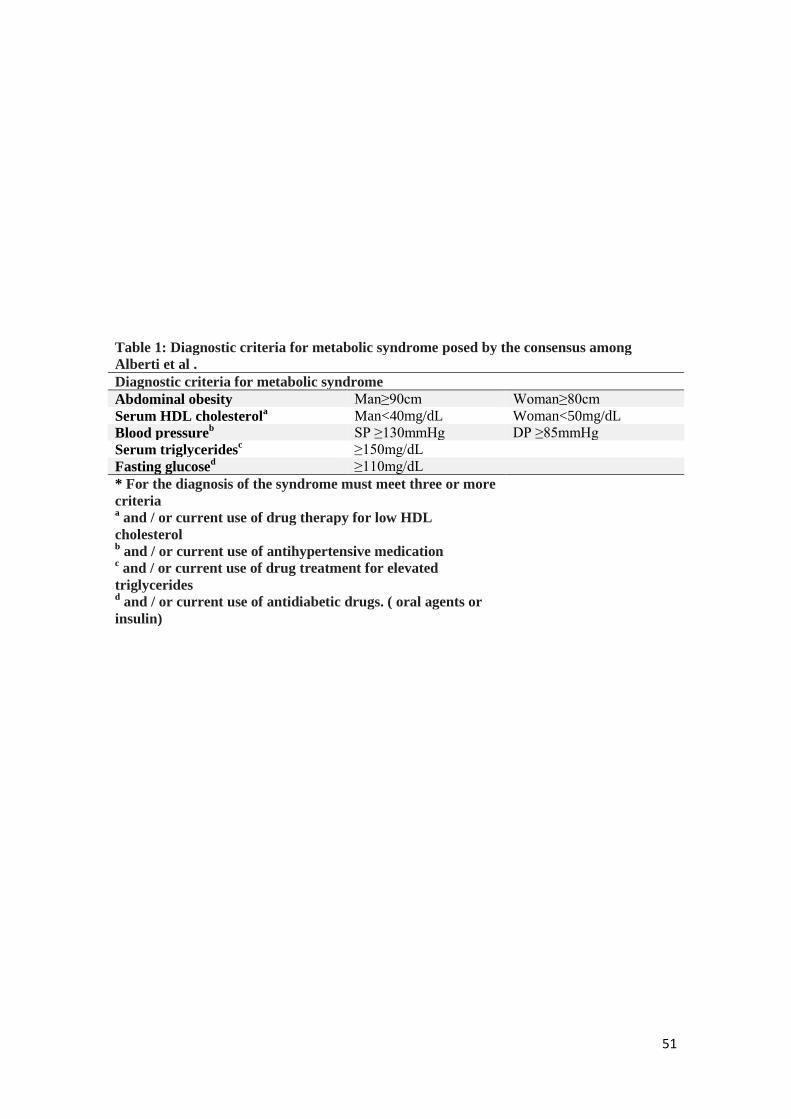
51
Table 1: Diagnostic criteria for metabolic syndrome posed by the consensus among
Alberti et al .
Diagnostic criteria for metabolic syndrome
Abdominal obesity Man≥90cm Woman≥80cm
Serum HDL cholesterola Man<40mg/dL Woman<50mg/dL
Blood pressureb SP ≥130mmHg DP ≥85mmHg
Serum triglyceridesc ≥150mg/dL
Fasting glucosed ≥110mg/dL
* For the diagnosis of the syndrome must meet three or more
criteria a and / or current use of drug therapy for low HDL
cholesterol b and / or current use of antihypertensive medication
c and / or current use of drug treatment for elevated
triglycerides d and / or current use of antidiabetic drugs. ( oral agents or
insulin)

52
Table 2. General characteristics of patients with and without thyroid nodule of a national
hospital in Lima
Patients with
nodules
Patients without
nodules
P
n=91 (%) n=91 (%)
Gender
Man 8 8,79 16 17,58 0,080
Woman 83 91,21 75 82,42
Age
<40 23 25,27 28 30,77 0,559
40 a 59 45 49,45 38 41,76
60 or more 23 25,27 25 27,47
BMI
Underweight 33 36,26 27 29,67 0,682
Normoweight 1 1,10 2 2,2
Overweight 34 37,36 40 43,96
Obese 23 25,27 22 24,18
Measurements
BMI* (kg/m2) 26.92 4,36 26,62 4,42 0,647
Size*(m) 1,56 0,08 1,57 0,08 0,378
Weight**(kg) 64 17 64 15 0,880
Abdominal circumference**(cm)
Man 91,5 25,25 93 18,5 0,689
Woman 90 23,9 89 12,5 0,259
TSH**(mlU/L) 2.71 2.07 2.50 1.18 0.395
T4L**(ng/L) 1.33 0.28 1.00 0.17 0.918
HDL**(mg/dL)
Man 49 12,25 54 8,5 0,242
Woman 45 12,40 52 17 0,035
Glucose**(mg/dL) 95 35 90 26,4 0,108
Triglycerides**(mg/dL) 121 68 115 69,5 0,279
Systolic blood pressure**(mmHg) 120 20 115 20 0,281
Diastolic blood pressure**(mmHg) 70 10 70 11 0,739
Nodule size** (mm) 12,6 11,9 - - -
Background
Diabetes 20 21.98 11 12.09 0,076
Hypertension 26 28.57 22 24.17 0,501
TG Dyslipidemia 10 10.99 15 16.48 0,282
HDL Dyslipidemia 9 6.59 4 4.39 0,150
* The mean and standard deviation is described
** The median and interquartile range is described.
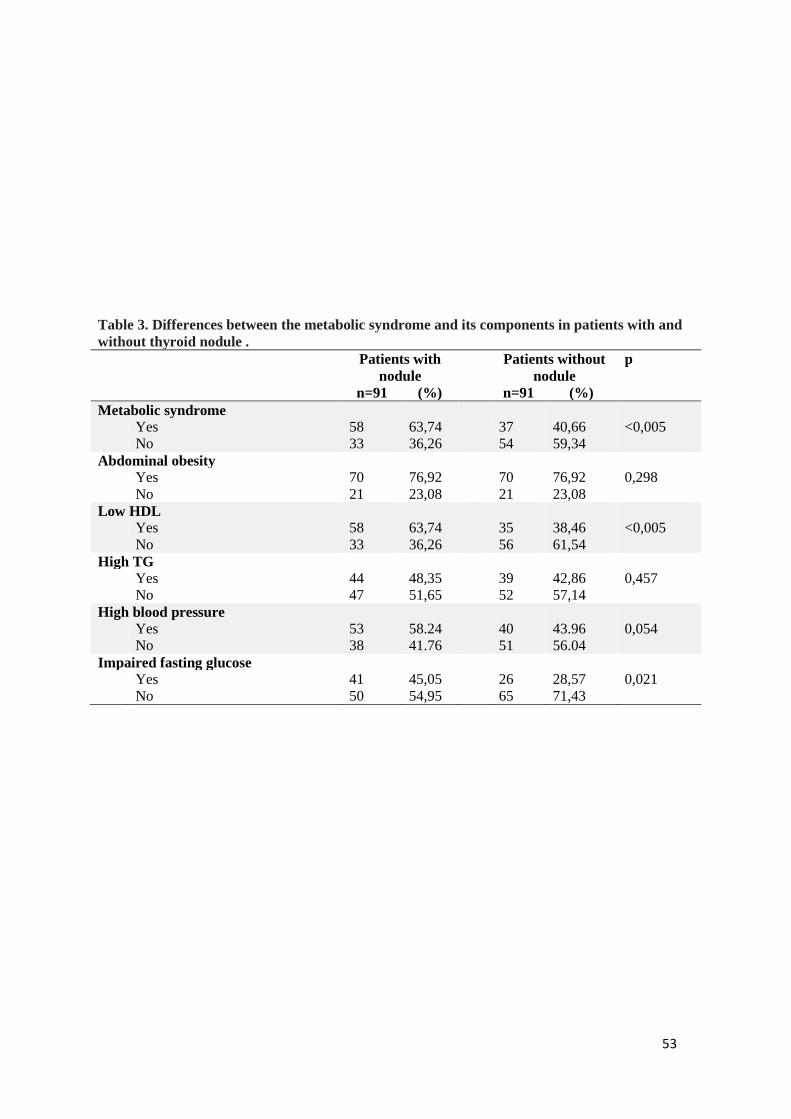
53
Table 3. Differences between the metabolic syndrome and its components in patients with and
without thyroid nodule .
Patients with
nodule
Patients without
nodule
p
n=91 (%) n=91 (%)
Metabolic syndrome
Yes 58 63,74 37 40,66 <0,005
No 33 36,26 54 59,34
Abdominal obesity
Yes 70 76,92 70 76,92 0,298
No 21 23,08 21 23,08
Low HDL
Yes 58 63,74 35 38,46 <0,005
No 33 36,26 56 61,54
High TG
Yes 44 48,35 39 42,86 0,457
No 47 51,65 52 57,14
High blood pressure
Yes 53 58.24 40 43.96 0,054
No 38 41.76 51 56.04
Impaired fasting glucose
Yes 41 45,05 26 28,57 0,021
No 50 54,95 65 71,43

54
Table 4. Bivariate and multivariate analysis between metabolic syndrome and thyroid
nodule
Bivariate model Multivariate model*
OR (IC95%) p OR (IC95%) P
Metabolic Syndrome
Yes 2,56 1,41-4,66 <0,005 2,96 1,47-5,95 <0,005
No 1 Reference 1 Reference
Individual components
Abdominal obesity 1,00 0,5-1,99 1,00 0.68 0,27-1,67 0,4
Low HDL 2,81 1,54-5,12 <0,005 2,77 1,44-5,3 <0,005
High Triglycerides 1,24 0,69-2,23 0,457 1,29 0,66-2,51 0,440
High blood pressure 1,77 0,98-3,19 0,05 1,76 0,92-3,38 0,080
Impaired fasting
glucose
2,05 1,10-9,78 0,022 2,23 1,14-4,34 0,018
* Adjusted for age , gender and nutritional status (BMI )
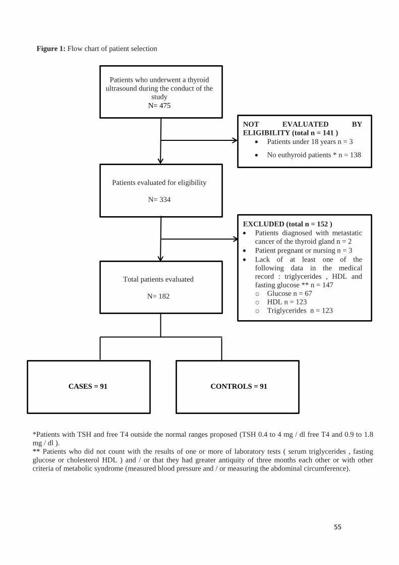
55
Total patients evaluated
N= 182
Patients evaluated for eligibility
N= 334
Patients who underwent a thyroid
ultrasound during the conduct of the
study
N= 475
NOT EVALUATED BY
ELIGIBILITY (total n = 141 )
Patients under 18 years n = 3
No euthyroid patients * n = 138
EXCLUDED (total n = 152 )
Patients diagnosed with metastatic
cancer of the thyroid gland n = 2
Patient pregnant or nursing n = 3
Lack of at least one of the
following data in the medical
record : triglycerides , HDL and
fasting glucose ** n = 147
o Glucose n = 67
o HDL n = 123
o Triglycerides n = 123
CASES = 91 CONTROLS = 91
*Patients with TSH and free T4 outside the normal ranges proposed (TSH 0.4 to 4 mg / dl free T4 and 0.9 to 1.8
mg / dl ).
** Patients who did not count with the results of one or more of laboratory tests ( serum triglycerides , fasting
glucose or cholesterol HDL ) and / or that they had greater antiquity of three months each other or with other
criteria of metabolic syndrome (measured blood pressure and / or measuring the abdominal circumference).
Figure 1: Flow chart of patient selection

56
VIII.REVISTA ELEGIDA
Nombre de la revista: The Journal of Clinical Endocrinology & Metabolism
Pais: Estados Unidos de Norteamerica
Se eligió la revista The Journal of Clinical Endocrinology & Metabolism porque
esta representa el espacio ideal para el contenido del presente estudio. Esta
es una revista indizada con alto índice de impacto, en la cual se publican
trabajos de gran calidad elaborados por diversos centros endocrinológicos
clínicos y experimentales, la revista publica artículos de Revisión y Editoriales
escritos por reconocidos especialistas de la endocrinología con el fin de
actualizar conocimientos y dar a conocer los avances más relevantes en la
actualidad.

57
IX.PROCESO DE REVISIÓN
Recepción del artículo en la revista The Journal of Clinical Endocrinology &
Metabolism para su revisión.

58
X.ESTADO DE LA PUBLICACIÓN
Desde el 26 de febrero del 2015, el artículo de investigación se encuentra en
proceso de revisión por pares por parte de la Revista Norteamericana The
Journal of Clinical Endocrinology & Metabolism. Nos comprometemos a
corregir todas las observaciones planteadas por la revista. En caso no se
acepte, el artículo será enviado a otra revista indizada a SCOPUS para su
publicación.
Raisa Amelia Cornejo Champin Wilmer Gian Franco Silva Caso
Andrea Soria Montoya Alejandro Piscoya