Córdoba, España, 2015 - core.ac.uk · especies de peces en el mundo, ... endemic and particularly...
121
i UNIVERSIDAD DE CÓRDOBA FACULTAD DE VETERINARIA DEPARTAMENTO DE SANIDAD ANIMAL TESIS DOCTORAL Patogenia de piscirickettsiosis: estudio sobre la infectividad de Piscirickettsia salmonis y el efecto de sus productos extracelulares Pedro Alejandro Smith Schuster Director de tesis: Alfonso Carbonero Martínez Córdoba, España, 2015
Transcript of Córdoba, España, 2015 - core.ac.uk · especies de peces en el mundo, ... endemic and particularly...
i
UNIVERSIDAD DE CÓRDOBA FACULTAD DE VETERINARIA
DEPARTAMENTO DE SANIDAD ANIMAL
TESIS DOCTORAL
Patogenia de piscirickettsiosis: estudio sobre la infectividad de Piscirickettsia salmonis y el efecto de sus productos
extracelulares
Pedro Alejandro Smith Schuster
Director de tesis: Alfonso Carbonero Martínez
Córdoba, España,
2015
TITULO: Patogenia de la piscirickettsiosis: estudio sobre la infectividad dePiscirickettsia salmonis y efecto de sus productos extracelulares
AUTOR: Pedro Alejandro Smith Schuster
© Edita: Servicio de Publicaciones de la Universidad de Córdoba. 2016 Campus de RabanalesCtra. Nacional IV, Km. 396 A14071 Córdoba
www.uco.es/[email protected]

TÍTULO DE LA TESIS: Patogenia de piscirickettsiosis: estudio sobre la infectividad de Piscirickettsia salmonis y el efecto de sus productos extracelulares DOCTORANDO/A: Pedro Alejandro Smith Schuster
INFORME RAZONADO DEL/DE LOS DIRECTOR/ES DE LA TESIS (se hará mención a la evolución y desarrollo de la tesis, así como a trabajos y publicaciones derivados de la misma).
La tesis presentada por el doctorando previamente mencionado se ha realizado en el período comprendido entre 2011 y 2015, tratando sobre la patogenia de la infección de Piscirickettsia salmonis, probablemente el agente infeccioso con mayor impacto económico en la producción animal de Chile (se han estimado pérdidas superiores a cuatro mil millones de dólares sólo en este país). A lo largo del estudio se han ido desvelando importantes incógnitas el los mecanismos de la infección por este agente, como sus rutas de acceso a sus hospedadores, tanto desde un punto de vista orgánico como celular, o la capacidad de este agente para secretar exotoxinas que tendrían un papel fundamental en la virulencia de este patógeno. Adicionalmente, se ha demostrado que P. salmonis replica bien en la línea celular ASK, hecho de especial relevancia de cara al diagnóstico e investigación ulteriores con este agente. El doctorado se ha realizado en el marco de un programa de colaboración entre la Universidad de Córdoba y la Universidad de Concepción en Chile, accediendo el alumno al doctorado tras obtener el diploma de estudios avanzados (DEA) por la Universidad de Córdoba. El doctorando ha demostrado en todo momento gran entusiasmo y una gran capacidad para trabajar a unas cotas elevadísimas de calidad, algo no tan sorprendente conociendo la enorme experiencia investigadora previa a la realización de este doctorado. A pesar de la distancia, el trabajo se ha llevado con facilidad, debido fundamentalmente a que su gran autonomía para trabajar ha sido una constante. La tesis doctoral ha dado lugar a la siguiente publicación en revistas indexadas en el JCR:
Rojas, M.E., Galleguillos, M., Díaz, S., Machuca, A., Carbonero, A. & Smith, P.A. (2013) Evidence of exotoxin secretion of Piscirickettsia salmonis, the causative agent of piscirickettsiosis. J Fish Dis, 36, 703-709.
Smith, P.A., Contreras, J.R., Rojas, M.E., Guajardo, A., Díaz, S. & Carbonero, A. (2015) Infectivity of Piscirickettsia salmonis in immersion-bath exposed rainbow trout Oncorhynchus mykiss (Walbaum) fry. J Fish Dis, 38, 765-770.
• Smith, P.A., Díaz, F.E., Rojas, M.E. , Díaz, S., Galleguillos, M. & Carbonero, A. (2015) Effect of Piscirickettsia salmonis inoculation on the ASK continuous cell line. J Fish Dis, 38, 321-324.
Por todo ello, se autoriza la presentación y defensa de la tesis doctoral titulada "Patogenia de piscirickettsiosis: estudio sobre la infectividad de Piscirickettsia salmonis y el efecto de sus productos extracelulares" elaborada por D. Alejandro Smith Schuster para la obtención del título de doctor.
Córdoba, 1 de diciembre de 2015
Firma del director

ii
ÍNDICE
AGRADECIMIENTOS………………………………………………………………………….
DEDICATORIA…………………………………………………………………………………
RESUMEN………………………………………………………………………………………
SUMMARY……………………………………………………………………………………..
JUSTIFICACIÓN………………………………………………………………………………..
REVISIÓN BIBLIOGRÁFICA………………………………………………………………….
Introducción…………………………………………………………………………………..
Agente etiológico……………………………………………………………………………..
Epidemiología………………………………………………………………………………..
Aspectos clínico-patológicos…………………………………………………………………
Patogénesis de la piscirickettsiosis y mecanismos de virulencia de Piscirickettsia salmonis..
Diagnóstico …………………………………………………………………………………..
Tratamiento y control........……………………………………………………………………
Bibliografía……………………………………………………………………………...........
OBJETIVOS ESPECÍFICOS Y LOS ARTÍCULOS RESPECTIVOS DONDE ÉSTOS
FUERON ABORDADOS…………………………………………………………………………
ARTÍCULOS…………………………………………………………………………………….
Artículo 1.1. "Infectivity study of Piscirickettsia salmonis in CHSE-214 cells by confocal
and transmission electron microscopy".....................................................................................
Artículo 1.2. “Effect of Piscirickettsia salmonis inoculation on the ASK continuous cell
line”...........................................................................................................................................
Artículo 2.1. “Infectivity of Piscirickettsia salmonis in immersion-bath exposed rainbow
trout Oncorhynchus mykiss (Walbaum) fry”.............................................................................
Artículo 3.1. “Evidence of exotoxin secretion of Piscirickettsia salmonis, the causative
agent of piscirickettsiosis"........................................................................................................
CONCLUSIONES…………………………………………………………………………..…..
i
ii
1
2
3
10
10
11
14
18
20
25
26
32
51
52
52
68 79 95
114

iii
AGRADECIMIENTOS

iv
DEDICATORIA

1
RESUMEN
La piscirickettsiosis es una enfermedad contagiosa sistémica de los peces teleósteos
cuyo agente causal es Piscirickettsia salmonis, una bacteria gram negativa intracelular
facultativa. Esta enfermedad se ha descrito esporádicamente en distintas áreas geográficas y
especies de peces en el mundo, pero es endémica y particularmente severa en salmónidos
criados en agua de mar en Chile. En esta tesis se investigaron algunos aspectos de la
patogénesis de esta enfermedad, estudiándose la infectividad de P. salmonis, tanto in vitro
como in vivo, y buscándose además evidencias de la capacidad de secretar exotoxinas por
parte de esta bacteria. Los ensayos de infectividad en células CHSE-214, procedentes de
embrión de salmón chinook (Oncorhynchus tshawytscha), mostraron que existe una rápida
adherencia de la bacteria a la superficie de la membrana plasmática (≤ 5 min
posinoculación) seguida de su incorporación al citoplasma de estas células, proceso que
ocurre entre las 3 y las 6 h posinoculación. Por su parte, el estudio de infectividad in vivo,
que se realizó en trucha arcoiris (O. mykiss), reveló que este proceso comprende tres etapas
principales: (i) una fase de rápida adhesión a células epiteliales principalmente de piel y
branquias, pero también del canal alimentario; (ii) una invasión progresiva desde los sitios
de entrada hacia tejidos más profundos hasta alcanzar el torrente sanguíneo y; (iii) una
rápida diseminación vía hematógena para alcanzar virtualmente todos los tejidos
corporales. Finalmente, se demostró que P. salmonis puede secretar exotoxinas
termolábiles que tienen un efecto citotóxico selectivo según la célula blanco expuesta y
que, probablemente, son parte de los factores de virulencia involucrados en la patogénesis
de la piscirickettsiosis.
2
SUMMARY
Piscirickettsiosis is a contagious systemic disease of teleost fish caused by
Piscirickettsia salmonis, a gram negative facultative intracellular bacterium. This condition
has been reported sporadically in different geographical areas of the world, but it is
endemic and particularly severe in maricultured salmonid fish in Chile. In this research
some aspects of the piscirickettsiosis pathogenesis were studied including the infectivity of
P. salmonis, by means of in vitro and in vivo time-course assays, and the searching for
evidence of exotoxin secretion of this bacterium. The in vitro infectivity assays conducted
in CHSE-214 cells, which are derived from chinook salmon (Oncorhynchus tshawytscha)
embryo cells, showed that there is a fast attachment step of this bacterium to the cell
surface (≤ 5 min post-inoculation) followed by a plasma membrane penetration to reach
cell cytoplasm, which occurs mainly between 3 and 6 h post-inoculation. In turn, the in vivo
infectivity study, which was carried out in rainbow trout (O. mykiss), revealed that this
process would comprise three major stages: (i) a rapid attachment to epithelial cells mainly
of skin and gills, but also of the alimentary canal; (ii) a progressive invasion from the sites
of entry to deeper underlying tissues until reaching the bloodstream and; (iii) a rapid
haematogenous spread to virtually all body tissues. Finally, it was shown that P. salmonis
can secrete heat-labile exotoxins, that produce a selective cytotoxicity depending on the
type of targeted cell, which may be some of the virulence factors of this bacterium and
probably play a role in the piscirickettsiosis pathogenesis.

3
JUSTIFICACIÓN
La población humana requiere productos alimenticios en cantidad, calidad y
variedad suficiente para satisfacer en primer término sus necesidades nutricionales (United
Nations, 2013), pero también, entre otras importantes, aquellas de tipo gastronómicas y
sociales (Connor y Armitage, 2002; Fox, 2014). A pesar de las notables mejorías en la
producción y distribución de alimentos que han existido en el último siglo, aún hoy a nivel
global hay alrededor de 800 millones de personas que sufren desnutrición (Wu et al., 2014).
Además, y según indican las proyecciones demográficas, habrá en las próximas décadas un
aumento de la población humana, con expectativas de que existan en total cerca de diez mil
millones de personas el año 2050, por lo cual se espera que el requerimiento de alimentos
se incremente significativamente en el futuro venidero (United Nations, 2015). En cuanto a
la composición de la futura demanda, ésta debería aumentar en mayor proporción para los
alimentos de origen animal que la de aquellos de fuente vegetal debido a que se espera un
cambio en tal sentido empujado por un, también supuesto, aumento de los ingresos
económicos promedios de la población, especialmente en los países en vías de desarrollo
(Alexandratos y Bruinsma, 2012; Wu et al., 2014).
Los animales acuáticos, principalmente peces, moluscos y crustáceos, representan
un componente fundamental del total de alimentos de origen animal en la actualidad, y se
prevé que jugarán un papel aún más relevante en el futuro para contribuir a la seguridad
alimentaria del ser humano (Tacon y Metian, 2013; TWB, 2013). La producción mundial
de animales acuáticos, en cifras del año 2012, alcanzó un total de 158 millones de toneladas
(MMT). De éstas, aproximadamente un 58% (91,3 MMT) provino de la pesca de captura y
un 42 % (66,3 MMT) de la acuicultura (FAO, 2014b). Aunque los desembarcos pesqueros
del año 2012 tuvieron un leve incremento respecto de períodos anteriores, los volúmenes de
4
peces capturados a nivel mundial se han mantenido relativamente constantes ya desde
finales de la década de 1980 alcanzando las cantidades que, según se estima, están cercanas
a su máximo sustentable (FAO, 2014b). En contraste, la acuicultura se ha incrementado
significativamente durante las últimas décadas expandiéndose progresivamente el número
de especies cultivadas, las áreas geográficas dedicadas a esta actividad, su valor económico
y su volumen de producción (Naylor et al., 2000), proporcionando actualmente casi la
mitad de todo el pescado usado para consumo humano (FAO, 2014a). Así, en el año 1982,
globalmente se generaron 5,7 MMT de productos derivados de la acuicultura de animales
acuáticos, cifra que creció progresivamente de tal manera que tras sólo tres décadas, en
2012, alcanzó un valor 10 veces superior respecto al mencionado inicialmente (FAO,
2015). Uno de los grupos de especies de mayor importancia en la acuicultura mundial, por
su relativamente elevado valor económico y vigorosa demanda en distintos mercados, es el
de los peces teleósteos de la subfamilia Salmoninae, que comprende las distintas especies
de truchas y salmones, también denominados genéricamente como salmónidos (Asche y
Bjørndal, 2011). A nivel mundial, entre los grupos de especies de peces, los salmónidos
sólo son superados por aquellos de la familia Ciprinidae (carpas), aunque por gran
diferencia, en tamaño de producción por acuicultura (Naylor et al., 2000; FAO, 2015).
Chile tiene una serie de ventajas comparativas para la acuicultura de salmónidos (Heen et
al., 1993; Asche y Bjørndal, 2011) y es el segundo productor mundial de estas especies,
después de Noruega, cosechando el año 2014 un total bruto aproximado de 955 mil
toneladas (SERNAPESCA, 2015a) con un valor free on board de las exportaciones de este
rubro superior a los cuatro mil millones de dólares (González, 2015). La industria
salmonicultora es de gran importancia económica para Chile, especialmente en las regiones
donde ésta se desarrolla, siendo el segundo producto más exportado después del cobre y

5
estimándose que genera más de 70 mil puestos de trabajo entre empleos directos e
indirectos (SALMONCHILE, 2015).
Los peces salmónidos no son nativos del hemisferio sur y cuando su cultivo en
jaulas flotantes se inició en Chile, a inicios de la década de 1980, los problemas
ocasionados por enfermedades estaban prácticamente ausentes en estos peces (Heen et al.,
1993). Sin embargo, con el incremento de la biomasa de peces, numerosas enfermedades
infecciosas han afectado progresivamente estas poblaciones de salmónidos mantenidos en
cautiverio. En términos de impacto económico, la anemia infecciosa del salmón (ISA) y la
piscirickettsiosis (también denominada “septicemia rickettsial salmonídea” o por su sigla
“SRS”) han sido las condiciones sanitarias más severas de la acuicultura en Chile (Smith et
al., 2011). Aunque ISA causó un enorme daño económico durante los años 2007 y 2009
(Asche et al., 2009), esta afección ha sido controlada, manifestándose con muy pocos
brotes subsecuentes hasta la fecha. Contrariamente, la piscirickettsiosis es una enfermedad
endémica en el ambiente marino y estuarino de la zona sur de Chile (Rees et al., 2014;
Rozas y Enríquez, 2014). A pesar de que hay numerosas vacunas comerciales contra P.
salmonis que se aplican en Chile (SAG, 2015) no se ha logrado controlar la enfermedad,
cuya incidencia incluso ha tendido a aumentar en los últimos años (Rees et al., 2014),
generando elevadas mortalidades en los peces que a su vez derivan en pérdidas cuantiosas
que regularmente han excedido los 100 millones de dólares anuales (Bustos, 2006; Bravo y
Midtlyng, 2007; Almendras, 2015). Un problema adicional es el alto uso de antibióticos
administrados en intentos terapéuticos contra la piscirickettsiosis, lo que conlleva una mala
percepción de los consumidores y un eventual riesgo de salud pública. Al respecto, el año
2014 se usaron aproximadamente 563 toneladas de antibióticos en salmonicultura en Chile
de los cuales el 86% se usó para el tratamiento de piscirickettsiosis (SERNAPESCA,

6
2015c). Debido a la importancia de esta enfermedad en Chile, se instauró un programa
oficial gubernamental destinado a su control a partir del año 2013 (SERNAPESCA, 2012).
Por las razones expuestas es indudable que el desarrollo de conocimientos y
herramientas que contribuyan efectivamente a prevenir y/o controlar esta enfermedad es
actualmente un desafío de real importancia para Chile.
Piscirickettsia salmonis es una bacteria gram negativa intracelular facultativa
(Gómez et al., 2009), agente causal de la piscirickettsiosis (Fryer et al., 1992). Esta última
es una enfermedad infecto-contagiosa muy severa de los salmónidos criados en jaulas
marinas en Chile (Venegas et al., 2004), lugar donde esta entidad fue por primera vez
descrita a nivel mundial (Bravo y Campos, 1989; Cubillos et al., 1990; Fryer et al., 1990;
Cvitanich et al., 1991). La presencia de piscirickettsiosis en salmónidos ha sido también
documentada en Noruega (Olsen et al., 1997; Karatas et al., 2008), en las costas del océano
Pacífico y del Atlántico de Canadá (Brocklebank et al., 1993; Jones et al., 1998), Irlanda
(Rodger y Drinan, 1993) y Escocia (Grant et al., 1996; Birrel et al., 2003). Aunque los
hospedadores principales de P. salmonis son los salmónidos, esta bacteria ha sido
detectada, por aislamiento o reacción en cadena de la polimerasa, en otras especies de peces
teleósteos enfermos tales como Atractoscion nobilis (Arkush et al., 2006) y Dicentrarchus
labrax (McCarthy et al., 2005; Zrnčić et al., 2015).
En las últimas dos décadas, numerosas bacterias fastidiosas gram negativas y que se
replican intracelularmente en sus células hospedadoras, ya sea de modo obligado o
facultativo, han sido reconocidas como agentes causales de enfermedades importantes en
animales acuáticos incluidos moluscos, crustáceos y peces teleósteos (Mauel et al., 2008;
Galeotti et al., 2015). Este grupo emergente de patógenos por sus similitudes fenotípicas
han sido denominados laxamente como “rickettsia-like organisms” y/o “Piscirickettsia-like

7
organisms” (Mauel y Miller, 2002). Dentro de este grupo de microorganismos, P. salmonis
fue el primer caso en que se demostró que era un patógeno de alta virulencia y que
ocasionaba una enfermedad devastadora en animales acuáticos, particularmente en
salmónidos (Fryer y Hedrick, 2003).
Se han realizado numerosos estudios sobre P. salmonis y varios de ellos han
permitido obtener importantes avances como, por ejemplo, obtener una clasificación
taxonómica más precisa, comprender, al menos en parte, la respuesta inmune contra esta
enfermedad y recientemente conocer la secuencia del genoma de algunas cepas de esta
especie bacteriana (Fryer et al., 1992; Marshall et al., 2007; Rozas y Enríquez, 2014; Yañez
et al., 2014; Pulgar et al., 2015). No obstante lo anterior, el conocimiento sobre la
patogénesis de la piscirickettsiosis en general y, en forma más específica, acerca de la
relación patógeno-hospedador y de los mecanismos de virulencia de esta bacteria es aún
muy limitado y fueron estos aspectos el foco de estudio de la presente tesis.
Aunque esta investigación fue fundamentalmente descriptiva, en congruencia con
cada uno de los tres objetivos específicos se plantearon las siguientes hipótesis: 1) “P.
salmonis se adhiere rápidamente (en minutos postexposición) a la membrana de células
hospedadoras y luego en las siguientes horas es endocitada e incorporada al citoplasma
dentro de vacuolas donde procede a multiplicarse por fisión binaria”; 2) “P. salmonis tras
ingresar por piel y mucosas en sus hospedadores, invade progresivamente tejidos
subyacentes hasta alcanzar el torrente sanguíneo para distribuirse entonces a todo el
organismo” y 3) “P. salmonis secreta productos extracelulares como parte de sus factores
de virulencia los cuales contienen exotoxinas que generan un efecto citotóxico en células
hospedadoras”.

8
En cuanto a los antecedentes que respaldan la primera hipótesis se debe señalar que
P. salmonis es un patógeno intracelular facultativo que es capaz de crecer en varias líneas
celulares continuas derivadas de tejidos de peces (Rozas y Enríquez, 2014). Entre ellas, la
principal es la línea CHSE-214, de tipo epitelioide y originada de embrión de salmón
chinook (Oncorhynchus tshawytscha), según lo descrito por Lannan et al. (1984). Debido a
que se sabe que las células CHSE-214 son susceptibles a la bacteria, ellas fueron las que se
usaron en esta investigación como modelo biológico para describir la secuencia de invasión
y la estructura o compartimento de la célula hospedera donde se encuentra P. salmonis en
un tiempo determinado posexposición. Se esperaba que la adhesión fuese rápida debido a
que estudios anteriores habían demostrado que la bacteria se une a la superficie de las ovas
en un tiempo muy breve (Larenas et al., 2003). Respecto a la segunda hipótesis, se había
descrito previamente que P. salmonis podría utilizar como rutas de ingreso a sus
hospedadores tanto piel como epitelio branquial ya que se generaron mortalidades, tanto en
trucha arcoíris (O. mykiss) como en salmón coho (O. kisutch), luego de usar parches de
contacto embebidos en el patógeno para exponer dichas localizaciones (Smith et al., 1999;
Smith et al., 2004). Como consecuencia, se conjeturó que si los peces fuesen expuestos en
forma completa, mediante baños con una suspensión que contuviese el patógeno, en la
forma en que se llevó a cabo en este estudio, la bacteria usaría estos epitelios a modo de
puerta de ingreso y posteriormente invadiría progresivamente los tejidos subyacentes, como
fue planteado en la hipótesis, generando finalmente una infección sistémica usando la vía
hematógena en los peces infectados. En cuanto a la tercera hipótesis, se sabe que muchas
bacterias gram negativas del ser humano y de animales mamíferos secretan factores y
exotoxinas como factores de virulencia (Wilson, 2006; Ye y Blanke, 2006), lo que
igualmente podría ocurrir en P. salmonis. Esta posibilidad se veía adicionalmente apoyada
9
por cuanto también la secreción de productos extracelulares (PECs) con función de
exotoxinas se había descrito en otras bacterias gram negativas específicamente patógenas
de peces, como es el caso de Aeromonas salmonicida y de Yersinia ruckeri (Tobback et al.,
2007), y además debido al descubrimiento de genes que codifican exotoxinas en
Francisella novicida (Siddaramappa et al., 2011), una bacteria que está taxonómica y
genéticamente relacionada con P. salmonis.
El fracaso de las acciones hasta ahora emprendidas para el control de la
piscirickettsiosis indica que el estudio científico de esta enfermedad y su agente causal es
necesario, ya que permitirá tener las bases para el eventual descubrimiento de las medidas
terapéuticas y de control adecuadas para mitigar el efecto de esta patología. El
conocimiento de la patogenia de una enfermedad resulta fundamental, ya que, según se
deduce de numerosos ejemplos en medicina humana y veterinaria, esta información puede
permitir detener o intervenir este proceso en favor del hospedador, ya sea mediante
medidas terapéuticas o profilácticas (Mims et al., 2001; Gyles y Prescott, 2010). En esta
tesis se hicieron investigaciones dirigidas a comprender la patogenia de la piscirickettsiosis
en salmónidos; avanzándose concretamente en la descripción de la cinética de infección de
P. salmonis en células y tejidos de sus hospedadores y en identificar la capacidad de
secretar exotoxinas con efecto citotóxico que serían parte de los mecanismos de virulencia
usados por este importante patógeno acuático para generar el daño patológico en los peces
afectados.
10
REVISIÓN BIBLIOGRÁFICA
Introducción
La piscirickettsiosis es una enfermedad septicémica infecto-contagiosa que puede
tener un curso agudo o crónico y sus hospedadores principales corresponden a especies
salmónidas. Su agente causal es una bacteria intracelular facultativa gram negativa aeróbica
perteneciente al orden Thiotrichales denominada Piscirickettsia salmonis (Fryer y Hedrick,
2003; Rozas y Enríquez, 2014). Esta patología se evidencia normalmente en peces durante
su etapa de engorda en ambiente marino y, en cierto grado, en estuarios, donde puede
causar mortalidades muy significativas (Rozas y Enríquez, 2014). Debe destacarse que
antes de su observación en Chile no había registros de la existencia de esta enfermedad en
ningún otro lugar del mundo (Cvitanich et al., 1990; Fryer et al., 1990). Esta entidad
ictiopatológica ha causado daños económicos muy importantes en la acuicultura chilena.
Así, desde el surgimiento de los primeros brotes de piscirickettsiosis -los cuales ocurrieron
a finales de la década de 1980- hasta la actualidad, se estima que las pérdidas para la
salmonicultura en Chile superarían con creces los dos mil millones de dólares (Almendras,
2015). La piscirickettsiosis, a pesar de la enorme gravitación de la anemia infecciosa del
salmón (ISA) ocurrida entre los años 2007 y 2009 (Asche et al., 2009; Mardones et al.,
2009), a largo plazo, ha sido la enfermedad con mayor impacto económico en el cultivo de
salmónidos en Chile y actualmente sigue siendo la causa primordial de mortalidad por
enfermedades de los peces en fase marina (SERNAPESCA 2015b) que es la etapa donde el
precio de cada ejemplar tiene un valor superior. Junto con un elevado impacto económico
directo, esta afección ha obligado a un alto uso de antimicrobianos para su tratamiento
(SERNAPESCA 2015c), con implicación añadida de riesgo de daño ambiental y de

11
percepción negativa de los consumidores. Adicionalmente, supone un problema de
bienestar animal en los peces que enferman (Smith et al., 2011). Todo lo anterior señala
que la piscirickettsiosis es una entidad patológica relevante en acuicultura y los estudios
conducentes a aumentar el conocimiento sobre esta enfermedad (y su agente causal) son de
importancia, ya que deberían contribuir a encontrar adecuados métodos de tratamiento e
idealmente de prevención de la misma.
Agente etiológico
Como ya se ha mencionado, el agente causal de la piscirickettsiosis es el
microorganismo denominado actualmente como Piscirickettsia salmonis. Esta relación
causal quedó demostrada en forma conclusiva mediante el aislamiento de esta bacteria
como agente único desde peces que cursaban la enfermedad en forma natural y a través de
ensayos posteriores en que se completaron los postulados clásicos de Koch,
reproduciéndose la enfermedad y reaislándose el agente en peces inoculados
experimentalmente (Cvitanich et al., 1990; Fryer et al., 1990; Garcés et al., 1991).
El primer aislamiento de P. salmonis se realizó desde un salmón coho con signos
clínicos de la enfermedad (Fryer et al., 1990) en cultivos celulares de la línea CHSE-214.
Este microorganismo fue denominado con la sigla LF-89 y corresponde a la cepa tipo de P.
salmonis (Fryer et al., 1992). Esta cepa es la que se encuentra mejor caracterizada (Mauel y
Miller, 2002) y está depositada en la “American Type Culture Collection” como ATCC VR
1361 (Fryer et al., 1992; Lannan y Fryer, 1993).
El agente aislado fue clasificado inicialmente en el orden Rickettsiales y en la
familia Rickettsiaceae (Fryer et al., 1990) y más tarde ubicado en un nuevo género,
Piscirickettsia, proponiéndose el nombre de la especie como Piscirickettsia salmonis (Fryer

12
et al., 1992). El análisis comparativo de las secuencias de los genes que codifican el ácido
ribonucleico ribosómico (rRNA, por su sigla en inglés) de la subunidad 16S reveló que este
microorganismo debía ser clasificado como miembro del subgrupo gamma de las
Proteobacterias (Mauel et al., 1999). Se demostró además que P. salmonis estaba
filogenéticamente relacionada, aunque distantemente, con los géneros Francisella (Mauel
et al., 1999; Titball et al., 2003) y Coxiella (Mauel et al., 1999). Subsecuentemente, el
género Piscirickettsia fue ubicado en un nuevo orden, Thiotrichales, y en una nueva
familia, Piscirickettsiaceae, claramente separado de las bacterias de la familia
Rickettsiaceae (Fryer y Hedrick, 2003; Garrity et al., 2005).
Las bacterias de la familia Piscirickettsiaceae se caracterizan por ser gram
negativas, aeróbicas, con forma cocoide, de bacilo o de espiral, ocasionalmente
pleomórficas y por ser comúnmente aisladas desde ambientes acuáticos marinos. Esta
familia está formada por seis géneros; Piscirickettsia, Cycloclasticus, Hydrogenovibrio,
Methylophaga, Thioalkalimicrobium y Thiomicrospira, que están relacionados
filogenéticamente, pero que poseen pocas características fenotípicas en común (Fryer y
Hedrick, 2003; Fryer y Lannan, 2005).
P. salmonis es la única especie descrita en el género Piscirickettsia (Fryer y
Hedrick, 2003) y corresponde a una bacteria inmóvil, no capsulada, pleomórfica,
generalmente cocoide, pero que también se encuentra en pares o en forma de anillo (Fryer
et al., 1990). Normalmente se divide por fisión binaria (Cvitanich et al., 1990; Fryer et al.,
1990), pero en ocasiones puede replicar por gemación (McCarthy et al., 2008). La bacteria
tiene un tamaño aproximado 0,5-1,5 μm de diámetro (Fryer y Hedrick, 2003), aunque se
describe que puede generar variantes pequeñas con tamaños inferiores a 0,2 μm (Rojas et
al., 2008).

13
Este patógeno se multiplica dentro de vacuolas intracitoplasmáticas de sus células
hospedadoras y genera efecto citopático (ECP) en varias líneas celulares provenientes de
peces (Fryer et al., 1990; Cvitanich et al., 1991; Fryer et al., 1992) y también en una línea
originada desde una especie de ranas (Xenopus laevis) y en otra proveniente del insecto
Spodoptera frugiperda (Birkbeck et al., 2004).
El rango de temperatura óptimo para el crecimiento de P. salmonis está entre los 15
y 18 °C, disminuyendo significativamente su replicación bajo los 10 °C y sobre los 20 °C y
no visualizándose sobre los 25 °C (Fryer et al., 1992). Los cultivos de células CHSE-214
que presentan ECP completo alcanzan títulos del orden de 106 a 107 dosis infectantes de
cultivo de tejidos 50% por mL (DICT50/mL) (Fryer et al., 1990). Es interesante señalar que
se consiguen títulos más elevados que en las células CHSE-214 cuando esta bacteria es
multiplicada en una línea celular de insecto (Birkbeck et al., 2004). La multiplicación
bacteriana en la línea CHSE-214 se evidencia por la presentación gradual de un ECP típico,
que comienza entre los días 5 a 6 posinfección y que se caracteriza por la aparición de
grupos de células globosas que aumentan en número, para posteriormente desprenderse
progresivamente hasta que la monocapa celular es destruida completamente a los 14 días
posinfección (Fryer et al., 1990; Garcés et al., 1991). Cabe indicar que si bien se estimaba
tradicionalmente que P. salmonis era un patógeno intracelular estricto, informaciones más
recientes sugieren que en realidad se trataría de un organismo intracelular facultativo
(Gómez et al., 2009). La afirmación anterior es congruente con lo descrito en cuanto a que
P. salmonis puede desarrollarse, aunque en forma limitada, en medios de cultivo
enriquecidos pero libres de células eucariotas (Mauel et al., 2008; Mikalsen et al., 2008;
Yañez et al., 2012; Henriquez et al., 2013). Sin embargo, resulta probable que en la
naturaleza esta bacteria sólo se multiplique intracelularmente debido a que en

14
observaciones microscópicas de tejidos de peces infectados siempre se le ha encontrado
dividiéndose dentro de vacuolas intracitoplasmáticas y no extracelularmente (Fryer et al.,
1990; Cvitanich et al., 1990; Garcés et al., 1991; McCarthy et al., 2008). Esta situación
podría ser análoga a lo observado con F. tularensis, en que luego de conocerse e
interpretarse su genoma se concluyó que en la naturaleza actúa como un patógeno
intracelular obligado a pesar de que puede ser cultivado en medios de cultivo
bacteriológicos acelulares apropiados (Larsson et al., 2005; Birkbeck, 2011).
Epidemiología
Históricamente, los primeros brotes de piscirickettsiosis que registra la literatura se
presentaron en Chile en el año 1989 (Bravo y Campos, 1989; Cvitanich et al., 1990), en
canales marinos de la localidad de Calbuco en la X Región (Alvarado et al., 1990; Cubillos
et al., 1990), lugares que tenían una gran densidad de sitios de cultivo con salmón coho. A
pesar de ello, existen antecedentes de que los brotes ya habían comenzado a finales del año
1988 y mediante análisis retrospectivos de muestras de tejidos de salmón coho se ha
concluido que la bacteria se encontraba al menos desde 1987. En los meses que siguieron a
los brotes detectados en Calbuco (Bravo y Campos, 1989; Alvarado et al., 1990), esta
condición se extendió a los centros costeros ubicados en otras áreas de la X Región para
posteriormente alcanzar también los sitios con cultivos marinos de la XI Región. En los
últimos años se ha registrado la presencia de piscirickettsiosis entre las Regiones IX y la
XIII pero con mayor incidencia y prevalencia en las Regiones X y XI que son las zonas de
mayor concentración de centros marinos de cultivo de salmónidos (Mancilla y Bustos,
2015; SERNAPESCA, 2015b)

15
Normalmente, en un lote de peces determinado, la enfermedad se comienza a
manifiestar una vez transcurridas seis a doce semanas después de la transferencia de los
“smolts” al agua de mar (Alvarado et al., 1990; Fryer et al., 1990). Al respecto, se ha
propuesto que el proceso de esmoltificación puede incrementar la susceptibilidad de los
peces infectados (Graumann et al., 1997). Originalmente, las epizootias surgían en el otoño
y la primavera de cada año (Lannan y Fryer, 1994), cuando las temperaturas del agua
fluctuaban entre los 9 y los 16 °C (Cvitanich et al., 1991; Lannan y Fryer, 1994).
Posteriormente su presentación cambió sin tener la marcada estacionalidad anteriormente
señalada, puesto que esta enfermedad se comenzó a presentar en cualquier época del año
(Monasterio, 2008). En los últimos años la piscirickettsiosis, si bien ocurre durante todo el
año, tiene con una mayor severidad durante los meses de verano y en las fases finales del
ciclo de engorde en el mar (SERNAPESCA, 2015b).
La piscirickettsiosis se considera básicamente una enfermedad marina. Esta
aseveración se sustenta sobre la base de que la vasta mayoría de los brotes ocurre en peces
criados en jaulas localizadas en el mar y en menor medida en aquellos ubicados en aguas
estuarinas que tienen influencia marina. No obstante lo anterior, excepcionalmente la
enfermedad se ha reportado en agua dulce y la literatura describe dos hallazgos en este tipo
de ambiente acuático, uno de ellos comunicado por Bravo (1994) y el otro por Gaggero et
al. (1995).
Si bien esta enfermedad se describió originalmente sólo en el salmón coho (Bravo y
Campos, 1989; Cubillos et al., 1990; Branson y Nieto Díaz-Muñoz, 1991; Cvitanich et al.,
1991), en los años que siguieron afectó a todas las especies de salmónidos producidas en
Chile, describiéndose en salmón del Atlántico, salmón chinook, trucha arcoíris (Garcés et
al., 1991; Fryer et al., 1992; Fryer y Mauel, 1997) y salmón cereza (O. masou) según lo

16
descrito por Bravo (1994). El salmón coho resultó más susceptible que la trucha arcoíris en
una infección experimental (Smith et al., 1996a). No obstante lo anterior, un estudio
epidemiológico de la situación de campo reciente en Chile mostró que al comparar los
brotes de piscirickettsiosis entre el salmón del Atlántico y la trucha arcoíris, en esta última
especie se registraron las mayores mortalidades y además los brotes de esta enfermedad se
iniciaban antes (Jakob et al., 2014).
Aunque los hospedadores principales de P. salmonis son peces de los géneros
Oncorhynchus y Salmo de la subfamilia Salmoninae, existen reportes de la presencia de
esta bacteria en otras especies de peces como D. labrax (McCarthy et al., 2005; Zrnčić et
al., 2015) y A. nobilis (Arkush et al., 2006). Ambos brotes de piscirickettsiosis en D. labrax
han ocurrido en peces cultivados en el mar Mediterráneo cursando con cuadros de
encefalitis severa. El primero de ellos tuvo lugar en las costas de Grecia y en muestras de
tejidos de estos peces se reconoció por criterios serológicos, histológicos y de análisis
genómico la presencia de P. salmonis (McCarthy et al., 2005); el segundo caso es más
reciente, ocurrió en la costa de Croacia y la bacteria fue detectada en tejidos de los peces
mediante hibridación in situ y secuenciación del gen que codifica el rRNA 16S (Zrnčić et
al., 2015). Por su parte, la identificación de P. salmonis en A. nobilis ocurrió desde peces
juveniles, cultivados en estanques con agua de mar en California (EE.UU.), que cursaban
con mortalidades y lesiones hepáticas similares a las que se presentan en peces salmonídeos
con piscirickettsiosis. En este caso, la bacteria fue aislada y se demostró también su
patogenicidad para salmón coho mediante inoculaciones experimentales (Chen et al.,
2000). Finalmente, aún cuando no serían hospedadores naturales, se ha logrado generar
mortalidades de un 10% en embriones de peces zebra (Danio rerio) inyectados con P.
17
salmonis, lo que serviría de modelo experimental para el estudio de esta enfermedad
(Berger, 2014).
Posteriormente al aislamiento de P. salmonis en Chile, esta bacteria se ha
encontrado en salmónidos cultivados en diferentes zonas geográficas del mundo,
registrándose casos de piscirickettsiosis en Canadá, en sus costas este (Cusack et al., 2002)
y oeste (Brocklebank et al., 1992), Irlanda (Palmer et al., 1997), Escocia (Birrell et al.,
2003), y Noruega (Olsen et al., 1997). En el hemisferio sur, la identificación específica e
inequívoca de la presencia de P. salmonis sólo ha ocurrido en Chile, aún cuando una
bacteria con características fenotípicas similares fue aislada desde salmones del Atlántico
cultivados en la isla de Tasmania en el sur de Australia (Corbeil et al., 2005). Aunque ya
existía la descripción de algunos hallazgos microscópicos de organismos similares a P.
salmonis, el descubrimiento de la piscirickettsiosis aumentó el interés de la comunidad
científica para investigar la presencia de este tipo de agentes causales y su papel en
enfermedades de otros animales acuáticos. Así, se han detectado bacterias denominadas
como “organismos similares a rickettsias” (“Rickettsia like organisms” o “RLO”) y/o
“organismos similares a Piscirickettsia” (“Piscirickettsia like organisms” o “PLO”) en una
amplia variedad de especies acuáticas, incluidos moluscos, crustáceos y peces, y en algunos
casos son agentes causales de enfermedades de importancia por su alta prevalencia y/o tasa
de mortalidad (Correal, 1995; Venegas, 1996; Mauel y Miller, 2002). Cabe señalar, sin
embargo, que a pesar de la similitud fenotípica de los RLO y los PLO con P. salmonis, el
conocimiento sobre las relaciones taxonómicas y filogenéticas entre estos
microorganismos es aún incompleto.
En cuanto a la transmisión de P. salmonis en el ambiente acuático, en función de un
estudio epidemiológico en Chile, se concluyó que este patógeno puede diseminarse a través
18
del agua a distancias bastantes considerables (hasta 10 km) desde un centro de cultivo
marino afectado a otro con peces sanos (Rees et al., 2014) lo cual sería uno de los factores
que dificultaría el control de esta enfermedad. Estos mismos autores determinaron que otros
factores de riesgo que condicionan el inicio y la severidad de los brotes de
piscirickettsiosis, además de la distancia con un centro de cultivo afectado, son la cantidad
de otros centros de cultivo de salmónidos vecinos y la temperatura del agua, en el sentido
de que un aumento de ésta incrementa el riesgo (Rees et al., 2014).
Aspectos clínico-patológicos
Como ya se ha mencionado, los cuadros clínicos se presentan en los salmónidos
cultivados casi exclusivamente cuando están en el mar o en aguas estuarinas (Smith et al.,
2011; Rozas y Enríquez, 2014). Los peces enfermos exhiben signos clínicos inespecíficos
tales como un desplazamiento cerca de la superficie, errático, incoordinado, lento, y a veces
en tirabuzón, lo que refleja alteraciones del sistema nervioso en estos individuos (Cvitanich
et al., 1991; Larenas et al., 1995; Larenas et al., 2000). Además, se ha descrito letargia,
anorexia, choque contra las mallas de las balsas-jaulas y orillamiento (Olsen et al., 1997;
Larenas et al., 2000).
Las alteraciones macroscópicas externas más relevantes incluyen oscurecimiento y
descamación de la piel (Cvitanich et al., 1991; Larenas et al., 1995; Monasterio, 2008),
además de lesiones en este tejido que comprenden desde pequeñas áreas de
solevantamiento hasta úlceras hemorrágicas de hasta 2 cm de diámetro (Branson y Nieto
Díaz-Muñoz, 1991). También se describe la presencia de hemorragias petequiales y
equimóticas en la piel de la base de las aletas, del vientre y de la zona perianal y en algunos
casos, hemorragias perioculares, que generalmente son bilaterales (Alvarado et al., 1990;

19
Cubillos et al., 1990). Otro hallazgo importante es la aparición de palidez branquial, lo que
refleja una anemia severa, corroborada por bajos niveles de hematocrito (Olsen et al.,
1997). Además, se ha observado neutrofilia, con niveles 10 a 20 veces más altos que los
rangos normales (Branson y Nieto Díaz-Muñoz, 1991).
En el examen anátomo-patológico macroscópico de órganos internos, se aprecia que
la mayoría de los peces afectados presenta diferentes grados de ascitis y de peritonitis
(Branson y Nieto Díaz-Muñoz, 1991). En relación al primer hallazgo, Larenas et al. (1995)
señalan que la ascitis sólo se observa en peces moribundos y, por lo tanto, sugieren que
podría considerarse más bien como una alteración de tipo terminal. Por otra parte, se
aprecian frecuentemente hemorragias petequiales en estómago, intestino, ciegos pilóricos,
vejiga natatoria, grasa visceral (Cvitanich et al., 1991), musculatura abdominal (Larenas et
al., 1995) y peritoneo (Branson y Nieto Díaz-Muñoz, 1991). En cuanto al hígado, éste
exhibe un aumento de volumen (Branson y Nieto Díaz-Muñoz, 1991); y además puede
mostrar un aspecto “moteado”, debido a la presencia de pequeños nódulos subcapsulares
blanquecinos a amarillentos distribuidos difusamente en toda su superficie (Larenas et al.,
1995; Olsen et al., 1997). Al respecto, se señala que la presencia de estos nódulos puede ser
señal de un grado más avanzado de cronicidad del cuadro clínico (Almendras et al., 2000).
Ocasionalmente, este órgano se ve pálido y hemorrágico (Cvitanich et al., 1991). La
vesícula biliar está generalmente pletórica (Alvarado et al., 1990; Cubillos et al., 1990),
evidenciando que los peces no han comido por un tiempo prolongado (Alvarado et al.,
1990). En tanto, el riñón exhibe pérdida de su apariencia brillante, tornándose marrón-
grisáceo opaco (Alvarado et al., 1990) y con aumento de su tamaño (Fryer et al., 1990;
Branson y Nieto Díaz-Muñoz, 1991). A nivel del bazo, se presenta esplenomegalia en
diferentes grados, observándose en reiteradas ocasiones las mismas lesiones blanquecinas
20
descritas en el hígado (Alvarado et al., 1990; Cubillos et al., 1990; Fryer et al., 1990). En el
estómago, se encuentra un líquido transparente seromucoso (Alvarado et al., 1990; Schäfer
et al., 1990), lo cual confiere la impresión de que el pez ha tragado agua (Alvarado et al.,
1990). El intestino generalmente no posee alimento (Alvarado et al., 1990; Branson y Nieto
Díaz-Muñoz, 1991), evidenciándose enteritis en el tercio distal (Alvarado et al., 1990);
adicionalmente, el lumen de esta víscera puede contener un gran volumen de mucus
amarillento (Schäfer et al., 1990). El corazón, en ocasiones, también presenta focos
necróticos y hemorragias petequiales de tipo difuso (Cubillos et al., 1990) y en un 2% de
los peces moribundos se observa la presencia de una pseudomembrana que lo cubre,
sugiriendo la presencia de pericarditis (Cvitanich et al., 1991). Además, se aprecia un
aumento en el volumen del líquido cefalorraquídeo, acompañado en muchos casos de
congestión de las meninges (Larenas et al., 1995). Por la gran variedad de órganos que
presentan lesiones, se puede desprender que esta enfermedad tiene una naturaleza sistémica
(Fryer y Hedrick, 2003) y, aunque ciertos tejidos tienen un daño más severo, como el
inmunopoyético renal (Venegas et al., 2004) y el cardíaco (Monasterio, 2008), la bacteria
se multiplica en una variedad muy amplia de células, mostrando una escasa especificidad
en este sentido.
Patogénesis de la piscirickettsiosis y mecanismos de virulencia de Piscirickettsia
salmonis
Para establecer y mantener una infección exitosamente, los microorganismos
patógenos en general han desarrollado una variedad de estrategias para invadir el
hospedador, evitando o resistiendo la respuesta inmune innata, el daño de las células y
multiplicándose en regiones específicas y normalmente estériles (Tizard, 1992; Cossart y
21
Sansonetti, 2004). Al igual que en mamíferos, las bacterias patógenas de peces tienen
mecanismos de infección que están directamente relacionados con los factores de virulencia
de éstas mediante los cuales pueden colonizar y penetrar las barreras epiteliales de sus
hospedadores para luego subsistir, multiplicarse en sus tejidos y producir las alteraciones
patológicas características, superando las barreras inmunológicas de estos animales
acuáticos (Evelyn, 1996). Conviene señalar que la relativa importancia de una enfermedad
infecciosa para la salud de humanos y animales no siempre coincide con la profundidad en
el entendimiento de su patogénesis (Peterson, 1996). La piscirickettsiosis se encuentra en
esta situación, puesto que la patogénesis de ésta y los factores de virulencia de P. salmonis
son, en general, pobremente conocidos. La nueva localización taxonómica de P. salmonis
ha mostrado que tiene una relación filogenética con bacterias del género Francisella y no
así con aquellas del género Rickettsia como se había pensado originalmente, debido a
similitudes en las características fenotípicas (Fryer y Hedrick, 2003). Este hecho es
interesante puesto que últimamente se ha descrito el aislamiento de bacterias del género
Francisella en distintas especies de peces (Hsieh et al., 2007; Mauel et al., 2007; Mikalsen
et al., 2007; Ottem et al., 2007 a y b), incluido en salmónidos cultivados en Chile (Birkbeck
et al., 2007). F. tularensis, la especie más conspicua de este género, es el agente causal de
tularemia, enfermedad que afecta en forma severa a humanos y a una variedad de especies
animales (Splettstoesser et al., 2005). Al igual que P. salmonis, esta bacteria es capaz de
sobrevivir y multiplicarse dentro de macrófagos y su patogenia también es conocida en
forma muy limitada (Carlson et al., 2007). Al respecto, se ha descrito que, así como en el
caso de muchas otras bacterias, F. tularensis también secreta un conjunto de proteínas que
según Hager et al. (2006) actuarían como factores de virulencia. Así, mediante el uso de
proteómica y bioinformática se identificaron siete proteínas exportadas por F. tularensis,

22
las cuales emplean el sistema secretor tipo IV. Dos de estas proteínas serían quitinasas y las
otras, con funciones de proteína de unión a la quitina, proteasa y glucoxidasa,
respectivamente (Hager et al., 2006). Otros autores, comparando una cepa viva que se usa
experimentalmente como vacuna contra F. tularensis denominada LCV y una variante de
ésta, consignada como ACV que origina una mayor producción de factor de necrosis
tumoral y de varias interleuquinas, por medio de electroforesis en dos dimensiones,
demostraron que hay una expresión diferencial de varias proteínas las que actuarían como
factores de virulencia (Carlson et al., 2007). No obstante los hallazgos recién señalados,
dado que no se ha logrado demostrar la presencia de genes que codifiquen exotoxinas en F.
tularensis se ha puesto en duda que esta bacteria sea capaz de sintetizar este tipo específico
de proteínas (Siddaramappa et al., 2011).
En cuanto a la patogénesis de la piscirickettsiosis los principales aspectos en que se
ha avanzado son en el conocimiento de algunas de sus rutas de ingreso a sus hospedadores,
la detección de estructuras de adhesión a las ovas, las fuentes de excreción del agente
causal desde los peces infectados y la capacidad de P. salmonis de persistir y multiplicarse
en células macrofágicas. Respecto de los sitios de ingreso, hay tres trabajos en que se
infectaron experimentalmente ciertos lugares anatómicos seleccionados en peces
salmónidos para conocer los tejidos que usa como sitio de entrada P. salmonis en sus
hospedadores. El primero de ellos se efectuó en salmones del Atlántico juveniles,
exponiendo los peces a la bacteria mediante inyección intraperitoneal, intubación gástrica e
instilación branquial. Las mortalidades acumuladas obtenidas fueron 57, 41 y 45%,
respectivamente, sugiriendo que las rutas que usa P. salmonis son la branquial y la oral
(Almendras et al., 1997). En la segunda de estas investigaciones, truchas arcoíris juveniles
fueron expuestas a P. salmonis mediante las siguientes seis diferentes vías: intraperitoneal,

23
subcutánea, piel, branquias (en ambos casos aplicando por 1 min parches de contacto
embebidos con dosis conocidas de la bacteria), intraintestinal e intragástrica (las dos
últimas mediante intubación). Las mortalidades acumuladas resultantes fueron 98, 100, 52,
24, 24 y 2%, respectivamente, concluyéndose que los principales sitios de entrada de esta
bacteria son piel y branquias y que la ruta oral, contrariamente a lo sugerido por Almendras
et al. (2007), no sería el método más eficiente para iniciar la infección en sus hospedadores,
o al menos en esta especie de salmónidos, por parte de P. salmonis (Smith et al., 1999).
Finalmente, un tercer estudio sobre esta materia se llevó a cabo en salmones coho juveniles,
los cuales fueron expuestos, con dos dosis distintas, por inyección intraperitoneal,
instilación de piel y branquias e intubación intestinal y gástrica. Las mortalidades
acumuladas con la dosis más elevada fueron 82, 46, 28 y 20% y con la dosis menor 58, 32,
16 y 12%, respectivamente, indicando que las vías de ingreso más efectivas de P. salmonis
en salmón coho son la piel seguida por las branquias, dado que si bien la intubación
intestinal generó altas mortalidades, esa manera de administrar la bacteria no simula las
condiciones de exposición natural de los hospedadores (Smith et al., 2004). En relación a
estructuras que permitan la adhesión de P. salmonis, se ha documentado por microscopía de
barrido la presencia de unas prolongaciones de su superficie que permitirían su anclaje a las
ovas, en este caso de trucha arcoíris, y por lo tanto favorecerían la eventual transmisión
vertical de esta bacteria (Larenas et al., 2003). No obstante lo anterior, es dudoso que la
transmisión vertical ocurra realmente en condiciones de campo, ya que los brotes de
piscirickettsiosis en agua dulce han sido muy excepcionales, habiéndose reportado sólo dos
casos en la literatura científica (Bravo, 1994; Gaggero et al., 1995). En cuanto a las vías de
excreción de P. salmonis desde sus hospedadores, si bien éstas no están totalmente
aclaradas, se ha documentado que alevines de truchas arcoíris, experimentalmente

24
infectados, excretan este patógeno a través de las heces y también, aunque en menor
medida, desde la bilis y la orina (Salinas, 1998). La eliminación de esta bacteria a través de
las heces ha sido también corroborada en la misma especie, pero en condiciones de cultivo
(Peirano, 2015). Un aspecto de importancia en el conocimiento de la patogénesis de la
piscirickettsiosis consistió en la verificación de que esta bacteria es capaz de sobrevivir y
multiplicarse en macrófagos obtenidos de riñón anterior de trucha arcoíris (McCarthy et al.,
2005) lo que fue coherente con las numerosas observaciones histológicas ya existentes de
peces infectados natural y experimentalmente con P. salmonis (Branson y Nieto Díaz-
Muñoz, 1991; Cvitanich et al., 1991, Venegas et al., 2004). Es interesante mencionar que
P. salmonis no usaría el método de otras bacterias intracelulares, de escapar al citoplasma
antes de la fusión del fagolisosoma, como mecanismo para sobrevivir dentro de los
macrófagos (McCarthy et al., 2005).
Respecto a los factores de virulencia de P. salmonis, además de las estructuras de
adhesión observadas cuando interactúan con ovas de trucha arcoíris, éstos son poco
conocidos. Sin embargo, tras la reciente secuenciación del genoma de algunas cepas de P.
salmonis se han detectado distintos genes que podrían codificar varios factores de
virulencia de acuerdo a distintos análisis bioinformáticos. En ese contexto distintos autores
describen la presencia de genes que codifican para el sistema de secreción tipo IV (SSTIV)
y de flagelos bacterianos (Eppinger et al., 2013; Yañez et al., 2014). Es curioso que P.
salmonis tenga la información genética para sintetizar flagelos puesto que hasta ahora se ha
caracterizado como una bacteria inmóvil y tampoco se ha visto por microscopía electrónica
la presencia de estas estructuras. Junto con el hallazgo de genes que codifican para SSTIV,
tanto Yañez et al. (2014) como Pulgar et al. (2015) describen la presencia de genes
relacionados con la adquisición de hierro que codificarían posibles factores de virulencia

25
(sideróforos) de importancia para esta bacteria. Finalmente, además del SSTIV, Yañez et
al. (2014) descubrieron en una cepa de P. salmonis la presencia de genes de los sistemas de
secreción tipo I y II, ampliando el repertorio de posibles factores de virulencia de este
patógeno. No se había detectado la capacidad de producir ECPs con propiedades de
exotoxinas en P. salmonis antes de las investigaciones que se presentan en uno de los
artículos de la presente tesis. A pesar de que no es común que las bacterias intracelulares
secreten exotoxinas, había dos antecedentes que nos indicaban que P. salmonis podía
liberar este tipo de proteínas. El primero de ellos fue una evidencia indirecta, ya que en
función del tipo de daño patológico se sospechó de la acción de la presencia de eventuales
hepatotoxinas en casos clínicos de piscirickettsiosis en Noruega (Olsen et al., 1997). El
segundo antecedente es que se había observado desde el punto vista lesional microscópico
que con ciertas cepas, como la SLGO-95, se produce un severo daño en algunos tejidos de
los peces sin encontrarse la presencia de la bacteria en éstos o en niveles muy bajos no
compatibles con la severidad de las alteraciones observadas (Rojas, 2000), sugiriendo que
existe algún componente soluble que actuaría a distancia, probablemente PECs con efecto
de exotoxinas. En consecuencia, algunas de las investigaciones de esta tesis se orientaron a
intentar aclarar si P. salmonis tiene la capacidad de sintetizar algún tipo de exotoxinas que
medie su acción patogénica.
Diagnóstico
El diagnóstico presuntivo se realiza sobre la base de los antecedentes epidemiológicos, la
signología clínica y los hallazgos anatomopatológicos macroscópicos de acuerdo a lo
descrito anteriormente (Fryer et al., 1990; Fryer y Hedrick, 2003; Monasterio, 2008). Este
diagnóstico presuntivo se puede acompañar de tinciones de frotis de tejidos como Gram,

26
Giemsa y azul de toluidina para observar la morfología bacteriana, y de análisis
histológicos para corroborar las lesiones microscópicas consistentes con lo descrito para
esta enfermedad (Branson y Nieto Díaz-Muñoz, 1991; Cvitanich et al., 1991; Larenas et al.,
1995). La identificación definitiva de la presencia de P. salmonis en los peces afectados se
hace a través de técnicas inmunológicas como inmunofluoresencia (Lannan et al., 1991) e
inmunohistoquímica (Alday et al., 1994) o de reacción en cadena de la polimerasa (Mauel
et al., 1996; Marshall et al., 1998). No obstante sus dificultades técnicas, mayor tiempo y
coste del procedimiento, el aislamiento y posterior identificación de la bacteria es el
estándar de oro para el diagnóstico de la presencia de P. salmonis viable en peces
sospechosos de estar cursando un cuadro de piscirickettsiosis (OIE, 2006).
Tratamiento y control
Una vez que la enfermedad se ha desencadenado, una de las principales medidas de
control que se emplea es el tratamiento de los peces mediante el uso de distintos
antimicrobianos administrados vía oral o por inyección intraperitoneal (Almendras y
Fuentealba, 1997; Groff y LaPatra, 2000; Larenas et al., 2000). Si bien inicialmente la
aplicación de estos productos tenía un resultado positivo en cuanto a la reducción de las
mortalidades, con el transcurrir de los años la efectividad fue disminuyendo y, en definitiva,
la terapia con estas drogas ha tenido un éxito sólo parcial en el control de esta enfermedad
(Smith et al., 1995; OIE, 2006; Mancilla y Bustos, 2015). Se ha sugerido que la
localización intracelular de esta bacteria puede hacer posible que un número considerable
de microorganismos esté fuera del alcance de las concentraciones bactericidas y, de esta
manera, la infección puede persistir (Lannan y Fryer, 1993). Más aún, las pérdidas debido a
este agente patógeno se han incrementado progresivamente debido, aparentemente, a la

27
resistencia de las cepas actuantes a los antibióticos (Smith et al., 1996b). Antimicrobianos
como oxitetraciclina, ácido oxolínico y flumequina, han sido utilizados ampliamente por
varios años en Chile para el tratamiento de piscirickettsiosis y, por lo tanto, una selección
genética de cepas resistentes puede ser la razón de la pérdida de la sensibilidad de P.
salmonis a estas drogas. Por otro lado, debido a las marcadas variaciones en los patrones de
sensibilidad de los diferentes aislados de P. salmonis estudiados, no se recomienda el uso
de un régimen universal para el tratamiento de todos los brotes (Smith et al., 1996b).
Aunque históricamente se han usado varios antimicrobianos para el tratamiento de esta
enfermedad tales como oxitetraciclina, ácido oxolínico, flumequina, enrofloxacina,
sarafloxacina y florfenicol (Bravo et al., 2005), en los últimos años estas terapias se
encuentran restringidas casi exclusivamente al uso de florfenicol y de oxitetraciclina
(SERNAPESCA, 2015c).
Debido a las limitaciones existentes en la eficacia de los antibióticos y a los
problemas derivados de su uso excesivo, varios grupos de investigación han dirigido sus
esfuerzos para intentar desarrollar una vacuna efectiva contra la piscirickettsiosis (House y
Fryer, 2002; OIE, 2006; Bravo y Midtlyng, 2007; Olson y Criddle, 2008). Como sucede en
el caso de varias enfermedades de peces, la vacunación ha demostrado ser una herramienta
eficiente para ayudar a prevenir la presentación de brotes infecciosos en estos animales
acuáticos (Bravo y Midtlyng, 2007; Gjedrem y Baranski, 2009), siendo además considerada
por algunos autores como la principal causa de la notoria reducción en el uso de
antimicrobianos que ha experimentado la salmonicultura en algunos países (Bravo y
Midtlyng, 2007). Ensayos iniciales que utilizaron bacterinas producidas con la bacteria
completa mostraron resultados variables e inconsistentes (Smith et al., 1995; Smith et al.,
1997; Fryer y Hedrick, 2003). Posteriormente, en un ensayo efectuado en salmones coho,

28
se incrementó el porcentaje de supervivencia relativa de éstos al ser inyectados con
bacterinas acompañadas con adyuvantes acuosos u oleosos (Kuzyk et al., 2001a).
Por otro lado, se han realizado varios estudios de la caracterización antigénica de P.
salmonis (Kuzyk et al., 1996; Barnes et al., 1998; House y Fryer, 2002; Henríquez et al.,
2003; Yuksel et al., 2006b). En este mismo ámbito, también se ha experimentado con
vacunas de ADN (Miquel et al., 2003) y de proteínas recombinantes (Kuzyk et al., 2001a;
Kuzyk et al., 2001b; Sommerset et al., 2005; Wilhelm et al., 2006). También se ha descrito
que una vacuna desarrollada para prevenir la enfermedad bacteriana del riñón, compuesta
por células vivas de una bacteria ambiental no patógena para peces, llamada Arthrobacter
davidanieli, es efectiva para reducir la mortalidad en salmones coho infectados con P.
salmonis, tanto en condiciones de laboratorio como de campo (Salonius et al., 2005). Esta
vacuna ha sido autorizada para su uso en salmónidos cultivados en Canadá (Salonius et al.,
2005) y en Chile (Sommerset et al., 2005), si bien ha tenido una aceptación limitada en el
mercado chileno (Bravo y Midtlyng, 2007). La primera vacuna comercial contra P.
salmonis fue lanzada al mercado chileno en 1999 y desde entonces han ido apareciendo
nuevos productos de este tipo pero, a diferencia de los resultados logrados en otras
enfermedades bacterianas, la vacunación contra este agente patógeno no ha reducido
significativamente la necesidad de continuar tratando los cuadros de piscirickettsiosis con
antibióticos, y el uso de éstos, en términos cuantitativos, se mantiene a niveles similares
(Bravo y Midtlyng, 2007). Actualmente, hay ocho laboratorios comerciales que tienen
vacunas registradas en Chile contra P. salmonis. Algunas de éstas están dirigidas sólo
contra P. salmonis y otras son multivalentes, en diferentes combinaciones con otros
antígenos bacterianos y/o virales, con un repertorio de 37 preparados antigénicos. Una de
estas vacunas se aplica por inmersión, dos de ellas por vía oral y las 34 restantes se

29
administran por inyección intraperitoneal (SAG, 2015). Es interesante señalar que un
estudio de carácter epidemiológico en Chile concluyó que no hay diferencias en los niveles
de mortalidad acumulada atribuidas a P. salmonis entre peces inmunizados con vacunas
comerciales contra piscirickettsiosis y aquellos no vacunados (Flores, 2012). En igual
sentido, y más recientemente, Jakob et al. (2014) describen que estas vacunas no reducen la
mortalidad acumulada ocasionada por la piscirickettsiosis y sólo retardarían la aparición de
los brotes. Los antecedentes anteriores y la experiencia histórica de campo en general
sugiere que estos productos son poco efectivos (Mancilla y Bustos, 2015).
Como sucede en la mayoría de las enfermedades infectocontagiosas que afectan a
peces de cultivo, existe un conjunto de medidas de manejo que se recomienda poner en
práctica, tanto para la prevención como para el control de la piscirickettsiosis (OIE, 2006;
Yuksel et al., 2006a; Olson y Criddle, 2008). En este sentido, se preconiza la formulación y
aplicación de programas sanitarios así como la adopción de procedimientos generales de
bioseguridad (Groff y LaPatra 2000; Fryer y Hedrick, 2003; OIE, 2006). Entre las medidas
operacionales que se han recomendado para disminuir la incidencia de la piscirickettsiosis
se encuentran: desinfectar la superficie de las ovas con yodóforos (Fryer y Hedrick, 2003),
seleccionar “stocks” o familias de peces que hayan demostrado mayor resistencia a brotes y
menor mortalidad (Cassigoli, 1994), disminuir las manipulaciones de los peces
(vacunaciones, cambios de redes, tratamientos inyectables, etc.) para así aminorar el estrés
(Bravo y Campos, 1989; Bravo y Gutiérrez, 1991; Larenas et al., 2000; Leal y Woywood,
2007), evitar altas densidades de biomasa por jaula, centro y por área geográfica (Cassigoli,
1994; Olson y Criddle, 2008), ingresar los peces a los centros marinos a número final, es
decir mantener el número de individuos que se destina a una jaula hasta la cosecha (Leal y
Woywood, 2007), trasladar los “smolts” directamente desde agua dulce a salada, sin pasar

30
por estuarios (Leal y Woywood, 2007), extraer rápidamente los peces enfermos y muertos
desde las balsas-jaulas (Cassigoli, 1994; Almendras y Fuentealba, 1997; Larenas, 1999),
realizar pruebas diagnósticas para detectar la presencia temprana de la bacteria (Marshall et
al., 1998), evitar que la materia orgánica derivada de las cosechas llegue al mar (Cassigoli,
1994), fomentar el descanso sanitario de los centros previo al ingreso de nuevos peces
(Cassigoli, 1994; Fryer y Hedrick, 2003), evitar la presentación de factores primarios que
produzcan lesiones de la piel (erosiones y úlceras), ya sea de origen traumático (Larenas et
al., 2000) o por ectoparásitos (Smith et al., 1999; Larenas et al., 2000; Fryer y Hedrick,
2003) y mantener limpias las redes de las balsas-jaulas (Olson y Criddle, 2008).
Además de su relevancia histórica, la pisciricketsiosis sigue siendo una enfermedad
de primera importancia para la salmonicultura chilena. Actualmente subsiste en forma
endémica con brotes frecuentes en una amplia zona marina. A pesar de que la erradicación
de P. salmonis del ambiente acuático es virtualmente imposible, ya que se ha detectado que
poblaciones de salmónidos asilvestrados son portadores de esta bacteria (Pérez et al.,
1998), por diferentes razones debiera ser prioritario continuar los esfuerzos para disminuir
la incidencia de piscirickettsiosis en nuestros cultivos marinos. Además del daño evidente
derivado de la mortalidad de peces que ocasiona la enfermedad, otro efecto indeseable es el
uso de altos niveles de antimicrobianos para su tratamiento. Por otra parte, un factor que
normalmente no se considera, pero que es de importancia desde la óptica veterinaria, es que
los salmones enfermos padecen de distintos grados de sufrimiento y que un nivel de
morbilidad tan alta implica un problema de bienestar animal que se debe intentar disminuir
al máximo posible. Los crecientes avances en la generación de conocimientos científicos,
tanto básicos como aplicados, deberían contribuir a entregar mejores herramientas
específicas para el control de esta enfermedad. Es un hecho constatable que con las actuales

31
medidas de tratamiento, control y prevención no se ha logrado mitigar el enorme daño que
causa la piscirickettsiosis en Chile. Por lo anterior, es evidente que falta mayor
investigación que provea las bases para elaborar productos biológicos que permitan una
terapéutica y prevención eficaz de esta bacteria intracelular. Uno de los variados tópicos
donde se requiere profundizar es acerca de la patogénesis in vitro e in vivo de la
piscirickettsiosis así como de la posible producción de exotoxinas de P. salmonis como
parte de sus factores de virulencia y son éstos los aspectos que fueron abordados en los
artículos científicos que aquí se presentan.
32
BIBLIOGRAFÍA
ALDAY-SANZ, V.; RODGER, H.D.; TURNBULL, T.; ADAMS, A.; RICHARDS,
R. 1994. An immunohistochemical diagnostic test for rickettsial disease. Fish
Dis. 17:189-191.
ALEXANDRATOS, N.; BRUINSMA, J. 2012.World Agriculture Towards 2030/2050:
the 2012 Revision. Global Perspective Studies Team. FAO Agricultural
Development Economics Division Agricultural Development Economics (ESA).
ESA Working Paper Nº 12-03. The Food and Agriculture Organization of the
United Nations. Roma, Italia, 147 pp.
ALMENDRAS, F.E.; FUENTEALBA, I.C. 1997. Salmonid rickettsial septicemia
caused by Piscirickettsia salmonis: a review. Dis. Aquat. Org. 29:137-144.
ALMENDRAS, F.E.; FUENTEALBA, I.; JONES, S.R.M.; MARKHAM, F.;
SPANGLER, E. 1997. Experimental infection and horizontal transmission of
Piscirickettsia salmonis in freshwater-raised Atlantic salmon, Salmo salar L. J.
Fish Dis. 20:409-418.
ALMENDRAS, F.E.; FUENTEALBA, I.C.; MARKHAM, R.F.F.; SPEARE, D.J.
2000. Pathogenesis of liver lesions caused by experimental infection with
Piscirickettsia salmonis in juvenile Atlantic salmon, Salmo salar L. J. Vet.
Diagn. Invest. 12:552-557.
ALMENDRAS, F. 2015. Disminuyendo costos: el aporte de las vacunas al control de
SRS. Revista Digital Aqua Acuicultura y Pesca. 8:1 [en línea]
<http://www.aqua.cl/2015/08/11/farmaceuticas-entregan-claves-para-reducir-
costos-de-produccion-por-srs/> [Consulta 21.10.2015].
ALVARADO, V.; SCHÄFER, W.; ENRÍQUEZ, R.; MONRÁS, M.; CUBILLOS, V.;
FARÍAS, C.; ALBERDI, A. 1990. Síndrome del salmón coho (S.S.C.), nueva
enfermedad de salmonídeos cultivados en fase de agua de mar en Chile. Situación
actual. Patología Animal 4:10-13.

33
ARKUSH, K.D.; EDES, H.L.; McBRIDE, A.M.; ADKISON, M.A.; HEDRICK,
R.P. 2006. Persistence of Piscirickettsia salmonis and detection of serum
antibodies to the bacterium in white seabass Atractoscion nobilis following
experimental exposure. Dis. Aquat. Org. 73:131-139.
ASCHE, F.; HANSEN, H.; TVETERAS, R.; TVETERAS, S. 2009. The salmon
disease crisis in Chile. Mar. Resour. Econ. 24:405-411.
ASCHE, F.; BJØRNDAL, T. 2011. The Supply of Salmon. In: The Economics of
Salmon Aquaculture. Second ed. Wiley-Blackwell. Chichester, Reino Unido, pp.
17-42.
BARNES, M.N.; LANDOLT, M.L.; POWELL, D.B.; WINTON, J.R. 1998.
Purification of Piscirickettsia salmonis and partial characterization of antigens. Dis.
Aquat. Org. 33:33-41.
BERGER, E.K. 2014. Piscirickettsia salmonis; characterization and infection in the
zebra fish model. Theses for the Master`s degree in Molecular Biosciences. The
Faculty of Mathematics and Natural Sciences. University of Oslo. 74 pp.
BIRKBECK, T.H.; GRIFFEN, A.A.; REID, H.I.; LAIDLER, L.A.;
WADSWORTH, S. 2004. Growth of Piscirickettsia salmonis to high titers in
insect tissue culture cells. Infect. Immun. 72:3693-3694.
BIRKBECK, T.H.; BORDEVIK, M.; FROYSTAD, M.K.; BAKLIEN A. 2007.
Identification of Francisella sp. from Atlantic salmon, Salmo salar L., in Chile.
J. Fish Dis. 30:505-507.
BIRKBECK, H. 2011. Francisellosis. In: Avendaño-Herrera R (Ed.). Enfermedades
Infecciosas del Cultivo de Salmónidos en Chile y el Mundo. Niva Chile S.A.,
Puerto Varas, Chile, pp. 105-128.
BIRRELL, J.; MITCHELL, S.; BRUNO, D.W. 2003. Piscirickettsia salmonis in
farmed Atlantic salmon, Salmo salar in Scotland. Bull. Eur. Assoc. Fish. Pathol.
23:213-218.

34
BRANSON, E.J.; NIETO DÍAZ-MUÑOZ, D. 1991. Description of a new disease
condition occurring in farmed coho salmon, Oncorhynchus kisutch (Walbaum), in
South America. J. Fish Dis. 14:147-156.
BRAVO, S. 1994. Piscirickettsiosis in freshwater. Bull. Eur. Assoc. Fish Pathol.
14:137-138.
BRAVO, S.; CAMPOS, M. 1989. Coho salmon syndrome in Chile. AFS/FHS
Newsletter. 17:3.
BRAVO, S.; GUTIÉRREZ, S. 1991. Avances en el estudio del síndrome del salmón
coho. Chile Pesquero 64:39-42.
BRAVO, S.; MIDTLYNG, P. 2007. The use of fish vaccines in the Chilean salmon
industry 1999-2003. Aquaculture 270:36-42.
BRAVO. S.; DÖLZ, H.; SILVA, M.T.; LAGOS, C.; MILLANAO, A.; URBINA, M.
2005. Diagnóstico del uso de fármacos y otros productos químicos en la
acuicultura. Informe final Proyecto Nº 2003-28. Universidad Austral de Chile,
Facultad de Pesquerías y Oceanografía, Instituto de Acuicultura. Puerto Montt,
Chile. 256 pp.
BROCKLEBANK, J.R.; SPEARE, D.J.; ARMSTRONG, R.D.; EVELYN, T. 1992.
Septicemia in farmed Atlantic and chinook salmon due to a rickettsia-like agent.
AFS/FHS Newsletter 20:1.
BROCKLEBANK, J.R.; EVELYN, T.P.T.; SPEARE, D.J.; ARMSTRONG, D.J.
1993. Rickettsial septicemia in farmed Atlantic and chinook salmon in British
Columbia: Clinical presentation and experimental transmission. Can. Vet. J.
34:745-748.
BUSTOS, P. 2006. Growing incidence of Piscirickettsia infection in fish worldwide:
mechanisms for prevention and control. Nutritional Biotechnology in the Feed
and Food Industries: Proceedings of Alltech’s 22nd Annual Symposium,
Lexington, Kentucky, pp. 397-401.

35
CARLSON, P.E.; CARROLL, J.A.; O´DEE, D.M.; NAU, G.J. 2007. Modulation of
virulence factors in Francisella tularensis determines human macrophage
responses. Microb. Pathog. 42:204-214.
CASSIGOLI, J. 1994. Patología y nutrición en el desarrollo de la acuicultura: factores
de éxito. In: Patología y nutrición en el desarrollo de la acuicultura. Puerto
Montt, Chile. 3-7 octubre. Fundación Chile. (sin número en la carpeta).
CHEN, M.F.; YUN, S.; MARTY, G.D.; McDOWELL, T.S.; HOUSE, M.L.;
APPERSEN, J.A.; GUENTHER, T.A.; ARKUSH, K.D.; HEDRICK, R.P.
2000. A Piscirickettsia salmonis-like bacterium associated with mortality of
white seabass Atractoscion nobilis. Dis. Aquat. Org. 43:117-126.
CONNOR, M.; ARMITAGE, C.J. 2002. Chapter 7. Conclusions. In: Sutton S. (Ed.).
The Social Psychology of Food. Applying Social Psychology Series. Open
University Press, Buckingham, Reino Unido, pp. 133-144.
CORBEIL, S.; HYATT, A.D.; CRANE, M.St.J. 2005. Characterisation of an
emerging rickettsia-like organism in Tasmanian farmed Atlantic salmon Salmo
salar. Dis. Aquat. Org. 64:37-44.
CORREAL, P. 1995. Prospección de Piscirickettsia salmonis en fauna marina
silvestre asociada al cultivo intensivo de salmónidos. Memoria Título Médico
Veterinario. Santiago, Chile. U. Chile, Fac. Cs. Veterinarias y Pecuarias. 52 p.
COSSART, P.; SANSONETTI, J. 2004. Bacterial invasion: the paradigms of
enteroinvasive pathogens. Science 304:242-248.
CUBILLOS, V.; FARÍAS, C.; ALBERDI, A.; ALVARADO, V.; SCHÄFER, W.;
MONRÁS, M. 1990. Características anatomopatológicas del “síndrome del
salmón coho” (SSC), nueva enfermedad de los salmonídeos. Patol. Animal 4:14-
17.
CUSACK, R.R.; GROMAN, D.B.; JONES, S.R.M. 2002. Rickettsial infection in
farmed Atlantic salmon in eastern Canada. Can. Vet. J. 43:435-440.

36
CVITANICH, J.D.; GÁRATE, O.; SMITH, C.E. 1990. Etiological agent in a Chilean
coho disease isolated and confirmed by Koch's postulates. AFS/FHS Newsletter
18:1-2.
CVITANICH, J.D.; GÁRATE, O.; SMITH, C.E. 1991. The isolation of a rickettsia-
like organism causing disease and mortality in Chilean salmonids and its
confirmation by Koch's postulate. J. Fish Dis. 14:121-145.
DEVOLD, M.; KROSSØY, B.; ASPEHAUG, V.; NYLUND, A. 2000. Use of RT-
PCR for diagnosis of infectious salmon anaemia virus (ISAV) in carrier sea trout
Salmo trutta after experimental infection. Dis. Aquat. Org. 40:9-18.
EPPINGER, M.; MCNAIR, K.; ZOGAJ, X.; DINSDALE, E.A.; EDWARDS, R.A;
KLOSE, K.E. 2013. Draft genome sequence of the fish pathogen Piscirickettsia
salmonis. Genome Announc. DOI: 10.1128/genomeA.00926-13.
EVELYN, T.P.T. 1996. Infection and Disease. In: The Fish Immune System.
Academic Press. San Diego, California, EE.UU., pp. 339-366.
FAO (FOOD AND AGRICULTURE ORGANIZATION OF THE UNITED
NATIONS). 2014a. Foreword. In: The State of World Fisheries and
Aquaculture. Opportunities and Challenges. FAO Department of Fisheries and
Aquaculture. Roma, Italia, p. iii.
FAO (FOOD AND AGRICULTURE ORGANIZATION OF THE UNITED
NATIONS). 2014b. Part I. Review of Fisheries and Aquaculture. Status and
Trends. In: The State of World Fisheries and Aquaculture. Opportunities and
Challenges. FAO Department of Fisheries and Aquaculture. Roma, Italia, pp. 3-
98.
FAO (FOOD AND AGRICULTURE ORGANIZATION OF THE UNITED
NATIONS). 2015. Fishery Statistical Collections. Global Aquaculture
Production 1950-2013 [en línea] <http://www.fao.org/fishery/statistics/global-
aquaculture-production/query/en> [Consulta 12.10. 2015].
FLORES, C. 2012. Determinación de los factores de riesgo para mortalidad por
septicemia rickettsial del salmón en las regiones de Los Lagos y de Aysén, Chile.

37
Memoria título Médico Veterinario. Santiago, Chile. U. Chile. Fac. Cs.
Veterinarias y Pecuarias. 70 p.
FOX, R. 2014. Food and Eating: An Anthropological Perspective. Robin Social Issues
Research Centre Publications. Oxford, Reino Unido, 21 pp. [en línea]
<http://www.sirc.org/publik/foxfood.pdf> [consulta: 15.10.2015]
FRYER, J.L.; LANNAN, C.N.; GARCÉS, L.H.; LARENAS, J.J.; SMITH, P.A. 1990.
Isolation of a rickettsiales-like organism from diseased coho salmon (Oncorhynchus
kisutch) in Chile. Fish Pathol. 25:107-114.
FRYER, J.L.; LANNAN, C.N.; GIOVANONNI, S.J.; WOOD, N.D. 1992.
Piscirickettsia salmonis gen. nov., sp. nov., the causative agent of an epizootic
disease in salmonid fishes. Int. J. Syst. Bacteriol. 42:120-126.
FRYER, J.L.; MAUEL, M. 1997. The Rickettsia: an emerging group of pathogens in
fish. Emerg. Infect. Dis. 3:137-144.
FRYER, J.L.; HEDRICK, R.P. 2003. Piscirickettsia salmonis: a Gram-negative
intracellular bacterial pathogen of fish. J. Fish Dis. 26:251-262.
FRYER, J.L.; LANNAN, C.N. 2005. Family II. Piscirickettsiaceae fam. nov. In:
Garrity, G.M. (Ed.). Bergey´s Manual of Systematic Bacteriology. 2th ed., vol II
The Proteobacteria, Part B The Gammaproteobacteria. Springer. New York,
EE.UU., pp. 180-184.
GAGGERO, A.; CASTRO, H.; SANDINO, A.M. 1995. First isolation of
Piscirickettsia salmonis from coho salmon, Oncorhynchus kisutch (Walbaum),
and rainbow trout, Oncorhynchus mykiss (Walbaum), during the freshwater stage
of their life cycle. J. Fish Dis. 18:277-279.
GALEOTTI, M.; MANZANO, M., CECCHINI, F.; VOOLPATTI, D.; BULFON,
P.; BERALDO, G.; ROSSI, G.; MAGI, G. 2015. T.E.M. and biomolecular
detection of Rickettsiales in tissues of rainbow trout and their potential role as
red mark syndrome (RMS) etiological agents. 17th International Conference on
Diseases of Fish and Shellfish. European Association of Fish Pathologists. Las

38
Palmas de Gran Canaria, España, Septiembre 7 a 11. Abstract Book, O166, p.
174.
GARCÉS, L.H.; LARENAS, J.J.; SMITH, P.A.; SANDINO, S.; LANNAN, C.N.;
FRYER, J.L. 1991. Infectivity of a rickettsia isolated from coho salmon
(Oncorhynchus kisutch). Dis. Aquat. Org. 11:93-97.
GARRITY, G.M.; BELL, J.A.; LILBURN, T. 2005. The Revised Road Map to the
Manual. In: Garrity, G.M. (Ed.). Bergey´s Manual of Systematic Bacteriology.
2th ed., vol II The Proteobacteria, Part A Introductory Essays. Springer. New
York, EE.UU., pp. 159-220.
GJEDREM, T.; BARANSKI, M. 2009. Initiating Breeding Programs. In: Selective
Breeding in Aquaculture: An Introduction. Springer. Dordrecht, The
Netherlands, pp. 3-85.
GÓMEZ, F.; HENRÍQUEZ, V.; MARSHALL, S.H. 2009. Additional evidence of
the facultative intracellular nature of the fish bacterial pathogen Piscirickettsia
salmonis. Arch. Med. Vet. 41:261-267.
GONZÁLEZ, J. 2015. Sector en cifras: exportaciones de salmónidos. Revista Aqua
182:61.013
GRANT, A.N.; BROWN, A.G.; COX, D.I.; BIRBECK, T.H.; GRIFFEN, A.A. 1996.
Rickettsia-like organism in farmed salmon. Vet. Rec. 138:423.
GRAUMANN, R.; ENRÍQUEZ, R.; MONRÁS, M.; BROWN, A.W.;
ROSENTHAL, H. 1997. Experimental challenge of Piscirickettsia salmonis in
postsmolts of Atlantic salmon (Salmo salar). In: 8th International Conference on
Diseases of Fish and Shellfish. Edinburgh, Scotland. September, 14-19.
European Association of Fish Pathologists (EAFP), pp. P-056.
GROFF, J.M.; LaPATRA, S.E. 2000. Infectious diseases impacting the commercial
culture of salmonids. J. Appl. Aquacult. 10:17-90.
GYLES, C.L.; PRESCOTT, J.F. 2010. Themes in Bacterial Pathogenic Mechanisms.
In: Gyles, C.L.; Prescott, J.F.; Songer, G.; Thoen, C.O. (Eds.). Pathogenesis of
Bacterial Infections in Animals. 4th ed. Wiley-Blackwell. Iowa, EE.UU., pp. 3-8.

39
HAGER, A.J.; BOLTON, D.L.; PELLETIER, M.R.; BRITTNACHER, M.J.;
GALLAGHER, L.A.; KAUL, R.; SKERRETT, S.J.; MILLER, S.I.; GUINA,
T. 2006. Type IV pili-mediated secretion modulates Francisella virulence. Mol.
Microbiol. 62:227-237.
HEEN, K.; THORPE, J.; RIDLER, N.; MONAHAN, R.L.; MAHNKEN, C.;
LINDBERG, J. 1993. The Distribution of Salmon Aquaculture. In: K. Heen;
R.L. Monahan; F. U. (Eds.). Salmon Aquaculture. Wiley Blackwell. Oxford,
Reino Unido, pp. 10-58.
HENRIQUEZ, M.; GONZALEZ, E.; MARSHALL, S.H.; HENRIQUEZ, V.;
GOMEZ, F.A.; MARTINEZ, I.; ALTAMIRANO, C. 2013. A novel liquid
medium for the efficient growth of the salmonid pathogen Piscirickettsia
salmonis and optimization of culture conditions Plos One. DOI:
10.1371/journal.pone.0071830.
HENRÍQUEZ, V.; ROJAS, M.V.; MARSHALL, S.H. 2003. An alternative efficient
procedure for purification of the obligate intracellular fish bacterial pathogen
Piscirickettsia salmonis. Appl. Environ. Microbiol. 69:6268-6271.
HOUSE, M.L.; FRYER, J.L. 2002. The Biology and Molecular Detection of
Piscirickettsia salmonis. In: Cunningham, C.O. (Ed.). Molecular Diagnosis of
Salmonid Diseases. Kluwer Academic Publishers. Dordrecht, Holanda, pp. 141-
155.
HSIEH, C.Y.; WU, Z.B.; TUNG, M.C.; TSAI, S.S. 2007. PCR and in situ
hybridization for the detection and localization of a new pathogen Francisella-
like bacterium (FBL) in ornamental cichlids. Dis. Aquat. Org. 75:29-36.
JAKOB, E.; STRYHN, H.; YU, J.; MEDINA, M.; REES, E.E.; SANCHEZ, J.; ST-
HILAIRE, S. 2014. Epidemiology of piscirickettsiosis on selected Atlantic
salmon (Salmo salar) and rainbow trout (Oncorhynchus mykiss) saltwater
aquaculture farms in Chile. Aquaculture 433:288-294.
JONES, S.M.; MARKHAM, R.J.F.; GROMAN, D.B.; CUSACK, R.R. 1998.
Virulence and antigenic characteristics of a cultured rickettsiales-like organism
40
isolated from farmed Atlantic salmon Salmo salar in eastern Canada. Dis. Aquat.
Org. 33:25-31.
KARATAS, S.; MIKALSEN, J.; STEINUM, T.M.; TAKSDAL, T.; BORDEVIK,
M.; COLQUHOUN, D.J. 2008. Real time PCR detection of Piscirickettsia
salmonis from formalin-fixed paraffin-embedded tisues. J. Fish Dis. 31:747-753.
KUZYK, M.A.; THORTON, J.C.; KAY, W.W. 1996. Antigenic characterization of the
salmonid pathogen Piscirickettsia salmonis. Infect. Immun. 64:5205-5210.
KUZYK, M.A.; BURIAN, J.; MACHANDER, D.; DOLHAINE, D.; CAMERON,
S.; THORNTON, J.C.; KAY, W.W. 2001a. An efficacious recombinant
subunit vaccine against the salmonid rickettsial pathogen Piscirickettsia
salmonis. Vaccine 19:2337-2344.
KUZYK, M.A.; BURIAN, J.; THORNTON, J.C.; KAY, W.W. 2001b. OspA, a
lipoprotein antigen of the obligate intracellular bacterial pathogen Piscirickettsia
salmonis. J. Mol. Microbiol. Biotechnol. 3:83-93.
LANNAN C.N.; WINTON J.R.; FRYER J.L. 1984. Fish cell lines: establishment
and characterization of nine cell lines from salmonids. In Vitro 20:671-676.
LANNAN, C.N.; EWING, S.A.; FRYER, J.L. 1991. A fluorescent antibody test for
detection of the rickettsia causing disease in Chilean salmonids. J. Aquat. Anim.
Health 3:229-234.
LANNAN, C.N.; FRYER, J.L. 1993. Piscirickettsia salmonis, a major pathogen of
salmonid fish in Chile. Fish Res. 17:115-121.
LANNAN, C.N.; FRYER, J.L. 1994. Extracellular survival of Piscirickettsia
salmonis. J. Fish Dis. 17:545-548.
LARENAS, J.; HIDALGO, L.; GARCÉS, H.; FRYER, J.; SMITH, P. 1995.
Piscirickettsiosis: Lesiones en salmón del Atlántico (Salmo salar) infectados
naturalmente con Piscirickettsia salmonis. Av. Cs. Vet. 10:53-58.
LARENAS, J.J. 1999. Evaluación experimental clínico patológica del efecto de la
densidad poblacional, temperatura e infección concominante con Renibacterium

41
salmoninarum sobre la presentación de piscirickettsiosis. Memoria Grado
Magíster en Ciencias Animales y Veterinarias, Mención Patología Animal.
Santiago, Chile. U. Chile, Fac. Cs. Veterinarias y Pecuarias. 149 p.
LARENAS, J.; CONTRERAS, J.; SMITH, P. 2000. Piscirickettsiosis: uno de los
principales problemas en cultivos de salmones en Chile. Tecnovet 6:28-30.
LARENAS, J.; BARTHOLOMEW, J.; TRONCOSO, O.; FERNÁNDEZ, S.;
LEDEZMA, H.; SANDOVAL, N.; VERA, P.; CONTRERAS, J.; SMITH, P.
2003. Experimental vertical transmission of Piscirickettsia salmonis and in vitro
study of attachment and mode of entrance into the fish ovum. Dis. Aquat. Org.
56:25-30.
LARSSON, P.; OYSTON, P.C.F.; CHAIN, P.; CHU, M.C.; DUFFIELD, M. et al.
(otros 22 co-autores). 2005. The complete genome sequence of Francisella
tularensis, the causative agent of tularemia. Nat. Genet. 37:153-159.
LEAL, J.; WOYWOOD, D. 2007. Piscirickettsiosis en Chile: avances y perspectivas
para su control [en línea]
<http://www.salmonchile.cl/salmociencia/002/Paper1.pdf> [consulta:
14.12.2011]
MANCILLA, M.; BUSTOS, P. 2015. Desafíos para avanzar a un control efectivo de la
piscirickettsiosis. ¿Por qué podríamos llegar a estar optimistas a mediano plazo?.
Research Gate, 9 pp. [en línea]
<http://www.researchgate.net/publication/281377738>
[Consulta 2.11. 2015].
MARDONES, F.O.; PEREZ, A.M.; CARPENTER, T.E. 2009. Epidemiologic
investigation of the re-emergence of infectious salmon anemia virus in Chile.
Dis. Aquat. Org. 84:105-114.
MARSHALL, S.; HEATH, S.; HENRÍQUEZ, V.; ORREGO, C. 1998. Minimally
invasive detection of Piscirickettsia salmonis in cultivated salmonids via the
PCR. Appl. Environ. Microbiol. 64:3066-3069.
42
MARSHALL, S.H.; CONEJEROS, P.; ZAHR, M.; OLIVARES, J.; GÓMEZ, F.;
CATALDO, P.; HENRÍQUEZ, V. 2007. Immunological characterization of a
bacterial protein isolated from salmonid fish naturally infected with
Piscirickettsia salmonis. Vaccine 25:2095-2102.
MAUEL, M.J.; GIOVANNONI, S.J.; FRYER, J.L. 1996. Development of
polymerase chain reaction assays for detection and differentiation of
Piscirickettsia salmonis. Dis. Aquat. Org. 26:189-195.
MAUEL, M.J.; GIOVANNONI, S.J.; FRYER, J.L. 1999. Phylogenetic analysis of
Piscirickettsia salmonis by 16S, internal transcribed space (ITS) and 23S ribosomal
DNA sequencing. Dis. Aquat. Org. 35:115-123.
MAUEL, M.J.; MILLER, D.L. 2002. Piscirickettsiosis and piscirickettsiosis-like
infections in fish: a review. Vet. Microbiol. 87:279-289.
MAUEL, M.J.; SOTO, E.; MORALIS, J.A.; HAWKE, J. 2007. A piscirickettsiosis-
like syndrome in cultured Nile tilapia in Latin America with Francisella spp. as the
pathogenic agent. J. Aquat. Anim. Health 19:27-34.
MAUEL, M.J.; WARE, C.; SMITH, P.A. 2008. Culture of Piscirickettsia salmonis
on enriched blood agar. J. Vet. Diagn. Invest. 20:213-214.
McCARTHY, Ú.; STEIROPOULOS, N.A.; THOMPSON, K.D.; ADAMS, A.;
ELLIS, A.E.; FERGUSON, H.W. 2005. Confirmation of Piscirickettsia
salmonis as a pathogen in European sea bass Dicentrarchus labrax and
phylogenetic comparison with salmonid strains. Dis. Aquat. Org. 64:107-119.
McCARTHY, Ú.M.; BRON, J.E.; BROWN, L.; POURAHMAD, F.; BRICKNELL,
I.R.; THOMPSON, K.D.; ADAMS, A.; ELLIS, A.E. 2008. Survival and
replication of Piscirickettsia salmonis in rainbow trout head kidney
macrophages. Fish Shellfish Immun. 25:477-484.
MIKALSEN, J.; OLSEN, A.B.; TENGS, T.; COLQUHOUN, D.J. 2007.
Francisella philomiragia subsp. noatunensis subsp. nov., isolated from farmed
Atlantic cod (Gadus morhua L.). Int. J. Syst. Evol. Microbiol. 57:1960-1965.
43
MIKALSEN, J.; SKJÆRVIK, O.; WIIK-NIELSEN, J.; WASMUTH, M.A.
COLQUHOUN, D.J. 2008. Agar culture of Piscirickettsia salmonis, a serious
pathogen of farmed salmonid and marine fish. FEMS Microbiol. Lett. 278:43-
47.
MIMS, C.A.; NASH, A.; STEPHEN, J. 2001. Mim’s Pathogenesis of Infectious
Diseases. 5th ed. Academic Press Elsevier. San Diego, EE.UU., 474 pp.
MIQUEL, A.; MÜLLER, I.; FERRER, P.; VALENZUELA, P.; BURZIO, L. 2003.
Immunoresponse of coho salmon immunized with a gene expression library from
Piscirickettsia salmonis. Biol. Res. 36:313-323.
MONASTERIO, M. 2008. Descripción patológica en salmones coho (Oncorhynchus
kisutch) coinfectados para reproducir experimentalmente piscirickettsiosis y
anemia hemolítica del salmón. Memoria Título Médico Veterinario. Santiago,
Chile. U. Chile, Fac. Cs. Veterinarias y Pecuarias. 98 pp.
NAYLOR, R.L.; GOLDBURG, R.J.; PRIMAVERA, J.H.; KAUTSKY, N.;
BEVERIDGE, M.C.M.; CLAY, J.; FOLKE, C.; LUBCHENCO, J.;
MOONEY, H.; TROELL, M. 2000. Effect of aquaculture on world supplies.
Nature 405:1017-1024.
OIE (ORGANIZACIÓN MUNDIAL DE SANIDAD ANIMAL). 2006.
Piscirickettsiosis (Piscirickettsia salmonis). In: Manual de Pruebas de
Diagnóstico para los Animales Acuáticos 2006. 5th ed. Organización Mundial de
Sanidad Animal. París, Francia, pp. 250-256.
OLSEN, A.B.; MELBY, H.P.; SPEILBERG, L.; EVENSEN, Ø.; HÅSTEIN, T.
1997. Piscirickettsia salmonis infection in Atlantic salmon Salmo salar in
Norway-epidemiological, pathological and microbiological findings. Dis. Aquat.
Org. 31:35-48.
OLSON, T.K.; CRIDDLE, K.R. 2008. Industrial evolution: a case study of Chilean
salmon aquaculture. Aquaculture Econ. Manage. 12:89-106.
OTTEM, K.F.; NYLUND, A.; KARLSBAKK, E.; FRIIS-MOLLER, A.; KROSSØY,
B. 2007a. Characterization of Francisella sp., GM2212, the first Francisella

44
isolate from marine fish, Atlantic cod (Gadus morhua). Arch. Microbiol. 187:343-
350.
OTTEM, K.F.; NYLUND, A.; KARLSBAKK, E.; FRIIS-MOLLER, A.;
KROSSØY, B.; KNAPPSKOG, D. 2007b. New species in the genus
Francisella (Gammaproteobacteria; Francisellaceae) Francisella piscicida sp.
nov. isolated from cod (Gadus morhua). Arch. Microbiol. 188:547-550.
PALMER, R.; RUTTLEDGE, M.; CALLANAN, K.; DRINAN, E. 1997. A
piscirickettsiosis-like disease in farmed Atlantic salmon in Ireland-isolation of
the agent. Bull. Eur. Assoc. Fish Pathol. 17:68-72.
PEIRANO, P. 2015. Determinación de la presencia y asociación de Piscirickettsia
salmonis en heces, hígado y riñón de trucha arcoíris (Oncorhynchus mykiss) en
condiciones de cultivo de mar. Memoria Título Médico Veterinario. Santiago,
Chile. U. Chile, Fac. Medicina Veterinaria. 29 pp.
PÉREZ, B.A.; ALERT, A.A.; CONTRERAS, J.R.; SMITH, P.A. 1998. Detection
of Piscirickettsia salmonis in up-stream- migrating coho salmon Oncorhynchus
kisutch, in Chile. Bull. Eur. Assoc. Fish Pathol. 18:189-191.
PETERSON, J.W. 1996. Bacterial Pathogenesis. In: Baron S. (Ed.). Medical
Microbiology. 4th ed. University of Texas Medical Branch at Galveston.
Galveston, Texas, EE.UU. [en línea]
<http://www.ncbi.nlm.nih.gov/books/NBK8526/> [consulta 16.01.2012].
PULGAR, R.; TRAVISANY, D.; ZUÑIGA, A.; MAASS, A.; CAMBIAZO, V. 2015.
Complete genome sequence of Piscirickettsia salmonis LF-89 (ATCC VR-1361)
a major pathogen of farmed salmonid fish. J. Biotechnol. 212:30-31.
REES, E.E.; IBARRA, R.; MEDINA, M.; SANCHEZ, J.; JAKOB, E.;
VANDERSTICHEL, R.; ST-HILAIRE, S. 2014. Transmission of
Piscirickettsia salmonis among salt water salmonid farms in Chile. Aquaculture
428-429:189-194.
RODGER, H.D.; DRINAN, E.M. 1993. Observation of a rickettsia-like organism in
Atlantic salmon, Salmo salar L., in Ireland. J. Fish Dis. 16:361-369.
45
ROJAS, M.E. 2000. Comparación de la virulencia entre tres aislados de Piscirickettsia
salmonis en trucha arco iris (Oncorhynchus mykiss). Memoria Título Médico
Veterinario. Santiago, Chile. U. Chile, Fac. Cs. Veterinarias y Pecuarias. 93 pp.
ROJAS, M.V.; OLIVARES, J.; DEL RÍO, R.; MARSHALL, S.H. 2008.
Characterization of a novel and genetically different small infective variant of
Piscirickettsia salmonis. Microb. Pathog. 44:370-378.
ROZAS, M.; ENRÍQUEZ, R. 2014. Piscirickettsiosis and Piscirickettsia salmonis in
fish: a review. J. Fish Dis. 37:163-188.
SAG (SERVICIO AGRÍCOLA Y GANADERO). 2015. Productos Biológicos
Inmunológicos con Registro Provisional Uso en Salmónidos. Última
Modificación 17 de Agosto del 2015. In: Medicamentos de Uso Veterinario.
Registros y Listas. 12 pp. [en línea]
<http://www.sag.cl/sites/default/files/lista_salmonidos_registro_provisional_18-
8-2015.pdf > [Consulta 21.10. 2015].
SALINAS, G. 1998. Efecto de la densidad poblacional sobre la presentación y
transmisión horizontal de piscirickettsiosis en truchas arco iris (Oncorhynchus
mykiss) inoculadas experimentalmente. Memoria Título Médico Veterinario.
Santiago, Chile. U. Chile, Fac. Medicina Veterinaria. 65 pp.
SALMONCHILE. 2015. Reseña Histórica y Características Actuales de la Industria
Salmonicultora en Chile [en línea] <http://www.salmonchile.cl/es/quienes-
somos.ph> [Consulta 2.11. 2015].
SALONIUS, K.; SIDERAKIS, C.; MACKINNON, A.M.; GRIFFITHS, S.G. 2005.
Use of Arthrobacter davidanieli as a live vaccine against Renibacterium
salmoninarum and Piscirickettsia salmonis in salmonids. Dev. Biol. 121:189-
197.
SALUJA, R.; SAINI, R.; MITRA, K.; BAJPAI, V.K.; DIKSHIT, M. 2010.
Ultrastructural immunogold localization of nitric oxide synthasa isoforms in rat
and humans-eosinophils. Cell Tissue Res. 340:381-389.

46
SCHÄFER, J.W.; ALVARADO, V.; ENRÍQUEZ, R.; MONRÁS, M. 1990. The
"coho salmon syndrome" (CSS): a new disease in Chilean salmon, reared in sea
water. Bull. Eur. Assoc. Fish Pathol. 10:130-132.
SERNAPESCA (SERVICIO NACIONAL DE PESCA Y ACUICULTURA). 2012.
Programa Sanitario Específico de Vigilancia y Control de Piscirickettsiosis
(PSEVCPiscirickettsiosis). In: Informes Sanitarios y Ambientales.
SERNAPESCA, Ministerio de Economía, Fomento y Turismo. Valparaíso,
Chile, 16 pp. [en línea]
<file:///C:/Documents%20and%20Settings/Patologia/Mis%20documentos/Downl
oads/Res.Ex.3174-2012.pdf > [Consulta 21.10.2015].
SERNAPESCA (SERVICIO NACIONAL DE PESCA Y ACUICULTURA). 2015a.
Chile, Cosechas de Centros de Acuicultura por Especie y Región. In: Anuario
Estadístico de Pesca 2014. Subsector Acuicultura. SERNAPESCA, Ministerio de
Economía, Fomento y Turismo. Valparaíso, Chile, p.1 [en línea]
<https://www.sernapesca.cl/index.php?option=com_content&task=view&id=201
0&Itemid=889> [Consulta 17.10.2015].
SERNAPESCA (SERVICIO NACIONAL DE PESCA Y ACUICULTURA). 2015b.
Informe Sanitario Salmonicultura en Centros Marinos año 2014. In: Informes
Sanitarios y Ambientales Acuicultura. SERNAPESCA, Ministerio de Economía,
Fomento y Turismo. Valparaíso, Chile, 34 pp. [en línea]
<https://www.sernapesca.cl/index.php?option=com_remository&Itemid=246&fu
nc=startdown&id=11083> [Consulta 29.10. 2015].
SERNAPESCA (SERVICIO NACIONAL DE PESCA Y ACUICULTURA). 2015c.
Informe sobre el Uso de Antimicrobianos 2014. In: Informes Sanitarios y
Ambientales. SERNAPESCA, Ministerio de Economía, Fomento y Turismo.
Valparaíso, Chile, 17 pp. [en línea]
<https://www.sernapesca.cl/index.php?option=com_remository&Itemid=246&fu
nc=startdown&id=11465> [Consulta 21.10.2015].
SIDDARAMAPPA, S.; CHALLACOMBE, J.F.; PETERSEN, J.M.; PILLAI, S.;
HOGG, G.; KUSKE, C.R. 2011. Common ancestry and novel genetic traits of

47
Francisella novicida-like isolates from North America and Australia as revealed
by comparative genomic analyses. Appl. Environ. Microbiol. 77:5110-5122.
SMITH, P.A.; LANNAN, C.N.; GARCÉS, L.H.; JARPA, M.; LARENAS, J.;
CASWELL-RENO, P.; WHIPPLE, M.; FRYER, J.L. 1995.
Piscirickettsiosis: a bacterin field trial in coho salmon (Oncorhynchus kisutch).
Bull. Eur. Assoc. Fish Pathol. 15:137-141.
SMITH, P.A.; CONTRERAS, J.R.; GARCÉS, L.H.; LARENAS, J.J.; OYANEDEL,
S.; CASWELL-RENO, P.; FRYER, J.L. 1996a. Experimental challenge of
coho salmon (Oncorhynchus kisutch) and rainbow trout (Oncorhynchus mykiss)
with Piscirickettsia salmonis. J. Aquat. Anim. Health 8:130-134.
SMITH, P.A.; VECHIOLA, I.M.; OYANEDEL, S.; GARCÉS, L.H.; LARENAS,
J.; CONTRERAS, J. 1996b. Antimicrobial sensitivity of four isolates of
Piscirickettsia salmonis. Bull. Eur. Assoc. Fish Pathol. 16:164-168.
SMITH, P.A.; CONTRERAS, J.R.; LARENAS, J.J.; AGUILLÓN, J.C.; GARCÉS,
L.H.; PÉREZ, B.; FRYER, J.L. 1997. Immunization with bacterial antigens:
piscirickettsiosis. Dev. Biol. Stand. 90:161-166.
SMITH, P.A.; PIZARRO, P.; OJEDA, P.; CONTRERAS, J.; OYANEDEL, S.;
LARENAS, J. 1999. Routes of entry of Piscirickettsia salmonis in rainbow
trout Oncorhynchus mykiss. Dis. Aquat. Org. 37:165-172.
SMITH, P.A.; ROJAS, M.E.; GUAJARDO, A.; CONTRERAS, J.; MORALES,
M.A.; LARENAS, J. 2004. Experimental infection of coho salmon
Oncorhynchus kisutch by exposure of skin, gills and intestine with Piscirickettsia
salmonis. Dis. Aquat. Org. 61:53-57.
SMITH, P.A.; ROJAS, M.E.; MANNESCHI, G. 2011. Piscirickettsiosis. In:
Avendaño-Herrera R (Ed.). Enfermedades Infecciosas del Cultivo de Salmónidos
en Chile y el Mundo. Niva Chile S.A., Puerto Varas., Chile, pp. 161-179.
SOMMERSET, I.; KROSSØY, B.; BIERING, E.; FROST, P. 2005. Vaccines for
fish in aquaculture. Expert Rev. Vaccines 4:89-101.
48
SPLETTSTOESSER, W.D.; TOMASO, H.; AL DAHOUK, S.; NEUBAUER, H.;
SCHUFF-WERNER, P. 2005. Diagnostic procedures in tularaemia with
special focus on molecular and immunological techniques. J. Vet. Med. 52:249-
261.
TACON, A.G.J.; METIAN, M. 2013. Fish matters: importance of aquatic foods in
human nutrition and global food supply. Rev. Fish. Sc. 21:22-38.
TITBALL, R.W.; JOHANSSON, A.; FORSMAN, M. 2003. Will the enigma of
Francisella tularensis virulence soon be solved?. Trends Microbiol. 11:118-123.
TIZARD, I. 1992. Resistance to Bacteria and Related Organisms. In: Veterinary
Immunology: An Introduction. W.B. Saunders Company. Philadelphia, EE.UU.,
pp. 277-288.
TOBBACK, E.; DECOSTERE, A.; HERMANS, K.; HAESEBROUCK, F.;
CHIERS, K. 2007. Yersinia ruckeri infections in salmonid fish. J. Fish Dis.
30:257-268.
TWB (THE WORLD BANK). 2013. Chapter 5. Discussion. In: Fish to 2030.
Prospects for Fisheries and Aquaculture. Agriculture and Environmental Paper
03. World Bank Report Number 83177-GLB. The World Bank Publisher,
Washington (D.C.), EE.UU., pp. 71-75.
UNITED NATIONS. 2013. Global Food and Nutrition Scenarios Final Report.
Millennium Institute. Washington (D.C.), EE.UU., 23 pp. [en línea]
<http://www.un.org/en/development/desa/policy/wess/wess_bg_papers/bp_wess
2013_millennium_inst.pdf> [Consulta 15.10.2015].
UNITED NATIONS. 2015. World Population Prospects: The 2015 Revision, Key
Findings and Advance Tables. Department of Economic and Social
Affairs/Population Division of the United Nations. New York, EE.UU., 59 pp.
VENEGAS, C. 1996. Piscirickettsia salmonis: prospección en fauna marina asociada a
cultivos de salmónidos infectados. Período invierno-primavera. Memoria Título
Médico Veterinario. Santiago, Chile. U. Chile, Fac. Cs. Veterinarias y Pecuarias.
95 pp.

49
VENEGAS, C.A.; CONTRERAS, J.R.; LARENAS, J.J.; SMITH, P.A. 2004. DNA
hybridization assays for the detection of Piscirickettsia salmonis in salmonid
fish. J. Fish Dis. 27:431-433.
WILHELM, V.; MIQUEL, A.; BURZIO, L.O.; ROSEMBLATT, M.; ENGEL, E.;
VALENZUELA, S.; PARADA, G.; VALENZUELA, P.D.T. 2006. A vaccine
against the salmonid pathogen Piscirickettsia salmonis based on recombinants
proteins. Vaccine 24:5083-5091.
WILSON, J.W. 2006. Bacterial Protein Secretion Mechanisms. In: Nickerson, C.A.;
Schurr, M.J. (Eds.). Molecular Paradigms of Infectious Disease: A Bacterial
Perspective. Springer Science +Business Media. New York, EE.UU., pp. 274-
320.
WU, G.; FANZO, J.; MILLER, D.D.; PINGALI, P.; POST, M.; STEINER, J.L.;
THALACKER-MERCER, A.E. 2014. Production and supply of high-quality
food protein for human consumption: sustainability, challenges, and innovations
Ann. N. Y. Acad. Sci. 1321:1-19.
YAÑEZ, A.J.; VALENZUELA, K.; SILVA, H.; RETAMALES, J.; ROMERO, A.;
ENRIQUEZ, R.; FIGUEROA, J.; CLAUDE, A.; GONZALEZ, J.;
AVENDAÑO-HERRERA, R.; CARCAMO, J.G. 2012. Broth medium for the
successful culture of the fish pathogen Piscirickettsia salmonis. Dis.Aquat. Org.
97:197-205.
YAÑEZ, A.J.; MOLINA, C.; HARO, R.E.; SANCHEZ, P.; ISLA, A.; MENDOZA,
J. et al. (otros 9 co-autores). 2014. Draft genome sequence of virulent strain
AUSTRAL-005 of Piscirickettsia salmonis, the etiological agent of
piscirickettsiosis. Genome Announc. DOI:10.1128/genomeA.00990-14
YE, D.; BLANKE, S.R. 2006. Toxins as Host Cell Modulators. In: Nickerson, C.A.;
Schurr, M.J. (Eds.). Molecular Paradigms of Infectious Disease: A Bacterial
Perspective. Springer Science +Business Media. New York, EE.UU., pp. 321-
403.
50
YUKSEL, S.A.; THOMPSON, K.D.; ADAMS, A. 2006a. Rickettsial infections of
fish. Turk. J. Fish. Aquat. Sci. 6:63-78.
YUKSEL, S.A.; THOMPSON, K.D.; ELLIS, A.E.; ADAMS, A. 2006b. Improved
purification of Piscirickettsia salmonis using Percoll gradients. J. Microbiol.
Meth. 66:251-262.
ZRNČIĆ, S.; VENDRAMIN, N.; BOUTRUP, T.S.; BOYE, M.; BRUUN, M.S.;
BRNIĆ, D.; ORAIĆ, D. 2015. Piscirickettsia salmonis infection in European
sea bass – An emerging disease in Mediterranean mariculture. 17th International
Conference on Diseases of Fish and Shellfish. European Association of Fish
Pathologists. Las Palmas de Gran Canaria, España, Septiembre 7 a 11. Abstract
Book, O-45, p. 153.

51
OBJETIVOS ESPECÍFICOS Y LOS ARTÍCULOS RESPECTIVOS DONDE
ÉSTOS FUERON ABORDADOS
Objetivo 1. Describir la secuencia de infección de P. salmonis en cultivos de células
CHSE-214 y establecer la permisividad de la línea celular ASK a este patógeno.
1.1. Smith, P.A., Reveco, F., Contreras, J., Rojas, M.E., Venegas, C., Guajardo, A.
2010. Infectivity study of Piscirickettsia salmonis in CHSE-214 cells by
confocal and transmission electron microscopy. Bulletin of the European
Association of Fish Pathologists 30:128-136.
1.2. Smith, P.A., Díaz, F.E., Rojas, M.E., Díaz, S., Galleguillos, M., Carbonero, A.
2015. Effect of Piscirickettsia salmonis inoculation on the ASK continuous
cell line. Journal of Fish Diseases 38:321-324.
Objetivo 2. Determinar la secuencia de infección de P. salmonis en tejidos de trucha
arcoíris (O. mykiss).
2.1. Smith, P.A., Contreras, J.R., Rojas, M.E., Guajardo A., Díaz, S., Carbonero, A.
2015. Infectivity of Piscirickettsia salmonis in immersion-bath exposed
rainbow trout Oncorhynchus mykiss (Walbaum). Journal of Fish Diseases
38:765-770.
Objetivo 3. Determinar el efecto de los productos extracelulares de P. salmonis en
cultivos celulares.
3.1. Rojas, M.E., Galleguillos, M., Díaz, S., Machuca, A., Carbonero, A.; Smith,
P.A. 2013. Evidence of exotoxin secretion of Piscirickettsia salmonis, the
causative agent of piscirickettsiosis. Journal of Fish Diseases 36: 703-709.
52
ARTÍCULOS
Artículo 1.1. (Objetivo 1). Bull. Eur. Assoc. Fish Pathol 30:128-136. 2010.
“Infectivity study of Piscirickettsia salmonis in CHSE-214 cells by confocal and
transmission electron microscopy”
P.A. Smith, F. Reveco, J. Contreras, M. E. Rojas, C. Venegas and A. Guajardo
Department of Animal Pathology
Faculty of Veterinary Sciences, University of Chile, Santiago, Chile
Casilla 2 Correo 15 Santiago, Chile
ABSTRACT
Piscirickettsia salmonis is the etiological agent of piscirickettsiosis, a fish disease described in
different locations of the world but particularly severe in maricultured salmonids in Chile. A time-
course study was performed in order to observe the infection process of P. salmonis on CHSE-214
cells by confocal laser microscopy (CLM) and transmission electron microscopy with both a
standard procedure (TEM) and immunogold (IG-TEM). For the CLM examination, monolayers
seeded on round coverslips were studied at the following post-inoculation (pi) times: 5 min, 10
min, 15 min, 30 min, 1 h, 3 h, 6 h, 12 h and 24 h. P. salmonis was visualized using an indirect
fluorescence antibody test. In TEM and IG-TEM, monolayers grown in 25 mL culture flasks were
infected and later fixed at the following pi times: 15 min, 1 h, 4 h, 6 h, 24 h, 45 h and 8 days.
Results showed that P. salmonis was found attached to the plasma membrane as early as 5 min pi.
The microorganisms remained on the cell surface from 5 to 30 min pi. At 1 and 3 h pi the bacteria

53
were observed on the cell surface and/or embedded in the plasma membranes. Finally, from 4 h
and longer pi times, P. salmonis was found inside the cell cytoplasm. According to the results it
seems that the early infectivity events of P. salmonis in CHSE-214 cells involve a fast attachment
step (≤5 min pi) and a further cell penetration through the plasma membrane which occurs mainly
between 3 and 6 h pi.
INTRODUCTION
Piscirickettsia salmonis is a gram-negative intracellular bacterial pathogen of fish
(Fryer & Hedrick, 2003). This bacterium is the causative agent of piscirickettsiosis (Fryer
et al., 1992) which is a major infectious disease of salmonid fish cultured in sea cages in
Chile (Venegas et al., 2004) where the first worldwide isolation of this agent took place
(Fryer et al., 1990; Cvitanich et al., 1991). Piscirickettsiosis has also been reported in
salmonid fish in Norway (Olsen et al., 1997), Canada (Brocklebank et al., 1993), Ireland
(Rodger & Drinan, 1993) and Scotland (Grant et al., 1996;). Although the principal hosts
for P. salmonis are salmonid fish, there are reports of its isolation from diseased fish of
other species such as Dicentrarchus labrax (McCarthy et al., 2005) and Atractoscion
nobilis (Arkush et al., 2006). P. salmonis can be grown in cell lines of an insect, a frog
(Birkbeck et al., 2004) and of a number of fish (Fryer & Hedrick, 2003), the Chinook
salmon embryo (CHSE-214) cells (Lannan et al., 1984) being the most commonly used to
culture this bacterium (Birkbeck et al., 2004). Besides cell cultures, it has recently been
reported that P. salmonis may also be grown in cell-free media (Mauel et al., 2008;
Mikalsen et al., 2008). In this work, a time-course study of the infectivity of the CHSE-214
by P. salmonis was performed using confocal, standard (TEM) and immunogold (IG-TEM)
electron microscopy.

54
MATERIAL AND METHODS
Cells. The CHSE-214 cell line, kindly provided by the late Prof. J.L. Fryer (Oregon
State University, USA), was used. Cells were grown in Eagle´s Minimum Essential
Medium with Earle's salts (Automod, Sigma-Aldrich Inc., MO, USA), supplemented with
10% foetal calf serum (MEM-10) (Gibco BRL, NY, USA) without antibiotics and
antimycotics.
Bacterium. The SLGO-95 strain of P. salmonis (Smith et al., 1996) was used. Prior
to being used as inoculum to infect the CHSE-214 cells, the bacterium was cultured in
monolayers of the same cell line at 18 °C and harvested when cytopathic effect reached
approximately 100%. The titre of this infectious supernatant, which was obtained by end-
point dilution assay in 96-well plates with 6 wells per dilution (Reed & Muench, 1938),
was 106.5 TCID50 mL-1. This bacterial suspension was filtrated with 5 μm pore membrane
(Millipore, Orange Scientific, Belgium) to remove debris aggregates and non-disrupted
cells and then was used as inoculum.
Confocal microscopy. CHSE-214 cells were cultured as previously described to
obtain monolayers on round coverslips of 15 mm in diameter placed in 24-well microplates
(Nunc, NY, USA). These monolayers, were infected with 100 µL of the P. salmonis
suspension described above. Then, coverslips were washed three times with phosphate
buffer solution 0.1 M pH 7.2 (PBS) and fixed with pure methanol for 3 min at the following
post-inoculation (pi) times: 5 min, 10 min, 15 min, 30 min, 1 h, 3 h, 6 h, 12 h and 24 h. P.
salmonis was labelled by means of an indirect fluorescent antibody test (IFAT) according
to the method of Lannan et al. (1991), but using monoclonal antibodies against P. salmonis
(Bios Chile, Chile) and a rabbit anti-mouse Ig G (whole molecule)-FITC as a secondary
antibody (Sigma-Aldrich Inc., MO, USA). Working dilutions of the antibodies were 1:100
and 1:70, respectively. At each observation time, non-inoculated monolayers, which also

55
underwent the IFAT procedure, were included as negative controls. Two coverslips were
used for each pi time and observations were made under a confocal microscope (Zeiss
Axiovert 135M, Germany), using 488 nm as excitation wavelength and the LSM 3.9
program for image analysis.
Standard TEM. A pool of cells obtained by trypsinization of monolayers from two
150 cm2 flasks (Corning Glass Works, Corning, NY, USA) was seeded in eight 25 cm2
flasks (Corning Glass Works, Corning, NY, USA) which were incubated at 18 °C for three
days. Seven of them were then infected with 3 mL of the inoculum each and the remaining
flask was left without inoculation. The infected flasks were fixed at the following pi times:
15 min, 1 h, 4 h, 6 h, 24 h, 45 h and 8 days. The non-infected monolayer was used as
negative control and it was fixed at 45 h pi. The monolayers of each flask were fixed with
3mL of PBS containing paraformaldehyde (2%) and glutaraldehyde (0.2%) for 20 min.
After discarding the supernatant the cells were scarified and suspended in 3 mL of the same
fixative solution. Cell suspension was divided in two aliquots of 1.5 mL and centrifuged
(1500 g for 10 min); one of the pellets was processed for standard TEM and the other for
IG-TEM. The cell pellets used for standard TEM were held for 18 h at room temperature
(rt) with a fixative of paraformaldehyde (4%) and glutaraldehyde (2%) in a 0.1 M sodium
cacodylate buffer (pH 7.2). The fixative was then discarded and the pellets were kept for 12
h at 4 °C with the 0.1 M sodium cacodylate buffer (pH 7.2) The pellets were post-fixed
with 1% osmium tetroxide in 0.1 M sodium cacodylate buffer (pH 7.2) for 90 min at rt.
They were washed three times with distilled water and stained with 1% uranyl acetate for
60 min at rt. Preparations were dehydrated with acetone (50, 70, 95 and 100%) for 20 min
at each concentration with the last one (100% acetone) carried out three times. Then, they
were infiltrated with epon: acetone (1:1) overnight and pre-included in pure epon resin
(Pelco Medcast Resin, Ted Pella Inc, CA) for 24 h at rt. These samples were included in a
flat mould (Ted Pella Inc, CA) with pure epon resin and polymerized at 60 °C for 48 h. The
56
blocks were first cut to obtain 1 µm thick sections (Sorvall Porter Blum MT-IIB) which
were stained with 1% toluidine blue and 1% sodium borate to be observed under a light
microscope. After selecting the appropriate areas, 60 to 80 nm ultrathin sections were
obtained with a diamond knife, and placed on 200 mesh Cu grids (Ted Pella Inc, CA).
Preparations were contrasted with 4% uranyl acetate in pure methanol for 2 min and with
lead citrate (Reynolds, 1963) for 5 min. Finally, the sections were observed and
photographed in a transmission electron microscope (Philips, Tecnai 12 Bio-Twin,
Holland) at 80 kV.
IG-TEM. The cell pellets used for this procedure were first held in PBS containing
2% paraformaldehyde and 0.2% glutaraldehyde for 3 h at 4 °C. They were then washed
with PBS for 12 h at 4 °C. Preparations were dehydrated for 20 min with acetone once at 50
and 70% and thrice at 95% concentration. After dehydration, samples were infiltrated with
LR White (London Resin Company, England) 1:1 (v/v) with ethanol and held under
agitation at 4 °C overnight and next included in pure LR White for 24 h at rt changing the
resin three times during the latter procedure. Preparations were placed in size 2 gel capsules
(Ted Pella Inc, CA) and resins polymerized at 50 °C for 24 h. The blocks were treated with
the same method as that described for the standard TEM but the ultrathin sections were
placed in 200 mesh nickel grids (Polysciences, Inc. PA). Grids with the sections were
placed upside-down on calibrated drops (30 µL) with 1% of bovine serum albumin (BSA)
diluted in Tris buffer 0.5 M pH 7.5 with 0.02% triton (TBS), for 30 min at rt. After a gentle
touch with a filter paper, grids were placed on 30 µL drop containing rabbit polyclonal
antibodies against P. salmonis (1:50) (Lannan et al., 1991) and incubated in a humid
chamber 12 h at 4 °C. They were then washed four times with TBS (30 µL) for five min
each time. After the washing step, the secondary antibody, which was a goat anti rabbit IgG
conjugated with 10 nm gold spheres (Sigma-Aldrich Inc., MO, USA) diluted 1:50 in 0.1%
BSA/TBS, was applied for 1 h at rt. Afterwards, grids were washed four times for 5 min

57
each at rt, the first two times with 40 µL of TBS and the last ones with 40 µL of double
distilled water. After this washing step grids were dried with a filter paper. Samples were
counterstained for 5 min first with 2% uranyl acetate and then with 2% lead citrate
(Reynolds, 1963) and observed in the same microscope described above for the standard
TEM at 80 kV. Two kinds of negative control of the immunogold technique were used.
One of them consisted in performing the immunogold technique as previously described
from infected and non-infected monolayers with P. salmonis, but substituting TBS for the
first antibody. The other negative control was performed using the whole immunogold
procedure in samples derived from non-infected cell monolayers.
RESULTS
Confocal microscopy: Results showed that P. salmonis was found attached to the
plasma membrane as early as 5 min pi. The microorganisms remained on the cell surface
from 5 to 30 min pi. At 1 and 3 h pi the bacteria were observed on the cell surface and/or
embedded in the plasma membranes. Finally, from 6 h and longer pi times (12 and 24 h) P.
salmonis was found inside the cytoplasm of the cells. Figures 1 and 2 illustrate some of the
results obtained with the confocal microscopy, showing different orthogonal sections of the
selected preparations.
Standard TEM: At 15 min pi a number of rounded empty structures, made up of a
single or double membrane and similar in size to P. salmonis, were found outside but close
to the plasma membrane of the eukaryotic cells. At 1h and 4 h pi P. salmonis was observed
close to or attached to the plasma membrane. At 6 h, 24 h, 45 h and 8 days pi, P. salmonis
was found in membrane-bound vacuoles inside the cytoplasm of the CHSE-214 cells.
IG-TEM: At 15 min pi neither the bacteria nor the empty structures observed in
standard TEM were detected. At 1 h pi the bacteria were observed either close to or
attached to the cell surface. At 4 h and 6 h pi P. salmonis was detected close to, attached to

58
the cell surface, or in vacuoles inside the cytoplasm located near the plasma membrane.
Finally, as in the standard TEM, from 24 h and longer pi times (45 h and 8 days) P.
salmonis was found in vacuoles within the cytoplasm of the CHSE-214 cells. Figures 3, 4,
5 and 6 show photomicrographs of the standard TEM and IG-TEM results.
DISCUSSION
Results in the present work showed that the use of different methods to visualize the
infection process by P. salmonis is valuable because they give complementary information
helping to have a more accurate idea of the phenomenon under study.
Although resolution with confocal microscopy was lower than with standard and
IG-TEM, the bacteria were readily located in all observations, which did not happen with
the other techniques. Thus, among the methods used, confocal microscopy appeared as the
most appropriate one for a screening procedure for having a faster picture of the invasion
process.
In IG-TEM samples, some non-specific randomly distributed gold spheres appeared
as a background, but their concentration was clearly higher when associated to the bacteria.
It should be noted that the bacteria appeared gold-labelled in both their outer membranes
and inner structures. IG-TEM allowed one to observe P. salmonis earlier in the cell
cytoplasm than the standard TEM, showing a higher sensitivity in detecting the bacterium
but, as expected, morphological details were more distinctly seen with the standard TEM.
As to the attachment of P. salmonis to the eukaryotic plasma membrane, it was
detected as soon as 5 min pi. Hence this is a rather fast process. For the success of in vivo
infection, a short attachment time would be necessary to prevent the water flow from
restraining the pathogen-host interaction. The fact that horizontal transmission of P.
salmonis may occur in fresh water (Almendras et al., 1997) even though the bacterium is

59
inactivated quite rapidly in this environment (Lannan & Fryer, 1994; Chen et al., 2000 )
also suggests that the pathogen should have a fast attachment step for surviving and
infecting its target cells.
The invasion sequence of P. salmonis in cell cultures appeared to be rather similar
to that of other gram-negative fish pathogenic bacteria such as Edwardsiella tarda, and
Photobacterium damselae subsp. piscicida but faster relative to Listonella anguillarum and
Photobacterium damselae subsp. damselae.
In studies of infectivity of E. tarda, Ling et al. (2000) reported that both virulent and
avirulent strains were detected at 30 min pi already attached and inside the cytoplasm in
epithelioma papillosum of carp (EPC) cells. Similar results were obtained by Rao et al.
(2001) with blue gourami (Trichogaster trichopterus) phagocytes. Photobacterium
damselae subsp. piscicida, in EPC showed adherence at 15 min pi and internalization at 1 h
pi reaching a plateau at 3 h pi (López-Doriga, 2000). In turn, and also in EPC cell cultures,
a number of Vibrio anguillarum (at present L. anguillarum) strains showed some bacterial
adherence but minimum internalization at 24 h pi. At the same pi time Vibrio damselae (at
present P. damselae subsp. damselae) showed a lower level of adhesion and invasion was
negligible (Wang et al., 1998). Although the adherence times were 15 and 30 min for P.
damselae subsp. piscicida and E. tarda, respectively, it is not possible to state that P.
salmonis is faster in attaching to cells (5 min) because shorter times were not considered for
those bacteria.
It is interesting to notice that no surface projections and/or attachment structures
were seen in P. salmonis, not even when observed very close or when attached to the
surface of the plasma membrane of the CHSE-214 cells (Fig. 3). This is different to the
findings described by Larenas et al., (2003) where filament structures described as
attachment factors were reported. This difference could be explained by the fact that the

60
study of Larenas et al. (2003) was performed using scanning electron microscopy and the
bacteria in their study were attached to ova experimentally infected with this pathogen and
not to in vitro cultured cells.
The pathogenesis of piscirickettsiosis is very likely a complex interplay between the
bacteria and different types of cell and other tissue components of the hosts. Although
tissue culture models are useful in providing information about this disease pathogenesis, it
is essential to examine the invasion pathway in a natural host to fully understand the
infection process of P. salmonis.
61
REFERENCES
Almendras FE, Fuentealba IC, Jones SRM, Markham F and Spangler E (1997).
Experimental infection and horizontal transmission of Piscirickettsia salmonis in
freshwater-raised Atlantic salmon, Salmo salar L. Journal of Fish Diseases 20, 409-418.
Arkush K, Edes HL, McBride AM, Adkison MA and Hedrick RP (2006). Persistence of
Piscirickettsia salmonis and detection of serum antibodies to the bacterium in white sea
bass Atractoscion nobilis following experimental exposure. Diseases of Aquatic Organisms
73, 131-139.
Birkbeck TH, Griffen AA, Reid HI, Laidler LA and Wadsworth S (2004). Growth of
Piscirickettsia salmonis to high titers in insect tissue culture cells. Infection and Immunity
72, 3693-3694.
Brocklebank JR, Evelyn TPT, Speare DJ and Armstrong DJ (1993). Rickettsial septicemia
in farmed Atlantic and chinook salmon in British Columbia: Clinical presentation and
experimental transmission. Canadian Veterinary Journal 34, 745-748.
Chen MF, Yun S, Marty GD, McDowell TS, House ML, Appersen JA, Guenther TA,
Arkush KD and Hedrick RP (2000). A Piscirickettsia salmonis-like bacterium associated
with mortality of white seabass Atractoscion nobilis. Diseases of Aquatic Organisms 43,
117-126.
Cvitanich JD, Gárate O and Smith CE (1991). The isolation of a rickettsia-like organism
causing disease and mortality in Chilean salmonids and its confirmation by Koch's
postulate. Journal of Fish Diseases 14, 121-145.
Fryer JL and Hedrick RP (2003). Piscirickettsia salmonis: a gram-negative intracellular
bacterial pathogen of fish. Journal of Fish Diseases 26, 251-262.

62
Fryer JL, Lannan CN, Garcés LH, Larenas JJ and Smith PA (1990). Isolation of a
rickettsiales-like organism from diseased coho salmon (Oncorhynchus kisutch) in Chile.
Fish Pathology 25, 107-114.
Fryer JL, Lannan CN, Giovanonni SJ and Wood ND (1992). Piscirickettsia salmonis gen.
nov., sp. nov., the causative agent of an epizootic disease in salmonid fishes. International
Journal of Systematic Bacteriology 42, 120-126.
Grant AN, Brown AG, Cox DI, Birkbeck TH and Griffen AA (1996). Rickettsia-like
organism in farmed salmon. Veterinary Record 138, 423-424.
Lannan CN and Fryer JL (1994). Extracellular survival of Piscirickettsia salmonis. Journal
of Fish Diseases 17, 545-548.
Lannan CN, Ewing SA and Fryer JL (1991). Fluorescent antibody test for detection of the
rickettsia causing disease in Chilean salmonids. Journal of Aquatic Animal Health 3, 229-
234.
Lannan CN, Winton JR and Fryer JL (1984). Fish cell lines: establishment and
characterization of nine cell lines from salmonids. In Vitro 20, 671-676.
Larenas JJ, Bartholomew J, Troncoso O, Fernández S, Ledezma H, Sandoval N, Vera P,
Contreras J and Smith P (2003). Experimental vertical transmission of Piscirickettsia
salmonis and in vitro study of attachment and mode of entrance into the fish ovum.
Diseases of Aquatic Organisms 56, 25-30.
Ling SHM, Wang XH, Xie L, Lim TM and Leung KY (2000). Use of green fluorescent
protein (GFP) to study the invasion pathways of Edwardsiella tarda in in vivo and in vitro
fish models. Microbiology 146, 7-19.

63
López-Dóriga MV, Barnes AC, dos Santos NMS and Ellis AE (2000). Invasion of fish
epithelial cells by Photobacterium damselae subsp. piscicida: evidence for receptor
specificity, and effect of capsule and serum. Microbiology 146, 21-30.
Mauel MJ, Ware C and PA Smith (2008). Culture of Piscirickettsia salmonis en enriched
blood agar. Journal of Veterinary Diagnostic Investigation 20, 213-214.
McCarthy U, Steiropoulos NA, Thompson KD, Adams A, Ellis AE and Ferguson H (2005).
Confirmation of Piscirickettsia salmonis as a pathogen in European seabass Dicentrarchus
labrax and phylogenetic comparison with salmonid strains. Diseases of Aquatic Organisms
64, 107-119.
Mikalsen J, Skjaervik O, Wiik-Nielsen J, Wasmuth MA and Colquhoun DJ (2008). Agar
culture of Piscirickettsia salmonis, a serious pathogen of farmed salmonid and marine fish.
FEMS Microbiology Letters 278, 43-47.
Olsen AB, Melby HP, Speilberg L, Evensen O and Hastein T (1997). Piscirickettsia
salmonis infection in Atlantic salmon Salmo salar in Norway - epidemiological,
pathological and microbiological findings. Diseases of Aquatic Organisms 31, 35-48.
Rao PSS, Lim TM and Leung KY (2001). Opsonized virulent Edwardsiella tarda strains
are able to adhere to and survive and replicate within fish phagocytes but fail to stimulate
reactive oxygen intermediates. Infection and Immunity 69, 5689-5697.
Reed LJ and Müench H (1938). A simple method of estimating fifty percent endpoints.
American Journal of Hygiene 27, 493-497.
Reynolds ES (1963). The use of lead citrate at high pH as an electron-opaque stain in
electron microscopy. Journal of Cell Biology 17, 208-212.
64
Rodger HD and Drinan EM (1993). Observation of a rickettsia-like organism in Atlantic
salmon, Salmo salar L., in Ireland. Journal of Fish Diseases 16, 361-369.
Smith PA, Vechiola IM, Oyanedel S, Garcés LH, Larenas J and Contreras J. (1996).
Antimicrobial sensitivity of four isolates of Piscirickettsia salmonis. Bulletin of the
European Association of Fish Pathologists 16, 164-168.
Venegas CA, Contreras JR, Larenas JJ and Smith PA (2004). DNA hybridization assays for
the detection of Piscirickettsia salmonis in salmonid fish. Journal of Fish Diseases 27, 431-
433.
Wang XH, Oon HL, Ho GWP, Wong WSF, Lim TM and Leung KY (1998). Internalization
and cytotoxicity are important virulence mechanisms in vibrio-fish epithelial cells
interactions. Microbiology 144, 2987-3002.
Figure legends:
Fig. 1. Confocal microscopy. CHSE-214 cells 3 h post-inoculation with Piscirickettsia salmonis. Over the main picture, two orthogonal sections (x and y axis) of one coordinate are shown. P. salmonis is embedded in the plasma membrane.
Fig. 2. Confocal microscopy. CHSE-214 cells 6 h post-inoculation with Piscirickettsia salmonis. Over the main picture, two orthogonal sections (x and y axis) of one coordinate are shown. P. salmonis is in the cells cytoplasm.
Fig.3. Immunogold. Ultrathin section of CHSE-214 cells 4 h post inoculation with P. salmonis. A bacterium is closely attached to the cell surface.
Fig. 4. Immunogold. Ultrathin section of CHSE-214 cells 6 h post inoculation with P. salmonis. A bacterium is visible inside the cell cytoplasm.
Fig. 5.Immunogold. Ultrathin section of CHSE-214 cells 8 days post inoculation with P. salmonis. A bacterium is within a vacuole inside the cell cytoplasm.
Fig. 6. Standard transmission electron microscopy. Ultrathin section of CHSE-214 cells 8 days post inoculation with P. salmonis. A bacterium is within a membrane-bound vacuole inside the cell cytoplasm. Note the rippled cell wall and electron-lucent structures.

65
Fig. 1. Confocal microscopy. CHSE-214 cells 3 h post-inoculation with Piscirickettsia salmonis. Over the main picture, it is shown two orthogonal sections (x and y axis) of one coordinate. P. salmonis is embedded in the plasma membrane.
Fig. 2. Confocal microscopy. CHSE-214 cells 6 h post-inoculation with Piscirickettsia salmonis. Over the main picture, it is shown two orthogonal sections (x and y axis) of one coordinate. P. salmonis is in the cells cytoplasm.
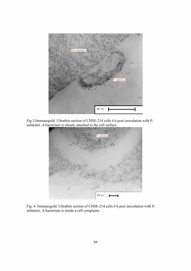
66
Fig.3.Immunogold. Ultrathin section of CHSE-214 cells 4 h post inoculation with P. salmonis. A bacterium is closely attached to the cell surface.
Fig. 4. Immunogold. Ultrathin section of CHSE-214 cells 6 h post inoculation with P. salmonis. A bacterium is inside a cell cytoplasm.

67
Fig. 5.Immunogold. Ultrathin section of CHSE-214 cells 8 days post inoculation with P. salmonis. A bacterium is within a vacuole inside the cell cytoplasm.
Fig. 6. Standard transmission electron microscopy. Ultrathin section of CHSE-214 cells 8 days post inoculation with P. salmonis. A bacterium is within a membrane-bound vacuole inside the cell cytoplasm. Note the rippled cell wall and electron-lucent structures.
68
Artículo 1.2. (Objetivo 1). J. Fish Diseases 38:321-324. 2015.
Title Page:
Full title: “Effect of Piscirickettsia salmonis inoculation on the ASK continuous cell
line”
Correspondence: P. A. Smith, Department of Animal Pathology, Faculty of
Veterinary Sciences, University of Chile, Santa Rosa 11735, Santiago, Chile
e-mail: [email protected]
Short running title: “Piscirickettsia salmonis in the ASK cell line”

69
SHORT COMMUNICATION Effect of Piscirickettsia salmonis inoculation on the ASK continuous cell line
P. A. Smith1, F.E. Díaz1, M.E. Rojas1, S. Díaz1, M. Galleguillos2 and A. Carbonero3
1 Department of Animal Pathology. Faculty of Veterinary Sciences. University of Chile,
Santiago, Chile.
2 Department of Animal Biological Sciences, Faculty of Veterinary Sciences. University of
Chile, Santiago, Chile.
3 Department of Animal Health, University of Córdoba, Spain.
Keywords: Piscirickettsia salmonis, ASK cells, cytopathic effect, titre
Piscirickettsia salmonis (P. salmonis) is a Gram-negative aquatic pathogen that causes
piscirickettsiosis, a contagious systemic disease which was first described in coho salmon
Oncorhynchus kisutch (Walbaum) cultured in sea net pens (Fryer, Lannan, Garcés, Larenas
& Smith 1990). Since then this condition has been detected in a variety of teleost fish from
distant geographic regions in the world, but so far it has been consistently more severe in
salmonid species reared in the South Pacific Ocean in Chile (Arkush & Bartholomew 2011;
Rojas, Galleguillos, Díaz, Machuca, Carbonero & Smith 2013).
First isolation of P. salmonis was in the chinook salmon, O. tshawytscha (Walbaum),
embryo (CHSE-214) cell line (Fryer et al. 1990), and these cells were since then the most
extensively used substrate for this bacterium propagation (Arkush & Bartholomew 2011;
Rojas et al. 2013). For some years it was thought that P. salmonis could replicate in vitro
only in cell cultures, but rather recently it was found that it can also be grown in some
enriched artificial media (Mauel, Ware & Smith 2008; Mikalsen, Skjærvik, Wiik-Nielsen,

70
Wasmuth & Colquhoun 2008) and, therefore, at present it is characterized as a facultative,
instead of an obligated, intracellular organism. Although several fish cell lines in which P.
salmonis replicates have been described, no attempts have been made to know if the
Atlantic salmon, Salmo salar L., kidney (ASK) cell line developed by Devold, Krossøy,
Aspehaug & Nylund (2000) is permissive to this bacterium. The objective of this work
was to determine the effect of P. salmonis inoculation on ASK cells through the cytopathic
effect (CPE) description and bacterial titration after cell exposure to this fish pathogen.
Monolayers of the ASK (ATCC CRL 2797) and the CHSE-214 (ATCC CRL 1681) cell
lines, both cultured at 18 °C in absence of antimicrobials, were used. The ASK cells were
cultured with L-15 Leivobitz medium with L-glutamine (2.05 mM) and supplemented with
β mercaptoethanol (38.5 μM) and foetal calf serum (FCS) at 10% (all from Gibco, Life
Technologies, Carlsbad, CA). The CHSE-214 cells were grown in Eagle´s minimal
essential medium with Earle´s salts (Automod Sigma-Aldrich, Lenexa, KS), L-glutamine (2
mM) and FCS (10%).
An isolate of P. salmonis obtained from an Atlantic salmon suffering clinical
piscirickettsiosis, sampled at a sea-site of the south of Chile, was used. It was cultured in
CHSE-214 cells. Infectious supernatant was harvested when CPE reached 100% and it was
titered by end-point dilution assay using 96-well microplates containing monolayers of
CHSE-214 cells, with 6 wells per dilution, employing the method of Reed & Müench
(1938) to estimate the tissue culture infectious dose 50% per mL (TCID50 mL-1). Finally,
the inoculum used to expose the cells in the infectivity assays explained below was a
bacterial suspension having 105.7 TCID50 contained in 0.2 mL.
71
In the infectivity assays, which were replicated once, fresh monolayers of ASK cells
cultured in 25 cm2 flasks were exposed to the P. salmonis inoculum and observed daily
under inverted microscope for 20 post-inoculation (p.i.) days. Concurrently, for
comparison, CHSE-214 cells were infected and treated in the same way as the ASK cells.
Non-infected ASK and CHSE-214 cells were also included as experimental controls. At the
end of the assays methanol-fixed smears from supernatants of the infected and non-infected
cell cultures were obtained. These smears were stained with Gram and Giemsa and also
labelled to detect P. salmonis by indirect immunofluorescence test (IFAT) according to
Lannan, Ewing & Fryer (1991).
Titration of ASK cell supernatants post P. salmonis infection was carried out once CPE
approached 100% in these cells, which occurred at 20 p.i. days. Supernatants were titrated
in microplates containing CHSE-214 cells as previously explained. Titration was also
conducted after a second passage of P. salmonis in ASK cells which, in turn, were infected
with 0.2 mL of supernatant of infected ASK cells with approximately 100% of CPE.
Monolayers of ASK cells exhibited a conspicuous and distinctive CPE after P. salmonis
infection. First morphological changes were evident quite early, starting at day 3 p.i., and
were characterized by the presence of rounded intracytoplasmatic vacuoles in few cells. As
time elapsed the number of affected cells and the vacuoles in them progressively increased
in such a way that in some cells more than 50 vacuoles of different size and morphology
could be counted. The smaller vacuoles were almost circular with a diameter starting from
ca. 1 µm. Some of the larger vacuoles showed the same shape of the smaller ones, but
others were more asymmetrical, some of them reaching ca. 5 µm in their longer axis, as
shown in Fig. 1 (B and C). Some vacuoles contained structures consistent with P. salmonis

72
morphology while others looked empty. In time, a progressive ASK cell detachment
occurred homogeneously without leaving the discrete large spaces observed in the CPE
when P. salmonis infects CHSE-214 cells (Fryer et al. 1990). The CPE difference between
ASK and CHSE-214 cells infected with P. salmonis can be seen in illustrations C and D
(Fig. 1) at 5 p.i. days and in E and F (Fig. 1) at 11 p.i. days. At day 20 p.i., detachment of
the infected ASK cells approached 100%. Detachment was clearly higher in ASK than in
CHSE-214 cells at a given time, as shown in pictures G and H (Fig. 1).
In advanced infection stages, P. salmonis compatible particles, extracellularly suspended,
were seen with inverted microscope in a higher number and larger size in ASK compared to
CHSE-214 cultures. Nevertheless, observation under standard optical microscopy of smears
of ASK cells infected with P. salmonis, stained either with Gram or Giemsa or IFAT
labelled, showed the typical morphological features widely described for this bacterium
(Fryer et al. 1990; Lannan et al. 1991; Fryer & Hedrick 2003; Arkush & Bartholomew
2011).
The replication ability of P. salmonis in ASK cell monolayers shown here was expected for
at least two reasons. The first one is that these cells originate from Atlantic salmon and it is
widely known that salmonid fish are highly susceptible to P. salmonis infection in natural
and under experimental conditions (Fryer et al. 1990; Garcés, Larenas, Smith, Sandino,
Fryer & Lannan 1991; Fryer & Hedrick 2003; Arkush & Bartholomew 2011). The second
reason is that the ASK line is derived from kidney cells (Devold et al. 2000) which are one
of the main target tissues of P. salmonis (Branson & Nieto Díaz-Muñoz 1991; Venegas,
Contreras, Larenas & Smith 2004; McCarthy, Bron, Brown, Pourahmad, Bricknell,
Thompson, Adams & Ellis 2008). It is worth mentioning that there are 11 cell lines so far

73
reported as permissive to P. salmonis, nine of them from teleosts (salmonid and non-
salmonid fish) and the remaining two, from an amphibian and an invertebrate species.
Salmonid cell lines, besides CHSE-214, are CSE-119 (coho salmon embryo), RTG-2
(rainbow trout gonad), CHH-1 (chum salmon heart) (Fryer et al. 1990), RTS11 (rainbow
trout spleen) (Rojas, Galanti, Bols & Marshall 2009) and SHK-1 (salmon head kidney)
(Vera, Isla, Cuevas & Figueroa 2012). From non-salmonid fish are EPC (epithelioma
papulosum cyprini), FHM (fathead minnow) (Fryer et al. 1990) and BB (brown bullhead)
cells (Almendras, Jones, Fuentealba & Wright 1997). The cell lines of amphibian and insect
origin are XTC-2 and Sf21 from Xenopus laevis (Daudin) and Spodoptera frugiperda (J.E.
Smith), respectively (Birkbeck, Griffen, Reid, Laidler & Wadsworth 2004).
The titre obtained from supernatants of infected ASK cells was 105.5 TCID50 mL-1. Same
results were achieved when P. salmonis inocula for infecting the ASK monolayers came
from infected CHSE-214 or ASK cell supernatants. These titres are approximately ten
times lower than the values reached in CHSE-214 cells in which normal titres range from
106 to 107 TCID50 mL-1 (Fryer et al. 1990; Cvitanich, Garate & Smith 1991). It should be
noted that the highest yields described so far are reached in the Sf21 insect cell line in
which 100 times more P. salmonis infecting units are obtained compared to those in CHSE-
214 cells (Birkbeck et al. 2004). The relatively low titres obtained in ASK cells indicate
that these cells would not be a good option for high production of viable P. salmonis.
In conclusion ASK cells are permissive to the P. salmonis infection and therefore they
expand the cell line repertoire that can be used for primary isolation as a diagnostic tool
and, most importantly, for a wide scope of research studies, such as those focussed to
understand the host-parasite interactions.
74
Acknowledgements
Authors are grateful to Mrs. D. Vega, Dr. J. Palomino and Mrs. A.M. Espinoza for
their valuable help in cell culture handling, microphotography and critical review of this
manuscript, respectively. This work was supported by Grant Fondecyt (Chile) 1080692.

75
References
Almendras F.E., Jones S.R.M., Fuentealba C. & Wright G.M. (1997) In vitro infection of a
cell line from Ictalurus nebulosus with Piscirickettsia salmonis. Canadian Journal of
Veterinary Research 61, 66-68.
Arkush K.D. & Bartholomew J.L. (2011) Piscirickettsia, Francisella and Epitheliocystis.
In: Fish Diseases and Disorders. Volume 3 Viral, Bacterial and Fungal Infections (ed. by
P.T.K Woo & D.W. Bruno). 2nd edn, pp. 302-337. CAB International. Wallingford, U.K.
Birkbeck T.H., Griffen A.A., Reid H.I., Laidler L.A. & Wadsworth S. (2004) Growth of
Piscirickettsia salmonis to high titers in insect tissue culture cells. Infection & Immunity 72,
3693-3694.
Branson E.J. & Nieto Díaz-Muñoz D. (1991) Description for a new disease condition
occurring in farmed coho salmon, Oncorhynchus kisutch (Walbaum), in South America.
Journal of Fish Diseases 14, 147-156.
Cvitanich J., Garate O. & Smith C.E. (1991) The isolation of a rickettsia-like organism
causing disease and mortality in Chilean salmonids and its confirmation by Koch’s
postulate. Journal of Fish Diseases 14, 121-145.
Devold M., Krossøy B., Aspehaug V. & Nylund A. (2000) Use of RT-PCR for diagnosis of
infectious salmon anaemia virus (ISAV) in carrier sea trout Salmo trutta after experimental
infection. Diseases of Aquatic Organisms 40, 9-18.
76
Fryer J.L., Lannan C.N., Garcés L.H., Larenas J.J. & Smith P.A. (1990) Isolation of a
rickettsiales-like organism from diseased coho salmon (Oncorhynchus kisutch) in Chile.
Fish Pathology 25, 107-114.
Fryer J.L. & Hedrick R.P. (2003) Piscirickettsia salmonis: a Gram-negative intracellular
bacterial pathogen of fish. Journal of Fish Diseases 26, 251-262.
Garcés L.H., Larenas J.J., Smith P.A., Sandino S., Fryer J.L. & Lannan C.N. (1991)
Infectivity of a rickettsia isolated from coho salmon (Oncorhynchus kisutch). Diseases of
Aquatic Organisms 11, 93-97.
Lannan C.N., Ewing S.A. & Fryer J.L. (1991) A fluorescent antibody test for detection of
the rickettsia causing disease in Chilean salmonids. Journal of Aquatic Animal Health 3,
229-234.
Mauel M.J., Ware C. & Smith P.A. (2008) Culture of Piscirickettsia salmonis on enriched
blood agar. Journal of Veterinary Diagnostic Investigation 20, 213-214.
McCarthy Ú.M., Bron J.E., Brown L., Pourahmad F., Bricknell L.R., Thompson K.D.,
Adams A. & Ellis A.E. (2008) Survival and replication of Piscirickettsia salmonis in
rainbow trout head kidney macrophages. Fish & Shellfish Immunology 25, 477-484.
Mikalsen J., Skjærvik O., Wiik-Nielsen J., Wasmuth M.A. & Colquhoun D.J. (2008) Agar
culture of Piscirickettsia salmonis, a serious pathogen of farmed salmonid and marine fish.
FEMS Microbiology Letters 278, 43-47.
Reed L. & Müench H. (1938) A simple method of estimating fifty percent endpoints.
American Journal of Hygiene 27, 493-497.
77
Rojas M.E., Galleguillos M., Díaz S., Machuca A., Carbonero A. & Smith P.A. (2013)
Evidence of exotoxin secretion of Piscirickettsia salmonis, the causative agent of
piscirickettsiosis. Journal of Fish Diseases 36, 703-709.
Rojas V., Galanti N., Bols N.C. & Marshall S.H. (2009) Productive infection of
Piscirickettsia salmonis in macrophages and monocyte-like cells from rainbow trout, a
possible survival strategy. Journal of Cellular Biochemistry 108, 631-637.
Venegas C.A., Contreras J.R., Larenas J. & Smith P.A. (2004) DNA hybridization assays
for the detection of Piscirickettsia salmonis in salmonid fish. Journal of Fish Diseases 27,
431-433.
Vera T., Isla A., Cuevas A. & Figueroa J. (2012) Un nuevo medio de cultivo líquido para el
patógeno Piscirickettsia salmonis. Archivos de Medicina Veterinaria 44, 271-277.

78
Figure 1 Monolayers of ASK [(A), (B), (C), (E) and (G)] and CHSE-214 cells [(D), (F) and (H)]. Inverted microscope with phase contrast. (A) Uninfected ASK cells. 100x. Illustrations (B) to (H) show cells after different post-inoculation (p.i.) days with Piscirickettsia salmonis. (B) ASK cells after 5 p.i. days. 100x. Some cells show intracytoplasmatic vacuoles. (C) and (D), ASK and CHSE-214 cells, respectively, 5 p.i. days. 200x. (C) Intracytoplasmatic vacuoles are observed with higher magnification than in (B). (D) Typical clusters of rounded refringent cells observed at the beginning of the cytopathic effect (CPE) caused by P. salmonis in CHSE-214 cells. (E) and (F), ASK and CHSE-214 cells, respectively, 11 p.i. days. 100x. (E) Extensive cell vacuolization and detachment of some of them. (F) Typical CPE in CHSE-214 cells with discrete foci of cell detachment surrounded by rounded refringent cells. (G) and (H), ASK and CHSE-214 cells, respectively, 14 p.i. days. 100x. (G) Detachment of more than 60% of the ASK cell monolayer. (H) Massive presence of rounded refringent CHSE-214 cells, but with less than the 15% of detachment of the cell monolayer. Bars ≈ 20 µm.
(A) (B)
(C) (D)
(E) (F)
(G) (H)
79
Artículo 2.1. (Objetivo 2) J. Fish Dis. 38:765-770. 2015.
Title Page:
Full title: “Infectivity of Piscirickettsia salmonis in immersion-bath exposed rainbow trout Oncorhynchus mykiss (Walbaum) fry”
Correspondence: P. A. Smith, Department of Animal Pathology, Faculty of
Veterinary Sciences, University of Chile, Santa Rosa 11735, Santiago, Chile
e-mail: [email protected]
Short running title: “Piscirickettsia infectivity in trout”
80
SHORT COMMUNICATION
Infectivity of Piscirickettsia salmonis in immersion-bath exposed rainbow trout
Oncorhynchus mykiss (Walbaum) fry
P.A. Smith1, J.R. Contreras1, M.E. Rojas1, A. Guajardo1, S. Díaz1, A. and Carbonero2
1Department of Animal Pathology. Faculty of Veterinary Sciences. Pathology of Aquatic
Animal Unit. University of Chile, Santiago, Chile
2Department of Animal Health, University of Córdoba, Spain.
Keywords: Piscirickettsia, infectivity, rainbow trout, pathogeny, piscirickettsiosis
Piscirickettsia salmonis is an obligate bacterial pathogen which causes
piscirickettsiosis, a systemic disease affecting some anadromous and marine teleost fish
species. To investigate the pathogenesis of this disease, a time-course study was conducted,
using immunohistochemistry, after challenging rainbow trout Oncorhynchus mykiss
(Walbaum) fry by an immersion bath with P. salmonis.
To carry out this assay, fish (total n = 72; weight ≈ 2.5 g) were allotted to six
subgroups (12 fish each) held in individual 50-L tanks supplied with a flow-through
freshwater system (25 L h-1) at 15.4 °C (SD 0.8).
The SLGO-95 strain of P. salmonis (Smith, Vecchiola, Oyanedel, Garcés, Larenas
& Contreras 1996) was used. Bacteria, after thawing, were cultured and titered, by end-
point dilution assay, in the CHSE-214 cell line. Cells were cultured at 18 °C in Eagle’s
minimal essential medium with Earle’s salts (MEM), which is autoclavable (MEM Auto-

81
Mod Sigma-Aldrich, KS, USA), supplemented with 2 mM L-glutamine and 10% of foetal
calf serum (both from Gibco, Life Technologies, CA, USA).
For the experimental challenge, four fish subgroups (A1, A2, B1 and B2) were
exposed to P. salmonis, each one as an independent batch, by immersion for 15 min in a
50-mL bacterial suspension (in MEM) containing ≈105 tissue culture infectious dose 50%
per mL. After exposure, each subgroup was kept for 30 min in 1-L sterile MEM, although
some fish were sampled within this period, and was then replaced in the 50-L tanks. Fish
from subgroups A1 and A2 were used to record clinical signs, mortalities and gross and
histological lesions (standard haematoxylin and eosin) for 30 days after the immersion
challenge, as a bacterial virulence control. Methanol-fixed smears from the kidneys of dead
fish were obtained for Gram staining and also for labelling to detect P. salmonis by the
indirect immunofluorescence test (IFAT) according to Lannan, Ewing & Fryer (1991). Two
additional fish subgroups (C1 and C2) were included as non-infected controls. Excepting
that these fish were sham-exposed, they were treated as subgroups A1 and A2. All these
immersion procedures were carried out under permanent aeration at 16.5 (+/- 0.5) °C.
Fish of subgroups B1 and B2 were sequentially sampled. Three fish were euthanized
by anaesthetic overdose (Tricaine Sigma-Aldrich, MO, USA) at each of the following post-
exposure (post-exp) times counted from the start of the immersion with P. salmonis: 5 min,
15 min, 30 min, 1 h, 3 h, 6 h, 18 h and 3 days. Fish were formalin fixed and processed by
standard histological methods. Five-µm sagittal sections of the whole body, excepting the
tail, were examined, using an immunoperoxidase test to detect P. salmonis in the tissues.
No mortalities or clinical signs occurred in the mock-treated fish (subgroups C1 and
C2). In exposed fish (subgroups A1 and A2), cumulative mortalities reached 91.7% at day
23 post-exp (Fig. 1), indicating that fry were very susceptible to this P. salmonis strain.

82
Clinical signs of diseased fish were anorexia, skin darkness, lethargy and, usually when
near to death, uncoordinated swimming behaviour close to the water surface. Gross
pathology was characterized by pale gills, petechiae in the wall muscles of the coelomic
cavity and liver, spleen and kidney enlargement and mucohaemorrhagic content in the large
bowel. Histopathology showed haemorrhagic and necrotic foci mostly in haemopoietic
kidney, liver, spleen and intestine. These tissues also exhibited perivascular and endothelial
necrosis with associated thrombi. Examination of kidney smears, stained with Gram or
labelled with IFAT, showed organisms with the characteristic morphology widely
described for P. salmonis (Fryer, Lannan, Garcés, Larenas & Smith 1990; Lannan et al.
1991; Fryer & Hedrick 2003; Arkush & Bartholomew 2011). The clinical and pathological
findings were consistent with those associated with acute piscirickettsiosis in salmonid fish
in the field (Branson & Nieto Díaz-Muñoz 1991) or under experimental conditions (Garcés,
Larenas, Smith, Sandino, Fryer & Lannan 1991; Smith, Contreras, Garcés, Larenas,
Oyanedel, Caswell-Reno & Fryer 1996). This immersion-bath method allowed the
experimental reproduction of piscirickettsiosis, which could thus be used as a model to
develop challenging tests mimicking the natural way of fish exposure. Appropriate
challenging methods are required to test vaccines and therapeutic drugs against fish
pathogens in a reliable way (Adams, Leschen, Wilson & Horne 1987).
The outcome of the sequential tissue sample examination is shown in Table 1. In
summary, the time-course of events, considering the first post-exp time that P. salmonis
was detected in a particular tissue, was as follows: at 5 min it was found attached to the
surface of the external epithelial cell layer of the skin and gills; from 15 to 30 min it was
detected within the epithelial layer of skin, gills, oesophagus, pyloric caeca and intestine;
from 3 to 6 h it was present in the wall muscle layers of oesophagus and stomach and in

83
skin dermis, and from 18 h onward it was evidenced in the lumen of blood vessels and
virtually in all the fish organs.
Hence, these results show that there was a relatively steady progression of the
bacterial invasion at early stages from skin, respiratory and digestive epithelia to deeper
tissues, followed by a rapid spread to probably all the irrigated tissues of the fish body,
once the micro-organism reached the blood stream. As already mentioned, the bacterium
was found attached to the epithelial cells as early as 5 min post-exp (Fig. 2a), which is
consistent with the results obtained in an in vitro infectivity study of P. salmonis in CHSE-
214 cells (Smith, Reveco, Contreras, Rojas, Venegas & Guajardo 2010). An efficient
attachment ability is expected in successful aquatic pathogens because without it these
organisms would run the risk of being washed off the host or being voided from the host’s
gastrointestinal tract (Evelyn 1996). The mechanisms and/or structures used by P. salmonis
for attachment to the epithelial cells are unknown, although the membrane extensions that
are reported to allow the anchorage of this bacterium to the ova chorion (Larenas,
Bartholomew, Troncoso, Fernández, Ledezma, Sandoval, Vera, Contreras & Smith 2003)
may have occurred with a similar function in the present study.
The invasion kinetics from the attachment time to the moment when the blood
stream was reached by this bacterium was too rapid to be explained only by cell to cell
contiguity spreading. In this respect, although the multiplication rate of P. salmonis might
be higher in vivo than in vitro, at least in an axenic culture medium, the generation time of
P. salmonis (≈ 12 h) is relatively slow (Vera, Isla, Cuevas & Figueroa 2012) and the time it
takes in vitro to be incorporated into the cytoplasm after cell exposure is 1 h in rainbow
trout kidney macrophages (McCarthy, Bron, Brown, Pourahmad, Bricknell, Thompson,
Adams & Ellis 2008) and is 3 to 6 h in CHSE-214 cells (Smith et al. 2010). The

84
mechanisms that P. salmonis uses to invade fish tissues in vivo are unknown, but the outer
membrane lipopolysaccharides (Vadovič, Fodorová & Toman 2007; Vinogradov,
Frimmelova & Toman 2013) and the recently described exotoxins (Rojas, Galleguillos,
Díaz, Machuca, Carbonero & Smith 2013) of this bacterium might play a role in this
phenomenon.
In the digestive tube walls, attachment and invasion were earlier in the epithelial
layers of the anterior than in the posterior segments of this tract (Table 1), which probably
reflects the normal way of transit of the digestive content in the alimentary canal. It is
relevant to note that the oesophagus wall was invaded by P. salmonis (Table 1), including
the muscularis externa layer (image not shown). This site of bacterial entry has not been
described in previous infectivity studies of this pathogen in salmonid fish (Almendras,
Fuentealba, Jones, Markham & Spangler 1997; Smith, Pizarro, Ojeda, Contreras, Oyanedel
& Larenas 1999; Smith, Rojas, Guajardo, Contreras, Morales & Larenas 2004) and neither
has it been reported among the usual routes of entry of fish pathogens in general (Evelyn,
1996; Birkbeck & Ringø 2005). Nevertheless, the infectious haematopoietic necrosis virus
also uses the oesophagus as an entrance site in rainbow trout fry (Drolet, Rohovec & Leong
1994; Helmick, Bailey, La Patra & Ristow 1995), which suggests that this section of the
digestive tract might be a more common entry portal for fish infectious agents than what
has been reported to date.
Piscirickettsiosis is almost exclusively a sea-water disease (Fryer et al. 1990; Mauel
& Miller 2002; Arkush & Bartholomew 2011) and given the fact that fish in this
environment drink large volumes of water (Edwards & Marshall 2013), in which P.
salmonis can be suspended (Lannan & Fryer 1994; Olivares & Marshall 2010), it is

85
possible that the gut epithelium, especially that of the oesophagus and intestine, plays a role
as an attachment point and subsequent route of entry of P. salmonis in natural conditions.
Although the bacterium was also found attached to the gastric epithelial lining (Fig.
2b and Table 1) and inside the wall muscle of the stomach (Table 1), it was clearly present
in lower amounts than in the oesophagus and intestine (Table 1). It therefore appears that
the stomach wall might be a potential entry portal for P. salmonis, at least in rainbow trout
fry, although probably less efficient than other segments of the digestive tract. In a previous
work to assess the routes of entry of P. salmonis in juvenile rainbow trout (Smith et al.
1999), fish were exposed by patch contact in skin and gills and by intubation in stomach
and intestine. Cumulative mortalities were 52, 24, and 2 and 24 %, respectively. These
results are consistent with those obtained here with respect to skin and gills as important
entry portals and the gastric environment as a probable hostile medium, although not 100%
lethal, to P. salmonis.
A final consideration about the gut infectivity results is that the bacterium
reappeared in the intestine epithelium at advanced stages of infection (Table 1), which is
consistent with the severe enteritis found in fish having piscirickettsiosis in the field
(Branson & Nieto Díaz-Muñoz 1991) and in the exposed rainbow trout fry here.
In addition to the variety of cell types that P. salmonis has already been described to
infect, the bacterium in this study was also found in the cytoplasm of goblet cells of the
skin epidermis (Fig. 2c), which occurred from 15 min to 6 h post exp, and in skeletal
muscular cells in more advanced stages of infection, from 18 h post exp (Fig. 2d and Table
1). The role of the goblet cells in the establishment of P. salmonis infection is unknown, but
during differentiation these cells migrate to the superficial epithelial layers and remain there
(Roberts & Ellis 2001; Janice & Khan 2013); it could therefore be hypothesised that they

86
do not aid movement of the bacterium into deeper tissues and probably have a defensive
function instead. It has been well documented that goblet cell mucus is an important
component of non-specific immunity against infectious agents in a wide variety of
vertebrates including teleost fish (Ellis 2001; Janice & Khan 2013). However, it should also
be taken into account that fish mucus can be beneficial to some pathogens either as a source
of nutrients and/or chemoattractants, as it occurs with Vibrio (at present, Aliivibrio)
anguillarum (Garcia, Otto, Kjelleberg & Nelson 1997; O´Toole, Lundberg, Fredriksson,
Jansson, Nilsson & Wolf-Watz 1999).
Non-specific and specific immunity is developed to a large extent in salmonid fry of
even smaller size (weight) than those of 2.5 g used here (Tatner 1996; Gadan, Sandtrø,
Marjara, Santi, Munang’andu & Evensen 2013), but fully mature immunocompetence is
obtained at later ontogenic stages, which in rainbow trout occurs in fish weighing over 4 g
(Johnson, Flynn & Amend 1982). Due to this ontogenic variation in the immune response,
together with other physiological changes, it is not possible to directly infer the results
obtained here in fry to post-smolts or adult fish, in which the disease normally occurs.
In summary, infectivity of P. salmonis in rainbow trout fry would comprise three
major stages: 1) a rapid attachment to epithelial cells mainly of skin and gills, but also of
the alimentary canal; 2) a progressive invasion from the sites of entry to deeper underlying
tissues until reaching the bloodstream and; 3) a rapid haematogenous spread to virtually all
body tissues. It is evident that P. salmonis is very successful in systemically infecting its
hosts, but the mechanisms this bacterium uses to breach, circumvent, or use to its advantage
the many physical (starting from the mucus), anatomical and immunological barriers that
healthy fish possess to avoid invasion by pathogenic micro-organisms are elusive and

87
intriguing so far. Therefore further research is necessary to reveal the pathogenesis,
specially at a cellular and molecular level, of this significant fish disease.
Acknowledgements
This work was supported by Grant Fondecyt (Chile) 1010544.

88
References
Adams A., Leschen W., Wilson A. & Horne M.T. (1987) A bath challenge model for
furunculosis in rainbow trout, Salmo gairdneri Richardson, and Atlantic salmon, Salmo
salar L. Journal of Fish Diseases 10, 495–504.
Almendras F.E., Fuentealba I.C., Jones S.R.M., Markham F. & Spangler E. (1997)
Experimental infection and horizontal transmission of Piscirickettsia salmonis in
freshwater-raised Atlantic salmon, Salmo salar L. Journal of Fish Diseases 20, 409–418.
Arkush K.D. & Bartholomew J.L. (2011) Piscirickettsia, Francisella and Epitheliocystis.
In: Fish Diseases and Disorders. Volume 3 Viral, Bacterial and Fungal Infections (ed. by
P.T.K Woo & D.W. Bruno). 2nd edn, pp. 302–337. CAB International, Wallingford, U.K.
Birkbeck T.H. & Ringø E. (2005) Pathogenesis and the Gastrointestinal Tract of Growing
Fish. In: Microbial Ecology in Growing Animals. Biology of Growing Animals Series (ed.
by W.H. Holzapfel & P.J. Naughton). pp. 208–234. Elsevier, Edinburgh, U.K.
Branson E.J. & Nieto Díaz-Muñoz D. (1991) Description for a new disease condition
occurring in farmed coho salmon, Oncorhynchus kisutch (Walbaum), in South America.
Journal of Fish Diseases 14, 147–156.
Drolet B.S., Rohovec J.S. & Leong J.A. (1994) The route of entry and progression of
infectious haematopoietic necrosis virus in Oncorhynchus mykiss (Walbaum): a sequential
immunohistochemical study. Journal of Fish Diseases 17, 337–347.
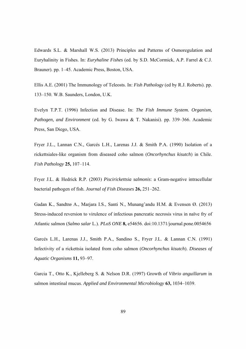
89
Edwards S.L. & Marshall W.S. (2013) Principles and Patterns of Osmoregulation and
Euryhalinity in Fishes. In: Euryhaline Fishes (ed. by S.D. McCormick, A.P. Farrel & C.J.
Brauner). pp. 1–45. Academic Press, Boston, USA.
Ellis A.E. (2001) The Immunology of Teleosts. In: Fish Pathology (ed by R.J. Roberts). pp.
133–150. W.B. Saunders, London, U.K.
Evelyn T.P.T. (1996) Infection and Disease. In: The Fish Immune System. Organism,
Pathogen, and Environment (ed. by G. Iwawa & T. Nakanisi). pp. 339–366. Academic
Press, San Diego, USA.
Fryer J.L., Lannan C.N., Garcés L.H., Larenas J.J. & Smith P.A. (1990) Isolation of a
rickettsiales-like organism from diseased coho salmon (Oncorhynchus kisutch) in Chile.
Fish Pathology 25, 107–114.
Fryer J.L. & Hedrick R.P. (2003) Piscirickettsia salmonis: a Gram-negative intracellular
bacterial pathogen of fish. Journal of Fish Diseases 26, 251–262.
Gadan K., Sandtrø A., Marjara I.S., Santi N., Munang’andu H.M. & Evensen Ø. (2013)
Stress-induced reversion to virulence of infectious pancreatic necrosis virus in naïve fry of
Atlantic salmon (Salmo salar L.). PLoS ONE 8, e54656. doi:10.1371/journal.pone.0054656
Garcés L.H., Larenas J.J., Smith P.A., Sandino S., Fryer J.L. & Lannan C.N. (1991)
Infectivity of a rickettsia isolated from coho salmon (Oncorhynchus kisutch). Diseases of
Aquatic Organisms 11, 93–97.
Garcia T., Otto K., Kjelleberg S. & Nelson D.R. (1997) Growth of Vibrio anguillarum in
salmon intestinal mucus. Applied and Environmental Microbiology 63, 1034–1039.

90
Helmick C.M., Bailey J.F., La Patra S. & Ristow S. (1995) The esophagus/cardiac stomach
region: site of attachment and internalization of infectious hematopoietic necrosis virus in
challenged juvenile rainbow trout Oncorhynchus mykiss and coho salmon O. kisutch.
Diseases of Aquatic Organisms 23, 189–199.
Janice J.K. & Khan W.I. (2013) Goblet cells and mucins: role in innate defense in enteric
infections. Pathogens 2, 55–70; doi:10.3390/pathogens2010055.
Johnson K.A., Flynn J.K. & Amend D.F. (1982) Duration of immunity in salmonids
vaccinated by direct immersion with Yersinia ruckeri and Vibrio anguillarum bacterins.
Journal of Fish Diseases 5, 207–213.
Lannan C.N., Ewing S.A. & Fryer J.L. (1991) A fluorescent antibody test for detection of
the rickettsia causing disease in Chilean salmonids. Journal of Aquatic Animal Health 3,
229–234.
Lannan C.N. & Fryer J.L. (1994) Extracellular survival of Piscirickettsia salmonis. Journal
of Fish Diseases 17, 545–548.
Larenas J.J., Bartholomew J., Troncoso O., Fernández S., Ledezma H., Sandoval N., Vera
P., Contreras J. & Smith P. (2003) Experimental vertical transmission of Piscirickettsia
salmonis and in vitro study of attachment and mode of entrance into the fish ovum.
Diseases of Aquatic Organisms 56, 25–30.
McCarthy U.M., Bron J.E., Brown L., Pourahmad F., Bricknell I.R., Thompson K.D.,
Adams A. & Ellis A.E. (2008) Survival and replication of Piscirickettsia salmonis in
rainbow trout head macrophages. Fish and Shellfish Immunology 25, 477–484.
Mauel M.J. & Miller D.L. (2002) Piscirickettsiosis and piscirickettsiosis–like infections in
fish: A review. Veterinary Microbiology 87, 279–289.
91
Olivares J. & Marshall S.H. (2010) Determination of minimal concentration of
Piscirickettsia salmonis in water columns to establish a fallowing period in salmon farms.
Journal of Fish Diseases 33, 261–266.
O´Toole R., Lundberg S., Fredriksson S., Jansson A., Nilsson B. & Wolf-Watz H. (1999)
The chemotactic response of Vibrio anguillarum to fish intestinal mucus is mediated by a
combination of multiple mucus components. Journal of Bacteriology 181, 4308–4317.
Roberts R.J. & Ellis A.E. (2001) The Anatomy and Physiology of Teleosts. In: Fish
Pathology (ed by R.J. Roberts). pp. 12–54. W.B. Saunders, London, U.K.
Rojas M.E., Galleguillos M., Díaz S., Machuca A., Carbonero A. & Smith P.A. (2013)
Evidence of exotoxin secretion of Piscirickettsia salmonis, the causative agent of
piscirickettsiosis. Journal of Fish Diseases 36, 703–709.
Smith P.A., Contreras J.R., Garcés L.H., Larenas J.J., Oyanedel S., Caswell-Reno P. &
Fryer J.L. (1996) Experimental challenge of coho salmon and rainbow trout with
Piscirickettsia salmonis. Journal of Aquatic Animal Health 8, 130–134.
Smith P.A., Pizarro P., Ojeda P., Contreras J., Oyanedel S. & Larenas J. (1999) Routes of
entry of Piscirickettsia salmonis in rainbow trout Oncorhynchus mykiss. Diseases of
Aquatic Organisms 37, 165–172.
Smith P.A., Reveco F., Contreras J., Rojas M.E., Venegas C. & Guajardo A. (2010)
Infectivity study of Piscirickettsia salmonis in CHSE-214 cells by confocal and
transmission electron microscopy. Bulletin of the European Association of Fish
Pathologists 30, 128–136.

92
Smith P.A., Rojas M.E., Guajardo A., Contreras J., Morales M.A. & Larenas J. (2004)
Experimental infection of coho salmon Oncorhynchus kisutch by exposure of skin, gills and
intestine with Piscirickettsia salmonis. Diseases of Aquatic Organisms 61, 53–57.
Smith P.A., Vecchiola I.M., Oyanedel S., Garcés L.H., Larenas J. & Contreras J. (1996)
Antimicrobial sensitivity of four isolates of Piscirickettsia salmonis. Bulletin of the
European Association of Fish Pathologists 16, 164–168.
Tatner M.F. (1996) Natural Changes in the Immune System of Fish. In: The Fish Immune
System. Organism, Pathogen, and Environment (ed. by G. Iwawa & T. Nakanisi). pp. 225–
287. Academic Press, San Diego, USA.
Vadovič P., Fodorová M. & Toman R. (2007) Structural features of lipid A of
Piscirickettsia salmonis, the etiological agent of the salmonid rickettsial septicemia. Acta
Virologica 51, 249–259.
Vera T., Isla A., Cuevas A. & Figueroa J. (2012) A new liquid medium for the pathogen
Piscirickettsia salmonis. Archivos de Medicina Veterinaria 44, 273–277.
Vinogradov E., Frimmelova M. & Toman R. (2013) Chemical structure of the carbohydrate
backbone of the lipopolysaccharide from Piscirickettsia salmonis. Carbohydrate Research
378, 108–113.
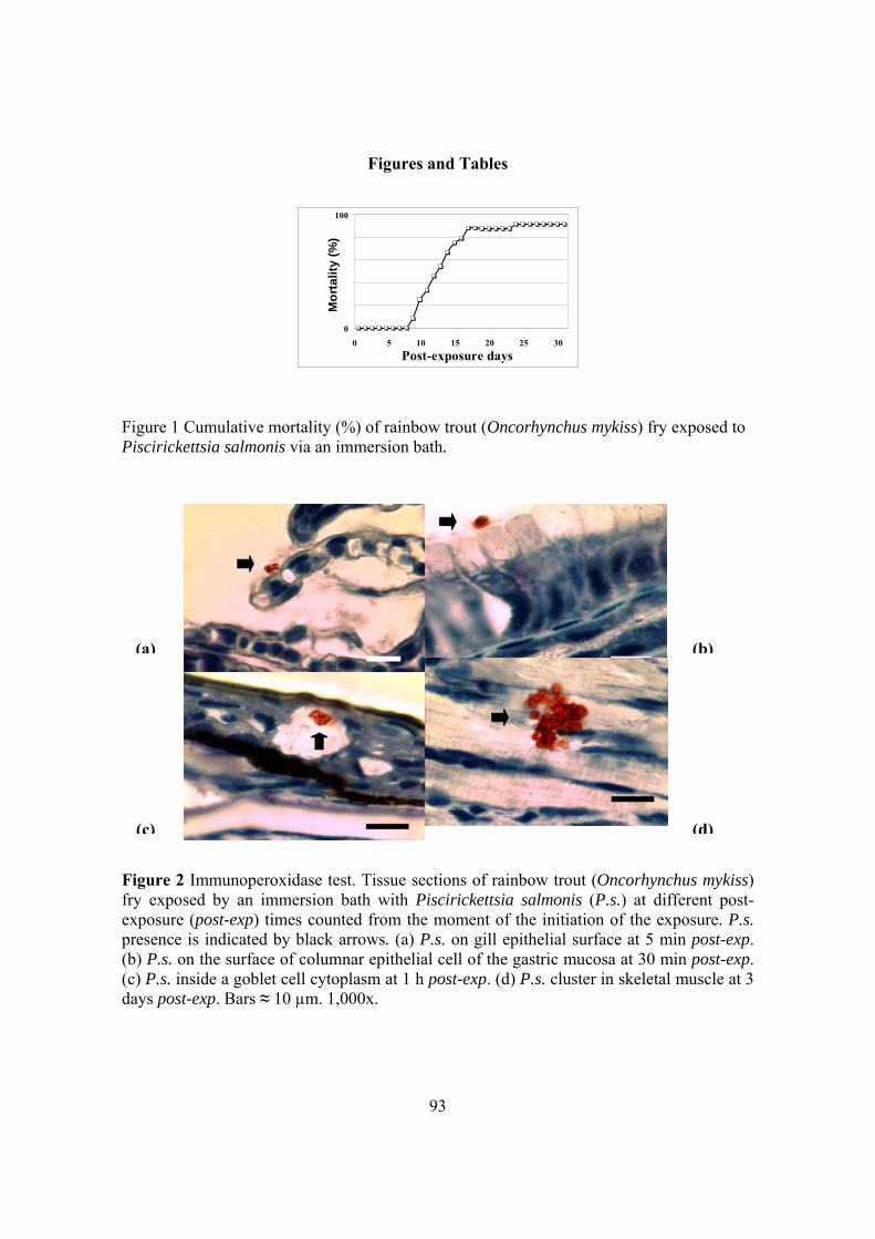
93
Figures and Tables
0
100
0 5 10 15 20 25 30
Post-exposure days
Mo
rta
lity
(%
)
Figure 1 Cumulative mortality (%) of rainbow trout (Oncorhynchus mykiss) fry exposed to Piscirickettsia salmonis via an immersion bath.
Figure 2 Immunoperoxidase test. Tissue sections of rainbow trout (Oncorhynchus mykiss) fry exposed by an immersion bath with Piscirickettsia salmonis (P.s.) at different post-exposure (post-exp) times counted from the moment of the initiation of the exposure. P.s. presence is indicated by black arrows. (a) P.s. on gill epithelial surface at 5 min post-exp. (b) P.s. on the surface of columnar epithelial cell of the gastric mucosa at 30 min post-exp. (c) P.s. inside a goblet cell cytoplasm at 1 h post-exp. (d) P.s. cluster in skeletal muscle at 3 days post-exp. Bars ≈ 10 µm. 1,000x.
(a) (b)
(c) (d)

94
Table 1 Time-course detection of Piscirickettsia salmonis by immunoperoxidase in selected
tissues obtained from rainbow trout (Oncorhynchus mykiss) exposed to this bacterium by an immersion bath1
Post-exposure time
Tissue 5 min 15 min 30 min 1 h 3 h 6 h 18 h 72 h
Gills
Epithelium surface 3 3 2 3 1 0 0 0
Epithelium 0 0 1 1 3 2 2 2
Skin
Epithelium surface 3 3 2 2 1 0 0 0
Epidermis 0 1 1 2 2 2 1 0
Dermis 0 0 0 0 0 1 1 1 Subcutaneous tissue 0 0 0 0 0 0 1 0
Oesophagus
Epithelium 0 3 2 2 1 0 0 0
Muscle layers 0 0 0 0 1 2 3 3
Stomach
Epithelium surface 0 0 1 0 0 0 0 0
Muscle layers 0 0 0 0 1 0 1 0 Pyloric caeca
Epithelium surface 0 0 1 0 1 0 0 0
Epithelium 0 0 1 0 2 1 0 0
Intestine
Epithelium surface 0 0 1 1 1 0 0 0
Epithelium 0 0 2 1 3 2 0 1
Muscle layers 0 0 0 0 0 0 0 1 Liver 0 0 0 0 0 0 3 3
Spleen 0 0 0 0 0 0 2 3
Skeletal muscle 0 0 0 0 0 0 3 3
Kidney 0 0 0 0 0 0 3 3
Cardiac muscle 0 0 0 0 0 0 3 3
Blood 0 0 0 0 0 0 3 3
Brain 0 0 0 0 0 0 1 1 Spinal cord 0 0 0 0 0 0 1 1
1Figures show the number of fish in which P. salmonis was detected in a particular tissue. The tissues were obtained from three fish at each sampling time.
95
Artículo 3.1. (Objetivo 3) J. Fish Dis.36:703-709. 2013.
Title Page:
Full title: “Evidence of exotoxin secretion of Piscirickettsia salmonis, the causative
agent of piscirickettsiosis"
Correspondence: P. A. Smith, Department of Animal Pathology, Faculty of
Veterinary Sciences, University of Chile, Santa Rosa 11735, Santiago, Chile
e-mail: [email protected]
Short running title: “Piscirickettsia salmonis exotoxin secretion”

96
Evidence of exotoxin secretion of Piscirickettsia salmonis, the causative agent of
piscirickettsiosis
M.E. Rojas1, M. Galleguillos2, S. Díaz1, A. Machuca1, A. Carbonero3 and P. A. Smith1
1 Department of Animal Pathology. Faculty of Veterinary Sciences. University of Chile,
Santiago, Chile.
2 Department of Animal Biological Sciences, Faculty of Veterinary Sciences. University of
Chile, Santiago, Chile.
3 Department of Animal Health, University of Córdoba, Spain.
Abstract
Piscirickettsia salmonis is the etiological agent of piscirickettsiosis, a disease which affects
a variety of teleost species and that is particularly severe in salmonid fish. Bacterial-free
supernatants, obtained from cultures of three isolates of Piscirickettsia salmonis, were
inoculated in Atlantic salmon (Salmo salar L) and in three continuous cell lines searching
for evidences of the secretion of extracellular products (ECPs) by this micro-organism.
Although steatosis was found in some liver samples, no mortalities or clinical signs
occurred in the inoculated fish. Clear cytotoxicity was observed after inoculation in the cell
lines CHSE-214 and ASK, derived from salmonid tissues, but not in MDBK, which has a
mammal origen. The degree of cytotoxicity of the ECPs was different among the P.
salmonis isolates tested. The isolate that evidenced the highest cytotoxicity in its ECPs
exhibited only an intermediate virulence level after challenging fish with bacterial
suspensions of the three P. salmonis isolates. Almost complete inhibition of the cytotoxic

97
activity of ECPs was seen after proteinase K treatment, indicating their peptidic nature, and
a total preclusion of the cytotoxicity was shown after their incubation at 50 °C for 30 min.
Results show that P. salmonis can produce ECPs and at least some of them are thermolabil
exotoxins that probably play a role in the pathogenesis of piscirickettsiosis.
Keywords: Piscirickettsia salmonis, pathogenicity, exotoxins, salmon, virulence.
Introduction
At present, a number of fastidious gram negative bacteria which replicates
intracellularly in their hosts, either obligated or in a facultative mode (Mauel, Ware &
Smith 2008), are associated as causative agents of important diseases in aquatic animals
including mollusks, crustaceans and teleost fishes. This emergent group of pathogens has
been named loosely as rickettsia-like organisms and/or Piscirickettsia-like organisms
(Mauel & Miller 2002). Within this bacterial group, Pisicrickettsia salmonis was the first
microorganism that was confirmed as a highly virulent pathogen, being the etiological
agent of piscirickettsiosis, a devastating disease in salmonid fish (Fryer & Hedrick 2003). A
significant number of studies on this bacterium have allowed to get important advances
such as obtaining a more accurate taxonomic classification and to understand, at least
partially, the immune response against it and the routes of entrance in its hosts (Fryer,
Lannan, Giovannoni & Wood 1992; Smith, Pizarro, Ojeda, Contreras, Oyanedel & Larenas
1999; Marshall, Conejeros, Zahr, Olivares, Gómez, Cataldo & Henríquez 2007).
Nevertheless, the virulence factors of this pathogen are poorly known. In this work, it was
explored if P. salmonis is able of exporting extracellular products (ECPs) through their
possible biological effects either in vitro in fish cell lines and/or in vivo in salmonid fish.

98
Material and Methods
Fish. In total, 560 Atlantic salmon (Salmo salar L.) pre-smolts, held in fiberglass tanks
supplied with a flow-through system of fresh water, were used. Some fish (n=400) were
employed for testing the virulence of the P. salmonis isolates and the remaining ones, to
test the effect of bacteria-free supernatants.
Cells. Monolayers of the chinook salmon (Oncorhynchus tschawytscha Walbaum) embryo
CHSE-214 (Lannan, Winton & Fryer 1984), Atlantic salmon kidney ASK (Devold,
Krossoy, Aspehaug & Nylund 2000) and Madin-Darby bovine (Bos taurus L.) kidney
MDBK (Madin & Darby 1958) cell lines were used. All of them were grown in absence of
antibiotics and antimycotics. Salmonid cells (CHSE-214 and ASK) were incubated at 17 ºC
and mammal cells (MDBK), unless explained, at 37 ºC. The CHSE-214 and MDBK cells
were cultured using Eagle´s minimal essential medium with Earle´s salts (Auto-mod,
Sigma, MO) added with L-glutamine (EMEM) and with 10% of foetal calf serum (EMEM-
10). The ASK cells were grown using L-15 Leibovitz medium with L-glutamine
supplemented with β-mercaptoethanol and with 10% of foetal calf serum (all of them from
Gibco, CA).
Bacteria. Three isolates of P. salmonis, named Ps-1, Ps-2 and Ps-3, obtained from rainbow
trout (O. mykiss Walbaum), Atlantic salmon and coho salmon (O. kisutch Walbaum),
respectively, were used in this study. Bacteria were isolated from fish suffering
piscirickettsiosis outbreaks from three different sea sites in the south of Chile. After the
primary isolation, bacteria were preserved at -196°C in liquid nitrogen until used. They
were cultured in all the experiments in monolayers of the CHSE-214 cell line and incubated

99
at 17 °C. Bacterial titration was carried out using the end-point dilution method (Reed &
Müench 1938) in 96 well microplates.
Virulence test of P. salmonis isolates. Ten experimental groups, with 40 fish each, were
used. Fish were distributed equally in two 2.5 m3 tanks, each having 200 tagged animals in
total, with 20 fish per experimental group. Fish were inoculated with three different doses
(103, 104 and 105 TCID50 fish-1) of each isolate. Bacterial suspensions were intraperitoneally
(ip) injected (0.1 mL per individual) in fish of the two tanks (i.e. 40 fish per dose of each
isolate). Forty sham-inoculated fish (20 individuals per tank), injected ip with 0.1 mL of a
sterile suspension of CHSE-214 cells, were used as control of the assay. Mortalities were
recorded up to 46 post-inoculation (p.i.) days. Dead fish were examined by necropsy.
Besides, kidney smears, fixed in absolute methanol, were gram stained and also analysed
by an indirect immunofluorescence test (IFAT) (Lannan, Ewing & Fryer 1991) to confirm
the presence of P. salmonis in these tissues.
Toxicity test in fish of P. salmonis-free supernatants (Ps-fs). Four experimental groups
with 40 fish each were used. Fish of three groups were injected ip with 0.1 mL of a Ps-fs
obtained from isolates Ps-1, Ps-2 and Ps-3, respectively. The fish belonging to the
remaining group were sham-inoculated. Fish of each group were allotted equally in two 100
L tanks (8 tanks in total). Piscirickettsia salmonis-free supernatants were obtained from
supernatants of CHSE-214 cells infected with P. salmonis and harvested when cytopathic
effect reached approximately 100%. Those supernatants were centrifuged twice at 5°C.
First centrifugation was at 1,000 x g for 15 min. The pellet was discarded and then the
supernantant centrifuged at 10,000 x g for 1h. The pellet was again discarded and finally
the supernatant was filtered through a membrane of mixed cellulose ester (DISMIC-25AS,
Toyo Roshi Kaisha, Ltd., Japan) with 0.2 µm size pores. The experiment lasted 47 p.i. days.

100
At the end of the assay, three fish per group were euthanised by anesthetic overdose
(Benzocaine at 200 mg/L) to obtain tissue samples (gills, heart, kidney, liver and spleen)
for histological examination.
Cytotoxicity test of Ps-fs in CHSE-214, ASK and MDBK cell cultures. Fresh
monolayers of cells grown in 25 cm2 flasks were inoculated in duplicated with 1 mL of
individual Ps-fs obtained from the isolates Ps-1, Ps-2 and Ps-3. Inoculated cells were
observed daily until day 25 p.i. under inverted optical microscope and their morphology
was compared with sham-inoculated cell monolayers. CHSE-214 and ASK cells were
always incubated at 17 ºC while MDBK cells were incubated in two different temperature
regimes: at 37 °C during all the assay or at 17 °C for 7 days after the inoculation time and
then at 37 °C until the end of the experiment at day 25 p.i..
Titration of Ps-fs cytotoxicity and effect of temperature and proteinase K on its
cytotoxic ability in CHSE-214 cell cultures. Only the Ps-fs obtained from the Ps-3 isolate
was tested. Unless otherwise explained, the incubations were at 17 °C and the general
method described just above was used in these assays. For titration, ten-fold dilutions of
Ps-fs were used. For the temperature test the Ps-fs were incubated at 50 °C and 60 °C for
30 min and then inoculated in cell monolayers. Proteinase K assay was carried out treating
a Ps-fs with 3.5 µM of this enzyme (United States Biological, Swampscott, MA). After a
48 h incubation period, the enzymatic reaction was stopped adding AEBS (4-(2-amino-
ethyl) benzenesulfonyl fluoride hydrochloride, Sigma, St. Louis, MO) at 0.5 mM.
Solutions were inoculated in cell cultures 40 min after AEBS addition. Besides, the
following solutions were used as controls of this assay: a) Ps-fs obtained from Ps-3 isolate;
b) EMEM-10 with proteinase K (3.5 µM); c) EMEM-10 with proteinase K (3.5 µM) and

101
AEBS (0.5 mM); d) EMEM-10 with AEBS (0.5 mM) and e) EMEM-10. After an
incubation period of 48 h, solutions a, b and e were directly inoculated in cells. In solutions
c and d the AEBS was added after the incubation of 48 h and 40 min later they were
inoculated in the cell monolayers.
Results
Virulence of P. salmonis isolates. Disease was reproduced with the three isolates
inoculated. Sick fish showed clinical signs of piscirickettsiosis and dead fish exhibited
gross pathology consistent with this disease. Examination of kidney smears of dead fish by
IFAT showed the presence of P. salmonis in these tissues. No clinical signs or mortality
occurred in the mocked-inoculated fish. Total cumulative mortalities of salmon inoculated
with doses 103, 104 and 105 TCID50 fish-1 were, respectively, 82.5%, 100% and 100% for
isolate Ps-1; 50%, 67.5% and 87.5% for isolate Ps-2 and finally, 65%, 77.5% and 100% for
isolate Ps-3. Within each of these doses there were significant differences in survivability
among fish groups (Log rank test p≤ 0.05). The order of the isolates, depending on the
survivability they caused in the inoculated fish, was Ps-2 > Ps-3 > Ps-1 (Fig. 1).
Ps-fs toxicity in fish. No diseased fish or mortalities were observed throughout the assay.
Histopathology showed lesions in the liver of the fish inoculated with the Ps-3 isolate
characterised by severe steatosis and focal necrosis. Livers of fish inoculated with isolates
Ps-1 and Ps-2 had moderate steatosis, but same findings were observed in sham-inoculated
fish.
Cytotoxicity test of Ps-fs in cell cultures. Monolayers of CHSE-214 cells inoculated with
Ps-fs of each of the three isolates showed morphological abnormalities compared with the

102
sham-inoculated cells. The most dramatic and fastest changes occurred in the cells
inoculated with a Ps-fs prepared from the isolate Ps-3, which exhibited from day 4 p.i. foci
of rounded refringent cells with morphology resembling cells infected with whole P.
salmonis. In addition, boundaries between cells were conspicuous and some cells showed
pleomorphism, hypertrophy and cytoplasm vacuolation (Fig. 2). Cytotoxicity progressed
with time reaching about 80% of the monolayer at day 15 p.i.. Cells exposed with Ps-fs of
isolates Ps-1 and Ps-2 also showed cytotoxicity, but the morphological changes appeared
later (at day 10 p.i.), with only about 50% of the monolayer being affected at day 25 p.i.. A
similar pattern, but milder cytotoxic effect, was observed in ASK cells. No evidence of
cytotoxicity was observed in MDBK cells either when they were always incubated at 37 °C
or when they were held for 7 days after inoculation at 17 °C and then at 37 °C.
Titration. Ps-fs of the isolate Ps-3 in CHSE-214 showed cytotoxic effect up to dilution 10-
2.
Temperature effect. No cytotoxic effect was caused by Ps-fs after these were subjected to
either 50 or 60 °C for 30 min.
Proteinase K. The cytotoxic effect of the Ps-fs treated with proteinase K was completely
inhibited until day 18 p.i., but from day 19 to 25 p.i. the intercellular boundaries were more
distinctly, although subtly, observed. As expected, cells inoculated with solution a, from
day 4 p.i. exhibited a clear cytotoxic effect. Addition of AEBS did not cause cytotoxicity
(solution d) and suppressed the proteinase K effect on cells (solution c).
103
Discussion
Bacterial pathogens contain genes that code for virulence factors which in turn
allow these organisms to infect and frequently cause disease in their hosts (Mims, Nash &
Stevens 2000; Alberts, Johnson, Lewis, Raff, Roberts & Walter 2008). To succeed as
infectious agent, pathogenic bacteria must colonize their host, reach an appropriate niche,
avoid host defenses, replicate, exit from the invaded eucaryote and be transmitted to a new
susceptible host (Mims et al. 2000; Gyles & Prescott 2010). In spite of these basic steps of
all bacterial infections, the specific mechanisms and virulence factors that pathogens use to
cause disease in their hosts are as diverse as these microorganims themselves (Alberts et al.
2008).
There are no published works related to ECPs of P. salmonis and their pathogenic
function but there is some information about other possible virulence mechanisms of this
bacterium. Surface projections that would allow the attachment of P. salmonis to the
chorion of the salmonid ova have been observed by scanning electron microscopy (Larenas,
Troncoso, Ledezma, Fernández, Sandoval, Vera, Contreras & Smith 2003). Generation of
antibiotic resistance of P. salmonis has been reported (Smith, Vecchiola, Oyanedel, Garcés,
Larenas & Contreras 1996), although the molecular mechanisms which would explain this
phenomenon are unknown. The ability of P. salmonis to survive and replicate in the
cytoplasm of macrophages has been documented (McCarthy, Bron, Brown, Pourahmad,
Bricknell, Thompson, Adams & Ellis 2008). A similar pathogenic strategy is also used for
other intracellular pathogens (Alberts et al. 2008). Besides macrophages, also P. salmonis
invades a variety of cells that are normally non-phagocytic (Branson & Nieto Díaz-Muñoz
1991; Cvitanich, Gárate & Smith 1991). Therefore, it is likely that this bacterium induces

104
its own endocytosis by such cells. On the other hand, structural features of the predominant
P. salmonis lipid A, representing the hexaacyl form, show a high degree of similarity with
these classical forms of enterobacterial lipid A (Vadovič, Fodorová & Toman 2007). This
finding suggests that the lipopolysaccharides (LPS) of this bacterium may have a strong
endotoxic activity which could explain the cause of the disseminated intravascular
coagulation described in cultured coho salmon infected with P. salmonis (Cvitanich et al.
1991). The transcriptomic response of Atlantic salmon experimentally inoculated with P.
salmonis showed a downregulation of genes involved in the adaptive immune response, G
protein signaling pathway and apoptotic process which may be reflective of virulence
mechanisms used by this pathogen to survive, replicate and escape host defenses (Tacchi,
Bron, Taggart, Secombes, Bickerdike, Adler, Takle & Martin 2011).
Results here in show that P. salmonis secretes ECPs and at least one of their
components have cytotoxic effects in vitro and probably mediates some tissular damage in
vivo in salmonid fish infected with this microorganism. The almost complete inhibition of
the in vitro effect of the P. salmonis ECPs by proteinase K treatment indicates their peptidic
nature and therefore they can be categorized as exotoxins. The slight residual cytotoxicity
of ECPs may have been caused by an incomplete hydrolysis of proteins because of an
insufficient incubation time and/or dose used of the proteinase K or, although unlikely, by
the presence of some proteins resistant to that enzyme (Butler, Kotani, Kong, Frick,
Evancho, Stanbridge & McGarrity 1991; Dabaghian, Zerr, Heinemann & Zanusso 2008).
Another hypothetical explanation could be the action of LPS released to the culture
medium (Brunson & Nicolson 1978).
The effect of these exotoxins seems to be selective depending on the target cells
because any of the ECPs of the three P. salmonis isolates tested produced damage in

105
eucaryotic cells (MDBK) and all of them caused higher cytotoxicity in CHSE-214
compared with ASK cells. On the other hand, it appears that the quality and/or the quantity
of the secreted ECPs may vary depending on the P. salmonis strain. This is supported by
the fact that the degree of the cytotoxic effect of the ECPs in the fish cell lines was clearly
different according to the isolate used to obtain these supernatants.
There was no clear association between the virulence degree of the isolates,
measured through fish inoculation with bacterial suspensions, and the cytotoxic effect of
their ECPs on cell monolayers. The isolate Ps-3 showed only an intermediate virulence,
compared with the other two isolates, but its ECPs had the highest cytotoxic effect on cell
cultures. In turn, ECPs of the most virulent isolate (i.e. Ps-1) exhibited a lower cytotoxicity
compared with those of the isolate Ps-3. These findings, along with the fact that the ECPs
injection in fish did not reproduce the disease, suggest that there are other virulence factors
besides the exotoxins evidenced here that are important in the pathogenicity of P. salmonis
in salmonid fish.
The in vitro toxigenic potency of P. salmonis ECPs appears to be significant
because they still cause cytotoxicity in cultured cells after a 10-2 dilution. Conversely, it
seems that the in vivo potency of these exotoxins would be low because, as already
mentioned, fish injected with undiluted ECPs did not show clinical signs of
piscirickettsiosis. These last results, however, should be interpreted cautiously because the
inoculation procedure used in this work, in terms of dose, administration via and/or other
factors, may have not been adequate to evidence the pathogenic effect of these ECPs.
Probably, P. salmonis secretes ECPs permanently while infecting their hosts, therefore a
single dose, which was the method used here to inoculate the fish, may not replicate the
effect produced in the natural course of the disease. Histopathology of fish, both sham-
106
inoculated and inoculated with ECPs of the three isolates, showed some level of liver
steatosis, but the severity of such lesions were clearly higher in fish inoculated with ECPs
of the isolate Ps-3. This finding is consistent with the highest cytotoxicity of ECPs in vitro
shown by the Ps-3 isolate, suggesting an association between the in vitro and in vivo effect
of exotoxins of P. salmonis. Nevertheless, since the histopathology was carried out only in
a reduced number of animals and, in addition, this was not a time-course study these results
are not conclusive.
Although there are no publications referred to P. salmonis ECPs, many authors have
reported their presence and significance as virulence factors in a number of other gram
negative salmonid pathogens, including bacteria of the genera Aeromonas, Flavobacterium,
Moritella, Photobacterium, Tenacibaculum, Vibrio (Listonella) and Yersinia (Austin &
Austin 2007; Tobback, Decostere, Hermans, Haesebrouck & Chiers 2007; van Gelderen,
Carson & Nowak 2009; Bjornsdottir, Gudmundsdottir & Gudmundsdottir 2011; Frans,
Michiels, Bossier, Willems, Lievens & Rediers 2011). Bacteria belonging to the genera
Francisella and Coxiella are phylogenetically related to P. salmonis and thus it is worth
discussing, at least briefly, if their virulence mechanisms include the ability of secreting
exotoxins. With respect to Francisella tularensis, the most conspicuous species of this
genus, there is no consensus opinion on the synthesis of exotoxins, and genes encoding
toxins have not been found in this species. Nevertheless, in F. novicida-like isolates, which
are close relatives of F. tularensis, genes coding for putative RTX exotoxins have been
recently found (Siddaramappa, Challacombe, Petersen, Pillai, Hogg & Kuske 2011). In the
case of Coxiella burnetii no exotoxins have been described and LPS is indeed the only
defined virulence factor (Gilk, Voth & Heinzen 2009). However, it shoud be noticed that C.
burnetii isolates have genes encoding a type IV secretion system predicted to deliver to the

107
host cytosol effector proteins that mediate the formation of the parasitophorus vacuole, in
which it replicates, and other cellular events (Voth, Beare, Howe, Sharma, Samoilis,
Cockrell, Omsland & Heinzen 2011).
In summary, main conclusions of this work are that P. salmonis secretes ECPs
which contain heat-labile exotoxins that produce a selective cytotoxicity in vitro depending
on the type of targeted cell; the quality and/or quantity of the secreted exotoxins is
heterogeneous among P. salmonis strains; and, finally, these exotoxins probably play a role
in the piscirickettsiosis pathogenesis. It is important to know the virulence factors of
pathogenic microorganisms for several practical reasons such as to develop vaccines,
therapeutic and diagnostic methods and to discover epidemiological markers. As it is also
widely known, toxoids have been succesfully employed to prevent some important bacterial
diseases in humans and domestic animals and therefore this is an obvious potential use of
P. salmonis exotoxins. Although this work has provided some information about the
virulence factors of P. salmonis it is evident that further knowledge is required to fully
understand the mechanisms of pathogenicity of this bacterium. Identification and
characterization of the P. salmonis exotoxins is one, among many, of the tasks that remain
to be done and the use of genomic and proteomic tools should be very helpful to
accomplish these objectives.
Acknowledgements
Authors would like to thank Mrs. D. Vega for its valuable technical help with cell
cultures. This work was supported by Grant Fondecyt (Chile) 1080692.

108
References
Alberts B., Johnson A., Lewis J., Raff M., Roberts K. & Walter P. (2008) Pathogens,
infection, and innate immunity. In: Molecular Biology of The Cell, 5th edn. Reference
Edition, pp. 1485-1537. Garland Science, Taylor & Francis Group, New York.
Austin B. & Austin D. (2007) Pathogenicity. In: Bacterial Fish Pathogens, 4th edn., pp.
283-321. Springer-Praxis, Chichester, U.K.
Bjornsdottir B., Gudmundsdottir T. & Gudmundsdottir B.K. (2011) Virulence properties of
Moritella viscosa extracellular products. Journal of Fish Diseases 34, 333-343.
Branson E.J. & Nieto Díaz-Muñoz D. (1991) Description for a new disease condition
occurring in farmed coho salmon, Oncorhynchus kisutch (Walbaum), in South America.
Journal of Fish Diseases 14, 147-156.
Brunson K.W. & Nicolson G.L. (1978) Lipopolysaccharide effects on sensitive and
resistant variant Chinese hamster ovary cell lines. Journal of Supramolecular Structure 9,
231-242.
Butler G.H., Kotani H., Kong L., Frick M., Evancho S., Stanbridge E.J. & McGarrity G.J.
(1991) Identification and characterization of proteinase K-resistant proteins in members of
the class Mollicutes. Infection and Immunity 59, 1037-1042.
Cvitanich J., Gárate O. & Smith C.E. (1991) The isolation of a rickettsia-like organism
causing disease and mortality in Chilean salmonids and its confirmation by Koch’s
postulate. Journal of Fish Diseases 14, 121-145.

109
Dabaghian R., Zerr I., Heinemann U. & Zanusso G. (2008) Detection of proteinase K
resistant proteins in the urine of patients with Creutzfeldt-Jakob and other
neurodegenerative diseases. Prion 2, 170-178.
Devold M., Krossoy B., Aspehaug V. & Nylund A. (2000) Use of RT-PCR for diagnosis of
infectious salmon anaemia virus (ISAV) in carrier sea trout Salmo trutta after experimental
infection. Diseases of Aquatic Organisms 40, 9-18.
Frans I., Michiels C.W., Bossier P., Willems K.A., Lievens B. & Rediers H. (2011) Vibrio
anguillarum as a fish pathogen: virulence factors, diagnosis and prevention. Journal of Fish
Diseases 34, 643-661.
Fryer J.L., Lannan C.N., Giovannoni S.J. & Wood N.D. (1992) Piscirickettsia salmonis
gen. nov., sp. nov., the causative agent of an epizootic disease in salmonid fishes.
International Journal of Systematic Bacteriology 42, 120-126.
Fryer J.L. & Hedrick R.P. (2003) Piscirickettsia salmonis: a Gram-negative intracellular
bacterial pathogen of fish. Journal of Fish Diseases 26, 251-262.
Gilk S.D.,Voth D.E. & Heinzen R.A. (2009) Coxiella burnetii. In: Intracellular Niches of
Microbes: A Pathogens Guide Through the Host Cell. (ed. by U.E. Schaible & A. Haas) pp.
287-300. Wiley-VCH. Weinhein, Germany.
Gyles C.L. & Prescott J.F. (2010) Themes in bacterial pathogenic mechanisms. In:
Pathogenesis of Bacterial Infections in Animals (ed. by C.L. Gyles, J.F. Prescott, G. Songer
& C.O. Thoen), 4th edn, pp. 3-8.Wiley-Blackwell, Iowa.
Lannan C.N., Ewing S.A. & Fryer J.L. (1991) A fluorescent antibody test for detection of
the rickettsia causing disease in Chilean salmonids. Journal of Aquatic Animal Health 3,
229-234.

110
Lannan C.N., Winton J.R. & Fryer J.L. (1984) Fish cell lines: establishment and
characterization of nine cell lines from salmonids. In Vitro 20, 671-676.
Larenas J., Troncoso O., Ledezma H., Fernández S., Sandoval N., Vera P., Contreras, J. &
Smith P. (2003) Experimental vertical transmission of Piscirickettsia salmonis and in vitro
study of attachment and mode of entrance into the fish ovum. Diseases of Aquatic
Organisms 56, 25-30.
Madin S.H. & Darby N.B. (1958) Established kidney cell lines of normal adult bovine and
ovine origin. Proccedings of the Society for Experimental Biology and Medicine 98, 574-
576.
Marshall S.H., Conejeros P., Zahr M., Olivares J., Gómez F., Cataldo P. & Henríquez V.
(2007) Immunological characterization of a bacterial protein isolated from salmonid fish
naturally infected with Piscirickettsia salmonis. Vaccine 25, 2095-2102.
Mauel M.J. & Miller D.L. (2002) Piscirickettsiosis and piscirickettsiosis-like infections in
fish: a review. Veterinary Microbiology 7, 279-289.
Mauel M.J., Ware C. & Smith P.A. (2008) Culture of Piscirickettsia salmonis on enriched
blood agar. Journal of Veterinary Diagnostic Investigation 20, 213-214.
McCarthy Ú.M., Bron J.E., Brown L., Pourahmad F., Bricknell I.R., Thompson K.D.,
Adams A. & Ellis A.E. (2008) Survival and replication of Piscirickettsia salmonis in
rainbow trout head kidney macrophages. Fish and Shellfish Immunology 25, 477-484.
Mims C., Nash A. & Stephen J. (2000) General principles. In: Mims’ Pathogenesis of
Infectious Disease, 5th edn, pp. 1-8. Academic Press, London, U.K.
111
Reed L. & Müench H. (1938) A simple method of estimating fifty percent endpoints.
American Journal of Hygiene 27, 493-497.
Siddaramappa S., Challacombe J.F., Petersen J.M., Pillai S., Hogg G. & Kuske C.R. (2011)
Common ancestry and novel genetic traits of Francisella novicida-like isolates from North
America and Australia as revealed by comparative genomic analyses. Applied and
Environmental Microbiology 77, 5110-5122.
Smith P.A., Pizarro P., Ojeda P., Contreras J., Oyanedel S. & Larenas J. (1999) Routes of
entry of Piscirickettsia salmonis in rainbow trout (Oncorhynchus mykiss). Diseases of
Aquatic Organisms 37, 165-172.
Smith P.A., Vecchiola I.M., Oyanedel S., Garcés L.H., Larenas J. & Contreras J. (1996)
Antimicrobial sensitivity of four isolates of Piscirickettsia salmonis. Bulletin of European
Association of Fish Pathologists 16, 164-168.
Tacchi L., Bron J.E., Taggart J.B., Secombes C.J., Bickerdike R., Adler M.A., Takle H. &
Martin S.A. (2011) Multiple tissue transcriptomic responses to Piscirickettsia salmonis in
Atlantic salmon (Salmo salar). Physiological Genomics 43, 1241-1254.
Tobback E., Decostere A., Hermans K., Haesebrouck F. & Chiers K. (2007) Yersinia
ruckeri infections in salmonid fish. Journal of Fish Diseases 30, 257-268.
Vadovič P., Fodorová M. & Toman R. (2007) Structural features of lipid A of
Piscirickettsia salmonis, the etiological agent of the salmonid rickettsial septicemia. Acta
Virologica 51, 249-259.
Van Gelderen R., Carson J. & Nowak B. (2009) Effect of extracellular products of
Tenacibaculum maritimum in Atlantic salmon, Salmo salar L. Journal of Fish Diseases 32,
727-731.

112
Voth D.E., Beare P.A., Howe D., Sharma U.M., Samoilis G., Cockrell D.C., Omsland A. &
Heinzen R.A. (2011) The Coxiella burnetii cryptic plasmid is enriched in genes encoding
type IV secretion system substrates. Journal of Bacteriology 193, 1493-1503.

113
Figures
Figure 1 Survival curves of Atlantic salmon inoculated with high (HD) and intermediate (ID) doses of three P. salmonis isolates (Ps-1, Ps-2 and Ps-3). Abbreviation p. means probability. HD and ID were 105 and 104 TCID50 fish-1, respectively.
Figure 2 Monolayers of CHSE-214 cells. Right: uninoculated. Left: cytotoxic effect nine days after inoculation with extra cellular products of Piscirickettsia salmonis (isolate Ps-3). Phase contrast 100x.

114
CONCLUSIONES 1-Los eventos iniciales de la infectividad in vitro de P. salmonis en células CHSE-214,
originadas de embrión de salmón chinook, involucrarían una etapa de adherencia rápida de
la bacteria a la superficie de la membrana plasmática (≤ 5 min posinoculación) seguida de
una penetración de ésta e incorporación al citoplasma celular, proceso que ocurre entre las
3 y las 6 h posinoculación.
2-Las células ASK, derivadas de riñón de salmón del Atlántico, son permisivas a la
infección con P. salmonis donde esta bacteria es capaz de multiplicarse y generar un efecto
citopático característico.
3-El proceso de infectividad in vivo de P. salmonis en alevines de trucha arcoíris
comprendería tres etapas principales: (i) una fase de rápida adhesión a células epiteliales
principalmente de piel y branquias, pero también del canal alimentario; (ii) una invasión
progresiva desde los sitios de entrada hacia tejidos más profundos hasta alcanzar el torrente
sanguíneo y; (iii) una rápida diseminación vía hematógena para alcanzar virtualmente todos
los tejidos corporales.
4-Piscirickettsia salmonis puede secretar exotoxinas termolábiles, que tienen un efecto
citotóxico selectivo según la célula blanco expuesta, y, que probablemente son parte de los
factores de virulencia involucrados en la patogénesis de la piscirickettsiosis.