ANE MURUETA-GOYENA LARRAÑAGA ZUZENDARIAK
212
Neurozientzien Saila DOKTOREGO-TESIA Ingurune aberastuaren eragina MK-801 eskizofrenia animalia-ereduan: hobekuntza kognitiboaren oinarri neurokimikoak. ANE MURUETA-GOYENA LARRAÑAGA ZUZENDARIAK Harkaitz Bengoetxea Odriozola José Vicente Lafuente Sánchez Leioa, 2018
Transcript of ANE MURUETA-GOYENA LARRAÑAGA ZUZENDARIAK

Neurozientzien Saila
DOKTOREGO-TESIA
Ingurune aberastuaren eragina
MK-801 eskizofrenia animalia-ereduan:
hobekuntza kognitiboaren oinarri neurokimikoak.
ANE MURUETA-GOYENA LARRAÑAGA
ZUZENDARIAK
Harkaitz Bengoetxea Odriozola
José Vicente Lafuente Sánchez
Leioa, 2018


Department of Neuroscience
DOCTORAL THESIS
Neurochemical Basis of Cognitive Enhancement in
MK-801 Animal Model of Schizophrenia:
The Role of Enriched Environment
ANE MURUETA-GOYENA LARRAÑAGA
SUPERVISORS
Harkaitz Bengoetxea Odriozola
José Vicente Lafuente Sánchez
Leioa, 2018

ANE MURUETA-GOYENA LARRAÑAGA-k
aurkeztutako doktorego-tesia
Lan honek hurrengo diru-laguntzak jaso ditu:
Euskal Herriko Unibertsitatea UPV/EHU (UFI 11/32, EHU 14/33, PPG 17/51),
eta Eusko Jaurlaritza (GIC IT 901/16).
Ane Murueta-Goyena-k Euskal Herriko Unibertsitateko (UPV/EHU)
doktoratu aurreko laguntza jaso du.
.
ZUZENDARIAK
Harkaitz Bengoetxea Odriozola
José Vicente Lafuente Sánchez
Neurozientzien Saila
Medikuntza eta Erizaintza Fakultatea
Euskal Herriko Unibertsitatea UPV/EHU
Leioa, 2018
(cc)2018 ANE MURUETA-GOYENA LARRAÑAGA (cc by -nc 4.0)


Doctoral thesis presented by
ANE MURUETA-GOYENA LARRAÑAGA
This work was supported by grants from the
University of the Basque Country UPV/EHU (UFI 11/32, EHU 14/33, PPG 17/51),
and by the Government of the Basque Country (GIC IT 901/16).
Ane Murueta-Goyena was provided a predoctoral fellowship
of the University of the Basque Country UPV/EHU.
SUPERVISORS
Dr. Harkaitz Bengoetxea Odriozola
Dr. José Vicente Lafuente Sánchez
Department of Neuroscience
Faculty of Medicine and Nursery
University of the Basque Country UPV/EHU
Leioa, 2018
(cc)2018 ANE MURUETA-GOYENA LARRAÑAGA (cc by -nc 4.0)

ESKERTZAK
Amai, atxai, Irenei eta Beñatei. Neure euskarri handixena zazelako.
Kalandiko Tomasai eta, bereziki, Goikoi.
Harkaitzei. Nire “ikasle” bizitxako azkenengoko txanpan zeu ixan zazelako bidi
argitxuztana eta lagunduztuna lan hau aurrea ataten. Bazkal osteko kafe momentuk beti
neuaz erungotelako eta tesi garaiko momento gatxak errez bihurtzeko gai ixan zazelako.
A José Vicente, por su fuente de sabiduría que me ha permitido crecer como
investigadora, pero sobre todo, como persona. Tus consejos, comentarios y propuestas,
siempre han conseguido que me devanara los sesos… y eso me encanta.
Naiarai. Zure laguntzi ezinbesteku ixan da tesise aurrea eruteko. Ofizialki halan ez ixan
arren be, neure zuzendari eta tutore morun ikusten zatxutelako eta zeuri esker beti
mantendu ahal ixan dot buru argi. Bir ixan dotenin jakin ixazulako ni zelan lasaitxu eta
zelan bideratu nire lana, eskerrik asko.
To Zsuzsanna Callerts-Vegh, Rudi D’Hooge, and the Laboratory of Biological
Psychology, for giving me the opportunity to have such a great professional and
personal experience in Leuven.
A mis segundos padres, Mari Paz y Santi, y a mi cuñadituki Ainhoa, porque desde el día
que conocisteis me habéis tratado como a una hija y una hermana.
A Iñigo. Tu apoyo incondicional hace que pueda cumplir todas las metas que me
proponga.


Amai, atxai eta aumai


“Ikertzea mundu guztiak ikusi duena ikustea da eta inork pentsatu ez duena pentsatzea”
(Albert Szent-Györyi)
“The function of education is to teach one to think intensively and to think critically.
Intelligence plus character - that is the goal of true education”
(Martin Luther King, Jr.)
“A mind that has been stretched by a new experience can never go back to its old
dimensions”
(Oliver Wendell Holmes, Jr.)


Aurkibidea


ABSTRACT ......................................................................................................................... 1
1. Sarrera ............................................................................................................................. 5
1.1. Ikasketa eta oroimena .................................................................................................. 7
1.1.1. Ikasketa eta oroimen prozesuetan parte hartzen duten garuneko eskualdeak .............. 8
1.1.1.1. Hipokanpoa eta kortex prefrontalaren arteko elkarrekintza ........................... 8
1.1.1.2. Hipokanpo eta kortex prefrontalaren anatomia .............................................. 9
1.1.1.3. Hipokanpoaren eta kortex prefrontal medialaren aldaketak nerabezaroan .. 12
1.1.2. Interneuronak ............................................................................................................. 13
1.1.2.1. Interneuronen sailkapena .............................................................................. 14
1.1.2.2. Interneurona GABAergikoen heltze-prozesua ............................................. 17
1.1.2.3. Interneuronen funtzioa .................................................................................. 19
1.1.3. N-metil-D-aspartato (NMDA) hartzaileak ................................................................. 25
1.1.3.1. NMDA hartzaileen estruktura eta sintesia .................................................... 25
1.1.3.2. NMDA hartzaileen garrantzia heltze-prozesuan, ikasketan eta oroimenean 26
1.2. Eskizofreniaren aurrekari neuropatologikoak ........................................................ 28
1.2.1. Gizakietan egin diren aurkikuntzak ........................................................................... 28
1.2.2. NMDA hartzaileen hipofuntzioa eskizofreniaren substratu patofisiologiko gisa ..... 31
1.2.3. Eskizofreniaren MK-801 animalia-eredua ................................................................ 32

1.3. Ingurune aberastua ..................................................................................................... 36
1.3.1. Ingurune aberastuaren kontzeptua ............................................................................. 36
1.3.2. Ingurune aberastuak eragindako aldaketa kognitibo eta estrukturalak ..................... 38
1.3.3. Hazkuntza faktoreak ingurune aberastuak eragindako aldaketen bitartekari ........... 39
2. Hipotesia ......................................................................................................................... 43
3. Helburuak ...................................................................................................................... 47
3.1. Oinarrizko helburua ...................................................................................................... 47
3.2. Helburu operatiboak..................................................................................................... 47
4. Material eta metodoak .................................................................................................. 51
4.1. Animaliak ...................................................................................................................... 51
4.2. Prozedura farmakologikoak ......................................................................................... 51
4.2.1. MK-801-aren administrazioa........................................................................... 51
4.2.2. BrdU-ren administrazioa ................................................................................. 51
4.3. Talde esperimentalak ................................................................................................... 52
4.4. Portaera eta proba kognitiboak .................................................................................... 53
4.4.1. Eremu irekiko proba ........................................................................................ 53
4.4.2. Morrisen ur labirintoa ...................................................................................... 54
4.4.3. Objektu berrien ezagutza ................................................................................. 55
4.4.4. Objektua-tokian ataza ...................................................................................... 56
4.5. Inmunohistokimia ........................................................................................................ 57
4.6. Inmunofluoreszentzia bikoitza ..................................................................................... 59
4.7. BrdU-ren inmunohistokimia ........................................................................................ 60
4.8. Butiril kolinesterasaren histokimia .............................................................................. 60

4.9. Western Blot ................................................................................................................ 61
4.10. Zelulen kuantifikazioa ............................................................................................... 66
4.10.1. Estereologia ez-alboratua. ............................................................................. 66
4.10.2. Parbalbumina eta GAD67 ko-adierazten duten zelulen kuantifikazioa ........ 69
4.11. Analisi estatistikoa eta irudien prestaketa .................................................................. 70
5. Results ............................................................................................................................ 73
5.1. Body weight gain is affected by MK-801 during treatment period ............................. 73
5.2. Locomotor activity in Open-Field Test........................................................................ 74
5.3. EE restores spatial learning and recognition memory impairment induced by
MK-801 ................................................................................................................................ 75
5.4. Associative recognition memory deficit is partially reversed by EE ........................... 78
5.5. Volume of medial prefrontal cortex and hippocampus ................................................. 80
5.6. Vascular changes ......................................................................................................... 81
5.7. Cell proliferation and survival in subgranular zone of dentate gyrus .......................... 82
5.8. Number of calcium-binding proteins and somatostatin-expressing interneurons in
hippocampus and medial prefrontal cortex ........................................................................ 83
5.9. Lack of hippocampal inhibition in MK-801-treated rats without overall cell loss ....... 89
5.10. EE increases the number of PV+ interneurons co-expressing GAD67 ..................... 90
5.11. Relationship between cognitive functions and number of interneurons .................... 93
5.12. Adult exposure to EE upregulates BDNF-TrkB signaling after postnatal MK-801
administration ..................................................................................................................... 94
5.13. Activation of Akt and ERK pathways in mPFC and hippocampus ........................... 96
5.14. EE reverts MK-801-induced downregulation of NR1 in hippocampus and increases
NR2A, NR2B and PSD-95 expression ............................................................................... 98
5.15. Recovery of GABAAR subunit ß2/3 expression following EE .............................. 101

6. Discussion ..................................................................................................................... 105
6.1. Locomotor activity along neurodevelopment ........................................................... 106
6.2. EE improves cognitive functions .............................................................................. 107
6.3. Structural brain changes ............................................................................................ 109
6.4. Early life MK-801 administration reduces the number of PV and CR-expressing
interneurons...................................................................................................................... 110
6.5. Alterations in somatostatin-expressing GABAergic interneurons ............................ 112
6.6. EE promotes the expression of GABAergic markers ............................................... 114
6.7. EE reverses deficits on PV and GAD67 co-expression in CA1 and increases the
relative ratio of co-expression in mPFC .......................................................................... 116
6.8. MK-801 increases the number of proliferative cells is subgranular layer and EE
increases immature granule cells phenotype in hippocampus ......................................... 117
6.9. Molecular modifications underlying EE-induced cognitive enhancement ............... 118
6.9.1. NMDAR subunits ......................................................................................... 119
6.9.2. BDNF-TrkB signaling .................................................................................. 121
6.9.3. ERK and Akt pathways ................................................................................ 122
6.9.4. GABAA receptor subunit ß2/3 ...................................................................... 124
6.10. General discussion and future perspectives ............................................................ 125
7. Conclusions ................................................................................................................ 131
References ...................................................................................................................... 135
Appendix 1 Solutions and buffers..................................................................................... 167
Appendix 2 Mean and SD of stereological estimations .................................................... 175
Appendix 3 Contributions ................................................................................................. 181

Abstract
- 1 -
ABSTRACT
NMDAR hypofunction hypothesis has been notably successful in explaining the
pathophysiological findings and symptomatology of schizophrenia. Thereby, NMDAR
blockade in rodents may represent a useful tool to identify new therapeutic approaches
for cognitive and neurochemical alterations associated with the disease. In this respect,
enriched environment (EE) could play an essential role. Investigating the underlying
neurochemical modifications would shed new light on the neurobiological mechanism
used by EE to reverse the actions of MK-801.
Using a multilevel approach of behavior, cellular quantification and protein
analysis, we tested whether adult exposure to EE could improve behavioral, cognitive,
structural and cellular impairments resulting from early postnatal MK-801 treatment
(0.5mg/kg, P10-P20). Here we demonstrate for the first time that adult-life short exposure
to EE ameliorated postnatal MK-801-induced cognitive alterations in Morris Water
Maze, Novel Object Recognition and Object-in-Place tasks, and counteracted the
reductions in brain volume, microvascular supply and interneurons markers. Moreover,
we found increased cell proliferation because of MK-801, but only EE-housed animals
presented increased granule cell survival. We also examined how MK-801 and EE
influenced on NMDAR subunit expression, BDNF-TrkB signaling pathway, and
downstream signaling pathways to provide a molecular basis to cellular and cognitive
improvements. In the present study, we report that cognitive and cellular changes were
associated with upregulated BDNF-TrkB signaling and a set of modifications on the
components of glutamatergic neurotransmission.
Taken together, our findings reveal new insights into the beneficial effects of
experience in EE in adulthood of animals treated postnatally with MK-801, and renders
EE a useful strategy to improve cognitive impairments, network disturbances and
neurochemical alterations relevant to schizophrenia.


1. Sarrera


1. Sarrera
- 5 -
Eskizofrenia gizartearen %1ari eragiten dion buru nahasmendua da. Gaixo
eskizofrenikoek erikortasun eta heriotza tasa altuak erakusten dituzte eta haien bizi-
itxaropena %20an murrizten da biztanleri orokorrarekin alderatuta (Auquier et al., 2007).
Eskizofreniaren ikuspegi klinikotik azpimarratzekoak dira sintoma kognitiboak, hauek
eskizofrenia pairatzen duten gaixoen gizarteratzea eragozten baitute. Gaur egun, sintoma
kognitiboak tratagaitzak dira eta luzeko ezintasun eta langabeziarekin erlazio zuzena
dute. Hauen artean, aipatzekoak dira oroimen hutsegiteak, lan-memoriako defizitak eta
arrazoiketa prozesuetan zein ahozko jariotasunean erakusten dituzten zailtasunak.
Azken bi hamarkadetan, eskizofreniaren disfuntzio kognitiboa ulertzeko ahalegin
ugari egin dira. Eragin handiko patologia izan arren, bere jatorri etiopatogenikoa ez da
guztiz ezaguna. Hala, gaixotasunaren erantzule, besteak beste faktore genetikoak,
ingurumen faktoreak edo hauen arteko konbinazio konplexuak proposatzen dituzten
hainbat hipotesi argitaratu dira (Singh et al., 2014; Uher, 2014). Garuneko eskanerrek eta
post mortem egindako ikerketek aldaketa esanguratsuak azaleratu dituzte eskizofrenia
duten gaixoetan, batez ere kortex prefrontal eta lobulu tenporaleko eremuetan. Hauen
artean, kortexeko bolumen murriztua (Ordóñez et al., 2016), zirkuituen konektibitate
arazoak (Garey L, 2010) eta neuronen aldaketak deskribatu dira (Liu et al., 2014).
Aldaketa guzti hauek eskizofreniaren ezaugarri den disfuntzio kognitiboarekin
erlazionatuta daude. Horrez gain, hipokanpoko barne- eta kanpo-zirkuituak ere aldatuta
aurkitzen dira (Benes, 2000), zelula GABAergikoen zirkuituak barne. Interneurona
GABAergikoek zelula kitzikatzaileekin duten tokiko elkarrekintza nolabait eraldatuta
dago eskizofrenian eta honek gaitasun kognitiboaren galera sortzen du (Rotaru et al.,
2012). Era berean, hipokanpoa eta kortex prefrontalaren arteko konexio aferente eta
eferenteak ere kaltetuak daudela deskribatu izan da (Godsil et al., 2013). Post mortem
egindako ikerlanak eskizofreniaren etiologia kortex prefrontaleko eta hipokanpoko zelula
GABAergiekoetan aurkitzen denaren euskarri dira. Zehazki, ikerketa
inmunohistokimikoek parbalbumina eta somatostatina adierazten duten interneuronen
murrizketa egiaztatu dute (Hashimoto et al., 2003).
Eskizofreniaren oinarriak ikertu eta baliagarriak izan daitezkeen terapia eta
estrategiak aztertzeko hainbat animalia-eredu erabili izan dira. Animalia-ereduak
gaixotasun psikiatrikoak ikertzeko erabiltzearen eragozpenetako bat gaixotasun hauen
oinarri neurobiologikoen ezagutza urria da. Hala ere, eskizofreniaren prebalentzia handia
eta gaixotasun honi lotutako ondorio kaltegarriak kontuan izanik, animalia-ereduak
1. Sarrera
- 6 -
ezinbesteko tresna bilakatu dira. Karraskarietan eskizofrenia eragiteko hainbat eredu
proposatu badira ere, jaiotza inguruan N-metil-D-aspartato hartzaileak (NMDAR)
blokeatzeak eskizofrenia pairatzen duten gizakien antzeko sintomak eta aurkikuntza
patologikoak azaleratzen ditu: kortex prefrontalaren bolumenaren murrizketa,
hiperaktibitatea eta ahalmen kognitiboaren akatsak, besteak beste. Beraz, animalia-eredu
hau bereziki interesgarria da gaixotasunaren aurkako terapia edo estrategia egokiak
bilatzeko (Bubeníková-Valesová et al., 2008; Nakazawa et al., 2017).
Gaixotasun neurologikoen sintomatologia imitatzen duten animalietan badago
eritasun hauen sintomatologiaren murrizketan funtzio garrantzitsua betetzen duen eragile
bat: ingurune aberastua. Ingurune aberastuak zentzumenen erabilera, ariketa fisikoa eta
sozializazioa areagotzen ditu, eta hauen bidez, ikas-ahalmenaren eta oroimenaren
areagotzea eragiten du, bai baldintza patologiko zein baldintza arruntetan. Ingurune
aberastuko kaiolak ohiko laborategi-kaiolak baino handiagoak dira eta bertan tamaina,
kolore eta testura desberdinetako objektuak kokatzen dira, hala nola, jostailuak, tunelak,
aldapak eta ariketa fisikoa egiteko gurpilak. Halaber, aberastutako ingurunean hazitako
animaliek neurogenesia, gliogenesia eta dendriten adarkatze handiagoa erakusten dute
(van Praag et al., 2000) eta gertakari guzti hauek urrituta daude eskizofrenian. Nerbio
sistema zentralaren gainean eragiten duten beste hainbat gaixotasunetan ingurune
aberastuak efektu onuragarriak dituela egiaztatu da (Nithianantharajah eta Hannan,
2006). Emaitza faboragarri hauek kontuan izanik, ingurune aberastuak eskizofrenian izan
dezakeen eragina aztertzea da lan honen xedea. Orain arte egindako ikerketa guztietan,
ingurune aberastuak animalien garunetan eragindako onurak nabarmenak dira, bai
gaitasun kognitiboari dagokionez, bai aldaketa zelular eta molekularrei dagokienez ere.
Hala ere, ingurune aberastua eta eskizofrenia uztartzen dituzten lan gutxi argitaratu dira.
Eskizofrenia jaio ondorengo garapeneko gaixotasun bat dela susmatzen den arren,
sintomak nerabezaro/helduaro goiztiarreraino agertzen ez direnez, erronka
garrantzitsuenetako bat iraupen luzeko aberrazio sinaptikoak dituzten zirkuitoak
eraldatzea da. Ildo honetatik jarraituz, NMDA hartzaileen hipofuntzioa eragiten zaion
animalia-eredu batean ingurune aberastuak izan ditzakeen efektu onuragarriak ikertzeak,
farmako espezifikoagoak garatzen lagunduko luke. Azpimarratzekoa da baita, ingurune
aberastua albo-ondoriorik gabeko estrategia seguru bat dela, eskizofreniaren kasuan
sintoma kongnitiboen tratamendurako interes translazionala duelarik.

1. Sarrera
- 7 -
1.1. Ikasketa eta Oroimena
Ikasketa eta oroimena ingurumenari buruzko informazio eguneratua barneratzea
ahalbidetzen duten prozesu dinamikoak dira, azken xede bezala egokitzapenezko portaera
gidatzea dutelarik (Preston eta Eichenbaum, 2013). Ikasketa ingurunetik zentzumen-
informazio ugari biltzen duen prozesu gisa definitzen bada, oroimenak informazio hori
kodifikatzea, biltzea eta berreskuratzea ahalbidetzen du. Ikasketa eta oroimen-sistemak
kategoria desberdinetan bana daitezke eta oroimen-sistema horietako bakoitza garuneko
egitura eta zirkuitu neuronal desberdinetan oinarritzen da (1. irudia). Hala ere, objektu,
leku eta gertaerei buruzko informazioa egitura azpikortikaletara konektatuta dauden
garuneko integrazio multimodal edo asoziazio eremuetara helduko da, eta egitura
azpikortikalek kortexeko adierazpena moldatuko dute. Elkarrekintza konplexu horien
oinarriak oraindik argitzeko badaude ere, ikasketa eta oroimenak egunerokotasunean
duen funtsezko rola dela eta, mekanismo zelular, molekular eta zirkuitu mekanismoak
argitzeko ahalegin nabarmenak egiten ari dira.
1. irudia. Oroimen-sistema ezberdinen irudikapen eskematikoa

1. Sarrera
- 8 -
1.1.1. Ikasketa eta oroimen prozesuetan parte hartzen duten garuneko
eremuak
1.1.1.1. Hipokanpoa eta kortex prefrontalaren arteko elkarrekintza
Asko dira ikasketa eta oroimen prozesuetan parte hartzen duten eremu
telentzefalikoak, baina hipokanpoak eta kortex prefrontal medialak (mPFC) jokatzen
duten papera nabarmentzekoa da. Hipokanpoak objektu espezifikoak eta haien kokapenak
kodifikatzen ditu testuinguru batean, karraskarien nabigazio espazialaren gaitasunerako
bereziki garrantzitsua delarik (Preston eta Eichenbaum, 2013). Gizakien kasuan aldiz,
hipokanpoa adierazpen episodikoaren oroimena kodifikatzeko behar-beharrezko egitura
da eta mPFC-ak funtzio exekutiboak gobernatzen ditu, arreta eta malgutasun kognitiboa
barne. Horrez gain, mPFC-ak rol garrantzitsua betetzen du urruneko, azkenaldiko eta epe
laburrerako oroitzapenetan ere (Euston et al., 2012).
2. irudia. Hipokanpo eta kortex prefrontalaren arteko informazio-fluxu bideak. Preston eta
Eichenbaum. Curr Biol. 2013-tik hartua eta moldatua.

1. Sarrera
- 9 -
Hipokanpoaren eta mPFC-aren zereginak oso desberdinak badirudite ere, bi eremu
hauen eta beste eskualde kortikalen arteko elkarreragina funtsezkoa da portaera kognitibo
egoki bat izateko (Euston et al., 2012). Izan ere, hipokanpoa eta mPFC-a anatomikoki
norantza bakarreko bide batez daude loturik (Godsil et al., 2013). Sabelaldeko
hipokanpotik eta subikulutik mPFC-era doazen proiekzioak bereziki indartsuak dira.
Hipokanpo-prefrontal bideak testuinguruari buruzko informazioa bidaltzen du mPFC-era,
testuinguruaren, gertaeren eta erantzunen arteko asoziazioen formazio eta sendotze
azkarra ahalbidetzeko. Horrela, mPFC-ak egungo testuinguruaren eta gertaeren
informazioa jasotzen du hipokanpotik eta iraganeko esperientzietan oinarrituz erantzun
moldagarriena iragartzen du (Euston et al., 2012). Kortex prefrontal medialak, atzeranzko
proiekzioak bidaltzen ditu zeharkako bideetatik, kortex perirrinal eta alboko
entorrinaletik, hipokanpoko jardueran eraginez hain zuzen (2. irudia). Halaber, talamoko
nuleus reuniens deritzon nukleotik hipokanpora iristen den azpikortexeko bide bat ere
badago. Beraz, kortex prefrontal mediala eta hipokanpoa elkarlanean ari dira ikasketa eta
oroimen prozesuak sustatzeko.
1.1.1.2. Hipokanpo eta kortex prefrontalaren anatomia
Hipokanpoa sistema linbikoaren atala da eta eskualde ezberdinek osatzen dute:
hortz-jiroa (HJ), cornus ammonis 3 (CA3) eta cornus ammonis 1 (CA1) (3a. irudia).
Hortz-jiroan hiru geruza bereiz daitezke: geruza molekularra, granularra eta
polimorfikoa. Geruza granularrean, zelula granularren dentsitate handia dago eta hauen
dendritak geruza molekularrera zabaltzen dira bertan bide zulatzaileko axoi terminalekin
sinapsia egiteko. Bide zulatzaileko zuntz gutxi batzuk interneuronekin ere burutzen dute
sinapsia. Granaila-zuntzak (mossy fibers) CA3ra proiektatzen duten zelula granularren
axoiak dira. Axoi hauek zuntz kolateral batzuk ematen dituzte geruza polimorfikoan
granaila zelulekin (mossy cells) eta somatostatina adierazten duten zelula inhibitzaileekin
kontaktatzen dutelarik. Hipokanpoko CA1 eta CA3 eskualdeak ere geruza desberdinetan
banatzen dira. Stratum oriens-a da kanporengo geruza eta bertan, zelula piramidalen
hurbileko dendritak aurkitzeaz gain, saski-zelulen eta zelula horizontalen somak ere
aurkitzen dira. Zelula kitzikatzaileen somak geruza piramidalean daude. Horrez gain,
geruza honetan kokatzen dira interneurona ugariren somak ere, zelula axo-axoniko edo
biestratifikatuenak kasu. Stratum lucidum-a CA3 eskualdean aurkitzen den geruza fin

1. Sarrera
- 10 -
bat da. Granaila-zuntzak CA3ko geruza honetatik igarotzen dira CA2ko mugara heldu
arte. Cornus ammonis 3-ko zelulen axoiak Schafferren bide kolateralaren bidez iristen
dira CA1eko zelula piramidaletaraino eta komisura zuntzen bidez kontrako hipokanpoko
CA1eraino, bidean zuntz hauek stratum radiatum-etik pasatzen direlarik. Zelula
piramidalen urruneko dendrita apikalak bide zulatzaileko zuntzekin eta stratum oriens-
eko zelulen axoiekin egiten duten sinapsia stratum lacunosum-moleculare geruzan.
CA3ko zelula piramidalen antzera, CA1eko neuronak bide zulatzailetik informazio
zuzena jaso eta subikulura bidaltzen dute. Ondoren, neurona hauek hipokanpoko
seinaleak berriro kortex entorrinalera bidaltzen dituzte, begizta bat sortuz (3b. irudia).
Funtzioaren ikuspuntutik, hipokanpoko eskualde nagusietan (HJ, CA3, CA1) bi atal
bereiz daitezke: eremu dortsala eta sabelaldeko eremua. Eremu dortsalak (atzeko
hipokanpoa gizakietan) funtzio kognitiboak bideratzen ditu eta sabelaldeko eremua
(aurreko hipokanpoa gizakietan) hipokanpo afektibotzat jotzen da (Fanselow eta Dong,
2010). Ikerketa anatomikoen arabera, hipokanpo dortsalaren eta sabelaldekoaren konexio
aferente eta eferenteak desberdinak dira. Hipokanpo dortsalak objektuei eta tokiei
buruzko informazioa kodifikatzen duen bitartean, sabelaldeko hipokanpoak testuinguru
3. irudia. Karraskarien hipokanpoko
zirkuitu neuralak. Deng et al. Nature
Rev Neurosci. 2010-tik hartuta.
1. Sarrera
- 11 -
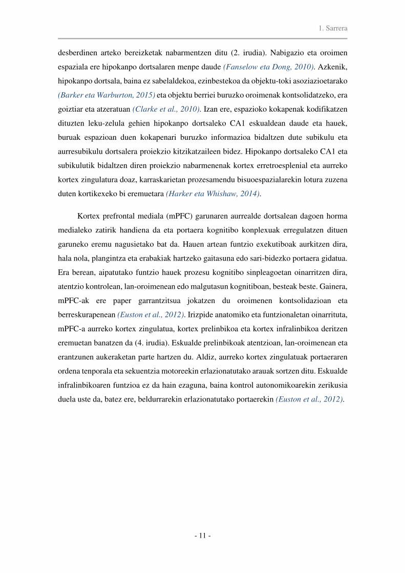
desberdinen arteko bereizketak nabarmentzen ditu (2. irudia). Nabigazio eta oroimen
espaziala ere hipokanpo dortsalaren menpe daude (Fanselow eta Dong, 2010). Azkenik,
hipokanpo dortsala, baina ez sabelaldekoa, ezinbestekoa da objektu-toki asoziazioetarako
(Barker eta Warburton, 2015) eta objektu berriei buruzko oroimenak kontsolidatzeko, era
goiztiar eta atzeratuan (Clarke et al., 2010). Izan ere, espazioko kokapenak kodifikatzen
dituzten leku-zelula gehien hipokanpo dortsaleko CA1 eskualdean daude eta hauek,
buruak espazioan duen kokapenari buruzko informazioa bidaltzen dute subikulu eta
aurresubikulu dortsalera proiekzio kitzikatzaileen bidez. Hipokanpo dortsaleko CA1 eta
subikulutik bidaltzen diren proiekzio nabarmenenak kortex erretroesplenial eta aurreko
kortex zingulatura doaz, karraskarietan prozesamendu bisuoespazialarekin lotura zuzena
duten kortikexeko bi eremuetara (Harker eta Whishaw, 2014).
Kortex prefrontal mediala (mPFC) garunaren aurrealde dortsalean dagoen horma
medialeko zatirik handiena da eta portaera kognitibo konplexuak erregulatzen dituen
garuneko eremu nagusietako bat da. Hauen artean funtzio exekutiboak aurkitzen dira,
hala nola, plangintza eta erabakiak hartzeko gaitasuna edo sari-bidezko portaera gidatua.
Era berean, aipatutako funtzio hauek prozesu kognitibo sinpleagoetan oinarritzen dira,
atentzio kontrolean, lan-oroimenean edo malgutasun kognitiboan, besteak beste. Gainera,
mPFC-ak ere paper garrantzitsua jokatzen du oroimenen kontsolidazioan eta
berreskurapenean (Euston et al., 2012). Irizpide anatomiko eta funtzionaletan oinarrituta,
mPFC-a aurreko kortex zingulatua, kortex prelinbikoa eta kortex infralinbikoa deritzen
eremuetan banatzen da (4. irudia). Eskualde prelinbikoak atentzioan, lan-oroimenean eta
erantzunen aukeraketan parte hartzen du. Aldiz, aurreko kortex zingulatuak portaeraren
ordena tenporala eta sekuentzia motoreekin erlazionatutako arauak sortzen ditu. Eskualde
infralinbikoaren funtzioa ez da hain ezaguna, baina kontrol autonomikoarekin zerikusia
duela uste da, batez ere, beldurrarekin erlazionatutako portaerekin (Euston et al., 2012).

1. Sarrera
- 12 -
4. irudia. Kortex prefrontaleko eskualde nagusien irudikapen eskematikoa eta bertako informazio
sarrera eta irteera nagusiak. ACd, aurreko kortex zingulatu dortsala; PL, kortex prelinbikoa; DP,
kortex pedunkular dortsala; VO, sabelaldeko kortex orbitala; LO, alboko kortex orbitala. Euston
et al. Neuron 2012-tik hartua.
1.1.1.3. Hipokanpoaren eta kortex prefrontal medialaren aldaketak nerabezaroan
Nerabezaroa haurtzarotik heldutasunerako trantsizioa da. Nerabezaroan zehar,
gizarte-jokabidean eta garapen kognitiboan aldaketak agertzen dira eta aldaketa horiek
kortex prefrontal medialeko eta hipokanpoko zirkuitu neuronalen garapenarekin irmoki
lotuta daude.
Nerabezaroan zehar kortex prefrontaleko sinapsien murrizketa bat gertatzen da,
ustez, sinapsi glutamatergikoetan (Petanjek et al., 2011). Bestalde, mielinizazioa
areagotzearen ondorioz, lobulu tenporal eta frontalen bolumenak handitu egiten dira
(Benes et al., 1994). Gainera, garai honetan sabelaldeko hipokanpotik kortex
prefrontalera doazen konexio glutamatergiko berriak sortzen dira. Kontaktu
monosinaptiko hauek, kortex prefrontaleko zelula piramidal eta interneurona
parbalbumina-positiboak inerbatzen dituzte (Gabbott et al., 2002) eta karraskarien jaio
ondorengo 50. egunean heltzen dira (Caballero et al., 2014b). Hipokanpoa eta kortex
prefrontalaren arteko bidearen osotasuna funtsezkoa da oroimen mota desberdinetarako
eta gaixotasun psikiatrikoetan bide honen konexioak ahulak dira (Godsil et al., 2013).
1. Sarrera
- 13 -
Kortex prefrontalaren inerbazio doparminergikoa prozesu kognitiboetarako
beharrezkoa da eta inerbazio hau nerabezaroan zehar heltzen da. Aurreko kortex
zingulatuko zuntz dopaminergikoen dentsitatea jaio ondorengo 20. eta 35. egunen artean
garatzen da guztiz. Eskualde prelinbikoan, aldiz, jaio ondorengo 60. eguneraino luzatzen
da. Dopaminaren D1 eta D2 hartzaileak ere areagotuz doaz heldutasunera iritsi arte.
Bestalde, terminal dopaminergikoak zelula GABAergikoen soma eta dendritekin aposizio
estuan daudela ikusi da eta nerabezaroan zehar esanguratsuki areagotzen dira kortex
prefrontaleko V. eta VI. geruzetan, parbalbumina-positiboak diren interneuronetan batez
ere (Caballero et al., 2016). Arratoi gazteetan (15-30. jaio ondorengo eguna) egindako
interneurona GABAergikoen erregistroen arabera, D1 motako dopaminaren hartzaileek
jaurtiketa-arineko interneuronen kitzikapena areagotzen dute; ez ordea D2 motako
hartzaileek. Hala ere, jaio ondorengo 50. egunetik aurrera, interneuronek bai D1 bai D2
hartzaileak adierazten dituzte eta dopaminak kortex prefrontaleko eragin GABAergikoa
areagotuko du (Tseng eta O’Donnell, 2007). Beraz, deskribatutakoaren arabera,
dopaminarekin erlazionatutako inhibizioaren kontrola kortex prefrontalean GABAren
jarduera erraztean datza.
Orain arte aipatutako aldaketez gain, nerabezaroan seinalizazio GABAergikoak eta
neurotransmisio glutamatergikoak ere aldaketa garrantzitsuak jasaten dituzte. Aldaketa
hauek 1.1.2.2. atalean (Interneurona GABAergikoen heltze-prozesua) eta 1.1.3.2. atalen
(NMDA hartzaileen garrantzia heltze-prozesuan, ikasketan eta oroimenean) deskribatuko
dira hurrenez hurren.
1.1.2. Interneuronak
Interneuronak ezinbestekoak dira zelula kitzikatzaileen arteko elkarrekintza
konplexuak doitzeko. Gaur egungo joeren arabera, interneuronen aniztasunak garunak
duen indar konputazionala irudikatzen du, non interneuronek hainbat ataza burutzen
dituzten (Moore et al., 2010). Interneuronek ez dute soilik gehiegizko kitzikapena
ekiditeko burutzen inhibizioa. Sareko eragiketa konplexuetan parte hartzen dute, hala
nola, erantzunaren aukeraketan, irabazi kontrolean edo zelula kitzikatzaileen jaurtiketa
patroi espazio-tenporalak doitzen eta garun oszilazioak ere sortzen dituzte. (Wehr eta
Zador, 2003; Roux eta Buzsáki, 2015). Garun osasuntsuetan, jarduera guzti hauek
dinamikoki egokitzen dira kanpoko eskakizunei behar bezala erantzuteko. Interneuronek

1. Sarrera
- 14 -
ikasketa eta oroimenean duten inplikazioa ikerketa ugaritan dokumentatu da, nahiz eta
azpimota bakoitzak parte hartze ezberdina izan garuneko eskualde eta atazaren arabera.
1.1.2.1. Interneuronen sailkapena
Interneurona GABAergikoak nerbio sistema zentraleko inhibizioaren iturri nagusia
dira (Jentsch et al., 2002) eta neurona guztien %10-25a osatzen dute, garuneko
eskualdearen arabera (Le Magueresse eta Monyer, 2013). Interneuronetan fenotipo ugari
ezberdintzen dira, hurrengo irizpide hauen arabera sailkatuz: (1) morfologia, (2)
propietate elektrofisiologikoak, (3) markatzaile molekularrak: kaltzio-ligatzaileak
(parbalbumina, kalretinina edo kalbindina), neuropeptidoak (somatostatina, hodi-hesteen
peptidoa, Y neuropeptidoa, reelina) eta hartzaileak (5-HT3R, mGluR1, CB1), (4)
zuzentzen diren neuronen domeinu azpizelularra eta (5) jatorri eta transkripzio-kontrolen
arabera duten patua (5. irudia) .
Garun-kortex eta hipokanpoko interneuronen jatorria enbrioiaren azpipalioko hiru
eskualdeetan dago: isats-gongoil eminentzian (IGE), gongoil eminentzia medialean
(GEM) eta zona preoptikoan (ZPO). Kortex eta hipokanpoari dagokionez, IGE-k sortzen
ditu interneurona gehien. Hauen artean parbalbumina adierazten duten jaurtiketa arineko
saski eta argimutil zelulak (Chandelier cells) eta somatostatina adierazten duten
interneuronak daude, azken hauek kalretinina (KR), Y-neuropeptidoa (YNP) edo reelina
koadierazi dezaketelarik. Isats-gongoil eminentziak kolezistokinina (KZK), kalretinina,
hodi-hesteen peptidoa (HHP), reelina eta neuroglia-formako zelulak sortzen ditu, baina
ez somatostatina (SST) adierazten duten interneuronak. Sailkapen gisa erabiltzen diren
molekulak, hala nola, proteina kaltzio-ligatzaileak (parbalbumina, kalretinina eta
kalbindina) edo neuropeptidoak (somatostatina, hodi-hesteen peptidoa eta
kolezistokinina) interneuronen azpipopulazioak desberdintzeko oso erabiliak dira
ikerketa anatomikoetan eta propietate elektrofisiologikoekin korrelazioa dute.
Parbalbumina (PB) jaurtiketa arineko saski-zelula eta argimutil zeluletan
aurkitzen da. Lehenak, soma eta hurbileko dendritak inhibitzen ditu eta azkenak axoien
hasierako segmentua. Parbalbuminaren erdiespena neurogarapenak erregulatzen du.
Hipokanpoan eta kortexean, parbalbuminaren adierazpenaren areagotze handiena jaio
ondorengo lehen eta hirugarren asteen bitartean gertatzen da. Jaiotzean ez da

1. Sarrera
- 15 -
baina etengabe areagotzen da jaio ondorengo bigarren astean zehar. Neurogarapeneko
hirugarren astean, igoera izugarria ematen da eta parbalbuminaren adierazpena apurka-
apurka gehituz doa nerabezaroan zehar, 40. joe eta 55. joe bitartean heldu mailak lortzen
diren arte (Caballero et al., 2014a). Parbalbumina adierazten duten interneuronek
ezinbesteko garrantzia dute inhibizio tonu egokia lortzeko. Inhibizioak eskualde kortikal
ezberdinen aldi plastikoak kontrolatzen ditu eta, beraz, kognizioan nahita nahiezko
eragina dauka.
5. irudia. Interneuronen sailkapena beraien morfologia, konektibitatea, markatzaile molekular eta
propietate elektrofisiologikoen arabera. Kepecs eta Fishell. Nature. 2014-tik hartua eta moldatua.

1. Sarrera
- 16 -
Kalretinina (KR) adierazten duten interneuronen azpipopulazio GABAergikoak
oso heterogeneoak dira. Kalretinina adierazten duten interneuronen gehiengoa zelula
bipolarrak dira, bi azpipopulazio ezberdintzen direlarik: hodi-hesteen proteina adierazten
duten zelula bipolarrak (HHP) eta somatostatina koadierazten duten Martinotti-bezalako
zelulak. Azpipopulazio hauek isats-gongoil eminentziatik eta gongoil eminentzia
medialetik datoz, hurrenez hurren. Interneurona KR-positiboak luzaroan ezezagunak izan
badira ere, azken ikerketek interneurona HHP-positiboen antzeko ezaugarriak dituztela
deskribatu dute (Cauli et al., 2014). Zelula hauen funtzio nagusia “zirkuitu
desinhibitzailea”-n zelula inhibitzaileei inhibizioa ematea da (Gulyás et al., 1996). Kortex
prefrontalean, zelula KR-positiboak normalean interneurona SST- eta PB-positiboetara
zuzentzen dira (Pi et al., 2013). Aitzitik, hipokanpoan interneurona kalbindina-positibo
eta beste interneurona KR-positiboetara zuzentzen dira batik bat (Gulyás et al., 1996),
ustez, somatostatina ere adierazten dutenetara (Gulyás et al., 1996; Somogyi et al., 2005)
eta ez dute interneurona PB-positiboekin kontaktatzen eskualde honetan.
Kalbindina (KB)-ren behin behineko adierazpena 21. joe-rako eskuratzen da
interneuronetan. Interneurona KB-positiboek ere populazio heterogeneoa osatzen dute eta
hauen artean buket-bikoitzeko zelulak (58%), zelula multipolarrak eta neuroglia-formako
zelulak (31%) ezberdindu daitezke. Kalbindinaren markaketa ahula erakusten duten
zelula batzuk zelula piramidalen morfologia dute neokortexeko II. eta III. geruzetan eta
hipokanpoko geruza piramidal eta granularrean.
Neuropeptidoak neuronen kitzikapenaren modulatzaile dira. Neuropeptidoak
neurotransmisore klasikoekin alderatuta, tamaina, sintesi eta jarduera mekanismo
ezberdinak dituzte. Neuropeptidoak inguruko neuronen egoera modulatzen duten ko-
transmisoreak dira. Beraien jarduera neurotransmisore klasikoena (glutamatoa eta
GABA) baino askoz ere motelagoa da. Ikerketa farmakologikoek erakutsi dutenez,
neokortexean adierazten diren neuropeptidoek emoziozko prozesuetan eta prozesu
kognitiboetan parte hartzen dute.
Somatostatina (SST) axoi eta dendritetako besikuletatik askatzen den
neuropeptido bat da (Ludwig eta Pittman, 2003). Bere askapenerako frekuentzia handiko
seinale aferenteak behar dira (Kits eta Mansvelder, 2000). Somatostatinaren eragin
zelular eta sinaptikoak nahikoa ondo ulertzen diren arren, garuneko zirkuituetan,
portaeran eta kognizioan dituen ondorioak oraindik argitzeko daude (Baraban eta Tallent,

1. Sarrera
- 17 -
2004; Liguz-Lecznar et al., 2016). Somatostatina adierazten duten interneuronak oso
zatikatuta dauden zuhaitz dendritikoetara daude zuzenduta eta integrazio sinaptikoa
aktiboki moldatzen dute. Hipokanpoan, SST-k hiperpolarizazio postsinaptikoa eragiten
du (Pittman eta Siggings, 1981) potasio korronteak areagotuz (Schweitzer et al., 1998)
eta kaltzio korronteak murriztuz (Ishibashi eta Akaike, 1995). Somatostatinak GABAB
hartzaile presinaptikoen bidez zelula kitzikatzaileetako glutamatoaren askapena
inhibitzen du (Boehm eta Betz, 1997; Urban-Ciecko et al., 2015). Alabaina, SST
GABAren potentzial postsinaptiko inhibitzailea murrizteko gai da, hau da, GABAren
eragin inhibitzailea indargabetu dezake despolarizazio bat sortuz (Scharfman eta
Schwartzkroin, 1989; Leresche et al., 2000). Honek, interneurona berdinetik GABA eta
SST askatzen direla iradokitzen du, baina baldintza ezberdinetan eta helburu
ezberdinetarako. Gainera, SST adierazten duen interneurona azpimota espezifiko bat
aurkitu da kortexeko IV. geruzan. Interneurona SST+ hauek ez dira II/III. geruzetan
kokatzen diren eta I. geruzako dendrita apikaletara proiekzioak bidaltzen dituzten
Martinotti interneuronen berdinak (Ma et al., 2006). Laugarren geruzako interneurona
SST-positiboak X-94 zelulen antzeko zelulak dira, jaurtiketa arineko zelulen ezaugarriak
dituzte eta dirudienez jaurtiketa arineko interneurona PB-positiboekin kontaktatzen dute
era lokalean (Ma et al., 2006). Beraz, zelula SST-positiboen azpimota bat era selektiboan
interneuronetara zuzenduta dauden interneurona multzo batek osatzen du eta hauek,
seinale GABAergikoak bidaltzen dizkiete interneuronei beraien mintz-potentzialak
erregulatzeko eta, ondorioz, aldaketa plastikoak mugatzeko.
1.1.2.2. Interneurona GABAergikoen heltze-prozesua
Interneurona GABAergikoek zelula piramidalen aktibitatea erregulatzen dute,
baina jaiotza aurretiko eta jaiotza ondorengo garapenean plastikotasun kortikalean, lotura
sinaptikoan eta oszilazioen sorreran ere rol garrantzitsua jokatzen dute (Le Magueresse
eta Monyer, 2013). Zirkuitu GABAergikoen garapenaren ezaugarrietako bat bere iraupen
luzea da. Heltze-prozesua enbrioi-sasoian hasten da eta hainbat urrats ematen ditu
zelularen berezko ezaugarriak guztiz garatu arte. Aldaketa guzti hauen artean hurrengoak
dira aipatzekoak: GABAren askatze eta jasotzearen heltzea, neuronen gaitasuna sinapsiak
osatzeko garapeneko garai zehatzetan eta zelulen seinaleztapena erregulatzen duten

1. Sarrera
- 18 -
proteina berezien adierazpena zelulek behin-betiko propietate elektrofisiologikoak
lortzeko.
Neurogarapen goiztiarrean, GABA neurotransmisore despolarizatzailea da, zelula
barruan ematen den kloro pilaketagatik. Jaio ondorengo lehen astearen inguruan, potasio-
kloro kotransportatzailearen (KCC2) adierazpena izugarri areagotzen da prosentzefaloan
eta GABA hiperpolarizatzaile bihurtzen da (Rivera et al., 1999). KCC2-ren adierazpena
lekuko neuronen jardueraren menpe dago, beraz, neuronen migrazioa gelditzeko seinale
gisa balio du (Bortone eta Polleux, 2009). Jaio ondorengo bigarren eta hirugarren
asteetan, hainbat aldaketa gertatzen dira interneurona GABAergikoetan: 1)
inmunoerreaktibitate GABAergikoaren heltzea (Del Rio et al., 1992); 2) helduaroko
propietate elektrofisiologiak agertzen hasten dira (jaurtiketa frekuentziaren areagotzea,
frekuentzia altuko azpiatariko mintz potentzialen oszilazioak eta mintzaren
erresistentzian aldaketak) (Doischer et al., 2008; Le Magueresse eta Monyer, 2013); 3)
kortexeko interneuronen axoi-plexuen heltzea (Doischer et al., 2008). Parbalbumina
adierazten duten zelulek jaio ondorengo 4-5 asteetan zehar guztiz garatzen dituzte
neuritak (Doischer et al., 2008). Lehen hilabetean, sinapsi GABAergikoen areagotze bat
gertatzen da. GABAA hartzaileen konposaketa ere aldatuz doa neurona kortikalen
garapenean zehar eta helduaroko azpiunitateak 3-4 asteetara ikus daitezke (Laurie et al.,
1992). Azpiunitateen truke hori neokortex eta hipokanpoko interneurona PB-positiboen
potentzial postsinaptiko inhibitzaileen heltzearekin batera ematen da (Doischer et al.,
2008). GABAB hartzaileak, G proteinei-loturiko hartzaileak dira eta neurogarapenean
zehar azpiunitate desberdinak adierazten dituzte (Fritschy et al., 1999) (6. irudia).
Garapenari lotutako azpiunitateen adierazpenak inplikazio funtzional garrantzitsuak ditu
propietate fisiologikoetan.
Interneurona GABAergikoen inhibizioa oso baxua da neurogarapen goiztiarrean eta
heldutasuneko ezaugarriak nerabezaro eta heldutasun goiztiarrean lortzen dira (Le
Magueresse eta Monyer, 2013). Inhibizio tonuaren areagotzea PB-ren areagotzearekin
bat dator (Caballero et al., 2014a; Caballero et al., 2014b). Ikusi denaren arabera,
nerabezaroan zehar emari glutamatergiko kitzikatzailea interneurona PB-positiboengan
areagotu egiten da (Caballero et al., 2014a), eta hau V-VI geruzetako potentzial
inhibitzaile postsinaptiko espontaneoen frekuentziaren handitzearekin batera gertatzen
da, hau da, jarduera GABAergikoaren areagotzearekin batera (Cass et al., 2014).

1. Sarrera
- 19 -
6. irudia. Neurogarapenean zehar sistema GABAergikoan ematen diren aldaketen irudikapen
eskematikoa.
1.1.2.3. Interneuronen funtzioa
Oinarrizko mikrozirkuituen funtzioak
Aurreraelikadura inhibizioak emari kitzikatzaileen aldiberekotasunaren detekzio
leihoa laburtzen du neurona piramidaletan. Zelula glutamatergiko aferenteak zelula
printzipalekin eta zelula GABAergikoekin paraleloan burutzen dute sinapsia (Buzsáki,
1984) (7. irudia). Emari inhibitzailea glutamatoak zelula piramidalengan eragindako
despolarizazioa baino lehenago heltzen bada, aurreraelikadura inhibizioak mintz
potentziala despolarizazio-atariaren behetik mantenduko du eta zelula kitzikatzaileen
ekintza-potentzialak ekidingo ditu. Konduktantzia inhibitzailearen areagotzea eta zelula
piramidalaren despolarizazioa ez badira aldiberekoak, milisegundo gutxi batzuk irauten
duen desoreka iragankor bat gertatzen da inhibizioak berriz oreka berreskuratu arte.
Ondorioz, interneuronek potentzial postsinaptiko kitzikatzaileen batuketa tenporala
mugatzen dute eta ekintza-potentzialen sorrera eta kadentzia erregulatzen dute.
Aurreraelikadura inhibizioan parte hartzen duten interneurona askok atzeraelikadura
inhibizioan ere parte hartzen dute. Dena dela, hipokanpoko zelula GABAergiko batzuk

1. Sarrera
- 20 -
aurreraelikadurako seinaleztapenean espezializatuta daudela dirudi. Zelula espezializatu
hauek, jasotzen duten aferente glutamatergikoen arabera izendatzen dira, hots, geruza
molekularreko bide zulatzaileari uztartuta dauden zelulak (Li et al., 2013), granaila
zuntzei uztartutako interneuronak (CA3ko stratum lucidum-eko interneuronak) (Vida eta
Frotscher, 2000), eta Schafferren kolateralei uztartutako interneuronak (CA1 stratum
radiatum-ean) (Cope et al., 2002). Interneurona guzti hauek aurreraelikadura inhibizioa
bideratzen dute adar dendritikoetan. Hala ere, zelula printzipalengan inhibizio
perisomatikoa gauzatzen duten interneuronak ere aurreraelikadurako inhibizio iturri
garrantzitsua dira. Neokortex eta hipokanpoan, parbalbumina (PB) eta kolezistokinina
(KZK) adierazten duten saski-zelulak dira domeinu perisomatikora zuzentzen diren
interneurona nagusiak (Freund eta Katona, 2007; Basu et al., 2013). Ondorioz,
kontaktatzen duten zelula piramidalen domeinuaren arabera, aurreraelikadura inhibizioak
jaurtiketa dendritikoak murriztu ditzake edo zelula kitzikatzaileen seinaleak kontrolatu.
Interneurona GABAergiko ugarik parte hartzen dute atzeraelikadura edo inhibizio
errepikakorren begizten sorreran, aurretik aipatutako PB+ eta KZK+ saski-zelulak barne,
baina baita PB adierazten duten argimutil zelula axo-axonikoak ere. Hauek, saski-zelula
PB positiboen antzera, propietate gisa jaurtiketa arina erakusten dute (7. irudia).
Neokortexeko II. eta III. geruzetan, somatostatina adierazten duten Martinotti zelulak
zelula printzipalen dendrita apikaletara daude zuzenduta eta atzeraelikadura inhibizioan
laguntzen dute. Hipokanpoan, kortexeko Martinotti zelulen antzerakoak diren
interneuronen populazio bat dago. Hauen somak stratum oriens-en aurkitzen dira eta
beraien axoiak stratum lacunosum-moleculare-ra zuzentzen dira (OLM: oriens
lacunosum-moleculare zelulak). Hiloko bide zulatzailera uztartuta dauden zelulak (HBZ)
OLM zelulen baliokideak dira hortz-jiroan, baina zelula granularren dendritetara daude
zuzenduta. Atzeraelikadura inhibizio mekanismoetan, zelula kitzikatzaileen jarduerak
neurona inhibitzaile postsinaptikoak errekrutatzen ditu eta neurona inhibitzaile hauen
deskargak zelula kitzikatzaileen ekintza-potentzialak ekiditen ditu (Miles, 1990).
Jaurtiketa arineko interneurona PB+ bakoitzak milaka neurona piramidalekin
burutu dezake sinapsia eta zelula piramidal bakoitzak hainbat saski-zeluletatik jaso
ditzake seinale inhibitzaileak. Beraz, jaurtiketa arineko zelula PB-positiboen kokapena
funtsezkoa da jomuga-neuronen jarduera erabakitzeko eta hau batez ere interesgarria da
zelula printzipalen jaurtiketa erritmoa sinkronizatzeko (Pinto et al., 2000). Bestalde,
interneurona SST-positiboak zelula piramidalen urruneko dendrita apikaletara zuzentzen

1. Sarrera
- 21 -
direnez, dendriten arantzetara heltzen diren seinaleak doitzen edo moldatzen dituzte.
Parbalbumina eta SST adierazten duten interneuronen bidez eragindako inhibizioak
osagarriak dira. Inhibizio goiztiarra perisomara zuzentzen diren jaurtiketa arineko zelulek
eragiten dute, oso sentikorrak direnez, estimuluari arin erantzuten diotelako. Ostera,
informazio sentsoriala etengabea denean, interneurona PB-positiboek eragindako
inhibizioa murriztuz joaten da. Ondoren, inhibizioa interneurona SST-positiboen esku
geratzen da, hauen eragin inhibitzailearen agerpena motelagoa delako eta inhibizio
berantiarra dela esan ohi da (Pouille eta Scanziani, 2004).
7. irudia. Mikrozirkuitu inhibitzailearen antolaketaren irudikapen eskematikoa. PB,
parbalbumina; SST, somatostatina; HHP: hodi-hesteen proteina; KR: kalretinina
Interneuronak ezinbestekoak dira portaera kognitibo arrunterako
Interneuronen artean aniztasun handia dago, baina PB eta SST adierazten duten
interneuronak ikertu dira gehien. Somatostatinak ikasketa eta oroimenean funtsezko rol
bat duela jakina da, somatostatina-hartzaileen knock-out (KO) saguek ikasketa
espazialean defizita erakusten baitute (Dutar et al., 2002). Horretaz gain, zahartzaroan
gizakien eta arratoien kortexean SST gutxiago dagoela ikusi da eta hau ikasteko
gaitasunaren murrizketarekin dago korrelazionatuta (Dournaud et al., 1995). Bestalde,

1. Sarrera
- 22 -
SST-ren injekzio intrahipokanpal eta intrabentrikularrek ikasketa espaziala hobetzen
dutela ikusi da hainbat atazetan (Vécsei et al., 1984; Lamirault et al., 2001). Hiloko
interneuronen isiltze optogenetikoak (gehienak SST adierazten dute) ikasketa eta oroimen
espaziala eten zuten Morrisen ur labirintoan, epe laburreko lan-oroimenean, koordinazio
motorean eta jarduera esploratzailean eraginik izan gabe (Andrews-Zwilling et al., 2012).
Interneurona PB-positiboen neurotransmisio inhibitzailearen asaldatzea ere
oroimen urritasunarekin dago erlazionatuta. Adibidez, interneurona PB-positiboetatik
ziklinaren menpeko kinasa 5-aren erauzketa genetikoak hiperinhibizio batera darama eta,
era berean, amigdalaren menpe dauden testuinguru eta pistak eragindako beldur-
oroimenak murrizten ditu eta erreferentzia oroimen espaziala kaltetzen du Morrisen ur
labirintoan (Rudenko et al., 2015). Gainera, kortex prefrontal medialeko jaurtiketa
arineko interneurona PB-positiboak rol garrantzitsua jokatzen dute sari-bidezko portaera
gidatuan, lan-oroimenean eta malgutasun kognitiboan (Murray et al., 2015), eta beraien
isiltze optogenetikoak atentzio prozesamendua hondatzen du, hau informazio berria
eskuratzeko beharrezkoa delarik (Kim et al., 2016). Hipokanpoko CA1eko zelula PB-
positiboen desaktibazio funtzionalak interneurona hauek lan-oroimen espazialerako
beharrezkoak direla egiaztatu dute (Murray et al., 2011). Are gehiago, M1 azetilkolinaren
hartzaile muskarinikoa interneurona PB-positiboen geneetatik ezabatuta duten sagu KO-
ek ezagutza memoria urrituta daukate eta, neurri txikiagoan, lan-oroimen espaziala
murriztua dute (Yi et al., 2014). Bestalde, interneurona PB-positiboetan NMDA
hartzailearen neurotransmisiorik ez duten sagu mutanteak kognizio mailan hutsegite
selektiboak erakusten dituzte, hala nola, lan-oroimenean eta ikasketa asoziatiboan
(Carlén et al., 2012).
Interneurona mota desberdinak ez daude bata bestearengandik isolatuta, baizik eta
zirkuitu-mailan funtzionatzeko konektatuta daude. Gutxi dira interneurona ezberdinen
arteko elkarrekintzak ordena-altuko funtzio kognitiboetan duen eragina aztertu dutenak.
Kortex prefrontal medialeko jaurtiketa arineko interneurona PB-positiboak helburura-
zuzendutako portaera gidatzen duten arren, interneurona SST-positiboek ere parte hartzen
dute, bi hauek aurkako jarduera erakusten dutelarik (Kim et al., 2016). Aurreko kortex
zingulatuan, saritutako atazen fase ezberdinetan parte hartzen dutela erakutsi zuten
Kvitsiani eta lankideek (Kvitsiani et al., 2013), non interneurona SST-positiboak
saritutako gunera hurreratzean aktibatzen ziren eta interneurona PB-positiboak saritutako
zona uztean erantzuten zuten eta aurreko egonaldiaren denbora kodifikatzen zuten.

1. Sarrera
- 23 -
Behaketa hauek interneuronak adierazpen kognitibo arrunt baterako beharrezkoak
direla nabarmentzen dute eta, dirudienez, kognizio egokia bermatzen duten zirkuitu
kortikalen dinamika koordinatzeko, interneurona azpimota bakoitzaren funtzioa espazio-
denboran bananduta dago, rol osagarriak betez.
Inhibizioaren aurkako erregulazioa oroimenaren jabetze eta finkatze prozesuetan
Hainbat ebidentziak iradokitzen dute inhibizioaren eta desinhibizioaren arteko
elkarrekintzak rol garrantzitsua jokatzen duela ikasketa eta oroimenaren formakuntzan.
Iragankorra eta espezifikoa den inhibizioak kausazko inplikazioa du ikasketarekin
erlazionatutako plastikotasunean, hain zuzen ere. Lehen ebidentziek SST adierazten
duten interneuronen jarduera basala mugimenduan eta sentsazio aktiboan zehar
murriztuta zegoela azpimarratu zuten (Urban-Ciecko eta Barth, 2016). Azken urteotan,
interneurona PB-positiboen estimulazio farmakologikoek eta optogenetikoek erakutsi
dutenez, beraien jarduerak entzumen beldur ikasketa hondatzen du (Letzkus et al., 2011).
Froga hauetan oinarriturik, autore batzuk desinhibizioa plastikotasun sinaptikoaren eta
ikasketaren mesedegarri den mekanismo orokor bat izan daitekela proposatu dute
(Letzkus et al., 2015). Izan ere, Gambino eta Holtmaatek (Gambino eta Holtmaat, 2012)
zuzenki egiaztatu zuten kortex somatosentsorialean desinhibizioak epe-luzeko
potentziazioa errazten zuela. Hipotesi honen euskarri dira baita beste lan batzuetan
aurkitutako emaitzak. Denbora luzez jakin da interneurona PB-positiboen inhibizioaren
murrizketa iragankor batek edo beraien sare perineuronalak kentzean ikusmen kortexeko
aldi kritikoa berriz irekitzen dela begi-dominantziaren plastikotasuna areagotuz (van
Versendaal et al., 2012). Aitzitik, interneurona PB-positiboen aurreraelikaduraren
aktibazioa beharrezkoa da amigdalaren menpe dagoen beldur ikasketarako (Wolff et al.,
2014). Era berean, barril kortexeko IV. geruzan SST adierazten duten interneuronen
zenbakia areagotu egiten da baldintzapen klasikoko ikasketa aldian (Cybulska-Klosowicz
et al., 2013). Halere, azken kasu hauetan interneuronen jardueraren efektua sarean
berdina izango litzateke: zelula piramidalen desinhibizioa. Ustez, amigdalako
interneurona PB-positiboek SST adierazten duten interneuronak isiltzen dituztenez eta
IV. geruzako interneurona SST-positiboak interneurona PB-positiboetara zuzendutako
zirkuitu desinhibitzaileko parte direnez, beraien jarduerak dudagabe zelula piramidalen
ekintza-potentzialak bermatuko lituzkete. Honekin bat, sabelaldeko eskualde

1. Sarrera
- 24 -
tegmentaleko proiekzio GABAergikoak accumbens nukleoko interneurona kolinergikoen
aktibitate espontaneoa geldiarazten dute ikasketa asoziatiboa hobetzeko (Brown et al.,
2012). Guzti hau kontutan izanik, ikasketan zehar desinhibizioak aldaketa plastikoak
errazten dituela esan genezake, zeinek espezifikotasun handiz integrazio sinaptikoa
aktiboki moldatzen duen, ziur aski esperientziak eragindako plastikotasun sinaptikorako
garrantzitsuak diren frekuentzia-handiko ekintza-potentzialak ahalbidetuz (Golding et al.,
2002; Kampa et al., 2007).
Azken behaketek, ikasketa prozesua bukatu ondorengo zirkuituen plastikotasunean
inhibizioaren areagotzeak duen rolaren garrantzia azpimarratu dute. Cornus ammonis 1
(CA1)-eko SST-positiboak diren oriens-lacunosum moleculare (OLM) zelulek
eragindako inhibizio dendritikoa beharrezkoa da beldur-oroimenak eratzeko (Lovett-
Barron et al., 2014). Halaber, asoziazio ikasketa burutu eta gero, seinaleztapen
GABAergikoaren areagotze bat dago: GABAren kontzentrazio presinaptikoak handitzen
dira, gune postsinaptikoetan GABAA hartzailearen α1 azpiunitatea gorantz erregulatzen
da eta korronte postsinaptiko inhibitzaile espontaneoen frekuentzia handitzen da
(Tokarski et al., 2007; Jasinska et al., 2010). Interneurona SST-positiboak ere GABA eta
SST gehiago adierazten dute ikasketa asoziatiboa eta gero (Cybulska-Klosowicz et al.,
2013). Azken pare bat urteetan, ikerketa gutxi batzuk esan dutenez, interneurona PB-
positiboen azpimultzo ezberdinak ikasketa eta oroimen prozesuetako fase ezberdinetan
dihardute. Esperimentu hauen arabera, interneurona mota bakoitzaren azpimultzo
desberdinak espezifikotasun handiz erantzuten diote ataza desberdinei, interneurona mota
bakoitzak eragindako inhibizioa are gehiago zatikatu daitekeela iradokiz garunaren
gaitasun konputazionala handitzeko. Donato eta lankideek (Donato et al., 2015) jaiotze
berantiarreko PB-positiboak diren saski-zelulak ataza berrien jabetzean daudela
inplikatuta diote eta, berriz, jaiotza goiztiarreko PB-positiboak diren saski-zeluletan
aldaketa plastikoak arauen finkapenaren ondoren gertatzen dira. Interneuronen berariazko
interneurona HHP-positiboek eragindako inhibizioak jaiotze berantiarreko neurona PB-
positiboetan paper garrantzitsua jokatzen du ikasketarekin-erlazionatutako plastikotasun
sinaptikoan (Donato et al., 2015). Honekin bat, interneurona PB-positiboen inhibizio
farmakologikoak plastikotasun sinaptiko estrukturala hobetu zuen eta antzerako joera
ikusi zen labirintoko nabigazioko entrenamendu fasean edo ingurune aberastuan haztean.
Bestalde, interneurona PB-positiboen aktibazioak balioztatutako arauen finkapena
sustatu zuen (Caroni, 2015). Beranduago, Lager eta lankideek (Lagler et al., 2016) PB

1. Sarrera
- 25 -
adierazten zuten saski-zelulak jaurtiketa patroi ezberdina zuten neurona talde
ezberdinetan biltzen zirela erakutsi zuten oroimenez-gidatutako aukeraketa portaeran,
zelula mota bakar baten barruan, atazarekin erlazionatuta egon daitezkeen
espezializazioak daudela aditzera emanez.
Aurkikuntza hauek ikasketa eta oroimen prozesuetan kitzikapen/inhibizio orekak
egiten duen ekarpenaren ulermena hobetzen laguntzen dute. Gero eta ebidentzia fidagarri
gehiagok desinhibizioak neurotransmisio kitzikatzailean laguntzen duela eta ikasketarako
mekanismo orokorra izan daitekeela adierazten dute. Bestalde, arauen finkapenak
konfigurazio inhibitzaile altu baten beharra du (Donato et al., 2013; Donato et al., 2015),
ikasketarekin erlazionatutako aldaketa plastikoak eta oroimenaren formakuntza inhibizio
mailak zehazten duela iradokiz. Beraz, oreka egoki hau nahasten duen edozein faktorek
eragin kaltegarriak izan ditzake kognizioan.
1.1.3. N-metil-D-asparato (NMDA) hartzaileak
1.1.3.1. NMDA hartzaileen estruktura eta sintesia
N-metil-D-aspartato (NMDA) hartzaileak 4 azpiunitate ezberdinez osatuta daude,
derrigorrezko NR1 azpiunitateaz eta hautazko NR2 (A, B, C edo D) edo NR3 (A edo B)
azpiunitateez, heterotetramero bat osatuz. NR3 azpiunitateak garapen goiztiarrean
aurkitzen dira batez ere; NR2 azpiunitateek kanalaren irekitzea erregulatzen dute; NR2A
azpiunitatea nerbio sistema zentraleko azpiunitaterik ugariena da; aldiz, NR2B
prosentzefaloan eta hipokanpoan da nagusi (Monyer et al., 1994).
NMDA hartzailearen azpiunitateen konbinazioen arabera, NMDA hartzaileen
propietate elektrofisiologikoak aldatu egiten dira. NR1-NR2B konbinazioak NR1-NR2A
konplexuak baino potentzial postsinaptiko kitzikatzaile luzeagoak ditu in vitro (Monyer
et al., 1994). NMDA hartzaileen azpiunitateek plastikotasun sinaptikoan parte hartzen
dute baita: hartzaile jakin bateko azpiunitateen adierazpenaren aldaketak hartzailearen
propietate funtzionalak aldatu ditzake. Izan ere, NR2Bren txertatzeak sinapsien
aldiberekotasunerako denboraldia luzatu dezake, beraz, eraginkortasun sinaptikoa
hobetuz eta, ondorioz, oroimenean eraginez (Yashiro et al., 2008).

1. Sarrera
- 26 -
NMDA hartzailaren azpiunitateak lotune guneetan ere desberdintzen dira: NR1
azpiunitateak glizina lotzeko guneak ditu eta NR2 azpiunitateak glutamatoa lotzeko
guneak. Glizinak ko-agonista bezala jokatzen du, hau da, bere lotura beharrezko baldintza
bat da NMDA hartzailearen aktibazioa bermatzeko. D-serinak ere ko-agonista bezala joka
dezake NMDAR-ren B-glizina guneetara lotzen denean (8. irudia). Mintzeko potentzia
atseden-egoeran dagoenean, magnesio ioiak kanalaren poroetan sartzen dira eta ioien
fluxua ekiditen dute. Magnesioaren blokeoa aldaratzeko eta, beraz, ioien fluxua
bermatzeko, mintzaren despolarizazioa gertatu behar da. Ondorioz, magnesioak sortzen
duen blokeoa gainditzea NMDA hartzaileen aktibazioa bermatzeko beste baldintza bat
izango da. Heterotetrameroaz gain, NMDA hartzaileek dentsitate postsinaptikoak dituzte,
sinapsi glutamatergikoei egituran eta funtzioan egonkortasuna ematen dieten proteina
multzoak.
8. irudia. N-metil-D-aspartato (NMDA) hartzailearen irudikapen eskematikoa. Mg2+,
magnesio ioiak; PCP: fenziklidina.
1.1.3.2. NMDA hartzailearen garrantzia heltze-prozesu, ikasketa eta oroimenean
Glutamatoak zelula barneko jauziak aktibatzen ditu hartzaile ionotropiko eta
metabotropikoen bidez. Glutamatoa neurogarapenerako ezinbestekoa da sinaptogenesia,
sareko plastikotasuna, dendriten adarkatzea, eta neurona aitzindarien hedatze eta
migrazioa erregulatzen dituelako (Snyder eta Gao, 2013). Jaio ondorengo neurogarapen
goiztiarrean NMDA hartzaileak dira glutamatoaren hartzaile bakarrak, AMPA hartzaile
funtzionalik ez baitago (Ben-Ari et al., 1997). Hori dela eta, NMDA hartzaileak garuneko
heltze-prozesuarekin irmoki lotuta daude. NMDA hartzaileen azpiunitateen adierazpena
1. Sarrera
- 27 -
eta funtzioa hipokanpoaren garapen egokirako beharrezkoa da. Izan ere, hartzaile hauen
erregulazio ezak sinaptogenesian eta zirkuituen heltze-prozesuan akatsak sortzen ditu
(Brigman et al., 2010).
NMDA hartzailearen jarduera ere funtsezkoa da interneurona PB-positiboen heltze-
prozesuan (Zhang eta Sun, 2011). NMDA hartzailearen seinaleztapena neurogarapen
goiztiarrean etenez gero, zelula GABAergikoen jardueraren epe-luzeko murrizketa
gertatzen da eta honek, kortex prefrontal medialeko zelula piramidalen desinhibizioa
sortzen du (Belforte et al., 2010; Moreau et al., 2013). Kortex prefrontal medialean,
tangentzialki migratzen ari diren interneuronen aitzindarietan NMDA hartzaileak
adierazten dira (Soria eta Valdeolmillos, 2002). Garuneko zirkuituen heltzea NMDA
hartzailearen azpiunitateen aldaketarekin bat dator, gaztaroko prozesamendu neuraletik
heldutasunerako trantsizioa markatuz. NR2 azpiunitatearen aldaketa zelula-espezifikoa
da kortex prefrontalean: zelula piramidaletan NR2B-ren maila egonkorra da
heldutasuneraino, baina jaurtiketa arineko interneuronetan NR2B azpiunitatea pixkanaka
NR2A azpiunitatearekin ordezkatzen da, batez ere nerabezaroan (Wang et al., 2008;
Wang eta Gao, 2009). Garapenean zehar plastikotasun-atarian gertatzen diren aldaketei
azalpena eman diezaiokeen mekanismoetako bat NR2B azpiunitatean gertatzen diren
aldaketak dira. Azpiunitate hau duten NMDA hartzaileek zinetika moteleko korronteak
erakusten dituzte, kaltzio korronteen batuketa tenporala ahalbidetuz. NR2A azpiunitatea
duten NMDA hartzaileak, aldiz, zinetika arinagoa dute, kaltzio seinaleen bereizmen
tenporal handiago bat ahalbidetuz. Azpiunitateek jasaten dituzten aldaketen ondorioz,
NMDA hartzaileak arrisku faktoreekiko bereziki sentikorrak bihurtzen dira. Hala,
ingurumen faktoreek zein faktore genetikoek garun garapen arraunta kaltetu dezakete
(Spear, 2000).
NMDA hartzaileek oinarrizko funtzioa betetzen dute garunaren plastikotasunean
eta, beraz, ikasketa eta oroimenean. Transmisio sinaptiko kitzikatzaileak erregulatzeko
duten ahalmenari esker eragiten dute NMDA hartzaileek ikasketa eta oroimen
prozesuetan: epe-luzeko potentziazioaren (ELP) bidez sinapsiak indartu dezakete edo
hauek ahuldu epe-luzeko depresioaren (ELD) bidez (Yashiro et al., 2008). NMDA
hartzaileen bidezko kaltzio fluxua da ELP eta ELD-ren arduraduna eta, aldi berean, bi
mekanismo hauek deskribatu dira ikasketa eta oroimenaren erantzule zelular gisa.
Gainera, NMDA hartzaileak jarduera pre- eta postsinaptikoen aldiberekotasuna

1. Sarrera
- 28 -
nabarmentzeko gai dira eta korrelazio tenporal honek ekintza pre- eta postsinaptikoen
denboraren araberako plastikotasuna ahalbidetzen du.
1.2. Eskizofreniaren aurrekari neuropatologikoak
Eskizofrenia ezgaitasun psikiatrikoa da eta bere etiopatogenia oraindik ez dago
guzti argituta. Gaur egunera arte, garuneko ikerketak gaixotasun neurologikoetan zentratu
dira gehienbat, eta gaixotasun mentalei buruzko ikerketak azken hamarkadetan areagotu
dira, batik bat. Informazio gabezia honek eskizofreniaren oinarri neurobiologikoen
ulermena eragotzi du eta medikuak eskizofreniaren diagnostikoa sintometan oinarrituta
egitera behartuak ikusi dira. Eskizofrenian sintoma positiboak, negatiboak eta
kognitiboak agertzen dira (Saha et al., 2005). Sintoma positiboak ohiz kanpoko funtzio
mentalen irudi dira, hala nola, haluzinazioak eta eldarnioak. Sintoma negatiboen artean,
besteak beste, isolamendu soziala, motibazio eza eta sentipen lautuak daude. Sintoma
kognitiboak funtzio exekutibo eskasekin daude erlazionatuta, batez ere, arretaren eta
oroimenaren hutsegiteekin. Sintoma hauen agerpen tipikoa nerabezaro berantiar eta
heldutasun goiztiarraren artean gertatzen da (Saha et al., 2005), neurogarapeneko
prozesuek paper garrantzitsu bat jokatzen duten arren eskizofrenian. Eskizofreniaren
etiologiari buruz hainbat teoria proposatu dira, hala nola, joera genetikoa (Greenwood et
al., 2013; Singh et al., 2014), jaio aurreko infekzioak (Labouesse et al., 2015),
ingurumeneko eraginak (Pishva et al., 2014) edo aurretik aipatutako faktoreen
konbinazioa (Uher, 2014). Eskizofrenia izateko arriskua areagotzen duten geneak
eskizofrenian etenda-1 (DISC-1), disbindina eta neuroregulina-1 (NRG-1) dira, besteak
beste (Le Magueresse eta Monyer, 2013; Salgado eta Sandner, 2013) (9. irudia).
1.2.1. Gizakietan egin diren aurkikuntzak
Azken bi hamarkadetan eskizofreniaren disfuntzio kognitiboaren jatorria ulertzeko
ahalegin asko egin dira. Hori dela eta, prozesu kognitiboek dituzten oinarri
neurobiologikoak ulertzeko interes handia dago. Gaur egun, medikamentu
antipsikotikoak eraginkorrak dira sintoma positiboak gutxitzeko, baina onura urria dute
kognizioan. Arazo kognitiboek gaixo eskizofrenikoei eguneroko bizitzan eragiten diote

1. Sarrera
- 29 -
eta ezgaitasun kronikoari eta langabeziari oso lotuta daude (Green et al., 2000). Hala ere,
gaur egungo tratamenduak edo terapiak ez dira gai sintoma kognitiboak arintzeko.
Disfuntzio kognitiboak sakon eragiten du eta beste sintomekiko mendekotasunik ez du
erakusten (Gold, 2004). Ezaugarririk nabarmenena lan-oroimen gaitasun murriztua da,
batez ere informazio kantitate handiak batera prozesatu behar direnean (Silver et al.,
2003; Barch et al., 2006). Kortex prefrontal dortsolaterala (KPFDL) lan-oroimenaren
hutsegiteetan dago inplikatuta eta eskaner bidez egindako garun ikerketek behin eta berriz
erakutsi dute KPFDL-ren aktibazioan alterazioak daudela eskizofrenia duten
gizabanakoetan, batez ere, ataza kognitiboak egiten arin direnean (Arnsten eta Jin, 2014;
Brunoni eta Vanderhasselt, 2014; Wolf et al., 2015). Eskizofrenia duten gaixoek ikasketa
asoziatiborako gaitasuna ere murriztuta dute eta hau kortex prefrontaleko eta
hipokanpoko zirkuituen anatomia eta funtzioarekin lotu dute.
9. irudia. Eskizofreniaren sorreran parte hartzen duten arrisku faktore nagusien sailkapena.
Interneurona GABAergikoen heltze-prozesuaren asaldura baten ebidentziak ere
aurkitu dira eskizofrenian. Hainbat ikerketetan garapenean zehar interneuronen migrazio
oker bat gertatzen dela ikusi dute. Lehenik, gaixo eskizofrenikoen substantzia zurian
interneuronen dentsitatea handiagoa da (Eastwood eta Harrison, 2003; Joshi et al., 2012).
Bigarrenik, eskizofrenia jasateko sentikortasunari lotuta dagoen DISC-1 genearen
beheratze bat eta gero, gongoil eminentzia medialetik datozen interneuronen migrazio
tangentzialean aldaketak nabarmentzen dira (Steinecke et al., 2012). Azkenik,
neurorregulina-1 (NRG-1) eta bere hartzaile den ErB4 ere eskizofrenia jasateko arriskuari
1. Sarrera
- 30 -
lotuta daude eta ErB4 interneurona inhibitzaileetan baino ez da adierazten, batez ere, PB-
positiboak diren interneuronetan (Fazzari et al., 2010). Gainera, kitzikapen/inhibizio
zirkuituen oreka galdu egiten dela dirudi. Alde batetik, saski-zelula eta zelula piramidalen
arteko sinapsietan GABAA hartzailearen α1 azpiunitatearen adierazpena murriztuta
dagoela aurkitu da eta argimutil zeluletan α2 azpiunitatearen areagotzea ikusi da (Volk et
al., 2002). Bestalde, sinapsi axo-axonikoak ere murriztuta daude eta sagu-ereduetan ikusi
denez, PB-positibo diren saski-zeluletan sinapsi glutamatergiko gutxiago daude (Zheng
et al., 2011). Emaitza hauek parbalbumina adierazten duten interneuronen konexioak
garapenean zehar ezegokiak direla erakusten dute eta honen ondorioz heldutasunean
sarearen aktibitate eta plastikotasun aberranteak eman daitezke. Halaber, nerabezaro
berantiarrean sistema GABAergikoan ematen diren aldaketa nabarmenek eta
eskizofreniaren sintomen sorrerak adin mendekotasun profil berbera dute.
Post mortem egindako ikerketek kortex prefrontal eta hipokanpoko alterazio
GABAergikoak eskizofreniaren sorreran inplikatuta daudenaren ideia are gehiago
indartzen dute. Batez ere, parbalbumina eta azido glutamiko deskarboxilasa 67 (GAD67)-
aren beherakada da aurkikuntzarik trinkoena gaixo eskizofrenikoen kortex prefrontalean
(Volk et al., 2000; Zhang et al., 2002; Hashimoto et al., 2003), aldi berean kalretinina eta
kalbindina interneuronen inmunoerreaktibitatea galdu gabe (Reynolds et al., 2004;
Gonzalez-Burgos et al., 2015). Parbalbumina eta GAD67-ren adierazpena aktibitate
kortikalak erregulatzen duenetik (Qin et al., 1994; Gierdalski et al., 1999), beraien
gabeziak gaixo eskizofrenikoen interneurona PB-positiboetan, interneurona PB-
positiboen jardueraren gabezia edo beherakada bat adierazten du. Interneurona mota
guztietatik beherakada soilik PB duten zeluletara zergatik mugatzen den jakin ez arren,
PB-ren adierazpenaren aldaketek garapenean zehar proteina hau duten zelulak bereziki
kalteberak direla egiaztatu dute.
Berriki, post mortem egindako garunen ikerketek somatostatinaren proteina,
somatostatinaren mRNA adierazpenena eta neuropeptido hau duten interneurona
GABAergikoak ere murriztuta daudela erakutsi dute gaixo eskizofrenikoetan (Joshi et
al., 2015; Alherz et al., 2017). Hashimoto eta lankideek (Hashimoto et al., 2008) PCR
bidez erakutsi zuten, eskizofrenia zuten gaixoen KPFDL-ean, SSTren mRNAn %44ko
beherakada ematen zela kontrol osasuntsuekin alderatuta, eta batezbesteko %57ko
beherakada aurreko kortex zingulatuan eta ikusmen kortex primarioan. Zelula mailan,
SST-ren adierazpen erlatiboa zelulako ere gutxituta zegoen %31n II/III geruzetan eta

1. Sarrera
- 31 -
%25ean KPFDL-ko V. geruzan (Morris et al., 2008). Era berean, Konradi eta lankideek
(Konradi et al., 2011) beherakada esanguratsua aurkitu zuten SST adierazten zuten
interneuronen zenbaki eta dentsitatean, eta baita somatostatinaren mRNAren adierazpen
mailan eskizofrenian. Emaitza hauek iradokitzen dutenez, interneurona PB-positiboetaz
gain, beste interneurona mota batzuk ere egon daitezke eskizofreniari lotuta.
1.2.2. NMDA hartzaileen hipofuntzioa eskizofreniaren substratu
patofisiologiko gisa
Eskizofreniari dagokionez, dopaminaren hipotesia izan da ideiarik iraunkorrena
psikiatrian. Eredu hori dopamina-hartzaileen antagonistek efektu antipsikotikoak
zituztela ikusi ondoren proposatu zuten. Izan ere, gaixo eskizofrenikoen sistema linbikoan
hartzaile horien gehiegizko adierazpena aurkitzen da. Hala eta guztiz ere, dopaminaren
hipotesiak ez du eskizofreniaren patogenesia guztiz azaltzen, medikamentu
antipsikotikoak gaixotasunaren sintoma positiboetarako eraginkorrak baitira, baina
sintoma negatiboak eta kognitiboak ez dituztelako hobetzen. Gaur egun, D2 hartzaileen
gehiegizko aktibazioa nahasmendu honen sinapsietan ikusten diren desoreka kimiko
orokorren efektu bat baino ez dela uste da (Seeman eta Kapur, 2000).
Azken frogek gizabanako eskizofrenikoetan ezohiko transmisio glutamatergikoa
eta NMDA hartzaileen hipofuntzio bat dagoela onartzen dute (Snyder eta Gao, 2013).
Lehenik eta behin, eskizofrenia izateko arriskua handitzen duten gene askok NMDAR-n
bidezko seinaleztapenarekin dute zerikusia (Moghaddam, 2003; Harrison eta
Weinberger, 2005). Eskizofrenia garatzeko arrisku geneak neuronen ugaritzea, migrazioa
eta sinaptogenesia erregulatzen dute. Bigarrenik, gaixo eskizofrenikoen garunetan
NMDA hartzailearen NR1 azpiunitatea murriztua egoteak, NMDA hartzailearen funtzioa
asaldatuta dagoenaren idea sendotzen du (Weickert et al., 2013). Hirugarrenik, NMDA
hartzailearen adierazpen baxua duten sagu transgenikoek eta NMDA hartzailearen
antagonismoko animalia-ereduek eskizofrenia gogorarazten duten sintomak erakusten
dituzte. NMDA hartzailearen antagonistek portaera aldaketak sortzeaz gain,
eskizofreniarekin zerikusia duten alterazio metaboliko eta neurokimikoak ere eragiten
dituzte (Morris et al., 2005). Gainera, gero eta onartuago dago eskizofrenia

1. Sarrera
- 32 -
neurogarapeneko nahasmendu bat dela eta garuneko garapen goiztiarrari eragiten diola
(Lewis et al., 2008; Nakazawa et al., 2017).
1.2.3. Eskizofreniaren MK-801 animalia-eredua
Animalia-ereduak baliagarriak dira gaixotasun ugariren mekanismo
fisiopatologikoak argitzeko eta tratamendu berriak garatzeko. Nahasmendu psikiatrikoak
ikertzeko animalia-ereduak erabiltzearen oztopo nagusietako bat beraien oinarri
neurobiologikoen ezagutza gutxi dugula da. Gainera, eskizofrenia giza nahasmendua dela
iradoki dute, batez ere pertzepzioari, pentsamenduari, hizkuntzari eta arretari eragiten
diolako. Ezaugarri hauek behe-mailako ugaztunetan modelatzeak zeresana eman du,
baina eskizofreniaren prebalentzia handiak (biztanleriaren %1) eta gaixotasunaren
ondorio kaltegarriek animalia-ereduak erabiltzearen beharra justifikatzen dute.
Hainbat hurbilketa planteatu dira eskizofrenia karraskarietan modelatzeko eta hiru
talde nagusitan bana daitezke: neurogarapeneko ereduak, eredu genetikoak eta eredu
farmakologikoak. Neurogarapeneko ereduen artean, haurdunaldian zehar sortutako
zailtasunak daude, hala nola, ernaldiko malnutrizioa edo jaio aurretik gripe birusaren
eraginpean jartzea (Salgado eta Sadner, 2013). Eragile goiztiarrak ere erabili izan dira
neurogarapeneko ereduetan, adibidez, kumeak amarengandik banatzea edo isolamendu
soziala (Jones et al., 1992; Geyer et al., 1993), baina normalean eredu genetiko edo
farmakologikoekin batera (Gilabert-Juan et al., 2013). Jaioberrien sabelaldeko
hipokanpoko lesioak ere anitz erabili izan dira, baina eragindako kaltea eskizofrenian
baino askoz ere larriagoa da (Daenen et al., 2003; Rueter et al., 2004). Dena dela,
hurbilketa guzti hauen kausazko rola zalantzagarria da. Eskizofrenia oso heredagarria da
eta gaixotasunaren osagai genetikoa ezin da alde batera utzi. Geneek eta ingurumen
faktoreek elkarri eragiten diote eta elkarreragin horrek nahasmendua azaleratuko den edo
ez erabakiko du. Animalia-eredu genetiko batzuk gaixotasuna xehetasun handiz
kopiatzen dute, baina eskizofrenian gene ugarik hartzen dute parte eta hauek faktore
estokastiko eta ingurumenekoekin duten elkarrekintza konplexua dela eta, eskizofrenia
animalia-eredu genetikoen bidez bakarrik errepikatzea ezinezkoa bihurtzen da (Green et
al., 2010).

1. Sarrera
- 33 -
NMDA hartzailearen antagonista ez-lehiakorren animalia-ereduak dira gaur egun
hobeen deskribaturiko eredu farmakologikoak. Fenziklidina (PCP) eta ketaminak
psikosia eragiteko gai dira gizaki osasuntsuetan eta gaixo eskizofrenikoetan sintoma
positiboak areagotzen dituzte (Lahti et al., 2001). Honek eskizofreniaren patofisiologian
NMDA hartzaileen parte hartzea iradokitzen du. NMDA hartzaile ez-lehiakorren efektua
ez da guztiz ezagutzen, baina neurotransmisio glutamatergiko, dopaminergiko eta
GABAergikoarekin interakzio konplexuak izan ditzakeela dirudi (Grüter et al., 2015).
Glutamatoaren transmisioan ematen diren aldaketak eskizofrenian ikusten diren sintoma
negatibo eta kognitiboekin erlazionatuak izan dira. NMDA hartzaileen antagonistek,
dopaminak ez bezala, eskizofreniaren kognizio eta arreta defizitak aztertzeko balio dute
(Marcotte et al., 2001; van der Staay et al., 2009). Autore batzuk diotenez, NMDA
hartzaileen antagonistek ez dituzte neurogarapeneko prozesuak kontutan hartzen, batez
ere dosi akutuak erabili izan direlako eta epe motzeko ondorioak neurtu direlako, epe
luzeko efektuak aztertu beharrean. Hala eta guztiz ere, azken urteetan NMDA hartzaileen
antagonisten administrazio kroniko eta azpikroniko errepikatuak eman izan zaizkie
animaliei jaio ondorengo garai goiztiarrean eskizofreniaren defizit kognitiboak
modelatzeko eta epe luzeko portaera aldaketak aztertu dira (nerabezaroan edo
heldutasunean) (Bubeníková-Valesová et al., 2008; Lim et al., 2012). MK-801,
dizolzipina bezala ere ezagutzen dena, NMDA hartzaile ez-lehiakorren artean farmakorik
selektiboena eta indartsuena da eta, beraz, zabal erabili da eskizofrenia animalia-ereduak
sortzeko karraskarietan (Wong et al., 1986).
MK-801-ek NMDA hartzailea fisikiko blokeatzen du kanal barruan sartuz, PCP-
ren guneetara lotuz eta kanalean zehar katioien fluxua saihestuz. NMDA hartzaileen
blokeoak glutamatoaren gehiegizko askapena eragiten du eta bai blokeatutako neuronan
bai garuneko beste eskualde batzuetan eragin kaltegarriak izan ditzake. Izan ere, MK-
801-k efektu proapoptotikoak ditu farmakoa eman ondorengo epe motzean. Bide
apoptotikoak aktibatzeko duen gaitasuna NMDA hartzailearen blokeoaren iraupenaren
eta indarraren araberakoa da. Normalean, 0.25mg/kg baino dosi handiagoak dira
beharrezkoak atzeraezina den endekapena eta zelularen heriotza eragiteko (Ikonomidou
et al., 1999). Zelularen heriotza aktibatzeko mekanismoak ez daude guztiz argi, baina
neurogarapen goiztiarrean NMDA hartzailea ERK1/2-CREB bideei lotzea beharrezkoa
da bere akzio neurotrofikoetarako eta dirudienez, MK-801-k eragindako zelulen heriotza
NMDA hartzailea ERK1/2 seinaleztapen bidetik disoziatzearen ondorioa da (Lim et al.,

1. Sarrera
- 34 -
2012). Ikonomidou eta lankideek (Ikonomidou et al., 1999) esandakoaren arabera, MK-
801-ek eragindako zelulen heriotzak bakarrik neuronei eragin zien, zelula glialen
aktibaziorik gabe. Azken hamarkadako ikerketek, ostera, eskizofrenia duten gaixoetan
gliaren narriadura dagoela iradokitzen dute, hala nola, glia zelulen tamaina txikiagoa
kortex prefrontalean eta glia zelulen disfuntzioa kortex prefrontal eta hipokanpoan
(Cotter et al., 2001; Kondziella et al., 2007). MK-801-ek eragindako kalteen
minberatasunak NMDA hartzaileen adierazpen altuarekin eta garunaren hazkundearekin
du zerikusia. NMDA hartzailearen antagonismoaren ondoren, glutamatoaren gehiegizko
askapena dago eta honek eragindako exzitotoxizitatea bide apoptotikoetatik at doa,
hazkuntza konoen aktibitatea eta neuriten luzapen eta adarkatzeari erasanez (Ringler et
al., 2008). Post mortem aztertutako gaixo eskizofrenikoen garunetan ere lesio neuronalak
ikus daitezke, esate baterako, dendriten atrofia (Garey, 2010).
Glutamatoak eragindako exzitotoxizitateak ondorio funtzional handiak ditu.
Interneurona GABAergikoak zelula piramidalak baino hamar aldiz sentikorragoak dira
NMDA hartzaileen antagonismoarekiko (Grunze et al., 1996). Bereziki, PB adierazten
duten jaurtiketa arineko interneuronak dira sentikorrenak, antza, eta haien alterazioa
nahikoa da portaeran eskizofreniaren sintomak diruditen antzeko ezaugarriak sortzeko.
Hainbat ikerketek frogatu dute MK-801-aren administrazio errepikatuek PB-ren
inmunoerreaktibitatea gutxitzen dutela. NMDA hartzaileen blokeo kronikoak arratoi
gazte eta helduetan interneurona PB-positiboen dentsitatea murrizten du hipokanpoan,
batez ere hortz-jiroan eta CA1 eskualdean (Rujescu et al., 2006; Braun et al., 2007).
Neurogarapeneko ereduak ere aurkikuntza hauek errepikatzeko gai izan dira, baina
normalean 0.5mg/kg baino gehiagoko dosiak behar dira epe luzeko aldaketa estruktural
eta anatomikoak sortzeko (van der Staay et al., 2011). Dosi handi hauek ez dituzte
bakarrik hipokanpoko PB dentsitateak aldatzen, baita kortex prefrontal medialeko zelula
PB-positiboenak ere, eta eskualde hau orden-altuko funtzio kognitiboetarako beharrezkoa
da (Coleman et al., 2009; Li et al., 2015). Are gehiago, Nakazawa eta lankideek
(Nakazawa et al., 2012) sagu transgenikoak erabiliz, NMDA hartzaileak kortexeko eta
hipokanpoko interneurona GABAergikoetatik kentzeak nahikoa zela eskizofrenia-
bezalako ezaugarriak agerrarazteko erakutsi zuten. Kaltetutako interneurona gehienek PB
inmunoerreaktibitatea zuten, izan ere. MK-801 administratu ondoren jaurtiketa arineko
zelula PB-positiboen sentikortasun selektiboaren mekanismoak jakiteke daude oraindik,
baina hainbat hipotesi plazaratu dira. Jaurtiketa arineko zelulek potasio kanal berezi bat

1. Sarrera
- 35 -
dute, hots, Kv1.3 kanala, eta honek mintzaren errepolarizazio arin bat bermatzen du
hurrengo ekintza-potentziala azkar sortzeko. Frekuentzia altuko ekintza-potentzialak
jaurtitzean NMDA hartzaileen irekitze probabilitatea askoz ere handiago da jaurtiketa
arineko zeluletan, frekuentzia baxuagoan jarduten diren beste zelula GABAergikoekin
edo zelula kitzikatzaileekin alderatuz. MK-801 farmako ez-lehiakorra denetik, ioi kanala
irekita egon behar da hartzailea blokeatu ahal izateko eta, beraz, jaurtiketa arineko
NMDA hartzaileak blokeatzeko aukera handiagoa dute. Wang eta Gao-k (Wang eta Gao,
2012) beste mekanismo bat deskribatu zuten. Ikerketa elektrofisiologikoen bidez eta MK-
801-en dosi azpikronikoak erabiliz zelula piramidaletara edo jaurtiketa arineko
interneuronetara zuzendutako terminal glutamatergikoetako NMDA hartzaileak MK-
801-en ondorioz ezberdin zeudela aldatuta erakutsi zuten. NMDA hartzaile
presinaptikoak oso garrantzitsuak dira neurotransmisoreen askapena errazteko eta
modulatzeko. Autore hauen arabera, MK-801-k guztiz blokeatu zituen jaurtiketa arineko
interneuronetara zuzenduta zeuden terminal glutamatergikoetako NMDA hartzaileak eta,
aldiz, zelula piramidalekin kontaktatzen zuten terminal presinaptikoetan NMDA hartzaile
berrien txertaketa ahalbidetu zuen. Datu hauek are gehiago berresten dute NMDA
hartzaileen blokeoaren mekanismo sinaptikoak zelula mota desberdinekiko espezifikoak
direla. Bi hipotesi hauek ez dira bateraezinak eta ziur aski MK-801-k NMDA hartzaileak
hainbat mekanismoren bidez aldatzen ditu, baina guztiak emaitza berdinarekin:
interneurona PB-positiboen azpijarduera, kortexeko zelula piramidalen desinhibizio
orokor bat eraginez. Bestalde, NR2C eta NR2D azpiunitateak dituzten NMDA hartzaileak
bereziki kalteberak izan daitezkeela proposatu dute hauek magnesioarekiko duten
afinitate baxua dela eta (Kotermanski et al., 2009). NR2C daukaten NMDA hartzaileak
interneurona PB+ eta SST+-etan baino ez dira adierazten eta NR2D azpiunitatea
jaurtiketa arineko interneuronetan adierazten da batez ere (von Engelhardt et al., 2015).

1. Sarrera
- 36 -
1.3. Ingurune aberastua
1.3.1. Ingurune aberastuaren kontzeptua
1940ko hamarkadaren amaieran Hebb izan zen ingurune aberastua (IA) kontzeptu
esperimental gisa lehenengo aldiz proposatu zuena. Etxean maskota gisa hezitako
arratoiek laborategiko arratoiekin alderatuz portaera hobekuntzak zituztela ikusi zuen
(Hebb, 1947). 1960ko hamarkadaren hasieran, Rosenzweig eta lankideek ingurune
aberastua testa daitekeen kontzeptu zientifiko bezala proposatu zuten (Rosenzweig et al.,
1962). Lehen ikerketek, “garunaren pisua” edo “garunaren proteina totala” bezalako
parametroak erabili zituzten ingurumeneko estimuluen efektuak aztertzeko (Rosenzweig
et al., 1969). Bestalde, hainbat ikerketek ondorioztatu zuten ingurunearen estimulazioak
erantzun plastikoa eragiten zuela, erantzun hau aldaketa biokimikoetan, adarkatze
dendritikoan (Holloway, 1966; Greenough eta Volkmar, 1973), gliogenesian (Altman eta
Gas, 1964; Diamond et al., 1966), neurogenesian (Kempermann et al., 1997) eta ikasketa
prozesuan islatzen zelarik.
10. irudia. Baldintza estandarren (a) eta ingurune aberastuaren (b) irudikapen eskematikoak.
Bengoetxea H et al. Neural Plast. 2012-tik hartua.
Ingurune aberastuaren definizio estandarra hurrengo hau da: “bizigabeko objektuen
eta gizarte-estimulazioaren konbinaketa konplexua”. Egoera esperimentalean,
“aberastutako” animaliak kaiola eta talde handiagotan bizi dira, non harreman sozial
konplexuagoak edukitzeko aukera duten. Ingurunea konplexua da eta ikerketak dirauen
bitartean, inguruneko ezaugarriak aldatu egiten dira konplexutasuna mantentzeko
helburuarekin. Hala, ingurune aberastuko kaiolak tunelez, habia egiteko materialez eta

1. Sarrera
- 37 -
jolas ezberdinez ornituta daude. Gainera, materialen zein janariaren kokapena
maiztasunez aldatzen da. Bestalde, animaliei borondatezko ariketa fisikoa egiteko aukera
ematen zaie kaioletan gurpil bat jarriz (10. irudia).
Ingurune aberastuak estimulazio sentsorial, motore eta kognitiboen konbinazio
konplexua baimentzen du eta honek molekula, zelula eta portaera mailako aldaketak
eragiten ditu (11. irudia). Ingurune aberastuko elementu desberdinek bakarka ere efektu
esangarriak izan ditzakete garun plastikotasunean. Adibidez, borondatezko ariketa
fisikoak zelulen ugaritzea eta neurona berrien integrazioa bideratzen du hortz-jiroan (van
Praag et al., 1999). Bestalde, ingurune berri baten eraginpean jartzeak soilik oroimen
espaziala hobetu dezake (Yang eta Tang, 2011). Ingurune aberastuaren definizioan ezin
da faktore bakoitzaren eragina erraz isolatu. Izan ere, ingurune aberastuko osagai
bakoitzaren efektua besteengandik guztiz banatzea oso zaila da eta neurtzen den
parametroaren araberakoa izan daiteke. Hala ere, arrazoi nahikoa daude ingurune
aberastuaren funtsezko elementua faktore ezberdinen elkarrekintza dela onartzeko.
11. irudia. Ingurune aberastuan egon ondoren gertatzen diren aldaketa erakusten dituen diagrama.
Ingurune aberastuaren iraupenak eta denboraldiak eragin handia dute lortutako
emaitzetan (Amaral et al., 2008). Ikerketa anitzetan, IA garai goiztiarrean hasten da, hala
nola, jaiotze egunean edo edoskitzearen amaieran. Kasu hauetan, ingurumen konplexuan
hazteak antsietate-bezalako portaera murrizten duela frogatu da, besteak beste
(Hendershott et al., 2016). Animali helduak inguren aberastuan ezartzeak ere efektu ditu

1. Sarrera
- 38 -
(Beauquis et al., 2010; de Witt et al., 2011). Hala ere, gutxieneko IA-ren iraupen bat
beharrezkoa da portaera eta aldaketa morfologiak gerta daitezen. Denboraldi hau aste bete
eta lau aste bitartean ezarri da, beti ere aztertzen den espeziearen eta parametroaren
araberakoa izanik. Esaterako, heldutasunean IA-n bitartekotza motz bat (10 egunekoa)
nahikoa da neurogenesia, sare baskularra eta dendriten konplexutasuna hipokanpoan
areagotzeko (Beauquis et al., 2010). Bestalde, IA-ko esposizio motzak (6h/eguneko)
etengabeko IA-ren hobekuntzen antzerakoak dira funtzio motoreei eta kognitiboei
dagokienez (de Witt et al., 2011). Gainera, ingurune aberastuak eragindako onuren
iraunkortasuna, IA-ren iraupenaren araberakoa da. Zenbat eta luzeagoa izan IA-ren
iraupena, orduan eta denbora gehiagoz irauten dute efektu onuragarriak.
1.3.2. Ingurune aberastuak eragindako aldaketa kognitibo eta
estrukturalak
Ingurune aberastua eraginkorra da ikasketa eta oroimen funtzioak hobetzeko eta
zahartzaro osasuntsuan ematen den disfuntzio kognitiboa murrizteko gai dela baieztatu
da. Ingurune aberastuan hazitako animaliek baldintza estandarretan hazitako animaliek
baino hobeto egiten dute espazio-oroimeneko proba Morrisen ur labirintoan. Era berean,
IA-n hazitako animaliek, kaioletan isolatuta baina ariketa fisikoa egiteko gurpila duten
animaliek baino lan-oroimen espazial hobea dutela egiaztatu da (Bernstein, 1973).
Ingurune aberastuak eragiten dituen onura kognitiboak, partzialki honek duen efektu
antsiolitikoaren bidez azaldu daitezke. Izan ere, IA-n hazitako sagu zein arratoiek
antsietate-bezalako portaera murriztua erakusten dute “igotako gurutze labirintoan”
(Elevated Plus Maze) eta eremu irekiko proban, aldi berean jarduera esploratzailea
areagotuz (Hendershott et al., 2016).
Ingurune aberastuan bizi diren karraskariek aldaketa estruktural esanguratsuak
jasaten dituzte, esateko, garun tamaina eta pisu handiagoa edota kortexeko zelulen
nukleoen tamaina handiagoa (Diamond et al., 1967). Ingurune aberastuak hipokanpoko
neurogenesia areagotzen du arratoi gazte eta helduetan eta, aldi berean, diferentziatutako
zelula granularren biziraupena bermatzen du hortz-jiroan. Ingurune aberastuari buruzko
lehen ikerketek erakutsi zutenez, hipokanpoko hortz-jiroko zelula granularrak eta CA1
eta CA3ko zelula piramidalak dendrita gehiago zuten neuronako (Walsh et al., 1969;

1. Sarrera
- 39 -
Altschuler, 1979). Beranduago, IA-k dendriten adarkatzea eta arantzen dentsitatearen
areagotzea eragiten zuela baieztatu zen (Globus et al., 1973), kontaktu sinaptiko berrien
sorrera ondorioztatuz. Hala ere, IA-ren ondoren kortexeko bolumenaren areagotzea ezin
zaio soilik neurogenesiari eta sinaptogenesiari atxiki, angiogenesia eta gliogenesia ere
ikusi izan baita (Altman eta Das, 1964; Diamond et al., 1966; Greenough eta Volkmar,
1973). Baskularizazioaren areagotzeak glukosaren erabilgarritasuna eta zitobabesleak
diren faktoreen askapena erraztuko luke. Ingurune aberastuak plastikotasun
sinaptikoarekin erlazionatuta dauden gene-adierazpena modulatzen duela jakin arren,
ikasketa eta oroimenean duen efektu onuragarrien mekanismo molekular zehatzak
argitzeke daude.
1.3.3. Hazkuntza faktoreak Ingurune Aberastuak eragindako aldaketen
bitartekari
Hazkuntza faktoreak neuropeptido familia handi bat dira eta zelula kanpoko
seinaleztapenerako garrantzitsuak dira. Hala, nerbio sistema zentraleko zelula aitzindari
eta ama zelulen ugalketa eta desberdintze prozesuak erregulatzeko ezinbestekoak dira
(Calof, 1995). Baldintza estandarretan hazitako animalietan, garapen garaia edozein dela
ere, hazkuntza faktoreek zelulen ugalketa eta biziraupena, axoi eta dendriten hazkuntza
eta birmoldaketa, mintzen trafikoa eta fusioa zein sinapsien sorkuntza, funtzioa eta
plastikotasuna sustatzen dute.
Ingurune aberastuak gene adierazpena eta hazkuntza faktore desberdinen proteina
mailak areagotzen ditu, adibidez, nerbio-hazkuntza faktorea (NGF) (Mohammed et al.,
1990; Pham et al., 1999), garunetik eratorritako hazkuntza faktorea (BDNF) (Falkenberg
et al., 1992), gliatik eratorritako faktore neurotrofikoa (GDNF) (Young et al., 1999) edo
hazkuntza faktore endotelial-baskularra (VEGF) (During eta Cao, 2006). Ikerketa asko
burutu dira helduen garunetan faktore hauen rola zehazki ezagutzeko. Dirudienez,
organismo helduetan faktore hauek neurogenesian, plastikotasun sinaptikoan eta
ikasketan parte har dezakete. Adibidez, intsulina-bezalako hazkuntza faktoreen infusio
intrazerebrobentrikularrak neurogenesia areagotzen du arratoien hortz-jiroan (Aberg et
al., 2000).

1. Sarrera
- 40 -
Era berean, BDNF-k plastikotasun sinpatikoan eta IA-ri loturiko hobekuntza
kognitiboan funtsezko rola jokatzen du (Novkovic et al., 2015). Hipokanpotik BDNF-ren
genea ezabatzen denean, “Objektu berrien ezagutza” eta Morrisen ur labirintoan ikasketa
espaziala baliogabetzen dira (Heldt et al., 2007). Hazkuntza faktore honek Tropomiosin
Kinasa B hartzailea (TrkB) aktibatzen du neuronen diferentziazio, biziraupen eta
plastikotasun sinaptikoa eraldatzeko hainbat seinalizazio jauziren bidez, hala nola, ERK
edo Akt (12. irudia).
12. irudia. Tropomiosin kinasa B hartzailearen (TrkB) beherantzako seinaleztapen bideek zelulen
biziraupena eta diferentziazioa, axoien hazkuntza eta jarduera sinaptikoa erregulatzen duten
proteinen sintesia ahalbidetzen dute. Begni et al. Clinical Science. 2016-tik hartua eta moldatua.
2. Hipotesia


2. Hipotesia
- 43 -
Ingurune aberastua (IA) erabilera anitzeko eta balio terapeutiko handiko tresna da.
Zahartze osasuntsuan eta neuroendekapeneko gaixotasunetan dituen efektu onuragarriak
balioztatuak izan diren arren, gaixotasun neuropsikiatrikoetan duen balio potentziala ez
da sakon aztertu. Ingurumen faktoreek eskizofreniaren agerpena ahalbidetu dezakete,
beraz, gaixotasunaren sorrera edo areagotzea saihesteko gaitasuna ere badute.
Eskizofreniaren inguruko ikerketen erronka nagusietako bat garunean era iraunkorrean
kalteturik dauden zirkuituak aldatzen saiatzea da, eskizofrenia neurogarapeneko desoreka
bada ere, sintomak nerabezaroan edo heldutasun goiztiarrean agertzen baitira.
Ingurune aberastuak eskizofreniaren sintoma kognitiboengan ondorio onuragarririk
duen edo ez aztertzeak interes handia du, bai prebentzio tresna gisa, bai sendabide gisa.
Arazo kognitiboengan eragin positiboa izan dezakeen interbentzio ez-farmakologikoa
litzateke. Izan ere, ingurune aberastuak eragindako geneen aktibazio eta proteinen
sintesiak interneuronen adierazpena eta haien jarduera areagotu dezake. Honek, aldi
berean, eragin zuzena izango luke portaeran eta kognizioan. Aldaketa hauek
eskizofreniaren galera kognitiboak arintzeko funtsezkoak lirateke.
Beraz, lan honen helburua MK-801 bidez sortutako eskizofrenia animalia-ereduan,
bai portaeran zein maila zelularrean eta molekularrean garatutako alterazioak ingurune
aberastuaren bidez nola aldatzen diren aztertzea da.
Honenbestez, tesi hau garatzeko honako hipotesia landu dugu:
Ingurune aberastuak hazkuntza faktoreen, NMDA hartzaileen, kaltzioari loturiko
proteinen eta neuropeptidoen adierazpena modulatzen du bai kortex prefrontal zein
hipokanpoan eta guzti hauek aldatuta daude eskizofrenian. Hala, heldutasunaren hasieran
animaliak denbora labur batez ingurune aberastuan haziz gero, eskizofreniaren esparruan
sor daitezkeen defizit kognitiboak gainditzeko estrategia egokia izango litzateke.


3. Helburua


3. Helburuak
- 47 -
3.1. Oinarrizko helburua
Neurogarapen goiztiarrean, MK-801 administratu zaien arratoi helduetan ingurune
aberastuak alterazio kognitibo eta neurokimikoetan duen eragin positiboa egiaztatzea, eta
hobekuntza hauen oinarri molekularrak aztertzea.
3.2. Helburu operatiboak
1. Funtzio kognitibo desberdinak aztertzea: ezagutza memoria “objektu berrien
ezagutza” atazaren bidez, memoria asoziatiboa “objektua-tokian” atazaren bidez eta
memoria bisuoespaziala “Morrisen ur labirintoaren” bidez.
2. Aldaketa estrukturalak analizatzea: kortex prefrontal medialeko eta hipokanpoko
bolumenaren neurketa, eta butiril kolinesteraren aurkako histokimiaren bitartez sare
mikrobaskularraren luzeraren estimazioa.
3. Interneuronen azpipopulazio desberdinen eta neurona kopuru totalaren kuantifikazioa:
ordenagailuz lagundutako estereologiaren bidez, parbalbumina, kalretinina,
kalbindina, somatostatina, azido glutamiko deskarboxilasa eta NeuN proteinen
aurkarko inmunohistokimien kuantifikazioa kortex prefrontalean eta hipokanpoan.
4. Aldaketa molekularrak analizatzea: Western Blot teknikaren bitartez, NMDA
hartzaileen azpiunitateen, dentsitate postsinaptikoa-95 proteinaren (PSD-95), GABAA
hartzailearen ß2/3 azpiunitatearen, garunetik eratorritako hazkuntza faktorearen
(BDNF) eta TrkB-ren adierazpenaren, zein hauekin erlazionatutako zelula barneko
seinaleztapen-bideen aldaketa erlatiboen kuantifikazioa.
5. Ingurune aberastuak aurrez izendatutako parametroetan duen onura potentzialen
identifikazioa.

4. Material eta metodoak


4. Material eta metodoak
- 51 -
4.1. Animaliak
Long Evans arratoi emeak kume arrekin erosi ziren Janvier Labs enpresan
(Frantzia). Jaiotze eguna, 0 jaio ondorengo eguntzat (joe) hartu zen eta 21. joe izan zen
edoskitze amaiera. Animali guztiak argi/ilunpeko hamabi orduko ziklotan hazi ziren
(argiak 8etan piztuz) eta ura eta janaria ad libitum izan zuten esperimentu guztian zehar.
Prozedura guztiak 2007/526/EE Europako Gomendioarekin bat zetozen, eta Euskal
Herriko Unibertsitateko (UPV/EHU) Animaliekin egiten diren Esperimentaziorako Etika
Batzordearen oniritziarekin egin ziren (CEEA/410/2015/LAFUENTE SANCHEZ).
4.2. Prozedura farmakologikoak
4.2.1. MK-801-aren administrazioa
MK-801 [(5S,10R)-(+)-5-Metil-10,11-dihidro-5H-dibenzo[a,d] ziklohepten-5,10-
imina hidrogeno maleatoa, Dizozilpina hidrogeno maleatoa] Sigma-Aldrichen erosi zen
(Erref: M107, St. Louis, MO, EEBB). Aurretiaz egindako ikerketetan oinarrituta,
0.5mg/kg-ko dosia aukeratu genuen, dosi honek portaera alterazio iraunkorrak eragiten
baititu (van der Staay et al., 2011). MK-801 kloruro sodikoa %0.9an (NaCl) diluituz eta
injekzio intraperitonealaren bidez administratu zitzaien arratoi kumeei 10. joe-tik 20. joe-
ra. Animali kontrolak NaCl-aren bolumen berdina jaso zuten (VH: eramailea soilik
daramaten animalien taldea). Injekzioak goizeko 9etan ematen zitzaizkien. Tratamenduak
iraun bitartean, animaliak egunero pisatu ziren eta hortik aurrera bi astetik behin. MK-
801-aren stock soluzioa NaCl-tan prestatu eta -20ºC-tan gorde zen, 2.5mg/ml-ko
kontzentrazioan. Stock soluzioa erabiltzerako orduan NaCl-tan diluitu zen 0.5mg/ml-ko
kontzentraziora.
4.2.2. BrdU-ren administrazioa
Talde bakoitzeko 6 animalik timidinaren analogoa den 5’bromodesoxiuridina-ren
(5’BrdU, Sigma-Aldrich, Espainia) injekzio intraperitoneal bakarra jaso zuten NaCl-tan.
Bihotzean zeharkako perfusioa burutu baino 24h arinago eman zitzaien 200mg/kg-ko dosi
batean.

4. Material eta metodoak
- 52 -
4.3. Talde esperimentalak
Lau talde esperimental desberdin erabili ziren (n=10-12) (13. irudia):
a) VH: Laborategiko baldintza estandarretan hazi eta 10. joe-tik 20. joe-ra NaCl jaso
zuten arratoiak.
b) MK-801: Laborategiko baldintza estandarretan hazi eta 10. joe-tik 20. joe-ra MK-
801 (0.5mg/kg) jaso zuten arratoiak.
c) MK-801+EE: 0. joe-tik 55. joe-ra laborategiko baldintza estandarretan eta 55.
joe-tik 73. joe-ra ingurune aberastuan hazitako animaliak. Talde honetako
animaliek 10. joe-tik 20. joe-ra MK-801-n injekzioak (0.5mg/kg) jaso zituzten.
d) VH+EE: 0. joe-tik 55. joe-ra laborategiko baldintza estandarretan eta 55. joe-tik
73. joe-ra ingurune aberastuan hazitako animaliak. Talde honetako animaliek 10.
joe-tik 20. joe-ra NaCl-aren injekzioak jaso zituzten.
13. irudia. Tratamendu farmakologiko eta hazkuntza baldintzen kronograma. Joe, jaio ondorengo
eguna; IA, ingurune aberastua (EE, Enriched Environment).
Laborategiko baldintza estandarretan, 2-3 animali hazi ziren 500 x 280 x 140 mm
tamainako kaioletan. Ingurune aberastuko kaiolak 720 x 550 x 300 mm-ko tamaina
daukate eta 6 animali hazi ziren kaiola bakoitzean. Kaiola hauek tamaina eta kolore
desberdinetako hainbat objektu dituzte (tunelak, aterpeak, jolasak, etab.) eta hauek bi
egunetik behin aldatzen zaizkie. Horrez gain, ingurune aberastuko kaioletan
borondatezko ariketa fisikoa egiteko gurpil bat zuten erabilgarri. Baldintza
desberdinetako prozeduren sailkapena 13. irudian ikus daiteke.

4. Material eta metodoak
- 53 -
4.4. Portaera proba eta ataza kognitiboak
Portaera atazetarako 10-12 animali erabili genituen taldeko. Ingurune aberastuan
zeuden animaliak, baldintza berdinetan mantendu ziren atazen iraupenean zehar.
Animaliak 21. joe eta 24. joe-etan Eremu irekiko proba (EIP) burutu zuten, MK-801-aren
epe laburreko efektuak ikusteko. 55. joe-ean eta 73. joe-ean testatu ziren berriz EIP-n
antsietate-bezalako portaera eta lokomozioa neurtzeko. Morrisen ur labirintoa (MUL) 65.
joe eta 70. joe-en bitartean burutu zen. Ondoren, Objektu berrien ezagutza (OBE) eta
Objektu-tokian ataza (OT), 71. joe-ean eta 72. joe-ean egin ziren, hurrenez hurren (14.
irudia).
14. irudia. Portaera atazen kronograma. EIP, Eremu irekiko proba; MUL, Morrisen ur labirintoa;
OBE, Objektu berrien ezagutza; OT, Objektu-tokian ataza; BE, baldintza estandarrak; IA,
ingurune aberastua. Joe, jaio ondorengo eguna.
4.4.1. Eremu irekiko proba
Berezko lokomozio-jarduera Eremu Irekian neurtu genuen (15. irudia). Jardueraren
detekzioa erabat automatizatua zegoen Actitrack diseinu pertsonalizatutako softwarearen
bidez (Panlab, Espainia). Eremu irekia plexiglasez egindako eremu karratu bat zen (44 x
44 x 35 cm), animalien detekzio egokia lortzeko 16 x 16 cm-ko infragorrizko bi habe
paraleloz inguratuta zegoelarik. Esparrua bi zatitan bananduta zegoen, erdialdeko gunea
eta gune periferikoa. Arratoi bakoitza esparruaren erdian jarri eta bere jarduera 10
minutuz erregistratu zen. Erdian eta periferian egindako desplazamendu horizontalak
neurtu ziren. Saiakera bakoitzaren ondoren, esparrua %96ko alkoholarekin garbitzen zen.
Animali bakoitzak lau sesio egin zituen jaio ondorengo 21., 24., 55. eta 73. egunetan.

4. Material eta metodoak
- 54 -
Esparruaren erdialdean egindako distantzia erlatiboa formula honen bidez lortu zen: 100
x [erdialdean mugitutako distantzia / periferian mugitutako distantzia].
15. irudia. Eremu irekiko proba. (a) Eremu irekiko esparrua eta Actritrak tresna. (b) Esparruko
erdigunearen eta periferiaren goiko ikuspegiaren irudikapen eskematikoa.
4.4.2. Morrisen ur labirintoa
Morrisen ur labirintoa karraskarien ikasketa eta oroimen espaziala neurtzeko
erabiltzen da eta hauek inguruan dauden aztarnak erabiliz ur azpian dagoen plataformara
nabigatzeko duten gaitasunean oinarritzen da. Esperimentu honetarako, 175 cm-ko
diametroko igerilekua 22 ± 1ºC tenperaturako urez bete eta pintura zuri ez-toxikoarekin
ura opaku bihurtu zen. Jomuga den plataforma zuria uretan murgildu zen, uraren
gainazaletik 2 cm-ra. Lau ikusmen-aztarna jarri ziren igerilekua inguratzen zuten
errezeletan (16. irudia). Arratoiak lau puntu kardinaletatik askatzen ziren egunero, eta
abiatze-puntu horiek egunero aldatzen ziren probak iraun bitarteko 5 egunetan ausazko
modu batean. Plataforma, aldiz, toki berdinean mantendu zen proba guztian zehar.
Saiakera bakoitzean, arratoiak paretari begira askatzen ziren eta 120 segundoz uzten
zitzaien igeri egiten plataformarekin topo egin arte eta ondoren, 30 segundoz uzten ziren
bertan. Arratoiak 120 segundotan plataforma aurkitzeko gai ez baziren, ikertzaileak
gidatzen zituen animaliak bertara. Saioen arteko tartea 20 minutukoa zen. SMART Bideo
Jarraipen Softwarea (Panlab. SL, Bartzelona) erabili zen ibilbideak grabatzeko. Software
honen bidez, ihes-latentzia, ibilbidearen luzera, igeri-abiadura eta jomugarako
batezbesteko distantzia parametroak neurtu ziren saiakera bakoitzeko. Bost eguneko

4. Material eta metodoak
- 55 -
ikasketa prestakuntza eta gero, seigarren egunean plataforma kendu eta erreferentzia
oroimena aztertzeko jomugarako batezbesteko distantzia (JBD) parametroa neurtu zen.
JBD hurbiltasun neurri bat da eta parametro hau ohiko eta ezagunagoak diren “helburuko
koadrantean egindako denbora” edo “plataforma gurutzatze zenbakia” bezalako
parametroak baino sentikorragoa eta fidagarriagoa da (Maei et al., 2009). Arratoiei azken
azterketa bat egin zitzaien ageriko plataforma saioan, arazo bisual eta motoreak izanez
gero, animaliak analisitik baztertzeko.
16. irudia. Morrisen ur labirintoaren irudikapen eskematikoa. Ezkutatutako plataforma lerro
etenen bidez adierazten da. Lau aztarna bisual jarri ziren plataforma inguruan, koadrante
bakoitzean bana, animaliak orientatzeko.
4.4.3. Objektu berrien ezagutza
Objektu berrien ezagutza (OBE) atazak animaliek objektu berriak antzemateko
duten gaitasuna neurtzen du eta karraskariek inguruneko objektu berriak esploratzeko
duten berezko joeran oinarritzen da. Ataza honek 3 fase ditu: ohitura, jabetze eta ezagutza
faseak. Ohitura fasean, arratoiak banan-banan jarri ziren 90 x 90 x 40 cm-ko esparru huts
batean 10 minutuz ingurunea askatasun guztiz miatu zezaten. 90 minutu igaro ondoren,
animaliak jabetze fasea burutu zuten. Fase honetan, plastikoz egindako bi objektu berdin
(Legoko piezez eginak) jarri ziren eremuan eta objektu bakoitza esploratzen igaro zuten
denbora neurtu genuen 5 minutuko iraupeneko proban. Arratoiak paretari begira askatu
ziren hertsapena ekiditeko asmoz. Saio arteko 90 minutuko tartea eta gero, arratoiak
berriz itzuli ziren eremura 3 minutuko ezagutza probarako, non objektu ezagunetako bat
objektu berri batengatik aldatu zen (17. irudia). Objektu berria objektu ezagunaren

4. Material eta metodoak
- 56 -
material berdinarekin zegoen eginda. Proba honetan ez genuen aztarna bisualik erabili.
17. irudia. Objektu berrien ezagutzarako atazaren irudikapen eskematikoa. Ezkerretara, esparru
hutsa ohitura faserako. Erdian, jabetze fasean erakusten zaizkien bi objektu berdinak. Eskuinean,
ezagutza fasea, non objektu ezagun bat berri batengatik aldatu den.
4.4.4. Objektua-tokian ataza
Objektua-tokian ataza (OT) Objektu berrien ezagutza atazaren oso antzekoa da.
Ataza hau aukera bakarreko ezagutza oroimena ebaluatzeko proba bat da eta ez du
prestakuntzarik edo indargarririk behar. Objektua-tokian asoziazio proba bat da, non
animaliek objektu bat aurretiaz ikusitako toki konkretu batekin erlazionatu beharko duten.
Objektu berrien ezagutzarako erabilitako esparru eta material berdinak erabili ziren ataza
honetan. Jabetze fasean, altura berdintsuko lau objektu erabili ziren. Bi ikusmen aztarna
jarri ziren esparruko paretetan orientazio espaziala hobetzeko (18. irudia). Arratoiak
esparruaren erdian jarri eta objektuak esploratzeko 5 minutu eman zitzaien berriz kaiolara
bueltatu aurretik. 90 minututako atxikitze tartea igaro ondoren, ezagutza fasean, bi
objektu lekuz aldatu ziren. Hala, 3 minututako proban zehar mugitutako eta ez-
mugitutako objektuak esploratzen emandako denbora neurtu zen. Objektu-toki asoziazio-
oroimen egoki bat izanez gero, animaliak denbora gehiagoz ariko dira mugitutako
objektuak esploratzen, mugitu ez diren objektuekin alderatuz.
Bai OBE bai OT atazetan, hiru minutuko proba nahikoa sentikorra da azterketa edo
ezagutza faserako. Iraupen luzeagoa duten probek objektuen ezagutza gutxitu dezakete,
objektu berria gero eta ezagunagoa bihurtzen baita denbora pasa ahala. Animali batzuk
objektu edo toki konkretu batzuk nahiago izan ditzaketela kontutan hartuta, objektuak eta

4. Material eta metodoak
- 57 -
beraien kokapena aldatzen joan ginen animali desberdinen artean. Argi ahul batek
argitzen zuen esparrua argi-isladak ekidinez. Usain aztarnak saihesteko asmoz, esparrua
%96ko alkoholarekin garbitzen zen saiakera bakoitzaren ondoren.
Bi ataza hauetan, animaliek objektuak usaindu, aurreko zangoekin ukitu edo
zentimetro batetik behera zuzenki begiratzen ziotenean hartzen genuen esploraziotzat.
Gutxienez, 5 minututako jabetze fasean zehar 12 segundoz esploratu behar zituzten
objetuak animalia analisian sartu ahal izateko. Bereizketa indizea hurrengo formula hau
erabiliz lortzen da: (DB - DE) / (DB + DE) [DB = objektu berria edo mugitutako
objektuak esploratzen eman duen denbora, DE = objektu ezaguna edo mugitu gabeko
objektuak esploratzen eman duen denbora].
18. irudia. Objektua-tokian atazaren irudikapen eskematikoa. Ezkerretara, Objektu berrien
ezagutza eta Objektua-tokian atazaren artean igarotako denbora. Erdian, jabetze fasean
aurkeztutako lau objektuak koadrante bakoitzean kokatuta. Paretetako bi ikusmen aztarnek
informazio espaziala eskuratzen laguntzen dute. Eskuinean, azterketa edo ezagutza proban bi
objektu lekuz aldatuta daude.
4.5. Inmunohistokimia
Analisi inmunohistokimikoa talde bakoitzeko 6 animalitan burutu zen.
Pentobarbital anestesikoaren farmako-gaindosi bat erabiliz sakrifikatu ziren animaliak.
Ondoren, bihotzean zeharreko perfusioa burutu zitzaien, lehenengo serum
fisiologikoarekin (kloruro sodikoa %0.9an ur destilatuan) odoleztatze-sistemako
hematiak garbitzeko asmoz eta garbiketaren ondoren, ehunaren finkatzea burutu zen

4. Material eta metodoak
- 58 -
paraformaldehidoa %4 gatz fosfato tanpoian (PBS) 0.1M (pH 7.4) erabiliz. Garbiketa eta
finkatze prozesuak presio konstanteko perfusio bonba baten bidez egin ziren, 12 mmHg
presiopean. Ehunen finkatzea animalia hil ostean ehuna ez endekatzeko egiten da, honela
arratoiaren ehuna bizi egoeraren antzerako egoera batean gordetzeko. Finkatzeak zelula
barneko elementuen insolubilizazioa bideratzen du eta ehunari gogortasun puntu bat
ematen dio errazago maneiatu ahal izateko.
Perfusioaren ostean, garuna garezurretik atera eta finkatzerako erabili zen
substantzia berdinean post-finkatu zen 24 orduz 4ºC-tan. Ondoren, garun osoak sakarosa
%30ean duen PBS 0.1M soluzioan sartu ziren kriobabesteko. Materiala
kriobabestutakoan, 50 µm lodierako ebaketa koronalak egin ziren kriotomoan (Leica,
Wetzlar, Germany). Flotazioko sekzioei 5 minutuko 2 garbiketa egin zitzaizkien eta 20
minutuz %3ko hidrogeno peroxidotan inkubatu ziren ehunaren berezko peroxidasak
neutralizatzeko. Hiru garbiketa burutu eta gero, lotura gune ez-espezifikoak blokeatzeko
asmoz, zaldiaren seruma %5ean erabili zen PBS 0.1M eta %0.5 Triton X-100-etan (PBS-
TX) diluituta ordubetez. Gero, flotazioko ebaketak antigorputz primarioarekin inkubatu
ziren blokeo soluzioa erabiliz, 4ºC-tan gau osoan zehar (1. taula). GAD67-ren
inmunohistokimiarako TX alde batera utzi zen pausu guztietan. Hurrengo egunean, beste
3 garbiketa egin ondoren, sekzioak bigarren antigorputzarekin inkubatu ziren (1. taula)
giro-tenperaturan ordu betez eta PBS-TX-etan diluituta. Berriz, PBS 0.1M-etan 5
minutuko 3 garbiketa egin ondoren, mozketak abidina-biotina-peroxidasa
konplexuarekin (Vectastain Elite ABC kit, Vector Laboratories) inkubatu ziren. Ondoren,
3,3´-diaminobenzidina (DAB, erref: D5637, Sigma-Aldrich, Espainia) kromogenoa
erabiliz errebelatu ziren mozketak, peroxidasen substratu bezala H2O2 erabiliz. Azkenik,
sekzioak portetan jarri, 48 orduz deshidratatu eta Xiloletan 2 orduz garbitu ondoren,
DPX-ekin estali ziren (Erref: 06522, Sigma-Aldrich, Espainia).
Kalbindina, somatostatina eta NeuN-en inmunodetekzioarako antigenoen
azaleratzea burutu zen. Horretarako, lehen pausu bezala mozketak sodio zitratoarekin
(pH 6.0) 10 minutuz berotu ziren 95ºC-tara. Ondoren, ebaketak bi orduz inkubatu ziren
blokeo soluzioan eta antigorputz primarioa jarri zitzaien gau osoan zehar (1. taula).
Hurrengo egunean, ebaketak garbitu eta antigorputz sekundarioarekin beste 2 orduz
inkubatu ziren. Kalbindina eta somatostatinaren aurkako inmunohistokimietan
antigorputz sekundarioa 1:200-eko kontzentrazioan erabili zen eta NeuN-en

4. Material eta metodoak
- 59 -
inmunohistokimian, aldiz, 1:1000-ko kontzentrazioan. Azkenik, mozketak portetan jarri,
48 orduz deshidratatu eta Xiloletan 2 orduz garbitu ondoren, DPX-ekin estali ziren (Erref:
06522, Sigma-Aldrich, Espainia).
1. taula. Antigorputzen kontzentrazioak eta erreferentziak
Antigorputz primarioak
Sagu anti-NeuN 1:2000 Erref: MAB3377, Merck Millipore, Alemania
Sagu anti-kalbindina 1:2000 Erref: CB D-28k 300, Swant, Suitza
Untxi anti-somatostatina 1:5000 Erref: T-4103, Peninsula Laboratories, San Carlos, CA
Sagu anti-parbalbumina 1:5000 Erref: PV 235, Swant, Suitza
Sagu anti-kalretinina 1:2000 Erref: 6B3, Swant, Suitza
Sagu anti-GAD67 1:10000 Erref: MAB5406, Merck Millipore, Alemania
Antigorputz sekundarioak
Zaldi anti-sagu IgG 1:200 Erref: PK-6102, Vectastain ABC Kit, Vector
Laboratories, EEBB
Zaldi anti-untxi IgG 1:200 Erref: PK-6200, Vectastain ABC Kit, Vector
Laboratories, EEBB
4.6. Inmunofluoreszentzia bikoitza
Parbalbumina eta GAD67-ren inmunofluoreszentzia bikoitza burutzeko, lehenik
mozketak bi aldiz garbitu ziren PBS 0.1M-etan 5 minutuz eta gero, %5eko behi-albumina
seruma (BSA) zuen PBS 0.1M-etan blokeatu ziren ordu betez giro-tenperaturan. Segidan,
ebaketak GAD67-ren aurkako antigorputzarekin inkubatu ziren (1:10000, Erref:
MAB5406; Merck Millipore, Alemania) %0.5 BSA zuen PBS 0.1M-etan diluituta 4ºC-
tan gau osoan zehar. PBS 0.1M-ekin 5 minutuku 3 garbiketa egin ondoren, ebaketak
saguaren aurkako Alexa Fluor 488 (1:300, Erref: A-11029, Invitrogen, Carlsbad, CA,
EEBB) antigorputz sekundarioarekin inkubatu ziren ordu betez. Ondoren, mozketei 5
minutuko 3 garbiketa egin eta gau osoan zehar untxian egindako parbalbumina
antigorputz primarioan (1:5000, Erref: PV 25, Swant, Suitza) inkubatu ziren, PBS 0.1M,
%0.1 Triton X-100 eta %0.5 BSA-tan zuen soluzioan diluituz antigorputza. Hurrengo
egunean, untxiaren aurkako Alexa Fluor 568 (1:300, Erref: A-11036, Invitrogen,

4. Material eta metodoak
- 60 -
Carlsbad, CA, EEBB) antigorputz sekundarioa gehitu zitzaien. Bost minutuko 3 garbiketa
eta gero, mozketak Vectashield (Vector Labs, Peterborough, EEBB) erabiliz muntatu
ziren. Azkenik, sekzioak Apotome erantsita duen fluoreszentzia mikroskopioan (Zeiss)
aztertu ziren.
4.7. BrdU-ren inmunohistokimia
BrdU-ren inmunohistokimiarako erabilitako protokoloa aurretik deskribatutako
inmunohistokimia protokoloaren oso antzekoa da. Desberdintasun bakarra aurretik DNA-
ren desnaturalizazioa burutu beharra dagoela da. Horretarako, garun sekzioak %50
formamidaz gatz sodio zitratotan (pH 7) aurretratatuak izan ziren 2 orduz 65ºC-tan.
Ondoren, DNA 2N HCl-z baliatuz desnaturalizatu zen 37ºC-tan 30 minutuz eta ebaketak
PBS 0.1M-etan (pH 7.4) neutralizatu ziren 5 minutuko 3 garbiketa eginez.
Puntu honetatik aurrera, aurretik deskribatutako era berean jarraitu zuen
protokoloak. Laburtuz, ebaketen peroxidasak neutralizatu, ordu betez zaldiaren serum
normalean blokeatu eta, ondoren, antigorputz primarioan inkubatu (sagu anti-BrdU,
1:200, Erref: B2531, Sigma-Aldrich, Espainia) ziren. Hurrengo egunean, sekzioak
antigorputz sekundarioan inkubatu ziren (saguaren aurkako zaldi IgG-a, 1:200, Erref: PK-
6102, Vector Laboratories, EEBB) ordubetez eta abidina-biotina-peroxidasa konplexuan
(Vectastain Elite ABC kit, Vector Laboratories, EEBB) beste ordubetez, DABrekin
errebelatu aurretik (Erref: D5637, Sigma-Aldrich, Espainia). Azkenik, ebaketak portetan
jarri, 48 orduz deshidratatu eta Xiloletan 2 orduz garbitu ondoren, DPX-ekin estali ziren
(Erref: 06522, Sigma-Aldrich, Espainia).
4.8. Butiril kolinesterasaren histokimia
Butiril kolinesterasaren histokimia sare kapilarraren bistaratze egoki bat
ahalbidetzen duen teknika bat da. Flotazioko ebaketak bi aldiz garbitu ziren Tris Maleato
tanpoian (pH 6.0) 20 minutuz. Gero, azetil kolinesterasaren jarduera inhibitu zen Tris
Maleato tanpoia eta 1.416mg BW284CS1 (1,5-bis (4-allildimetilamoniofenil)-pentan-3-
bat dibromida; Erref: A-9013, Sigma-Aldrich, Espainia) 20 minutuz erabiliz. Mozketak

4. Material eta metodoak
- 61 -
gau osoan zehar inkubatu ziren inkubazio soluzioan (1. eranskina), giro-tenperaturan eta
aluminioarekin estalita. Hurrengo egunean, 10 minutuko 2 garbiketa egin ziren Tris
Maleato tanpoiarekin. Azkenik, ebaketak portetan jarri eta 48 orduz ilpunpean
deshidratatu ondoren, Xiloletan 2 orduz garbitu eta DPX-ekin (Erref: 06522, Sigma-
Aldrich, Espainia) estali ziren (19. irudia).
19. irudia. Butiril kolinesterasaren histokimiak kortexeko mikrobaskularizazioa ikusarazten du.
(a) Baskularizazioaren adarkatzearen irudi orokorra. (b) Irudiaren erdialdean dagoen disektore
esferikoa odol-hodien intersekzioak kontatzeko erabili da.
4.9. Western Blot
Proteina lisatuen prestaketa
Arratoiak anestesiatu ondoren, burua moztu zitzaien gillotina baten laguntzaz.
Garunak garezurretik atera ziren kraniektomia bidez eta ondoren bisturi eta lupa baten
laguntzaz, bi hemisferiotako kortex prefrontalak eta hipokanpoak moztu genituen.
Hipokanpo osoa moztu eta gorde arren, hipokanpo dortsala baino ez dugu erabili Western
Blot analisietarako. Kortex prefrontala ebakitzeko orduan, alde orbitalak kendu egin
ziren. Ehuna polipropilenozko mikrotutuetan jarri eta hauek segidan -80ºC-tako
izozkailuan gorde ziren. Ehuna homogeneizatzeko, gutxi gora behera 60 µl lisi tanpoi
erabili ziren ehun miligramo bakoitzeko. Lisi tanpoi mililitro bakoitzari 15 µl proteasen
inhibitzaile (Erref: P3840, Sigma-Aldrich, Espainia) gehitu zitzaion. Behin ehuna
homogeneizatu eta gero, lisatua izotzetan mantendu zen 30 minutuz eta ondoren
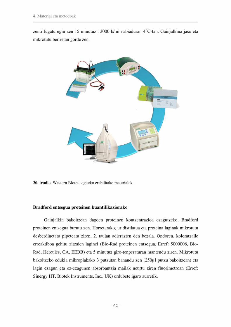
4. Material eta metodoak
- 62 -
zentrifugatu egin zen 15 minutuz 13000 b/min abiaduran 4°C-tan. Gainjalkina jaso eta
mikrotutu berrietan gorde zen.
20. irudia. Western Bloteta egiteko erabilitako materialak.
Bradford entsegua proteinen kuantifikaziorako
Gainjalkin bakoitzean dagoen proteinen kontzentrazioa ezagutzeko, Bradford
proteinen entsegua burutu zen. Horretarako, ur distilatua eta proteina laginak mikrotutu
desberdinetara pipeteatu ziren, 2. taulan adierazten den bezala. Ondoren, koloratzaile
erreaktiboa gehitu zitzaien laginei (Bio-Rad proteinen entsegua, Erref: 5000006, Bio-
Rad, Hercules, CA, EEBB) eta 5 minutuz giro-tenperaturan mantendu ziren. Mikrotutu
bakoitzeko edukia mikroplakako 3 putzutan banandu zen (250µl putzu bakoitzean) eta
lagin ezagun eta ez-ezagunen absorbantzia mailak neurtu ziren fluorimetroan (Erref:
Sinergy HT, Biotek Instruments, Inc., UK) ordubete igaro aurretik.

4. Material eta metodoak
- 63 -
Laginak prestatzea eta gela korritzea
Lagin bakoitzetik beharrezkoa zen proteinen kantitatea jakiteko Bradford entseguaz
baliatu ginen. Laginak 5µl 4x lagin tanpoiarekin nahastu eta ur distilatua gehitu zitzaien
20 µl-ko azken bolumena lortu arte. Laginak 10 minutuz irakin ziren eta zentrifugazio
motz baten ondoren (30s), lagin kantitate berdinak kargatu ziren poliakrilamidazko
geletan (Criterion™ TGX™ Precast Gels, Bio-Rad, Espainia). Laginekin batera,
kaleetako baten, pisu molekularraren markatzailea (Fisher BioReagents™ EZ-Run™
Prestained Rec Protein Ladder, Fisher Scientific, Espainia) jarri zen. Gelak 200V-tan
korritu ziren. Pisu molekular handiko proteinen (>150KDa) detekzioarako, 45 µg
proteina kargatu genituen %4-15 geletan. Aldiz, 150 KDa baino txikiagoak diren
proteinen detekzioareko 10-20 µg kargatu ziren %12ko geletan.
2. taula. Ezagunak eta ez-ezagunak diren proteinen kontzentrazioak zehazteko
beharrezkoak diren lagin kantitateak.
Proteinen transferentzia geletik mintzera
Pisu molekular baxuko proteinen detekziorako (<150KDa) transferentzia erdi-
lehorra egin zen Trans-Blot® Turbo™ Transfer System (Bio-Rad, Hercules, CA, EEBB)
aparatu komertziala erabiliz. Zazpi minutuko transferentzia burutu zen, intentsitate
konstantean (2.5A), 25V-ko tentsio maximo batekin eta PVDF mintzak erabiliz (Trans-
H2Od Koloratzaile
erreaktiboa
BSA
Hutsa 800 µl 200 µl --
4 796 µl 200 µl 4 µl
8 792 µl 200 µl 8 µl
12 788 µl 200 µl 12 µl
16 784 µl 200 µl 16 µl
20 780 µl 200 µl 20 µl
Lagina 800 µl 200 µl Laginaren µl 1

4. Material eta metodoak
- 64 -
Blot® Turbo™ Midi PVDF Transfer Packs, Erref: 1704157, Bio-Rad, Hercules, CA,
EEBB).
Transferentzia hezerako, nitrozelulosazko mintzak (Erref: 10600012, GE
Healthcare, Espainia) erabili ziren eta hauek, filtro papera eta zuntzezko kuxinekin batera
transferentzia tanpoian murgilduak izan ziren 15 minutuz. Proteinak tentsio konstantean
(30V) transferitu ziren 16 orduz, 4ºC-tan. Tranferentzia mota hau 150KDa baino pisu
molekular handiagoko proteinak detektatzeko erabili zen.
Markaketa antigorputzekin
Mintzak bi ordu t’erdiz inkubatu ziren hauts-esnetan lotura ez-espezifikoak
blokeatzeko. Horretarako hauts-esnea TBST-tan diluitu zen %5ean. Antigorputz
primarioan inkubazioa (3. taula) TBST + %5 BSA-tan egin zen gau osoan zehar 4ºC-tan.
Hurrengo egunean, mintzak TBST tanpoiarekin 3 aldiz garbitu ziren eta antigorputz
sekundarioan inkubatu ziren blokeo soluzioan eta %1 BSA erabiliz ordu betez giro-
tenperaturan. Horren ondoren, 5 minutuko 3 garbiketa egin eta seinalea errebelatzeko
kimioluminiszente bat erabili zen (Erref: 34076, SuperSignal® West Dura Extended
Duration Substrate, Fisher Scientific, Espainia).
Datuen analisia
Errebelatutako mintzen argazkiak ChemiDoc™ XRS+ Imaging System (Bio-Rad,
Hercules, CA, EEBB) eskanerraren bidez lortu ziren (20. Irudia). Argazki hauek Image
Studio Digits 3.1 (LI-COR Biotechnology, Cambridge, UK) programan ireki ziren
dentsitate optikoak kuantifikatzeko. Horretarako, laukizuzen bat marraztu eta bertako
seinalearen intentsitatea neurtu zen. Goi eta beheko intentsitateen batezbestekoa erabili
zen atzeko seinale gisa.
Banda bakoitzaren seinalea proteina bakar batekiko normalizatu zen, hala nola,
aktina (lagin guztietan maila berean agertzen den proteina) edo proteina kantitate
guztiarekiko. Azken kasu honetan, intereseko proteina detektatu eta gero (proteinen
egitura fosforilatuak, batik bat), mintzak garbiketa tanpoian (Restore Western Blot
stripping buffer, Erref: 21059, Thermo Fisher Scientific, Espainia) inkubatu ziren 15
minutuz, berriro antigorputz primarioan inkubatu ahal izateko. Proteinen ugaritasun
erlatiboa lortzeko, banden dentsitate optikoak animali kontrolekiko normalizatu ziren.

4. Material eta metodoak
- 65 -
Emaitza zehatzak ziurtatzeko, intereseko proteinak eta hauen kontrolak mintz berean
detektatuak izan ziren, eta kontrolekiko normalizazioa mintz berberean zeuden banden
artean burutu zen bakarrik. Datu guztiek, beraz, eramailea daramaten animaliekiko
areagotze edo murrizte erlatiboak adierazten dute.
Soluzioei eta tanpoiei buruzko informazio zehatzagoa 1. Eranskinean aurki daiteke.
3. taula. Antigorputzen kontzentrazioak eta erreferentziak
Antigorputz primarioak
Sagu anti-NR2A 1:1000 Erref: MAB5216, Merck-Millipore, EEBB
Untxi anti-NR2B 1:1000 Erref: AB1557, Merck-Millipore, EEBB
Sagu anti-NR1 1:1000 Erref: 05432, Merck-Millipore, EEBB
Untxi anti-PSD95 1:1000 Erref: Af628, Frontier Institute, EEBB
Untxi anti-phospho-Akt (Ser
473)
1:1000 Erref: 9271, Cell Signaling Technology Inc,
EEBB
Untxi anti-Akt 1:1000 Erref: 9272, Cell Signaling Technology Inc,
EEBB
Untxi anti-phospho-p44/42
MAPK (Erk 1/2) (Thr
202/204)
1:1000 Erref: 9101, Cell Signaling Technology Inc,
EEBB
Untxi anti-p44/42 MAPK (Erk
1/2)
1:1000 Erref: 9102, Cell Signaling Technology Inc,
EEBB
Untxi anti-phospho-TrkB
(Tyr 706)
1:250 Erref: sc-135645, Santa Cruz Biotechnology Inc,
Espainia
Untxi anti-TrkB (794) 1:1000 Erref: sc-12, Santa Cruz Biotechnology Inc,
Espainia
Untxi anti-BDNF 1:2000 Erref: NBP1-46750, Novus Biologicals, EEBB
Untxi anti-aktina 1:2000 Erref: A2066, Sigma-Aldrich, Espainia
Antigorputz sekundarioak
Anti-untxi IgG HRP-
konjugatua
1:20000 Erref: A-6154, Sigma-Aldrich, Espainia
Anti-sagu IgG HRP-
konjugatua
1:20000 Erref: A-9044, Sigma-Aldrich, Espainia

4. Material eta metodoak
- 66 -
4.10. Zelulen kuantifikazioa
4.10.1. Estereologia ez-alboratua
Kortex prefrontal medialean (kortex prelinbikoa eta aurreko kortex zingulatua) eta
hipokanpoan (hortz-jiroa eta cornus ammonis 1) zelula inmunoerreaktiboen estimazioak
burutzeko estereologia inpartzialaz baliatu ginen. Kortex prefrontal medialaren laginketa
Bregmatik 4.68 mm eta 1.92 mm bitartean egin zen. Bestalde, hipokanpoan Bregmatik
-2.40 mm eta -5.76 mm bitartean egin zen (21. irudia). Kortex prefrontal medialaren eta
hipokanpoaren mugak zehazteko, Paxinos eta Watsonen atlas estereotaxikoa erabili zen.
Desberdintasun zitoarkitektonikoak ez zirenez erraz nabarmentzen, batez ere zati
prelinbiko eta infralinbikoaren artean, beheko muga bezala aurreko burmuin azigo arteria
ezarri genuen. Hori dela eta, lan honetan zati prelinbikoari buruz ari garenean, kontutan
hartu behar da zati infralinbikoaren alderdi dortsalena ere nolabait barneratuta dagoela.
21. irudia. (a) Planoek arratoiaren garuneko intereseko eskualdeak adierazten dituzte. (b) Kortex
prefrontal medialeko ebaketa koronalak. Grisez, intereseko eskualdeak. (c) Hipokanpoko ebaketa
koronala. Grisez, intereseko eskualdea.

4. Material eta metodoak
- 67 -
Mercator Irudien Analisi sistema (Explora-Nova, La Rochelle, Frantzia) erabili
genuen kamera digital bat eta 3-ardatzetako plaka motorizatua atxikita duen Olympus
BX51 mikroskopioarekin batera (22. irudia). Zelula inmunopositiboak 40x aldiz
handiagotzen duen objektiboarekin zenbatu ziren Zatitzaile Optikoaren planteamendua
erabiliz. Zatitzaile Metodoa, Zorizko Laginketa Sistematikoan (ZLS) oinarrituz, zelula
zenbaki totala estimatzeko gai da interesezko eskualde konkretu batean. Horretarako,
eremu osoa X, Y eta Z norabideetan distantzia uniformea duten kontaketa espazio
birtualen multzo batez estaltzen da. Zelula kopuru totala zehazteko hurrengo formula hau
erabiltzen da: N=∑Q x 1/slz x 1/elz x 1/alz, non Q-k espezimen bakoitzean kontatu diren
zelula kopurua adierazten duen eta N-k estimatutako zelula kopurua. Ikerketa honetan
erabilitako sekzio laginketa zatikia (slz) 1/8 da kortex prefrontalerako eta 1/10
hipokanporako.
22. irudia. Estereologia ez-alboraturako erabilitako estazio estereologikoa.
Intereseko eremuak 4x handiagotzeko objektiboarekin mugatu ziren. Lauki eta
kontaketa markoaren (disektorea) tamaina desberdinak erabili ziren intereseko eremu
bakoitzean (4. taula) eta %5eko zaintza zona bat aukeratu zen. Kontaketak estereologiaren
kontaketa arauen arabera burutu ziren. Hipokanpoko interneuronen kantitatea
zenbatesteko, elz (eremuaren laginketa zatikia) eta alz (alturaren laginketa zatikia)-ri 1

4. Material eta metodoak
- 68 -
balorea ezarri genien. Orokorrean, animali bakoitzeko gutxi gora behera 7-8 sekzio
kontatu ziren intereseko eremu bakoitzean. Bolumenen estimazioak Cavalieri-ren
metodoa erabiliz lortu ziren.
4. taula. Zatitzaile Metodoan erabilitako elz baloreak neuronen zenbaki kopurua eta
sare mikrobaskularraren luzera zenbatesteko.
Eremua
Disektorearen
tamaina(µm)
Disektoreen
arteko tartea
(µm)
NeuN
mPFC PL, AC 25x25 350x350
HPC
Geruza molekularra (HJ) 50x50 75x75
Geruza granularra (HJ) - -
Geruza polimorfikoa (HJ) 25x25 75x75
CA1 (str.oriens eta str.lacunosum) 50x50 150x150
BCh mPFC PL, AC r=50 200x200
HPC CA1, CA3, HJ r=50 200x200
GAD67 mPFC PL, AC 80x80 270x270
HPC CA1, CA3, HJ 80x80 270x270
IN (PB,
KR, KB) mPFC PL, AC
80x80 220x220
SST mPFC PL, AC 80x80 270x270
HPC CA1, CA3, geruza polimorfikoa (HJ) 50x50 50x50
elz, eremuaren laginketa zatikia; mPFC, kortex prefrontal mediala; HPC, hipokanpoa; NeuN,
neuronen nukleoaren markatzailea; BCh, butiril kolinesterasaren histokimia; IN, interneuronak;
PB, parbalbumina; KR, kalretinina; KB, kalbindina; GAD67, azido glutamiko deskarboxilasa 67;
SST, somatostatina; PL, kortex prelinbikoa; AC, aurreko kortex zingulatua; CA1, hipokanpoko
cornus ammonis 1; CA3, hipokanpoko cornus ammonis 3; HJ, hipokanpoko hortz-jiroa; r,
disektore esferikoaren erradioa.
Somatostatina adierazten duten interneuronak hortz-jiroko geruza polimorfikoan
baino ez direnez agertzen, eskualde honetako geruza hau bakarrik kuantifikatu zen. Era
berean, CA1 dortsaleko zelula parbalbumina- eta somatostatina-positiboen

4. Material eta metodoak
- 69 -
estimaziorako, stratum oriens eta stratum pyramidale bakarrik hartu ziren aintzat. Are
gehiago, CA1-eko geruza piramidala eta hortz-jiroko geruza granularra baztertuak izan
ziren zelula kalbindina-positiboen estimazioetatik, bertan zelula kitzikatzaileek ere
kalbidina adierazten dutelako. Halaber, kalbindinaren aurkako antigorputzak kortex
prefrontaleko II/III. geruzetan dauden zelula piramidalak ere orbaintzen dituenez, geruza
sakonak baino ez dira irudikatzen kalbindinaren estimazioetan.
Odol-hodien luzera zenbatesteko, Zatitzaile Metodoa erabili zen baina disektore
optikoak espazio-baloiengatik (disektore esferikoak) aldatuz. Laginketa irizpideak 4.
taulan azaltzen dira.
Kontaketa parametroak zehazteko errore koefizientearen (CE) bariantza osorako
ekarpena %20a baino txikiagoa zela frogatu genuen atariko proban, hurrengo formula
honen bidez: CE2/CV2, non CE2 zenbateste estereologikoen bariantza den eta CV2
bariantza biologikoa. Normalean, zatikia 0.5 baino txikiagoa denean zenbateste
estereologikoak nahikoa zehatzak direla esan ohi da eta estimazioak ontzat ematen dira.
Honen arabera, esan genezake gure laginketa prozedura zuzena izan dela.
4.10.2. Parbalbumina eta GAD67 ko-adierazten duten zelulen kuantifikazioa
Kasu honetan, kuantifikazioa mPFC-eko 6 animaliren 4 sekziotan egin zen eta
hipokanpoan 3 animaliren 3 sekzio desberdinetan. Sekzioak 400 µm-tan banandu ziren
mPFC-ean eta 500 µm-tan hipokanpoan. Irudiak 20x-ko objektiboa erabiliz atera ziren
ApoTomea atxikita zuen Zeiss fluoreszentziako mikroskopian. Irudiak berreraiki
ondoren, Fiji-ko zelula kontatzaile tresnarekin zenbatu ziren. mPFC-ean 150x150 µm-ko
karratu bat erabili zen bai parbalumina bakarrik zein parbalbumina eta GAD67 ko-
adierazten zuten interneuronak kontatzeko. Karratu horietako bat eremu linbikoaren parte
dortsalean (area prelinbikoan, batik bat) zegoen zentratuta eta bestea aurreko kortex
zingulatuaren erdian. Hipokanpoan, sekzio osoak erretratatu ziren eta fotomuntaien bidez
berreraiki. Hipokanpoko CA1, CA3 eta HJ eskualde osoak mugatu ziren eta bertako PV
eta PV-GAD67 ko-adierazten zuten interneurona guztiak zenbatu ziren.
Dentsitateak PV eta GAD67 adierazten duten soma kopurua mm2-ko bezala ematen
dira. Ostean, PV-GAD67 ratioak PV eta GAD67 adierazten duten soma kopurua zati PV+
interneurona zenbaki orokorra bezala azaltzen dira.
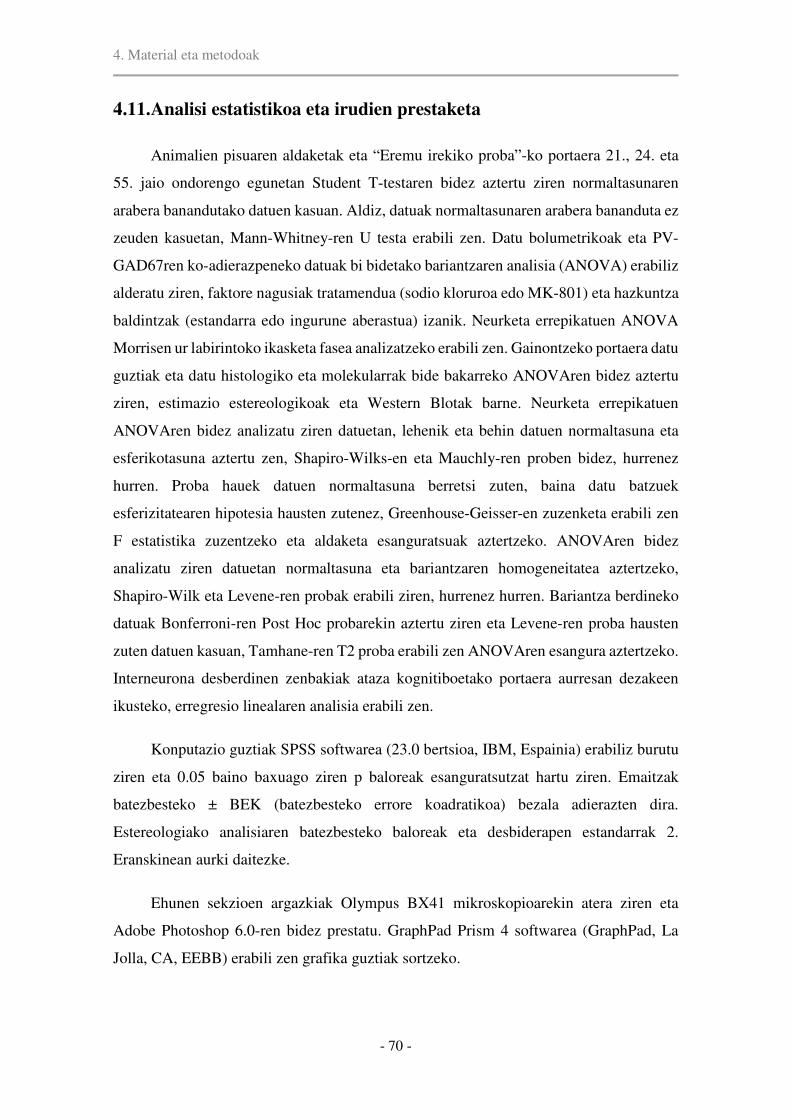
4. Material eta metodoak
- 70 -
4.11. Analisi estatistikoa eta irudien prestaketa
Animalien pisuaren aldaketak eta “Eremu irekiko proba”-ko portaera 21., 24. eta
55. jaio ondorengo egunetan Student T-testaren bidez aztertu ziren normaltasunaren
arabera banandutako datuen kasuan. Aldiz, datuak normaltasunaren arabera bananduta ez
zeuden kasuetan, Mann-Whitney-ren U testa erabili zen. Datu bolumetrikoak eta PV-
GAD67ren ko-adierazpeneko datuak bi bidetako bariantzaren analisia (ANOVA) erabiliz
alderatu ziren, faktore nagusiak tratamendua (sodio kloruroa edo MK-801) eta hazkuntza
baldintzak (estandarra edo ingurune aberastua) izanik. Neurketa errepikatuen ANOVA
Morrisen ur labirintoko ikasketa fasea analizatzeko erabili zen. Gainontzeko portaera datu
guztiak eta datu histologiko eta molekularrak bide bakarreko ANOVAren bidez aztertu
ziren, estimazio estereologikoak eta Western Blotak barne. Neurketa errepikatuen
ANOVAren bidez analizatu ziren datuetan, lehenik eta behin datuen normaltasuna eta
esferikotasuna aztertu zen, Shapiro-Wilks-en eta Mauchly-ren proben bidez, hurrenez
hurren. Proba hauek datuen normaltasuna berretsi zuten, baina datu batzuek
esferizitatearen hipotesia hausten zutenez, Greenhouse-Geisser-en zuzenketa erabili zen
F estatistika zuzentzeko eta aldaketa esanguratsuak aztertzeko. ANOVAren bidez
analizatu ziren datuetan normaltasuna eta bariantzaren homogeneitatea aztertzeko,
Shapiro-Wilk eta Levene-ren probak erabili ziren, hurrenez hurren. Bariantza berdineko
datuak Bonferroni-ren Post Hoc probarekin aztertu ziren eta Levene-ren proba hausten
zuten datuen kasuan, Tamhane-ren T2 proba erabili zen ANOVAren esangura aztertzeko.
Interneurona desberdinen zenbakiak ataza kognitiboetako portaera aurresan dezakeen
ikusteko, erregresio linealaren analisia erabili zen.
Konputazio guztiak SPSS softwarea (23.0 bertsioa, IBM, Espainia) erabiliz burutu
ziren eta 0.05 baino baxuago ziren p baloreak esanguratsutzat hartu ziren. Emaitzak
batezbesteko ± BEK (batezbesteko errore koadratikoa) bezala adierazten dira.
Estereologiako analisiaren batezbesteko baloreak eta desbiderapen estandarrak 2.
Eranskinean aurki daitezke.
Ehunen sekzioen argazkiak Olympus BX41 mikroskopioarekin atera ziren eta
Adobe Photoshop 6.0-ren bidez prestatu. GraphPad Prism 4 softwarea (GraphPad, La
Jolla, CA, EEBB) erabili zen grafika guztiak sortzeko.

5. Results

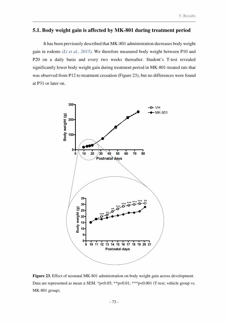
5. Results
- 73 -
5.1. Body weight gain is affected by MK-801 during treatment period
It has been previously described that MK-801 administration decreases body weight
gain in rodents (Li et al., 2015). We therefore measured body weight between P10 and
P20 on a daily basis and every two weeks thereafter. Student’s T-test revealed
significantly lower body weight gain during treatment period in MK-801-treated rats that
was observed from P12 to treatment cessation (Figure 23), but no differences were found
at P31 or later on.
Figure 23. Effect of neonatal MK-801 administration on body weight gain across development.
Data are represented as mean ± SEM. *p<0.05; **p<0.01; ***p<0.001 (T-test; vehicle group vs.
MK-801 group).

5. Results
- 74 -
5.2. Locomotor activity in Open-Field Test
In the Open Field Test (OFT), the total distance traveled was significantly shorter
in MK-801-treated rats than in saline controls 24-hours after last injection (Mann-
Whitney U test, p < 0.001; Figure 24a). In addition, the relative distance moved in the
center of the arena was also significantly shorter (Mann-Whitney U test, p < 0.001; Figure
24b). On P24, these differences were absent (p=0.378) and normal locomotor activity was
preserved until P55 (p=0.403).
Figure 24. Effects of chronic MK-801 treatment on locomotor activity in the Open Field Test at
(a,b) postnatal days (P) 21, P24 and P55 and (c,d) after exposure to enriched environment on P73.
Data in (a,c) represent the total distance traveled whereas (b,d) show the percentage of distance
moved in center vs. periphery. (e,f) Representative tracks of locomotor activity in Actitrack at
P21 in (e) vehicle group and (f) MK-801 group. Data are plotted as mean ± SEM. *p<0.05;
**p<0.01; ***p<0.001.

5. Results
- 75 -
After a short exposure to EE in early adulthood, rats exhibited significant changes
in locomotor activity in terms of total distance moved (F(3,35)=22.31, p<0.0001) and the
relative distance spent in the center (F(3,34)=7.31, p<0.001). Post Hoc analysis revealed
that there was not any difference between standard environment raised animals in either
parameter on P73. However, MK-801+EE showed significantly less activity than MK-
801 group (p<0.0001; Figure 24c). Comparisons of relative distance in center showed
that VH+EE traveled about one-third as much distance in center compared to the other 3
groups (Figure 24d). The tendency to avoid the periphery of the OFT is widely used to
assess anxiolytic-like effects of treatments or interventions.
5.3. EE restores spatial learning and recognition memory impairment
induced by MK-801
Morris Water Maze
To determine the presence of cognitive deficits after neonatal MK-801 treatment,
and to evaluate the potential therapeutic effects of EE, we tested the animals in Morris
Water Maze from P65 to P70. Repeated-measures ANOVA showed significant
differences of performance during the acquisition phase in Morris Water Maze in terms
of escape latencies (F(3,42)=7.877, p<0.0001, Figure 25a) and path length
(F(3,42)=7.942, p<0.0001, Figure 25b). Post Hoc comparisons showed that MK-801-
treated rats showed a significant increase in both time and distance to find the hidden
platform when compared to VH (time p<0.0001; distance p=0.01) and VH+EE rats (time
p=0.005; distance p=0.001) (Figure 25a and b), representing major impairment of spatial
learning. This impairment in spatial learning of MK-801-treated rats allowed dissecting
the role of EE in cognitive enhancement. Indeed, MK-801+EE rats showed normal spatial
learning (MK-801+EE vs. VH - time and distance p=1.00; MK-801+EE vs. VH+EE -
time p=1.000, distance p=0.365). Bonferroni Post Hoc test showed that MK-801+EE
group spent less time to locate the platform compared to MK-801 rats in standard
conditions (p=0.004), and traveled less distance (p=0.044), which strongly suggests
improvement in spatial learning abilities.

5. Results
- 76 -
All rats showed a significant improvement in the acquisition phase of Morris Water
Maze. Escape latency and distance to the platform decreased over the 5 days of training
(F(4,39)=43.858, p<0.001; (F(4,39)=26.908, p<0.0001), respectively), indicating that
animals spent less time to reach the platform as the training proceeded. Representative
traces of the swimming paths during last day of training are shown in Figure 25c.
After 5 days training, probe trial was performed with the platform removed to
evaluate reference memory. The parameter “mean distance to target” (MDT) provided by
SMART software was used, which is a measure of proximity and directionality to the
location of the platform. Some studies have concluded that this parameter is consistently
more sensitive at detecting group differences than more commonly used parameters, like
percentage of time or number of crossing of target quadrant (Gallagher et al., 1993; Maei
et al., 2009). Swimming speed was measured throughout the test, and significant
difference was observed in the mean swimming speed between groups in probe trial
(F(3,39)=8.211, p<0.0001). Nevertheless, MDT is minimally affected by changes in
velocity. We found significant differences in reference memory between groups
(F(3,35)=11.135, p<0.0001; Figure 25d). Bonferroni Post Hoc test showed that MK-801
treatment during neurodevelopment did not affect reference memory in the Morris Water
Maze (VH vs. MK-801, p=1.00). However, rats that had experienced EE showed lower
values of MDT, suggesting improved reference memory (EE groups vs. VH, p<0.0001;
MK-801+EE vs. MK-801, p=0.01; VH+EE vs. MK-801, p=0.007; Figure 25d).
In the visible platform test there were no differences between groups in time
(F(3,39)=2.425, p=0.08) or distance (F(3,39)=1.324, p=0.28), which indicates that the
differences in performance in spatial learning acquisition may not be ascribed to
differences in general sensorimotor skills (Figure 25a and b, “cue”).

5. Results
- 77 -
Figure 25. Spatial learning and memory in the Morris Water Maze. MK-801 animals were less
efficient in finding the platform, as demonstrated by increased (a) average time and (b) distance
during acquisition trials, and this was reverted by adult short-term exposure to EE. (c) Tracks are
depicting schematic representation of swimming paths on Morris Water Maze on day 5 in each
group. (d) Mean distance to target during Morris Water Maze probe trial showed no significant
differences between standard condition animals, but enriched animals presented improved
reference memory. Results are presented as mean ± SEM. In a and b, *significance MK-801 vs.
VH (**p < 0.01; ***p < 0.001); +significance MK-801 vs. MK-801+EE (+p < 0.05; ++p < 0.01);
#significance MK-801 vs. VH+EE (##p < 0.01; ###p < 0.001), using Repeated-Measures ANOVA.
In d, *p < 0.05; **p < 0.01; ***p < 0.001 (one-way ANOVA).

5. Results
- 78 -
Novel Object Recognition task
Novel Object Recognition (NOR) test is widely used to assess cognitive function
in animal models. In the acquisition phase, the total exploration time was comparable
between groups (F(3,27)=1.930, p=0.149). However, there was a significant difference in
in the test trial (F(3,26)=15.75, p<0.0001; Figure 26). Post Hoc analysis showed that early
MK-801 treatment led to cognitive dysfunction, as discrimination index was significantly
lower in MK-801 group compared to saline controls (p=0.04).
MK-801+EE group presented normal recognition memory (vs. VH, p=0.412), and
discrimination index was significantly augmented when compared to MK-801 group
(p=0.012), representing major memory improvement. Rats in VH+EE group showed the
greatest discrimination index, but not statistically different from MK-801+EE group (VH
vs. VH+EE, p=0.001; MK-801 vs. VH+EE, p<0.0001; Figure 26).
Figure 26. Discrimination Index in Novel Object Recognition task performed on P71. MK-801
group exhibited impaired object recognition memory that was reverted by EE. Data are
represented as mean ± SEM. *p<0.05; **p<0.01; ***p<0.001 (One-way ANOVA).
5.4. Associative recognition memory deficit is partially reversed by EE
Object-in-Place is a type of object-place paired-associative task that highly relies
on the interplay between hippocampus and mPFC. During the sample trials in the Object-

5. Results
- 79 -
in-Place tests, the interaction time with objects was comparable across groups
(F(3,37)=1.768, p=0.17). In the test trial, one-way ANOVA showed differences between
groups (F(3,35)=7.016, p<0.0001; Figure 27). Neonatal MK-801 exposure significantly
reduced discrimination index (VH vs. MK-801, p=0.047), and enrichment produced a 3-
fold increase in MK-801 animals, although it was not significantly different from MK-
801 group. However, the average discrimination index of MK-801+EE group (15.2%)
was not statistically different from the VH group either (22.02%, p=1.00), showing a
partial recovery of associative memory. VH+EE showed the highest preference index
(30.5%), doubling the index of MK-801+EE rats.
Figure 27. Graph shows the discrimination index in Object-in-Place (OiP) task performed on
P72. MK-801 group exhibited impaired object recognition associative memory that was partially
reverted by EE. Data are represented as mean ± SEM. *p<0.05; **p<0.01; ***p<0.001 (One-way
ANOVA).
Since we failed to find significant differences between MK-801 and MK-801+EE
groups, we performed a two-way ANOVA analysis to unravel the effects of MK-801 and
EE as independent variables. We found that both MK-801 (F(1,35)=14.714, p=0.001) and
EE (F(1,35)=4.921, p=0.033) had a significant effect on discrimination index in Object-
in-Place task.

5. Results
- 80 -
5.5. Volume of medial prefrontal cortex and hippocampus
Medial prefrontal cortex (mPFC) and hippocampal volumes were measured in the
same sections used for NeuN quantification. Two-way ANOVA showed a significant
decrease in prelimbic (F(1,20)=8.32, p=0.009) and anterior cingulate (F(1,20)=8.19,
p=0.01) volume associated with early life MK-801 administration (Figure 28). Similarly,
the treatment significantly reduced hippocampal CA1 volume (F(1,20)=9.08, p=0.007),
but not that of dentate gyrus (F(1,20)=1.48, p=0.237). Contrarily, EE significantly
increased mPFC volume (prelimbic cortex F(1,20)=7.02, p=0.015; anterior cingulate
cortex F(1,20)=11.57, p=0.003), and the volume of DG (F(1,29)=4.99, p=0.04). No
significant changes were detected in CA1 of hippocampus caused by EE (Figure 28).
Figure 28. Estimates of mPFC and hippocampal volumes by Cavalieri’s method. Histograms
show the estimated volumes of the (a) PL, prelimbic cortex, (b) AC, anterior cingulate cortex, (c)
CA1, cornus ammonis 1, and (d) DG, dentate gyrus of hippocampus in different experimental
groups. Values represent mean ± SEM. *Significance of treatment (*p<0.05; **p<0.01), #significance of housing (#p<0.05; ##p<0.01).

5. Results
- 81 -
5.6. Vascular changes
Neurons, astrocytes, and blood vessels are organized in a functional unit, termed as
“neurogliovascular unit”, in which the vasculature can affect neuronal and glial activity
and, in turn, dynamically adjust to their changes. Micro- and macrovascular abnormalities
have direct implications on the pathophysiology of schizophrenia (Katsel et al., 2017).
For assessing if MK-801 or EE induced vascular changes, endothelial cells of vessels
were revealed by butyryl cholinesterase histochemistry, and total vessel length was
estimated by stereological means in mPFC and hippocampus. We failed to find
differences in mPFC, although one-way ANOVA was near significance (F(3,20)=2.965,
p=0.057; Figure 29). However, we found significant changes in hippocampus
(F(3,19)=7.039, p=0.002), mainly due to decreased microvascular length in MK-801-
treated animals (MK-801 vs. VH, p=0.031; MK-801 vs. MK-801+EE, p=0.013; MK-801
vs. VH+EE, p=0.003).
Figure 29. Estimated vessel length by stereology using Space Ball (spherical probe) in medial
prefrontal cortex (left) and hippocampus (right). Post Hoc analysis showed that EE reverted MK-
801-induced loss of vasculature in hippocampus. mPFC, medial prefrontal cortex; HPC,
hippocampus. Graphs show mean ± SEM. *p<0.05; **p<0.01; ***p<0.001.
To further assess whether MK-801 and EE could have significant effects as
independent variables, we performed two-way ANOVA in hippocampus and mPFC.
Similar to the results obtained in one-way ANOVA, no effect of either factor was found
in mPFC [MK-801 (F(1,19)=0.006, p=0.937); EE (F(1,19)=0.023, p=0.88)]. However, in
hippocampus, we found that MK-801 had a significant effect (F(1,19)=7.420, p=0.013),
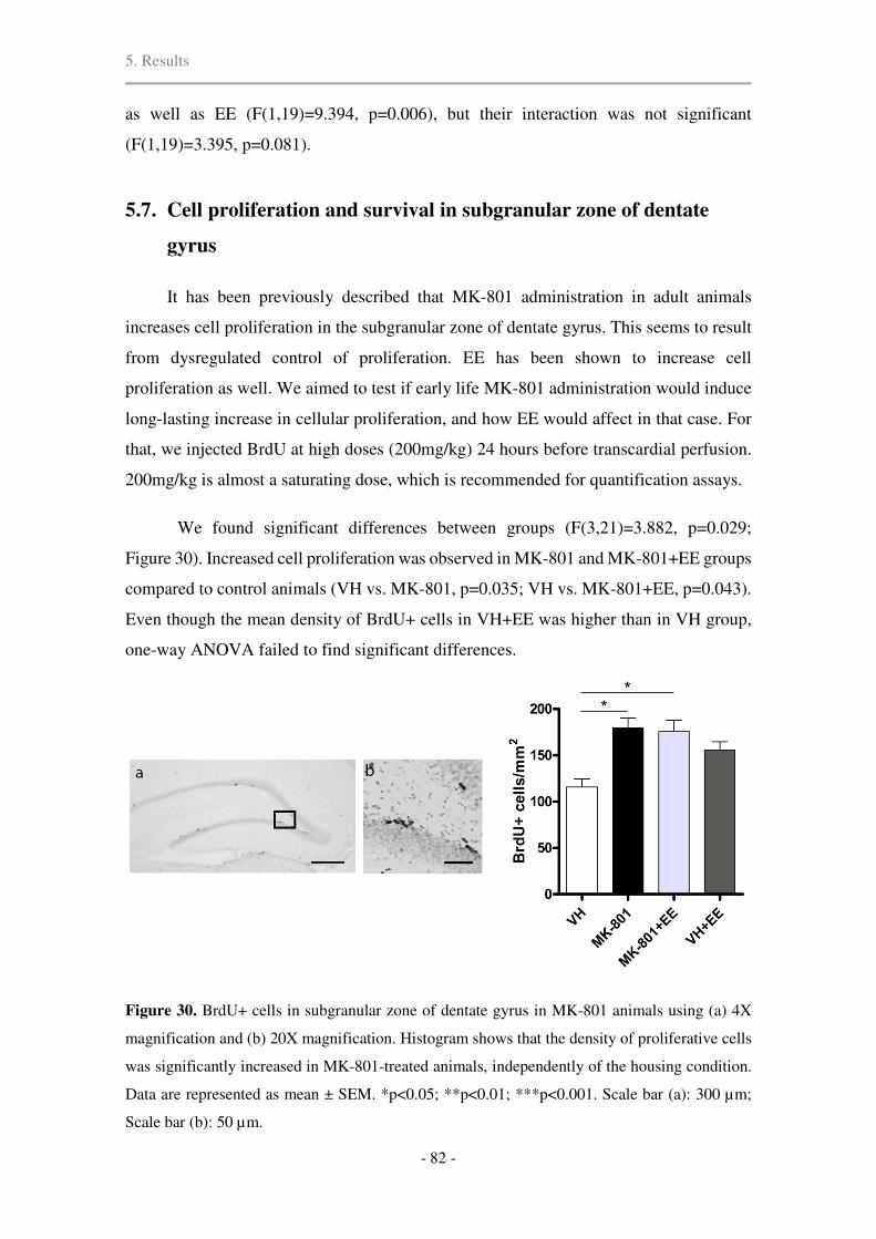
5. Results
- 82 -
as well as EE (F(1,19)=9.394, p=0.006), but their interaction was not significant
(F(1,19)=3.395, p=0.081).
5.7. Cell proliferation and survival in subgranular zone of dentate
gyrus
It has been previously described that MK-801 administration in adult animals
increases cell proliferation in the subgranular zone of dentate gyrus. This seems to result
from dysregulated control of proliferation. EE has been shown to increase cell
proliferation as well. We aimed to test if early life MK-801 administration would induce
long-lasting increase in cellular proliferation, and how EE would affect in that case. For
that, we injected BrdU at high doses (200mg/kg) 24 hours before transcardial perfusion.
200mg/kg is almost a saturating dose, which is recommended for quantification assays.
We found significant differences between groups (F(3,21)=3.882, p=0.029;
Figure 30). Increased cell proliferation was observed in MK-801 and MK-801+EE groups
compared to control animals (VH vs. MK-801, p=0.035; VH vs. MK-801+EE, p=0.043).
Even though the mean density of BrdU+ cells in VH+EE was higher than in VH group,
one-way ANOVA failed to find significant differences.
Figure 30. BrdU+ cells in subgranular zone of dentate gyrus in MK-801 animals using (a) 4X
magnification and (b) 20X magnification. Histogram shows that the density of proliferative cells
was significantly increased in MK-801-treated animals, independently of the housing condition.
Data are represented as mean ± SEM. *p<0.05; **p<0.01; ***p<0.001. Scale bar (a): 300 µm;
Scale bar (b): 50 µm.

5. Results
- 83 -
In adult hippocampal neurogenesis, immature neurons transiently express CR
before differentiating into dentate granule cells and fully integrating in the hippocampal
circuit. To establish differences in neuronal differentiation and survival, the number of
CR-expressing cells was estimated in the granule cell layer of dentate gyrus. The total
number of CR-expressing cells was increased in granule cell layer of VH+EE group when
compared all other groups (F(3,20)=4.97, p<0.01; Figure 31).
Figure 31. Images show the calretinin-positive (CR+) cells in the granular layer of dentate gyrus
of different experimental groups indicated by arrows. Histogram shows the differences in the total
number of calretinin-expressing cells between groups. GCL, granular cell layer of dentate gyrus.
Data are represented as mean ± SEM. *p<0.05; **p<0.01. Scale bar: 300 µm.
5.8. Number of calcium-binding protein and somatostatin-expressing
interneurons in hippocampus and medial prefrontal cortex
The total number of different populations of interneurons was estimated by
unbiased stereology in prelimbic cortex and anterior cingulate cortex of mPFC, and in
CA1 and dentate gyrus of hippocampus. Interneurons were identified upon their calcium-
binding protein expression, i.e. parvalbumin (PV) (Figure 33 and 34), calretinin (CR)
(Figure 35 and 36), and calbindin (CB), and upon their somatostatin expression (Figure
37 and 38).
One-way ANOVA showed that there was a significant difference in the number of
PV and CR-expressing interneurons, but not in CB-expressing ones. PV+ differences

5. Results
- 84 -
were present in all analyzed regions [prelimbic (F(3,20)=11.77, p<0.0001); anterior
cingulate (F(3,20)=11.66, p<0.0001); CA1 (F(3,20)=10.40, p<0.0001); and DG
(F(3,20)=7.14, p=0.02)]. Similar differences were found in CR-expressing interneurons
[prelimbic (F(3,20)=5.97, p=0.004); anterior cingulate (F(3,20)=14.13, p<0.0001); CA1
(F(3,20)=3.64, p=0.03), except for DG (F(3,20)=2.30, p=0.11)].
Figure 32. Effects of prenatal exposure to MK-801 and enriched environment on the number of
different interneuron immunoreactivity in (a,c,e) medial prefrontal cortex (mPFC) and (b,d,f)
hippocampus. Interneurons are identified by their calcium-binding protein expression: (a,b)
parvalbumin, (c,d) calretinin, and (e,f) calbindin. PL, prelimbic cortex; AC, anterior cingulate
cortex; CA1, cornus ammonis 1; DG, dentate gyrus of hippocampus. Data are represented as mean
± SEM. *p<0.05; **p<0.01; ***p<0.001.

5. Results
- 85 -
In mPFC, MK-801-treated rats showed a dramatic decrease in the number of PV+
cells in prelimbic (p=0.022) and anterior cingulate (p=0.01) cortices compared to vehicle
group (Figure 32a and 33). Regarding the hippocampal PV+ cell estimates, a significant
decrease was also observed in CA1 (p<0.0001) and DG (p=0.024) (Figure 32b and 34).
These deficits in PV+ interneuron expression were partially recovered by a short exposure
to EE, as the number of PV+ cells in MK-801+EE group was not statistically different
from the control group in either region, except for CA1 (p=0.042). The number of PV+
interneurons in MK-801+EE was different from MK-801-treated rats without
environmental intervention in anterior cingulate cortex (p=0.004) and DG (p=0.023), but
not in prelimibic cortex (p=0.081) or CA1 (p=0.244). VH+EE group showed augmented
mean PV+ interneurons in mPFC compared to all other groups (Figure 32a), but it was
only significantly higher than MK-801+EE group in prelimbic region (p=0.027). Similar
to vehicle group, VH+EE was significantly different from MK-801 group in all regions
that were analyzed (prelimbic, p<0.0001; anterior cingulate, p<0.0001; CA1, p=0.003;
DG, p=0.002) (Figure 32a and b).
Figure 33. Parvalbumin-positive interneurons in prelimbic region of medial prefrontal cortex.
Scale bar: 200 µm.

5. Results
- 86 -
Figure 34. Representative images showing distinct expression of parvalbumin (PV) and glutamic
acid decarboxylase-67 (GAD67) somata in hippocampal cornus ammonis 1 (CA1) and dentate
gyrus (DG) of different experimental groups. Scale bar: 300 µm.
The decrease in CR-expressing interneurons in MK-801 group was less evident,
still significantly different from vehicle animals in both regions of mPFC (prelimbic
cortex, p=0.023; anterior cingulate cortex, p=0.001) and in CA1 of hippocampus
(p=0.035) (Figure 32c, 32d and 35). Similar to what happened with PV+ interneurons,
EE partially reversed the loss of CR-immunoreactivity (MK-801+EE vs. VH; prelimbic
p=0.59; anterior cingulate, p=1.0; CA1, p=0.1) (Figure 32c, 32d and 36).
No significant differences were found in either region of mPFC or hippocampus of
CB-expressing interneurons [prelimbic (F(3,20)=0.87, p=0.48), anterior cingulate
(F(3,20)=2.23, p=0.12), CA1 (F(3,20)=1.52, p=0.24), and DG (F(3,20)=0.14, p=0.94);
Figure 32e and f]. It is worth to mention that only deep layers of mPFC were considered
for CB+ interneuron estimations to avoid confounding effects of CB-expressing
pyramidal cells of LII/III.

5. Results
- 87 -
Figure 35. Calretinin-positive interneurons in prelimbic region of medial prefrontal cortex. Scale
bar: 150 µm.
Figure 36. Calretinin-positive interneurons in cornus ammonis 1 (CA1) region of hippocampus.
Scale bar: 150 µm.

5. Results
- 88 -
Regarding the number of somatostatin (SST)-expressing interneurons in mPFC and
HPC (Figure 37), we found statistically significant differences in all regions, except for
dentate gyrus [prelimibic (F(3,20)=7.355, p=0.002); anterior cingulate (F(3,20)=3.233,
p=0.044); CA1 (F(3,20)=7.043, p=0.002); CA3 (F(3,20)=11.016, p<0.0001); DG
(F(3,20)=2.405, p=0.098); Figure 38].
Figure 37. Representative images showing differences in somatostatin-expressing interneurons
in distinct hippocampal sections. DG, dentate gyrus of hippocampus; CA1, cornus ammonis 1;
CA3, cornus ammonis 3. Scale bar: 300 µm.
MK-801 treatment during neurodevelopment decreased the number of SST+
interneurons in prelimbic region (p=0.004), CA1 (p=0.002), and CA3 (p=0.001)
compared to VH group. EE was effective in increasing the number of SST+ interneurons
in MK-801+EE group in the affected three regions, but those changes only reached
statistical significance in CA3 (p=0.001) when compared to MK-801 group. In the
remaining two regions, i.e. prelimbic cortex and CA1, EE partially reverted SST-

5. Results
- 89 -
expression deficits. Contrarily, environmental enrichment did not alter the number of
SST+ interneurons in any region of VH+EE animals compared to VH, suggesting that EE
only promoted SST-expression in MK-801+EE group.
Figure 38. Effects of postnatal exposure to MK-801 and enriched environment on the number of
interneurons immunoreactive to somatostatin in (a) medial prefrontal cortex (mPFC) and (b)
hippocampus. PL, prelimbic cortex; AC, anterior cingulate cortex; CA1, cornus ammonis 1; CA3,
cornus ammonis 3; DG, dentate gyrus of hippocampus. Data are represented as mean ± SEM.
*p<0.05; **p<0.01; ***p<0.001.
5.9. Lack of hippocampal inhibition in MK-801-treated rats without
overall cell loss
One-way ANOVA on NeuN immunoreactivity showed no differences between
groups in the total number of cells in mPFC and hippocampus [NeuN: prelimbic
(F(3,20)=2.35, p=0.10), anterior cingulate (F(3,20)=0.38, p=0.77), CA1 (F(3,20)=0.13,
p=0.94), and DG (F(3,20)=1.63, p=0.21); Figure 39a and b]. Contrarily, the number of
GAD67-expressing GABAergic interneurons was significantly changed in all regions of
mPFC [prelimbic cortex (F(3,20)=4.94, p=0.01); anterior cingulate cortex (F(3,20)=5.88,
p<0.005)] and hippocampus [CA1 (F(3,17)=28.16, p<0.0001); DG (F(3,19)=6.96,
p=0.002); Figure 39c and d)].
In prelimbic cortex, the number of GAD67 immunoreactive cells in VH+EE group
was significantly increased compared to MK-801 (p=0.015) and MK-801+EE (p=0.033)
groups. In anterior cingulate cortex, VH+EE only differed from MK-801 group
(p=0.015). In hippocampal CA1, a relevant reduction of GAD67 could be found between

5. Results
- 90 -
vehicle and MK-801 rats (p<0.0001) that was recovered by EE (MK-801 vs. MK-
801+EE, p=0.006) (Figure 34). VH+EE group presented the greatest number of GAD67+
cells in CA1 (VH vs. VH+EE, p=0.036; MK-801 vs. VH+EE, p<0.0001; MK-801+EE
vs. VH+EE, p=0.001). Only the animals exposed to EE (MK-801+EE and VH+EE)
presented significantly increased GAD67 immunoreactivity in DG compared to MK-801-
treated rats (MK-801 vs. MK-801+EE, p=0.001; MK-801 vs. VH+EE, p=0.02) (Figure
39).
Figure 39. Number of (a,b) NeuN and (c,d) glutamic acid decarboxylase 67 (GAD67)
immunoreactive cells in different regions of medial prefrontal cortex (mPFC) and hippocampus.
PL, prelimbic cortex; AC, anterior cingulate cortex; CA1, cornus ammonis 1; DG, dentate gyrus
of hippocampus. Graphs show mean ± SEM. *p<0.05; **p<0.01; ***p<0.001.
5.10. EE increases the number of PV+ interneurons co-expressing
GAD67
Based on stereological counting of PV and GAD67-expression separately, we found
no differences in GAD67-immunoreactivity in mPFC between VH and MK-801 groups.
To further explore the possibility of a selective downregulation of GAD67 in specific
interneurons populations, we labeled PV+ interneurons that co-expressed GAD67. Two-

5. Results
- 91 -
way ANOVA revealed that there was no interaction between MK-801 and EE conditions.
MK-801 treatment decreased the density of PV and GAD67 co-expression that was
significant in prelimbic region (F(1,20)=14.172, p=0.001), CA1 (F(1,8)=17.8, p=0.003)
and CA3 (F(1,8)=13.167, p=0.007) (Figure 40a). In anterior cingulate cortex, MK-801
induced a decrease in GAD67-expression in PV+ interneurons was near significance
(F(1,20)=4.013, p=0.059). 18 days of adult EE were sufficient to invariably increase the
density of PV interneurons co-expressing GAD67 in all regions [prelimbic
(F(1,20)=11.942, p=0.002); anterior cingulate (F(1,20)=4.568, p=0.045); CA1
(F(1,8)=8.586, p=0.019); CA3 (F(1,8)=15.119, p=0.005); DG (F(1,8)=7.355), p=0.027)]
(Figure 40b).
Figure 40. Density of PV and GAD67 coexpressing interneurons in (a) mPFC and (b) dorsal
hippocampus expressed as number of somata per mm2. PL, prelimbic cortex; AC, anterior
cingulate cortex; CA1, cornus ammonis 1; CA3, cornus ammonis 3; DG, dentate gyrus of
hippocampus. Graphs show mean ± SEM. *significance of treatment (**p<0.01; ***p<0.001); #significance of housing (#p<0.05; ##p<0.01).
However, decreased PV and GAD67 co-expression densities could be partially
accounted for MK-801-induced PV+ interneuron deficiency. In an attempt to avoid this
bias, we further analyze the data, and looked for differences in the relative number as a
function of cells co-expressing PV and GAD67 divided by the total number of PV+
interneurons in the selected region (PV and GAD67 co-expression / total number of PV+
interneurons). Two-way ANOVA revealed that in mPFC, EE promoted GAD67

5. Results
- 92 -
expression in PV+ interneurons [PL (F(1,23)=31.953, p<0.0001) and AC
(F(1,23)=10.283, p=0.004)], but MK-801 had no effect [PL (F(1,23)=0.13, p=0.91) and
AC (F(1,23)=0.597, p=0.449)]. In hippocampus, MK-801 decreased GAD67 content in
PV+ interneurons in CA1 region (F(1,11)=7.121, p=0.028) and in dentate gyrus
(F(1,11)=6.289, p=0.036), and was near significance in CA3 (F(1,11)=4.543, p=0.066).
On the other hand, EE significantly increased it in CA1 (F(1,11)=13.249, p=0.007) and
CA3 (F(1,11)=9.651, p=0.015), and almost in dentate gyrus (F(1,11)=4.52, p=0.066).
There was no interaction between MK-801 and EE in any region (Figure 41).
Figure 41. Representative images of PV (red staining) and GAD67 (green staining) co-expressing
(yellow) interneurons in (a) medial prefrontal cortex (mPFC) and (b) dorsal hippocampus,
indicated by arrows. (a) Histograms show increased relative co-expression of PV and GAD67 in
mPFC in rats housed in EE during adulthood. (b) Perinatal MK-801 administration disturbs
GAD67 expression in PV+ interneurons in DG and CA1 regions of hippocampus. SC, standard
conditions; EE, enriched environment; mPFC, medial prefrontal cortex; HPC, hippocampus; PV,
parvalbumin; GAD67, glutamic-acid decarboxylase 67; PL, prelimbic region; AC, anterior
cingulate; CA1, cornus ammonis 1; CA3, cornus ammonis 3; DG, dentate gyrus. *significance of
treatment (*p < 0.05), #significance of housing (#p < 0.05, ##p < 0.01, ###p < 0.001). Scale bar: 50
µm.
5. Results
- 93 -
5.11. Relationship between cognitive function and number of
interneurons
The observed alterations of PV+ and SST+ cells in mPFC and hippocampus, as well
as the differences in GAD67 expression in hippocampus could affect on cognitive tests.
Pearson’s correlation analysis revealed that the number of PV+ and SST+ interneurons
tended to be positively correlated with the performance in Novel Object Recognition and
in Object-in-Place tasks. This correlation was from moderate to high, but we failed to find
significant linear correlation between interneuron markers and spatial memory in Morris
Water Maze. Table 5 shows the Pearson’s correlation coefficients (r) and significance
(p).
NOR, Novel Object Recognition test; OiP, Object-in-Place task; PV, parvalbumin; GAD67,
glutamic acid decarboxylase-67; SST, somatostatin; PL, prelimbic cortex; AC, anterior cingulate
cortex; CA1, cornus ammonis 1 of hippocampus; CA3; cornus ammonis 3 of hippocampus; DG,
dentate gyrus of hippocampus. r: Pearson’s correlation coefficient; p: significance of the
correlation. *p<0.05, **p<0.01, ***p<0.001. Significant correlations are highlighted in light
gray.
Furthermore, simple regression analysis with stepwise method manifested that the
number of PV+ cells in anterior cingulate cortex and in DG predicted the performance in
Table 5. Significance of correlations between cognitive tasks and the number of interneurons.
r p r p r p
PL 0,515** 0,010 0,622*** 0,001 -0,249 0,241AC 0,641*** 0,001 0,612*** 0,001 -0,153 0,475
CA1 0,418* 0,042 0,401 0,052 -0,181 0,398DG 0,636*** 0,001 0,447* 0,028 -0,288 0,172PL 0,205 0,335 0,543** 0,006 -0,030 0,891AC 0,445* 0,029 0,624*** 0,001 -0,158 0,461
CA1 0,586** 0,003 0,477* 0,019 -0,144 0,503DG 0,667*** 0,000 0,259 0,222 -0,178 0,406PL 0,406* 0,049 0,764*** 0,000 -0,212 0,319AC 0,410* 0,047 0,324 0,122 -0,148 0,490
CA1 0,179 0,401 0,656*** 0,001 -0,087 0,685CA3 0,566** 0,004 0,535** 0,007 -0,084 0,697DG 0,053 0,804 0,459* 0,024 -0,177 0,408
Spatial memory
PV
GAD67
SST
NOR OiP

5. Results
- 94 -
NOR, whereas SST+ interneurons from prelimbic region predicted the discrimination
index in Object-in-Place task.
5.12. Adult exposure to EE upregulates BDNF-TrkB signaling after
postnatal MK-801 administration
BDNF is involved in several molecular cascades related to synaptic plasticity. It is
the most studied neurotrophic factor and the binding of BDNF to its receptor TrkB is
thought to be the main responsible of environmental-induced changes. Therefore, protein
expression levels of BDNF were examined in the mPFC and hippocampus.
We observed slight changes in mPFC (F(3,17)=18.729, p<0.0001). BDNF levels
were significantly increased only in VH+EE animals (VH vs. VH+EE, p=0.022; MK-801
vs. VH+EE, p=0.004), and the average increase in MK-801+EE group was not
statistically different from VH group (Figure 42a). A small but still significant increase
in BDNF protein expression levels was found in the hippocampus of EE housed rats
compared to their standard housing littermates in both vehicle and MK-801 rats. More
precisely, one-way ANOVA (F(3,20)=26.882, p<0.001) showed that the levels of BDNF
in MK-801+EE animals were statistically significant compared to the standard housing
animals (p<0.0001). Although the increase in BDNF levels in VH+EE animals was
significantly lower than in MK-801+EE (p=0.038), the changes were still significant
compared to VH (p<0.001) and MK-801 animals (p=0.002) (Figure 42d).
To gain more insight into the downstream effects of BDNF modulated by EE, we
sought to investigate whether EE-induced upregulation of BDNF was accompanied by a
parallel upregulation of its receptor TrkB. Here we show that TrkB protein levels were
significantly changed in mPFC (F(3,19)=6.560, p=0.003; Figure 42b), but unchanged in
hippocampus (F(3,20)=0.198, p=0.897; Figure 42e). MK-801 animals showed decreased
TrkB receptor in mPFC compared to VH animals (p=0.008) and VH+EE group
(p=0.008). MK-801+EE animals presented increased TrkB receptor, but we failed to find
statistical differences compared to MK-801 animals.
Regarding the phosphorylation levels of TrkB receptor, one-way ANOVA revealed
significant differences in mPFC (F(3,21)=10.457, p<0.0001). MK-801-treated rats

5. Results
- 95 -
showed decreased TrkB phosphorylation (VH vs. MK-801, p=0.028) that was increased
to normal values after EE, although no statistical significance was reached. As expected,
VH+EE animals showed a notorious increase in pTrkB (MK-801 vs. VH+EE, p=0.025)
(Figure 42c).
Figure 42. Protein levels for assessing BDNF-TrkB signaling (n=6 per group) in (a,b,c) mPFC
and (d,e,f) dorsal hippocampus. (a) Perinatal MK-801 injections (0.5mg/kg, i.p.) had no effect on
prefrontal BDNF levels of adult animals, (b) but decreased the expression of its high-affinity
receptor TrkB and (c) its phosphorylation. Adult intervention with EE promoted (a) BDNF and
(b) TrkB expression and (c) its phosphorylation. In hippocampus, (d) BDNF levels were
significantly increased in MK-801+EE animals, (e) without relative changes in TrkB expression
between groups, and (f) upregulated TrkB phosphorylation after EE. Histograms represent optical
densities obtained for each primary antibody compared to actin, expressed as the ratio relative to
VH animals. Data are represented as mean ± SEM. *p < 0.05; **p < 0.01; ***p < 0.001; One-
way ANOVA with Tamhane’s Post Hoc test was used to obtain significances.

5. Results
- 96 -
However, in hippocampus (F(3,21)=30.063, p<0.001) EE-housed animals showed
increased TrkB receptor phosphorylation (VH vs. MK-801+EE, p=0.002; VH vs.
VH+EE, p=0.044; MK-801 vs. MK-801+EE, p=0.003, MK-801 vs. VH+EE, p=0.003;
Figure 42f) without any differences between standard housed animals.
Changes in the protein levels of BDNF, TrkB and phospho-TrkB in mPFC were not
as significant as in hippocampus. Some concerns were raised when we failed to find
statistically significant differences between MK-801 and MK-801+EE groups in the
above-mentioned parameters. Therefore, a two-way ANOVA analysis was performed to
determine if MK-801 and EE had significant effects as independent factors and we
provide the statistical power of each of these factors (represented as partial eta squared)
in mPFC (Table 6).
Table 6. Main effects of MK-801 and EE on the expression of BDNF, TrkB and phospho-TrkB
in mPFC.
Factor Protein F Significance Eta squared
MK-801 BDNF 5.843 0.029* 0.280
TrkB 4.099 0.057 0.177
pTrkB 13.452 0.002** 0.473
EE BDNF 32.686 <0.0001*** 0.685
TrkB 15.797 0.001*** 0.454
pTrkB 10.579 0.005** 0.414
As shown in the above table, both MK-801 and EE had significant effects on BDNF
and pTrkB levels in mPFC, and EE had also an effect on the expression of TrkB receptors
in mPFC. Overall, the effect size of EE was from moderate to large, whereas the size
effect of MK-801 was relatively small. There was no interaction between these two
factors.
5.13. Activation of Akt and ERK pathways in mPFC and hippocampus
The PI3K-Akt and MEK-ERK pathways are the principal pathways that contribute
to neuronal survival, differentiation and synaptic plasticity. Both pathways are known to
be common downstream signal transduction systems to the NMDA and growth factor

5. Results
- 97 -
receptors. To address whether experience of vehicle and MK-801-treated rats in EE
upregulated the activity of these pathways, the relative ratio of phosphorylated Akt (pAkt)
to total Akt and phosphorylated ERK (pERK) to total ERK was calculated by Western
Blot analysis in the mPFC and hippocampus.
Figure 43. Activation of ERK1/2 and Akt pathways in animal housed in standard conditions and
after adult enriched environment (EE) housing in (a,c) medial prefrontal cortex (mPFC) and (b,d)
hippocampus (HPC). Western blots (upper panels) and quantification (histograms) of band
densities for (a,b) relative phospho-ERK1/2 (pERK) to total ERK protein levels and for (c,d)
relative phospho-Akt (pAkt) to total Akt protein expression. Data are represented as mean ± SEM.
*p < 0.05; **p < 0.01; ***p < 0.001 (one-way ANOVA).
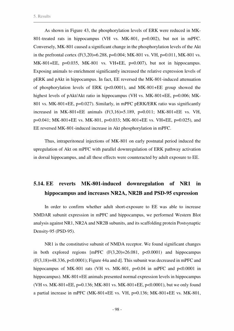
5. Results
- 98 -
As shown in Figure 43, the phosphorylation levels of ERK were reduced in MK-
801-treated rats in hippocampus (VH vs. MK-801, p=0.002), but not in mPFC.
Conversely, MK-801 caused a significant change in the phosphorylation levels of the Akt
in the prefrontal cortex (F(3,20)=6.288, p=0.004; MK-801 vs. VH, p=0.011, MK-801 vs.
MK-801+EE, p=0.035, MK-801 vs. VH+EE, p=0.007), but not in hippocampus.
Exposing animals to enrichment significantly increased the relative expression levels of
pERK and pAkt in hippocampus. In fact, EE reversed the MK-801-induced attenuation
of phosphorylation levels of ERK (p<0.0001), and MK-801+EE group showed the
highest levels of pAkt/Akt ratio in hippocampus (VH vs. MK-801+EE, p=0.006; MK-
801 vs. MK-801+EE, p=0.027). Similarly, in mPFC pERK/ERK ratio was significantly
increased in MK-801+EE animals (F(3,16)=5.189, p=0.011; MK-801+EE vs. VH,
p=0.041; MK-801+EE vs. MK-801, p=0.033; MK-801+EE vs. VH+EE, p=0.025), and
EE reversed MK-801-induced increase in Akt phosphorylation in mPFC.
Thus, intraperitoneal injections of MK-801 on early postnatal period induced the
upregulation of Akt on mPFC with parallel downregulation of ERK pathway activation
in dorsal hippocampus, and all these effects were counteracted by adult exposure to EE.
5.14. EE reverts MK-801-induced downregulation of NR1 in
hippocampus and increases NR2A, NR2B and PSD-95 expression
In order to confirm whether adult short-exposure to EE was able to increase
NMDAR subunit expression in mPFC and hippocampus, we performed Western Blot
analysis against NR1, NR2A and NR2B subunits, and its scaffolding protein Postsynaptic
Density-95 (PSD-95).
NR1 is the constitutive subunit of NMDA receptor. We found significant changes
in both explored regions [mPFC (F(3,20)=26.081, p<0.0001) and hippocampus
(F(3,18)=48.336, p<0.0001); Figure 44a and d]. This subunit was decreased in mPFC and
hippocampus of MK-801 rats (VH vs. MK-801, p=0.04 in mPFC and p<0.0001 in
hippocampus). MK-801+EE animals presented normal expression levels in hippocampus
(VH vs. MK-801+EE, p=0.136; MK-801 vs. MK-801+EE, p<0.0001), but we only found
a partial increase in mPFC (MK-801+EE vs. VH, p=0.136; MK-801+EE vs. MK-801,

5. Results
- 99 -
p=0.199). Enrichment promoted NR1 expression in VH+EE animals in mPFC (VH vs.
VH+EE, p=0.026; MK-801 vs. VH+EE, p<0.001; MK-801+EE vs. VH+EE, p=0.002).
Figure 44. Effects of adult life exposure to EE after neonatal MK-801 treatment on NMDAR
subunit expression. (a) The NMDAR vital subunit NR1 was significantly decreased in mPFC by
MK-801 and EE had little effect on its expression in MK-801+EE animals. (b) Prefrontal NR2A
subunit was significantly increased in VH+EE rats and (c) NR2B subunit increase was notorious
in EE groups. (d) MK-801-induced attenuation of hippocampal NR1 subunit was increased up to
normal values after EE. In addition, EE exposure increased (e) NR2A and (f) NR2B subunits in
hippocampus. Histograms represent optical densities obtained for each primary antibody
compared to actin, expressed as the ratio relative to vehicle-injected animals. Data are represented
as mean ± SEM (n=6). *p < 0.05, **p < 0.01, ***p < 0.001; One-way ANOVA with Tamhane’s
T2 Post Hoc test was used to obtain significances.

5. Results
- 100 -
In mPFC, there were significant changes in NR2A subunit (F(3,18)=67.159,
p<0.0001; Figure 44b) accounted for a 3-fold increase in VH+EE group (VH+EE vs. the
other 3 groups, p<0.0001). EE did not increase protein levels of NR2A in mPFC of MK-
801+EE group (MK-801+EE vs. VH, p=0.689). However, we found a 3-fold increase of
NR2B in mPFC of MK-801+EE animals (F(3,20)=16.205, p<0.0001), and was
statistically different from standard condition groups (MK-801+EE vs. VH, p=0.001;
MK-801+EE vs. MK-801, p=0.004). NR2B subunit expression was doubled in VH+EE
compared to VH animals (p=0.04) (Figure 44c).
One-way ANOVA revealed a significant increase of NR2A subunit in HPC after
EE (F(3,19)=11.051, p<0.0001; VH vs. MK-801+EE, p=0.025; VH vs. VH+EE, p=0.02;
MK-801 vs. MK-801+EE, p=0.03) and the average increase was greater in MK-801+EE
group compared to VH+EE group (Figure 44e). Similarly, regarding NR2B subunit in
hippocampus, there were significant changes between groups (F(3,19)=56.485,
p<0.0001). Similar to NR2A findings, NR2B increase was most notorious in MK-
801+EE group (MK-801+EE vs. VH, p<0.001; MK-801+EE vs. MK-801, p<0.0001;
MK-801+EE vs. VH+EE, p=0.004). Although expression levels of NR2B subunits in
VH+EE group were also increased (Figure 44f), we failed to find statistically significant
differences from standard condition animals.
Postsynaptic density-95 (PSD-95) is a scaffolding protein located in neural
postsynaptic densities. It is associated with NR2 subunits of NMDA receptor, and it is
required for synaptic plasticity associated with NMDA receptor signaling. In mPFC
(F(3,16)=16.953, p<0.0001), PSD-95 levels tended to be downregulated in MK-801
group, but no statistical differences were found when compared to VH group. Animals
housed in EE showed more PSD-95 than standard housed animals (VH vs. VH+EE,
p=0.012; MK-801 vs. MK-801+EE, p=0.031; MK-801 vs. VH+EE, p<0.001; Figure 45a).
PSD-95 levels in hippocampus were statistically different among groups (F(3,18)=9.4,
p<0.001; Figure 45b). Increased protein expression was found in MK-801+EE group
compared to VH group (p=0.047). Differences were near significance between MK-801
and MK-801+EE groups (p=0.06) and VH and VH+EE groups (p=0.066).

5. Results
- 101 -
Figure 45. Protein levels of the scaffolding protein postsynaptic density 95 (PSD95) in (a) mPFC
and (b) dorsal hippocampus. Histograms represent optical densities normalized to actin levels and
are expressed as the ratio relative to vehicle-injected animals. Data are represented as mean ±
SEM (n=6). *p < 0.05; **p < 0.01; ***p < 0.001 (one-way ANOVA).
Taken together, adult environmental intervention triggered a set of modifications
on the components of glutamatergic neurotransmission, including the upregulation of the
deficient NR1 subunit and NR2B-containing NMDARs with a parallel increase of the
scaffolding protein PSD-95 and hippocampal NR2A subunit expression.
5.15. Recovery of GABAAR subunit ß2/3 expression following EE
The ionotropic receptor GABAA is necessary for the fast inhibition carried out by
GABA neurotransmitter. We found significant differences of GABAAR subunit ß2/3 in
mPFC (F(3,19)=40.088, p<0.0001) and hippocampus (F(3,19)=4.623, p=0.014; Figure
46). Post Hoc analysis showed that MK-801 treatment significantly reduced its expression
in mPFC compared to control group (p=0.044).
After EE, this subunit was upregulated up to normal values in MK-801+EE group
(MK-801 vs. MK-801+EE, p=0.039), and the average increase was more pronounced in
VH+EE group (VH vs. VH+EE, p=0.002; MK-801 vs. VH+EE, p<0.0001; MK-801+EE

5. Results
- 102 -
vs. VH+EE, p=0.044). On the other hand, the decrease in hippocampal expression of
GABAAR subunit ß2/3 in MK-801 group was not statistically significant compared to
VH group (p=0.081), although a significant increase was found in MK-801+EE group
compared to MK-801 group (p=0.012).
Figure 46. Protein levels of GABAAR subunit ß2/3 in (a) mPFC and (b) dorsal hippocampus.
Histograms represent optical densities normalized to actin levels and are expressed as the ratio
relative to vehicle-injected animals. Data are represented as mean ± SEM (n=6). *p < 0.05; **p
< 0.01; ***p < 0.001 (one-way ANOVA).

6. Discussion


6. Discussion
- 105 -
There is growing body of evidence suggesting that EE is remarkably beneficial in
improving cognitive functions in health and disease. However, the impact of EE after
neonatal MK-801 chronic administration in rats has not been profoundly studied,
although it might be an effective strategy to manage cognitive symptoms and pathological
findings related to schizophrenia. In this study, we administered Long Evans rats the
NMDAR antagonist MK-801 chronically during neurodevelopment, and exposed rats to
EE in adulthood in an attempt to reverse cognitive and neurochemical alterations that
could be relevant for schizophrenia. Even though schizophrenia is considered a
neurodevelopmental disorder, symptoms usually emerge at late adolescence/early
adulthood. We tried to mimic the time course of the disease and exposed the animals to
EE at a similar neurodevelopmental stage as for the diagnosis of schizophrenia in humans
is done. This period usually coincides with prefrontal cortex maturation, myelization of
frontal and temporal lobes, and with the rewiring of cortical circuits.
In our current study, several important findings are shown. First, we found that MK-
801 impaired spatial learning, recognition memory, and associative memory. Secondly,
MK-801 treatment reduced cortical and hippocampal volumes, as well as the number of
PV, SST and CR-expressing interneurons in mPFC and hippocampus. MK-801 also
diminished GAD67 immunoreactivity without overall cell loss, suggesting an impairment
of excitatory-inhibitory balance. According to our results, a short-term exposure to EE in
early adulthood improved cognitive functions and long-lasting GABAergic marker
deficits. In addition, cell proliferation and hippocampal vasculature were altered in
response to MK-801 and EE. At a molecular level, MK-801 impaired BDNF-TrkB
signaling in mPFC, and EE upregulated this pathway in mPFC and hippocampus.
Moreover, the obligatory NMDAR subunit NR1 was decreased by chronic MK-801
administration similar to GABAAR ß2/3 subunit. Adult EE exposure promoted the
upregulation of NMDAR subunits, particularly in hippocampus. Alternatively, EE
promoted the expression of GABAAR ß2/3 more notably in mPFC. Finally, MK-801
influenced the downstream signal transduction pathways that are common to the NMDA
and TrkB receptors, namely ERK and Akt, in region specific manner.
The present study reveals new insights into the beneficial effects of EE exposure in
adulthood of animals treated postnatally with MK-801, and renders EE a useful strategy
to improve cognitive impairments, network disturbances and neurochemical alterations
relevant to schizophrenia. Our findings propose that EE-mediated upregulation of BDNF-

6. Discussion
- 106 -
TrkB signaling, and the regulation of NMDARs and related proteins, might be crucial
neural mechanisms that mediate synaptic plasticity and improvement of cognitive
alterations emerging upon NMDAR blockade.
6.1. Locomotor activity along neurodevelopment
The OFT was used to measure locomotor activity at different time points (P21, P24,
P55) in response to repeated administration of MK-801 during postnatal period.
Quantifying the total distance traveled and the relative distance in center, we found that
the locomotor alterations produced by MK-801 did not persist long beyond treatment.
The hypoactivity seen 24-hours after treatment cessation was absent at 96 hours and
normal locomotor activity was conserved until P55. Our results coincide with those of
Latysheva & Rayevsky (Latysheva & Rayevsky, 2003), who found a transient decrease in
spontaneous locomotor activity 23-hours after last injection, but failed to find any
difference after 6 days or 4 months. Nevertheless, studies in the literature have mentioned
either a robust increase of locomotion or decreased locomotor activity shortly after
treatment (Lim et al., 2012).
Long-term behavioral effects of MK-801 are also conflicting. For instance, some
studies have found decreased locomotion in adulthood (P60) (Baier et al., 2009), but
others failed to observe any abnormalities (Zhao et al., 2013; Su et al., 2014). The
variability in results might be attributed to the diversity of dosing regimen. In line with
other studies using similar doses (Zhao et al., 2013; Su et al., 2014), we confirmed that
MK-801 treatment had not long-term effects in locomotor activity. A number of authors
have shown EE to reduce locomotor activity in OFT and habituate more rapidly than
controls. This appears to be a relatively consistent finding independently of the employed
protocol (Zimmermann et al., 2001; Brenes et al., 2009). Our results are in line with
earlier findings regarding decreased traveled distance in OFT after EE (Zimmermann et
al., 2001; Brenes et al., 2009). Moreover, after EE rodents also displayed reduced
anxiety-like behavior in OFT indicated by the distance spent in center relative to
periphery. Unfortunately, we only observed anxiolytic effects in saline controls housed
in EE, and not in neonatally MK-801-injected rats with EE.

6. Discussion
- 107 -
6.2. EE improves cognitive functions
EE is a widely used paradigm to stimulate cognitive processes, as mice and rats
generally show improved performance in learning and memory tasks after EE than
standard housed control animals (van Praag et al., 2000). Many studies initiate EE at
relatively early stages. Environmentally stimulating conditions from birth or weaning
prevent cognitive and behavioral deficits secondary to chronic MK-801 treatment (Nozari
et al., 2014; Rahati et al., 2016). However, no single study to date has assessed the
potential benefits of EE in adulthood after neonatal NMDAR blockade, although studies
in which EE has started at adult stages also exhibited beneficial effects in other diseases,
like diabetes (Beauquis et al., 2010). In this work, we demonstrated that an intervention
with EE in early adulthood enhanced cognitive functions of postnatally MK-801-treated
animals.
The results of the MWM showed that rats of MK-801 group spent more time to find
the platform than vehicle animals, indicating that MK-801 impaired spatial learning. On
the other hand, adult intervention with EE reversed the spatial learning impairment.
Previous studies have shown that early life MK-801 administration results in disturbed
spatial learning in adult rodents. In fact, altered water maze performance seems to be
predominantly manifested after maturity. In line with our results, Gorter and de Bruin
(Gorter & de Bruin, 1992) also reported a slower learning in adult rats (P120-P140)
following chronic neonatal MK-801 administration (0.2mg/kg from P8 to P19), even
though reference memory was preserved. Similarly, Németh et al., (Németh et al., 2002)
noted poorer water maze learning in comparison to controls, which is consistent with the
results of Enomoto et al., (Enomoto et al., 2008) who found that MK-801 (0.15 mg/kg
i.p.) significantly increased escape latency and distance. Few studies have addressed the
potential benefits of enriched environment (EE) on MK-801-induced spatial learning
deficits. According to Nozari et al., (Nozari et al., 2014), EE prevented spatial learning
and memory deficits in Morris Water Maze in juvenile rats after chronic high doses of
MK-801 (1mg/kg) from P6-P10.
Novel Object Recognition (NOR) is a widely used test to assess memory function
in schizophrenic humans and animal models. Previous studies have shown that acute
administration of MK-801 before NOR impairs acquisition and encoding of object
recognition memory (Nilsson et al., 2007) and it therefore decreases discrimination index
6. Discussion
- 108 -
(Karasawa et al., 2008; Wierońska et al., 2013; Vishnoi et al., 2015). Nevertheless, long-
term effects have been somewhat controversial. Adopting a once daily 0.25mg/kg dose
from P6 to P21, Baier et al., (Baier et al., 2009) concluded that MK-801 did not had long-
term adverse consequences on NOR. Likewise, Lim et al., (Lim et al., 2012) failed to find
any impairment in recognition memory in adult rats using a daily dose of 0.2mg/kg
between P7 and P10. It is now known that drug administration schedule highly influences
NOR performance in adulthood. Chronic daily doses of 0.5mg/kg or higher during
neurodevelopment seem to be necessary for long-lasting recognition memory impairment
in adult animals (van der Staay et al., 2011) – the threshold dose for apoptotic injury
(Ikonomidou et al., 1999). On this background, Li et al., (Li et al., 2015) administered
0.25mg/kg MK-801 twice daily from P5 to P14, and demonstrated an impaired object
recognition memory in adult rats that was already present in juvenile animals. This is
consistent with our results, in which a daily dose of 0.5mg/kg from P10 to P20 impaired
recognition memory. The low performance of MK-801-treated rats in NOR was not
attributable to malnutrition or anxiety, in view of normal body weight and locomotor
activity in Open Field Test. Previous studies have consistently shown that EE improves
discrimination ratio in NOR task in healthy animals (Kazlauckas et al., 2011; Doulames
et al., 2014), and also in animal models of Parkinson’s Disease (Campêlo et al., 2017),
Alzheimer’s disease (Polito et al., 2014) or after traumatic brain injury (Schreiber et al.,
2014). Nevertheless, effect of environmental stimulation in reversing memory deficits in
NOR has not been studied in animal models of neuropsychiatric disorders, including
schizophrenia.
On the other hand, MK-801 disrupted associative recognition memory typified by
decreased discrimination index in Object-in-Place task, and adult intervention with EE
enhanced associative memory impairment. Altered performance of rats in Object-in-Place
task resembles the impaired performance in working memory binding task (Burglen et
al., 2004) and pattern-location association (Wood et al., 2002) of human patients with
schizophrenia. In fact, associative memory seems to be more impaired in schizophrenic
patients than recognition memory, which is often used to measure cognitive abilities in
animal models of schizophrenia (Achim & Lepage, 2003; Lepage et al., 2006; Luck et al.,
2009). This might be so, because in the object–place associative tasks mPFC is more
selectively involved than in recognition tasks (Lee & Solivan, 2008). Actually, Object-in-
Place associative memory depends on an interaction between the hippocampus (HPC),

6. Discussion
- 109 -
perirhinal (PRH), and medial prefrontal (mPFC) cortices, and on the excitatory
neurotransmission in any pair of structures within this networks mediated by AMPA and
NMDARs (Barker & Warburton, 2015). Li and colleagues (Li et al., 2015) demonstrated
that early life MK-801 administration also altered associative memory in object-in-
context task. However, it should be noted that object-in-context task is more dependent
on neural ensembles of ventral hippocampus than of dorsal hippocampus, which is
primarily involved in object-location representations.
As far as we know, this is the first time that late exposure to EE demonstrates to be
effective in reversing spatial learning impairment in Morris Water Maze after postnatal
MK-801 exposure. Similarly, this is the first study that demonstrates a beneficial effect of adult
intervention with EE in recognition and associative memories in a neurodevelopmental model of
schizophrenia.
6.3. Structural brain changes
Regarding the volumetric changes, our results are in agreement with previous
reports describing a decrease in mPFC and hippocampal volume in Lister Hooded rats
after perinatal MK-801 administration (Gilabert-Juan et al., 2013). In contrast, this study
showed that the volume of anterior cingulate was unaltered and mPFC reduction was
mainly to be due to decreased volume in prelimbic cortex. Reductions in the volume of
the PFC and hippocampus have also been found in schizophrenic patients, and this
anatomical abnormality is often considered a hallmark of the disease, at it is present at
the onset of illness (Levitt et al., 2010; van den Noort et al., 2010). Decreased neuronal
and glial size, and reduced microvascular density in schizophrenia patients might also
contribute to cortical volume decrease (Rajkowska et al., 1998; Stark et al., 2004; Schmitt
et al., 2009; Hercher et al., 2014). One interesting result of the present study is that the
environmental enrichment increased cortical and hippocampal volumes, thereby
reversing MK-801-induced structural changes.
Another aspect of structural plasticity might be related to decline in microvascular
structure. Altered capacity of cerebrovascular angiogenesis may result in a failure of
vascular homeostasis, which in turn will affect the adjustment of dynamic changes in
oxygen supply and demand. As increased neural activity induces changes in blood flow

6. Discussion
- 110 -
and microvascular density, any abnormalities in the vasculature will lead to functional
impairments and will alter neuronal integrity. In fact, several micro- and macrovascular
abnormalities have been recently identified as having direct implications on the
pathophysiology of schizophrenia. However, studies of microvascular brain changes in
schizophrenia are scarce and sometimes contradictory. A post mortem stereological
assessment of capillary diameters in anterior cingulate cortex in long-lasting
schizophrenic patients, found no differences compared to controls, but smaller cortical
thicknesses (layers III and V) were identified (Sinka et al., 2012). In another study, an
atypically simplified angioarchitecture and failure of normal arborization of brain vessels
was verified (Senitz & Winkelmann, 1991), supporting the existence of an abnormal
vascular organization. We found by stereological means that MK-801-treated animals
displayed reduced microvascular length in hippocampus, but not in mPFC. Interestingly,
environmental intervention in adult life increased microvascular structure in MK-801-
treated animals up to normal values.
6.4. Early life MK-801 administration reduces the number of PV and
CR-expressing interneurons
Deficit in PV immunoreactive interneurons is the most consistent finding in animal
models of schizophrenia (Rujescu et al., 2006; Abekawa et al., 2007; Braun et al., 2007;
Coleman et al., 2009; Li et al., 2015) and human patients (Volk et al., 2000; Zhang &
Reynolds, 2002; Hashimoto et al., 2003). Our results showing a reduced number of PV+
interneurons in PL and AC cortices in mPFC, and CA1 and DG of hippocampus are in
agreement with previous works. Studies using repeated injections of NMDAR antagonists
have demonstrated a reduction in PV density in mPFC (Nakatani-Pawlak et al., 2009;
Redrobe et al., 2012) and hippocampus (Abekawa et al., 2007; Powell et al., 2012), and
neurodevelopmental models further support this finding (Abekawa et al., 2007; Coleman
et al., 2009; Li et al., 2015). MK-801 is a non-competitive NMDAR antagonist that
selectively disrupts GABAergic cells. A number of hypotheses have been proposed to
explain the mechanisms by which PV+ can be selectively susceptible to damage after
MK-801 administration. The open-probability of NMDAR of PV cells is higher than in
other types of interneurons, as most of PV-expressing interneurons are fast-spiking (FS)
cells. Thus, the probability of MK-801 for blocking NMDAR of FS-PV+ cells increases.
6. Discussion
- 111 -
Moreover, Wang & Gao (Wang & Gao, 2012) showed that NMDAR in presynaptic
glutamatergic terminals targeting pyramidal cells and FS interneurons were distinctly
affected after subchronic MK-801 exposure. Presynaptic NMDAR are critical to
modulate and facilitate neurotransmitter release. The authors demonstrated that MK-801
completely blocked presynaptic NMDARs of terminals that targeted FS-PV interneurons,
whereas NMDARs of glutamatergic terminals that synapsed with pyramidal neurons were
upregulated, shifting the excitation inhibition balance towards excitation.
Another hypothesis that supports a presumable selective disruption of PV+
interneurons is that our MK-801 administration schedule coincides with the
developmental expression of PV (Lema Tomé et al., 2007; Mukhopadhyay et al., 2009).
PV expression is low to absent at P7 and gradually increases until P21 (Lema Tomé et al.,
2007; Mukhopadhyay et al., 2009). It has been suggested that calcium-binding proteins,
especially PV, play a neuroprotective role when facing dysregulation of calcium
homeostasis (Lema Tomé et al., 2008). On the other hand, PV is the only calcium-binding
protein that has been linked to specific brain functions, like attention (Kim et al., 2016)
or cognitive flexibility (Murray et al., 2015). Interneurons that express PV are also
involved in feed-back loops that create gamma oscillation – the physiological correlate
of higher cognitive functions (Somogyi et al., 2005). It was therefore expected that our
finding of persistent PV deficiency would result in cognitive impairment. In fact, we
found a remarkable correlation between the number of PV+ interneurons and recognition
memory. However, Bygrave et al., (Bygrave et al., 2016) claim that an exclusive NMDAR
hypofunction of PV interneurons might not be the starting point of schizophrenia, but
rather support the idea of NMDA hypofunction in several cells types.
Unlike what has been observed in the present study, CR-expressing interneurons
are not altered in schizophrenic brains (Daviss & Lewis, 1995; Reynolds et al., 2001a;
Reynolds et al., 2001b; Abekawa et al., 2007), neither in animal models of MK-801
treatment (Turner et al., 2010; Li et al., 2015). One of the reasons might be that usually
cell densities are reported instead of absolute cell numbers, although reductions in cortical
and hippocampal volume due to MK-801 have been repeatedly documented in the
scientific literature (Gilabert-Juan et al., 2013). This means that small changes in the
number of CR+ interneurons could be masked by paralleled changes in volume. In fact,
Gilabert-Juan et al., (Gilabert-Juan et al., 2013) found that CR gene expression was
reduced in mPFC caused by MK-801 treatment. A plausible explanation could be that CR
6. Discussion
- 112 -
is downregulated as a compensatory mechanism for brain hyperactivity. Hippocampal
hyperactivity is a core feature of schizophrenia that has been replicated in MK-801 animal
models (Heckers & Konradi, 2015). CR+ interneurons are specialized in innervating
other interneurons in mPFC and hippocampus (Gulyás et al., 1996; DeFelipe J, 1997). In
mPFC, they usually target somatostatin (SST) and PV-expressing interneurons (Pi et al.,
2013), whereas in hippocampus they predominantly contact with CB+ interneurons and
other CR-expressing interneurons (Gulyás et al., 1996), presumably those that co-express
somatostatin (Gulyás et al., 1996; Somogyi et al., 2005). Hence, CR+ interneurons are
part of the dishinhibitory circuit, and they are in the position to govern the inhibition
carried out by other interneurons (Pi et al., 2013). CR could be downregulated in an
attempt to compensate for network overactivation triggered by MK-801, but this
hypothesis needs to be further confirmed.
Regarding the number of CB+ cells, conflicting results are found in humans and
animal models. Some studies have found increased CB expression in human
schizophrenic patients (Woo et al., 2008; Fung et al., 2010), others have found no changes
(Tooney & Chahl, 2004), and some have reported reduced CB immunoreactivity (Beasley
et al., 2002). Results from animal studies are equally variable. In this study, we excluded
from stereological estimates those layers in which CB expression could be confounded
by CB-expressing excitatory cells, like pyramidal layer and granular layer of
hippocampus and LII/III of mPFC, and found no differences between groups. In an
attempt to solve this question, Li et al., (Li et al., 2015) used double
immunohistochemistry with CB and SST, and concluded that CB+ interneurons were
decreased in both superficial and deep layer of mPFC as a consequence of MK-801
administration. However, it should be noted that not all CB+ cells express SST, neither
all SST interneurons are CB+. Contrarily, Gilabert-Juan et al., (Gilabert-Juan et al., 2013)
found increased CB mRNA in mPFC that was paralleled by an increase in the number of
CB-expressing cells. More studies should be conducted to solve this discrepancy.
6.5. Alterations in somatostatin-expressing GABAergic interneurons
Post mortem mRNA studies of patients with schizophrenia have shown that SST
decreased more significantly than any other biomarker in hippocampus and medial

6. Discussion
- 113 -
prefrontal cortex (Hashimoto et al., 2008; Morris et al., 2008; Konradi et al., 2011), and
it seems that these differences are already noticeable during neurodevelopment (Fung et
al., 2010). Other studies have also examined the number and density of SST-expressing
interneurons, showing a loss of this subpopulation (Konradi et al., 2011). Reduced SST
level is not pathognomonic of schizophrenia, as this observation is common to other
psychiatric and neurological disorders such as bipolar disorder, Alzheimer’s disease or
Parkinson’s disease (Lin & Sibille, 2013). Nevertheless, a pattern of SST reduction in
various areas implicate SST+ interneurons in the pathophysiology of schizophrenia
(Alherz et al., 2017).
We found that postnatal MK-801 treatment reduced the number of SST+
interneurons in mPFC and hippocampus, similar to what we observed in PV+
interneurons. Previous studies have also reported decreased SST-immunoreactivity after
MK-801 administration (Arif et al., 2006), as well as structural remodeling of spines and
axonal boutons of hippocampal SST+ interneurons (Perez-Rando et al., 2017). Although
there are limited studies about the effect of NMDAR antagonists on this type of
interneurons, MK-801 could presumably block NR2C and NR2D-containing NMDARs
more selectively, due to their low affinity for magnesium (Kotermanski et al., 2009).
NMDAR subunit NR2C is preferentially expressed in PV+ and SST+ interneurons, and
NR2D in PV+ fast-spiking cells (von Engelhardt et al., 2015), making this two subtypes
of interneurons especially vulnerable to NMDAR blockade. As far as we know, this is the
first study that quantifies the number of SST+ interneurons in MK-801-treated animals.
Somatostatin is closely related to cognitive functions, as the deletion of
somatostatin receptor 2 leads to cognitive impairment (Dutar et al., 2002), and reductions
in SST levels have been attributed to the cognitive decline seeing in healthy aging
(Dournaud et al., 1995). In this work, we observed that the number of SST+ interneurons
was highly correlated with the performance in Novel Object Recognition and Object-in-
Place tasks, and successfully predicted the discrimination index of the latter, further
suggesting their contribution to learning and memory processes. However, the precise
role of SST-expressing interneurons in cognition is still poorly understood. Synapse
distribution between SST+ interneurons and pyramidal cells are crucial for determining
the effects of local circuits and network inhibition (Urban-Ciecko & Barth, 2016).
Likewise, GABA release from axon terminals of SST+ interneurons activates fast
synaptic GABAAR, whereas somatostatin release slowly but persistently activates

6. Discussion
- 114 -
metabotropic GABABRs (Urban-Ciecko & Barth, 2016). Therefore, distinct
subpopulation of SST+ interneurons inhibit pyramidal cells at multiple levels and
timescales that might lead to different functional outcomes (Scheyltjens & Arckens,
2016).
Taken together, SST-interneurons are likely to be important mediators of learning
and memory processes, as they are ideally situated to integrate, process, and modulate
afferent inputs, but the precise mechanism are still to be determined.
6.6. EE promotes the expression of GABAergic markers
Calcium-binding proteins
EE intervention increased GABAergic cell immunoreactivity without changing
overall cell quantity. This indicates that MK-801 reduced the expression of calcium-
binding proteins in interneurons, and consequently their activity, instead of promoting
programmed cell death. Nevertheless, the rate of apoptosis that previous studies have
reported after MK-801 administration is relatively small compared to the total number of
cells (Ikonomidou et al., 1999). Thus, MK-801 probably augmented programmed cell
death postnatally, but this loss could not be detected by stereological estimations in
adulthood. In fact, our dose of 0.5mg/kg has been documented to be the threshold dose
for apoptotic damage (Ikonomidou et al., 1999).
It has been shown that hippocampal PV+ cells are especially sensitive to NMDAR
blockade. This could partly explain why PV immunoreactivity in CA1 region was only
partially increased. EE completely restored the number of PV+ and CR+ interneurons in
anterior cingulate and PV+ interneurons in DG. However, only a partial restoration was
found in PV+ and CR+ cells in prelimbic region of mPFC, and in CR+ in CA1. The partial
recovery demonstrates the beneficial effects of EE, but it remains to be determined if
longer periods in EE could further increase calcium-binding protein markers up to normal
values, or the lack of complete recovery is caused by the postnatal MK-801-induced cell
death.
6. Discussion
- 115 -
Somatostatin
Somatostatin expression is CREB-dependent, and thus regulated by neuronal
activity (Urban-Ciecko & Barth, 2016). Therefore, the observed decrease in the number
of SST+ interneurons after MK-801 would suggest a decrease in SST content resulting
from NMDAR blockade rather than a loss of this type of interneurons. Based on this fact,
we propose that the increase in the number of SST+ interneurons after EE exposure in
adulthood was attributable to enhanced activity of these interneurons that would in turn
promote the synthesis and expression of SST. However, we found that this effect was
region-dependent, as the number of SST+ interneurons was completely rescued in CA3
region of hippocampus, but minimally in CA1 region. The heterogeneity within SST-
expressing interneurons in hippocampus could explain these regional differences. For
instance, even though cholinergic and noradrenergic inputs enhance the SST+ interneuron
activity, cholinergic receptors are restricted to oriens-lacunosum moleculare (OLM)
interneurons in CA1 (Leão et al., 2011). Moreover, some OLM engage in gamma
oscillations, but those that expression 5HT3A receptors do not (Chittajallu et al., 2013).
In addition, neocortical SST+ interneurons also display anatomical, molecular and
electrophysiological differences and their functional roles are featured by their specific
cortical wiring patterns that will invariably determine cognitive performance.
Glutamic-acid decarboxylase 67
In the mammalian brain, the primary inhibitory neurotransmitter GABA is mainly
synthesized by GAD67. This isoform of glutamic acid decarboxylase is the responsible
for over 90% of GABA production (Asada et al., 1997). Post mortem studies of
schizophrenic individuals have revealed reduced levels of GAD67 in the PFC (Akbarian
et al., 1995; Guidotti et al., 2000; Hashimoto et al., 2008). Despite the scarcity of
investigations, the available findings thus far suggest that GAD67 expression is also
decreased in hippocampus (Thompson Ray et al., 2011). The present results are partially
in accordance with human findings, showing reduction in GAD67 expression in
hippocampus, albeit the expression in mPFC was maintained. It is noteworthy that
GAD67 deficiency is primarily present in a subset of GABAergic cells, namely PV
interneurons (Hashimoto et al., 2003; Lewis et al., 2005). Given that we considered the
overall GAD67 expression in mPFC, we were not able to detect significant differences.

6. Discussion
- 116 -
Interestingly, EE in vehicle rats led to increased number of GAD67-positive cells. EE
enhances glutamatergic neurotransmission through BDNF and its receptor TrkB (Tyler et
al., 2001; Tyler et al., 2006), and genetic approaches have demonstrated that GAD67
levels directly contribute to the strength of synaptic inhibition (Lazarus et al., 2015). We
speculate that the mild increase in GAD67 cells in VH+EE animals might occur in an
attempt to counterbalance increased excitatory input.
6.7. EE reverses deficits on PV and GAD67 co-expression in CA1 and
increases the relative ratio of co-expression in mPFC
Several lines of evidence point to alterations in inhibitory circuits as one of the main
factors to explain the neurobiological basis of schizophrenia. Deficiencies in parvalbumin
(PV) and glutamic-acid decarboxylase (GAD67) expression in post mortem brain studies
of schizophrenics and in animal models of schizophrenia have been well established in
the literature (Turner et al., 2010; Gilabert-Juan et al., 2013; Fujihara et al., 2015).
Moreover, GAD67 deficit in PV+ interneurons is thought to contribute to the disinhibition
of cortical networks that result in cognitive impairment. Previous studies have shown that
EE can prevent or reverse the loss of PV+ interneurons secondary to a variety of causes,
including ischemia, maternal separation or stress. Here we show that adult EE exposure
increases the number of PV+ interneurons co-expressing GAD67 in mPFC and
hippocampus. However, this might be biased, in part, by decreased number of PV+
interneurons after MK-801 administration
Perinatal NMDAR antagonism instills disturbances in the GABAergic system like
reduced PV and GAD67 expression that are consistently reported in post mortem brain of
schizophrenic patients (Lim et al., 2012). On the other hand, GAD67 seems to be
selectively reduced in prefrontal basket and chandelier cells, two major subtypes of PV-
expressing interneurons (Hashimoto et al., 2003; Lazarus et al., 2015). We failed to find
any differences in the relative number of interneurons co-expressing PV and GAD67 in
mPFC between vehicle and MK-801-treated animals. Nevertheless, the reduced number
of PV+ interneurons in this region, as previously reported (van der Staay et al., 2011;
Murueta-Goyena et al., 2017), could have biased the results. In dorsal hippocampus, we
found that MK-801 reduced the expression of GAD67 in PV+ interneurons in CA1 and

6. Discussion
- 117 -
dentate gyrus, similar to what Belforte et al., (Belforte et al., 2010) found in PV cells
deficient for NMDARs. The connection between GAD67 reduction and altered inhibitory
transmission have been difficult to predict, but a recent work by Lazarus et al., (Lazarus
et al., 2015) has demonstrated that the deletion of one allele of GAD67 gene from PV+
interneurons contributed to reduced synaptic inhibition that could explain the increased
activity of pyramidal neurons found in schizophrenia (Heckers & Konradi, 2015).
Interestingly, in the present work we found that EE increased the co-expression of PV
and GAD67, probably in basket cells, as experience specifically modulates PV and
GAD67 content in this subtype of interneurons (Donato et al., 2013). These results might
have important implications in cortical activity because GAD67-mediated GABA
synthesis regulates the axon branching and perisomatic synapse formation of PV+
interneurons (Chattopadhyaya et al., 2007).
6.8. MK-801 increases the number of proliferative cells is subgranular
layer and EE increases immature granule cells phenotype in
hippocampus
Cell division markers like BrdU are incorporated into DNA when DNA is
synthesized during cell division. This marker is often used to assess cell proliferation in
subgranular zone of dentate gyrus. It has been previously shown that the administration
of both competitive and non-competitive NMDAR antagonists increases proliferation in
this hippocampal region (Nacher et al., 2001; Nacher et al., 2003), suggesting that adult
neurogenesis may be regulated by NMDARs present in precursor cells and in
differentiating granule neurons, and control the development of adult-born neurons at
most stages of their differentiation (Nácher et al., 2007). This is in accordance with our
results, in which we found increased and long-lasting cell proliferation after postnatal
MK-801 treatment. EE-induced cell proliferation in VH+EE rats was not as high as cell
proliferation secondary to MK-801 treatment. This finding lead us to question which
proportion of this cells will differentiate into granule cells that could potentially integrate
into hippocampal circuits.
Mature granule cells of dentate gyrus express CB and NeuN. Heretofore post-
mitotic neuronal cells transiently express CR, before being fully integrated in the

6. Discussion
- 118 -
hippocampal circuitry. Former studies have speculated about the immature dentate gyrus
as being a potential endophenotype of neuropsychiatric disorders, including
schizophrenia (Walton et al., 2012). Post mortem immunohistochemical analysis of
schizophrenic brain revealed that they displayed significantly increased CR expression in
dentate gyrus compared to normal controls (Walton et al., 2012; Shin et al., 2013).
Considering that glutamate inhibits cell proliferation and NMDAR blockade increases it
in the subgranular zone of DG (Cameron et al., 1995), it seemed possible that MK-801
administration could increase CR+ cells in granular cell layer. Nevertheless, we failed to
find any difference in CR-expressing cells in granular cell layer, which indicates that
chronic MK-801 administration during neurodevelopment does not promote immature
phenotype of DG, at least, on a long-term basis. Other studies have demonstrated long-
lasting increased cell proliferation and neurogenesis after MK-801 administration, but in
adult rats (Nacher et al., 2001; Nacher et al., 2003; Petrus et al., 2009). Contrarily, we
observed significantly more CR+ cells in VH+EE animals compared to all other 3 groups.
Several studies confirm that EE increases adult neurogenesis, and this is often paralleled
with improvements in learning and memory tasks (Kempermann et al., 2002; Nilsson et
al., 2007). It has been discussed that EE promotes survival of newborn neurons rather
than increasing cell proliferation (van Praag et al., 2000). Independently of the
mechanism, any of these options will result in more CR+ expressing immature neurons
in granule cell layer, which coincides with our results.
6.9. Molecular modifications underlying EE-induced cognitive
enhancement
The mechanisms by which EE is able to improve cognition and restore GABAergic
cell immunoreactivity are not fully uncovered. Previous studies have reported that
exercise alone has also beneficial effects at anatomical and behavioral levels (van Praag,
2009), and exposure to novelty improves spatial memory (Yang & Tang, 2011). EE
promotes structural changes in the brain like increased spine density and dendritic
branching, induces changes in neurotransmitters, and enhances the gene expression and
protein levels of different growth factors (van Praag et al., 2000; Mohammed et al.,
2002), such as nerve growth factor (NGF) (Mohammed et al., 1990; Pham et al., 1999),
brain-derived neurotrophic factor (BDNF) (Falkenberg et al., 1992), glial-derived

6. Discussion
- 119 -
neurotrophic factor (GDNF) (Young et al., 1999) or vascular endothelial growth factor
(VEGF) (Bengoetxea et al., 2008). Accumulating evidence has yielded to the notion that
NMDAR hypofunction underlies cognitive dysfunction and anatomical alterations of
schizophrenia. Albeit the scarcity of investigations about the pathways that regulate
NMDAR trafficking and mobility related to EE, it is known that EE increases
neurotransmitter receptors, including NR1, NR2A and NR2B subunits of NMDARs in
several regions of the brain, including hippocampus, forebrain and amygdala (Tang et al.,
2001). Based on this evidences, we focused on studying the expression levels of NMDAR
subunits and the signaling pathway BDNF-TrkB that might have relevant implications in
schizophrenia, and could be the underlying molecular cascades involved in cognitive and
cellular improvements.
6.9.1. NMDAR subunits
NMDAR antagonists, including MK-801, lead to a decrease in GABAergic
interneurons and behaviors reminiscent to schizophrenia in animals (Olney & Farber,
1995; Abekawa et al., 2007; Murueta-Goyena et al., 2017). In this study, we demonstrate
that neonatal MK-801 administration in rats downregulates the obligatory NR1 subunit
of NMDAR in hippocampus and mPFC that further supports NMDAR hypofunction and
its relation to the pathophysiology of schizophrenia. Weickert et al., (Weickert et al.,
2013) provided evidences of reduced mRNA and protein levels of NR1 subunit in
dorsolateral prefrontal cortex of schizophrenic patients, and more recently, Catts et al.,
(Catts et al., 2015) have corroborated these results. After adult environmental exposure,
NR1 subunit increased in dorsal hippocampus of both saline and MK-801-treated rats
with a parallel increase in NR2A and NR2B subunits that was more evident in MK-
801+EE group. These data indicate that EE-promoted synthesis and expression of
NMDAR subunits is particularly notorious in rats neonatally treated with MK-801,
perhaps as a compensatory mechanism for the induced NMDAR blockade. However, in
mPFC EE-mediated NR1 and NR2A protein level enhancement was minimal, but we
found a major increase in NR2B levels. NR2B-containing NMDARs reveal long currents,
carry more charge for a single synaptic event than NR2A-containg NMDARs (Zhu et al.,
2002; Erreger et al., 2005) and couple to intracellular signaling cascades, like Ras-GRF1,
RasGAP and CaMKII (Barria & Malinow, 2005) that might favor LTP induction, and
6. Discussion
- 120 -
consequently could enhance memory functions as seen in MK-801+EE rats, but this
viewpoint remains highly speculative.
Moreover, NMDA channel gating and surface expression is regulated by
postsynaptic density protein 95 (PSD95) (Lin et al., 2004) – a scaffolding protein that is
required for activity-driven synapse stabilization (Ehrlich et al., 2007). Most molecular
studies of PSD-95 protein levels in prefrontal cortex find no change in individuals with
schizophrenia (Funk et al., 2009; Kristiansen et al., 2010), though studies of the anterior
cingulate cortex have observed a decrease in PSD-95 protein levels (Funk et al., 2009;
Kristiansen et al., 2010; Funk et al., 2012). Nonetheless, Catts et al., (Catts et al., 2015)
found a 20% decrease in NR1 protein and a 30% decrease in PSD-95 in postsynaptic
density-enriched fractions from individuals with schizophrenia relative to unaffected
controls in prefrontal cortex. Although the decrease in PSD-95 in mPFC of our animal
model was not statistically significant, environmental intervention increased PSD-95
expression levels, suggesting increased NMDAR-mediated neurotransmission after EE
intervention. Nithianantharajah et al., (Nithianantharajah et al., 2004) also reported
increased PSD-95 after 30 days of EE, which is consistent with the results of Rampon et
al., (Rampon et al., 2000) who also found increases in PSD-95 gene expression or protein
levels following EE.
Furthermore, both PV and GAD67 are expressed upon the activation of NMDARs
containing the NR2A subunit (Kinney et al., 2006). According to our results, perinatal
NMDAR antagonism instills disturbances in the GABAergic system like reduced PV and
GAD67 expression that are consistently reported in post mortem brain of schizophrenic
patients (Lim et al., 2012). In animal studies, genetic ablations of NMDARs have been
found to reduce PV and GAD67 in cortex and hippocampus (Belforte et al., 2010). These
results, in combination with studies in cultured neurons support the notion that NMDARs
are necessary for maintaining PV and GAD67 expression in cortical interneurons,
especially NR2A-containing NMDARs (Kinney et al., 2006).
These results suggest that EE increases the expression of different NMDAR
subunits and upregulates PSD-95. This can be due to increased number of synaptic
boutons after EE or merely the insertion of new receptor in preexisting synapses. In MK-
801+EE animals the insertion of new NMDARs would indicate enhanced NMDAR-
mediated neurotransmission necessary for the synthesis of learning-related proteins, but

6. Discussion
- 121 -
also for the expression of interneurons markers that are essential for the proper
functioning of the brain.
6.9.2. BDNF-TrkB signaling
BDNF is a neurotrophic factor that has been related to cognitive functions, mainly
learning and memory (Yamada & Nabeshima, 2003; Bekinschtein et al., 2014). In human
patients, the mRNA levels of BDNF and its receptor TrKB are decreased in prefrontal
cortex (Weickert et al., 2003; Weickert et al., 2005), hippocampus (Durany et al., 2001),
as well as plasma levels of BDNF (Toyooka et al., 2002). Hippocampal reduction of
BDNF, TrkB receptor and TrKB receptor phosphorylation in hippocampus of rats treated
with MK-801 during development have also been documented (Yang et al., 2014). In
addition, decreased GABA synthesis in PV interneurons has been attributed to a
decreased signaling of BDNF through TrkB receptor (Lewis et al., 2005). In fact, mice
hypomorphic for TrkB demonstrate a reduction in GAD67 and parvalbumin expression
(Hashimoto et al., 2005). Taken together, these findings indicate the relevance of BDNF-
TrkB signaling in schizophrenia, as has been suggested previously (Hashimoto et al.,
2005).
It has been reported that hippocampal-specific deletion of the BDNF gene impairs
novel object recognition as well as spatial learning in the water maze (Heldt et al., 2007).
Object-place association task is also impaired in BDNF lacking mice (Aarse et al., 2016).
Rats treated with MK-801 during development show hippocampal reduction of BDNF,
TrkB receptor and TrKB receptor phosphorylation (Yang et al., 2014). However, reduced
expression of BDNF in hippocampus after MK-801 seems to take place over a short
period of time (Fumagalli et al., 2003). Hill et al., (Hill et al., 2015) showed a decrease
in the BDNF levels in hippocampus 24h post-injection (0.05mg/kg MK-801) that was
recovered at 48h in male Wistar rats. On the other hand, Guo et al., (Guo et al., 2010)
administered MK-801 (0.5mg/kg twice daily) to Sprague-Dawley rats from postnatal day
5 to 14. BDNF protein levels in hippocampus and prefrontal cortex were analyzed on
P15, P42 and P77. According to their results, different patterns of BDNF increase were
found in prefrontal cortex and hippocampus: they found an early and transient increase in
BNDF levels in prefrontal cortex after MK-801 injection, whereas a late increase was

6. Discussion
- 122 -
observed in hippocampus. We failed to find any differences in BDNF levels in either
region between vehicle and MK-801-treated rats, but the expression and activation of
TrkB receptor was significantly decreased in mPFC, suggesting impaired BDNF-TrkB
signaling in this region that could account for cognitive deficits. EE-related cognitive
enhancement is elicited by increased availability of mature BDNF, thus prompting the
hypothesis that upregulated BDNF-TrkB signaling might be a crucial neural mechanism
that mediates the molecular, cellular and cognitive improvements that we observed after
EE. In line with our results, EE promoted the upregulation of BDNF and subsequent
phosphorylation of TrkB receptor.
The increase in interneurons markers after EE might be not only the consequence
of enhanced NMDA-mediated neurotransmission, but also of upregulated BDNF-TrkB
signaling. Primary neocortical cultures have shown increased NR1 subunits peptide levels
in response to BDNF, and BDNF-mediated NR1 transcription depends upon induction of
ERK pathway through activation of the TrKB receptor (Kim et al., 2012). However, other
neurotrophic factors could also be contributing to these modifications, such as NGF, a
neurotrophic factor involved in the upregulation of NR1 promoter (Liu et al., 2001).
Vascular endothelial growth factor (VEGF) not only has effects on the vasculature, but
also acts as a neurotrophic factor. Its role in synaptic function is poorly understood, but
VEGF is known to be involved in synaptic plasticity. According to Licht et al., (Licht et
al., 2011), increases in VEGF expression improve memory, primarily by affecting
synaptic plasticity, rather than promoting neurogenesis or vasculogenesis. In fact,
silencing VEGF-R2 in neural cells impair hippocampal-dependent synaptic plasticity (De
Rossi et al., 2016).
6.9.3. ERK and Akt pathways
Several lines of evidence converge in implicating abnormal ERK and Akt activity
in the pathogenesis of schizophrenia. (Funk et al., 2012; Hino et al., 2016). ERK1/2
pathway is involved in the mechanisms of synaptic plasticity, learning and memory. ERK
activation is required for the full expression of long-term potentiation (LTP), the principal
cellular mechanism thought to underlie neuronal plasticity (Giovannini, 2006). ERK
pathway is known to be a downstream signal transduction pathway that is common to the
6. Discussion
- 123 -
NMDA and TrkB signaling pathways, and mediates short- and long-term effects of
intracellular signaling in neurons (Ishii et al., 2010). Abnormal activity of ERK-
associated pathways has been described in frontal cortical areas of schizophrenic patients
(Funk et al., 2012), and the uncoupling of NMDAR-ERK pathway has been proposed as
one of the mechanisms MK-801-induced damage. Moreover, NMDAR-
mediated ERK signaling is involved in memory processing, including associative
memories (Al Rahim et al., 2009), and its lack of activation in MK-801-treated animals
in dorsal hippocampus could account for the deficiencies in Object-in-Place task.
Previous studies have also reported that MK-801 induces a decrease in the level of ERK
phosphorylation (Al Rahim et al., 2009) that seems to be exclusive of hippocampus,
because according to Ishii et al., 1.0 mg/kg treatment with MK-801 did not affect
pERK/ERK ratio in frontal cortex of mice (Ishii et al., 2010). In line with this, Goeldner
et al., (Goeldner et al., 2008) showed that MK-801 concomitantly suppressed ERK
activation in hippocampus and disrupted recognition memory.
A variety of studies have addressed the phosphorylation levels of Akt pathways
in MK-801 animal model of schizophrenia. Results are diverse and highly depend on the
dosing and timing of the treatment. According to Ahn et al., (Ahn et al., 2005)
phosphorylation of Ser473-Akt was increased after injection of 1 mg/kg of MK-801 in
the rat frontal cortex but not in hippocampus. This increase peaked at 30 min and was
maintained until 90 min after injection. The phosphorylation showed a dose-dependent
increase up to 1 mg/kg MK-801, followed by a decrease at higher dosage. Moreover,
same authors later found that administration of MK-801 from 0.25 to 1 mg/kg caused
ERK phosphorylation to decrease, but Akt-GSK-3 phosphorylation to increase in frontal
cortex of rats (Ahn et al., 2006), which is consistent with our results. Similarly, Seo et al.,
(Seo et al., 2007) observed that repeated administration of 0.5, 1, and 2 mg/kg
of MK-801 increased the phosphorylation levels of Akt-GSK-3beta pathway in the rat
frontal cortex. Although it is still unclear why, it might be a compensatory mechanism to
counteract the prefrontal hyperactivity, as Akt-mediated phosphorylation increases the
number of GABAAR on the plasma membrane surface, thereby increasing fast inhibitory
neurotransmission. In this respect, it is worth noting that adult exposure to EE normalized
phosphorylation levels of Akt and ERK in previously MK-801-treated rats.

6. Discussion
- 124 -
6.9.4. GABAA receptor subunit β2/3
The inhibitory neurotransmitter GABA can act through GABAA ionotropic
receptors or GABAB metabotropic receptors. GABAA receptors are ligand-gated chloride
channels, which usually reduce the membrane excitability by reducing the membrane
potential to chloride potential, and thus inhibiting neurotransmitter release. The β subunit
of the GABAAR is vital for the regulation of ion selectivity and general properties of the
chloride channel (Chang et al., 1996). Beneyto et al., (Beneyto et al., 2011) observed a
reduction in mRNA for the β2-subunit in DLPFC of subjects with schizophrenia.
However, there have been conflicting results regarding mRNA levels of β3-subunit.
According to Hashimoto et al., (Hashimoto et al., 2008) this protein was reduced in
DLPFC, but a separate study found no change (Beneyto et al., 2011). Similarly, Bullock
et al., (Bullock et al., 2008) did not found differences in GABAAR β3-subunit mRNA
expression in lateral cerebella of subject with schizophrenia, but Fatemi et al., (Fatemi et
al., 2013) found an increase.
On the other hand, Kim et al., (Kim et al., 2000) demonstrated that chronic blockade
of calcium influx though NMDAR with MK-801 reduced β3-subunit and increased β2-
subunit mRNA in hippocampus, but they failed to find differences in protein expression.
In this study, we found downregulated expression of β2/3, which is consistent with
previous studies of rats chronically injected with MK-801 (Matthews et al., 2000),
although our current data supports larger changes in medial prefrontal cortex than in
hippocampus. GABRβ3 KO mice display deficits in learning and memory (DeLorey et
al., 2008), suggesting that reduced expression of this subunit is relevant to the cognitive
symptomatology of schizophrenia.
Electrophysiological studies have shown that NMDAR activation is essential for
GABAAR-mediated inhibition in the developing neocortex (Chan & Yeh, 2013),
suggesting that alterations in GABAAR subunits could be in part accounted for changes
in NMDARs, although this viewpoint is speculative. Previous studies have reported that
environmental stimulation increases GABAARs (He et al., 2010), including subunit β3
(Cai et al., 2010), which is consistent with our results. However, those changes have not
been previously described in the literature in MK-801 neurodevelopmental model of
schizophrenia.

6. Discussion
- 125 -
6.10. General discussion and future perspectives
The present study reveals new insights into the beneficial effects of adult life
exposure to EE of neonatally MK-801-treated animals as a strategy to improve cognitive
impairments and neurochemical alterations relevant to schizophrenia. NMDAR
hypofunction is one of the leading hypothesis in the field of schizophrenia to explain the
neurochemical, metabolic, functional and behavioral alterations of the disease (Snyder &
Gao, 2013; Weickert et al., 2013). NMDAR antagonists, including MK-801, lead to a
decrease in GABAergic interneurons and behaviors reminiscent to schizophrenia in
animals (Olney & Farber, 1995; Abekawa et al., 2007; Murueta-Goyena et al., 2017). In
this study, we demonstrate that adult short-exposure to EE is able to improve most of the
pathophysiological findings related to MK-801 administration.
NMDAR blockade in perinatal period confers long-lasting brain and behavioral
changes that recapitulate the human schizophrenia phenotype (Bubeníková-Valesová et
al., 2008; Lim et al., 2012). In early postnatal development, GABAergic cells express
more NMDARs than in late neurodevelopment, especially PV+ interneurons, and they
are thought to be particularly sensitive to NMDAR blockade (Grunze et al., 1996). Even
though the precise mechanism for this selectivity is unknown, it could be accounted for
the higher open probability of fast-spiking cells (PV+ interneurons) and the subunit
composition of NMDARs that might make them especially vulnerable (Kotermanski et
al., 2009; Wang & Gao, 2009). Reduction on inhibitory tone during development would
impair cortical maturation process and lead to increased excitability of cortical circuits,
as seen in schizophrenic patients. Indeed, studies in human patients show abnormal
hippocampal activity (Heckers & Konradi, 2015). This is consistent with the presence of
glutamate metabolites in schizophrenic patients before the onset of the disease (Merritt et
al., 2016). Accordingly, the administration of NMDAR antagonists perturbs the
maturation of GABAergic circuits, including disrupted synaptic integration in early brain
development and modified network activity and plasticity in adulthood. NMDAR
blockade also generates a dyssynchrony in gamma oscillations, thus disturbs attention
maintenance, executive functions and overall cognition in humans and rodents.
According to the current literature, there is unifying hypothesis of NMDAR hypofunction
in schizophrenia, and this might take place at two dissociable time points (Nakazawa et
al., 2017). The first NMDAR hypofunction might occur in GABAergic cells in early

6. Discussion
- 126 -
development, as previously described, and a second period of NMDAR hypofunction will
result during adolescence by internalizing NMDARs, in an attempt to counterbalance the
overly activated cortical and hippocampal circuits. This second NMDAR hypofunction is
probably no longer selective of GABAergic cells, and presumably takes place at
pyramidal cells. This could explain why NMDAR deletion from PV interneurons does
not completely recapitulate the disease (Bygrave et al., 2016) (Figure 47).
Figure 47. Dual NMDAR hypofunction hypothesis. During early development NMDAR
hypofunction in GABAergic cells could cause disinhibition of pyramidal cells that in turn would
lead to a generalized homeostatic downregulation of NMDARs. Extracted and adapted from:
Nakazawa et al. NPJ Schizophr. 2017.
The complex interaction of genetic, epigenetic and environmental factors that
contribute to the onset of schizophrenia and the lack of consistent molecular basis in
human patients make difficult to find effective treatments. Environment affects the
symptomatology of schizophrenia, as isolation usually worsen them. In addition, enriched
environment increases neurotrophic factors, upregulates the expression of
neurotransmitter receptors, increases the complexity of dendritic branching and enhances
learning and memory processes (van Praag et al., 2000), all of which are disturbed in
schizophrenia. Based on those findings, we proposed that rearing adult animals in EE
could be successful in managing schizophrenia-related alterations. The major challenge

6. Discussion
- 127 -
of the present study was to modify enduring cellular and molecular alterations.
Interestingly, our findings suggest that short-term EE exposure in adulthood may be a
useful approach to manage enduring perturbations linked to schizophrenia and shed new
light on the neurobiological modifications used by EE to reverse the actions of MK-801
(Figure 48). In this respect, increased BDNF-TrkB signaling and upregulated expression
NMDARs after EE were associated with improved structural and cellular findings and
better cognitive outcomes.
Figure 48. Major modifications following exposure to enriched environment (EE) in adulthood
of postnatally MK-801-treated animals. EE-induced changes range from behavior and cognition
to structural brain changes, cells and protein expression.

6. Discussion
- 128 -
In spite of the evidences of this study linking adult intervention with EE to improved
cognitive performance in a neurodevelopmental animal model of schizophrenia, several
aspects need to be further investigated. Firstly, although we found concomitant increase
in BDNF-TrkB signaling and neurochemical and behavioral modifications, we did not
determine the mechanism of action and the cause-effect relationship between these
factors. In addition, we focused this study in adult animals and we used short intervention
with environmental enrichment. However, it would be important to explore the temporal
boundaries of MK-801 and EE-induced changes, like when the functional downregulation
of NR1 occurs or determine if earlier and/or longer environmental intervention would
result in different functional outcomes. Moreover, monitoring the animals while housed
in EE would help to better establish the relationship between activity and functional
outcomes after EE. Finally, NMDAR hypofunction contributes to the manifestation of
the disorder, but the etiopathology of schizophrenia itself remains unexplained. A
complex combination of genetic, epigenetic and environmental factors could render
NMDARs hypoactive, and a better understanding of the neurobiological changes is
required to further support of EE as a possible intervention for schizophrenia. While
future studies are required to address these issues, our results establish the foundation for
future research in environmental interventions and open possibilities for novel therapeutic
targets.

7. Conclusions


7. Conclusions
- 131 -
1. Administration of MK-801 (0.5mg/kg) during early postnatal development impaired
spatial learning in Morris Water Maze, decreased discrimination index in Novel Object
Recognition task and altered object-place associative memory assessed in Object-in-
Place task.
2. Cortical and hippocampal volumes were decreased in MK-801 group. However, total
microvascular length was only diminished in hippocampus.
3. Interneurons expressing parvalbumin (PV), calretinin (CR), somatostatin (SST) were
decreased in mPFC and hippocampus secondary to NMDAR antagonism, measured
by stereological means. Similarly, the total number of inhibitory cells expressing
GAD67 was significantly decreased in hippocampus, although the overall number of
neurons was unaltered after MK-801 treatment.
4. Western Blot analysis showed a decrease in the obligatory subunit NR1 of NMDAR
in MK-801-treated animals without altering the expression levels of NR2A and NR2B
subunits or PSD-95. Similarly, GABAAR subunit ß2/3 was decreased because of
NMDAR blockade.
5. Exposure to Environmental Enrichment (EE) in adult life (P55-P73) improved the
above-mentioned parameters. Environmental intervention restored spatial learning and
recognition memory impairment. However, object-place associative memory was
partially reversed. In a similar vein, it promoted the increase in cortical and
hippocampal volumes, as well as in the length of the microvasculature. Similarly, the
expression of interneurons markers was increased after EE.
6. Adult environmental intervention triggered a set of modifications on the components
of glutamatergic neurotransmission, including the upregulation of the deficient NR1
subunit and NR2B-containing NMDARs with a parallel increase of the scaffolding
protein PSD-95 and hippocampal NR2A subunit expression, as well as upregulation
of inhibitory drive by increasing GABAAR subunit ß2/3 expression.
7. Intraperitoneal injections of MK-801 on early life induced long-lasting BDNF-TrkB
signaling disruption and upregulated Akt activation on mPFC with parallel
downregulation of ERK pathway activation in dorsal hippocampus, and all these
effects were counteracted by short-term exposure to EE in adulthood.


References


References
- 135 -
Aarse J, Herlitze S, Manahan-Vaughan D (2016). The requirement of BDNF for hippocampal
synaptic plasticity is experience-dependent. Hippocampus. 26(6):739-51.
Abekawa T, Ito K, Nakagawa S, Koyama T (2007). Prenatal exposure to an NMDA receptor
antagonist, MK-801 reduces density of parvalbumin-immunoreactive GABAergic neurons
in the medial prefrontal cortex and enhances phencyclidine-induced hyperlocomotion but
not behavioral sensitization to methamphetamine. Psychopharmacology (Berl). 192(3):303-
16.
Aberg MA, Aberg ND, Hedbäcker H, Oscarsson J, Eriksson PS (2000). Peripheral infusion of
IGF-I selectively induces neurogenesis in the adult rat hippocampus. J Neurosci. 20(8):2896-
903.
Achim AM, Lepage M (2003). Is associative recognition more impaired than item recognition
memory in Schizophrenia? A meta-analysis. Brain Cogn. 53(2):121-4.
Ahn YM, Seo MS, Kim SH, Kim Y, Juhnn YS, Kim YS (2006). The effects of MK-801 on the
phosphorylation of Ser338-c-Raf-MEK-ERK pathway in the rat frontal cortex. Int J
Neuropsychopharmacol. 9(4):451-6.
Ahn YM, Seo MS, Kim SH, Kim Y, Yoon SC, Juhnn YS, Kim YS (2005). Increased
phosphorylation of Ser473-Akt, Ser9-GSK-3beta and Ser133-CREB in the rat frontal cortex
after MK-801 intraperitoneal injection. Int J Neuropsychopharmacol. 8(4):607-13.
Akbarian S, Kim JJ, Potkin SG, Hagman JO, Tafazzoli A, Bunney WE Jr, Jones EG (1995). Gene
expression for glutamic acid decarboxylase is reduced without loss of neurons in prefrontal
cortex of schizophrenics. Arch Gen Psychiatry. 52(4):258-66.
Al Rahim M, Nakajima A, Saigusa D, Tetsu N, Maruyama Y, Shibuya M, Yamakoshi H, Tomioka
Y, Iwabuchi Y, Ohizumi Y, Yamakuni T (2009). 4'-Demethylnobiletin, a bioactive
metabolite of nobiletin enhancing PKA/ERK/CREB signaling, rescues learning impairment
associated with NMDA receptor antagonism via stimulation of the ERK cascade.
Biochemistry. 48(32):7713-21.
Alherz F, Alherz M, Almusawi H (2017). NMDAR hypofunction and somatostatin-expressing
GABAergic interneurons and receptors: A newly identified correlation and its effects in
schizophrenia. Schizophr Res Cogn. 8:1-6.
Altman J, Das GD (1964). Autoradiographic examination of the effects of enriched environment
on the rate of glial multiplication in the adult rat brain. Nature. 204:1161-3.

References
- 136 -
Altschuler, RA (1979). Morphometry of the effect of increased experience and training on
synaptic density in area CA3 of the rat hippocampus. J Histochem Cytochem. 27:1548–1550,
1979.
Amaral OB, Vargas RS, Hansel G, Izquierdo I, Souza DO (2008). Duration of environmental
enrichment influences the magnitude and persistence of its behavioral effects on mice.
Physiol Behav. 93(1-2):388-94.
Andrews-Zwilling Y, Gillespie AK, Kravitz AV, Nelson AB, Devidze N, Lo I, Yoon SY, Bien-
Ly N, Ring K, Zwilling D, Potter GB, Rubenstein JL, Kreitzer AC, Huang Y (2012). Hilar
GABAergic interneuron activity controls spatial learning and memory retrieval. PLoS One.
7(7):e40555.
Arif M, Ahmed MM, Kumabe Y, Hoshino H, Chikuma T, Kato T (2006). Clozapine but not
haloperidol suppresses the changes in the levels of neuropeptides in MK-801-treated rat brain
regions. Neurochem Int. 49(3):304-11. , 2006.
Arnsten AF, Jin LE (2014). Molecular influences on working memory circuits in dorsolateral
prefrontal cortex. Prog Mol Biol Transl Sci. 122:211-31.
Asada H, Kawamura Y, Maruyama K, Kume H, Ding RG, Kanbara N, Kuzume H, Sanbo M,
Yagi T, Obata K (1997). Cleft palate and decreased brain gamma-aminobutyric acid in mice
lacking the 67-kDa isoform of glutamic acid decarboxylase. Proc Natl Acad Sci U S A.
94(12):6496-9.
Auquier P, Lançon C, Rouillon F, Lader M (2007). Mortality in schizophrenia.
Pharmacoepidemiol Drug Saf. 16(12):1308-12.
Baier PC, Blume A, Koch J, Marx A, Fritzer G, Aldenhoff JB, Schiffelholz T (2009). Early
postnatal depletion of NMDA receptor development affects behaviour and NMDA receptor
expression until later adulthood in rats--a possible model for schizophrenia. Behav Brain
Res. 205(1):96-101.
Baraban SC, Tallent MK (2004). Interneuron diversity series: interneuronal neuropeptides -
endogenous regulators of neuronal excitability. Trends Neurosci. 27(3):135-42.
Barch DM (2006). What can research on schizophrenia tell us about the cognitive neuroscience
of working memory? Neuroscience. 139(1):73-84, 2006.
Barker GR, Warburton EC (2015). Object-in-place associative recognition memory depends on
glutamate receptor neurotransmission within two defined hippocampal-cortical circuits: a

References
- 137 -
critical role for AMPA and NMDA receptors in the hippocampus, perirhinal, and prefrontal
cortices. Cereb Cortex. 25(2):472-81, 2015.
Barria A, Malinow R (2005). NMDA receptor subunit composition controls synaptic plasticity
by regulating binding to CaMKII. Neuron. 48:289–301, 2005.
Basu J, Srinivas KV, Cheung SK, Taniguchi H, Huang ZJ, Siegelbaum SA (2013). A cortico-
hippocampal learning rule shapes inhibitory microcircuit activity to enhance hippocampal
information flow. Neuron. 79(6):1208-21.
Beasley CL, Zhang ZJ, Patten I, Reynolds GP (2002). Selective deficits in prefrontal cortical
GABAergic neurons in schizophrenia defined by the presence of calcium-binding proteins.
Biol Psychiatry. 52(7):708-15.
Beauquis J, Roig P, De Nicola AF, Saravia F (2010). Short-term environmental enrichment
enhances adult neurogenesis, vascular network and dendritic complexity in the hippocampus
of type 1 diabetic mice. PLoS One. 5(11):e13993.
Bekinschtein P, Cammarota M, Medina JH (2014). BDNF and memory processing.
Neuropharmacology. 76 Pt C:677-83.
Belforte JE, Zsiros V, Sklar ER, Jiang Z, Yu G, Li Y, Quinlan EM, Nakazawa K (2010). Postnatal
NMDA receptor ablation in corticolimbic interneurons confers schizophrenia-like
phenotypes. Nat Neurosci. 13(1):76-83.
Ben-Ari Y, Khazipov R, Leinekugel X, Caillard O, Gaiarsa JL (1997). GABAA, NMDA and
AMPA receptors: a developmentally regulated 'ménage à trois'. Trends Neurosci.
20(11):523-9.
Benes FM (2000). Emerging principles of altered neural circuitry in schizophrenia. Brain Res
Brain Res Rev. 31(2-3):251-69.
Benes FM, Turtle M, Khan Y, Farol P (1994). Myelination of a key relay zone in the hippocampal
formation occurs in the human brain during childhood, adolescence, and adulthood. Arch
Gen Psychiatry. 51(6):477-84.
Beneyto M, Abbott A, Hashimoto T, Lewis DA (2011). Lamina-specific alterations in cortical
GABA(A) receptor subunit expression in schizophrenia. Cereb Cortex. 21(5):999-1011.
Bengoetxea H, Argandoña EG, Lafuente JV (2008). Effects of visual experience on vascular
endothelial growth factor expression during the postnatal development of the rat visual
cortex. Cereb Cortex. 18(7):1630-9, 2008.
References
- 138 -
Bernstein L (1973). A study of some enriching variables in a free-environment for rats. J
Psychosom Res. 17(2):85-8.
Boehm S, Betz H (1997). Somatostatin inhibits excitatory transmission at rat hippocampal
synapses via presynaptic receptors. J Neurosci. 17(11):4066-75.
Bortone D, Polleux F (2009). KCC2 expression promotes the termination of cortical interneuron
migration in a voltage-sensitive calcium-dependent manner. Neuron. 62(1):53-71, 2009.
Braun I, Genius J, Grunze H, Bender A, Möller HJ, Rujescu D (2007). Alterations of hippocampal
and prefrontal GABAergic interneurons in an animal model of psychosis induced by NMDA
receptor antagonism. Schizophr Res. 97(1-3):254-63.
Brenes JC, Padilla M, Fornaguera J (2009). A detailed analysis of open-field habituation and
behavioral and neurochemical antidepressant-like effects in postweaning enriched rats.
Behav Brain Res. 197(1):125-37.
Brigman JL, Wright T, Talani G, Prasad-Mulcare S, Jinde S, Seabold GK et al (2010). Loss of
GluN2B-containing NMDA receptors in CA1 hippocampus and cortex impairs long-term
depression, reduces dendritic spine density, and disrupts learning. J Neurosci. 30(13):4590-
600, 2010.
Brown MT, Tan KR, O'Connor EC, Nikonenko I, Muller D, Lüscher C (2012). Ventral tegmental
area GABA projections pause accumbal cholinergic interneurons to enhance associative
learning. Nature. 492(7429):452-6.
Brunoni AR, Vanderhasselt MA (2014). Working memory improvement with non-invasive brain
stimulation of the dorsolateral prefrontal cortex: a systematic review and meta-analysis.
Brain Cogn. 86:1-9, 2014.
Bubeníková-Valesová V, Horácek J, Vrajová M, Höschl C (2008). Models of schizophrenia in
humans and animals based on inhibition of NMDA receptors. Neurosci Biobehav Rev.
32(5):1014-23.
Bullock WM, Cardon K, Bustillo J, Roberts RC, Perrone-Bizzozero NI (2008). Altered
expression of genes involved in GABAergic transmission and neuromodulation of granule
cell activity in the cerebellum of schizophrenia patients. Am J Psychiatry. 165(12):1594-603.
Burglen F, Marczewski P, Mitchell KJ, van der Linden M, Johnson MK, Danion JM, Salamé P
(2004). Impaired performance in a working memory binding task in patients with
schizophrenia. Psychiatry Res. 125(3):247-55.

References
- 139 -
Buzsáki G (1984). Feed-forward inhibition in the hippocampal formation. Prog Neurobiol.
2(2):131-53.
Bygrave AM, Masiulis S, Nicholson E, Berkemann M, Barkus C, Sprengel R, Harrison PJ,
Kullmann DM, Bannerman DM, Kätzel D (2016). Knockout of NMDA-receptors from
parvalbumin interneurons sensitizes to schizophrenia-related deficits induced by MK-801.
Transl Psychiatry. 6:e778.
Caballero A, Flores-Barrera E, Cass DK, Tseng KY. 2014a. Differential regulation of
parvalbumin and calretinin interneurons in the prefrontal cortex during adolescence. Brain
Struct Funct. 219(1):395-406., 2014a.
Caballero A, Granberg R, Tseng KY (2016). Mechanisms contributing to prefrontal cortex
maturation during adolescence. Neurosci Biobehav Rev. 70:4-12, 2016.
Caballero A, Thomases DR, Flores-Barrera E, Cass DK, Tseng KY. 2014b. Emergence of
GABAergic-dependent regulation of input-specific plasticity in the adult rat prefrontal cortex
during adolescence. Psychopharmacology (Berl). 231(8):1789-96, 2014b.
Cai R, Zhou X, Guo F, Xu J, Zhang J, Sun X (2010). Maintenance of enriched environment-
induced changes of auditory spatial sensitivity and expression of GABAA, NMDA, and
AMPA receptor subunits in rat auditory cortex. Neurobiol Learn Mem. 94(4):452-60, 2010.
Calof, AL (1995). Intrinsic and extrinsic factors regulating vertebrate neurogenesis. Curr Opin
Neurobiol. 5(1):19-27.
Cameron HA, McEwen BS, Gould E (1995). Regulation of adult neurogenesis by excitatory input
and NMDA receptor activation in the dentate gyrus. J Neurosci. 15(6):4687-92.
Campêlo CLC, Santos JR, Silva AF, Dierschnabel AL, Pontes A, Cavalcante JS et al (2017).
Exposure to an enriched environment facilitates motor recovery and prevents short-term
memory impairment and reduction of striatal BDNF in a progressive pharmacological model
of parkinsonism in mice. Behav Brain Res. 328:138-148.
Carlén M, Meletis K, Siegle JH, Cardin JA, Futai K, Vierling-Claassen D, Rühlmann C, Jones
SR, Deisseroth K, Sheng M, Moore CI, Tsai LH (2012). A critical role for NMDA receptors
in parvalbumin interneurons for gamma rhythm induction and behavior. Mol Psychiatry.
17(5):537-48.
Caroni P (2015). Regulation of Parvalbumin Basket cell plasticity in rule learning. Biochem
Biophys Res Commun. 460(1):100-3.

References
- 140 -
Cass DK, Flores-Barrera E, Thomases DR, Vital WF, Caballero A, Tseng KY (2014). CB1
cannabinoid receptor stimulation during adolescence impairs the maturation of GABA
function in the adult rat prefrontal cortex. Mol. Mol Psychiatry. 19(5):536-43.
Catts VS, Derminio DS, Hahn CG, Weickert CS (2015). Postsynaptic density levels of the NMDA
receptor NR1 subunit and PSD-95 protein in prefrontal cortex from people with
schizophrenia. NPJ Schizophr. 1:15037, 2015.
Cauli B, Zhou X, Tricoire L, Toussay X, Staiger JF (2014). Revisiting enigmatic cortical
calretinin-expressing interneurons. Front Neuroanat. 24;8:52.
Chan CH, Yeh HH (2013). Enhanced GABA(A) receptor-mediated activity following activation
of NMDA receptors in Cajal-Retzius cells in the developing mouse neocortex. J Physiol.
550(Pt 1):103-11.
Chang Y, Wang R, Barot S, Weiss DS (1996). Stoichiometry of a recombinant GABAA receptor.
J Neurosci. 16(17):5415-24.
Chattopadhyaya B, Di Cristo G, Wu CZ, Knott G, Kuhlman S, Fu Y, Palmiter RD, Huang ZJ
(2007). GAD67-mediated GABA synthesis and signaling regulate inhibitory synaptic
innervation in the visual cortex. Neuron. 54(6):889-903.
Chittajallu R, Craig MT, McFarland A, Yuan X, Gerfen S, Tricoire L et al (2013). Dual origins
of functionally distinct O-LM interneurons revealed by differential 5-HT(3A)R expression.
Nat Neurosci. 16(11):1598-607, 2013.
Clarke JR, Cammarota M, Gruart A, Izquierdo I, Delgado-García JM (2010). Plastic
modifications induced by object recognition memory processing. Proc Natl Acad Sci U S A.
107(6):2652-7, 2010.
Coleman LG Jr, Jarskog LF, Moy SS, Crews FT (2009). Deficits in adult prefrontal cortex neurons
and behavior following early post-natal NMDA antagonist treatment. Pharmacol Biochem
Behav. 2009 Sep;93(3):322-30.
Cope DW, Maccaferri G, Márton LF, Roberts JD, Cobden PM, Somogyi P (2002).
Cholecystokinin-immunopositive basket and Schaffer collateral-associated interneurones
target different domains of pyramidal cells in the CA1 area of the rat hippocampus.
Neuroscience. 109(1):63-80.
Cotter DR, Pariante CM, Everall IP (2001). Glial cell abnormalities in major psychiatric
disorders: the evidence and implications. Brain Res Bull. 55(5):585-95.

References
- 141 -
Cybulska-Klosowicz A, Posluszny A, Nowak K, Siucinska E, Kossut M, Liguz-Lecznar M
(2013). Interneurons containing somatostatin are affected by learning-induced cortical
plasticity. Neuroscience. 254:18-25.
Daenen EW, Wolterink G, Van Der Heyden JA, Kruse CG, Van Ree JM (2003). Neonatal lesions
in the amygdala or ventral hippocampus disrupt prepulse inhibition of the acoustic startle
response; implications for an animal model of neurodevelopmental disorders like
schizophrenia. Eur Neuropsychopharmacol. 13(3):187-97.
Daviss SR, Lewis DA (1995). Local circuit neurons of the prefrontal cortex in schizophrenia:
selective increase in the density of calbindin-immunoreactive neurons. Psychiatry Res. 59(1-
2):81-96.
De Rossi P, Harde E, Dupuis JP, Martin L, Chounlamountri N, Bardin M, Watrin C, Benetollo C,
Pernet-Gallay K, Luhmann HJ, Honnorat J, Malleret G, Groc L, Acker-Palmer A, Salin PA,
Meissirel C (2016). A critical role for VEGF and VEGFR2 in NMDA receptor synaptic
function and fear-related behavior. Mol Psychiatry. 21(12):1768-1780.
de Witt BW, Ehrenberg KM, McAloon RL, Panos AH, Shaw KE, Raghavan PV, Skidmore ER,
Kline AE (2011). Abbreviated environmental enrichment enhances neurobehavioral
recovery comparably to continuous exposure after traumatic brain injury. Neurorehabil
Neural Repair. 25(4):343-50.
DeFelipe, J (1997). Types of neurons, synaptic connections and chemical characteristics of cells
immunoreactive for calbindin-D28K, parvalbumin and calretinin in the neocortex. J Chem
Neuroanat. 14(1):1-19.
Del Rio JA, Soriano E, Ferrer I (1992). Development of GABA-immunoreactivity in the
neocortex of the mouse. J Comp Neurol. 326(4):501-26.
DeLorey TM, Sahbaie P, Hashemi E, Homanics GE, Clark JD (2008). Gabrb3 gene deficient mice
exhibit impaired social and exploratory behaviors, deficits in non-selective attention and
hypoplasia of cerebellar vermal lobules: a potential model of autism spectrum disorder.
Behav Brain Res. 187(2):207-20.
Diamond MC, Law F, Rhodes H, Lindner B, Rosenzweig MR, Krech D, Bennett EL (1966).
Increases in cortical depth and glia numbers in rats subjected to enriched environment. J
Comp Neurol. 128(1):117-26.

References
- 142 -
Diamond MC, Lindner B, Raymond A (1967). Extensive cortical depth measurements and neuron
size increases in the cortex of environmentally enriched rats. J Comp Neurol. 131(3):357-
364, 1967.
Doischer D, Hosp JA, Yanagawa Y, Obata K, Jonas P, Vida I et al (2008). Postnatal
differentiation of basket cells from slow to fast signaling devices. J Neurosci. 28(48):12956-
68, 2008.
Donato F, Chowdhury A, Lahr M, Caroni P (2015). Early- and late-born parvalbumin basket cell
subpopulations exhibiting distinct regulation and roles in learning. Neuron. 85(4):770-86.
Donato F, Rompani SB, Caroni P (2013). Parvalbumin-expressing basket-cell network plasticity
induced by experience regulates adult learning. Nature. 504(7479):272-6, 2013.
Doulames V, Lee S, Shea TB (2014). Environmental enrichment and social interaction improve
cognitive function and decrease reactive oxidative species in normal adult mice. Int J
Neurosci. 124(5):369-76.
Dournaud P, Delaere P, Hauw JJ, Epelbaum J (1995). Differential correlation between
neurochemical deficits, neuropathology, and cognitive status in Alzheimer's disease.
Neurobiol Aging. 16(5):817-23.
Durany N, Michel T, Zöchling R, Boissl KW, Cruz-Sánchez FF, Riederer P, Thome J (2001).
Brain-derived neurotrophic factor and neurotrophin 3 in schizophrenic psychoses. Schizophr
Res. 52(1-2):79-86.
During MJ, Cao L (2006). VEGF, a mediator of the effect of experience on hippocampal
neurogenesis. Curr Alzheimer Res. 3(1):29-33.
Dutar P, Vaillend C, Viollet C, Billard JM, Potier B, Carlo AS, Ungerer A, Epelbaum J (2002).
Spatial learning and synaptic hippocampal plasticity in type 2 somatostatin receptor knock-
out mice. Neuroscience. 112(2):455-66.
Eastwood SL, Harrison PJ (2003). Interstitial white matter neurons express less reelin and are
abnormally distributed in schizophrenia: towards an integration of molecular and
morphologic aspects of the neurodevelopmental hypothesis. Mol Psychiatry. 8(9):769, 821-
31.
Ehrlich I, Klein M, Rumpel S, Malinow R (2007). PSD-95 is required for activity-driven synapse
stabilization. Proc Natl Acad Sci U S A. 104(10):4176-81.

References
- 143 -
Enomoto T, Ishibashi T, Tokuda K, Ishiyama T, Toma S, Ito A (2008). Lurasidone reverses MK-
801-induced impairment of learning and memory in the Morris water maze and radial-arm
maze tests in rats. Behav Brain Res. 186(2):197-207.
Erreger K, Dravid SM, Banke TG, Wyllie DJ, Traynelis SF (2005). Subunit-specific gating
controls rat NR1/ NR2A and NR1/NR2B NMDA channel kinetics and synaptic signalling
profiles. J. J Physiol. 563:345–358.
Euston DR, Gruber AJ, McNaughton BL (2012). The Role of Medial Prefrontal Cortex in
Memory and Decision Making. Neuron. 76(6): 1057–1070.
Falkenberg T, Mohammed AK, Henriksson B, Persson H, Winblad B, Lindefors N (1992).
Increased expression of brain-derived neurotrophic factor mRNA in rat hippocampus is
associated with improved spatial memory and enriched environment. Neurosci Lett.
138(1):153-6.
Fanselow MS, Dong HW (2010). Are The Dorsal and Ventral Hippocampus functionally distinct
structures? Neuron. 65(1):7.
Fatemi SH, Folsom TD, Rooney RJ, Thuras PD (2013). Expression of GABAA α2-, β1- and ε-
receptors are altered significantly in the lateral cerebellum of subjects with schizophrenia,
major depression and bipolar disorder. Transl Psychiatry. 3:e303.
Fazzari P, Paternain AV, Valiente M, Pla R, Luján R, Lloyd K et al (2010). Control of cortical
GABA circuitry development by Nrg1 and ErbB4 signalling. Nature. 464(7293):1376-80,
2010.
Freund TF, Katona I (2007). Perisomatic inhibition. Neuron. 56(1):33-42.
Fritschy JM, Meskenaite V, Weinmann O, Honer M, Benke D, Mohler H (1999). GABAB-
receptor splice variants GB1a and GB1b in rat brain: developmental regulation, cellular
distribution and extrasynaptic localization. Eur J Neurosci. 11(3):761-8.
Fujihara K, Miwa H, Kakizaki T, Kaneko R, Mikuni M, Tanahira C, Tamamaki N, Yanagawa Y
(2015). Glutamate Decarboxylase 67 Deficiency in a Subset of GABAergic Neurons Induces
Schizophrenia-Related Phenotypes. Neuropsychopharmacology. 40(10):2475-86.
Fumagalli F, Molteni R, Roceri M, Bedogni F, Santero R, Fossati C, Gennarelli M, Racagni G,
Riva MA (2003). Effect of antipsychotic drugs on brain-derived neurotrophic factor
expression under reduced N-methyl-D-aspartate receptor activity. J Neurosci Res.
72(5):622-8.

References
- 144 -
Fung SJ, Webster MJ, Sivagnanasundaram S, Duncan C, Elashoff M, Weickert CS (2010).
Expression of interneuron markers in the dorsolateral prefrontal cortex of the developing
human and in schizophrenia. Am J Psychiatry. 167(12):1479-88.
Funk AJ, McCullumsmith RE, Haroutunian V, Meador-Woodruff JH (2012). Abnormal activity
of the MAPK- and cAMP-associated signaling pathways in frontal cortical areas in
postmortem brain in schizophrenia. Neuropsychopharmacology. 37(4):896-905.
Funk AJ, Rumbaugh G, Harotunian V, McCullumsmith RE, Meador-Woodruff JH (2009).
Decreased expression of NMDA receptor-associated proteins in frontal cortex of elderly
patients with schizophrenia. Neuroreport. 20(11):1019-22.
Gabbott P, Headlam A, Busby S (2002). Morphological evidence that CA1 hippocampal afferents
monosynaptically innervate PV-containing neurons and NADPH-diaphorase reactive cells
in the medial prefrontal cortex (Areas 25/32) of the rat. Brain Res. 946(2):314-22.
Gallagher M, Burwell R, Burchinal M (1993). Severity of spatial learning impairment in aging:
development of a learning index for performance in the Morris water maze. Behav Neurosci.
107(4):618-26.
Gambino F, Holtmaat A (2012). Spike-timing-dependent potentiation of sensory surround in the
somatosensory cortex is facilitated by deprivation-mediated disinhibition. Neuron.
75(3):490-502.
Garey L (2010). When cortical development goes wrong: schizophrenia as a neurodevelopmental
disease of microcircuits. J Anat. 217(4):324-33.
Geyer MA, Wilkinson LS, Humby T, Robbins TW (1993). Isolation rearing of rats produces a
deficit in prepulse inhibition of acoustic startle similar to that in schizophrenia. Biol
Psychiatry. 34(6):361-72.
Gierdalski M, Jablonska B, Smith A, Skangiel-Kramska J, Kossut M (1999). Deafferentation
induced changes in GAD67 and GluR2 mRNA expression in mouse somatosensory cortex.
Brain Res Mol Brain Res. 71(1):111-9.
Gilabert-Juan J, Belles M, Saez AR, Carceller H, Zamarbide-Fores S, Moltó MD et al (2013). A
"double hit" murine model for schizophrenia shows alterations in the structure and
neurochemistry of the medial prefrontal cortex and the hippocampus. Neurobiol Dis. 59:126-
40.

References
- 145 -
Gilabert-Juan J, Belles M, Saez AR, Carceller H, Zamarbide-Fores S, Moltó MD, Nacher J
(2013). A "double hit" murine model for schizophrenia shows alterations in the structure and
neurochemistry of the medial prefrontal cortex and the hippocampus. Neurobiol Dis. 59:126-
40.
Giovannini MG (2006). The role of the extracellular signal-regulated kinase pathway in memory
encoding. Rev Neurosci. 17(6):619-34.
Globus A, Rosenzweig MR, Bennett EL, Diamond MC (1973). Effects of differential experience
on dendritic spine counts in rat cerebral cortex. J Comp Physiol Psychol. 82(2):175-81.
Godsil BP, Kiss JP, Spedding M, Jay TM (2013). The hippocampal-prefrontal pathway: the weak
link in psychiatric disorders? Eur Neuropsychopharmacol. 23(10):1165-81, 2013.
Goeldner C, Reiss D, Wichmann J, Meziane H, Kieffer BL, Ouagazzal AM (2008). Nociceptin
receptor impairs recognition memory via interaction with NMDA receptor-dependent
mitogen-activated protein kinase/extracellular signal-regulated kinase signaling in the
hippocampus. J Neurosci. 28(9):2190-8.
Gold JM (2004). Cognitive deficits as treatment targets in schizophrenia. Schizophr Res.
72(1):21-8.
Golding NL, Staff NP, Spruston N (2002). Dendritic spikes as a mechanism for cooperative long-
term potentiation. Nature. 418(6895):326-31.
Gonzalez-Burgos G, Cho RY, Lewis DA (2015). Alterations in cortical network oscillations and
parvalbumin neurons in schizophrenia. Biol Psychiatry. 77(12):1031-40.
Gorter JA, de Bruin JP (1992). Chronic neonatal MK-801 treatment results in an impairment of
spatial learning in the adult rat. Brain Res. 580(1-2):12-7.
Green JG, McLaughlin KA, Berglund PA, Gruber MJ, Sampson NA, Zaslavsky AM et al (2010).
Childhood adversities and adult psychiatric disorders in the national comorbidity survey
replication I: associations with first onset of DSM-IV disorders. Arch Gen Psychiatry.
67(2):113-23, 2010.
Green MF, Kern RS, Braff DL, Mintz J (2000). Neurocognitive deficits and functional outcome
in schizophrenia: are we measuring the "right stuff"? Schizophr Bull. 26(1):119-36.
Greenough WT, Volkmar FR (1973). Pattern of dendritic branching in occipital cortex of rats
reared in complex environments. Exp Neurol. 40(2):491-504.

References
- 146 -
Greenwood TA, Swerdlow NR, Gur RE, Cadenhead KS, Calkins ME, Dobie DJ et al (2013).
Genome-wide linkage analyses of 12 endophenotypes for schizophrenia from the
Consortium on the Genetics of Schizophrenia. Am J Psychiatry. 170(5):521-32.
Grunze HC, Rainnie DG, Hasselmo ME, Barkai E, Hearn EF, McCarley RW et al (1996). NMDA-
dependent modulation of CA1 local circuit inhibition. J Neurosci. 16(6):2034-43.
Grüter T, Wiescholleck V, Dubovyk V, Aliane V, Manahan-Vaughan D (2015). Altered neuronal
excitability underlies impaired hippocampal function in an animal model of psychosis. Front
Behav Neurosci. 20;9:117, 2015.
Guidotti A, Auta J, Davis JM, Di-Giorgi-Gerevini V, Dwivedi Y, Grayson DR, Impagnatiello F,
Pandey G, Pesold C, Sharma R, Uzunov D, Costa E (2000). Decrease in reelin and glutamic
acid decarboxylase67 (GAD67) expression in schizophrenia and bipolar disorder: a
postmortem brain study. Arch Gen Psychiatry. 57(11):1061-9.
Gulyás AI, Hájos N, Freund TF (1996). Interneurons containing calretinin are specialized to
control other interneurons in the rat hippocampus. J Neurosci. 16(10):3397-411.
Guo C, Yang Y, Su Y, Si T (2010). Postnatal BDNF expression profiles in prefrontal cortex and
hippocampus of a rat schizophrenia model induced by MK-801 administration. J Biomed
Biotechnol. 2010:783297.
Harker KT, Whishaw IQ (2014). Impaired place navigation in place and matching-to-place
swimming pool tasks follows both retrosplenial cortex lesions and cingulum bundle lesions
in rats. Hippocampus. 14(2):224-31, 2014.
Harrison PJ, Weinberger DR (2005). Schizophrenia genes, gene expression, and neuropathology:
on the matter of their convergence. Mol Psychiatry. 10(1):40-68, 2005.
Hashimoto T, Arion D, Unger T, Maldonado-Avilés JG, Morris HM, Volk DW, Mirnics K, Lewis
DA (2008). Alterations in GABA-related transcriptome in the dorsolateral prefrontal cortex
of subjects with schizophrenia. Mol Psychiatry. 13(2):147-61.
Hashimoto T, Bazmi HH, Mirnics K, Wu Q, Sampson AR, Lewis DA (2008). Conserved regional
patterns of GABA-related transcript expression in the neocortex of subjects with
schizophrenia. Am J Psychiatry. 165(4):479-89.
Hashimoto T, Bergen SE, Nguyen QL, Xu B, Monteggia LM, Pierri JN, Sun Z, Sampson AR,
Lewis DA (2005). Relationship of brain-derived neurotrophic factor and its receptor TrkB to
altered inhibitory prefrontal circuitry in schizophrenia. J Neurosci. 25(2):372-83.

References
- 147 -
Hashimoto T, Volk DW, Eggan SM, Mirnics K, Pierri JN, Sun Z et al (2003). Gene expression
deficits in a subclass of GABA neurons in the prefrontal cortex of subjects with
schizophrenia. J Neurosci. 23(15):6315-26.
He S, Ma J, Liu N, Yu X (2010). Early enriched environment promotes neonatal GABAergic
neurotransmission and accelerates synapse maturation. J Neurosci. 30(23):7910-6.
Hebb, DO (1947). The effects of early experience on problem-solving at maturity. Am Psychol.
2:306–307, 1947.
Heckers S, Konradi C (2015). GABAergic mechanisms of hippocampal hyperactivity in
schizophrenia. Schizophr Res. 167(1-3):4-11, 2015.
Heldt SA, Stanek L, Chhatwal JP, Ressler KJ (2007). Hippocampus-specific deletion of BDNF
in adult mice impairs spatial memory and extinction of aversive memories. Mol Psychiatry.
12(7):656-70.
Hendershott TR, Cronin ME, Langella S, McGuinness PS, Basu AC (2016). Effects of
environmental enrichment on anxiety-like behavior, sociability, sensory gating, and spatial
learning in male and female C57BL/6J mice. Behav Brain Res. 1;314:215-25.
Hercher C, Chopra V, Beasley CL (2014). Evidence for morphological alterations in prefrontal
white matter glia in schizophrenia and bipolar disorder. J Psychiatry Neurosci. 39(6): 376–
385.
Hill XL, Richeri A, Scorza C (2015). Measure of anxiety-related behaviors and hippocampal
BDNF levels associated to the amnesic effect induced by MK-801 evaluated in the modified
elevated plus-maze in rats. Physiol Behav. 147:359-63.
Hino M, Kunii Y, Matsumoto J, Wada A, Nagaoka A, Niwa S, Takahashi H, Kakita A, Akatsu
H, Hashizume Y, Yamamoto S, Yabe H (2016). Decreased VEGFR2 expression and
increased phosphorylated Akt1 in the prefrontal cortex of individuals with schizophrenia. J
Psychiatr Res. 82:100-8, 2016.
Holloway RL Jr (1966). Dendritic branching: some preliminary results of training and complexity
in rat visual cortex. Brain Res. 2(4):393-6.
Ikonomidou C, Bosch F, Miksa M, Bittigau P, Vöckler J, Dikranian K et al (1999). Blockade of
NMDA receptors and apoptotic neurodegeneration in the developing brain. Science.
283(5398):70-4.

References
- 148 -
Ishibashi H, Akaike N (1995). Somatostatin modulates high-voltage-activated Ca2+ channels in
freshly dissociated rat hippocampal neurons. J Neurophysiol. 74(3):1028-36.
Ishii D, Matsuzawa D, Kanahara N, Matsuda S, Sutoh C, Ohtsuka H, Nakazawa K, Kohno M,
Hashimoto K, Iyo M, Shimizu E (2010). Effects of aripiprazole on MK-801-induced prepulse
inhibition deficits and mitogen-activated protein kinase signal transduction pathway.
Neurosci Lett. 471(1):53-7.
Jasinska M, Siucinska E, Cybulska-Klosowicz A, Pyza E, Furness DN, Kossut M, Glazewski S
(2010). Rapid, learning-induced inhibitory synaptogenesis in murine barrel field. J Neurosci.
30(3):1176-84.
Jentsch TJ, Stein V, Weinreich F, Zdebik AA (2002). Molecular structure and physiological
function of chloride channels. Physiol Rev. 82(2):503-68.
Jones GH, Hernandez TD, Kendall DA, Marsden CA, Robbins TW (1992). Dopaminergic and
serotonergic function following isolation rearing in rats: study of behavioural responses and
postmortem and in vivo neurochemistry. Pharmacol Biochem Behav. 43(1):17-35.
Joshi D, Catts VS, Olaya JC, Shannon Weickert C (2015). Relationship between somatostatin and
death receptor expression in the orbital frontal cortex in schizophrenia: a postmortem brain
mRNA study. NPJ Schizophr. 1:14004.
Joshi D, Fung SJ, Rothwell A, Weickert CS (2012). Higher gamma-aminobutyric acid neuron
density in the white matter of orbital frontal cortex in schizophrenia. Biol Psychiatry.
72(9):725-33.
Kampa BM, Letzkus JJ, Stuart GJ (2007). Dendritic mechanisms controlling spike-timing-
dependent synaptic plasticity. Trends Neurosci. 30(9):456-63.
Karasawa J, Hashimoto K, Chaki S (2008). D-Serine and a glycine transporter inhibitor improve
MK-801-induced cognitive deficits in a novel object recognition test in rats. Behav Brain
Res. 186(1):78-83.
Katsel P, Roussos P, Pletnikov M, Haroutunian V (2017). Microvascular anomaly conditions in
psychiatric disease. Schizophrenia - angiogenesis connection. Neurosci Biobehav Rev.
77:327-339, 2017.
Kazlauckas V, Pagnussat N, Mioranzza S, Kalinine E, Nunes F, Pettenuzzo L et al (2011).
Enriched environment effects on behavior, memory and BDNF in low and high exploratory
mice. Physiol Behav. 102(5):475-80.

References
- 149 -
Kempermann G, Gast D, Gage FH (2002). Neuroplasticity in old age: sustained fivefold induction
of hippocampal neurogenesis by long-term environmental enrichment. Ann Neurol.
52(2):135-43.
Kempermann G, Kuhn HG, Gage FH (1997). More hippocampal neurons in adult mice living in
an enriched environment. Nature. 386(6624):493-5.
Kim D, Jeong H, Lee J, Ghim JW, Her ES, Lee SH, Jung MW (2016). Distinct Roles of
Parvalbumin- and Somatostatin-Expressing Interneurons in Working Memory. Neuron.
92(4):902-915, 2016.
Kim H, Ährlund-Richter S, Wang X, Deisseroth K, Carlén M (2016). Prefrontal Parvalbumin
Neurons in Control of Attention. Cell. 164(1-2):208-18.
Kim HS, Choi HS, Lee SY, Oh S (2000). Changes of GABA(A) receptor binding and subunit
mRNA level in rat brain by infusion of subtoxic dose of MK-801. Brain Res. 880(1-2):28-
37.
Kim JH, Roberts DS, Hu Y, Lau GC, Brooks-Kayal AR, Farb DH, Russek SJ (2012). Brain-
derived neurotrophic factor uses CREB and Egr3 to regulate NMDA receptor levels in
cortical neurons. J Neurochem. 120(2):210-9.
Kinney JW, Davis CN, Tabarean I, Conti B, Bartfai T, Behrens MM (2006). A specific role for
NR2A-containing NMDA receptors in the maintenance of parvalbumin and GAD67
immunoreactivity in cultured interneurons. J Neurosci. 26(5):1604-15.
Kits KS, Mansvelder HD (2000). Regulation of exocytosis in neuroendocrine cells: spatial
organization of channels and vesicles, stimulus-secretion coupling, calcium buffers and
modulation. Brain Res Brain Res Rev. 33(1):78-94.
Kondziella D, Brenner E, Eyjolfsson EM, Sonnewald U (2007). How do glial-neuronal
interactions fit into current neurotransmitter hypotheses of schizophrenia? Neurochem Int.
50(2):291-301.
Konradi C, Yang CK, Zimmerman EI, Lohmann KM, Gresch P, Pantazopoulos H, Berretta S,
Heckers S (2011). Hippocampal interneurons are abnormal in schizophrenia. Schizophr Res.
131(1-3):165-73.
Kotermanski SE, Johnson JW (2009). Mg2+ imparts NMDA receptor subtype selectivity to the
Alzheimer's drug memantine. J Neurosci. 29(9):2774-9.

References
- 150 -
Kristiansen LV, Patel SA, Haroutunian V, Meador-Woodruff JH (2010). Expression of the
NR2B-NMDA receptor subunit and its Tbr-1/CINAP regulatory proteins in postmortem
brain suggest altered receptor processing in schizophrenia. Synapse. 64(7):495-502.
Kvitsiani D, Ranade S, Hangya B, Taniguchi H, Huang JZ, Kepecs A (2013). Distinct behavioural
and network correlates of two interneuron types in prefrontal cortex. Nature. 498(7454):363-
6.
Labouesse MA, Langhans W, Meyer U (2015). Long-term pathological consequences of prenatal
infection: beyond brain disorders. Am J Physiol Regul Integr Comp Physiol. 309(1):R1-R12.
Lagler M, Ozdemir AT, Lagoun S, Malagon-Vina H, Borhegyi Z, Hauer R, Jelem A, Klausberger
T (2016). Divisions of Identified Parvalbumin-Expressing Basket Cells during Working
Memory-Guided Decision Making. Neuron. 91(6):1390-401.
Lahti AC, Weiler MA, Tamara Michaelidis BA, Parwani A, Tamminga CA (2001). Effects of
ketamine in normal and schizophrenic volunteers. Neuropsychopharmacology. 25(4):455-
67.
Lamirault L, Guillou JL, Micheau J, Jaffard R (2001). Intrahippocampal injections of
somatostatin dissociate acquisition from the flexible use of place responses. Eur J Neurosci.
14(3):567-70.
Latysheva NV, Rayevsky KS (2003). Chronic neonatal N-methyl-D-aspartate receptor blockade
induces learning deficits and transient hypoactivity in young rats. Prog
Neuropsychopharmacol Biol Psychiatry. 27(5):787-94.
Laurie DJ, Wisden W, Seeburg PH (1992). The distribution of thirteen GABAA receptor subunit
mRNAs in the rat brain. III. Embryonic and postnatal development. J Neurosci. 12(11):4151-
72.
Lazarus MS, Krishnan K, Huang ZJ (2015). GAD67 deficiency in parvalbumin interneurons
produces deficits in inhibitory transmission and network disinhibition in mouse prefrontal
cortex. Cereb Cortex. 25(5):1290-6.
Le Magueresse C, Monyer H (2013). GABAergic interneurons shape the functional maturation
of the cortex. Neuron. 77(3):388-405.
Leão KE, Leão RN, Walmsley B (2011). Modulation of dendritic synaptic processing in the lateral
superior olive by hyperpolarization-activated currents. Eur J Neurosci. 33(8):1462-70.

References
- 151 -
Lee I, Solivan F (2008). The roles of the medial prefrontal cortex and hippocampus in a spatial
paired-association task. Learn Mem. 15(5): 357–367.
Lema Tomé CM, Miller R, Bauer C, Nottingham C, Smith C, Blackstone K, Brown L, Bryan R,
Leigh A, Brady M, Busch J, Turner CP (2007). Decline in age-dependent, MK801-induced
injury coincides with developmental switch in parvalbumin expression: cingulate and
retrosplenial cortex. Dev Psychobiol. 49(6):606-18.
Lema Tomé CM, Miller R, Bauer C, Smith C, Blackstone K, Leigh A, Busch J, Turner CP (2008).
Decline in age-dependent, MK801-induced injury coincides with developmental switch in
parvalbumin expression: somatosensory and motor cortex. Dev Psychobiol. 50(7):665-79.
Lepage M, Montoya A, Pelletier M, Achim AM, Menear M, Lal S (2006). Associative memory
encoding and recognition in schizophrenia: an event-related fMRI study. Biol Psychiatry.
60(11):1215-23.
Leresche N, Asprodini E, Emri Z, Cope DW, Crunelli V (2000). Somatostatin inhibits
GABAergic transmission in the sensory thalamus via presynaptic receptors. Neuroscience.
98(3):513-22.
Letzkus JJ, Wolff SB, Lüthi A (2015). Disinhibition, a Circuit Mechanism for Associative
Learning and Memory. Neuron. 88(2):264-276, 2015.
Letzkus JJ, Wolff SB, Meyer EM, Tovote P, Courtin J, Herry C, Lüthi A (2011). A disinhibitory
microcircuit for associative fear learning in the auditory cortex. Nature. 480(7377):331-5.
Levitt JJ, Bobrow L, Lucia D, Srinivasan P (2010). A selective review of volumetric and
morphometric imaging in schizophrenia. Curr Top Behav Neurosci. 4:243-81.
Lewis DA, Gonzalez-Burgos G (2008). Neuroplasticity of Neocortical Circuits in Schizophrenia.
Neuropsychopharmacology. 33:141–165, 2008.
Lewis DA, Hashimoto T, Volk DW (2005). Cortical inhibitory neurons and schizophrenia. Nat
Rev Neurosci. 6(4):312-24.
Li JT, Zhao YY, Wang HL, Wang XD, Su YA, Si TM (2015). Long-term effects of neonatal
exposure to MK-801 on recognition memory and excitatory-inhibitory balance in rat
hippocampus. Neuroscience. 308:134-43.
Li Y, Stam FJ, Aimone JB, Goulding M, Callaway EM, Gage FH (2013). Molecular layer
perforant path-associated cells contribute to feed-forward inhibition in the adult dentate
gyrus. Proc Natl Acad Sci U S A. 110(22):9106-11.

References
- 152 -
Licht t, Goshen I, Avital A, Kreisel T, Zubedat S, Eavri R, Segal M, Yirmiya R, Keshet E (2011).
Reversible modulations of neuronal plasticity by VEGF. Proc Natl Acad Sci U S A.
108(12):5081-6.
Liguz-Lecznar M, Urban-Ciecko J, Kossut M (2016). Somatostatin and Somatostatin-Containing
Neurons in Shaping Neuronal Activity and Plasticity. Front Neural Circuits. 10:48.
Lim AL, Taylor DA, Malone DT (2012). A two-hit model: behavioural investigation of the effect
of combined neonatal MK-801 administration and isolation rearing in the rat. J
Psychopharmacol. 26(9):1252-64, 2012.
Lin LC, Sibille E (2013). Reduced brain somatostatin in mood disorders: a common
pathophysiological substrate and drug target? Front Pharmacol. 4:110.
Lin Y, Skeberdis VA, Francesconi A, Bennett MV, Zukin RS (2004). Postsynaptic density
protein-95 regulates NMDA channel gating and surface expression. J Neurosci.
24(45):10138-48.
Liu A, Prenger MS, Norton DD, Mei L, Kusiak JW, Bai G (2001). Nerve growth factor uses
Ras/ERK and phosphatidylinositol 3-kinase cascades to up-regulate the N-methyl-D-
aspartate receptor 1 promoter. J Biol Chem. 276(48):45372-9, 2001.
Liu J, Corbera S, Wexler BE (2014). Neural activation abnormalities during self-referential
processing in schizophrenia: an fMRI study. Psychiatry Res. 222(3):165-71.
Lovett-Barron M, Kaifosh P, Kheirbek MA, Danielson N, Zaremba JD, Reardon TR, Turi GF,
Hen R, Zemelman BV, Losonczy A (2014). Dendritic inhibition in the hippocampus supports
fear learning. Science. 343(6173):857-63.
Luck D, Montoya A, Menear M, Achim AM, Lal S, Lepage M (2009). Selective pair recognition
memory impairment with no response bias in schizophrenia. Psychiatry Res. 169(1):39-42.
Ludwig M, Pittman QJ (2003). Talking back: dendritic neurotransmitter release. Trends Neurosci.
26(5):255-61.
Ma Y, Hu H, Berrebi AS, Mathers PH, Agmon A (2006). Distinct subtypes of somatostatin-
containing neocortical interneurons revealed in transgenic mice. J Neurosci. 26(19):5069-
82.
Maei HR, Zaslavsky K, Teixeira CM, Frankland PW (2009). What is the Most Sensitive Measure
of Water Maze Probe Test Performance? Front Integr Neurosci. 3:4.

References
- 153 -
Marcotte ER, Pearson DM, Srivastava LK (2001). Animal models of schizophrenia: a critical
review. J Psychiatry Neurosci. 26(5): 395–410.
Matthews DB, Kralic JE, Devaud LL, Fritschy JM, Morrow AL (2000). Chronic blockade of N-
methyl-d-aspartate receptors alters γ-aminobutyric acid type A receptor peptide expression
and function in the rat. J Neurochem. 74(4):1522-8.
Merritt K, Egerton A, Kempton MJ, Taylor MJ, McGuire PK (2016). Nature of Glutamate
Alterations in Schizophrenia: A Meta-analysis of Proton Magnetic Resonance Spectroscopy
Studies. JAMA Psychiatry. 73(7):665-74.
Miles R (1990). Synaptic excitation of inhibitory cells by single CA3 hippocampal pyramidal
cells of the guinea-pig in vitro. J Physiol. 428:61-77.
Moghaddam B (2003). Bringing order to the glutamate chaos in schizophrenia. Neuron.
40(5):881-4.
Mohammed AH, Zhu SW, Darmopil S, Hjerling-Leffler J, Ernfors P, Winblad B, Diamond MC,
Eriksson PS, Bogdanovic N (2002). Environmental enrichment and the brain. Prog Brain
Res. 138:109-33.
Mohammed AK, Winblad B, Ebendal T, Lärkfors L (1990). Environmental influence on
behaviour and nerve growth factor in the brain. Brain Res. 528(1):62-72.
Monyer H, Burnashev N, Laurie DJ, Sakmann B, Seeburg PH (1994). Developmental and
regional expression in the rat brain and functional properties of four NMDA receptors.
Neuron. 12(3):529-40.
Moore CI, Carlen M, Knoblich U, Cardin JA (2010). Neocortical interneurons: from diversity,
strength. Cell. 142(2):189-93.
Moreau AW, Kullmann DM (2013). NMDA receptor-dependent function and plasticity in
inhibitory circuits. Neuropharmacology. 74:23-31.
Morris BJ, Cochran SM, Pratt JA (2005). PCP: from pharmacology to modelling schizophrenia.
Curr Opin Pharmacol. 5(1):101-6.
Morris HM, Hashimoto T, Lewis DA (2008). Alterations in somatostatin mRNA expression in
the dorsolateral prefrontal cortex of subjects with schizophrenia or schizoaffective disorder.
Cereb Cortex. 18(7):1575-87.

References
- 154 -
Mukhopadhyay A, McGuire T, Peng CY, Kessler JA (2009). Differential effects of BMP
signaling on parvalbumin and somatostatin interneuron differentiation. Development.
136(15):2633-42.
Murray AJ, Sauer JF, Riedel G, McClure C, Ansel L, Cheyne L, Bartos M, Wisden W, Wulff P
(2011). Parvalbumin-positive CA1 interneurons are required for spatial working but not for
reference memory. Nat Neurosci. 14(3):297-9.
Murray AJ, Woloszynowska-Fraser MU, Ansel-Bollepalli L, Cole KL, Foggetti A, Crouch B,
Riedel G, Wulff P (2015). Parvalbumin-positive interneurons of the prefrontal cortex support
working memory and cognitive flexibility. Sci Rep. 5:16778.
Murueta-Goyena A, Ortuzar N, Gargiulo P, Lafuente JV, Bengoetxea H (2017). Short-Term
Exposure to Enriched Environment in Adult Rats Restores MK-801-Induced Cognitive
Deficits and GABAergic Interneuron Immunoreactivity Loss. Mol Neurobiol. [Epub ahead
of print].
Nacher J, Alonso-Llosa G, Rosell DR, McEwen BS (2003). NMDA receptor antagonist treatment
increases the production of new neurons in the aged rat hippocampus. Neurobiol Aging.
24(2):273-84.
Nacher J, Rosell DR, Alonso-Llosa G, McEwen BS (2001). NMDA receptor antagonist treatment
induces a long-lasting increase in the number of proliferating cells, PSA-NCAM-
immunoreactive granule neurons and radial glia in the adult rat dentate gyrus. Eur J Neurosci.
13(3):512-20.
Nácher J, Varea E, Miguel Blasco-Ibáñez J, Gómez-Climent MA, Castillo-Gómez E, Crespo C,
Martínez-Guijarro FJ, McEwen BS (2007). N-methyl-d-aspartate receptor expression during
adult neurogenesis in the rat dentate gyrus. Neuroscience. 144(3):855-64, 2007.
Nakatani-Pawlak A, Yamaguchi K, Tatsumi Y, Mizoguchi H, Yoneda Y (2009). Neonatal
phencyclidine treatment in mice induces behavioral, histological and neurochemical
abnormalities in adulthood. Biol Pharm Bull. 32(9):1576-83.
Nakazawa K, Jeevakumar V, Nakao K (2017). Spatial and temporal boundaries of NMDA
receptor hypofunction leading to schizophrenia. NPJ Schizophr. 3:7.
Nakazawa K, Zsiros V, Jiang Z, Nakao K, Kolata S, Zhang S et al (2012). GABAergic interneuron
origin of schizophrenia pathophysiology. Neuropharmacology. 62(3):1574-83.

References
- 155 -
Németh H, Varga H, Farkas T, Kis Z, Vécsei L, Horváth S, Boda K, Wolff JR, Toldi J (2002).
Long-term effects of neonatal MK-801 treatment on spatial learning and cortical plasticity
in adult rats. Psychopharmacology (Berl). 160(1):1-8.
Nilsson M, Hansson S, Carlsson A, Carlsson ML (2007). Differential effects of the N-methyl-d-
aspartate receptor antagonist MK-801 on different stages of object recognition memory in
mice. Neuroscience. 149(1):123-30.
Nithianantharajah J, Hannan AJ (2006). Enriched environments, experience-dependent plasticity
and disorders of the nervous system. Nat Rev Neurosci. 7(9):697-709.
Nithianantharajah J, Levis H, Murphy M (2004). Environmental enrichment results in cortical
and subcortical changes in levels of synaptophysin and PSD-95 proteins. Neurobiol Learn
Mem. 81(3):200-10.
Novkovic T, Mittmann T, Manahan-Vaughan D (2015). BDNF contributes to the facilitation of
hippocampal synaptic plasticity and learning enabled by environmental enrichment.
Hippocampus. 25(1):1-15, 2015.
Nozari M, Shabani M, Hadadi M, Atapour N (2014). Enriched environment prevents cognitive
and motor deficits associated with postnatal MK-801 treatment. Psychopharmacology (Berl).
231(22):4361-70.
Olney JW, Farber NB (1995). Glutamate receptor dysfunction and schizophrenia. Arch Gen
Psychiatry. 52(12):998-1007.
Ordóñez AE, Luscher ZI, Gogtay N (2016). Neuroimaging findings from childhood onset
schizophrenia patients and their non-psychotic siblings. Schizophr Res. 173(3):124-131. ,
2016.
Perez-Rando M, Castillo-Gómez E, Guirado R, Blasco-Ibañez JM, Crespo C, Varea E, Nacher J
(2017). NMDA Receptors Regulate the Structural Plasticity of Spines and Axonal Boutons
in Hippocampal Interneurons. Front Cell Neurosci. 11:166.
Petanjek Z, Judaš M, Šimic G, Rasin MR, Uylings HB, Rakic P, Kostovic I (2011). Extraordinary
neoteny of synaptic spines in the human prefrontal cortex. Proc Natl Acad Sci U S A.
108(32):13281-6.
Petrus DS, Fabel K, Kronenberg G, Winter C, Steiner B, Kempermann G (2009). NMDA and
benzodiazepine receptors have synergistic and antagonistic effects on precursor cells in adult
hippocampal neurogenesis. Eur J Neurosci. 29(2):244-52.

References
- 156 -
Pham TM, Ickes B, Albeck D, Söderström S, Granholm AC, Mohammed AH (1999). Changes in
brain nerve growth factor levels and nerve growth factor receptors in rats exposed to
environmental enrichment for one year. Neuroscience. 94(1):279-86.
Pi HJ, Hangya B, Kvitsiani D, Sanders JI, Huang ZJ, Kepecs A (2013). Cortical interneurons that
specialize in disinhibitory control. Nature. 503(7477):521-4, 2013.
Pinto DJ, Brumberg JC, Simons DJ (2000). Circuit dynamics and coding strategies in rodent
somatosensory cortex. J Neurophysiol. 83(3):1158-66.
Pishva E, Kenis G, van den Hove D, Lesch KP, Boks MP, van Os J et al (2014). The epigenome
and postnatal environmental influences in psychotic disorders. Soc Psychiatry Psychiatr
Epidemiol. 49(3):337-48.
Pittman QJ, Siggins GR (1981). Somatostatin hyperpolarizes hippocampal pyramidal cells in
vitro. Brain Res. 221(2):402-8.
Polito L, Chierchia A, Tunesi M, Bouybayoune I, Kehoe PG, Albani D, Forloni G (2014).
Environmental enrichment lessens cognitive decline in APP23 mice without affecting brain
sirtuin expression. J Alzheimers Dis. 42(3):851-64.
Pouille F, Scanziani M (2004). Routing of spike series by dynamic circuits in the hippocampus.
Nature. 429(6993):717-23.
Powell SB, Sejnowski TJ, Behrens MM (2012). Behavioral and neurochemical consequences of
cortical oxidative stress on parvalbumin-interneuron maturation in rodent models of
schizophrenia. Neuropharmacology. 62(3):1322-31.
Preston AR, Eichenbaum H (2013). Interplay of hippocampus and prefrontal cortex in memory.
Curr Biol. 23(17): R764–R773.
Qin ZH, Zhang SP, Weiss B (1994). Dopaminergic and glutamatergic blocking drugs
differentially regulate glutamic acid decarboxylase mRNA in mouse brain. Brain Res Mol
Brain Res. 21(3-4):293-302.
Rahati M, Nozari M, Eslami H, Shabani M, Basiri M (2016). Effects of enriched environment on
alterations in the prefrontal cortex GFAP- and S100B-immunopositive astrocytes and
behavioral deficits in MK-801-treated rats. Neuroscience. 326:105-16.
Rajkowska G, Selemon LD, Goldman-Rakic PS (1998). Neuronal and glial somal size in the
prefrontal cortex: a postmortem morphometric study of schizophrenia and Huntington
disease. Arch Gen Psychiatry. 55(3):215-24.
References
- 157 -
Rampon C, Jiang CH, Dong H, Tang YP, Lockhart DJ, Schultz PG, Tsien JZ, Hu Y (2000).
Effects of environmental enrichment on gene expression in the brain. Proc Natl Acad Sci U
S A. 97(23):12880-4.
Redrobe JP, Elster L, Frederiksen K, Bundgaard C, de Jong IE, Smith GP, Bruun AT, Larsen PH,
Didriksen M (2012). Negative modulation of GABAA α5 receptors by RO4938581
attenuates discrete sub-chronic and early postnatal phencyclidine (PCP)-induced cognitive
deficits in rats. Psychopharmacology (Berl). 221(3):451-68.
Reynolds GP, Abdul-Monim Z, Neill JC, Zhang ZJ (2004). Calcium binding protein markers of
GABA deficits in schizophrenia--postmortem studies and animal models. Neurotox Res.
6(1):57-61.
Reynolds GP, Zhang ZJ, Beasley CL (2001)a. Neurochemical correlates of cortical GABAergic
deficits in schizophrenia: selective losses of calcium binding protein immunoreactivity.
Brain Res Bull. 55(5):579-84.
Reynolds GP, Zhang ZJ, Beasley CL (2001)b. Selective deficits in prefrontal cortical GABAergic
neurons in schizophrenia defined by the presence of calcium-binding proteins. Biol. Brain
Res Bull. 55(5):579-84.
Ringler S, Aye J, Byrne E, Anderson M, Turner C (2008). Effects of disrupting calcium
homeostasis on neuronal maturation: early inhibition and later recovery. Cell Mol Neurobiol.
28(3): 389–409.
Rivera C, Voipio J, Payne JA, Ruusuvuori E, Lahtinen H, Lamsa K et al (1999). The K+/Cl- co-
transporter KCC2 renders GABA hyperpolarizing during neuronal maturation. Nature.
397(6716):251-5.
Rosenzweig MR, Bennett EL (1969). Effects of differential environments on brain weights and
enzyme activities in gerbils, rats, and mice. Dev Psychobiol. 2(2):87-95.
Rosenzweig MR, Krech D, Bennett EL, Diamond MC (1962). Effects of environmental
complexity and training on brain chemistry and anatomy: a replication and extension. J
Comp Physiol Psychol. 55:429-37.
Rotaru DC, Lewis DA, Gonzalez-Burgos G (2012). The role of glutamatergic inputs onto
parvalbumin-positive interneurons: relevance for schizophrenia. Rev Neurosci. 23(1):97-
109.

References
- 158 -
Roux L, Buzsáki G (2015). Tasks for inhibitory interneurons in intact brain circuits.
Neuropharmacology. 88:10-23.
Rudenko A, Seo J, Hu J, Su SC, de Anda FC, Durak O, Ericsson M, Carlén M, Tsai LH (2015).
Loss of cyclin-dependent kinase 5 from parvalbumin interneurons leads to hyperinhibition,
decreased anxiety, and memory impairment. J Neurosci. 35(6):2372-83.
Rueter LE, Ballard ME, Gallagher KB, Basso AM, Curzon P, Kohlhaas KL (2004). Chronic low
dose risperidone and clozapine alleviate positive but not negative symptoms in the rat
neonatal ventral hippocampal lesion model of schizophrenia. Psychopharmacology (Berl).
176(3-4):312-9.
Rujescu D, Bender A, Keck M, Hartmann AM, Ohl F, Raeder H et al (2006). A pharmacological
model for psychosis based on N-methyl-D-aspartate receptor hypofunction: molecular,
cellular, functional and behavioral abnormalities. Biol Psychiatry. 59(8):721-9.
Saha S, Chant D, Welham J, McGrath J (2005). A systematic review of the prevalence of
schizophrenia. PLoS Med. 2(5):e141.
Salgado JV, Sandner G (2013). A critical overview of animal models of psychiatric disorders:
challenges and perspectives. Rev Bras Psiquiatr. 35 Suppl 2:S77-81.
Scharfman HE, Schwartzkroin PA (1989). Selective depression of GABA-mediated IPSPs by
somatostatin in area CA1 of rabbit hippocampal slices. Brain Res. 493(2):205-11.
Scheyltjens I, Arckens L (2016). The Current Status of Somatostatin-Interneurons in Inhibitory
Control of Brain Function and Plasticity. Neural Plast. 2016:8723623, 2016.
Schmitt A, Steyskal C, Bernstein HG, Schneider-Axmann T, Parlapani E, Schaeffer EL, Gattaz
WF, Bogerts B, Schmitz C, Falkai P (2009). Stereologic investigation of the posterior part
of the hippocampus in schizophrenia. Acta Neuropathol. 117(4):395-407, 2009.
Schreiber S, Lin R, Haim L, Baratz-Goldstien R, Rubovitch V, Vaisman N, Pick CG (2014).
Enriched environment improves the cognitive effects from traumatic brain injury in mice.
Behav Brain Res. 271:59-64.
Schweitzer P, Madamba SG, Siggins GR (1998). Somatostatin increases a voltage-insensitive K+
conductance in rat CA1 hippocampal neurons. J Neurophysiol. 79(3):1230-8.
Seeman P, Kapur S (2000). Schizophrenia: More dopamine, more D2 receptors. Proc Natl Acad
Sci U S A. 97(14): 7673–7675.

References
- 159 -
Senitz D, Winkelmann E (1991). Neuronal structure abnormality in the orbito-frontal cortex of
schizophrenics. J Hirnforsch. 32(2):149-58.
Seo MS, Kim SH, Ahn YM, Kim Y, Jeon WJ, Yoon SC, Roh MS, Juhnn YS, Kim YS (2007).
The effects of repeated administrations of MK-801 on ERK and GSK-3beta signalling
pathways in the rat frontal cortex. Int J Neuropsychopharmacol. 10(3):359-68.
Shin R, Kobayashi K, Hagihara H, Kogan JH, Miyake S, Tajinda K, Walton NM, Gross AK,
Heusner CL, Chen Q, Tamura K, Miyakawa T, Matsumoto M (2013). The immature dentate
gyrus represents a shared phenotype of mouse models of epilepsy and psychiatric disease.
Bipolar Disord. 15(4):405-21.
Silver H, Feldman P, Bilker W, Gur RC (2003). Working memory deficit as a core
neuropsychological dysfunction in schizophrenia. Am J Psychiatry. 160(10):1809-16, 2003.
Singh S, Kumar A, Agarwal S, Phadke SR, Jaiswal Y (2014). Genetic insight of schizophrenia:
past and future perspectives. Gene. 535(2):97-100.
Sinka L, Kovari E, Santos M, Herrmann FR, Gold G, Hof PR, Bouras C, Giannakopoulos P
(2012). Microvascular changes in late-life schizophrenia and mood disorders: stereological
assessment of capillary diameters in anterior cingulate cortex. Neuropathol Appl Neurobiol.
38(7):696-709, 2012.
Snyder MA, Gao WJ (2013). NMDA hypofunction as a convergence point for progression and
symptoms of schizophrenia. Front Cell Neurosci. 27;7:31.
Somogyi P, Klausberger T (2005). Defined types of cortical interneurone structure space and
spike timing in the hippocampus. J Physiol. 562(Pt 1):9-26.
Soria JM, Valdeolmillos M (2002). Receptor-activated calcium signals in tangentially migrating
cortical cells. Cereb Cortex. 12(8):831-9.
Spear LP (2000). The adolescent brain and age-related behavioral manifestations. Neurosci
Biobehav Rev. 24(4):417-63.
Stark AK, Uylings HB, Sanz-Arigita E, Pakkenberg B (2004). Glial cell loss in the anterior
cingulate cortex, a subregion of the prefrontal cortex, in subjects with schizophrenia. Am J
Psychiatry. 161(5):882-8.
Steinecke A, Gampe C, Valkova C, Kaether C, Bolz J (2012). Disrupted-in-Schizophrenia 1
(DISC1) is necessary for the correct migration of cortical interneurons. J Neurosci.
32(2):738-45, 2012.

References
- 160 -
Su YA, Huang RH, Wang XD, Li JT, Si TM (2014). Impaired working memory by repeated
neonatal MK-801 treatment is ameliorated by galantamine in adult rats. Eur J Pharmacol.
725:32-9.
Tang YP, Wang H, Feng R, Kyin M, Tsien JZ (2001). Differential effects of enrichment on
learning and memory function in NR2B transgenic mice. Neuropharmacology. 41(6):779-
90.
Thompson Ray M, Weickert CS, Wyatt E, Webster MJ (2011). Decreased BDNF, trkB-TK+ and
GAD67 mRNA expression in the hippocampus of individuals with schizophrenia and mood
disorders. J Psychiatry Neurosci. 36(3):195-203.
Tokarski K, Urban-Ciecko J, Kossut M, Hess G (2007). Sensory learning-induced enhancement
of inhibitory synaptic transmission in the barrel cortex of the mouse. Eur J Neurosci.
26(1):134-41.
Tooney PA, Chahl LA (2004). Neurons expressing calcium-binding proteins in the prefrontal
cortex in schizophrenia. Prog Neuropsychopharmacol Biol Psychiatry. 28(2):273-8.
Toyooka K, Asama K, Watanabe Y, Muratake T, Takahashi M, Someya T, Nawa H (2002).
Decreased levels of brain-derived neurotrophic factor in serum of chronic schizophrenic
patients. Psychiatry Res. 110(3):249-57.
Tseng KY, O'Donnell P (2007). Dopamine modulation of prefrontal cortical interneurons changes
during adolescence. Cereb Cortex. 17(5):1235-40, 2007.
Turner CP, DeBenedetto D, Ware E, Stowe R, Lee A, Swanson J, Walburg C, Lambert A, Lyle
M, Desai P, Liu C (2010). Postnatal exposure to MK801 induces selective changes in GAD67
or parvalbumin. Exp Brain Res. 201(3):479-88.
Tyler WJ, Pozzo-Miller LD (2001). BDNF enhances quantal neurotransmitter release and
increases the number of docked vesicles at the active zones of hippocampal excitatory
synapses. J Neurosci. 21(12):4249-58.
Tyler WJ, Zhang XL, Hartman K, Winterer J, Muller W, Stanton PK, Pozzo-Miller L (2006).
BDNF increases release probability and the size of a rapidly recycling vesicle pool within
rat hippocampal excitatory synapses. J Physiol. 574(Pt 3):787-803.
Uher R (2014). Gene-environment interactions in severe mental illness. Front Psychiatry. 5:48.
Urban-Ciecko J, Barth AL (2016). Somatostatin-expressing neurons in cortical networks. Nat Rev
Neurosci. 17(7):401-9, 2016.

References
- 161 -
Urban-Ciecko J, Fanselow EE, Barth AL (2015). Neocortical somatostatin neurons reversibly
silence excitatory transmission via GABAb receptors. Curr Biol. 25(6):722-31.
van den Noort M, Bosch P, Mondt K, Lim S (2010). Progressive volume reduction and its relation
to the different stages of schizophrenia. Schizophr Res. 117(1):99-100.
van der Staay FJ, Arndt SS, Nordquist RE (2009). Evaluation of animal models of
neurobehavioral disorders. Behav Brain Funct. 25;5:11, 2009.
van der Staay FJ, Rutten K, Erb C, Blokland A (2011). Effects of the cognition impairer MK-801
on learning and memory in mice and rats. Behav Brain Res. 220(1):215-29, 2011.
van Praag H, Kempermann G, Gage FH (2000). Neural consequences of environmental
enrichment. Nat Rev Neurosci. 1(3):191-8.
van Praag H (2009). Exercise and the brain: something to chew on. Trends Neurosci. 32(5):283-
90.
van Versendaal D, Rajendran R, Saiepour MH, Klooster J, Smit-Rigter L, Sommeijer JP, De
Zeeuw CI, Hofer SB, Heimel JA, Levelt CN (2012). Elimination of inhibitory synapses is a
major component of adult ocular dominance plasticity. Neuron. 74(2):374-83.
Vécsei L, Király C, Bollók I, Nagy A, Varga J, Penke B, Telegdy G (1984). Comparative studies
with somatostatin and cysteamine in different behavioral tests on rats. Pharmacol Biochem
Behav. 21(6):833-7.
Vida I, Frotscher M (2000). A hippocampal interneuron associated with the mossy fiber system.
Proc Natl Acad Sci U S A. 97(3):1275-80.
Vishnoi S, Raisuddin S, Parvez S (2015). Modulatory effects of an NMDAR partial agonist in
MK-801-induced memory impairment. Neuroscience. 311:22-33.
Volk DW, Austin MC, Pierri JN, Sampson AR, Lewis DA (2000). Decreased glutamic acid
decarboxylase67 messenger RNA expression in a subset of prefrontal cortical gamma-
aminobutyric acid neurons in subjects with schizophrenia. Arch Gen Psychiatry. 57(3):237-
45.
Volk DW, Pierri JN, Fritschy JM, Auh S, Sampson AR, Lewis DA (2002). Reciprocal alterations
in pre- and postsynaptic inhibitory markers at chandelier cell inputs to pyramidal neurons in
schizophrenia. Cereb Cortex. 12(10):1063-70.

References
- 162 -
von Engelhardt J, Bocklisch C, Tönges L, Herb A, Mishina M, Monyer H (2015). GluN2D-
containing NMDA receptors-mediate synaptic currents in hippocampal interneurons and
pyramidal cells in juvenile mice. Front Cell Neurosci. 9:95.
Walsh RN, Budtz-Olsen OE, Penny JE, Cummins RA (1969). The effects of environmental
complexity on the histology of the rat hippocampus. J Comp Neurol. 137(3):361-6.
Walton NM, Zhou Y, Kogan JH, Shin R, Webster M, Gross AK, Heusner CL, Chen Q, Miyake
S, Tajinda K, Tamura K, Miyakawa T, Matsumoto M (2012). Detection of an immature
dentate gyrus feature in human schizophrenia/bipolar patients. Transl Psychiatry. 2:e135.
Wang H, Stradtman GG 3rd, Wang XJ, Gao WJ (2008). A specialized NMDA receptor function
in layer 5 recurrent microcircuitry of the adult rat prefrontal cortex. Proc Natl Acad Sci U S
A. 105(43):16791-6.
Wang HX, Gao WJ (2009). Cell type-specific development of NMDA receptors in the
interneurons of rat prefrontal cortex. Neuropsychopharmacology. 34(8):2028-40, 2009.
Wang HX, Gao WJ (2012). Prolonged Exposure to NMDAR Antagonist Induces Cell-type
Specific Changes of Glutamatergic Receptors in Rat Prefrontal Cortex. Neuropharmacology.
62(4): 1808–1822.
Wehr M, Zador AM (2003). Balanced inhibition underlies tuning and sharpens spike timing in
auditory cortex. Nature. 426(6965):442-6.
Weickert CS, Fung SJ, Catts VS, Schofield PR, Allen KM, Moore LT, Newell KA, Pellen D,
Huang XF, Catts SV, Weickert TW (2013). Molecular evidence of N-methyl-D-aspartate
receptor hypofunction in schizophrenia. Mol Psychiatry.18(11):1185-92, 2013.
Weickert CS, Hyde TM, Lipska BK, Herman MM, Weinberger DR, Kleinman JE (2003).
Reduced brain-derived neurotrophic factor in prefrontal cortex of patients with
schizophrenia. Mol Psychiatry. 8(6):592-610.
Weickert CS, Ligons DL, Romanczyk T, Ungaro G, Hyde TM, Herman MM, Weinberger DR,
Kleinman JE (2005). Reductions in neurotrophin receptor mRNAs in the prefrontal cortex
of patients with schizophrenia. Mol Psychiatry. 10(7):637-50.
Wierońska JM, Sławińska A, Stachowicz K, Łasoń-Tyburkiewicz M, Gruca P, Papp M, Pilc A
(2013). The reversal of cognitive, but not negative or positive symptoms of schizophrenia,
by the mGlu₂/₃ receptor agonist, LY379268, is 5-HT₁A dependent. Behav Brain Res.
256:298-304.

References
- 163 -
Wolf DH, Satterthwaite TD, Calkins ME, Ruparel K, Elliott MA, Hopson RD (2015). Functional
neuroimaging abnormalities in youth with psychosis spectrum symptoms. JAMA Psychiatry.
72(5):456-65, 2015.
Wolff SB, Gründemann J, Tovote P, Krabbe S, Jacobson GA, Müller C, Herry C, Ehrlich I,
Friedrich RW, Letzkus JJ, Lüthi A (2014). Amygdala interneuron subtypes control fear
learning through disinhibition. Nature. 509(7501):453-8.
Wong EH, Kemp JA, Priestley T, Knight AR, Woodruff GN, Iversen LL (1986). The
anticonvulsant MK-801 is a potent N-methyl-D-aspartate antagonist. Proc Natl Acad Sci U
S A. 83(18):7104-8.
Woo TU, Shrestha K, Lamb D, Minns MM, Benes FM (2008). N-methyl-D-aspartate receptor
and calbindin-containing neurons in the anterior cingulate cortex in schizophrenia and
bipolar disorder. Biol Psychiatry. 64(9):803-9.
Wood SJ, Proffitt T, Mahony K, Smith DJ, Buchanan JA, Brewer W, Stuart GW, Velakoulis D,
McGorry PD, Pantelis C (2002). Visuospatial memory and learning in first-episode
schizophreniform psychosis and established schizophrenia: a functional correlate of
hippocampal pathology? Psychol Med. 32(3):429-38.
Yamada K, Nabeshima T (2003). Brain-derived neurotrophic factor/TrkB signaling in memory
processes. J Pharmacol Sci. 91(4):267-70.
Yang YJ, Li YK, Wang W, Wan JG, Yu B, Wang MZ, Hu B (2014). Small-molecule TrkB agonist
7,8-dihydroxyflavone reverses cognitive and synaptic plasticity deficits in a rat model of
schizophrenia. Pharmacol Biochem Behav. 122:30-6.
Yang Z, Tang AC (2011). Novelty-induced enhancement in spatial memory: is infancy a critical
period? Behav Brain Res. 219(1):47-54.
Yashiro K, Philpot BD (2008). Regulation of NMDA receptor subunit expression and its
implications for LTD, LTP, and metaplasticity. Neuropharmacology. 55(7):1081-94.
Yi F, Ball J, Stoll KE, Satpute VC, Mitchell SM, Pauli JL, Holloway BB, Johnston AD,
Nathanson NM, Deisseroth K, Gerber DJ, Tonegawa S, Lawrence JJ (2014). Direct
excitation of parvalbumin-positive interneurons by M1 muscarinic acetylcholine receptors:
roles in cellular excitability, inhibitory transmission and cognition. J Physiol. 592(16):3463-
94, 2014.

References
- 164 -
Young D, Lawlor PA, Leone P, Dragunow M, During MJ (1999). Environmental enrichment
inhibits spontaneous apoptosis, prevents seizures and is neuroprotective. Nat Med. 5(4):448-
53.
Zhang Z, Sun QQ (2011). Development of NMDA NR2 subunits and their roles in critical period
maturation of neocortical GABAergic interneurons. Dev Neurobiol. 71(3):221-45.
Zhang ZJ, Reynolds GP (2002). A selective decrease in the relative density of parvalbumin-
immunoreactive neurons in the hippocampus in schizophrenia. Schizophr Res. 55(1-2):1-10.
Zhao YY, Li JT, Wang XD, Li YH, Huang RH, Su YA, Si TM (2013). Neonatal MK-801
treatment differentially alters the effect of adolescent or adult MK-801 challenge on
locomotion and PPI in male and female rats. J Psychopharmacol. 27(9):845-53.
Zheng K, An JJ, Yang F, Xu W, Xu ZQ, Wu J et al (2011). TrkB signaling in parvalbumin-
positive interneurons is critical for gamma-band network synchronization in hippocampus.
Proc Natl Acad Sci U S A. 108(41):17201-6.
Zhu JJ, Qin Y, Zhao M, Van Aelst L, Malinow R (2002). Ras and Rap control AMPA receptor
trafficking during synaptic plasticity. Cell. 110:443–455.
Zimmermann A, Stauffacher M, Langhans W, Würbel H (2001). Enrichment-dependent
differences in novelty exploration in rats can be explained by habituation. Behav Brain Res.
121(1-2):11-20.

Appendix 1


- 167 -
SOLUTIONS
4% Paraformaldehyde
• Add 800 mL of PBS 0.1M to a glass beaker on a stir plate in a ventilated hood.
Heat while stirring to approximately 60 °C.
• Add 40 g of paraformaldehyde powder (Ref: 141451.1211, Panreac, Spain) to
the heated PBS solution.
• Add 7-8 tablets of NaOH (Sodium hydroxide, Ref: 30620, Sigma-Aldrich,
Spain) to raise the pH.
• Once the paraformaldehyde is dissolved, cool the solution.
• Adjust the volume of the solution to 1 L with PBS 0.1M.
• Adjust the pH to 7.4 with 37% hydrochloric acid (Ref: 131020.1611, Panreac,
Spain)
• Filter the solution.
Sodium Chloride 0.9%
• 9 g of Sodium Chloride (Ref: 31434, Sigma-Aldrich, Spain) to 1L of H2Od.
• Stir until the solution completely dissolves.
30% sucrose solution
• 150g of sucrose (Ref: 131621.1211, Panreac, Spain) in 350ml of PBS 0.1M
• Mix and adjust the volume to 500 ml.
PBS 1M
• Dissolve the following in 1L of H2Od:
1. 3,45g Sodium phosphate monobasic (Ref: S9638, Sigma-Aldrich,
Spain)

- 168 -
2. 10.65g Sodium phosphate dibasic (Ref: S9763, Sigma-Aldrich,
spain)
3. 83g Sodium Chloride (Ref: 31434, Sigma-Aldrich, Spain)
• Mix to dissolve and adjust pH to 7.4
• Store this solution at room temperature
PBS 0.1M
• Dissolve 100 ml of PBS 1M in 900 ml of deionized water.
• Adjust pH to 7.4 if necessary.
PBS 0.1M + 0.5% Triton X-100
• Add 500 µL of Triton X-100 (Ref: T9284, Sigma-Aldrich, Spain) in 100 ml of
PBS 0.1M.
PBS 0.1M + 4% sodium azide
• Add 2g of sodium azide (Ref: S-2002, Sigma-Aldrich, Spain) to 50 ml of
distilled water.
• Use 1ml of previous solution and mix with 200 ml of PBS 0.1M.
TrisHCl 0.05M
• 0,605g Trizma Base (Ref: T-1503, Sigma-Aldrich, Spain)
• 100 ml of distilled water
• Adjust pH to 7.4 with 37% HCl (Ref: 131020.1611, Panreac, Spain)
Sodium Citrate Buffer
• Solution A: 21,10g of citric acid (Ref: 3655188, Sigma-Aldrich, Spain) in 1L of
H2Od

- 169 -
• Solution B: 29,41g of sodium citrate (Ref: 6448, Merck, Germany) in 1L of
H2Od
• Mix 9ml of solution A with 41ml of solution B in 450ml of H2Od
DAB (50mg/ml)
• Mix 50 mg of DAB (Ref: D-5637, Sigma-Aldrich, Spain) for every ml of H2Od
• Use laboratory hood.
Pretreatment solution for BrdU IHC
• Saline-Sodium Citrate (20x SSC):
a) 175,3g NaCl (Ref: 31434, Sigma-Aldrich, Spain)
b) 88,2g Tri-Sodium Citrate (Ref: S1804, Sigma-Aldrich, Spain)
c) 800 ml H2Od.
d) Adjust the pH to 7 and add 200ml of H2Od
• Use 50 ml of SSC 20x
• Add 250ml of formamide (Ref: 1.04008.1000, Merck, Germany).
• Add 200ml H2Od
Denaturing solution of DNA for BrdU IHC (2N HCl)
• 10N HCl 365g HCl en 1L H2Od
• Use 20ml of 10N HCl and add 80ml of H2Od.
Preparation of gelatin-coated slides
• Prepare the gelatin-coating solution by dissolving 3g of gelatin (Ref: 4078.500,
Merck, Germany) in 250 ml of heated, deionized H2O (60-80ºC)
• After the gelatin has dissolved, add 0,125mg of chromium (III) potassium sulfate
dodecahydrate (Ref: 1035, Merck, Germany). Chromium potassium sulfate

- 170 -
dodecahydrate will positively charge the slides allowing them to attract negatively
charged tissue sections.
• Stir until solution is completely dissolved.
• Place the histological slides into racks made of glass.
• Dip the racks containing the slides (~10 seconds) into the gelatin-coating solution.
• Remove the racks containing the slides and let them drain. Blot excess solution
from the racks onto filter paper (gently tap the racks against the filter paper for
better drainage).
• Place the racks containing the slides on the oven at 37ºC for 24-48 hours.
• Put back dried slides into the original boxes, and store at room temperature until
use.
Tris Maleate Buffer
• 12,1g Trizma Base (Ref: T-1503, Sigma-Aldrich, Spain)
• 11,6g Maleic Acid (Ref: M-0375, Sigma-Aldrich, Spain)
• 1L of H2Od
Adjust pH to 6.0 with NaOH (Sodium hydroxide, Ref: 30620, Sigma-Aldrich, Spain)
pills.
Inhibitory solution for butyrylcholinesterase histochemistry
• 50ml of Tris Maleate Buffer (pH 6.0)
• 1,416mg of BW284CS1 0,05M (1,5-bis (4-allyldimethylammoniumphenyl)-
pentan-3-one dibromide) (Ref: A-9013, Sigma-Aldrich, Spain).
Incubation Solution
• 32,5ml Tris Maleate 0.1M (pH 6.0)
• 50mg Butyryl thiocholine iodide (1mg/ml)
• 2,5ml Sodium citrate 0.1M (Ref: 6448, Merck, Germany)
• 5ml Cooper sulphate 30mM – dropwise

- 171-
• 5ml Potassium ferricyanide (5mM) – dropwise
• 5ml of H2Od
RIPA Lysis Buffer
• 50 mM Trizma Base (Ref: T-1503, Sigma-Aldrich, Spain) - 0,302g
• 150 mM NaCl (Ref: 31434, Sigma-Aldrich, Spain) - 0,438g
• 0.5% Sodium Deoxycholate (Ref: D6750, Sigma-Aldrich, Spain) - 0,25g
• 0.1% SDS (Dodecyl sulfate sodium, Ref:1.13760.0100, Merck, Germany) -
0,05g
• 1% Triton X-100 - 0,5ml
Add 15 µL of protease inhibitor (Ref: P3840, Sigma-Aldrich, Spain) every 1 ml lysis
buffer
LAEMMLI Sample Buffer 4x
• 1,25ml Tris 2M pH: 6.8
• 4 ml glycerol 100%
• 2,67ml 30% SDS (Dodecyl sulfate sodium salt, Ref: 1.13760.0100, Merck,
Germany)
• 83 µl 1% BFB (Bromophenol Blue, Ref: B-5525, Sigma-Aldrich, Spain)
Add 100 µl of 2-mercaptoethanol (Ref: 8.05740.0250, Merck, Germany) for every 400
µl of SB4x.
Running buffer (EB) 10x
• 15,5g Trizma Base (Ref: T1503, Sigma-Aldrich, Spain)
• 72g Glycine (Ref: G8818, Sigma-Aldrich, Spain)
• 500ml H2Od

- 172 -
Store at 4ºC
Running buffer (EB) 1x
• Dilute EB 10x (100ml EB10x + 900 ml H2Od).
• Add 10 ml of 10% SDS.
Transfer Buffer (for wet transfer)
• 900ml of EB 1x
• 100 ml of methanol (Ref: 131091.1211, Panreac, Spain).
TBS 5x
• 6,08g Trizma Base (Ref: T1503, Sigma-Aldrich, Spain)
• 20g NaCl (Ref: 31434, Sigma-Aldrich, Spain)
• Adjust the volume to 500ml with H2Od
• Adjust pH to 7,6
TBST
• 100 ml TBS 5x
• 400 ml H2Od
• 500 µl Tween-20 (Ref: P1379, Sigma-Aldrich, Spain)

Appendix 2

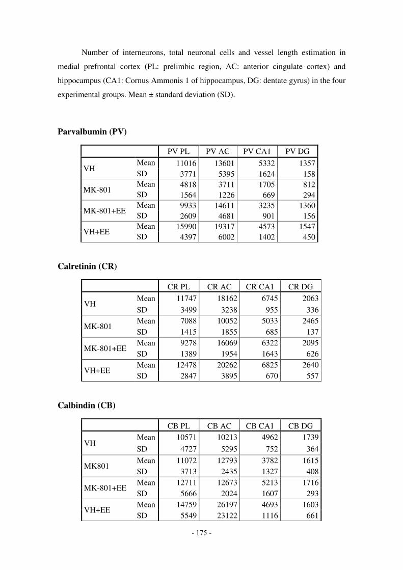
- 175 -
Number of interneurons, total neuronal cells and vessel length estimation in
medial prefrontal cortex (PL: prelimbic region, AC: anterior cingulate cortex) and
hippocampus (CA1: Cornus Ammonis 1 of hippocampus, DG: dentate gyrus) in the four
experimental groups. Mean ± standard deviation (SD).
Parvalbumin (PV)
PV PL PV AC PV CA1 PV DG
VH Mean 11016 13601 5332 1357 SD 3771 5395 1624 158
MK-801 Mean 4818 3711 1705 812 SD 1564 1226 669 294
MK-801+EE Mean 9933 14611 3235 1360 SD 2609 4681 901 156
VH+EE Mean 15990 19317 4573 1547 SD 4397 6002 1402 450
Calretinin (CR)
CR PL CR AC CR CA1 CR DG
VH Mean 11747 18162 6745 2063
SD 3499 3238 955 336
MK-801 Mean 7088 10052 5033 2465 SD 1415 1855 685 137
MK-801+EE Mean 9278 16069 6322 2095 SD 1389 1954 1643 626
VH+EE Mean 12478 20262 6825 2640 SD 2847 3895 670 557
Calbindin (CB)
CB PL CB AC CB CA1 CB DG
VH Mean 10571 10213 4962 1739
SD 4727 5295 752 364
MK801 Mean 11072 12793 3782 1615 SD 3713 2435 1327 408
MK-801+EE Mean 12711 12673 5213 1716 SD 5666 2024 1607 293
VH+EE Mean 14759 26197 4693 1603 SD 5549 23122 1116 661

- 176 -
Somatostatin (SST)
SST PL SST AC SST CA1 SST DG
VH Mean 28762 40410 11273 9756
SD 4363 7467 1022 1509
MK801 Mean 20399 31632 8529 8641 SD 2791 7048 1255 1214
MK-801+EE Mean 26058 38754 9595 8556 SD 3645 4672 1173 1038
VH+EE Mean 28839 41296 10557 9895 SD 3328 3968 917 473
Total interneurons number (GAD67)
GAD67 PL
GAD67 AC
GAD67 CA1
GAD67 DG
VH Mean 44468 41808 25809 15338 SD 12714 8523 4103 1895
MK801 Mean 33859 37605 16298 11223 SD 6643 5730 3131 1999
MK-801+EE Mean 35960 44964 24255 19188 SD 7611 12305 6384 4100
VH+EE Mean 54883 59622 26702 16060 SD 13456 10817 5292 3428
Total neuronal number (NeuN)
NeuN PL NeuN AC NeuN CA1 NeuN DG
VH Mean 405206 549775 44764 36657 SD 34813 67933 5718 4745
MK801 Mean 337989 537363 43251 31975 SD 57216 56291 7543 6059
MK-801+EE Mean 379244 541020 45313 38117 SD 35628 70440 6795 4205
VH+EE Mean 376755 570172 45609 34938 SD 45755 31888 8210 5075

- 177 -
Vessel length (BCh histochemistry)
BCh mPFC BCh HPC
VH Mean 26930870 27880545 SD 1265949 1665099
MK-801 Mean 30062633 23425169 SD 2986637 2159999
MK-801+EE Mean 27436287 28213173 SD 3168025 2655144
VH+EE Mean 30136982 29073083 SD 1654461 2595177
Number of proliferative cells in the subgranular zone of dentate gyrus of the
hippocampus (BrdU+) and number of immature granule cells (CR+ cells) in granular
cell layer of dentate gyrus in the four experimental groups. Mean ± standard deviation
(SD).
BrdU+ and CR+ cells in granule cell layer
BrdU+ CR+ granular layer
VH Mean 116 587 SD 20 118
MK-801 Mean 168 712 SD 35 200
MK-801+EE Mean 166 715 SD 33 271
VH+EE Mean 156 1063 SD 20 274


Appendix 3


Short-Term Exposure to Enriched Environment in Adult RatsRestores MK-801-Induced Cognitive Deficits and GABAergicInterneuron Immunoreactivity Loss
Ane Murueta-Goyena1 & Naiara Ortuzar1 & Pascual Gargiulo2 &
José Vicente Lafuente1,3,4 & Harkaitz Bengoetxea1
# Springer Science+Business Media, LLC 2017
Abstract Perinatal injections of N-methyl-D-aspartate(NMDA) receptor antagonist in rodents emulate some cogni-tive impairments and neurochemical alterations, such as de-creased GABAergic (gamma aminobutyric acid) interneuronimmunoreactivity, also found in schizophrenia. These featuresare pervasive, and developing neuroprotective orneurorestorative strategies is of special interest. In this work,we aimed to investigate if a short exposure to enriched envi-ronment (EE) in early adulthood (P55–P73) was an effectivestrategy to improve cognitive dysfunction and to restore inter-neuron expression in medial prefrontal cortex (mPFC) andhippocampus (HPC). For that purpose, we administeredMK-801 intraperitoneally to Long Evans rats from postnataldays 10 to 20. Twenty-four hours after the last injection, MK-801 produced a transient decrease in spontaneous motor ac-tivity and exploration, but those abnormalities were absent atP24 and P55. The open field test on P73 manifested that EEreduced anxiety-like behavior. In addition, MK-801-treatedrats showed cognitive impairment in novel object recognition
test that was reversed by EE. We quantified different inter-neuron populations based on their calcium-binding proteinexpression (parvalbumin, calretinin, and calbindin),glutamic acid decarboxylase 67, and neuronal nuclei-positive cells by means of unbiased stereology and foundthat EE enhanced interneuron immunoreactivity up to nor-mal values in MK-801-treated rats. Our results demonstratethat a timely intervention with EE is a powerful tool toreverse long-lasting changes in cognition and neurochemi-cal markers of interneurons in an animal model ofschizophrenia.
Keywords MK-801 . Interneurons . Cognitive dysfunction .
Calcium-binding proteins . Enriched environment
Introduction
Interneurons are the main source of inhibitory input in thecentral nervous system, and gamma aminobutyric acid(GABA) is their primary neurotransmitter. AlthoughGABAergic interneurons only account for 10–25% of to-tal cell number, depending on the brain region, they dis-play extremely distinct chemical, morphological, andfunctional features, making their classification a challeng-ing task. It has been postulated that the rich variety ofinterneurons is essential for providing constant matchedinhibitory input to the remarkably diverse incoming stim-uli [1]. In addition, the computational diversity they pro-vide allows proper dynamics for higher cognitive func-tions. Nevertheless, GABAergic interneurons also playan important role during postnatal development [2].GABA promotes the migration of glutamatergic andGABAergic neurons and dictates the final location of dif-ferent subpopulations of interneurons. GABA also
* Ane [email protected]
1 LaNCE, Department of Neuroscience, University of the BasqueCountry (UPV/EHU), Barrio Sarriena, E-48940 Leioa, Bizkaia,Spain
2 Laboratory of Neurosciences and Experimental Psychology, Area ofPharmacology, Department of Pathology, Faculty of MedicalSciences, National University of Cuyo, National Council ofScientific and Technical Research, Mendoza, Argentina
3 Nanoneurosurgery Group, BioCruces Health Research Institute,48903 Barakaldo, Bizkaia, Spain
4 Faculty of Health Science, UniversidadAutónoma deChile, Santiagode Chile, Chile
Mol NeurobiolDOI 10.1007/s12035-017-0715-z


337© Springer International Publishing AG 2017 P.Á. Gargiulo, H.L. Mesones-Arroyo (eds.), Psychiatry and Neuroscience Update - Vol. II, DOI 10.1007/978-3-319-53126-7_25
Neuropathological Background of MK-801 for Inducing Murine Model of Schizophrenia
Ane Murueta-Goyena Larrañaga, Harkaitz Bengoetxea Odriozola, Pascual Ángel Gargiulo, and José Vicente Lafuente Sánchez
A. Murueta-Goyena Larrañaga, MSc (*) Laboratory of Clinical and Experimental Neurosicence (LaNCE), Department of Neuroscience, University of the Basque Country UPV/EHU, Leioa, Spain
University of the Basque Country, Laboratory of Clinical and Experimental Neuroscience, Department of Neuroscience, Faculty of Medicine and Nursing, Barrio Sarriena s/n, Leioa, Bizkaia 48940, Spaine-mail: [email protected]
H.B. Odriozola • J.V. Lafuente Sánchez Laboratory of Clinical and Experimental Neurosicence (LaNCE), Department of Neuroscience, University of the Basque Country UPV/EHU, Leioa, Spain
P.Á. Gargiulo Laboratory of Neurosciences and Experimental Psychology, CONICET, Area of Pharmacology, Department of Pathology, Faculty of Medical Sciences, National University of Cuyo, Mendoza, Argentina
25
Abstract
Schizophrenia is a complex psychiatric disorder with a developmental component that compromises neural circuits. Understanding the neuro-pathological basis of schizophrenia remains a major challenge for estab-lishing new therapeutic approaches. In this review, causal factors for abnormal brain development in schizophrenia are discussed, with partic-ular focus on N-methyl-D-aspartate (NMDA) receptor hypofunction and GABAergic circuit-mediated neurotransmission. Changes in interneuron structure and function have been reported in schizophrenia, and current evidence points to a specific involvement of interneuronal NMDA recep-tor signaling. Furthermore, altered gamma-band oscillations in schizo-phrenic patients drew attention to a possible deficit in fast-spiking parvalbumin-expressing interneurons, which play an essential role in regulating complex interaction between pyramidal cells, and represent a key to the understanding of network operations. Here, we describe the major biochemical, neuropathological, and cognitive deficits present in schizophrenic human individuals, and the faithfulness of animal models for mimicking those impairments. In NMDA receptor antagonism-based animal models, repeated injections of MK-801 (dizocilpine) during early postnatal brain development, disrupt the excitation/inhibition balance.


Thi
s ce
rtifi
es th
at
Ane
Mur
ueta
-Goy
ena
part
icip
ated
in a
nd p
rese
nted
a o
ral p
rese
ntat
ion
at th
e
13th
Int
erna
tiona
l Con
fere
nce
on
Neu
ropr
otec
tive
Age
nts
Sept
embe
r 18-
21, 2
016
Bilb
ao/B
izka
ia, S
pain
_
____
____
____
____
____
____
____
____
__
Se
cret
ary


25
PREDICTIVE-CODING MECHANISMS OF PSYCHOSIS
Guillermo Horga MD, PhD
Columbia University, New York, USA
THE EFFECTS OF PHYSICAL EXERCISE ON BRAIN
FUNCTION: MENTAL HEALTH, COGNITION AND ACADEMIC PERFORMANCE. CROSS-SECTIONAL
RESULTS OF THE ACTIVEBRAINS PROJECT Irene Esteban-Cornejo, PhD
University of Granada
BIOMARKERS FOR PSYCHOTIC DISORDERS:
TRANSLATING RESEARCH INTO CLINICAL UTILITY
Laura Pina-Camacho MD, PhD
Child and Adolescent Psychiatry Department, Hospital General Universitario Gregorio Marañón, IiSGM, School
of Medicine, Universidad Complutense, CIBERSAM,
Madrid, Spain
UNTANGLING HIPPOCAMPAL CHANGES IN A
NEURODEVELOPMENTAL MODEL OF SCHIZOPHRENIA: THE ROLE OF ENRICHED
ENVIRONMENT Ane Murueta-Goyena, José Vicente Lafuente,
Bengoetxea Harkaitz Faculty of Medicine and Nursing. University of the
Basque Country (UPV/EHU), Leioa (Bizkaia), Spain
Room Camara
08:30-11:00
Symposium 14: Intellectual Disability: New
Opportunities for Understanding the Neurobiology
and Advancing Therapy
Chair: Mara Dierssen
ALTERATIONS OF PREFRONTO-HIPPOCAMPAL
NEURAL NETWORK DYNAMICS UNDERLIE COGNITIVE IMPAIRMENT IN BEHAVING MICE
MODELS OF INTELLECTUAL DISABILITY
Maria Victoria Puig Velasco Hospital del Mar Medical Research Institute, Barcelona
Biomedical Research Park
THE ENDOCANNABINOID SYSTEM AS A
PHARMACOLOGICAL TARGET FOR INTELLECTUAL