ADVERTIMENT. Lʼaccés als continguts dʼaquesta tesi queda ... · La cirugía de glaucoma es un...
135
ADVERTIMENT. Lʼaccés als continguts dʼaquesta tesi queda condicionat a lʼacceptació de les condicions dʼús establertes per la següent llicència Creative Commons: http://cat.creativecommons.org/?page_id=184 ADVERTENCIA. El acceso a los contenidos de esta tesis queda condicionado a la aceptación de las condiciones de uso establecidas por la siguiente licencia Creative Commons: http://es.creativecommons.org/blog/licencias/ WARNING. The access to the contents of this doctoral thesis it is limited to the acceptance of the use conditions set by the following Creative Commons license: https://creativecommons.org/licenses/?lang=en
Transcript of ADVERTIMENT. Lʼaccés als continguts dʼaquesta tesi queda ... · La cirugía de glaucoma es un...

ADVERTIMENT. Lʼaccés als continguts dʼaquesta tesi queda condicionat a lʼacceptació de les condicions dʼúsestablertes per la següent llicència Creative Commons: http://cat.creativecommons.org/?page_id=184
ADVERTENCIA. El acceso a los contenidos de esta tesis queda condicionado a la aceptación de las condiciones de usoestablecidas por la siguiente licencia Creative Commons: http://es.creativecommons.org/blog/licencias/
WARNING. The access to the contents of this doctoral thesis it is limited to the acceptance of the use conditions setby the following Creative Commons license: https://creativecommons.org/licenses/?lang=en
1
PAUROMERAROMERO
Directores:Dr.JordiLoscosArenas,Dr.XavierValldeperasBelmonte
Tutor:Prof.JoséGarcía-Arumí
Resultadosenpacientesintervenidosdeglaucomamedianteesclerectomíaprofundaconimplante
uveoescleralyanálisismorfológicodeláreaquirúrgicacontomografíaópticadesegmentoanterior
TESISDOCTORAL
DepartamentdeCirurgiaUniversitatAutònomade
Barcelona
2018


TesisDoctoralporcompendiodepublicaciones
Resultadosenpacientesintervenidosdeglaucomamediante
esclerectomíaprofundaconimplanteuveoescleralyanálisis
morfológicodeláreaquirúrgicacontomografíaópticade
segmentoanterior
Resultatsenpacientsintervingutsdeglaucomamitjançantesclerectomiaprofunda
ambimplantuveoescleralianàlisimorfològicdel’àreaquirúrgicaambtomografia
òpticadesegmentanterior
Outcomesinpatientsundergoingdeepsclerectomywithuveoscleralimplant
glaucomasurgeryandmorphologicalanalysisofthesurgicalareabyanterior
segmentopticaltomography
DepartamentdeCirurgia
FacultatdeMedicina
UniversitatAutònomadeBarcelona
Año2018
Autor:PAUROMERAROMERO
Directores:Dr.JordiLoscosArenas,Dr.XavierValldeperasBelmonte
Tutor:Prof.JoséGarcía-Arumí


Certificadodelosdirectoresytutor
El Dr. Jordi Loscos Arenas, Doctor en Medicina y Cirugía por la Universidad
Autónoma de Barcelona y cap clínic de la unidad de glaucoma del servicio de
oftalmologíadelHospitalUniversitariGermansTriasiPujol,Badalona,ydirectorde
estatesis:
El Dr. Xavier Valldeperas, Doctor en Medicina y Cirugía por la Universidad
AutónomadeBarcelonaymédicoadjuntodelserviciodeoftalmologíadelHospital
UniversitariGermansTriasiPujol,Badalona,ydirectordeestatesis:
ElProfesor JoséGarcía-Arumí,Catedrático enOftalmología por la Universidad
AutónomadeBarcelonayjefedelserviciodeoftalmologíadelHospitalUniversitario
Valld’Hebron,Barcelona,ytutordeestatesis:
CERTIFICAN:Queelpresentetrabajotitulado“Resultadosenpacientesintervenidos
deglaucomamedianteesclerectomíaprofundaconimplanteuveoescleralyanálisis
morfológico del área quirúrgica con tomografía óptica de segmento anterior”,
presentadocomotesisdoctoralporcompendiodeartículos,hasidorealizadopor
Pau Romera Romero bajomi dirección o tutorización, pudiendo ser presentado
comoTesisparalaobtencióndelgradodeDoctor.
Yparaqueasíconste,firmoelpresentecertificadoenBarcelona,mayo2018
Fdo.
Dr.JordiLoscosArenasDr.XavierValldeperas Prof.JoséGarcía-Arumí


Todoesimportante,
porquesóloatravésdelascosaspequeñas
sepuedehacerunagrancosa
JoanManuelSerrat


Agradecimientos
En primer lugar,me gustaría agradecer de forma especial al Dr. Jordi Loscos su
confianza.Unodelosmotivosporlosquemedecidíarealizaresteproyectodetesis
doctoralfuelafequedepositóenmíparaseguirsustrabajospreviosenestamateria
ymotivarmeparainvolucrarmeenproyectosdeinvestigación.Sinsutrabajodiario
esteproyectonohabríavistolaluz.Ensegundolugar,alaDra.M.AngelsParera,por
susconsejosysuapoyodurantetodosestosaños.Tambiénalsegundotutordemi
tesisdoctoral,alDr.XavierValldeperasporlavocacióncientíficaqueimplementaal
servicio y su implicación en este proyecto, y en general a todo el servicio de
OftalmologíadelHospitalGermansTriasiPujol,empezandoporeljefedeServicio,
misantiguosadjuntos, residentes, compañerosex residentes,yoptometristas,ya
que crean un ambiente de trabajo muy positivo sin el cual no tendría tanta
motivaciónparaseguiradelantecontodoslosproyectosquetenemosenmarcha.
Tambiénqueríaagradeceratodosloscompañerosquemehanenseñado,aunlado
yaotrodelocéano,estemagníficooficioqueeslaoftalmología,yenespecialaMr.
Nitin Anand, por sus consejos para iniciarme en el glaucoma y su ayuda en el
segundoartículodeestaTesisdoctoral.
Porúltimo,queríaagradecerleespecialmentesuapoyoalagentequequieroyme
rodea,porhabermeayudadoyaguantadoenlosmomentosdifíciles,yqueséque

seguirán apoyándome en un futuro. A Blas y Dolores por haberme enseñado,
motivado y ayudado económicamente durante todos estos años. A Antonio,
Marcelino,AnayCruzporhaberdemostradosiempreloorgullososqueestabande
mí,aJordiporsusconsejosconlasnuevastecnologías,aAlbertporsurespaldoy
recordarmequenodejelatesisaunladoyaEsther,AinayRicardporllevartantos
años embarcados conmigo en estemundo que es lamedicina y en lo que no es
medicina.Yengeneral,atodoslosquemehabéisacompañadoenestacarrerade
obstáculosqueahoraculminaunabonitaetapa.

Abreviaturas
EPNPEsclerectomíaprofundanoperforante
HUGTiPHospitalUniversitariGermansTriasiPujol
ECEsnoperClip®
OCT-SATomografíaópticadesegmentoanterior
PIOPresiónintraocular
AVAgudezavisual
NONervioóptico
DMAEDegeneraciónmacularasociadaalaedad
CVCampovisual
OCTTomografíadecoherenciaóptica
GPAAGlaucomaprimariodeánguloabierto
GNTGlaucomanormotensivo
GCAGlaucomaporcierreangular
MTMallatrabecular
BMUBiomicroscopíaultrasónica
HAHumoracuoso
HTOHipertensiónocular
CNTGSCollaborativeNormalTensionGlaucomaStudy
EMGTEarlyManifestGlacuomaTrial
OHTSOcularHypertensionTreatmentStudy
CIGTSCollaborativeInitialGlaucomaTreatmentStudy
SLTTrabeculoplastiaselectivelaser
MIGSMinimallyinvasiveglaucomasurgery
MTDMembranatrabeculodescemética
MMCMitomicinaC
5FU5-Fluorouracilo
VEGFFactordecrecimientoendotelialvascular
GPGoniopuntura
NNúmerodepacientes
FPacientesdelsexofemenino
GPXGlaucomapseudoexfoliativo
GpigmGlaucomapigmentario
GUveitGlaucomauveítico
MedMedicación
CACámaraanteriordelojo
DCDesprendimientodecoroides
PCprogresióndelacatarata
MPMicroperforación

Índice
pág.
1JUSTIFICACIÓNDELAUNIDADTEMÁTICA......................................................................... 15
2HIPÓTESIS............................................................................................................................................. 17
3OBJETIVOS............................................................................................................................................ 19
4.INTRODUCCIÓN................................................................................................................................ 21
4.1ElGlaucoma...................................................................................................................... 21
4.1.1Epidemiologia
4.1.2TiposdeGlaucoma
4.1.3AnatomíayfisiopatologíaimplicadaenelGlaucoma
4.1.4Factoresderiesgo
4.2TratamientodelGlaucoma...................................................................................... 38
4.2.1Ensayosclínicosaleatorizados
4.2.2Tratamientomédico
4.2.3Tratamientoláser
4.2.4CirugíafiltranteenGlaucoma
4.2.5CirugíaMIGS(minimallyinvasiveglaucomasurgery)
4.2.6CirugíanoperforanteenGlaucoma
4.3Esclerectomíaprofundanoperforante............................................................. 49
4.3.1Evoluciónhistórica
4.3.2Modulacióndelafibrosispostquirúrgica
4.3.3Goniopuntura
4.3.4Needling
435Implantesenlaesclerectomíaprofundanoperforante
436Estudiospreviospublicados
pág.
4.4Evaluacióneconómica............................................................................................... 64
4.5Tomografíadecoherenciaópticadesegmentoanterior
(OCT-SA)enelglaucoma.......................................................................................... 66
5.MATERIALYMÉTODOS.................................................................................................................73
5.1Obtencióndelamuestra,criteriosdeinclusiónyexclusión................73
5.2Técnicaquirúrgica........................................................................................................74
5.3Materialquirúrgico...................................................................................................... 76
5.3.1Implanteuveoescleral(EsnoperClip®)
5.4Materialesexploratoriosypruebascomplementarias.......................... 79
5.5Tratamientopostoperatorio................................................................................. 80
5.6Cronogramadevisitasoftalmológicas............................................................. 81
5.7Análisismorfológicocontomogragíadecoherenciaóptica
desegmentoanterior(OCT-SA)............................................................................ 82
5.8AnálisisEstadístico...................................................................................................... 83
6.RESULTADOSYARTÍCULOSPUBLICADOS........................................................................ 85
6.1ResumenArtículo1+Artículo.............................................................................. 85
6.2ResumenArtículo2+Artículo.............................................................................. 93
7.DISCUSIÓN............................................................................................................................................103
8.CONCLUSIONES.................................................................................................................................. 115
9.BIBLIOGRAFÍA....................................................................................................................................117

15
1. Justificacióndelaunidadtemática
El glaucoma es una enfermedad neurodegenerativa sobre la que hoy en día persisten
muchasincógnitas.Fuedescritoyaenlaépocahipocráticaconeltérminoglaukosisquese
manifestaba con el cambio de color a azulado de la pupila. Es una enfermedad muy
discapacitante,dadasunaturalezasilenteeirreversible.
Lacirugíadeglaucomaesuncampoencontinuodesarrolloquehaidoevolucionadodesde
susinicios,desdeelmomentoqueseentendióquedisminuirlapresiónintraocularproducía
unadisminucióndelriesgodeprogresióndeestaenfermedad.Lacirugíamásrealizadahoy
endíasiguesiendolatrabeculectomía,peroenmanosexpertas,laesclerectomíaprofunda
noperforante(EPNP)tieneunosresultadosqueseasemejanalaanterior.ElEsnoper®es
unimplantequefuediseñadoparaevitarelcolapsodeláreaquirúrgicaenelpostoperatorio
deesta cirugía.DiseñadoeneldepartamentodeGlaucomadelHospitalGermansTrias i
Pujol(HUGTiP),sehamostradocomounaherramientaeficazparaestetipodecirugías.El
Esnoper Clip® (EC) es la evolución del implante Esnoper® y tiene la ventaja de ser
especialmentediseñadoparafavorecerlavíauveoescleral.
Elobjetivodeestatesisdoctoralesmostrarlaeficaciadeestenuevoimplanteamedioplazo
enlaEPNP.Hemospublicadodosartículos,unoalañoyotroalosdosañosdeseguimiento,
mostrando los resultados tensionales, tasa de medicación postoperatoria y las
complicaciones quirúrgicas y postquirúrgicas. En el segundo artículo hemos añadido un
análisis del área quirúrgica con tomografía óptica de segmento anterior (OCT-SA),
16
aportando ideas nuevas de la importancia de la altura del área quirúrgica en el
postoperatorio tras la cirugía. Consideramos que estos dos artículos representan una
unidadtemáticaclaradelautilizacióndelimplanteuveoescleralenlaEPNP,siendoútilsu
lecturaatodoaquelqueestéinteresadoporlacirugíanoperforantedeglaucomaydelas
posibilidades que nos ofrece la vía uveoescleral, así como valorar la utilización de este
implante.
17
2. Hipótesis
La esclerectomía profundano perforante (EPNP) con el implante uveoescleral (Esnoper
Clip®)esunatécnicasegurayefectivaparamanteneruncontroltensionalcorrectoconuna
bajatasadecomplicaciones,acompañadodeunabajatasademedicaciones.Al igualque
otrascirugíasparaelglaucoma,permitemantenerunbuencontroltensionaltras24meses
delaintervención.
La tomografíaópticadesegmentoanterior (OCT-SA)permiteanalizareláreaquirúrgica
traslacirugíayesunatécnicaútilparadescribirfactoresasociadosaléxitoofracasodela
misma.

18

19
3. Objetivos
3.1 Demostrar que la EPNP con implante uveoescleral reduce la PIO de forma
significativahasta24mesesposterioresalaintervención.
3.2 DemostrarquelaEPNPconimplanteuveoescleralreducelatasademedicaciónde
formasignificativahasta24mesesposterioresalaintervención.
3.3 DemostrarquelaEPNPconimplanteuveoescleralesunatécnicaseguraypresenta
unabajatasadecomplicaciones.
3.4 DescribirmedianteOCT-SAfactoresdeláreaquirúrgicaasociadosaléxitotensional
alargoplazo.Losfactoresanalizadosincluyen:
3.4.1Alturadellagointraescleral
3.4.2Tipodeampolla(difusa,quística,planaoencapsulada)
3.4.3Presenciadeflujotransescleral
3.4.4Presenciadeflujouveoescleral
3.4.5Reflectividadsubconjuntivalysupraescleral
3.4.6Presenciademicroquistes

20

21
4. Introducción
4.1ElGlaucoma
Elglaucomaesunaenfermedadcrónicaydegenerativaqueincluimosdentrodelgrupode
lasneuropatíasópticas,dondeencontramoscausasisquémicas,inflamatorias,infecciosasy
traumáticas.Esunadelascausasdeceguerasmásprevalentesentodoelmundo.Aunhoy
endíaesdifícildeterminarcuáleselmecanismopatológicoqueproduceundañodelnervio
óptico trasunaumentodepresión intraocular (PIO),y seha llegadoal consensodeque
estamosanteunproblemadeorigenmultifactorial.Elúnicofactorderiesgoquehastala
fechapodemosmanipularparamejorarelcursodelaenfermedadeslaPIO.ElGlaucomaes
unaneuropatíaprogresivaeirreversiblequetienecomoresultadolapérdidadelascélulas
ganglionaresdelaretina.Lascaracterísticasdelnervioóptico(NO)sondistintasalrestode
neuropatías, observándose un incremento de la excavación papilar al perderse el anillo
neurorretiniano,deformafocalogeneralizada,atrofiaperipapilar,estrechamientodelos
vasos papilares y en algunos casos hemorragias peripapilares. El defecto del NO se
correlacionaconundefectofuncionalenelcampovisual.EnelpasadoelaumentodelaPIO
eraclaveparasudiagnóstico,siendohoyendíaunfactorimportanteperonoexcluyente.
LaPIOmediasegúnestudiospoblacionalesesdeaproximadamente16mmHg,ylosrangos
denormalidadentre10y21mmHg(dosdesviacionesestándar).1
HoyendíaexistenpruebasestructuralesdelNOyfuncionalesquenosayudanacontrolar
laevolucióndelaenfermedadennuestrospacientes.Elcampovisual(CV)esunaprueba
22
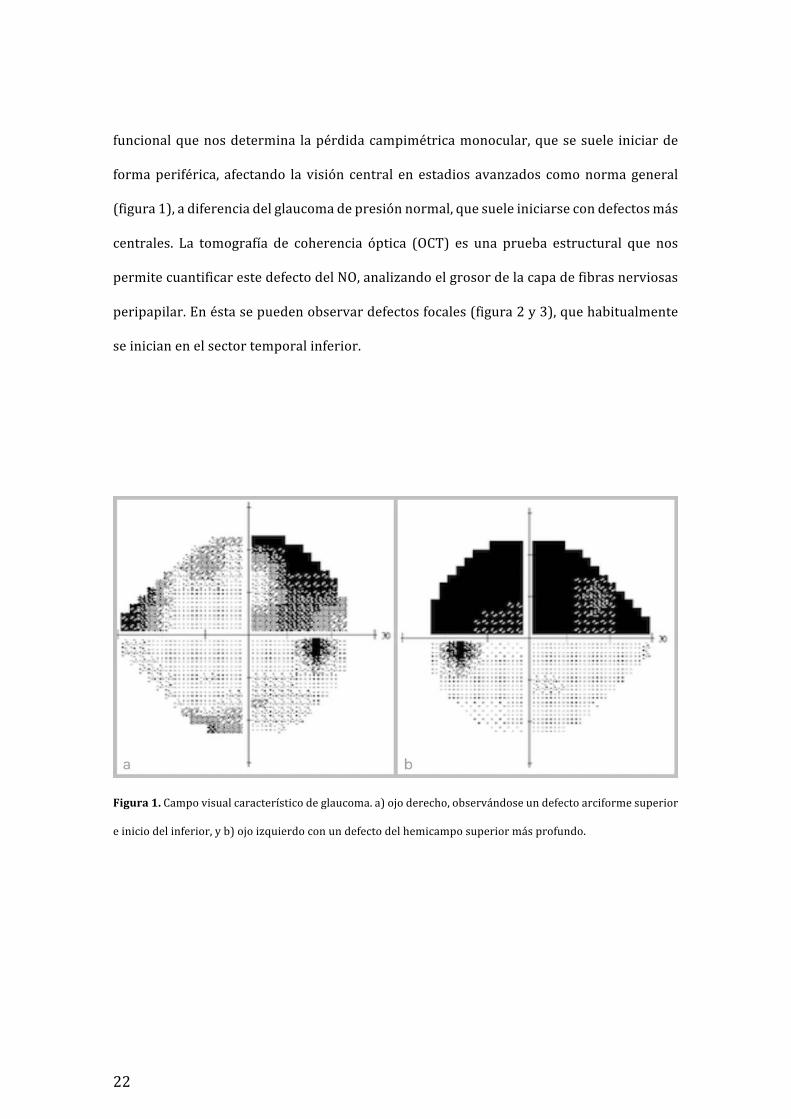
funcionalquenosdetermina lapérdidacampimétricamonocular,quesesuele iniciarde
formaperiférica, afectando la visión central en estadios avanzados comonormageneral
(figura1),adiferenciadelglaucomadepresiónnormal,quesueleiniciarsecondefectosmás
centrales. La tomografía de coherencia óptica (OCT) es una prueba estructural que nos
permitecuantificarestedefectodelNO,analizandoelgrosordelacapadefibrasnerviosas
peripapilar.Enéstasepuedenobservardefectosfocales(figura2y3),quehabitualmente
seinicianenelsectortemporalinferior.
Figura1.Campovisualcaracterísticodeglaucoma.a)ojoderecho,observándoseundefectoarciformesuperior
einiciodelinferior,yb)ojoizquierdoconundefectodelhemicamposuperiormásprofundo.

23
Figura 2. a) Tomografía de coherencia óptica de la capa de fibras nerviosas peripapilares que describe undefectoinferiordelanilloneurorretinianodelnervioópticoconaumentodelaexcavaciónydisminucióndelgrosordelacapadefibrasnerviosasperipapilardelojoderecho,quepodríacorresponderconelcampovisualdelafigura1a.b)Nervioópticodelojoizquierdoconanilloneurorretinianoconservado.
Figura3.Tomografíadecoherenciaópticadepapiladelmismopacientequelafigura2.a)datoscuantitativosdel análisis de capa de fibras peripapilar y disco óptico (RNFL: retinal nerve fiber layer; C/D: tamañoexcavación/tamañodisco).b)grosordelacapadefibrasperipapilarencadasector(OD:ojoderecho;OI:ojoizquierdo;TEMP:temporal;SUP:superior;NAS:nasal;INF:inferior).
24
4.1.1Epidemiología
Elglaucomaesunadelascausasmásfrecuentedecegueraenlospaísesoccidentales.Enel
año2000,laprevalenciadepersonasconmuybajavisión(agudezavisualmenora20/200)
enlosEEUUsegúnKlein2erade937.000personas,ylaprevalenciaeramayorenhispanos
yafroamericanos.Deéstas,lacausamásfrecuentefuelacatarataentreafroamericanose
hispanos y la degeneraciónmacular (DMAE) en caucásicos. El glaucoma fue la segunda
causamásfrecuenteenafroamericanos(26%enafroamericanos,28%enhispanosy6,4%
encaucásicos).SegúnestimacionesdelEyeDiseasesPrevalenceResearchGroup(EDPRG),3
2.218.000personastienenglaucomaenEEUU, loquecorrespondeaunaprevalenciadel
2%.EstudiosepidemiológicosrealizadosenEspañatambiénconfirmanestaprevalenciadel
2%.4Esmásfrecuenteentremujeresjóvenesquehombresjóvenes,peroseigualaapartir
delos70años.Laprevalenciaesmayorenafroamericanosqueencaucásicos,estandolos
hispanosentreambos.Eslasegundacausadecegueraenlossubsaharianos,representando
el15%delosciegosenÁfricaysuprogresiónyseveridadesmásaltaqueenloscaucásicos.5
Seestimaque80millonesdepersonasentodoelmundotendránglaucomaen2020,yque
lamitaddeellosnosabránquepadecenlaenfermedad.
En nuestro medio, la frecuencia más alta corresponde al glaucoma primario de ángulo
abierto (GPAA), pero la prevalencia de glaucomade ángulo cerrado esmayor de lo que
sospechábamos, siendodel0,4%, con la edad, el sexo femeninoy la etniaoriental como
factoresderiesgo.6

25
4.1.2TiposdeGlaucoma
Existendiferentesclasificacionesdelglaucoma.Sinosfijamosenlaanatomíadelángulode
lacámaraanteriordelojo,podemosclasificarlosendossubgrupos,quenosdeterminará
unaestrategiaterapéuticaclaramentediferenciada:
- Glaucoma primario de ángulo abierto, dentro del cual encontramos al glaucoma
normotensivo(GNT)
- Glaucomaporcierreangular(GCA)
Si nos centramos en la causa, podemos añadir a estas dos categorías los glaucomas
secundarios,tantodeángulocerrado,entrelosqueencontramosglaucomasneovasculares,
facomórfico, misdireccional o maligno; y de ángulo abierto, como el inflamatorio, por
dispersiónpigmentaria,pseudoexfoliativo,uveíticoyfacolítico.
LaTabla1describelosdiferentestiposdeglaucoma,segúnsucausacomolaanatomíadel
ángulodelacámaraanterior.

26
Tabla1.Clasificacióndelglaucomabasadaenla“TerminolologyandGuidelinesforGlaucoma4thEdition”,delasociedadEuropeadeGlaucoma.7
Elglaucomaprimariodeánguloabiertoeselmáscomúnennuestromedioy,juntoalos
decausasecundaria,esenelquesecentraestatesisdoctoral.Mayormenteseentiendeque
existeunaumentodelapresiónintraoculardebidoaunaumentodelaresistenciadesalida
delhumoracuoso,yaseaenlamembranajuxtacanaliculardelamallatrabecularyporción
endotelialdelcanaldeSchlemm,debidoaunamayorrigidezenlascélulasdelcanaldela
malla trabecular y a las características de lamatriz extracelular,8 o en la entradade los
conductoscolectoresdelcanaldeSchlemm,9 loquecomportaríaunaumentode laPIOy
dañoglaucomatosodelnervioóptico.Lascausassecundariasproduciríanunbloqueode
dichamallapordiferentes agentes.Elglaucomanormotensivo es fisiopatológicamente
similaraldeánguloabierto,peroaunapresiónmenoro iguala21mmHg.10Representa
Glaucomacongénitoprimario
•Glaucomainfantil
•Glaucomaasociadoaanomalíascongénitas
Glaucomadeánguloabierto
•Primarios•Glaucomajuvenilprimario•Glaucomaprimariodeánguloabierto•Glaucomanormotensivo
•Secundarios•Origenocular(exfoliativo,facolítico,pigmentario,traumático,hemorrágico,uveítico)•Iatrogénico(corticoideo,trascirugíaocular)•Causadoporcondicionesextrabulbares(incrementodelapresiónvenosaepiescleral)
Glaucomaporcierreangular
•Primarios•Agudo•Intermitente•Crónico
•Secundarios•Conbloqueopupilar•Conmecanismodeempujeposterior•Conmecanismodetracción

27
entreel30-40%detodos losglaucomadeánguloabierto,11yposiblementeensuorigen
intervienenmásfactorescardiovascularesyrelacionadosconlapresiónintracranealque
eneldeánguloabierto.Tambiénseharelacionadoconlaapneaobstructivadelsueño.12
Elglaucomaporcierreangular(GCA)esfisiopatológicamentedistintodelglaucomade
ánguloabierto.1314Elprocesoprimarioeselbloqueodelamallatrabecular(MT)poreliris
periférico,tantoaposicionalcomosinequial,produciendounaumentodelaPIO.Existiría
un cierto bloqueo a nivel pupilar haciendo que la presión en la cámara posterior fuera
mayoralacámaraanterior,produciéndoseundesplazamientoanteriordelirisperiférico
cerrando el ángulo. Consideramos cierre angular primario cuando existe contacto irido-
trabecular en más de 180º. Cuando se produce un bloqueo de la MT 360º por el iris
periférico,conaumentodePIO,dolor,visiónborrosa,dehalosyvómitos,estamosanteun
cierreangularagudo.Losfactoresderiesgoatenerencuentasonunalongitudaxialcorta,
unacámaraanteriorestrecha,raízdelirisgruesa,uncristalinograndeyconelvaultanterior
aumentado(entendiendoelvaultcristalinianocomoladistanciaquesobresalelaporción
anterior del cristalino sobre la perpendicular al espolón escleral). El análisis con
biomicroscopíaultrasónica(BMU)hapermitidoseccionarelsegmentoanterioryobservar
quelaposicióndelcuerpociliaresotrofactoratenerencuenta.Tieneunadisposiciónmás
anteriorencasosdeglaucomadeángulocerradoporirisplateau.14Dentrodeltratamiento,
lairidotomíaperiféricaeseltratamientodeeleccióntantoparaaumentarelángulodela
cámaraanterioryalejar la raízdel irisde laMT, comoparaevitar la crisisdeglaucoma
agudoporcierreangular.Traslamismanoseobservaunclarodescensotensional,porlo
quesehaanalizadosilafacoemulsificaciónprecozpuedeserunaopciónválidaencasosde
hipertensiónocularconosinglaucoma.ElestudioEAGLE,15querealizafacoemulsificación
precozenpacientesmayoresde50añosconPIOmayoresde30mmHg,hademostradoque
esmáseficazymáscoste-efectivaalos10añosdeltratamientoquelairidotomíaperiférica
ydeberíaserconsideradacomoprimeraopciónterapéutica.16
28
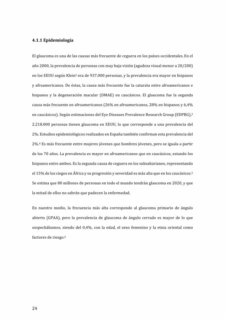
Figura4.ImagendeOCT-SAdondeseobservaunclaroaumentodetodoslosparámetrosangularesdelacámaraanteriortrasfacoemulsificaciónenglaucomaporcierreangular.a)previoalafacoemulsificación.b)posterioralafacoemulsificación.
Tambiénhademostradoquesuponeunamejoríadelacalidaddevidaparaelpaciente,ya
seapormejorarlaagudezavisualsincorrección,mejorcontroldelaPIOomenornecesidad
de medicación tópica.15 La facoemulsificación ha demostrado aumentar los parámetros
angulares(figura4),yprovocarundescensodelaPIOmayorqueenloscasosdeglaucoma
deánguloabierto.1718

29
4.1.3AnatomíayfisiopatologíaimplicadaenelGlaucoma
Enelojoexistenvariasáreasanatómicasimplicadasenlafisiopatologíadelglaucoma.
Córnea
Lacórneaeseltejidomásanteriordegloboocular,estransparente,avascularyatravésde
ellapenetranlosrayos lumínicoshaciael interiordelojo.Sehaobservadoqueelgrosor
cornealesunfactorindependientepredictordeldesarrollodeglaucoma.19Porotrolado,
sabemosqueelgrosorcornealysuhistéresisinfluencianenlamedicióndelaPIOconla
tonometríadeGoldman,yaquelaPIOsueleestarsobreestimadaenpacientesconcórneas
gruesas.20 21 En casos de pacientes con hipertensión ocular (HTO) y un grosor corneal
disminuido,sehaasociadoaunmayorriesgodeprogresiónaglaucoma.22
Humoracuoso
El humor acuoso (HA) es el fluido que ocupa la cámara anterior y posterior del ojo. El
balance entre su formación y su drenaje es el quemantiene la PIO. Aporta nutrientes y
oxígenoalasestructurasquerodea.Esproducidodeformaactivaporelepiteliodelcuerpo
ciliar, se dirige luego hacia la cámara anterior del ojo y hacia el ángulo de la cámara
anterior.8
Víasdedrenajedelhumoracuoso(HA)
Clásicamente existen dos vías de drenaje del HA, la vía trabecular y la vía uveoescleral
(figura5).Tambiénsehadescritodrenajeatravésde losvasos iridianos,cuerpociliary
haciavítreoycoroides,perosiendopocoinfluyentesenlaPIO.

30
Figura5.Víasdedrenajedelhumoracuoso(cortesíadelDr.ManuelRomera).
Víatrabecular:ElHApasaatravésdelamallatrabecular(MT),formadaportrescapas(la
uveal,lacorneoescleralylajuxtacanalicular).LauvealeslaqueestáencontactoconelHA
delacámaraanteriordelojo,estácompuestaporfibrasdecolágenoycélulasendoteliales
congrandesporos(25-75µm)(figura6).Lacorneoescleraleslaqueseextiendedesdeel
espolónesclerala la líneadeSchwalbe,yes laconocidacomotrabeculodescemética.Por
último,lajuxtacanalicularestáíntimamenteunidaalaparedinternadelcanaldeSchlemm,
tieneunosporosmáspequeñosyeslacapaqueaportamayorresistenciaylamásimplicada
enelglaucomadeánguloabierto,tantoprimariocomosecundario.Supartemásexternase
correspondeconelendoteliodelcanaldeSchlemm.Alpasaratravésdeesteendotelio,bien
porporosovacuolas,2324seintroduceenelcanalysedirigehacialosconductoscolectores
yplexosvenososepiesclerales.25Estavíaespresióndependiente.
Vía convencionalCanal de SchlemmVenas epiesclerales
Vía subconjuntivalFístula quirúrgica
Vía no convencionalVía uveoescleralVía uveovorticosa
31
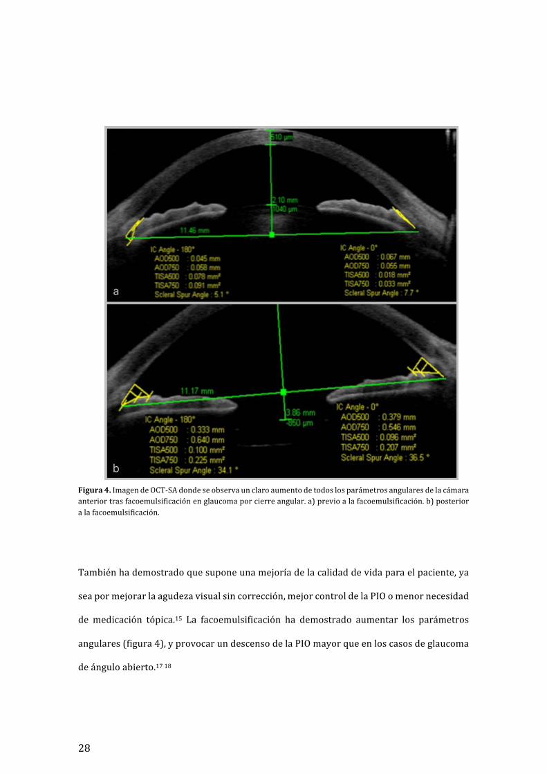
Figura6.Reconstrucción3Ddelamallatrabecular.Seobservanfibrasdecolágenorodeandoespaciosllenosdefluido.Visióndelamallatrabeculardesdelacámaraanterior.(Ammar,D.A.,etal.(2010).Two-photonimagingofthetrabecularmeshwork.MolecularVision,16,935–944).24
Vía uveoescleral: El HA se dirige desde la raíz del iris y cuerpo ciliar hacia el espacio
supraciliar y supracoroideo. Se cree que en humanos representa entre el 10-20% del
drenaje del HA, pudiendo llegar a representar el 40% en personas jóvenes. Funciona a
presionesmásbajasquelaanterioryespresiónindependiente.Larelajacióndelasfibras
delmúsculo ciliar facilita el drenaje.26 Lasprostaglandinasmodifican la estructurade la
matrizextracelulardelmúsculociliar,incrementandoelflujoporestavía.27ElHAdespués
es reabsorbido por los vasos uveales y perforantes esclerales.28 Es una posible vía de
abordajequirúrgico,tantoencirugíanoperforanteconimplantaciónsupraciliarcomoen
cirugíamínimamente invasiva con implantes especialmentediseñadospara incrementar
estavía,comoelCypass®(Alcon-Novartis,Basilea,Suiza).
CanaldeSchlemm
El canal de Schlemm es una estructura cilíndrica que envuelve el globo ocular
circunferencialmente, localizada sobre y externamente al espolón escleral. Su lumen
internoesvariable,entre190-350µm.Suendotelio lo forma lacapamásexternade la

32
membrana yuxtacanalicular y su parte externa está perforada por más de 20 canales
colectores, más frecuentes en el cuadrante nasal inferior. El canal está atravesado por
septos,entreloscanalescolectoresyelendotelio,impidiendosucolapso.Entreun25-50%
delaobstruccióndesalidadelHApodríalocalizarseaestenivel.Sehaobservadocomolos
mecanismoscompensatoriosalaumentodelaPIOqueseproducenenelcanaldeSchlemm
yenloscanalescolectoresestándisminuidosenpacientesconglaucomaprimariodeángulo
abierto.9
Vasosepiesclerales
Loscanalescolectoresdrenanalasvenasdelacuosoyéstashacialosvasosepiesclerales.
En la población general, la PIO normal es de 16 mmHg de media, y la presión venosa
epiescleralde7-8mmHg,observándoseungradientedepresiónentreambasquefacilitala
salidadelHA.Enelglaucomadeánguloabiertoexistemayoritariamenteunaumentodela
resistenciade salidadelHA,pero tambiénpuedeproducirsedebidoaunaumentode la
presión venosa epiescleral, como es el caso de la fístula carótido-cavernosa, varices,
tumoresorbitariosyoftalmopatíatiroidea.25
Nervioóptico
Elnervioóptico(NO)esuntractomielinizadodeaproximadamente1,2millonesdeaxones
queseiniciadesdelascélulasganglionaresdelaretinayacaba5cmposterioralquiasma
óptico.Laporciónintraoculardelnervioópticoeslamásrelevanteenglaucoma,yaquees
enlaqueobservamoslaexcavaciónpapilaraumentadacaracterísticadeestaenfermedad,
que la diferenciamos con el resto de neuropatías ópticas que producen nervios demás
palidezyatrofiaqueexcavación.Podemosdividirloenunaporciónprelaminar,unaporción
laminar(alaalturadelacoroides),quesonlasqueobservamosalrealizarelfondodeojo
(figura7y8),yunaporciónretrolaminaroretrobulbar(alaalturadelaesclera).

33
Clásicamente se ha descrito que el daño glaucomatoso suele iniciarse en el sector
inferotemporal del nervio óptico, seguido del superotemporal, luego del temporal y por
últimodelnasal.29Existebastanteconsensoenqueelorigendelglaucomaesmultifactorial,
existiendodiferentesteoríassobrelosmotivosqueproduciríaneldañoquedesencadenaría
estaenfermedad:
Figura7.a)papilaópticaconglaucoma,aumentodelaexcavaciónpapilarconhemorragiaenastillasuperior;b)defecto focaldelanilloneurorretiniano inferiorconpérdidasectorialde lacapade fibrasnerviosasde laretina(espacioentrelasdosflechasnegras).
- Teoríamecánica: la PIO alta dañaría la lámina cribosa y bloquearía los axones
nerviosos,loquealteraríaelmetabolismocelularproduciendoapoptosiscelular.30
- Teoría vascular: se produce una reducción del flujo anterior del nervio óptico
debidoafactorescirculatorios,comopuedeserlahipotensiónarterialsosteniday
34
la diabetes mellitus. Existe aún controversia en la relación entre diabetes y
glaucoma,todoqueenmodelosanimalessehapodidorelacionar.31
- Teoría autoinmune: autoanticuerpos circulantes directamente o bien de forma
indirecta activarían una reacción que induciría la apoptosis de las células
ganglionaresdelaretina,32loquenosabriríauncamponuevoparaeltratamiento
delglaucoma.
- Teoríadellíquidocefalorraquídeo(LCR):descritaporFleishmanyBerdahl,que
indicaqueelgradientedepresióntranslaminarpuedeestarcausadoporcambios
tantoenlaPIOcomoenlapresióndelLCR,yquetenerungradientedemasiadoalto
por cualquiera de estos dos motivos haría desplazar la lámina cribosa hacia
posterioryaumentaríaeldañoaxonal.33
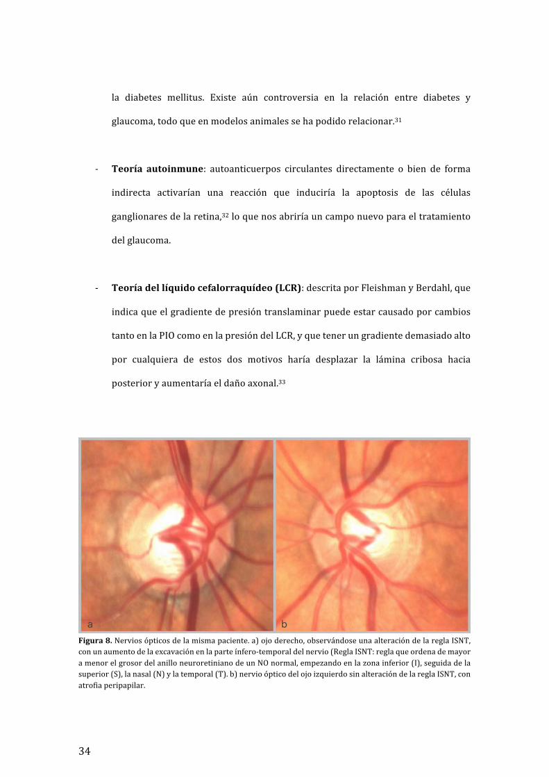
Figura8.Nerviosópticosdelamismapaciente.a)ojoderecho,observándoseunaalteracióndelareglaISNT,conunaumentodelaexcavaciónenlaparteínfero-temporaldelnervio(ReglaISNT:reglaqueordenademayoramenorelgrosordelanilloneuroretinianodeunNOnormal,empezandoenlazonainferior(I),seguidadelasuperior(S),lanasal(N)ylatemporal(T).b)nervioópticodelojoizquierdosinalteracióndelareglaISNT,conatrofiaperipapilar.

35
Esclera
Eseltejidomásrígidodelosqueconformanelpoloposteriordelojo,aportandosoporte
estructural al mismo. Está formada por fibras y bandas de colágeno y elastina.
Anteriormentesecontinuaconlacórneayposteriormenteconladuramadre,querodeaal
nervioóptico.Alaalturadelacabezadelnervioóptico,losdosterciosposterioresloforman
elcanalescleralyeltercioanteriorlaláminacribosa.Laparteanteriordelaesclerasoporta
un menor estrés que la parte posterior, que se deforma más al aumentar la PIO. Las
propiedadesbiomecánicasdelaesclerainfluyenenlaspropiedadesdelacabezadelnervio
ópticoyéstascambianconlaedad.Noquedaclarosiesclerasmásrígidasestánasociadasa
unmayorriesgodeglaucoma(debidoalmenormovimientotrasincrementosdePIO)obien
locontrario.3435
Laláminacribrosaeselsoportefibrosodelnervioópticoenelpoloposteriordelojo.Es
unamallapordondepenetralasfibrasnerviosas.Debidoaloscambiosdepresión,lalámina
cribosasedeformahaciaposteriorenelglaucomayhaciaanteriorenelpapiledema.La
láminacribosabasculaentredospresiones,laPIOylapresiónretrobulbaryescrucialpara
entenderlafisiopatologíadelglaucoma.Elgrosordelaláminacribosaesdealrededor450
micrasyelgradientedepresiónesde1mmHgcada100micras.Elgrosordelamismayel
tejidoquelaenvuelvejueganunpapelrelevanteenmantenerlacondicionesalrededordel
nervioóptico.3637
Coroides
La coroides es una gran área vascularizada situada entre la esclera y la retina, que
transcurredesdeelcuerpociliarhastarodearelnervioóptico,pudiendotenerimplicación
enlacorrectavascularizacióndelascélulasnerviosasdelaretinaydelNO(figura9).Sehan
descrito cambios de su grosor tras cambios de presión intraocular y tras cirugía de

36
glaucomaqueanalizadosconcautelapodríanaportaralgunasconclusionesinteresantes.38
3940
Figura9.Tomografíadecoherenciaópticamacular.ObservamoselgrosorcoroideoenpacienteintervenidodeEPNP(laslíneasverticalesdelimitaneltejidocoroideo).
4.1.4Factoresderiesgo
Los factores de riesgo que se han asociado con el glaucoma son múltiples. Los más
importantessonlaPIOalta,laedadavanzada,historiafamiliar(factoresgenéticos),raza,
grosorcornealcentraldisminuidoybajapresióndeperfusióndiastólica(diferenciaentre
presióndiastólicayPIO).TambiénsehadescritoelíndicePresiónSistólica/PIO,aportando
mayorriesgocuandoésteesbajo.41
SonvariosestudioslosquehanrelacionadolaPIOylaprogresiónaglaucoma(comentados
enelsiguienteapartado).Entrandoenlasdiferenciasraciales, losafroamericanostienen
mayorPIO ymenor grosor corneal central. El sexono influye en el riesgode glaucoma,
aunque hay estudios que atribuyen mayor riesgo al sexo masculino, sobretodo en

37
glaucomassecundariosydebidosatraumatismos.Tambiénesconocidoelmayorriesgodel
sexofemeninoydelarazaasiáticaenelglaucomadeángulocerrado.14
Tenerunapresióndellíquidocefalorraquídeo(LCR)bajapodríasertambiénunfactorde
riesgo.36ElLCRes formadoen losplexoscoroideos,en losventrículos laterales, tercery
cuartoventrículo.CuandomedimoslapresióndelLCRporpunciónlumbar,estapresiónse
correlacionadeformadirectaconlapresiónintracraneal,yestamismaserelacionaconla
presiónretrolaminar(siempreque lamidamosendecúbito).Por lo tanto,podemosusar
ambaspresionesdeformaindistinta.Aniveldelaláminacribosa,losaxonespasandeuna
PIO alta a una presión retrobulbar/retrolaminarmás baja, conocido como gradiente de
presióntranslaminar,ylapresióndelLCRbajapuedetenerunefectoigualmentedañino
paraelnervioóptico.36
La fluctuación de la PIO parece ser que es un factor de riesgo para la progresión del
glaucoma, aunque no se ha demostrado estadísticamente.42 Cuando controlamos a un
pacienteennuestraconsulta,loqueobtenemosesundatodelaPIOenesemomento,pero
estamosperdiendo la curva tensionalde las24horas.LaPIOsigueunritmocircadiano,
siendo mayor por la mañana entre las 8 y las 11, y menor entre las 12 y las 2 de la
madrugada.Lavariaciónduranteeldíapuedeserdehasta3-5mmHg.AnteunamismaPIO,
aquellospacientes intervenidosdecirugía filtrante tendránunavariacióndiurnamenor,
conmenospicostensionales,silocomparamosconaquellospacientesentratamientocon
gotasotrabeculoplastialaserselectiva(SLT).434445
38
4.2TratamientodelGlaucoma
4.2.1Ensayosclínicosaleatorizados
GraciasalaevidenciacientíficasehapodidodemostrarquealdisminuirlaPIOseproduce
unadisminuciónenlaprogresióndelglaucoma.ElEarlyManifestGlacuomaTrialyOcular
Hypertension Treatment Study fueron algunos los primeros estudios en demostrar esta
teoría.Acontinuación,sedescribenalgunosdelosmásrelevantes:
EMGT(EarlyManifestGlacuomaTrial,1998):Compararontratamientovsnotratamiento
enpacientesconglaucomanotratadopreviamente.Observaronqueunareducciónde la
PIOdel25%reducíaelriesgodeprogresiónenun50%.Factoresquedeterminaronuna
mayorprogresiónfueronunaPIOmayor,síndromepseudoexfoliativo,edadtardía,mayor
dañoglaucomatosobasal,mayoredad,presenciadehemorragiaperipapilar,menorgrosor
corneal,ymenorpresiónsanguínea.46
CNTGS (CollaborativeNormalTensionGlaucomaStudy): incluíapacientesconglaucoma
normotensivoenprogresiónaleatorizadosentratamientoynotratamiento.Enaquellosque
setratóylaPIOdisminuyóun30%,laprogresiónfueenel12%,vsel35%delosquenose
trataron.47
OHTS (OcularHypertensionTreatment Study,1994): incluíapacientes conhipertensión
ocular, valorando si el hechode iniciar ono tratamientohipotensor ocular disminuía el
riesgodeevolucionaraglaucoma.Alos5años,un4,4%delostratadosvsel9%delosno

39
tratadosevolucionaronaglaucoma,loquesignificóunareduccióndelriesgodel50%.Alos
13añosestaproporciónfuedel16%vsel22%.4849
CIGTS(CollaborativeInitialGlaucomaTreatmentStudy):comparaeltratamientoinicialcon
cirugía (trabeculectomía) vs el tratamientomédico. Se observó unamayor disminución
tensionalenlatrabeculectomía,deun48%,vsun34%delamedicación.Alos8años,un
21% de los tratados quirúrgicamente vs un 21% de los tratados médicamente habían
progresado.50
4.2.2Tratamientomédico
El tratamientomédicosigueocupandoelprimerescalónterapéuticodenuestrapráctica
clínicadiaria.Encasosdeintoleranciaaltratamientomédico,bajaefectividad,oprogresión
delaenfermedad,tenemoslaopcióndeterapiaconláser(trabeculoplastiaselectivalaser,
SLT)yyacomoúltimoescalóneltratamientoquirúrgico.
Actualmenteexistencincofamiliasdedrogasparaeltratamientodelahipertensiónocular.
Los mióticos, los beta-bloqueantes, los alfa-agonistas, los inhibidores de la anhidrasa
carbónicaylosanálogosdelasprostaglandinas.51Latabla2y3enumeralosfármacosde
lasdiferentesfamilias.
Inicialmente se prefiere un tratamiento enmonoterapia a una terapia combinada, para
determinar la eficacia del fármaco en cuestión y las posibles complicaciones, cosa que
perdemosaliniciarconunaterapiacombinada.

40
FAMILIAS
Beta-Bloqueantes Timolol,Levubonol,Carteolol,Betaxolol
Análogosdeprostaglandinas Latanoprost,Travoprost,Bimatoprost,Tafluprost
Inhibidoresdelaanhidrasa
carbónica
Dorzolamida,Brinzolamida,Acetazolamidaoral
Alfa-agonistas Brimonidima,Apraclonidina
Mióticos Pilocarpina
Tabla2.Familiasfarmacológicasconsusrespectivosprincipiosactivospresentesenelmercado.Noincluimosnombrescomerciales.Fármacosdisponiblesenmonoterapia.
COMBINACIONESFIJAS
Beta-Bloqueante(timolol)+
Análogodeprostaglandinas Latanoprost,Travoprost,Bimatoprost,Tafluprost
Inhibidoresdelaanidrasa
carbonica(IAC)
Dorzolamida,Brinzolamida
Alfa-agonistas Brimonidina
IAC+Alfa-agonista Brinzolamida+Brimonidina
Tabla3.Combinaciones fijas con sus respectivos principios activos presentes en elmercado.No incluimosnombrescomerciales.
Análogosdeprostaglandinas
Losanálogosdeprostaglandinasuelenser laterapiadeprimeraelección,aportandouna
disminución tensionaldeun30%demedia.Enunprimermomentoactúan sobre la vía
supraciliar relajando el músculo ciliar. A largo plazo producen un remodelado de las

41
estructurasdeestavía;sehaencontradoqueestimulanlaproduccióndemetaloproteasas,
incrementando los espacios entre el músculo ciliar.26 Los efectos secundarios más
frecuentes son los locales, produciendo un crecimiento de las pestañas, lipodistrofia
orbitaria, hiperemia e hiperpigmentación periocular, hiperpigmentación del iris,
inflamaciónintraocularyalteracióndelasuperficieocular,muchasvecesdebidoalusode
conservantes.
Beta-bloqueantes
Losbeta-bloqueantesseconsiderantambiénunfármacodeprimeraelección.Disminuyen
algomenos lapresiónque losanálogosde lasprostaglandinasyactúandisminuyendola
producción de humor acuoso. Presentan efectos adversos a nivel local, como alergia,
lagrimeo,sensacióndecuerpoextraño,ysobre todohayquetenerencuenta losefectos
adversos a nivel sistémico, como broncoconstricción (contraindicados en pacientes
asmáticos o con enfermedad pulmonar obstructiva crónica), hipotensión arterial y
bradicardia (contraindicados en pacientes con bloqueos cardíacos). También pueden
inducirdepresión,disfunciónsexualynáuseas.
Agonistasadrenérgicos
Los agonistas adrenérgicos son fármacos de segunda línea, siendo la brimonidina y la
apraclonidinalosmásusados(agonistasalfa-2adrenérgicos).Actúantantodisminuyendo
la producción de humor acuoso como aumentando la salida del mismo por la vía
uveoescleral.Comoefectoslocalespresentanunblanqueamientoconjuntivalinicial,pero

42
enalgunoscasosseguidadeunaconjuntivitismuymaltolerada.Tambiénsehandescrito
casosdeuveítisanteriorgranulomatosa.
Inhibidoresdelaanhidrasacarbónica
Losinhibidoresdelaanhidrasacarbónica,comolaacetazolamidaoral(edemox®)obienlas
formulacionestópicas(dorzolamidaybrinzolamida)tambiénformanpartedelosfármacos
de segunda línea. Actúan disminuyendo la producción de humor acuoso. Como efectos
adversos encontramos sobre todo los sistémicos, produciendo hipopotasemia, acidosis
metabólica,depresióndelamédulaósea,náuseasyvómitos,parestesiasycefalea.
Mióticos(agonistascolinérgicos)
Losmióticos(oagonistascolinérgicos)actúanaumentandolasalidadelhumoracuosopor
lamallatrabecular.Losagonistascolinérgicossonmuypocousadosdebidoasusefectos
adversos locales y sistémicos. Se han relacionado con aumento de riesgo de
desprendimiento de retina y sistémicamente con cefalea frontal, náuseas, vómitos y
broncoespasmo.
Elusodeconservantesenlasfórmulasfarmacéuticas,comoelclorurodebenzalconio,seha
relacionadoconlapresenciadequeratitistóxicasmuchasvecesdedifíciltratamiento.Seha
observado como el conservante induce el aumento demoléculas proinflamatorias en la
superficie corneal, disminuyendo significativamente al retirarlo.52 Últimamente
disponemos de fórmulas sin conservantes, en monodosis, aprobadas para su uso en

43
glaucomaalobservarqueelefectoessimilaraldelmismocomponenteconelconservante,
ymuchomejortoleradas.53Lascombinacionesfijas(dedosmoléculasusadasenunasola
gota),porotrolado,noshanpermitidoincrementarlaadherenciaaltratamiento,asícomo
disminuirladosisdeconservanteinstilado.54
Lainvestigaciónennuevasmoléculassiguesucurso.Aniveldelamallatrabecularseestá
trabajandoenencontrarmoléculasquedisminuyan la adherenciaentre las célulasde la
malla, entre lamembrana juxtacanalicular y el canal de Schlemm, y otrasque alteren la
contractilidad de las células de lamisma. También se están investigandomoléculas que
actúensobrelaproduccióndehumoracuoso,elaumentodelflujovascularsobreelnervio
óptico y otras con efecto neuroprotector. Algunos ejemplos serían la Rho quinasa,
endodotelina-1, TGF-beta, óxido nítrico y canabinoides.51 También se han ideado
formulacionesligandovariasmoléculas,comoellatanoprostyelóxidonítrico,aportando
elincrementodelflujouveoescleraldelprimerojuntoalarelajacióndelamallatrabecular
delsegundo.55
EltratamientodelglaucomaestáenfocadoendisminuirlaPIO.Actualmentenoexisteun
tratamientoeficazparatratareldañodelnervioópticoproducidoporelglaucoma.Elcampo
delascélulasmadre,célulasconcapacidaddeautoreplicarseydiferenciarseencualquier
céluladelorganismo,30abrenuevoscaminosparaqueenunfuturopuedallegaraserun
pilarimportantedenuestrotratamiento.
4.2.3Tratamientoláser
Laterapialáserllevamuchasdécadasusándoseenglaucomayconunefectomuyvariable.

44
Sobreeltrabéculum
EltratamientoconlásersobrelaMTseiniciaen1979,conláserargón,mientrasqueenla
actualidadseutilizaninstrumentosmásespecíficosquerequierendemenorenergíaycon
similarefectoterapéutico.LaSLTotrabeculoplastialaserselectiva(micropulsosdedoble
frecuencia (Q-switched) de laser de neodymium: yttrium-aluminum-garnet) fue
desarrolladaen1997porLatinayhamostradoserunaopcióneficazinclusocomoprimera
líneaterapéutica.Produceunefectobiomecánicosobrelamallatrabecular,disminuyendo
laPIOun30%demedia,conunarespuestadel70%.Muestraunapérdidaprogresivade
efecto con el paso del tiempo, pero con la posibilidad de repetir el tratamiento para
recuperaresteefectoprevio.56
Sobreelcuerpociliar
Laciclofotocoagulaciónláser(conláserdiodo)utilizaunláserde longituddeondaentre
810y840nmparaproducirunacoagulaciónde losprocesosciliares (ciclodestrucción).
Existelaopciónderealizarlodeformatransescleralcomodeformadirectamedianteuna
sondaintraocular.5758Esunprocedimientoreservadoacasosdeglaucomaterminal,enojos
conpocaviabilidad,perodolorosos.Estoesdebidoaqueesunprocedimientoconunefecto
pocopredecibleyposiblescomplicacionesdevastadorascomolaptisisbulbi.Actualmente
existenopcionesconlásermicropulsado,muchomejortoleradosporlospacientes.
4.2.4Cirugíafiltrantedelglaucoma
Hayquetransportarsesiglosatrásparaentender laevolucióndelacirugíadelglaucoma
hasta las técnicas que utilizamos en la actualidad. No fue hasta la época en la que
científicamentedominabaelislam,entreelsigloXyXIVcuandoseasocióporprimeravez
45
incrementodePIOyglaucoma.Yaenelrenacimiento,RichardBanister(1622)relacionael
glaucoma con la induración del globo ocular. Con el desarrollo del oftalmoscopio se
describió la papila glaucomatosa (Von Graefe, 1828-1870) y se empezaron a proponer
tratamientosquirúrgicos.Seempezóporlaparacentesis,pasandoalairidectomíaensector,
quefuncionabaenalgunoscasos.Másadelante,LouisdeBecker(1832-1906)describióla
esclerotomía anterior, asociada a la iridectomía deVonGraefe. A principios del S XX se
describieronlairidencleisisdeHolth(1863-1937),laciclodiálisisdeHeine(1870-1940)y
latrepanacióndeElliot,(1864-1936)enlaquesetrepanabalaescleraanivel limbarsin
asociarningúnrecubrimiento.Viendolascomplicacionesquecomportabatenerunafístula
completamenteabierta,en1969,HomsyDannheimdescribieronlatrabeculotomía,yen
1961,Sugaryen1968,CainesdescribieronlaTrabeculectomía.
LaTrabeculectomíasiguesiendoel“goldstandard”enlacirugíadeglaucoma.Consisteen
realizar una comunicación entre el espacio intracamerular y el espacio subconjuntival,
dejandounflapdeescleracomorecubrimiento.Almismotiemposeavanzabaentécnicas
noperforantes,comolaSinusiotomía,en1964porKrasnov,laTrabeculectomíaabexterno
noperforante,en1979porZimmermann;hastaencontrarenlosaños90laesclerectomía
profundanoperforantetalycomolaconocemosenlaactualidad.Paralelamentesetrabajó
endispositivos dedrenaje, que tenían la peculiaridadde introducir un tubo en el globo
ocular y un reservorio en la parte externa. Actualmente disponemos de dispositivos
valvuladosynovalvulados,entrelosqueencontramoslaválvuladeAhmedyKrupinentre
losvalvulados,yeldispositivodeBaerveldtyMoltenoentrelosnovalvulados.
Posteriormenteseempezaronautilizardispositivosasociadosalatrabeculectomíaconla
funcióndeaumentarlaresistenciaalflujoydisminuirlascomplicaciones.Unodelosque
llevamástiempoenelmercadoy,porlotanto,delquedisponemosdemásliteraturaesel

46
Ex-press®(AlconInc,ForthWorth,TX).Esunpequeñodispositivodeaceroinoxidable,no
valvulado,queseinsertaenlacámaraanterioraniveldeltrabéculumbajoelflapescleral.
Estudios comparativos con la trabeculectomía muestran una tasa de éxito similar con
menortasadecomplicacionesprecocesyalargoplazo.59Tienelaventajadenonecesitar
iridectomíaperiférica
4.2.5CirugíaMIGS(minimallyinvasiveglaucomasurgery)
Labúsquedadetécnicascadavezmenosagresivas,ademásdebuscarelmáximodescenso
tensionalnoshadeparadonuevasperspectivasenlacirugíadeglaucoma.Porunlado,nos
encontramosconpacienteshipermedicadosquepuedenllegarapresentarcomplicaciones
severas a los fármacos, y por otro, las técnicas quirúrgicas del siglo XXI nos tienen que
permitirobtenerbuenosresultadoscon lasmínimascomplicacionesposiblesyelmenor
trauma quirúrgico. Han ido apareciendomúltiples alternativas,mínimamente invasivas,
queactuaríandisminuyendolaPIOdeformamásseguraquelacirugíatradicional,conuna
eficaciatodavíadesconocida.EstastecnologíasseautodenominancomoMIGS(Minimally
InvasiveGlaucomaSurgery).Dentrodeestegrupoencontramosmúltiplestécnicas,yasean
ab-interno o ab-externo, que tienen en común un bajo trauma quirúrgico. La evidencia
científicaquemuestran,porotro lado,aúnsiguesiendobaja,60yaque lamayoríade los
estudiosquelasapoyansonnocomparativos,peroseguramentequeenunfuturonomuy
lejano podremos disponer demuchosmás. Las podemos clasificar según la localización
anatómica sobre la que producen la filtración: vía subconjuntival, vía supraciliar o vía
trabecular(restituyendolavíafisiológicadeldrenajedelhumoracuoso).
47
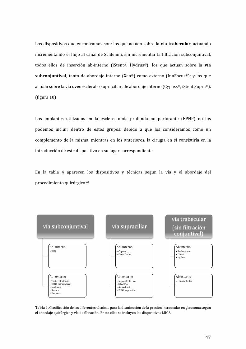
Losdispositivosqueencontramosson: losqueactúansobre lavíatrabecular,actuando
incrementandoel flujoalcanaldeSchlemm,sin incrementar la filtraciónsubconjuntival,
todos ellos de inserción ab-interno (iStent®, Hydrus®); los que actúan sobre la vía
subconjuntival, tanto de abordaje interno (Xen®) como externo (InnFocus®); y los que
actúansobrelavíauveoescleralosupraciliar,deabordajeinterno(Cypass®,iStentSupra®).
(figura10)
Los implantes utilizados en la esclerectomía profunda no perforante (EPNP) no los
podemos incluir dentro de estos grupos, debido a que los consideramos como un
complemento de la misma, mientras en los anteriores, la cirugía en sí consistiría en la
introduccióndeestedispositivoensulugarcorrespondiente.
En la tabla 4 aparecen los dispositivos y técnicas según la vía y el abordaje del
procedimientoquirúrgico.61
Tabla4.Clasificacióndelasdiferentestécnicasparaladisminucióndelapresiónintraocularenglaucomasegúnelabordajequirúrgicoyvíadefiltración.EntreellasseincluyenlosdispositivosMIGS.
víasubconjuntival
Ab- interno• XEN
Ab- externo• Trabeculectomía• EPNPintraescleral• Innfocus• Shunts• Ex-press
víasupraciliar
Ab- interno• Cypass• iStentSubra
Ab- externo• ImplantedeOro• STARFlo• Aquashunt• EPNPsupraciliar
víatrabecular(sinfiltraciónconjuntival)
Ab.interno• Trabectome• iStent• Hydrus
Abexterno• Canaloplastia

48
Figura10.RepresentacióndealgunosdelosdispositivosMIGSdisponibles.a)Xen®(Allergan,Dublin,Irlanda)b)InnFocus®(SantenPharmaCo,Osaka,Japon),c)Cypass®(Alcon-Novartis,Basilea,Suiza),d)iStent®(Glaukos,SanClemente,California,EEUU).
4.2.4Cirugíanoperforanteenglaucoma
Unodelosretosmásimportantesenlacirugíadelglaucomahasidomantenerlaeficacia
tensional mejorando el perfil de seguridad de las mismas, y en esta tesitura es donde
colocamos a la cirugía no perforante, que se caracteriza por no producir una fístula
perforantea lacámaraanterior,preservandoelprivilegio inmunede lacámaraanterior.
DentrodeestegrupoincluimoslaEPNP,lacanaloplastiaylaviscocanalostomía.

49
4.3Esclerectomíaprofundanoperforante(EPNP)
4.3.1Evoluciónhistórica
LosprimerospasoshacialaEPNPtalycomolaconocemosenlaactualidadfuerondadosen
primerlugarporEpstein,KrasnovyZimerman.Afinalesdelosaños50yprincipiodelos
60,Epsteinobservócómoexistíafiltraciónaldisecarprofundamentepterigiums,porloque
desarrollóunaesclerotomíaprofundaperilimbarsobrelos180ºsuperioresquecubría
conconjuntiva.Másadelante,Krasnovdescribióalgosimilarylollamósinusotomía,6263
conbuenosresultadostensionalesacortoplazo.Consistíaendisecarunabandadeesclera
sobre los120ºsuperiores,de10a2h,hastaelcanaldeSchlemm,sinpenetrarlo,yaque
pensabaquelaresistenciaalflujodehumoracuosoestabaaniveldelosvasosepiesclerales
perilimbares.Nofueunatécnicamuyexitosadebidoasudificultadtécnica.Debidoaléxito
delatrabeculectomía,yposteriormentealtenerlanecesidaddedisminuirsualtatasade
complicaciones, fue cuando otros autores volvieron a revisar los trabajos de Krasnov.
Zimmerman, en 1984, describió la trabeculectomía ab externo no perforante, muy
similara la sinusotomía, introduciendoun flapescleraly resecando lapared internadel
canaldeSchlemm.64Mástarde,laescuelarusalideradaporFyodorovyKozlovmejoraron
latécnica,yadenominadaesclerectomíaprofundanoperforante(EPNP).65Adiferenciade
la sinusotomía, la filtración provenía del trabéculum anterior y de la membrana de
Descemet, y no del trabéculum posterior, por lo que era necesario eliminar también el
estromacornealanterior.

50
LaEPNPesunamodificacióndelatrabeculectomíaenlaquenoseperforaporcompletoel
trabéculumanterior,sinoquesedejaeltejidoquecorresponderíaalascapasmásinternas
delmismo,porloquelafiltraciónseproducedesdelacámaraanteriordelojoalespacio
subconjuntival o supraciliar a través de esta membrana, incrementándose en parte la
resistenciaalpasodelhumoracuoso.Debidoaellotieneunatasadecomplicacionesmenor
(menor tasa de hipotonía, atalamia, desprendimiento de coroides o maculopatía
hipotónica).Alnorealizarseiridectomía,comoenelcasodelatrabeculectomía,deberíamos
evitarlaencasosdeglaucomadeángulocerrado,porlaproximidaddelirisalamembrana
trabeculodescemética(MTD).Tambiénsuefectividadseríamenorenaquelloscasosenlos
que sospechemos que se ha producido una fibrosis de la MTD. Por lo tanto, como
indicacionesseincluiránpacientesconglaucomadeánguloabierto,tantoprimariocomo
secundario, miópico y afáquico. En casos de uveítis disminuye el riesgo de inflamación
postoperatoriaypreservaelprivilegioinmunedelacámaraanterior.Alserunatécnicamás
segura,tambiénnospermitirárealizarlaenunmomentomásinicialdelaenfermedad,yno
tenerqueesperarafasestardíasenlasquetengamosmenosmargen.
Unamaniobracrucialparaeléxitodelacirugía,despuésdehaberdisecadoelflapprofundo
escleral(vermaterialymétodosparaladescripcióndelatécnicaquirúrgicacompleta),es
la retiradadeun tejido fibrosoque cubre el área trabecular, si hemos sido lo suficiente
profundos.EstudiosconmicroscopioconfocalrealizadosporHamard,66demostraronque
la membrana pelada correspondía a la pared interna del canal de Schlemm y al tejido
juxtacanalicular(la imagendelparedinternaresecadaquirúrgicamentesemuestraenla
figura11).
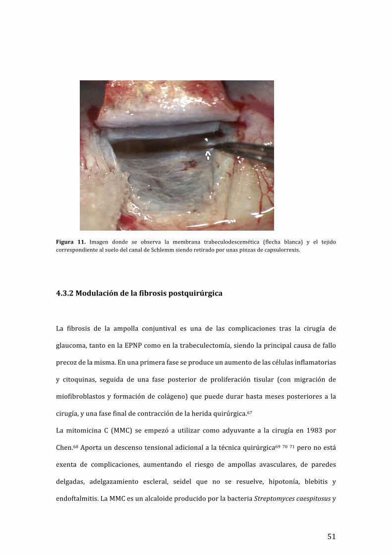
51
Figura 11. Imagen donde se observa la membrana trabeculodescemética (flecha blanca) y el tejidocorrespondientealsuelodelcanaldeSchlemmsiendoretiradoporunaspinzasdecapsulorrexis.
4.3.2Modulacióndelafibrosispostquirúrgica
La fibrosis de la ampolla conjuntival es una de las complicaciones tras la cirugía de
glaucoma,tantoenlaEPNPcomoenlatrabeculectomía,siendolaprincipalcausadefallo
precozdelamisma.Enunaprimerafaseseproduceunaumentodelascélulasinflamatorias
y citoquinas, seguida de una fase posterior de proliferación tisular (con migración de
miofibroblastosy formaciónde colágeno)quepuededurarhastamesesposterioresa la
cirugía,yunafasefinaldecontraccióndelaheridaquirúrgica.67
Lamitomicina C (MMC) se empezó a utilizar como adyuvante a la cirugía en 1983 por
Chen.68Aportaundescensotensionaladicionalalatécnicaquirúrgica697071peronoestá
exenta de complicaciones, aumentando el riesgo de ampollas avasculares, de paredes
delgadas, adelgazamiento escleral, seidel que no se resuelve, hipotonía, blebitis y
endoftalmitis.LaMMCesunalcaloideproducidoporlabacteriaStreptomycescaespitosusy
52
actúainhibiendolasíntesisdeRNA,conefectocitotóxico.Estudiosinvivohandemostrado
lainducciónalaapoptosisdefibroblastostrassuaplicación,72ytambiénsehademostrado
que penetra en las capas profundas de la esclera, y que la penetrancia es directamente
proporcional a la concentración y al tiempo de exposición.73 Paramejorar la técnica de
aplicaciónydisminuirel riesgodeampollasavasculareses recomendableusaresponjas
absorbentesseguidasdeirrigaciónconsuerosalino.Seutilizaenconcentracionesquevan
de0,1mg/mla0,5mg/ml(ennuestrocaso0,2mg/mldurante2minutos).
Tambiénsehausadoel5-fluorouracilo(5FU)comoadyuvante,conunosresultadospeores
alaMMC.74EsunanálogodepirimidinaqueinterfiereconlasíntesisdelARNribosomal,y
actúatambiéninduciendolaapoptosisdelosfibroblastos.75
Traslacirugíayelusodeantimetabolitostambiénpodemosmodularelgradodefibrosis.
Los corticoesteroides tópicos son cruciales para inhibir la proliferación de mediadores
proinflamatorios.Esclavesuusoporunperiododetiempoprolongado,hasta2y4meses
posterioresa lacirugía,yaqueesenesteperiododetiempocuandoseproducirámayor
fibrosisycicatrizaciónpostquirúrgica,quecondicionaráeléxitotensional.
Los fármacos antiangiogénicos, como los anti-VEGF (factor de crecimiento endotelial
vascular)sonútilesparamodularlafibrosisconjuntivalpostoperatoria.Estudiosinvitro
handemostradoreducirlatransformacióndelosmiofibroblastos,presentesenlafasede
cicatrizaciónconjuntival.76Sehaobservadounareduccióndelavascularizaciónconjuntival
6semanastrassuinyecciónenampollasvascularizadas,77peronoquedaclarosisuadicción
juntoaMMCocomoalternativaaéstaproduceunefectobeneficiososobre lacirugíade
glaucoma(yaseatrabeculectomíaoEPNP)osobreelneedling,observándosediscrepancias
entreestudios.78–80
53
4.3.3Goniopuntura
Lagonipuntura(GP)esclaveparamejorareléxitodelaEPNP,yconvierteunacirugíano
perforante en perforante. Amedida que pasan losmeses se suele ver una tendencia de
incrementodelaPIO,nopresenteentodosloscasos,debidoaprocesosfibróticosdelaMTD
obienpordepósitosdepigmentosuotrassustancias,loquecomportaquesevuelvamenos
funcionalydisminuya la filtración.81Consisteenunamicropunciónde laMTDmediante
laserYAG,habitualmente2,enlosdoslateralesdelamembrana,lomásanteriorposible,
utilizandoimpactosúnicosde2-4mJdepotencia.LaGPnolaconsideramoscomofracaso
de lacirugía,yaque formapartedelprocesoquirúrgicoen laEPNP.Encasosen losque
observemosundesplazamientoanteriordelirispodemosasociarlaairidoplastiasectorial,
evitandoasílaincarceracióndeliris.LaGPesefectivacuandoelincrementedelaPIOes
debidoaestapocafuncionalidaddelaMTD,peronocuandoexisteunafibrosisdelaampolla
excesiva,porloqueobservarelaspectodelaampollamediantelámparadehendidurau
OCT-SAseráclaveparadeterminarsuéxito.
4.3.4Needling
El needling consiste en reabrir la fístula quirúrgica, habitualmente con una aguja de 27
gauges,cuandolosbordesdelaesclerectomíasehanfibrosadooenquistado.Sepuedeusar
con laadyuvanciadeMMC,5FUoBevacizumab, siendomáseficaz laprimera.79 82 Seha
demostrado eficaz en el control de la PIO a largo plazo en aquellas ampollas que han
fracasado.83Algunosestudiosloincluyencomofracasodelacirugía,noennuestrocaso.
54
4.3.5Implantesenlaesclerectomíaprofundanoperforante
Fyodorov65yKozlov84fueronlosprimerosenusarimplantesdecolágenoenlacirugíano
perforante. Uno de los posibles problemas en la EPNP es el colapso y fibrosis de las
estructuras de filtraciónmeses después del acto quirúrgico. Para intentar resolver este
problema se diseñaron implantes o espaciadores que se colocarían o bien en el espacio
intraescleralobienenelespaciosupraciliarparaevitarelcolapsoyconsiguientefracasode
esta técnica, con resultadosprometedores.8586 87 108 Inicialmente seoptópormateriales
reabsorbiblescomoelcolágeno,queevitabanelcolapsoenlafaseprecozqueesenlaque
encontramosmayorcicatrización,peroestudioscomparandosueficaciaconlanoadicción
deimplantevieronmayordiferenciadePIOpasadoelañodelacirugía,87porloqueseoptó
por implantes no reabsorbibles biocompatibles.88 Es crucial para la correcta evolución
postoperatoriaqueelmaterialsealomásinerteposibleparalasestructurasoculares,ya
que no incrementará las señales profibróticas que se producen, y que no desprenda
partículasnisedegradeconlosaños.
Los implantes los podemos dividir en reabsorbibles y no reabsorbibles. Entre los
reabsorbibles encontramos elAquaflow® (Staar SurgicalAG,Nidau, Suiza), actualmente
fuerademercado,decolágenoporcinoydeformatubular(2.5mmx1mm),quesesutura
enelespaciointraescleralysedegradaentre6y9mesesposterioresalacirugía.89Esuno
delosqueclásicamentemássehausadoenseriesalargoplazo;909192elSkGel®(Corneal
laboratorios, París, Francia), de ácido hialurónico reticulado modificado mediante
crosslinking,quesecolocatambiénenelespacio intraescleral(loencontramosdeforma
triangularotrapezoidal,3.5x3.5x3.5mmo4.5x3.0x0.5mm);9394yHealaflow®(Anteis,
Geneva,Switzeland),tambiéndehialuronatodesodioqueseintroduceenformadegelen
elespaciointraescleralquedandosolidificadohorasdespués.959697Sepuedecolocartanto
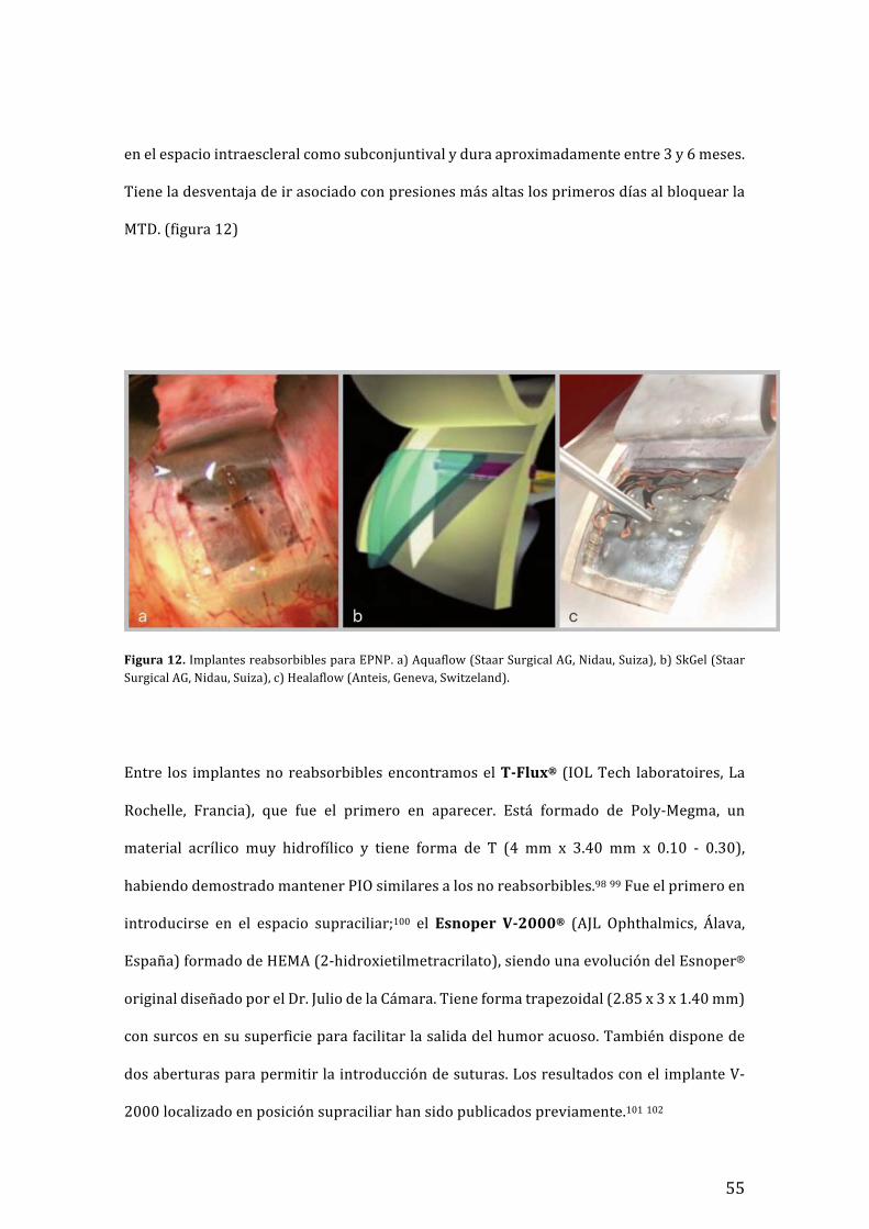
55
enelespaciointraescleralcomosubconjuntivalyduraaproximadamenteentre3y6meses.
Tieneladesventajadeirasociadoconpresionesmásaltaslosprimerosdíasalbloquearla
MTD.(figura12)
Figura12.ImplantesreabsorbiblesparaEPNP.a)Aquaflow(StaarSurgicalAG,Nidau,Suiza),b)SkGel(StaarSurgicalAG,Nidau,Suiza),c)Healaflow(Anteis,Geneva,Switzeland).
Entre los implantesnoreabsorbiblesencontramoselT-Flux® (IOLTech laboratoires,La
Rochelle, Francia), que fue el primero en aparecer. Está formado de Poly-Megma, un
material acrílico muy hidrofílico y tiene forma de T (4 mm x 3.40 mm x 0.10 - 0.30),
habiendodemostradomantenerPIOsimilaresalosnoreabsorbibles.9899Fueelprimeroen
introducirse en el espacio supraciliar;100 el Esnoper V-2000® (AJL Ophthalmics, Álava,
España)formadodeHEMA(2-hidroxietilmetracrilato),siendounaevolucióndelEsnoper®
originaldiseñadoporelDr.JuliodelaCámara.Tieneformatrapezoidal(2.85x3x1.40mm)
consurcosensusuperficieparafacilitarlasalidadelhumoracuoso.Tambiéndisponede
dosaberturasparapermitirlaintroduccióndesuturas.LosresultadosconelimplanteV-
2000localizadoenposiciónsupraciliarhansidopublicadospreviamente.101102

56
Por último, el implante sobre el que trata esta Tesis Doctoral, el Esnoper Clip® (AJL
Ophthalmics,Álava,España),eslaevolucióndelimplanteEsnoperV-2000®.Sudiseñonos
aportará los beneficios de ambas formas de implantación, tanto la supraciliar como la
intraescleral,porloqueconstadeundobleplatoqueplegadoincrementaelflujoenestas
dosvías.ElEsnoperClip®tieneformatrapezoidal,de5,5mmx1,3mmx2,2mmy0,2mm
degrosor,quealquedarplegadoocupaungrosorde0,4mm.Elplatopequeñotienedos
muescaslaterales,loquepermitirádisminuirelmovimientoenelespaciosupraciliar(ynos
indicará que parte se introduce en dicho espacio). Los resultados con este implante se
muestranenestaTesisDoctoral.(figura13)
Figura13.ImplantesnoreabsorbiblesparaEPNP.a)T-Flux®(IOLTechlaboratoires,LaRochelle,Francia),b)EsnoperV-2000®(AJLOphthalmics,Álava,España),c)EsnoperClip®(AJLOphthalmics,Álava,España).

57
4.3.6Estudiospreviospublicados
Existen numerosos estudios previos que tratan sobre la EPNP. Éstos son difíciles de
comparar,debidoaque los criteriosde inclusióny las característicasde lamuestra son
distintas, la duración de los mismos varía, la técnica quirúrgica es algo diferente y los
criteriosdeéxitodifierentambiénentreellos.
Simiramoslaliteraturapublicada,conunabúsquedasimpleenPubmed,encontramosmás
de400estudiosquetratanlaEPNP,conunatendenciacrecienteenlosúltimosaños.(figura
14y15)
figura14y15: capturas de “pubmed”donde semuestra comobuscar la literaturadeEPNPy los estudiospublicadoshastalafecha.
Dadalaabundanciadeliteraturaalrespecto,lasiguientetabla(tabla5)resumelosestudios
publicadoshastalafecha,conuntamañomuestralimportante(n>30),conunaduraciónde

58
medio-largo plazo (mayor o igual a 2 años), descartando todos aquellos que hablen de
glaucomascongénitosyaquellosenlosquenosemuestrenlosvalorestensionalesprey
postoperatorios(yquepuedancentrarseenotrosaspectosquirúrgicos).7886879093–95101103–
118

59

60
Tabla5:resumendeestudiospublicadoshastalafechadeEPNP,conn>30pacientesyseguimientomayora2años. (N= número de pacientes; m= meses; GPAA= glaucoma primario de ángulo abierto; GPX= glaucomapseudoexfoliativo;Gpigm=glaucomapigmentario;GUveit=glaucomauveítico;med=medicación;PIO=presiónintraocular; GP= tasa de goniopuntura; 5-FU= 5-Fluorouracilo; MMC=Mitomicina C; pac= pacientes; PC=progresióndelacatarata;DC=desprendimientodecoroides;mp=tasademicroperforación)

61
62
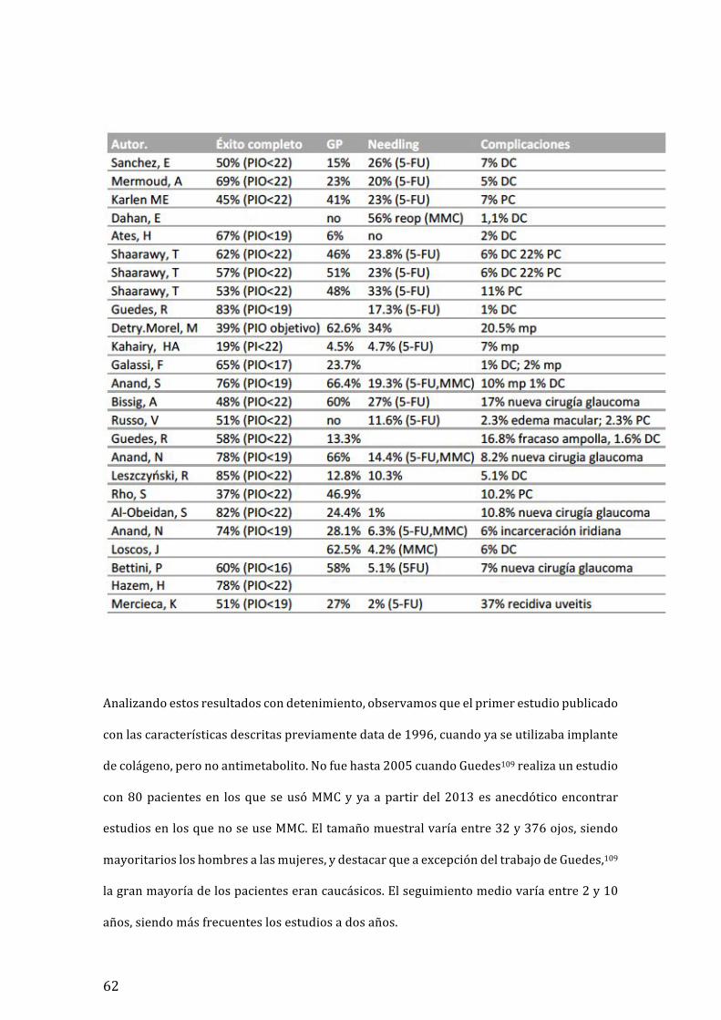
Analizandoestosresultadoscondetenimiento,observamosqueelprimerestudiopublicado
conlascaracterísticasdescritaspreviamentedatade1996,cuandoyaseutilizabaimplante
decolágeno,peronoantimetabolito.Nofuehasta2005cuandoGuedes109realizaunestudio
con80pacientesen losque seusóMMCyyaapartirdel2013esanecdóticoencontrar
estudiosenlosquenoseuseMMC.Eltamañomuestralvaríaentre32y376ojos,siendo
mayoritariosloshombresalasmujeres,ydestacarqueaexcepcióndeltrabajodeGuedes,109
lagranmayoríadelospacienteserancaucásicos.Elseguimientomediovaríaentre2y10
años,siendomásfrecuenteslosestudiosadosaños.

63
Mayoritariamente los pacientes presentaban GPAA, y únicamente un estudio de los
incluidostratamonográficamentedelglaucomauveítico.118Laedadmediavaríaentrelos
43ylos79años.Lapresiónpreviavaríaentre20,6y33,6mmHgyladelúltimocontroltras
la cirugía, entre 8,9 y 18,5 mmHg. Los pacientes con presiones medias más altas eran
uveíticosyenlosquelapresiónmediaenelúltimocontrolfuemayornoseusóniimplante
niantimetabolito.LadisminuciónmediadelaPIOdetodoslosestudiosfuedel39,5%,la
tasadecomplicacionesfuebaja,conentre1y7%dedesprendimientodecoroides,entre2
y22%deprogresióndecataratas,yentre2y20%demicroperforacionesintraoperatorias.

64
LamedicaciónmediaenelúltimocontrolvaríadesdelanoadiciónenelcasodeDahan,104
a2,2medicamentosporpaciente.Latasadegoniopunturasvaríaentrealgúnestudioenel
quenolarealizaban,al66,4%(mediadel37%),yelneedlingdesdenorealizarloal34%
(mediadel16,1%).En lamayoríade loscasosdeneedlingseusó5-FUcomoadyuvante.
Finalmente,latasadeéxitocompleto(queincluyepacientesalosquenoseleshaañadido
medicación)varíaentre19y83%,peroteniendoencuentaqueloscriteriosdeéxitovarían
entreellos,siendomáshabitualpresioneslímitemenoresoigualesa21mmHgque18o15
mmHg.Aquellosestudiosconmenostiempodeseguimientosuelenirasociadosamayor
tasadeéxitocompleto(sinmedicaciónasociada).
4.4Evaluacióneconómica
Elglaucomaeslasegundacausamásprevalentedecegueraenelmundo.Hastalafecha,el
control de la PIO es el factor de riesgo modificable más importante y reduciéndola se
disminuyeelriesgodeprogresión.
Losestudiosdecoste-efectividadsonescasosenlaliteraturamédica.En1988,elGlasgow
trialdeterminóqueelcostedeltratamientomédicoeramayoralatrabeculectomía.119120
En2012,Guedestambiénconcluyóquelacirugíanoperforanteeramásefectivaymenos
costosaqueeltratamientomédico.121
Loscostes lospodemosdividirendirectose indirectos.Entre losdirectos incluiremosel
costede lasvisitasypruebascomplementarias,el transportehasta lasmismas,elde las

65
intervencionesquirúrgicasyeldelamedicación.Entrelosindirectosincluimoslashoras
perdidasporlasvisitas,bajaslaboralespost-cirugíasyproductividad.
Valorando todos los costesposibles, seha llegado a la conclusióndeque el tratamiento
médicoeselquesellevalamayorpartedelosmismos.Rylander122estimóqueelcosteanual
deunaúnicamedicaciónenlosEstadosUnidosdeAméricaen2008variabaentre150$y
873$.SihacemosestemismocálculoenEspaña,esdesuponerqueelcostedelamedicación
seríamenordebidoanuestrosistemadecompra farmacéuticaypolíticadeprecios.Los
pacientes con glaucomamoderado-severo produciránmás costes que los pacientes con
glaucomaprecoz,123ylospacientespococumplidoresproduciránmáscostesquelosmás
cumplidores.Alcostedeltratamientomédicoyelsobre-tratamiento,habráquesumarun
mayor número de visitas y pruebas complementarias ya que su enfermedad tendrá la
tendenciaaprogresarmásrápidamente.Enestoscasosunacirugíaprecozseríamuchomás
coste-efectiva.124
Elanálisiseconómicodenuestrasintervenciones,sobretodoenestaépocadecrisiseterna
enlaqueestamosinmersos,debeserpartedenuestraprácticadiaria,perounbuenanálisis
rigurosoesnecesarioparapoderextraerconclusionesfidedignas.SicomparamoslaEPNP
conlatrabeculectomía,conquiensecomparanlagranmayoríadeestudiosdebidoaseruna
de las cirugías más extendidas y estandarizadas, podemos concluir que
preoperatoriamente, el número de visitas sería similar en ambas técnicas. El coste
farmacológicoseríainferiorenlaEPNP,debidoquealserunatécnicaconmenortasade
complicaciones la haríamos en un estadio más precoz de la enfermedad. Los costes
quirúrgicosseríansimilaresenambastécnicasaexcepcióndelimplantequeseutilizaenla
EPNP,yloscostespostquirúrgicosentendemosquepodríanserinferioresenlaEPNP,ya
queelnúmerodevisitaspostoperatoriassueleserinferioralatrabeculectomía,debidoa
66
unamayor tasa de complicaciones en la segunda, asociado a unmanejo postoperatorio
inmediatoconvisitasmásfrecuentes.Porúltimo,elcostedeltratamientopostoperatorio
entendemosqueseríasimilarenambastécnicas.
4.5Tomografíadecoherenciaópticadesegmentoanterior(OCT-
SA)enelglaucoma
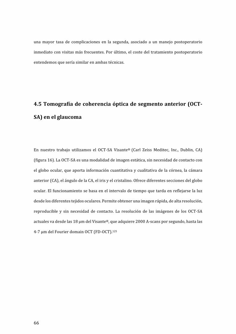
En nuestro trabajo utilizamos el OCT-SA Visante® (Carl Zeiss Meditec, Inc., Dublin, CA)
(figura16).LaOCT-SAesunamodalidaddeimagenestática,sinnecesidaddecontactocon
elgloboocular,queaporta informacióncuantitativaycualitativade lacórnea, lacámara
anterior(CA),elángulodelaCA,elirisyelcristalino.Ofrecediferentesseccionesdelglobo
ocular.Elfuncionamientosebasaenel intervalodetiempoquetardaenreflejarsela luz
desdelosdiferentestejidosoculares.Permiteobtenerunaimagenrápida,dealtaresolución,
reproducible y sin necesidad de contacto. La resolución de las imágenes de los OCT-SA
actualesvadesdelas18µmdelVisante®,queadquiere2000A-scansporsegundo,hastalas
4-7µmdelFourierdomainOCT(FD-OCT).125
67
Figura16.aspectodelOCT-SAVisante®(CarlZeissMeditec,Inc.,Dublin,CA).
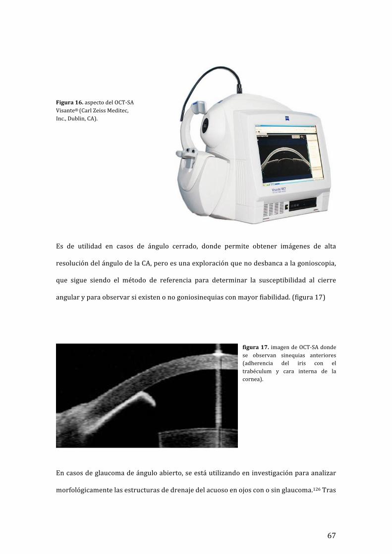
Es de utilidad en casos de ángulo cerrado, donde permite obtener imágenes de alta
resolucióndelángulodelaCA,peroesunaexploraciónquenodesbancaalagonioscopia,
que sigue siendo el método de referencia para determinar la susceptibilidad al cierre
angularyparaobservarsiexistenonogoniosinequiasconmayorfiabilidad.(figura17)
figura17. imagendeOCT-SAdondese observan sinequias anteriores(adherencia del iris con eltrabéculum y cara interna de lacornea).
Encasosdeglaucomadeánguloabierto,seestáutilizandoeninvestigaciónparaanalizar
morfológicamentelasestructurasdedrenajedelacuosoenojosconosinglaucoma.126Tras

68
cirugía de glaucoma, podemos analizar la presencia de ampolla conjuntival, su pared y
altura,lapresenciadeespaciosupraciliar,eltamañodellagointraescleral,yelgrosoryla
longituddelamembranamembranatrabeculodescemética(MTD).LaOCT-SAnospermite
obtenerimágenesdealtacalidaddelasampollasdefiltraciónynosdainformaciónquea
menudo no podemos obtener únicamente con lámpara de hendidura. La imagen de las
ampollas de filtración tras trabeculectomía fue observada en primer lugar en
biomicroscopía ultrasónica (BMU).127 128 Se han descrito cuatro tipos morfológicos de
ampollasdefiltración:difusas,quísticas,encapsuladasyplanas.Másrecientementeseha
realizadolomismoconOCT-SA.129Envariosestudiosseanalizancaracterísticasinternasde
laampolladefiltraciónyseobtieneunmayoréxitotensionalenlasdemayorgrosor,129en
aquellasconparedeshiporreflectantes,yconmayornúmerodequistes.Porelcontrario,en
lasampollasfallidassuelehabercontactoentrelaescleraylaconjuntiva,oaposiciónentre
elcolgajoescleralysulecho.130131
Figura 18. imagenmacroscópica del áreaquirúrgica tras EPNP conEsnoper Clip®,observándoseunaampolladifusa posteiror y unamplio espaciointraescleral.
LaOCT-SAesmuyútilenelpostoperatorioinmediatodelacirugíanoperforantedebidoa
quenorequierecontacto.PermitevalorarlaintegridaddelaMTD,sulongitudysurelación
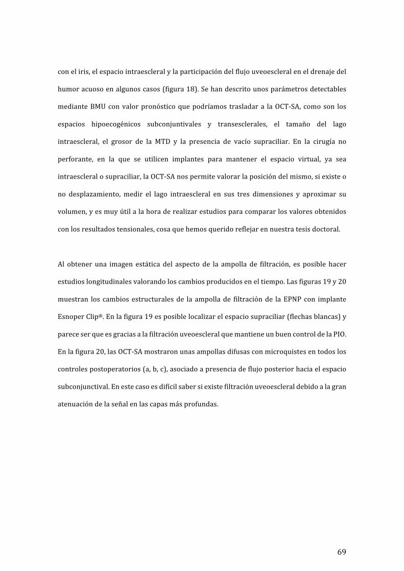
69
coneliris,elespaciointraescleralylaparticipacióndelflujouveoescleraleneldrenajedel
humoracuosoenalgunoscasos(figura18).Sehandescritounosparámetrosdetectables
medianteBMUconvalorpronósticoquepodríamos trasladara laOCT-SA, comoson los
espacios hipoecogénicos subconjuntivales y transesclerales, el tamaño del lago
intraescleral, el grosor de la MTD y la presencia de vacío supraciliar. En la cirugía no
perforante, en la que se utilicen implantes para mantener el espacio virtual, ya sea
intraescleralosupraciliar,laOCT-SAnospermitevalorarlaposicióndelmismo,siexisteo
no desplazamiento, medir el lago intraescleral en sus tres dimensiones y aproximar su
volumen,yesmuyútilalahoraderealizarestudiosparacompararlosvaloresobtenidos
conlosresultadostensionales,cosaquehemosqueridoreflejarennuestratesisdoctoral.
Al obtener una imagen estática del aspecto de la ampolla de filtración, es posible hacer
estudioslongitudinalesvalorandoloscambiosproducidoseneltiempo.Lasfiguras19y20
muestran los cambiosestructuralesde la ampollade filtraciónde laEPNPcon implante
EsnoperClip®.Enlafigura19esposiblelocalizarelespaciosupraciliar(flechasblancas)y
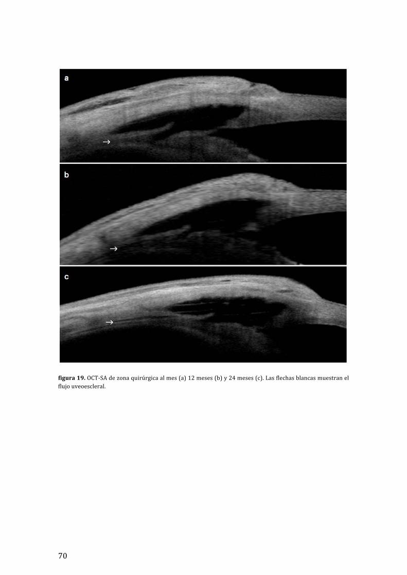
pareceserqueesgraciasalafiltraciónuveoescleralquemantieneunbuencontroldelaPIO.
Enlafigura20,lasOCT-SAmostraronunasampollasdifusasconmicroquistesentodoslos
controlespostoperatorios(a,b,c),asociadoapresenciadeflujoposteriorhaciaelespacio
subconjunctival.Enestecasoesdifícilsabersiexistefiltraciónuveoescleraldebidoalagran
atenuacióndelaseñalenlascapasmásprofundas.
70
figura19.OCT-SAdezonaquirúrgicaalmes(a)12meses(b)y24meses(c).Lasflechasblancasmuestranelflujouveoescleral.

71
Figura20.OCT-SAdezonaquirúrgicaalmes (a)12meses (b)y24meses (c), observándoseuna filtracióntransescleralimportanteentodasellas(asterisco).

72
73
5. Materialymétodos
Realizamos un análisis prospectivo de pacientes con glaucoma de ángulo abierto para
analizarlaseguridadyefectividaddelimplanteEsnoperClip®enlaEPNPaisladaalañoen
unprimerartículo,yposteriormenteEPNPaisladaycombinadaconfacoemulsificacióna
losdosañosjuntoalanálisismorfológicodeláreaquirúrgicaenunsegundoartículo.
5.1Obtencióndelamuestra,criteriosdeinclusiónyexclusión
Los pacientes fueron seleccionados de forma consecutiva. Incluimos pacientes con
glaucomadeánguloabierto,conPIOaltaapesardeutilizarelmáximotratamientomédico
tolerado.Loscriteriosdeexclusiónfueroncualquiercirugíaocularenlos6mesesprevios,
cualquiercirugíadeglaucomaprevia,retinopatíadiabéticamoderadaosevera,cualquier
causa de neovascularización ocular, glaucoma juvenil, afaquia, glaucoma inflamatorio o
traumático.Elestudioestáadheridoa lospostuladosde laDeclaracióndeHelsinkiy fue
aprobado por el comité de ética del Hospital Germans Trias i Pujol. El consentimiento
informado fue discutido y firmado por cada uno de los pacientes. Para determinar que
existíaprogresióndelglaucomayporlotantoqueserequeríaunprocedimientoquirúrgico
para disminuir aún más la PIO, nos basamos en criterios tanto tensionales como de
empeoramientodeldefectomedioenelcampovisual(Humphreyvisual field).Todas las
cirugíasfueronrealizadasporunúnicocirujano(J.L.A)
74
Lascaracterísticasdelamuestraestándescritasenlosresultadosyartículosadjuntos.
5.2Técnicaquirúrgica
Trasunacorrectadesinfeccióndelgloboocularconbetadine®diluidoal50%,colocamos
unasuturadetraccióncornealsuperiordeNylon5/0quenospermitirácentrarelcampo
quirúrgicoeneláreaescleralsuperior.Realizamosunaperitomíaconjuntivalsuperiorde
unas3horasconbasefundus, lomásposteriorposible,evitandodañarelmúsculorecto
superior.Traséstaprocedemosacoagularlazonadellechoescleralmediantediatermia.El
primerflaplodisecamosdeformacuadrada,de5x5mm,ydeunterciodelgrosorescleral,
extendiéndose2mmacórneaclara.Después impregnamosesponjasconmitomicinaCa
unaconcentraciónde0.2mg/ml,ylascolocamosbajoellechodelprimerflapyenelespacio
subconjuntival posterior. Las dejamos 2 minutos y al retirarlas drenamos el área
subconjuntival con solución salina en abundancia. Posteriormente disecamos un flap
profundo,de4x4mm,deotroterciodelgrosorescleral,iniciándoloenlapartemásposterior
yavanzandohaciaeláreatrabecular.Alllegaralespolónescleralylazonacorneal,cortamos
elflapprofundoconunaspinzas“vanas”yconunaspinzasdecapsulorrexisconunapunta
romaretiramoselsuelodelcanaldeSchlemmconlamembranajuxtacanalicularadherida
almismo.Enalgunasocasionesesdifícildiscernirsilatotalidaddelcanalseharetirado,o
biensihemosdisecadolosuficienteprofundo.Conestasmismaspinzasesposibleintentar
extraereltejidofibrosoqueconsideremosnecesarioretirar.Trasesto,desempaquetamos
el implante uveoescleral Esnoper Clip® (AJL Ophtalmic, Álava, Spain) y realizamos una
75
pequeña incisiónescleralhorizontala2mmdelespolónescleral,donde introducimosel
platopequeñodelimplante,quedandoenelespaciosupraciliar.Trascolocarlo,loplegamos
ydejamoselplatograndesobreellechoescleral.Noesnecesariosuturarelimplante,ya
quedisponededosmuescaslateralesqueevitansudesplazamientoanterior.Porúltimo,
aplicamoselflapsuperficialescleralytenemoslaopcióndesuturarloensuparteposterior
con unNylon 10/0. Solo nos quedará suturar la conjuntiva en sus dos pilares laterales
también con Nylon 10/0. En las cirugías combinadas introducimos profilácticamente el
antibióticocefuroximaencámaraanterior,exceptoenloscasosdealergiaalapenicilina,en
losqueutilizamosvancomicina.
En las cirugías combinadas con facoemulsificación, realizamos la cirugía de la catarata
mediantenuestratécnicaestandarizadaenunprimerlugar, introduciendoacetilcolinaal
acabarparaproducirmiosisysuturamoslaincisiónprincipalparamantenerlaestanquidad
de lacámaraanteriordurante todoelprocesode laEPNPyevitarentradaaccidentalde
MMCenlacámaraanterior.
En el siguiente códigoQR es posible visualizar la cirugía segúnnuestra técnica habitual
(figura21).Enlafigura22semuestranlospasosmásimportantesdelacirugía.
Figura 21. Código QR para visualizar la cirugía EPNP con implanteEsnoperClip®talycomoseexponeenestaTesis.
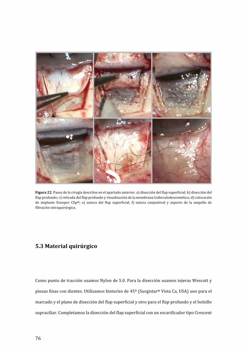
76
Figura22.Pasosdelacirugíadescritosenelapartadoanterior.a)diseccióndelflapsuperficial;b)diseccióndelflapprofundo;c)retiradadelflapprofundoyvisualizacióndelamembranatrabeculodescemética;d)colocaciónde implante Esnoper Clip®; e) sutura del flap superficial; f) sutura conjuntival y aspecto de la ampolla defiltraciónintraquirúrgica.
5.3Materialquirúrgico
ComopuntodetracciónusamosNylonde5.0.ParaladisecciónusamostejerasWescotty
pinzasfinascondientes.Utilizamosbisturíesde45º(Surgistar®VistaCa,USA)unoparael
marcadoyelplanodediseccióndelflapsuperficialyotroparaelflapprofundoyelbolsillo
supraciliar.CompletamosladiseccióndelflapsuperficialconunescarificadortipoCrescent

77
(Crescentknifeangledbevelup,Surgistar®Ca,USA)yparaladiseccióndelflapprofundo
unescarificadordehojamáspequeñay circular (MicrosurgeryknifeAlcon-Grieshaber®,
Switzeland). Para retirar el suelo del canal de Schlemm utilizamos unas pinzas de
capsulorrexis de punta roma y para las suturas, Nylon 10.0, un porta y pinzas finas sin
dientes.Enlascirugíascombinadasconfacoemulsificación,utilizamosviscoelásticoHealon
(Abbottmedicaloptics,AMO,USA),pinzasdecapsulorrexisyequipodefacoemulsificación
Stellaris(Bausch+Lomb,Rochester,NuevaYork,EstadosUnidos).Lafigura23muestrala
organizacióndelcampoestérildelaenfermerainstrumentista.
Figura23.Campoestérilconelmaterialquirúrgicoencirugíadeesclerectomíaprofundanoperforante.

78
5.3.1ImplanteEsnoperClip®
ElEsnoperClip®esunimplanteuveoescleralnoreabsorbibledeHEMA(Hidroxietil
Metacrilato).Tieneformatrapezoidal,de0,2mmdegrosorydimensiones5,50mmX
1,30mmX2,20mm.Elplatopequeñoseintroduceenelespaciosupraciliar,ypara
localizarlodisponededosmuescaslateralesquepermitirándisminuirsumovimientoen
esteespacio.Elplatograndesedejaenelespaciointraescleral.Suestructuranoestá
marcadaconsurcostransversales,adiferenciadelEsnoperV-2000®.(figuras24y25)
Figura 24. Esnoper Clíp®, tanto enforma abierta como semiplegada. Elsegmentoconlasmuescaslateraleseslaque se introduce en el espaciosupraciliar.

79
Figura25.localizacióndelimplanteenellechoescleral.a)deformaabierta,conelsegmentoestrecho
insertadoenelespaciosupraciliar,yb)yaplegado.Lasdosbisagraslateralesquedanencontactoconla
membranatrabeculodescemética.
5.4Materialesexploratoriosypruebascomplementarias
Enlassucesivasvisitasoftalmológicas,cuandofueranecesario,utilizamoslossiguientes
instrumentosoftalmológicos:
- Optotipos de agudeza visual: mejor agudeza visual corregida mediante test de
Snellen.
- Tonometría: tonómetro de aplanación de Goldmann acoplado a una lámpara de
hendidura,conprocesodecalibradoperiódicamentecada6mesesconlabarrade
verificaciónquesesuministracomoaccesorio

80
- Paquimetria ultrasónica: medida de grosor corneal con paquímetro UP-1000®
(NidekCoJapan).
- Campimetría:medianteelcampímetroHumphrey®(CarlZeissMeditec,Inc.,Dublin,
CA).
- Tomografia de coherencia óptica de SA (OCT-SA):mediante Visante® (Carl Zeiss
Meditec,Inc.,Dublin,CA).
- Láser:Elláserloutilizamosparalairidoplastiaylagoniopuntura;Visulas®YAGIII
Combi(CarlZeissMeditec,Inc.,Dublin,CA).
- Lentes:Paralavaloracióndelfondodeojoutilizamoslentede90º(Volk)yparala
goniopunturayvisualizacióndelángulo,laMagnaViewGonioLaserLens(Ocular
Instruments®Bellevue,WAUSA).
5.5Tratamientopostoperatorio
Como tratamiento postoperatorio se usó Ofloxacino (Exocin; Alcon Cusí SA, El Masnou,
Barcelona,España)4vecesaldíadurante1semana,yacetatodeprednisona(10mg/mL)
(Predforte; Allergan Pharmaceuticals Ireland, Westport, Ireland) 6 veces al día y
posteriormenteenpautadescendentedurante6semanas.
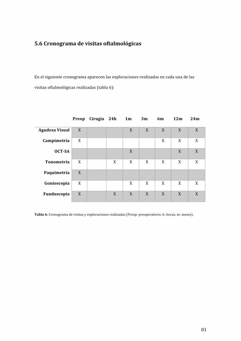
81
5.6Cronogramadevisitasoftalmológicas
Enelsiguientecronogramaaparecenlasexploracionesrealizadasencadaunadelas
visitasoftalmológicasrealizadas(tabla6):
Preop Cirugía 24h 1m 3m 6m 12m 24m
AgudezaVisual X X X X X X
Campimetría X X X X
OCT-SA X X X
Tonometría X X X X X X X
Paquimetría X
Gonioscopia X X X X X X
Funduscopia X X X X X X X
Tabla6:Cronogramadevisitasyexploracionesrealizadas(Preop:preoperatorio;h:horas;m:meses).

82
5.7Análisismorfológico con tomografíade coherenciaópticade
segmentoanterior(OCT-SA)
En el segundo artículo presentado, para analizar los cambios producidos en el área
quirúrgica utilizamos la OCT-SA Visante®. Sus características y sus aplicaciones en
glaucomaestándescritasenlaintroduccióndeestaTesisdoctoral.
ElprotocolodiseñadoparaelanálisisdeláreaquirúrgicafueunaOCT-SAseriadaalmes,a
los12mesesyalos24mesestraslacirugía.Prestamosespecialatenciónalossiguientes
parámetros:
- la existencia o no de espacio intraescleral (comprendido entre el flap escleral
superficialyellechoescleralremanente).Tambiénanalizamoslaalturadelmismo.
- laexistenciaonodeespaciosupraciliar(líneahiporeflectivabajoellechoescleral,
entreesteyelcuerpociliarocoroidesensupartemásposterior).
- eltipodeampollaconjuntival(yaseaquística,difusa,planaoencapsulada;verla
página931delsegundoartículoparaunadescripciónvisual).
- la presencia de microquistes (espacios quísticos hiporeflectivos en el espacio
subconjuntival).
- la reflectividad del espacio supraescleral y subconjuntival (ya sea hiporeflectiva,
intermedioohiperreflectivo).
83
5.8Análisisestadístico
Elanálisisestadísticoserealizóutilizandoel softwareSPSS21.0 (SPSS Inc.,Chicago, IL).
Todos los datos que aparecen en los artículos constan en forma demedia aritmética y
desviación estándar. El test de Saphiro-Wilk se ha usado para analizar si las variables
cuantitativasseguíanunadesviaciónnormal,yenlasqueloseguían,seutilizóelanálisisde
variancia(ANOVA)paradeterminarlasdiferenciasentreestasvariablescuantitativas.En
losotroscasosseutilizólapruebanoparamétricadeFriedmanyeltestdelosrangosde
Wilcoxon. Se determinó una p<0,05 para que los valores fueran estadísticamente
significativos.EltestcorrelacióndePearsonsehausadoparadeterminarlarelaciónentre
PIO y espacio intraescleral, mientras que para la relación entre la PIO y las diferentes
variablescualitativasdelanálisisdelOCT-SAseusarontantoeltestUdeMann-Whitneyy
el de Kruskal-Wallis. La probabilidad de éxito se analizó mediante las curvas de
supervivenciadeKaplan-Meier.

84

85
6. Resultadosyartículospublicados
6.1 Primerartículo
DeepSclerectomyWithaNewNonabsorbableUveoscleralImplant(Esnoper-
Clip®):1-YearOutcomes
Esclerectomíaprofundanoperforanteconunnuevo implanteuveoescleralnoreabsorvible
(Esnoper-Clip®):Resultadosa1año.
PublicadoenJournalofGlaucoma,Volumen24,Número6,agostode2015
ISSN:1057-0829;OnlineISSN:1536-481X;ImpactFactor:2.102
LaEPNPesunprocedimientoparaeltratamientodelglaucomadeánguloabierto.Éstase
puedebeneficiardelusodeMMCintraoperatoriaodeimplantesdiseñadosparaevitarel
colapsodeláreaquirúrgica.Éstossepuedenintroducirtantoenposiciónintraescleralcomo
enposiciónsupraciliar.Losobjetivosdeesteprimertrabajofueronevaluarlaseguridady
efectividadde laEPNPconEsnoperClip®,diseñadoeneldepartamentodeglaucomadel
HospitalGermansTriasiPujol,asícomolosresultadosalprimeraño.
Muestra:
Untotalde27ojosde27pacientesfueronintroducidosdeformaprospectivaenelestudio,
entreoctubrede2011yenerode2013.Entreellosencontramos16pseudofáquicosy11
fáquicos.Lascaracterísticasdemográficassepuedenencontrarenlatabla1delartículo.

86
Resultados
Lapresiónmediapasóde26.6±5.2mmHga15.3±5mmHg(P<0.001)alos12mesesdela
cirugía, lo que representaba una reducción tensional del 42.5%. También hubo una
reducciónsignificativadelnúmerodemedicamentos,pasandode2.5demediaprevioala
cirugíaa0,3demediaalos12meses(P<0.001).
Respectoa laseguridad,nohubocomplicacionesintraoperatorias.Delascomplicaciones
postoperatorias,encontramosSeidelpositivoalas24hen2ojos(7.4%),hiphemaen2ojos
(7.4%),desprendimientodecoroidesen1ojo(3.7%),todosellosresueltos;ynecesidadde
inyecciónadicionaldeMMCen4ojos(14.8%)conunamediade1.5inyeccionesenestos
ojos. No se encontró diferencia estadística en la variación de la agudeza visual mejor
corregida(p=0.507).
Porúltimo,lagoniopunturaconláserYAGlarealizamosen12ojos(44.4%)conunamedia
de4.3mesesentrelacirugíaylamisma.ComocomplicacionesencontramosuncasodeDC
queseresolviósincomplicación.LaPIOpasóde19.2mmHga15.5mmHgdemediatrasla
goniopuntura,siendoestadiferenciaestadísticamentesignificativa.
También analizamos la altura y volumenmedio de la ampolla, siendo la altura de 0.7 ±
0.1mmyelvolumende3.9±1.3mm3,perono realizamoscorrelacionesentreestay las
diferentesvariables.91100102132–140

87
Deep Sclerectomy With a New Nonabsorbable UveoscleralImplant (Esnoper-Clip): 1-Year Outcomes
Jordi Loscos-Arenas, MD, PhD, Angels Parera-Arranz, MD,Pau Romera-Romera, MD, Jordi Castellvi-Manent, DUE,
Antoni Sabala-Llopart, MD, and Julio de la Camara-Hermoso, MD, PhD
Purpose: To report the safety and the effectiveness of deep scler-ectomy (DS) with a new nonabsorbable uveoscleral hema implant(Esnoper-Clip) designed to increase trabecular and uveoscleraloutflow and to achieve higher intrascleral blebs.
Materials and Methods: Twenty-seven eyes of 27 patients withopen-angle glaucoma, who underwent DS with an Esnoper-Clipimplant, were included in this study. All patients were followed upafter 12 months.
Results: A significant decrease in intraocular pressure was observedafter surgery, changing from a preoperative mean of 26.6±5.2mmHg to a postoperative mean of 15.3±5mm Hg (P<0.001) at 12months. There was also a significant reduction in the number ofglaucoma drugs needed, varying from 2.5 per patient to 0.3(P<0.001) 1 year after surgery. The main intrascleral lake heightand volume at 12 months was 0.7±0.1mm and 3.9±1.3mm3,respectively. No intraoperative complications occurred. The mainpostoperative complications were a positive Seidel test result at 24hours in 2 eyes (7.4%), hyphema in 2 eyes (7.4%), and choroidaldetachment in 1 eye (3.7%). All these complications resolved suc-cessfully. The need for additional mitomycin-C injections wasrecorded in 4 eyes (14.8%), twice in 2 of them. Twelve eyes (44.4%)underwent postsurgical Nd:YAG laser goniopuncture with a meantime between surgery and this procedure of 4.3 months. Meanintraocular pressure after Nd:YAG laser goniopuncture decreasedfrom 19.2 to 15.5mm Hg (P<0.001).
Conclusion: DS with an uveoscleral hema implant (Esnoper-Clip) isa safe and effective technique for the management of open-angleglaucoma.
Key Words: deep sclerectomy, open-angle glaucoma, Esnoper-Clip
(J Glaucoma 2015;24:421–425)
Glaucoma is a complex eye disease which is the secondcause of blindness in the world. It consists of the
progressive deterioration of the layer of retinal nerve fibersand of the optic nerve, which lead to the appearance ofdefects in the visual field and of atrophy of the optic nerve.In the majority of cases, this pathology is associated withincreased intraocular pressure (IOP) due to the inhibition ofcorrect drainage of the aqueous humor of the eye. Themanagement of glaucoma requires chronic treatment with aspectrum of therapeutic options including drugs, lasertreatment, incisional filtration surgery, drainage devices,and surgical implants.
Deep sclerectomy (DS) is a nonpenetrating procedurefor the treatment of open-angle glaucoma (OAG). This canbe enhanced with intraoperative mitomycin-C (MMC) orwith the use of implants designed to avoid secondary col-lapse by maintaining the space created after removing thedeep scleral flap. These devices can be implanted intra-sclerally or in the supraciliary space, with a view to facili-tating trabecular and uveoscleral outflow. Its implantationin the scleral bed has been more widely practiced, but thereare also a few reports of the placement of the implant in thesupraciliary space1,2 which indicate that it may be apromising alternative. It is still unknown whether placing
FIGURE 1. The new foldable uveoscleral hema implant (Esnoper-Clip) designed and developed at the Glaucoma Unit of theHospital Universitari Germans Trias i Pujol, Barcelona (UniversitatAutonoma of Barcelona), Spain.
Received for publication July 21, 2014; accepted February 15, 2015.From the Department of Ophthalmology, Hospital Universitari Ger-
mans Trias i Pujol, Servicio de Oftalmologıa, Universitat Auto-noma Barcelona, Badalona, Barcelona, Spain.
AJL Ophthalmics supports the Health Sciences Research Institute“Germans Trias i Pujol” Foundation.
Disclosure: The authors declare no conflict of interestReprints: Jordi Loscos-Arenas, MD, PhD, Department of Oph-
thalmology, Hospital Universitari Germans Trias i Pujol, Serviciode Oftalmologıa, Universitat Autonoma Barcelona, Carretera deCanyet s/n, Badalona 08916, Barcelona, Spain (e-mail: [email protected]).
Copyrightr 2015 Wolters Kluwer Health, Inc. All rights reserved. Thisis an open-access article distributed under the terms of the CreativeCommons Attribution-Non Commercial-No Derivatives License 4.0(CCBY-NC-ND), where it is permissible to download and share thework provided it is properly cited. The work cannot be changed in anyway or used commercially.DOI: 10.1097/IJG.0000000000000253
ORIGINAL STUDY
J Glaucoma ! Volume 24, Number 6, August 2015 www.glaucomajournal.com | 421
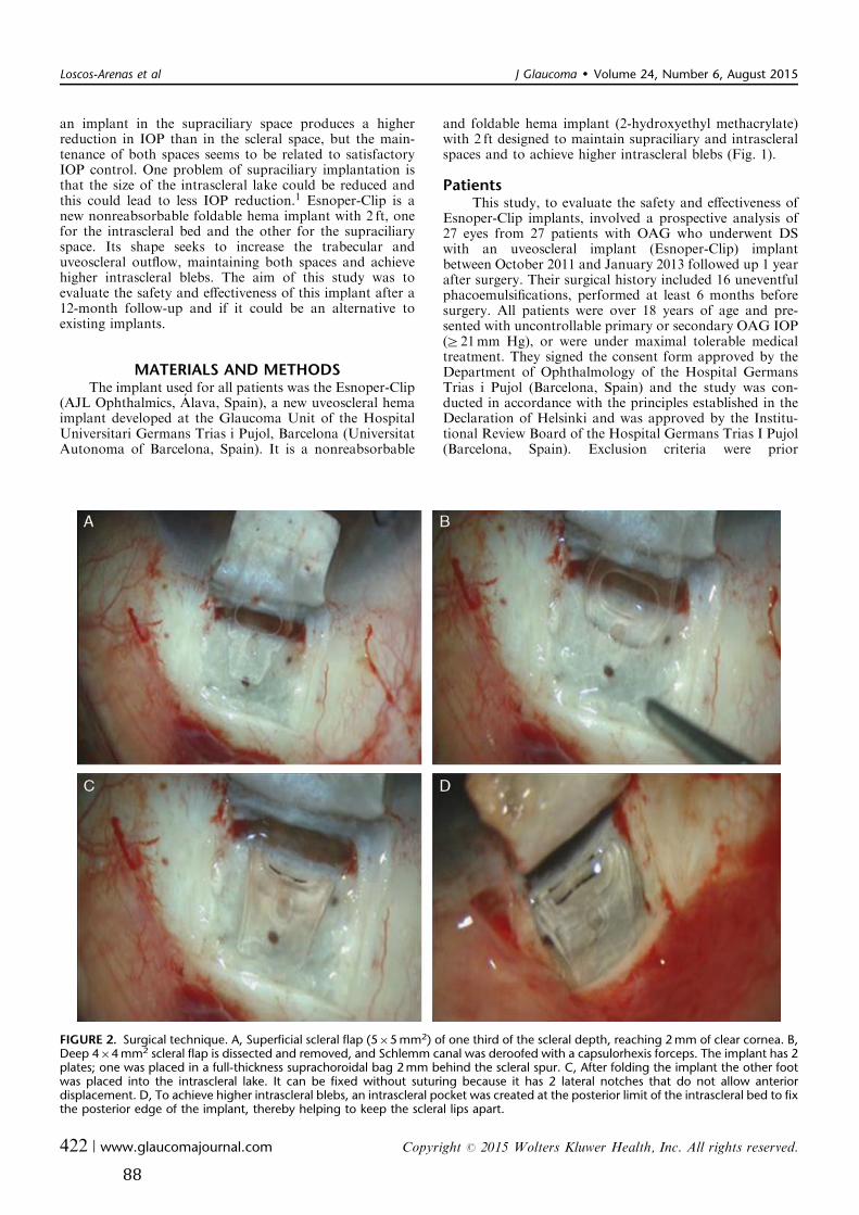
88
an implant in the supraciliary space produces a higherreduction in IOP than in the scleral space, but the main-tenance of both spaces seems to be related to satisfactoryIOP control. One problem of supraciliary implantation isthat the size of the intrascleral lake could be reduced andthis could lead to less IOP reduction.1 Esnoper-Clip is anew nonreabsorbable foldable hema implant with 2 ft, onefor the intrascleral bed and the other for the supraciliaryspace. Its shape seeks to increase the trabecular anduveoscleral outflow, maintaining both spaces and achievehigher intrascleral blebs. The aim of this study was toevaluate the safety and effectiveness of this implant after a12-month follow-up and if it could be an alternative toexisting implants.
MATERIALS AND METHODSThe implant used for all patients was the Esnoper-Clip
(AJL Ophthalmics, Alava, Spain), a new uveoscleral hemaimplant developed at the Glaucoma Unit of the HospitalUniversitari Germans Trias i Pujol, Barcelona (UniversitatAutonoma of Barcelona, Spain). It is a nonreabsorbable
and foldable hema implant (2-hydroxyethyl methacrylate)with 2 ft designed to maintain supraciliary and intrascleralspaces and to achieve higher intrascleral blebs (Fig. 1).
PatientsThis study, to evaluate the safety and effectiveness of
Esnoper-Clip implants, involved a prospective analysis of27 eyes from 27 patients with OAG who underwent DSwith an uveoscleral implant (Esnoper-Clip) implantbetween October 2011 and January 2013 followed up 1 yearafter surgery. Their surgical history included 16 uneventfulphacoemulsifications, performed at least 6 months beforesurgery. All patients were over 18 years of age and pre-sented with uncontrollable primary or secondary OAG IOP(Z21mm Hg), or were under maximal tolerable medicaltreatment. They signed the consent form approved by theDepartment of Ophthalmology of the Hospital GermansTrias i Pujol (Barcelona, Spain) and the study was con-ducted in accordance with the principles established in theDeclaration of Helsinki and was approved by the Institu-tional Review Board of the Hospital Germans Trias I Pujol(Barcelona, Spain). Exclusion criteria were prior
FIGURE 2. Surgical technique. A, Superficial scleral flap (5!5 mm2) of one third of the scleral depth, reaching 2 mm of clear cornea. B,Deep 4!4 mm2 scleral flap is dissected and removed, and Schlemm canal was deroofed with a capsulorhexis forceps. The implant has 2plates; one was placed in a full-thickness suprachoroidal bag 2 mm behind the scleral spur. C, After folding the implant the other footwas placed into the intrascleral lake. It can be fixed without suturing because it has 2 lateral notches that do not allow anteriordisplacement. D, To achieve higher intrascleral blebs, an intrascleral pocket was created at the posterior limit of the intrascleral bed to fixthe posterior edge of the implant, thereby helping to keep the scleral lips apart.
Loscos-Arenas et al J Glaucoma " Volume 24, Number 6, August 2015
422 | www.glaucomajournal.com Copyright r 2015 Wolters Kluwer Health, Inc. All rights reserved.

89
antiglaucoma surgery, or any other surgery which couldhave affected the conjunctiva in the region of the inter-vention, including cataract surgery 6 months before thepresent intervention, moderate or severe diabetic retinop-athy, and other causes of ocular neovascularization. Wealso excluded glaucoma patients with a high risk of failure,that is those presenting with neovascularization, aphakia,inflammation, juvenile cases, and cases of posttrauma andpostsurgery glaucoma.
In the preoperative assessment, patients underwent acomplete examination including best-corrected visual acuity(BCVA) on a decimal scale, biomicroscopy, Goldmanntonometry, pachymetry (UP-1000; Nidek, Japan) gonio-scopy, fundoscopy, and the glaucoma therapy was recorded.
Follow-up visits were scheduled at 24 hours, 1, 3, 6,and 12 months after surgery, and included BCVA, slit-lampexamination, tonometry, gonioscopy, and fundoscopy.Anterior segment optical coherence tomography (AS-OCT)(Visante OCT; Carl Zeiss Meditec, Jena, Germany) whichprovides an objective method to assess the anterior segmentof the eye and the surgical area, was performed at months 1,3, 6, and 12, to the intrascleral bleb height at 90 and 180degrees and the highest was chosen. To determinate thevolume of the intrascleral lake, the semiellipsoid has beenconsidered to be the most similar geometric shape. Wecalculated its value from the height (H), the vertical dia-meter or length (L), and the horizontal diameter or width(W) using the formula Volume=1/6!p!H!L!W. AS-OCT measurements and analysis were performed by thesame investigator (J.C.M.).
Surgical TechniqueAll surgical operations were performed by the same
surgeon (J.L.A.). A superior 4/0 nylon traction suture waspassed through the superior cornea and a fornix-basedconjunctival flap was dissected (Fig. 2A), followed bycauterization of bleeding vessels and dissection of thesuperficial scleral flap (5!5mm2) of one third of the scleraldepth, extending 2mm into clear cornea. Afterwards, aMMC impregnated sheet was laid between the scleral flapand the remaining sclera as well as over the flap, and leftthere for 2 minutes and then irrigated thoroughly withbalanced salt solution. Subsequently, a deeper 4!4mm2
scleral flap was dissected and removed, and Schlemm canalwas deroofed with a capsulorhexis forceps. The implant has2 plates; 1 was placed in a full-thickness suprachoroidal bag2mm behind the scleral spur, after the technique firstlydescribed by Munoz1 (Fig. 2B). After folding the implantthe other foot was placed into the intrascleral lake(Fig. 2C). It can be fixed without suturing because it has 2lateral notches that do not allow anterior displacement. Toachieve higher intrascleral blebs, an intrascleral pocket wascreated at the posterior limit of the intrascleral bed to fixthe posterior edge of the implant, thereby helping to keepthe scleral lips apart (Fig. 2D). No suture or a loose one,was used for the superficial scleral flap and the conjunctivawas sutured with a nylon 10/0. Postoperative treatmentconsisted of topical ofloxacin (Exocin; Alcon Cusı SA, ElMasnou, Barcelona, Spain) 4 times a day during 1 week andprednisolone acetate (10mg/mL) (Predforte; AllerganPharmaceuticals Ireland, Westport, Ireland) 6 times a day,the latter in a descending dosage over 6 weeks. Gonio-puncture with the YAG laser (Visulas YAG III Combi;Carl Zeiss Meditec, Dublin, CA) was performed whenpercolation of aqueous humor was considered insufficient.
Statistical AnalysisStatistical analyses were performed using SPSS soft-
ware for Windows (version 15.0; SPSS Inc., Chicago, IL).All data were reported as means with SD. The differences incontinuous variables before and after surgery were com-pared statistically using the nonparametric Friedman test.Differences in presurgery IOP values and those measured ateach follow-up were analyzed using the Wilcoxon matched-pairs signed-rank test. Probability values <0.05 were con-sidered to be statistically significant.
RESULTSDemographics and characteristics of patients included
in this study are shown in Table 1. Mean preoperative IOPwas 26.6±5.2mm Hg. Twelve months later, we found astatistically significant reduction in IOP which had reached amean of 15.3±5mm Hg (P<0.001). This change repre-sents a 42.5% reduction in IOP reduction (Table 2). Therewas also a statistically significant reduction in the number ofmedications after surgery, starting with a mean of 2.5 andarriving at 0.3 at the end of the study (P<0.001) (Fig. 3).The most common type of glaucoma that was included in thestudy was primary open-angle glaucoma (88.9% of patients).The mean volume and height of the intrascleral bleb 12months after surgery were 3.9±1.3mm3 and 0.7±0.1mm,respectively (Fig. 3).
No intraoperative complications occurred. The mainpostoperative complications were Seidel at 24 hours in 2eyes (7.4%), hyphema in 2 eyes (7.4%), choroidal detach-ment in 1 eye (3.7%), all of which resolved successfully andthe need for additional MMC injections in 4 eyes (14.8%)(in 2 eyes, on 2 occasions). No statistically significant var-iation was found between preoperative and postoperativeBCVA (P=0.507). Twelve eyes (44.4%) underwent post-surgical Nd:YAG goniopuncture (Nd:YAG GP) with amean time between surgery and this procedure of 4.3
TABLE 1. Clinical and Demographic Characteristics of PatientsIncluded in the Study
Age (y±SD) 71.7±7.1Sex [n (%)]Male 11 (40.74)Female 16 (59.26)
Eyes [n (%)]Right 15 (55.56)Left 12 (44.44)
Phakic [n (%)] 11 (40.76)Pseudophakic [n (%)] 16 (59.24)Type of glaucoma [n (%)]NTG 1 (3.70)POAG 24 (88.89)PSXG 2 (7.41)
Pachimetry (mm±SD) 533.2±32.8BCVA (decimal±SD)Preoperative 0.67±0.25Postoperative 0.78±0.19
Preoperative IOP (mm Hg±SD)Preoperative 26.6±5.2Postoperative 15.3±5.0
Medications (n±SD)Preoperative 2.52±0.89Postoperative 0.26±0.69
BCVA indicates best-corrected visual acuity; IOP, intraocular pressure;NTG, normal tension glaucoma; POAG, primary open-angle glaucoma;PSXG, pseudoexfoliative glaucoma.
J Glaucoma " Volume 24, Number 6, August 2015 Uveoscleral Implant (Esnoper Clip) in Deep Sclerectomy
Copyright r 2015 Wolters Kluwer Health, Inc. All rights reserved. www.glaucomajournal.com | 423

90
months. Complications related to goniopuncture werelimited to choroidal detachment in 1 eye that resolveduneventfully. In this case, IOP dropped from 20 to 2mmHg. The mean IOP after Nd:YAG GP decreased from 19.2to 15.5mm Hg (P<0.001).
DISCUSSIONDS with a supraciliary implant is an alternative that
has been shown to be safe and effective with different typesof implants.1,2 The main problem with this class of implantsis that they are not specifically designed for this space, andso their effectiveness can be limited. The intrascleral lakecan lose height or even collapse over time and probably loseefficacy, because intrascleral bleb height plays an importantrole in lowering IOP.3–5 In contrast, intrascleral implantsdo not facilitate uveoscleral outflow as well as supraciliaryimplants.
Reports of supraciliary implantation are somewhatscarce and until now no studies with ultrasound bio-microscopy (UBM) or AS-OCT have been published.Munoz1 reported isolated DS and Bonilla et al2 describedphaco-DS resulting in IOP reductions from 26.4±6.9 to14±3.3mm Hg and 23±5 to 18±3mm Hg, respectively,after 1 year. In these series, the number of medications wasreduced from 2.8 to 0.3 and from 2.5 to 0.7, respectively. Inour series, IOP decreased from 26.6 to 15.3mm Hg and thenumber of drugs to be taken was reduced from 2.5 to 0.26at 12 months. The incidence of complications is similar tothat associated with intrascleral or supraciliary implanta-tion described by other authors. We can conclude that thepresent technique has no more additional complications forDS than other alternatives.
The importance and significance of the intrascleral andsuprachoroidal spaces is a controversial issue, but currentlyit is widely accepted that both are good prognostic factors,but not the ones. This controversy may be due to discrep-ancies in findings using AS-OCT or UBM. UBM meas-urements have limited resolution and with AS-OCT, it isvery difficult to determine the presence or absence ofuveoscleral outflow.
Mavrakanas et al3 and Fernandez-Buenaga et al4 usingAS-OCT, Perez-Rico without impant and Marchini et al5
using UBM reported a positive inverse correlation betweenpostoperative IOP and intrascleral bleb height. Mavrakanaset al3 only investigated eyes having DS with a collagen implantand postoperatively clinically flat blebs, whereas Fernandez-Buenaga et al4 investigated a heterogeneous group of eyes afterDS using 3 different implants (Aquaflow, Esnoper, SK-Gel).Both authors found bleb heights between 0.58 and 0.65mm,respectively, and found a significant correlation between heightand IOP with Esnoper and Aquaflow implants. Here, we havenot studied the correlation between IOP and intrascleral vol-ume or height, but we did calculate the mean median blebheight (0.78±0.1mm) and volume (3.9±1.3mm3) 12months after operation. These values in volume (P=0.706) orheight (P=0.586) were not significantly different to thosebefore operation.
Another point to be considered is that the shape of ourimplant tries to keep the sclera lips apart, thereby facili-tating transcleral outflow (Fig. 4). This could decrease theheight and volume of the intrascleral lake but in contrast,transcleral outflow represents a link between the intrasclerallake and the conjunctival filtering blebs and it had beenfound withUBM6 to be a positive prognostic factor. Perez-Rico et al5 using AS-OCT reported transcleral outflow tobe a positive factor in 89.4% of eyes, 5 years after DS
TABLE 2. Intraocular Pressure Evolution and Numbers of Medications
Preoperative 24H 1Mo 3Mo 6Mo 12Mo
Intraocular pressure (mm Hg±SD) 26.6±5.2 7.3±8.1 14.2±6.5 12.7±4.5 15.3±4.6 15.3±5.0No. drugs (n±SD) 2.52±0.89 0±0 0±0 0±0 0.16±0.62 0.26±0.69Goniopuncture (%) — 0 3.7 18.5 25.9 44.4
FIGURE 3. A, Mean changes in intraocular pressure (IOP) and number of medications (treatment). B, Mean changes in volume andheight of the intrascleral lake.
Loscos-Arenas et al J Glaucoma ! Volume 24, Number 6, August 2015
424 | www.glaucomajournal.com Copyright r 2015 Wolters Kluwer Health, Inc. All rights reserved.

91
without implant. This finding brings into question themandatory use of implants in DS. The same authorreported the association between lower IOP and maximumanteroposterior length, height, and intrascleral volume.
It had been published, in studies with UBM, as thetissue heals with scarring occupying the intrascleral lake6
over time in almost 10% of eyes with7 or without implants.8
Using AS-OCT in eyes which have undergone DS withoutimplant, the presence of the intrascleral lake had beenrevealed in 100% of the eyes after 5 years.5 Discrepancies inthese results are probably related to the trabeculo-Descemetmembrane (TDM) thickness and to the fact that if there isinsufficient peeling of the inner wall of Schlemm canal,enough outflow will not be achieved to maintain theintrascleral lake.
Uveoscleral outflow in different series has a prevalencefrom 7% to 60%.9,10 However, these series are hardlycomparable because they used different implants withvarying follow-up times. Using UBM7,11 it has been foundthat eyes with suprachoroidal filtration have lower IOPthan those without suprachoroidal filtration, whereas otherauthors8 could find no evidence of suprachoroidal filtrationin any case, 1 year after surgery. Ravinet et al12 also founda similar rate of IOP and postoperative treatments in eyeswith or without hypoechoic areas after DS with T-Flux andHealon GV. Supraciliary implantation probably favorseither a ciliary body detachment with subsequent decreasein aqueous production, or choroidal resorption leading tolow postoperative IOP. Although it seems logical to thinkthat this could encourage late chronic ocular hypotony,there is no evidence of this in the literature or in the presentseries. Nevertheless, it should not be forgotten that youngmyopics are probably more likely to suffer from hypotonywith supraciliary implantation.
The reported incidence of goniopuncture varies dras-tically from 4.7% to 63%. This wide variation is likely dueto the application of different criteria, implants, and IOP
targets. The type of implant is likely a very significant factorin Nd:YAG GP as some implants may enhance more fib-rosis over the TDM, or displacement above the TDM. Thisnew device which we report here has 2 lateral notches thatprevent this from happening, and even in cases in which itdoes not remain fixed in the suprachoroidal bag, its shapedoes not collapse the TDM which remains always freewithout any filtration limitations.
On the basis of the present findings, the uveoscleralimplant (Esnoper-Clip) can be considered to be a promisingalternative because it ensures the maintenance of bothspaces helping to avoid collapse over time. Further clinicaltrials with long-term follow-up and a larger numbers ofpatients will be needed to assess the effectiveness of this newuveoscleral implant, the morphology of the surgical area,the real relevance of the intrascleral lake characteristics,and their correlation with reduced IOP.
REFERENCES
1. Munoz G. Nonstitch suprachoroidal technique for T-fluxs
implantation in deep sclerectomy. J Glaucoma. 2009;18:262–264.2. Bonilla R, Loscos J, Valldeperas X, et al. Supraciliary hema
implant in combined deep sclerectomy and phacoemulsifica-tion: one year results. Open Ophthalmol J. 2012;6:59–62.
3. Mavrakanas N, Mendrinos E, Shaarawy T. Postoperative IOPis related to intrascleral bleb height in eyes with clinically flatblebs following deep sclerectomy with collagen implant andmitomycin. Br J Ophthalmol. 2010;94:410–413.
4. Fernandez-Buenaga R, Rebolleda G, Casas-Llera P, et al. Acomparison of intrascleral bleb height by anterior segmentOCT using three different implants in deep sclerectomy. Eye(Lond). 2012;26:552–556.
5. Perez-Rico C, Gutierrez-Ortız C, Moreno-Salgueiro A, et al.Visante anterior segment optical coherence tomographyanalysis of morphologic changes after deep sclerectomy withintraoperative mitomycin-C and no implant use. J Glaucoma.2014;23:e86–e90.
6. Marchini G, Marraffa M, Brunelli C, et al. Ultrasoundbiomicroscopy and intraocular-pressure-lowering mechanismsof deep sclerectomy with reticulated hyaluronic acid implant.J Cataract Refract Surg. 2001;27:507–517.
7. Kazakova D, Roters S, Schnyder CC, et al. Ultrasoundbiomicroscopy images: long-term results after deep sclerectomywith collagen implant. Graefes Arch Clin Exp Ophthalmol.2002;240:918–923.
8. Khairy HA, Atta HR, Green FD, et al. Ultrasound biomicro-scopy in deep sclerectomy. Eye (Lond). 2005;19:555–560.
9. Chiou AG, Mermoud A, Underdahl JP, et al. An ultrasoundbiomicroscopic study of eyes after deep sclerectomy withcollagen implant. Ophthalmology. 1998;105:746–750.
10. Contreras I, Noval S, Munoz-Negrete FJ, et al. Ultrasoundbiomicroscopy in deep sclerectomy with a new acrylic implant.Arch Soc Esp Oftalmol. 2006;81:445–450.
11. Cabrejas L, Rebolleda G, Munoz-Negrete FJ, et al. Anultrasound biomicroscopy study of filtering blebs after deepsclerectomy with a new acrylic implant. Eur J Ophthalmol.2010;22:391–399.
12. Ravinet E, Bovey E, Mermoud A. T-Fluxs implant versusHealons GV in deep sclerectomy. J Glaucoma. 2004;13:46–50.
FIGURE 4. Anterior segment optical coherence tomography. Theintrascleral lake and supraciliary space after the implantation withtranscleral outflow (arrows) can be observed.
J Glaucoma ! Volume 24, Number 6, August 2015 Uveoscleral Implant (Esnoper Clip) in Deep Sclerectomy
Copyright r 2015 Wolters Kluwer Health, Inc. All rights reserved. www.glaucomajournal.com | 425

92
93
6.2 Segundoartículo
Two-year Results After Deep SclerectomyWith Nonabsorbable Uveoscleral
Implant (Esnoper-Clip®): Surgical Area Analysis Using Anterior Segment
OpticalCoherenceTomography
Resultados a dos años tras EPNP con implante uveoescleral y análisis del área quirúrgica
usandoTomografiadecoherenciaópticadesegmentoanterior
PublicadoenelJournalofGlaucoma,volumen26,Número10,octubre2017
ISSN:1057-0829;OnlineISSN:1536-481X;ImpactFactor:2.102
Traselprimerartículoenelquevaloramoslosresultadostensionalesalañodelacirugía,
viendo la seguridad y efectividad de esta nueva técnica, en este segundo artículo nos
centramos en los resultados a los dos años con un mayor número de pacientes, y
adjuntamosunanálisiscuantitativoycualitativodelaampollaconjuntivalmedianteOCT-
SA.
Muestra
Incluimos41ojosde41pacientes,todoselloscaucásicos,seguidosduranteunmínimode
12 meses y en 36 ojos durante 24 meses (87.8%). Entre ellos, 31 eran fáquicos y 10
pseudofáquicos;yserealizócirugíacombinadaconfacoemulsificaciónen16casos.Otros
datosdemográficossepuedenencontrarenlatabla1delartículo.

94
Resultados
La PIO pasó de 27.3 ± 6.3 mmHg en el preoperatorio con 2.54 ± 0.78 medicamentos
antihipertensivosdemedia,a15.3±5.2mmHgalos24mesesdelacirugía,con0.36±0.76
fármacos de media (P<0.0001 en ambos casos). No encontramos diferencias entre las
cirugías de EPNP y las combinadas con facoemulsificación excepto a las 24 horas
postquirúrgicas(P=0.004).Eléxitoabsolutoalos24mesesfuedel61.1%,definidocomo
pacientes con PIO inferiores o iguales a 18 mmHg sin adición de medicación
antiglacuomatosa. La AV mejor corregida no cambió significativamente en la EPNP,
mientrasqueenlascirugíascombinadasconcataratasmejoró,pasandode0,53a0,75(P=
0,034) (medido mediante optotipo de Snellen). De complicaciones postoperatorias
encontramosSeidelen4ojos(10.3%),hiphemaen3ojos(7.7%),yDCen1ojo(2.6%),todos
ellosresueltosconéxitoalaspocassemanasdelacirugía.
En el análisis conOCT-SA realizado almes, a los 12 y 24meses, observamos el espacio
intraescleralentodosloscasos.Lamediadelaalturadelespaciointraescleralfuede0.77±
0.19mmalmes,0.78±0.22mmalos12y0.78±0.22mmalos24meses,nosiendouna
diferencia significativa estadísticamente. Observamos una correlación positiva,
estadísticamentesignificativa,entrelaalturadelespaciointraescleralylaPIOalmesyalos
12meses(r=+0.496;P=0.006yr=+0.410;P=0.026,respectivamente),loquesignifica
queamayorPIO,mayorespaciointraescleral.Otrosparámetrosanalizadosaparecenen
latabla3delartículo.Encontramosunaasociaciónestadísticaentrelanouniformidaddela
señalaniveldelaampollaconunamenorPIO(almesyalos24mesescorrelacionándolo
con la PIO de los 24 meses) y una asociación entre la presencia de microquistes
conjuntivalesconunamenorPIO(alos12y24mesescorrelacionándoloconlaPIOdelos
24meses).88100103108111114129132–134141–159
95
Two-year Results After Deep Sclerectomy WithNonabsorbable Uveoscleral Implant (Esnoper-Clip): SurgicalArea Analysis Using Anterior Segment Optical Coherence
TomographyPau Romera-Romero, MD, Jordi Loscos-Arenas, MD, PhD, Aina Moll-Udina, MD,
Nevena Romanic-Bubalo, MD, Jordi Castellvi-Manent, OD,and Xavier Valldeperas, MD, PhD, FEBO
Purpose: To report 2-year results of deep sclerectomy (DS) withmitomycin C and the uveoscleral implant Esnoper-Clip (AJLOphthalmics, Álava, Spain), a nonabsorbable hema implant, andthe morphologic analysis of the surgical area with anterior segmentoptical coherence tomography (AS-OCT).
Materials and Methods: In total, 41 eyes of 41 patients with medi-cally uncontrolled open angle glaucoma who underwent DS withthe uveoscleral implant were included in this prospective interven-tional study. Patients were recruited and selected sequentially. Allpatients were followed at least for 12 months and 36 of them for24 months. Absolute success was defined as intraocular pressure(IOP)≤ 18mmHg and ≥ 20% of IOP reduction without topicalmedication. Relative success was defined with the same criteria butwith the addition of any antihypertensive medication. Primaryoutcomes were IOP decrease in the postoperative stage. Secondaryoutcomes were number of medication, best-corrected visual acuitychanges, rate of goniopuncture, needle revision, and AS-OCTanalysis of the bleb area. AS-OCT analysis, using Visante OCT, wasperformed at 1, 12, and 24 months.
Results: Absolute success rate was 68.3% at 12 months and 61.1% at2 years. Relative success was 78.0% and 71.4%, at 12 and24 months, respectively. A significant IOP decrease was observed,from 27.3± 6.3 to 14.9± 4.4 mmHg at 12 months and15.3± 5.2 mmHg at 24 months (P< 0.001). There was also a sig-nificant reduction in the number of medications, dropping from amean of 2.5 to 0.28 one year and 0.36 two years after the surgery(P< 0.001). Main postoperative complications were transient: 4 eyeswith seidel phenomenon at 24 hours (10.3%), 3 hyphema (7.7%),and 1 choroidal detachment (2.6%). Mean intrascleral space height,measured by AS-OCT, was 0.78mm and it showed a significantpositive correlation with IOP at 12 months. Neodymium-dopedyttrium aluminium garnet laser goniopuncture was performed in25 eyes (61%) at 12 months and in 27 (66.8%) by 24 months.
Conclusions: DS with the uveoscleral implant is a safe and effectiveprocedure to lower IOP in open angle glaucoma patients. IOPreduction is maintained over 2 years and is correlated to the post-operative height of the intrascleral bleb. Supraciliary implantationreduces intrascleral lake dependency, being a simultaneous drainagealternative to the subconjunctival pathway.
Key Words: ophthalmology, glaucoma, deep sclerectomy, uveoscl-eral implant, nonpenetrating glaucoma surgery, anterior segmentoptical coherence tomography, AS-OCT
(J Glaucoma 2017;26:929–935)
Open angle glaucoma (OAG) is a progressive opticneuropathy that leads to blindness in its final stage.
Lowering intraocular pressure (IOP) is the only provenintervention to arrest the disease progression.1 A surgicalapproach is indicated when medical or laser therapy isunable to sufficiently decrease the rate of visual field loss inthese patients. Trabeculectomy is the most commonly per-formed surgical procedure although deep sclerectomy (DS)is a safe and effective option.2–14 The use of spacer implants,to avoid future collapse of the intrascleral space, mayimprove the long-term success of DS.2,6–9 Some authorshave reported better outcomes placing these devices in theintrascleral space8,11 and some others in the supraciliary13,14space. Other authors, though, could not find any benefitafter using these devices.8,12
Esnoper-Clip (AJL Ophthalmics. Álava, Spain) (EC) isa nonabsorbable foldable hema (2-hydroxyethyl meth-acrylate) uveoscleral implant developed by our group at theGlaucoma Unit of the Hospital Universitari Germans Triasi Pujol (Universitat Autònoma de Barcelona. Barcelona,Spain). AJL Ophthalmic (Alava, Spain) supports the Insti-tute for Health Science Research Germans Trias i Pujol(IGTP). It has been designed with 2 footplates (Fig. 1): oneplaced in the intrascleral space and the other in the supra-ciliary space. The shape of the device has been designedto maintain the patency of both spaces and the aqueoushumor outflow in the long postoperative period. We havepreviously published the first-year results after DS withEC.15 In this study we report the 2-year results of thistechnique and the morphologic evaluation of the blebarea using anterior segment optical coherence tomography(AS-OCT).15–30
MATERIALS AND METHODSPatients with OAG, with uncontrolled IOP after
standardized maximum antihypertensive medications wereprospectively enrolled in this study. Patients were selectedsequentially. Exclusion criteria were any previous glaucomasurgery, cataract surgery in the previous 6 months beforeinclusion, moderate or severe diabetic retinopathy, any
Received for publication April 19, 2017; accepted July 22, 2017.From the Ophthalmology department, carretera de Canyet, S/N, 08916
Badalona, Barcelona, Spain.Disclosure: The authors declare no conflict of interest.Reprints: Pau Romera-Romero, MD, “Carrer Pompeu Fabra num 10, 2° 7ª,
Cornellà de Llobregat, 08940, Barcelona, Spain” for “Ophthalmologydepartment, carretera de Canyet, S/N, 08916 Badalona, Barcelona,Spain”, (e-mail: [email protected]).
Copyright r 2017 Wolters Kluwer Health, Inc. All rights reserved.DOI: 10.1097/IJG.0000000000000756
ORIGINAL STUDY
J Glaucoma ! Volume 26, Number 10, October 2017 www.glaucomajournal.com | 929
Copyright © 2017 Wolters Kluwer Health, Inc. All rights reserved.
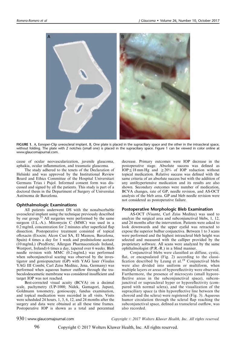
96
cause of ocular neovascularization, juvenile glaucoma,aphakia, ocular inflammation, and traumatic glaucoma.
The study adhered to the tenets of the Declaration ofHelsinki and was approved by the Institutional ReviewBoard and Ethics Committee of the Hospital UniversitariGermans Trias i Pujol. Informed consent form was dis-cussed and signed by all the patients. This study is part of adoctoral thesis in the Department of Surgery of UniversitatAutònoma de Barcelona.
Ophthalmologic ExaminationsAll patients underwent DS with the nonabsorbable
uveoscleral implant using the technique previously describedby our group.15 All surgeries were performed by the samesurgeon (J.L.-A.). Mitomycin C (MMC) was used in a0.2 mg/mL concentration for 2 minutes after superficial flapdissection. Postoperative treatment consisted of topicalofloxacin (Exocin; Alcon Cusí SA, El Masnou, Barcelona,Spain) 4 times a day for 1 week and prednisolone acetate(10 mg/mL) (Predforte; Allergan Pharmaceuticals Ireland,Westport, Ireland) 6 times a day, tapered over 6 weeks. Blebneedle revision with MMC (0.2 mg/mL) was performedwhen subconjunctival scaring was observed by the inves-tigator and goniopuncture (GP) with YAG laser (VisulasYAG III Combi; Carl Zeiss Meditec, Jena, Germany) wasperformed when aqueous humor outflow through the tra-beculodescemetic membrane was considered insufficient andtarget IOP was not reached.
Best-corrected visual acuity (BCVA) on a decimalscale, pachymetry (UP-1000; Nidek, Gamagori, Japan),Goldmann tonometry, gonioscopy, fundus examination,and topical medication were recorded in all visits. Visitswere scheduled 24 hours, 1, 3, 6, 12, and 24 months after thesurgery and data were obtained at all these time frames.Postoperative IOP is shown as a total and percentual
decrease. Primary outcomes were IOP decrease in thepostoperative stage. Absolute success was defined asIOP≤ 18 mmHg and ≥ 20% of IOP reduction withouttopical medication. Relative success was defined with thesame criteria of an absolute success but with the addition ofany antihypertensive medication and its results are alsoshown. Secondary outcomes were number of medication,BCVA changes, rate of GP, needle revision, and AS-OCTanalysis of the bleb area. GP and bleb needle revision werenot considered as postoperative failure.
Postoperative Morphologic Bleb ExaminationAS-OCT (Visante, Carl Zeiss Meditec) was used to
analyze the surgical area and subconjunctival blebs, 1, 12,and 24 months after the intervention. Patients were asked tolook downwards and the upper eyelid was retracted toexpose the superior bulbar conjunctiva. Between 1 to 3 scanswere performed and the highest intrascleral bleb height wasselected and measured with the calliper provided by theproprietary software. All scans were analyzed by the sameophthalmologist (P.R.-R.) in a blind manner.
Conjunctival blebs were classified as diffuse, cystic,flat, or encapsulated (Fig. 2) according to the classi-fication described by Leung et al.19 Conjunctival blebswere also divided into uniform or multiform, whenmultiple layers or areas of hyporeflectivity were observed.Furthermore, the presence of microcysts (small hypore-flective areas in the subconjunctival space), subcon-junctival or suprascleral hyper or hyporeflectivity (com-pared with normal sclera), and the visualization of thesupraciliary space (a thin hyporeflective line between thechoroid and the sclera) were registered (Fig. 3). Aqueoushumor circulation through the scleral flap reaching thesubconjunctival space, defined as transcleral outflow, wasalso recorded.
FIGURE 1. A, Esnoper-Clip uveoscleral implant. B, One plate is placed in the supraciliary space and the other in the intrascleral space,without folding. The plate with 2 notches (small one) is placed in the supraciliary space. Figure 1 can be viewed in color online atwww.glaucomajournal.com.
Romera-Romero et al J Glaucoma ! Volume 26, Number 10, October 2017
930 | www.glaucomajournal.com Copyright r 2017 Wolters Kluwer Health, Inc. All rights reserved.
Copyright © 2017 Wolters Kluwer Health, Inc. All rights reserved.
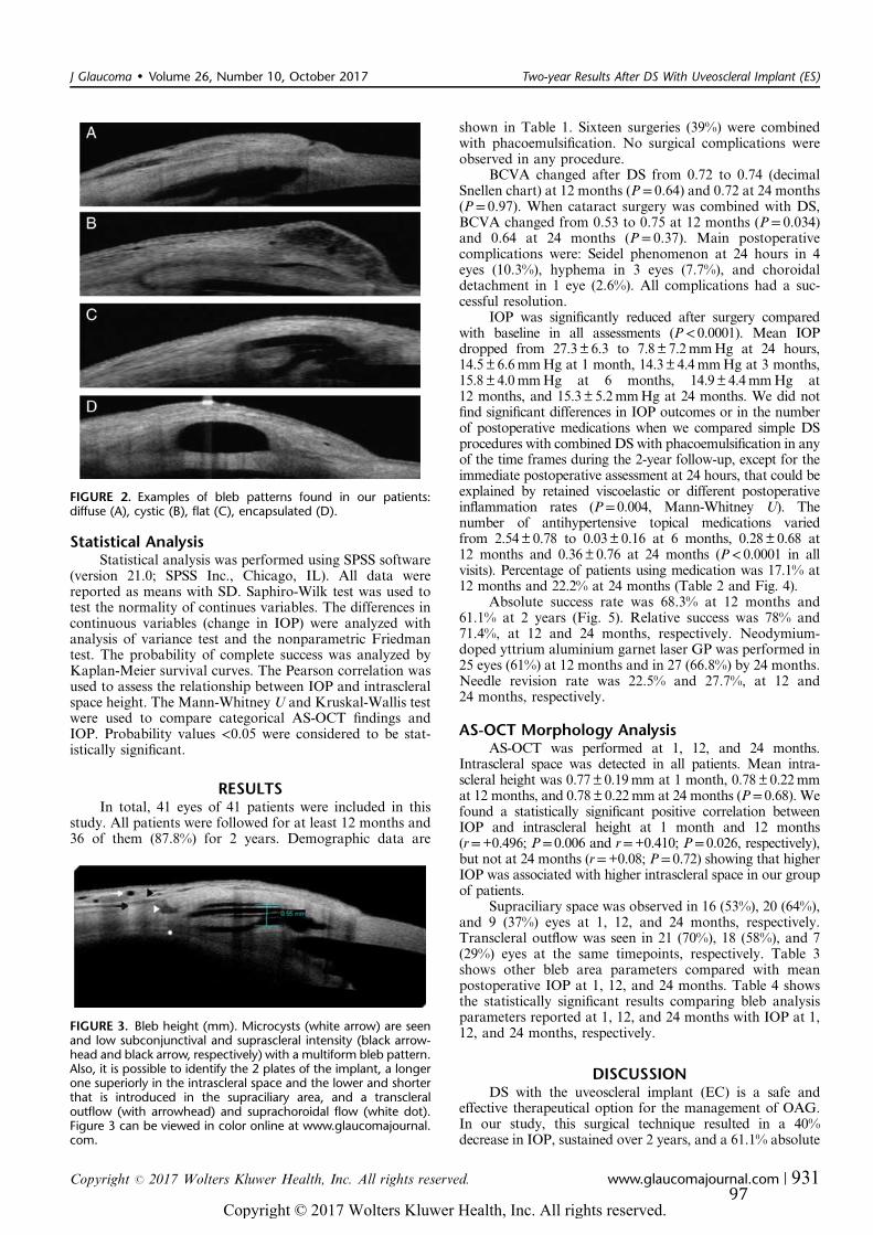
97
Statistical AnalysisStatistical analysis was performed using SPSS software
(version 21.0; SPSS Inc., Chicago, IL). All data werereported as means with SD. Saphiro-Wilk test was used totest the normality of continues variables. The differences incontinuous variables (change in IOP) were analyzed withanalysis of variance test and the nonparametric Friedmantest. The probability of complete success was analyzed byKaplan-Meier survival curves. The Pearson correlation wasused to assess the relationship between IOP and intrascleralspace height. The Mann-Whitney U and Kruskal-Wallis testwere used to compare categorical AS-OCT findings andIOP. Probability values <0.05 were considered to be stat-istically significant.
RESULTSIn total, 41 eyes of 41 patients were included in this
study. All patients were followed for at least 12 months and36 of them (87.8%) for 2 years. Demographic data are
shown in Table 1. Sixteen surgeries (39%) were combinedwith phacoemulsification. No surgical complications wereobserved in any procedure.
BCVA changed after DS from 0.72 to 0.74 (decimalSnellen chart) at 12 months (P= 0.64) and 0.72 at 24 months(P= 0.97). When cataract surgery was combined with DS,BCVA changed from 0.53 to 0.75 at 12 months (P= 0.034)and 0.64 at 24 months (P= 0.37). Main postoperativecomplications were: Seidel phenomenon at 24 hours in 4eyes (10.3%), hyphema in 3 eyes (7.7%), and choroidaldetachment in 1 eye (2.6%). All complications had a suc-cessful resolution.
IOP was significantly reduced after surgery comparedwith baseline in all assessments (P< 0.0001). Mean IOPdropped from 27.3±6.3 to 7.8±7.2mmHg at 24 hours,14.5±6.6mmHg at 1 month, 14.3±4.4mmHg at 3 months,15.8±4.0mmHg at 6 months, 14.9±4.4mmHg at12 months, and 15.3±5.2mmHg at 24 months. We did notfind significant differences in IOP outcomes or in the numberof postoperative medications when we compared simple DSprocedures with combined DS with phacoemulsification in anyof the time frames during the 2-year follow-up, except for theimmediate postoperative assessment at 24 hours, that could beexplained by retained viscoelastic or different postoperativeinflammation rates (P=0.004, Mann-Whitney U). Thenumber of antihypertensive topical medications variedfrom 2.54±0.78 to 0.03±0.16 at 6 months, 0.28±0.68 at12 months and 0.36±0.76 at 24 months (P< 0.0001 in allvisits). Percentage of patients using medication was 17.1% at12 months and 22.2% at 24 months (Table 2 and Fig. 4).
Absolute success rate was 68.3% at 12 months and61.1% at 2 years (Fig. 5). Relative success was 78% and71.4%, at 12 and 24 months, respectively. Neodymium-doped yttrium aluminium garnet laser GP was performed in25 eyes (61%) at 12 months and in 27 (66.8%) by 24 months.Needle revision rate was 22.5% and 27.7%, at 12 and24 months, respectively.
AS-OCT Morphology AnalysisAS-OCT was performed at 1, 12, and 24 months.
Intrascleral space was detected in all patients. Mean intra-scleral height was 0.77±0.19mm at 1 month, 0.78±0.22mmat 12 months, and 0.78±0.22mm at 24 months (P=0.68). Wefound a statistically significant positive correlation betweenIOP and intrascleral height at 1 month and 12 months(r=+0.496; P=0.006 and r=+0.410; P=0.026, respectively),but not at 24 months (r=+0.08; P=0.72) showing that higherIOP was associated with higher intrascleral space in our groupof patients.
Supraciliary space was observed in 16 (53%), 20 (64%),and 9 (37%) eyes at 1, 12, and 24 months, respectively.Transcleral outflow was seen in 21 (70%), 18 (58%), and 7(29%) eyes at the same timepoints, respectively. Table 3shows other bleb area parameters compared with meanpostoperative IOP at 1, 12, and 24 months. Table 4 showsthe statistically significant results comparing bleb analysisparameters reported at 1, 12, and 24 months with IOP at 1,12, and 24 months, respectively.
DISCUSSIONDS with the uveoscleral implant (EC) is a safe and
effective therapeutical option for the management of OAG.In our study, this surgical technique resulted in a 40%decrease in IOP, sustained over 2 years, and a 61.1% absolute
FIGURE 2. Examples of bleb patterns found in our patients:diffuse (A), cystic (B), flat (C), encapsulated (D).
FIGURE 3. Bleb height (mm). Microcysts (white arrow) are seenand low subconjunctival and suprascleral intensity (black arrow-head and black arrow, respectively) with a multiform bleb pattern.Also, it is possible to identify the 2 plates of the implant, a longerone superiorly in the intrascleral space and the lower and shorterthat is introduced in the supraciliary area, and a transcleraloutflow (with arrowhead) and suprachoroidal flow (white dot).Figure 3 can be viewed in color online at www.glaucomajournal.com.
J Glaucoma ! Volume 26, Number 10, October 2017 Two-year Results After DS With Uveoscleral Implant (ES)
Copyright r 2017 Wolters Kluwer Health, Inc. All rights reserved. www.glaucomajournal.com | 931
Copyright © 2017 Wolters Kluwer Health, Inc. All rights reserved.

98
success at 24 months. Our results are in concordance toprevious publications on DS.2–14,31 Mermoud et al2 andShaarawy et al8 had an absolute success rate, at 24 monthsusing a collagen implant, of 69% and 63.4%, respectively, butthey defined success as IOP< 21mmHg. Anand4 reported a75.9% success rate at 24 months with the same threshold andwith hyaluronic acid implant (SKGEL, Corneal Inc., France)and MMC. The number of postoperative medications tolower IOP in our study was quite low, being 0.28 and 0.36 at12 and 24 months, respectively, compared with otherauthors.6–8,11,14,31 Our GP rate (61%) is similar to reportedin other publications,4,6,8–11,14 between 41% to 71%. But wedo not consider GP as a surgical failure as we perform itroutinely when target IOP is not satisfactory in any of thepostoperative visits.
From the eyes classified as failure, 30% of them were sowithin the first month after DS. We attribute this to asuperficial dissection during surgery and poor filtrationthrough the trabeculodescemetic membrane or to an earlysubconjunctival scarring process after the surgical inter-vention. Consistently, eyes that had a uniform bleb patternin AS-OCT 1 month after the DS (no multiple layers visiblein conjunctival bleb analysis) had a significantly higher IOPat 24 months.
Morphologic Analysis of the Surgical AreaAS-OCT analysis of surgical area has been described
in previous reports,12,16–30 either in trabeculectomy and DS.Vieira et al18 published a morphologic analysis of 5 cases ofDS with EC without describing changes in the morphologyof the surgical area over time. Our study is the firstreport doing a systematic and sequential bleb analysis withAS-OCT in DS with EC.
We observed intrascleral space in 100% of patients.Previous reports found it in 22% to 100% of cases, with noimplant useage.12,26,29 These huge differences betweenstudies could be explained by the criteria used for thedetection of the intrascleral space, considering a positivefinding either true spaces or mild hyporeflective areas. In all
TABLE 1. Patient Demographics
No. eyes 41Mean age (years± SD) 71± 7.6Sex (male/female) 22/19Race: white 100%Type of glaucoma (no. eyes)Primary open angle glaucoma 32Pigmentary dispersion glaucoma 1Pseudoexfoliation glaucoma 5Myopic glaucoma 1Corticoid-related glaucoma 1Normal tension glaucoma 1
Lens status (no. eyes)Phakic 31Pseudophakic 10
Surgery (no. eyes)DS 25DS+phacoemulsification 16
Preoperative IOP (mean±SD) (mmHg) 27.3± 6.3Preoperative medications (mean±SD) 2.5 ± 0.8Preoperative VA (Snellen, mean±SD) 0.65± 0.23VA 12mo after DS (mean±SD) 0.75± 0.26Pachymetry (mean±SD) (µm) 533± 35
DS indicates deep sclerectomy; IOP, intraocular pressure; VA, visualacuity.
TABLE 2. IOP Variation During the Preoperative and PostoperativeStage in Deep Sclerectomy With the Uveoscleral Implant
IOP (mmHg) % Reduction Meds P
Preoperative 27.3± 6.3 2.54±0.7824 h 7.8 ± 7.2 70± 28 0.00 < 0.00011mo 14.5± 6.6 44± 26 0.00 < 0.00013mo 14.3± 4.4 45± 20 0.00 < 0.00016mo 15.8± 4 39± 21 0.03±0.16 < 0.000112mo 14.9± 4.4 42± 20 0.28±0.68 < 0.000124mo 15.3± 5.2 40.2± 24 0.33±0.73 < 0.0001
Results in mean±SD.IOP indicates intraocular pressure; meds, number of medications.
FIGURE 4. IOP variation during the preoperative and post-operative deep sclerectomy with uveoscleral implant. IOP indi-cates intraocular pressure; pre, preoperative stage. Figure 4 canbe viewed in color online at www.glaucomajournal.com.
FIGURE 5. Kaplan-Meier survival curve of absolute success(IOP≤18mmHg and ≥20% IOP reduction). IOP indicatesintraocular pressure. Figure 5 can be viewed in color online atwww.glaucomajournal.com.
Romera-Romero et al J Glaucoma ! Volume 26, Number 10, October 2017
932 | www.glaucomajournal.com Copyright r 2017 Wolters Kluwer Health, Inc. All rights reserved.
Copyright © 2017 Wolters Kluwer Health, Inc. All rights reserved.

99
of our patients, intrascleral height was large enough tocontain the implant. Although we found a positive corre-lation between IOP and intrascleral bleb height, otherauthors found an inverse correlation.12,16,25,26 Mavrakanaset al25 found an inverse correlation in eyes with DS andclinically absent subconjunctival blebs at 8± 5 months after
the surgery. This apparent contradiction may be due to thesize of the uveoscleral implant and to the supraciliaryimplantation of our device. We found a mean intrascleralheight of 0.78 ± 0.22 mm, whereas Mavrakanas et al25 founda 0.58 ± 0.16 mm with an absorbable porcine device(AquaFlow), Konstantopoulos et al26 0.44 ± 0.16 mm andGutiérrez-Ortiz et al29 0.33 ± 0.16 mm, both without usingany device. The EC implant footplate is placed in thesupraciliary space and it is possible that a significant part ofthe outflow drains through this route rather than the classicsubconjunctival pathway. This would reverse the correlationbetween IOP and intrascleral bleb height. Theoretically, thepresence of both a transcleral (subconjunctival) and asuprachoroidal flow would make this space flatter, whereaswhen the scleral flap is completely sealed, the intrascleralspace will increase due to a direct pressure effect (Fig. 6).Fakhraie et al,23 in trabeculectomy, found a positive cor-relation between the bleb height and IOP, and also justifiedit as a “pushing effect of IOP through the surgical area.”
We observed a transcleral outflow in 58% eyes at12 months and 29% eyes at 24 months, with no correlationwith better IOP control. Konstantopoulos et al26 found it in55% of eyes, also with no correlation with IOP. Supraciliaryspace was seen in 64% of eyes at 12 months and 37% eyes at24 months. The same author did not detect it in any casewhereas other authors12,29 found it in nearly 80% of cases, inDS or phaco-DS. There is an extreme variability in this
TABLE 3. AS-OCT Analysis of Categorical Variables in Bleb Morphology and IOP 1, 12, and 24 Months After Deep Sclerectomy WithUveoscleral Implant
IOP Factor 1 mo (n= 30) 12 mo (n= 31) 24 mo (n= 24)
Transcleral flowSeen 14.5± 4.6 (n= 21) 15.4± 4.5 (n= 18) 15.7± 5.9 (n= 7)Not seen 15.3± 4.6 (n= 9)
(P= 0.482)15.82± 4 (n= 13)
(P= 0.97)16.5± 5.2 (n= 17)
(P= 0.85)Supraciliary spaceSeen 14.6± 6.2 (n= 16) 15.6± 4.1 (n= 20) 16± 5.7 (n= 9)Not seen 15.0± 7.4 (n= 14)
(P= 0.73)15.4± 4.7 (n= 11)
(P= 0.93)16.4± 5.3 (n= 15)
(P= 0.86)MicrocystsSeen 13.4± 4.7 (n= 24) 14.8± 4.4 (n= 7) 8.7 ± 3.1 (n= 3)Not seen 20± 10.2 (n= 6)
(P= 0.532)15.7± 4.3 (n= 24)
(P= 0.69)17.3± 4.6 (n= 21)(P= 0.008)
Subconjunctival spaceLow intensity 13.1± 4.5 (n= 19) 14.1± 5.7 (n= 10) 12.3± 5.6 (n= 6)Medium intensity 17.5± 7.3 (n= 8) 17.1± 3.3 (n= 11) 17± 4.1 (n= 7)High intensity 17.3± 12.9 (n= 3)
(P= 0.68)15.25± 2.9 (n= 10)
(P= 0.75)18± 5.1(n= 11)(P= 0.08)
Suprascleral spaceLow intensity 11.6± 4 (n= 9) 14.5± 6 (n= 8) 14± 6.5 (n= 4)Medium intensity 14.3± 4.1 (n= 16) 14.8± 3.7 (n= 9) 16± 3.7 (n= 9)High intensity 20.2± 11.1 (n= 5)
(P= 0.52)16.7± 3.6 (n= 14)(P= 0.844)
17.2± 6 (n= 11)(P= 0.66)
UniformityNot uniform 13.6± 4.7 (n= 27) 13.3± 4.6 (n= 8) 13.3± 5.1 (n= 9)Uniform 22.7± 12.9 (n= 3)
(P= 0.24)16.4± 3.9 (n= 23)
(P= 0.44)18.1± 4.7 (n= 15)(P= 0.024)
Bleb morphologyFlat 20± 12 (n= 3) 16.4± 3.3 (n= 18) 18.1± 4.7 (n= 15)Diffuse 14.9± 6.3 (n= 20) 14.3± 5.6 (n= 11) 13.7± 5.8 (n= 7)Cystic 11.5± 2.4 (n= 5) 14± 2.8 (n= 2) 12 (n= 1)Encapsulated 12.5± 0.7 (n= 2)
(P= 0.52)(n= 0)
(P= 0.63)12 (n= 1)
(P= 0.12)
Results are expressed in mean±SD.Statistical analysis Mann-Whitney U test or Kruskal-Wallis test.AS-OCT indicates anterior segment optical coherence tomography; IOP, intraocular pressure.
TABLE 4. Statistically Significant Results Comparing Bleb AnalysisParameters Exposed in Table 3 With IOP at 1, 12, and 24 Months
Factor IOP (24 mo) P
Uniformity 1 moNot uniform 14.7± 4.3 0.038*Uniform 20.7± 4.2
Uniformity 24moNot uniform 13.3± 5.1 0.024*Uniform 18.1± 4.7
Microcysts 12 moSeen 11.6± 3.3 0.029*Not seen 16.7± 4.5
Microcysts 24 moSeen 8.7 ± 3.1 0.008*Not seen 16.6± 3.9
Results are expressed in mean±SD.*Statistical analysis Mann-Whitney U test.IOP indicates intraocular pressure (mmHg).
J Glaucoma ! Volume 26, Number 10, October 2017 Two-year Results After DS With Uveoscleral Implant (ES)
Copyright r 2017 Wolters Kluwer Health, Inc. All rights reserved. www.glaucomajournal.com | 933
Copyright © 2017 Wolters Kluwer Health, Inc. All rights reserved.

100
finding, and it could be explained either due to techniquevariations or low detection of the supraciliary space withVisante in some cases, for example, in patients with thickbleb walls and elevated blebs, as signal is attenuated beforegetting into that depth. We believe that ultrasound bio-microscopy could be more useful to detect this space in thesesituations.
In the conjunctival bleb analysis, eyes with multiformblebs had significantly lower IOP than those with uniformbleb walls. In addition, visualization of microcystic areascorrelated with a significantly lower IOP at 24 months. Ourfindings are in agreement with other reports, whichattempted to correlate early postoperative morphologicfeatures of the conjunctival bleb after trabeculectomy withIOP, measured with AS-OCT.23,30
In summary, we report favorable outcomes 2 yearsafter DS and EC implantation with maintenance of supra-ciliary and intrascleral spaces (the second in 100% of cases)avoiding scleral collapse. We observed that a higher intra-scleral lake height was not related with IOP control, becausesupraciliar implantation reduces intrascleral lake depend-ency. The limitations of this study include not having acontrol group, decreased number of patients with goodquality AS-OCT scans over time, which could have an effectin the results. Other studies comparing different type ofimplants are necessary to give more information about thebenefit of supraciliary implantation in glaucoma surgery.
ACKNOWLEDGMENTSThis work was undertaken in Hospital Germans Trias i
Pujol facilities and is part of a doctoral thesis in UniversitatAutònoma de Barcelona. The authors acknowledge NitinAnand for his contribution and comments that greatlyimproved the manuscript.
REFERENCES1. Leske MC, Heijl A, Hyman L, et al. The early manifest
glaucoma trial group. Early manifest glaucoma trial: design andbaseline data. Ophthalmology. 1999;106:2144–2153.
2. Mermoud A, Schnyder CC, Sickenberg M, et al. Comparison ofdeep sclerectomy with collagen implant and trabeculectomy inopen-angle glaucoma. J Cataract Refract Surg. 1999;25:323–331.
3. El Sayyad F, Helal M, El-Kholify H, et al. Nonpenetratingdeep sclerectomy versus trabeculectomy in bilateral primaryopen-angle glaucoma. Ophthalmology. 2000;107:1671–1674.
4. Anand S, Anand N. Combined phacoemulsification and deepsclerectomy (PDS) with intraoperative mitomycin C (MMC)augmentation. Eye. 2008;22:1040–1049.
5. Cheng JW, Xi GL, Wei RL, et al. Efficacy and tolerability ofnonpenetrating filtering surgery in the treatment of open-angleglaucoma: a meta-analysis. Ophthalmologica. 2010;224:138–146.
6. Karlen ME, Sanchez E, Schnyder CC, et al. Deep sclerectomywith collagen implant: medium term results. Br J Ophthalmol.1999;83:6–11.
7. Mansouri K, Shaarawy T, Wedrich A, et al. Comparingpolymethylmethacrylate implant with collagen implant in deepsclerectomy: a randomized controlled trial. J Glaucoma.2006;15:264–270.
8. Shaarawy T, Nguyen C, Schnyder C, et al. Comparative studybetween deep sclerectomy with and without collagen implant:long term follow up. Br J Ophthalmol. 2004;88:95–98.
9. Devloo S, Deghislage C, Van Malderen L, et al. Non-penetrating deep sclerectomy without or with autologous scleralimplant in open-angle glaucoma: Medium-term results. GraefesArch Clin Exp Ophthalmol. 2005;243:1206–1212.
10. Anand N, Kumar A, Gupta A. Primary phakic deepsclerectomy augmented with mitomycin C. Long-term out-comes. J Glaucoma. 2011;20:21–27.
11. Shaarawy T, Mermoud A. Deep sclerectomy in one eye vs deepsclerectomy with collagen implant in the contralateral eye of thesame patient: long-term follow-up. Eye. 2005;19:298–302.
12. Pérez-Rico C, Gutiérrez-Ortíz C, Moreno-Salgueiro A, et al.Visante anterior segment optical coherence tomography anal-ysis of morphologic changes after deep sclerectomy withintraoperative mitomycin-C and no implant use. J Glaucoma.2014;23:86–90.
13. Muñoz G. Nonstitch supraciliary technique for T-flux implan-tation in deep sclerectomy. J Glaucoma. 2009;18:262–264.
14. Loscos J, Valldeperas X, Langhor K, et al. Deep sclerectomywith supraciliary hema implant (Esnoper V-2000): results andcomplications. Int Ophthalmol. 2015;35:693–699.
15. Loscos-Arenas J, Parera-Arranz A, Romera-Romera P, et al.Deep sclerectomy with a new nonabsorbable uveoscleralimplant (Esnoper-Clip): 1-year outcomes. J Glaucoma. 2015;24:421–425.
16. Fernández-Buenaga R, Rebolleda G, Casas-Llera P, et al. Acomparison of intrascleral bleb height by anterior segment OCTusing three different implants in deep sclerectomy. Eye.2012;26:552–556.
17. Sharma R, Sharma A, Arora T, et al. Application of anteriorsegment optical coherence tomography in glaucoma. SurvOphthalmol. 2014;59:311–327.
18. Vieira L, Noronha M, Lemos V, et al. Anterior segment opticalcoherence tomography imaging of filtering blebs after deepsclerectomy with Esnoper-Clip implant: one-year follow-up.J Curr Glaucoma Pract. 2014;8:91–95.
19. Leung CK, Yick DW, Kwong YY, et al. Analysis of blebmorphology after trabeculectomy with Visante anterior seg-ment optical coherence tomography. Br J Ophthalmol. 2007;91:340–344.
20. Singh M, See JLS, Aquino MC, et al. High-definition imagingof trabeculectomy blebs using spectral domain optical coher-ence tomography adapted for the anterior segment. ClinExperiment Ophthalmol. 2009;37:345–351.
FIGURE 6. Intrascleral bleb height is not related with IOP control12 months after surgery. A, Uveoscleral outflow decreases intra-scleral height dependence (IOP 10mmHg). B, Bleb height isnot related with IOP control (IOP 18mmHg). IOP indicatesintraocular pressure. Figure 6 can be viewed in color online atwww.glaucomajournal.com.
Romera-Romero et al J Glaucoma ! Volume 26, Number 10, October 2017
934 | www.glaucomajournal.com Copyright r 2017 Wolters Kluwer Health, Inc. All rights reserved.
Copyright © 2017 Wolters Kluwer Health, Inc. All rights reserved.

101
21. Hirooka K, Takagishi M, Baba T, et al. Stratus opticalcoherence tomography study of filtering blebs after primarytrabeculectomy with a fornix-based conjunctival flap. ActaOphthalmol. 2010;88:60–64.
22. Nakano N, Hangai M, Nakanishi H, et al. Early trabeculec-tomy bleb walls on anterior-segment optical coherence tomog-raphy. Graefes Arch Clin Exp Ophthalmol. 2010;248:1173–1182.
23. Fakhraie G, Kohansal S, Eslami Y, et al. Correlation betweenfiltering bleb clinical morphology, anterior segment opticalcoherence tomography findings, and intraocular pressure.Iranian J Ophthalmol. 2011;23:21–28.
24. Park HYL, Ahn MD. Imaging of trabeculectomy blebs withVisante anterior segment optical coherence tomography afterdigital ocular compression. Japan J Ophthalmol. 2012;56:38–45.
25. Mavrakanas N, Mendrinos E, Shaarawy T. Postoperative IOPis related to intrascleral bleb height in eyes with clinically flatblebs following deep sclerectomy with collagen implant andmitomycin. Br J Ophthalmol. 2010;94:410–413.
26. Konstantopoulos A, Yadegarfar ME, Yadegarfar G, et al.Deep sclerectomy versus trabeculectomy: a morphologicalstudy with anterior segment optical coherence tomography.Br J Ophthalmol. 2013;97:708–714.
27. Mastropasqua R, Fasanella V, Agnifili L, et al. Anteriorsegment optical coherence tomography imaging of conjunctivalfiltering blebs after glaucoma surgery. Biomed Res Int.2014;2014:610623.
28. Napoli PE, Zucca I, Fossarello M. Qualitative and quantitativeanalysis of filtering blebs with optical coherence tomography.Can J Ophthalmol. 2014;49:210–216.
29. Gutiérrez-ortiz C, Pérez-rico C, Moreno-salgueiro A, et al.Visante anterior segment optical coherence tomography anal-ysis of morphologic changes in the anterior segment structuresafter deep sclerotomy versus phaco-deep sclerotomy withintraoperative Mitomycin-C and no implant use. SeminOphthalmol. 2015;13:1–7.
30. Khamar MB, Soni SR, Mehta SV, et al. Morphology offunctioning trabeculectomy blebs using anterior segment opticalcoherence tomography. Indian J Ophthalmol. 2013;62:711–714.
31. Mesci C, Erbil HH, Karakurt Y, et al. Deep sclerectomyaugmented with combination of absorbable biosyntheticsodium hyaluronate scleral implant and mitomycin C or withmitomycin C versus trabeculectomy: long-term results. ClinExperiment Ophthalmol. 2012;40:197–207.
J Glaucoma ! Volume 26, Number 10, October 2017 Two-year Results After DS With Uveoscleral Implant (ES)
Copyright r 2017 Wolters Kluwer Health, Inc. All rights reserved. www.glaucomajournal.com | 935
Copyright © 2017 Wolters Kluwer Health, Inc. All rights reserved.

102
103
7. Discusión
Lacirugíaenelglaucomasuponeretoparaelcirujanooftalmólogo,siemprebuscandoun
equilibrioentrelapresiónaltaylahipotonía,entrelafibrosisylaavascularidadextrema,
entrelahipoehiperfiltración,entrelaaltaefectividadyaltatasadecomplicacionesolabaja
efectividad con mayor seguridad. La esclerectomía profunda no perforante (EPNP)
actualmenterepresentaelequilibrioentodoesteuniversodeprocedimientosenelquenos
vemos envueltos. Es una técnica filtrante que nos permite obtener PIO similares a la
trabeculectomía,obteniendounasampollasde filtraciónmáspredeciblesydifusasycon
unatasadecomplicacionesmuyinferioraésta.Estoesdebidoaquelaresistenciaalflujo
nolarealizaúnicamentelaescleraylaconjuntiva,sinoqueestáfocalizadaenlamembrana
trabeculodescemética(MTD),quenosseparará lacámaraanteriordelojoconelespacio
intraescleralysubconjuntival.
Esclerectomíaprofundanoperforante(EPNP)conelimplanteuveoescleral
DentrodelosimplantesdelaEPNP,elimplanteuveoescleralEsnoperClip®(EC)eselúnico
quenospermiteactuarsobrelasdosvíasdedrenaje(subconjuntivalyuveoescleral).
Segúnhemospodidocomprobarennuestrosdosartículospublicados,laEPNPconECes
una técnica segura y efectiva en el manejo del glaucoma de ángulo abierto. La cirugía
supraciliarcon los implantesquedisponíamosanteriormente tenían ladesventajadeno
104
estardiseñadosespecíficamenteparaesteespacio,porloquesuefectividadpodríaestar
limitada. Fue Muñoz100 quien primero la describió con el T-flux® en EPNP aislada y
posteriormenteBonilla102conelEsnoper®encirugíascombinadasconfacoemulsificación,
presentandounosdescensostensionalesde26.4±6.9a14±3.3mmHgy23±5a18±3
mmHgrespectivamente,alañodelacirugía,yundescensodelnúmerodefármacostópicos
de2.8a0.3yde2.5a0.7 respectivamente.Unode losmayoresproblemasquepodrían
presentar los implantes en posición supraciliar diferentes al EC es que el espacio
intraescleralfueraperdiendosualturaconeltiempoeinclusocolapsarse,debidoaqueno
existe porción del implante que ocupe dicho espacio. Con el EC, como hemos visto en
nuestratesisdoctoral,observamoscómosemantienealosdosañosposterioresalacirugía.
Seguridad
Concluimos que es una técnica segura porque no hemos encontrado complicaciones
severas. En primer lugar, en ninguno de los dos estudios publicados observamos
complicaciones intraoperatorias. De las complicaciones postoperatorias, en el primer
trabajoencontramosSeidelpositivoalas24hen2ojos(7.4%),hiphemaen2ojos(7.4%),
desprendimientodecoroidesen1ojo(3.7%),todosellosresueltosenmenosde1semana.
En el segundo trabajo, Seidel en 4 ojos (10.3%), hiphema en 3 ojos (7.7%), y
desprendimientocoroideoen1ojo(2.6%),quecorrespondealmismopacientequeenel
primertrabajo,todosellosresueltosconéxito.Alcompararnuestrascomplicacionesconlo
publicadohemosobservadolosiguiente:
- Complicaciones intraoperatorias severas. No tuvimos casos de hemorragia
supracoroidea,temidacomolapeorcomplicaciónposible.Esunacomplicaciónmuy
raraymásfrecuentetrastrabeculectomíaycirugíaconimplantededrenaje(válvula
105
deAhmed,tubodeBaerveldt,etc.).160161Bettin162ladescribetrasgoniopunturaen
unpacienteintervenidodeEPNP,siendoésteelúnicocasopublicadoalrespecto.
- MacroperforacióndelaMTD:notuvimosningúncaso,debidoaqueenestoscasos
lacirugíalasolemosreconvertiratrabeculectomía,yenmuchoscasosnousamos
ningúnimplante,porloquenolosincluiríamosenesteestudio.Estadependedela
habilidaddelcirujanoydelaanatomíadelángulocamerular.Sehadescritoentreel
0y20,5%ydesciendeamedidaqueavanzamosennuestracurvadeaprendizaje.94
95110111163164
- Desprendimientode coroides (DC):nuestra tasa fuebaja, de1 caso en ambos
trabajos,dadoqueenlaliteraturasedescribeentre1y7%.Esimportanteremarcar
querealizarunaimplantaciónsupraciliarnohaidoasociadoaunaumentodelatasa
deDCnimaculopatía,yquetantoennuestrocasocomoenlamayoríadetrabajos
seresuelvenensutotalidadantesdelmesposterioralacirugía.
- Seidel:esmoderadamentefrecuenteenelposoperatorioprecoz,debidoaunmal
ajustedelassuturas,fugaporelbordeconjuntivalmalposicionadooporunexceso
defiltraciónlosprimerosdíastraslacirugía.Ennuestrotrabajoapareceentreel7-
10%deloscasos,similaralopublicado(1-22%).95101
- Hiphemaomicrohiphemapostoperatorioprecoz:suanálisispuede llegaraser
algo subjetivo, siendo infradiagnosticado en muchos casos. Comparado con lo
publicadoentreun2-16%,nosotrosloencontramosenun7%deloscasos.108101111
- Atalamia: no tuvimos ningún caso de atalamia completa. Algún grado de
hipotalamia,sobretodoenloscasosdegrandescensotensionalesfrecuente,pero
subjetivo a la hora de cuantificarlo. Se ha descrito entre el 0%y 11,6%.78 108 113
Anand114 tuvo una tasa del 15% en aquellos casos en los que se había
microperforadolaMTDintraoperatoriamente.
106
- Hipotoníatardía:notuvimosningúncaso.Algunoscasospublicadosaparecenen
losartículosdeMercieca,118quedescribe2/43enpacientesconuveítis;Loscos,101
con1caso(2,08%)conimplanteEsnoperV-2000®enposiciónsupraciliar;2,6%de
Al-Obeidan164enunestudioretrospectivode200casosdeEPNP;yAnand114en3
casos(2,5%)todosellostraslagoniopuntura.
- Incarceración iridiana en la MTD: no tuvimos ningún caso. Al realizar la
goniopuntura,encasosenlosquelaraíziridianaseencuentramuyaproximadaala
MTD,sobretodoenángulosgrado2-3,acostumbramosarealizarunairidoplastia
sectorialpara alejarla e impedir sudesplazamientoanterior.Algunosestudios la
recogenentreun2,1%y5,8%.7895101110164Tambiéncreemosquelautilizacióndel
implanteesbeneficiosaalahoradeevitarpicosaltosdePIOtrasesta.Losbordes
anterioresdelimplante,alquedarplegado,creancomounaespeciedebuclequeal
desplazarsehaciaanteriorycontactaconlaMTD,lacompartimentanen3zonas(las
zonaslateralesylaquequedaenelinteriordelosdospilaresdelimplante).Deesta
manera,siexisteunaincarceración,éstasueleocuparunodeestostresespacios,y
nolatotalidad,permitiendoqueexistazonadelaMTDfuncionanteysusceptiblede
recibirunagoniopuntura.
- Migración del implante: no tuvimos ningún caso demigración fuera del lecho
escleral.Al-Obedan164describeuncasodemigracióndelSKgel(0,5%).
- Glaucomamaligno: no tuvimos ningún caso. Descrito en un 2% por Sanchez,86
2,3%porAnand,781casoporKarlen87yBissig.90
- Catarata:sehadescritounaprogresióndelacatarataentreel2y22%deloscasos.
Ennuestrostrabajosnohemosrealizadounanálisisquecontemplenilaevolución
ni la necesidad de cirugía de cataratas tras la EPNP, dado que se requiere un
seguimiento amás largo plazo. Este podría ser uno de los posibles objetivos de
futuro.

107
AgudezaVisual
La agudeza visual semantuvo estadísticamente sin cambios o bienmejoró en los casos
combinadosconcirugíadecataratas.Estoestáenconcordanciaconestudiosprevios,101103
106115116118mientrasqueenotrosdetectaronunadisminuciónenlavisitafinalrespectoa
lavisitapreoperatoria.110118Cabeseñalarqueenmuchoscasosesdebidoaqueseformó
catarata significativa que producía una disminución de la agudeza visual reversible.
Mermoud103comparólaEPNPconlatrabeculectomía,observandounaestabilizaciónvisual
enlaprimeramientrasqueexistíaundescensovisualsignificativoenlasegunda.
ReduccióndelaPresiónintraocular(PIO)
Ennuestrosestudioshemosdescritounadisminucióndemásdel40%de laPIOque se
manteníaalolargodedosaños,conunatasadeéxitodemásdel60%alos24meses.Enel
primer trabajo, laPIOpasóde26.6 a15.3mmHga los12meses conuna reduccióndel
númerodefármacosde2.5demediaa0.26.Enelsegundo,laPIOpasóde27.3mmHgcon
2.54medicamentosantihipertensivosdemedia,a15.3mmHgcon0.36fármacosdemedia,
alos24mesesdelacirugía.Eléxitoabsoluto(PIO<19mmHgsinadicióndetratamiento
tópico) fue del 61,1%. Estos resultados están en concordancia con estudios publicados
previamente. Si analizamos los estudios publicados con un periodo de seguimiento de
medio-largoplazo,enprimerlugarhayquenombraraMermoudyShaarawy,unodelos
padresdelaEPNP,103108145quienesencontraronalos24mesestambiénéxitosdel69%y
63,4%respectivamente con lautilizaciónde implantede ácidohialurónico, pero conun
objetivotensionalmayor(<22mmHg).Anand111reportaun75,9%deéxitoconelmismo
objetivo tensional que en nuestra tesis y con implante de SkGel. Analizando el resto de

108
estudios,laspresionesfinalesvariaronentre8,9y18,5mmHgyloséxitosabsolutosentre
un19%enunestudioa36mesesy85%enunestudioa24mesesconelimplanteSkGel.La
tasa de medicación variaba entre 0,1 y 2,2 fármacos antihipertensivos de media.
Observamosqueescasi imposiblecompararunosestudiosconotros,yaseapor tipode
pacientes, edades, zonas geográficas distintas, técnica quirúrgica, utilización o no de
implanteytipodeantimetabolitos.Peropodemosconcluirqueenel95%delosanalizados
laPIOdisminuyómásdel30%,loquerepresentaríalaconsecucióndelobjetivotensional.
Ennuestrosegundo trabajo,de losojosqueclasificamoscomo fracasos,debidoaque la
presióneramayora18mmHgendosomáscontrolessucesivosobienhabíamosañadido
tratamiento tópico, el 30% de ellos lo hicieron durante el primermes. Atribuimos este
hechoobienaunadisecciónnolosuficienteprofundadelaMTD,obiendebidoaunproceso
cicatricialprecozyseveroenestospacientes.Estolopudimosconstataralobservarquelos
casosenlosqueexistíaunaampolladefiltraciónuniforme(sinvisualizacióndevariascapas
enelanálisisconOCT-SA)almesdelacirugía,laPIOalos24meseserasignificativamente
superioraloscasosenquelaampollaeradeaspectomultiforme,loqueesindicativodeque
lascirugíasno funcionabande formasatisfactoriadesdeunprincipio.Y lomismocon la
visualizacióndemicroquistes,señaldequeexistíaunacorrectafiltraciónconjuntival.
Tasadegoniopuntura(GP)
LaincidenciadeGPvaríaentre6%y71%enlosestudiosanalizados(ennuestrocasofue
del61%alos12mesesydel66%alos24meses).Estavariabilidadesdebidaalautilización
dediferentescriteriospararealizarla,yaseaporrequerirdeunaspresionesobjetivomás
bajas, o la de evitar a toda costa la adición de terapia hipotensora tópica. Nosotros no

109
consideramos laGP como fracasode la cirugía, yaque creemosque espartedel propio
procedimientoquirúrgicodebidaalafibrosisqueseproduceenlaMTDenmuchoscasos,
loqueconvierteunprocedimientonoperforanteenunoperforante.Lasventajasqueaporta
elEsnoperClip®respectoalrestodeimplantesesque,aunquesedesplaceanteriormente
en el postoperatorio y contacte con laMTD, al quedarplagadonunca llega a bloquearla
completamente,dejandoespaciocentrallibreentrelasdosporcionesdelimplantequeevita
elcolapsodelamisma,ysiendoesteespaciodondeseráposiblerealizarlaGP.
Needling
La revisión con aguja (needling) en el postoperatorio la realizamos cuando existe una
fibrosisquebloquealafiltraciónaniveltransescleralysubconjuntival,observándoseunas
ampollasaplanadasyunosbordesdelflapescleraldeaspectosellado.Ennuestrocasofue
del 22.5% a los 12 meses y del 27,7% a los 24 meses. En todos los casos añadimos
MitomicinaC (MMC)para evitar la fibrosis posterior. Revisando los estudios analizados
vemosque suutilización tambiénesmuyvariableentreespecialistas, entreun1%yun
34%.Clásicamenteseutilizaba5-FUcomoantifibrótico,perolaMMCesmáspotentepara
evitarlafibrosisycicatrizaciónposterior,yeslaquemásseusaenlaactualidad.Hayque
tenerencuentaqueelefectodelaMMCesaditivo,porloqueencasosenlosqueexista
riesgodeampollasavascularesesmejornoutilizarla.Ennuestrocasoconsideramosqueel
needlinghasidoefectivosiPIOdisminuyeamenosde10mmHgynuncalorealizamosen
másde2ocasiones.
110
Análisismorfológicodeláreaquirúrgica
Laimportanciadelaampollaintraescleralyelflujouveoescleralenlacirugíadeglaucoma
son aspectos controvertidos, pero está aceptado que su presencia es un factor de buen
pronósticoenelpostoperatorio.
Enelprimertrabajoúnicamenteanalizamoslaevolucióndelvolumenylaalturaalolargo
delprimeraño,observandounatendenciaasureducciónsinsignificaciónestadística.Enel
segundorealizamosunanálisismáscompleto,localizandoelespaciointraescleralentodos
loscasos,noasíelespaciosupraciliar.Peroobtuvimosunasignificaciónestadísticapositiva
entrelaPIOylaalturadellagointraescleralalmesyalañodelacirugía,adiferenciade
Mavrakanas,132 Fernandez-Buenaga133 y Perez-Rico.134 Mavrakanas132 encontró una
correlacióninversaenojosoperadosdeEPNPconampollaconjuntivalclínicamenteausente
(alos8mesesdelacirugíademedia).Estaaparentecontradicciónentrelosdostrabajos
podría ser debida al tamañodel implante EC y a la implantación supraciliar delmismo.
Nosotrosencontramosunaalturamediadelespaciointraescleralalañode0.78±0.22mm,
mientrasMavrakanas laencontróde0.58±0.16mmconel implanteporcinoabsorbible
AquaFlow®enposiciónintraescleral.Konstantopoulos154de0.44±0.16mmyGutiérrez-
Ortizde0.33±0.16mm,losdossinusarningúnimplante.UnodelosplatosdelECsecoloca
enposiciónsupraciliaryesposiblequeunapartesignificativadeldrenajelohagasobreesta
vía.EstoloqueproduciráesunainversióndelacorrelaciónentrelaPIOylaalturadellago
intraescleral.Teóricamente, lapresenciadeambasvíasde filtración, la transescleraly la
uveoescleralharáelespaciointraescleralmásestrecho,mientrasquecuandoelflapescleral
estétotalmenteselladoalaescleracircunscritaonoexistafiltraciónsupraciliar,elespacio
intraescleralcrecerádebidoaunefectodirectodelaPIO.Fakhraie152yadescribióelefecto

111
directodelaPIOsobrelaalturadelaampollatrastrabeculectomía.Esteefectolopodemos
observarenlafigura6denuestrosegundoartículo,enlapágina934.166
Tambiénanalizamosotrosfactoresmorfológicosdelaampolladefiltraciónconjuntival.En
primer lugar, valoramos la uniformidad de este espacio, observando una relación
estadísticamentesignificativaentrelapresenciadeespaciosnouniformesalmesyalos24
mesesdelacirugíaylapresiónalos24mesesdelacirugía.Lomismopasóconlapresencia
demicroquistesalos12y24mesesdelacirugía.Enamboscasos,laPIOfinaleramásbaja
queenloscasosdeampollasdecaracterísticasuniformesyenlosquenoseencontraban
microquistes.Todosestoshallazgosestánenconcordanciaconestudiospreviosencirugía
detrabeculectomía,quienesintentaroncorrelacionaraspectospostoperatoriosprecocesde
lasampollasdefiltraciónconeléxitotensionalalos6mesesdelacirugía,152158viendoque
aquellas ampollas de aspectomultiforme se correlacionaban con PIOmás bajas a los 6
mesesdelacirugía.
Tambiénbuscamos lapresenciade filtración transescleral,queestaría representadopor
una continuación de la zona hiporeflectiva del lago intraescleral hacia el espacio
subconjuntival.Ennuestrocasofuevisibleenel58%delosojosalos12mesesyenel29%
de losmismos a los24meses, pero sin encontraruna correlación conunmejor control
tensional. Konstantopoulos154 también lo describió en un 55% de los casos, tampoco
encontrandocorrelaciónconelcontroldelaPIO,mientrasquePerez-Rico134lodescribió
comounfactorpronósticopositivo,encontrándoloenun89,4%deloscasosalos5años
posterioresaEPNPsinimplante.HayquetenerenconsideraciónqueelOCT-SAnosrealiza
unúnicocortede2dimensiones,porloquenoobservemosfiltracióntransescleralvisible
ennuestroscortesnosignificaquenoexista.

112
Respectoalflujouveoescleral,loencontramosenel64%delosojosalos12mesesyenel
37%delosojosalos24meses,noencontrandounacorrelaciónentresupresenciayuna
PIOmásbaja.Elmismoautor154noladetectóenningúncasomientrasqueotros134157la
encontraroncasienel80%deloscasos.Observamoscomoexisteunagranvariabilidaden
estosdatos,yaseadebidaaladifícilmedicióneinterpretacióndelosresultados,obienala
bajacapacidaddedeteccióndelespaciosupraciliarporelOCT-SA(sobretodoencasosde
ampollas gruesas en las que la señal se atenúa antes de llegar a la profundidad de este
espacio).Enestoscasoseldisponerdelabiomicroscopíaultrasónica(BMU)nosseríade
granutilidad.Lapresenciadeflujouveoescleraltrascirugíadeglaucomaescomúnincluso
sinimplantaciónsupraciliar,yseharelacionadoconunmayordescensotensional.91
Respectoalanálisisdelaintensidaddelosespaciossubconjuntival,supraciliaryaltipode
ampolla(plana,difusa,quísticaoencapsulada),noseencontrarondiferenciassignificativas
enningunode losespacios temporales,aunquesiobservamos losdatosde la tabla3de
nuestrosegundoartículo,enlapágina933,166vemoscomoexisteunatendenciaaobtener
presionesmásaltasenaquellasampollasplanasrespectoalasdifusasalmes,12y24meses,
presionesmásaltasen lasampollasuniformes tambiénen todos loscontroles, así como
presiones más altas en aquellas ampollas que presentaban espacios subconjuntivales y
supraescleralesdemoderada/altaintensidadrespectoalosdebajaintensidad.Todosestos
datosnoshacenintuirquelapresenciadeampollaconjuntivalsiguesiendoclaveparael
éxitodelaEPNP,inclusoaunquedispongamosdefiltraciónuveoescleral.
LautilizaciónonodeimplanteenlaEPNPsiguesiendohoyendíacuestióndedebateentre
los cirujanos de glaucoma. Desde que Mermoud108 en 2004 describiera PIO
significativamentemásbajasenelgrupocon implantedecolágeno (15,1vs12,6a los2
años),seentendióqueelusodelimplanteaportaríaunplusaestacirugía,sobretodoen
113
aquelloscasosenquemesesposterioresalamismarequirierandegoniopuntura,yaqueel
implante facilitaríaelmantenimientodeunespacioque teóricamentehabríadisminuido
porculpadeldescensode la filtracióna travésde laMTD.Lautilizacióndeun implante
colocadoanivelsupraciliar loquebuscaes justamente incrementareste flujo,conseguir
presionesobjetivomásbajasevitandoelcolapsodeesteespacioconel tiempo.Además,
también favorecería a un desprendimiento del cuerpo ciliar parcial con la subsiguiente
disminucióndelaproduccióndehumoracuoso.Aunasí,atenordenuestrosresultados,son
rarosloscasosdehipotoníaclínicamentesignificativaenestetipodeimplantación.

114
115
8. Conclusiones
Unavezllegadosaestepunto,esimportantedarunarespuestaaaquellosobjetivosquenos
impusimosaliniciodeltrabajo.Concluimosque:
8.1 LaEPNPcon implanteuveoescleralreduce laPIOde formasignificativahasta24
mesesposterioresalaintervención,observandoundescensotensionaldel40.2±
24%alos24meses(p<0.0001).
8.2 La EPNP con implante uveoescleral reduce la tasa de medicación de forma
significativahasta24mesesposterioresalaintervención,pasandode2.54±0.78
enelpreoperatorioa0.33±0.73alos24meses.
8.3 LaEPNPconimplanteuveoescleralesunatécnicaseguraquepresentaunabajatasa
decomplicaciones,nopresentandoningunacomplicacióngraveysolucionándose
todasellasalospocosdíaspostcirugía.
8.4 Al describir los factores del área quirúrgica relacionados con el éxito tensional,
concluimos:
8.4.1 ExistíaunacorrelaciónpositivaentrelaalturadellagointraescleralylaPIO
almesyalañodelacirugía.
8.4.2 NoexisteunadiferenciadePIOsignificativasegúneltipodeampolla.
116
8.4.3 LapresenciadeflujotransescleralnocondicionaunmejorcontroldelaPIO
deformaestadísticamentesignificativa.
8.4.4 LapresenciadeflujouveoescleralnocondicionaunmejorcontroldelaPIO
deformaestadísticamentesignificativa.
8.4.5 La presencia de baja reflectividad subconjuntival y supraescleral no
condicionaunmejorcontroldelaPIOdeformaestadísticamentesignificativa.
8.4.6 Lapresenciademicroquistesalos12y24mesesylapresenciaampollasde
filtracióndeaspectonouniformealmesyalos24mesesdelacirugíaestá
asociadoaunmejorcontroldelaPIOdeformaestadísticamentesignificativa
alos24mesesdelacirugía.
Latécnicaquirúrgicadescritaenestatesisdoctoralsiguesiendoyseguramenteloseguirá
enunfuturonuestratécnicadeelecciónenelglaucomadeánguloabierto,debidoasubuen
perfildeseguridad,bajatasadecomplicacionesybuenasatisfacciónporpartedelpaciente.
Lautilizacióndelimplanteuveoescleralnosaporta,desdenuestropuntodevistayatenor
denuestrosresultados,unplusquenodebemosdejardelado.Porotrolado,aúnquedan
aspectos amejorar, como son la fibrosis conjuntival que condiciona enmuchos casos la
ampollaconjuntival,ylaproliferaciónfibróticasobrelamembranatrabeculodescemética,
queharíadescenderlafiltraciónatravésdelamisma.
117
9. Bibliografía
1. KleinBE,KleinR,LintonKL. Intraocularpressure inanAmericancommunity.TheBeaver
DamEyeStudy.InvestOphthalmolVisSci.1992;33:2224-2228.
2. KleinR,KleinBEK.Theprevalenceofage-relatedeyediseasesandvisualimpairmentinaging:
currentestimates.InvestOphthalmolVisSci.2013;54:5-13.
3. CongdonN, O’Colmain B, Klaver CCW, et al. Causes and Prevalence of Visual Impairment
AmongAdultsintheUnitedStates.ArchOphthalmol.2004;122:477-485.
4. AntónA,AndradaMT,MujicaV,etal.Prevalenceofprimaryopen-angleglaucomainaSpanish
population:theSegoviastudy.JGlaucoma.2004;13:371-376.
5. KyariF,AbdullMM,BastawrousA,etal.Epidemiologyofglaucoma insub-saharanAfrica:
prevalence,incidenceandriskfactors.MiddleEastAfrJOphthalmol.20:111-125.
6. Day AC, Baio G, Gazzard G, et al. The prevalence of primary angle closure glaucoma in
Europeanderivedpopulations:asystematicreview.BrJOphthalmol.2012;96:1162-1167.
7. EuropeanGlaucomaSociety.TerminolologyandGuidelinesforGlaucoma4thEdition;2014.
8. ChowdhuryUR,HannCR,StamerWD,etal.Aqueoushumoroutflow:Dynamicsanddisease.
InvestigOphthalmolVisSci.2015;56:2993-3003.
9. HannCR,VercnockeAJ,BentleyMD,etal.AnatomicChangesinSchlemm’sCanalandCollector
ChannelsinNormalandPrimaryOpen-AngleGlaucomaEyesUsingLowandHighPerfusion
Pressures.InvestigOpthalmologyVisSci.2014;55:5834-5841.
10. Esporcatte BLB, Tavares IM. Normal-tension glaucoma: An update. Arq Bras Oftalmol.
2016;79:270-276.
11. BonomiL,MarchiniG,MarraffaM,etal.Prevalenceofglaucomaand intraocularpressure
distribution in a defined population. The Egna-Neumarkt Study. Ophthalmology.
1998;105:209-215.
12. SergiM,SalernoDE,RizziM,etal.PrevalenceofNormalTensionGlaucomainObstructive

118
SleepApneaSyndromePatients.JGlaucoma.2007;16:42-46.
13. NongpiurME,HaalandBa.,PereraSa.,etal.Developmentofascoreandprobabilityestimate
fordetectingangleclosurebasedonanteriorsegmentopticalcoherencetomography.AmJ
Ophthalmol.2014;157:32-38.
14. SunX,DaiY,ChenY,etal.PrimaryangleclosureglaucomawhatweKNOWandwhatwedon’t
know.ProgRetinEyeRes.2016:1-20.
15. Azuara-Blanco A, Burr J, Ramsay C, et al. Effectiveness of early lens extraction for the
treatmentofprimaryangle-closureglaucoma(EAGLE):arandomisedcontrolledtrial.Lancet.
2016;388:1389-1397.
16. JavanbakhtM,Azuara-BlancoA,Burr JM, et al. Early lens extractionwith intraocular lens
implantationforthetreatmentofprimaryangleclosureglaucoma:aneconomicevaluation
basedondatafromtheEAGLEtrial.BMJOpen.2017;6:e013254.
17. KakoulidisK,CernakA,CernakM.Morphologicchangesofanteriorsegmentoftheeyeafter
cataractsurgery.CeskSlovOftalmol.2011;67:111-114.
18. ShamsPN,FosterPJ.Clinicaloutcomesafterlensextractionforvisuallysignificantcataractin
eyeswithprimaryangleclosure.JGlaucoma.2012;21:545-550.
19. Medeiros FA,Weinreb RN. Is corneal thickness an independent risk factor for glaucoma?
Ophthalmology.2012;119:435-436.
20. Brandt JD,Beiser JA,KassMA, et al.Central corneal thickness in theOcularHypertension
TreatmentStudy(OHTS).Ophthalmology.2001;108:1779-1788.
21. Argus WA. Ocular hypertension and central corneal thickness. Ophthalmology.
1995;102:1810-1812.
22. HarwerthRS,WheatJL,FredetteMJAD.LinkingStructureandFunctioninGlaucoma.Prog
RetinEyeRes.2010;29:249-271.
23. BraakmanST,ThomasReadA,W-HChanD,etal.ColocalizationofOutflowSegmentationand
PoresAlongtheInnerWallofSchlemm’sCanal.ExpEyeRes.2015;130:87-96.
24. AmmarDA,LeiTC,GibsonEA,etal.Two-photonimagingofthetrabecularmeshwork.Mol
Vis.2010;16:935-944.
25. Carreon T, van der Merwe E, Fellman RL, et al. Aqueous outflow - A continuum from

119
trabecularmeshworktoepiscleralveins.ProgRetinEyeRes.2016;57:108-133.
26. Gigon A, Shaarawy T. The Suprachoroidal Route in Glaucoma Surgery 1Medical Student
(FinalYear).JCurrGlaucomaPr.2016;1010:13-20.
27. OcklindA.EffectofLatanoprostontheExtracellularMatrixoftheCiliaryMuscle.AStudyon
CulturedCellsandTissueSections.ExpEyeRes.1998;67:179-191.
28. Pederson JE, Gaasterland DE, MacLellan HM. Uveoscleral aqueous outflow in the rhesus
monkey:importanceofuvealreabsorption.InvestOphthalmolVisSci.1977;16:1008-7.
29. AiraksinenPJ,TuulonenA,AlankoHI.Rateandpatternofneuroretinalrimareadecreasein
ocularhypertensionandglaucoma.ArchOphthalmol(Chicago,Ill1960).1992;110:206-210.
30. ZhouY-S,Xu J,Peng J, et al.Researchprogressof stemcellsonglaucomatousopticnerve
injury.IntJOphthalmol.2016;9:1226-1229.
31. Costa L, Cunha JP, Amado D, et al. Diabetes Mellitus as a Risk Factor in Glaucoma’s
Physiopathology and Surgical Survival Time: A Literature Review. J Curr glaucoma Pract.
2015;9:81-85.
32. BellK,undHohenstein-BlaulNvonT,TeisterJ,etal.Modulationoftheimmunesystemfor
thetreatmentofglaucoma.CurrNeuropharmacol.2017;15.
33. YangG,SauC,LaiW,etal.Themorphologicaldifferencebetweenglaucomaandotheroptic
neuropathies.JNeuroophthalmol.2015;344:1173-1178.
34. JiaX,YuJ,LiaoS-H,etal.Biomechanicsofthescleraandeffectsonintraocularpressure.IntJ
Ophthalmol.2016;9:1824-1831.
35. Coudrillier B, Pijanka JK, Jefferys JL, et al. Glaucoma-related changes in the mechanical
properties and collagen micro-architecture of the human sclera. PLoS One. 2015;
10:e0131396.
36. Roy Chowdhury U, P FautschM. Intracranial Pressure and Its Relationship to Glaucoma:
Current Understanding and Future Directions.Med hypothesis, Discov Innov Ophthalmol.
2015;4:71-80.
37. BarrancosC,RebolledaG,OblancaN,etal.Changesinlaminacribrosaandprelaminartissue
afterdeepsclerectomy.Eye.2014;28:58-65.
38. SaeediO,PillarA,JefferysJ,etal.Changeinchoroidalthicknessandaxiallengthwithchange

120
inintraocularpressureaftertrabeculectomy.BrJOphthalmol2014;98976–979.
39. KadziauskieneA,KuolieneK,AsoklisR,etal.Changesinchoroidalthicknessafterintraocular
pressurereductionfollowingtrabeculectomy.ActaOphthalmol.2016;94:586-591.
40. Chen S, Wang W, Gao X, et al. Changes in Choroidal Thickness After Trabeculectomy in
PrimaryAngleClosureGlaucoma.InvestOphthalmolVisSci.2017;55:2608-2613.
41. LeskeM,ConnellA,WuS-Y,etal.RiskFactorsforOpen-angleGlaucoma:TheBarbadosEye
Study.JAMAOphthalmol.1995;113:918-924.
42. SinghK,ShrivastavaA. Intraocularpressure fluctuations:howmuchdo theymatter?Curr
OpinOphthalmol.2009;20:84-87.
43. RossAH,JacksonTE,WertheimMS,etal.Analysisofthediurnalintraocularpressureprofile
pre and post trabeculectomy using 24-hour monitoring of intraocular pressure. Eur J
Ophthalmol.2011;21:400-403.
44. KiddeeW,AtthavuttisilpS.Theeffectsofselectivelasertrabeculoplastyandtravoproston
circadianintraocularpressurefluctuations.Medicine(Baltimore).2017;96:e6047.
45. KonstasAGP,TopouzisF, LeliopoulouO, et al. 24-Hour IntraocularPressureControlwith
MaximumMedicalTherapyComparedwithSurgeryinPatientswithAdvancedOpen-Angle
Glaucoma.Ophthalmology.2006;113:761-765.
46. ErbC.“EarlyManifestGlaucomaTrial”(EMGT):Update2004.Ophthalmologe.2005;102:219-
221.
47. Collaborative Normal-Tension Glaucoma Study Group. Comparison of Glaucomatous
ProgressionBetweenUntreatedPatientsWithNormal-TensionGlaucomaandPatientsWith
TherapeuticallyReducedIntraocularPressures.AmJOphthalmol.1998;9394:487-497.
48. KassMA,HeuerDK,HigginbothamEJ, et al. TheOcularHypertensionTreatment Study: a
randomizedtrialdeterminesthattopicalocularhypotensivemedicationdelaysorprevents
theonsetofprimaryopen-angleglaucoma.ArchOphthalmol.2002;120:701-713.
49. GordonMO,BeiserJA,BrandtJD,etal.TheOcularHypertensionTreatmentStudyBaseline
Factors That Predict the Onset of Primary Open-Angle Glaucoma. Arch Ophthalmol.
2002;120:714-720.
50. MuschDC,LichterPR,GuireKE,etal.TheCollaborativeInitialGlaucomaTreatmentStudy.

121
Ophthalmology.1998;106:653-662.
51. Rocha-SousaA,Rodrigues-AraújoJ,GouveiaP,etal.NewTherapeuticTargetsforIntraocular
PressureLowering.ISRNOphthalmol.2013;2013:261386.
52. Martinez-de-la-CasaJM,Perez-BartolomeF,UrcelayE,etal.Tearcytokineprofileofglaucoma
patientstreatedwithpreservative-freeorpreservedlatanoprost.OculSurf.2017;15(4):723-
729.
53. Negrete FJM, Lemij HG, Erb C. Switching to preservative-free latanoprost: Impact on
tolerabilityandpatientsatisfaction.ClinOphthalmol.2017;11:557-566.
54. GandolfiSA,LimJ,SanseauAC,etal.RandomizedTrialofBrinzolamide/BrimonidineVersus
BrinzolamidePlusBrimonidineforOpen-AngleGlaucomaorOcularHypertension.AdvTher.
2014;31:1213-1227.
55. CavetME,DeCoryHH.TheRoleofNitricOxideintheIntraocularPressureLoweringEfficacy
ofLatanoprosteneBunod:ReviewofNonclinicalStudies.JOculPharmacolTher.2018;34:52-
60.
56. Polat J, GranthamL,Mitchell K, et al. Repeatability of selective laser trabeculoplasty.Br J
Ophthalmol.2016;100:1437–1441.
57. LinSC.EndoscopicandTransscleralCyclophotocoagulationfortheTreatmentofRefractory
Glaucoma.JGlaucoma.2008;17:238-247.
58. Kaplowitz K, Kuei A, Klenofsky B, et al. The use of endoscopic cyclophotocoagulation for
moderatetoadvancedglaucoma.ActaOphthalmol.2015;93:395-401.
59. deJongL,LafumaA,AguadéAS,etal.Five-yearextensionofaclinicaltrialcomparingtheEX-
PRESSglaucomafiltrationdeviceandtrabeculectomyinprimaryopen-angleglaucoma.Clin
Ophthalmol.2011;5:527-533.
60. LaviaC,DallortoL,MauleM,etal.Minimally-invasiveglaucomasurgeries(MIGS)foropen
angleglaucoma:Asystematicreviewandmeta-analysis.PLoSOne.2017;12:e0183142.
61. Shaarawy T. Glaucoma surgery: Taking the sub-conjunctival route. Middle East Afr J
Ophthalmol.2015;22:53.
62. KrasnovMM.ExternalizationofSchlemm’scanal(sinusotomy)inglaucoma.BrJOphthalmol.
1968;52:157-161.

122
63. MermoudA.Sinusotomyanddeepsclerectomy.Eye(Lond).2000;14:531-535
64. ZimmermanTJ,KoonerKS,FordVJ,etal.Trabeculectomyvs.nonpenetratingtrabeculectomy:
aretrospectivestudyoftwoproceduresinphakicpatientswithglaucoma.OphthalmicSurg.
1984;15:734-740.
65. Fyodorov, SN. Non-penetrating deep sclerectomy in open angle glaucoma.EyeMicrosurg.
1989;1:52-55.
66. Hamard P, Valtot F, Sourdille P, et al. Confocal microscopic examination of trabecular
meshworkremovedduringabexternotrabeculectomy.BrJOphthalmol.2002;86:1046-1052.
67. SkutaGL,ParrishRK.Woundhealinginglaucomafilteringsurgery.SurvOphthalmol.32:149-
170.
68. ChenCW.Enhancedintraocularpressurecontrollingeffectivenessoftrabeculectomybylocal
applicationofmitomycine-C.TransAsia-PacificAcadOphthalmol.1983;9:172–173.
69. AnandN, Bong C. Deep SclerectomyWith Bevacizumab andMitomycin C: A Comparative
Study.JGlaucoma.2013;0:1-7.
70. Anand N, Atherley C. Deep sclerectomy augmented with mitomycin C. Eye (Lond).
2005;19:442-450.
71. YamamotoT,SakumaT,KitazawaY.Anultrasoundbiomicroscopicstudyof filteringblebs
aftermitomycinCtrabeculectomy.Ophthalmology.1995;102:1770-1776.
72. Crowston JG, Akbar AN, Constable PH, et al. Antimetabolite-induced apoptosis in Tenon’s
capsulefibroblasts.InvestOphthalmolVisSci.1998;39:449-454.
73. GeorgopoulosM,VassC,ElMenyawiI,etal. InvitroDiffusionofMitomycin-CintoHuman
ScleraafterEpiscleralApplication:ImpactofDiffusionTime.ExpEyeRes.2000;71:453-457.
74. Singh K, Egbert PR, Byrd S, et al. Trabeculectomy with intraoperative 5-fluorouracil vs
mitomycinC.AmJOphthalmol.1997;123:48-53.
75. Khaw PT, Sherwood MB, MacKay SL, et al. Five-minute treatments with fluorouracil,
floxuridine, andmitomycin have long-term effects on human Tenon’s capsule fibroblasts.
ArchOphthalmol.1992;110:1150-1154.
76. Park H-YL, Kim JH, Park CK. VEGF Induces TGF-β1 Expression and Myofibroblast
TransformationafterGlaucomaSurgery.AmJPathol.2013;182:2147-2154.

123
77. CooteMA,RuddleJB,QinQ,etal.VascularChangesAfterIntra-blebInjectionofBevacizumab.
JGlaucoma.2008;17:517-518.
78. KahookMY,SchumanJS,NoeckerRJ.Needleblebrevisionofencapsulatedfilteringblebwith
bevacizumab. OphthalmicSurgLasersImaging.2006;37:148-50.
79. Simsek T, Cankaya AB, Elgin U. Comparison of Needle Revision with Subconjunctival
Bevacizumaband5-FluorouracilInjectionofFailedTrabeculectomyBlebs.JOculPharmacol
Ther.2012;28:542-546.
80. NilforushanN,YadgariM,KishSK,etal.SubconjunctivalbevacizumabversusmitomycinC
adjunctivetotrabeculectomy.AmJOphthalmol.2012;153:352-357.
81. Al Obeidan SA. Incidence, efficacy and safety of YAG laser goniopuncture following
nonpenetrating deep sclerectomy at a university hospital inRiyadh, SaudiArabia.Saudi J
Ophthalmol.2015;29:95-102.
82. ShettyRK,WartluftL,MosterMR.Slit-lampneedlerevisionoffailedfilteringblebsusinghigh-
dosemitomycinC.JGlaucoma.2005;14:52-56.
83. Koukkoulli A, Musa F, Anand N. Long-term outcomes of needle revision of failing deep
sclerectomyblebs.Graefe’sArchClinExpOphthalmol.2015;253:99-106.
84. KozlovVI,BagroySN,AnisimovaSYetal.Nonpentratingdeepsclerectomywithcollagen.Eye
Microsurg.1990;3:44-50.
85. AtesH,UretmenO,AndaçK,etal.Deepsclerectomywithanonabsorbableimplant(T-Flux):
preliminaryresults.CanJOphthalmol.2003Oct;38:482-8.
86. SanchezE,SchnyderCC,SickenbergM,ChiouAG,HédiguerSE,MermoudA.Deepsclerectomy:
resultswithandwithoutcollagenimplant.IntOphthalmol.1996;20:157-162.
87. KarlenME,SanchezE,SchnyderCC,SickenbergM,GoninJ.Deepsclerectomywithcollagen
implant :mediumtermresults.BrJOphthalmol.1999;83:6-11.
88. Mansouri K, Shaarawy T, Wedrich A, Mermoud A. Comparing polymethylmethacrylate
implantwithcollagenimplantindeepsclerectomy:arandomizedcontrolledtrial.JGlaucoma.
2006;15:264-270.
89. ChiouAG,MermoudA,HédiguerSE,etal.Ultrasoundbiomicroscopyofeyesundergoingdeep
sclerectomywithcollagenimplant.BrJOphthalmol.1996;80:541-544.
124
90. BissigA,RivierD,ZaninettiM,etal.Tenyearsfollow-upafterdeepsclerectomywithcollagen
implant.JGlaucoma.2008;17:680-686.
91. KazakovaD,RotersS,SchnyderCC,etal.Ultrasoundbiomicroscopyimages:long-termresults
after deep sclerectomy with collagen implant. Graefes Arch Clin Exp Ophthalmol.
2002;240:918-923.
92. AnandN,BongC.DeepsclerectomywithbevacizumabandmitomycinC:acomparativestudy.
JGlaucoma.2015;24:25-31.
93. Galassi F, Giambene B. Deep sclerectomywith SkGel implant: 5-year results. J Glaucoma.
2008;17:52-56.
94. LeszczyńskiR,Formińska-KapuścikM,Bubała-StachowiczB,etal.Nonpenetratingverydeep
sclerectomywithhyaluronicacidimplantvstrabeculectomy-a2-yearfollow-up.GraefesArch
ClinExpOphthalmol.2012;250:1835-1841.
95. BettinP,DiMatteoF,RabioloA,etal.DeepSclerectomyWithMitomycinCand Injectable
Cross-linkedHyaluronicAcidImplant:Long-termResults.JGlaucoma.2016;25:e625-9.
96. RoyS,ThiHDo,FeusierM,etal.Crosslinkedsodiumhyaluronateimplantindeepsclerectomy
forthesurgicaltreatmentofglaucoma.EurJOphthalmol.2012;22:70-76.
97. VälimäkiJO.Pilotstudyofglaucomadrainageimplantsurgerysupplementedwithreticulated
hyaluronicacidgelinsevereglaucoma.EurJOphthalmol.2015;25:140-144.
98. Schreyger F, Scharioth G, Baatz H. SKGEL® Implant Versus T-Flux® Implant in the
Contralateral Eye in Deep Sclerectomy with Phacoemulsification: Long-Term Follow-Up.
OpenOphthalmolJ.2008;2:57-61.
99. Studeny P, Baxant A-D, Vranova J, Kuchynka P, Pokorna J. Deep Sclerectomy with
NonabsorbableImplant(T-Flux)inPatientswithPseudoexfoliationGlaucoma.JOphthalmol.
2017;2017:1-5.
100. MuñozG.NonstitchsuprachoroidaltechniqueforT-fluximplantationindeepsclerectomy.J
Glaucoma.2009;18:262-264.
101. LoscosJ,ValldeperasX,LangohrK,etal.Deepsclerectomywithsupraciliaryhemaimplant
(Esnoper®V-2000):resultsandcomplications.IntOphthalmol.2015;35:693-699.
102. Bonilla R, Loscos J, Valldeperas X, et al. Supraciliary Hema Implant in Combined Deep
125
SclerectomyandPhacoemulsification:OneYearResults.OpenOphthalmolJ.2012;6:59-62.
103. MermoudA,SchnyderCC,SickenbergM,etal.Comparisonofdeepsclerectomywithcollagen
implantandtrabeculectomyinopen-angleglaucoma.JCataractRefractSurg.1999;25:323-
331.
104. DahanE,DrusedauMU.Nonpenetratingfiltrationsurgeryforglaucoma:controlbysurgery
only.JCataractRefractSurg.2000;26:695-701.
105. AtesH,AndacK,UretmenO.Non-penetratingdeepsclerectomyandcollagenimplantsurgery
inglaucomapatientswithadvancedfieldloss.IntOphthalmol.2001;23:123-128.
106. ShaarawyT,KarlenM,SchnyderC,etal.Five-yearresultsofdeepsclerectomywithcollagen
implant.JCataractRefractSurg.2001;27:1770-1778.
107. Shaarawy T, Mansouri K, Schnyder C, et al. Long-term results of deep sclerectomy with
collagenimplant.JCataractRefractSurg.2004;30:1225-1231.
108. ShaarawyT,NguyenC,SchnyderC,etal.Comparativestudybetweendeepsclerectomywith
andwithoutcollagenimplant:longtermfollowup.BrJOphthalmol.2004;88:95-98.
109. GuedesRAP,GuedesVMP.NonpenetratingdeepsclerectomyinBrazil:a3-yearretrospective
study.JFrOphtalmol.2005;28:191-196.
110. Detry-MorelM,DetryMB.Five-yearexperiencewithnonpenetratingdeepsclerectomy.Bull
laSociétébelged’ophtalmologie.2006;299:83-94.
111. Anand S, Anand N. Combined phacoemulsification and deep sclerectomy (PDS) with
intraoperativemitomycinC(MMC)augmentation.Eye(Lond).2008;22:1040-1049.
112. RussoV,ScottIU,StellaA,etal.Nonpenetratingdeepsclerectomywithreticulatedhyaluronic
acid implant versus punch trabeculectomy: a prospective clinical trial. Eur J Ophthalmol.
2008;18:751-757.
113. Guedes RAP, Guedes VMP, Chaoubah A. Does phacoemulsification affect the long-term
successofnon-penetratingdeepsclerectomy?OphthalmicSurgLasersImaging.2010;41:228-
235.
114. AnandN,KumarA,GuptaA.PrimaryphakicdeepsclerectomyaugmentedwithmitomycinC:
long-termoutcomes.JGlaucoma.2011;20:21-27.
115. Rho S, Kang SY,Hong S, et al. Long-term results of deep sclerectomywith small collagen
126
implantinKorean.KoreanJOphthalmol.2013;27:34-38.
116. Al-Obeidan SA, Mousa A, Naseem A, et al. Efficacy and safety of non-penetrating deep
sclerectomysurgeryinSaudipatientswithuncontrolledopenangleglaucoma.SaudiMedJ.
2013;34:54-61.
117. HelmyH.Phacoemulsificationcombinedwithdeepsclerectomyaugmentedwithmitomycin
andamnioticmembraneimplantationinchronicprimaryopenangleglaucomawithcataract.
Electronphysician.2016;8:2218-2226.
118. Mercieca K, Steeples L, AnandN, et al. Deep sclerectomy for uveitic glaucoma: long-term
outcomes.Eye(Lond).2017;31:1008-1019.
119. AinsworthJR,JayJL.Costanalysisofearlytrabeculectomyversusconventionalmanagement
inprimaryopenangleglaucoma.Eye(Lond).1991;5:322-328.
120. BurrJ,Azuara-BlancoA,AvenellA,etal.Medicalversussurgicalinterventionsforopenangle
glaucoma.CochraneDatabaseofSystematicReviews.2012; 12;:CD004399
121. Guedes RAP, Guedes VMP, Chaoubah A. Cost-effectiveness comparison between non-
penetratingdeepsclerectomyandmaximum-toleratedmedicaltherapyforglaucomawithin
theBrazilianNationalHealthSystem(SUS).ArqBrasOftalmol.2012;75:11-15.
122. Rylander NR, Vold SD. Cost analysis of glaucoma medications. Am J Ophthalmol.
2008;145:106-113.
123. Guedes RAP, Guedes VMP, Chaoubah A. Resources use, costs and effectiveness of non-
penetratingdeepsclerectomyaccordingtoglaucomastage.ArqBrasOftalmol.2011;74:400-
404.
124. Ting NS, Li Yim JF, Ng JY. Different strategies and cost-effectiveness in the treatment of
primaryopenangleglaucoma.ClinicoeconOutcomesRes.2014;6:523-530.
125. Li H, Jhanji V, Dorairaj S, et al. Anterior Segment Optical Coherence Tomography and its
ClinicalApplicationsinGlaucoma.2006;1000:68-74.
126. Hong J, Xu J,Wei A, et al. Spectral-domain optical coherence tomographic assessment of
Schlemm’s canal in Chinese subjects with primary open-angle glaucoma. Ophthalmology.
2013;120:709-715.
127. AvitabileT,RussoV,UvaMG,etal.Ultrasound-biomicroscopicevaluationoffilteringblebs

127
afterlasersuturelysistrabeculectomy.Ophthalmologica.1998;212:17-21.
128. McWhaeJA,CrichtonAC.Theuseofultrasoundbiomicroscopyfollowingtrabeculectomy.Can
JOphthalmol.1996;31:187-191.
129. SinghM, See JLS,AquinoMC, et al.High-definition imagingof trabeculectomyblebsusing
spectraldomainopticalcoherencetomographyadapted for theanteriorsegment.ClinExp
Ophthalmol.2009;37:345-351.
130. KawanaK,KiuchiT,YasunoY,etal.Evaluationoftrabeculectomyblebsusing3-dimensional
corneaandanteriorsegmentopticalcoherencetomography.Ophthalmology.2009;116:848-
855.
131. PfenningerL,SchneiderF,Funk J. Internal reflectivityof filteringblebsversus intraocular
pressure inpatientswithrecent trabeculectomy. InvestOphthalmolVisSci.2011;52:2450-
2455.
132. MavrakanasN,MendrinosE,ShaarawyT.Postoperative IOP is related to intrascleralbleb
heightineyeswithclinicallyflatblebsfollowingdeepsclerectomywithcollagenimplantand
mitomycin.BrJOphthalmol.2010;94:410-413.
133. Fernández-BuenagaR,RebolledaG, Casas-LleraP, et al.A comparisonof intrascleral bleb
height by anterior segment OCT using three different implants in deep sclerectomy.Eye.
2012;26:552-556.
134. Pérez-RicoC,Gutiérrez-OrtízC,Moreno-SalgueiroA,etal.Visanteanteriorsegmentoptical
coherence tomography analysis of morphologic changes after deep sclerectomy with
intraoperativemitomycin-Candnoimplantuse.JGlaucoma.2014;23:e86-90.
135. MarchiniG,MarraffaM,BrunelliC,etal.Ultrasoundbiomicroscopyandintraocular-pressure-
lowering mechanisms of deep sclerectomy with reticulated hyaluronic acid implant. J
CataractRefractSurg.2001;27:507-517.
136. ChiouAG,MermoudA,UnderdahlJP,etal.Anultrasoundbiomicroscopicstudyofeyesafter
deepsclerectomywithcollagenimplant.Ophthalmology.1998;105:746-750.
137. ContrerasI,NovalS,Muñoz-NegreteFJ,etal.Ultrasoundbiomicroscopyindeepsclerectomy
withanewacrylicimplant.ArchSocEspOftalmol.2006;81:445-450.
138. Cabrejas L, Rebolleda G, Muñoz-Negrete FJ, et al. An ultrasound biomicroscopy study of
128
filtering blebs after deep sclerectomy with a new acrylic implant. Eur J Ophthalmol.
2011;21:391-399.
139. Ravinet E, Bovey E,MermoudA. T-Flux implant versusHealon GV in deep sclerectomy. J
Glaucoma.2004;13:46-50.
140. KhairyHA,AttaHR,GreenFD, vet al.Ultrasoundbiomicroscopy indeep sclerectomy.Eye
(Lond).2005;19:555-560.
141. LeskeMC,HeijlA,HymanL,etal.EarlyManifestGlaucomaTrial:designandbaselinedata.
Ophthalmology.1999;106:2144-2153.
142. El Sayyad F, Helal M, El-Kholify H, et al. Nonpenetrating deep sclerectomy versus
trabeculectomyinbilateralprimaryopen-angleglaucoma.Ophthalmology.2000;107:1671-
1674.
143. ChengJW,XiGL,WeiRL,etal.Efficacyandtolerabilityofnonpenetratingfilteringsurgeryin
the treatment of open-angle glaucoma: A meta-analysis.Ophthalmologica. 2010;224:138-
146.
144. DevlooS,DeghislageC,VanMalderenL,etal.Non-penetratingdeepsclerectomywithoutor
withautologousscleralimplantinopen-angleglaucoma:Medium-termresults.Graefe’sArch
ClinExpOphthalmol.2005;243:1206-1212.
145. ShaarawyT,Mermoud A.Deepsclerectomyinoneeyevsdeepsclerectomywithcollagen
implant in the contralateral eye of the same patient: long-term follow-up. Eye (Lond).
2005;19:298-302.
146. Loscos-Arenas J,Parera-ArranzA,Romera-RomeraP, et al.DeepSclerectomyWithaNew
Nonabsorbable Uveoscleral Implant (Esnoper-Clip): 1-Year Outcomes. J Glaucoma.
2015;24:421-425.
147. Sharma R, Sharma A, Arora T, et al. Application of anterior segment optical coherence
tomographyinglaucoma.SurvOphthalmol.59:311-327.
148. Vieira L, Noronha M, Lemos V, et al. Anterior Segment Optical Coherence Tomography
Imaging of Filtering Blebs after Deep Sclerectomywith Esnoper- Clip Implant : One-year
Follow-up.2014;8:91-95.
149. LeungCK,YickDW,KwongYY,etal.AnalysisofBlebMorphologyafterTrabeculectomywith

129
Visante Anterior Segment Optical Coherence Tomography. The British journal of
ophthalmology.2007;91:340-344
150. HirookaK,TakagishiM,BabaT,etal.Stratusopticalcoherencetomographystudyoffiltering
blebsafterprimarytrabeculectomywithafornix-basedconjunctivalflap.ActaOphthalmol.
2010;88:60-64.
151. NakanoN,HangaiM,NakanishiH,etal.Earlytrabeculectomyblebwallsonanterior-segment
opticalcoherencetomography.Graefe’sArchClinExpOphthalmol.2010;248:1173-1182.
152. FakhraieG,KohansalS,EslamiY,etal.Correlationbetweenfilteringblebclinicalmorphology,
anterior segment optical coherence tomography findings, and intraocular pressure. Iran J
Ophthalmol.2011;23:21-2.
153. ParkHYL,AhnMD.ImagingoftrabeculectomyblebswithVisanteanteriorsegmentoptical
coherencetomographyafterdigitalocularcompression.JpnJOphthalmol.2012;56:38-45.
154. Konstantopoulos A, Yadegarfar ME, Yadegarfar G, et al. Deep sclerectomy versus
trabeculectomy : a morphological study with anterior segment optical coherence
tomography.BrJOphthalmol.2013:708-714.
155. Mastropasqua R, Fasanella V, Agnifili L, et al. Anterior Segment Optical Coherence
TomographyImagingofConjunctivalFilteringBlebsafterGlaucomaSurgery.BioMedResInt.
2014:610623.
156. NapoliPE,ZuccaI,FossarelloM.Qualitativeandquantitativeanalysisoffilteringblebswith
opticalcoherencetomography.CanJOphthalmol.2014;49: 210–216.
157. Gutiérrez-ortizC,Pérez-ricoC,Moreno-salgueiroA,etal.VisanteAnteriorSegmentOptical
CoherenceTomographyAnalysisofMorphologicChangesintheAnteriorSegmentStructures
afterDeepSclerotomyVersusPhaco-DeepSclerotomywithIntraoperativeMitomycin-Cand
NoImplantUseVisant.SeminarsinOphthalmology.2015;538:1-7.
158. KhamarMB,SoniSR,MehtaSV,etal.Morphologyoffunctioningtrabeculectomyblebsusing
anteriorsegmentopticalcoherencetomography.IndianJOphthalmol.2014;62:711-714.
159. Mesci C, Erbil HH, Karakurt Y, et al. Deep sclerectomy augmented with combination of
absorbable biosynthetic sodium hyaluronate scleral implant and mitomycin C or with
mitomycinCversustrabeculectomy:Long-termresults.ClinExpOphthalmol.2012;40:e197.

130
160. DukeR,IkpemeA.PrimaryCongenitalGlaucomawithDelayedSuprachoroidalHemorrhage
following Combined Trabeculotomy Trabeculectomy and 5-Fluorouracil. Case Rep
OphthalmolMed.2015;2015:1-6.
161. SchwartzS,VaziriK,KishorK,etal.Incidenceofpostoperativesuprachoroidalhemorrhage
afterglaucomafiltrationsurgeriesintheUnitedStates.ClinOphthalmol.2015;9:579-584.
162. RabioloA,BettinP,FioriM,etal.DelayedsuprachoroidalhemorrhagefollowingNd:YAGlaser
goniopuncture:acasereport.EurJOphthalmol.2015;25:e40-1.
163. KhairyHa,GreenFD,NassarMK,etal.Controlofintraocularpressureafterdeepsclerectomy.
Eye(Lond).2006;20:336-340.
164. Al-ObeidanSA,MousaA,NaseemA,etal.SurgeryinSaudipatientswithuncontrolledopen
angleglaucoma.2013;34:54-61.
165. Kitsos G, Aspiotis M, Alamanos Y, et al. Results of a modified non-penetrating deep
sclerectomy in the treatment of open angle glaucoma with or without cataract. Clin
Ophthalmol.2010;4:695-701.
166. Romera-Romero P, Loscos-Arenas J, Moll-Udina A, et al. Two-year Results After Deep
Sclerectomy With Nonabsorbable Uveoscleral Implant (Esnoper-Clip). J Glaucoma.
2017;26:929-935.


© 2018 Pau Romera Romero