2010 y Cirugía Bucal - Mis Implants€¦ · Cir Bucal. 2010, 15 (1): e119-25 2010 January 2010...
Ryan Jeong, Charles Marin, Rodrigo Granato, Marcelo Suzuki, Jose N. Gil, Jose M. Granjeiro, Paulo G. Coelho. Early bone healing around implant surfaces treated with variations in the resorbable blasting media method. A study in rabbits. Med Oral Patol Oral Cir Bucal. 2010, 15 (1): e119-25 2010 January 2010 Medicina Oral, Patología Oral y Cirugía Bucal MEDICINA ORAL PATOLOGÍA ORAL Y CIRUGÍA BUCAL Published in: Our Research is Your Success... Early bone healing around implant surfaces treated with variations in the resorbable blasting media method. A study in rabbits” Ryan Jeong, Charles Marin, Rodrigo Granato, Marcelo Suzuki, Jose N. Gil, Jose M. Granjeiro, Paulo G. Coelho. © MIS Corporation. All Rights Reserved.
Transcript of 2010 y Cirugía Bucal - Mis Implants€¦ · Cir Bucal. 2010, 15 (1): e119-25 2010 January 2010...
Ryan Jeong, Charles Marin, Rodrigo Granato, Marcelo Suzuki, Jose N. Gil, Jose M. Granjeiro, Paulo G. Coelho. Early bone healing around implant surfaces treated with variations in the resorbable blasting media method. A study in rabbits. Med Oral Patol Oral Cir Bucal. 2010, 15 (1): e119-25
2010
January 2010 Medicina Oral,
Patología Oral y Cirugía Bucal
MEDICINA ORAL PATOLOGÍA ORAL Y CIRUGÍA BUCAL
Published in:
Our Research is Your Success...
Early bone healing around implant surfaces treated with variations in the resorbable blasting media method. A study in rabbits”Ryan Jeong, Charles Marin, Rodrigo Granato, Marcelo Suzuki, Jose N. Gil, Jose M. Granjeiro, Paulo G. Coelho.
© MIS Corporation. All Rights Reserved.
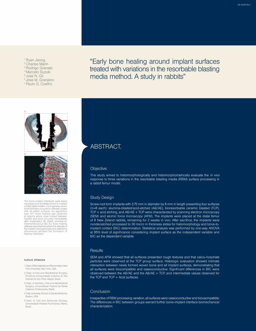
Objective
This study aimed to histomorphologically and histomorphometrically evaluate the in vivo response to three variations in the resorbable blasting media (RBM) surface processing in a rabbit femur model.
Study Design
Screw root form implants with 3.75 mm in diameter by 8 mm in length presenting four surfaces (n=8 each): alumina-blasted/acid-etched (AB/AE), bioresorbable ceramic blasted (TCP), TCP + acid etching, and AB/AE + TCP were characterized by scanning electron microscopy (SEM) and atomic force microscopy (AFM). The implants were placed at the distal femur of 8 New Zeland rabbits, remaining for 2 weeks in vivo. After sacrifice, the implants were nondecalcified processed to 30 micro m thickness slides for histomorphology and bone-to-implant contact (BIC) determination. Statistical analysis was performed by one-way ANOVA at 95% level of significance considering implant surface as the independent variable and BIC as the dependent variable.
Results
SEM and AFM showed that all surfaces presented rough textures and that calciu-hosohate particles were observed at the TCP group surface. Histologic evaluation showed intimate interaction between newly formed woven bone and all implant surfaces, demonstrating that all surfaces were biocompatible and osseoconductive. Significant differences in BIC were observed between the AB/AE and the AB/AE + TCP, and intermediate values observed for the TCP and TCP + Acid surfaces.
Conclusion
Irrespective of RBM processing variation, all surfaces were osseoconductive and biocaompatible. The differences in BIC between groups warrant further bone-implant interface biomechanical characterization.
ABSTRACT.
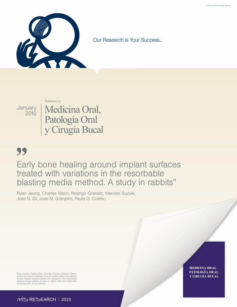
The bone-implant interfaces were easily visualized and facilitated bone-to-implant contact determination. (A) In general, woven bone formation occurred in intimate contact with all tested surfaces. An apposition type (AT) bone healing was observed at regions where close contact between implant and bone occurred immediately after implantation (B, detail), whereas an intramembranous-like (IL) bone healing pathway was observed where the interplay of the implant macrogeometry and osteotomy dimensions allowed the formation of healing chambers.
1 Dept. of Biomaterials and Biomimetics, New York University, New York, USA.
2 Dept. of Oral and Maxillofacial Surgery, Pontificia Universidade Catolica do Rio Grande do Sul, Porto Alegre, Brazil.
3 Dept. of Dentistry, Oral and Maxillofacial Surgery, Universidade Federal de Santa Catarina, Florianopolis, Brazil.
4 Tufts University School of Dental Medicine, Boston, USA.
5 Dept. of Cell and Molecular Biology, Universidade Federal Fluminense, Niteroi, Brazil.
Authors’ affiliations
A
B
MC-RL007 Rev. 1
“Early bone healing around implant surfaces treated with variations in the resorbable blasting media method. A study in rabbits”
1 Ryan Jeong2 Charles Marin3 Rodrigo Granato 4 Marcelo Suzuki 3 Jose N. Gil5 Jose M. Granjeiro1 Paulo G. Coelho
Eitan Barnea, Ido Alt, Roni Kolerman, Joseph Nissan. Accuracy of a laboratory-based computer implant guiding system. Oral Surg Oral Med Oral Pathol Oral Radiol Endod. 2010, 109: e6-e10.
2010
May2010
Oral Surgery, Oral Medicine, Oral Pathology, Oral Radiology and Endodontology
Published in:
Our Research is Your Success...
Accuracy of a laboratory-based computer implant guiding system”Eitan Barnea, DMD; Ido Alt, DMD; Roni Kolerman, DMD; Joseph Nissan, DMD.
© MIS Corporation. All Rights Reserved.
Objective
Computer-guided implant placement is a growing treatment modality in partially and totally edentulous patients, though data about the accuracy of some systems for computer-guided surgery is limited. The purpose of this study was to evaluate the accuracy of a laboratory computer-guided system.
Study design
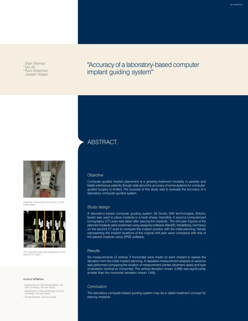
A laboratory-based computer guiding system (M Guide; MIS technologies, Shlomi, Israel) was used to place implants in a fresh sheep mandible. A second computerized tomography (CT) scan was taken after placing the implants . The drill plan figures of the planned implants were positioned using assigned software (Med3D, Heidelberg, Germany) on the second CT scan to compare the implant position with the initial planning. Values representing the implant locations of the original drill plan were compared with that of the placed implants using SPSS software.
Results
Six measurements (3 vertical, 3 horizontal) were made on each implant to assess the deviation from the initial implant planning. A repeated-measurement analysis of variance was performed comparing the location of measurement (center, abutment, apex) and type of deviation (vertical vs. horizontal). The vertical deviation (mean -0.168) was significantly smaller than the horizontal deviation (mean 1.148).
Conclusion
The laboratory computer-based guiding system may be a viable treatment concept for placing implants.
ABSTRACT.
Authors’ affiliations
Implants placement according to the metal tubes.
MC-RL008 Rev. 1
The initial drill plan was positioned on the second CT scan.
“Accuracy of a laboratory-based computer implant guiding system”
1 Eitan Barnea2 Ido Alt3 Roni Kolerman1 Joseph Nissan
1 Department of Oral Rehabilitation, Tel-Aviv University, Tel-Aviv, Israel.
2 Department of Periodontology, Tel-Aviv University, Tel-Aviv, Israel.
3 Private Practice, Tel-Aviv, Israel.
Michael Peleg, Yon Sawatari, Robert N. Marx, Joseph Santoro, Jonathan Cohen, Pablo Bejarano, Theodore Malinin. Use of Corticocancellous Allogeneic Bone Blocks for Augmentation of Alveolar Bone Defects. INT J ORAL MAXILLOFAC IMPLANTS 2010, 25:153-162
2010
January2010
Published in:
Our Research is Your Success...
Use of Corticocancellous Allogeneic Bone Blocks for Augmentation of Alveolar Bone Defects”Michael Peleg, DMD; Yon Sawatari, DDS; Robert N. Marx, DDS; Joseph Santoro, DDS; Jonathan Cohen, DDS; Pablo Bejarano, MD; Theodore Malinin, MD.
© MIS Corporation. All Rights Reserved.
PurposeThe use of autogenous block bone grafts in bone regeneration procedures for alveolar ridge augmentation can be limited by donor site morbidity and complications. The purpose of the present study was to evaluate the efficacy of allogeneic corticocancellous iliac block grafts used for ridge augmentation prior to implant placement.
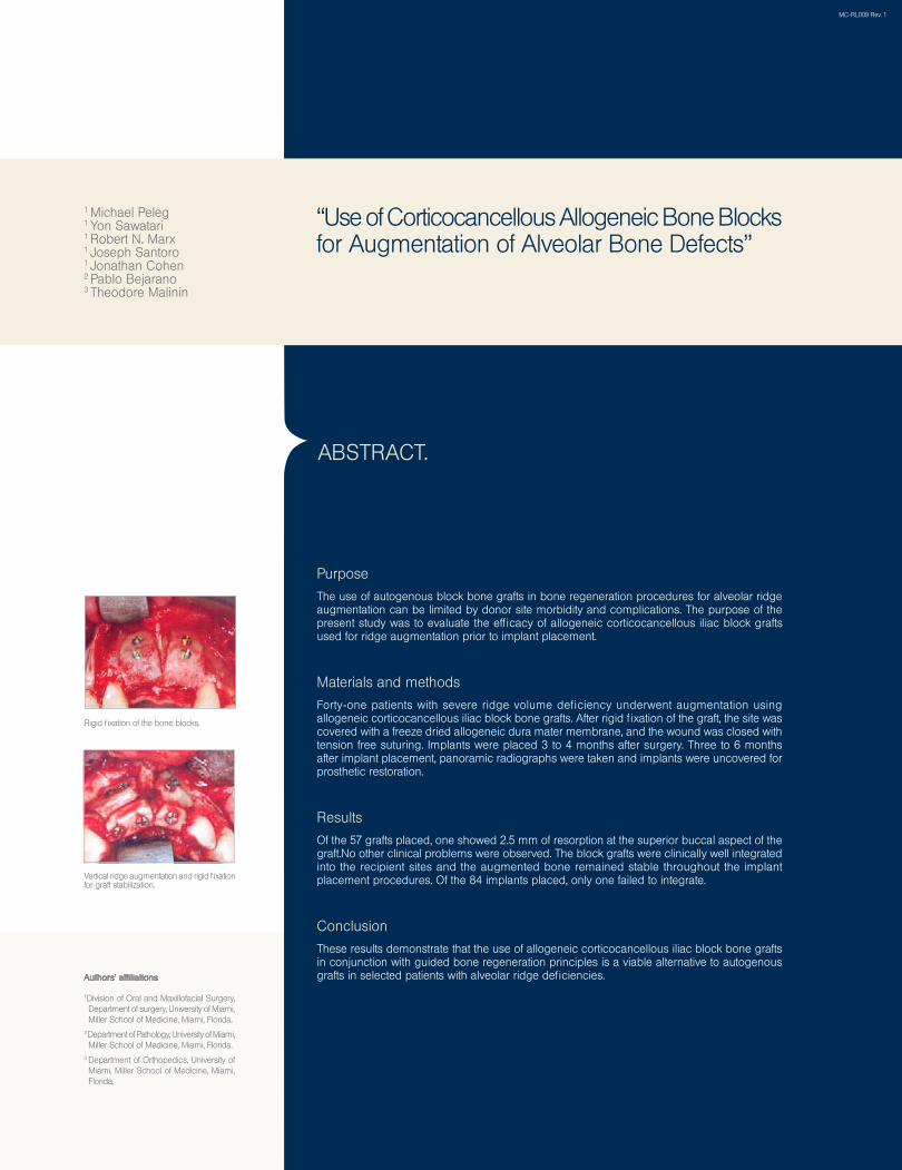
Materials and methodsForty-one patients with severe ridge volume deficiency underwent augmentation using allogeneic corticocancellous iliac block bone grafts. After rigid fixation of the graft, the site was covered with a freeze dried allogeneic dura mater membrane, and the wound was closed with tension free suturing. Implants were placed 3 to 4 months after surgery. Three to 6 months after implant placement, panoramic radiographs were taken and implants were uncovered for prosthetic restoration.
ResultsOf the 57 grafts placed, one showed 2.5 mm of resorption at the superior buccal aspect of the graft.No other clinical problems were observed. The block grafts were clinically well integrated into the recipient sites and the augmented bone remained stable throughout the implant placement procedures. Of the 84 implants placed, only one failed to integrate.
ConclusionThese results demonstrate that the use of allogeneic corticocancellous iliac block bone grafts in conjunction with guided bone regeneration principles is a viable alternative to autogenous grafts in selected patients with alveolar ridge deficiencies.
ABSTRACT.
1Division of Oral and Maxillofacial Surgery, Department of surgery, University of Miami, Miller School of Medicine, Miami, Florida.
2 Department of Pathology, University of Miami, Miller School of Medicine, Miami, Florida.
3 Department of Orthopedics, University of Miami, Miller School of Medicine, Miami, Florida.
Authors’ affiliations
Vertical ridge augmentation and rigid fixation for graft stabilization.
Rigid fixation of the bone blocks.
MC-RL009 Rev. 1
“Use of Corticocancellous Allogeneic Bone Blocks for Augmentation of Alveolar Bone Defects”
1 Michael Peleg 1 Yon Sawatari 1 Robert N. Marx 1 Joseph Santoro 1 Jonathan Cohen 2 Pablo Bejarano3 Theodore Malinin
Charles Marin, Rodrigo Granato, Marcelo Suzuki, Malvin N. Janal, Jose N. Gil, Carlos Nemcovsky, Estevam A. Bonfante, Paulo G. Coelho. Biomechanical and histomorphometric analysis of etched and non-etched resorbable blasting media processed implant surfaces: An experimental study in dogs. J Mech Behav Biomed Mater. 2010, 3(5): 382-91.
2010
July2010
Journal of the Mechanical Behavior Of Biomedical Materials.
Published in:
Our Research is Your Success...
Biomechanical and histomorphometric analysis of etched and non-etched resorbable blasting media processed implant surfaces: An experimental study in dogs”Charles Marin, Rodrigo Granato, Marcelo Suzuki, Malvin N. Janal, Jose N. Gil, Carlos Nemcovsky, Estevam A. Bonfante, Paulo G. Coelho.
© MIS Corporation. All Rights Reserved.
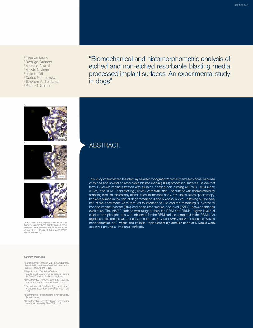
This study characterized the interplay between topography/chemistry and early bone response of etched and no- etched resorbable blasted media (RBM) processed surfaces. Screw- root form Ti-6Al-4V implants treated with alumina blasting/acid -etching (AB/AE), RBM alone (RBM), and RBM + acid -etching (RBMa) were evaluated. The surface was characterized by scanning electron microscopy, atomic force microscopy, and X- ray photoelectron spectroscopy. Implants placed in the tibia of dogs remained 3 and 5 weeks in vivo. Following euthanasia, half of the specimens were torqued to interface failure and the remaining subjected to bone -to -implant contact (BIC) and bone area fraction occupied (BAFO) between threads evaluation. The AB/AE surface was rougher than the RBM and RBMa. Higher levels of calcium and phosphorous were observed for the RBM surface compared to the RBMa. No significant differences were observed in torque, BIC, and BAFO between surfaces. Woven bone formation at 3 weeks and its initial replacement by lamellar bone at 5 weeks were observed around all implants’ surfaces.
ABSTRACT.
1 Department of Oral and Maxillofacial Surgery, Pontificia Universidade Catolica do Rio Grande do Sul, Porto Alegre, Brazil.
2 Department of Dentistry, Oral and Maxillofacial Surgery, Universidade Federal de Santa Catarina, Florianopolis, Brazil.
3 Department of Prosthodontics, Tufts University School of Dental Medicine, Boston, USA.
4 Department of Epidemiology and Health Promotion, New York University, New York, USA.
5 Department of Periodontology, Tel Aviv University, Tel Aviv, Israel.
6 Department of Biomaterials and Biomimetics, New York University, New York, USA.
At 5 weeks, initial replacement of woven bone by lamellar bone (lightly stained bone between threads) was observed for all the (A) AB/AE, (B) RBM, (C) RBMa groups (color on the Web only).
A
B
Authors’ affiliations
MC-RL010 Rev. 1
“Biomechanical and histomorphometric analysis of etched and non-etched resorbable blasting media processed implant surfaces: An experimental study in dogs”
1 Charles Marin2 Rodrigo Granato3 Marcelo Suzuki 4 Malvin N. Janal 2 Jose N. Gil5 Carlos Nemcovsky6 Estevam A. Bonfante 6 Paulo G. Coelho
C
400um
400um
400um
2010
Our Research is Your Success...
Interaction between osteoblasts and titanium surfaces: application in dental implants”Juan Carlos Vanegas Acosta, Diego Garzón-Alvarado, Martín Casale
© MIS Corporation. All Rights Reserved.
Juan Carlos Vanegas Acosta, Diego Garzón-Alvarado, Martín Casale, Interaction between osteoblasts and titanium surfaces: application in dental implants. Rev. Cubana. Invest. Bioméd. 2010, 29(1): 51-68.
*
March 2010
*
Published in:
Re v i s t a C u b a n a d e
I N V E S T I G A C I O N E S
The bone integration of dental implants is dependent of type of material of implant, its superficial topography and also by the type of coating. Although there are many the materials used for the manufacture of dental implants, currently the titanium is the choice material by its inert behavior when it contact with biological tissues. As the superficial topography influenced on cellular adhesion and proliferation, the dental implants underwent treatments creating superficial irregularities in the micro and sub-micrometric scale. Likewise, the coats based on organic-origin minerals are used to improve the bone deposition on implant.
The aim of present paper is to carry out the osteoblasts cells culture on Ti-6AI-4V substrates using four different types surfaces typically used in dental implants to determine which of them have the better behavior in terms of cellular adhesion, cellular proliferation and biomineralization.
The osteoblasts cells were cultivated during 24 hr for the adhesion assay and during 1, 2, 5, 6 and 7 days for the proliferation assay. The biomineralization was assessed by characterization with estereozoom and SEM of the mineral depots colored using dying technique with red alizarin. The quantization of these mineralization depots was performed using a tool of images processing. The results obtained showed that the rough surfaces and coated have a better behavior. None of the study surfaces had a toxic character for cells and all showed cellular adhesion profiles, cellular proliferation and biomineralization.
ABSTRACT.
1Group of Modeling and Numerical Methods in Engineering-GNUM, National University of Colombia, Bogota, Colombia.2Faculty of Engineering, National University of Colombia, Bogota, Colombia.3School of Dentistry, National University of Colombia, Bogota, Colombia.
Authors’ affiliations
“Interaction between osteoblasts and titanium surfaces: application in dental implants”
1Juan Carlos Vanegas Acosta2Diego Garzón-Alvarado3Martín Casale
MC-RL011 Rev. 1
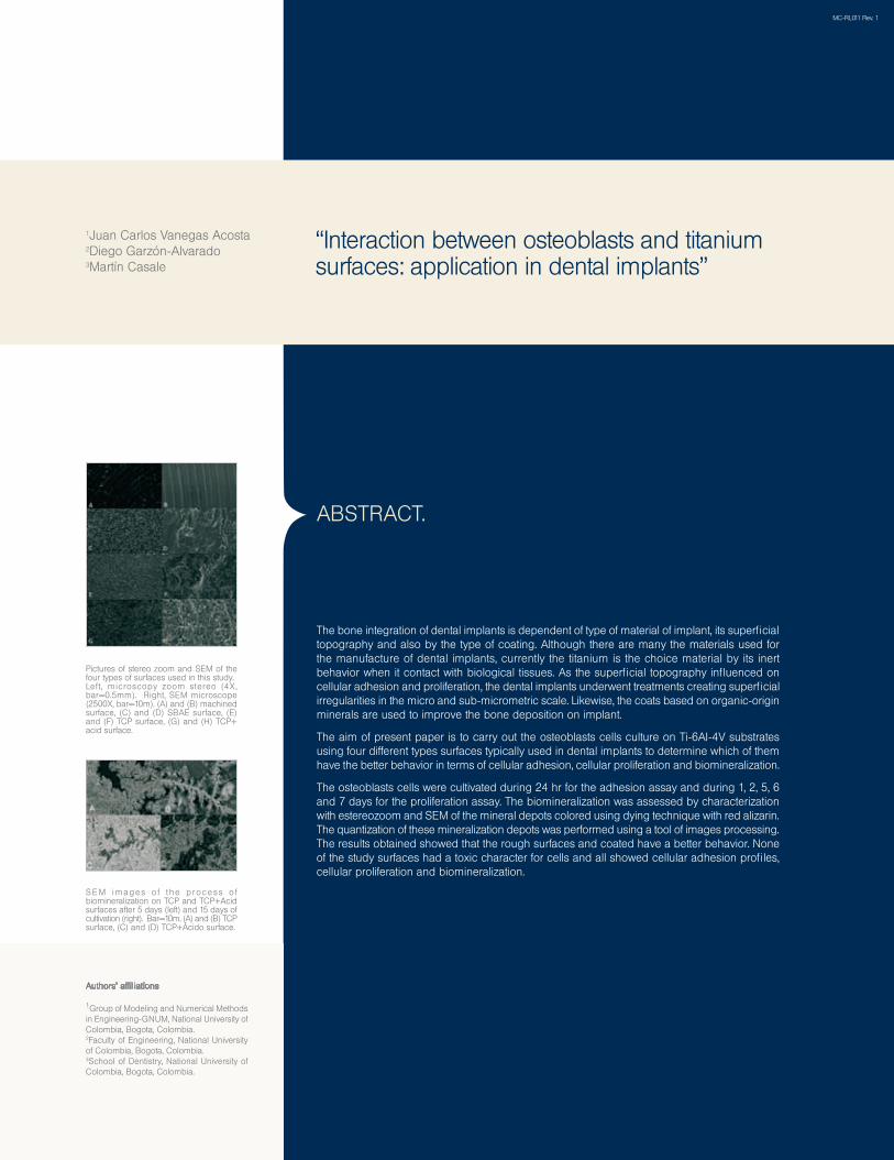
SE M images o f t he p rocess o f biomineralization on TCP and TCP+Acid surfaces after 5 days (left) and 15 days of cultivation (right). Bar=10m. (A) and (B) TCP surface, (C) and (D) TCP+Ácido surface.
Pictures of stereo zoom and SEM of the four types of surfaces used in this study.Left , microscopy zoom stereo (4X, bar=0.5mm). Right, SEM microscope (2500X, bar=10m). (A) and (B) machined surface, (C) and (D) SBAE surface, (E) and (F) TCP surface, (G) and (H) TCP+ acid surface.
Get a free abutment, abutment analog and an abutment transfer with each implant…
Horwitz J, Zuabi O, Machtei EE. Accuracy of a computerized tomography-guided template-assisted implant placement system: an in vitro study. Clin. Oral Impl. Res. xx, 2009; 000–000.doi: 10.IIII/j.I600-050I.2009.0I748.x
2010
September2010
Published in:
Our Research is Your Success...
Titanium hydride and hydrogen concentration in acid-etched commercially pure titanium and titanium alloy implants: a comparative analysis of five implant systems”S. Szmukler-Moncler, M. Bischof, R. Nedir, M. Ermrich
© MIS Corporation. All Rights Reserved.
S. Szmukler-Moncler, M. Bischof, R. Nedir, M. Ermrich. Titanium hydride and hydrogen concentration in acid-etched commercially pure titanium and titanium alloy implants: a comparative analysis of five implant systems. Clinical Oral Implants Research. 2010, 21(9): 944-950.
*
*
Objectives
Acid etching is a popular method to texture the surface of dental implants. During etching, the titanium oxide protective layer is dissolved and small native hydrogen ions diffuse into the unprotected implant surface. They enrich the implant surface with hydrogen and precipitate into titanium hydride (TiH). The aim of this study was to measure the concentration of TiH at the implant surface and the total concentration of Hydrogen at five commercially available implant systems, made of either commercially pure (cp) titanium or titanium alloy.
Materials and methods
X-Ray diffraction (XRD) was conducted on each implant system to determine the compounds present at the implant surface. Following a TiH2/Ti calibration curve, the concentration of TiH was determined. Concentration of hydrogen in the implants was measured by the inert gas fusion thermal conductivity/infrared detection method.
Results
XRD data showed that TiH was present on all cp titanium implants but not on the alloyed implants. TiH concentration varied between 5% and 37%. Hydrogen concentration varied between 43 and 108 ppm, no difference in uptake was found between the cp titanium and alloyed implants. Low solubility of hydrogen in α-titanium is responsible for precipitation into TiH. Stronger etching conditions led to higher concentration of TiH2-x.
Conclusions
High solubility of hydrogen in the β-phase of the alloy is preventing hydrogen from precipitating into TiH. All implants, even those lacking TiH at the surface, were enriched with hydrogen. In all implants, hydrogen concentration was within the normative limit of 130 ppm.
ABSTRACT.
1Department of Stomatology & Maxillo-Facial Surgery, University of Paris, Paris, France. 2Department of Odontology, Section of Implantology and Oral Rehabilitation, Galeazzi Orthopeadic Institute, University of Milano, Milano, Italy.3Ardentis Clinique Dentaire Vevey, Swiss Dental Clinics Group, 3 rue du Col le `ge , Vevey, Swi tzer land. 4Department of Stomatology and Oral Surgery, School of Dental Medicine, University of Geneva, Geneva, Switzerland. 5X-Ray Laboratory Dr Ermrich, Reinheim/ Odw, Germany.
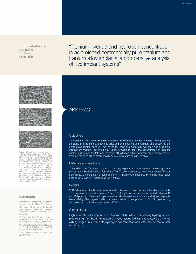
Scanning electron microscopic micrographs of the implant surfaces. (A) Ankylos Cell-Plus implant. Strong etching conditions have carved the commercially pure (cp) Ti surface with pores. Line is 10µm (originalmagnification x 2000). (B) Straumann SLActive implant. Sandblasting and etching with boiling acids created this complex carved topography. The line is 10µm (original magnification x 2000). (C) MIS BioCom implant. Strong etching conditions dissolved the b-phase as well and carved pores in the alloyed material. At this magnification, the effect of sandblasting is not evidenced on this picture. The line is 10µm (original magnification x 2000).
C.
A.
B.
Authors’ affiliations
“Titanium hydride and hydrogen concentration in acid-etched commercially pure titanium and titanium alloy implants: a comparative analysis of five implant systems”
1,2S. Szmukler-Moncler3,4M. Bischof 3,4R. Nedir 5M. Ermrich
MC-RL012 Rev. 1
Get a free abutment, abutment analog and an abutment transfer with each implant…
Horwitz J, Zuabi O, Machtei EE. Accuracy of a computerized tomography-guided template-assisted implant placement system: an in vitro study. Clin. Oral Impl. Res. xx, 2009; 000–000.doi: 10.IIII/j.I600-050I.2009.0I748.x
2010
September2010
Published in:
Our Research is Your Success...
Skeletal Implant Anchorage in the Treatment of Impacted Teeth-A Review of the State of the Art”Stella Chaushu and Gavriel Chaushu
© MIS Corporation. All Rights Reserved.
Stella Chaushu, Gavriel Chaushu. Skeletal Implant Anchorage in the Treatment of Impacted Teeth-A Review of the State of the Art. Seminars in Orthodontics. 2010, 16(3): 234-241.
*
*
The surgical-orthodontic approach is the most commonly used modality in patients with impacted teeth. It is also the most challenging. The prognosis of treatment is uncertain, treatment takes much longer and is more painful, and the enhanced biomechanical efforts required to bring the tooth to its ideal position frequently cause deleterious side effects to the remainder of the dentition. Skeletal anchorage is a useful aid in these cases. It can be judiciously exploited to assess the prognosis of canine movement, open adequate space for the canine, and build up a reliable anchorage unit that will allow the application of controlled directional forces with fewer side effects on the anchorage teeth. Treatment is facilitated and shortened because the resolution of the impaction may be performed separately before or even simultaneously with treatment of the remainder of the dentition, as 2 distinct force systems. The present article reviews some of the main indications for implant anchorage in treatment of impacted teeth.
ABSTRACT.
1Department of Orthodontics, Hebrew University-Hadassah School of Dental Medicine, Jerusalem, Israel. 2Department of Oral and Maxillofacial Surgery, Maurice and Gabriela Goldschleger School of Dental Medicine, Tel-Aviv University, Tel-Aviv, Israel.
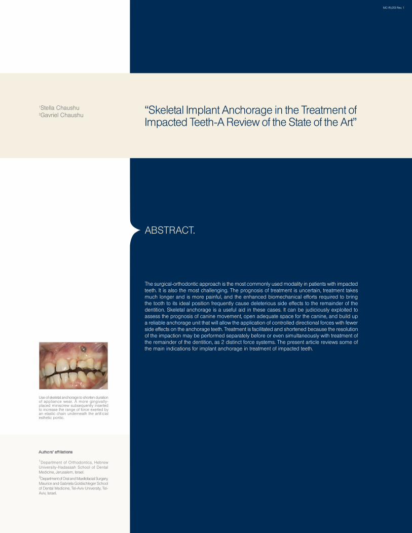
Use of skeletal anchorage to shorten duration of appliance wear. A more gingivally-placed miniscrew subsequently inserted to increase the range of force exerted by an elastic chain underneath the artificial esthetic pontic.
Authors’ affiliations
“Skeletal Implant Anchorage in the Treatment of Impacted Teeth-A Review of the State of the Art”
1Stella Chaushu 2Gavriel Chaushu
MC-RL013 Rev. 1
Get a free abutment, abutment analog and an abutment transfer with each implant…
2010
September2010
Published in:
Our Research is Your Success...
A comparative study of bone densitometry during osseointegration: Piezoelectric surgery versus rotary protocols”
(Quintessence International)
Luca Di Alberti, DDS, PhD; Federica Donnini, RDH; Claudio Di Alberti, DDS; Michele Camerino, DDS
© MIS Corporation. All Rights Reserved.
Luca Di Alberti, Federica Donnini, Claudio Di Alberti, Michele Camerino. A comparative study of bone densitometry during osseointegration: Piezoelectric surgery versus rotary protocols.Quintessence International. 2010, 41(8): 639-641.
*
*
Objectives
To date, there have been no studies on the outcome of osseointegration of alveolar bone around dental implants inserted with piezoelectric osteotomy versus conventional osteotomy. The aim of this study was to compare the radiographic differences, through evaluation of peri-implant bone density, between implant insertion using traditional surgical technique and piezoelectric technique.
Materials and methods
Forty patients were selected whose treatment consisted of a minimum of two implants placed in nonpathologic native bone. A single type of implant surface (SLA) was chosen. The implants were placed following the manufacturer protocol for traditional surgical technique and piezoelectric technique. Radiographs were taken following surgery and 30, 60, and 90 days after surgery. The bone density was studied with the densitometry application.
Results and Conclusions
All patients completed the study period with success. Despite a limited numver of treated patients, the results of this pilot study demonstrated that (1) piezoelectric implant site preparation promotes better bone density and osteogenesis, and (2) the piezoelectric technique is predictable, with a 100% success rate in this study.
ABSTRACT.
1Department of Dental Sciences, University of Foggia, Foggia, Italy.2Pescara, Italy.3Chieti, Italy.
Authors’ affiliations
“A comparative study of bone densitometry during osseointegration: Piezoelectric surgery versus rotary protocols”
1Luca Di Alberti2Federica Donnini2Claudio Di Alberti3Michele Camerino
MC-RL014 Rev. 1
Omer Cohen, Eran Gabay, Eli E. Machtei. Cooling Profile Following Prosthetic Preparation of 1-Piece Dental Implants. Journal of Oral Implantology. 2010, 36(4): 273-279.
*
2010
Published in:
Our Research is Your Success...
Cooling Profile Following ProstheticPreparation of 1-Piece Dental Implants”Omer Cohen, DMD, PhD; Eran Gabay, DMD, PhD; Eli E. Machtei, DMD
(Journal of Oral Implantology)
© MIS Corporation. All Rights Reserved.
August2010
*
JOI
The aim of this study was to evaluate the effect of water irrigation on heat dissipation kinetics following abutment preparation of 1-piece dental implants. UNO 1-piece dental implants were mounted on Plexiglas apparatus clamping the implant at the collar. T-type thermocouple was attached to the first thread of the implant and recorded thermal changes at 100 millisecond intervals. Implants were prepared using highspeed dental turbine at 400 000 RPM with a coarse diamond bur. Once temperature reached 47ºC, abutment preparation was discontinued.
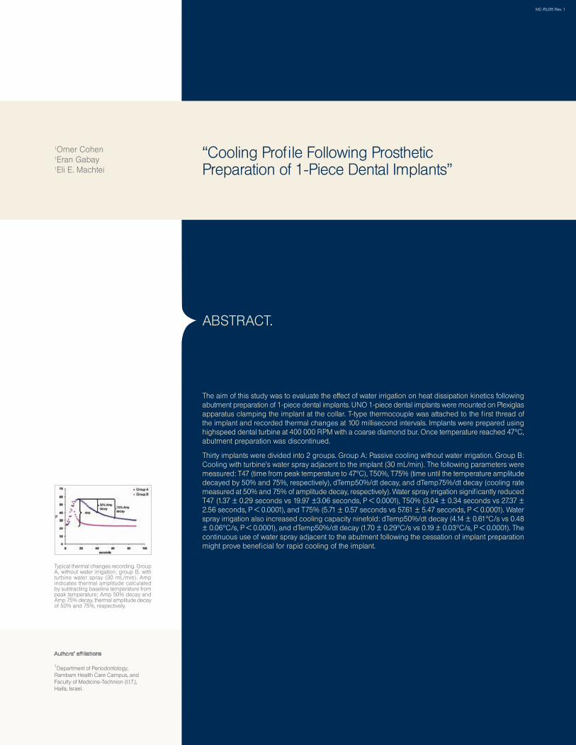
Thirty implants were divided into 2 groups. Group A: Passive cooling without water irrigation. Group B: Cooling with turbine’s water spray adjacent to the implant (30 mL/min). The following parameters were measured: T47 (time from peak temperature to 47ºC), T50%, T75% (time until the temperature amplitude decayed by 50% and 75%, respectively), dTemp50%/dt decay, and dTemp75%/dt decay (cooling rate measured at 50% and 75% of amplitude decay, respectively). Water spray irrigation significantly reduced T47 (1.37 ± 0.29 seconds vs 19.97 ±3.06 seconds, P < 0.0001), T50% (3.04 ± 0.34 seconds vs 27.37 ± 2.56 seconds, P < 0.0001), and T75% (5.71 ± 0.57 seconds vs 57.61 ± 5.47 seconds, P < 0.0001). Water spray irrigation also increased cooling capacity ninefold: dTemp50%/dt decay (4.14 ± 0.61ºC/s vs 0.48 ± 0.06ºC/s, P < 0.0001), and dTemp50%/dt decay (1.70 ± 0.29ºC/s vs 0.19 ± 0.03ºC/s, P < 0.0001). The continuous use of water spray adjacent to the abutment following the cessation of implant preparation might prove beneficial for rapid cooling of the implant.
ABSTRACT.
1Department of Periodontology, Rambam Health Care Campus, and Faculty of Medicine-Technion (I.I.T.), Haifa, Israel.
Typical thermal changes recording. Group A, without water irrigation; group B, with turbine water spray (30 mL/min). Amp indicates thermal amplitude calculated by subtracting baseline temperature from peak temperature; Amp 50% decay and Amp 75% decay, thermal amplitude decay of 50% and 75%, respectively.
Authors’ affiliations
“Cooling Profile Following Prosthetic Preparation of 1-Piece Dental Implants”
1Omer Cohen1Eran Gabay1Eli E. Machtei
MC-RL015 Rev. 1
Get a free abutment, abutment analog and an abutment transfer with each implant…
2010
November2010
Published in:
Our Research is Your Success...
Clinical evaluation of particulate allogeneic with and without autogenous bone grafts and resorbable collagen membranes for bone augmentation of atrophic alveolar ridges”Ilan Beitlitum, Zvi Artzi, Carlos E. Nemcovsky
© MIS Corporation. All Rights Reserved.
Beitlitum I, Artzi Z, Nemcovsky CE. Clinical evaluation of particulate allogeneic with and without autogenous bone grafts and resorbable collagen membranes for bone augmentation of atrophic alveolar ridges. Clin. Oral Impl. Res. 2010, 21(11): 1242–1250.
*
*
Objectives
To evaluate the clinical outcome of bone augmentation with the use of particulate mineralized freeze-dried bone allograft (FDBA) with or without the addition of autogeneous bone chips, applied in a bi-layered (BL) technique, covered by a resorbable cross-linked collagen membrane.
Materials and methods
Fifty patients presenting with a vertical and/or lateral ridge deficiency of at least 3mm were included: Group FDBA, N=27 patients, particulate FDBA was the only graft; and Group BL, N=23 patients, a BL bone grafting procedure where autogenous bone chips were the inner layer and FDBA the outer. Bone graft was covered with a ribose cross-linked collagen barrier membrane. Ridge dimensions were clinically or radiographically (computerized tomography scan) measured at the time of the bone augmentation procedure and at implant placement or uncovering and the maximum linear vertical or horizontal calcified tissue gain was calculated. Statistical analysis consisted of linear regression analysis, with maximum bone gain being the dependent variable.
Results
In the FDBA group, mean vertical bone gain was 3.47mm (SD 1.25) and the horizontal, 5mm (SD 1.28), while in the BL values were 3.5mm (SD 1.2) and 3.6mm (SD 1.72), respectively. Addition of autogenous bone does not appear to statistically significantly enhance the results. Spontaneous membrane exposure occurred in 24% of the cases and was the only variant that significantly influenced results (P<0.001).
Conclusions
Large vertical and/or horizontal ridge deficiencies may be treated with FDBA and ribose cross-linked collagen barrier membranes with good clinical outcome. No added effect of the application of a layer of autogenous bone in these bone augmentation procedures could be demonstrated. Spontaneous membrane exposure was the only parameter to affect the degree of new calcified tissue formation.
ABSTRACT.
1Department of Periodontology, The Maurice and Gabriela Goldschleger School of Dental Medicine, Tel Aviv University, Tel Aviv, Israel.
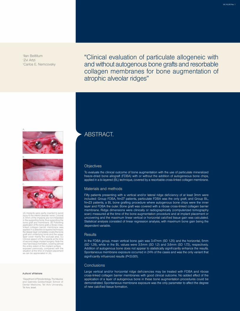
(A) Implants were partly inserted to avoid injury to the inferior alveolar nerve. Coronal 3–4mm of the implants are not imbedded in the supporting bone, thus supporting the bone graft and membrane. (B) Following application of the bone graft, a ribose cross-linked collagen barrier membrane was applied in a selective bi-layered technique; the deeper layer completely covers the bone graft and underlying bone and the upper layer cover mainly the occlusal area. (C) Clinical aspect of the implants at the time of second-stage implant surgery. Note the new hard tissue formation, covering almost the entire extent of the implant surfaceexposed previously, compared with the situation at the time of implant placement as can be appreciated in (A).
Authors’ affiliations
“Clinical evaluation of particulate allogeneic with and without autogenous bone grafts and resorbable collagen membranes for bone augmentation of atrophic alveolar ridges”
1Ilan Beitlitum1Zvi Artzi1Carlos E. Nemcovsky
MC-RL016 Rev. 1
C.
B.
A.