401 Sent. Que Dejo en Firme Laudo Telebucaramanga e Innovatec
Universidad de Navarra
Facu l tad de Medic ina
Studies on the role of HDAC5, SIRT2 and
VGLUT1 as pharmacological targets involved
in antidepressant action
Irene Muñoz-Cobo Orosa
Pamplona, Octubre 2017

Universidad de Navarra
Studies on the role of HDAC5, SIRT2 and
VGLUT1 as pharmacological targets involved in
antidepressant action
Irene Muñoz-Cobo Orosa
Pamplona, Octubre 2017

Irene Muñoz-Cobo Orosa
U n i v e r s i d a d d e N a v a r r a
F a c u l t a d d e M e d i c i n a
Studies on the role of HDAC5, SIRT2 and VGLUT1
as pharmacological targets involved in
antidepressant action
Memoria presentada por Dª Irene Muñoz-Cobo Orosa para aspirar al
grado de Doctor por la Universidad de Navarra
El presente trabajo ha sido realizado bajo mi dirección en el Departamento
de Farmacología y Toxicología y autorizo su presentación ante el Tribunal
que lo ha de juzgar.
Pamplona, 06 de octubre de 2017
Dra. Rosa María Tordera Baviera


Para mis padres y mi tata.
A mi presente.
A mi pasado.
A mi futuro.


ACKNOWLEDGMENTS
“Observar sin pensar es tan peligroso como pensar sin observar."
Santiago Ramón y Cajal


Quizá sea más poetisa que neurocientífica o me resulta más natural escribir de este modo, aunque
no sencillo. Al escribir agradecimientos lo primero que ven mis ojos y siente mi corazón son las
palabras AGRADECER y CIMIENTOS. Creo que no hay dos palabras que puedan definir
mejor estas líneas.
Yo tengo mucho que agradecer en todo. Me siento privilegiada desde que nací, desde cada instante
en el que se me ha brindado una oportunidad, tanto merecida como regalada.
En primer lugar quiero agradecer:
A la Universidad de Navarra, concretamente a la Facultad de Farmacia donde recibí gran parte
de mi formación académica, y a la Facultad de Medicina donde he llevado a cabo mi máster y mi
programa de doctorado. He recibido una excelente formación.
A la Subdirección General de Recursos Humanos para la Investigación del Ministerio de
Economía y Competitividad del Gobierno de España por concederme la ayuda económica
necesaria para realizar mi tesis.
Al Departamento de Farmacología y Toxicología, especialmente a la parte de “Farma”, por todo
el apoyo que he tenido desde que empecé el máster. Y, por supuesto, a las numerosas personas de
otros departamentos que me han ayudado tan desinteresadamente a poder desarrollar este trabajo.
En segundo lugar, quiero ante todo dar las gracias en estas líneas a mi familia, siempre los más
importantes. Gracias por ser la mejor que me podría haber tocado. Tengo más de 1000 gracias,
10000 mil gracias e ∞ os quiero. Porque sin vosotros, mamá, papá y Tatiz, sin vosotros ninguna
de estas líneas cobraría vida y ninguna de estas palabras tendría todo su sentido. Gracias por
dárselo y devolvérmelo cuando no lo he sabido verlo con claridad.
También gracias a todos los amigos que han hecho suyo mi doctorado y que me han animado a
dar lo mejor de mí. Y a mi chico, que cuando he tenido un día más sensible sintiendo que no podía
con todo, también me ha dado el sí puedes. A todos mis amigos, gracias eternas por estar a mi
lado.
No quiero hacer una memoria de cada etapa de estos 5 años vividos en el Depar de Farma. Supe
que era el mejor sin duda al empezar el máster. No voy a decir nombres o dedicar a cada uno
individualmente unas palabras como hice en el proyecto de final de máster porque sé que cada
persona que me lea va a verse reflejada.
Algunas veces he sido buena doctoranda, aunque no siempre me ha sido fácil tomar mi “auto-
relevo” en esta carrera de fondo. En todo momento me habéis guiado y no siempre he estado
preparada, sobre todo para mí… He aprendido mucho, muchísimo, de todos y por eso sé que a
quién más debo y puedo exigir es a mí misma. Ésta ha sido una gran lección.
Por todo esto… GRACIAS. Gracias Rosa por ser mi amiga, mi guía, mi tutora o “jefa” (como
no te gusta que te llame) porque no podría haber tenido a nadie mejor tan cerca de mí.
Eternamente gracias por apostar por mí y pelear sin que yo estuviera siempre a tu lado.
Muchas gracias a todos por estar, por ayudarme, por animarme, por impulsarme… Cuando leo
las palabras que os dediqué al finalizar mi trabajo de fin de máster y comenzar el doctorado, veo
mi crecimiento y soy consciente de todo el esfuerzo.
Igual que os doy las gracias, dejo aquí escrita mi más sincera disculpa por los momentos en los que
no he sido esa Irene que empezó tan llena de ilusión. Pero no me quedo en ese estado más triste,
vuelvo a daros los abrazos presentando mi gratitud en todo.
Aunque pueda parecer extraño, quiero dar las gracias a todos los animalitos que, sin saberlo,
contribuyen tanto a nuestra investigación, mi más sincero respeto hacia todos ellos.
Resumiendo para todos, y como ya que sabéis que soy una persona sensible, se la dedico a cada
persona que ha dado sentido a mis lágrimas y mis sonrisas porque mi corazón estaba en cada una
de ellas. Se me da mejor escribir poesías que tesis, así que esta poesía es para todos vosotros:

Agradezco mi cimiento
que son horas a tu lado
duras, en casa, no en vano
salvándonos los momentos.
Ir y venir, movimiento,
surgir al desvanecernos
preparando cada paso
de sutil enraizamiento.
Literatura velando
el fácil conocimiento
de la ciencia al resurgir
con un nuevo experimento.
Gracias por estar ahí
donde seguiréis creciendo
aquí hoy quiero transmitir
lo adquirido en este tiempo.


ABREVIATTIONS


ABREVIATTIONS
5-HT 5-Hydroxytryptamine, serotonin
AcH Acetylated histone
AAV Adeno-Associated virus
BDNF Brain-derived neurotrophic factor
DSM-V Diagnostic and statistical manual of mental disorders, 5th edition
DRN Dorsal raphe nucleus
GABA -aminobutyric acid
HDAC Histone deacetylase
HAT Histone acetyltransferase
IL-PFC Infralimbic prefrontal cortex
MAOI Monoaminoxidase inhibitor
MAO Monoaminoxidase
MDD Major depression disorder
MRN Medial raphe nucleus
NA Noradrenaline
NAD Nicotinamide adenine dinucleotide
NMDA N-methyl-d-aspartate
PFC Prefrontal cortex
p-HDAC5 Phospho-histone deacetylase 5

RT-PCR Real-Time PCR
SIRT Sirtuin
SNRI Selective noradrenaline reuptake inhibitor
SSRI Selective serotonin reuptake inhibitor
Syn Synapsin
TLDA Taqman low density array
VGLUT Vesicular glutamate transporter
WT Wild type
YFP Yellow fluorescence protein

Abstract
Depression is a chronic disorder characterized mainly by depressive mood and
anhedonia. The monoaminergic hypothesis has resulted deficient to explain the
biological changes that may be caused in the depressive brain. Other hypotheses like
alterations in the neuroplasticity, in glutamate transmission, as well as in epigenetics
have been proposed. Antidepressant action has been linked to increased synaptic
plasticity in which epigenetic mechanisms such as histone posttranslational acetylation
could be involved. Interestingly, previous studies in our laboratory have shown that the
histone deacetylases HDAC5 and SIRT2 are oppositely regulated by stress and
antidepressants in mice prefrontal cortex (PFC). Besides, the neuroblastoma SH-SY5Y
line is an in vitro neuronal model reliable to study drug effects with clear advantages
over animal models. This study focuses on the possible role of two selected epigenetic
targets (histone deacetylase 5, HDAC5, and sirtuin 2, SIRT2) and one glutamate target
(vesicular glutamate transporter 1, VGLUT1) in antidepressant action.
The first aim was to further explore the role of HDAC5 and SIRT2 in the
molecular mechanisms of antidepressants using the in vitro SH-SY5Y cellular model.
This study shows that nucleocytoplasmic export of HDAC5 and SIRT2 downregulation
mediated by antidepressants could enhance synaptic plasticity markers leading to
antidepressant action.
The second aim of this study was to evaluate the therapeutic potential of SIRT2
for depression treatment. A course of treatment with the selective SIRT2 inhibitor 33i
reversed anhedonia in mice heterozygous for the vesicular glutamate transporter 1
(VGLUT1+/-), considered a genetic model of depression.
In parallel, we initiated a study directed to examine the role of the glutamate
target VGLUT1 in the long-loop mechanisms of control of 5-HT activity as well as in the
modulation of depressive-like behaviors. Using the adeno-associated virus (AAV)
technology, VGLUT1 expression was induced in the PFC of VGLUT1+/-. Interestingly,
these preliminary studies have evidenced that VGLUT1 expression in the PFC of
VGLUT1+/- mice is linked to antidepressant action.
Altogether, HDAC5 and SIRT2 as well as the synaptic plasticity marker VGLUT1
could be proposed as pharmacological targets involved in antidepressant action.


CONTENTS


INTRODUCTION 01
1. What is major depression? 03
2. Pharmacological treatment of major depression 04
3. Neurobiology of major depression 06
3.1. The monoaminergic hypothesis of major depression 06
3.2. Glutamate hypothesis of major depression 07
3.2.1. NMDA receptors 07
3.2.2. The vesicular glutamate transporter 1 08
3.2.3. Prefrontal cortex glutamate modulation of 5-HT activity in raphe 09
3.3. GABAergic hypothesis 11
3.4. Neuroplasticity hypothesis 11
3.5. Epigenetics and depression 13
3.5.1. HDAC5 in depression and antidepressant action 16
3.5.2. SIRT2 in depression and antidepressant action 17
3.6. Other hypothesis 18
4. The SH-SY5Y neuroblastoma cell line as an in vitro neuronal model: study of the
mechanisms of antidepressant 19
APPROACH AND OBJECTIVES 21
MATERIAL AND METHODS 27
1. Cell cultures 29
2. Animals 29
3. Drugs and treatments 30
4. Experimental design 31
4.1. Experimental design 1: Role of HDAC5 and SIRT2 in the molecular
mechanisms of antidepressant 31
4.2. Experimental design 2: Effect of SIRT2 inhibition on anhedonic behaviour
of the VGLUT1+/- depression model 32
4.3. Experimental design 3: Effect of VGLUT1-induced expression in the
PFC in antidepressant action 33
5. Enantioseparation of MC3822 and MC3823 34
6. HDAC1-9 isoforms inhibition assay 36
7. RT-PCR studies 36
8. Western Blot studies 37
9. Immunofluorescence studies in SH-SY5Y cells 39
10. TaqMan Low Density Arrays (TLDAs) 39
11. In vitro affinity assays 41
12. Monoaminooxidase activity assay 42
13. Adeno-associated viral (AAV) vector generation 42
14. Expression of pAAV-pSyn-VGLUT1mCherryminisog-SMD2 in primary
cultures 45
15. Stereotaxic injections of pAAV-pSyn-VGLUT1mCherryminisog and
pAAV-pSyn-YFP 45
16. Imaging of VGLUT1mCherryminisog and YFP expression in the brain 46
17. Sucrose intake test 47
18. Behavioural tests 48
19. Body temperature studies: 8-OH-DPAT induced hypothermia 49
20. Statistical analysis 49
RESULTS 51
1. Role of HDAC5 and SIRT2 in the molecular mechanisms of antidepressants 53
1.1. Antidepressant treatment regulate HDAC5 in vitro 53
1.2. MC3822 and MC3823 affinity to the different HDAC enzymes 56
1.3. HDAC5 inhibition enhances histone acetylation and synaptic plasticity
markers 57
1.4. Antidepressant regulation of SIRT2 in vitro 59
1.5. SIRT2 inhibition enhances histone acetylation and synaptic plasticity
markers 60

1.6. Effect of MC3822 and 33i on HDAC5 and SIRT2 expression in vitro 62
1.7. Antidepressant treatment regulate histone acetylation and synaptic plasticity
markers in vitro 64
1.8. Muscarinic receptor blockade does not affect histone acetylation and synaptic
plasticity markers in vitro 67
1.9. Chronic antidepressant treatment regulate HDAC5 and SIRT2 in vivo 69
1.10. Chronic antidepressant treatment regulate epigenetic and synaptic plasticity
markers in the mouse PFC 71
2. Therapeutic potential of SIRT2 for depression treatment in the VGLUT1+/-
model 73
2.1. The mRNA expression of the HDAC superfamily in the PFC of
VGLUT1+/- mice and wild type littermates 73
2.2. VGLUT1+/- mice show no changes in histone acetylation, HDAC5 and SIRT2 74
2.3. The SIRT2 inhibitor 33i reverses anhedonia in the VGLUT1+/- mice model 76
2.4. Study on the activity of the SIRT2 inhibitor 33i towards specific
monoaminergic molecular targets for depression 78
3. Role of induced expression of VGLUT1 in the PFC in antidepressant action 79 79
3.1. Functionality of the generated plasmid pAAV-pSyn-VGLUT1-mCherryminisog-
SMD2 in rat hippocampal cultures 79
3.2. pAAV-pSyn-VGLUT1mCherryminisog and pAAV-pSyn-YFP imaging in the PFC,
in the DRN and other projecting areas on VGLUT1+/- mice
and WT littermates 80
3.3. VGLUT1 induced expression in the PFC is able to rescue the
anhedonic-like behaviour of VGLUT1+/- mice 83
3.4. Behavioural studies 85
3.5. Effect of VGLUT1-induced expression in the PFC on the hypothermic response
mediated by a 5-HT1A agonist 86
3.6. Age-dependent 5-HT1A mRNA expression in VGLUT1+/- mice and WT
littermates 88
DISCUSSION 89
1. Nucleocytoplasmic export of HDAC5 and SIRT2 downregulation:
two epigenetic mechanisms by which antidepressants enhance synaptic
plasticity markers 91
1.1. Nucleocytoplasmic export of HDAC5 by antidepressants enhance
synaptic plasticity markers 91
1.2. SIRT2 downregulation by antidepressants enhance synaptic
plasticity markers 94
1.3. Antidepressants increase histone acetylation and synaptic
plasticity markers 95
2. SIRT2 inhibition reverses anhedonia in the VGLUT1+/- depression model 96
3. Role of VGLUT1 in antidepressant action 100
3.1. VGLUT1-induced expression in the PFC and DRN using AAV-technology 100
3.2. VGLUT1 induced expression in the PFC rescues depressive-like behaviour
in VGLUT1+/- mice 101
CONCLUSIONS 105
REFERENCES 111

INTRODUCTION

Introduction
3
1. What is major depression?
Major depression disorder (MDD) is a primary mood disorder characterized by
episodes of extreme low mood and loss of interest or inability to experience pleasure in
usually pleasant activities, a phenomenon called anhedonia (Skolnick and Basile 2007;
Bromet et al. 2011). In addition to these core symptoms, other characteristic symptoms
that may appear are sleep disturbances, fatigue, futility of guilt, low self-esteem,
changes in body weight, negative thoughts with suicidal tendencies and cognitive
deficit (Wong and Licinio 2001). According to the Diagnostic and Statistical Manual of
Mental Disorders (DMS-V) of the American Psychiatric Association, the diagnoses of
MDD is established when the patient presents 3-4 of these symptoms together with
one of the core symptoms and persist for at least two weeks.
MDD is one of the leading causes of disability worldwide and has a strong
impact both on the daily lives of the patients as well as on the socioeconomic costs to
the society. It is the second cause of disability in the world being today the first in
industrialized countries. At a global level, MDD affects about 350 million people, an
equivalent to 4.7% of the world’s population and the number of patients is expected to
be increased in the next years. Although MDD affects people of all ages, from all ways
of life, the risk of becoming depressed is increased by poverty, unemployment,
problems caused by alcohol and drug use, physical illnesses and other stressful life
events. Depression is more common among women than men. The World Health
Organization declares that depression is one of the main causes of disability,
considering itself the most expensive mental disorder in economic terms in Europe,
since it represents 33% of the total cost for mental health.
MDD can be classified according to different criteria. Firstly, it can be classified
as primary depression when it is not associated to an affective disorder and secondary
depression when it is associated to a previous medical or psychological altered
condition. According to the evolution of the disease it can be unipolar if there are only
depressive episodes or bipolar when they alternate with obsessive states. A recurrent
depressive disorder is the one in which depressive episodes are alternated. Whereas
depressive symptoms have a mild but prolonged intensity lasting longer than two
years, it is classified as dysthymic disorder. Depressive symptoms may occasionally
Introduction
4
appear in certain periods of the year, thus is described as seasonal affective disorder.
Adaptive disorders include mild depressive symptoms in which anxiety is produced by
identifiable psychosocial factors. Finally, ethologically MDD may be endogenous or
reactive depending on whether it has been triggered by external stressors or not.
Among the risk factors, MDD is believed to be caused by the interaction of
environmental, social, genetic and epigenetic factors that provoke changes at the
biochemical, cytoarchitectural and functional levels in specific areas of the brain
(Mazure et al. 2000; Belmaker 2008; Krishnan and Nestler 2008).
2. Pharmacological treatment of major depression
According to the classical monoaminergic hypothesis of depression,
pharmacological treatment of depression is based on restoring levels of the
neurotransmitters serotonin (5-HT) and/ or noradrenaline (NA) that are usually deficient
in the MDD (Schildkraut 1965).
First antidepressants discovered in the 1960s include the tricyclic
antidepressants (for instance imipramine, amitriptyline or clomipramine), which block
the reuptake of biogenic amines, NA and 5-HT, and the monoamine oxidase inhibitors
(MAOIs) (like phenelzine), that inhibit the enzyme responsible for the amine
degradation, the monoamine oxidase (MAO). However, the frequency of side effects
linked to the use of tricyclics or the hypertensive crises produced by MAOIs in
interaction with tyramine-rich foods, as well as their inefficacy in a significant
percentage of patients, led to the development of new drugs.
Thus, in the 1980s, selective serotonin reuptake inhibitors (SSRIs) (for instance
fluoxetine, sertraline, paroxetine or citalopram) emerged and became highly relevant
because of their lack of cholinergic adverse effects present in tricyclic antidepressants,
apart from being safer drugs.
Other more recent pharmacological drugs include selective noradrenaline
reuptake inhibitors (SNRIs) such as reboxetine and inhibitors of both 5-HT and NA
transporters such as venlafaxine or duloxetine. Other clinical efficient drugs combine
Introduction
5
the action of monoamine reuptake inhibitors and modulators of different receptors
such as mirtazapine (5-HT2A and 2-adrenergic receptor antagonists), or trazodone (5-
HT2A antagonist). Finally, the dopamine and NA reuptake inhibitor bupropion has also
shown a broad antidepressant action. Still, all these compounds have not been able to
improve either the efficacy of classic antidepressants or the latency time of several
weeks to obtain a clinical effect (Blier 2003; Machado-Vieira et al. 2010).
In the last decade, innovative antidepressant treatments have been proposed.
For instance, agomelatine is a melatoninergic agonist that activates the melatonin (MT)
receptors MT1 and MT2, and restores circadian rhythms, which are usually altered
during the course of depression. Additionally, it antagonizes 5-HT2C receptors,
promoting monoaminergic transmission, especially in the prefrontal cortex (PFC). The
efficacy, tolerability and safety of agomelatine has already been confirmed in several
double-blind studies (Delagrange and Boutin 2006; Kennedy et al. 2008; Jockers et al.
2008; Guardiola-Lemaitre et al. 2014).
On the other hand, recent studies have highlighted the importance of glutamate
as a target for the development of new antidepressant drugs, particularly for those
cases of treatment-resistant depression (Diazgranados et al. 2010; Ibrahim et al. 2011).
Ketamine is an N-methyl-d-aspartate (NMDA) receptor antagonist. It is currently
approved by the US Food and Drug Administration as an anaesthetic agent and it is
also used as an off-label medication for the management of chronic pain (Wan et al.
2015). In the last year, FDA data support ketamine as a possible depression therapy and
in agreement, recent studies have evidenced that ketamine shows rapid and strong
antidepressant effects in humans and animal models. However, the use of ketamine has
some drawbacks such as its potential abuse and neurotoxicity after chronic treatment
(Chaki and Fukumoto 2015). In the last years, new antagonists of NMDA receptors
drugs have been proposed for depression treatment. However, these drugs are still
following a clinical trial, for instance, AZD 6765 (Astra Zeneca) is in phase II and
Delucemine (NPS Pharmaceuticals) is in phase I.

Introduction
6
3. Neurobiology of major depression
The biological factors that contribute to the development of depression remain
unknown. This fact may be due in part to the complexity of the diagnosis of depression
(Fried 2017), the difficulty to study pathological changes in the human brain and the
limitations of post-mortem brain studies (Frewen et al. 2008).
First investigations hypothesized that the development of the disease is linked
to an imbalance in brain monoamines (NA, 5-HT, and dopamine). On the other hand,
antidepressant action is due to an increase in monoaminergic neurotransmission.
Further, it is currently thought that there are other neurotransmitter systems involved
such as glutamate and -aminobutyric acid (GABA). In addition, a key role of proteins
involved in synaptic plasticity like neurotrophic factors and synaptic proteins together
with epigenetic mechanisms, genetic polymorphisms or inflammation mechanisms have
been proposed. Until now there is no explanation that can unify all these theories,
suggesting that MDD could be a heterogeneous disorder. The most relevant theories
and hypothesis about the neurobiology of depression are described below.
3.1 The monoaminergic hypothesis of major depression
The monoaminergic hypothesis is the first one postulated on the
pathophysiology of MDD. It states that the disorder is caused by the lower availability
of monoamines in the brain, mainly 5-HT and NA, and also because their transmission
is attenuated (Albert et al. 2012). So, the therapeutic strategy is based on administering
antidepressant drugs that increase brain levels of 5-HT and NA. This is achieved by
inhibiting the reuptake or by inhibiting the degradation of the monoamines to promote
monoaminergic transmission (Delgado and Moreno 2000).
However, several findings suggest that it is not possible to explain the
development of depression through biogenic amine deficiency exclusively. Firstly,
traditional antidepressants are effective only in approximately 60% of patients
(Schatzberg 2000) and their therapeutic effect does not begin within several weeks of
treatment, despite the immediate increase in levels of monoamines (Jasiak and
Bostwick 2014). Moreover, only 30 to 50% of patients show complete remission
Introduction
7
(Arrol et al. 2005). On the other hand, more than 20% of patients with MDD do not
respond to any type of pharmacological intervention (Labermaier et al. 2013).
Additionally, drugs that increase monoamine levels such as cocaine or amphetamines
do not improve depressive symptoms. Finally, experimental depletion of monoamines
produces a moderate worsening of mood in untreated depressed patients, but does
not affect healthy controls at all (Charney 1998).
Taking into account these limitations and given the latency time of
antidepressant efficacy, current research focuses on the study of the long-term
neurochemical adaptations occurring in the brain following chronic antidepressant
treatment. In this line it has been proposed that clinical effects following chronic
antidepressant treatment could be associated to 5-HT1A autoreceptor desensitization
(Artigas et al. 1996), increased neuroplasticity (Duman et al. 1999), activity of glutamate
neurotransmission or epigenetic regulation (Nestler et al. 2002).
3.2 Glutamate hypothesis of major depression
The glutamatergic system has emerged in recent years as a key modulator of
neuronal activity of various neurotransmitter systems and appears to be involved in
synaptic plasticity in several brain structures. Accordingly, the glutamatergic system has
been implicated in a variety of behavioural functions, including among others the
regulation of emotional states, affective, and cognitive processes (Palmfeldt et al. 2016).
3.2.1. NMDA receptors
Alterations in NMDA receptors in the PFC affect plasticity in this area and
contribute to the pathophysiology of depression (Petrie et al. 2000; Pfleiderer et al.
2003; Serafini et al. 2013; Shipton and Paulsen 2014). Conversely, a growing number of
studies are now reporting that NMDA receptor antagonists including ketamine and
various selective GluN2B NMDA subunit antagonists induce a rapid antidepressant
response in patients (Skolnick et al. 2009; Koike et al. 2011). In these studies it has been
suggested that the modulation of glutamatergic receptors could facilitate
neuroplasticity and the release of neurotransmitters such as glutamate and
monoamines in the prefrontal cortex (PFC) leading to antidepressant action.
Introduction
8
3.2.2. The vesicular glutamate transporter 1
The vesicular glutamate transporters (VGLUT1-3), identified in 2000´s decade,
are H+-dependent carriers that concentrate glutamate into synaptic vesicles, and have a
key role on synaptic release and efficacy of glutamatergic transmission (Wojcik et al.
2004). Of these, VGLUT1 is the predominant isoform that accounts for most of
excitatory glutamatergic terminals in the cortex and hippocampus (Fremeau et al. 2001;
Herzog et al. 2001; Takamori et al. 2001; Gras et al. 2002; Varoqui et al. 2002).
VGLUT1 and VGLUT2 are the predominant isoforms and are highly homologous,
expressing exclusively in excitatory glutamatergic terminals and displaying
complementary expression patterns. VGLUT1 predominates in the cerebral and
cerebellar cortices and hippocampus, whereas VGLUT2 is widely expressed in the
diencephalon, brainstem and spinal cord (Fremeau et al. 2001; Herzog et al. 2001).
Furthermore, there is a developmental switch from VGLUT2 to VGLUT1 at P14
(Miyazaki et al. 2003; Fremeau et al. 2004). The third isoform, VGLUT3, defines a discrete
subpopulation of neurons and is co-expressed with cholinergic, serotonergic or
GABAergic markers (Schäfer et al. 2002; Herzog et al. 2004).
Genetic inactivation of VGLUT1 drastically reduces glutamatergic
neurotransmission in cortical and hippocampal neurons (Fremeau et al.
2004; Wojcik et al. 2004). Meanwhile, overexpression of VGLUT1 boosts presynaptic
quantal size over WT values (Daniels et al. 2004; Wojcik et al. 2004). Interestingly,
VGLUT1-/- mice show a progressive neurological phenotype including blindness,
uncoordination, enhanced startle response and increased lethality rate that starts 2-
3 weeks after birth, coincident with the developmental switch from VGLUT2 to VGLUT1
in telencephalic areas (Fremeau et al. 2004; Wojcik et al. 2004).
The segregated distribution of VGLUT1 and VGLUT2 provides an opportunity to
distinguish between cortical and subcortical glutamatergic neurons and to specifically
study their pathophysiology. Particularly, prefrontal cortex circuits, in which VGLUT1-
positive excitatory neurons are central, play a key operational executive role in
integrating affective imprints.

Introduction
9
Our lab, has investigated how a down-regulation of VGLUT1 transporter might
influence anxiety and depressive-like behavior. We firstly showed that VGLUT1-
heterozygous mice expressed half the amount of transporter compared to WT. In
addition, a reduction in the reserve pool of synaptic vesicles of hippocampal excitatory
terminals and a 35-45% reduction in GABA in the PFC and the hippocampus were
observed in the mutant mice. Moreover these mice exhibited increased anxiety in the
light–dark exploration test and depressive-like behavior in the forced swimming test
(Tordera et al. 2007). Moreover, these mice show an increased vulnerability to
anhedonia after chronic stress that can be reverted by antidepressants (Garcia-Garcia et
al. 2009) suggesting that reduced VGLUT1 could be a potential biological risk factor of
MDD. Conversely, a course of antidepressant treatment or electroconvulsive shock
upregulates VGLUT1 expression in frontal, orbital, cingulate and parietal cortices, and
regions of the hippocampus (Tordera et al. 2005; Motsuimilli et al. 2005). Finnally, a
clinical study (Uezato et al. 2009) has linked decreased levels of VGLUT1 in the PFC to
depressive-like behaviour.
3.2.3. Prefrontal cortex glutamate modulation of 5-HT activity in raphe
5-HT neurons are the most abundant neurons in the dorsal and medial raphe
nuclei (DRN and MRN, respectively) and provide dense 5-HT innervation throughout
the forebrain. So far, most attempts to model 5-HT system dysfunctions linked to mood
disorders have been focused on genetic manipulation of 5-HT targets (5-HT1A, 5-HT1B,
5-HT2A, serotonin transporter SERT, etc.) (Gingrich and Hen 2001). Yet, findings indicate
additional complexity on the regulation of 5-HT activity, specifically in the form of local
and long-loop feedback mechanisms in the brainstem involving aminoacid
neurotransmitter systems. Particularly, glutamatergic descending pathways from the
PFC modulate 5-HT activity in the DRN by stimulating directly 5-HT cell bodies (Celada
et al. 2001) or indirectly through GABAergic interneurons that inhibit 5-HT release
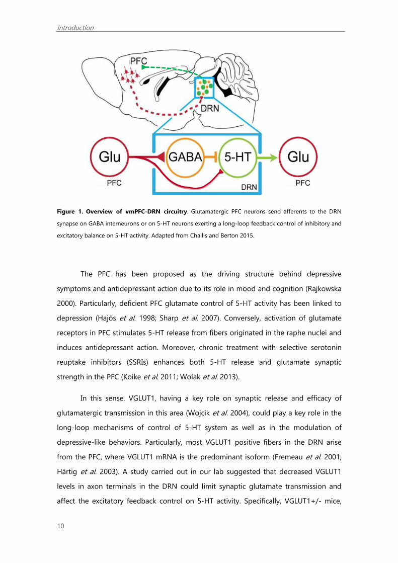
(Hajós et al. 1998; Varga 2001; Tao and Auerbach 2003; Amat et al. 2005) (Figure 1).
Introduction
10
Figure 1. Overview of vmPFC-DRN circuitry. Glutamatergic PFC neurons send afferents to the DRN
synapse on GABA interneurons or on 5-HT neurons exerting a long-loop feedback control of inhibitory and
excitatory balance on 5-HT activity. Adapted from Challis and Berton 2015.
The PFC has been proposed as the driving structure behind depressive
symptoms and antidepressant action due to its role in mood and cognition (Rajkowska
2000). Particularly, deficient PFC glutamate control of 5-HT activity has been linked to
depression (Hajós et al. 1998; Sharp et al. 2007). Conversely, activation of glutamate
receptors in PFC stimulates 5-HT release from fibers originated in the raphe nuclei and
induces antidepressant action. Moreover, chronic treatment with selective serotonin
reuptake inhibitors (SSRIs) enhances both 5-HT release and glutamate synaptic
strength in the PFC (Koike et al. 2011; Wolak et al. 2013).
In this sense, VGLUT1, having a key role on synaptic release and efficacy of
glutamatergic transmission in this area (Wojcik et al. 2004), could play a key role in the
long-loop mechanisms of control of 5-HT system as well as in the modulation of
depressive-like behaviors. Particularly, most VGLUT1 positive fibers in the DRN arise
from the PFC, where VGLUT1 mRNA is the predominant isoform (Fremeau et al. 2001;
Härtig et al. 2003). A study carried out in our lab suggested that decreased VGLUT1
levels in axon terminals in the DRN could limit synaptic glutamate transmission and
affect the excitatory feedback control on 5-HT activity. Specifically, VGLUT1+/- mice,

Introduction
11
expressing half of cerebral VGLUT1 compared to wild type (WT) littermates, show
decreased 5-HT neuronal activity and 5-HT1A autoreceptor desensitization in the DRN
(Garcia-Garcia et al. 2013). Thus, deficient VGLUT1 in the PFC could affect 5-HT activity
and contribute to trigger depressive symptoms.
3.3 GABAergic hypothesis
Beyond the monoamine theory, in recent years, other theories have emerged
suggesting that alterations in GABA may contribute to the development of depression
(Sanacora et al. 2008). GABA is the main inhibitory neurotransmitter in the brain,
whereas glutamate is the main excitatory neurotransmitter. A proper balance between
glutamate and GABA levels is essential for normal brain function.
GABA deficiency has been proposed as a model of anxiety and depression.
GABA levels in plasma and cerebrospinal fluid have been observed to be low in people
with depression (Sanacora and Saricicek 2007). In addition, a cortical GABA deficiency
has been observed in depressed patients, which could lead to an excitatory-inhibitory
imbalance that can conversely be restored by chronic antidepressant treatment
(Sanacora et al. 2004; Bhagwagar et al. 2007).
3.4 Neuroplasticity hypothesis
Neuroplasticity is the ability of the brain to perceive, adapt and respond to both
internal and external stimuli. Plasticity is essential for nervous system healing after
injury, stroke or other pathologies allowing a functional recovery of the brain.
Neuroplasticity processes include synaptic plasticity, cell growth and remodelling and
neurogenesis. Alterations in the regulation of these processes may contribute to the
development of a variety of neuropsychiatric diseases. It is postulated that depression
could be the result of an inability to adapt to adverse environmental factors, resulting
from a dysfunction in normal mechanisms of neuroplasticity (Duman et al. 1999).
Preclinical and clinical studies, in which structural and cellular alterations are observed
in depressed patients, support this theory (Duman 2002). Conversely, enhanced
Introduction
12
neuronal plasticity, neurogenesis, dendritic branching, synaptogenesis, appears to be a
shared mechanism for antidepressants.
It has been said that neurotrophic factors, most notably the brain derived
neurotrophic factor (BDNF), has a role in depression. Stress and depression decrease
the expression and function of BDNF in PFC and hippocampus and thus not allowing
the proper maintenance of synaptic connections. Conversely, antidepressant treatments
(SSRIs) increase BDNF (Björkholm and Monteggia 2016).
Patients with major depression have shown decreased levels of BDNF in brain
(Altar et al. 2009) and serum (Cunha et al. 2006; Palomino et al. 2006; Sen et al. 2008). It
has been suggested that the delay of the antidepressant effect is due to the time
necessary to produce neuroadaptive mechanisms that can improve neuronal plasticity
(Kozisek et al. 2008; Pittenger and Duman 2008). In line with this hypothesis, several
studies have shown that BDNF could mediate the therapeutic action of antidepressants
(Berton and Nestler 2006; Groves 2007; Martinowich et al. 2008). Several studies show
that chronic antidepressant treatments, including selective serotonin reuptake
inhibitors and electroconvulsive shock, increase the expression of BDNF in the
hippocampus in animal models (Russo-Neustadt 2003; Castrén et al. 2007). In line with
this hypothesis, mice deficient in BDNF, are used as a model of depression. It has been
observed that direct infusion of BNDF into the brain produces an antidepressant effect
and potentiates the efficacy of antidepressant treatment, while blockade of BDNF
signalling does not (Castrén and Rantamäki 2010; Lindholm and Castrén 2014).
Introduction
13
3.5 Epigenetics and depression
Growing evidence suggests that epigenetic mechanisms play a key role in
neuronal plasticity. Currently, epigenetics is defined as the study of stable and heritable
modifications of chromatin that occur without changes in DNA sequence and influence
the phenotypic features of living organisms (Riccio 2010). Studies in human depressed
subjects and rodent models, both in brain tissue and in blood cells, have reported
epigenetic alterations, associated to depressive behaviour (Covington et al. 2009;
Hobara et al. 2010; Bagot et al. 2014).
Among the different epigenetic mechanisms, DNA methylation, non-coding
RNA expression and histone modification are the most investigated. Chromatin
remodelling is a dynamic process that modulates gene expression. It can be in a
condensed state, heterochromatin, which avoids gene transcription, or in an open
active state, euchromatin, which allows the genes to be transcribed. Histone
modifications affecting chromatin happen mainly in the N-terminal of histones 3 and 4.
These modifications include acetylation, methylation, phosphorylation, ubiquitination,
SUMOylation and ADP-ribosylation (Karlic et al. 2010). Among them, the most studied
modification is the acetylation of lysine residues, a process carried out by histone
acetyltransferases (HAT) and histone deacetylases (HDAC).
HATs are enzymes that acetylate the lysine residues of histones through a
transfer of an acetyl group from an acetyl-CoA molecule, which unwind the DNA-
histone conformation, allowing the transcription factors interact with DNA and
facilitating gene expression. Conversely, HDACs remove acetyl groups from the lysine
residues of the N-terminal tail of histones and other proteins. This deacetylation results
in a more compact chromatin state and, therefore, a gene silencing (Figure 2).

Introduction
14
Figure 2. Acetylation and deacetylation of nucleosomal histones play an important role in the
modulation of chromatin structure and gene expression. Histone acetyltransferases (HATs) and histone
deacetylases (HDACs) are two opposing classes of enzymes, which tightly control the equilibrium of
histone acetylation. Adapted from Pearson Education 2011 (Inc. publishing as Prentice Hall).
The HDACs belong to an evolutionarily conserved family, which is divided into
four classes (Haberland et al. 2009). Class I, II and IV are similar as they require Zn2+ as a
cofactor (de Ruijter et al. 2003), whereas class III requires nicotinamide adenine
dinucleotide (NAD+) (Sauve et al. 2006). Class I (HDACs 1, 2, 3 and 8) is found almost
exclusively in the nucleus of the cells and is widely expressed in the brain, with the
exception of HDAC8 that is muscle specific (Kazantsev and Thompson 2008).
Based on structural parameters, class II is divided into two subclasses: class IIa
(HDACs 4, 5, 7 and 9) and class IIb (HDAC 6 and 10). HDAC6 is found predominantly in
the cytoplasm, whereas the rest of those subclass II HDACs move between the nucleus
and the cytoplasm, through a mechanism regulated by kinase phosphorylation
dependent on calcium-calmodulin (Gregoretti et al. 2004).
On the other hand, class III HDACs, also called sirtuins (SIRT1-SIRT7) are widely
present in several organelles of the cell (Michan et al. 2007) and the seven isoforms
Deacetylation Acetylation
«Open» Relaxed
Closed «inactive»
Euchromatin
Heterochromatin
Introduction
15
have location in the brain (Frye 2000). SIRT1, 2, 6, and 7 are found in the cytoplasm and
nucleus, whereas SIRT3, 4 and 5 have mitochondrial localization (Michishita et al. 2005).
Class IV consists only of HDAC11. It is mainly found in the nucleus and is
expressed during the development of the central nervous system and possibly has a
role in inflammation, through its inhibitory effect on the expression of interleukin 10
(Villagra et al. 2009).
Changes in histone acetylation have been suggested to play an important role
in the pathophysiology of MDD. For instance, clinical studies have confirmed a positive
correlation between the expression of some HDACs and the depressive state of patients
(Belzeaux et al. 2010). Specifically HDAC2, HDAC4 and HDAC5 mRNA was increased in
patients suffering of MDD compared to controls, while mRNA expression of HDAC6
and HDAC8 was decreased (Hobara et al. 2010). Other clinical study suggests that
altered SIRT1, 2 and 6 mRNA expression could be associated with the pathophysiology
of MDD (Abe et al. 2011). Further, non-specific HDAC inhibitors were proved to have
antidepressant effects in patients (Machado-Vieira et al. 2011). In addition,
experimental models based on exposure to stress, have linked upregulation of the
histone deacetylases of class I (HDAC1 and HDAC2), class IIa (HDAC4 and HDAC5) and
class III (SIRT2) in the PFC and the hippocampus to depressive-like behaviours
(Tsankova et al. 2006; Renthal et al. 2007; Sarkar et al. 2014; Erburu et al. 2015a). By
contrast, specific inhibition of these enzymes were reported to have antidepressant-like
activity (Covington et al. 2009; Jochems et al. 2014; Schroeder et al. 2013; Erburu et al.
2017). Moreover, repeated monoaminergic antidepressants decrease the levels or the
activity of some HDACs (Tsankova et al. 2006; Covington et al. 2011; Réus et al. 2013;
Erburu et al. 2015a). Thus, HDACs could be promising targets for the treatment of
depression (Schroeder et al. 2007; Jochems et al. 2014).
Recent studies from our laboratory have shown that chronic treatment with
antidepressants increased histone 3 and 4 acetylation (AcH3 and AcH4) in mice
prefrontal cortex (PFC). In addition in this study we have identified the enzymes HDAC5
and SIRT2, from the class IIa and III HDAC superfamily respectively, as being oppositely
regulated by chronic stress and antidepressants (Erburu et al. 2015). Thus, decreased
Introduction
16
function of these enzymes in the mice PFC might promote histone 3 and 4 acetylation
(AcH3 and AcH4) and could play a key role in the mechanisms by which
antidepressants enhance synaptic plasticity (Koppel and Timmusk 2013; Erburu et al.
2015a).
3.5.1. HDAC5 in depression and antidepressant action
HDAC5 belongs to the class IIa HDAC superfamily and shuttles from the nucleus
to the cytoplasm through a phosphorylation mechanism (McKinsey et al. 2001; Chawla
et al. 2003; Renthal et al. 2007). The amino-terminal domain of the enzyme is subject to
a reversible phosphorylation that controls its nucleo-cytoplasmic distribution. The non-
phosphorylated fraction remains in the nucleus, bound to the chromatin and repressing
the transcription while the phosphorylated fraction leaves the nucleus, allowing the
expression of its target genes (Parra and Verdin 2010).
A relevant study has shown that overexpression of HDAC5 blocks the
antidepressant effect of imipramine in the chronic social defeat stress (CSDS) model
and this effect is due to the repressive effect of HDAC5 over BDNF expression
(Tsankova et al. 2006). In agreement with this study, an increase in HDAC5 mRNA in the
cerebral cortex of subjects with MDD compared to control has been observed (Hobara
et al. 2010). In addition, we have shown in our lab that while the CSDS model
upregulates nuclear HDAC5, the tricyclic imipramine and the selective noradrenaline
reuptake inhibitor reboxetine increased the phosphorylated form of HDAC5 (p-HDAC5),
mainly located in the cytoplasm (Erburu et al. 2015a). Interestingly, non-selective
inhibition of the class II HDAC superfamily leads to an increase in synaptic plasticity
markers (Erburu et al. 2015a). Yet, the effect of a selective HDAC5 inhibition on synaptic
plasticity has not been evaluated yet.
On the other hand, HDAC5 has been implicated in other physiological functions.
For instance, it has been shown that the phosphorylation of HDAC5 in cardiac tissue
induces cardiac hypertrophy (Lehmann et al. 2014), while in the vascular endothelium it
induces angiogenesis (Urbich et al. 2008) and also stimulates the expression of anti-
inflammatory genes (Wang et al. 2010). Finally, recent research associates an increase in
nuclear HDAC5 to tumor formation, presenting p-HDAC5 as a possible biomarker of

Introduction
17
efficacy in the response to chemotherapy (Kin and Benchimol 2013). Taken together,
these findings raise the question of whether depressive state or antidepressant
treatment regulates the expression of genes directly associated to HDAC5 function.
3.5.2. SIRT2 in depression and antidepressant action
SIRT2 belongs to the class III NAD+-dependent histone deacetylases. Due to its
participation in metabolic homeostasis and brain aging, sirtuins have aroused a
growing interest in neurodegenerative diseases. Among all sirtuins, SIRT2 is the most
expressed at the brain level (Pandithage et al. 2008). SIRT2 resides in the nucleus,
performing its histone deacetylase function, but also, in an important way, in the
cytoplasm where it participates in the organization of the cytoskeleton, targeting α-
tubulin (North et al. 2003). Particularly, a polymorphism of the SIRT2 gene has been
related to depression in patients with Alzheimer's disease (Porcelli et al. 2013). On the
other hand, the inhibition of SIRT2 exerts neuroprotective effects in various models of
neurodegenerative diseases, including Parkinson's disease (Outeiro et al. 2007) and
Huntington (Taylor et al. 2011).
Studies in our laboratory have shown an increase in expression of mRNA and
protein of SIRT2 in the PFC of mice exposed to CSDS. Regarding the mechanism of
action involved in this effect of imipramine, both the selective noradrenaline reuptake
inhibitor, reboxetine, and the selective serotonin reuptake inhibitor, fluoxetine,
decreased the expression of SIRT2, suggesting that this could be a shared mechanism
of monoaminergic antidepressants in general. In the same way these antidepressants
increased the acetylation of the cytoskeletal protein α-tubulin, a cytoplasmic SIRT2
substrate (Erburu et al. 2015a). Further, the specific inhibition of SIRT2 by the chronic
administration of 33i produced an increase in the expression of AcH4, CREB and pro-
BDNF in the PFC, suggesting that the inhibition of SIRT2 modulates synaptic plasticity
(Erburu et al. 2015a; 2017).
Taken together, these results suggest that inhibition of SIRT2 could be another
mechanism by which antidepressants stimulate the expression of genes involved in
synaptic plasticity. In line with this hypothesis, our results showed an increase in the
Introduction
18
expression of SIRT2 mRNA in the PFC of depressed patients confirming the potential
interest of this enzyme for the therapeutic intervention.
3.6 Other hypothesis
There are other hypotheses that are suggested for MDD development. For
instance, neuroendocrine hypothesis suggests that MDD is associated with an
inadequate stress response due to hypothalamic-pituitary-adrenal axis dysfunction
(Holsen et al. 2013). Further, increased levels of glucocorticoids affect hippocampal
neurogenesis contributing to the development of the disease (Anacker et al. 2013). In
addition, there is evidence suggesting that corticosteroids modify BDNF function, which
would indicate its potential involvement in the pathogenesis of MDD (Kumamaru 2008).
On the other hand, several papers suggest that the immune system is also
implicated in MDD (Makhija and Karunakaran 2013). In fact, depressed patients show
increased levels of glucocorticoids and proinflammatory cytokines (Liu et al. 2012).
Proinflammatory cytokines have important metabolic and endocrine effects, including
neurotransmitter metabolism, neuroendocrine function and neuronal neuroplasticity.
Other emerging suggested theories have implicated the role or intestinal
microbiota that could be influencing normal brain chemistry and, therefore, behaviour
(Hoban et al. 2016). As a matter of fact, it has been found that bacteria translocation
from the intestinal tract to the brain could generate neuroinflamation linked to
depressive-like behaviour (Bravo et al. 2012; Rogers et al. 2016; Martín-Hernández et al.
2016). Peptides produced at the gastrointestinal level such as leptin, ghrelin and
cholecystokinin (Kluge et al. 2011), among others, have a direct influence on the central
nervous system, including neurogenesis, which may be involved in depressive disorder
(Villanueva 2013).
Introduction
19
4. The SH-SY5Y neuroblastoma cell line as an in vitro neuronal model: study of
the mechanisms of antidepressants
In vitro neuronal cell lines provide a major advantage for the study of new drugs
in the central nervous system compared to animal models because of the accessibility
of the drugs to the cells and the controllability of many conditions. The neuroblastoma
SH-SY5Y cell line is an in vitro neuronal model retaining many biochemical and
functional properties of neurons (Biedler et al. 1978; Ross et al. 1983). This cell model
has been widely used to study physiology, metabolism, neurodegeneration and
neuroadaptation processes in neurons. Interestingly, in this system, a variety of
monoaminergic antidepressants have been tested in these cells. For instance, several
studies show the induction of BDNF (Donnici et al. 2008) and other neurotrophic factors
by antidepressants and their neuroprotective effects (Shadfar et al. 2016). Moreover,
new compounds with a potential antidepressant action have been tested in this cell
model (Fukuda et al. 2016) together with the exploration of different cell signalling
pathways involved in the mechanisms of action of antidepressants (Hu et al. 2014).


APPROACH AND OBJECTIVES

Approach and objectives
23
Major depression is a mental disorder characterized mainly by extreme
depressive mood and anhedonia together with other psychosomatic associated
symptoms. This illness can cause moderate or severe disrepair in the patient life,
including personal areas. It is turning it into one of the leading causes of disability
worldwide. For instance, in Spain, it is estimated that almost 5 million people present
mild depressive symptoms while another 1 million show moderate to severe
symptomatology. Given that this is a chronic disease, these data reveal a huge social
problem in terms of human suffering for the patients and their families, as well as a
high economic cost for developed countries.
The monoaminergic deficiency hypothesis has for years been the basis of
therapeutic approaches for the treatment of depression. However, this hypothesis has
been questioned due to the absence of an immediate efficacy of the treatments, to the
failure of these treatments as well as to the numerous relapses that some patients
present. Therefore, it is believed that a chain of biological events of greater complexity
than the simple alterations of cerebral monoamine levels might take place. Other
hypotheses have been proposed, like for instance alterations in neuroplasticity or in
glutamate transmission as well as in epigenetics. Yet, at present, there is no common
explanation that brings together the different theories.
Recent observations based on the effect of environmental factors on epigenetic
targets suggest that biological risk factors of depression could be epigenetic. In
addition, such epigenetic modifications could explain the enormous inter-individual
variability towards adversity or response to treatment. In this context, current research
focuses on the antidepressant regulation of epigenetic targets as well as its role in
neuroplasticity.
Antidepressant action has been linked to increased synaptic plasticity in the
prefrontal cortex (PFC), in which epigenetic mechanisms could play a key role. Of these,
the histone deacetylase (HDACs) enzymes have been proposed as epigenetic targets
involved in the pathophysiology of depression and antidepressant-like action.
Specifically, previous studies carried out in our lab have identified the class IIa histone
deacetylase 5 (HDAC5) and the Class III sirtuin 2 (SIRT2) as being oppositely regulated

Approach and objectives
24
by stress and antidepressants. Importantly, decreased function of these enzymes might
promote histone acetylation and induce the expression of different plasticity markers
highly linked to depression and antidepressant action.
In vitro neuronal cell lines provide a major advantage for the study of new drugs
in the central nervous system compared to animal models because of the accessibility
of the drugs to the cells and the controllability of many conditions. The neuroblastoma
SH-SY5Y cell line is an in vitro neuronal model retaining many biochemical and
functional properties of neurons. This cell model has been widely used to study
physiology, metabolism, neurodegeneration and neuroadaptation processes in
neurons. Interestingly, several studies have tested in these cells the effect of
antidepressants on neuroplasticity.
Accordingly, in the frame of this project our first aim was to further explore the
role of HDAC5 and SIRT2 in the molecular mechanisms of antidepressants. Using the in
vitro SH-SY5Y cellular model we have studied the antidepressant regulation of these
epigenetic targets and their involvement in synaptic plasticity. Specifically, the
expression of the synaptic plasticity markers the brain derived neurotrophic factor
(BDNF) and the vesicular glutamate transporter 1 (VGLUT1), both highly linked to
antidepressant action, have been studied.
Subsequently, our second aim was to study the therapeutic potential of SIRT2
for depression treatment. Specifically, the potential antidepressant effect of the
selective SIRT2 inhibitor 33i was tested in mice heterozygous for VGLUT1 (VGLUT1+/-),
previously characterized as a genetic model of depressive-like behaviour.
On the other hand, clinical and preclinical studies suggest a key role of the PFC
glutamatergic signaling and a therapeutic value for glutamatergic targets in major
depression. Particularly, glutamatergic descending pathways from the PFC, in which
VGLUT1-positive excitatory neurons are central, modulate 5-HT activity in the dorsal
raphe nuclei. Here we have initiated a study directed to examine the role of the PFC
VGLUT1 in the long-loop mechanisms of control of 5-HT system as well as in the
modulation of depressive-like behaviors. Firstly, an adeno-associated virus (AAV)
expressing the VGLUT1 gene, has been designed. Subsequently, using the VGLUT1+/-

Approach and objectives
25
model, preliminary studies directed to evaluate the effect of induced VGLUT1
expression in the PFC on depressive-like behaviour as well as on 5-HT1A function have
been carried out.
The specific aims of this project are:
1. To explore the role of HDAC5 and SIRT2 in the molecular mechanisms of
antidepressants. Specifically, using the SH-SY5Y cell line we have studied:
1.1. The effect of 2 and 24 hours incubation with the antidepressants imipramine,
fluoxetine and reboxetine on HDAC5 and SIRT2 expression. In addition, the
effect of the selective HDAC5 inhibitor MC3822 and the selective SIRT2 inhibitor
33i on histone acetylation, AcH3 and AcH4, and on the synaptic plasticity
markers VGLUT1 and BDNF has been studied.
1.2. The effect of 2 and 24 hours incubation with the antidepressants imipramine,
fluoxetine and reboxetine on these epigenetic targets, histone acetylation and
synaptic plasticity markers.
1.3. The effect of the muscarinic cholinergic receptor antagonist scopolamine on
histone acetylation and synaptic plasticity markers.
1.4. The long-term antidepressant regulation of all these epigenetic and synaptic
plasticity markers in vivo, in the mice PFC, comparatively to the in vitro studies.
2. To explore the therapeutic potential of SIRT2 for depression treatment in an
animal model. Specifically, using the VGLUT1+/- model we have studied
whether:
2.1. The VGLUT1+/- model shows alterations in the expression of the HDAC
superfamily or in histone acetylation in the PFC.
2.2. SIRT2 inhibition, using the selective inhibitor 33i, reverses anhedonic behaviour
in the VGLUT1+/- mice. The antidepressant imipramine was used as a reference
compound.
Approach and objectives
26
2.3. The selective SIRT2 inhibitor 33i interacts with specific monoaminergic
molecular targets for depression treatment such as the serotonin or
noradrenaline transporters as well as the monoaminooxidase enzyme.
3. To study the role of induced expression of VGLUT1 in the PFC in antidepressant
action. Specifically, using the VGLUT1+/- model, the following studies have
been carried out:
3.1. Design of the plasmid pAAV-pSyn-VGLUT1-mCherryminisog-SMD2 and
evaluation of its functionality in rat hippocampal cultures. Subsequently, design
of the adenovirus pAAV-pSyn-VGLUT1mCherryminisog.
3.2. Determination of the optimal concentration required for imaging
VGLUT1mCherryminisog expression both in the PFC and in glutamatergic descending
fibbers in the dorsal raphe nucleus (DRN) of VGLUT1+/- and WT littermates.
3.3. Effect of VGLUT1 induced expression in the PFC in the anhedonic-like behaviour
of VGLUT1+/- mice compared to WT as well as in a battery of behaviours.
3.4. Effect of VGLUT1 induced expression in the PFC on the hypothermic response
mediated by a 5-HT1A agonist as a simple functional approach to evaluate the 5-
HT1A autoreceptor sensitivity.

MATERIAL AND METHODS


Material and methods
29
1. Cell cultures
Human SH-SY5Y neuroblastoma cells (ECACC; Sigma Aldrich, St. Louis, MO, USA;
ATCC® CRL-2266™) were cultured at 37°C, in humidified air with 5% CO2, in Dubbelco`s
Modified Eagle Medium (DMEM; gibco, Life Technologies, Paisley, UK) supplemented
with 10% of fetal bovine serum (FBS; gibco, Life Technologies, Paisley, UK), and
Penicillin/Streptomycin solution (Lonza, 17-602E) at 100 U/mL. The medium was
changed twice a week and cells were split at about 80% confluence. Cells were grown in
tissue culture flask of 75cm2 (BD Falcon, Franklin Lakes, NJ, USA) and cells were never
cultivated beyond passage 25. Cells were seeded in 6 well cell culture clusters (Costar,
Corning, NY, USA) at the density of 200,000 cells per well. For each experiment, 6 cell
cultured wells per drug treatment or vehicle were used.
2. Animals
Male C57BL/6J mice (Harlan, France, 8 weeks of age, n=8/group) were housed in
individual cages and allowed to habituate for 2 weeks before beginning antidepressant
treatments.
One male Wistar rat (180–220 g) (Harlan, France) for the affinity for serotonin
transporter (SERT) and noradrenaline synaptosomal uptake studies was used.
A colony of heterozygous VGLUT1 (VGLUT1+/-) and wild-type (WT; C57BL/6)
male mice (8-10 weeks old, n= 7-9 animals/group for the 33i in vivo experiment and 9-
11 animals/group for the AAV experiment) were bred in the animal house of the
University of Navarra from heterozygous fathers (Dr. S. Wojcik, Gottingen, Germany)
(Wojcik et al. 2004) and WT mothers (Harlan, France). Mice were weaned and
genotyped at the age of three weeks. VGLUT1+/- mice were studied and compared to
their WT littermates. Heterozygous mice exhibited no apparent phenotypic
abnormalities during development and adulthood.
Food and water were available ad libitum for the duration of the experiments.
Animals were maintained at a temperature (21±1°C) and humidity-controlled room
(55±2%) on a 12 h light-dark cycle (lights on at 08:00 h).
Material and methods
30
Experimental procedures and animal husbandry were conducted according to
the principles of laboratory animal care as detailed in the European Communities
Council Directive (2013/53/EC) and approved by the Ethical Committee of University of
Navarra.
3. Drugs and treatments
For cell culture experiments the antidepressants imipramine HCl (Sigma Aldrich,
USA), fluoxetine HCl (Interchim, Mountluçon, France) and reboxetine HCl (kindly
donated by Servier Laboratories, Paris, France) and also the antimuscarinic drug
scopolamine HCl (Sigma Aldrich, USA) were dissolved in sterile DMSO to a stock
concentration of 1 mM. The selective HDAC4/5 inhibitors MC3822 and MC3823 and the
SIRT2 inhibitor 33i were dissolved in sterile DMSO to a stock concentration of 1 mM.
MC3822 and MC3823 were kindly donated by Dr Valente (Sapienza University of Rome,
Italy). The compound 33i, was kindly donated by Dr Suzuki from Kyoto Prefectural
University of Medicine, Japan.
For in vivo administration, Imipramine HCl, fluoxetine HCl and reboxetine HCl
were dissolved in saline (0.9%). The compound 33i was prepared in suspension using
18% tween 80 and 5% DMSO in saline.
The antidepressant imipramine is a serotonin and noradrenaline reuptake
inhibitor that increases the levels of neurotransmitters, noradrenaline and serotonin, in
the synaptic cleft. As a tricyclic antidepressant, it has also a weak affinity for muscarinic
(M1), histaminic (H1) and adrenergic (α1) receptors. Fluoxetine is, in turn, a selective
serotonin reuptake inhibitor whereas reboxetine is a selective noradrenaline reuptake
inhibitor.
The compound MC3822 (1R,2R,3R)-N-hydroxy-2-(4-(oxazol-5-yl)phenyl)-3-
phenylcyclopropane-1-carboxamide) has been reported as specific HDAC4/5 inhibitor,
(Bu rli et al. 2013) while no information is available for the enantiomer MC3823
(1S,2S,3S)-N-hydroxy-2-(4-(oxazol-5-yl)phenyl)-3-phenylcyclopropane-1-carboxamide).
Thus, the corresponding racemate was prepared by Dr Valente team, and the two pure

Material and methods
31
enantiomers were separated and identified through analytical methods. Afterwards, the
two compounds were tested against HDAC1-9 enzymes to assess their inhibition
capabilities.
The compound 33i is a 3'-phenethyloxy-2-anilinobenzamide analogue (2-{3-(3-
fluorophenethyloxy)phenylamino}benzamide) representing a new class of potent SIRT2-
selective inhibitors. In previous studies it has shown IC50 of 570 nM towards SIRT2 and
no affinity for the rest of sirtuins (Suzuki et al. 2012).
4. Experimental design
4.2. Experimental design 1: Role of HDAC5 and SIRT2 in the molecular
mechanisms of antidepressant
Cell cultures. Two independent experiments were carried out. In the first
experiment, cultured wells were incubated with imipramine, fluoxetine, reboxetine and
scopolamine to a final concentration of 10 µM or vehicle containing 1% of dimethyl
sulfoxide (DMSO), for 2 and 24 hours at 37ºC. In a second experiment, cells were
incubated with the selective HDAC4/5 inhibitors MC3822 and MC3823, the SIRT2
inhibitor 33i (5 µM for 2 and 24 h) or vehicle. Then, cells were collected and epigenetic
and synaptic plasticity markers were studied by Western blot and immunofluorescence.
Animal treatments. Mice were randomly divided into four groups and received
daily i.p. injections of imipramine (10 mg/kg), fluoxetine (15 mg/kg), reboxetine (15
mg/kg) or saline once a day (at 10 a.m.) for three weeks. A single daily dose was
selected in order to minimize the stress due to the injection. At this dose these
antidepressants have shown to effectively enhance extracellular levels of noradrenaline
and/or serotonin.
Twenty four hours after the last i.p. injection animals were sacrificed by cervical
dislocation and their brains were rapidly removed and dissected in an acrylic mouse
brain slicer matrix with 1.0 mm coronal slice intervals (Zivic Instruments, Pittsburgh, PA,
USA). Using a mouse brain atlas (Hof et al. 2000), a 1mm slice was taken from the
infralimbic section of the PFC (bregma 2.20 mm through bregma 1.20 mm) and
Material and methods
32
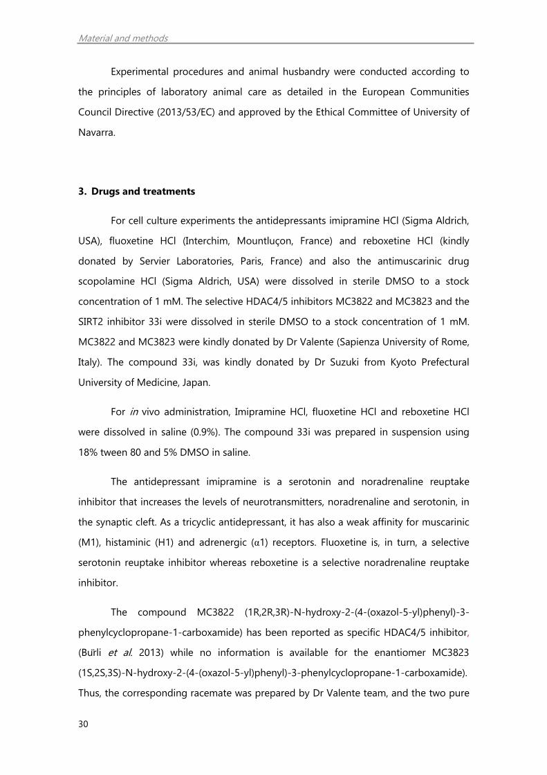
dissected out bilaterally using a scalpel and kept at -80°C. Then, Western blot and RT-
PCR studies were carried out directed to study the expression of synaptic plasticity and
epigenetic targets (Figure 3).
Figure 3. Experimental design 1. Antidepressant regulation of epigenetic targets linked to synaptic
plasticity in vitro and in vivo.
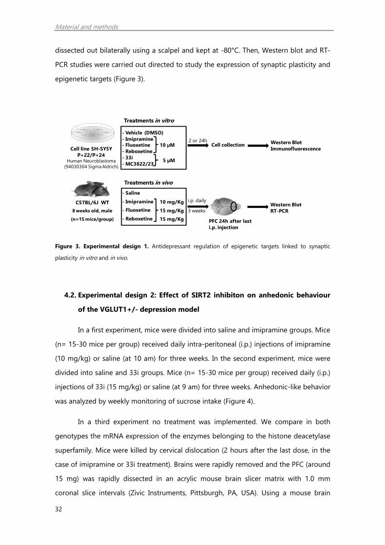
4.2. Experimental design 2: Effect of SIRT2 inhibiton on anhedonic behaviour
of the VGLUT1+/- depression model
In a first experiment, mice were divided into saline and imipramine groups. Mice
(n= 15-30 mice per group) received daily intra-peritoneal (i.p.) injections of imipramine
(10 mg/kg) or saline (at 10 am) for three weeks. In the second experiment, mice were
divided into saline and 33i groups. Mice (n= 15-30 mice per group) received daily (i.p.)
injections of 33i (15 mg/kg) or saline (at 9 am) for three weeks. Anhedonic-like behavior
was analyzed by weekly monitoring of sucrose intake (Figure 4).
In a third experiment no treatment was implemented. We compare in both
genotypes the mRNA expression of the enzymes belonging to the histone deacetylase
superfamily. Mice were killed by cervical dislocation (2 hours after the last dose, in the
case of imipramine or 33i treatment). Brains were rapidly removed and the PFC (around
15 mg) was rapidly dissected in an acrylic mouse brain slicer matrix with 1.0 mm
coronal slice intervals (Zivic Instruments, Pittsburgh, PA, USA). Using a mouse brain
C57BL/6J WT
8 weeks old, male
(n=15 mice/group)
Cell line SH-SY5Y
P+22/P+24Human Neuroblastoma
(94030304 Sigma Aldrich)
PFC 24h after last
i.p. injection
3 weeks
i.p. daily
Cell collection2 or 24h
- Saline
- Imipramine
- Fluoxetine
- Reboxetine
Treatments in vitro
Treatments in vivo
- Vehicle (DMSO)
- Imipramine
- Fluoxetine
- Reboxetine
- 33i
- MC3822/23
10 μM
10 mg/Kg
15 mg/Kg
15 mg/Kg
5 μM
Western Blot
Immunofluorescence
Western Blot
RT-PCR

Material and methods
33
atlas (Hof et al. 2000), a 1mm slice was taken from the infralimbic section of the PFC
(bregma 2.20 mm through bregma 1.20 mm) and dissected out bilaterally using a
scalpel and kept at -80 °C for neurochemical studies.
Figure 4. Experimental design 2. Effect of SIRT2 inhibition on anhedonic behaviour of VGLUT1+/- mice.
4.3. Experimental design 3: Effect of VGLUT1-induced expression in the PFC in
antidepressant action
Both VGLUT1+/- and WT littermates were divided in two groups of 8-9 mice
and received stereotaxic injections of either pAAV-pSyn-VGLUT1mCherryminisog or pAAV-
pSyn-YFP in the prefrontal cortex (PFC).
Anhedonic-like behavior was analyzed by monitoring sucrose intake before
stereotaxic injections (week 0) and after (weeks 1, 2, 3 and 7). On the fourth and fifth
week, a battery of behavioural test was applied in the following order: spontaneous
motor activity and open field test (day 1), social interaction test (day 4), marble burying
test (day 7). In addition, core body temperature and hypothermic response induced by
8-OH-DPAT was tested on day 10.
Mice were killed by anaesthetic overdose on week 8, perfused with intracardial
injection of paraformaldehyde and brains were processed for tissue imaging
experiments (Figure 5).
51 2 3 4Week
Imipramine (10 mg/kg, i.p.)
0
Sucrose intake test
33i (15 mg/kg, i.p.)
C57BL/6J
VGLUT1+/- and WT8 weeks old, male
(n=15-30 mice/group)

Material and methods
34
Figure 5. Experimental design 3. Effect of VGLUT1 induced-expression in the PFC and projecting areas in
depressive-like behaviour of VGLUT1+/- mice.
5. Enantioseparation of MC3822 and MC3823
The direct HPLC enantioseparation of MC3822 and MC3823 was carried out on
the polysaccharide-based Chiralcel OJ-H chiral stationary phase (CSP) using the mixture
n-hexane-ethanol-TFA 65:35:0.1 as a mobile phase. In order to identify the
enantiomeric elution order, the circular dichroism signal at the single wavelength of
254 nm was monitored during chromatography and compared with that obtained in
the enantioselective analysis of MC3822 and MC3823 on the Chiralpak IC CSP under
the same elution conditions. As previously reported, using the latter CSP the (S,S,S)-
enantiomer (MC3823) was eluted before than the (R,R,R) form (MC3822).
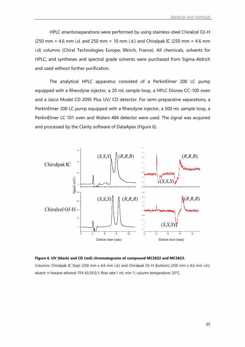
The analysis of the chromatograms obtained by simultaneous on-line CD and
UV detection (Figure 6) provides evidence that CD peaks pertinent to the first eluting
enantiomers in both enantioselective methods have the same negative sign and,
consequently, the same (S,S,S) absolute configuration.
81 2 3 4 5 6 7Week 0
Sucrose intake test
Behavioural tests
Spontaneous motor activity
Open fieldSocial interaction
Marble burying
Sacrifice
Brain imaging
studies
8-OH-DPAT induced hypothermia
C57BL/6J
VGLUT1+/- and WT8 weeks old, male
(n=8-9 mice/group)
Material and methods
35
HPLC enantioseparations were performed by using stainless-steel Chiralcel OJ-H
(250 mm × 4.6 mm i.d. and 250 mm × 10 mm i.d.) and Chiralpak IC (250 mm × 4.6 mm
i.d) columns (Chiral Technologies Europe, Illkirch, France). All chemicals, solvents for
HPLC, and syntheses and spectral grade solvents were purchased from Sigma-Aldrich
and used without further purification.
The analytical HPLC apparatus consisted of a PerkinElmer 200 LC pump
equipped with a Rheodyne injector, a 20 mL sample loop, a HPLC Dionex CC-100 oven
and a Jasco Model CD 2095 Plus UV/ CD detector. For semi-preparative separations, a
PerkinElmer 200 LC pump equipped with a Rheodyne injector, a 500 mL sample loop, a
PerkinElmer LC 101 oven and Waters 484 detector were used. The signal was acquired
and processed by the Clarity software of DataApex (Figure 6).
Figure 6. UV (black) and CD (red) chromatograms of compound MC3822 and MC3823.
Columns: Chiralpak IC (top) (250 mm x 4.6 mm i.d.) and Chiralpak OJ-H (bottom) (250 mm x 4.6 mm i.d.);
eluent: n-hexane-ethanol-TFA 65:35:0.1; flow rate:1 mL min-1; column temperature: 25°C.
Material and methods
36
6. HDAC1-9 isoforms inhibition assay
MC3822 and MC3823 were tested in ten-dose IC50 mode with threefold serial
dilution starting from 200 µM (HDAC1,2,3), from 100 µM (HDAC6,8) and from 50 µM
(HDAC4,5,7,9) solutions. Individual IC50 values for each HDAC isozyme were measured
with the homogeneous fluorescence release HDAC assay. Purified recombinant
enzymes were incubated with serial diluted inhibitors at the indicated concentration.
The deacetylase activities of HDACs 1-9 were determined by assaying enzyme activity
using for HDAC1, 2, 3, 6 the fluorogenic peptide from p53 residues 379-382
(RHKK(Ac)AMC) substrate, for HDAC4, 5, 7, 9 the fluorogenic HDAC Class IIa substrate
(Trifluoroacetyl Lysine) and for HDAC 8 the fluorogenic peptide from p53 residues 379-
382 (RHK(Ac)K(Ac)AMC) substrate. Deacetylated AMC-substrates were sensitive toward
lysine peptidase, and free fluorogenic 4-methylcoumarin-7-amide was generated,
which can be excited at 355 nm and observed at 460 nm (Reaction Biology Corporation,
MD, USA). Data were analyzed on a plate-to-plate basis in relationship to the control
and imported into analytical software (GraphPad Prism, CA, USA).
7. RT-PCR studies
Total RNA extraction. Total RNA of prefrontal cortex (PFC) samples of mice
chronically treated with imipramine, fluoxetine and reboxetine was isolated according
to manufacturer's instructions (NucleoSpin RNA II kit, Macherey-Nagel, Germany). Total
RNA was isolated separately from each individual cortex (n= 8/group).
The frozen PFC samples were lysed and homogenized in the presence of a
highly denaturing ß-mercaptoethanol containing buffer, which immediately inactivates
RNases. Ethanol was added to provide appropriate binding conditions, and the sample
was then applied to an RNeasy Mini spin column, where the total RNA bound to the
membrane and contaminants were washed away. RNA was then eluted in 40 μL RNase-
free water. The eluates were stored at -80°C.
Real time-PCR. RT-PCR was used to validate the differential expression of Hdac5
and Sirt2. Reverse transcription to cDNA was performed using random hexamers as
Material and methods
37
primers and Superscript reverse transcriptase III (Invitrogen, Cergy Pontoise, France)
with 0.3 mg total RNA for each sample. The eluates were stored at -20°C.
RT-PCR was performed in an ABI PRISM 7000 HT Sequence Detection System
following manufacturer’s recommendations (Applied Biosystems, CA, USA). Thermal
cycling conditions were 2 minutes at 50°C, 10 minutes at 95°C, followed by 40 cycles of
denaturation at 95°C for 15 seconds and annealing and extension at 60°C for 1 minute.
Primers for mice genes hdac5 (Mn01246076_m1) and sirt2 (Mn01149204_m1) were
used (Applied Byosistems, CA, USA). Every cDNA prepared was used in triplicate for the
RT-PCR procedures for each gene tested, and the results were calculated as the average
of triplicated results. PCR products were analyzed using the SDS 2.3 and the RQ
Manager 1.2 Software (Applied Biosystems, CA, USA). 18S (Hs99999901_s1) was
employed as an internal control to normalize RNA amount used from different samples.
Samples were analyzed by the double delta CT (∆∆CT) method. Delta CT (∆CT)
values represent normalized target genes levels with respect to the internal control.
Normalization was based on a single reference housekeeping gene (18s). Delta CT
(∆∆CT) values were calculated as the ∆CT of each test sample (imipramine, fluoxetine,
reboxetine) minus the mean ∆CT of the calibrator samples (controls) for each target
gene (hdac5 and sirt2). The fold change was calculated using the equation 2(−∆∆CT).
8. Western Blot studies
SH-SY5Y cells, scrapped from 6 well cell culture clusters (Costar, Corning, NY,
USA) with Dubelcco’s Phosphate Buffered Saline (DPBS; gibco, Life Technologies,
Paisley, UK) were centrifuged (827 g for 5 minutes). Each pellet from the cell cultures or
the prefrontal cortex (PFC) dissected from the mice was sonicated in a cold lysis buffer
with protease inhibitors (0.2M NaCl, 0.1M HEPES, 10% glycerol, 200mM NaF, 2mM
Na4P2O7, 5mM EDTA, 1mM EGTA, 2mM DTT, 0.5mM PMSF, 1mM Na3VO4, 1mM
benzamidine, 10 mg/mL leupeptin, 400 U/mL aprotinin). The homogenate was
centrifuged at 14,000 g at 4ºC for 20 min and the supernatant aliquoted and stored at -
80ºC. Protein concentration was determined by Bradford (BIO-RAD, Hercules, CA, USA).

Material and methods
38
Western-blot. 20μg of total protein per lane were loaded of each sample after
being mixed with equal volume of loading buffer (0.16M Tris-HCl pH 6.8, 4% SDS, 20%
glycerol, 0.01% bromophenol blue, 0.1M DTT). Then, they were separated by
electrophoresis on a sodium dodecyl sulfate-polyacrylamide gel (8%) under reducing
conditions and transferred to a nitrocellulose membrane (Hybond-ECL; Amersham
Bioscience). The trans-blots were blocked for 1 hour with 10% skimmed milk powder in
TBS buffer containing 0.1% Tween20 and then incubated overnight at 4ºC with the
following primary antibodies: rabbit polyclonal anti-vesicular glutamate transporter 1
(VGLUT1; 1:2,000, kindly donated by Dr. S. El Mestikawy, Paris, France and previously
validated by Herzog et al. 2001); rabbit polyclonal anti-brain derived neurotrophic
factor (BDNF; 1:1,000, #sc-546; Santa Cruz Biotechnology, Wembley, UK); rabbit
polyclonal anti-acetylated histone 3 (AcH3; 1:1,000) and rabbit polyclonal anti-
acetylated histone 4 (AcH4; 1:1,000) (#06-942 and #07-329, respectively;
MerckMillipore, Billerica, MA, USA); rabbit polyclonal anti-phosphorylated histone
deacetylase 5 (p-HDAC5; 1:1,000, #Y011193; Applied Biological Materials, Richmond,
BC, Canada); mouse monoclonal anti-histone deacetylase 5 (HDAC5; 1:1,000), rabbit
monoclonal anti-sirtuin 2 (SIRT2; 1:1,000), mouse polyclonal anti-acetylated α-tubuline
(1:10,000) and mouse polyclonal anti-β-actin (1:10,000) (#H4538, #S8447, #T7451 and
#A1978, respectively; Sigma-Aldrich, St. Louis, MO, USA). These antibodies are
commercially available and have been extensively used for their specificity.
Secondary antibodies conjugated to IRDye 800CW or IRDye 680CW (LI-COR
BIOSCIENCES, Lincoln, NE, USA) were diluted up to 1:5,000 in TBS with 5% BSA. Bands
were visualized using ODYSSEY Infrared Imaging System (LI-COR BIOSCIENCES, Lincoln,
NE, USA). β-actin was used as loading control. Results were expressed as the
percentage of optical density (O.D.) values relative to saline treated animals or control
cells (DMSO) in cell culture experiments.

Material and methods
39
9. Immunofluorescence studies in SH-SY5Y cells
Human SH-SY5Y neuroblastoma cells were seeded in glass bottom culture
dishes (MatTek, Ashland, MA, USA) in 24 well-plates (Greiner Bio-one, Monroe, NC,
USA). Treatments were performed after 24 hours in serum-free media, and then cells
were prefixed with 2% paraformaldehyde for 2 minutes and fixed with 4%
paraformaldehyde (PFA, Histolab, Gothenburg, Sweden) for 20 minutes. Afterwards,
cells were washed three times with PBS. All cover slips were blocked for 30 minutes in
PBS with 0.1% Triton-X and 1% bovine serum albumin (all from Sigma-Aldrich, St. Louis,
MO, USA). The primary antibody used was mouse monoclonal anti histone deacetylase
5 1:100 (HDAC5; #H4538; Sigma Aldrich, St. Louis, MO, USA). The secondary antibody
used was Alexa fluor 488 donkey anti-mouse (1:1,000). Cover slips were first incubated
overnight with the primary antibodies at 4ºC in humid chamber and then for 30
minutes at room temperature with secondary antibodies and DAPI (Invitrogen, Eugene,
Oregon, USA) to identify the nuclei of cell bodies. Finally, the cover slips were rinsed in
PBS and mounted using Immu-mount (Thermo Scientific, Kalamazoo, MI, USA). The
cover slips were thoroughly washed in PBS between different steps. The primary
antibody was omitted as a negative control. Confocal imaging was performed with a
Zeiss LSM 510 META confocal laser scanning system (Carl Zeiss, Jena, Germany). The
fluorescence of DAPI and Alexa 488 were recorded through separate channels with a
40× lens. Images were obtained using the Zen software (Zeiss).
10. TaqMan Low Density Arrays (TLDAs)
RNA extraction and RT-PCR. In order to study the expression of the HDAC
superfamily enzymes genes in the prefrontal cortex of VGLUT1+/- and WT littermate
mice TaqMan Low Density Arrays (TLDA) microfluidic card technology from Applied
Biosystems (Foster City, CA, USA) was used. Total RNA was isolated separately from
each individual frozen prefrontal cortex sample as describe above. As previously
described by Tordera et al. 2011, for each tissue sample, 500 ng of reverse-transcribed
RNA were diluted to 50 μL with sterile water, combined with an equal volume of
TaqMan Universal PCR Master Mix (2x; Applied Biosystems, Foster City, CA, USA), mixed

Material and methods
40
by inversion, and spun briefly in an Eppendorf® 5415C microcentrifuge (Brinkmann
Instruments, Westbury, NY, USA). After TLDAs were brought to room temperature, 100
µL master mix were loaded into each port connected to reaction wells. TLDAs were
placed in Sorvall®/Heraeus® Custom Buckets (Applied Biosystems, Foster City, CA,
USA) and centrifuged in a Sorvall Legend™ centrifuge (Kendro Scientific, Asheville, NC,
USA) for 1 min at 331 for g followed closely by a second 1-min centrifugation at 331 for
g. Cards with excess sample in the fill reservoir were spun for an additional 1 min.
Immediately following centrifugation, the cards were sealed with a TaqMan LDA Stylus
Staker (Applied Biosystems, Foster City, CA, USA), and the loading ports excised.
Quantitative RT-PCR amplifications were run on an ABI Prism® 7900HT Sequence
Detection System (Applied Biosystems, Foster City, CA, USA) with a TaqMan LDA cycling
block and an automation accessory upgrade. Thermal cycling conditions were 2 min at
50°C, 10 min at 95°C, followed by 40 cycles of denaturation at 95°C for 15 s and
annealing and extension at 60°C for 1 min. Each test sample was processed in duplicate
on individual TLDA cards, thus allowing eight samples to be processed on each card.
Analysis of quantitative RT-PCR data. RT-PCR TaqMan instrumentation monitors
gene-specific products with fluorescent dye chemistry. A cycle threshold (CT) for each
reaction is the number of cycles at which the reaction crosses a selected threshold. The
threshold is defined as a straight line drawn above noise/ baseline and positioned
within the linear region of the semi-log amplification plot. The fewer cycles required to
reach threshold fluorescence intensity, the lower the CT value and the greater the initial
amount of input target. Results for each target on TLDAs were quantified concurrently
using the same baseline and threshold for a target gene in order to limit interplate
errors in the analysis.
Samples were analyzed by a double delta CT (∆∆CT) method. Delta CT (∆CT)
values represent normalized target genes levels with respect the internal control.
Normalization was based on a single reference housekeeping gene (18s). Delta CT
(∆∆CT) values were calculated as the ∆CT of each test sample (WT and VGLUT1+/-
genotype) minus the mean ∆CT of the control samples for each target gene. The fold
change was calculated using the equation 2(−∆∆CT).

Material and methods
41
11. In vitro affinity assays
Chemicals. For the in vitro assays, fluoxetine (Interchim, Mountluçon, France),
reboxetine (kindly donated by Servier Laboratories, France), clorgyline and selegiline
(Sigma-Aldrich, USA) were dissolved in distilled water. 33i was dissolved in DMSO,
stored as frozen aliquots and diluted (1%) before each in vitro experiment. The
radiolabelled compounds used were [3H]paroxetine and [3H]noradrenaline (Amersham,
UK).
Affinity for SERT transporter. The compound 33i was studied for its ability to
inhibit [3H]paroxetine binding to the 5-HT transporter in rat PFC homogenates
(Marcusson and Eriksson, 1988). Fluoxetine was used as reference compound. Briefly,
rat cortical tissue was homogenized in Tris-HCl buffer (50 mM, pH 7.4) and centrifuged
at 4°C for 10 min at 50,000 g. The supernatant was discharged, and the pellet was
suspended in Tris buffer and preincubated at 37°C for 10 min. The suspension was then
washed twice by centrifugation and resuspended up to a concentration cortical
membrane (4 mg tissue/mL). Each tube was filled with 1200 µl of Tris buffer pH 7.4,
containing NaCl 120 mM and KCl 5 mM, 400 µl of tissue preparation, and 400 µl of 0.1
nM [3H]paroxetine in Tris buffer and incubated by 60 min at 23°C. The final incubation
volume was 2 mL, and nonspecific binding was determined using 10 µM fluoxetine,
included in the Tris buffer, as the cold displacer.
Noradrenaline synaptosomal uptake. The ability of the compound 33i to inhibit
[3H]noradrenaline uptake in PFC synaptosomal preparations (Tordera et al. 2002) was
studied. Reboxetine was used as reference compound. Briefly, rats were killed and the
cerebral cortex was dissected and homogenized in 15 volumes of 0.32 M ice-cold
sucrose. The homogenates were centrifuged for 10 min at 1,000 g and 4ºC and the
supernatants centrifuged again at 48,000 g for 15 min. The resulting pellet was gently
suspended in 0.27 M ice-cold sucrose. In this crude synaptosomal preparation, the
protein concentration was 0.3 - 0.5 mg/mL. An aliquot (250 µL) of the synaptosomal
preparation was added into 750 µL of Krebs buffer (pH 7.4) containing (mM): NaCl 115,
KCl 4.97, CaCl2 1, MgSO4 1.22, KH2PO4 1.2, NaHCO3 25, glucose 11.1, pargyline 0.01,

Material and methods
42
ascorbic 1.7 and [3H]noradrenaline in the absence (total uptake) or presence of 33i
(three concentrations from 10-5 to 10-9 µM). After 6 min at 37ºC, the reaction was
finished by quick filtration through Whatman GF/B filters.
12. Monoaminooxidase activity assay
The effect of 33i (0.01 µM to 100 µM) on monoaminooxidase activity (total
MAO, MAO-A and MAO-B activities) was studied following manufacturer’s instructions
(Catalog #K795-100Bio Vision, Milpitas, CA, USA). The monoaminooxidase inhibitors
clorgyline and selegiline (10 µM) were used as reference compounds.
13. Adeno-associated viral (AVV) vector generation
New adeno-associated viral (AAV) vectors were cloned using the Gateway
Multisite technology (Invitrogen, Carlsbad, CA). Gateway MultiSite recombination
cloning is a two-step process. In the first step, DNA fragments containing flanking attB
sites are cloned via the BP reaction into attPflanked pDONR vectors. The two entry
clones (pENTR) created are combined with a ‘‘destination’’ vector (pDEST) via the LR
reaction (a recombination reaction between attL and attR sites). The second step of
MultiSite technology involves integrating the fragments contained in the entry clones
into a destination vector via the LR reaction. In the LR reaction, distinct attL and attR
sites ensure position and orientation-specific insertion. Finally, combined expression
clones were introduced in the virus through a service facility (Molecular Imaging
Research Center, MIRCen/ CEA, 18 route du Panorama, F-92265 Fontenay-aux-Roses,
France).
Firstly, amplification of the VGLUT1mCherryminisog chimaera for the BP reaction was
performed by PCR (annealing 60°C and elongation 2 min). The mCherryminisog was a
kind gift from Pr Roger Tsien. The primers used for these genes were GGG-GAC-CAC-
TTT-GTA-CAA-GAA-AGC-TGG-GTA-GAT-TTA-GCC-GTC-CAG-CTG-CAC and GGG-GAC-
AAC-TTT-GTA-TAC-AAA-AGT-TGT-AAT-GGA-GTT-CCG-GCA-GGA-GGA-G, respectively.
The B5-VGLUT1-mCherryminisog-B2 (2733bp) amplified fragment was recombined with
Material and methods
43
pDONR-P5-P2 in the BP reaction to create pENTR-L5-VGLUT1-mCherry-minisog-L2
(first entry clone). The LR recombination reaction was performed combining pENTR-L5-
VGLUT1-mCherry-minisog-L2 and the second entry clone pENTR-L1-pSyn-R5
containing the Synapsin1 promoter sequence with pDest-AAV-R1-R2-SMD2-Rfa-WPRE.
With this recombination step we created pAAV-pSyn-VGLUT1-mCherry-minisog-SMD2-
Rfa-WPRE. Afterwards, the AAV vector pAAV-pSyn-VGLUT1mCherryminisog was produced by
the team of Dr Alexis Bemelmans together with pAAV-pSyn-YFP (Molecular Imaging
Research Center, MIRCen/ CEA, 18 route du Panorama, F-92265 Fontenay-aux-Roses,
France).
A schematic diagram outlining the steps of this cloning procedure and
restriction map of the final product is shown in Figures 7A and 7B respectively.
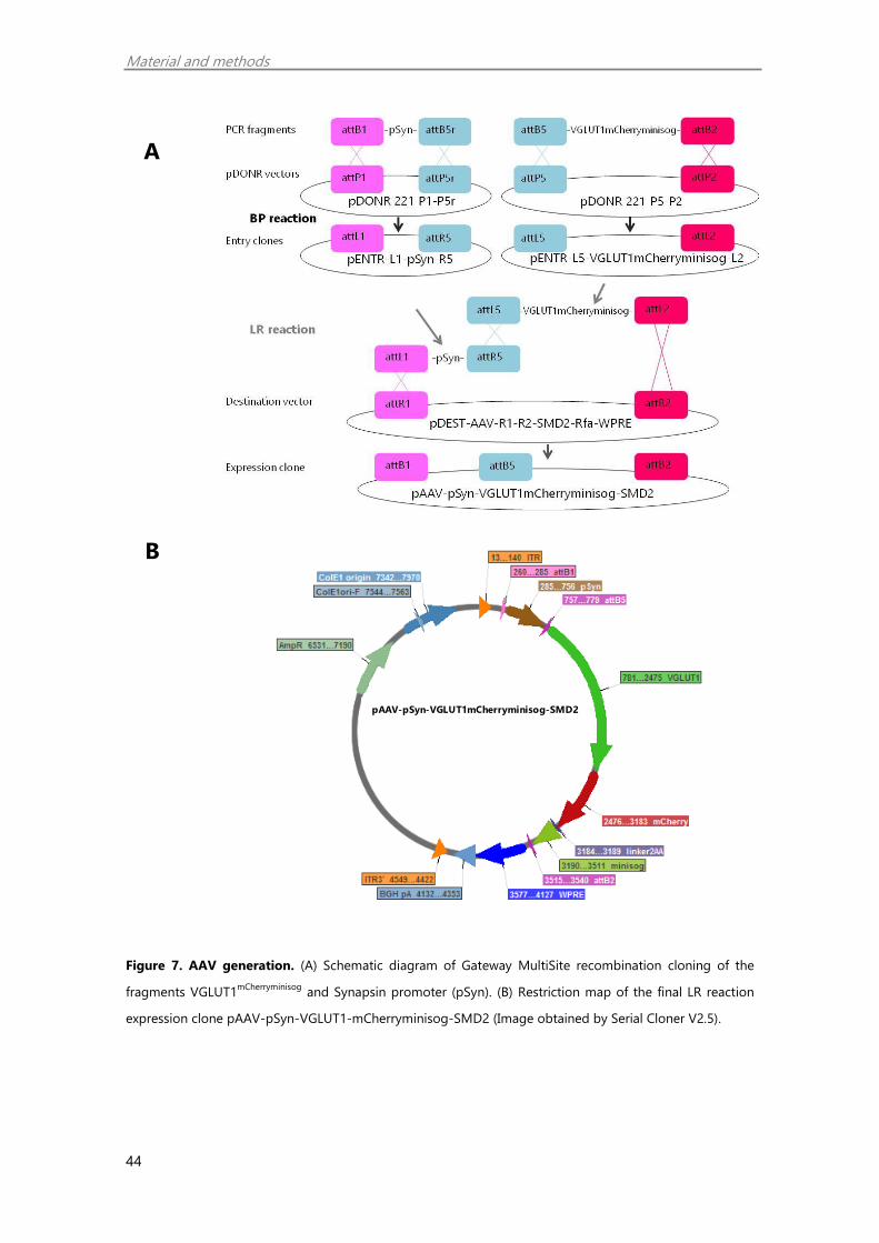
Material and methods
44
Figure 7. AAV generation. (A) Schematic diagram of Gateway MultiSite recombination cloning of the
fragments VGLUT1mCherryminisog
and Synapsin promoter (pSyn). (B) Restriction map of the final LR reaction
expression clone pAAV-pSyn-VGLUT1-mCherryminisog-SMD2 (Image obtained by Serial Cloner V2.5).
A
B
pAAV-pSyn-VGLUT1mCherryminisog-SMD2
Material and methods
45
14. Expression of pAAV-pSyn-VGLUT1mCherryminisog-SMD2 in primary cultures
Plasmids were first tested in primary rat hippocampal cultures to ensure that
cloning of the synapsin promoter allowed transcription of the downstream
VGLUT1mCherryminisog construct.
The expression clone pAAV-pSyn-VGLUT1mCherryminisog-SMD2 was tested in
vitro in primary cultures of rat hippocampal neurons (embryo at E18 days) by
electroporation. In rodents, VGLUT1 mRNA is expressed from postnatal day 14 (PN14)
(Wojcik et al. 2004).
Briefly, cells were transiently transfected using the commercially available Amaxa
MeP1 Nucleofector kit, including nucleofector solution for rat neurons and certified
cuvettes, according to the manufacturer’s instructions (Amaxa biosystems, Cologne,
Germany). In brief, 2 µg of DNA (vector) were dissolved in 8 µL nucleofector solution for
rat neurons at 4°C. 500,000 cells were suspended in 3 mL nucleofector solution for rat
neurons at room temperature. After centrifugation (5 min 1,000 g), the cell suspension
was mixed with the 8 µL DNA-containing solution by pipetting three times up and
down. The mixture was transferred to a cuvette that was inserted into the Nucleofector
device (Amaxa biosystems). After electroporation the neurons were transferred from
the cuvette into dishes with 4 µL of pre-warmed DMEM 1X HorseSerum prewarmed
(37°C).
This step was carried out between days 2 and 5 of the in vitro cultures. Four
dilutions of the original batch were tested: 1/1,000; 1/10,000; 1/50,000; 1/100,000. Every
5 days the medium was changed. Images were taken between days 17 and 21.
15. Stereotaxic injections of pAAV-pSyn-VGLUT1mCherryminisog and pAAV-pSyn-YFP
For stereotaxic delivery of the AAV vector, adult mice were randomly assigned
to treatment with the pAAV-pSyn-VGLUT1mCherryminisog or control groups injected with
pAAV-pSyn-YFP (n= 9-11 mice per group) of each genotype. Mice were anesthetized
by intraperitoneal injection of ketamine/xylazine (0.1/0.05 g/kg of body weight) and
positioned on a stereotactic frame (Steeling, Wood Dale, USA). Mice were then injected

Material and methods
46
using 0.5 μL of either pAAV-pSyn-YFP or pAAV-pSyn-VGLUT1mCherryminisog (1.47 E12 and
2.01E14 viral genomes/mL, respectively) bilaterally into the IL-PFC (+2 mm AP, 1 mm ML,
-1.7 mm DV from bregma), of adult mice based on established coordinates (Hof et al.
2000). Vector delivery was performed at a rate of 200 nL/min using a Hamilton syringe
(Hamilton company, Sarasota, FL, USA) with 33 gauges needle (World Precision
Instruments) in a stereotaxic frame (Kopf Instruments, Tujunga, CA, USA). After the
injection, the needle remained in place for two more minutes and was then carefully
retracted in order to avoid vector backflow. Subsequently, the scalp was sutured and
the mouse was caged individually until full recovery from anesthesia.
16. Imaging of VGLUT1mCherryminisog and YFP expression in the brain
Tissue preparation. The rostro-caudal extent of transgene expression of all
animals used in behavioural experiments was assessed by the fluorophore YFP as
control or mCherrymisinog as target.
Mice were sacrificed nine weeks post-injection (age of animals: 16 weeks). The
animals received an anesthetic overdose of sodium pentobarbital (sodium
pentobarbital 60 mg/mL, Merck) and were transcardially perfused with 0.9% saline
followed by ice cold 4% paraformaldehyde in 0.1M phosphate buffer (pH= 7.4) prior to
brain extraction. For histological processing, the whole brain was removed, post-fixed in
4% paraformaldehyde (2h), cryoprotected in a 30% sucrose solution (in phosphate
buffer) and stored at 4°C until they sank. Serial coronal microtome sections (40 µm)
were collected throughout the frontal cortex (Bregma 2.40 to 1.40) and the dorsal
raphe nuclei (Bregma -4.10 to -5.40) and were stored in cryoprotectant solution at
-20°C until processing.
Serial free-floating sections (one every four 40 µm sections) of the whole
prefrontal cortex (PFC) and dorsal raphe nuclei (DRN) were used. Sections were
mounted on gelatin-coated slides, air-dried in a light protected chamber. Subsequently,
mounting medium (Immu-mount, Thermo Scientific, Kalamazoo, MI, USA) was added
and cover-slipped.

Material and methods
47
Image capture. Fluorescence of YFP and VGLUT1mCherryminisog was assessed using
a confocal fluorescence microscope (LSM 510 META, Carl Zeiss, Jena, Germany) or a
conventional epifluorescence microscope (Eclipse Ni, Nikon Instruments Europe BV,
Amsterdam, Netherland).
Image quantification. Detection and quantification of mCherryminisog or YFP
fluorescent signal in brain sample images was carried out using a plugin developed for
Fiji/ImageJ, an open-source Java-based image processing software (Schneider et al.
2012). The plugin was developed by the Imaging Platform of the Center for Applied
Medical Research (CIMA).
First of all, a region of interest (ROI) is manually delineated in each of the brain
hemispheres. A background subtraction is then carried out using the “Rolling Ball
Background Subtraction”, plugin developed for Fiji by Michael Castle and Janice Keller,
from the Mental Health Research Institute of the University of Michigan, which was
originally based on the work of Sternberg (Stenberg 1983). Positive fluorescent signal in
each ROI is then retrieved by thresholding the resulting image. A global histogram-
derived thresholding method is applied for this purpose, specifically Otsu's clustering
algorithm that searches for the threshold that minimizes the intra-class variance (Otsu
1979). Finally, the relative fluorescence signal area in each hemisphere ROI is quantified
from the final segmentation.
17. Sucrose intake test
Anhedonic-like behaviour was evaluated in WT and VGLUT1+/- mice by
monitoring of sucrose intake (Elizalde et al. 2008). Mice were first trained for 1 week to
drink the 2.5% sucrose solution. After this preliminary phase, mice were food deprived
and exposed to the sucrose solution and tap water for 15 hours once a week. The
intake baseline for the sucrose solution was established, which corresponded to the
average of three consecutive measurements of sucrose intake carried out every 4-5
days.

Material and methods
48
Then, WT and VGLUT1+/- mice were divided into two subgroups matched for
sucrose consumption and body weight in order to start drug treatments (imipramine or
33i, experimental design 2) or stereotaxic injections with the adeno-associated viruses
(AAVs, experimental design 3). Once a week, mice were given a 15-h exposure to the
sucrose solution and tap water in their home cage as described above. The position of
the two bottles (right/left) was varied randomly from trial to trial. Body weight
measurements were taken weekly and relative sucrose intake and sucrose preference
(sucrose intake/total intake) was calculated as absolute intake (g) per mouse body
weight.
18. Behavioural tests
Spontaneous locomotor activity and open field. Locomotor activity was
measured in an open field consisting of 8 black square arenas (43 x 50 x 45) using a
video tracking system (Ethovision XT 11 plus multiple body point module, Noldus
Information Technology, Wageningen, The Netherlands) in a softly illuminated
experimental room. One mouse was placed in each cage and distance travelled (cm)
was recorded during a 30 minute period. Time spent (s) in center zone (25 cm2) was
also recorded.
Social interaction test. This test measures the approach-avoidance behaviour
towards an unfamiliar social target (Tsankova et al. 2006; Venzala et al. 2012). The arena
is a transparent plastic open field (45 × 45 × 45 cm3) maintained in a very softly
illuminated room (50 lx). Each experimental mouse was introduced into the open field
and its trajectory was tracked for two consecutive sessions of 2.5 min. During the first
session (“no target”) the open field contained an empty metallic mesh cage (10.5 × 9
cm2) located at one end of the field. During the second session (“target”), the
conditions were identical except that a social target animal (a mouse from the same
strain and of similar weight) had been introduced into the cage. Between the two
sessions, the experimental mouse was removed from the arena, and was placed back
into its home cage for approximately 1 min. The time that the experimental mouse
spent in the “interaction zone” (7 cm wide corridor surrounding the little metallic cage)

Material and methods
49
both in the “no target” and “target” conditions were recorded (Ethovision XT 11 plus
multiple body point module, Noldus Information Technology, Wageningen, The
Netherlands) and differences (“target-no target”) were calculated for each mouse
(Venzala et al. 2012).
Marble Burying Test. Natural burying behaviour was assessed with this test.
Twelve marbles (1.5 cm diameter) were placed uniformly in a cage (45 × 28 × 20 cm)
containing a constant amount of sawdust (3 cm deep). Mice were placed in the center
of the cage and left for 30 minutes. The number of marbles buried was recorded.
19. Core body temperature studies: 5-HT1A agonist induced hypothermia
Baseline body temperature was measured using a thermistor probe inserted
2 cm into the mice rectum seven weeks after stereotaxic injections. Mice received a
subcutaneous injection of the 5-HT1A receptor agonist 8-hydroxy-N,N-dipropyl-2-
aminotetralin (8-OH-DPAT) 0.4mg/kg (RBI, Wayland, MA, U.S.A) and core body
temperature was measured at 30 minutes intervals for up to 90 minutes. Data were
presented as the difference in core body temperature over basal values.
20. Statistical analysis
The effect of different antidepressants (imipramine, fluoxetine and reboxetine)
on mRNA and protein expression of different epigenetic and synaptic plasticity markers
were analyzed using One-way ANOVA (treatment) followed by post-hoc Dunnett test.
The effect of the compounds 33i, MC3822, MC3823 or scopolamine on protein
expression was evaluated by Student’s t test.
Differences in the mRNA abundance of each HDAC enzyme as well as AAV
infection efficiency were analysed using student t-test in the PFC of VGLUT1+/- and WT
mice.
Sucrose intake test, body weight gain and differences of temperature were
analyzed through two-way ANOVA (genotype by drug treatment or genotype by AAVs

Material and methods
50
treatment) with repeated measures followed by two-way ANOVA for each time point.
Different parameters measured in behavioural tests (motor activity, open field,
social interaction and marble burying) were analyzed by two-way ANOVA (genotype by
AAVs treatment).
All results were expressed as mean ± standard error of the mean (SEM).

RESULTS

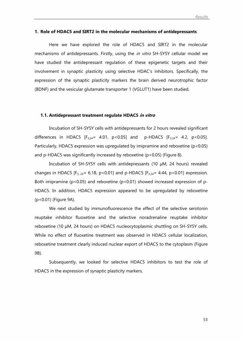
Results
53
1. Role of HDAC5 and SIRT2 in the molecular mechanisms of antidepressants
Here we have explored the role of HDAC5 and SIRT2 in the molecular
mechanisms of antidepressants. Firstly, using the in vitro SH-SY5Y cellular model we
have studied the antidepressant regulation of these epigenetic targets and their
involvement in synaptic plasticity using selective HDAC’s inhibitors. Specifically, the
expression of the synaptic plasticity markers the brain derived neurotrophic factor
(BDNF) and the vesicular glutamate transporter 1 (VGLUT1) have been studied.
1.1. Antidepressant treatment regulate HDAC5 in vitro
Incubation of SH-SY5Y cells with antidepressants for 2 hours revealed significant
differences in HDAC5 [F3,24= 4.01, p<0.05] and p-HDAC5 [F3,24= 4.2, p<0.05].
Particularly, HDAC5 expression was upregulated by imipramine and reboxetine (p<0.05)
and p-HDAC5 was significantly increased by reboxetine (p<0.05) (Figure 8).
Incubation of SH-SY5Y cells with antidepressants (10 µM, 24 hours) revealed
changes in HDAC5 [F3, 24= 6.18, p<0.01] and p-HDAC5 [F3,24= 4.44, p<0.01] expression.
Both imipramine (p<0.05) and reboxetine (p<0.01) showed increased expression of p-
HDAC5. In addition, HDAC5 expression appeared to be upregulated by reboxetine
(p<0.01) (Figure 9A).
We next studied by immunofluorescence the effect of the selective serotonin
reuptake inhibitor fluoxetine and the selective noradrenaline reuptake inhibitor
reboxetine (10 µM, 24 hours) on HDAC5 nucleocytoplasmic shuttling on SH-SY5Y cells.
While no effect of fluoxetine treatment was observed in HDAC5 cellular localization,
reboxetine treatment clearly induced nuclear export of HDAC5 to the cytoplasm (Figure
9B).
Subsequently, we looked for selective HDAC5 inhibitors to test the role of
HDAC5 in the expression of synaptic plasticity markers.
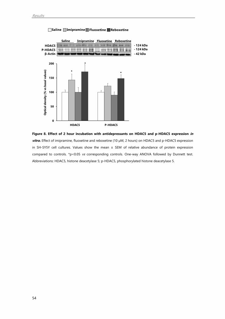
Results
54
Figure 8. Effect of 2 hour incubation with antidepressants on HDAC5 and p-HDAC5 expression in
vitro. Effect of imipramine, fluoxetine and reboxetine (10 µM, 2 hours) on HDAC5 and p-HDAC5 expression
in SH-SY5Y cell cultures. Values show the mean ± SEM of relative abundance of protein expression
compared to controls. *p<0.05 vs corresponding controls. One-way ANOVA followed by Dunnett test.
Abbreviations: HDAC5, histone deacetylase 5; p-HDAC5, phosphorylated histone deacetylase 5.
Saline Imipramine Fluoxetine Reboxetine
ImipramineSaline
- 124 kDa
- 42 kDa
HDAC5
P-HDAC5
β-Actin
- 124 kDa
Fluoxetine Reboxetine
*
*
*
0
50
100
150
200
HDAC5 P-HDAC5
Op
tical d
en
sity
(%
vs
basa
l valu
es)

Results
55
Figure 9. Antidepressant regulation of HDAC5 in vitro. (A) Effect of imipramine, fluoxetine and
reboxetine (10 µM, 24 hours) on HDAC5 and p-HDAC5 expression in SH-SY5Y cell cultures. Data show the
mean ± SEM of relative abundance of protein expression compared to controls. **p<0.01, *p<0.05 vs
corresponding controls. One-way ANOVA followed by Dunnett test. Abbreviations: HDAC5, histone
deacetylase 5; p-HDAC5, phosphorylated histone deacetylase 5. (B) Representative photomicrographs of
HDAC5 and nuclear (DAPI) immunofluorescence in SH-SY5Y cell cultures incubated with vehicle (1%
DMSO), fluoxetine and reboxetine (10 µM, 24 hours). HDAC5+ (green), DAPI+ (blue) and double
(HDAC5+/DAPI+) cells are shown. Scale bar: 10 µm.
Saline Imipramine Fluoxetine Reboxetine
B
***
**
0
50
100
150
200
HDAC5 P-HDAC5
Op
tic
al d
en
sit
y (%
vs
ba
sa
l va
lue
s)
AImipramineSaline
- 124 kDa
- 42 kDa
HDAC5
P-HDAC5
β-Actin
- 124 kDa
Fluoxetine Reboxetine

Results
56
1.2. MC3822 and MC3823 affinity to the different HDAC enzymes
Data from Table 1 show the effect of MC3822 and MC3823 in HDAC1-9
activity. MC3822 clearly selectively inhibits HDAC4 and 5 at double digit
nanomolar level (IC50= 0.038 and 0.012 respectively), HDAC7, 8 and 9 at
submicromolar level (IC50= 0.13, 0.31 and 0.38 respectively), and HDAC6 at
single-digit micromolar concentration. The isomer MC3823 displayed similar
trend of HDAC inhibition with lower potency showing micromolar inhibition for
HDAC4-9. Moreover, MC3822 was totally inactive against HDAC1-3.
Table 1. Biochemical activity of MC3822 and MC3823 against HDAC isoforms.
Cpd HDAC isoforms (IC50, µM or % of inhibition at 200 µM)
HDAC1 HDAC2 HDAC3 HDAC4 HDAC5 HDAC6 HDAC7 HDAC8 HDAC9
MC3822
(1R,2R,3R)-N-
hydroxy-2-(4-
(oxazol-5-
yl)phenyl)-3-
phenylcyclopropane-
1-carboxamide
644 23% 19% 0.038 0.012 3.61 0.13 0.31 0.38
MC3823
(1S,2S,3S)-N-
hydroxy-2-(4-
(oxazol-5-
yl)phenyl)-3-
phenylcyclopropane-
1-carboxamide
42% 20% 0% 7.28 1.54 11.4 18.2 2.25 26.6

Results
57
1.3. HDAC5 inhibition enhances histone acetylation and synaptic plasticity
markers
We next studied the in vitro effect of the selective HDAC5 inhibitors MC3822
and MC3823 on histone acetylation and synaptic plasticity markers. Incubation of SH-
SY5Y cell line with these compounds (5 µM, 24 hours) regulated both AcH3 [F2,14= 3.24,
p<0.05] and AcH4 [F2,14= 2.75] levels. Between the two enantiomers the one with higher
potency to inhibit HDAC5, MC3822, increased significantly AcH3 and AcH4 expression
(p<0.05) (Figure 10A).
Subsequently, we tested the effect of MC3822 on synaptic plasticity markers
expression levels. MC3822 (5 µM, 24 hours) induced a significant upregulation of
VGLUT1 (p<0.05) and BDNF expression levels (p<0.01) (Figure 10B).

Results
58
Figure 10. HDAC5 inhibition increases histone acetylation and synaptic plasticity markers in vitro.
Effect of MC3822 and MC3823 (5 µM, 24 hours) on (A) AcH3 and AcH4 expression and on (B) VGLUT1 and
BDNF expression SH-SY5Y cell cultures. Values show the mean ± SEM of relative abundance of protein
expression compared to controls. **p<0.01, *p<0.05 vs corresponding controls. One-way ANOVA followed
by Dunnett test in (A) and Student t-test in (B). Abbreviations: AcH3, acetylated histone 3; AcH4, acetylated
histone 4; VGLUT1, vesicular glutamate transporter 1; BDNF, brain-derived neurotrophic factor.
Saline MC3822 MC3823A
**
0
50
100
150
200
AcH3 AcH4
Op
tic
al d
en
sit
y (%
vs
ba
sa
l va
lue
s)
MC3822Saline
- 10 kDa
- 42 kDa
AcH3
AcH4
β-Actin
- 17 kDa
MC3823
BMC3822Saline
- 16 kDa
VGLUT1
β-Actin
- 60 kDa
BDNF
- 42 kDa
*
**
0
50
100
150
200
VGLUT1 BDNF
Op
tic
al d
en
sit
y (%
vs
ba
sa
l va
lue
s)
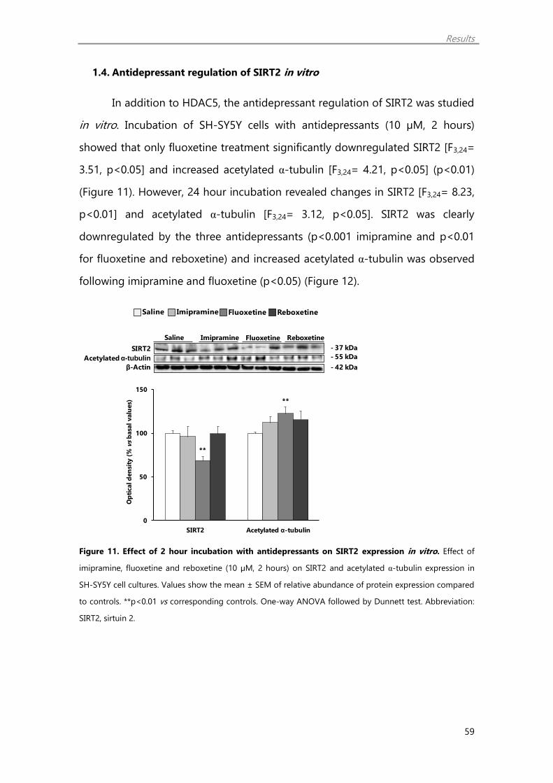
Results
59
1.4. Antidepressant regulation of SIRT2 in vitro
In addition to HDAC5, the antidepressant regulation of SIRT2 was studied
in vitro. Incubation of SH-SY5Y cells with antidepressants (10 µM, 2 hours)
showed that only fluoxetine treatment significantly downregulated SIRT2 [F3,24=
3.51, p<0.05] and increased acetylated α-tubulin [F3,24= 4.21, p<0.05] (p<0.01)
(Figure 11). However, 24 hour incubation revealed changes in SIRT2 [F3,24= 8.23,
p<0.01] and acetylated α-tubulin [F3,24= 3.12, p<0.05]. SIRT2 was clearly
downregulated by the three antidepressants (p<0.001 imipramine and p<0.01
for fluoxetine and reboxetine) and increased acetylated α-tubulin was observed
following imipramine and fluoxetine (p<0.05) (Figure 12).
Figure 11. Effect of 2 hour incubation with antidepressants on SIRT2 expression in vitro. Effect of
imipramine, fluoxetine and reboxetine (10 µM, 2 hours) on SIRT2 and acetylated α-tubulin expression in
SH-SY5Y cell cultures. Values show the mean ± SEM of relative abundance of protein expression compared
to controls. **p<0.01 vs corresponding controls. One-way ANOVA followed by Dunnett test. Abbreviation:
SIRT2, sirtuin 2.
Saline Imipramine Fluoxetine Reboxetine
ImipramineSaline
- 55 kDa
- 42 kDa
SIRT2
Acetylatedα-tubulin
β-Actin
- 37 kDa
Fluoxetine Reboxetine
**
**
0
50
100
150
SIRT2 Acetylated α-tubulin
Op
tical d
en
sity
(%
vs
basa
l valu
es)

Results
60
Figure 12. Antidepressant regulation of SIRT2 in vitro. Effect of imipramine, fluoxetine and reboxetine
(10 µM, 24 hours) on SIRT2 and acetylated α-tubulin expression in SH-SY5Y cell cultures. Data show the
mean ± SEM of relative abundance of mRNA or protein expression compared to controls. ***p<0.001,
**p<0.01, *p<0.05 vs corresponding controls. One-way ANOVA followed by Dunnett test. Abbreviations:
SIRT2, Sirtuin 2.
1.5. SIRT2 inhibition enhances histone acetylation and synaptic plasticity
markers
Subsequently, the in vitro effect of the SIRT2 inhibitor 33i (Suzuki et al. 2012) (5
µM, 24 hours) on histone acetylation and synaptic plasticity markers was studied here.
A significant upregulation of both AcH3 and AcH4 expression was observed (p<0.01)
(Figure 13A). In addition, the synaptic plasticity markers VGLUT1 and BDNF were also
upregulated by 33i (p<0.05) (Figure 13B).
Saline Imipramine Fluoxetine Reboxetine
***
*
**
*
**
0
50
100
150
SIRT2 Acetylated α-tubulin
Op
tic
al d
en
sit
y (%
vs
ba
sa
l va
lue
s)
ImipramineSaline
- 55 kDa
- 42 kDa
SIRT2
Acetylated α-tubulin
β-Actin
- 37 kDa
Fluoxetine Reboxetine

Results
61
Figure 13. SIRT2 inhibition increases histone acetylation and synaptic plasticity markers in vitro.
Effect of 33i (5 µM, 24 hours) on (A) AcH3 and AcH4 expression and on (B) VGLUT1 and BDNF expression
SH-SY5Y cell cultures. Values show the mean ± SEM of relative abundance of protein expression compared
to controls. **p<0.01, *p<0.05 vs corresponding controls, Student t-test.
Saline 33iA
33iSaline
- 10 kDa
- 42 kDa
AcH3
AcH4
β-Actin
- 17 kDa
**
**
0
50
100
150
200
250
AcH3 AcH4
Op
tic
al d
en
sit
y (%
vs
ba
sa
l va
lue
s)
B33iSaline
- 16 kDa
VGLUT1
BDNF
- 60 kDa
- 42 kDaβ-Actin
**
0
50
100
150
200
VGLUT1 BDNF
Op
tic
al d
en
sit
y (%
vs
ba
sa
l va
lue
s)

Results
62
1.6. Effect of MC3822 and 33i on HDAC5 and SIRT2 expression in vitro
We next studied whether the SIRT2 inhibitor 33i and the HDAC5 inhibitor
MC3822 affect to the expression of SIRT2 and acetylated α-tubulin together with
HDAC5 and p-HDAC5. Particularly, 33i (5 µM, 24 hours) increased acetylated α-tubulin
(p<0.05) and p-HDAC5 (p<0.001) in SH-SY5Y cells (Figures 14A and 14B). On the other
hand, neither of these proteins were affected by MC3822 (Figures 14C and 14D).
Subsequently the in vitro effect of the selective SIRT2 inhibitor 33i (5 µM, 24
hours) on HDAC5 localization was studied in SH-SY5Y cells. We observed that 33i
clearly induced nuclear export of HDAC5 to the cytoplasm (Figure 14E).

Results
63
Figure 14. SIRT2 inhibition regulates HDAC5 cellular localization in SH-SY5Y cell cultures. (A, B) Effect
of the HDAC5 inhibitor MC3822 on (A) SIRT2 and acetylated α-tubulin and (B) HDAC5 and p-HDAC5
expression in SH-SY5Y cell cultures. (C-E). Effect of the SIRT2 inhibitor 33i (5 µM, 24 hours) on (C) SIRT2 and
acetylated α-tubulin, (D) HDAC5 and p-HDAC5 expression and on (E) HDAC5 cellular localization in SH-
SY5Y cell cultures. Values show the mean ± SEM of relative abundance of protein expression compared to
controls. ***p<0.01, *p<0.05 vs corresponding controls, Student t-test. In (E), representative
photomicrographs of HDAC5 and nuclear (DAPI) immunofluorescence in SH-SY5Y cell cultures incubated
with vehicle (1% DMSO) or 33i (5 µM, 24 hours). HDAC5+ (green), DAPI+ (blue) and double
(HDAC5+/DAPI+) cells are shown. Scale bar: 10 µm.
Saline 33i
A 33iSaline- 37 kDa- 55 kDa
SIRT2
β-ActinAcetylated α-tubulin
- 42 kDa
*
0
50
100
150
SIRT2 Acetylated α-tubulin
Op
tic
al d
en
sit
y (%
vs
ba
sa
l va
lue
s)
B 33iSaline- 124 kDa- 124 kDa
HDAC5
β-ActinP-HDAC5
- 42 kDa
***
0
50
100
150
200
250
HDAC5 P-HDAC5
Op
tic
al d
en
sit
y (%
vs
ba
sa
l va
lue
s)
Saline MC3822
MC3822Saline- 37 kDa- 55 kDa
SIRT2
β-ActinAcetylated α-tubulin
- 42 kDa
0
50
100
150
SIRT2 Acetylated α-tubulin
Op
tic
al d
en
sit
y (%
vs
ba
sa
l va
lue
s)
C MC3822Saline- 124 kDa- 124 kDa
HDAC5
β-ActinP-HDAC5
- 42 kDa
D
0
50
100
150
HDAC5 P-HDAC5
Op
tic
al d
en
sit
y (%
vs
ba
sa
l va
lue
s)
E
Co
ntr
ol
33
i
HDAC5 MergeDAPI

Results
64
1.7. Antidepressant treatment regulate histone acetylation and synaptic
plasticity markers in vitro
In order to recapitulate previous in vivo findings (Erburu et al. 2015a), we
studied the antidepressant regulation of histone acetylation and synaptic plasticity
markers in vitro.
Incubation of SH-SY5Y cells with imipramine, fluoxetine and reboxetine (10 µM,
2 hours) influenced AcH3 [F3,24= 4.87, p<0.01] and AcH4 [F3,24= 6.35, p<0.001]
expression levels. Specifically, AcH3 expression was significantly increased by
imipramine (p<0.05) and fluoxetine (p<0.01). Similarly, AcH4 was increased by
fluoxetine (p<0.001) and imipramine (p<0.05) (Figure 15A). However, incubation of SH-
SY5Y cells with antidepressants for 2 hours did not affect to the expression of VGLUT1
and BDNF (Figure 15B).
A pronounced increase on AcH3 [F3,24= 8.15, p<0.001] and AcH4 [F3,24= 6.01,
p<0.01] could be observed on SH-SY5Y cells, following 24 hour antidepressant
treatment (10 µM). Both AcH3 and AcH4 expression were increased by imipramine
(p<0.01), fluoxetine (p<0.001 for AcH3 and p<0.01 for AcH4) and reboxetine (p<0.05)
(Figure 16A).
Incubation of SH-SY5Y cells with imipramine and fluoxetine (10 µM, 24 hours)
also regulated the expression of these synaptic plasticity markers [F3,24= 16.99, p<0.001
and F3,24= 4.06, p<0.001 for VGLUT1 and BDNF respectively]. Both imipramine and
fluoxetine increased VGLUT1 (p<0.01; p<0.001) and BDNF expression levels (p<0.01;
p<0.05; for VGLUT1 and BDNF, respectively) (Figure 16B). Although no statistically
significant, reboxetine showed a tendency to increase VGLUT1 and BDNF compared to
saline.
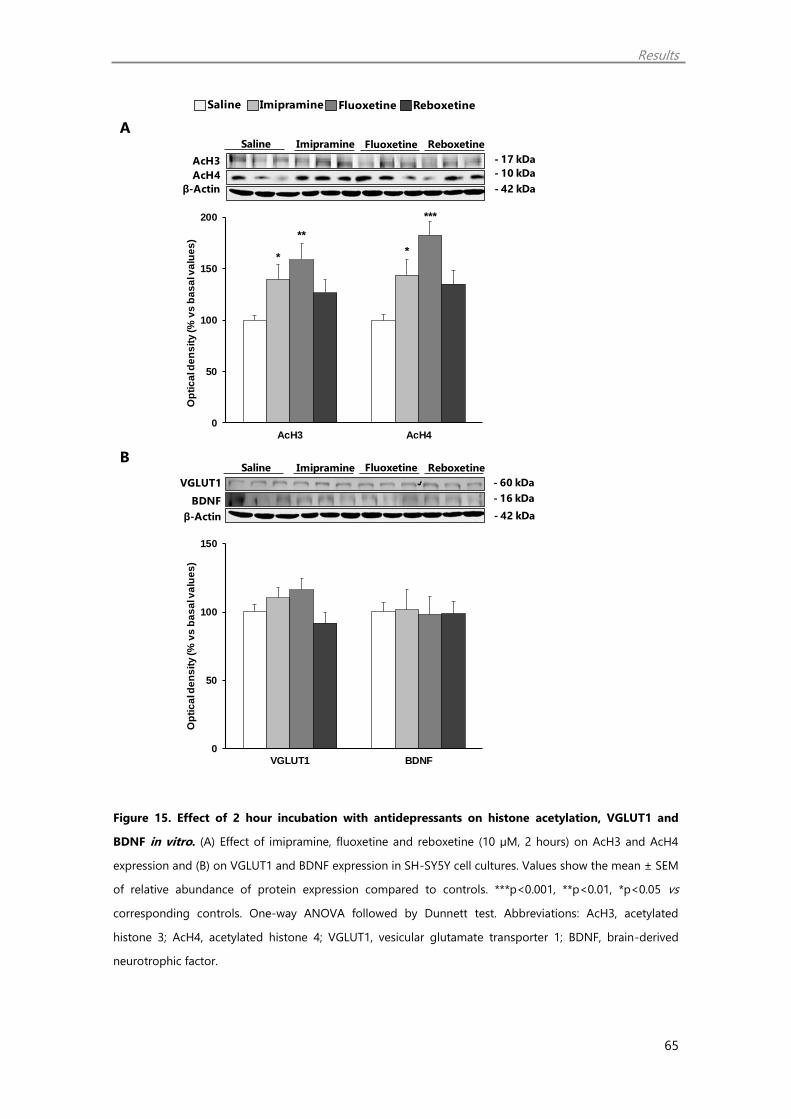
Results
65
Figure 15. Effect of 2 hour incubation with antidepressants on histone acetylation, VGLUT1 and
BDNF in vitro. (A) Effect of imipramine, fluoxetine and reboxetine (10 µM, 2 hours) on AcH3 and AcH4
expression and (B) on VGLUT1 and BDNF expression in SH-SY5Y cell cultures. Values show the mean ± SEM
of relative abundance of protein expression compared to controls. ***p<0.001, **p<0.01, *p<0.05 vs
corresponding controls. One-way ANOVA followed by Dunnett test. Abbreviations: AcH3, acetylated
histone 3; AcH4, acetylated histone 4; VGLUT1, vesicular glutamate transporter 1; BDNF, brain-derived
neurotrophic factor.
Saline Imipramine Fluoxetine Reboxetine
A
**
**
***
0
50
100
150
200
AcH3 AcH4
Op
tic
al d
en
sit
y (%
vs
ba
sa
l va
lue
s)
ImipramineSaline
- 10 kDa
- 42 kDa
AcH3
AcH4
β-Actin
- 17 kDa
Fluoxetine Reboxetine
B
0
50
100
150
VGLUT1 BDNF
Op
tic
al d
en
sit
y (%
vs
ba
sa
l va
lue
s)
ImipramineSaline
- 16 kDa
- 42 kDa
VGLUT1
BDNF
β-Actin
- 60 kDa
Fluoxetine Reboxetine

Results
66
Figure 16. Antidepressant incubation for 24 hours increases histone acetylation, VGLUT1 and BDNF
in vitro. (A) Effect of imipramine, fluoxetine and reboxetine (10 µM, 24 hours) on AcH3 and AcH4
expression and (B) on VGLUT1 and BDNF expression SH-SY5Y cell cultures. Values show the mean ± SEM
of relative abundance of protein expression compared to controls. ***p<0.001, **p<0.01, *p<0.05 vs
corresponding controls. One-way ANOVA followed by Dunnett test. Abbreviations: AcH3, acetylated
histone 3; AcH4, acetylated histone 4; VGLUT1, vesicular glutamate transporter 1; BDNF, brain-derived
neurotrophic factor.
Saline Imipramine Fluoxetine Reboxetine
A
BImipramineSaline
- 16 kDa
VGLUT1
BDNF
- 60 kDa
Fluoxetine Reboxetine
- 42 kDaβ-Actin
**
*****
**
**
0
50
100
150
200
250
AcH3 AcH4
Op
tic
al d
en
sit
y (%
vs
ba
sa
l va
lue
s)
ImipramineSaline
- 10 kDa
- 42 kDa
AcH3
AcH4
β-Actin
- 17 kDa
Fluoxetine Reboxetine
***
**** *
0
50
100
150
200
250
VGLUT1 BDNF
Op
tic
al d
en
sit
y (%
vs
ba
sa
l va
lue
s)
Results
67
1.8. Muscarinic receptor blockade does not affect histone acetylation and
synaptic plasticity markers in vitro
In order to discard any possible anticholinergic action on epigenetic targets, the
antimuscarinic drug scopolamine was studied. Incubation with the antimuscarinic drug
scopolamine (10 µM, 24 hours), did not affect the expression of AcH3, AcH4 (Figure
17A) as well as VGLUT1 and BDNF (Figure 17B).

Results
68
Figure 17. Scopolamine does not affect histone acetylation or synaptic plasticity markers in vitro.
Effect of scopolamine (10 µM, 24 hours) on (A) AcH3 and AcH4 expression and on (B) VGLUT1 and BDNF
expression SH-SY5Y cell cultures. Values show the mean ± SEM of relative abundance of protein expression
compared to controls. Student t-test.
Saline ScopolamineA
B
0
25
50
75
100
125
VGLUT1 BDNF
Op
tical d
en
sit
y (
% v
s b
asal valu
es)
ScopolamineSaline
- 16 kDa
VGLUT1
BDNF
- 60 kDa
- 42 kDaβ-Actin
Saline
- 10 kDa
- 42 kDa
AcH3
AcH4
β-Actin
- 17 kDa
Scopolamine
0
25
50
75
100
125
AcH3 AcH4
Op
tical d
en
sit
y (
% v
s b
asal valu
es)

Results
69
1.9. Chronic antidepressant treatment regulate HDAC5 and SIRT2 in vivo
Finally, the long term antidepressant regulation of HDAC5 and SIRT2 was
studied in the mice PFC.
RT-PCR studies showed that Hdac5 mRNA abundance in PFC was affected by
chronic antidepressant treatment [F3,24= 6.08, p<0.01]. Both repeated imipramine
(p<0.05) and reboxetine (p<0.01) significantly increased the expression of this gene
compared to control saline treated mice (Figure 18A). On the other hand, while HDAC5
protein expression in the PFC was not affected by chronic antidepressants [F3,24= 2.15,
p>0.05], changes in the expression of the phosphorylated form (p-HDAC5) were
observed [F3,24= 4.29, p<0.001]. Repeated imipramine and reboxetine treatment
confirmed a significant p-HDAC5 upregulation (p<0.05) (Figure 18B).
Sirt2 mRNA expression in PFC was influenced by chronic antidepressant
treatment [F3,24= 5.47, p<0.05]. Chronic fluoxetine treatment significantly
downregulated Sirt2 mRNA levels (p<0.05) compared to control saline (Figure 18C).
Moreover, significant effects of chronic antidepressant treatment on SIRT2 [F3,24= 5.28,
p<0.01] and SIRT2 cytoplasmic substrate acetylated α-tubulin [F3,24= 4.68, p<0.01]
expression levels were observed. Specifically, SIRT2 was downregulated by the three
antidepressants compared to saline treated mice (p<0.05 for imipramine and
fluoxetine; p<0.01 for reboxetine). Moreover, imipramine and reboxetine increased
acetylated α-tubulin (p<0.05) (Figure 18D).

Results
70
Figure 18. Antidepressant regulation of HDAC5 and SIRT2 in vivo. (A, B) Effect of chronic treatment
with imipramine (10 mg/kg), fluoxetine (15 mg/kg) and reboxetine (15 mg/kg) for three weeks on (A)
Hdac5 mRNA abundance and (B) HDAC5 and p-HDAC5 expression in mice prefrontal cortex. (C, D) and on
(C) Sirt2 mRNA abundance and (D) SIRT2 and cytoplasmic SIRT2 substrate acetylated α-tubulin in mice
prefrontal cortex. Data show the mean ± SEM of relative abundance of mRNA or protein expression
compared to controls. **p<0.01, *p<0.05 versus corresponding controls. One-way ANOVA followed by
Dunnett test. Abbreviations: HDAC5, histone deacetylase 5; p-HDAC5, phosphorylated histone deacetylase
5; SIRT2, Sirtuin 2.
Saline Imipramine Fluoxetine Reboxetine
ImipramineSaline
- 124 kDaHDAC5
P-HDAC5
- 124 kDa
Fluoxetine Reboxetine
- 42 kDaβ-Actin
A
***
0
50
100
150
200
hdac5
mR
NA
ex
pre
ssio
n (%
vs
co
ntr
ol s
alin
e)
**
0
50
100
150
200
250
HDAC5 P-HDAC5
Op
tic
al d
en
sit
y (%
vs
ba
sa
l va
lue
s)
B
*
0
25
50
75
100
125
sirt2
mR
NA
ex
pre
ssio
n (%
vs
co
ntr
ol s
alin
e)
C D
*
*
***
*
0
50
100
150
SIRT2 Acetylated α-tubulin
Op
tic
al d
en
sit
y (%
vs
ba
sa
l va
lue
s)
ImipramineSaline
- 55 kDaSIRT2
Acetylated α-tubulin
- 37 kDa
Fluoxetine Reboxetine
- 42 kDaβ-Actin
Results
71
1.10. Chronic antidepressant treatment regulate epigenetic and synaptic
plasticity markers in the mouse PFC
The long-term antidepressant regulation of all these epigenetic and synaptic
plasticity markers in the mice PFC was studied comparatively to the in vitro studies.
Chronic antidepressant treatment for 21 days regulated the expression of AcH3 and
AcH4. Specifically, the three antidepressants caused an increase (p<0.05) on AcH3
expression [F3,24= 3.71, p<0.05]. In addition, AcH4 levels were increased by imipramine
(p<0.05), reboxetine (p<0.01) and fluoxetine (p<0.001) [F3,24= 9.83, p<0.001] (Figure
19A).
In addition, chronic antidepressant treatment for 21 days regulated the
expression of the synaptic vesicle protein VGLUT1 [F3,24= 4.23, p<0.01] and the
neurotrophin BDNF [F3,24= 3.00, p<0.05] in the prefrontal cortex (PFC). In particular,
VGLUT1 expression was significantly increased by imipramine (10 mg/kg) (p<0.05),
fluoxetine (15 mg/kg) (p<0.01) and reboxetine (15 mg/kg) (p<0.05) following three
weeks of treatment. Moreover, the expression of BDNF was significantly increased after
imipramine, fluoxetine and reboxetine (p<0.05) (Figure 19B).

Results
72
Figure 19. Antidepressants increase histone acetylation and synaptic plasticity markers in vivo. (A)
Effect of chronic treatment with imipramine (10 mg/kg), fluoxetine (15 mg/kg) and reboxetine (15 mg/kg)
for three weeks on AcH3 and AcH4 expression and (B) on VGLUT1 and BDNF expression in mice prefrontal
cortex. Values show the mean ± SEM of relative abundance of protein expression compared to controls.
***p<0.001, **p<0.01, *p<0.05 vs corresponding controls. One-way ANOVA followed by Dunnett test.
Abbreviations: AcH3, acetylated histone 3; AcH4, acetylated histone 4; VGLUT1, vesicular glutamate
transporter 1; BDNF, brain-derived neurotrophic factor.
Saline Imipramine Fluoxetine ReboxetineA
**
*
***
***
0
50
100
150
200
250
300
AcH3 AcH4
Op
tic
al d
en
sit
y (%
vs
ba
sa
l va
lue
s)
ImipramineSaline
- 10 kDa
- 42 kDa
AcH3
AcH4
β-Actin
- 17 kDa
Fluoxetine Reboxetine
B ImipramineSaline
- 16 kDa
VGLUT1
BDNF
- 60 kDa
Fluoxetine Reboxetine
- 42 kDaβ-Actin
***
**
* *
0
50
100
150
200
VGLUT1 BDNF
Op
tic
al d
en
sit
y (%
vs
ba
sa
l va
lue
s)
Results
73
2. Therapeutic potential of SIRT2 for depression treatment in the VGLUT1+/-
model
The second aim of our study was to evaluate the therapeutic potential of SIRT2
for depression treatment. Specifically, the potential antidepressant effect of the
selective SIRT2 inhibitor 33i was tested in mice heterozygous for VGLUT1 (VGLUT1+/-),
previously characterized as a genetic model of depressive-like behavior. Firstly, we
tested whether the VGLUT1+/- model shows alterations in the expression of the HDAC
superfamily or in histone acetylation in the PFC. Subsequently, the effect of 33i and
reference antidepressant imipramine in the anhedonic behaviour of VGLUT1+/- mice
was studied. Finally, we tested the activity of 33i for specific monoaminergic molecular
targets in order to discard classic antidepressant mechanisms.
2.1. The mRNA expression of the HDAC superfamily in the PFC of VGLUT1+/-
mice and wild type littermates
In this experiment we have studied the mRNA abundance of the Hdac’s
superfamily including class I (Hdac1, Hdac2, Hdac3 and Hdac8), class II (Hdac4, Hdac5,
Hdac6, Hdac7, Hdac9 and Hdac10) and IV (Hdac11) as well as the NAD+-dependent
class III (Sirt1-7) in the PFC of VGLUT1+/- mice, as a genetic model of depression
(Garcia-Garcia et al. 2009). No alterations in the mRNA expression of any of these
enzymes were found, indicating that the depressive-like behavior of this model is not
linked to variations in Hdac mRNA expression (Figure 20). Yet, further studies should
confirm these results at protein level.

Results
74
Figure 20. Hdac superfamily is not altered in the PFC of VGLUT1+/- mice. mRNA abundance of
histone deacetylase enzymes (Hdac’s) (A) Hdac’s class I and IV, (B) class II and (C) class III in mice prefrontal
cortex of VGLUT1+/- mice and WT littermates. Data show average ± SEM of relative abundance of mRNA
expression compared to WT values (Student t-test, n=8-9 mice/group).
2.2. VGLUT1+/- mice show no changes in histone acetylation, HDAC5 and
SIRT2
Subsequently, we tested the therapeutic value of SIRT2 inhibition on HDAC5, p-
HDAC5 (Figure 21A), SIRT2, acetylated α-tubulin (Figure 21B) AcH3 and AcH4 (Figure
21C). We observed that the protein levels of AcH3 and AcH4 were not altered in the
A WT VGLUT1+/-
C
0
20
40
60
80
100
120
140
Hdac1 Hdac2 Hdac3 Hdac8 Hdac11
Exp
ress
ion
mR
NA
(%
vs
con
tro
l)
0
20
40
60
80
100
120
140
Hdac4 Hdac5 Hdac6 Hdac7 Hdac9 Hdac10
Exp
ress
ion
mR
NA
(%
vs
con
tro
l)
B
0
20
40
60
80
100
120
Sirt1 Sirt2 Sirt3 Sirt4 Sirt5 Sirt6 Sirt7
Ex
pre
ssio
n m
RN
A (
% v
sco
ntr
ol)
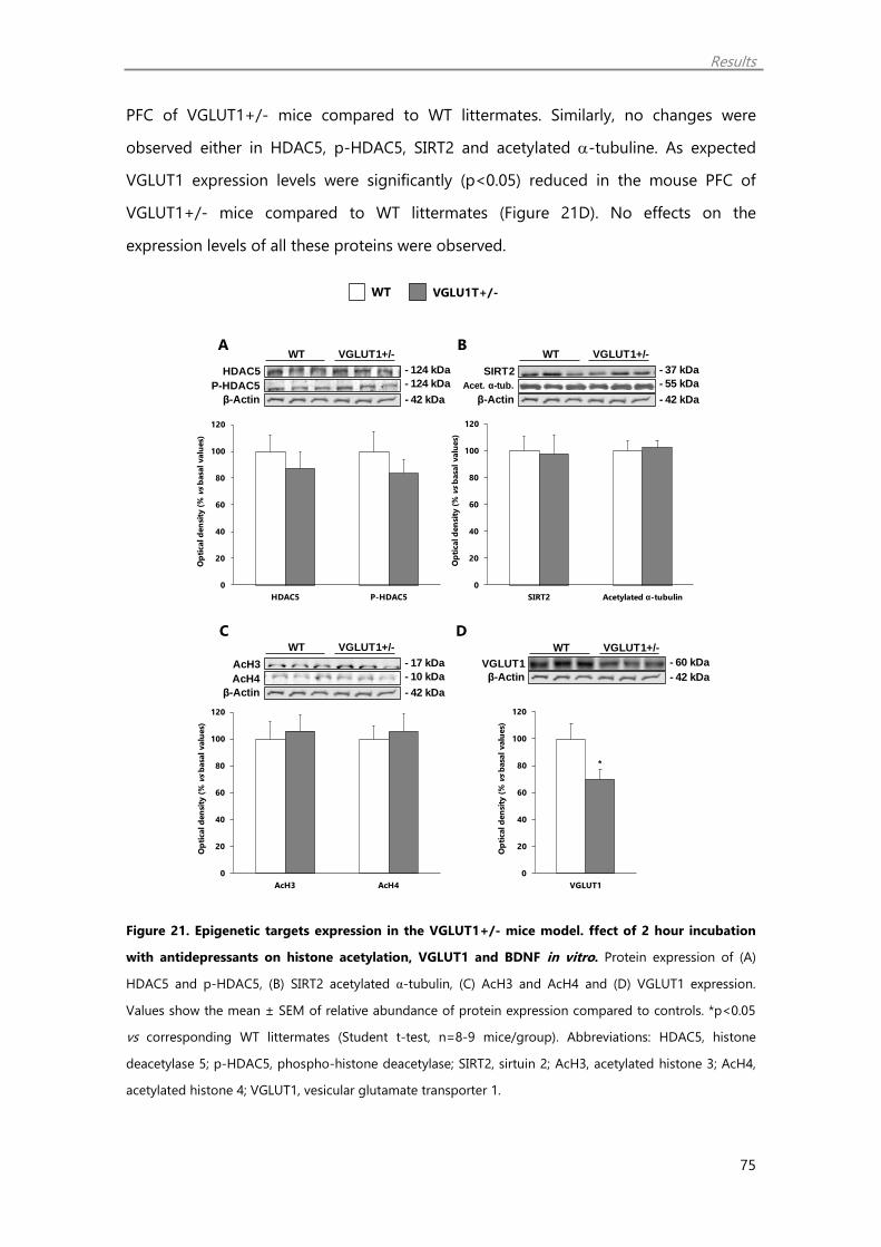
Results
75
PFC of VGLUT1+/- mice compared to WT littermates. Similarly, no changes were
observed either in HDAC5, p-HDAC5, SIRT2 and acetylated -tubuline. As expected
VGLUT1 expression levels were significantly (p<0.05) reduced in the mouse PFC of
VGLUT1+/- mice compared to WT littermates (Figure 21D). No effects on the
expression levels of all these proteins were observed.
Figure 21. Epigenetic targets expression in the VGLUT1+/- mice model. ffect of 2 hour incubation
with antidepressants on histone acetylation, VGLUT1 and BDNF in vitro. Protein expression of (A)
HDAC5 and p-HDAC5, (B) SIRT2 acetylated α-tubulin, (C) AcH3 and AcH4 and (D) VGLUT1 expression.
Values show the mean ± SEM of relative abundance of protein expression compared to controls. *p<0.05
vs corresponding WT littermates (Student t-test, n=8-9 mice/group). Abbreviations: HDAC5, histone
deacetylase 5; p-HDAC5, phospho-histone deacetylase; SIRT2, sirtuin 2; AcH3, acetylated histone 3; AcH4,
acetylated histone 4; VGLUT1, vesicular glutamate transporter 1.
A B
C D
WT VGLU1T+/-
0
20
40
60
80
100
120
HDAC5 P-HDAC5
Op
tical d
en
sity
(%
vs
basa
l valu
es)
0
20
40
60
80
100
120
AcH3 AcH4
Op
tical d
en
sity
(%
vs
basa
l valu
es)
VGLUT1+/-WT
- 10 kDa
- 42 kDa
AcH3
AcH4
β-Actin
- 17 kDa
VGLUT1+/-WT
VGLUT1 - 60 kDa
- 42 kDaβ-Actin
VGLUT1+/-WT
- 124 kDa
- 42 kDa
HDAC5
P-HDAC5
β-Actin
- 124 kDa
VGLUT1+/-WT
- 55 kDa
- 42 kDa
SIRT2
Acet. α-tub.
β-Actin
- 37 kDa
*
0
20
40
60
80
100
120
VGLUT1
Op
tical d
en
sity
(%
vs
basa
l valu
es)
0
20
40
60
80
100
120
SIRT2 Acetylated α-tubulin
Op
tical d
en
sity
(%
vs
basa
l valu
es)
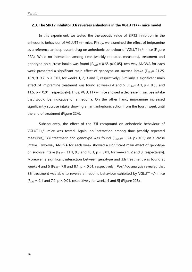
Results
76
2.3. The SIRT2 inhibitor 33i reverses anhedonia in the VGLUT1+/- mice model
In this experiment, we tested the therapeutic value of SIRT2 inhibition in the
anhedonic behaviour of VGLUT1+/- mice. Firstly, we examined the effect of imipramine
as a reference antidepressant drug on anhedonic behaviour of VGLUT1+/- mice (Figure
22A). While no interaction among time (weekly repeated measures), treatment and
genotype on sucrose intake was found [F4,256= 0.65 p>0.05], two-way ANOVA for each
week presented a significant main effect of genotype on sucrose intake [F1,64= 21.25,
10.9, 9, 9.7 p < 0.01, for weeks 1, 2, 3 and 5, respectively]. Similarly, a significant main
effect of imipramine treatment was found at weeks 4 and 5 [F1,64= 4.1, p < 0.05 and
11.5, p < 0.01, respectively]. Thus, VGLUT1+/- mice showed a decrease in sucrose intake
that would be indicative of anhedonia. On the other hand, imipramine increased
significantly sucrose intake showing an antianhedonic action from the fourth week until
the end of treatment (Figure 22A).
Subsequently, the effect of the 33i compound on anhedonic behaviour of
VGLUT1+/- mice was tested. Again, no interaction among time (weekly repeated
measures), 33i treatment and genotype was found [F4,252= 1.24 p>0.05] on sucrose
intake. Two-way ANOVA for each week showed a significant main effect of genotype
on sucrose intake [F1,63= 11.1, 9.3 and 10.3, p < 0.01, for weeks 1, 2 and 3, respectively].
Moreover, a significant interaction between genotype and 33i treatment was found at
weeks 4 and 5 [F1,63= 7.8 and 8.1, p < 0.01, respectively]. Post hoc analysis revealed that
33i treatment was able to reverse anhedonic behaviour exhibited by VGLUT1+/- mice
[F3,63 = 9.1 and 7.9, p < 0.01, respectively for weeks 4 and 5] (Figure 22B).

Results
77
Figure 22. SIRT2 inhibition rescues anhedonic behaviour of VGLUT1+/- mice. Effect of (A) imipramine
and (B) 33i treatment on depressive like behaviour in the sucrose intake test. Data show mean ± SEM of
different groups. *p<0.05, main effect of treatment; ^^p<0.01 main effect of genotype; ##p<0.01
VGLUT1+/- treated with 33i vs VGLUT1+/- treated with saline (two way ANOVA followed by Tukey-post
hoc, n= 15-20 mice per group).
^^
^^^^ ^^
^^
##
##
0
0,2
0,4
0,6
0,8
1
1,2
0 1 2 3 4
Su
cro
se i
nta
ke (
g/b
od
y w
eig
ht)
WT-saline
WT-33i
VGLUT1-saline
VGLUT1-33i
B
1.2
1
0
0.8
0.6
0.4
0.2
*
*
^^^^ ^^
^^
* *
0
0,2
0,4
0,6
0,8
1
1,2
0 1 2 3 4
Su
cro
se i
nta
ke (
g/b
od
y w
eig
ht)
WT-saline
WT-Imi
VGLUT1-saline
VGLUT1-Imi
A 1.2
1
0
0.8
0.6
0.4
0.2
Weeks
Weeks
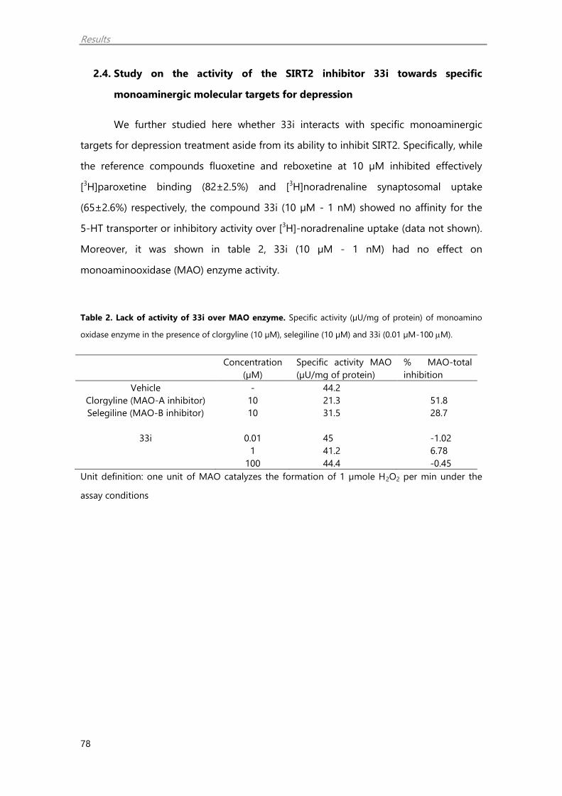
Results
78
2.4. Study on the activity of the SIRT2 inhibitor 33i towards specific
monoaminergic molecular targets for depression
We further studied here whether 33i interacts with specific monoaminergic
targets for depression treatment aside from its ability to inhibit SIRT2. Specifically, while
the reference compounds fluoxetine and reboxetine at 10 µM inhibited effectively
[3H]paroxetine binding (82±2.5%) and [3H]noradrenaline synaptosomal uptake
(65±2.6%) respectively, the compound 33i (10 µM - 1 nM) showed no affinity for the
5-HT transporter or inhibitory activity over [3H]-noradrenaline uptake (data not shown).
Moreover, it was shown in table 2, 33i (10 µM - 1 nM) had no effect on
monoaminooxidase (MAO) enzyme activity.
Table 2. Lack of activity of 33i over MAO enzyme. Specific activity (µU/mg of protein) of monoamino
oxidase enzyme in the presence of clorgyline (10 µM), selegiline (10 µM) and 33i (0.01 µM-100 M).
Concentration
(µM)
Specific activity MAO
(µU/mg of protein)
% MAO-total
inhibition
Vehicle - 44.2
Clorgyline (MAO-A inhibitor) 10 21.3 51.8
Selegiline (MAO-B inhibitor) 10 31.5 28.7
33i 0.01 45 -1.02
1 41.2 6.78
100 44.4 -0.45
Unit definition: one unit of MAO catalyzes the formation of 1 µmole H2O2 per min under the
assay conditions
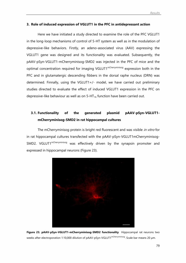
Results
79
3. Role of induced expression of VGLUT1 in the PFC in antidepressant action
Here we have initiated a study directed to examine the role of the PFC VGLUT1
in the long-loop mechanisms of control of 5-HT system as well as in the modulation of
depressive-like behaviors. Firstly, an adeno-associated virus (AAV) expressing the
VGLUT1 gene was designed and its functionality was evaluated. Subsequently, the
pAAV-pSyn-VGLUT1-mCherryminisog-SMD2 was injected in the PFC of mice and the
optimal concentration required for imaging VGLUT1mCherryminisog expression both in the
PFC and in glutamatergic descending fibbers in the dorsal raphe nucleus (DRN) was
determined. Finnally, using the VGLUT1+/- model, we have carried out preliminary
studies directed to evaluate the effect of induced VGLUT1 expression in the PFC on
depressive-like behaviour as well as on 5-HT1A function have been carried out.
3.1. Functionality of the generated plasmid pAAV-pSyn-VGLUT1-
mCherryminisog-SMD2 in rat hippocampal cultures
The mCherryminisog protein is bright red fluorescent and was visible in vitro for
in rat hippocampal cultures transfected with the pAAV-pSyn-VGLUT1mCherryminisog-
SMD2. VGLUT1mCherryminisog was effectively driven by the synapsin promoter and
expressed in hippocampal neurons (Figure 23).
Figure 23. pAAV-pSyn-VGLUT1-mCherryminisog-SMD2 functionality Hippocampal rat neurons two
weeks after electroporation 1:10,000 dilution of pAAV-pSyn-VGLUT1mCherryminisog
. Scale bar means 20 µm.
Results
80
3.2. pAAV-pSyn-VGLUT1mCherryminisog and pAAV-pSyn-YFP imaging in the PFC, in
the DRN and other projecting areas on VGLUT1+/- mice and WT
littermates
In preliminary experiments, of the different dilutions tested, for AAV-pSyn-
VGLUT1mCherryminisog (1:40, 1:16, 1:4, 1:2 and 1:1), the 1:2 and 1:1 dilutions imaging
studies revealed VGLUT1mCherryminisog expression in both cell bodies and fibbers in the
PFC as well as in fibbers in the dorsal raphe nucleus (DRN). Yet, using lower dilutions
(1:4-1:40) of the AAV, no signal at the level of the DRN was detected. Therefore the 1:2
dilution was selected for subsequent studies.
Hence, in vivo gene transfer efficacy was determined by imaging analysis using
the epifluorescence microscope (4x, 40x). Fluorescence was visible in serial sliced
sections of the PFC of mouse brain injected with pAAV-pSyn-VGLUT1mCherryminisog under
synapsin promoter. Abundant ectopic VGLUT1 could be detected specifically in the
mPFC of pAAV-pSyn-VGLUT1mCherryminisog, including infralimbic (Figures 24A) and other
cortical areas (data not shown). The rostro-caudal extension of the vector spread was
observed in all the slices around the injection site. At higher magnification (40X) ectopic
VGLUT1 expression was visualized in neuronal soma and the neuropil (Figures 24A’).
Further, analysis of mCherryminisog or YFP fluorescence in the whole PFC of both
genotypes, WT and VGLUT1+/-, revealed no differences in terms of area by intensity of
fluorescence (Figures 24C and 24C’).
The YFP protein is bright green fluorescent. pAAV-pSyn-YFP injected animals
showed fluorescence in serial sliced sections of the PFC (Figure 24B and 24B’).
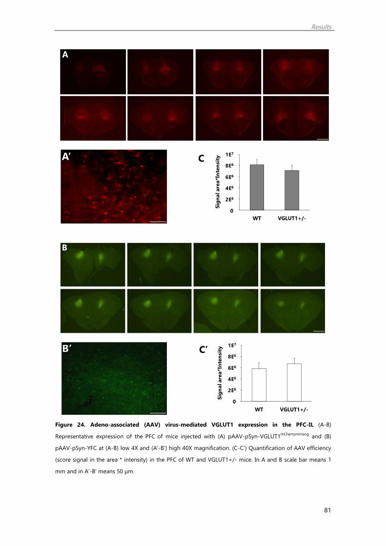
Results
81
Figure 24. Adeno-associated (AAV) virus-mediated VGLUT1 expression in the PFC-IL (A-B)
Representative expression of the PFC of mice injected with (A) pAAV-pSyn-VGLUT1mCherryminisog
and (B)
pAAV-pSyn-YFC at (A-B) low 4X and (A’-B’) high 40X magnification. (C-C’) Quantification of AAV efficiency
(score signal in the area * intensity) in the PFC of WT and VGLUT1+/- mice. In A and B scale bar means 1
mm and in A’-B’ means 50 µm.
0,E+00
2,E+06
4,E+06
6,E+06
8,E+06
1,E+07
WT VGLUT1+/-Sig
nal are
a*In
ten
sit
y
0
2E6
4E6
6E6
8E6
1E7
0,E+00
2,E+06
4,E+06
6,E+06
8,E+06
1,E+07
WT VGLUT1+/-
Sig
nal are
a*In
ten
sit
y
0
2E6
4E6
6E6
8E6
1E7
C
C’

Results
82
In the dorsal raphe nucleus (DRN), we observed ectopic VGLUT1 expression in
fibbers (Figure 25A). In addition, signal of YFP fluorescence was detected in this area
showing that the injected point selected has projections to this area (Figure 25B). In
addition at the level of the dorsal hippocampus, VGLUT1 immunoreactivity could be
observed in hypothalamic areas, in the amygdala and in the dentate girus of the
hippocampus (Figure 26).
Figure 25. Adeno-associated (AAV) virus-mediated VGLUT1 expression in the in the raphe nuclei.
Representative expression of the raphe nuclei of mice injected with (A) pAAV-pSyn-VGLUT1mCherryminisog
and
(B) pAAV-pSyn-YFC at (A-B) low 10X and (A’-B’) high 40X magnification. In A and B scale bar means 500
µm and in A’-B’ means 50 µm.
Figure 26. Adeno-associated (AAV) virus-mediated VGLUT1 expression in PFC projecting areas.
Representative expression of the Hippocampus and the amygdale of mice injected with (A) pAAV-pSyn-
VGLUT1mCherryminisog
and (B) pAAV-pSyn-YFC at 4X magnification. Scale bar means 1 mm.
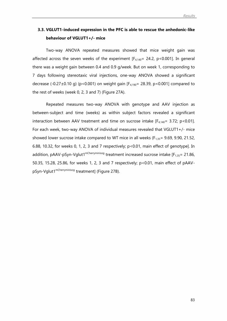
Results
83
3.3. VGLUT1-induced expression in the PFC is able to rescue the anhedonic-like
behaviour of VGLUT1+/- mice
Two-way ANOVA repeated measures showed that mice weight gain was
affected across the seven weeks of the experiment [F4,140= 24.2, p<0.001]. In general
there was a weight gain between 0.4 and 0.9 g/week. But on week 1, corresponding to
7 days following stereotaxic viral injections, one-way ANOVA showed a significant
decrease (-0.27±0.10 g) (p<0.001) on weight gain [F4,190= 28.39, p<0.001] compared to
the rest of weeks (week 0, 2, 3 and 7) (Figure 27A).
Repeated measures two-way ANOVA with genotype and AAV injection as
between-subject and time (weeks) as within subject factors revealed a significant
interaction between AAV treatment and time on sucrose intake [F4,140= 3.72; p<0.01].
For each week, two-way ANOVA of individual measures revealed that VGLUT1+/- mice
showed lower sucrose intake compared to WT mice in all weeks (F1,35= 9.69, 9.90, 21.52,
6.88, 10.32, for weeks 0, 1, 2, 3 and 7 respectively; p<0.01, main effect of genotype]. In
addition, pAAV-pSyn-Vglut1mCherryminisog treatment increased sucrose intake [F1,35= 21.86,
50.35, 15.28, 25.86, for weeks 1, 2, 3 and 7 respectively; p<0.01, main effect of pAAV-
pSyn-Vglut1mCherryminisog treatment] (Figure 27B).

Results
84
Figure 27. Effect of VGLUT1-induced expression in the IL-PFC on body weight gain (A) and sucrose
intake (B) of WT and VGLUT1+/- mice. Data show the mean ± SEM (n= 9-11 mice/ group) of the
weekly body weight gain (g) (A) and sucrose intake (g per body weight). **p<0.01, main effect of genotype;
^^p<0.01 main effect of pAAV-pSyn-VGLUT1mCherryminisog
treatment (Two- way ANOVA).
***
-1
-0,5
0
0,5
1
1,5
0 1 2 3 7
Weig
ht
gain
(g
)
Weeks
A
-1
-0.5
0
0.5
1.5
//
1
B
^^ ^^^^
^^
^^**
**** **
**
**
^^** ^^**^^**
0
0,2
0,4
0,6
0,8
1
1,2
0 1 2 3 4 7
Su
cro
se in
take (
g/b
od
y w
eig
ht)
Weeks
WT YFP
WT
mCherryminisogVglut1+/-YFP
Vglut1+/-
mCherryminisog
//
0.2
0.4
0.6
0.8
1.2
1
0
**
^^
^
-3
-2,5
-2
-1,5
-1
-0,5
0
0,5
30 60 90
ᐃT
em
pera
ture
(ºC
) WT YFP
WT
mCherryminisog
Vglut1+/- YFP
Vglut1+/-
mCherryminisog
-0.5
-1.5
-0.5
-2.5
WT VGLUT1mCherryminisog
VGLUT1+/- YFP
WT YFP
VGLUT1+/- VGLUT1mCherryminisog

Results
85
3.4. Behavioural studies
Locomotor Activity. WT and VGLUT1 mice treated with either pAAV-pSyn-
VGLUT1mCherryminisog or pAAV-pSyn-YFP did not differ in their spontaneous locomotor
activity during the 30-minutes test period (Figure 28A).
Open field. In the open field test, stereotaxic injection of pAAV-pSyn-
VGLUT1mCherryminisog decreased significantly time spent in center zone in both WT and
VGLUT1+/- mice [F1,35= 5.59, p<0.05] (Figure 28B). However no differences in latency
time to go to the center zone were observed among the groups (data not shown).
Social Interaction Test. No significant differences among the groups (WT YFP,
WT VGLUT1mCherryminisog, VGLUT1+/- YFP and VGLUT1+/- VGLUT1mCherryminisog) were
observed in interaction time [F1,35= 1.34, p>0.05] (Figure 28C). In addition freezing times
were not altered [F1,35= 0.18, p>0.05] (data not shown).
Marble burying test. In the marble burying test, stereotaxic injection of pAAV-
pSyn-VGLUT1mCherryminisog decreased significantly the number of buried marbles in both
WT and VGLUT1+/- mice [F1,35= 6.01, p<0.05] (Figure 28D).
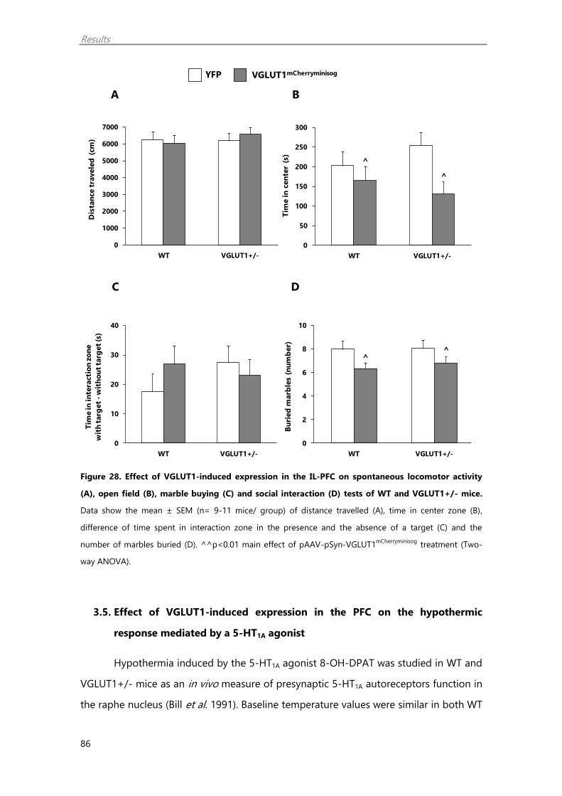
Results
86
Figure 28. Effect of VGLUT1-induced expression in the IL-PFC on spontaneous locomotor activity
(A), open field (B), marble buying (C) and social interaction (D) tests of WT and VGLUT1+/- mice.
Data show the mean ± SEM (n= 9-11 mice/ group) of distance travelled (A), time in center zone (B),
difference of time spent in interaction zone in the presence and the absence of a target (C) and the
number of marbles buried (D). ^^p<0.01 main effect of pAAV-pSyn-VGLUT1mCherryminisog
treatment (Two-
way ANOVA).
3.5. Effect of VGLUT1-induced expression in the PFC on the hypothermic
response mediated by a 5-HT1A agonist
Hypothermia induced by the 5-HT1A agonist 8-OH-DPAT was studied in WT and
VGLUT1+/- mice as an in vivo measure of presynaptic 5-HT1A autoreceptors function in
the raphe nucleus (Bill et al. 1991). Baseline temperature values were similar in both WT
YFP VGLUT1mCherryminisog
A B
C D
0
1000
2000
3000
4000
5000
6000
7000
WT VGLUT1+/-
Dis
tan
ce t
ravele
d (
cm
)
^
^
0
50
100
150
200
250
300
WT VGLUT1+/-
Tim
e in
cen
ter
(s)
^^
0
2
4
6
8
10
WT VGLUT1+/-
Bu
ried
marb
les
(nu
mb
er)
0
10
20
30
40
WT VGLUT1+/-
Tim
e in
in
tera
cti
on
zo
ne
wit
h t
arg
et
-w
ith
ou
t ta
rget
(s)
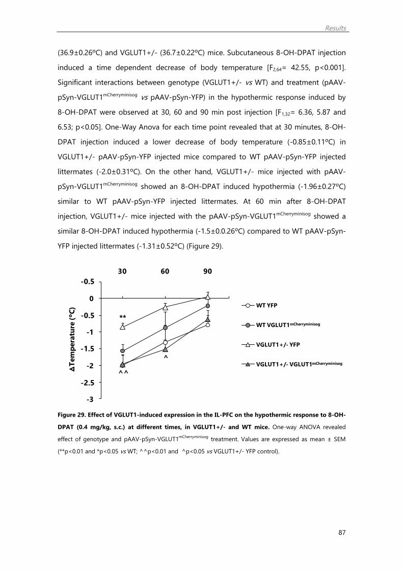
Results
87
(36.9±0.26ºC) and VGLUT1+/- (36.7±0.22ºC) mice. Subcutaneous 8-OH-DPAT injection
induced a time dependent decrease of body temperature [F2,64= 42.55, p<0.001].
Significant interactions between genotype (VGLUT1+/- vs WT) and treatment (pAAV-
pSyn-VGLUT1mCherryminisog vs pAAV-pSyn-YFP) in the hypothermic response induced by
8-OH-DPAT were observed at 30, 60 and 90 min post injection [F1,32= 6.36, 5.87 and
6.53; p<0.05]. One-Way Anova for each time point revealed that at 30 minutes, 8-OH-
DPAT injection induced a lower decrease of body temperature (-0.85±0.11ºC) in
VGLUT1+/- pAAV-pSyn-YFP injected mice compared to WT pAAV-pSyn-YFP injected
littermates (-2.0±0.31ºC). On the other hand, VGLUT1+/- mice injected with pAAV-
pSyn-VGLUT1mCherryminisog showed an 8-OH-DPAT induced hypothermia (-1.96±0.27ºC)
similar to WT pAAV-pSyn-YFP injected littermates. At 60 min after 8-OH-DPAT
injection, VGLUT1+/- mice injected with the pAAV-pSyn-VGLUT1mCherryminisog showed a
similar 8-OH-DPAT induced hypothermia (-1.5±0.0.26ºC) compared to WT pAAV-pSyn-
YFP injected littermates (-1.31±0.52ºC) (Figure 29).
Figure 29. Effect of VGLUT1-induced expression in the IL-PFC on the hypothermic response to 8-OH-
DPAT (0.4 mg/kg, s.c.) at different times, in VGLUT1+/- and WT mice. One-way ANOVA revealed
effect of genotype and pAAV-pSyn-VGLUT1mCherryminisog
treatment. Values are expressed as mean ± SEM
(**p<0.01 and *p<0.05 vs WT; ^^p<0.01 and ^p<0.05 vs VGLUT1+/- YFP control).
**
^^
^
-3
-2,5
-2
-1,5
-1
-0,5
0
0,5
30 60 90
ᐃT
em
pera
ture
(ºC
) WT YFP
WT
mCherryminisog
Vglut1+/- YFP
Vglut1+/-
mCherryminisog
-0.5
-1.5
-0.5
-2.5
WT VGLUT1mCherryminisog
VGLUT1+/- YFP
WT YFP
VGLUT1+/- VGLUT1mCherryminisog
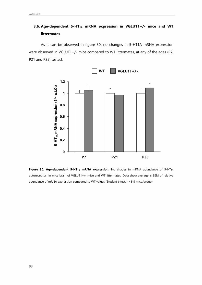
Results
88
3.6. Age-dependent 5-HT1A mRNA expression in VGLUT1+/- mice and WT
littermates
As it can be observed in figure 30, no changes in 5-HT1A mRNA expression
were observed in VGLUT1+/- mice compared to WT littermates, at any of the ages (P7,
P21 and P35) tested.
Figure 30. Age-dependent 5-HT1A mRNA expression. No chages in mRNA abundance of 5-HT1A
autoreceptor in mice brain of VGLUT1+/- mice and WT littermates. Data show average ± SEM of relative
abundance of mRNA expression compared to WT values (Student t-test, n=8-9 mice/group).
WT VGLU1T+/-
0
0,2
0,4
0,6
0,8
1
1,2
P7 P21 P35
5-H
T1
Am
RN
A e
xp
ress
ion
(2
^-ᐃ
ᐃC
t)
0.2
0.4
0.6
0.8
1.2
1
0

DISCUSSION


Discussion
91
1. Nucleocytoplasmic export of HDAC5 and SIRT2 downregulation: two
epigenetic mechanisms by which antidepressants enhance synaptic plasticity
markers
This study presents two epigenetic mechanisms, by which antidepressants could
enhance synaptic plasticity markers linked to antidepressant action. Briefly, the three
antidepressants tested (fluoxetine, reboxetine and imipramine) regulated differently the
histone deacetylases (HDACs) HDAC5 and SIRT2. Particularly, imipramine and
reboxetine increased the phosphorylated form of HDAC5 (p-HDAC5), indicating that
noradrenaline mediates cytoplasmic export of this enzyme. On the other hand, the
three antidepressants downregulated SIRT2. Specific inhibition of HDAC5 with MC3822
(Bürli et al. 2013) and SIRT2 with 33i compound (Suzuki et al. 2012) increased synaptic
plasticity markers too. Finnally, as expected, antidepressant treatment increased
synaptic plasticity markers and histone acetylation. Thus, nucleocytoplasmic export of
HDAC5 and SIRT2 downregulation mediated by antidepressants could enhance
synaptic plasticity markers leading to antidepressant action.
1.1. Nucleocytoplasmic export of HDAC5 by antidepressants enhance synaptic
plasticity markers
Growing evidence suggests that changes in histone acetylation are important
posttranslational regulators of gene expression and therefore may play a key role in
neuronal plasticity (Abel and Zukin 2008; Dulac 2010). Acetylation lowers the affinity
between histones and DNA allowing chromatin to adopt a more relaxed structure and
favoring transcription. Deacetylation, mediated by HDACs, is generally associated with
transcriptional repression (Gallinari et al. 2007).
The HDAC5 enzyme belongs to the class IIa protein superfamily (HDAC4, 5, 7
and 9). This class suffers synaptic activity-dependent nucleo-cytoplasmic shuttling
(McKinsey et al. 2001; Chawla et al. 2003; Renthal et al. 2007; Erburu et al. 2015a). The
phosphorylated form in turn shuttles out of the nucleus, allowing the expression of its
target genes (Parra and Verdin 2010). HDAC5 phosphorylates at S259 and S498,
favouring its cytoplasmic localization (Vega et al. 2004).

Discussion
92
In the current study, we found that the tricyclic imipramine and the selective
noradrenaline reuptake inhibitor reboxetine increased the content of the
phosphorylated form of HDAC5 (p-HDAC5) in vitro in cell cultures. On the other hand,
the selective serotonin reuptake inhibitor fluoxetine did not affect to HDAC5
phosphorylation state. Therefore, it is suggested that HDAC5 phosphorylation is
mediated specifically by increased levels of noradrenaline in the synaptic cleft, a
mechanism shared by imipramine and reboxetine. Interestingly, a role for 1
noradrenaline receptor in the phosphorylation and export of HDAC5 to the cytoplasm
has been described (Sucharov et al. 2011).
Supporting western-blot results, the nucleocytoplasmic shuttling induced by
noradrenaline was confirmed by immunofluorescence in cell cultures. Specifically,
reboxetine induced a striking increase of HDAC5 immunoreactivity in the cytoplasm of
cultured neurons, while a decrease of this enzyme was found in the nucleus.
Interestingly, at 2 h incubation time, reboxetine increased p-HDAC5 suggesting
that nuclear export of HDAC5 is a rapid-acting effect mediated by noradrenaline that
would contribute to histone acetylation. In keeping with this, nuclear export of HDAC5,
4 hours after cocaine (Renthal et al. 2007) and 3-6 hours after ketamine (Choi et al.
2015) has been shown.
Comparatively, chronic imipramine and reboxetine upregulated p-HDAC5 in
vivo in the PFC of mice, a result that agrees with a previous study carried out in our lab
(Erburu et al. 2015a). Altogether, these findings suggest that both antidepressants
attenuated HDAC5 repressive influence on transcription by inducing the nuclear export
of HDAC5 to the cytoplasm. Matching with this, Bdnf promoter has been reported to be
controlled by HDAC5 (Tsankova et al. 2006).
Subsequently, we tested two enantiomers (R,R,R and S,S,S) of a new selective
HDAC5 inhibitor (MC3822 an MC3823 respectively). Between the two and matching
with its HDAC4/5 inhibitory activity, MC3822 was found to increase more effectively
AcH3 and AcH4 and was selected as the active enantiomer. Subsequently, MC3822 was
selected to study the specific inhibition of HDAC5 on neuroplasticity. Particularly, the
synaptic plasticity markers the brain derived neurotrophic factor (BDNF) and the
Discussion
93
vesicular glutamate transporter 1 (VGLUT1) were selected.
The neurotrophin hypothesis of depression postulates that BDNF is involved in
depression as well as in the clinical response to antidepressants. It stimulates growth,
differentiation and survival of neurons and its expression is oppositely regulated by
stress and antidepressant treatment (Alonso et al. 2004; Czéh et al. 2007; Duman and
Monteggia 2006). In patients, while low levels of brain and serum BDNF have been
associated to major depression (Nestler et al. 2002), antidepressant treatment boosts
BDNF (Duman 2014).
Besides neurotrophins, VGLUT1 has been also associated to depression and
antidepressant action. On the whole, VGLUT1 is the predominant isoform of excitatory
terminal of telencephalic regions (Fremeau et al. 2001; Gras et al. 2002) having a key
role on synaptic release of glutamatergic transmission (Wojcik et al. 2004; Balschun et
al. 2010). A course of antidepressant treatment upregulate VGLUT1 expression in PFC
and hippocampus (Tordera et al. 2005; Moutsimilli et al. 2005; Cooke et al. 2014).
Interestingly, this effect could be mediated, at least in part, by BDNF (Farley et al. 2012).
Conversely, decreased VGLUT1 levels in the frontal cortex of depressed subjects have
been found (Uezato et al. 2009). Moreover, decreased VGLUT1 has been associated to a
depressive-like phenotype in rodents (Tordera et al. 2007; Garcia-Garcia et al. 2009).
MC3822 increased VGLUT1 and BDNF expression indicating that specific
inhibition of HDAC5 upregulates neuroplasticity. Additionally, this fact further strengths
the idea that the effect of noradrenergic antidepressants on synaptic plasticity could be
partially mediated by decreased Hdac5 nuclear function (Tsankova et al. 2006; Erburu et
al. 2015a).
Nevertheless, further behavioural studies should be carried out to establish a
causal link between HDAC5 inhibition and antidepressant action. Intriguingly, the
potential antidepressant action of HDAC5 inhibition has been recently suggested (Choi
et al. 2015). For instance, genes involved in cocaine experience-dependent reward
sensitivity are silenced by HDAC5 inhibition (Renthal et al. 2007). In addition, HDAC5
over-expression blocks antidepressant action in the CSDS model (Tsankova et al. 2006).
Curiously enough, an upregulation of cortical Hdac5 mRNA has been observed in
Discussion
94
subjects with major depressive disorder and bipolar disorder, compared to controls and
patients in remission, supporting a clinical relevance for these changes (Hobara et al.
2010).
1.2. SIRT2 downregulation by antidepressants enhance synaptic plasticity
markers
SIRT2 is a class III histone deacetylase playing a role in transcriptional repression
of genes encoding for DNA binding proteins as well as transcription factors that
participate in synaptic plasticity, cell proliferation, differentiation and cell survival
(Eskandarian et al. 2013). Previous findings showing SIRT2 mRNA upregulation in the
PFC of depressed patients supports the interest of this target for therapeutic
intervention (Erburu et al. 2015a). In line with this, preliminary studies carried out in our
laboratory have remarkably shown that repeated treatment with the SIRT2 inhibitor 33i
causes an antianhedonic action in depression mouse models (Erburu et al. 2017).
We observed here that all monoaminergic antidepressants down-regulated the
expression of SIRT2 in SH-SY5Y cell cultures suggesting that this could be a shared
effect for all antidepressants. Matching with this effect, hyperacetylated α-tubulin levels,
a SIRT2 cytoplasmic substrate, were observed. Further in vivo studies, revealed again
SIRT2 downregulation following chronic antidepressant treatment as previously
observed (Erburu et al. 2015a). Interestingly, only chronic fluoxetine dowregulated Sirt2
mRNA in vivo, suggesting that imipramine and reboxetine act on SIRT2 at post-
transductional level.
Furthermore, specific inhibition of SIRT2 by 33i compound also induced an up-
regulation of AcH3 and AcH4 as well as VGLUT1 and BDNF in SH-SY5Y cultures. The
increase in synaptic plasticity markers induced by 33i is in line with previous studies
suggesting that SIRT2 inhibition could increase glutamate function (Erburu et al. 2017).
In addition, 33i hyperacetylated α-tubulin in cell cultures (Suzuki et al. 2012; Mangas-
Sanjuan et al. 2015; Rumpf et al. 2015). Moreover, we observed that 33i treatment
increased p-HDAC5 suggesting perhaps that HDAC5 could be also an enzyme
Discussion
95
downstream cytoplasmic SIRT2 function. Altogether, SIRT2 downregulation could be
another mechanism by which antidepressants stimulate neuroplasticity.
In summary, this study supports the validity of SH-SY5Y cultures as an in vitro
cellular model for studying epigenetic changes linked to synaptic plasticity induced by
antidepressants as well as the effect of selective HDAC inhibitors. Specifically we
present here two epigenetic mechanisms by which antidepressants might enhance
neuronal plasticity and presumably, antidepressant action. Firstly, antidepresssant that
elevate noradrenaline levels, induce nucleocytoplasmic export of HDAC5. Secondly,
monoaminergic antidepressants downregulate SIRT2. Thus, it is highlighted the
therapeutic potential of HDAC5 and SIRT2 as epigenetic targets for major depression.
1.3. Antidepressants increase histone acetylation and synaptic plasticity
markers
Here, we studied in SH-SY5Y cells the antidepressant regulation of AcH3, AcH4
as well as BDNF and VGLUT1 as good antidepressant state markers. Interestingly, all
these proteins were upregulated by incubation for 24 hours of SH-SY5Y cell cultures
with imipramine, fluoxetine and reboxetine. These effects might be directly due to
enhanced levels of monoamines, including in the case of imipramine, given that the
antimuscarinic drug scopolamine showed no changes.
It is noteworthy to mention that antidepressant incubation of cells for only 2 h
also increased histone acetylation. Yet, the fact that neither VGLUT1 nor BDNF levels
were changed, suggest that histone acetylation precedes in time the synthesis of
plasticity markers. According to these results, other authors have also reported that
BDNF is upregulated following longer incubation times (Donnici et al. 2008).
Comparatively, repeated antidepressant treatment upregulated AcH3, AcH4 in
vivo in the PFC of mice as previously reported (Erburu et al. 2015a) as well as BDNF and
VGLUT1. Our experimental design differs from the study of Erburu et al. 2015a in that
mice were sacrificed 24 h following the last drug injection. Thus, the observed

Discussion
96
maintained increase in histone acetylation and synaptic plasticity markers could be
neuroadaptive changes associated to antidepressant therapy.
All these studies support the need to study the therapeutic potential of HDAC5
and SIRT2 for depression treatment. In the following section we studied the effect of
the selective SIRT2 inhibitor 33i in the VGLUT1+/- depression model. Unfortunately,
due to the lack of enough amount of compound for in vivo studies, we could not study
the effect of the selective HDAC5 inhibitor in this model.
2. SIRT2 inhibition reverses anhedonia in the VGLUT1+/- depression model
We aimed to study the possible antidepressant effect of the specific SIRT2
inhibitor 33i in the VGLUT1+/- depression model. In this research, we showed that the
expression levels of the different members of the superfamily of Hdac enzymes are not
altered in the VGLUT1+/- model. On the other hand, the SIRT2 inhibitor 33i showed an
antianhedonic action in the VGLUT1+/- mouse model. Furthermore, this compound
showed no interaction with specific monoaminergic targets such as serotonin or
noradrenaline transporters as well as no affinity to the monoaminooxidase enzyme
(MAO).
Chromatin remodeling such as changes in histone acetylation has been
suggested to play an important role in the pathophysiology of depression. For instance,
experimental models based on exposure to stress, have linked upregulation of the
histone deacetylases of class I (Hdac1 and Hdac2), class IIa (Hdac4 and Hdac5) and
class III (Sirt2) in the prefrontal cortex (PFC) and the hippocampus to depressive-like
behaviours (Tsankova et al. 2006; Renthal et al. 2007; Sarkar et al. 2014; Erburu et al.
2015a). By contrast, specific inhibitions of these enzymes were reported to have
antidepressant-like activity (Schroeder et al. 2013; Erburu et al. 2017). In keeping with
this data, clinical studies have confirmed a positive correlation between the expression
of some HDACs and the depressive state of patients (Belzeaux et al. 2010; Hobara et al.
2010; Watanabe et al. 2015). Further, non-specific HDAC inhibitors were proved to have
antidepressant effects in patients (Machado-Vieira et al. 2011).

Discussion
97
Here we have studied the mRNA abundance of the Hdac’s superfamily including
class I (hdac1, hdac2, hdac3 and hdac8), class II (hdac4, hdac5, hdac6, hdac7, hdac9 and
hdac10) and IV (hdac11) as well as the NAD+-dependent class III (Sirt1-7) in the PFC of
VGLUT1+/- mice, as a genetic model of depression (Garcia-Garcia et al. 2009). No
alterations in the mRNA expression of any of these enzymes were found. Further this
model showed no changes in protein expression of HDAC5, SIRT2 or histone
acetylation. Thus, the depressive-like behaviour of VGLUT1+/- mice is not linked to
variations in Hdac mRNA expression.
Among the different Hdac’s, the class III NAD+-dependent histone deacetylase
SIRT2 plays a role in transcriptional repression of transcription factors that participate in
synaptic plasticity, cell proliferation, differentiation and cell survival (Eskandarian et al.
2013). Previous studies carried out in our laboratory (Erburu et al. 2015a, 2017) shows
that SIRT2 could contribute to both stress-induced neuronal adaptations as well as to
antidepressant action. Specifically, while chronic stress induces SIRT2 expression, this
enzyme is downregulated by monoaminergic antidepressants (fluoxetine, reboxetine
and imipramine). Moreover, we have also observed that specific inhibition of SIRT2 with
the compound 33i, a 3'-phenethyloxy-2-anilinobenzamide analogue (Suzuki et al.
2012), induces the expression of the brain derived neurotrophic factor (BDNF) and
shows antidepressant-like activity in the chronic mild stress model (Erburu et al. 2015a).
In this experiment, we tested the therapeutic value of SIRT2 inhibition in the
anhedonic behaviour of VGLUT1+/- mice (Garcia-Garcia et al. 2009). Anhedonia, a core
symptom of clinical depression is defined as the decreased ability to experience
pleasure. In rodents, it has been largely reported that decreased reactivity to rewards
parallels anhedonia in humans, and it is mostly studied by analyzing the intake of, or
the preference for sweet solutions over drinking water (Willner, 2005). Firstly, we
examined the effect of imipramine as a reference antidepressant drug on anhedonic
behaviour of VGLUT1+/- mice. Thus, VGLUT1+/- mice showed a decrease in sucrose
intake that would be indicative of anhedonia. On the other hand, imipramine increased
significantly sucrose intake showing an antianhedonic action from the fourth week until
the end of treatment.

Discussion
98
Subsequently, the effect of the 33i compound on anhedonic behaviour of
VGLUT1+/- mice was tested and interestingly 33i treatment was able to reverse
anhedonic behaviour exhibited by VGLUT1+/- mice.
So far, experimental studies linking changes in HDAC’s to depressive-like
behaviours focus on animal models based on chronic stress (Tsankova et al. 2006;
Renthal et al. 2007; Sarkar et al. 2014; Erburu et al. 2015b; Zheng et al. 2016). However,
anhedonia can also be triggered by deficient glutamate transmission (Lally et al. 2015),
as is the case for the VGLUT1+/- model. We suggest that while anhedonia induced by
adverse environmental risk factors could be linked to epigenetic changes, anhedonia
induced by this genetic risk factor might be independent. On the other hand,
anhedonic behaviour of VGLUT1+/- mice was fully reversed by 33i treatment after two
weeks of treatment. Besides, this antidepressant action was maintained in the third
week of treatment. Additionally, this compound had no effect in WT mice suggesting
that it has the property to reverse anhedonia of “depressed” mice without altering the
natural reward reactivity of healthy mice. In agreement with these results, we have
previously shown that chronic 33i treatment reverses CMS-induced anhedonia whereas
it does not affect the hedonic behaviour of control animals (Erburu et al. 2017). On the
other hand, the tricyclic antidepressant imipramine, that elevates 5-HT and
noradrenaline in the synaptic cleft, showed an antianhedonic action in both VGLUT1+/-
and WT mice matching with previous studies showing the ability of noradrenaline to
increase reward reactivity (Tong et al. 2006).
Former studies have shown that subchronic 33i treatment increases BDNF,
serotonin tissue levels and glutamate receptor subunits in the PFC of mice (Erburu et al.
2015a, 2017). Interestingly, these effects could be responsible for the antidepressant
profile of 33i. Matching with this idea, antidepressant-like effects of monoaminergic
antidepressants have been linked to elevation of BDNF and monoamines in the
synaptic cleft as well as to activation of glutamate receptors in the PFC (Stasiuk et al.
2017). We further studied here whether 33i interacts with specific monoaminergic
targets for depression treatment aside from its ability to inhibit SIRT2. Specifically, the
compound 33i showed no affinity for the 5-HT transporter or inhibitory activity over
[3H]-noradrenaline uptake. Moreover, 33i had no effect on activity MAO enzyme

Discussion
99
activity. Thus, these studies suggest that the antidepressant profile of 33i is not linked
to a direct interaction with these targets. Yet, we cannot exclude the possibility that the
observed effects of 33i on BDNF, serotonin and glutamate receptors could be mediated
by other mechanisms independent of SIRT2 inhibition. Chromatin immunoprecipitation
studies should be carried out in the future in order to answer this relevant question.
Altogether, these results show that while anhedonia in the VGLUT1+/- model is
not linked to variations in Hdac mRNA expression, the selective SIRT2 inhibitor 33i
reverses anhedonia in different animal models. It is highlighted the need to further
investigate the role of SIRT2 inhibitors as antidepressant agents. Similarly, we are
currently studying the role of selective HDAC5 inhibitors as potential antidepressant
agents in animal models.
Discussion
100
3. Role of VGLUT1 in antidepressant action
In the present study we have used the VGLUT1+/- heterozygous mouse model
to further examine the role of prefrontocortical glutamatergic signaling on depressive-
like behaviour. Specifically, using the adeno-associated virus (AAV) technology to
express the VGLUT1 gene, we aimed to increase VGLUT1 expression in PFC neurons of
VGLUT1+/- and WT mice. Firstly, we studied the efficiency of VGLUT1 expression in this
area as well as in projecting areas such as glutamatergic descending fibbers going to
the dorsal raphe nucleus (DRN). The consequences of this manipulation on depressive
behaviour as well as on 5-HT1A function were subsequently investigated.
3.1. VGLUT1 induced expression in the PFC and DRN using AAV-technology
The functionality of the designed plasmid (pAAV-pSyn-VGLUT1-mCherry-
minisog-SMD2) was confirmed in rat hippocampal cultures. As expected,
VGLUT1mCherryminisog was highly expressed in hippocampal neurons both in the soma and
in fibbers which confirms the correct distribution of it. Given that VGLUT1 gene was
under the synapsin promoter we can assume that VGLUT1 expression occurs only in
neurons.
Subsequently, preliminary experiments were performed to determine the
optimal dose of AAV required for imaging VGLUT1mCherryminisog expression in the PFC of
WT and VGLUT1+/- mice. Using the highest dilution (1:2), imaging studies revealed
VGLUT1mCherryminisog expression in both cell bodies and fibbers in the PFC as well as in
fibbers in the DRN. Yet, using lower dilutions (1:4-1:40) of the AAV, no signal at the
level of the DRN was detected. Therefore the 1:2 dilution was selected for subsequent
studies, as the optimal AAV concentration that allows VGLUT1mCherryminisog expression not
only in the site of injection but also in PFC innervating areas. Further, analysis of
mCherryminisog or YFP fluorescence in the whole PFC of both genotypes, WT and
VGLUT1+/-, revealed no differences in the amount of fluorescence detected (signal
area x mean intensity) indicating that reduced VGLUT1 levels in the heterozygous mice
(Tordera et al. 2007; Garcia-Garcia et al. 2009) does not affect to the efficiency of the
AAV infection. Given the lack of a good antibody against VGLUT1mCherryminisog, we
propose the analysis of amount of fluorescence in the whole area of infection to

Discussion
101
estimate the efficiency of expression. Yet, future studies using a specific antibody will
allow us to detect whether at this dilution we are able to rescue VGLUT1 levels in the
heterozygous mice.
Of the three VGLUT isoforms, VGLUT1 is the most abundant isoform in cortical
neurons compared to other areas (Fremeau et al. 2001; Hartig et al. 2003). Previous
studies carried out in our laboratory have confirmed that VGLUT1 heterozygous mice
expressed half the amount of transporter in the PFC) compared to WT littermates
(Tordera et al. 2007; Garcia-Garcia et al. 2009). Moreover, reduced VGLUT1 expression
was also detected in the DRN (Garcia-Garcia et al. 2013). Here, we show a method by
which we could increase the expression of VGLUT1 in the PFC and DRN of VGLUT1+/-
and WT mice. Importantly, the observation that VGLUT1mCherryminisog is detected in
fibbers suggest that VGLUT1 is able to reach synaptic terminals, in where, it should
exert its function as a carrier of glutamate into the synaptic vesicles. Moreover, our
results agree with previous findings showing that glutamate innervation to the DRN
arises in part from the PFC (Artigas et al. 1999; Hajós et al. 1998; Gonçalves et al. 2009;
Soiza-Reilly and Commons 2014). In addition, the observed VGLUT1 containing axon
terminals in the DRN gives direct evidence of the PFC raphe glutamate pathway and
strongly suggest that VGLUT1 is the only isoform responsible for this pathway.
3.2. VGLUT1 induced expression in the PFC rescues depressive-like behaviour in
VGLUT1+/- mice
Anhedonia has been largely associated to a dysfunction of the PFC in depressed
patients (Keedwell et al. 2005) and in rodents (Tordera et al. 2007). Previous studies in
our laboratory has shown that VGLUT1+/- mice show depressive-like behaviour and
increased vulnerability to anhedonia after chronic stress (Garcia-Garcia et al. 2009)
suggesting that reduced VGLUT1 could be a potential biological risk factor of major
depression. In keeping with these studies, VGLUT1+/- mice showed anhedonia
measured as a decrease of sucrose intake compared to WT littermates. PFC pAAV-
pSyn-VGLUT1mCherryminisog injection increased significantly sucrose intake in both
genotypes from the first week suggesting that VGLUT1 expression induces a rapid
antianhedonic action. Interestingly this antianhedonic action was maintained up to

Discussion
102
week 7 after which, mice were sacrificed. This is the first evidence that link VGLUT1 to
antianhedonic behaviour.
The mechanisms by which increased expression of VGLUT1 could lead to anti-
anhedonic behaviour should be further explored. VGLUT1 has a key role in regulation
of glutamate secretion (Wojcik et al. 2004) and this neurotransmitter is linked to
mechanisms of neural plasticity. One possible explanation is that VGLUT1
overexpression could enhance glutamate transmission in the PFC leading to an
antidepressant action. Matching with this, activation of glutamate receptors in PFC has
been linked to antidepressant like effects in patients and rodents (Li et al. 2010; Koike
et al. 2011). Importantly, induced VGLUT1 expression in the PFC of adult VGLUT1+/-
mice was sufficient to induce antidepressant action suggesting that early life
neuroadaptive changes or other brain regions have little contribution to anhedonia in
this model.
Another possible explanation is that, VGLUT1 overexpression could enhance
glutamate release from fibbers located in the raphe nuclei and facilitate serotonin
release in the forebrain inducing an antidepressant effect. In keeping with this,
glutamatergic descending pathways from the PFC are suggested to modulate 5-HT
activity in the DRN (Celada et al. 2001; Hajós et al. 1998; Varga 2001; Tao and Auerbach
2003). Interestingly, the observed inhibition of marble burying behaviour in PFC AAV-
pSyn-VGLUT1mCherryminisog injected mice would agree with this idea, since 5-HT active
compounds, specially selective serotonin reuptake inhibitors (SSRIs) (Hirano et al. 2005;
Elizalde et al. 2010) also inhibit marble burying behaviour. Further, the sensitivity of the
inhibitory 5-HT1A autoreceptors of 5-HT cell bodies having a key role on 5-HT release
and mood (Blier and de Montigny 1983; Verge et al. 1985; Sprouse and Aghajanian
1987; Richardson-Jones et al. 2010) could be also modulated by VGLUT1. In keeping
with this, 5-HT1A desensitization observed in the VGLUT1+/- mice (Garcia-Garcia et al.
2013) could be a compensatory action for the low glutamate release in the DRN from
fibbers descending from the PFC. Conversely, increasing VGLUT1 expression in the PFC
of these mice could lead to a recovery of the long-loop excitatory modulation of 5-HT
activity and consequently of the 5-HT1A autoreceptor function. In order to prove this
hypothesis, we initiated here studies directed to stablish a link between VGLUT1

Discussion
103
expression and 5-HT1A autoreceptor sensitivity. While no chanches in 5-HT1A mRNA was
found along development, systemic administration of the 5-HT1A agonist 8-OH-DPAT
induced a lower hypothemic response in VGLUT1+/- compared to WT littermates which
would be indicative of 5-HT1A autoreceptor desensitization (Bill et al. 1991), as
previously described (Garcia-Garcia et al. 2013). Interestingly, injection of AAV-pSyn-
VGLUT1mCherryminisog in the PFC of VGLUT1+/- mice induced a hypothermic response to
8-OH-DPAT similar to that observed in WT mice. These results suggest that increased
VGLUT1 rescues the sensitivity of 5-HT1A autoreceptors in the DRN. Yet further
electrophysioloical and GTP--binding biochemical studies should be carried out to
confirm for the first time whether VGLUT1 levels modulate 5-HT1A autoreceptor
function in the DRN.
Finally, future studies should explore whether VGLUT1 overexpression in the PFC
could also modulate other glutamatergic descending pathways to brain areas involved
in the processing of other behaviours. For instance, the mild levels of anxiety observed
in PFC AAV-pSyn-VGLUT1mCherryminisog injected mice could be associated to a stimulation
of the PFC-amygdala pathway.
Altogether, this study shows for the first time that the stereotaxic injection of an
adeno-associated viral vector encoding vesicular glutamate transporter 1 into the PFC
results in sustained expression of VGLUT1 at levels sufficient to correct the depressive
phenotype in VGLUT1 heterozygous mice (VGLUT1+/-) and to induce antidepressant
action in WT.


CONCLUSIONS

Conclusions
107
1. The antidepressants imipramine and reboxetine increased the phosphorylated form
of the histone deacetylase 5 (p-HDAC5) both in vitro and in vivo in the prefrontal
cortex (PFC) of mice, and promoted cytoplasmic export of this enzyme from the
nucleus of the cells. Further, specific inhibition of HDAC5 with the compound
MC3822 increased histone acetylation and synaptic plasticity markers. These studies
suggest that nucleocytoplasmic export of HDAC5 is an epigenetic mechanism by
which these antidepressants stimulate neuroplasticity.
2. The antidepressants fluoxetine, reboxetine and imipramine downregulated the
histone deacetylase SIRT2 both in vitro and in vivo. Further, specific inhibition of
SIRT2 with the compound 33i increased histone acetylation and synaptic plasticity
markers. Thus, SIRT2 downregulation is another epigenetic mechanism by which
antidepressants stimulate neuroplasticity.
3. Nucleocytoplasmic export of HDAC5 could be an epigenetic mechanism exclusive of
antidepressants able to elevate noradrenaline levels. On the other hand, SIRT2
downregulation could be an epigenetic mechanism of antidepressants that elevate
either serotonin or noradrenaline in the synaptic cleft. These are two epigenetic
mechanisms by which antidepressants might contribute to increase synaptic
plasticity.
4. The selective SIRT2 inhibitor 33i reversed anhedonia in mice heterozygous for the
vesicular glutamate transporter 1 (VGLUT1+/-), considered a genetic model of
depression. Yet, this model showed no changes in the expression of the selected
epigenetic markers (HDAC's, histone acetylation) or in the syaptic plasticity markers.
This result evidence that SIRT2 inhibition is able to reverse depressive-like
behaviours even when these are not associated to epigenetic alterations.
5. The stereotaxic injection of the designed adeno-associated viral (AAV) vector
encoding vesicular glutamate transporter 1 (VGLUT1) into the PFC resulted in
sustained expression of VGLUT1mCherryminisog in both cell bodies and fibbers in the PFC
as well as in fibbers in the dorsal raphe nucleus (DRN).

Conclusions
108
6. VGLUT1-induced expression in the PFC of adult mice triggered an antianhedonic
action both in VGLUT1+/- and WT mice. In addition, it appears that it rescues the 5-
HT1A autoreceptor function in the heterozygous mice. Altogether this is the first
evidence linking VGLUT1 to antidepressant action.
7. Taken together, the histone deacetylase enzymes HDAC5 and SIRT2 as well as the
synaptic plasticity marker VGLUT1 could be proposed as pharmacological targets
involved in antidepressant action. The therapeutic potential of these targets should
be further investigated.
Conclusions
109
1. Los antidepresivos imipramina y reboxetina aumentaron la forma fosforilada de la
histona desacetilasa 5 (p-HDAC5) tanto in vitro como in vivo, en la corteza prefrontal
de los ratones. Es decir el tratamiento con estos antidepresivos favorece la
traslocación de esta enzima del núcleo al citoplasma de las células. Además, la
inhibición específica de HDAC5 con el compuesto MC3822 provocó un aumento de
la acetilación de histonas y de los marcadores de plasticidad sináptica. Estos
estudios sugieren que la traslocación nucleocitoplasmática de HDAC5 es un
mecanismo epigenético mediante el cual estos antidepresivos estimulan la
neuroplasticidad.
2. Los antidepresivos fluoxetina, reboxetina e imipramina disminuyeron la expresión
de la histona desacetilasa SIRT2 tanto in vitro como in vivo. Además, la inhibición
específica de SIRT2 con el compuesto 33i provocó un aumento de la acetilación de
histonas y de los marcadores de plasticidad sináptica. Por lo tanto, la disminución de
SIRT2 es otro mecanismo epigenético por el cual los antidepresivos estimulan la
neuroplasticidad.
3. La exportación nucleocitoplasmática de HDAC5 podría ser un mecanismo
epigenético exclusivo de los antidepresivos cuyo mecanismo de acción eleva los
niveles de noradrenalina. Por otro lado, la disminución de SIRT2 podría ser un
mecanismo epigenético de los antidepresivos que elevan tanto la serotonina como
la noradrenalina en la sinapsis. Estos son dos mecanismos epigenéticos por medio
de los cuales los antidepresivos podrían aumentar la plasticidad sináptica.
4. El inhibidor selectivo SIRT2 33i revirtió la anhedonia en ratones heterocigotos para el
transportador de glutamato vesicular 1 (VGLUT1+/-), considerado un modelo
genético de depresión. Sin embargo, este modelo no mostró cambios en la
expresión de los marcadores epigenéticos seleccionados (enzimas histona
desacetilasas y acetilación de histonas) ni en los marcadores de la plasticidad
sináptica. Este resultado evidencia que la inhibición de SIRT2 es capaz de revertir un
fenotipo depresivo también cuando éste no está asociado a alteraciones
epigenéticas.
Conclusions
110
5. La inyección estereotáxica en la corteza prefrontal del virus adenoasociado diseñado
en este estudio que codifica el transportador de glutamato vesicular 1 (VGLUT1), dio
como resultado un aumento de VGLUT1mCherryminisog tanto en el soma de las neronas,
como en fibras en la corteza prefrontal, así como en fibras en el núcleo dorsal del
rafe.
6. La expresión inducida de VGLUT1 en la corteza prefrontal de ratones adultos
provocó una acción antianhedónica tanto en ratones VGLUT1+/- como en ratones
WT. Además, parece que la función del autorreceptor 5-HT1A de los ratones
heterocigóticos se normaliza. En conjunto, esta es la primera evidencia que relaciona
directamente VGLUT1 con la acción antidepresiva.
7. Recapitulando, podrían proponerse como dianas farmacológicas implicadas en la
acción antidepresiva tanto las enzimas histona desacetilasa HDAC5 y SIRT2, como el
marcador de plasticidad sináptica VGLUT1. El potencial terapéutico de estas dianas
farmacológicas debería investigarse.

REFERENCES

References
113
Abe, N., Uchida, S., Otsuki, K., Hobara, T., Yamagata, H., Higuchi, F. & Watanabe, Y.
(2011). Altered sirtuin deacetylase gene expression in patients with a mood
disorder. Journal of Psychiatric Research, 45(8), 1106–1112.
Abel, T., & Zukin, R. (2008). Epigenetic targets of HDAC inhibition in neurodegenerative
and psychiatric disorders. Current Opinion in Pharmacology, 8(1), 57–64.
Albert, P. R., Benkelfat, C., & Descarries, L. (2012). The neurobiology of depression-
revisiting the serotonin hypothesis. I. Cellular and molecular mechanisms.
Philosophical Transactions of the Royal Society B: Biological Sciences, 367(1601),
2378–2381.
Alonso, R., Griebel, G., Pavone, G., Stemmelin, J., Le Fur, G., & Soubrié, P. (2004).
Blockade of CRF(1) or V(1b) receptors reverses stress-induced suppression of
neurogenesis in a mouse model of depression. Molecular Psychiatry, 9(3), 278–86,
224.
Altar, C. A., Vawter, M. P., & Ginsberg, S. D. (2009). Target identification for CNS
diseases by transcriptional profiling. Neuropsychopharmacology : Official
Publication of the American College of Neuropsychopharmacology, 34(1), 18–54.
Amat, J., Baratta, M. V, Paul, E., Bland, S. T., Watkins, L. R., & Maier, S. F. (2005). Medial
prefrontal cortex determines how stressor controllability affects behavior and
dorsal raphe nucleus. Nature Neuroscience, 8(3), 365–71.
Anacker, C., Cattaneo, A., Musaelyan, K., Zunszain, P. A., Horowitz, M., Molteni, R., &
Pariante, C. M. (2013). Role for the kinase SGK1 in stress, depression, and
glucocorticoid effects on hippocampal neurogenesis. Proceedings of the National
Academy of Sciences of the United States of America, 110(21), 8708–13.
Arroll, B., Macgillivray, S., Ogston, S., Reid, I., Sullivan, F., Williams, B., & Crombie, I.
(2005). Efficacy and Tolerability of Tricyclic Antidepressants and SSRIs Compared
With Placebo for Treatment of Depression in Primary Care: A Meta-Analysis. The
Annals of Family Medicine, 3(5), 449–456.

References
114
Artigas, F., Romero, L., de Montigny, C., & Blier, P. (1996). Acceleration of the effect of
selected antidepressant drugs in major depression by 5-HT1A antagonists. Trends
in Neurosciences, 19(9), 378–83.
Bagot, R. C., Labonté, B., Peña, C. J., & Nestler, E. J. (2014). Epigenetic signaling in
psychiatric disorders: stress and depression. Dialogues in Clinical Neuroscience,
16(3), 281–95.
Balschun, D., Moechars, D., Callaerts-Vegh, Z., Vermaercke, B., Van Acker, N., Andries, L.,
& D’Hooge, R. (2010). Vesicular Glutamate Transporter VGLUT1 Has a Role in
Hippocampal Long-Term Potentiation and Spatial Reversal Learning. Cerebral
Cortex, 20(3), 684–693.
Belmaker, R. H. (2008). The future of depression psychopharmacology. CNS Spectrums,
13(8), 682–7.
Belzeaux, R., Formisano-Tréziny, C., Loundou, A., Boyer, L., Gabert, J., Samuelian, J.-C., &
Ibrahim, E. C. (2010). Clinical variations modulate patterns of gene expression and
define blood biomarkers in major depression. Journal of Psychiatric Research,
44(16), 1205–1213.
Berton, O., McClung, C. A., Dileone, R. J., Krishnan, V., Renthal, W., Russo, S. J., & Nestler,
E. J. (2006). Essential Role of BDNF in the Mesolimbic Dopamine Pathway in Social
Defeat Stress. Science, 311(5762), 864–868.
Bhagwagar, Z., Wylezinska, M., Jezzard, P., Evans, J., Ashworth, F., Sule, A., & Cowen, P. J.
(2007). Reduction in occipital cortex gamma-aminobutyric acid concentrations in
medication-free recovered unipolar depressed and bipolar subjects. Biological
Psychiatry, 61(6), 806–12.
Biedler, J. L., Roffler-Tarlov, S., Schachner, M., & Freedman, L. S. (1978). Multiple
neurotransmitter synthesis by human neuroblastoma cell lines and clones. Cancer
Research, 38(11 Pt 1), 3751–7.

References
115
Bill, D. J., Knight, M., Forster, E. A., & Fletcher, A. (1991). Direct evidence for an
important species difference in the mechanism of 8-OH-DPAT-induced
hypothermia. British Journal of Pharmacology, 103(4), 1857–64. Retrieved from
Björkholm, C., & Monteggia, L. M. (2016). BDNF – a key transducer of antidepressant
effects. Neuropharmacology, 102, 72–79.
Blier, P., & De Montigny, C. (1983). Electrophysiological investigations on the effect of
repeated zimelidine administration on serotonergic neurotransmission in the rat.
The Journal of Neuroscience : The Official Journal of the Society for Neuroscience,
3(6), 1270–8.
Blier, P. (2003). The pharmacology of putative early-onset antidepressant strategies.
European Neuropsychopharmacology : The Journal of the European College of
Neuropsychopharmacology, 13(2), 57–66.
Bravo, J. A., Julio-Pieper, M., Forsythe, P., Kunze, W., Dinan, T. G., Bienenstock, J., &
Cryan, J. F. (2012). Communication between gastrointestinal bacteria and the
nervous system. Current Opinion in Pharmacology, 12(6), 667–672.
Bromet, E., Andrade, L. H., Hwang, I., Sampson, N. A., Alonso, J., de Girolamo, G., &
Kessler, R. C. (2011). Cross-national epidemiology of DSM-IV major depressive
episode. BMC Medicine, 9(1), 90.
Bürli, R. W., Luckhurst, C. A., Aziz, O., Matthews, K. L., Yates, D., Lyons, K. A., &
Dominguez, C. (2013). Design, synthesis, and biological evaluation of potent and
selective class IIa histone deacetylase (HDAC) inhibitors as a potential therapy for
Huntington’s disease. Journal of Medicinal Chemistry, 56(24), 9934–54.
Castrén, E., Voikar, V., & Rantamaki, T. (2007). Role of neurotrophic factors in
depression. Current Opinion in Pharmacology, 7(1), 18–21.
Castrén, E., & Rantamäki, T. (2010). The role of BDNF and its receptors in depression
and antidepressant drug action: Reactivation of developmental plasticity.
Developmental Neurobiology, 70(5), 289–297.

References
116
Celada, P., Puig, M. V, Casanovas, J. M., Guillazo, G., & Artigas, F. (2001). Control of
dorsal raphe serotonergic neurons by the medial prefrontal cortex: Involvement of
serotonin-1A, GABA(A), and glutamate receptors. The Journal of Neuroscience :
The Official Journal of the Society for Neuroscience, 21(24), 9917–29.
Chaki, S., & Fukumoto, K. (2015). Potential of Glutamate-Based Drug Discovery for Next
Generation Antidepressants. Pharmaceuticals, 8(3), 590–606.
Challis, C., & Berton, O. (2015). Top-Down Control of Serotonin Systems by the
Prefrontal Cortex: A Path toward Restored Socioemotional Function in Depression.
ACS Chemical Neuroscience, 6(7), 1040–1054.
Charney, D. S. (1998). Monoamine dysfunction and the pathophysiology and treatment
of depression. The Journal of Clinical Psychiatry, 59 Suppl 14, 11–4.
Chawla, S., Vanhoutte, P., Arnold, F. J. L., Huang, C. L.-H., & Bading, H. (2003). Neuronal
activity-dependent nucleocytoplasmic shuttling of HDAC4 and HDAC5. Journal of
Neurochemistry, 85(1), 151–9.
Choi, M., Lee, S. H., Wang, S. E., Ko, S. Y., Song, M., Choi, J.-S., & Son, H. (2015).
Ketamine produces antidepressant-like effects through phosphorylation-
dependent nuclear export of histone deacetylase 5 (HDAC5) in rats. Proceedings of
the National Academy of Sciences of the United States of America, 112(51),
15755–60.
Cooke, J. D., Cavender, H. M., Lima, H. K., & Grover, L. M. (2014). Antidepressants that
inhibit both serotonin and norepinephrine reuptake impair long-term potentiation
in hippocampus. Psychopharmacology, 231(23), 4429–4441.
Covington, H. E., Maze, I., LaPlant, Q. C., Vialou, V. F., Ohnishi, Y. N., Berton, O., &
Nestler, E. J. (2009). Antidepressant Actions of Histone Deacetylase Inhibitors.
Journal of Neuroscience, 29(37), 11451–11460.
Covington, H. E., Maze, I., Vialou, V., & Nestler, E. J. (2015). Antidepressant action of
HDAC inhibition in the prefrontal cortex. Neuroscience, 298, 329–335.
References
117
Covington, H. E., Vialou, V. F., LaPlant, Q., Ohnishi, Y. N., & Nestler, E. J. (2011).
Hippocampal-dependent antidepressant-like activity of histone deacetylase
inhibition. Neuroscience Letters, 493(3), 122–126.
Cunha, A. B. M., Frey, B. N., Andreazza, A. C., Goi, J. D., Rosa, A. R., Gonçalves, C. A., &
Kapczinski, F. (2006). Serum brain-derived neurotrophic factor is decreased in
bipolar disorder during depressive and manic episodes. Neuroscience Letters,
398(3), 215–219.
Czéh, B., Müller-Keuker, J. I. H., Rygula, R., Abumaria, N., Hiemke, C., Domenici, E., &
Fuchs, E. (2007). Chronic Social Stress Inhibits Cell Proliferation in the Adult Medial
Prefrontal Cortex: Hemispheric Asymmetry and Reversal by Fluoxetine Treatment.
Neuropsychopharmacology, 32(7), 1490–1503. 5
Daniels, R. W., Collins, C. A., Gelfand, M. V, Dant, J., Brooks, E. S., Krantz, D. E., &
DiAntonio, A. (2004). Increased expression of the Drosophila vesicular glutamate
transporter leads to excess glutamate release and a compensatory decrease in
quantal content. The Journal of Neuroscience : The Official Journal of the Society
for Neuroscience, 24(46), 10466–74.
de Ruijter, A. J. M., van Gennip, A. H., Caron, H. N., Kemp, S., & van Kuilenburg, A. B. P.
(2003). Histone deacetylases (HDACs): characterization of the classical HDAC
family. The Biochemical Journal, 370(Pt 3), 737–49.
Delagrange, P., & Boutin, J. A. (2006). Therapeutic Potential of Melatonin Ligands.
Chronobiology International, 23(1–2), 413–418.
Delgado, P. L., & Moreno, F. A. (2000). Role of norepinephrine in depression. The
Journal of Clinical Psychiatry, 61 Suppl 1, 5–12.
Diazgranados, N., Ibrahim, L., Brutsche, N. E., Newberg, A., Kronstein, P., Khalife, S., &
Zarate, C. A. (2010). A Randomized Add-on Trial of an N-methyl-D-aspartate
Antagonist in Treatment-Resistant Bipolar Depression. Archives of General
Psychiatry, 67(8), 793.

References
118
Donnici, L., Tiraboschi, E., Tardito, D., Musazzi, L., Racagni, G., & Popoli, M. (2008). Time-
dependent biphasic modulation of human BDNF by antidepressants in
neuroblastoma cells. BMC Neuroscience, 9(1), 61.
Dulac, C. (2010). Brain function and chromatin plasticity. Nature, 465(7299), 728–735.
Duman, R. S. (2002). Synaptic plasticity and mood disorders. Molecular Psychiatry, 7(s1),
S29–S34.
Duman, R. S., Heninger, G. R., & Nestler, E. J. (1997). A molecular and cellular theory of
depression. Archives of General Psychiatry, 54(7), 597–606.
Duman, R. S., Malberg, J., & Thome, J. (1999). Neural plasticity to stress and
antidepressant treatment. Biological Psychiatry, 46(9), 1181–91.
Duman, R. S. (2014). Neurobiology of stress, depression, and rapid acting
antidepressants: remodeling synaptic connections. Depression and Anxiety, 31(4),
291–6.
Duman, R. S., & Monteggia, L. M. (2006). A Neurotrophic Model for Stress-Related
Mood Disorders. Biological Psychiatry, 59(12), 1116–1127.
Elizalde, N., Gil-Bea, F. J., Ramírez, M. J., Aisa, B., Lasheras, B., Del Rio, J., & Tordera, R. M.
(2008). Long-lasting behavioral effects and recognition memory deficit induced by
chronic mild stress in mice: effect of antidepressant treatment.
Psychopharmacology, 199(1), 1–14.
Elizalde, N., Pastor, P. M., Garcia-García, Á. L., Serres, F., Venzala, E., Huarte, J., &
Tordera, R. M. (2010). Regulation of markers of synaptic function in mouse models
of depression: chronic mild stress and decreased expression of VGLUT1. Journal of
Neurochemistry, 114(5), no-no.
Erburu, M., Cajaleon, L., Guruceaga, E., Venzala, E., Muñoz-Cobo, I., Beltrán, E., &
Tordera, R. M. (2015). Chronic mild stress and imipramine treatment elicit opposite
changes in behavior and in gene expression in the mouse prefrontal cortex.
Pharmacology Biochemistry and Behavior, 135, 227–236.
References
119
Erburu, M., Muñoz-Cobo, I., Domínguez-Andrés, J., Beltran, E., Suzuki, T., Mai, A., &
Tordera, R. M. (2015). Chronic stress and antidepressant induced changes in Hdac5
and Sirt2 affect synaptic plasticity. European Neuropsychopharmacology, 25(11),
2036–2048.
Erburu, M., Muñoz-Cobo, I., Diaz-Perdigon, T., Mellini, P., Suzuki, T., Puerta, E., &
Tordera, R. M. (2017). SIRT2 inhibition modulate glutamate and serotonin systems
in the prefrontal cortex and induces antidepressant-like action.
Neuropharmacology, 117, 195–208.
Eskandarian, H. A., Impens, F., Nahori, M.-A., Soubigou, G., Coppée, J.-Y., Cossart, P., &
Hamon, M. A. (2013). A role for SIRT2-dependent histone H3K18 deacetylation in
bacterial infection. Science (New York, N.Y.), 341(6145), 1238858.
Farley, S., Dumas, S., El Mestikawy, S., & Giros, B. (2012). Increased expression of the
Vesicular Glutamate Transporter-1 (VGLUT1) in the prefrontal cortex correlates
with differential vulnerability to chronic stress in various mouse strains: effects of
fluoxetine and MK-801. Neuropharmacology, 62(1), 503–17.
Fremeau, R. T., Troyer, M. D., Pahner, I., Nygaard, G. O., Tran, C. H., Reimer, R. J., &
Edwards, R. H. (2001). The expression of vesicular glutamate transporters defines
two classes of excitatory synapse. Neuron, 31(2), 247–60.
Fremeau, R. T., Kam, K., Qureshi, T., Johnson, J., Copenhagen, D. R., Storm-Mathisen, J.,
& Edwards, R. H. (2004). Vesicular Glutamate Transporters 1 and 2 Target to
Functionally Distinct Synaptic Release Sites. Science, 304(5678), 1815–1819.
Frewen, P., Dozois, D., & Lanius, R. (2008). Neuroimaging studies of psychological
interventions for mood and anxiety disorders: Empirical and methodological
review. Clinical Psychology Review, 28(2), 228–246.
Fried, E. I. (2017). The 52 symptoms of major depression: Lack of content overlap
among seven common depression scales. Journal of Affective Disorders, 208, 191–
197.

References
120
Fried, E. I., van Borkulo, C. D., Cramer, A. O. J., Boschloo, L., Schoevers, R. A., &
Borsboom, D. (2017). Mental disorders as networks of problems: a review of recent
insights. Social Psychiatry and Psychiatric Epidemiology, 52(1), 1–10.
Frye, R. A. (1999). Characterization of five human cDNAs with homology to the yeast
SIR2 gene: Sir2-like proteins (sirtuins) metabolize NAD and may have protein ADP-
ribosyltransferase activity. Biochemical and Biophysical Research Communications,
260(1), 273–9.
Fukuda, M., Takatori, A., Nakamura, Y., Suganami, A., Hoshino, T., Tamura, Y., &
Nakagawara, A. (2016). Effects of novel small compounds targeting TrkB on
neuronal cell survival and depression-like behavior. Neurochemistry International,
97, 42–8.
Gallinari, P., Di Marco, S., Jones, P., Pallaoro, M., & Steinkühler, C. (2007). HDACs,
histone deacetylation and gene transcription: from molecular biology to cancer
therapeutics. Cell Research, 17(3), 195–211.
Garcia-Garcia, A. L., Elizalde, N., Matrov, D., Harro, J., Wojcik, S. M., Venzala, E., &
Tordera, R. M. (2009). Increased Vulnerability to Depressive-Like Behavior of Mice
with Decreased Expression of VGLUT1. Biological Psychiatry, 66(3), 275–282.
Garcia-Garcia, A. L., Venzala, E., Elizalde, N., Ramírez, M. J., Urbiola, A., Del Rio, J., &
Tordera, R. M. (2013). Regulation of serotonin (5-HT) function by a VGLUT1
dependent glutamate pathway. Neuropharmacology, 70, 190–199.
Gingrich, J. A., & Hen, R. (2001). Dissecting the role of the serotonin system in
neuropsychiatric disorders using knockout mice. Psychopharmacology, 155(1), 1–
10.
Gonçalves, L., Nogueira, M. I., Shammah-Lagnado, S. J., & Metzger, M. (2009). Prefrontal
afferents to the dorsal raphe nucleus in the rat. Brain Research Bulletin, 78(4–5),
240–247.
Gras, C., Herzog, E., Bellenchi, G. C., Bernard, V., Ravassard, P., Pohl, M., & El Mestikawy,
S. (2002). A third vesicular glutamate transporter expressed by cholinergic and
References
121
serotoninergic neurons. The Journal of Neuroscience : The Official Journal of the
Society for Neuroscience, 22(13), 5442–51.
Gregoretti, I. V, Lee, Y.-M., & Goodson, H. V. (2004). Molecular evolution of the histone
deacetylase family: functional implications of phylogenetic analysis. Journal of
Molecular Biology, 338(1), 17–31.
Groves, J. O. (2007). Is it time to reassess the BDNF hypothesis of depression? Molecular
Psychiatry, 12(12), 1079–1088.
Guardiola-Lemaitre, B., De Bodinat, C., Delagrange, P., Millan, M. J., Munoz, C., &
Mocaër, E. (2014). Agomelatine: mechanism of action and pharmacological profile
in relation to antidepressant properties. British Journal of Pharmacology, 171(15),
3604–19.
Haberland, M., Montgomery, R. L., & Olson, E. N. (2009). The many roles of histone
deacetylases in development and physiology: implications for disease and therapy.
Nature Reviews Genetics, 10(1), 32–42.
Hajós, M., Richards, C. D., Székely, A. D., & Sharp, T. (1998). An electrophysiological and
neuroanatomical study of the medial prefrontal cortical projection to the midbrain
raphe nuclei in the rat. Neuroscience, 87(1), 95–108.
Härtig, W., Riedel, A., Grosche, J., Edwards, R. H., Fremeau, R. T., Harkany, T., & Arendt, T.
(2003). Complementary distribution of vesicular glutamate transporters 1 and 2 in
the nucleus accumbens of rat: Relationship to calretinin-containing extrinsic
innervation and calbindin-immunoreactive neurons. Journal of Comparative
Neurology, 465(1), 1–10.
Herzog, E., Bellenchi, G. C., Gras, C., Bernard, V., Ravassard, P., Bedet, C., & El Mestikawy,
S. (2001). The existence of a second vesicular glutamate transporter specifies
subpopulations of glutamatergic neurons. The Journal of Neuroscience : The
Official Journal of the Society for Neuroscience, 21(22), RC181.

References
122
Herzog, E., Gilchrist, J., Gras, C., Muzerelle, A., Ravassard, P., Giros, B., & El Mestikawy, S.
(2004). Localization of VGLUT3, the vesicular glutamate transporter type 3, in the
rat brain. Neuroscience, 123(4), 983–1002.
Hirano, K., Seki, T., Sakai, N., Kato, Y., Hashimoto, H., Uchida, S., & Yamada, S. (2005).
Effects of continuous administration of paroxetine on ligand binding site and
expression of serotonin transporter protein in mouse brain. Brain Research,
1053(1–2), 154–161.
Hoban, A. E., Stilling, R. M., Ryan, F. J., Shanahan, F., Dinan, T. G., Claesson, M. J., &
Cryan, J. F. (2016). Regulation of prefrontal cortex myelination by the microbiota.
Translational Psychiatry, 6(4), e774.
Hobara, T., Uchida, S., Otsuki, K., Matsubara, T., Funato, H., Matsuo, K., … Watanabe, Y.
(2010). Altered gene expression of histone deacetylases in mood disorder patients.
Journal of Psychiatric Research, 44(5), 263–270.
Hof, P.R., Young, W.G., Bloom, F.E., Belichenko, P.V., Celio, M. R. (2000). Comparative
Cytoarchitectonic Atlas of the C57BL6 and 129Sv Mouse Brains. (Elsevier, Ed.). New
Yorrk: Elsevier.
Holsen, L. M., Lancaster, K., Klibanski, A., Whitfield-Gabrieli, S., Cherkerzian, S., Buka, S.,
& Goldstein, J. M. (2013). HPA-axis hormone modulation of stress response
circuitry activity in women with remitted major depression. Neuroscience, 250,
733–742.
Hu, Y., Liu, M.-Y., Liu, P., Dong, X., & Boran, A. D. W. (2014). Neuroprotective Effects of
3,6′-Disinapoyl Sucrose Through Increased BDNF Levels and CREB Phosphorylation
via the CaMKII and ERK1/2 Pathway. Journal of Molecular Neuroscience, 53(4),
600–607.
Ibrahim, L., Diazgranados, N., Luckenbaugh, D. A., Machado-Vieira, R., Baumann, J.,
Mallinger, A. G., & Zarate, C. A. (2011). Rapid decrease in depressive symptoms
with an N-methyl-d-aspartate antagonist in ECT-resistant major depression.
Progress in Neuro-Psychopharmacology & Biological Psychiatry, 35(4), 1155–9.
References
123
Jasiak, N. M., & Bostwick, J. R. (2014). Risk of QT/QTc prolongation among newer non-
SSRI antidepressants. The Annals of Pharmacotherapy, 48(12), 1620–8.
Jochems, J., Boulden, J., Lee, B. G., Blendy, J. A., Jarpe, M., Mazitschek, R., & Berton, O.
(2014). Antidepressant-Like Properties of Novel HDAC6-Selective Inhibitors with
Improved Brain Bioavailability. Neuropsychopharmacology, 39(2), 389–400.
Jockers, R., Maurice, P., Boutin, J. A., & Delagrange, P. (2008). Melatonin receptors,
heterodimerization, signal transduction and binding sites: what’s new? British
Journal of Pharmacology, 154(6), 1182–95.
Karlic, R., Chung, H.-R., Lasserre, J., Vlahovicek, K., & Vingron, M. (2010). Histone
modification levels are predictive for gene expression. Proceedings of the National
Academy of Sciences, 107(7), 2926–2931.
Kazantsev, A. G., & Thompson, L. M. (2008). Therapeutic application of histone
deacetylase inhibitors for central nervous system disorders. Nature Reviews Drug
Discovery, 7(10), 854–868.
Keedwell, P. A., Andrew, C., Williams, S. C. R., Brammer, M. J., & Phillips, M. L. (2005). The
Neural Correlates of Anhedonia in Major Depressive Disorder. Biological
Psychiatry, 58(11), 843–853.
Kennedy, S. H., Rizvi, S., Fulton, K., & Rasmussen, J. (2008). A Double-Blind Comparison
of Sexual Functioning, Antidepressant Efficacy, and Tolerability Between
Agomelatine and Venlafaxine XR. Journal of Clinical Psychopharmacology, 28(3),
329–333.
Kim, S. S., & Benchimol, S. (2013). HDAC5—A Critical Player in the p53 Acetylation
Network. Molecular Cell, 52(3), 289–290.
Kluge, M., Schüssler, P., Dresler, M., Schmidt, D., Yassouridis, A., Uhr, M., & Steiger, A.
(2011). Effects of ghrelin on psychopathology, sleep and secretion of cortisol and
growth hormone in patients with major depression. Journal of Psychiatric
Research, 45(3), 421–6.
References
124
Koike, H., Iijima, M., & Chaki, S. (2011). Involvement of AMPA receptor in both the rapid
and sustained antidepressant-like effects of ketamine in animal models of
depression. Behavioural Brain Research, 224(1), 107–11.
Koppel, I., & Timmusk, T. (2013). Differential regulation of Bdnf expression in cortical
neurons by class-selective histone deacetylase inhibitors. Neuropharmacology, 75,
106–15.
Kozisek, M. E., Middlemas, D., & Bylund, D. B. (2008). The differential regulation of BDNF
and TrkB levels in juvenile rats after four days of escitalopram and desipramine
treatment. Neuropharmacology, 54(2), 251–257.
Krishnan, V., & Nestler, E. J. (2008). The molecular neurobiology of depression. Nature,
455(7215), 894–902.
Kumamaru, E., Numakawa, T., Adachi, N., Yagasaki, Y., Izumi, A., Niyaz, M., & Kunugi, H.
(2008). Glucocorticoid Prevents Brain-Derived Neurotrophic Factor-Mediated
Maturation of Synaptic Function in Developing Hippocampal Neurons through
Reduction in the Activity of Mitogen-Activated Protein Kinase. Molecular
Endocrinology, 22(3), 546–558.
Labermaier, C., Masana, M., & Müller, M. B. (2013). Biomarkers Predicting
Antidepressant Treatment Response: How Can We Advance the Field? Disease
Markers, 35, 23–31.
Lally, N., Nugent, A. C., Luckenbaugh, D. A., Niciu, M. J., Roiser, J. P., & Zarate, C. A.
(2015). Neural correlates of change in major depressive disorder anhedonia
following open-label ketamine. Journal of Psychopharmacology, 29(5), 596–607.
Lehmann, M., Hoffmann, M. J., Koch, A., Ulrich, S. M., Schulz, W. A., & Niegisch, G.
(2014). Histone deacetylase 8 is deregulated in urothelial cancer but not a target
for efficient treatment. Journal of Experimental & Clinical Cancer Research : CR,
33(1), 59.

References
125
Li, N., Lee, B., Liu, R.-J., Banasr, M., Dwyer, J. M., Iwata, M., & Duman, R. S. (2010). mTOR-
dependent synapse formation underlies the rapid antidepressant effects of NMDA
antagonists. Science (New York, N.Y.), 329(5994), 959–64.
Lin, P.-Y. (2015). Regulation of proteolytic cleavage of brain-derived neurotrophic factor
precursor by antidepressants in human neuroblastoma cells. Neuropsychiatric
Disease and Treatment, 11, 2529.
Lindholm, J. S. O., & Castrén, E. (2014). Mice with altered BDNF signaling as models for
mood disorders and antidepressant effects. Frontiers in Behavioral Neuroscience,
8, 143.
Liu, Y., Ho, R. C.-M., & Mak, A. (2012). Interleukin (IL)-6, tumour necrosis factor alpha
(TNF-α) and soluble interleukin-2 receptors (sIL-2R) are elevated in patients with
major depressive disorder: a meta-analysis and meta-regression. Journal of
Affective Disorders, 139(3), 230–9.
Machado-Vieira, R., Ibrahim, L., & Zarate Jr., C. A. (2011). Histone Deacetylases and
Mood Disorders: Epigenetic Programming in Gene-Environment Interactions. CNS
Neuroscience & Therapeutics, 17(6), 699–704.
Machado-Vieira, R., Salvadore, G., DiazGranados, N., Ibrahim, L., Latov, D., Wheeler-
Castillo, C., & Zarate, C. A. (2010). New Therapeutic Targets for Mood Disorders.
The Scientific World JOURNAL, 10, 713–726.
Makhija, K., & Karunakaran, S. (2013). The role of inflammatory cytokines on the
aetiopathogenesis of depression. Australian & New Zealand Journal of Psychiatry,
47(9), 828–839.
Mangas-Sanjuan, V., Oláh, J., Gonzalez-Alvarez, I., Lehotzky, A., Tőkési, N., Bermejo, M.,
& Ovádi, J. (2015). Tubulin acetylation promoting potency and absorption efficacy
of deacetylase inhibitors. British Journal of Pharmacology, 172(3), 829–40.
Marcusson, J., & Eriksson, K. (1988). Dissociation kinetics of [3H]paroxetine binding to
rat brain consistent with a single-site model of the antidepressant binding/5-
hydroxytryptamine uptake site. Journal of Neurochemistry, 51(5), 1477–81.
References
126
Martín-Hernández, D., Caso, J. R., Bris, Á. G., Maus, S. R., Madrigal, J. L. M., García-Bueno,
B., MacDowell, K. S., Alou, S., Gómez-Lus, M. L. & Leza, J. C. (2016). Bacterial
translocation affects intracellular neuroinflammatory pathways in a depression-like
model in rats. Neuropharmacology, 103, 122–133.
Martinowich, K., & Lu, B. (2008). Interaction between BDNF and Serotonin: Role in
Mood Disorders. Neuropsychopharmacology, 33(1), 73–83.
Mazure, C. M., Bruce, M. L., Maciejewski, P. K., & Jacobs, S. C. (2000). Adverse Life Events
and Cognitive-Personality Characteristics in the Prediction of Major Depression
and Antidepressant Response. American Journal of Psychiatry, 157(6), 896–903.
McKinsey, T. A., Zhang, C. L., & Olson, E. N. (2001). Identification of a signal-responsive
nuclear export sequence in class II histone deacetylases. Molecular and Cellular
Biology, 21(18), 6312–21.
Meylan, E. M., Halfon, O., Magistretti, P. J., & Cardinaux, J.-R. (2016). The HDAC inhibitor
SAHA improves depressive-like behavior of CRTC1-deficient mice: Possible
relevance for treatment-resistant depression. Neuropharmacology, 107, 111–121.
Michan, S., & Sinclair, D. (2007). Sirtuins in mammals: insights into their biological
function. Biochemical Journal, 404(1), 1–13.
Michishita, E., Park, J. Y., Burneskis, J. M., Barrett, J. C., & Horikawa, I. (2005).
Evolutionarily conserved and nonconserved cellular localizations and functions of
human SIRT proteins. Molecular Biology of the Cell, 16(10), 4623–35.
Miyazaki, T., Fukaya, M., Shimizu, H., & Watanabe, M. (2003). Subtype switching of
vesicular glutamate transporters at parallel fibre-Purkinje cell synapses in
developing mouse cerebellum. The European Journal of Neuroscience, 17(12),
2563–72.
Moutsimilli, L., Farley, S., Dumas, S., El Mestikawy, S., Giros, B., & Tzavara, E. T. (2005).
Selective cortical VGLUT1 increase as a marker for antidepressant activity.
Neuropharmacology, 49(6), 890–900.
References
127
Nestler, E. J., Barrot, M., DiLeone, R. J., Eisch, A. J., Gold, S. J., & Monteggia, L. M. (2002).
Neurobiology of depression. Neuron, 34(1), 13–25.
Nestler, E. J., Gould, E., Manji, H., Buncan, M., Duman, R. S., Greshenfeld, H. K., &
Zalcman, S. (2002). Preclinical models: status of basic research in depression.
Biological Psychiatry, 52(6), 503–28.
North, B. J., Marshall, B. L., Borra, M. T., Denu, J. M., & Verdin, E. (2003). The human Sir2
ortholog, SIRT2, is an NAD+-dependent tubulin deacetylase. Molecular Cell, 11(2),
437–44.
Otsu, N. "A threshold selection method from gray-level histograms", IEEE Trans. Sys.,
Man., Cyber. 1979. 9: 62-66.
Outeiro, T. F., Kontopoulos, E., Altmann, S. M., Kufareva, I., Strathearn, K. E., Amore, A.
M., & Kazantsev, A. G. (2007). Sirtuin 2 inhibitors rescue alpha-synuclein-mediated
toxicity in models of Parkinson’s disease. Science (New York, N.Y.), 317(5837), 516–
9.
Palmfeldt, J., Henningsen, K., Eriksen, S. A., Müller, H. K., & Wiborg, O. (2016). Protein
biomarkers of susceptibility and resilience to stress in a rat model of depression.
Molecular and Cellular Neuroscience, 74, 87–95.
Palomino, A., Vallejo-Illarramendi, A., González-Pinto, A., Aldama, A., González-Gómez,
C., Mosquera, F., & Matute, C. (2006). Decreased levels of plasma BDNF in first-
episode schizophrenia and bipolar disorder patients. Schizophrenia Research,
86(1–3), 321–2.
Pandithage, R., Lilischkis, R., Harting, K., Wolf, A., Jedamzik, B., Lüscher-Firzlaff, J., &
Lüscher, B. (2008). The regulation of SIRT2 function by cyclin-dependent kinases
affects cell motility. The Journal of Cell Biology, 180(5), 915–29.
Parra, M., & Verdin, E. (2010). Regulatory signal transduction pathways for class IIa
histone deacetylases. Current Opinion in Pharmacology, 10(4), 454–460.
References
128
Petrie, R. X., Reid, I. C., & Stewart, C. A. (2000). The N-methyl-D-aspartate receptor,
synaptic plasticity, and depressive disorder. A critical review. Pharmacology &
Therapeutics, 87(1), 11–25.
Pfleiderer, B., Michael, N., Erfurth, A., Ohrmann, P., Hohmann, U., Wolgast, M., &
Heindel, W. (2003). Effective electroconvulsive therapy reverses
glutamate/glutamine deficit in the left anterior cingulum of unipolar depressed
patients. Psychiatry Research, 122(3), 185–92.
Pittenger, C., & Duman, R. S. (2008). Stress, Depression, and Neuroplasticity: A
Convergence of Mechanisms. Neuropsychopharmacology, 33(1), 88–109.
Porcelli, S., Salfi, R., Politis, A., Atti, A. R., Albani, D., Chierchia, A., & Serretti, A. (2013).
Association between Sirtuin 2 gene rs10410544 polymorphism and depression in
Alzheimer’s disease in two independent European samples. Journal of Neural
Transmission (Vienna, Austria : 1996), 120(12), 1709–15.
Rajkowska, G. (2000). Histopathology of the prefrontal cortex in major depression: what
does it tell us about dysfunctional monoaminergic circuits? In Progress in brain
research (Vol. 126, pp. 397–412).
Renthal, W., Maze, I., Krishnan, V., Covington, H. E., Xiao, G., Kumar, A., & Nestler, E. J.
(2007). Histone Deacetylase 5 Epigenetically Controls Behavioral Adaptations to
Chronic Emotional Stimuli. Neuron, 56(3), 517–529.
Réus, G. Z., Abelaira, H. M., dos Santos, M. A. B., Carlessi, A. S., Tomaz, D. B., Neotti, M.
V., & Quevedo, J. (2013). Ketamine and imipramine in the nucleus accumbens
regulate histone deacetylation induced by maternal deprivation and are critical for
associated behaviors. Behavioural Brain Research, 256, 451–456.
Riccio, A. (2010). Dynamic epigenetic regulation in neurons: enzymes, stimuli and
signaling pathways. Nature Neuroscience, 13(11), 1330–7.
Richardson-Jones, J. W., Craige, C. P., Guiard, B. P., Stephen, A., Metzger, K. L., Kung, H.
F., & Leonardo, E. D. (2010). 5-HT1A Autoreceptor Levels Determine Vulnerability
to Stress and Response to Antidepressants. Neuron, 65(1), 40–52.

References
129
Rogers, G. B., Keating, D. J., Young, R. L., Wong, M.-L., Licinio, J., & Wesselingh, S. (2016).
From gut dysbiosis to altered brain function and mental illness: mechanisms and
pathways. Molecular Psychiatry, 21(6), 738–48.
Ross, R. A., Spengler, B. A., & Biedler, J. L. (1983). Coordinate morphological and
biochemical interconversion of human neuroblastoma cells. Journal of the
National Cancer Institute, 71(4), 741–7.
Rubio-Casillas, A., & Fernández-Guasti, A. (2016). The dose makes the poison: from
glutamate-mediated neurogenesis to neuronal atrophy and depression. Reviews in
the Neurosciences, 27(6), 599–622.
Rumpf, T., Schiedel, M., Karaman, B., Roessler, C., North, B. J., Lehotzky, A., & Jung, M.
(2015). Selective Sirt2 inhibition by ligand-induced rearrangement of the active
site. Nature Communications, 6, 6263.
Russo-Neustadt, A. (2003). Brain-derived neurotrophic factor, behavior, and new
directions for the treatment of mental disorders. Seminars in Clinical
Neuropsychiatry, 8(2), ascnp50014.
Sanacora, G., Gueorguieva, R., Epperson, C. N., Wu, Y.-T., Appel, M., Rothman, D. L., &
Mason, G. F. (2004). Subtype-specific alterations of gamma-aminobutyric acid and
glutamate in patients with major depression. Archives of General Psychiatry, 61(7),
705–13.
Sanacora, G., & Saricicek, A. (2007). GABAergic contributions to the pathophysiology of
depression and the mechanism of antidepressant action. CNS & Neurological
Disorders Drug Targets, 6(2), 127–40.
Sanacora, G., Zarate, C. A., Krystal, J. H., & Manji, H. K. (2008). Targeting the
glutamatergic system to develop novel, improved therapeutics for mood
disorders. Nature Reviews Drug Discovery, 7(5), 426–437.
Sarkar, A., Chachra, P., Kennedy, P., Pena, C. J., Desouza, L. A., Nestler, E. J., & Vaidya, V.
A. (2014). Hippocampal HDAC4 Contributes to Postnatal Fluoxetine-Evoked
Depression-Like Behavior. Neuropsychopharmacology, 39(9), 2221–2232.

References
130
Sauve, A. A., Wolberger, C., Schramm, V. L., & Boeke, J. D. (2006). The biochemistry of
sirtuins. Annual Review of Biochemistry, 75(1), 435–65.
Schäfer, M. K.-H., Varoqui, H., Defamie, N., Weihe, E., & Erickson, J. D. (2002). Molecular
cloning and functional identification of mouse vesicular glutamate transporter 3
and its expression in subsets of novel excitatory neurons. The Journal of Biological
Chemistry, 277(52), 50734–48.
Schatzberg, A. F. (2000). New indications for antidepressants. The Journal of Clinical
Psychiatry, 61 Suppl 11, 9–17.
Schildkraut, J. J. (1995). The catecholamine hypothesis of affective disorders: a review of
supporting evidence. 1965 [classical article]. The Journal of Neuropsychiatry and
Clinical Neurosciences, 7(4), 524–533.
Schneider, C. A., Rasband, W. S., & Eliceiri, K. W. (2012). NIH Image to ImageJ: 25 years
of image analysis. Nature Methods, 9(7), 671–5.
Schroeder, F. A., Lewis, M. C., Fass, D. M., Wagner, F. F., Zhang, Y.-L., Hennig, K. M., &
Petryshen, T. L. (2013). A Selective HDAC 1/2 Inhibitor Modulates Chromatin and
Gene Expression in Brain and Alters Mouse Behavior in Two Mood-Related Tests.
PLoS ONE, 8(8), e71323.
Schroeder, F. A., Lewis, M. C., Fass, D. M., Wagner, F. F., Zhang, Y.-L., Hennig, K. M., &
Petryshen, T. L. (2013). A Selective HDAC 1/2 Inhibitor Modulates Chromatin and
Gene Expression in Brain and Alters Mouse Behavior in Two Mood-Related Tests.
PLoS ONE, 8(8), e71323.
Schroeder, F. A., Lin, C. L., Crusio, W. E., & Akbarian, S. (2007). Antidepressant-Like
Effects of the Histone Deacetylase Inhibitor, Sodium Butyrate, in the Mouse.
Biological Psychiatry, 62(1), 55–64.
Sen, S., Duman, R., & Sanacora, G. (2008). Serum Brain-Derived Neurotrophic Factor,
Depression, and Antidepressant Medications: Meta-Analyses and Implications.
Biological Psychiatry, 64(6), 527–532.
References
131
Serafini, G., Pompili, M., Innamorati, M., Dwivedi, Y., Brahmachari, G., & Girardi, P. (2013).
Pharmacological properties of glutamatergic drugs targeting NMDA receptors and
their application in major depression. Current Pharmaceutical Design, 19(10),
1898–922.
Shadfar, S., Kim, Y.-G., Katila, N., Neupane, S., Ojha, U., Bhurtel, S., … Choi, D.-Y. (2016).
Neuroprotective Effects of Antidepressants via Upregulation of Neurotrophic
Factors in the MPTP Model of Parkinson’s Disease. Molecular Neurobiology.
Sharp, T., Boothman, L., Raley, J., & Quérée, P. (2007). Important messages in the “post”:
recent discoveries in 5-HT neurone feedback control. Trends in Pharmacological
Sciences, 28(12), 629–636.
Shipton, O. A., & Paulsen, O. (2013). GluN2A and GluN2B subunit-containing NMDA
receptors in hippocampal plasticity. Philosophical Transactions of the Royal
Society B: Biological Sciences, 369(1633), 194–201.
Skolnick, P., & Basile, A. S. (2007). Triple reuptake inhibitors ("broad
spectrum" antidepressants). CNS & Neurological Disorders Drug Targets,
6(2), 141–9.
Skolnick, P., Popik, P., & Trullas, R. (2009). Glutamate-based antidepressants: 20 years
on. Trends in Pharmacological Sciences, 30(11), 563–569.
Soiza-Reilly, M., & Commons, K. G. (2014). Unraveling the architecture of the dorsal
raphe synaptic neuropil using high-resolution neuroanatomy. Frontiers in Neural
Circuits, 8, 105.
Sprouse, J. S., & Aghajanian, G. K. (1987). Electrophysiological responses of
serotoninergic dorsal raphe neurons to 5-HT1A and 5-HT1B agonists. Synapse
(New York, N.Y.), 1(1), 3–9.
Stasiuk, W., Szopa, A., Serefko, A., Wyska, E., Świąder, K., Dudka, J., & Poleszak, E. (2017).
Influence of the selective antagonist of the NR2B subunit of the NMDA receptor,
traxoprodil, on the antidepressant-like activity of desipramine, paroxetine,

References
132
milnacipran, and bupropion in mice. Journal of Neural Transmission, 124(3), 387–
396.
Sternberg S. "Biomedical Image Processing", IEEE Computer, January 1983.
Sucharov, C. C., Dockstader, K., Nunley, K., McKinsey, T. A., & Bristow, M. (2011). β-
Adrenergic receptor stimulation and activation of protein kinase A protect against
α1-adrenergic-mediated phosphorylation of protein kinase D and histone
deacetylase 5. Journal of Cardiac Failure, 17(7), 592–600.
Suzuki, T., Khan, M. N. A., Sawada, H., Imai, E., Itoh, Y., Yamatsuta, K., & Miyata, N.
(2012). Design, synthesis, and biological activity of a novel series of human sirtuin-
2-selective inhibitors. Journal of Medicinal Chemistry, 55(12), 5760–73.
Takamori, S., Rhee, J. S., Rosenmund, C., & Jahn, R. (2001). Identification of
differentiation-associated brain-specific phosphate transporter as a second
vesicular glutamate transporter (VGLUT2). The Journal of Neuroscience : The
Official Journal of the Society for Neuroscience, 21(22), RC182.
Tao, R., & Auerbach, S. B. (2003). Influence of inhibitory and excitatory inputs on
serotonin efflux differs in the dorsal and median raphe nuclei. Brain Research,
961(1), 109–20.
Tong, J., Hornykiewicz, O., & Kish, S. J. (2006). Identification of a noradrenaline-rich
subdivision of the human nucleus accumbens. Journal of Neurochemistry, 96(2),
349–354.
Tordera, R. M., Totterdell, S., Wojcik, S. M., Brose, N., Elizalde, N., Lasheras, B., & Del Rio,
J. (2007). Enhanced anxiety, depressive-like behaviour and impaired recognition
memory in mice with reduced expression of the vesicular glutamate transporter
1 (VGLUT1). European Journal of Neuroscience, 25(1), 281–290.
Tordera, R. M., Pei, Q., & Sharp, T. (2005). Evidence for increased expression of the
vesicular glutamate transporter, VGLUT1, by a course of antidepressant treatment.
Journal of Neurochemistry, 94(4), 875–883.
References
133
Tordera, R. M., Monge, A., Del Río, J., & Lasheras, B. (2002). Antidepressant-like activity
of VN2222, a serotonin reuptake inhibitor with high affinity at 5-HT1A receptors.
European Journal of Pharmacology, 442(1–2), 63–71.
Tordera, R. M., Garcia-García, A. L., Elizalde, N., Segura, V., Aso, E., Venzala, E., & Del Rio,
J. (2011). Chronic stress and impaired glutamate function elicit a depressive-like
phenotype and common changes in gene expression in the mouse frontal cortex.
European Neuropsychopharmacology, 21(1), 23–32.
Tsankova, N. M., Berton, O., Renthal, W., Kumar, A., Neve, R. L., & Nestler, E. J. (2006).
Sustained hippocampal chromatin regulation in a mouse model of depression and
antidepressant action. Nature Neuroscience, 9(4), 519–525.
Uezato, A., Meador-Woodruff, J. H., & McCullumsmith, R. E. (2009). Vesicular glutamate
transporter mRNA expression in the medial temporal lobe in major depressive
disorder, bipolar disorder, and schizophrenia. Bipolar Disorders, 11(7), 711–725.
Varga, V., Székely, A. D., Csillag, A., Sharp, T., & Hajós, M. (2001). Evidence for a role of
GABA interneurones in the cortical modulation of midbrain 5-hydroxytryptamine
neurones. Neuroscience, 106(4), 783–92.
Varoqui, H., Schäfer, M. K. H., Zhu, H., Weihe, E., & Erickson, J. D. (2002). Identification of
the differentiation-associated Na+/PI transporter as a novel vesicular glutamate
transporter expressed in a distinct set of glutamatergic synapses. The Journal of
Neuroscience : The Official Journal of the Society for Neuroscience, 22(1), 142–55.
Vega, R. B., Harrison, B. C., Meadows, E., Roberts, C. R., Papst, P. J., Olson, E. N., &
McKinsey, T. A. (2004). Protein kinases C and D mediate agonist-dependent
cardiac hypertrophy through nuclear export of histone deacetylase 5. Molecular
and Cellular Biology, 24(19), 8374–85.
Venzala, E., García-García, A. L., Elizalde, N., Delagrange, P., & Tordera, R. M. (2012).
Chronic social defeat stress model: behavioral features, antidepressant action, and
interaction with biological risk factors. Psychopharmacology, 224(2), 313–325.
References
134
Verge, D., Daval, G., Patey, A., Gozlan, H., el Mestikawy, S., & Hamon, M. (1985).
Presynaptic 5-HT autoreceptors on serotonergic cell bodies and/or dendrites but
not terminals are of the 5-HT1A subtype. European Journal of Pharmacology,
113(3), 463–4.
Vieira, R. M., Baumann, J., Wheeler-Castillo, C., Latov, D., Henter, I. D., Salvadore, G., &
Zarate Jr., C. A. (2010). The Timing of Antidepressant Effects: A Comparison of
Diverse Pharmacological and Somatic Treatments. Pharmaceuticals, 3(1), 19–41.
Villagra, A., Cheng, F., Wang, H.-W., Suarez, I., Glozak, M., Maurin, M., & Sotomayor, E.
M. (2009). The histone deacetylase HDAC11 regulates the expression of interleukin
10 and immune tolerance. Nature Immunology, 10(1), 92–100.
Villanueva, R. (2013). Neurobiology of Major Depressive Disorder. Neural Plasticity,
2013, 1–7.
Wan, L.-B., Levitch, C. F., Perez, A. M., Brallier, J. W., Iosifescu, D. V., Chang, L. C., &
Murrough, J. W. (2015). Ketamine Safety and Tolerability in Clinical Trials for
Treatment-Resistant Depression. The Journal of Clinical Psychiatry, 76(3), 247–252.
Wang, W., Ha, C. H., Jhun, B. S., Wong, C., Jain, M. K., & Jin, Z. G. (2010). Fluid shear
stress stimulates phosphorylation-dependent nuclear export of HDAC5 and
mediates expression of KLF2 and eNOS. Blood, 115(14), 2971–2979.
Willner, P. (2005). Chronic Mild Stress (CMS) Revisited: Consistency and Behavioural-
Neurobiological Concordance in the Effects of CMS. Neuropsychobiology, 52(2),
90–110.
Wojcik, S. M., Rhee, J. S., Herzog, E., Sigler, A., Jahn, R., Takamori, S., & Rosenmund, C.
(2004). An essential role for vesicular glutamate transporter 1 (VGLUT1) in
postnatal development and control of quantal size. Proceedings of the National
Academy of Sciences, 101(18), 7158–7163.

References
135
Wolak, M., Siwek, A., Szewczyk, B., Poleszak, E., Pilc, A., Popik, P., & Nowak, G. (2013).
Involvement of NMDA and AMPA receptors in the antidepressant-like activity of
antidepressant drugs in the forced swim test. Pharmacological Reports : PR, 65(4),
991–7.
Wong, M.-L., & Licinio, J. (2001). Research and treatment approaches to depression.
Nature Reviews Neuroscience, 2(5), 343–351.
Zheng, Y., Fan, W., Zhang, X., & Dong, E. (2016). Gestational stress induces depressive-
like and anxiety-like phenotypes through epigenetic regulation of BDNF
expression in offspring hippocampus. Epigenetics, 11(2), 150–162.