Targeted liposomes for boron neutron capture therapy (BNCT ... · Targeted liposomes for boron...
20
Targeted liposomes for boron neutron capture therapy (BNCT): importance of target receptor density Marjan M. Fretz, Gerard C. Krijger, Ula Woroniecka, Sander A. Nievaart, Wim Jiskoot, Raymond Moss,Gerben A. Koning and Gert Storm Manuscript in preparation
Transcript of Targeted liposomes for boron neutron capture therapy (BNCT ... · Targeted liposomes for boron...
Targeted liposomes for boron neutron capture
therapy (BNCT): importance of
target receptor density
Marjan M. Fretz, Gerard C. Krijger, Ula Woroniecka, Sander A. Nievaart,
Wim Jiskoot, Raymond Moss,Gerben A. Koning and Gert Storm
Manuscript in preparation

Targeted liposomes for BNCT
123
ABSTRACT Targeted liposomes are promising candidates as drug delivery vehicles for Boron Neutron Capture Therapy (BNCT). Their capacity to encapsulate high amounts of boron-compounds is a major advantage, since the effectiveness of BNCT is dependent on the tumour-specific delivery of a relatively large number of Boron-10 atoms. The epidermal growth factor receptor (EGFR) is an attractive target receptor, since many tumours overexpress this receptor. This study investigates EGFR-targeted liposomes containing a hydrophilic boronated compound for BNCT. These EGFR-targeted liposomes were compared to non-targeted PEG-L using two tumour cell lines, A431 and OVCAR-3. The results demonstrate that OVCAR-3 cells, which showed only a low level of EGFR expression, do not or hardly bind EGFR-targeted liposomes. Differences in binding and uptake of targeted versus non-targeted liposomes were much more pronounced when A431 cells were used, which was in good agreement with the relatively high level of EGFR expression in case of A431 cells. Accordingly, the BNCT effect mediated by EGFR-L proved to be only slightly better than PEG-L in case of OVCAR-3 cells, while the EGFR-L were clearly superior over PEG-L in case of A431 cells. Apparently, the degree of EGFR expression is a major success factor for the use of EGFR-targeted liposomes for BNCT.
Chapter 7
124
INTRODUCTION Boron neutron capture therapy (BNCT) is based on the delivery of the stable isotope Boron-10 to tumour cells and the production of local radioactivity upon irradiation with a thermal neutron beam. The nuclear reaction is as follows: 10B + neutron 4He (α-particle) + 7Li + 2.31 MeV Both the resulting α-particles and the Li nuclei are high linear energy transfer (LET) particles contributing to the cytotoxic effect. LET particles have a short range of radiation (approximately 10 µm), which limits the radiation damage to those cells that contain Boron-10. By selective targeting of the boron compounds to the tumour cells, damage to surrounding tissue will be limited. For more information on BNCT, the reader is referred to (1, 2). The major challenge for successful BNCT-based therapy lies in the relatively large amount of Boron-10 which needs to be delivered to tumour cells, namely over 15 µg/g tumour tissue (3). Therefore, the tumour selective delivery of boron compounds using a tumour-targeted vehicle is an attractive concept. Several strategies are being explored. Covalent coupling of boron-containing molecules to macromolecular vehicles, like antibodies, dendrimers and dextrans, is addressed in the literature (reviewed in (4)). The use of tumour-targeted liposomes as delivery vehicle for BNCT is also a good option. Liposomes are vesicles composed of a phospholipid bilayer surrounding an aqueous core. High quantities of hydrophilic compounds can be encapsulated in the aqueous compartment, which is an appealing property for BNCT. Liposomes can be coated with hydrophilic polymers like poly(ethylene)glycol (PEG), which gives them long-circulating properties (5). A long circulation time favours tumour accumulation, enabled by the so-called EPR (enhanced permeability and retention) effect. Tumour vasculature is often ‘leaky’ and nanoparticles like liposomes can extravasate from the bloodstream into tumour tissue provided that they circulate for a prolonged period of time (passive targeting) (6). In addition to this passive targeting effect, coupling of targeting moieties, like antibodies and other receptor ligands to the surface of liposomes may improve the selective targeting to tumour cells and promote tumour cell uptake. Because of these properties, liposomes have been studied as BNCT delivery vehicles and promising results have been reported (7-12). The epidermal growth factor receptor (EGFR) is overexpressed on a variety of tumours cells and is generally considered as a rational target for drug delivery (13, 14). The EGFR has already been studied as target for boron-containing liposomes (9, 10, 15) and boronated dendrimer-EGF bioconjugates (16-18), but value for BNCT has only been demonstrated at the level of targeting and not at the level of therapeutic efficacy. Although the expression of the EGFR is enhanced in many tumours, the absolute expression levels between different tumour cell types may differ considerably. In this study, we
Targeted liposomes for BNCT
125
compared two different tumour cell lines (OVCAR-3 and A431) expressing the EGFR to a different extent and studied the binding, uptake, cellular retention and intracellular distribution of boron-loaded EGFR-targeted and PEG-liposomes. Furthermore, we compared the two cells lines with regard to their response to BNCT using boron-loaded EGFR-targeted liposomes.
Chapter 7
126
EXPERIMENTAL METHODS Materials Dipalmitoylphosphatidylcholine (DPPC) and 1,2-distearoyl-glycero-3-phosphoethanolamine-N-[poly(ethylene glycol)2000] (PEG2000-DSPE) were obtained from Lipoid GmbH (Ludwigshafen, Germany). Maleimide-PEG2000-DSPE was obtained from Shearwater Polymers (Huntsville, AL, USA). Cholesterol (CHOL) and N-succinimidyl-S-acetylthioacetate (SATA) were from Sigma-Aldrich Co. (St.Louis, MO, USA). Lissamine rhodamine B-labeled glycerophosphoethanolamine (Rho-PE) was purchased from Avanti Lipids Inc. (Alabaster, AL, USA). Murine mAb425 of isotype IgG2b directed against the human epidermal growth factor receptor (EGFR) was kindly donated by Merck KgA (Darmstadt, Germany). Dodecahydrododecaborate sodium salt (Na2[B12H12], Boron-10 enriched; abbreviated as DHDB) was supplied by Katchem (Prague, Czech Republic). EGF-Alexa488 and Texas Red-1,2-dihexadecanoyl-sn-glycero-3-phosphoethanolamine, triethylammonium salt (TxR-PE) was purchased from Molecular Probes Europe BV (Leiden, The Netherlands). All other reagents were commercially available reagents of at least analytical grade. Liposome preparation and characterisation For the preparation of liposomes, a lipid film was prepared from a mixture of DPPC, CHOL, PEG2000-DSPE, maleimide-PEG2000-DSPE (1.85:1:0.09:0.06 molar ratio) in absolute ethanol by solvent evaporation. The fluorescent labels Rho-PE or TxR-PE were incorporated in the lipid bilayer at 0.1 mol% for confocal microscopy and flow cytometry analysis, respectively. Before hydration, the lipid film was flushed with nitrogen for at least 30 minutes. Liposomes (50 µmol) were formed by hydration of the lipid film with 1ml of a solution containing 30 mg/ml DHDB in HEPES buffer (10 mM, pH 7.5). The lipid dispersion was sequentially extruded through polycarbonate membrane filters (Osmonic, Livermore CA, USA) with pore sizes varying from 0.05 µm to 0.65 µm using Lipex high-pressure extrusion equipment (Northern Lipids, Vancouver, Canada). The non-encapsulated DHDB was removed by ultracentrifugation (1 hr, 60,000 rpm, 4°C) and the liposome pellet was resuspended in HEPES buffered saline (HBS; 10 mM HEPES, 137 mM NaCl, pH 6.5). For liposomes targeting the EGF-receptor, the monoclonal antibody mAb425 was coupled to the maleimide groups at the distal end of PEG essentially as described by Koning et al. (19). Briefly, free sulfhydryl groups were introduced in the antibody by modifying the mAb425 with an 8-fold molar excess of SATA for 45 minutes. Unconjugated SATA was removed by centrifugation (5500 rpm, 15 minutes, 4°C) using a Vivaspin concentrator (MWCO 30,000 Da) (Depex, Houten, The Netherlands). Directly before conjugation to the liposomes, the SATA groups were deacetylated to obtain free sulfhydryl groups by adding a hydroxylamine solution (0.5 M HEPES, 0.5 M hydroxylamine-HCl and 0.25 mM EDTA of
Targeted liposomes for BNCT
127
pH 7.0). One micromole of liposomes was incubated with 30 µg of antibody overnight at 4°C. Gel permeation chromatography using Sepharose CL-4B (Amersham Pharmacia Biotech, Uppsala, Sweden) was used to separate non-coupled antibody from the liposomes. The phospholipid concentration of the liposome formulations was determined by the colorimetric method of Rouser et al. (20). Mean particle size and size distribution were determined by dynamic light scattering with a Malvern 4700 system (Malvern Ltd., Malvern, UK). The amount of encapsulated boron was determined by inductively coupled plasma-optical emission spectroscopy (ICP-OES) using a Perkin-Elmer 4300 Dual View ICP-OES spectrometer. Before analysis, liposome samples were diluted up to 1000 fold in water containing 1 % (w/v) Triton X-100. Cell culture The human ovarian carcinoma cell line NIH:OVCAR-3 and the human epidermoid carcinoma cell line A431 were obtained from the ATCC (Manassas, USA). The cells were cultured in complete media which consists of Dulbecco’s modified Eagle’s medium containing 3.7 g/l sodium bicarbonate, 4.5 g/l L-glucose and supplemented with L-glutamine (2 mM), penicillin (100 IU/ml), streptomycin (100 µg/ml) and amphotericin B (0.25 µg/ml). For the culturing of OVCAR-3 cells the medium was supplemented with 10% heat-inactivated foetal calf serum (FCS) whereas for A431 7.5% FCS was used. Culture conditions were 37°C with 5% CO2 in humidified air. All cell-culture related materials were obtained from Gibco (Grand Island, NY, USA). Flow cytometry analysis For flow cytometry analysis, 105 OVCAR-3 or A431 cells in a volume of 500 µl were incubated with complete media containing EGF-Alexa488, final concentrations varying from 5 to 1000 ng/ml or with complete media containing fluorescently labeled liposomes (PEG-L or EGFR-L) with final concentrations varying from 25 to 500 nmol/ml total lipid. After 1 hr incubation at 4°C, cells were washed 3 times with PBS by centrifugation and resuspended in 300 µl PBS. Flow cytometry analysis was done by a FACScalibur (Beckton&Dickinson, Mountain View, CA, USA) and data were analysed using WinMDI 2.8 software, which was kindly provided by Joseph Trutter. Cell-associated liposomal boron OVCAR-3 and A431 cells were seeded (2 x 10⁵ cells/well) into a 6-well plate in compete media and grown until 80% confluency. DHDB-containing liposomes, both PEG-L and EGFR-L were added at various concentrations in complete media and after 1 hr incubation at 4°C, the cells were washed 2 times with ice-cold PBS. To lyse the cells a 1% (w/v) Triton X-100 solution in water was added and the cell lysates were frozen at -20°C until analysis.
Chapter 7
128
The cell lysate was analysed for boron content using ICP-OES and total protein content using a Pierce BCA protein assay (Pierce, Rockford, IL, USA). Cellular uptake and retention of liposomal boron OVCAR-3 and A431 cells were seeded (2 x 10⁵ cells/well) into a 6-well plate in complete media and grown until 80% confluency. For uptake studies, cells were incubated with PEG-L and EGFR-L containing in total 15 µg Boron-10 in complete media for different times at 37°C. For retention studies, cells were incubated with PEG-L and EGFR-L containing in total 15 µg Boron-10 in complete media for 4 hr, cells were washed and subsequently incubated for various times in complete culture media at 37°C. Cell lysis before analysis by ICP-OES was identical to the procedure described in the previous section. Intracellular localisation of boron-containing liposomes OVCAR-3 and A431 cells were seeded (1 x 10⁴ cells/well) onto 16-well chamber slides in complete media and cultured overnight. The cells were incubated with 500 µM Rho-PE labeled PEG-L or EGFR-L containing DHDB in complete media for 24 hrs. Cells were washed twice with PBS, fixed with 4% paraformaldehyde and mounted with FluorSave (CalbioChem, San Diego, CA, USA). The samples were visualised with a Leica TCS-SP confocal laser scanning microscope. Laser power and settings for detection were kept identical to ensure appropriate comparison of the results. In vitro BNCT efficacy OVCAR-3 and A431 cells were seeded (5 x 10³ cells/well) into 96-wells plates in complete media and cultured overnight. The cells were incubated with different concentrations of DHDB-containing PEG-L or EGFR-L for 24 hrs. To prevent scattering of the thermal neutrons, autoclaved reversed osmosis water was added in the space between the wells. The cells were transported on ice in HEPES (final concentration 10 mM) buffered complete culture media to the High Flux Reactor in Petten (The Netherlands) and irradiated for 1 hr with 1 x 10⁹ epithermal (neutron energy 0.025 - 1 eV) neutrons/cm2/sec at room temperature. After transportation on ice, the cell culture medium was replaced and the cells were subsequently cultured for 48 hrs before the cell viability was assessed by using the XTT assay (21).
Targeted liposomes for BNCT
129
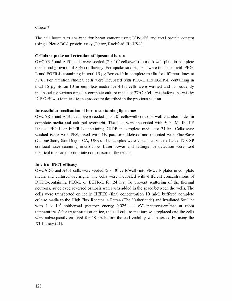
RESULTS Liposome characteristics As already described before by Krijger et al., we were able to efficiently encapsulate the hydrophilic Boron-10 enriched compound dodecahydrododecaborate (DHDB) into PEG-liposomes. These liposomes are stable for over two weeks time with respect to size and encapsulated boron (22). The liposomes were sized to 100-120 nm and had a low polydispersity index (≤ 0.25), which is a measure for the size distribution and varies from 0 (absolutely monodisperse) to 1 (extremely polydisperse). We were able to encapsulate up to 30 µg boron per µmol lipid as determined with ICP-OES, which corresponds to an encapsulation efficiency of about 5%. This is in good agreement with the expected aqueous volume present within this type of liposomes. Flow cytometry It is well known that the epidermal growth factor receptor (EGFR) is upregulated on the surface of certain tumour cell types. Using flow cytometry, the extent of binding of the natural ligand EGF labeled with the fluorochrome Alexa488 to both OVCAR-3 and A431 cells was measured. Figure 1A shows that after incubation for 1 hr at 4°C the binding of EGF-Alexa488 to OVCAR-3 cells was much lower when compared to A431 cells. The difference in degree of binding varies from a factor 15 to 65 depending on the concentration EGF added to the cells. This implies a strong difference in receptor expression level and confirmed western blot data (Sabrina Oliveira, Utrecht University, personal communication).
Figure 1. Binding of EGF-Alexa488 (A) or liposomes (B) to OVCAR-3 and A431 cells. (A) Different concentrations of EGF-Alexa488 were incubated with OVCAR-3 (■) or A431 (●) cells for 1 hr at 4ºC. The cells were washed and analyzed with flow cytometry. (B) Different concentrations of fluorescently labelled EGFR-L (● and ○) or PEG-L (■ and □) were incubated with OVCAR-3 cells (closed symbols) or A431 cells (closed symbols). Data represent mean ± SD (n=3). Error bars are within plot symbols when not visible.
Chapter 7
130
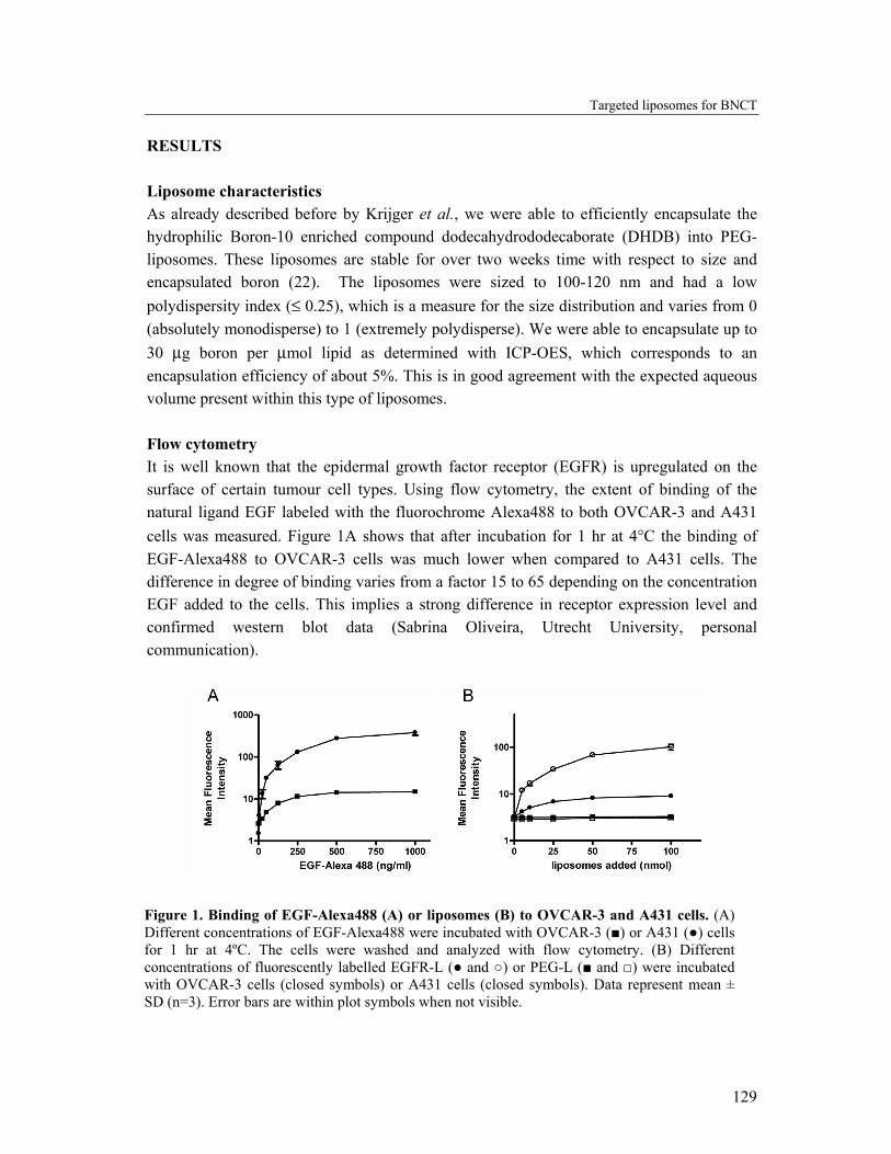
The degree of cell binding of fluorescently labeled PEG-liposomes (PEG-L) and EGFR-targeted liposomes (EGFR-L) to both cell types was investigated. Different amounts of PEG-L and EGFR-L were incubated with both cell types for 1 hr at 4°C. As shown in Figure 1B, the extent of binding of the targeted, but not of the untargeted liposomes differs between the cell lines. The EGFR-L bound more extensively to A431 cells than to OVCAR-3 cells, which is in line with the EGF binding results seen in Figure 1A. PEG-L did not show significant interaction with either cell line, confirming that binding is mediated by specific interaction between the anti-EGFR antibody and the EGFR. Specific binding of EGFR-L to OVCAR-3 cells has been shown in previous studies of our group (23). Cell-associated liposomal boron To investigate the extent of cellular binding of the two types of liposomes with respect to the amount of boron associated with the cells, OVCAR-3 and A431 cells were incubated for 1 hr at 4°C with both boron-loaded liposome types. After washing, the cells were lysed using a detergent and the amount of boron was measured and expressed relative to the cellular protein content (Figure 2).
Figure 2. Liposomal boron: Cellular association at 4°C. OVCAR-3 (A) or A431 (B) cells were incubated with different concentrations of boron-loaded EGFR-L (●) or PEG-L (■) for 1 hr at 4ºC. The cells were washed, lysed and cellular boron and protein content were determined. Data represent mean ± SD (n=3). Error bars are within plot symbols when not visible.
Targeted liposomes for BNCT
131
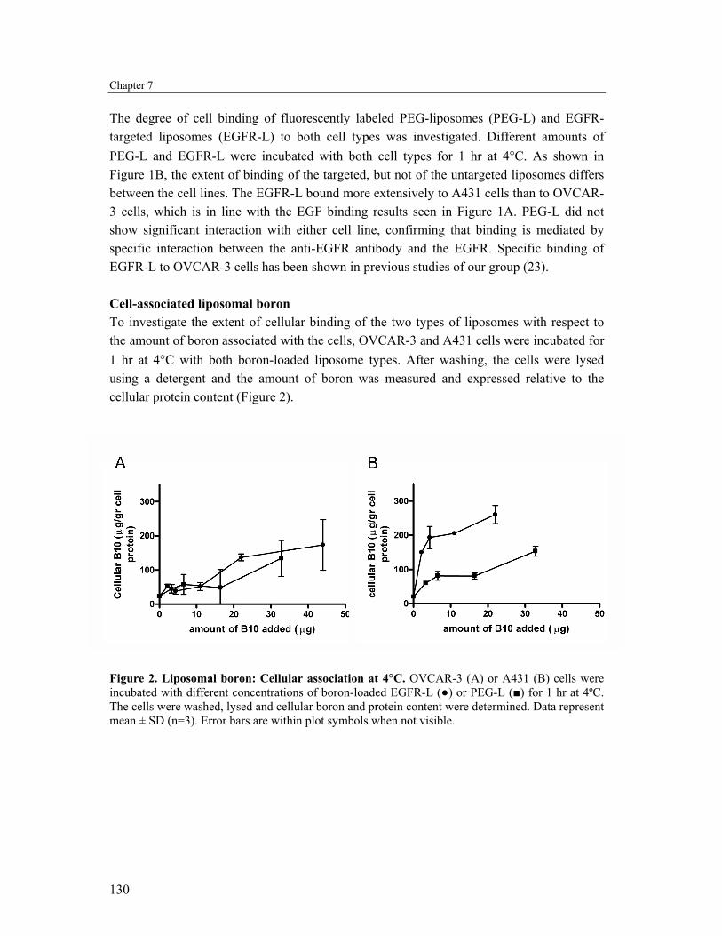
Despite the difference in cell binding (Figure 1B), no significant difference in the amount of OVCAR-3 cell-bound boron between PEG-L and EGFR-L incubations could be seen (Figure 2A). The amount of cell-associated boron increased for both PEG-L and EGFR-L to a similar extent with increasing amounts of added liposomal boron. This is in opposite to the results found with A431 cells; here the cell-bound boron was significantly higher when the cells were incubated with EGFR-L compared to PEG-L (Figure 2B). When cells, either OVCAR-3 or A431 are incubated with free DHDB, no binding (or uptake) of boron could be detected compared to non-treated cells (data not shown). Cellular uptake and retention of liposomal boron OVCAR-3 and A431 cells were incubated for varying times with 15 µg boron/ml encapsulated in PEG-L and EGFR-L at 37°C. For successful BNCT over 15 µg Boron-10/g tumour is necessary. This requirement is met by both PEG-L and EGFR-L in both cell lines (Figure 3), clearly illustrating the value of liposomal delivery of boron to tumour cells.
Figure 3. Liposomal boron: Cellular association at 37°C. OVCAR-3 (A) or A431 (B) cells were incubated with boron-loaded EGFR-L (●) or PEG-L (■) for varying times at 37ºC. The cells were washed, lysed and cellular boron and protein content were determined. Data represent mean ± SD (n=3). Error bars are within plot symbols when not visible.
However, no apparent difference over time was observed between EGFR-L and PEG-L in case of OVCAR-3 cells (Figure 3A). These results are opposite to those obtained with A431 cells, where EGFR-L are taken up to a much larger extent: using EGFR-L up to 2.5 times more boron is present in the A431 cells compared to A431 incubated with PEG-L (Figure 3B).
Chapter 7
132
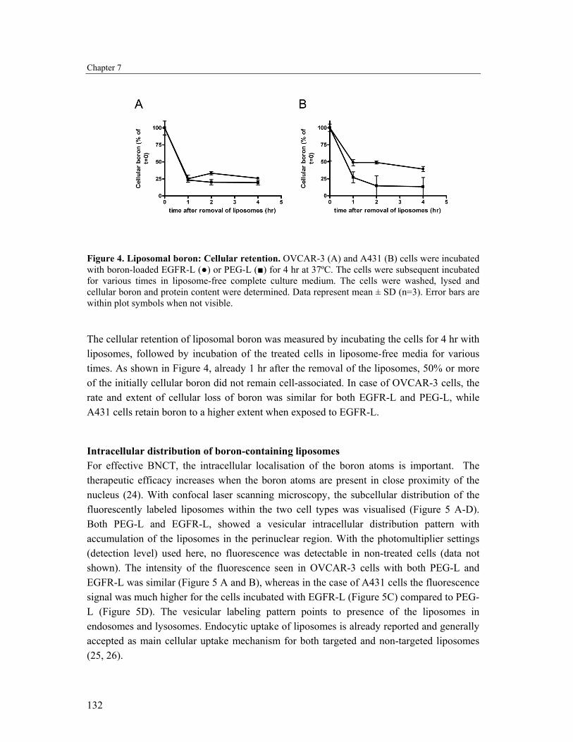
Figure 4. Liposomal boron: Cellular retention. OVCAR-3 (A) and A431 (B) cells were incubated with boron-loaded EGFR-L (●) or PEG-L (■) for 4 hr at 37ºC. The cells were subsequent incubated for various times in liposome-free complete culture medium. The cells were washed, lysed and cellular boron and protein content were determined. Data represent mean ± SD (n=3). Error bars are within plot symbols when not visible.
The cellular retention of liposomal boron was measured by incubating the cells for 4 hr with liposomes, followed by incubation of the treated cells in liposome-free media for various times. As shown in Figure 4, already 1 hr after the removal of the liposomes, 50% or more of the initially cellular boron did not remain cell-associated. In case of OVCAR-3 cells, the rate and extent of cellular loss of boron was similar for both EGFR-L and PEG-L, while A431 cells retain boron to a higher extent when exposed to EGFR-L.
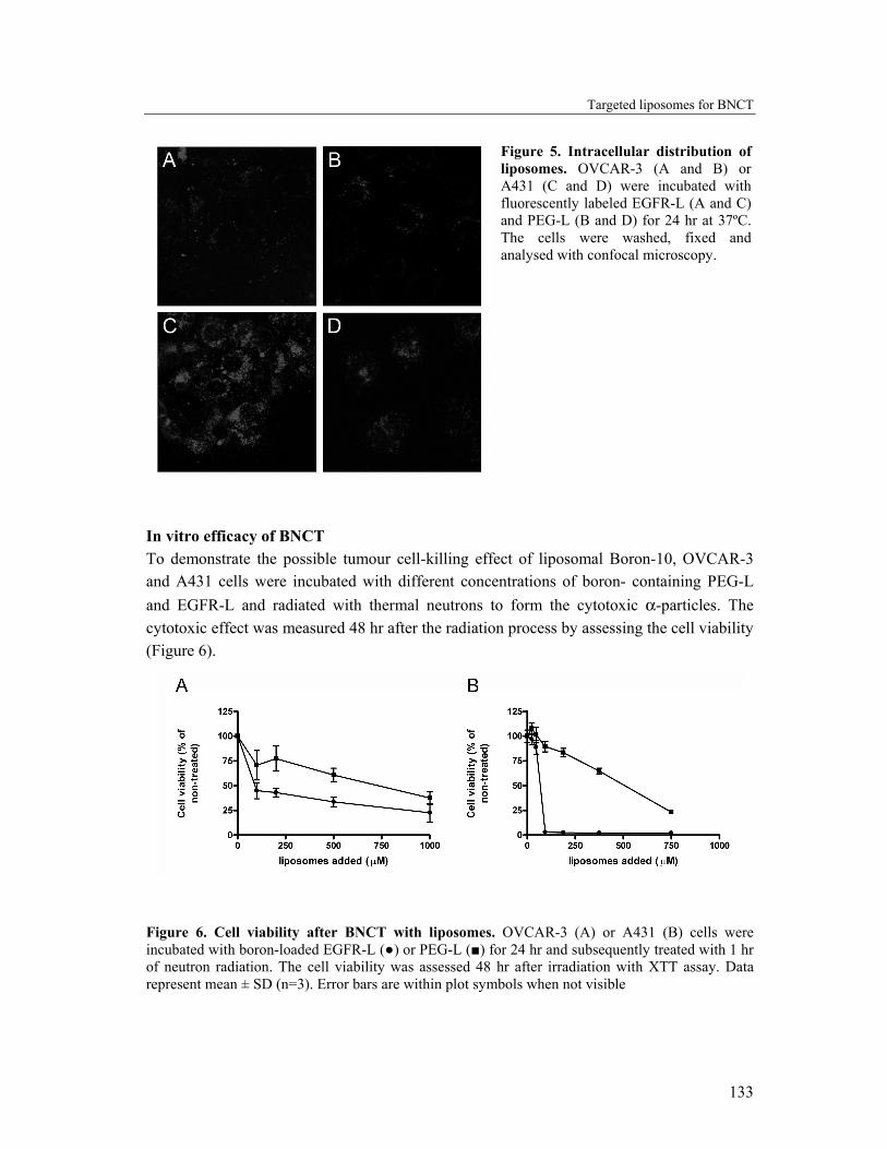
Intracellular distribution of boron-containing liposomes For effective BNCT, the intracellular localisation of the boron atoms is important. The therapeutic efficacy increases when the boron atoms are present in close proximity of the nucleus (24). With confocal laser scanning microscopy, the subcellular distribution of the fluorescently labeled liposomes within the two cell types was visualised (Figure 5 A-D). Both PEG-L and EGFR-L, showed a vesicular intracellular distribution pattern with accumulation of the liposomes in the perinuclear region. With the photomultiplier settings (detection level) used here, no fluorescence was detectable in non-treated cells (data not shown). The intensity of the fluorescence seen in OVCAR-3 cells with both PEG-L and EGFR-L was similar (Figure 5 A and B), whereas in the case of A431 cells the fluorescence signal was much higher for the cells incubated with EGFR-L (Figure 5C) compared to PEG-L (Figure 5D). The vesicular labeling pattern points to presence of the liposomes in endosomes and lysosomes. Endocytic uptake of liposomes is already reported and generally accepted as main cellular uptake mechanism for both targeted and non-targeted liposomes (25, 26).
Targeted liposomes for BNCT
133
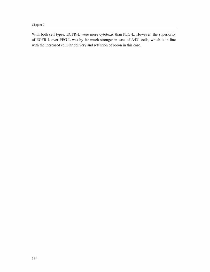
In vitro efficacy of BNCT To demonstrate the possible tumour cell-killing effect of liposomal Boron-10, OVCAR-3 and A431 cells were incubated with different concentrations of boron- containing PEG-L and EGFR-L and radiated with thermal neutrons to form the cytotoxic α-particles. The cytotoxic effect was measured 48 hr after the radiation process by assessing the cell viability (Figure 6).
Figure 6. Cell viability after BNCT with liposomes. OVCAR-3 (A) or A431 (B) cells were incubated with boron-loaded EGFR-L (●) or PEG-L (■) for 24 hr and subsequently treated with 1 hr of neutron radiation. The cell viability was assessed 48 hr after irradiation with XTT assay. Data represent mean ± SD (n=3). Error bars are within plot symbols when not visible
Figure 5. Intracellular distribution of liposomes. OVCAR-3 (A and B) or A431 (C and D) were incubated with fluorescently labeled EGFR-L (A and C) and PEG-L (B and D) for 24 hr at 37ºC. The cells were washed, fixed and analysed with confocal microscopy.
Chapter 7
134
With both cell types, EGFR-L were more cytotoxic than PEG-L. However, the superiority of EGFR-L over PEG-L was by far much stronger in case of A431 cells, which is in line with the increased cellular delivery and retention of boron in this case.
Targeted liposomes for BNCT
135
DISCUSSION Since the EGFR is overexpressed by many tumour cell types, the EGFR has been exploited previously to target drugs and delivery vehicles to tumour cells (13, 14). EGFR-targeted conjugates have been synthesised and evaluated for BNCT (9, 10, 15-18, 27, 28). Preparation of conjugates between boron and EGF or anti-EGFR-antibodies have been described (27, 28). The disadvantage of this approach is that only a small number of boron molecules can be attached, as with higher amounts coupled the specificity of the targeting ligand is lost (29). Drug delivery vehicles that are able to carry relatively large numbers of boron atoms are therefore favourable. Boronated dendrimers and liposomes targeted to the EGFR have been described (9, 10, 15-18). Although targeting to EGFR-expressing cells has been achieved, the effects of the level of receptor expression on the degree of targeting and BNCT mediated cytotoxicity have not been addressed so far. This issue is important, as the absolute level of EGFR expression will vary between different tumour types and even within one tumour the expression can be heterogeneous (30). In this chapter, the effect of targeting liposomes to the EGFR for BNCT was compared using two human carcinoma cell lines, human epidermoid carcinoma A431 and human ovarian carcinoma OVCAR-3. Both cell lines are considered EGFR-positive (31, 32), but no comparison has been made between the two cell lines with respect to the level of EGFR expression. Using flow cytometry, a minor but significant increase in binding was observed when OVCAR-3 cells were incubated with EGFR-L compared with the binding of PEG-L to OVCAR-3 cells. The binding of PEG-L to A431 cells was comparable to that of OVCAR-3 cells, but EGFR-L were bound to a much higher extent (Figure 1B). Taking into account that the EGF-Alexa488 was also bound to a higher extent to A431 cells than to OVCAR-3 cells (Figure 1A), the differences in binding of EGFR-L between both cells lines is likely to be explained by the differences in receptor level expression. Remarkably, when OVCAR-3 cell-associated boron was measured, both binding and uptake of the liposomes were not significantly enhanced in case of EGFR-L compared to PEG-L (Figure 2A and 3A). Using A431 cells, which express high levels of EGFR, a substantial increase in both binding and cellular uptake of EGFR-L compared to PEG-L was seen (Figure 2B and 3B). The cellular retention of liposome encapsulated boron was poor also in the case of A431 cells, but A431 cells clearly retained EGFR-L delivered boron to a higher extent than PEG-L delivered boron. Nevertheless, the poor cellular retention of liposomally delivered boron is a concern for further in vivo application. To measure therapeutic efficacy, in this study the cytotoxicity after BNCT. No considerable differences between EGFR-L and PEG-L could be seen with OVCAR-3 cells, whereas in A431 the EGFR-L were significantly more effective than PEG-L. The literature also reports on absence of improved efficacy of targeted liposomal formulations related to low EGFR expression levels. Mamot et al. showed that EGFR-targeted liposomes mediate uptake and efficient delivery of the encapsulated doxorubicin resulting in cytotoxic effects towards
Chapter 7
136
EGFR-overexpressing cells. In contrast, with cells lacking the expression of EGFR, uptake and cytotoxic effects were comparable to control liposomes devoid of the EGFR-targeting ligand (33). In addition, both in vitro and in vivo data reported by Park and colleagues confirmed the importance of the receptor expression level for the efficacy of a targeted liposome type. They showed that MCF-7 breast cancer cells, which have a relatively low or basal level of expression of the HER2-receptor (~104 receptor/cell), do not internalise HER2-targeted immunoliposomes more efficiently than liposomes lacking the targeting ligand. Also, HER-2 targeted liposomal doxorubicin was equally effective in delaying in vivo MCF-7 of tumour growth as control liposomes. This is in contrast to several other cell and xenograft models, which express the HER2 receptor to a larger extent (≥105 receptors/cell) and where significant improvement in tumour growth delay was seen when doxorubicin was encapsulated in HER2-targeted liposomes compared to liposomal doxorubicin without targeting ligand. These results point to a minimal threshold of HER2 density to attain therapeutic efficacy (26, 34), which supports our ‘threshold hypothesis’. Recently, liposomes targeting the transferrin receptor, transferrin-PEG-liposomes (Tf-liposomes), were used to target mercaptoundecahydrododecaborate (BSH) to tumour cells in vitro and in vivo. The in vitro targeting effect of Tf-liposomes compared to non-targeted liposomes comprised an approximate 5-fold increase in binding and uptake of boron by tumour cells. In vivo studies showed that significant difference in boron concentration in tumour cells was observed between targeted and non-targeted liposomes, but only at 72 hours after i.v. administration of the BSH-containing liposomes. When the tumours were irradiated with neutrons 72 hr after liposome administration, both PEG-liposomes and Tf-liposomes showed similar tumour-killing capacity, suggesting that the targeting effect was not strong enough to enhance the BNCT mediated cytotoxicity (8). CONCLUSION In this study, the use of EGFR-targeted liposomes for BNCT was evaluated in vitro using two tumour cell lines. The two cell types differed significantly regarding the level of EGFR expression. It is demonstrated here that successful BNCT with targeted liposomes is dependent on the target receptor expression level and that a certain threshold expression level is required to achieve successful BNCT.
Targeted liposomes for BNCT
137
REFERENCES 1. A. H. Soloway, W. Tjarks, B. A. Barnum, F. G. Rong, R. F. Barth, I. M. Codogni and J. G. Wilson.
The Chemistry of Neutron Capture Therapy. Chem Rev. 98: 1515-1562 (1998) 2. R. F. Barth, J. A. Coderre, M. G. Vicente and T. E. Blue. Boron neutron capture therapy of cancer:
current status and future prospects. Clin Cancer Res. 11: 3987-4002 (2005) 3. R. F. Barth, A. H. Soloway, R. G. Fairchild and R. M. Brugger. Boron neutron capture therapy for
cancer. Realities and prospects. Cancer. 70: 2995-3007 (1992) 4. G. Wu, R. F. Barth, W. Yang, R. J. Lee, W. Tjarks, M. V. Backer and J. M. Backer. Boron containing
macromolecules and nanovehicles as delivery agents for neutron capture therapy. Anticancer Agents Med Chem. 6: 167-84 (2006)
5. M. C. Woodle and D. D. Lasic. Sterically stabilized liposomes. Biochem Biophys Acta. 1132: 171-99 (1992)
6. H. Maeda, J. Wu, T. Sawa, Y. Matsumura and K. Hori. Tumor vascular permeability and EPR effect in macromolecular therapeutics: a review. J Control Release. 65: 271-84 (2000)
7. J. Carlsson, E. B. Kullberg, J. Capala, S. Sjoberg, K. Edwards and L. Gedda. Ligand liposomes and boron neutron capture therapy. J Neurooncol. 62: 47-59 (2003)
8. K. Maruyama, O. Ishida, S. Kasaoka, T. Takizawa, N. Utoguchi, A. Shinohara, M. Chiba, H. Kobayashi, M. Eriguchi and H. Yanagie. Intracellular targeting of sodium mercaptoundecahydrododecaborate (BSH) to solid tumors by transferrin-PEG liposomes, for boron neutron-capture therapy (BNCT). J Control Release. 98: 195-207 (2004)
9. E. B. Kullberg, M. Nestor and L. Gedda. Tumor-cell targeted epiderimal growth factor liposomes loaded with boronated acridine: uptake and processing. Pharm Res. 20: 229-36 (2003)
10. E. B. Kullberg, Q. Wei, J. Capala, V. Giusti, P. U. Malmstrom and L. Gedda. EGF-receptor targeted liposomes with boronated acridine: Growth inhibition of cultured glioma cells after neutron irradiation. Int J Radiat Biol. 81: 621-9 (2005)
11. H. Yanagie, H. Kobayashi, Y. Takeda, I. Yoshizaki, Y. Nonaka, S. Naka, A. Nojiri, H. Shinnkawa, Y. Furuya, H. Niwa, K. Ariki, H. Yasuhara and M. Eriguchi. Inhibition of growth of human breast cancer cells in culture by neutron capture using liposomes containing 10B. Biomed Pharmacother. 56: 93-9 (2002)
12. H. Yanagie, K. Maruyama, T. Takizawa, O. Ishida, K. Ogura, T. Matsumoto, Y. Sakurai, T. Kobayashi, A. Shinohara, J. Rant, J. Skvarc, R. Ilic, G. Kuhne, M. Chiba, Y. Furuya, H. Sugiyama, T. Hisa, K. Ono, H. Kobayashi and M. Eriguchi. Application of boron-entrapped stealth liposomes to inhibition of growth of tumour cells in the in vivo boron neutron-capture therapy model. Biomed Pharmacother. (2005)
13. S. Oliveira, P. M. Van Bergen en Henegouwen, G. Storm and R. M. Schiffelers. Molecular biology of epidermal growth factor receptor inhibition for cancer therapy. Expert Opin Biol Ther. 6: 498-543 (2006)
14. J. Mendelsohn. Targeting the epidermal growth factor receptor for cancer therapy. J Clin Oncol. 20: 1S-13S (2002)
15. E. B. Kullberg, N. Bergstrand, J. Carlsson, K. Edwards, M. Johnsson, S. Sjoberg and L. Gedda. Development of EGF-conjugated liposomes for targeted delivery of boronated DNA-binding agents. Bioconjug Chem. 13: 737-43 (2002)
16. W. Yang, R. F. Barth, G. Wu, A. K. Bandyopadhyaya, B. T. Thirumamagal, W. Tjarks, P. J. Binns, K. Riley, H. Patel, J. A. Coderre, M. J. Ciesielski and R. A. Fenstermaker. Boronated epidermal growth factor as a delivery agent for neutron capture therapy of EGF receptor positive gliomas. Appl Radiat Isot. 61: 981-5 (2004)
17. G. Wu, R. F. Barth, W. Yang, M. Chatterjee, W. Tjarks, M. J. Ciesielski and R. A. Fenstermaker. Site-specific conjugation of boron-containing dendrimers to anti-EGF receptor monoclonal antibody cetuximab (IMC-C225) and its evaluation as a potential delivery agent for neutron capture therapy. Bioconjug Chem. 15: 185-94 (2004)
18. R. F. Barth, W. Yang, D. M. Adams, J. H. Rotaru, S. Shukla, M. Sekido, W. Tjarks, R. A. Fenstermaker, M. Ciesielski, M. M. Nawrocky and J. A. Coderre. Molecular targeting of the epidermal growth factor receptor for neutron capture therapy of gliomas. Cancer Res. 62: 3159-66 (2002)
Chapter 7
138
19. G. A. Koning, H. W. Morselt, A. Gorter, T. M. Allen, S. Zalipsky, G. L. Scherphof and J. A. Kamps. Interaction of differently designed immunoliposomes with colon cancer cells and Kupffer cells. An in vitro comparison. Pharm Res. 20: 1249-57 (2003)
20. G. Rouser, S. Fkeischer and A. Yamamoto. Two dimensional then layer chromatographic separation of polar lipids and determination of phospholipids by phosphorus analysis of spots. Lipids. 5: 494-6 (1970)
21. D. A. Scudiero, R. H. Shoemaker, K. D. Paull, A. Monks, S. Tierney, T. H. Nofziger, M. J. Currens, D. Seniff and M. R. Boyd. Evaluation of a soluble tetrazolium/formazan assay for cell growth and drug sensitivity in culture using human and other tumor cell lines. Cancer Res. 48: 4827-33 (1988)
22. G. C. Krijger, M. M. Fretz, U. D. Woroniecka, O. M. Steinebach, W. Jiskoot, G. Storm and G. A. Koning. Tumor cell and tumor vasculature targeted liposomes for neutron capture therapy. Radiochim Acta. 93: 589-593 (2005)
23. E. Mastrobattista, G. A. Koning, L. Van Bloois, A. C. Filipe, W. Jiskoot and G. Storm. Functional Characterization of an Endosome-disruptive Peptide and Its Application in Cytosolic Delivery of Immunoliposome-entrapped Proteins. J Biol Chem. 277: 27135-43 (2002)
24. D. Gabel, S. Foster and R. G. Fairchild. The Monte Carlo simulation of the biological effect og the 10B(n, alpha)7Li reaction in cells and its implication for boron neutron capture therapy. Radiat Res. 111: 14-25 (1987)
25. R. M. Straubinger, K. Hong, D. S. Friend and D. Papahadjopoulos. Endocytosis of liposomes and intracellular fate of encapsulated molecules: encounter with a low pH compartment after internalization in coated vesicles. Cell. 32: 1069-1079 (1983)
26. D. B. Kirpotin, J. W. Park, K. Hong, S. Zalipsky, W. Li, P. Carter, C. C. Benz and D. Papahadjopoulos. Sterically stabilized anti-HER2 immunoliposomes: design and targeting to human breast cancer cell in vitro. Biochemistry. 36: 66-75 (1997)
27. J. Capala, R. F. Barth, M. Bendayan, M. Lauzon, D. M. Adams, A. H. Soloway, R. A. Fenstermaker and J. Carlsson. Boronated epidermal growth factor as a potential targeting agent for boron neutron capture therapy of brain tumors. Bioconjug Chem. 7: 7-15 (1996)
28. N. Carlsson, L. Gedda, C. Gronvik, T. Hartman, A. Lindstrom, P. Lindstrom, H. Lundqvist, A. Lovqvist, J. Malmqvist and P. Olsson. Strategy for boron neutron capture therapy against tumor cells with overexpression of the epidermal growth factor receptor. Int J Radiat Biol Phys. 30: 105-115 (1994)
29. S. C. Mehta and D. R. Lu. Targeted drug delivery for boron neutron capture therapy. Pharm Res. 13: 344-51 (1996)
30. D. S. Salomon, R. Brandt, F. Ciardiello and N. Normanno. Epidermal growth factor-related peptides and their receptor in human malignancies. Crit Rev Oncol Hematol. 19: 183-232 (1995)
31. E. Mastrobattista. Comparison of immunoliposomes targeting to three different receptors expressed by human ovarian carcinoma (OVCAR-3) cells. Dissertation thesis. (2001)
32. P. Nagy, D. J. Arndt-Jovin and T. Jovin. Small interfering RNAs suppress the expression of endogenous and GFP-fused epidermal growth factor receptor (erbB1) and induce apoptosis in erbB1-overexpressing cells. Exp Cell Res. 285: 39-49 (2003)
33. C. Mamot, D. C. Drummond, U. Greiser, K. Hong, D. B. Kirpotin, J. D. Marks and J. W. Park. Epidermal growth factor receptor (EGFR)-targeted immunoliposomes mediate specific and efficient drug delivery to EGFR- and EGFRvIII-overexpressing tumor cells. Cancer Res. 63: 3154-3161 (2003)
34. J. W. Park, K. Hong, D. B. Kirpotin, G. Colbern, R. Shalaby, J. Baselga, Y. Shao, U. B. Nielsen, J. D. Marks, D. Moore, D. Papahadjopoulos and C. C. Benz. Anti-HER2 immunoliposomes: enhanced efficacy attributtable to targeted delivery. Clin Cancer Res. 8: 1172-1181 (2002)
35. M. M. Fretz, A. Hogset, G. A. Koning, W. Jiskoot and G. Storm. Cytosolic delivery of liposomally targeted proteins induced by photochemical internalization. Pharm Res Accepted for publication.

Targeted liposomes for BNCT
139


![arXiv:1110.4729v1 [cond-mat.str-el] 21 Oct 2011 · cent study of the dielectric constant in this material.22 However, microscopic studies such as NMR or neutron scattering measurements](https://static.fdocuments.ec/doc/165x107/5e7e2e837220a71d6c22cf2e/arxiv11104729v1-cond-matstr-el-21-oct-2011-cent-study-of-the-dielectric-constant.jpg)