Semblanzas Ictiológicas Fabiana Laura Lo Nostro · Cuestionario -Un libro: La hermana, ... azul...
21
Transcript of Semblanzas Ictiológicas Fabiana Laura Lo Nostro · Cuestionario -Un libro: La hermana, ... azul...


Semblanzas Ictiológicas
Fabiana Laura Lo Nostro
Museo de la Ciencia Nemo, Amsterdam, 2013
Hugo L. López y Justina Ponte Gómez
ProBiota División Zoología Vertebrados
Museo de La Plata FCNyM, UNLP
Octubre de 2014
Imagen de Tapa Dr. Harry Grier y Fabiana Lo Nostro, The Florida Aquarium, USA, 2002

El tiempo acaso no exista. Es posible que no pase y sólo pasemos nosotros.
Tulio Carella
Cinco minutos bastan para soñar toda una vida, así de relativo es el tiempo. Mario Benedetti
Semblanzas Ictiológicas
A través de esta ser ie intentar emos conocer difer entes facetas persona les de los integrantes de nuestra “comunidad”.
El cuest ionar io, además de su pr incipal objet ivo, con sus r espuestas quizás nos ayude a encontrar entr e nosotros puntos en común que vayan más allá de nuestros t emas de trabajo y sea un aporte a futuros estudios histór icos.
Esperamos que esta iniciativa pueda ser otro nexo entr e los ict iólogos de la r egión, ya que cons ideramos que el r esu ltado general trascender ía nuestras fronteras.
Hugo L. López

ProBiota, Serie Técnica y Didáctica 21(53) - 2014 4
Nombre y apellido completos: FABIANA LAURA LO NOSTRO
Lugar de nacimiento: Ciudad Autónoma de Buenos Aires (C.A.B.A.)
Lugar, provincia y país de residencia: C.A.B.A, Argentina
Título máximo, Facultad y Universidad: Doctor en Ciencias Biológicas, Facultad de Ciencias Exactas y Naturales (FCEyN), Universidad de Buenos Aires (UBA)
Posición laboral: Profesor Adjunto, FCEyN, UBA e Investigador Independiente CONICET
Lugar de trabajo: Laboratorio de Ecotoxicología Acuática, Departamento de Biodiversidad y Biología Experimental (DBBE), FCEyN, UBA e IBBEA, CONICET-UBA, Argentina
Especialidad o línea de trabajo: Reproducción en peces; Ecotoxicología acuática
Correo electrónico: [email protected]
Cuestionario
- Un libro: La hermana, Paola Kaufmann - Una película: Brazil, Terry Gillian - Un CD : Las cuatro estaciones, Vivaldi - Un artista: Mijaíl Baryshnikov - Un deporte: enseñar a bucear - Un color: azul - Una comida: pescado (en todas sus formas) - Un animal: perro - Una palabra: equipo - Un número: 20 - Una imagen: la llegada al Taj Mahal - Un lugar: Caviahue, Neuquén, Patagonia Argentina - Una estación del año: primavera - Un nombre: Lucia - Un hombre: Jacques I. Cousteau - Una mujer: Marie Curie - Un personaje de ficción: Peter Pan - Un superhéroe: Mafalda! - Un ictiólogo del pasado: Marcusd Bloch, Alemania, siglo XVIII - Un ictiólogo del presente: Roberto Menni y Hugo López (Argentina
actual!)

ProBiota, Serie Técnica y Didáctica 21(53) - 2014 5
Un festejo de tantos, 1994
Laura López Greco, Alejandra Volpedo y Fabiana Lo Nostro

ProBiota, Serie Técnica y Didáctica 21(53) - 2014 6
Delhi, India, 2011
F. Lo Nostro y la Prof. R. Guimarães Moreira de la Universidad de São Paulo, Brasil
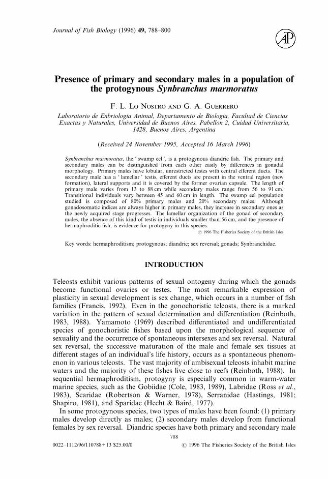
Journal of Fish Biology (1996) 49, 788–800
Presence of primary and secondary males in a population ofthe protogynous Synbranchus marmoratus
F. L. L N G. A. G
Laboratorio de Enbriologia Animal, Departamento de Biologia, Facultad de CienciasExactas y Naturales, Universidad de Buenos Aires. Pabellon 2, Cuidad Universitaria,
1428, Buenos Aires, Argentina
(Received 24 November 1995, Accepted 16 March 1996)
Synbranchus marmoratus, the ‘ swamp eel ’, is a protogynous diandric fish. The primary andsecondary males can be distinguished from each other easily by differences in gonadalmorphology. Primary males have lobular, unrestricted testes with central efferent ducts. Thesecondary male has a ‘ lamellar ’ testis, efferent ducts are present in the ventral region (newformation), lateral supports and it is covered by the former ovarian capsule. The length ofprimary male varies from 13 to 88 cm while secondary males range from 56 to 91 cm.Transitional individuals vary between 45 and 60 cm in length. The swamp eel populationstudied is composed of 80% primary males and 20% secondary males. Althoughgonadosomatic indices are always higher in primary males, they increase in secondary ones asthe newly acquired stage progresses. The lamellar organization of the gonad of secondarymales, the absence of this kind of testis in individuals smaller than 56 cm, and the presence ofhermaphroditic fish, is evidence for protogyny in this species.
? 1996 The Fisheries Society of the British Isles
Key words: hermaphroditism; protogynous; diandric; sex reversal; gonads; Synbranchidae.
INTRODUCTION
Teleosts exhibit various patterns of sexual ontogeny during which the gonadsbecome functional ovaries or testes. The most remarkable expression ofplasticity in sexual development is sex change, which occurs in a number of fishfamilies (Francis, 1992). Even in the gonochoristic teleosts, there is a markedvariation in the pattern of sexual determination and differentiation (Reinboth,1983, 1988). Yamamoto (1969) described differentiated and undifferentiatedspecies of gonochoristic fishes based upon the morphological sequence ofsexuality and the occurrence of spontaneous intersexes and sex reversal. Naturalsex reversal, the successive maturation of the male and female sex tissues atdifferent stages of an individual’s life history, occurs as a spontaneous phenom-enon in various teleosts. The vast majority of ambisexual teleosts inhabit marinewaters and the majority of these fishes live close to reefs (Reinboth, 1988). Insequential hermaphroditism, protogyny is especially common in warm-watermarine species, such as the Gobiidae (Cole, 1983, 1989), Labridae (Ross et al.,1983), Scaridae (Robertson & Warner, 1978), Serranidae (Hastings, 1981;Shapiro, 1981), and Sparidae (Hecht & Baird, 1977).In some protogynous species, two types of males have been found: (1) primary
males develop directly as males; (2) secondary males develop from functionalfemales by sex reversal. Diandric species have both primary and secondary male
788
0022–1112/96/110788+13 $25.00/0 ? 1996 The Fisheries Society of the British Isles

Involvement of the Gonadal Germinal Epithelium DuringSex Reversal and Seasonal Testicular Cycling in theProtogynous Swamp Eel, Synbranchus marmoratusBloch 1795 (Teleostei, Synbranchidae)F. Lo Nostro,1 H. Grier,2* L. Andreone,1 and G.A. Guerrero1
1Laboratorio de Embriologıa Animal, Departamento de Biodiversidad y Biologıa Experimental, Facultad de CienciasExactas y Naturales, Universidad de Buenos Aires, Buenos Aires, Argentina C1428EHA.2Stock Enhancement Research Facility, Florida Marine Research Institute, Palmetto, Florida, USA 34221-9620
ABSTRACT The swamp eel, Synbranchus marmoratus,is a protogynous, diandric species. During sex reversal,the ovarian germinal epithelium, which forms folliclescontaining an oocyte and encompassing follicle cells dur-ing the female portion of the life cycle, produces numerousinvaginations, or acini, into the ovarian stroma. Withinthe acini, the gonia that formerly produced oocytes becomespermatogonia, enter meiosis, and produce sperm. Theacini are bounded by the basement membrane of the ger-minal epithelium. Epithelial cells of the female germinalepithelium, which formerly became follicle (granulosa)cells, now become Sertoli cells in the developing testis.Subsequently, lobules and testicular ducts form. Theswamp eel testis has a lobular germinal compartment inboth primary and secondary males, although the germinalcompartment in testes of secondary males resides withinthe former ovarian lamellae. The germinal compartment,supported by a basement membrane, is composed of Ser-toli and germ cells that give rise to sperm. Histologicaland immunohistochemical techniques were used to de-scribe the five reproductive classes that were observed tooccur during the annual reproductive cycle: regressed,early maturation, mid-maturation, late maturation, andregression. These classes are differentiated by the pres-ence of continuous or discontinuous germinal epitheliaand by the types of germ cells present. Synbranchus mar-moratus has a permanent germinal epithelium. Differ-ences between the germinal compartment of the testes ofprimary and secondary males were not observed. J. Mor-phol. 257:107–126, 2003. © 2003 Wiley-Liss, Inc.
KEY WORDS: synbranchiformes; swamp eels; germinalepithelium; sex reversal; testis; annual cycle
Numerous descriptions of the stages of germ cellmaturation and development that are observed inteleosts during annual reproductive cycles havebeen published (Ruby and McMillan, 1970; Billardand Breton, 1978; Scott and Sumpter, 1989; Ntibaand Jaccarini, 1990; Smith and Young, 1996). How-ever, the mechanisms of testicular regression and ofthe cycling of cells between annual breeding seasonsare known for only a few species (Grier, 1993; Grierand Taylor, 1998; Taylor et al., 1998; Brown-
Peterson and Warren, 2001; Brown-Peterson et al.,2002). The germinal epithelium (GE), which is foundin both the testes and the ovaries of fish, has onlyrecently been defined (Grier, 2000, 2002; Grier andLo Nostro, 2000). Histological changes in the GE’smorphology have been used to document a sequenceof five reproductive classes during the annual repro-ductive cycle of the male common snook, Centropo-mus undecimalis (Taylor et al., 1998; Grier and Tay-lor, 1998; Grier and Lo Nostro, 2000), and of thepelagic cobia, Rachycentron canadum (Brown-Peterson et al., 2002). These reproductive classeswere based on the alternation of the GE betweencontinuous and discontinuous types and the stagesof germ cells present. Continuous and discontinuousGE refer to whether both Sertoli cells and germ cellsform a continuum or whether the germ cells arescattered and separated by Sertoli cells, respectively(Grier, 1993; Grier and Lo Nostro, 2000). Recogni-tion of these two different forms of GE made it clearthat among the chordates, a permanent GE thatpersists throughout annual reproductive cycles firstappears in the fishes (Grier and Taylor, 1998), not inamphibians, as originally reported (Lofts, 1987).However, description of the GE and its annualchanges, which can be used to describe reproductiveclasses, is wholly confined to the perciform teleosts.
Synbranchus marmoratus Bloch, 1795, the NewWorld swamp eel (Liem, 1963, 1968; Lo Nostro andGuerrero, 1996), is a protogynic, diandric, freshwa-
Contract grant sponsor: (to FLN, University of Buenos Aires) UBA;Contract grant numbers: TW/41, EX/157, EX/217; Contract grantsponsor: CONICET; Contract grant number: PIP4558.
*Correspondence to: Dr. Harry Grier, Stock Enhancement ResearchFacility, Florida Marine Research Institute, 14495 Harllee Road, Pal-metto, Florida 34221-9620 USA. E-mail: [email protected]
DOI: 10.1002/jmor.10105
JOURNAL OF MORPHOLOGY 257:107–126 (2003)
© 2003 WILEY-LISS, INC.

Vitellogenin detection in surface mucus of the South Americancichlid fish Cichlasoma dimerus (Heckel, 1840) induced by
estradiol-17b. Effects on liver and gonads
Natalia Moncaut, Fabiana Lo Nostro, Marıa Cristina Maggese *
Laboratorio de Embriologıa Animal, Departamento de Biodiversidad y Biologıa Experimental, Facultad de Ciencias Exactas y Naturales,
Universidad de Buenos Aires, Ciudad Universitaria, Pab. II, 4to piso Buenos Aires C1428EHA, Argentina
Received 19 March 2002; received in revised form 30 July 2002; accepted 16 September 2002
Abstract
During the last decade, special attention has been focused on the consequences of exposure to environmental
estrogens on reproduction in wild fish species. For this reason, characterization of biomarkers of such exposures could
result in useful tools for these studies. The detection of vitellogenin (Vtg), a precursor of yolk proteins, is being intensely
studied since its synthesis in the liver is regulated by the estradiol-17b and is influenced by other estrogenic compounds.
The aim of this work was to assess the presence of Vtg in the surface mucus of males of Cichlasoma dimerus (Teleostei,
Perciformes), a typical South American freshwater cichlid, after hormonal treatment with estradiol-17b (intraperitoneal
injections of 10 mg E2/g fish). Plasma and mucus from females and treated males analyzed by Western blot revealed that
different heterologous antisera against Vtg bind to putative protein of 180 & 120 kDa and 120 & 110 kDa, respectively,
whereas no reaction was found in samples of untreated males. The same profile was observed in mucus samples using
Dot blot, a very easy and direct technique. Using immunocytochemistry techniques, immunoreactive Vtg (ir-Vtg)
producing cells in the liver of females and treated males were detected. Testes and liver tissues were also assessed by
histological techniques. Marked alterations in both organs were observed, such as lower sperm production, presence of
immature germ cells in the lobular lumen of testes, and some morphology changes in the hepatocytes due to the
accumulation of Vtg. This is the first report about the effects of an estrogen in the Vtg synthesis and their consequences
on liver and gonads of a South American fresh water cichlid. These results also support the possibility of using Vtg from
surface mucus as a potential biomarker for estrogenic compounds through a noninvasive, useful and easy assay to
monitor the presence of endocrine disrupting chemicals in the environment.
# 2002 Elsevier Science B.V. All rights reserved.
Keywords: Cichlids; Cichlasoma dimerus ; Vitellogenin; Mucus; Estradiol; Liver and testes pathology
1. Introduction
During the last decades there has been a
significant effort to determine the effects of endo-
crine disrupting chemicals on reproduction,
* Corresponding author. Tel.: �/54-11-4576-3348; fax: �/54-
11-4576-3384
E-mail address: [email protected] (M.C. Maggese).
Aquatic Toxicology 63 (2003) 127�/137
www.elsevier.com/locate/aquatox
0166-445X/02/$ - see front matter # 2002 Elsevier Science B.V. All rights reserved.
PII: S 0 1 6 6 - 4 4 5 X ( 0 2 ) 0 0 1 7 5 - 3

Tissue & Cell 36 (2004) 221–231
Testicular interstitial cells, and steroidogenic detection in the protogynousfish,Synbranchus marmoratus(Teleostei, Synbranchidae)
Fabiana L. Lo Nostroa,∗, Fernanda N. Antonelib,c,Iranı Quagio-Grassiottoc, Graciela A. Guerreroa
a Laboratorio de Embriolog´ıa Animal, Departamento de Biodiversidad y Biolog´ıa Experimental, Facultad de Ciencias Exactas y Naturales (FCEN),UBA, Cuidad Universitaria, Buenos Aires C1428EHA, Argentina
b Departamento de Biologia Celular, Instituto de Biologia, UNICAMP, Campinas 13083-970, Brazilc Departamento de Morfologia, Instituto de Biociˆencias, UNESP, Botucatu 18618-000, Brazil
Received 28 May 2003; received in revised form 27 November 2003; accepted 3 March 2004
Abstract
The swamp eel,Synbranchus marmoratus, is a freshwater protogynic diandric species. Primary males develop directly as males whilesecondary males arise from the sex reversal of females. Fishes from Argentine and Brazil inland waters were collected, examined andcompared for this study. In order to characterize the interstitial testicular compartment, light and electron microscopy techniques and anenzyme histochemical examination for steroidogenic cells detection were used. The interstitial compartment ofS. marmoratusis composedof Leydig and myoid cells, collagen fibers, blood cells, macrophages,and amyelinic nerves. At the ultrastructural level, no differences wereobserved in the interstitial tissue, either between specimens from the different sampling sites or between primary and secondary males.Leydig cells are present in all testes examined throughout the year. A cytoplasmatic reaction of 3�-HSD was detected only in Leydig cellsduring sex reversal and in both type of males, mainly during the regressed and early maturation classes (autumn and winter). Leydig cellspossess the typical fine structural characteristics associated with steroidogenesis. Furthermore, in both type of males, during sex reversaland after the spawning period, the number of granulocytes and macrophages present in the testes increased, suggesting that they could beinvolved in phagocytosis and resorption of damaged cells.© 2004 Elsevier Ltd. All rights reserved.
Keywords:Synbranchiformes; Sex reversal; Protogynous; Interstitial compartment; Leydig cells; Blood cells
1. Introduction
During the last 50 years, numerous studies focused ontesticular interstitial tissue morphology have been performedin different teleost species (Billard et al., 1982; Nagahama,1987; Dodd and Sumpter, 1984; Grier, 1993). The presenceof Leydig cells in the testis appears to be typical of teleostsas in the rest of the vertebrate lineage (Nagahama, 1983).However, in testes of primary and secondary males of theprotogynous wrasseCoris julis, Reinboth (1962)reporteda lack of Leydig cells. The same absence of Leydig cellswas reported in the protogynous Caribbean labrids (Roede,1975). However,Bruslé (1987)demonstrated the presenceof Leydig cells inC. julis using an ultrastructural approach.Histochemistry, electron microscopy (Hurk van den et al.,1978a; Grier, 1981; Nagahama, 1987; Grier et al., 1989),
∗ Corresponding author. Tel.:+54-11-4576-3348;fax: +54-11-4576-3384.
E-mail address:[email protected] (F.L. Lo Nostro).
and in vitro techniques (Loir, 1990a,b; Cinquetti, 1994; Hoarand Nagahama, 1978) have demonstrated interstitial Leydigcells in the testes of many fish species and confirmed thatthey are the main source of gonadal steroids.
Together with Leydig cells, the interstitial tissue containsfibroblasts, collagen fibers, myoid cells, and blood cells(Reinboth and Bruslé-Sicard, 1997; Cinquetti and Dramis,2003). Grier et al. (1989), using electron microscopy to studytestes of two species of pike, demonstrated a contractile net-work of myoid boundary cells in the interstitial compart-ment. The ultrastructure of myoid cells has been describedin carp and trout (Timmermans et al., 1993; Cauty and Loir,1995). In general, the information related to blood cells isscarce in fishes. The lymphohemopoietic cells with phago-cytic capacity are scattered randomly throughout the stroma(Quesada et al., 1990; Press et al., 1994) and sinusoidal cap-illaries (Zapata, 1979; Zapata et al., 1996).
As in synbranchid species,Monopterus albus(Liem,1963; Chang and Phillips, 1967; Nagahama, 1983), sexreversal is a natural life history process.Synbranchus
0040-8166/$ – see front matter © 2004 Elsevier Ltd. All rights reserved.doi:10.1016/j.tice.2004.03.001

Reproductive Histology of Tomeurus gracilisEigenmann, 1909 (Teleostei: Atherinomorpha:Poeciliidae) With Comments on Evolutionof Viviparity in Atherinomorph Fishes
Lynne R. Parenti,1* Fabiana L. LoNostro,2 and Harry J. Grier1
1Division of Fishes, National Museum of Natural History, Smithsonian Institution,Washington, District of Columbia 20013-70122Lab. de Embriologıa Animal, Depto. de Biodiversidad y Biologıa Experimental,Facultad de Ciencias Exactas y Naturales, Universidad de Buenos Aires C1428EHA and CONICET,Rivadavia 1917 C1033AAJ, Ciudad Autonoma de Buenos Aires, Argentina
ABSTRACT Tomeurus gracilis is a species long consid-ered pivotal in understanding the evolution of livebear-ing in atherinomorph fishes. Tomeurus gracilis is a zygo-parous or embryoparous poeciliid: internal fertilizationis followed by females laying fertilized eggs singly orretaining fertilized eggs until or near hatching. Tomeu-rus was hypothesized as the sister group of the vivipa-rous poeciliids until it was proposed as a close relativeof a derived viviparous poeciliid, Cnesterodon, hencenested among viviparous taxa rather than near the rootof the tree. Here, we describe and compare reproductivemorphological characters of the little-known Tomeuruswith those of representative atherinomorphs. In Tomeu-rus and Cnesterodon, sperm are packaged in nakedsperm bundles, or spermatozeugmata, in a configurationconsidered here diagnostic of viviparous poeciliids.Testes are single and free sperm are stored in the ovaryin both taxa in contrast to oviparous atherinomorphs inwhich testes are paired and sperm are not packaged andnot stored in the ovary. Efferent ducts in Cnesterodontestes and other viviparous poeciliids have a PAS-posi-tive secretion demonstrating presence of a glycoproteinthat inactivates sperm or prevents final sperm matura-tion. No PAS-positive staining secretion was observedin Tomeurus or oviparous atherinomorphs. Tomeurusshares apomorphic reproductive characters, such assperm bundle and testis morphology and a gonopodium,with viviparous poeciliids and plesiomorphic characters,such as a thick zona pellucida with filaments, with ovip-arous taxa. We do not postulate loss or reversal of vivi-parity in Tomeurus, and we corroborate its phylogeneticposition as sister to the viviparous poeciliids. J. Morphol.271:1399–1406, 2010. � 2010 Wiley-Liss, Inc.y
KEY WORDS: spermatozeugma; zona pellucida; eggmorphology; embryoparity/zygoparity; testis types
INTRODUCTION
Atherinomorph fishes, with an estimated 1,552species classified in three orders, Atheriniformes,Cyprinodontiformes, and Beloniformes, have longbeen featured in studies of reproductive biology(see Parenti, 2005; Nelson, 2006). Atherinomorph
monophyly is well-supported by a range of mor-phological characters (Rosen, 1964; Rosen andParenti, 1981; Parenti, 1993, 2005) and has beenrecovered in molecular analyses of bony fish phy-logeny (e.g., Setiamarga et al., 2008). Two diagnos-tic characters of atherinomorphs are explicitly ofreproductive morphology: a testis with spermato-gonia restricted to the distal ends of testis lobulesrather than distributed along the length of thelobule and a relatively large egg with fluid ratherthan granular yolk (Grier et al., 1980; Rosen andParenti, 1981; Grier, 1993; Parenti and Grier,2004). Internal fertilization and viviparity haveevolved multiple times within atherinomorphs, asinterpreted from the most parsimonious distribu-tion of morphological and molecular characters,many of which are not related directly to reproduc-tion (see Rosen, 1964; Parenti, 1981, 1993, 2005;Rosen and Parenti, 1981; Meyer and Lydeard,1993; Grier et al., 2005; Mank and Avise, 2006;Hrbek et al., 2007; Reznick et al., 2007a).
Tomeurus gracilis Eigenmann (1909) (Fig. 1A) isa diminutive, zygoparous or embryoparous atheri-nomorph poeciliid fish that lives in northeasternSouth America. A simple, straightforward classifi-
Harry J. Grier is currently at Fish and Wildlife Research Insti-tute, St. Petersburg, Florida 33701-5095.
*Correspondence to: Lynne R. Parenti, Division of Fishes,National Museum of Natural History, Smithsonian Institution,Washington, DC 20013-7012. E-mail: [email protected]
yThis article is the US Goverment work and, as such, is in the pub-lic domain in the United States of America.
Received 5 March 2010; Revised 27 May 2010;Accepted 19 June 2010
Published online 22 September 2010 inWiley Online Library (wileyonlinelibrary.com)DOI: 10.1002/jmor.10886
JOURNAL OF MORPHOLOGY 271:1399–1406 (2010)
� 2010 WILEY-LISS, INC.

Effect of the Organochlorine Pesticide Endosulfan on GnRHand Gonadotrope Cell Populations in Fish Larvae
Yanina G. Piazza • Matias Pandolfi •
Fabiana L. Lo Nostro
Received: 8 June 2010 / Accepted: 22 October 2010 / Published online: 26 November 2010
� Springer Science+Business Media, LLC 2010
Abstract Endocrine-disrupting chemicals can influence
the hypothalamus–pituitary–gonad axis and possibly affect
reproduction in vertebrates. We analyzed the effect of 30-
day endosulfan (ES) exposure in sexually undifferentiated
larvae of the cichlid fish Cichlasoma dimerus. The number,
area, mean cytoplasmic and nuclear diameter, and mean
cytoplasmic optical density of gonadotropin-releasing hor-
mone (GnRH) I, II, and III immunoreactive (ir-) neurons and
b follicle-stimulating hormone (bFSH) ir-cells were mea-
sured. Animals exposed to the highest ES concentration
(0.1 lg/l) showed a decrease in GnRH I nucleus/cytoplasm
area ratio upon exposure. Nuclear area and mean nuclear
diameter of bFSH ir-cells was higher in ES treated fish.
bFSH nucleus/cytoplasm area ratio was high in exposed
animals, and animals exposed to 0.1 lg/l ES showed smaller
mean cytoplasmic optical density. These findings suggest
that ES affects GnRH I and bFSH protein synthesis/release.
However, these responses seem to be insufficient to affect
gonadal differentiation at this stage of development.
The neuroendocrine system of the hypothalamus–pituitary–
gonad (HPG) axis regulates reproduction in vertebrates and
can be influenced by chemicals, therefore affecting the
reproductive system. Neurotoxic environmental contaminants
recognized as endocrine-disrupting chemicals (EDCs) have
aroused considerable interest in the field of neuroendocri-
nology (Gore 2000; Pillai et al. 2003; Panzica et al. 2005;
Gore 2008a, b). Among these pollutants, organochlorine
pesticides are considered to be hazardous because they are
very persistent, are nonbiodegradable, and are ubiquitously
found in the environment (Palmer and Palmer 1995; Dono-
hoe and Curtis 1996). After international recognition of their
long-term negative impacts on the global environment, the
use of organochlorines in global agriculture has been largely
banned (RAP-AL 2008; United Nations 2009). However,
endosulfan (ES) remains as one major exception. ES (6,7,8,
9,10,10-hexachloro-1,5,5a,6,9,9a-hexahydro-6,9-methano-
2,4,3benzo-dioxathiepin-3-oxide) is a cyclodiene organo-
chlorine insecticide used for the control of insects and mites
in crops of high commercial value (RAP-AL 2008). ES is
slightly soluble in water, but it dissolves in most organic
solvents (Harding 1979). Given that organic solvents are
commonly used in commercial formulations, they might
contribute to the overall effect of ES on the dysfunction of the
endocrine system (Hutchinson et al. 2006; Mortensen and
Arukwe 2006). Exposure of Thalassoma pavo to ES
decreased feeding behavior related to neuronal degeneration
in the mesencephalon and the hypothalamus (Giusi et al.
2005). Cytological and structural oogonia and oocyte mal-
formations, an important decrease in gonadotropins (GtHs)
neurosecretory activity, as well as damage of the axons that
innerve the pituitary were observed in adults of Sarother-
odon mossambicus after chronic exposure to ES (Shukla and
Pandey 1986). In Oryzias latipes, acute exposure to ES
caused alterations in development, sexual behavior, and
reproductive physiology (Gormley and Teather 2003).
The decapeptide gonadotropin-releasing hormone (GnRH)
is mainly synthesized in the central nervous system, and its
Y. G. Piazza � M. Pandolfi � F. L. Lo Nostro (&)
Laboratory of Animal Embryology, Department of Biodiversity
and Experimental Biology, Faculty of Exact and Natural
Sciences, University of Buenos Aires,
Buenos Aires C1428EHA, Argentina
e-mail: [email protected]
Y. G. Piazza � M. Pandolfi � F. L. Lo Nostro
National Council of Scientific and Technical Research
(CONICET), Buenos Aires C1033AAJ, Argentina
123
Arch Environ Contam Toxicol (2011) 61:300–310
DOI 10.1007/s00244-010-9621-3
Physiology & Behavior 106 (2012) 193–200
Contents lists available at SciVerse ScienceDirect
Physiology & Behavior
j ourna l homepage: www.e lsev ie r .com/ locate /phb
Aggressive behavior and reproductive physiology in females of the social cichlid fishCichlasoma dimerus
Cecilia Tubert a, Fabiana Lo Nostro a,b, Virginia Villafañe a, Matías Pandolfi a,b,⁎a Departamento de Biodiversidad y Biología Experimental, Facultad de Ciencias Exactas y Naturales, Universidad de Buenos Aires, Ciudad Universitaria, (C1428EHA),Buenos Aires, Argentinab CONICET, (C1033AAJ), Buenos Aires, Argentina
⁎ Corresponding author at: Departamento de BiodiverFacultad de Ciencias Exactas y Naturales, Universidad dedad Universitaria, C1428EHA, Ciudad Autónoma de Buentina. Tel.: +54 11 4576 3348; fax: +54 11 4576 3384.
E-mail address: [email protected] (M. Pandolfi
0031-9384/$ – see front matter © 2012 Elsevier Inc. Alldoi:10.1016/j.physbeh.2012.02.002
a b s t r a c t
a r t i c l e i n f oArticle history:Received 13 August 2011Received in revised form 31 January 2012Accepted 1 February 2012Available online 9 February 2012
Keywords:ReproductionCichlidsPituitaryNeuroendocrinologySteroidsGnRH
The South American cichlid fish Cichlasoma dimerus is a freshwater species that presents social hierarchies, ahighly organized breeding activity, biparental care and a high frequency of spawning. Spawning is followedby a period of parental care (about 20 days in aquaria conditions) during which the cooperative pair takescare of the eggs, both by fanning them and by removing dead ones. The different spawning events in the re-productive period were classified as female reproductive stages which can be subdivided in four phases,according to their offspring degree of development: (1) female with prespawning activity (day 0), (2) femalewith eggs (day 1 after fertilization), (3) female with hatched larvae (day 3 after fertilization) and (4) femalewith swimming larvae (FSL, day 8 after fertilization). In Perciform species gonadotropin-releasing hormonetype-3 (GnRH3) neurons are associated with the olfactory bulbs acting as a potent neuromodulator of repro-ductive behaviors in males. The aim of this study is to characterize the GnRH3 neuronal system in females ofC. dimerus in relation with aggressive behavior and reproductive physiology during different phases of the re-productive period. Females with prespawning activity were the most aggressive ones showing GnRH-3 neu-rons with bigger nuclear and somatic area and higher optical density than the others. They also presented thehighest levels of plasma androgen and estradiol and maximum gonadosomatic indexes. These results provideinformation about the regulation and functioning of hypothalamus–pituitary–gonads axis during reproduc-tion in a species with highly organized breeding activity.
© 2012 Elsevier Inc. All rights reserved.
1. Introduction
Aggression is a common behavior in the context of competition forlimited resources such as food or mating [1]. The behavior observedduring the reproductive season, either defending a territory, duringbreeding, or during the protection of the offspring, is critical for thereproductive success.
The South American cichlid fish Cichlasoma dimerus is a freshwaterspecies that presents social hierarchies, a highly organized breedingactivity, biparental care and a high frequency of spawning. The dom-inant pair will strongly defend the prospective spawning site. Spawn-ing is followed by a period of parental care (about 20 days in aquariaconditions) during which the cooperative pair take care of the eggs,both by fanning them and by removing dead ones. At a 26 °C watertemperature larvae hatch at the beginning of the third day after fertil-ization (DAF) and are transferred by both parents to a previously dug
sidad y Biología Experimental,Buenos Aires, Pabellón 2, Ciu-os Aires, Buenos Aires, Argen-
).
rights reserved.
pit. Larvae spend five days in the pit until they swim freely [2]. Ag-gressive behaviors include biting, mouth holding, chasing, fin erec-tion, while submissive displays include escape and fin retraction [3].
Gonadotropin-releasing hormone type-3 (GnRH3) neurons are as-sociated with the olfactory bulbs and they project axons to the retinaand pineal organ which suggest a role in the light and photoperiodicbehavioral and physiological responses [4,5]. In male tilapia Orechro-mis niloticus it was shown that GnRH3-immunoneutralization signifi-cantly decreases nest-building ability, nest size and aggressivebehavior [6]. In male dwarf gourami Colisa lalia GnRH3 modulates re-productive behaviors related to nest building [7]. It has been pro-posed that GnRH3 could be acting on the visual and olfactorysystem coordinating sensory inputs with reproductive requirements[8]. Those results provided evidence that GnRH3 is a potent neuromo-dulator of reproductive behaviors in males, but no studies have beenperformed in females.
Aggression has also been associated with androgen levels. Manip-ulation experiments have demonstrated that androgen removal de-creases aggression while androgen treatment rescues or increasesaggression [9–11]. In the male cichlid Orechromis mossambicus, an-drogen treatment increases aggression and territorial defense [12].These observations, paired with demonstrations that individuals
EC
RMa
b
c
Ad
a
ARR1A
KECETGS
1
oebclu6
f
tdf
0h
Aquatic Toxicology 126 (2013) 299– 305
Contents lists available at SciVerse ScienceDirect
Aquatic Toxicology
jou rn al h om epa ge: www.elsev ier .com/ locate /aquatox
ndocrine disruptive potential of endosulfan on the reproductive axis ofichlasoma dimerus (Perciformes, Cichlidae)
odrigo H. Da Cunaa,b, Matias Pandolfib,c, Griselda Genovesea,b, Yanina Piazzaa,artín Ansaldod, Fabiana L. Lo Nostroa,b,∗
Laboratorio de Ecotoxicología Acuática, DBBE, Facultad de Ciencias Exactas y Naturales, Universidad de Buenos Aires, Ciudad Autónoma de Buenos Aires, C1428EHA, ArgentinaConsejo Nacional de Investigaciones Científicas y Tecnológicas (CONICET). Ciudad Autónoma de Buenos Aires, C1033AAJ, ArgentinaLaboratorio de Neuroendocrinología y Comportamiento, DBBE, Facultad de Ciencias Exactas y Naturales, Universidad de Buenos Aires. Ciudad Autónoma de Buenos Aires, C1428EHA,rgentinaInstituto Antártico Argentino, Dirección Nacional del Antártico. Ciudad Autónoma de Buenos Aires, C1010AAZ, Argentina
r t i c l e i n f o
rticle history:eceived 16 July 2012eceived in revised form7 September 2012ccepted 22 September 2012
eywords:ndosulfanichlid fishndocrine disruptionestes pathologyonadotropins
a b s t r a c t
Endosulfan (ES), a persistent organochlorine pesticide, is widely used despite its toxicity to non-targetanimals. Upon reaching water bodies, ES can cause negative effects on aquatic animals, including disrup-tion of hormonal systems. However, the action of ES on fish reproductive axis has been hardly studiedthus far. The aim of the present work was to assess the endocrine disruptive potential of endosulfan on thepituitary gonadotropins levels and on the testes function due to ES in the South American freshwater fishCichlasoma dimerus, using in vitro and in vivo approaches. In vitro experiments showed that ES inhibitedthe LH-stimulated steroidogenesis in gonads; no change was observed in gonadotropins release frompituitaries in culture. Laboratory waterborne ES (0.1, 0.3 and 1 �g/L) exposure for two months causeddecrease in �FSH pituitary content and �GT activity in the testes (Sertoli cell function marker). Testic-ular histology revealed pathologies such as scarce intermediate stages of spermatogenesis, release ofimmature germ cells into the lobular lumen, presence of foam cells and interstitial fibrosis. As FSH and
ex steroids FSH-mediated steroidogenesis regulate spermatogenesis and Sertoli cell function, the effect of ES on FSHcould be responsible for the morphological alterations observed in testes. In vitro, ES disrupted steroido-genesis in gonads, therefore similar effects in vivo cannot be ruled out. Based on this evidence, ES exhibitsan endocrine disruptive action on the reproductive axis of C. dimerus, causing disruption at the pituitaryand/or at the gonad level. These effects could acquire ecological significance under prolonged exposure
.
to the pesticide in nature. Introduction
Water ecosystem pollution is, together with loss of habitat,ne of the main factors endangering wildlife species (Wilcovet al., 1998). As pesticide runoff constitutes a significant contri-ution to aquatic pollution, their use in agriculture is underonstant screening to ensure non-target animals’ welfare,
eading to the ban and restriction of a large number of prod-cts. The use of the organochlorine pesticide endosulfan (ES;,7,8,9,10,10-hexachloro-1,5,5a,6,9,9a-hexahydro-6,9-methano-2,Abbreviations: �GT, gamma-glutamyl transpeptidase; ES, endosulfan; FSH,ollicle-stimulating hormone; LH, luteinizing hormone.∗ Corresponding author at: Laboratorio de Ecotoxicología Acuática, DBBE, Facul-
ad de Ciencias Exactas y Naturales, Universidad de Buenos Aires, Ciudad Autónomae Buenos Aires, C1428EHA, Argentina. Tel.: +54 11 4576 3348;ax: +54 11 4576 3384.
E-mail address: [email protected] (F.L. Lo Nostro).
166-445X/$ – see front matter © 2012 Elsevier B.V. All rights reserved.ttp://dx.doi.org/10.1016/j.aquatox.2012.09.015
© 2012 Elsevier B.V. All rights reserved.
4,3-benzodioxathiepin-3-oxide) has been limited or discontinuedin recent years in many European and North American coun-tries due to its persistence in the environment and toxic effects(Sutherland et al., 2004). Endosulfan has been recently classified asa Persistent Organic Pollutant (POP) by the Stockholm Conventionon POPs, supporting the ban on its use and production (POPRC,2010). However, it is still widely used, particularly in developingcountries, as a broad spectrum insecticide to control insects andmites in crops of high commercial value (soy, cotton, tea, coffee,maize, fruits) (Capkin et al., 2006). Following application, ES canreach non-target aquatic animals through groundwater, surfacerunoff and air drift from nearby agricultural fields (Miglioranzaet al., 2002). Half life in water and soil for � isomer has beenreported as 7–75 days, whereas for � isomer and endosulfansulfate, the equally toxic main metabolite of ES degradation, half
lives can exceed 300 days, depending on environmental conditions(Singh et al., 2000; Weber et al., 2010). Isomers have been detectedin surface and groundwater in concentrations ranging from 0.05 to2.5 �g/L (Dalvie et al., 2003; Leong et al., 2007) as well as in fish at
Ne(
GFa
Bb
a
ARRAA
KCVEON
U
h0
Aquatic Toxicology 156 (2014) 30–40
Contents lists available at ScienceDirect
Aquatic Toxicology
j o ur na l ho me pag e: www.elsev ier .com/ locate /aquatox
onmonotonic response of vitellogenin and estrogen receptor � genexpression after octylphenol exposure of Cichlasoma dimerusPerciformes, Cichlidae)
. Genovesea,b,∗, M. Regueiraa, R.H. Da Cunaa,b, M.F. Ferreiraa, M.L. Varelaa,.L. Lo Nostroa,b
Laboratorio de Ecotoxicología Acuática, Departamento de Biodiversidad y Biología Experimental, Facultad de Ciencias Exactas y Naturales, Universidad deuenos Aires, C1428EGA, ArgentinaIBBEA, CONICET-UBA, Argentina
r t i c l e i n f o
rticle history:eceived 15 May 2014eceived in revised form 20 July 2014ccepted 24 July 2014vailable online 1 August 2014
eywords:ichlid fishitellogenin gene expressionstrogen receptorsctylphenolonmonotonic dose–response
a b s t r a c t
In oviparous vertebrates, vitellogenin (VTG) is mainly produced by the liver in response to estrogen(E2) and its synthesis is traditionally coupled to estrogen receptor alpha induction. Even though VTG isa female-specific protein, chemicals that mimic natural estrogens, known as xenoestrogens, can acti-vate its expression in males causing endocrine disruption to wildlife and humans. Alkylphenols such asnonylphenol (NP) and octylphenol (OP) are industrial additives used in the manufacture of a wide vari-ety of plastics and detergents, and can disrupt endocrine functions in exposed animals. For more than adecade, the freshwater cichlid fish Cichlasoma dimerus has been used for ecotoxicological studies in ourlaboratory. We recently found an up-regulation of VTG gene expression in livers of male fish exposed toOP, from a silent state to values similar to those of E2-induced fish. To better understand the underlyingmechanisms behind the action of xenoestrogens, the aim of this study was to analyze the dose–responserelationship of C. dimerus VTG and estrogen receptors (ERs) gene expression after waterborne exposureto 0.15, 1.5, 15, and 150 �g/L OP for up to 1 month (0, 1, 3, 7, 14, 21, and 28 days). At the end of theexperiment, histological features of exposed fish included active hepatocytes with basophilic cytoplasmand high eosinophilic content in their vascular system due to augmented expression of VTG. In testis,high preponderance of sperm was found in fish exposed to 150 �g/L OP. A classic dose–response down-regulation of the expression of Na+/K+-ATPase, a “non-gender specific gene” used for comparison, wasfound with increasing OP concentrations. No VTG and very low levels of ER� were detected in controlmale livers, but an up-regulation of both genes was found in males exposed to 0.15 or 150 �g/L OP. More-over, VTG transcripts were significant as early as day 3 or day 1 of exposure to these OP concentrations,respectively. Nearly no response was detected in 1.5 and 15 �g/L OP exposed-fish. Data was curve-fittedevidencing a nonmonotonic dose–response curve. Interestingly, ER�2 mRNA expression was augmented
above baseline levels only when males were exposed to the lowest OP concentration. We speculate thatgenomic control of vitellogenesis is under control of multiple steroid receptors with different affinitiesfor ligands. ER� isoform, only up-regulated with very low concentrations of ligand, would act as a sen-sors of OP (or E2) to induce ER� and VTG. With high OP concentrations, the expression of ER� isoform ispromptly augmented, with the concomitant VTG transactivation.© 2014 Elsevier B.V. All rights reserved.
Abbreviations: APE, alkylphenol polyethoxylates; Ctrl, control; E2, 17�-estradiol; OP,∗ Corresponding author at: Laboratorio de Ecotoxicología Acuática, Departamento deniversidad de Buenos Aires, C1428EGA, Argentina. Tel.: +54 11 4576 3348; fax: +54 11 4
E-mail addresses: [email protected], [email protected] (G. Genovese).
ttp://dx.doi.org/10.1016/j.aquatox.2014.07.019166-445X/© 2014 Elsevier B.V. All rights reserved.
4-tert-octylphenol; VTG, vitellogenin; NP, nonylphenol; AP, alkylphenols. Biodiversidad y Biología Experimental, Facultad de Ciencias Exactas y Naturales,576 3384.

ProBiota, Serie Técnica y Didáctica 21(53) - 2014 16
La mejor Directora: Dra. Graciela Guerrero, y equipo, 2007 De izquierda a derecha, adelante: Yanina Piazza, Fabiana Lo Nostro, Graciela Guerrero;
atrás: Fernando Meijide, Rodrigo Da Cuña y Graciela Rey Vázquez.
Grupo Ecotoxicología Acuática, 2014
De izquierda a derecha, adelante: Ma. Florencia Ferreira, Graciela Rey Vázquez, Griselda Genovese, Luciana Dorelle, Ma. Luisa Varela; atrás: Ma. Noelia Lonné, Rodrigo Da Cuña, Fabi Lo Nostro y Fernando Meijide.

ProBiota, Serie Técnica y Didáctica 21(53) - 2014 17
Fabiana Lo Nostro y equipo difundiendo la ciencia en la Feria del Libro, CABA, 2013

ProBiota, Serie Técnica y Didáctica 21(53) - 2014 18
Grupo “Fish Physiology”, Portugal, 2014 De izquierda a derecha: Denisse Vizziano, Penny Swanson, Matias Pandolfi, Fabi Lo Nostro y Berta Levavi-
Sivan; atrás, Fransesc Piferrer.

PPPrrroooBBBiiioootttaaa
SSSeeerrriiieee TTTééénnniiicccaaa yyy DDDiiidddáááccctttiiicccaaa 222111 --- CCCooollleeecccccciiióóónnn SSSeeemmmbbblllaaannnzzzaaasss IIIccctttiiiooolllóóógggiiicccaaasss
AAArrrccchhhiiivvvooosss EEEdddiiitttaaadddooosss Por Hugo L. López y Justina Ponte Gómez, en los casos que no se indica autor
01 – Pedro Carriquiriborde 02 – Pablo Agustín Tedesco 03 – Leonardo Ariel Venerus 04 – Alejandra Vanina Volpedo 05 – Cecilia Yanina Di Prinzio 06 – Juan Martín Díaz de Astarloa 07 – Alejandro Arturo Dománico . 08 – Matías Pandolfi 09 – Leandro Andrés Miranda 10 – Daniel Mario del Barco 11 – Daniel Enrique Figueroa 12 – Luis Alberto Espínola 13 – Ricardo Jorge Casaux 14 – Manuel Fabián Grosman 15 – Andrea Cecilia Hued 16 – Miguel Angel Casalinuovo 17 – Patricia Raquel Araya 18 – Delia Fabiana Cancino 19 – Diego Oscar Nadalin 20 – Mariano González Castro 21 – Gastón Aguilera 22 – Pablo Andrés Calviño Ugón 23 – Eric Demian Speranza 24 – Guillermo Martín Caille 25 – Alicia Haydée Escalante 26 - Roxana Laura García Liotta 27 – Fabio Baena 28 – Néstor Carlos Saavedra 29 – Héctor Alejandro Regidor 30 – Juan José Rosso 31 – Ezequiel Mabragaña

32 – Cristian Hernán Fulvio Pérez 33 – Marcelo Gabriel Schwerdt 34 – Paula Victoria Cedrola 35 – Pablo Augusto Scarabotti 36 – María Laura Habegger 37 – Liliana Sonia Ulibarrie. Hugo L. López, Elly A. Cordiviola y Justina Ponte Gómez 38 – Juan Ignacio Fernandino 39 – Leonardo Sebastián Tringali 40 – Raquel Noemí Occhi. Hugo L. López, Olga B. Oliveros y Justina Ponte Gómez 41 – Celia Inés Lamas 42 – Felipe Alonso 43 – Juan Manuel Molina 44 – Eva Carolina Rueda 45 – Sebastián Sanchez 46 – Marina Tagliaferro 47 – Gabriel Luis Paccioretti 48 – Claudia Soledad Reartes 49 – Pablo Miguel Sanzano 50 – Miguel Alberto Mancini 51 – Alberto Sergio Fenocchio 52- María Laura Ballesteros

Esta publicación debe citarse: López, H. L. & J. Ponte Gómez. 2014. Semblanzas Ictiológicas: Fabiana Laura Lo Nostro.
ProBiota, FCNyM, UNLP, La Plata, Argentina, Serie Técnica y Didáctica 21(53): 1-21. ISSN 1515-9329.
ProBiota (Programa para el estudio y uso sustentable de la biota austral)
Museo de La Plata
Facultad de Ciencias Naturales y Museo, UNLP Paseo del Bosque s/n, 1900 La Plata, Argentina
Directores
Dr. Hugo L. López [email protected]
Dr. Jorge V. Crisci
Versión electrónica, diseño y composición
Justina Ponte Gómez División Zoología Vertebrados
Museo de La Plata FCNyM, UNLP
http://ictiologiaargentina.blogspot.com/
http://raulringuelet.blogspot.com.ar/
http://aquacomm.fcla.edu
http://sedici.unlp.edu.ar/
Indizada en la base de datos ASFA C.S.A.