ORIGINAL ARTICLES -...
72
182 Volumen 34 November - December 2017 ISSN: 0212-8799 ORIGINAL ARTICLES Hypertrophy training improves glycaemic and inflammatory parameters in men with risk factors Strategies to reduce pre-competition body weight in mixed martial arts Preventing injuries using a pre-training administered rated perceived exertion scale Comparison of body composition and physical performance between college and professional basketball players REVIEWS Breathing at extreme altitudes. Scientific projects “EVEREST” (Second part) Frostbite: management update
Transcript of ORIGINAL ARTICLES -...

182 Volumen 34
November - December 2017
ISSN: 0212-8799
Volu
men
34
Nov
iem
bre
- Dic
iem
bre
2017
AR
CHIV
OS
DE
MED
ICIN
A D
EL D
EPO
RTE
1
82
ORIGINAL ARTICLES
Hypertrophy training improves glycaemic and inflammatory parameters in men with risk factors
Strategies to reduce pre-competition body weight in mixed martial arts
Preventing injuries using a pre-training administered rated perceived exertion scale
Comparison of body composition and physical performance between college and professional basketball players
REVIEWS
Breathing at extreme altitudes. Scientific projects “EVEREST” (Second part)
Frostbite: management update
UCAM Universidad Católica San Antonio de Murcia
Campus de los Jerónimos,Nº 135 Guadalupe 30107
(Murcia) - España
Tlf: (+34)968 27 88 01 · [email protected]

La Sociedad Española de Medicina del Deporte, en su incesante labor de expansión y consolidación de la Medicina del Deporte y, consciente de su vocación médica de preservar la salud de todas las personas, viene realizando diversas actuaciones en este ámbito desde los últimos años.
Se ha considerado el momento oportuno de lanzar la campaña de gran alcance, denominada CAMPAÑA DE APTITUD FÍSICA, DEPORTE Y SALUD relacionada con la promoción de la actividad física y depor-tiva para toda la población y que tendrá como lema SALUD – DEPORTE – DISFRÚTALOS, que aúna de la forma más clara y directa los tres pilares que se promueven desde la Medicina del Deporte que son el practicar deporte, con objetivos de salud y para la mejora de la aptitud física y de tal forma que se incorpore como un hábito permanente, y disfrutando, es la mejor manera de conseguirlo.
Campaña de aptitud física, deporte y salud

59
Sociedad Española de Medicina del Deporte
Junta de GobiernoPresidente: Pedro Manonelles MarquetaVicepresidente: Miguel E. Del Valle SotoSecretario General: Luis Franco BonafonteTesorero: Javier Pérez AnsónVocales: Carlos de Teresa GalvánJosé Fernando Jiménez Díaz Juan N. García-Nieto PortabellaTeresa Gaztañaga Aurrekoetxea José Naranjo Orellana
EditaSociedad Española de Medicina del DeporteIturrama, 43 bis. 31007 Pamplona. (España)Tel. 948 267 706 - Fax: 948 171 [email protected]:Ap. de correos 120731080 Pamplona (España)PublicidadESMON PUBLICIDADTel. 93 2159034 Publicación bimestralUn volumen por añoDepósito LegalPamplona. NA 123. 1984ISSN0212-8799Soporte válidoRef. SVR 389Indexada en: EMBASE/Excerpta Medica, Índice Médico Español, Sport Information Resource Centre (SIRC), Índice Bibliográfi co Español de Ciencias de la Salud (IBECS), y Índice SJR (SCImago Journal Rank).
La Revista Archivos de Medicina del Deporte ha obtenido el Sello de Calidad en la V Convo-catoria de evaluación de la calidad editorial y científi ca de las revistas científi cas españolas, de la Fundación Española para la Ciencia y la Tecnología (FECYT).
La dirección de la revista no acepta responsabilidades derivadas de las opiniones o juicios de valor de los trabajos publicados, la cual recaerá exclusivamente sobre sus autores.Esta publicación no puede ser reproducida total o parcialmente por ningún medio sin la autorización por escrito de los autores.Cualquier forma de reproducción, distribución, comuni-cación pública o transformación de esta obra sólo puede ser realizada con la autorización de sus titulares, salvo excepción prevista por la ley. Diríjase a CEDRO (Centro Español de Derechos Reprográ-fi cos, www.cedro.org) si necesita fotocopiar o escanear algún fragmento de esta obra.
Director
Pedro Manonelles Marqueta
Editor
Miguel E. Del Valle Soto
Administración
Mª Ángeles Artázcoz Bárcena
Comité EditorialNorbert Bachl. Centre for Sports Science and University Sports of the University of Vienna. Austria. Ramón Balius Matas. Consell Catalá de l'Esport. Generalitat de Catalunya. España. Araceli Boraita. Servicio de Car-diología. Centro de Medicina del Deporte. Consejo Superior de deportes. España. Josep Brugada Terradellas. Hospital Clinic. Universidad de Barcelona. España. Nicolas Christodoulou. President of the UEMS MJC on Sports Medicine. Chipre. Jesús Dapena. Indiana University. Estados Unidos. Franchek Drobnic Martínez. Servicios Médicos FC Barcelona. CAR Sant Cugat del Vallés. España. Tomás Fernández Jaén. Servicio Medi-cina y Traumatología del Deporte. Clínica Cemtro. España. Walter Frontera. Universidad de Vanderbilt. Past President FIMS. Estados Unidos. Pedro Guillén García. Servicio Traumatología del Deporte. Clínica Cemtro. España. Dusan Hamar. Research Institute of Sports. Eslovaquia. José A. Hernández Hermoso. Servicio COT. Hospital Universitario Germans Trias i Pujol. España. Pilar Hernández Sánchez. Universidad Católica San Antonio. Murcia. España. Markku Jarvinen. Institute of Medical Technology and Medical School. University of Tampere. Finlandia. Peter Jenoure. ARS Ortopedica, ARS Medica Clinic, Gravesano. Suiza. José A. López Calbet. Universidad de Las Palmas de Gran Canaria. España. Javier López Román. Universidad Católica San Antonio. Murcia. España. Alejandro Lucía Mulas. Universidad Europea de Madrid. España. Emilio Luengo Fernández. Servicio de Cardiología. Hospital General de la Defensa. España. Nicola Maffully. Universidad de Salerno. Salerno (Italia). Pablo Jorge Marcos Pardo. Universidad Católica San Antonio. Murcia. España. Alejandro Martínez Rodríguez. Universidad Católica San Antonio. Murcia. España. Estrella Núñez Delicado. Universidad Católica San Antonio. Murcia. España. Sakari Orava. Hospital Universitario. Universidad de Turku. Finlandia. Eduardo Ortega Rincón. Universidad de Extremadura. España. Nieves Palacios Gil-Antuñano. Centro de Medicina del Deporte. Consejo Superior de Deportes. España. Antonio Pelliccia. Institute of Sport Medicine and Science. Italia. José Peña Amaro. Facultad de Medicina y Enfermería. Universidad de Córdoba. España. Fabio Pigozzi. University of Rome Foro Italico, President FIMS. Italia. Per Renström. Stockholm Center for Sports Trauma Research, Karolinska Institutet. Suecia. Juan Ribas Serna. Universidad de Sevilla. España. Jordi Segura Noguera. Laboratorio Antidopaje IMIM. Presidente Asociación Mundial de Científi cos Antidopajes (WAADS). España. Giulio Sergio Roi. Education & Research Department Isokinetic Medical Group. Italia. Luis Serratosa Fernández. Servicios Médicos Sanitas Real Madrid CF. Madrid. España. Nicolás Terrados Cepeda. Unidad Regional de Medicina Deportiva del Principado de Asturias. Universidad de Oviedo. España. José Luis Terreros Blanco. Subdirector Adjunto del Gabinete del Consejo Superior de Deportes. España. Juan Ramón Valentí Nin. Universidad de Navarra. España. José Antonio Villegas García. Académico de número de la Real Academia de Medicina de Murcia. España. Mario Zorzoli. International Cycling Union. Suiza.
Sociedad Española de Medicina del Deporte


Volumen 34(6) - Núm 182. November - December 2017 / Noviembre - Diciembre 2017
Summary / Sumario
Editorial
Low Back Pain and sport; what role the pelvic ring? Dolor lumbar y deporte: ¿cuál es el papel del anillo pélvico? Mel Cusí ....................................................................................................................................................................................................................312
Original articles / Originales
Hypertrophy training improves glycaemic and inflammatory parameters in men with risk factors El entrenamiento de hipertrofia mejora los parámetros glucémicos e inflamatorios en hombres con factores de riesgo Liziane S. Vargas, Juliano B. Farinha, Chane B. Benetti, Aline A. Courtes, Thiago Duarte, Manuela S. Cardoso, Rafael N. Moresco, Marta M. Duarte, Félix A. Soares, Daniela L. Santos .............................................................................................................315
Strategies to reduce pre-competition body weight in mixed martial arts Estrategias para la reducción de peso corporal en competición de artes marciales mixtas Marcelo Romanovitch Ribas, Matheus Scheffel, Priscila Fernandes, Julio César Bassan, Eloy Izquierdo Rodríguez ..........................................321
Preventing injuries using a pre-training administered rated perceived exertion scale Prevención de lesiones usando la escala de percepción subjetiva del esfuerzo Víctor Murillo Lorente, Pablo Usán Supervía, Javier Álvarez Medina .................................................................................................................... 326
Comparison of body composition and physical performance between college and professional basketball players Comparación de la composición corporal y rendimiento físico entre jugadores de baloncesto universitario y profesional Pedro Delgado-Floody, Felipe Caamaño-Navarrete, Bastián Carter-Thuillier, Francisco Gallardo-Fuentes, Rodrigo Ramirez-Campillo, Mauricio Cresp Barría, Pedro Latorre-Román, Felipe García-Pinillos, Cristian Martínez-Salazar, Daniel Jerez-Mayorga .................................... 332
Reviews / Revisiones
Respirar en altitudes extremas — Proyectos científicos “EVEREST” (Segunda parte) Breathing at extreme altitudes — Scientific projects “EVEREST” (Second part) Eduardo Garrido, Oriol Sibila, Ginés Viscor ............................................................................................................................................................ 338
Frostbite: management update Actualización en el manejo de las congelaciones Anna Carceller, Manuel Avellanas, Javier Botella, Casimiro Javierre, Ginés Viscor ........................................................................................... 345
Books / Libros ........................................................................................................................................................................................................... 353
Agenda / Agenda ..................................................................................................................................................................................................... 354
Índice año 2017 ........................................................................................................................................................................................................357
Revisores 2017 .........................................................................................................................................................................................................372
Guidelines for authors / Normas de publicación ......................................................................................................................................... 374

Mel Cusí
312 Arch Med Deporte 2017;34(6):312-313
Editorial
Low Back Pain and sport; what role the pelvic ring?
Dolor lumbar y deporte: ¿cuál es el papel del anillo pélvico?
Mel CusíMBBS, FACSEP, FFSEM (UK), PhD. Sport & Exercise Physician. Adjunct Associate Professor. University of Notre Dame Australia. School of Medicine, Sydney. Australia.
Correspondencia: Mel Cusí E-mail: [email protected]
The twentieth century epidemic of low back pain has continued unabated into the 21st century, Up to 20% of the Australian population will experience low back pain at some stage of their lives1. Causes of low back pain remain protean and obscure to the point where 85% of patients2 will be classified as having ‘non-specific low back ain’ (NSLB). It is a nihilistic exercise and ultimately, an admission of the inability to establish an accurate or specific clinical diagnosis. The world of sport has not escaped the problem: a review of the literature suggests that in the context of sport up to 15% of injuries involve the spine3 regard-less of the type of sport: soccer4, sailing5, hockey6,7, golf8,9, swimming10, gymnastics and dancing11, among others. These studies do not include injuries involving muscle attachments to the pelvic ring (hamstrings, adductors, etc.), which is technically part of the lower back12 and has evident biomechanical and functional connections with the spine.
Extensive research into hamstring and groin injuries has yielded increased knowledge and consensus statements, but frustratingly poor results in terms of primary prevention and avoidance of recurrences13. Several years ago Mendiguchia et al14 had already raised the obvious question in their insightful editorial: “are we heading in the right direc-tion?” It is difficult to achieve meaningful results by looking at single parameters, when the origin of these pathologies is multifactorial. A different approach is warranted to remove the sports medicine commu-nity from this frustrating scenario. There is now emerging evidence that a multifactorial rehabilitation algorithm appears to yield better results15.
Since the 1980’s there has been a growing interest in the role of the sacro-iliac joint (SIJ) in the biomechanics of the lumbar spine and as a source of pain. The three yearly World Congresses on Low Back and Pelvic Pain have witnessed a dialogue between clinicians and researchers that has delivered much of the progress made in the last 25 years. From the 6th World Congress in Barcelona (2007) the sports medicine community has been an integral part of this dialogue. The dual mechanical role of load transmission and absorption of torsional stresses led to the proposed integrated model of function and the concepts of force and form closure16, a model that could greatly assist researchers in the sports medicine field.
The early work of Mens17 and colleagues established that in 40% of footballers with groin pain the cause of the problem was poor load
transfer through the SIJ. This basic understanding of pelvic biomecha-nics has facilitated the establishment of validated clinical examination standards. The European Guidelines - COST ACTION B13 “Low back pain: guidelines for its management” was issued by the European Commis-sion, Research Directorate-General, Department of Policy, Coordination and Strategy. It included a Working Group B4 to work on the European guidelines for the diagnosis and treatment of pelvic girdle pain18. These evidence-based guidelines stated that pelvic girdle pain is a group within the general classification of low back pain, and that the SIJ is a contributor to both. Diagnostic and treatment guidelines have become available for the practicing clinician to alleviate the burden of disease to what has been estimated 20-25% of patients diagnosed with “low back pain”. This has shown success in approximately 80% of cases with directed physiotherapy19.
The traditional imaging of the SIJ (X-rays, CT scan, scintigraphy and more recently magnetic resonance imaging) has proved its success in the diagnosis of many conditions, from trauma (fractures) to infection, tumours and inflammatory arthropathies. Only in recent years has the combination of scintigraphy with low-dose x-ray computed tomogra-phy (CT) – single photon emission computed tomography SPECT/CT been able to confirm the biomechanics of the SIJ both in a disease-free population and in those with mechanical failure of the joint20.
The term sacroiliac joint incompetence was coined to encompass both the post-partum variant of the pelvic girdle pain syndrome and localised trauma to the joint or pelvis. This is a relatively common con-dition that may account for over 20% of low back pain, especially after repeated pelvic micro-trauma (overuse due to falls, dismounts, jumps, in the sporting field), very low speed motor vehicle accidents or in women in the peri-partum period or in the puerperium. Many of these patients have previously been classified as either NSLBP or worse, as malingerers or manifestations of psychiatric disease. The clinical diagnosis requires meticulous attention to detail and expertise in physical examination that may be problematic in general usage. The majority of patients in one study had reportedly normal MRI studies, adding to the difficulty in identification by the standard medical paradigms. More recently, in a cohort of 1200 patients with the clinical diagnosis of SIJ incompetence and radiological confirmation (with SPECT/CT significant enthesopathies

Low Back Pain and sport; what role the pelvic ring?
313Arch Med Deporte 2017;34(6):312-313
were identified: hamstringss), adductors in over 70% of patients BEFORE they had developed clear symptoms of tendinitis, tendinosis or frank muscular tears21. Gluteus medius tendinopathy and hip impingement paralleled these findings. A significant small group of elite athletes (n=23) were part of this large cohort as their presenting pathology was a ham-string strain or tear rather than low back pain. It is therefore reasonable to think that many of these hamstrings (which have a predominance of Type II fibres) are forced on to a dual function of core stability in addition to fast movement, and the injuries the result of overuse.
In the context of sport, notably soccer, Nordic eccentric strengthe-ning of hamstrings has been advocated as an effective strategy for the rehabilitation of hamstring injuries. Mendiguchia, et al. have argued however that this requires eccentric strengthening of knee flexors with the hip in a fixed position. Furthermore, this requires a stable pelvis, i.e. a sacro-iliac joint that transmits loads correctly, in other words, appropriate core stability. This begs the question: are Nordic hamstring exercises effective as a result of a stable pelvis (i.e. with adequate dynamic neuro-motor control)? An interesting question that warrants further research.
Bibliography 1. Briggs A, Buchbinder R. Back pain: a National Health Priority Area in Australia? Med J
Aust. 2009;190(9):499-502.
2. Andersson G. Epidemiological features of chronic lower back pain. Lancet.1999;354 (9178):581-5.
3. Bono, C.M., Low-back pain in athletes. J Bone Joint Surg Am. 2004;86(2):382-96.
4. Waldén, M, Hägglund,M , Ekstrand, J., Injuries in Swedish elite football—a prospective study on injury definitions, risk for injury and injury pattern during 2001. Scand J Med Sci Sports. 2005;15:118-25.
5. Rosenbaum, DA, Dietz ET. Windsurfing Injuries. Phys Sportsmed. 2002;30(5):15-24.
6. Murtaugh, K., Field hockey injuries. Curr Sports Med Rep. 2009;8(5):267-72.
7. Murtaugh, K., Injury patterns among female field hockey players. Med Sci Sports Exerc. 2001;33(2):201-7.
8. Vad VB, Bhat, AL, Basrai D,Gebeh A, Aspergren DD, Andrews JR., Low Back Pain in Professional Golfers. Am J Sports Med. 2004;32(2):494-7.
9. Horton JF1, Lindsay DM, Macintosh BR. Abdominal muscle activation of elite male golfers with LBP. Med Sci Sports Exerc. 2001;33(10):1647-54.
10. Nyska M, Constantini N, Calé-Benzoor M, Back Z, Kahn G, Mann G. Spondylolysis as a Cause of Low Back Pain in Swimmers. Int J Sports Med. 2000;21(05):375-9.
11. Micheli LJ, Solomon R, Solomon J, Gerbino PG. Low back pain in dancers. Medscape Orthopaed Sports Med. 1999;3:5.
12. Vleeming A, Volkers ACW, Snijders CJ, Stoeckart R Relation between form and function in the sacroiliac joint. Part II: biomechanical aspects. Spine. 1990;15(2):133-6.
13. Brukner P., Hamstring injuries: prevention and treatment— an update. Br J Sports Med. 2015;49:1241-4.
14. Mendiguchia J, Alentorn-Geli E, Brighelli M., Hamstring strain injuries: are we heading in the right direction? Br J Sports Med. 2012;46:81-5.
15. Mendiguchia J, Martinez-Ruiz E, Edouard P, Morin JB, Martinez-Martinez F, Idoate F, et al A Multifactorial, Criteria-based Progressive Algorithm for Hamstring Injury Treatment. Med Sci Sports Exerc. 2017;49(7):1478-92.
16. Lee DG, Vleeming A . An integrated therapeutic approach to the treatment of pelvic girdle pain. In: Vleeming A, Mooney V, Stoeckart R, eds. Movement, stability & lumbo-pelvic pain, 2nd. London: Elsevier, 2007 p. 621.
17. Mens J,Inklaar H, Koes B, Stam H., A new view on adductor-related groin pain. Clin J Sport Med. 2006;16:15-9.
18. Vleeming A, Albert HB, Ostgaard HC, Sturesson B, Stuge B . European guidelines for the diagnosis and treatment of pelvic girdle pain. Eur Spine J. 2008;17(6):794-819.
19. Cusi M, Saunders J, Hungerford B, Wisbey-Roth T, Lucas P, Willson S. The use of pro-lotherapy in the sacroiliac joint. Br J Sports Med. 2010;44(2):100-4.
20. Cusi M, Saunders J, Van der Wall H, Fogelman I. Metabolic disturbances identified by SPECT-CT in patients with a clinical diagnosis of sacroiliac joint incompetence. Eur Spine J . 2013;22(7):1674-82.
21. Cusi M, Saunders J, Van der Wall H. Functional imaging of the sacro-iliac Joint in Health and Mechanical Injury. 9th Interdisciplinary World Congress on Low Back and Pelvic Pain, Progress in evidence based diagnosis and treatment. Vleeming A, et al, eds. Singapore October 31- November 4, Merc Ltd Pub. 2016; p 173-4.

Lactate Pro 2 LT-1730
Analizador Instantáneo de Lactato
Importador para España:
c/ Lto. Gabriel Miro, 54, ptas. 7 y 946008 Valencia Tel: 963857395Móvil: 608848455 Fax: 963840104info@bermellelectromedicina.comwww.bermellelectromedicina.com
● Sólo 0,3 μl de sangre
● Determinación en 15 segundos
● Más pequeño que su antecesor
● Calibración automática
● Memoria para 330 determinaciones
● Conexión a PC
● Rango de lectura: 0,5-25,0 mmol/litro
● Conservación de tiras reactivas a temperatura ambiente y
● Caducidad superior a un año
del
ciclismo
y fisiologíamedicina
Más informaciónFEMEDEwww.femede.es
FICHA TÉCNICAAutores: VV.AADirectores: José Fernando Jiménez Díaz, Nicolás Terrados Cepeda, J. Gerardo Villa VicenteCoordinador: Pedro Manonelles MarquetaDiseño: 17x24 cm - B/N2 VolúmenesVolumen 1: 780 páginasVolumen 2: en preparaciónEdita: FEMEDE / Nexus Médica EditoresAño 2009PVP (IVA incluido): 180 € + 15 € de gastos de envío.
VI Medicina del Deporte
Colegio de Médicos de BizkaiaBilbao, 27 y 28 de noviembre de 2015
Jornadas Nacionales de
VI Jornadas Nacionales de Medicina del Deporte

Hypertrophy training improves glycaemic and inflammatory parameters in men with risk factors
315Arch Med Deporte 2017;34(6):315-320
Original article
Resumen
Antecedentes y objetivos: Se ha destacado en la literatura un estrecho vínculo entre el síndrome metabólico (SM), la resis-tencia a la insulina, la inflamación crónica de bajo grado y las enfermedades cardiovasculares. Además de varios beneficios, el entrenamiento de resistencia (ER) ha producido resultados contradictorios en citoquinas, citoquinas derivadas de tejido adiposo y niveles de parámetros relacionados con la insulina. Este estudio tuvo como objetivo investigar los efectos del ER de hipertrofia como una sola intervención en los niveles de glucemia, citoquinas y adipoquinas en hombres con factores de riesgo de SM.Métodos: Veintiún hombres sedentarios (57,8 ± 7,74 años) se sometieron a ER durante 15 semanas (3 veces por semana), compuesto de nueve ejercicios realizados predominantemente en la zona de hipertrofia. Se tomaron muestras de sangre para el análisis de parámetros glucémicos, inflamatorios y hormonales. Los sujetos fueron alentados a mantener su ingesta dietética habitual durante la intervención y se utilizó la absorciometría de rayos X de energía dual para evaluar la composición corporal.Resultados: Los niveles de interleucina-1 beta (IL-1β), interleucina-6 (IL-6), interleucina-18 (IL-18), necrosis tumoral alfa (TNF-α), interferón gamma (IFN-γ), resistina, grelina y leptina disminuyeron, mientras que las concentraciones de interleucina-10 (IL-10) y adiponectina aumentaron después del ER. También, la intervención mejoró los parámetros glicémico e insulinémico, además de la composición corporal. La masa corporal, la circunferencia abdominal y la cintura, además del colesterol total y los triglicéridos permanecieron inalterados.Conclusión: La modulación significativa y positiva en los parámetros sistémicos glicémicos, insulinémicos e inflamatorios ha sido encontrada en los hombres con factores de riesgo de SM después de 15 semanas de entrenamiento de resistencia a la hipertrofia, paralelamente con mejoras en la composición corporal e independiente de la pérdida de peso.
Palabras clave: Entrenamiento de
fuerza. Inflamación. Salud. Diabetes
Mellitus. Ejercicio.
Summary
Background and aims: A close link between metabolic syndrome (MS), insulin resistance, chronic low-grade inflammation and cardiovascular diseases has been highlighted in the literature. However, resistance training (RT) has shown interesting results on inflammatory mediators, adipokines, and insulin-related parameters in this population, although results are still contradictory. This study aimed to investigate the effects of hypertrophy RT on glycaemic, cytokines and adipokines levels in men with MS risk factors.Methods: Twenty-one untrained men (57.8 ± 7.74 years old) underwent a RT for 15 weeks (3 times per week), comprised of nine exercises performed predominantly in the hypertrophy zone. Blood samples were drawn for analysis of glycaemic, inflammatory and hormonal parameters. Subjects were encouraged to maintain their habitual dietary intake during the intervention and dual-energy X-ray absorptiometry was used to assess body composition. Results: Levels of interleukin-1 beta (IL-1β), interleukin-6 (IL-6), interleukin-18 (IL-18), tumor necrosis factor alpha (TNF-α), interferon-gamma (IFN-γ), resistin, ghrelin and leptin decreased, while interleukin-10 (IL-10) and adiponectin concentrations increased after RT. Moreover, the intervention improved glycaemic and insulinemic parameters, besides body composition. Body mass, abdominal and waist circumferences, besides total cholesterol and triglycerides levels remained unaltered.Conclusion: Positive modulation of glycaemic, insulinemic and inflammatory parameters are found in men with MS risk factors after 15 weeks of hypertrophy resistance training, parallel with improvements on body composition and independent of weight loss.
Key words: Strength training.
Inflammation. Health. Diabetes Mellitus.
Exercise.
Received: 21.12.2016 Accepted: 29.05.2017
Hypertrophy training improves glycaemic and inflammatory parameters in men with risk factors
Liziane S. Vargas1, Juliano B. Farinha2, Chane B. Benetti1, Aline A. Courtes3, Thiago Duarte3, Manuela S. Cardoso4, Rafael N. Moresco4, Marta M. Duarte5, Félix A. Soares3, Daniela L. Santos1
1Departamento de Métodos e Técnicas Desportivas, Centro de Educação Física e Desportos, Universidade Federal de Santa Maria, Brazil. 2Escola de Educação Física, Fisioterapia e Dança, Universidade Federal do Rio Grande do Sul, Brazil. 3Departamento de Bioquímica e Biologia Molecular, Universidade Federal de Santa Maria, Brazil. 4Departamento de Análises Clínicas e Toxicológicas, Universidade Federal de Santa Maria, Brazil. 5Universidade Luterana do Brasil, Brazil.
El entrenamiento de hipertrofia mejora los parámetros glucémicos e inflamatorios en hombres con factores de riesgo
Correspondence: Juliano Boufleur Farinha E-mail: [email protected]

Liziane S. Vargas, et al.
316 Arch Med Deporte 2017;34(6):315-320
Introduction
The metabolic syndrome (MS) comprises insulin resistance, dysli-pidemia, hypertension and abdominal obesity, and it is associated with a lifestyle encompassing excessive energetic intake and low physical activity levels1. In this regard, it is estimated that 25% of the worldwide’ adults have MS2. In Brazil, MS prevalence is higher in middle-aged men than aged-matched women, with a prevalence ranging from 34% up to 79%, depending on overweight or obesity status, respectively3. Moreover, cardiovascular disorders such as abdominal aortic aneurysm, coronary heart disease, peripheral arterial disease and cerebrovascular diseases are closely related with MS prevalence4.
One of the main factors related to MS development is abdominal obesity1. Adipose tissue is recognized not only as a passive fat storage, but also an active metabolic and endocrine organ that secretes several peptide hormones responsible for energy balance, appetite modulation and inflammation, such as leptin, adiponectin, resistin, interleukin-6 (IL-6), and tumor necrosis factor alpha (TNF-α)5. In this regard, an im-balanced chronic inflammatory status is closely linked to abdominal obesity, atherosclerosis, age-related sarcopenia and type 2 diabetes mellitus (T2DM)6. In fact, an infiltration of immune cells in adipose tissue, muscle, liver and pancreas has been associated with a shift from an anti-inflammatory to a pro-inflammatory frame that may disrupt insulin signaling in peripheral tissues and induce β-cell dysfunction7.
Recent studies have also linked MS and obesity to poorer cancer outcomes including increased risk of recurrence and overall mortality8. Considering that higher levels of muscular strength are associated with lower cancer mortality risk in men9 and in order to avoid the progres-sion of obesity, subclinical inflammation10 and insulin resistance11 in middle-aged men, resistance training (RT) has been indicated. Howe-ver, RT has produced conflicting results on inflammatory cytokines, adipose-derived cytokines (adipokines) and insulin-related parameters levels12,13. In fact, most studies concerning RT and high risk populations have utilized training intensities below 80% of one repetition maximum (1RM)10-12, leaving aside possible benefits of hypertrophy RT programs on inflammatory profile. Therefore, the aim of this study was to investigate the effects of hypertrophy resistance training on glycaemic, cytokines and adipokines levels in men with metabolic syndrome risk factors.
Material and method
Subjects
After advertisements of the study and fully informed about the protocol, twenty-five men were recruited. The following inclusion criteria were considered: untrained14 men aged between 40 and 65 years, that had at least two MS risk factors, such as triglycerides (TG) levels ≥150 mg/dL or specific drug treatment, high-density cholesterol (HDL) levels ≤40 mg/dL or specific drug treatment, fasting glucose levels ≥100 mg/dL or specific drug treatment, systolic blood pressure ≥130 and/or diastolic ≥85 mmHg or specific drug treatment and waist circumference (WC) ≥90 cm1. Moreover, volunteers were instructed to maintain their habitual food intake during the protocol. This study was approved by the Ethics Committee of the Federal University of Santa Maria (UFSM) (permit
number: 0032.0.243.000-07), followed the statements of the Declara-tion of Helsinki and all participants signed a written informed consent.
Anthropometric Measurements
Subjects were weighted in a scale (Plenna, São Paulo, Brazil) and heighted with a stadiometer (Cardiomed, Curitiba, Brazil). The abdo-minal circumference was measured with a spring-loaded metal tape (Cardiomed, Curitiba, Brazil). Body composition was determined using dual-energy X-ray absorptiometry (DXA) with a densitometer machine (Hologic QDR Discovery, Waltham, USA) with the software “Body com-position with sub regional analysis”. Briefly, after 12 h fasting and 24 h without exercises and wearing only a light coat, subjects were laid in the designed corrected position on the DXA table and were instructed to remain still throughout the scanning procedure.
Functional Assessments
All tests described below were performed at same time of day, before and after the RT. A submaximal test was used to estimate 1RM in the bench press, rower machine, leg press and knee flexion machines. This test was utilized to estimate the largest load that an individual can move in a single maximal effort, and thus, to prescribe the training load15,16. Resting systolic blood pressure (SBP) and diastolic blood pressure (DBP) levels were measured with a digital sphygmoma-nometer (Omron, Kyoto, Japan). Furthermore, flexibility of lumbar and hamstring muscles was assessed by the sit-and-reach test16 and the longest distance reached on the measuring board was registered after three attempts. The cardiorespiratory fitness was assessed by Bruce’s modified protocol17 in a treadmill.
Resistance Training
The supervised RT was performed three days per week during 15 weeks, with 48-72 h of recovery between sessions. The RT protocol was briefly adapted from a previous study18. Sessions started with a low-intensity indoor walking for 10 min and was followed by the performance of alternating upper and lower limbs, and trunk exercises. Volunteers performed nine exercises: chest press, leg press, rower machine, leg curl, triceps extension, leg extension, biceps curl, trunk extension and abdominals19. The first two weeks of RT consisted of two sets of 15 repetitions at 55% of one repetition maximum (1RM). In weeks 3 and 4, subjects performed three sets of 12 repetitions at 65% 1RM. During weeks 5 to 8, the intensity ranged between 70-75% 1RM, and three sets of 10 repetitions were performed. During the last seven weeks, subjects worked out with three sets of 8 repetitions at 80% 1RM, designed to induce muscle hypertrophy20. There were rest periods of 1-2 min between sets and exercises21. After training sessions, volunteers performed stretching exercises: upper and lower back, shoulders, arms, chest, abdomen, thighs (back, front, inner and outer) and calves.
Biochemical Assays
Blood samples were drawn in the morning (07:00-08:30 a.m.) from a vein of the antecubital region after 12 h of fasting and 72 h without

Hypertrophy training improves glycaemic and inflammatory parameters in men with risk factors
317Arch Med Deporte 2017;34(6):315-320
exercise. Samples were collected into 4-mL serum separator or EDTA tubes (BD Diagnostics, Plymouth, UK), centrifuged at 1500 g for 15 min and supernatants were frozen at -80 ºC until analysis. Total cholesterol and HDL concentrations were determined using commercially available assay kits (Bioclin, Belo Horizonte, Brazil) on a Cobas MIRA® (Roche Diag-nostics, Basel, Switzerland) automated analyzer. Serum TG and glucose levels were determined using commercial kits (Bio Técnica, Varginha, Brazil). The levels of low-density cholesterol (LDL) were estimated22.
Serum levels of cytokines IL-1β, IL-6, IL-10, IL-18, TNF-α and interfe-ron-gamma (IFN-γ) were determined by enzyme-linked immunosorbent assay (ELISA) using commercial kits (eBIOSCIENCE, San Diego, USA), ac-cording to manufacturer’s instructions. IL-1β, IL-6 and IL-10 were sensitive to 2 pg/mL. TNF-α and IFN-γ were sensitive to 4 pg/mL and 4 µg/mL, respectively, while IL-18 was sensitive to 37 pg/mL. Plasma adiponectin (R & D Systems, Minneapolis, USA) and resistin (R & D Systems, Minnea-polis, USA) were performed by ELISA, which was sensitive to 0.25 ng/mL and 0.023 ng/mL, respectively. Serum leptin and ghrelin (Diagnostic System Laboratories, Leawood, USA) were also analyzed by ELISA, which was sensitive to 0.05 ng/mL and 0.07 ng/mL, respectively. Insulin levels were also measured by ELISA using commercial kits (eBIOSCIENCE, San Diego, USA). Insulin resistance (IR) and beta cell function (BF) indexes were calculated using homeostasis model assessment (HOMA), where HOMA-BF: (fasting insulin [mU/L] x 20) / (fasting glucose [mmol/L] – 3.5) and HOMA-IR: (fasting insulin [mU/L] x fasting glucose [mmol/L]) / 22.523.
Nutritional Data
To minimize a possible bias, subjects were encouraged to maintain their habitual dietary intake during intervention and filled in a 3-day diet record before and after the RT. A specific software (Dietwin, São Paulo, Brazil) was used to determine total caloric intake and the amount of macronutrients ingested.
Statistical Analysis
Shapiro-Wilk test was carried out to verify data distribution. Af-terwards, Student’s t test or Wilcoxon Rank Test were used to determine significant differences between pre and post-training results. Statistical Package for Social Sciences (SPSS 14.0, Chicago, USA) was used and statistical significance was set at p < 0.05. Data were expressed as mean ± standard deviation of the mean (SD).
Results
Twenty-one men (57.8 ± 7.74 years old) concluded the RT and were considered in the statistical analysis. Furthermore, the sample comprised three smokers and 18 nonsmokers, 39% of men took antihy-pertensive agents, 19% took lipid-lowering agents and 4.75% took oral hypoglycemic agents. Table 1 shows the results of submaximal strength test before and after RT. Increases in the load lifted/moved in the bench press (p <0.001), leg press (p < 0.001), rower machine (p < 0.001) and knee flexion (p < 0.001) exercises were registered.
Furthermore, Table 2 demonstrates that RT resulted in significant improvements in hip circumference (p = 0.028), body fat (p = 0.011),
lean mass (p = 0.018), and SBP (p = 0.023) levels, besides HDL reduc-tion (p < 0.001). Moreover, the stretching performed before and after exercise sessions could have improved flexibility (p = 0.001). However, body mass, BMI, VO2max, DBP, TG and total cholesterol levels remained unchanged.
It is observed in Table 3 that RT did not change insulin levels, while it decreased glucose levels (p < 0.001), HOMA-IR (p = 0.003) and increased HOMA-BF (p = 0.004).
No significant differences were found in total ingestion of calories and macronutrients, demonstrating the maintenance of habitual intake during the intervention (Table 4).
Changes in cytokines are given in Figure 1. Serum levels of IL-1β (p < 0.001), IL-6 (p < 0.001), IL-18 (p < 0.001), TNF-α (p < 0.001) and IFN-γ (p < 0.001) decreased after RT. Moreover, participants showed higher levels of IL-10 (p < 0.001) after intervention.
As shown in Figure 2, RT decreased resistin (77.8 ± 5.56 vs. 58.57 ± 8.11 ng/mL; p < 0.001), ghrelin (49.47 ± 5.7 vs. 40.23 ± 7.45 pg/mL; p < 0.001) and leptin (140.57 ± 7.76 vs. 83.9 ± 10.94 ng/mL; p < 0.001)
Table 1. Load moved in the strength test along intervention (n=21).
Exercises Before After
Bench Press (kg) 65.12 ± 16.79 74.11 ± 10.05**
Rower machine (kg) 49.93 ± 6.51 63.38 ± 8.36**
Leg Press (kg) 100.50 ± 14.57 119.03 ± 21.25**
Knee Flexion (kg) 18.46 ± 2.83 22.98 ± 3.42**
Values expressed as mean ± SD. * p < 0.05 and ** p < 0.001 after vs. before the resistance training.
Table 2. Effects of resistance training on anthropometric, functional and biochemical parameters of men with metabolic syndrome (n=21).
Parameters Before After
Body Mass (kg) 86.69 ± 13.82 86.32 ± 12.90
BMI (kg/m²) 28.98 ± 4.43 28.86 ± 4.17
Abdominal Circumference (cm) 105.60 ± 13.60 104.53 ± 13.10
Waist Circumference (cm) 101.30 ± 12.07 100.30 ± 12.18
Hip Circumference (cm) 107.07 ± 10.33 105.31 ± 9.45*
Body Fat Mass (%) 32.51 ± 5.02 31.90 ± 5.15*
Body Lean Mass (%) 64.12 ± 4.73 64.68 ± 4.87*
Systolic Blood Pressure (mmHg) 131.95 ± 16.29 124.23 ± 17.67*
Diastolic Blood Pressure (mmHg) 78.76 ± 9.66 75.52 ± 9.28
Flexibility (cm) 17.73 ± 11.56 21.08 ± 10.97*
VO2max (mL.kg-1•min-1) 37.61 ± 7.66 38.41 ± 9.48
Total Cholesterol (mg/dL) 206.61 ± 46.95 208.85 ± 40.96
Triglycerides (mg/dL) 174.87 ± 82.62 176.71 ± 58.62
HDL (mg/dL) 52.04 ± 14.17 43.47 ± 8.78**
LDL (mg/dL) 119.59 ± 43.21 130.03 ± 39.85
Values expressed as mean ± SD. BMI: body mass index. VO2max: maximal oxygen uptake. HDL: high-density cholesterol. LDL: low-density cholesterol. * p < 0.05 and ** p < 0.001 after vs. before resistance training.

Liziane S. Vargas, et al.
318 Arch Med Deporte 2017;34(6):315-320
levels, while it resulted in increased levels of adiponectin (39.09 ± 6.41 vs. 79.14 ± 12.98 ng/mL; p < 0.001).
Discussion
This study aimed to investigate the effects of a supervised RT on glycaemic parameters, inflammatory and hormonal profile in men with MS risk factors. The main findings are that 15 weeks of hypertrophy RT reduced several pro-inflammatory cytokines, fasting glucose levels and HOMA-IR, together with improvements in body composition, even in the absence of weight loss. Moreover, RT increased loads moved during 1RM test, indicating a functional efficacy in the stimulus generated from training sessions. RT also resulted in modulation of resistin, ghrelin, leptin and adiponectin concentrations, independently of maintenance of total calorie and macronutrients ingested along the intervention.
Regarding criteria for the MS classification (SBP, DBP, WC, TG, HDL and glucose levels)1, only fasting glucose concentrations and SBP were positively altered with the RT program. Indeed, a review with meta-analy-sis concerning the effect of RT on the treatment of MS characteristics and others variables showed no statistically significant effect of RT on HDL, TG and DBP13. Nevertheless, in the 13 interventions included in the afo-rementioned review, RT reduced resting SBP by 6.2 mmHg, similar with our findings. This SBP reduction is more prominent in RT programs with high volume (9 sets weekly per muscle group) than interventions with low volume (4-6 sets weekly per muscle group), and more pronounced in hypertensive patients at baseline13. This reduction of SBP induced by RT is independent of weight loss and probably linked with decreased catecholamine levels and systemic vascular resistance, with involvement of sympathetic nervous system and the renin-angiotensin system13,24.
Changes in fasting glucose levels, HOMA-IR and HOMA-BF were observed after the hypertrophy RT. The improvements of insulin sensi-tivity and β-cell function in men with MS risk factors are in accordance
Figure 2. Effects of 15 weeks of hypertrophy resistance training on resistin (A), adiponectin (B), ghrelin (C) and leptin (D) levels in 21 men with metabolic syndrome risk factors.
Data are expressed as mean ± SD. * p < 0.05 and ** p < 0.001 after vs. before training.
Figure 1. Effects of 15 weeks of hypertrophy resistance training on interleukin-1 beta (IL-1β) (A), interleukin-6 (IL-6) (B), interleukin-18 (IL-18) (C), tumor necrosis factor alpha (TNF-α) (D), interferon-gamma (INF- γ) (E) and interleukin-10 (IL-10) (F) levels in 21 men with metabolic syndrome risk factors.
Data are expressed as mean ± SD. * p < 0.05 and ** p < 0.001 after vs. before training.
Table 3. Effects of RT on glycaemic control parameters (n=21).
Exercises Before After
Glucose (mg/dL) 121.61 ± 34.28 96.09 ± 29.82**
Insulin (mU/L) 11.47 ± 5.96 10.42 ± 5.62
HOMA-BF (%) 87.25 ± 52.86 188.88 ± 174.7**
HOMA-IR index 3.54 ± 2.65 2.42 ± 1.36*
Values expressed as mean ± SD. *p < 0.05 and ** p < 0.001 after vs. before the resistance training. HOMA-BF: homeostasis model assessment insulin resistance β cell function. HOMA-IR: homeostasis model assessment insulin resistance.
Table 4. Total calorie and macronutrients ingested before and after training (n=21).
Variables Before After
Total Caloric Intake (kcal) 2,731.19 ± 262.07 2,719.37 ± 220.97
Carbohydrates (g) 317.84 ± 29.57 314.83 ± 26.25
Proteins (g) 109.81 ± 15.22 113.04 ± 12.92
Lipids (g) 113.63 ± 17.85 111.98 ± 14.79
Values expressed as mean ± SD.

Hypertrophy training improves glycaemic and infl ammatory parameters in men with risk factors
319Arch Med Deporte 2017;34(6):315-320
with results of another study involving a similar protocol of hypertrophy RT with sedentary, however, young men18. Several mechanisms have been proposed to explain reductions in glucose concentrations and insulin resistance after a RT program. Considering that exercise trai-ning increases both transporters GLUT-4 messenger RNA (mRNA) and protein expression, it is noteworthy that the expression of GLUT-4 at the plasma membrane of myocytes is associated with increased fi ber volume in both slow and fast fi bers25. Moreover, improvement of insulin-stimulated glucose uptake after exercise training has been attributed to enhanced intracellular postreceptor signaling via phosphatidylinositol 3-kinase (PI3K) activity and/or its phosphorylation26. It has also been demonstrated increased protein content of protein kinase B (Akt), Akt substrate of 160 kDa (AS160), GLUT4 and hexokinase, besides elevated activities of Akt and glycogen synthase in basal and in insulin-stimulated glucose uptake conditions, respectively, both following exercise tra-ining in healthy men27. Considering that insulin resistance over time leads to T2DM and its secondary complications, an attenuated insulin resistance after RT in men with MS risk factor is of major importance. It may be, therefore, hypothesized that improved β-cell function is due to decreased hepatic gluconeogenesis, attenuated insulin resistance in muscles and slowly wakening of β islets to secrete insulin, together with modulation of cytokines released by myocytes and adipocytes28.
Furthermore, exercise training may enhance muscular glucose uptake via insulin-independent mechanisms. After six weeks of RT with one leg while the other remained rested, it was reported increased protein content of AMP-activated protein kinase (AMPK) isoforms in trained compared with untrained muscles in healthy and T2DM patients, showing that RT results in an up-regulation of AMPK29. In addition, AMPK phosphorylates AS160 in response to muscle contraction, may result in muscle GLUT 4 expression, biogenesis and translocation30. Since distur-bances in fatty acid metabolism and the consequent accumulation of diacylglycerol and ceramide impair insulin signaling in skeletal muscle, AMPK activation results in the up-regulation of fatty acid oxidation26.
Following RT, there were reductions in leptin, resistin and ghrelin, as well as elevation in adiponectin levels. Leptin is a hormone released from adipose tissue that aff ects satiety and energy balance, may trigger the growth of several cancer cells, and when it signals directly to their receptors on the surface of mononuclear white cells (MNC), the synthe-ses of TNF-α and IL-6 is stimulated31. In this regard, IL-10 is an important physiological contributor to the central leptin action mediated by exercise32. Adiponectin is another mainly adipose tissue-derived protein inversely correlated with body fat levels and known by improving insulin sensitivity and increasing fat oxidation, presenting anti-atherogenic and anti-infl ammatory properties12,33. Adiponectin binds to adiponectin receptors AdipoR1 and AdipoR2, producing benefi cial on insulin sen-sitivity, glycaemia and lipid profi le via activation of AMPK, peroxisome proliferator-activated receptor gamma coactivator 1-alpha (PPAR-α) and p38 mitogen-activated protein kinase (P38 MAPK) pathways in skeletal muscle, adipose tissue and liver34,35. This link between improved glucose metabolism and adiponectin levels, as observed after our RT protocol, highlights a fi ne crosstalk between the diff erent markers measured.
In an interesting study, the impact of three diff erent intensities of RT on adipokines levels in sedentary elderly subjects was compared. Low (45-50% of 1RM), moderate (60-65% of 1RM) and high (80-85% of
1RM) intensities of training decreased leptin and increased adiponectin levels, however, the greater changes in both adipokines were found in the high-intensity group, showing an intensity-dependent eff ect33. Authors attribute the greater decline in leptin levels induced by the higher intensity due to augmented sympathoadrenal discharge and caloric expenditure, glycogen depletion and acidosis in the repeated sessions, besides long-term decreased body fat stores responsible by leptin secretion33. Moreover, only four weeks of intensive aerobic training increased the expression of AdipoR1 and AdipoR2 in skeletal muscle and subcutaneous adipose tissue and circulating adiponectin levels of individuals with normal or impaired glucose tolerance or T2DM36.
Ghrelin is synthesized and secreted from the stomach and small intestine, being responsible for appetite-stimulating and anti-infl am-matory functions37. Most investigations have demonstrated no eff ects of exercise training in the absence of weight loss on ghrelin levels37. In this regard, the intensity of our RT protocol may explain this change. A recent study showed that an intervention combining aerobic and resis-tance exercises produced increased levels of ghrelin and concomitant reductions in CD14+/CD16+ monocytes, possibly via interaction with its receptor, the growth hormone secretagogue receptor37. In addition to the discussed above, ghrelin, leptin and adiponectin may lead to the production of several cytokines from MNC37.
In the present study, hypertrophy RT also positively modulated several cytokines levels, lowering the subclinical low-grade infl ammatory status presented in patients with MS. According to the literature, RT has produced discrepant results on cytokines12, depending on age of subjects, basal levels of cytokines, infl uence of the last exercise session, biomarkers assessed, diff erences in subject populations, variation in frequency, duration and intensity of RT, among others. Evidences have shown that TNF-α is the fi rst cytokine produced by the infl ammatory cascade, is related to lower muscle mass and it causes insulin resistance by triggering diff erent key steps instead of the normal insulin signaling pathway, while IL-6 is a marker of the MS38. Still, IL-18 is closely related to the development of MS39.
It is important to distinguish the eff ects of chronic elevated levels of IL-6 (released by adipocytes and/or infi ltrated MNC) from the acute and drastic several fold IL-6 augmented levels provoked by muscle con-tractions (released by myocytes). Contrary to severe infections, exercise-induced IL-6 activation is independent of previous activation of TNF-α38, since intramuscular IL-6 is regulated by calcium/nuclear factor of activa-ted T cells, AMPK and glycogen/ P38 MAPK38,40. Moreover, studies have demonstrated that IL-6 released from myocytes is an essential regulator of skeletal muscle hypertrophy mediated by satellite-cells41, stimulates glucose uptake, IL-10 production and inhibits TNF-α production38. The cumulative eff ect of transitory increases on IL-6 levels promoted by sessions with resistance exercises is responsible for an important part of the anti-infl ammatory eff ect of RT. Furthermore, taking into account that adipose tissue is an endocrine organ38, a reduction in the adipose tissue content may infl uence the production and releasing of pro-infl ammatory markers and several adipokines, as confi rmed in the present study. Lastly, it has also been shown that RT leads to reduced mRNA expression of toll-like receptor (TLR4) and mRNA TNF-α in monocytes42.
In conclusion, signifi cant and positive modulation in systemic glycaemic, insulinemic and infl ammatory parameters are found in men with MS risk factors after 15 weeks of hypertrophy resistance training.

Liziane S. Vargas, et al.
320 Arch Med Deporte 2017;34(6):315-320
These findings are parallel with improvements on body composition and independent of weight loss. Thus, the present findings demonstrate that hypertrophy resistance training programs may serve as a strategy for treatment of populations at high cardiovascular risk. Limitations in the current study comprise the absence of a control group.
Acknowledgments
The authors greatly thank the Laboratório de Análises Clínicas (LABIMED) and the Clínica Osteolab (Instituto de Densitometria Óssea) for the technical support and research incentive.
Bibliography 1. Alberti KG, Eckel RH, Grundy SM, Zimmet PZ, Cleeman JI, Donato KA, et al. Harmoni-
zing the metabolic syndrome: a joint interim statement of the International Diabetes Federation Task Force on Epidemiology and Prevention; National Heart, Lung, and Blood Institute; American Heart Association; World Heart Federation; International Atherosclerosis Society; and International Association for the Study of Obesity. Circu-lation. 2009;120:1640-5.
2. Martins CC, Bagatini MD, Cardoso AM, Zanini D, Abdalla FH, Baldissarelli J, et al. Regular exercise training reverses ectonucleotidase alterations and reduces hyperaggregation of platelets in metabolic syndrome patients. Clin Chim Acta. 2016;454:66-71.
3. Rezende FA, Rosado LE, Ribeiro Rde C, Vidigal Fde C, Vasques AC, Bonard IS, et al. Body mass index and waist circumference: association with cardiovascular risk factors. Arq Bras Cardiol. 2006;87:728-34.
4. Gorter PM, Olijhoek JK, van der Graaf Y, Algra A, Rabelink TJ, Visseren FL. Prevalence of the metabolic syndrome in patients with coronary heart disease, cerebrovascular disease, peripheral arterial disease or abdominal aortic aneurysm. Atherosclerosis. 2004;173:363-9.
5. Falcao-Pires I, Castro-Chaves P, Miranda-Silva D, Lourenco AP, Leite-Moreira AF. Physio-logical, pathological and potential therapeutic roles of adipokines. Drug Discov Today. 2012;(15-16):880-9.
6. Esser N, Legrand-Poels S, Piette J, Scheen AJ, Paquot N. Inflammation as a link between obesity, metabolic syndrome and type 2 diabetes. Diabetes Res Clin Pract. 2014;105:141-50.
7. You T, Arsenis NC, Disanzo BL, Lamonte MJ. Effects of exercise training on chronic inflammation in obesity: current evidence and potential mechanisms. Sports Med. 2013;43:243-56.
8. Micucci C, Valli D, Matacchione G, Catalano A. Current perspectives between metabolic syndrome and cancer. Oncotarget. 2016;7:38959-72.
9. Ruiz JR, Sui X, Lobelo F, Lee DC, Morrow JR, Jr., Jackson AW, et al. Muscular strength and adiposity as predictors of adulthood cancer mortality in men. Cancer Epidemiol Biomarkers Prev. 2009;18:1468-76.
10. Silveira Martins M, Boufleur Farinha J, Basso Benetti C, Alves Courtes A, Duarte T, Nunes da Silva JC, et al. Positive effects of resistance training on inflammatory parameters in men with metabolic syndrome risk factors. Nutr Hosp. 2015;32:792-8.
11. Klimcakova E, Polak J, Moro C, Hejnova J, Majercik M, Viguerie N, et al. Dynamic strength training improves insulin sensitivity without altering plasma levels and gene expression of adipokines in subcutaneous adipose tissue in obese men. J Clin Endocrinol Metab. 2006;91:5107-12.
12. de Salles BF, Simao R, Fleck SJ, Dias I, Kraemer-Aguiar LG, Bouskela E. Effects of resistance training on cytokines. Int J Sports Med. 2010;31:441-50.
13. Strasser B, Siebert U, Schobersberger W. Resistance training in the treatment of the metabolic syndrome: a systematic review and meta-analysis of the effect of resistance training on metabolic clustering in patients with abnormal glucose metabolism. Sports Med. 2010;40:397-415.
14. Bloomer RJ, Fisher-Wellman KH. Blood oxidative stress biomarkers: influence of sex, exercise training status, and dietary intake. Gend Med. 2008;5:218-228.
15. Guedes DP, Guedes JRP. Manual prático para avaliação em educação física. Barueri: Editora Manole; 2006.
16. ACSM's. Guidelines for Exercise Testing and Prescription. Philadelphia: Lippincott Williams & Wilkins; 2013.
17. Bruce RA, Kusumi F, Hosmer D. Maximal oxygen intake and nomographic assessment of functional aerobic impairment in cardiovascular disease. Am Heart J. 1973;85:546-62.
18. Croymans DM, Paparisto E, Lee MM, Brandt N, Le BK, Lohan D, et al. Resistance training improves indices of muscle insulin sensitivity and beta-cell function in overweight/obese, sedentary young men. J Appl Physiol. (1985) 2013;115:1245-53.
19. Ramalho AC, de Lourdes Lima M, Nunes F, Cambui Z, Barbosa C, Andrade A, et al. The effect of resistance versus aerobic training on metabolic control in patients with type-1 diabetes mellitus. Diabetes Res Clin Pract. 2006;72:271-6.
20. Hulmi JJ, Walker S, Ahtiainen JP, Nyman K, Kraemer WJ, Hakkinen K. Molecular signaling in muscle is affected by the specificity of resistance exercise protocol. Scand J Med Sci Sports. 2012;22:240-8.
21. Libardi CA, De Souza GV, Cavaglieri CR, Madruga VA, Chacon-Mikahil MP. Effect of resistance, endurance, and concurrent training on TNF-alpha, IL-6, and CRP. Med Sci Sports Exerc. 2012;44:50-6.
22. Friedewald WT, Levy RI, Fredrickson DS. Estimation of the concentration of low-density lipoprotein cholesterol in plasma, without use of the preparative ultracentrifuge. Clin Chem. 1972;18:499-502.
23. Matthews DR, Hosker JP, Rudenski AS, Naylor BA, Treacher DF, Turner RC. Homeostasis model assessment: insulin resistance and beta-cell function from fasting plasma glucose and insulin concentrations in man. Diabetologia. 1985;28:412-9.
24. Fagard RH, Cornelissen VA. Effect of exercise on blood pressure control in hypertensive patients. Eur J Cardiovasc Prev Rehabil. 2007;14:12-7.
25. Gaster M, Vach W, Beck-Nielsen H, Schroder HD. GLUT4 expression at the plasma mem-brane is related to fibre volume in human skeletal muscle fibres. APMIS. 2002;110:611-9.
26. Hawley JA, Lessard SJ. Exercise training-induced improvements in insulin action. Acta Physiol (Oxf ). 2008;192:127-35.
27. Frosig C, Rose AJ, Treebak JT, Kiens B, Richter EA, Wojtaszewski JF. Effects of endurance exercise training on insulin signaling in human skeletal muscle: interactions at the level of phosphatidylinositol 3-kinase, Akt, and AS160. Diabetes. 2007;56:2093-102.
28. Madsen SM, Thorup AC, Overgaard K, Jeppesen PB. High Intensity Interval Training Improves Glycaemic Control and Pancreatic beta Cell Function of Type 2 Diabetes Patients. PLoS One. 2015;10:e0133286.
29. Wojtaszewski JF, Birk JB, Frosig C, Holten M, Pilegaard H, Dela F. 5'AMP activated protein kinase expression in human skeletal muscle: effects of strength training and type 2 diabetes. J Physiol. 2005;564:563-73.
30. Rockl KS, Witczak CA, Goodyear LJ. Signaling mechanisms in skeletal muscle: acute responses and chronic adaptations to exercise. IUBMB Life. 2008;60:145-53.
31. Tilg H, Moschen AR. Adipocytokines: mediators linking adipose tissue, inflammation and immunity. Nat Rev Immunol. 2006;6:772-83.
32. Ropelle ER, Flores MB, Cintra DE, Rocha GZ, Pauli JR, Morari J, et al. IL-6 and IL-10 anti-inflammatory activity links exercise to hypothalamic insulin and leptin sensitivity through IKKbeta and ER stress inhibition. PLoS Biol. 2010;8:1-20.
33. Fatouros IG, Tournis S, Leontsini D, Jamurtas AZ, Sxina M, Thomakos P, et al. Leptin and adiponectin responses in overweight inactive elderly following resistance training and detraining are intensity related. J Clin Endocrinol Metab. 2005;90:5970-5977.
34. Passos MC, Goncalves MC. Regulation of insulin sensitivity by adiponectin and its receptors in response to physical exercise. Horm Metab Res. 2014;46:603-8.
35. Yoon MJ, Lee GY, Chung JJ, Ahn YH, Hong SH, Kim JB. Adiponectin increases fatty acid oxidation in skeletal muscle cells by sequential activation of AMP-activated protein kinase, p38 mitogen-activated protein kinase, and peroxisome proliferator-activated receptor alpha. Diabetes. 2006;55:2562-70.
36. Bluher M, Williams CJ, Kloting N, Hsi A, Ruschke K, Oberbach A, et al. Gene expression of adiponectin receptors in human visceral and subcutaneous adipose tissue is related to insulin resistance and metabolic parameters and is altered in response to physical training. Diabetes Care. 2007;30:3110-5.
37. Markofski MM, Carrillo AE, Timmerman KL, Jennings K, Coen PM, Pence BD, et al. Exercise training modifies ghrelin and adiponectin concentrations and is related to inflammation in older adults. J Gerontol A Biol Sci Med Sci. 2014;69:675-81.
38. Petersen AM, Pedersen BK. The anti-inflammatory effect of exercise. J Appl Physiol. (1985) 2005;98:1154-62.
39. Stensvold D, Slordahl SA, Wisloff U. Effect of exercise training on inflammation status among people with metabolic syndrome. Metab Syndr Relat Disord. 2012;10:267-72.
40. Brandt C, Pedersen BK. The role of exercise-induced myokines in muscle homeostasis and the defense against chronic diseases. J Biomed Biotechnol. 2010;2010:520258.
41. Serrano AL, Baeza-Raja B, Perdiguero E, Jardi M, Munoz-Canoves P. Interleukin-6 is an essential regulator of satellite cell-mediated skeletal muscle hypertrophy. Cell Metab. 2008;7:33-44.
42. Flynn MG, McFarlin BK, Phillips MD, Stewart LK, Timmerman KL. Toll-like receptor 4 and CD14 mRNA expression are lower in resistive exercise-trained elderly women. J Appl Physiol. (1985) 2003;95:1833-42.

Strategies to reduce pre-competition body weight in mixed martial arts
321Arch Med Deporte 2017;34(6):321-325
Original article
Resumen
Introducción: En las artes marciales mixtas (MMA), al igual que en otros deportes de combate en los que existen categorías por masa corporal, es habitual el uso de estrategias de pérdida de masa corporal en fechas próximas a las competiciones con el fin de obtener algún tipo de ventaja. Objetivo: El presente estudio tuvo como objetivo verificar la frecuencia de la reducción del peso corporal en los últimos 12 meses antes de la competición y los métodos utilizados para lograrlo, en un grupo de competidores a nivel estatal y nacional de Curitiba, Paraná, Brasil. Los participantes fueron 25 combatientes con una edad media de 25,4 ± 4,1 años. Material y métodos: Para comprobar las estrategias empleadas en la reducción de la masa corporal, se aplicó a los deportistas un cuestionario validado, completado antes del pesaje. Resultados: Los 25 sujetos (100%), indicaron que utilizaron técnicas para reducir su masa corporal para competir, con la finalidad de hacerlo en una categoría inferior. La mayoría indicó una pérdida de 1 a 18 kg en el período anterior a la competi-ción, de una duración entre 3 a 60 días, de 1 a 10 veces al año a través de los siguientes métodos: Restricción de la ingesta de líquidos, sesiones de sauna, dieta gradual, aumento de la duración del entrenamiento y entrenamiento en lugares con altas temperaturas. La orientación para escoger un método de reducción de masa corporal proviene del entrenador, el compañero habitual de entrenamiento o atletas mayores. Conclusiones: La pérdida rápida de masa corporal fue común entre los luchadores de artes marciales mixtas estudiados. Los métodos más utilizados fueron: restricción de la ingesta de líquidos, sesiones de sauna, dieta gradual, aumento de la carga de entrenamiento por encima de lo habitual y entrenar en lugares con temperatura elevada.
Palabras clave: Reducción de peso. Atletas. Luchadores.
Summary
Introduction: In Mixed Martial Arts (MMA), just like other combat sports categorized by body weight, some athletes use rapid weight loss techniques to have certain advantages in the competition. Objective: Therefore, this study aimed to analyze the frequency of rapid body weight reduction in the period of 12 months before a competition and the methods used to achieve it, in a group of athletes from the City of Curitiba, Paraná, Brazil, from state and national competitions. Materials and methods: Twenty-five fighters, mean age of 24.4 ± 4.1 years, participated in the study. To analyze the strategies used in body weight reduction, a validated 3-section questionnaire for weight loss in fighters was applied before weighing. Results: Of total 25 volunteers, all 25 (100%) said that they have already used weight loss techniques before a competi-tion. Most of them reported the loss of 1 to 18 kg three to sixty days before a competition, up to 10 times a year, using the following methods: increased physical activity, gradual diet, training in heated areas, and reduced fluid intake. The coach, training colleagues, and older athletes influenced the athlete’s decision to adopt rapid weight loss methods as a supposedly competitive advantage. Conclusions: In conclusion, rapid weight loss was frequent among all Mixed Martial Arts fighters investigated in this study, and the most commonly used methods were increased physical activity, gradual diet, training in heated areas, and reduced fluid intake.
Key words: Weight loss. Athletes.
Fighters.
Received: 04.01.2017Accepted: 30.05.2017
Strategies to reduce pre-competition body weight in mixed martial arts
Marcelo Romanovitch Ribas1, Matheus Scheffel1, Priscila Fernandes2, Julio César Bassan2, Eloy Izquierdo Rodríguez3
1Exercise Physiology and Biochemistry Laboratory, Department of Physical Education, Faculdade Dom Bosco, Brazil. 2Federal University of Technology from Parana State, Post-Graduation program in Physical Education (PPGEF). 3University Valencia, Spain.
Estrategias para la reducción de peso corporal en competición de artes marciales mixtas
Correspondence: Marcelo Romanovitch Ribas E-mail: [email protected]

Marcelo Romanovitch Ribas, et al.
322 Arch Med Deporte 2017;34(6):321-325
Introduction
Mixed Martial Arts have gained attention in the sporting scene and among people in general, leading to the worldwide popularization of this sport category1. It should be noted that this type of fighting is considered complex, as it combines different techniques and styles of varied fights, of domain or percussion style2.
When determining the temporal structure, to help understand the physiological aspects of the predominance of each energy system and its changes with the stimuli, the fights have in general three to five rounds, 5-minute duration and 1-minute intervals, showing that it is an intermittent sporting practice, whose energy metabolism is anaerobic with aerobic demand2-4.
Then, pure strength, strength resistance and muscular power are important neuromuscular variables to be developed, so that the athlete is successful in the fight5. However, one characteristic of combat sports is the categories according to the athlete’s body weight. Therefore, in order to balance the competitions, the athletes fight opponents of similar weights, and many athletes use aggressive methods to reduce their body weight and have a supposed advantage in relation to their opponent6.
However, rapid weight loss is said to impact the athlete’s physical performance during the fight7. The harmful physiological effects caused by sudden weight loss include: smaller blood and plasma volume and lower muscle glycogen utilization rate8, loss of muscle mass9, which in turn directly affects the variable of skeletal muscle strength10, a prero-gative for fighters who wish to win7.
Regarding the effects of weight loss on strength, the results found in the literature are divergent11. When evaluating 20 judo athletes, De-goutte et al.12 observed that, after seven days of weight loss, the fighters presented a considerable loss of grip strength of the left arm. In another study, Ratamess et al.13, when analyzing 16 wrestling fighters who had weight loss 10 days before the competition, did not report any change in the variable of grip strength for these athletes.
Although weight loss is well documented for other types of fights, the literature has insufficient national studies on weight loss in MMA athletes. Therefore, this study aimed to analyze the frequency of rapid reduction of body weight in the period of 12 months before a competi-tion and the methods used to achieve it, in a group of state and national level competitors in the city of Curitiba, Paraná, Brazil.
Material and method
This is a cross-sectional study that analyzed a sample of 25 male Mixed Martial Arts (MMA) athletes, mean age of 24.4 ± 4.1 years, training 6 times a week for 3 hours a day and participating in state and national competitions, in the City of Curitiba, Paraná. Of these athletes, 11 were from the 52-66 kg categories, 13 were from the 70-93 kg categories, and 1 athlete was from the 100 kg category.
The study included athletes who: (1) were between 18 and 40 years old; (2) were participating, at the time of data collection, in regional tour-naments or more important competitions; (3) had practiced MMA for at least two years. The study excluded: (1) athletes who, at the moment
of data collection, decided not to participate in the study; (2) athletes who, after data collection, chose to withdraw their informed consent, so that their data could not be used in the study.
All athletes, after being informed of the procedures to which they would be submitted, signed an informed consent term. Then the questionnaire on weight loss was applied at the training site of the athletes. This study was approved by the ethics and research committee of the Faculdade Dom Bosco, in Curitiba, Paraná, under protocol n° 1.124.722.
Questionnaire on pre-competition rapid weight loss
A questionnaire was applied to obtain information on rapid weight loss. The questionnaire has 21 closed-ended questions on weight loss in the pre-competitive period of MMA athletes, and it is an instrument that has been validated for judo14. It had to be adapted to MMA, since there is no validated instrument for this sport. This instrument has three sections: the first collects general data of the participants, the second collects data on weight and diet history of every participant, and the third refers to the weight loss methods used by the athletes in the last 12 months.
Several items were evaluated with this instrument, such as current MMA category; whether the fighter has moved to higher categories; the participant’s weigh on his last vacation; whether he had already lost weight for the competition; the highest amount of weight he has ever lost for a competition and how many times it happened in the last 12 months; how many kilos he usually loses before a competition; how much time before a competition the athlete usually measures his weight; at what age he started to lose weight for a competition; how much weight he usually gains the week after a competition; degree of influence exerted for weight loss14.
Statistical analysis
Data were inserted in a Microsoft Excel® spreadsheet and proces-sed by software Bio State 5.0, year 2007, for data analysis. Shapiro-Wilk normality test was applied, which indicated a symmetrical distribution. The values of mean, standard deviation, minimum and maximum amplitudes, and standard error were calculated. In addition, absolute and relative frequencies and chi-square test were used to test different proportions with the answers provided. The statistical significance level considered in this study was p ≤0,05.
Results
Table 1 shows the values for the anthropometric and general characteristics of all 25 athletes who comprised the sample. The total body mass of MMA fighters presented values of 79.5 ± 12.7 kg and mean height of 176.4 ± 7.5 cm. The age at which the athletes began to practice MMA presented mean values of 16.0 ± 4.8 years in a range of 7–28 years. Regarding the number of competitions the athletes participated in 2015, the mean values were 3.0 ± 1.4 competitions in a range of 1 - 6 competitions. For the number of victories in 2015, the athletes presented 2.2 ± 1.1 victories, in a range of 1 - 6 victories.

Strategies to reduce pre-competition body weight in mixed martial arts
323Arch Med Deporte 2017;34(6):321-325
Table 2 shows the values of body weight history for the 25 athletes who comprised the sample. The weight of the fighters during the vacation period was heterogeneous 82.9 ± 12.1 kg due to the fact that they compete in different weight categories. The highest amount of weight the athletes lost was 13.9 ± 4.2 kg and they usually lose 9.3 ± 3.2 kg on average before a competition. The same athletes lost weight 3.3 ± 1.9 times in the last 12 months, and it takes approximately 24.5 ± 11.5 days. When observing the post-competition weight gain, the mean values were 9.5 ± 4.4 kg, in a range of 3 - 20 kg. The mean age at which the fighters started these weight loss cycles presented average values of 20.7 ± 2.9 years, in a range of 15 - 28 years.
Table 3 shows the most frequent weight loss techniques used by the MMA fighters who comprised the sample. Reduced fluid intake was the most representative technique (72%); other methods included sauna (60%), gradual diet to lose weight in two weeks (56%), exercise more than usual (52%), and training in hot places (32%).
Table 4 presents the percentages of individuals who influenced the MMA athletes of this study for rapid weight loss before a competition. The coach (72%) was the person who most encouraged the fighter to lose weight, followed by training colleagues and older athletes (both 64%). Relatives (72%) and the doctor (56%) were the people who showed no influence on the fighters.
Discussion
MMA is a recent sport, categorized by body weight, in which many fighters compete in categories whose weight limit is below their actual weight15. Regarding the anthropometric aspects, the Brazilian fighters of Curitiba, Paraná, are relatively larger and heavier than the athletes studied by Del Vecchio and Ferreira16, of the City of Pelotas, Rio Grande do Sul, which presented 170 ± 6 cm and 76.0 ± 10.27 kg versus 176.4 ± 7.5 cm and 79.5 ± 12.7 kg obtained in this study. As the MMA fight
Table 1. Anthropometrical variables and general data of Mixed Martial Arts athletes.
Variables (n=25) Mean±SD Min.–Max. Standard error
Age (years) 25.4±4.1 18-35 0.82
Total body weight (Kg) 79.5±12.7 60-104 2.53
Height (cm) 176.4±7.5 154-188 1.50
Age (years) when started practicing 16.0±4.8 7-28 0.95
Age (years) when started competing 19.6±3.7 12-28 0.74
Number of competitions 3.0±1.4 1-6 0.27
Number of victories 2.2±1.1 1-6 0.22
Table 2. Historical body weight in Mixed Martial Arts fighters.
Variable (n=25) Mean±SD Min.–Max. Standard error
Weight during vacation (kg) 82.2±12.1 65-105 2.47
Higher weight loss (kg) 13.9±4.2 3-21 0.83
How much weight lost (kg) 9.3±3.2 1-18 0.63
How many times the athlete lost weight 3.3±1.9 1-10 0.37
How long to lose weight (days) 24.5±11.5 3-60 2.29
Weight recovered after a competition (kg) 9.5±4.4 3-20 0.88
Table 3. Techniques of rapid weight loss used by Mixed Martial Arts athletes.
Techniques Always Sometimes Almost I don’t use Never Total P-value (n=25) n (%) n (%) never n (%) anymore n (%) n (%) n (%)
Gradual diet* 14(56) 9(36) - 2(8) - 25(100) 0.0001
Reduced fluid intake* 18(72) 6(24) 1(4) - - 25(100) 0.0001
Exercise more than usual* 13(52) 7(28) 4(16) - 1(4) 25(100) 0.0001
Training intentionally in heated rooms* 8(32) 13(52) 3(12) - 1(4) 25(100) 0.0001
Sauna* 15(60) 8(32) - - 2(8) 25(100) 0.0001
Training with rubber/plastic suits 11(44) 11(44) 1(4) - 2(8) 25(100) 1.00
*Chi-square test with significant level lower than 0.05 for the calculation of differences between methods

Marcelo Romanovitch Ribas, et al.
324 Arch Med Deporte 2017;34(6):321-325
the study conducted by Matthews and Nicholas19, 86% of MMA athletes adopted gradual diet, 86% reduced their fluid intake, 71% exercised more than usual, 43% used a sauna, 71% used a salt water hot tub, and 43% trained with plastic clothes. In the study of Ribas et al.6, performing more exercises than usual was the most representative technique 86.36%, followed by gradual diet to lose weight in two weeks 63.63%, reduced fluid intake 54.54%, and training in heated areas 54.54%. When investigating judo athletes, Artioli et al.26 observed dehydration in 68.4% of them, reduced energy intake in 63.1%, reduced intake of sweets and fats in 47.4%, practice of more exercises in 26.3%, and total or partial restriction of food intake at dinner as the most frequent techniques used by these athletes.
Another study conducted by Perón et al.27 with Brazilian Olympic boxing athletes, the authors found that 83.3% of the athletes had a strategy to increase sweating, and the same percentage of 83.3% used fasting or semi-fasting to reach the category weight. In Muay Thai fighters, Ribas et al.28 observed that diet 28% and dehydration 34% were the main rapid weight loss methods. These values agree with those obtained in this study, but the techniques used by MMA athletes seem to be more aggressive and promote higher weight loss when compared to other combat sports19.
However, two situations should be taken into account: first, the techniques that promote dehydration and the methods of extreme diet can cause reduced aerobic and anaerobic performance. Second, the fighters who use these methods do not recognize the harmful effects or do not realize the negative impact on the body26. If they could un-derstand that, they would not use sauna or wear plastic clothes during their training sessions, as these techniques have caused a fatality in MMA in 2013, according to the literature19.
The negative impacts on the fighter’s body include: reduced performance, reduced power, smaller blood and plasma volume, re-duced venous return, lower efficiency of the myocardium, reduction in maximum oxygen consumption, weakening of the thermoregulatory process and increase of central temperature during rest and exercise, increased GH and reduced testosterone levels, low immune function, and temporary interruption of growth11.
Regarding the persons who encouraged the MMA fighters to use weight loss techniques, when investigating boxing fighters, Lucena et al.29 found that the coach 42% as the person who most influenced the
is categorized by body weight, such anthropometric measures would not be a differentiation in the fight17.
Regarding the general characteristics of the fighters in this study, they are older, begin to practice and compete later when compared to the study conducted by Mazzoccante et al.18, who analyzed 18 Brazilian judo senior fighters, aged 22.7 ± 3.9 years, and who presented 13.8 ± 4.8 years of practice and 10.8 ± 2.1 years of competition. However, when correlating this study with the study conducted by Matthews and Nicholas19, who analyzed the weight loss in 7 MMA fighters from the United Kingdom aged 24.6 ± 3.5 years, the fighters presented body weight of 69.9 ± 5.7 kg and 3.1 ± 2.2 years of competition, values that are lower than those for the Brazilian fighters, which shows this sport has been practiced for a longer time in Brazil1.
Regarding the pre-competition rapid weight loss behavior, most athletes of combat sports reduce their body weight few days before the competition to have a competitive advantage over lighter opponents20. This study found that 100% of the fighters lost weight for a competition, with the athletes reporting weight loss of 1 to 18 kg in the period of three to sixty days before a competition, up to 10 times a year.
A study with taekwondo black belt fighters from the State of Rio Grande do Sul obtained 91.3% of fighters who lose weight for a compe-tition, which is very close to the value obtained in this study. Also in the same study, the athletes reported that they lost 1 to 3 kg three to four days before a competition, up to twice a year, which are lower values when compared to this study21. However, in another study with jiu jitsu fighters, Ribas et al.6 reported weight loss of 5.5 ± 4.3 kg about three to ninety days before a competition, up to four times a year.
When studying the pattern of Iranian Wrestlers, Kordi et al.22 ob-tained pre-competition weight losses of 3.3 ± 1.8 kg. When evaluating Brazilian judo athletes, Fabrini et al.23 observed losses of 4.5 ± 3.5 kg. Then, regardless of the type of fight, the athletes use weight reduction techniques; however, these methods may impact the performance and endanger the health of the athlete6. It should be noted that this cycle of weight loss and gain does not observe the Code of the World Anti-Doping Agency (WADA)24. In 2013, a Brazilian MMA fighter tried to lose 15 kg in seven days, but he died in a sauna the day before his fight25.
In terms of rapid weight loss methods, the taekwondo (TKD) fighters from Rio Grande do Sul reported that they skipped meals, practiced more exercises, and trained with warm, plastic or rubber clothes21. In
Table 4. People who influenced the Mixed Martial Arts fighters to lose weight.
People (n=25) P-value
No influence Little influence Unsure Some influence High influence Total n (%) n (%) n (%) n (%) n (%) n (%)
Training colleague* 1(4) 4(16) 1(4) 3(12) 16(64) 25(100) 0.0001
Fellow fighter* - 1(4) 2(8) 6(24) 16(64) 25(100) 0.0001
Doctor* 14(56) - 5(20) 2(8) 4(16) 25(100) 0.0001
MMA coach* 2(8) 1(4) - 4(16) 18(72) 25(100) 0.0001
Parents* 18(72) 2(8) - 2(8) 3(12) 25(100) 0.0001
*Chi-square test with significant level lower than 0.05 for the calculation of differences between the influencers.
Influence

Strategies to reduce pre-competition body weight in mixed martial arts
325Arch Med Deporte 2017;34(6):321-325
athlete to lose weight, followed by another fi ghter – boxing or another type of fi ght 21%. When investigating jiu jitsu fi ghters, Ribas et al.6
observed that the coach 63.6% was the person who most encouraged the fi ghters to lose weight, followed by training colleagues 40.9%, and older athletes 36.3%. When analyzing TKD athletes, Diniz et al.23 pointed out that physicians has almost no infl uence on the athlete, and training colleagues had a reasonable to high infl uence 62.5%. The person who most infl uenced was the TKD instructor, in 77.7% of the cases.
Although the studies have reported that the person who most encouraged weight loss of the athletes was the coach, Juzwiak and Ancona-Lopez30 suggest that this professional in the various types of fi ght many times does not have proper knowledge of weight loss strategies to conduct this process with the fi ghters.
Conclusions
In conclusion, this study observed that rapid weight loss is frequent among the investigated Mixed Martial Arts fi ghters, who tend to lose 1 to 18 kg during the competition season, and the most frequent methods were increased physical activity, gradual diet, training in heated areas, and reduced fl uid intake. It also observed that the MMA coach is the person who most encourages such practice, followed by training co-lleagues and older athletes. However, this study presents a limitation regarding its method, as the questionnaire has been validated for judo, not for MMA. However, it has been used for diff erent combat sports and fulfi lls the study objectives.
Bibliography 1. Paiva L. Pronto pra guerra: preparação física para luta & superação. Manaus. Editora OMP;
2009. p. 51.
2. La Bounty P, Campbell BI, Galvan E, Cooke M, and Antonio J. Strength and conditioning considerations for Mixed Martial Arts. Strength Cond J. 2011;33(1):56-67.
3. Drigo AJ, Amorin de AR, Martins, CJ, Molina R. Metabolic demand in fi ghts of the projetion and fl oor control in Judo: Study by blood lactate. Motriz. 1996;2(2):80-6.
4. Del Vecchio FB, Franchini E. Specifi city of high-intensity intermittent action remains important to MMA athletes’ physical conditioning: response to Paillard (2011). Percept Mot Skills. 2013;116(1):233-34.
5. Ratamess, N. Strength and conditioning for grappling sports. Strength Cond J. 2011; 33(6):18-24.
6. Ribas MR, Stainsack JM, Filho JS, Tambui PB, Zyla AL, Zonatto HA, Neto Oliveira ZC de, Bassan JC. Techniques to rapid weight loss in fi ghters of Jiu – Jitsu. Rev Uniandrade. 2015;16(1):15-21.
7. Zago A, Navarro AC. Analise sobre a perda de peso em atletas de MMA em período pré-competitivo e as respostas de força. Rev Bras Nutr Esport. 2010;4(22):330-5.
8. Polikar R, Burger AG, Scherrer U, Nicod P. The thyroid and the heart. Circulation. 1993; 87: 1435-41.
9. McCargar LJ, Crawford SM. Metabolic and anthropometric changes with weight cycling in wrestlers. Med Sci Sports Exerc. 1992; 23(11): 1270-275.
10. Britto CJ, Marins JCB. Characterization of the practices about hydration in athletes of judo in the state of “Minas Gerais”. Rev Bras Ci Mov. 2005; 13(2): 59-74.
11. Artioli GG, Franchini E, Lancha AH. Weight loss in grappling combat sports: review and applied recommendations. Rev Bras Cineantropom Desempenho Hum. 2006;8(2): 92 -101.
12. Degoutte F, Jouanel P, Bègue RJ, Colombier M, Lac G, Pequignot JM, Filaire E. Food restriction, performance, biochemical, psychological, and endocrine changes in judo athletes. Int J Sports Med. 2006;27(1):9-18.
13. Ratamess NA, Hoff man JR, Kraemer WJ, Ross RE, Tranchina CP, Rashti SL, Kelly NA, Vingren JL, Kang J, Faigenbaum AD. Eff ects of a competitive wrestling season on body composition, endocrine markers, and anaerobic exercise performance in NCAA collegiate wrestlers. Eur J Appl Physiol. 2013;113(5):1157-68.
14. Artioli GG, Scagliusi F, Kashiwagura D, Franchini E, Gualano B, Junior AL. Development, validity and reliability of a questionnaire designed to evaluate rapid weight loss patterns in Judo players. Scand J Med Sci Sports. 2009; 20(1):177-87.
15. Assis ML de, Silveira QJ de, Barbosa RM. Anthropometric, food intake and consumption of nutritional supplements of athletes and competitors of Mixed Martial Arts (MMA) from Araraquara. Rev Bras Nutr Esport. 2015;9(52):205-15.
16. Del Vecchio FB, Ferreira JLM. Mixed Martial Arts: conditioning routines and physical fi tness assessment of fi ghters from Pelotas/RS. Rev Bras Ciênc Esporte. 2013;35:611-26.
17. Del Vecchio FB, Hirata SM, E Franchini. A Review of Time-Motion Analysis and Combat Development in Mixed Martial Arts Matches at Regional Level Tournaments. Perceptual and Motor Skills. 2011;112: 639-48.
18. Mazzoccante RP, Sousa IC, Mendes LCV, Mendes MCV, Asano RY. Comparison of pre-valence of methods of weight loss pre-competition in diff erent categories. Rev Bras Ciênc Esporte. 2016;38:297-02.
19. Matthews JJ, Nicholas C. Extreme Rapid Weight Loss and Rapid Weight Gain Observed in UK Mixed Martial Arts Athletes Preparing for Competition. Int J Sport Nutr Exerc Metab. 2017;27:122-29.
20. Artioli GG, Franchini E, Nicastro H, Sterkowicz S, Solis MY and Lancha Junior AH. The need of a weight control management program in judo: A proposal based on the successful case of wrestling. J Int Soc Sports Nutr. 2010;7(15):1-5.
21. Diniz R, Braga C da L, Del Vecchio BF. Rapid weight loss in Taekwondo young compe-titors. Sci Med. 2014;24(1):54-60.
22. Kordi R, Ziaee V, Rostami M, Wallace WA. Patterns of weight loss and supplement consumption of male wrestlers in Tehran. Sports Med Arthrosc Rehabil Ther Technol. 2011;3(4):1-7.
23. Fabrini SP, Brito CJ, Mendes EL, Sabarense CM, Marins JCB, Franchini E. Practices for reducing body mass of judokas in the pre-competitive season. Rev Bras Educ Fís Esporte. 2010;24(2):165-77.
24. Artioli GG, Saunders B, Iglesias RT, and Franchini E. It is time to ban rapid weight loss from combat sports. Sports Medicine. 2016;46:1579-84.
25. Crighton B, Close GL, and Morton JP. Alarming weight cutting behaviours in mixed martial arts: a cause for concern and a call for action. British Journal of Sports Med. 2015; 0:1-2.
26. Artioli GG, Scagliusi FB, Polacow VO, Gualano B, Lancha Junior AH. Magnitude and methods of rapid weight loss in elite judo athletes. Rev Nutr. 2007;20(3):307-15.
27. Perón Apon de, Garcia SL da, Alvarez GFJ, Filho ZW, Silva WA. Nutritional profi le of Olympic boxers and evaluation of the impact of nutritional intervention in weight adjustment to boxing categories. O mundo da saúde. 2009;33(3):352-57.
28. Ribas RM, Bini T, Ribas RID, Urbinati SK, Bassan CJ. Estratégia para perda de peso em lutadores. Coleç Pesqui Educ Fís. 2008;3:253-60.
29. Lucena O de AM, Miranda de FE, Asano YR, Neto BJ, Silva da MKJ. Métodos e estratégias utilizadas para perda de peso pré-competição em lutadores de boxe. Rev Bras Nutri Esport. 2009;3(13):42-9.
30. Juzwiak C, Ancona-Lopez F. Evaluation of nutrition knowledge and dietary recom-mendation by coaches of adolescent Brazilian athletes. Int J Sport Nutr Exerc Metab. 2004;14(2):222-35.

Artículo original
326 Arch Med Deporte 2017;34(6):326-331
Original article
Resumen
El objeto de estudio fue desarrollar un protocolo de prevención de lesiones basado en la Percepción Subjetiva de la Fatiga antes del entrenamiento. De acuerdo con autores que utilizan la Percepción Subjetiva (RPE) mediante la Escala CR-10 de Borg para evaluar la fatiga del jugador antes y después del entrenamiento, analizamos la fatiga previa considerando que esta variable permite al deportista informar al entrenador de sus sensaciones antes de iniciar la actividad, posibilitando variar las cargas. Participaron 12 jugadores del equipo “Hormigoneras Umacón” de Primera División española de fútbol sala durante la temporada 2013/2014. Se recogieron datos durante 40 semanas en 225 sesiones de entrenamiento. Se registraron las lesiones producidas y los valores de Percepción Subjetiva de la Fatiga previa de cada jugador estableciendo que un RPE de 6, denominado “señal de alerta”, mostraba condiciones no óptimas para soportar las cargas planificadas. Los resultados muestran que los jugadores que menor número de señales tuvieron fueron los que mayor incidencia lesional reflejaron y viceversa (p<0,05). Además en los meses con mayor volumen de entrenamiento se consiguió que el número de lesiones no fuese mayor que el resto gracias a las señales de alerta obtenidas. Concluimos que la “señal de alerta” informa de cualquier alteración del estado del deportista antes de iniciar la actividad permitiendo modificar la carga disminuyendo el riesgo de lesión.
Palabras clave: Fútbol sala.
Carga de entrenamiento. Prevención de lesiones.
Percepción subjetiva.
Summary
The objective of this study was to develop an injury prevention protocol based on Rated Perceived Exertion (RPE) before and after training sessions was measured using the CR-10 Borg scale. Measuring pre-training exertion allows players to inform their coach about their state before initiating any activity, which helps the coach to adjust the training load. A total of 12 players from the Spanish first-division “Hormigoneras Umacón” futsal team were followed-up during the 2013/2014 season. Data were collected for 40 weeks in 225 training sessions. The injuries sustained and pre-training RPE obtained were recorded for each player. A RPE value of “6” was considered a “warning sign” that indicated that the player might not be in optimal conditions to support the planned training load. The results reveal that the incidence of injuries was lower (p<0,05) among the players showing a lower number of warning signs. In addition, in the months with a higher training volume, warning signs were useful in reducing the number of injuries sustained by the players. In conclusion, “warning signs” indicate alterations in the physical state of players before initiating any activity, which allows the coach to modify the training load and reduce the risk of injuries.
Key words: Futsal. Training load.
Prevention of injuries. Subjective perception.
Received: 24.02.2017Accepted: 12.06.2017
Preventing injuries using a pre-training administered rated perceived exertion scale
Víctor Murillo Lorente1, Pablo Usán Supervía2, Javier Álvarez Medina1
1Facultad de Ciencias de la Actividad Física y del Deporte. Universidad de Zaragoza. 2Facultad de Ciencias de la Educación. Universidad de Zaragoza.
Prevención de lesiones usando la escala de percepción subjetiva del esfuerzo
Correspondence: Víctor Murillo Lorente E-mail: [email protected]

Preventing injuries using a pre-training administered rated perceived exertion scale
327Arch Med Deporte 2017;34(6):326-331
Introduction
In any sport and, specifically, in team sports, one of the most com-mon causes of athletes' poor adaptation is the lack of communication with the coach1. Mutual athlete-coach trust and open communication allow the coach to assess the state of the athlete and avoid athlete's exposure to high-risk situations for their physical and mental state. As a consequence it is necessary that players and athletes get involved in the control of the training loads and the data provided to the coach are crucial in designing an individualized and successful training plan1.
One of the main qualitative methods used in the control of trai-ning is Rated Perceived Exertion (RPE)2 defined by Borg en late 19823
as "an individual's rating of exercise intensity or the level of exertion experienced by the athlete".
Fóster et al. (2001)4 suggestet that overall RPE allows to quantify exercise intensity and make calculations using a single number that represents the combined intensity of each of the drills included in the training session. Foster this reason, Foster stated that RPE is a simple and valid method for team sports.
Most studies on team and individual sports used overall RPE to control and assess the intensity of the effort made by the athletes5-11. However, other authors12, 13 used measured RPE using the CR-10 scale14 to gauge athlete's perceived exertion before and after the training session, and also to collect information useful to injury prevention before the training session is useful in collecting full information on the physical and mental state of the athlete in order to prevent injuries12,13. Player's feedback will be useful for the coach to assess the impact of previous training loads and evaluate player's state before initiating the training session. The coach compares the information provided by the player with his own observations made throughout the training process and with the planned training load values, which will allow him to adjust the training plan, as necessary. In this way, the object of the Study is to use perceived exertion prior to the training session (pre-training RPE) to detect improper adaptation to previous training loads and prevent injuries.
Material and method
Study design
An observational, longitudinal, repeated-measures, 40-week study including 225 training sessions in the 2013-2014 season.
Participants
The study included players from the first team of "Hormigoneras Umacón Zaragoza" of the Spanish First Divison Futsal League (n=12).
The methods employed were approved by the Ethics Committee of the University of Zaragoza, Spain, according to the guidelines of the Declaration of Helsinki regarding human experimentation, which was approved in 1974 and modified in 2008. Informed consent from all players was obtained. Participants were free to withdraw from the study at any time.
Characteristics of the sample
Age 27.00±5.12 years; height 1.75±0.05 m; weight 73.97±6.13 kg.
Inclusion criteria
Being a player of the first team or the youth team and attending training sessions every day.
Exclusion criteria
Not attending training sessions every day, having long-duration injuries (more than two months) and not having completed half of the season (twenty weeks).
Data Collect
We developed a method to inform the coaching team about player’s risk of injury before the training session, so that they could adjust the training load at individual or collective level. For this purpose, we established a RPE threshold value that indicated to the coaching team that the state of the player was compromised. Due to the scant literature available on pre-training RPE, we used the values obtained in a pilot study carried out during the 2012-2013 season with 13 players (age 26.34±4.11 years; height 1.82±0.09 m; weight 71.77±6.86 kg.) along with those reported by Del Campo (2004)12, which followed-up 10 basketball teams over 21 training sessions. In our pilot study, the mean RPE value obtained was 5.36±1.30, and was 4.89±1.12 for the Del Campo study. Basing on these data, we established that a player reporting a pre-training RPE value exceeding 6 –which corresponds to a feeling of "exertion" in the CR-10 scale– without any apparent reason is not in the optimal condition to bear the planned training load, which is considered a "warning signal". This value of 6 is above the means obtained in both studies, which is why we considered that it was a value from which the player could suffer a poor adaptation to the training loads and suffer risk of injury.
− Daily log of RPE as measured using the CR-10 scale (Figure 1) to assess player's level of exertion before initiating the training session. The physiotherapist and the players recorded player’s physical and mental state. The physical trainer would distribute the individual registration form to each player before starting the training and verify that all will register correctly.
− The following variables were also measured: duration of training (in minutes), missed training time, injuries sustained and training sessions missed due to injury. Injury data were recorded according to the guidelines of the Injury Consensus Group through the Fédé-ration Internationale de Football Association Medical Assessment and Research Centre (F-MARC)15. This way, we could compare the results obtained with those reported in other studies that used the same methodology16-20. Injury is understood as physical injury resulting from sports during a match or training regardless of having to receive medical attention or loss of training or match time16. The team doctor was the person in charge of recording and analyzing all the injuries produced.

Víctor Murillo Lorente, et al.
328 Arch Med Deporte 2017;34(6):326-331
Analysis of pre-training RPE
Since a warning sign is a subjective indicator, we analyzed them from three perspectives:
− Difference between normal and unusual exertion; − Differences in players' response; − Detection of warning signs.
Difference between normal and unusual exertionPlayer's perception of pain, discomfort or hard exertion was essential
for warning signals to be effective in preventing a potential injury. It was necessary that the player could distinguish between normal exertion during the training process and other unusual negative feelings. It is crucial that players learn to listen to their body in order to minimize potential injuries21. Therefore, it was very important to help players learn to distinguish between the pain and "normal" discomfort caused by regular training and the pain caused by an injury.
Thus, we achieved that players recorded perceived exertion cons-ciously. Players adapted to this change and started to devote some time to reflect on and record their training routine. This way, players learned to know themselves better and understand the response of their body to different training modalities. The RPE scale became a sports educative instrument for players.
Differences in players' responseThe experience and knowledge the coaching team had of the
players were essential to identify differences in perceived exertion rates. Weinberg and Gould (1999)22 highlighted the relevant role of the coa-ching team and, more specifically, the role of coaches in the incidence of injuries, since it is the coach who determines the time devoted by
each player to play and rest. In this study we observed that as the season progressed coaches progressively knew the players better. This allowed them to analyze the RPE of each player differently according to their characteristics so that their response could be analyzed individually.
Detection of warning signsWarning signs were analyzed according to the previous, planned
and pursued training drills. It was not a serious problem when a player showed a warning sign during a regenerative session, since the planned drills for that session were intended to accelerate players’ recovery and were beneficial to them. The preseason after the holidays involves a hard training process aimed at making players attain an optimal fitness state. Thus, some of the warning signs identified during the preseason may have been expected by the coaching team, who had foreseen and assumed the risk of injury23.
Once the warning sign was detected, we tried to identify the cause. The rate reported by the player, their observations and the reports pre-pared by the physiotherapist before the training session provided the coaching team with complementary information that helped them to find the causes of the warning sign. According to the criteria established by García et al. (1996)20, the most common causes of warning signs were:
− Fast increase in the training load; − Insufficient time for recovery between sessions; − Social and affective conflicts; − Toxic, sexual and dietary excesses; − Psychic disturbances; − Illness.
When hard exertion was caused by psychological factors, the player received moral support from the coach, who listened to them sym-pathetically. Extreme cases such as depression and associated disorders were not reported. Such a case would have required the intervention of the medical staff. Poorly controlled, stressing psychological problems derived from family conflicts, disputes with team mates or relationship problems may increase the risk of injury23 although they cannot cause an injury directly15,22. We agree with these authors that mutual player-coach trust and open communication allow the coach to evaluate the state of the player and avoid player’s exposure to situations that are deleterious for their physical and mental state.
When the cause of hard exertion was physical, in most cases the reason was that the player had not assimilated the efforts made. In theses cases, the coaching team adjusted training intensity and/or volume or modified the drills included in training sessions. According to Ander-son et al. (2003)21 “altering or modifying training programs may be the response to reducing player's susceptibility to injuries.” Piggot (2008)23
used a similar methodology with Australian football players. Thus, once the risks of injury had been identified, the coaching team modified the training program. Piggot affirmed that if an early intervention had not been made, the incidence of injuries would probably have been higher. This statement supports our hypothesis that adjusting training plans before they are initiated is effective in preventing injuries. The measures adopted were:
− Reducing training volume and/or intensity; − Modifying or removing specific drills and/or actions;
CR-10 Scale
Well-rested 1
2
Rested 3
Little tired 4
5
Tired 6
Pretty tired 7
Very tired 8
Exhausted 9
10
Figure 1. CR-10 Scale.

Preventing injuries using a pre-training administered rated perceived exertion scale
329Arch Med Deporte 2017;34(6):326-331
− Increasing rest periods during the training session; − Respecting the healing process of injuries before resuming training
with the team; − Introducing preventive programs to strengthen individual weak-
nesses.
Statistical analysis
Descriptive and inferential statistical analysis of the different varia-bles was performed using the SPS, version 19, software package (License property of the University of Zaragoza) and Excel.
− The descriptive statistical analysis was performed using mean values and standard deviation for quantitative variables, and per-centages for qualitative variables.
− We calculated the point-biserial correlation coefficient to assess the correlation between a quantitative variable (number of injuries) and a dichotomous variable (the team's pre-training RPE)(rbp).
Results
We observed that the players who showed a higher number of warning signs (players 3, 7 and 8) missed few training sessions and minutes of training. However, when comparing the number of warning signs and the incidence of injuries, we found that players 4 and 9 –who only showed one warning sign– were the players who sustained the highest number of injuries (Table 1).
The following values were noticeable: − Player 8: 43 warning signs, 2 injuries, 2 training sessions not com-
pleted. − Player 4: 1 warning sign, 4 injuries, and 2 training sessions and
1,320 minutes missed. In August, the volume of minutes missed was 3,990, a total of 55
warning signs were observed, three injuries were recorded and the incidence of injuries was 3.7 injuries/1,000h. From September, the trai-
ning volume decreased and was maintained constant throughout the season. In September, the number of warning signs decreased to 26. Nevertheless, this figure is higher than in the other months. Conversely, the number of injuries was maintained. April was the month with the highest number of injuries: 4; while December and March were the months with the lowest number of injuries: 1 (Table 2).
At the end of the season, to examine if there was a correlation between pre-training RPE and the number of injuries, we used a biserial correlation formula, considering that there were no injuries in 21 weeks, and injuries were detected in 19 weeks. There were no significant diffe-rences between means (r=0.09), which shows that a higher RPE did not involve a higher number of injuries (Table 3).
Table 1. Warning signs, injuries, missed sessions and injury incidence for each player.
Player Warning %Warning Injuries Missed Volume Injury signs signs sessions (hours) incidence
8 43 36.75% 2 2 372.17 5.37
3 22 18.81% 4 5 377.43 10.60
7 20 17.09% 2 3 383.98 5.21
10 14 12.00% 2 5 250.08 8.00
1 6 5.12% 3 14 350.30 8.56
5 4 3.41% 0 0 357.85 0.00
6 4 3.41% 1 2 385.47 2.59
2 2 1.71% 4 11 363.17 11.01
11 2 1.71% 0 0 297.18 0.00
12 2 1.71% 0 0 283.25 0.00
4 1 0.85% 4 16 357.88 11.18
9 1 0.85% 4 4 318.38 12.56
Team 117 100% 26 62 4.097.15
Table 2. Warning signs, injuries and injury incidence of each month.
Month Warning signs Injuries Volume Injury Incidence
August 55 3 3.990 3.76
September 26 3 2.860 5.24
October 8 3 2.600 5.77
November 9 3 2.605 5.76
December 3 1 2.640 1.89
January 7 3 1.675 8.96
February 4 3 2.920 5.14
March 4 1 2.180 2.29
April 1 4 2.255 8.87
May 0 2 2.185 4.57
Total 117 26 24.665 5.57

Víctor Murillo Lorente, et al.
330 Arch Med Deporte 2017;34(6):326-331
Discussion
Identifying warning signs, sessions missed due to injury and the incidence of injuries during the season
We observed that the players who showed a higher number of warning signs (Table 1) missed few training sessions and minutes of training. Conversely, the players who showed a lower number of warning signs were the ones with a higher incidence of injuries. We concluded that warning signs were a useful indicator of the risk of injury, which helped the coaching team to prevent them. Players were trained in the identification of warning signs. The results obtained in this study are consistent with those of Piggot (2008)23. In his 15-week study of an Australian football team Piggot concluded that the low number of injuries (only five) was due to the intervention of the coaching and the medical team. These teams identified any potential risk factor for player's health before initiating the training session.
Warning signs and incidence of injuries by months
The results displayed in Table 2 show a direct relationship bet-ween training volume and the number of warning signs. However, the number and incidence of injuries were not higher as compared to the other months. This situation was especially evident in August and September, the two months with the highest training volume. This period coincided with the preseason, where the training volume was higher, since the cumulative effect of all the previous sessions over the past weeks increased the risk of injury. This conclusion is in agreement with that of Anderson et al. (2003)21, who studied a III-division female basketball team of the NCAA and observed that the risk of injury was higher during the first weeks of the season and gradually decreased as the season progressed as a result of players' adaptation. Milanez et al. (2014)24 followed-up a professional female football team for five weeks. They reduced the training volume by 45% during the season with respect to the preseason and concluded that this was one of the main causes of the higher number of injuries sustained in the first weeks of training. Piggott (2008)23 stated that 40% of injuries could be due to the increase in the training load. Gabbett and Domrow (2007)25
observed that in contact sports, there was a correlation between the likelihood of sustaining an injury and the training load, especially during the preseason. In his study on semi-professional rugby players, Gabbett (2004)26 found that the incidence of injuries in training sessions was strongly correlated (r=0.86) with the training load.
In August, although the risk of injury was much higher –as the significant number of warning signs show–, the number of injuries was low, which confirms that warning signs helped to reduce the number of injuries during the preseason. Conversely, it is to be noticed that April was the month with the lowest number of warning signs –only one– and with the highest number of injuries.
As regards the incidence of injuries, we highlight the difference between the value obtained for April (8.87inj./1,000h) despite the fact that only one warning sign was identified, and the value for August (3.76inj./1,000h) where 55 warning signs were detected. These results demonstrate that it is crucial that players learn to identify warning signs correctly in order to inform the coaching team about their state and prevent injuries.
Correlation between pre-training RPE and the incidence of injuries
The correlation value obtained between previous RPE and the number of injuries (r=0.09) demonstrates that a higher pre-training RPE does not involve a higher number of injuries (Table 3). These results are logical, since according to Anderson et al. (2003)21, as soon as a warning sign is identified, training loads are adjusted to prevent injuries. This demonstrates the efficacy of the methodology employed in our study. The results obtained indicate that the threshold value of “6” established as a warning sign is a valid indicator of risk of injury.
The measures adopted were based on two key factors for team performance: the player's state of mind and their physical condition. According to the literature available, most injury prevention plans are aimed at correcting specific aspects such as postural changes, low force work levels or imbalance between time of exposure and rest periods27,28. However, these authors do not take into account that these factors do not operate individually but rather in complex interaction17. On the other hand, according to this author, prevention plans should not be evalua-ted only through experimental randomized, control-group studies, but also using more rigorous, semi-experimental studies including a more representative sample such as a team of professional athletes, and be performed in more realistic environments. Using pre-training RPE and warning signs allowed us to evaluate all these factors comprehensively and prevent injuries.
Despite the results obtained, we cannot ensure that they would have been different if we had not used RPE, since it would have been necessary to compare them with a control group28. However, this was not feasible for a professional team, where results are of paramount importance. Nevertheless, we could compare our results with those obtained by our research team in the 2004/2005 season16, where the RPE scale was not used but the data collection methodology, the characteristics of the study population, the sports level and training methods used were the same, which is essential for both studies to be comparable18. In the 2013/2014 season there was a very significant reduction in the incidence of injuries with respect to the 2004/2005 season, which was 5.27inj./1,000h and 19.72inj./1,000h, respectively. This indicated that pre-training RPE can be used as an injury preven-tion measure.
Table 3. PSF previous relationship - number of injuries.
Average perceived fatigue in weeks = 0 injuries 3.20
Average perceived fatigue in weeks ≠ 0 injuries 3.29
Standard desviation perceived fatigue 0.50
% weeks = 0 injuries 0.52 (21 weeks)
% weeks ≠ 0 injuries 0.48 (19 weeks)
Point biserial corelation = 0.09

Preventing injuries using a pre-training administered rated perceived exertion scale
331Arch Med Deporte 2017;34(6):326-331
Conclusions
− The use of a daily log requires some training of coaching teams and players.
− Pre-training RPE allows:- The use of warning signs indicating alterations in the state of
the player before initiating any activity.- The modifi cation of the training load and subsequent reduction
in the risk of injuries. − The methodology based on the identifi cation of warning signs
obtained using pre-training RPE for the prevention of injuries allowed us to reduce the incidence of injuries with respect to the previous season, which had similar characteristics.
− This study opens new lines of research and proposes viable in-jury prevention measures that can be included in the planning of training loads in team sports and will improve collective and individual performance.
Bibliography
1. Ahern DK, Lohr BA. Psychosocial factors in sports injury rehabilitation. Clin Sports Med. 1997;16:755-68.
2. Hopkins WG. Quantifi cation of training in competitive sports, methods and applica-tions. Sports Med. 1991;12(3):161-83.
3. Borg G. A category scale with ratio properties for intermodal and interindividual comparisons. En: Geissler HG, Petzold P. Psychophysical judgment and the process of perception. Hamburg: Collection Deutscher Verlag der Wissenschaften; 1982. p. 25-34.
4. Foster C, Heimann KM, Esten PL, Brice G, Porcari JP. Diff erences in Perceptions of Training by Coaches and Athletes. Sports Med. 2001;8:3-7.
5. Borresen J, Lambert MI. Quantifying training load: A comparison of subjective and objective methods. Int J Sports Physiol Perform. 208;3(1):16-30.
6. Alexiou H, Coutts AJ. A comparison of methods used for quantifying internal training load in women soccer players. Int J Sports Physiol Perform. 2008;3(3):320-30.
7. Green JM, McIntosh JR, Hornsby J, Timme L, Gover L, Mayes JL. Eff ect of exercise du-ration on session RPE at an individualized constant workload. J App Physiol. 2009;107 (5):501-7.
8. Little T, Williams AG. Measures of exercise intensity during soccer training drills with proiessional soccer players. J Strength Cond Res. 2007;21(2):367-71.
9. Hill-Haas SV, Rowsell GJ, Dawson BT, Coutts AJ. Acute physiological responses and time-motion characteristics of two small-sided training regimes in youth soccer players. J Strength Cond Res. 2009; 23(1):111-5.
10. Buchheit M, Lepretre PM, Behaegel AL, Millet GP, Cuvelier G, Ahmaidi S. Cardiorespi-ratory responses during running and sport-specifi c exercises in hadball players. J Sci Med Sport. 2009;12(3):399-405.
11. Manzi V, D`Ottavío S, Impellizzeri FM, Chaouachí A, Chamarí K, Castagna C. Profi le of weekly training load in elite male professional basketball players. J Strength Cond Res. 2010;24(5):1399-406.
12. Del Campo J. La intensidad del entrenamiento en jugadores de baloncesto medida a través de la percepción de esfuerzo y fatiga. Tesis doctoral, Universidad de Madrid, Madrid. 2004;102-98.
13. Drobnic F, Puigdellívol T, Bové T. Bases científi cas para la salud y un óptimo rendimiento en baloncesto. Madrid. Editorial Ergon; 2009. p. 30-2.
14. Borg G. A simple rating scale for use in physical work test. Fysio Sdllsk Lund Forhan. 1962;32:7-15.
15. Fuller CW, Ekstrand J, Junge A, Andersen TE, Bahr R, Dvorak J. Consensus statement on injury defi nitions and data collection procedures in studies of football (soccer) injuries. Br J Sports Med. 2006;40:193-201.
16. Álvarez J, Manonelles P, Giménez L, Nuviala A. Incidencia lesional y su repercusión en la planifi cación del entrenamiento en fútbol sala. Arch Med Deporte. 2009;26(4):261-72.
17. Noya S, Manuel S. Epidemiología de las lesiones en el fútbol profesional español en la temporada 2008-2009. Arch Med Deporte. 2012;150(4):750-66.
18. Gould D and Udry E. Psychology of knee injuries. En: Griffi n L. Rehabilitation of the injuried knee. Chicago: Collection Mosby Year Book; 1995. p. 86-98.
19. Ekstrand J, Walden M, Hagglund M. A congested football calendar and the wellbeing of players: correlation between match exposure of European footballers before the World Cup 2002 and their injuries and performances during that World Cup. Br J Sports Med. 2004;38(4):493-7.
20. García JM, Navarro J, Ruiz JA. Bases teóricas del entrenamiento deportivo. Principios y aplicaciones. Madrid. Editorial Gymnos; 1996. p. 87-92.
21. Anderson L, Triplett-McBride T, Foster C, Doberstein S, Brice G. Impact of training patterns on incidence of illness and injury during a women`s collegiate basketball season. J Strength Cond Res. 2003;17(4):734-8.
22. Weinberg RS, Gould D. Foundations of Sport and Exercise Psychology. Champaign. Editorial Human Kinetics; 1999. p. 23-8.
23. Piggott B. The relationship between training load and incidence of injury and illness over a pre-season at an Australian Football League Club. Tesis Doctoral, Cowan Uni-versity, Joondalup. 2008;100-23.
24. Milanez V, Ramos S, Okuno N, Boullosa D, Nakamura F. Evidence of Non-Linear Dose-Response Relationship between Training Load and Stress Markers in Elite Female Futsal Players. J Sport Sci Med. 2014;13:2-29
25. Gabbett, TJ, Domrow N. Relationships between training load, injury, and fi tness in sub-elite collision sport athletes. J Sports Sci. 2007;8:1-13.
26. Gabbett TJ. Reductions in pre-season training loads reduce injury rates in rugby league players. Br J Sports Med. 2004;38:743-9.
27. Van Tiggelen D, Wickes S, Stevens V, Roosen P, Witvrouw E. Eff ective prevention of sports injuries: a model integrating effi cacy, effi ciency, compliance and risk-taking behaviour. Br J Sports Med. 2008;42:648-52.
28. Dvorak J, Junge A, Chomiak J, Graf-Baumann T, Peterson L, Rosch D. Risk factor analysis of injuries in football players. Possibilities for a prevention program. Am J Sports Med. 2000;28(5):69-74.

Artículo original
332 Arch Med Deporte 2017;34(6):332-336
Original article
Resumen
Introducción: La composición corporal, antropométrica y el rendimiento físico de los jugadores de baloncesto, son fun-damentales en el desempeño deportivo. El propósito de este estudio fue evaluar y comparar la composición corporal y el rendimiento físico entre jugadores de baloncesto universitario y profesional. Método: La muestra estuvo constituida por 2 grupos de jugadores de baloncesto varones (n=17) (edad: 23,61±3,45), el gru-po 1 por universitarios (n=9; Edad: 22,48± 3,79 años) y el grupo 2 por profesionales (n=8; Edad: 24,88± 2,69 años). Se evaluó peso, talla, masa grasa y muscular. El rendimiento físico se midió a través del lanzamiento de balón (m/s), fuerza prensil (Kg), velocidad en 20 metros, capacidad de salto y fuerza reactiva. El grupo de jugadores de baloncesto universitario presentó una talla promedio de 179,44 ± 7,97 cm, peso corporal 83,61 ± 14,64 kg, índice de masa corporal (IMC) 25,94 ± 3,95 Kg/m2, % masa grasa 16,64±7,07, % masa muscular 47,59±4,01, en cambio los profesionales presentaron una talla de 181,50 ± 8,42 cm, peso corporal 89,73 ± 25,56 kg, IMC 26,94 ± 5,87 Kg/m2, % masa grasa 19,26 ± 8,20 y % masa muscular 46,26 ± 4,55. Resultados: En la comparación del % de grasa corporal y masa muscular no existieron diferencias significativas, resultados similares a los encontrados en fuerza prensil, velocidad de lanzamiento del balón y velocidad en 20 metros con y sin el balón (p >0,05). En relación al rendimiento físico solo el índice Q y el tiempo de contacto en Drop Jump (DJ-t) presentaron diferencias estadísticas (p <0,05), con mejores resultados en el grupo universitario. Conclusiones: Los jugadores de baloncesto universitario y profesional no muestran diferencias significativas en la composición corporal y las variables asociadas con el rendimiento físico sobretodo la capacidad de salto vertical que es menor en ambos grupos comparado con lo reportado en la literatura científica.
Palabras clave: Baloncesto. Composición corporal. Condición física.
Summary
Introduction: The body composition, anthropometrics and physical performance of basketball players are fundamental for their practice of the sport. The purpose of this study was to evaluate and compare body composition and physical performance among college and professional basketball players. Method: The sample consisted of 2 groups of male basketball players (n=17) (age: 23.61±3.45): Group 1 college players (n=9; age: 22.48±3.79 years), and Group 2 professional players (n=8; age: 24.88±2.69 years). Weight, height, fat mass and muscle mass were measured. Physical performance was measured in throwing the ball (m/s), gripping strength (Kg), speed over 20 metres, jumping and reactive strength. The mean height of the college basketball players was 179.44±7.97 cm, weight 83.61±14.64 kg, body mass index (BMI) 25.94±3.95 kg/m2, % body fat mass 16.64±7.07, % Muscle mass 47.59±4.01; the mean height of the professional players was 181.50±8.42 cm, weight 89.73±25.56 kg, BMI 26.94±5.87 kg/m2, % body fat mass 19.26±8.20, % Muscle mass 46.26±4.55. Results: There were no significant differences in the % of body fat and muscle mass, similar results to those found in handgrip strength, ball-throwing speed and speed in 20 meters with and without the ball (p >0.05). For physical performance only the Q index and the floor contact time (DJ-t) in drop jumps presented statistical differences (p <0.05), with better results achieved by the college players. Conclusion: College and professional basketball players do not show significant differences in body composition and variables associated with physical performance, especially the vertical jump capacity that is lower in both groups compared to what is reported in the scientific literature.
Key words: Basketball. Body
composition. Physical condition.
Received: 08.05.2017Accepted: 21.06.2017
Comparison of body composition and physical performance between college and professional basketball players
Pedro Delgado-Floody1, Felipe Caamaño-Navarrete2, Bastián Carter-Thuillier2, Francisco Gallardo-Fuentes3, Rodrigo Ramirez-Campillo3, Mauricio Cresp Barría2, Pedro Latorre-Román4, Felipe García-Pinillos1, Cristian Martínez-Salazar1, Daniel Jerez-Mayorga5. 1Department of Physical Education, Sport and Recreation, Universidad de La Frontera, Temuco, Chile. 2Faculty of Education, Universidad Católica de Temuco, Chile. 3Department of Physical Activity Sciences, Research Nucleus in Health, Physical Activity and Sport, Universidad de Los Lagos, Chile. 4Department of Corporal Expression, University of Jaen, Spain. 5Facultad de Ciencias de la Rehabilitacion, Universidad Andres Bello, Santiago, Chile.
Comparación de la composición corporal y rendimiento físico entre jugadores de baloncesto universitario y profesional
Correspondence: Daniel Alejandro Jerez Mayorga E-mail: [email protected]
Comparison of body composition and physical performance between college and professional basketball players
333Arch Med Deporte 2017;34(6):332-336
Introduction
Basketball is one of the most popular sports in the world1. It has been described as intermittent2, requiring players to alternate high intensity activities like jumping and sprinting with low intensity mo-vements like walking3. It has been established that anaerobic fitness is important for tactical movements (i.e. transitions between defensive/offensive play) and technical actions like shooting, however the 40 minutes that a match lasts require an important contribution by the aerobic metabolism4, while every position in the game makes different physical demands5.
Speed and agility are the essential aspects of almost all defensive and offensive movements carried out by basketball players in training sessions and matches6. The ability to repeat high intensity sprints interspersed with short recovery periods is considered to be a critical performance factor7, and testing the athlete's power and aerobic ca-pacity is fundamental in the modern sport8. Therefore to be successful, basketball players must develop a high level of physical aptitude and muscular strength appropriate to their role in the team, and also possess optimum body composition.
Study of the anthropometric characteristics and body composition of basketball players plays an important role in the selection process, since they may have a significant impact on performance9. Moreover basketball is influenced by body components, which offer a good bio-marker for physical capacities10. Recent studies have found an association between anthropometric measurements and handgrip strength in basketball players, which would facilitate certain tasks such as gripping and throwing the ball11-13.
However little data exists comparing the physical capacity and the anthropometrics of players with different skills.
Considering the above, the aim of this study was to evaluate the body composition and physical performance of basketball players as well as determining the influence of athletic performance level (college vs. professional).
Material and method
The investigation was a comparative descriptive study with trans-versal design and a quantitative approach. The sample was intentional and non probabilistic.
Participants
The sample consisted of 2 groups of male Chilean basketball players (n=17) (age: 23.61±3.45). Group 1 (G1) (n=9; age: 22.48±3.79) consisted of college players. Group 2 (G2) (n=8; age: 24.88±2.69) consisted of pro-fessional players of the second Chilean division. Both groups participated in basketball competitions in Chile.
The inclusion criteria were: (i) players should have at least 2 years experience in college and/or professional leagues (as appropriate); (ii) aged over 18; (iii) Chilean and; (iv) with no injury of any kind at the time of the physical assessment. Athletes who had not attended training sessions during the previous week were excluded.
The research respected the conditions of the 2013 Helsinki Decla-ration, and each player signed an informed consent to participation in the investigation.
Procedures
The assessments were carried out in March 2016, in a wooden-floored gymnasium with the regulatory dimensions for professional basketball. The anthropometric evaluations were carried out in one session at 09.00 h, after fasting ≥8 h. They were preceded by a general warm-up lasting 12 minutes, with cardio-respiratory activation, joint mobility, skipping, and changes of rhythm, direction and speed. Body composition was determined using a Biospace Inbody 120 segmental multi-frequency system (Biospace Inc, Japan®). The players stood upright on the machine and the information was obtained through 8 electrodes placed on the feet (metatarsus-calcaneus) and hands (metacarpals of the 2nd-5th finger and phalanx of the thumb). The results produced impedance measurements in 2 different frequencies (20kHz and 100kHz), recording mean values for body water content (L), proteins (Kg), minerals (Kg), body fat mass (Kg), muscle mass (Kg), BMI , % body fat mass, % fat-free mass, % muscle mass.
To assess ball-throwing, a ball weighing 8 pounds was used, brand Assess2Perform (Ballistic Ball™), fitted internally with a wireless accelero-meter. The subjects had to lean their backs against a wall with legs at a 90° angle, thus eliminating any function of the trunk. From this position they were asked to throw the ball as far and as fast as possible. The best performance in metres per second (m/s) achieved in the 3 attempts allowed was recorded.
The handgrip strength was determined using a Baseline® dyna-mometer (±1 kg) (Enterprises Inc. USA). The test protocol consisted of three maximum isometric contractions for 5 s, standing with the arm bent at 90°, with rest periods of 60 s; the best result was recorded. All the athletes used their dominant hand.
Speed over 20 metres was timed using an electronic timing system (Brower Timing System, Salt Lake City, UT). The participants sought maximum possible acceleration. To record the intermediate times, wireless photosensitive cut-out switches were placed every 5 metres. The speed over 20 m with a basketball was also evaluated, using the same protocol and materials described above plus a ball. The players had to achieve maximum speed over 20 metres while dribbling the ball. In each of these tests the players did a familiarisation test and then had three attempts, with the best time being recorded.
To measure jumping capacity and reactive strength a jumping platform was used (AXON JUMP 4.0, Bioengineering Sports, Argentina)14 for the following standardised tests: Squat Jump (SJ), Countermovement Jump (CMJ), Abalakov, Drop Jump (DJ) from a height of 50 cm. In all the tests the height achieved was recorded, and in the DJ the reactive strength (Q) and the floor contact time (DJ-t) were also measured. In all the jump tests the players did several familiarisation jumps, and were then allowed three attempts with the best result being recorded.
Statistical analysis
The data are presented as mean±standard deviation. The normality of the variables was assessed by the Shapiro-Wilk test. Student's t test
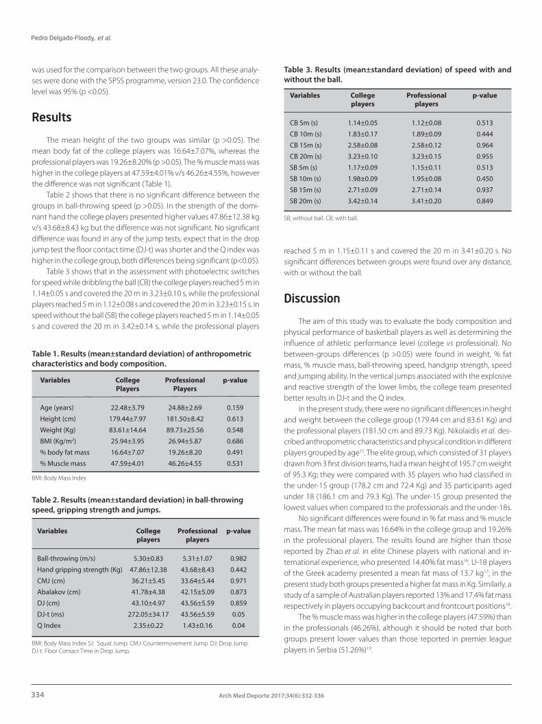
Pedro Delgado-Floody, et al.
334 Arch Med Deporte 2017;34(6):332-336
was used for the comparison between the two groups. All these analy-ses were done with the SPSS programme, version 23.0. The confidence level was 95% (p <0.05).
Results
The mean height of the two groups was similar (p >0.05). The mean body fat of the college players was 16.64±7.07%, whereas the professional players was 19.26±8.20% (p >0.05). The % muscle mass was higher in the college players at 47.59±4.01% v/s 46.26±4.55%, however the difference was not significant (Table 1).
Table 2 shows that there is no significant difference between the groups in ball-throwing speed (p >0.05). In the strength of the domi-nant hand the college players presented higher values 47.86±12.38 kg v/s 43.68±8.43 kg but the difference was not significant. No significant difference was found in any of the jump tests, expect that in the drop jump test the floor contact time (DJ-t) was shorter and the Q index was higher in the college group, both differences being significant (p<0.05).
Table 3 shows that in the assessment with photoelectric switches for speed while dribbling the ball (CB) the college players reached 5 m in 1.14±0.05 s and covered the 20 m in 3.23±0.10 s, while the professional players reached 5 m in 1.12±0.08 s and covered the 20 m in 3.23±0.15 s. In speed without the ball (SB) the college players reached 5 m in 1.14±0.05 s and covered the 20 m in 3.42±0.14 s, while the professional players
reached 5 m in 1.15±0.11 s and covered the 20 m in 3.41±0.20 s. No significant differences between groups were found over any distance, with or without the ball.
Discussion
The aim of this study was to evaluate the body composition and physical performance of basketball players as well as determining the influence of athletic performance level (college vs professional). No between-groups differences (p >0.05) were found in weight, % fat mass, % muscle mass, ball-throwing speed, handgrip strength, speed and jumping ability. In the vertical jumps associated with the explosive and reactive strength of the lower limbs, the college team presented better results in DJ-t and the Q index.
In the present study, there were no significant differences in height and weight between the college group (179.44 cm and 83.61 Kg) and the professional players (181.50 cm and 89.73 Kg). Nikolaidis et al. des-cribed anthropometric characteristics and physical condition in different players grouped by age15. The elite group, which consisted of 31 players drawn from 3 first division teams, had a mean height of 195.7 cm weight of 95.3 Kg; they were compared with 35 players who had classified in the under-15 group (178.2 cm and 72.4 Kg) and 35 participants aged under 18 (186.1 cm and 79.3 Kg). The under-15 group presented the lowest values when compared to the professionals and the under-18s.
No significant differences were found in % fat mass and % muscle mass. The mean fat mass was 16.64% in the college group and 19.26% in the professional players. The results found are higher than those reported by Zhao et al. in elite Chinese players with national and in-ternational experience, who presented 14.40% fat mass16. U-18 players of the Greek academy presented a mean fat mass of 13.7 kg17; in the present study both groups presented a higher fat mass in Kg. Similarly, a study of a sample of Australian players reported 13% and 17.4% fat mass respectively in players occupying backcourt and frontcourt positions18.
The % muscle mass was higher in the college players (47.59%) than in the professionals (46.26%), although it should be noted that both groups present lower values than those reported in premier league players in Serbia (51.26%)19.
Table 1. Results (mean±standard deviation) of anthropometric characteristics and body composition.
Variables College Professional p-value Players Players
Age (years) 22.48±3.79 24.88±2.69 0.159
Height (cm) 179.44±7.97 181.50±8.42 0.613
Weight (Kg) 83.61±14.64 89.73±25.56 0.548
BMI (Kg/m2) 25.94±3.95 26.94±5.87 0.686
% body fat mass 16.64±7.07 19.26±8.20 0.491
% Muscle mass 47.59±4.01 46.26±4.55 0.531
BMI: Body Mass Index
Table 2. Results (mean±standard deviation) in ball-throwing speed, gripping strength and jumps.
Variables College Professional p-value players players
Ball-throwing (m/s) 5.30±0.83 5.31±1.07 0.982
Hand gripping strength (Kg) 47.86±12.38 43.68±8.43 0.442
CMJ (cm) 36.21±5.45 33.64±5.44 0.971
Abalakov (cm) 41.78±4.38 42.15±5.09 0.873
DJ (cm) 43.10±4.97 43.56±5.59 0.859
DJ-t (ms) 272.05±34.17 43.56±5.59 0.05
Q Index 2.35±0.22 1.43±0.16 0.04
BMI: Body Mass Index SJ: Squat Jump. CMJ: Countermovement Jump. DJ: Drop Jump. DJ-t: Floor Contact Time in Drop Jump.
Table 3. Results (mean±standard deviation) of speed with and without the ball.
Variables College Professional p-value players players
CB 5m (s) 1.14±0.05 1.12±0.08 0.513
CB 10m (s) 1.83±0.17 1.89±0.09 0.444
CB 15m (s) 2.58±0.08 2.58±0.12 0.964
CB 20m (s) 3.23±0.10 3.23±0.15 0.955
SB 5m (s) 1.17±0.09 1.15±0.11 0.513
SB 10m (s) 1.98±0.09 1.95±0.08 0.450
SB 15m (s) 2.71±0.09 2.71±0.14 0.937
SB 20m (s) 3.42±0.14 3.41±0.20 0.849
SB; without ball. CB; with ball.

Comparison of body composition and physical performance between college and professional basketball players
335Arch Med Deporte 2017;34(6):332-336
As for the sprint performance there are no significant differences in any of the measurements of the two groups. These results can be compared to those of previous studies, since they were obtained using very reliable equipment, validated for this type of assessment20,21. In particular there is a previous study which compared the speed over 20 metres of team-sport players, showing that there were no significant differences in the test: 16 basketball players presented a performance of 3.14 s while a group of 20 handball players recorded 3.13 s over the same distance; this absence of a significant difference agrees with the results of the present study22.
However, Köklü reported significant differences in maximum accele-ration races when comparing basketball players of different competitive levels; there was even an inverse relation between the speed performan-ce and the competitive level, i.e. players at a lower competitive level may be significantly faster than those competing at the highest level5. Other studies by contrast have shown evidence that elite basketball players in different categories, when compared with lower level players, present an association between their high performance and their physiological, morphological and maturity profiles23,24.Thus studies exist with both positive and negative associations for the correlation between speed performance and competitive level in basketball players.
No significant differences were found in the variables SJ and CMJ when the two groups in the present research were compared. The mean result in the SJ was 33.54 cm in the college group and 34.64 cm in the professionals. These performance results are inferior compared to a young team of 18 players who took part in the Italian national championships and had six years' experience of basketball training; their SJ result was 39.3 cm25. They are also lower than those reported by Callejas et al in elite Spanish (47 cm) and Japanese players (44.6 cm)26.
The college group achieved a mean of 36.21 cm in the CMJ, and the professionals 36.36 cm (no statistical significance). These values are lower than reported in a study which compared an elite team with three years' experience at national or international level (56.6 cm) with a college second team (51.6 cm)27. Struzik et al. propose that the CMJ may be a good measure for determining the jumping capacity of basketball players in jump shots28. Although the results of the present investiga-tion are markedly lower than those previously reported27, the specific nature of training may in the long term alter players' performance in assessments of their strength and power-producing capacity29.
In the assessment of vertical jumps associated with the explosive and reactive strength of the lower limbs, significant differences were found in the DJ-t, which was longer in the professionals, and the Q index, which was higher in the college group. In both cases the college group obtained better results, while there were no differences in the DJ results. To date there have been few studies which focus on these variables, however earlier research has show that in team sports they have a significant correlation with speed and maximum acceleration capacity30, as well as degree of neuromuscular fatigue31 and fitness for sport32. Díaz et al. reported that in an analysis of the elastic component and the technical component, there were no significant differences between Spanish players classified as professionals and those classified as college. These results differ from the findings of the present study33.
In the application of the test to assess the action of the arms when executing a vertical jump with countermovement (Abalakov), the groups
presented no statistically significant differences; the same finding is reported in the study by Massuca & Fragoso, who separated a group of team sports athletes into 2 sub-groups (successful v/s less successful) and found no significant differences34. The vertical jump is prevalent in various technical actions in basketball, such as shooting at the basket and defensive or offensive bouncing; it should therefore be considered an important aspect to develop in the two groups in this study.
Proprioception and motor control influence the mechanics and efficiency of the shot35, in our study there were no significant differences in the velocity of the Ball-throwing, this may be associated with the level of performance of the players.
The limitations of the study were not to present a larger number of players by their tactical position in the game, so as to complement the study with comparisons by specific position, considering the functions of each team member. In future research it is proposed increase the sample size, incorporating a wider age range and generating a broader profile of the Chilean basketball player for use by trainers, physiologists and physical trainers of the professional and university leagues.
In conclusion, the results of this study indicate that the college and professional players don´t show significative differences in the body composition and variables associated with physical performance. Consequently, it can be inferred that the difference of competitive level between these players is determined by technical variables, as well as by tactical aspects associated to the understanding of game. The results from present investigation can be useful for basketball coach like so professionals and researchers associate to sport sciences and related fields. As a prospective, it seems important to carry out more studies that consider these variables in new contexts, as well as to de-velop research of this type that also incorporates the analysis of tactical aspects of the players.
Bibliography 1. Moreira A, Nosaka K, Nunes JA, Viveiros L, Jamurtas AZ, Aoki MS. Changes in muscle
damage markers in female basketball players. Biol Sport. 2014;31(1):3-7.
2. te Wierike SC, de Jong MC, Tromp EJ, Vuijk PJ, Lemmink KA, Malina RM, et al. Develo-pment of repeated sprint ability in talented youth basketball players. J Strength Cond Res. 2014;28(4):928-34.
3. Alemdaroglu U. The relationship between muscle strength, anaerobic performance, agility, sprint ability and vertical jump performance in professional basketball players. J Hum Kinet. 2012;31:149-58.
4. de Araujo GG, de Barros Manchado-Gobatto F, Papoti M, Camargo BH, Gobatto CA. Anaerobic and aerobic performances in elite basketball players. J Hum Kinet. 2014;42:137-47.
5. Koklu Y, Alemdaroglu U, Kocak FU, Erol AE, Findikoglu G. Comparison of chosen physical fitness characteristics of Turkish professional basketball players by division and playing position. J Hum Kinet. 2011;30:99-106.
6. Ziv G, Lidor R. Physical attributes, physiological characteristics, on-court performan-ces and nutritional strategies of female and male basketball players. Sports Med. 2009;39(7):547-68.
7. Stojanovic MD, Ostojic SM, Calleja-Gonzalez J, Milosevic Z, Mikic M. Correlation between explosive strength, aerobic power and repeated sprint ability in elite basketball players. J Sports Med Phys Fitness. 2012;52(4):375-81.
8. Sands WA, McNeal JR, Ochi MT, Urbanek TL, Jemni M, Stone MH. Comparison of the Wingate and Bosco anaerobic tests. J Strength Cond Res. 2004;18(4):810-5.
9. Alejandro V, Santiago S, Gerardo VJ, Carlos MJ, Vicente GT. Anthropometric Characte-ristics of Spanish Professional Basketball Players. J Hum Kinet. 2015;46:99-106.
10. El-Kiki AA, Ibrahim MM. The structural factor of the body components of male high level basketball players as a selection limitans. J Am Sci. 2015;11(5):97-104.

Pedro Delgado-Floody, et al.
336 Arch Med Deporte 2017;34(6):332-336
11. Pizzigalli L, Micheletti Cremasco M, A LAT, Rainoldi A, Benis R. Hand grip strength and anthropometric characteristics in Italian female national basketball teams. J Sports Med Phys Fitness. 2017;57(5):521-8.
12. Fallahi AA, Jadidian AA. The Effect of Hand Dimensions, Hand Shape and Some Anthropometric Characteristics on Handgrip Strength in Male Grip Athletes and Non-Athletes. J Hum Kinet. 2011;29:151-9.
13. Gerodimos V. Reliability of handgrip strength test in basketball players. J Hum Kinet. 2012;31:25-36.
14. Cleveland JD, Patterson J. Assessment of Vertical Leap Using a Vertec and Axon Jump Mat System. Med Sci Sports Exerc. 2010;42(5):370.
15. Nikolaidis P, Calleja-González J, Padulo J. The effect of age on positional differences in anthropometry, body composition, physique and anaerobic power of elite basketball players. Sport Sci Health. 2014;10(3):225-33.
16. Zhao J, Fan B, Wu Z, Xu M, Luo Y. Serum zinc is associated with plasma leptin and Cu-Zn SOD in elite male basketball athletes. J Trace Elem Med Biol. 2015;30:49-53.
17. Nikolaidis PT, Asadi A, Santos EJ, Calleja-Gonzalez J, Padulo J, Chtourou H, et al. Re-lationship of body mass status with running and jumping performances in young basketball players. Muscles Ligaments Tendons J. 2015;5(3):187-94.
18. Scanlan AT, Tucker PS, Dalbo VJ. A comparison of linear speed, closed-skill agility, and open-skill agility qualities between backcourt and frontcourt adult semiprofessional male basketball players. J Strength Cond Res. 2014;28(5):1319-27.
19. Muratović A, Vujovic D, Hadzic R. Comparative Study of Anthropometric Measure-ment and Body Composition between Elite Handball and Basketball Players. MJSSM. 2014;3(2):19-22.
20. Simperingham KD, Cronin JB, Ross A. Advances in Sprint Acceleration Profiling for Field-Based Team-Sport Athletes: Utility, Reliability, Validity and Limitations. Sports Med. 2016;46(11):1619-45.
21. Galy O, Zongo P, Chamari K, Chaouachi A, Michalak E, Dellal A, et al. Anthropometric and physiological characteristics of Melanesian futsal players: a first approach to talent identification in Oceania. Biol Sport. 2015;32(2):135-41.
22. Gardasević B, Jakovljević S, Pajić Z, Preljević A. Some anthropometric and power characteristics of elite junior handball and basketball players. APES. 2011;1: 5-9.
23. Boone J, Bourgois J. Morphological and physiological profile of elite basketball players in Belgian. Int J Sports Physiol Perform. 2013;8(6):630-8.
24. Torres-Unda J, Zarrazquin I, Gil J, Ruiz F, Irazusta A, Kortajarena M, et al. Anthropometric, physiological and maturational characteristics in selected elite and non-elite male adolescent basketball players. J Sports Sci. 2013;31(2):196-203.
25. Attene G, Laffaye G, Chaouachi A, Pizzolato F, Migliaccio GM, Padulo J. Repeated sprint ability in young basketball players: one vs. two changes of direction (Part 2). J Sports Sci. 2015;33(15):1553-63.
26. Calleja-González J, Jukic I, Ostosic SM, Milanovic L, Zubillaga A, Terrados N. Perfil condicional en jugadores de élite internacionales de baloncesto. Diferencias entre croatas y japoneses. Arch Med Deporte. 2010:181-90.
27. Delextrat A, Cohen D. Physiological testing of basketball players: toward a standard evaluation of anaerobic fitness. J Strength Cond Res. 2008;22(4):1066-72.
28. Struzik A, Pietraszewski B, Zawadzki J. Biomechanical analysis of the jump shot in basketball. J Hum Kinet. 2014;42:73-9.
29. Panoutsakopoulos V, Papachatzis N, Kollias IA. Sport specificity background affects the principal component structure of vertical squat jump performance of young adult female athletes. J Sport Health Sci. 2014;3(3):239-47.
30. Lockie RG, Murphy AJ, Knight TJ, Janse de Jonge XA. Factors that differentiate accele-ration ability in field sport athletes. J Strength Cond Res. 2011;25(10):2704-14.
31. Hamilton D. Drop jumps as an indicator of neuromuscular fatigue and recovery in elite youth soccer athletes following tournament match play. J. Aust. Strength Cond.. 2009;17(4):3-8.
32. Suchomel TJ, Nimphius S, Stone MH. The Importance of Muscular Strength in Athletic Performance. Sports Med. 2016;46(10):1419-49.
33. Díaz Hellín M, del Campo VL, Gómez Navarrete JS, Gómez-Valades JM, Barbado Murillo D, Sabido Solana R. Diferencias en tests isométricos de fuerza y tests de salto entre jugadores de baloncesto profesionales y amateurs. CCD. 2014;8:155-62.
34. Massuça L, Fragoso I. A multidisciplinary approach of success in team-handball. Apunts Med Esport. 2013;48(180):143-51.
35. Sevrez V, Bourdin C. On the Role of Proprioception in Making Free Throws in Basketball. Research quarterly for exercise and sport. 2015;86(3):274-80.
Comparison of body composition and physical performance between college and professional basketball players
337Arch Med Deporte 2017;34(6):332-336
· Gestión y Plani�cación de Servicios Sanitarios
· Gestión Integral del Riesgo Cardiovascular
· Ingeniería Biomédica
· Investigación en Ciencias Sociosanitarias
· Investigación en Educación Física y Salud
· Neuro-Rehabilitación
· Nutrición Clínica
· Nutrición y Seguridad Alimentaria
· Nutrición en la Actividad Física y Deporte
· Osteopatía y Terapia Manual
· Patología Molecular Humana
· Psicología General Sanitaria
· Actividad Física Terapéutica
· Alto Rendimiento Deportivo:
Fuerza y Acondicionamiento Físico
· Performance Sport:
Strength and Conditioning
· Audiología
· Balneoterapia e Hidroterapia
· Desarrollos Avanzados
de Oncología Personalizada Multidisciplinar
· Enfermería de Salud Laboral
· Enfermería de Urgencias,
Emergencias y Cuidados Especiales
· Fisioterapia en el Deporte
· Geriatría y Gerontología:
Atención a la dependencia
POSTGRADOS OFICIALES: SALUD Y DEPORTE
UniversitarioUniversitario
(1) Presencial (2) Semipresencial
(2)
(2)
(2)
(2)
(2)
(2)
(2)
(2)
(2)
(2)
(2)
(2)
(1)
(1)
(1)
(1)
(1)
(1)
(1)
(1)
(1)
(1)
Miguel Ángel LópezCampeón del Mundo en 20 km. marcha (Pekín, 2015)Estudiante y deportista de la UCAM
Artículo original
338 Arch Med Deporte 2017;34(6):338-344
Review
Summary
Climbing to the highest height on Earth, the Mt. Everest (8.848 m), without supplementary oxygen equipment involves a physiological demand that is close to the maximum human tolerance. Exposures at extreme altitudes drastically conditions lung function, stores of oxygen and physical performance. This review brings interesting aspects about respiration, blood gases and aerobic exercise reported by those scientific projects that have carried out physiological measurements between 8,000 m and 8,848 m above sea level, under real or simulated altitude: the Operations “Everest I” (1946), “Everest II” (1985), “Everest III-COMEX” (1997), and the Expeditions “AMREE” (1981), “British 40th Anniversary Everest” (1993), and “Caudwell Xtrem Everest” (2007). These fascinating scientific research events, along with other outstanding biomedical expeditions perfor-med above 5,500 m, like especially the “Silver Hut” (1960-61), “Italiana all’Everest” (1973), and “British Everest Medical” (1994), including those pioneer scientific reports made on the XIX century until the most recent research projects performed, have laid the foundations and knowledge on the human tolerance to such extreme levels of hypobaric hypoxia, where the lung, breathing and respiratory chain takes on a major role requiring fine physiological adjustments to ensure cellular oxygenation. Geophysical aspects, climatic factors and other environmental conditions that limit the biological viability and can affect the respiratory health of climbers on the upper troposphere zone at the subtropical latitude where that mountain is located are likewise reviewed and analyzed. Every year, hundreds of climbers try to reach the top of Mt. Everest, but only a few of them achieved their goal without inhaling supplemental oxygen, including some exceptionally gifted Sherpa natives, protagonist on unsuspected exploits in the highest mountain on terrestrial surface, whose summit touch the physiological limit of survival for the human being.
Key words: Altitude. Oxygen uptake.
Hypoxia. Mountaineering. Atmospheric pressure.
Respiration.
Resumen
Escalar el punto más alto de la Tierra, el Mt. Everest (8.848 m), sin equipos de oxígeno conlleva una demanda fisiológica que está próxima a la máxima capacidad de tolerancia humana. Exponerse a altitudes extremas condiciona drásticamente la función pulmonar, el nivel de oxígeno y el rendimiento físico. Esta revisión reúne interesantes aspectos respiratorios, de gases sanguíneos y ejercicio aeróbico aportados por aquellos proyectos científicos que han llevado a cabo mediciones fisiológicas entre 8.000 m y 8.848 m, en altitud real o simulada, como las Operaciones “Everest I” (1946), “Everest II” (1985) y “Everest III-COMEX” (1997), y las Expediciones “AMREE” (1981), “British 40th Anniversary Everest” (1993) y “Caudwell Xtrem Everest” (2007). Estos fascinantes eventos de investigación, junto a otros destacados proyectos biomédicos realizados a más de 5.500 m, muy especialmente las Expediciones “Silver Hut” (1960-61), “Italiana all’Everest” (1973) y “British Everest Medical” (1994), incluyendo aquellas pioneras observaciones científicas llevadas a cabo en el s.XIX hasta los más recientes proyectos de investigación realizados, han sentado las bases del conocimiento sobre la tolerancia humana ante niveles de hipoxia hipobárica extrema, donde el pulmón y la cadena respiratoria adquieren un trascendente protagonismo requiriéndose de finos ajustes fisiológicos que garanticen la oxigenación celular. Asimismo, se exponen ciertos aspectos geofísicos, factores climáticos y otros condicionantes ambientales que limitan la viabilidad biológica y pueden afectar la salud respiratoria de los alpinistas situados en las cotas superiores de la troposfera a la latitud subtropical donde se encuentra ubicada dicha montaña. Actualmente cientos de alpinistas intentan alcanzar la cumbre del Mt. Everest todos los años, pero solo algunos consiguen su objetivo sin inhalar oxígeno suplementario, entre ellos algunos excepcionalmente dotados nativos Sherpa, protagonistas de insospechadas hazañas en la montaña más elevada de la superficie terrestre, cuya cima roza el límite fisiológico de supervivencia para el ser humano.
Palabras clave:Altitud. Consumo de oxígeno. Hipoxia. Montañismo. Presión
atmosférica. Respiración.
Received: 03.03.2017Accepted: 31.05.2017
Breathing at extreme altitudes. Scientific projects “EVEREST” (Second part)
Eduardo Garrido1,2, Oriol Sibila3, Ginés Viscor2,4
1Servicio de Hipobaria y Fisiología Biomédica. Universidad de Barcelona. 2Instituto de Estudios de Medicina de Montaña. Barcelona. 3Servicio de Neumología. Hospital de la Santa Creu i Sant Pau. Barcelona. 4Departamento de Biología Celular, Fisiología e Inmunología. Universidad de Barcelona.
Respirar en altitudes extremas. Proyectos científicos “EVEREST” (Segunda parte)
Correspondence: Eduardo Garrido E-mail: [email protected]
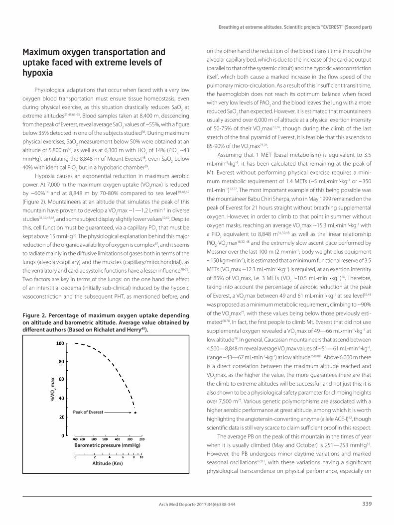
Breathing at extreme altitudes. Scientific projects “EVEREST” (Second part)
339Arch Med Deporte 2017;34(6):338-344
Maximum oxygen transportation and uptake faced with extreme levels of hypoxia
Physiological adaptations that occur when faced with a very low oxygen blood transportation must ensure tissue homeostasis, even during physical exercise, as this situation drastically reduces SaO2 at extreme altitudes31,48,63-65. Blood samples taken at 8,400 m, descending from the peak of Everest, reveal average SaO2 values of ~55%, with a figure below 35% detected in one of the subjects studied36. During maximum physical exercises, SaO2 measurement below 50% were obtained at an altitude of 5,800 m66, as well as at 6,300 m with FiO2 of 14% (PiO2 ~43 mmHg), simulating the 8,848 m of Mount Everest48, even SaO2 below 40% with identical PiO2 but in a hypobaric chamber39.
Hypoxia causes an exponential reduction in maximum aerobic power. At 7,000 m the maximum oxygen uptake (VO2max) is reduced by ~60%14 and at 8,848 m by 70-80% compared to sea level39,48,67 (Figure 2). Mountaineers at an altitude that simulates the peak of this mountain have proven to develop a VO2max ~1—1,2 L•min-1 in diverse studies31,39,48,68, and some subject display slightly lower values39,69. Despite this, cell function must be guaranteed, via a capillary PO2 that must be kept above 15 mmHg35. The physiological explanation behind this major reduction of the organic availability of oxygen is complex67, and it seems to radiate mainly in the diffusive limitations of gases both in terms of the lungs (alveolar/capillary) and the muscles (capillary/mitochondrial), as the ventilatory and cardiac systolic functions have a lesser influence70-72. Two factors are key in terms of the lungs: on the one hand the effect of an interstitial oedema (initially sub-clinical) induced by the hypoxic vasoconstriction and the subsequent PHT, as mentioned before, and
on the other hand the reduction of the blood transit time through the alveolar capillary bed, which is due to the increase of the cardiac output (parallel to that of the systemic circuit) and the hypoxic vasoconstriction itself, which both cause a marked increase in the flow speed of the pulmonary micro-circulation. As a result of this insufficient transit time, the haemoglobin does not reach its optimum balance when faced with very low levels of PAO2 and the blood leaves the lung with a more reduced SaO2 than expected. However, it is estimated that mountaineers usually ascend over 6,000 m of altitude at a physical exertion intensity of 50-75% of their VO2max73,74, though during the climb of the last stretch of the final pyramid of Everest, it is feasible that this ascends to 85-90% of the VO2max75,76.
Assuming that 1 MET (basal metabolism) is equivalent to 3.5 mL•min-1•kg-1, it has been calculated that remaining at the peak of Mt. Everest without performing physical exercise requires a mini-mum metabolic requirement of 1.4 METs (~5 mL•min-1•kg-1 or ~350 mL•min-1)32,77. The most important example of this being possible was the mountaineer Babu Chiri Sherpa, who in May 1999 remained on the peak of Everest for 21 hours straight without breathing supplemental oxygen. However, in order to climb to that point in summer without oxygen masks, reaching an average VO2max ~15.3 mL•min-1•kg-1 with a PiO2 equivalent to 8,848 m31,39,48 as well as the linear relationship PiO2-VO2max18,32, 48 and the extremely slow ascent pace performed by Messner over the last 100 m (2 m•min-1; body weight plus equipment ~150 kgm•min-1), it is estimated that a minimum functional reserve of 3.5 METs (VO2max ~12.3 mL•min-1•kg-1) is required, at an exertion intensity of 85% of VO2max, i.e. 3 METs (VO2 ~10.5 mL•min-1•kg-1)76. Therefore, taking into account the percentage of aerobic reduction at the peak of Everest, a VO2max between 49 and 61 mL•min-1•kg-1 at sea level39,48 was proposed as a minimum metabolic requirement, climbing to ~90% of the VO2max75, with these values being below those previously esti-mated68,78. In fact, the first people to climb Mt. Everest that did not use supplemental oxygen revealed a VO2max of 49—66 mL•min-1•kg-1 at low altitude79. In general, Caucasian mountaineers that ascend between 4,500—8,848 m reveal average VO2max values of ~51—61 mL•min-1•kg-1, (range ~43—67 mL•min-1•kg-1) at low altitude75,80,81. Above 6,000 m there is a direct correlation between the maximum altitude reached and VO2max, as the higher the value, the more guarantees there are that the climb to extreme altitudes will be successful, and not just this; it is also shown to be a physiological safety parameter for climbing heights over 7,500 m75. Various genetic polymorphisms are associated with a higher aerobic performance at great altitude, among which it is worth highlighting the angiotensin-converting enzyme (allele ACE-I)82, though scientific data is still very scarce to claim sufficient proof in this respect.
The average PB on the peak of this mountain in the times of year when it is usually climbed (May and October) is 251—253 mmHg33. However, the PB undergoes minor daytime variations and marked seasonal oscillations62,83, with these variations having a significant physiological transcendence on physical performance, especially on
Figure 2. Percentage of maximum oxygen uptake depending on altitude and barometric altitude. Average value obtained by different authors (Based on Richalet and Herry45).
%VO
2 max
Peak of Everest
Barometric pressure (mmHg)
Altitude (Km)

Eduardo Garrido, et al.
340 Arch Med Deporte 2017;34(6):338-344
VO2max33,34,77. Therefore, the success rate for climbing Everest without supplementary oxygen will depend largely on the PB prevailing on the day the peak is climbed. As Figure 3 shows, the PB is at its highest during the months of July and August (~255 mmHg), therefore, the VO2max will be higher and the mountain ascent will be more physiologically accessible; the PB is at its lowest in January and February (~243 mmHg) and this is when the greatest reductions of VO2max occur, approximately 10—12% compared to summer levels33,62,84. The current record of the number of ascents to Mount Everest is held by three Sherpas: Appa, Phurba Tashi and Kami Rita, with 21 ascents each. However, the Sherpa Ang Rita — known as the “snow leopard” because of his exceptional physical performance at extreme altitudes — is the only human being to ever scale this mountain on 10 separate occasions without inhaling supplemental oxygen, and is the only person ever to have performed this ascent in this way in winter. A few years after achieving this feat, performed on 22nd December 1987, we detected that his VO2max was 66.7 mL•min-1•kg-1 at sea level85, a value that is significantly higher than that measured in other internationally renowned Sherpas86. It is known that a higher VO2max at sea level entails a greater VO2max at great altitude39, a fact that enabled Ang Rita to scale the peak of Everest in winter without oxygen equipment, despite the drastic reduction of ~80% which his VO2max presumably experienced at the altitude of 8,848 m. A VO2max ~13.3 mL•min-1•kg-1 would align with the minimum value needed of 3.9 METs, which has been estimated to complete the ascent under these conditions76. On the day that Ang Rita achieved this winter climb, the PB was 247 mmHg at the same altitude and latitude of Mount Everest, i.e. 9 mmHg less than during the ascent performed by Messner on 20th August 1980, but 4 mmHg higher than the levels
registered on the peak mid winter, a fact that was decisive in the success of the sporting achievement accomplished by this sherpa84.
Despite minimum PB changes entailing very significant variations of VO2max at such extreme altitudes, it is worth highlighting that other advantageous factors associated with the chronic hypoxic adaptation shown in people from Tibetan lineage also influence their best physi-cal performance at such high altitudes. These natives present a lower reduction of maximum heart rate compared to levels observed in Caucasians; a larger surface of pulmonary gas exchange and alveolar-capillary diffusion capacity; minimal PHT and right ventricular overload; greater HVR (hypoxic ventilatory response) and a lack of, or lower rate of central apnoea and Cheyne-Stokes-type respiratory rhythms during sleep; less erythrocytosis; and blood viscosity with higher SaO2 values87-96. These adaptive mechanisms found in Tibetans, from whom phylogenetically the Sherpas descended97, are the best anthropologic example of adaptation to altitude, because their permanent exposure to environmental hypoxia originates from the Upper Palaeolithic Era, surpassing the Andean highlander natives by thousands of years89,96. More than ten genes have been described that are involved in their extraordinary adaptive response to great altitude, though a few of them specially related to blood-oxygen transportation, seem to play a particularly important role: allotype EPAS1 (HIF-2α) and allotype PHD2/EGLN1, its negative regulator98-100. A higher frequency of allele ACE-I has also been reported in Sherpas101.
In general, aside from climatic aspects, three physiological me-chanisms are particularly decisive in being able to climb to 8,848 m of altitude without oxygen canisters: the extreme hyperventilation generated reduces the PACO2 stabilising the PAO2, acute hypocapnic alkalosis facilitates the oxyhaemoglobin saturation in the lungs, and the PB increase, despite being minimal, significantly raises the VO2max48. Despite these extraordinary adaptations, which enable survival at such high altitudes, human beings have limiting physiological factors in terms of physical performance in this environment, despite this being a contested aspect. Within this limitation, PB has a relevant role to play, as well as the energy expenditure of hyperventilation, the gas diffusion capacity through the alveolar-capillary membrane, the peripheral PO2
and the transfer of oxygen to active muscles67,71,102,103. Moreover, the on-going sympathetic stimulus caused by chronic hypoxia inhibits the maximum chronotropic response, a fact that has been fundamentally linked to a progressive desensitisation of the beta-adrenergic myo-cardial receptors104. Recently, particular emphasis has been placed on the sporting success of the two mountaineering pioneers – Messner and Habeler - who reached the peak of Mount Everest in 1978 without supplemental oxygen. Whilst they did not have exceptional VO2max at low altitude76, they would have undergone transcendental physiological changes in the increase of their muscular capillary density, and there-fore, an optimum periphery diffusion of oxygen during that historical ascent105. While VO2max is drastically reduced at extreme altitudes, the anaerobic capacity of the skeletal-muscle paradoxically reveals a similar reduction when faced with chronic exposure to hypoxia106. This is due to the major depletion of plasma bicarbonate upon compensating the respiratory alkalosis, with minimal increments of lactate being detected when faced with intense physical exertions in altitudes over 7,500 m31,102.
Figure 3. Barometric pressure variation at the altitude of Mount Everest (8,848 m) in relation to different times of year and geographi-cal latitudes of the Northern hemisphere (Based on West et al.34,62).
Latitude (degrees North)
Baro
met
ric
pres
sure
at 8
,848
m (m
mH
g)
July
January
Peak of Everest
Breathing at extreme altitudes. Scientific projects “EVEREST” (Second part)
341Arch Med Deporte 2017;34(6):338-344
Geophysical aspects, extreme climatic and environmental factors on aerobic performance and the health of mountaineers
If we take the standard atmosphere model of PB as a reference, at the peak of Everest there would be a value of 236 mmHg, some 17 mmHg less compared to real atmospheric measurements taken in springtime, and this would cause a reduction of the PiO2 from 43 mmHg to 39.5 mmHg33. Because the logPiO2—VO2max relationship is very strong (~63 mL•min-1•mmHg-1), this loss of just 3.5 mmHg would produce a reduction of the VO2max of ~222 mL•min-1, i.e. adding ~21% more reduction to the altitude of 8,848 m, with a viable ascent to the peak of this mountain under these conditions seeming unlikely, as calculated by West et al.33. A PB of 236 mmHg in a real atmosphere is equivalent to an approximate altitude of some ~9,350 m at the latitude where this mountain is located, in other words, half a kilometre higher than Mount Everest. Due to the contraction shown by the troposphere towards the Earth’s polar regions, this PB at this time of year would correspond to Everest being geographically located on a more northerly latitude. Therefore, despite the extreme hypobaric hypoxia that prevails at the peak of Everest, given its subtropical position (28ºN), mountaineers are favoured by the equatorial dilation of the troposphere34,62. As shown in Figure 3, the PB at around 8,850 m during mid-winter in the Artic polar circle (66ºN) reaches ~214 mmHg34, i.e. this mountain would have a simulated elevation of a thousand metres higher.
Bailey calculated that where Mount Everest was located, and accor-ding to the PB that prevailed there at different times of year, an altitude of ~9,970 m could be the theoretical limit where the PiO2 would ensure a VO2max of 3.5 METs in summer, as well as ~9,660 m of 3.9 METs in winter, and at the altitude of ~11,900 m it would equal 1 MET76. A priori, the first and second figures would exceed already proposed estimations, and the third would exceed the experiment by Angelo Mosso, who simulated an altitude of 11,650 m in a hypobaric chamber. He reached a PB of 192 mmHg, equivalent to ~10,800 m of standard altitude107, though taking into account the slightly oxygen-enriched mixture that he inhaled during this trial (FiO2: 29.2%)25, it would have given him a PiO2 corresponding to ~8,850 m, similar to the peak of Everest. It is worth mentioning that during the final phase of Operation Everest I, a simu-lated altitude of ~15,400 m was achieved whilst acclimatised subjects breathed 100% oxygen, with these subjects revealing a certain degree of momentary tolerance to this atmosphere. Non-acclimatised flight pilots do not usually tolerate altitudes of 13,000 m, despite inhaling supplemental oxygen38. In fact, being exposed to altitudes over ~12,000 m, even while resting and breathing maximum concentration oxygen, the SaO2 drops drastically, and does not guarantee the cell viability of the organism, therefore pressurised equipment is needed108.
The Himalayan mountain range rises slowly but constantly, due to the compression between the tectonic plates of India and Eurasia.
Due to this fact, it is intriguing to consider speculations regarding the amount of time that it will be possible to ascend Mount Everest without using oxygen equipment. Considering that the annual elevation rate is ~3 cm, Bailey calculated that it will not be until approximately the year 29,000 and 39,000 - in winter and summer respectively - when Everest will reach an altitude at which the PiO2 will ensure the physiological limit that is compatible with developing a VO2max ~3.5 METs at its peak, and the year ~104,000 when it would equal ~1 MET76. However, it is clear that the precise future behaviour of continental platforms is unknown, as well as the exact characteristics of the troposphere in thousands of year’s time, due to the effect of global warming or climate changes that modify the composition of atmospheric gases. While there are numerous speculations regarding the effect of global warming on the Himalayan region, certain measurements reveal that since 1948 the troposphere has been increasing its pressure on average by ~1,8 mmHg each decade, therefore, in keeping with this trend it could be possible that progressively developing a greater VO2max on the peak of Mount Everest may be feasible, and climbing it may become more accessible aerobically in the future. However, within this environmental context, mountaineers will have to face new technical difficulties caused by the thermal increase, such as the risk of potential avalanches and rock-climbing sections caused by the disappearance of the ice on a large part of the routes109.
The wind that strikes the edges and walls of this great geological formation could reduce the PB due to the Venturi effect, though calculations using the Bernoulli equation reveal that local PB changes would be minimal, below 1 mmHg, for which it would have a very minor physiological impact on the VO2max34. This is particularly true before and after monsoons, when there is greater meteorological stability, and winds do not usually exceed 55 km•h-1, a time that is chosen to attempt the ascent with the highest possible chance of success and safety83. However, during these brief anti-cyclonical windows of opportunity occurring in May and October, sudden gales occur, and the high layer of the troposphere can even be fed with great masses of subtropical water vapour which interact with the stratospheric jet stream current, causing convective vortexes with strong hurricane-force storms over Everest110. During winter, this jet-stream current moves towards southern Central Asia, completely covering Himalayan mountain range, and cyclonic winds with speeds reaching around 300 km•h-1 can hit the peaks of over 8,000 m of altitude. Days with strong gales are avoided by mountaineers given the high risk of accidents that these conditions entail, as well as the chill factor associated with low temperatures that the wind has on the body. On the peak of Mount Everest, temperature of minus 60ºC can be reached when masses of Siberian polar air penetrate the Tibetan plateau, though during the climatic windows in May and October, the average wind temperature is usually around ~ minus 26˚C111, though the chill factor can give values below minus 50˚C83, with warmer temperatures more usual throughout the day during these periods34. The atmospheric factors largely explain why only 25% of mountaineers that attempt to climb Everest in spring or summer actually manage to reach the peak, and why this success rate drops to just 4% of those that attempt it in winter112, though the first percentage seems to have risen in recent years.

Eduardo Garrido, et al.
342 Arch Med Deporte 2017;34(6):338-344
Breathing low-temperature air increases the risk of suffering from a pulmonary pathology, and at extreme altitudes upper respiratory tract problems are frequent, which improve upon descent113,114. The low relative humidity in the high layers of the troposphere cause a large loss of water vapour through the lungs, exceeding 60—80 mL•h-1 during moderate-intensity physical exercise62. The nasal inspiratory flow increases with altitude, but at 8,000 m it is lower than expected in accordance with the prevailing low air density115. Episodes of bronchospasm are infrequent at high altitude, especially among well-controlled asthma suffers116, as atmospheric pollution reduces in high mountain regions and this environment has been shown to be clearly beneficial to these pacients117; even exposures at extreme altitude appear to be safe in cases of moderate-intensity asthma sufferers118. With the exception of bronchospasm episodes that are not directly related to pollen allergens (breathing cold, dry air, vigorous physical activity), it has not been proven that bronchial spasm episodes are favoured, specifically, by the hypoxia and hypocapnia of altitude, given that the oxygen supplement does not seem to increase ventilatory flows at extreme altitudes59. The stratospheric ozone, whose existence has been detected at a level very close to Mount Everest, represents a potential added health-risk for mountaineers, as it causes the inflammation of the mucous membranes of the airways, bronchoconstriction, coughing and/or dyspnoea, deteriorating pulmonary function62,119,120. The upper respiratory tracts re-heat low-temperature air inhaled quickly, for which it reaches optimum thermal graduation at the level of the alveolar, clearly not constituting a reason to support high-altitude pulmonary oedema HAPE, secondary non-cardiogenic oedema to PHT, already relatively frequent at heights below 5,000 m72,121-125. It is difficult to estimate the HAPE rate at altitudes over 8,000 m, but the presence of interstitial plasma cause by subclinical HAPE would be frequent, which would alter the gas exchange69,126, as observed in athletes at much lower altitude127-131. Even with no oedema, the low gradient alveolar-capillary oxygen pressure (because of the reduction of PB and excessive pulmonary blood transit) increases the degree of hypoxemia especially during physical activity131, an exercise capacity that is already limited by the PHT132.
Aside from the hypoxia caused by a HAPE, just breathing in an oxygen-deprived atmosphere causes deterioration to the central nervous system and the development of cerebral oedemas that could cause an accident on the mountain, or cause the mountaineer to lose consciousness10. Neuropsychological alterations are frequent133,134, some are transitory and are associated with a focal reduction of cerebral blood flow caused by extreme hypocapnia135. Paradoxically, subjects with a higher HVR - therefore a greater PaO2 and better adaption to hypoxia – suffer from more residual cognitive dysfunctions, possibly due to the reduction of blood flow caused by very low levels of PaCO2
136. The possibility of permanent encephalic hypoxic damage was suggested in Himalayan mountaineers137 and although the exact mechanism of brain damage in this demographic has not been well clarified138-141, the fact that Sherpas appear to have certain neurological protection against extreme altitudes is very suggestive142.
Conclusions
Since the pioneering scientific experiments from the 19th century, which used hypobaric chambers to attempt to establish if a “hypoxia” 26 or “acapnia” 25 respiratory situation was the result of certain limitations and pathological disorders that appear in high mountain areas, nume-rous later studies, especially the fascinating projects carried out in the Mount Everest region or simulating its altitude have established the basic concepts on the respiratory response mechanisms in humans exposed to this extreme environmental setting. Pulmonary adaptation to severe levels of hypoxia, especially during physical exercise, is astonishing, yet the diffusion capacity of gas exchange at an alveolar-capillary level seems to play a determining role as a limiting factor of oxygen availa-bility in the body. However, evidence reveals that pulmonary respiration can guarantee cell respiration, even faced with such levels of reduced partial pressure of oxygen, and on the peak of Mount Everest these levels are very close to the limit of human physiological tolerance. The subtropical geographic latitude upon which the Himalayas are found enable an ascent up to the highest point without inhaling supple-mental oxygen, due to the geoid effect or equatorial convexity of the troposphere, a fact labelled “cosmic coincidence” 143 by the prestigious physiologist John West. Even so, three physiological mechanisms will be particularly decisive in managing to reach the 8,848 m of altitude in this way: extreme hyperventilation, acute respiratory alkalosis, and favourable atmospheric pressure; factors that facilitate and increase the saturation, transportation and oxygen uptake during the day that the climb is attempted.
Some respiratory pathologies, such as asthma episodes or HAPE, must always be contemplated when at great altitudes, as they may evolve seriously, and they may be difficult to control in such a hostile environment, potentially endangering lives. Breathing hypoxic air fre-quently entails the appearance of neuropsychological disorders, with these being one of the main causes behind the accident rate at extreme altitudes. Despite the risks involved in attempting to scale the colossal pyramid of rock and ice that forms Mount Everest, its inhospitable peak is a coveted goal year after year for hundreds of mountaineers from all over the world. Achieving such a major challenge without any supple-mentary breathing equipment is only possible for very few people, even for natives with thousands of years of adaptation to great altitudes, such as those from the Tibetan lineage, with their exceptional genetics and physiological capacity to endure environmental hypoxia.
The testimony of the mythical mountaineer Reinhold Messner is highly compelling, as he describes the sensations he experienced when climbing the final metres of Mount Everest, becoming, alongside Peter Habeler, the first to climb it without using oxygen equipment: “…I have the feeling I am about to burst apart. As we get higher, it becomes necessary to lie down to recover our breath… Breathing becomes such a strenuous business that we scarcely have strength felt to keep moving forward… I am nothing more than a single, narrow, gasping lung, floating over the mists and the summits”144. Undoubtedly, overcoming the onslaught of the forces of nature inherent to the highest point on Earth represents a first-degree physiological challenge, a dangerous sporting feat that takes place on a colossal backdrop far from the public applause.

Breathing at extreme altitudes. Scientifi c projects “EVEREST” (Second part)
343Arch Med Deporte 2017;34(6):338-344
Acknowledgements
Dr Antoni Ricart (IMMED – Institute of Mountain Medicine Studies), Dr Josep L. Ventura and Dr Casimiro F. Javierre (University of Barcelona) for their support and valuable comments.
Bibliography
63. West JB. Pulmonary gas exchange on Mount Everest. Eur Respir J. 1997;10:1431-2.
64. Richalet JP. Operation Everest III: Comex’97. High Alt Med Biol. 2010;11:121-32.
65. Cerretelli P. Limiting factors to oxygen transport on Mount Everest. J Appl Physiol. 1976;40:658-67.
66. West JB, Lahiri S, Gill MB, Milledge JS, Pugh LGCE, Ward MP. Arterial oxygen saturation during exercise at high altitude. J Appl Physiol. 1962;17:617-21.
67. Ferretti G. On maximal oxygen consumption in hypoxic humans. Experientia. 1990;46:1188-94.
68. Sutton JR, Jones NL, Pugh LGCE. Exercise at altitude. Annu Rev Physiol. 1983;45:427-37.
69. Wagner PD, Sutton JR, Reeves JT, Cymerman A, Groves BM, Malconian MK. Operation Everest II: pulmonary gas exchange during a simulated ascent of Mt. Everest. J Appl Physiol. 1987;63:2348-59.
70. Wagner PD. The physiological basis of reduced VO2max in Operation Everest II. High Alt Med Biol. 2010;11:209-15.
71. Ferretti G. Limiting factors to oxygen transport on Mount Everest 30 years after: a critique of Paolo Cerretelli's contribution to the study of altitude physiology. Eur J Appl Physiol. 2003;90:344-90.
72. Schoene RB, Hultgren HN, Swenson ER. High-altitude pulmonary edema. En: Hornbein TF, Schoene RB. High Altitude: An Exploration of Human Adaptation. Nueva York: Marcel Dekker Inc.; 2001. p.777-814.
73. Pugh LGCE. Muscular exercise on Mt. Everest. J Physiol. 1958;141:233-61.
74. West JB, Schoene RB, Luks AM, Milledge JS. Altitude acclimatization and deterioration. En: West JB, Schoene RB, Luks AM, Milledge JS. High Altitude Medicine and Physiology. Boca Raton: CRC Press; 2013. p.53-64.
75. Richalet JP, Keromes A, Dersch B, Corizzi F, Mehdioui H, Pophillat B, et al. Caractéristiques physiologiques des alpinistes de haute altitude. Sci Sports. 1988;3:89-108.
76. Bailey DM. The last “oxygenless” ascent of Mt Everest. Br J Sports Med. 2001;35:294-6.
77. West JB, Wagner PD. Predicted gas exchange on the summit of Mt. Everest. Respir Physiol. 1980;42:1-16.
78. Buskirk ER. Observations of extraordinary performances in an extreme environment and in a training environment. Limits of human performance. Am Acad Phys Educat Papers. 1985;18:10-8.
79. Oelz O, Howald H, Di Prampero PE, Hoppeler H, Claassen H, Jenni R, et al. Physiological profi le of world-class high-altitude climbers. J Appl Physiol. 1986;60:1734-42.
80. Puthon L, Bouzat P, Rupp T, Robach P, Favre-Juvin A, Verges S. Physiological characte-ristics of elite high-altitude climbers. Scand J Med Sci Sports. 2016;26:1052-9.
81. Steinnacker JM, Liu Y, Böning D, Halder A, Maassen N, Thomas A, et al. Lung diff usion capacity, oxygen uptake, cardiac output and oxygen transport during exercise before and after an Himalayan expedition. Eur J Appl Physiol Occup Physiol. 1996;74:187-93.
82. Hennis PJ, O’Doherty AF, Levett DZH, Grocott MPW, Montgomery HM. Genetic factors associated with exercise performance in atmospheric hypoxia. Sports Med. 2015;45:745-61
83. Moore K, Semple J, Cristofanelli P, Bonasoni P, Stocchi P. Environmental conditions at the South Col of Mount Everest and their impact of hypoxia hypothermia experienced by mountaineers. Extrem Physiol Med. 2012;1:2.
84. West JB. Acclimatization and tolerance to extreme altitude. J Wilderness Med. 1993;4:17-26.
85. Garrido E, Rodas G, Javierre C, Segura R, Estruch A, Ventura JL. Cardiorespiratory res-ponse to exercise in elite Sherpa climbers transferred to sea level. Med Sci Sports Exer. 1997;29:937-42.
86. McIntosh SE, Testa M, Walker J, Wing-Gaia S, McIntosh SN, Litwin SE, et al. Physiological profi le of World-record-holder Sherpas. Wilderness Environ Med. 2011;22:65-71.
87. Groves BM, Droma T, Sutton JR, McCullough RG, McCullough RE, Zhuang J, et al. Mi-nimal hypoxic pulmonary hypertension in normal Tibetans at 3,658 m. J Appl Physiol. 1993;74:312-8.
88. Morpurgo G, Arese P, Bosia A, Pescarmona GP, Luzzana M, Modiano G, et al. Sherpas living permanently at high altitude: A new pattern of adaptation. Proc Nat Acad Sci. 1976;73:743-51.
89. Beall CM. Tibetan and Andean contrast in adaptation to high-altitude hypoxia. Adv Exp Med Biol. 2000;475:63-74.
90. Lahiri S, Milledge JS. Sherpa physiology. Nature. 1965;207:610-2.
91. Lahiri S, Milledge JS, Chattopadhyay HP, Bhattacharyya AK, Sinha AK. Respiration and heart rate of Sherpa highlanders during exercise. J Appl Physiol. 1967;23:545-54.
92. Gilbert-Kawai ET, Milledge JS, Grocott MP, Martin DS. King of the mountains: Tibetan and Sherpa physiological adaptations for life at high altitude. Physiology. 2014;29:388-402.
93. Garrido E, Javierre C, Segura R, Ventura JL. ECG of a record Everest Sherpa climber. High Alt Med Biol. 2003;4:259-60.
94. Rodas G, Javierre C, Garrido E, Segura R, Ventura JL. Normoxic ventilatory response in lowlander and Sherpa elite climbers. Respir Physiol. 1998;113:57-64.
95. Wu T, Li S, Ward MP. Tibetans at extreme altitude. Wilderness Environ Med. 2005;16:47-54.
96. Wu T, Kayser B. High altitude adaptation in Tibetans. High Alt Med Biol. 2006;7:193-208.
97. Bhandari S, Zhang X, Cui C, Yangla, Liu L, Ouzhuluobu, et al. Sherpas share genetic varia-tions with Tibetans for high-altitude adaptation. Mol Genet Genomic Med. 2016;5:76-84.
98. Simonson TS, McClain DA, Jorde LB, Prchal JT. Genetic determinants of Tibetan high-altitude adaptation. Hum Genet. 2012;13:527-33.
99. Lorenzo FR, Huff C, Myllymäki M, Olenchock B, Swierczek S, Tashi T, et al. A genetic mechanism for Tibetan high-altitude adaptation. Nat Genet. 2014;46:951-6.
100. Hu H, Petousi N, Glusman G, Yu Y, Bohlender R, Tashi T, et al. Evolutionary history of Tibetans inferred from whole-genome sequencing. PLoS Genet. 2017;13:e1006675.
101. Droma Y, Hanaoka M, Basnyat B, Arjyal A, Neupane P, Pandit A, et al. Adaptation to high altitude in sherpas: association with the insertion/deletion polymorphism in the angiotensin-converting enzyme gene. Wilderness Environ Med. 2008;19:22-9.
102. West JB. Limiting factors for exercise at extreme altitudes. Clin Physiol. 1990;10:265-72.
103. Wagner PD. A theoretical analysis of factors determining VO2MAX at sea level and altitude. Respir Physiol. 1996;106:329-43.
104. Richalet JP, Kacimi R, Antezana AM. The control of cardiac chronotropic function in hypobaric hypoxia. Int J Sports Med. 1992;13:S22-4.
105. Wagner PD. Operation Everest II and the 1978 Habeler/Messner ascent of Ever-est without bottled O2: What might they have in common? J Appl Physiol. 2017. doi:10.1152/japplphysiol.00140.2017.
106. Hochachka PW, Beatty CL, Burelle Y, Trump ME, McKenzie DC, Matheson GO. The lactate paradox in human high-altitude physiological performance. News Physiol Sci. 2002;17:122-6.
107. Di Giulio C, West JB. Angelo Mosso’s experiments at very low barometric pressures. High Alt Med Biol. 2013;14:78-9.
108. Guyton AC, Hall JE. Fisiología de la aviación, las grandes alturas y el espacio. En: Guyton AC, Hall JE. Tratado de Fisiología Médica. Méjico: McGraw-Hill; 2001. p.601-9.
109. Moore GWK, Semple JL. The impact of global warming on Mount Everest. High Alt Med Biol. 2009;10:383-5.
110. Moore GWK, Semple JL. Weather and death on Mount Everest: An analysis of the ‘Into Thin Air’ storm. Bull Am Meteorol Soc. 2006;87:465-80.
111. Moore GWK, Semple JL. High Himalayan meteorology: Weather at the South Col of Mount Everest. Geophys Res Lett. 2004;31:1-4.
112. Huey RB, Salisbury R. Success and death on Mount Everest. Am Alpine J. 2003;45:432-3.
113. Mason NP, Barry PW. Altitude-related cough. Pulm Pharmacol Ther. 2007;20:388-95.
114. Seys SF, Daenen M, Dilissen E, Van Thienen R, Bullens DM, Hespel P, et al. Eff ects of high altitude and cold air exposure on airway infl ammation in patients with asthma. Thorax. 2013;68:906-13.
115. Barry PW, Masson NP, Richalet JP. Nasal peak inspiratory fl ow at altitude. Eur Respir J. 2002;19:16-9.
116. Cogo A, Fiorenzano G. Bronchial asthma: advice for patients traveling to high altitude. High Alt Med Biol. 2009;10:117-21.
117. Vinnokov D, Khafagy A, Blanc PD, Brimkulov N, Steinmaus C. High-altitude alpine therapy and lung function in asthma: systematic review and meta-analysis. ERJ Open Res. 2016;2:00097-2015.

Eduardo Garrido, et al.
344 Arch Med Deporte 2017;34(6):338-344
118. Huismans HK, Douma WR, Kerstjens HA, Renkema TE. Asthma in patients climbing to high and extreme altitudes in the Tibetan Everest region. J Asthma. 2010;47:614-9.
119. Semple JL, Moore GWK. First observations of surface ozone concentration from the summit region of Mount Everest. Geophys Res Lett. 2008;35:1-5.
120. Semple JL, Moore GW, Koutrakis P, Wolfson JM, Cristofanelli P, Bonasoni P. High con-centrations of ozone air pollution on Mount Everest: Health implications for Sherpa communities and mountaineers. High Alt Med Biol. 2016;17:365-9.
121. West JB, Schoene RB, Luks AM, Milledge JS. High altitude pulmonary edema. En: West JB, Schoene RB, Luks AM, Milledge JS. High Altitude Medicine and Physiology. Boca Raton: CRC Press; 2013. p.309-32.
122. Heath D, Williams DR. High-altitude pulmonary oedema. En: Heath D, Williams DR. High-Altitude Medicine and Pathology. Nueva York: Oxford University Press; 1995. p.162-81.
123. Schoene RB, Swenson ER. High-altitude pulmonary edema (HAPE). En: Swenson ER, Bärtsch P. High Altitude: Human Adaptation to Hypoxia. Nueva York: Springer; 2014. p.405-27.
124. Garrido E. Efectos nocivos de la altitud. En: Farreras P, Rozman C. Medicina Interna. Barcelona: Elsevier; 2016. p.2529-31.
125. Bärtsch P, Swenson ER. High-altitude illnesses. N Engl J Med. 2013;368:2294-302.
126. Luks AM, Hopkins SR. Lung function and gas exchange. En: Swenson ER, Bärtsch P. High Altitude: Human Adaptation to Hypoxia. Nueva York: Springer; 2014. p.57-83.
127. Bussotti M, Di Marco S, Marchese G, Agostoni PG. Subclinical pulmonary edema in endurance athletes. Monaldi Arch Chest Dis. 2012;77:76-82.
128. Prefaut C, Durand F, Mucci P, Caillaud C. Exercise-induced arterial hypoxaemia in athletes: a review. Sports Med. 2000;30:47-61.
129. Pagé M, Sauvé C, Serri K, Pagé P, Yin Y, Schampaert E. Echocardiographic assessment of cardiac performance in response to high altitude and development of subclinical pulmonary edema in healthy climbers. Can J Cardiol. 2013;29:1277-84.
130. Cremona G, Asnaghi R, Baderna P, Brunetto A, Brutsaert T, Cavallaro C, et al. Pulmonary extravascular fluid accumulation in recreational climbers: a prospective study. Lancet. 2002;359:303-9.
131. Schoene RB. Limits of human lung function at high altitude. J Exp Biol. 2001;204:3121-7.
132. Naeije R, Huez S, Lamotte M, Retailleau K, Neupane S, Abramowicz D, et al. Pulmonary artery pressure limits exercise capacity at high altitude. Eur Respir J. 2010;36:1049-55.
133. Wilson MH, Newman S, Imray CH. The cerebral effects of ascent to high altitudes. Lancet Neurol. 2009;8:175-91.
134. Virués-Ortega J, Buela-Casal G, Garrido E, Alcázar B. Neuropsychological functioning associated with high-altitude exposure. Neuropsychol Rev. 2004;14:197-224.
135. Botella J, Garrido E, Catalá J. Transient motor aphasia at high altitude. Rev Clin Esp. 1993;193:296-8.
136. Hornbein TF, Townes BD, Schoene RB, Sutton JR, Houston CS. The cost to the central nervous system of climbing to extremely-high altitude. N Engl J Med. 1989;321:1714-9.
137. West JB. Do climbs to extreme altitude cause brain damage? Lancet. 1986;2:387-8.
138. Garrido E, Castelló A, Ventura JL, Capdevila A, Rodríguez FA. Cortical atrophy and other brain magnetic resonance imaging (MRI) changes after extremely high-altitude climbs without oxygen. Int J Sports Med. 1993;14:232-4.
139. Fayed N, Mondrego PJ, Morales H. Evidence of brain damage after high-altitude climbing by means of magnetic resonance imaging. Am J Med. 2006;119:168.e1-6.
140. Di Paola M, Bozzali M, Fadda L, Musicco M, Sabatini U, Caltagirone C. Reduced oxygen due to high-altitude exposure relates to atrophy in motor-function brain areas. Eur J Neurol. 2008;15:1050-7.
141. Kottke R, Pichler-Hefti J, Rummel C, Hauf M, Hefti U, Merz TM. Morphological brain changes after climbing to extreme altitudes—A prospective cohort study. PLoS One. 2015;10:e0141097.
142. Garrido E, Segura R, Capdevila A, Pujol J, Javierre C, Ventura JL. Are Himalayan Sherpas better protected against brain damage associated with extreme altitude climbs? Clin Sci. 1996;90:81-5.
143. West JB. Everest physiology pre-2008. Adv Exp Med Biol. 2016;903:457-63.
144. Messner R. Everest sin Oxígeno: Expedición al Punto Final. Barcelona: Editorial RM; 1979. p.159-61.

Frostbite: management update
345Arch Med Deporte 2017;34(6):345-352
Review
Resumen
La popularidad de los deportes de montaña conlleva que cada vez haya más individuos expuestos a patologías ambientales como son las congelaciones. De esta forma, el perfil de los pacientes está variando respecto a las descripciones clásicas, donde se consideraban lesiones propias del adulto con patología de base, principalmente alteraciones cognitivas que le impedían protegerse adecuadamente del frío, o bien como una enfermedad laboral en profesiones relacionadas con la exposición a las bajas temperaturas. Actualmente esta patología se presenta más frecuentemente en jóvenes sanos y deportistas que se exponen voluntariamente al ambiente frío para la práctica deportiva. Las congelaciones pueden presentarse como una pa-tología aislada o formando parte de un cuadro clínico más complejo, que puede incluir la hipotermia o patología traumática. Añadido a este hecho, es frecuente que se presenten en individuos debilitados por la fatiga y la desnutrición. La posibilidad de que esta patología tenga lugar en entornos remotos añade complejidad a su manejo y empeora el pronóstico debido al retraso del tratamiento definitivo. Las secuelas tras las congelaciones son frecuentes y potencialmente limitantes para la práctica deportiva posterior. En los últimos años se han hecho esfuerzos para basar los algoritmos de actuación de las patologías de montaña en la evidencia científica, destinados tanto al público deportivo como al personal sanitario. En síntesis, estos versan en la identificación y tratamiento inicial tempranos seguidos de tratamientos hospitalarios administrados de forma precoz en caso de ser necesarios y programas de rehabilitación específicos y prolongados. La presente revisión trata de describir las recomendaciones actuales, desde la identificación y clasificación de las congelaciones hasta los nuevos avances en el manejo sobre el terreno, médico inicial y hospitalario de las mismas.
Palabras clave: Congelación.
Recalentamiento. Iloprost. Amputación.
Summary
The popularity of winter sports is leading to an increase in the number of subjects exposed to environmental pathologies such as frostbite. This is the reason why the patient’s profile is changing from the classical descriptions of adults with pre-existing conditions, basically those with cognitive impairment that prevented them from a proper protection against cold or as an occupational illness in working routines related with low temperature exposures. Nowadays these disorders occur mainly in healthy athletic young patients who expose themselves voluntarily to the cold environment, both for professional or amateur aims. Frostbite can occur as a single pathology or can take part in a more complex clinical picture that includes more serious conditions, as hypothermia or trauma. In addition to this fact, it is not uncommon that frostbite appears in exhausted and dehydrated subjects. The likelihood of such injuries taking place in remote areas further complicates its management, primarily because of the delay in diagnosis and definitive treatment. Sequelae after frostbite are common and potentially limiting for the posterior sports career. In recent years, efforts have been made to establish algorithms intended for rescue and expedition doctors to manage mountain medical care based on scientific evidence. Current recommendations include prompt identification and immediate medical care, followed by early hospital treatment if necessary and specific long-term rehabilitation programmes. This review attempts to describe current knowledge of the physiopathology and the clinical as-pects of frostbite, in addition to new management perspectives, from in-situ emergency care through to hospital treatment.
Key words:Frostbite. Rewarming. Iloprost. Amputation.
Received: 14.02.2017Accepted: 03.07.2017
Frostbite: management update
Anna Carceller1,5, Manuel Avellanas2, Javier Botella3, Casimiro Javierre4, Ginés Viscor1
1Departament de Biologia Cel•lular, Fisiologia i Immunologia, Facultat de Biologia, Universitat de Barcelona, Barcelona. 2Unidad de Medicina Intensiva, Hospital General San Jorge, Huesca. 3Servicio de Medicina Intensiva, Hospital Universitario y Politécnico La Fe, Valencia. 4Departament de Ciències Fisiològiques, Facultat de Medicina, Universitat de Barcelona, L’Hospitalet de Llobregat. 5Medical Comission of the International Federation for Climbing and Mountaineering (UIAA MedCom).
Actualización en el manejo de las congelaciones
Correspondence: Anna Carceller E-mail: [email protected]

Anna Carceller, et al.
346 Arch Med Deporte 2017;34(6):345-352
Definition and background
Frostbite is the most common local injury due to cold and normally occurs when part of the body is exposed to temperatures below the freezing point of intact skin1, which is estimated to be around -0.55ºC2, without proper protection and for a sufficiently long period of time3. Frostbite has typically been described as an occupational injury (e.g. due to military, mining or industrial tasks), and as characteristic of subjects with permanent or transitory cognitive impairment that prevents them from protecting themselves against the cold4. In the last years, the rate among young, healthy adults has increased due to the popularity of winter sports such as skiing, mountaineering, ice climbing and tech-nical climbing/alpinism, at both professional and amateur levels. The incidence among winter mountaineers appears to be very high, as much as 37% in the only study published5. Moreover, given that the subjects most frequently affected are aged between 30 and 49 years6 and usua-lly are physically active, frostbite leads to a substantial interruption in their normal activity. It is worth considering that in most cases it leads to long-term sequelae, particularly if subject’s daily activities require exposure to low temperatures to ensure they are carried out safely, or if their job involves constant environmental low temperatures (e.g. ski patrollers, mountain guides, avalanche forecasters and workers involved in cold-chain maintenance).
Frostbite is among the most common consultation causes at Mount Everest medical post (27.5% of traumatic injuries) and Denali medical post (18.1% of total injuries)7, although these data probably underes-timate the actual number of cases, since the number of mild injuries not requiring medical attention is unknown. It is also worth noting that it is the most frequent reason for evacuation from Everest Base Camp8 and the leading cause of injury at altitude in the Karakoram mountain range9. Around 80 cases of frostbite are registered per year at Chamonix Hospital, two thirds of which are diagnosed as superficial.
Risk factors
Since human beings have limited physiological strategies to pro-tect themselves against the cold, any situation that compromises the body’s protection capacity in the general population (alcohol abuse, mental illness, very young and very old age, etc.) is considered a risk factor for frostbite. Other intrinsic characteristics of individuals, such as pathologies that affect the vascular bed, neuropathies, Raynaud’s syndrome, smoking, genetic predisposition (DD genotype for the angiotensin-converting enzyme) and previous history of frostbite, are predisposing factors that are widely described in the literature1,4,10. Other factors include preventable actions such as wearing external body piercing jewellery and constrictive elements (rings, snowboard bindings, elastic clamps, etc.)11.
It has been suggested that a lack of appropriate clothing and equipment among those who practice sports in cold and high-altitude conditions and the absence of a competent guide can lead to this kind of injury5, but more investigation is required to confirm this assumption. Any adverse event involving immobilization in a cold environment, including spinal injuries and fractures of large bones, increases the risk
of frostbite due to the increased exposure time and the possibility of vascular impairment. With respect to environmental factors, the abso-lute temperature reached and exposure time are relevant, especially the latter, given that the severity of frostbite is related to the length of time the tissue has been frozen1,6. Incidence increases at altitude, par-ticularly from 5100 meters above sea level, due to local factors such as haemoconcentration, a rise in vascular permeability and dehydration, and potential cognitive impairment secondary to hypoxia that can delay or limit self-protection reflexes9.
Location of injury
Distal areas are the most unprotected from the cold and the most exposed; in addition, the high surface-area-to-volume ratio of fingers makes retaining body heat very difficult, so hands and toes account for up to 90% of frostbite injuries. With respect to alpine climbing, when the terrain verticality is such that crampons and ice axes are required, heat loss by conduction from the distal parts of the extremities is accelerated by contact with snow and ice, which is associated with repetitive trauma during the ascent. Nose, ears and lips cool down more slowly than the extremities12, but may be affected if the area is not properly protected; other areas may be affected due to exposure in specific situations (e.g. the perineum in subjects sitting on metal surfaces, the penis in Nordic skiers and the knees in prolonged resuscitation manoeuvers)5,10.
Physiopathology of frostbite
The pathogenesis of frostbite is based on local ischemia, cellular injury and destruction caused by ice crystal formation, and damage resulting from reperfusion after rewarming.
The skin’s initial vasomotor response to cold is vasoconstriction, which preserves the core temperature against cutaneous heat loss. The intensity of this phenomenon depends on the severity of the cold and the individual’s intrinsic vasomotor response. Secondary ischemia resulting from this process and neuronal cooling cause the initial clinical alterations in sensitivity. If exposure continues, secondary vasodilata-tion takes place due to the reduction in smooth muscle sensitivity to adrenergic stimuli in five to 10 minute cycles13. This process occurs to provide a certain amount of local protection against cold stress. The extent of this phenomenon varies between individuals and increases with exposure, and it has therefore been suggested that there is some grade of acclimatisation to cold11. In the context of extremely low tem-peratures, freeze/thaw cycles result in a thrombotic stage, which causes a progressive local ischemia. This involves cellular death and endothelial destruction, which, in turn, activate a pro-inflammatory response that nourishes the oedema, platelet aggregation and thrombosis cycle1,4. If the extremity continues to cool down, arteriovenous shunts may open and generate a non-irrigated distal area that protects the central compartment from further temperature loss, thus sacrificing peripheral zones that are not essential for survival14.
On the other hand, if the skin continues to cool down, extracellular crystals cause extracellular oncotic pressure to increase, which can lead to dehydration, altered electrolytic balance, lysis and cellular death; if

Frostbite: management update
347Arch Med Deporte 2017;34(6):345-352
the reduction in temperature occurs rapidly, intracellular crystals may appear. These may expand and generate mechanical cell destruction by disrupting the organisation of the cellular membrane and intracellular organelles1.
During the rewarming process, inflammatory changes start taking place, with the appearance of oedema, vasodilation and vascular stasis preceding platelet aggregation and thrombosis, whose clinical manifes-tations are blisters and severe pain. Prostaglandins and thromboxane appear to play an important role in this process, and these molecules are emerging pharmacological targets for frostbite. After this process, and depending on the grade of the secondary microvascular impairment this sequence can result in two different situations: recovery with blood clot dissolution, resulting in viable tissue, or vascular collapse that results in cellular necrosis and the appearance of dry gangrene15. At this point, tissue damage is irreversible.
The consequences of refreezing a previously rewarmed area are devastating because of the massive cellular destruction caused by the formation of crystals in previously damaged tissue14,16.
Clinical manifestations
In the early stages, alarm symptoms are frequent and often feel unpleasant: a cold sensation and hyperaesthesia or hypoaesthesia are common, though not always present. The affected area becomes numb until frostbite is established, at which point there is a total loss of sensitivity and anaesthesia.
Clinical examination at this point shows a waxy tissue that is yellowish-white or spotted, and differentiating mild from severe injuries is complex.
The rewarming process is painful in most cases and may even require the use of opioid analgesics to control the pain. The tissue at this point is hyperaemic and, depending on the severity, blisters will appear within six to 24 hours; distally located, serum-filled blisters suggest a superficial injury, while proximal, haematic blisters may in-dicate a deeper injury1,14. Blisters can persist for seven to 10 days if not drained. The appearance of any sensation (e.g. paraesthesia, pain or a stinging sensation), oedema and the capacity of skin to warp under local pressure are associated with a better outcome11, although they do not change its clinical management. Severe frostbite can lead to local infection and systemic involvement. Black eschars are a sign of gangrene in deep tissue.
Classification
There are various proposals for classifying frostbite based on diffe-rent criteria, including depth of injury, topography and clinical outcome. Given the wide spectrum of injury severity, from reversible changes after rewarming to cellular destruction, it is possible to establish a simple retrospective classification in superficial or deep frostbite based on the preservation or loss of damaged tissue after recovery16, normally bet-ween three weeks and two months after injury. The Wilderness Medical Society guidelines suggest this same classification, but prospectively, after rewarming, based on the probability of tissue loss (Table 1).
Cauchy et al. (2001) proposed a predictive scale based on three aspects: topographic extension after first rewarming and, after 48 hours, the presence and aspect of blisters and radiotracer uptake in a bone scan (Table 2).
Table 1. Criteria for Classification of superficial or deep frostbite.
Superficial frostbite No or minimal anticipated tissue loss, corresponding to 1st- and 2nd-degree injury
Deep frostbite Deeper injury and anticipated tissue loss, corresponding to 3rd- and 4th-degree injury
According to McIntosh SE, et al., Wilderness Medical Society Practice Guidelines for the Prevention and Treatment of Frostbite, Wilderness Environ Medicine. 2014;25:4.
Table 2. Grading score for severity of frostbite injury.
Grade I Grade II Grade III Grade IV
Extent of initial lesion at day 0 after rapid rewarming
Absence of initial lesion Initial lesion on distal phalanx
Initial lesion on intermediary (and) proximal phalanx
Initial lesion on carpal/tarsal
Bone scanning at day 2 Useless Hypofixation of radiotracer uptake area
Absence of radiotracer Absence of radiotracer up-take area on the carpal/tarsal
Blisters at day 2 Absence of blisters Clear blisters Haemorrhagic blisters on the digit
Haemorrhagic blisters over carpal/tarsal
Prognosis at day 2 No amputation Tissue amputation Bone amputation of the digit Bone amputation of the limb +/- systemic involvement+/- sepsis
Sequelae No sequelae Fingernail sequelae Functional sequelae Functional sequelae
According to Cauchy E, et al., Retrospective study of 70 cases of severe frostbite lesions. A proposed new classification scheme, Wilderness Environ Med. 2001;12:248.

Anna Carceller, et al.
348 Arch Med Deporte 2017;34(6):345-352
This classification, which makes early prognosis possible, was designed in the context of injuries in the French Alps, where there is an effective rescue system that facilitates access to hospitals with the capacity to carry out complex radiological examinations within a short period. In more remote environments, immediate specialised medical attention is not possible within 48 hours of injury, so the outcome is estimated based on clinical examination alone, and the amount of tissue loss is highly unpredictable.
In-situ treatment of frostbite
Emergency treatment should be initiated as soon as frostbite is suspected. General recommendations from the International Commis-sion for Alpine Rescue (CISA-IKAR) and the Medical Commission of the International Climbing and Mountaineering Federation (UIAA MedCom) for immediate treatment must be adapted to each particular situation17:
− Move out of the wind. − Consider turning back. − Drink fluids (warm if possible). − Remove boots, but consider that there may be problems replacing
them if swelling occurs. − If wet, replace socks and gloves with dry ones. − Warm by placing foot/hand in companion’s armpit/groin for 10
minutes only. − Replace boots. − Give one aspirin or ibuprofen to improve circulation (if available
and not contraindicated) − Do not rub the affected part, since this may cause tissue damage. − Do not apply direct heat.
If sensation in the affected area returns, it is worth acting on the assumption that previous prevention strategies failed and that conti-nuing to expose the affected body parts under the same conditions is dangerous. If this does not happen, medical treatment may be needed and rewarming in a warm shelter or protected area is recommended.
Treatment of frostbite in base camp, hut or protected area
In the event that transferring the patient to a healthcare centre is difficult or will take too long (over two hours18), rewarming must be started in situ, as long as there is no possibility of refreezing and the en-vironment allows for this procedure to be carried out safely19. Although walking with established frostbite in the foot is not recommended, self-evacuation in remote areas may be necessary, and the priority is to reach a safe location protected from the cold, rather than remaining immobile in a hostile environment. If an assisted rescue is possible, the extremity should be protected and immobilised with a non-compression bandage. The objective is to reach a safe place, where rapid rewarming can be initiated, considering that the use of heat sources during the transfer should be avoided. Incidentally, frostbite can rewarm spontaneously during attempts to keep the victim warm during transport; in this case, it is not recommended that slow rewarming is actively avoided, but it is imperative to ensure that refreezing does not occur, since this would reduce the possibility of viable tissue1.
In general, frostbite, as a local injury, must be treated after life-threate-ning conditions and systemic disorders such as hypothermia and trauma.
Frostbite rewarming
Rewarming must be started as soon as possible and carried out in a water bath (ideally with a diluted antibacterial agent) at a generally accepted temperature of 37ºC-39ºC20. Considering that the benefits of faster rewarming are not clear, higher temperatures should be avoi-ded, since they cause more pain and may produce associated burn wounds19. Conversely, slow rewarming with lower water temperatures can induce ice crystal fusion, and thus create larger structures that are more damaging to tissue.
Reperfusion criteria are recovery of sensation, normal or red/purple coloration at the distal part of the extremity and pliability of the affected tissue, which occur after 30 minutes to 1 hour of hydrotherapy11,17. Active movements inside the heating vessel are beneficial during rewarming2. Patients must be informed of the possibility of pain intensification and macroscopic changes of the injury during this process. Early treatment is essential for bone reperfusion and posterior viability21 and the absence of recovery of sensation after rapid rewarming is a predictive factor for poor prognosis2.
Water baths should be continued twice a day. The affected area should be kept clean and dry, and the extremity should be elevated above heart level to prevent oedema and venous stasis4. Massage and rubbing are not recommended, as mechanical stress on the injured area can cause further damage.
Injuries may present different grades of severity in the same limb, so keeping graphic records can be useful for the clinical monitoring of the evolution of injuries. It can be assumed that if there is loss of tissue, it will be more distal than the damage initially observed22.
Basic treatment in situ
The use of NSAIDs is justified in order to reduce the oedema that can compromise blood flow and local circulation23. Acetylsalicylic acid irreversibly inhibits thromboxane-A2 synthesis in platelets, so many authors recommend its use15,17, although others prefer the administra-tion of ibuprofen1. There are no studies that demonstrate the superiority of one treatment over the other.
Oral vasodilators have been recommended on a theoretical basis and because of the low risk associated with their use. The capacity of pentoxifylline to increase erythrocyte deformability may improve blood flow in the damaged area if prescribed as an adjunctive therapy two to six weeks after injury24. Buflomedil is an alpha-adrenergic receptor inhibitor with good results in isolated cases that have not been repro-ducible in subsequent studies25. There is currently no scientific evidence to recommend the use of either medication14,15.
Antibiotic coverage should be reserved in cases of associated ce-llulitis or potentially contaminated injuries, or where there are septic or traumatic concomitant pathologies that require it, since frostbite itself is not an infectious disease and antibiotic prophylaxis does not prevent secondary infections.

Frostbite: management update
349Arch Med Deporte 2017;34(6):345-352
In-situ treatment with heparin has not demonstrated effi cacy in modifying the clinical course of frostbite, but it might be recommended to prevent deep vein thrombosis if prolonged immobilization of the patient is needed in the case of frostbite in the lower limbs.
There is consensus in favour of using needles to drain clear blisters if movement is restricted and for conservative management of haemo-rrhagic blisters, since there is assumed to be deep structural damage underlying them1,2. In any case, blisters drain spontaneously within a few days. After treatment of the wound, the area should be cleaned, dried, covered with a topical aloe vera gel26 and protected with a non-compression bandage that allows oedema to form without restricting blood fl ow. Dressings should be changed at least every six hours11, although this depends on the availability of supplies and the specifi c conditions prior to evacuation.
Frostbite usually occurs in patients who are debilitated by fatigue, dehydration and undernourishment, all of which limit the body’s ca-pacity to produce heat12. During treatment, it is important to maintain acceptable levels of blood volume, orally if the patient is alert and in-travenously if not, especially if clinical signs of dehydration are present, in which case small saline boluses are recommended14. In the presence of hypothermia, secondary to the suppression of vasopressin, larger volumes may be necessary, ideally warmed before infusion2. Rest and nutrition are essential for recovery, especially for patients in remote locations who face long return journeys.
The use of hyperbaric chambers at high altitude (>3500 m) has been proposed to prevent secondary intense vasoconstriction due to hypoxia and improve the benefi ts of in-situ treatment and rewarming27.
Supplementary oxygen is recommended above altitudes of 4500 m11 or if arterial oxygen saturation is lower than 90%, since tissue recovery depends to a great extent on suffi cient tissue oxygenation14.
Low-molecular weight dextran reduces blood viscosity and pre-vents microthrombi formation and could be a good therapeutic tool in the future considering their low anaphylactic risk and for those patients who are not good candidates for iloprost or thrombolytic therapy.
Advanced medical treatment in the fi eld
Recent publications of isolated cases suggest that emerging thera-pies reserved for hospital treatment, such as iloprost and rt-PA (human recombinant tissue plasminogen activator), could be used in the fi eld in the future for severe frostbite through resource-limited treatment strategies22, although there are no randomised trials that justify this procedure at present. It would be particularly useful to develop optimal in-situ medical care, particularly for patients with severe frostbite who are not close to a hospital, and since the therapeutic window of these drugs is the fi rst 12-48 hours.
Need for evacuation
If frostbite is considered the only reason to assess the possibility of evacuation, mild frostbite (grade I) does not justify ending the activity, but prevention strategies should be improved and the potential risk for refreezing assessed. Grade II frostbite does not require urgent evacua-
tion, but the need for medical care on the fi eld requires the activity to be discontinued for treatment and the regular application of dressings. Severe frostbite (grades III/IV) is a medical emergency in which a delay in treatment worsens prognosis, increases the risk of amputation and risks further systemic involvement.
Hospital management
The anamnesis of a patient admitted to hospital with frostbite should include the time the injury occurred (although this can be diffi -cult to defi ne), the moment in which fi rst rewarming took place, and the type and frequency of any medical treatment received.
Complementary examinations are not required as routine in mild frostbite. For severe frostbite with a risk of tissue loss, angiography can show residual vascular occlusions after rewarming, thus allowing local thrombolytic treatment to be carried out and its eff ectiveness monito-red28. The tendency to use Doppler ultrasounds to evaluate blood fl ow is becoming more popular these days, with angiography being reserved for when vascular interventions are required.
Scintigraphy with Tc99 can predict surgical indication and the extent of tissue loss after 48 hours of injury in 84% of cases29. While the application of this technique makes it possible for the patient to fi nd out the extent of their injury and their prognosis at an early stage, waiting for the natural demarcation of necrosis is still recommended before surgery is carried out.
Nuclear magnetic resonance makes it possible to view soft tissues, vessels and ischemic areas clearly and noninvasively29, although there is little experience of its use in frostbite.
Patients with severe frostbite who are attended within the fi rst 12-24 hours in a hospital with intensive-monitoring capacity are candidates for thrombolytic treatment with rt-PA, either intravenous or intra-arterial with catheter guidance in the absence of contraindications. The aim is to restore arterial fl ow by eliminating thrombotic residues when distal tissues are still viable, and thus signifi cantly reduce the number of am-putations30,31. Although there are published dosage recommendations2, no comparative studies have been made to strongly support an specifi c infusion titration. In addition to the possibility of bleeding, the most re-levant secondary eff ect is the appearance of post-reperfusion oedema that can lead to compartment syndrome by raising interstitial pressure10.
Infusion of vasodilators prior to rt-PA reverts the vasospasm asso-ciated with frostbite without any aditional adverse eff ects32,33. An open-label study showed that coadministration of heparin and rt-PA, both in intravenous or intra-artery delivery, appears to be a safe and eff ective practice for reducing vascular microthrombi formation34. Treatment with rt-PA should end when blood fl ow is restored in the distal vessels (observed with angiography) or after 48 or 72 hours in the absence of recovery31,33. Those patients at risk of tissue loss with a complete angio-graphic response have a very good prognosis35.
Given the good results of this intervention in several case reports and published studies, it seems that patients with severe frostbite should be rapidly evacuated to hospital in order to take advantage of the therapeutic window, although there is a shortage of randomised trials to support these measures28,34,36.
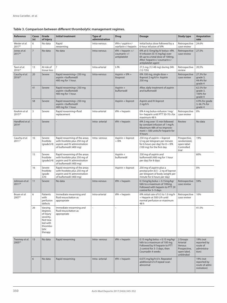
Anna Carceller, et al.
350 Arch Med Deporte 2017;34(6):345-352
Table 3. Comparison between different thrombolytic management regimes.
Reference Cases (n)
Gradeof injury
Initial treatment Type ofadministration
Drug Dosage Study type Amputation rate
Wexler et al. 201741
6 No data Rapid rewarming
Intra-venous tPA+/-aspirin+/-warfarin+/-heparin
initial bolus dose followed by a 6-hour infusion of tPA
Retrospective case review
24.6%
Jones et al. 201742
7 No data No data Intra-venous tPA + heparin +/- coumarin +/- antiplatelet
tPA at 0.15mg/kg IV bolus+ tPA. IV infusion (0.15 mg/kg) over 6h up to a total dose of 100mg. After: heparin+/-coumarin+/-antiplatelet agent
Retrospective case review
27.5%
Tavri et al. 201635
13 At risk of tissue loss
? Intra-arterial t-PA 27,5 mg (12-48 mg) during 34h (12-72h)
Retrospective review
20,5%
Cauchy et al. 201643
20 Severe Rapid rewarming+ 250 mg aspirin +buflomedil 400 mg for 1 hour.
Intra-venous Aspirin + tPA + ilosprost
tPA 100 mg, single dose + iloprost 2 ng/6 h+ Aspirin 250 mg
Retrospective case review
27.3% for grade 3,44.4% for grade 4
41 Severe Rapid rewarming+ 250 mg aspirin +buflomedil 400 mg for 1 hour.
Aspirin + buflomedil
After, daily treatment of aspirin and buflomedil
62.5% for grade 3,100% for grade 4
58 Severe Rapid rewarming+ 250 mg aspirin +buflomedil 400 mg for 1 hour.
Aspirin + iloprost Aspirin and IV iloprost 2 ng/6 h
4.9% for grade 3, 66.7% for grade 4
Ibrahim et al. 201528
3 Severe Rapid rewarming+fluid replacement
Intra-arterial tPA + heparin tPA 4 mg bolus+infusion 1mg/hr+ heparin until PTT 50-70 s for maximum 48 h
Retrospective case review
0%
Handford et al. 20142
- Severe - Intra- arterial tPA + heparin tPA 3 mg over 15 min followed by constant infusion of 1 mg/h. Maximum 48h of no improve-ment + 500 units/hr heparin for 4 hours
Review No data
Cauchy et al. 201121
16 Severe frostbite (grade3/4)
Rapid rewarming of the areas with frostbite plus 250 mg of aspirin and IV administrationof buflomedil (400 mg)
Intra -venous Aspirin + iloprost + tPA
250 mg of aspirin + iloprost (2 ng per kilogram per minute for 6 hours per day) for 8 + tPA (100 mg) for the first day
Prospective, randomized,open-label Controlled trial
19%
15 Severe frostbite (grade3/4)
Rapid rewarming of the areas with frostbite plus 250 mg of aspirin and IV administrationof buflomedil (400 mg)
Aspirin + buflomedil
250 mg of aspirin and buflomedil (400 mg for 1 hour per day) for 8 days
60%
16 Severe frostbite (grade 3/4)
Rapid rewarming of the areas with frostbite plus 250 mg of aspirin and IV administrationof buflomedil (400 mg)
Aspirin + iloprost 250 mg of aspirin plus a prostacyclin (0.5 - 2 ng of loprost per kilogram of body weight per minute for 6 hours per day)
0%
Johnson et al. 201136
11 Severe No data Intra-venous tPA + heparin 0.15mg/kg bolus + 0.15mg/kg/h6h to a maximum of 100mg. Followed with heparin to PTT 2X control for 3–5 days
Retrospective case review
59%
Bruen et al. 200731
6 Patients with perfusion defects
Immediate rewarming and fluid resuscitation as appropriate
Intra-arterial tPA + heparin tPA initial rate of 0.5 to 1.0 mg/h + Heparin at 500 U/h until normal perfusion or maximum 48 h
Retrospective case review
10%
26 Varyingdegrees of injury severity. Not trea-ted with thrombo-lytictherapy
Immediate rewarming and fluid resuscitation as appropriate
- - - 41.5%
Twomey et al. 200534
13 No data Rapid rewarming Intra- venous tPA + heparin 0.15 mg/kg bolus + 0.15 mg/kg/h6h to a maximum of 100 mg. Followed by IV heparin to PTT 2 control for 3–5 days, then Coumadin 4 weeks
2 GroupsArterialVenousProspective, open label, unblinded
19% (not reported by route of administra-tion)
6 No data Rapid rewarming Intra -arterial tPA + heparin 0.075 mg/kg/h 6 h. Repeatedadditional 6 h if repeat scan abnormal
19% (not reported by route of admi-nistration)

Frostbite: management update
351Arch Med Deporte 2017;34(6):345-352
On the other hand, iloprost is a prostacyclin analogue with vasodila-tor and antiplatelet properties that has been associated with reductions in digital amputations in severe frostbite, so many authors recommend its intravenous administration as a first-line treatment22,29. Dose titration in published clinical experiences is based on the appearance of adverse effects within the therapeutic range (starting at 0,5-2 ng/kg/min, with 0,5 ng/kg/min increases every 30 minutes until the maximal toleration rate is achieved and maintaining its infusion 6 hours/day for 5-8 days), with consideration for the fact that the patient must be maintained in the supine position to prevent orthostatic hypotension21. The effect of its association with rt-PA is not well known, although in accordance with a recent randomized trial it seems to be optimal in grade IV frostbite within the first 12 hours21. Contraindications of its use include unstable angina, recent cardio-vascular events and increased risk of bleeding. The advantages of iloprost over rt-PA are that it does not require in-terventionist procedures, the therapeutic window is larger, it can be administered in patients with trauma and intensive monitoring, other than blood pressure monitoring, is not required.
Tetanus vaccination is recommended, according to the usual schedule.
If amputation is required, surgical intervention must be delayed until viable tissue can be demarcated accurately, provided that an emergency justification for proceeding (e.g. gangrene, sepsis and compartment syndrome). This measure is justified by the possibility that tissue initially considered non-viable is restored37 and the risk of surgical trauma inter-fering with the healing of proximal tissues4,6. This is not carried out in normal conditions until at least four to six weeks after injury, including in patients receiving thrombolytic therapy, which can imply the need for psychological support.
Sequelae
Sequelae after frostbite are common and occur independently of its severity. In a study of 30 patients with grade II frostbite, 63% were found to have sequelae after four to 11 years from injury (cold sensitivity 53%, digital numbness 40%, reduction in touch sensitivity 33%)38. It is estimated that sensitivity disturbances are present for at least four years in nearly all those who have suffered from frostbite11.
Chronic pain secondary to frostbite is very common and is usually refractory to conventional analgesics. It sometimes responds to drugs designed for neuropathic pain (e.g. amitriptyline and gabapentin). Despite efforts to treat later symptoms (e.g. pain, paraesthesia and numbness) with chemical and surgical sympathectomies, there is no clear indication for their use.
Other common problems include hyperhidrosis, secondary to an abnormal response of the sympathetic system, trophic alterations in skin and fanerae, digital flexor retraction and high susceptibility to future cold-related injuries. Alterations of skin colouration, ranging from depigmentation to local cyanosis, are not uncommon.
Long-term sequelae include osteoporosis, and where the frostbite affected the joints, osteoarthrosis with damaged joint surfaces and a decline in joint mobility with tendinous retractions of the flexor mus-culature39.
Digital amputations (partial or total) involve functional limitations to daily life and sporting activities, given the alteration in the normal biomechanics of the limb. Gait will be severely impaired if frostbite affects the metacarpophalangeal joint in the foot. Risk factors related to amputation include duration of exposure to cold, absence of proper equipment, exposure to cold in remote areas, presence of infection and delay in treatment.
There is a broad consensus on the need to prioritise an early mul-tidisciplinary rehabilitation programme for patients who have under-gone amputation, including prompt controlled mobilisation to prevent tendinous retraction and reach optimal levels of functional recovery10,15, and long-term, non-aggressive treatment40 (Table 3).
Conclusions
Frostbite is no longer primarily an occupational pathology or characteristic of subjects with cognitive impairment. It has become a common cause of morbidity among healthy young adults who volun-tarily expose themselves to cold, usually while practising winter sports. Knowledge of activity planning, survival skills and cold protection is strongly recommended as basic prevention tools. Early recognition of frostbite is essential to ensure prompt diagnosis and early initial treatment, since a delay in first rewarming is associated with a worse prognosis. At present, in-field treatments are relatively basic and can be initiated by non-qualified subjects with the proper training. New perspectives are focusing on improving initial care by applying ad-vanced treatments under medical supervision. For superficial frostbite, there is no need for further complementary tests beyond the clinical monitoring of the injury. For severe frostbite, scintigraphy with Tc99 is a good prognosis predictor after 48 hours of injury. Angiography is both an imaging and a therapeutic tool, but less invasive options such as MRI and ultrasound appear to be good alternatives when direct thrombolysis is not required. Emerging hospital treatments have a therapeutic window that needs to be known to take fast and optimal decisions regarding patient evacuation, considering the rescue time lapses and the hospital resources of each mountain area and country. Surgical interventions must be delayed until there is a clear demarcation of the necrotic area. Long-term sequelae are prevalent among subjects with frostbite, even in non-severe injuries. A multidisciplinary approach to caring for patients with frostbite is needed in the management of long-term functional sequelae.
Bibliography 1. Murphy JV, Banwell PE, Roberts AH, McGrouther DA. Frostbite: pathogenesis and
treatment. J Trauma. 2000;48(1):171-8.
2. Handford C, Buxton P, Russell K, Imray CE, McIntosh SE, Freer L, et al. Frostbite: a practical approach to hospital management. Extreme Physiol Med. 2014;3:7.
3. Gross EA, Moore JC. Using thrombolytics in frostbite injury. J Emerg Trauma Shock. 2012;5(3):267–71.
4. Hallam M-J, Cubison T, Dheansa B, Imray C. Managing frostbite. BMJ. 2010;341:c5864.
5. Harirchi I, Arvin A, Vash JH, Zafarmand V. Frostbite: incidence and predisposing factors in mountaineers. Br J Sports Med. 2005;39(12):898-901; discussion 901.
6. Valnicek SM, Chasmar LR, Clapson JB. Frostbite in the prairies: a 12-year review. Plast Reconstr Surg. 1993;92(4):633-41.

Anna Carceller, et al.
352 Arch Med Deporte 2017;34(6):345-352
7. McIntosh SE, Campbell A, Weber D, Dow J, Joy E, Grissom CK. Mountaineering medical events and trauma on Denali, 1992-2011. High Alt Med Biol. 2012;13(4):275-80.
8. Nemethy M, Pressman AB, Freer L, McIntosh SE. Mt Everest Base Camp Medical Clinic “Everest ER”: epidemiology of medical events during the first 10 years of operation. Wilderness Environ Med. 2015;26(1):4-10.
9. Hashmi MA, Rashid M, Haleem A, Bokhari SA, Hussain T. Frostbite: epidemiology at high altitude in the Karakoram mountains. Ann R Coll Surg Engl. 1998;80(2):91–5.
10. Grieve AW, Davis P, Dhillon S, Richards P, Hillebrandt D, Imray CHE. A clinical review of the management of frostbite. J R Army Med Corps. 2011;157(1):73-8.
11. Auerbach P. Frostbite. In: Wilderness Medicine. 6th ed. Philadelphia: Mosby; 2012. p. 181–200.
12. Tikuisis P, Keefe A. Prediction of times of facial and finger freezing during cold air exposure.DRDC-TORONTO-TR-2004-140 — Technical Report. Goverment of Canada. R&D. [Internet]. 2004 [cited 2017 Feb 8]. Available from: http://pubs.drdc-rddc.gc.ca/BASIS/pcandid/www/engpub/DDW?W%3DSYSNUM=523617
13. Dana AS RI. The hunting reaction. Arch Dermatol. 1969;99(4):441-50.
14. Imray C, Grieve A, Dhillon S. Cold damage to the extremities: frostbite and non-freezing cold injuries. Postgrad Med J. 2009;85(1007):481-8.
15. McIntosh SE, Opacic M, Freer L, Grissom CK, Auerbach PS, Rodway GW, et al. Wilderness Medical Society practice guidelines for the prevention and treatment of frostbite: 2014 update. Wilderness Environ Med. 2014;25(4 Suppl):S43-54.
16. Zafren K. Frostbite: prevention and initial management. High Alt Med Biol. 2013;14(1):9-12.
17. Syme D. Position paper: on-site treatment of frostbite for mountaineers. High Alt Med Biol. 2002;3(3):297–8.
18. Roche-Nagle G, Murphy D, Collins A, Sheehan S. Frostbite: management options. Eur J Emerg Med Off J Eur Soc Emerg Med. 2008;15(3):173-5.
19. Martínez Villen G, García Julve G, Morandeira JR, Foray J, Merle M. Pathologie de la main liée a l’action du froid. In: Pathologie de la main chez le sportif. Leclerq C. París: Frison- Roche; 1996. p. 147–57.
20. Murkowski FH, Mandsager R, Choromanski HJ. State of Alaska cold injuries guideli-nes. Department of health and social services of Alaska [Internet]. 2014 [cited 2017]. Available from: http://dhss.alaska.gov/dph/Emergency/Documents/ems/documents/Alaska%20DHSS%20EMS%20Cold%20Injuries%20Guidelines%20June%202014.pdf
21. Cauchy E, Cheguillaume B, Chetaille E. A controlled trial of a prostacyclin and rt-PA in the treatment of severe frostbite. N Engl J Med. 2011;364(2):189-90.
22. Cauchy E, Davis CB, Pasquier M, Meyer EF, Hackett PH. A New Proposal for Management of Severe Frostbite in the Austere Environment. Wilderness Environ Med. 2016;27(1):92-9.
23. Richalet, JP HJ. Pathologie liée au froid. In: Médecine de l’alpinisme et des sports de montagne. 4a ed. París: Masson; 2006. p. 188.
24. Hayes DWJ, Mandracchia VJ, Considine C, Webb GE. Pentoxifylline. Adjunctive therapy in the treatment of pedal frostbite. Clin Podiatr Med Surg. 2000;17(4):715-22.
25. Daum PS, Bowers WD, Tejada J, Morehouse D, Hamlet MP. An evaluation of the ability of the peripheral vasodilator buflomedil to improve vascular patency after acute frostbite. Cryobiology. 1989;26(1):85-92.
26. Heggers JP, Robson MC, Manavalen K, Weingarten MD, Carethers JM, Boertman JA, et al. Experimental and clinical observations on frostbite. Environ Emergencies. 1987; 16(9):1056–62.
27. Cauchy E, Leal S, Magnan M-A, Nespoulet H. Portable hyperbaric chamber and management of hypothermia and frostbite: an evident utilization. High Alt Med Biol. 2014;15(1):95-6.
28. Ibrahim AE, Goverman J, Sarhane KA, Donofrio J, Walker TG, Fagan SP. The emerging role of tissue plasminogen activator in the management of severe frostbite. J Burn Care Res Off Publ Am Burn Assoc. 2015;36(2):e62-6.
29. Groechenig E. Treatment of frostbite with iloprost. Lancet Lond Engl. 1994;344(8930): 1152–3.
30. Wagner C, Pannucci CJ. Thrombolytic therapy in the acute management of frostbite injuries. Air Med J. 2011;30(1):39-44.
31. Bruen KJ, Ballard JR, Morris SE, Cochran A, Edelman LS, Saffle JR. Reduction of the incidence of amputation in frostbite injury with thrombolytic therapy. Arch Surg Chic Ill 1960. 2007;142(6):546–51; discussion 551-3.
32. Saemi AM, Johnson JM, Morris CS. Treatment of bilateral hand frostbite using trans-catheter arterial thrombolysis after papaverine infusion. Cardiovasc Intervent Radiol. 2009;32(6):1280-3.
33. Sheridan RL, Goldstein MA, Stoddard FJ, Walker TG. Case 41-2009. N Engl J Med. 2009;361(27):2654-62.
34. Twomey JA, Peltier GL, Zera RT. An open-label study to evaluate the safety and efficacy of tissue plasminogen activator in treatment of severe frostbite. J Trauma. 2005;59(6):1350-4; discussion 1354-5.
35. Tavri S, Ganguli S, Bryan RGJ, Goverman J, Liu R, Irani Z, et al. Catheter-Directed Intra-arterial Thrombolysis as Part of a Multidisciplinary Management Protocol of Frostbite Injury. J Vasc Interv Radiol JVIR. 2016;27(8):1228–35.
36. Johnson AR, Jensen HL, Peltier G, DelaCruz E. Efficacy of intravenous tissue plasminogen activator in frostbite patients and presentation of a treatment protocol for frostbite patients. Foot Ankle Spec. 2011;4(6):344–8.
37. Gonzaga T, Jenabzadeh K, Anderson CP, Mohr WJ, Endorf FW, Ahrenholz DH. Use of Intraarterial Thrombolytic Therapy for Acute Treatment of Frostbite in 62 Patients with Review of Thrombolytic Therapy in Frostbite. J Burn Care Res Off Publ Am Burn Assoc. 2016;37(4):323–34.
38. Ervasti O, Hassi J, Rintamaki H, Virokannas H, Kettunen P, Pramila S, et al. Sequelae of moderate finger frostbite as assessed by subjective sensations, clinical signs, and thermophysiological responses. Int J Circumpolar Health. 2000;59(2):137-45.
39. Foray J K-BF, Lanoye P. Les séquelles des gelures de montagne. Chirurgie. 1979;(105):37–46.
40. Woo E-K, Lee JW, Hur G-Y, Koh J-H, Seo D-K, Choi J-K, et al. Proposed treatment protocol for frostbite: a retrospective analysis of 17 cases based on a 3-year single-institution experience. Arch Plast Surg. 2013;40(5):510-6.
41. Wexler A, Zavala S. The Use of Thrombolytic Therapy in the Treatment of Frostbite Injury. J Burn Care Res Off Publ Am Burn Assoc. 2017. [Epub ahead of print].
42. Jones LM, Coffey RA, Natwa MP, Bailey JK. The use of intravenous tPA for the treatment of severe frostbite. Burns J Int Soc Burn Inj. 2017. [Epub ahead of print].
43. Cauchy E. Iloprost With and Without rt-PA: Treatment of 131 Cases of Severe Frostbite. In: Abstracts from the 7th World Congress of Mountain & Wilderness Medicine. Telluride, CO, USA; 2016.

Libros
353Arch Med Deporte 2017;34(6):353
Books
Esta obra singular es deporte y fuerza, movimiento y pasión. Es una guía distinta de cualquier otra. Desde los asombrosos dibujos hasta los 127 workouts, está diseñada para marcar di-ferencias. Tanto si se utiliza su contenido como complemento de un programa de entrenamiento ya existente, como si
EL ARTE MODERNO DEL ENTRENAMIENTO DE ALTA INTENSIDADPor: Aurélien Broussal-Derval y Stéphane Ganneau Edita: Ediciones Tutor-Editorial El Drac. Impresores 20. P.E. Prado del Espino. 28660 Boadilla del Monte. Madrid. Telf. 915 599 832 – Fax: 915 410 235 E-mail: [email protected] Web: www.edicionestutor.com Madrid 2017, 224 páginas, P.V.P: 25 euros
se quiere sustituir un programa de entre-namiento que se ha vuelto monótono y obsoleto, se ojeará y utilizará este libro una y otra vez.
Se verá el entrenamiento de alta intensidad con ojos completamente nuevos. En sus páginas se encontra-rán 40 ejercicios, cada uno detallado
y representado de manera artística, fotografías y variaciones; 127 workouts y circuitos para combinar cosas diferentes; recomendaciones para el calentamiento, la seguridad y la prevención de lesiones; y si se está dispuesto a asumir el reto: un programa original de 15 semanas de duración.
Puede que los triatletas com-prometidos sean de todos los de-portistas, los mayores entendidos en tecnología. Tienen los dispositivos de vanguardia y saben que los datos para mejorar su rendimiento están a mano, pero unirlo todo puede re-sultar una tarea confusa de enormes proporciones. El triatleta, entrena-dor, investigador y autor Jim Vance
TRIATLÓN 2.0Por: Jim Vance Edita: Ediciones Tutor-Editorial El Drac. Impresores 20. P.E. Prado del Espino. 28660 Boadilla del Monte. Madrid. Telf. 915 599 832 – Fax: 915 410 235 E-mail: [email protected] Web: www.edicionestutor.com Madrid 2017, 272 páginas, P.V.P: 25 euros
sostiene que, a pesar del acceso a la información adecuada, la mayoría de los triatletas empiezan las pruebas infraentrenados o sobreentrenados. Por eso ha desarrollado Triatlón 2.0, el primer programa que se aprovecha de lo último en ciencia y tecnología.
El libro examina los aparatos más utilizados en este deporte, como medi-dores de potencia (o vatímetros) para
ciclismo, rastreadores GPS y monitores de frecuencia cardíaca (o pulsómetros). Recoge las mediciones más precisas, descubre lo que significan e, igual de importante, lo que no. Y así después, el lector puede poner los números a su propio servicio: trasladando sus datos a un programa exhaustivo basado en sus necesidades de rendimiento y objetivos.
Correr es algo más que ponerse las zapatillas, tener voluntad y tratar de sumar kilómetros. Los corredores deben completar su preparación con ejercicios de fuerza específicos y de la zona media, el core, para maximizar la eficacia de carrera y desarrollar una óptima longitud y frecuencia de zanca-da. Esta obra ofrece una metodología innovadora para el desarrollo de la
ENTRENAMIENTO DEL CORE PARA CORREDORESPor: Christian Roberto López Rodríguez Edita: Ediciones Tutor-Editorial El Drac. Impresores 20. P.E. Prado del Espino. 28660 Boadilla del Monte. Madrid. Telf. 915 599 832 – Fax: 915 410 235 E-mail: [email protected] Web: www.edicionestutor.com Madrid 2017, 200 páginas, P.V.P: 18 euros
musculatura de la zona media basada en el empleo progresivo de movi-mientos funcionales, sin necesidad de máquinas de musculación, y teniendo en cuenta toda la temporada a través de una periodización deportiva bien planificada.
Los ejercicios, con variantes para progresar, se describen de tal manera que cualquier deportista puede llevar-
los a la práctica siguiendo las pautas metodológicas desde el comienzo del programa. Este libro no solo ser-virá a los runners de cualquier nivel, desde los de carreras populares hasta profesionales de élite, sino a todos los deportistas que en su disciplina, individual o de equipo, requieren de la carrera en los movimientos en el terreno de juego.

Agenda
354 Arch Med Deporte 2017;34(6):354-356
2018
Congrès francophone de médecine 17-21 Enero web: www.grimm-vs.ch de montagne Champéry, (Suiza)
II Jornadas Nacionales SETRADE 15-16 Marzo http://www.setrade.org/congresos/jornadasvitoria2018/ Vitoria
36 Congress International Society 15-17 Marzo web: http://www.sitemsh.org/ for Snowsports Medicine Arosa (Suiza)
15th International Scientific 12-15 Abril web: http://csakademija.me/conference/ Conference and 14th annual Bubva (Montenegro) Congress of the Montenegrin Sports Academy
World Congress on Osteoporosis, 19-22 Abril web: www.wco-iof-esceo.org/ Osteoarthritis and Musculoskeletal Cracovia (Polonia) Diseases
18th ESSKA Congress 9-12 Mayo web: http://esska-congress.org/ Glasgow (Reino Unido)
56 Congreso SERMEF 16-19 Mayo www.sermef.es Gijón-Asturias
7thWorld Conference on Women 17-20 Mayo web: www.icsspe.org/sites/default/files/e8_7TH%20 and Sport Gaborone (Bostwana) IWG%20Conference%20docx.pdf
XXII Curso Asoc. Española Med. 25-26 Mayo web: http://www.aemef.org/es/ del Futbol Málaga
XXVII Isokinetic Medical Group 2-4 Junio E-mail: [email protected] conference:”Football medicine Barcelona web: www.footballmedicinestrategies.com outcomes. Are we winning?”
5th International Congress of 5-10 Junio web: https://events.eventact.com/EventsList/ Exercise and Sport Sciences Netanya (Israel) 5sportsceince2017/General-Information
European Congress of Adapted 3-5 Julio Andrea Faull. E-mail: [email protected] Physical Activity (EUCAPA) Worcester (Reino Unido) Ken Black. E-mail: [email protected]
23rd Annual Congress of the 4-7 Julio web: www.ecss-congress.eu/2018/ European College of Sport Science Dublín (Irlanda)
World Congress of Biomechanics 8-12 Julio web: http://wcb2018.com/ Dublín (Irlanda)
12th World Congress of the 8-12 Julio web: http://isprm2018.com/ International Society of Physical París (Francia) and Rehabilitation Medicine (ISPRM)
The Annual World Congress of 25-27 Julio web: http://www.bitcongress.com/wcort2018 / Orthopaedics Milán (Italia) http://www.bitcongress.com/wcort2018/programlayout.asp
World Congress of the Association 25-28 Julio web: http://aiesep.org/ Internationale des Ecoles Edimburgo (Reino Unido) Supérieures d’Education Physique (AIESEP)

Agenda
355Arch Med Deporte 2017;34(6):354-356
2020
25th Annual Congress of the 1-4 Julio E-mail: [email protected] European College of Sport Science Sevilla
XXXVI Congreso Mundial de 24-27 Septiembre web: www.globalevents.gr Medicina del Deporte Atenas (Grecia)
2021
26th Annual Congress of the 7-10 Julio E-mail: [email protected] opean College of European College of Sport Science Glasgow (Reino Unido)
12th Biennial ISAKOS 12-16 Mayo web: www.isakos.com Cancún (México)
24th Annual Congress of the 3-6 Julio E-mail: [email protected] European College of Sport Science Praga (Rep. Checa)
14th International Congress of 17-20 Septiembre web: www.icses2019.org shoulder and elbow surgery Buenos Aires (Argentina) (ICSES)
XV Congreso Nacional de web: www.psicologiadeporte.org Psicología de la Act. Física y del Zaragoza Deporte
2019
XXXV Congreso Mundial de 12-15 Septiembre web: www.fims.org Medicina del Deporte Rio de Janeiro (Brasil)
5th International Scientific 27-29 Septiembre web: http://ists2018.com/ Tendinopathy Symposium (ISTS) Groningen (Países Bajos)
VII Congreso Asociación Octubre web: http://hispamef.com/ Hispanoamericana de Médicos Lima (Perú) del Fútbol
28º Congress European Society for surgery of the shoulder and the Ginebra (Suiza) web: www.secec.org elbow (SECEC-ESSSE)

Agenda
356 Arch Med Deporte 2017;34(6):354-356
Agenda
Curso “ELECTROCARDIOGRAFÍA PARA MEDICINA DEL DEPORTE” ACREDITADO POR LA COMISIÓN DE FORMACIÓN CONTINUADA (ON-LINE 15/10/2015 A 15/10/2016) CON 4,81 CRÉDITOS Curso dirigido a médicos destinado a proporcionar los conocimientos específicos para el estudio del sistema cardiocirculatorio desde el punto de vista del electrocardiograma (ECG).
Curso “FISIOLOGÍA Y VALORACIÓN FUNCIONAL EN EL CICLISMO” Curso dirigido a los titulados de las diferentes profesiones sanitarias y a los titulados en ciencias de la actividad física y el deporte, destinado al conocimiento profundo de los aspectos fisiológicos y de valoración funcional del ciclismo.
Curso “AYUDAS ERGOGÉNICAS” Curso abierto a todos los interesados en el tema que quieren conocer las ayudas ergogénicas y su utilización en el deporte.
Curso “CARDIOLOGÍA DEL DEPORTE” ACREDITADO POR LA COMISIÓN DE FORMACIÓN CONTINUADA (VÁLIDA DEL 15/10/2016 AL 15/10/2017) CON 8,78 CRÉDITOS Fecha límite de inscripción: 15/06/2017 Curso dirigido a médicos destinado a proporcionar los conocimientos específicos para el estudio del sistema cardiocirculatorio desde el punto de vista de la actividad física y deportiva, para diagnosticar los problemas cardiovasculares que pueden afectar al deportista, conocer la aptitud cardiológica para la práctica deportiva, realizar la prescripción de ejercicio y conocer y diagnosticar las enfermedades cardiovasculares susceptibles de provocar la muerte súbita del deportista y prevenir su aparición.
Curso “ALIMENTACIÓN, NUTRICIÓN E HIDRATACIÓN EN EL DEPORTE” Curso dirigido a médicos destinado a facilitar al médico relacionado con la actividad física y el deporte la formación precisa para conocer los elementos necesarios para la obtención de los elementos energéticos necesarios para el esfuerzo físico y para prescribir una adecuada alimentación del deportista.
Curso “ALIMENTACIÓN Y NUTRICIÓN EN EL DEPORTE” Curso dirigido a los titulados de las diferentes profesiones sanitarias (existe un curso específico para médicos) y para los titulados en ciencias de la actividad física y el deporte, dirigido a facilitar a los profesionales relacionados con la actividad física y el deporte la formación precisa para conocer los elementos necesarios para la obtención de los elementos energéticos necesarios para el esfuerzo físico y para conocer la adecuada alimentación del deportista.
Curso “ALIMENTACIÓN Y NUTRICIÓN EN EL DEPORTE” Para Diplomados y Graduados en Enfermería ACREDITADO POR LA COMISIÓN DE FORMACIÓN CONTINUADA (NO PRESENCIAL 15/12/2015 A 15/12/2016) CON 10,18 CRÉDITOS Curso dirigido a facilitar a los Diplomados y Graduados en Enfermería la formación precisa para conocer los elementos necesarios para la obtención de los elementos energéticos necesarios para el esfuerzo físico y para conocer la adecuada alimentación del deportista.
Cursos on-line SEMED-FEMEDE
Más información:
www.femede.es
Curso “ENTRENAMIENTO, RENDIMIENTO, PREVENCIÓN Y PATOLOGÍA DEL CICLISMO”Curso dirigido a los titulados de las diferentes profesiones sanitarias y a los titulados en ciencias de la actividad física y el deporte, destinado al conocimiento de las prestaciones y rendimiento del deportista, para que cumpla con sus expectativas competitivas y de prolongación de su práctica deportiva, y para que la práctica deportiva minimice las consecuencias que puede tener para su salud, tanto desde el punto de vista médico como lesional.
Curso “CINEANTROPOMETRÍA PARA SANITARIOS” Curso dirigido a sanitarios destinado a adquirir los conocimientos necesarios para conocer los fundamentos de la cineantropometría (puntos anatómicos de referencia, material antropométrico, protocolo de medición, error de medición, composición corporal, somatotipo, proporcionalidad) y la relación entre la antropometría y el rendimiento deportivo.
Curso “CINEANTROPOMETRÍA” Curso dirigido a todas aquellas personas interesadas en este campo en las Ciencias del Deporte y alumnos de último año de grado, destinado a adquirir los conocimientos necesarios para conocer los fundamentos de la cineantropometría (puntos anatómicos de referencia, material antropométrico, protocolo de medición, error de medición, composición corporal, somatotipo, proporcionalidad) y la relación entre la antropometría y el rendimiento deportivo.

Sumarios 2017
357Arch Med Deporte 2017;34(6):357-361

Sumarios 2017
358 Arch Med Deporte 2017;34(6):357-361
Sumarios 2017
Volumen 34(1) - Núm 177. Enero - Febrero 2017 / January - February 2017
Volumen 34(2) - Núm 178. Marzo - Abril 2017 / March -April 2017
Editorial
¿Han cambiado realmente las recomendaciones dietéticas en el siglo XXI? ¿Tenemos nuevos retos? Have dietary recommendations really changed in the 21st century? Are there any new challenges?. Teresa Gaztañaga Aurrekoetxea ...........................................................................................................................................................................................6
Originales / Original articles
The effect of weekly low frequency exercise on body composition and blood pressure of elderly women. El efecto de baja frecuencia semanal del ejercicio sobre la composición corporal y la presión arterial de las mujeres ancianas. Claudio Rosa, José Vilaça-Alves, Eduardo Borba Neves, Francisco José Félix Saavedra, Miriam Beatris Reckziegel, Hildegard Hedwig Pohl, Daniela Zanini, Victor Machado Reis ...................................................................9
Cardiac autonomic responses of trained cyclists at different training amplitudes. Respuesta del sistema cardiaco autónomo en ciclistas entrenados con diferentes amplitudes de entrenamiento. Luan M. Picanço, Gilberto Cavalheiro, Marcelo S. Vaz, Fabriíio B. Del Vecchio ...................................................................................15
Los ejercicios preventivos tras el calentamiento ayudan a reducir lesiones en fútbol. Preventive exercises after warming help to reduce injuries in soccer. Jorge Carlos-Vivas, Juan P. Martin-Martinez, Manuel Chavarrias, Jorge Pérez-Gómez ......................................................................................................................................................21
Análisis de la fuerza y movilidad de la cadera como factores de riesgo de lesión en fútbol femenino amateur: un estudio piloto. Analysis of hip strength and mobility as injury risk factors in amateur women’s soccer: a pilot study. Antonio Maestro, Joaquín Lago, Gonzalo Revuelta, Pablo del Fueyo, Lorenzo del Pozo, Carlos Ayán, Vicente Martín ......................................................................................................................................................................................................................................................25
Revisiones / Reviews
Implicaciones funcionales del entrenamiento de la fuerza en el adulto mayor: una revisión de literatura. Functional implications of the strength training on older adult: a literature review. Roberto Rebolledo-Cobos, Cleiton Silva Correa, Jesse Juliao-Castillo, Raúl Polo Gallardo, Olga Suarez Landazabal ..........31
Criterios para el retorno al deporte después de una lesión. Criteria to return to play sports after an injury. Tomás F. Fernández Jaén, Pedro Guillén García ............40
Agenda / Agenda.......................................................................................................................................................................................................................................................................................................................................48
Editorial
Fisiología clínica del Ejercicio: la fisiología del ejercicio aplicada a las patologías crónicas. Clinical Exercise Physiology: The role of exercise physiology in chronic diseases. Luis Franco Bonafonte ............................................................................................................................................................................................................................................................................62
Originales / Original articles
Valoración isocinética en cadena cinética cerrada en futbolistas: Prueba piloto. Kinetic closed chain isokinetic values in football players: Pilot test. Pavel Loeza Magaña, Wolfgang Fritzler-Happach, Joaquin Barrios-González ...........................................................................................................................................................................66
Peak oxygen uptake prediction in overweight and obese adults. Predicción del consumo pico de oxígeno en adultos con sobrepeso y obesidad. Eliane A. Castro, Rocío Cupeiro, Pedro J. Benito, Javier Calderón, Isabel R. Fernández, Ana B. Peinado .............................................................................................................72
Do the changes in acid-base status and respiratory gas exchange explain the voluntary termination of a test performed above the maximum lactate steady state? ¿Pueden los cambios del estado ácido-base e intercambio de gases respiratorios explicar el abandono de una prueba realizada por encima del máximo estado estable de lactato? Ana B. Peinado, María I. Barriopedro, Pedro J. Benito, Francisco J. Calderón .................................................................................................80
Estudio de la validez en la medición de los valores de lactato sanguíneo entre los dos modelos existentes de LactatePro. Validity of blood lactate measurements between the two LactatePro versions. Iñaki Arratibel-Imaz, Julio Calleja-González, Nicolás Terrados .............................................................................................86
Revisión / Review
Mejora del sueño en deportistas: uso de suplementos nutricionales. Sleep improvement in athletes: use of nutritional supplements. Fernando Mata Ordóñez, Antonio J. Sánchez Oliver, Pedro Carrera Bastos, Laura Sánchez Guillén, Raúl Domínguez ..........................................................................................................................................93
Revisión sobre aspectos genéricos acerca de la actividad física adaptada en la persona con lesión medular. Review of generic aspects about Adapted Physical Activity in the Person with Spinal Cord Injury. Miguel Á. Capó-Juan, Miguel Bennasar-Veny, Antonio Aguiló-Pons, Joan E. de Pedro-Gómez ............100
Agenda / Agenda....................................................................................................................................................................................................................................................................................................................................111

Sumarios 2017
359Arch Med Deporte 2017;34(6):357-361
Volumen 34(3) - Núm 179. Mayo - Junio 2017 / May - June 2017
Volumen 34(4) - Núm 180. Julio - Agosto 2017 / July - August 2017
Editorial
Antioxidantes macromoleculares: importancia en salud y perspectivas. Macromolecular Antioxidants: Importance in Health and Perspectives. Fulgencio Saura Calixto ...........................................................................................................................................................................................................................................................................................................188
Originales / Original articles
Isokinetic performance of knee extensors and flexor muscles in adolescent basketball players. Valoración isocinética de los extensores y flexores de la rodilla de jugadores de baloncesto adolescentes. Leandro Viçosa Bonetti, Franciele Piazza, Camila Marini, Bruno Soldatelli Zardo, Gerson Saciloto Tadiello ..........................................................................................................................................................................................................................................................................................................191
Effects of different automatic filters on the analysis of heart rate variability with Kubios HRV software. Efecto de los diferentes filtros automáticos en el análisis de la variabilidad de la frecuencia cardiaca con el software Kubios HRV. Carmen Aranda, Blanca de la Cruz, Jose Naranjo ....................................................................................196
Exceso de peso corporal y calidad de vida relacionada con la salud de adolescentes latino-americanos. Overweight and health-related quality of life in Latin American adolescents. Dartagnan Pinto Guedes, Hermán Ariel Villagra Astudillo, José María Moya Morales, Juan del Campo Vecino, Paulo Marcelo Pirolli, Raymundo Pires Júnior......................................................................................................................................................................................................................................................201
Análisis de la composición corporal empleando parámetros bioeléctricos en la población deportiva cubana. Body composition analysis using bioelectrical parameters in Cuban sporting population. Wiliam Carvajal Veitia, Yanel Deturnell Campo, Ibis M. Echavarría García, Dianelis Aguilera Chavez, Lázaro R. Esposito Gutiérrez, Antonio Cordova ..................................................................................................................................................................................................................................................207
Revisiones / Reviews
Actividad electromiográfica (EMG) durante el pedaleo, su utilidad en el diagnóstico de la fatiga en ciclistas. Electromiografic (EMG) activity during pedaling, its utility in the diagnosis of fatigue in cyclists. Alfredo Córdova, Iván Nuin, Diego Fernández-Lázaro, Ibán Latasa, Javier Rodríguez-Falces ................................217
Prevención de la muerte súbita por miocardiopatía arritmogénica del ventrículo derecho en deportistas. Arrhythmogenic right ventricular cardiomyopathy. Prevention of sudden death in athletes. Franc Peris, José Poveda, Diego Oliver, Luis Franco, Francisco J. Rubio, Alfredo Valero ............................................................224
Agenda / Agenda....................................................................................................................................................................................................................................................................................................................................242
Editorial
El paradigma de la recuperación en deportes de equipo. Paradigm for the recovery in team sports. Julio Calleja-González ....................................................................................126
Original articles / Originales
Static balance behavior along a deep water periodization in older men. Comportamiento del equilibrio estático a lo largo de una periodización de carrera en aguas profundas en hombres mayores. Ana C. Kanitz, Giane V. Liedtke, Thaís Reichert, Natalia A. Gomeñuca, Rodrigo S. Delevatti, Bruna M. Barroso, Luiz FM. Kruel .....................................................................................................................................................................................................................................................................................129
Respuesta fisiológica de una unidad paracaidista en combate urbano. Physiological Response of a Paratrooper Unit in Urban Combat Joaquín Sánchez Molina, José J. Robles-Pérez, Vicente J. Clemente-Suárez .............................................................................................................................................................................135
Occurrence and type of sports injuries in elite young Brazilian soccer players. Ocurrencia y el tipo de lesiones deportivas en los jóvenes jugadores de fútbol brasileños de élite. Carlos Herdy, Rodrigo Vale, Jurandir da Silva, Roberto Simão, Jefferson Novaes, Vicente Lima, Daniel Gonçalves, Erik Godoy, James Selfe, Rodolfo Alkmim ...............................................................................................................................................................................................................................................................140
Blood lactate concentration and strength performance between agonist-antagonist paired set, superset and traditional set training. La concentración de lactato en sangre y rendimiento de fuerza entre series emparejadas agonista-antagonista, super series y entrenamiento tradicional. João A.A.A. de Souza, Gabriel A. Paz, Humberto Miranda ................................................................................................................................................................................................................................................................................145
Reviews / Revisiones
Medicina del deporte versus del trabajo: caminos divergentes de dos especialidades con un pasado común. Sports medicine vs occupational medicine: two divergent specialties with a common past. Antonio Ranchal Sánchez .................................................................................................................................................................................................152
Abordaje del síncope relacionado con el deporte. Approach to syncope related to the sport. Aridane Cárdenes León, José Juan García Salvador, Clara A. Quintana Casanova, Alfonso Medina Fernández Aceytuno ................................................................................................................................................................................................157
Libros /
Agenda / Agenda....................................................................................................................................................................................................................................................................................................................................172

Sumarios 2017
360 Arch Med Deporte 2017;34(6):357-361
Volumen 34(5) - Núm 181. Septiembre - Octubre 2017 / September - October 2017
Volumen 34(6) - Núm 182. Noviembre - Diciembre 2017 / November - December 2017
Editorial
Dopaje genético. ¿Estamos dispuestos a arriesgar? Gene doping. Are we willing to risk it? Raquel Blasco Redondo .......................................................................................................256
Originales / Original articles
Inactividad física en el tiempo libre y auto percepción del estado de salud de colombianos entre los 18 y 64 años. Leisure time physical inactivity and self-perception of health status in colombian adults from 18 to 64 years old. Dario Mendoza Romero, Adriana Urbina ..............................................................................................260
Changes in the soleus muscular tissue of rats with experimental periodontitis under physical exercise influences. Cambios en el tejido muscular del sóleo de las ratas con periodontitis experimental bajo influencia del ejercicio físico. Lidiane Ura Afonso Brandão, Bruna Martinazzo Bortolini, Pedro Henrique de Carli Rodrigues, Ana Claudia Paiva Alegre-Maller, Christian Giampietro Brandão, Lucinéia de Fátima Chasko Ribeiro, Rose Meire Costa Brancalhão, Gladson Ricardo Flor Bertolini, Danielle Shima Luize, Marcela Aparecida Leite, Carlos Augusto Nassar, Patricia Oehlmeyer Nassar ....................................................................................................................................................................................................................................................................................................267
Influence of intermittent aerobic performance on the variables of static and dynamic apnea performances. Influencia del rendimiento aeróbico intermitente sobre las variables de rendimiento de la apnea estática y dinámica. Álvaro Cristian Huerta Ojeda, Sergio Andrés Galdames Maliqueo, Rafael Guisado Barrilao, Pablo Andrés Cáceres Serrano, Alejandra Paulina Araya Arancibia, Petar Danko Sironvalle Argandoña .........................................274
TOM-Scale: a new method to programme training sessions loads in football. TOM-Scale: un nuevo método para programar las cargas de sesiones de entrenamiento en el fútbol. Alejandro Muñoz López, Blanca de la Cruz, José Naranjo ..........................................................................................................................................................2805
Revisiones / Reviews
Rotura del ligamento cruzado anterior en la mujer deportista: factores de riesgo y programas de prevención. Anterior cruciate ligament injury in the female athlete: risk and prevention. África D. Lluna Llorens, Bárbara Sánchez Sabater, Isabel Medrano Morte, Elena M. García García, Sara Sánchez López, Juan F. Abellán Guillén ..............................................................................................................................................................................................................................................................................................................288
Respirar en altitudes extremas. Proyectos científicos “EVEREST” (Primera parte). Breathing at extreme altitudes. Scientific projects “EVEREST” (First part) Eduardo Garrido, Oriol Sibila, Ginés Viscor.............................................................................................................................................................................................................................................................293
Agenda / Agenda....................................................................................................................................................................................................................................................................................................................................299
Editorial
Low Back Pain and sport; what role the pelvic ring? Dolor lumbar y deporte: ¿Cuál es el papel del anillo pélvico? Mel Cusí .. .........................................................................................312
Originales / Original articles
Hypertrophy training improves glycaemic and inflammatory parameters in men with risk factors. El entrenamiento de hipertrofia mejora los parámetros glucémicos e inflamatorios en hombres con factores de riesgo. Liziane S. Vargas, Juliano B. Farinha, Chane B. Benetti, Aline A. Courtes, Thiago Duarte, Manuela S. Cardoso, Rafael N. Moresco, Marta M. Duarte, Félix A. Soares, Daniela L. Santos ............................................................................................315
Strategies to reduce pre-competition body weight in mixed martial arts. Estrategias para la reducción de peso corporal en competición de artes marciales mixtas. Marcelo Romanovitch Ribas, Matheus Scheffel, Priscila Fernandes, Julio César Bassan, Eloy Izquierdo Rodríguez .............................................................321
Preventing injuries using a pre-training administered rated perceived exertion scale. Prevención de lesiones usando la escala de percepción subjetiva del esfuerzo. Víctor Murillo Lorente, Pablo Usán Supervía, Javier Álvarez Medina ..........................................................................................................................................................................326
Comparison of body composition and physical performance between college and professional basketball players. Comparación de la composición corporal y rendimiento físico entre jugadores de baloncesto universitario y profesional. Pedro Delgado-Floody, Felipe Caamaño-Navarrete, Bastián Carter-Thuillier, Francisco Gallardo-Fuentes, Rodrigo Ramirez-Campillo, Mauricio Cresp Barría, Pedro Latorre-Román, Felipe García-Pinillos, Cristian Martínez-Salazar, Daniel Jerez-Mayorga .....................................................................................................................................................................................332
Revisiones / Reviews
Respirar en altitudes extremas. Proyectos científicos “EVEREST” (Segunda parte). Breathing at extreme altitudes. Scientific projects “EVEREST” (Second part) Eduardo Garrido, Oriol Sibila, Ginés Viscor.............................................................................................................................................................................................................................................................338
Frostbite: management update. Actualización en el manejo de las congelaciones. Anna Carceller, Manuel Avellanas, Javier Botella, Casimiro Javierre, Ginés Viscor ..........................................................................................................................................................................................................................................................................................345
Agenda / Agenda....................................................................................................................................................................................................................................................................................................................................355
Índices año 2017 .....................................................................................................................................................................................................................................................................................................................................357
Revisores 2017 .........................................................................................................................................................................................................................................................................................................................................372

Sumarios 2017
361Arch Med Deporte 2017;34(6):357-361
Volumen 34 (Suplemento 1)
Reconocimientos médicos para la aptitud deportiva. Documento de consenso de la Sociedad Española de Medicina del Deporte (SEMED-FEMEDE)
Presentación
Pedro Manonelles Marqueta .....................................................................................................................................................................................................................................................................................................8
Documento de consenso
Reconocimientos médicos para la aptitud deportiva. Documento de consenso de la Sociedad Española de Medicina del Deporte (SEMED-FEMEDE) Pedro Manonelles Marqueta, Luis Franco Bonafonte (coordinadores); José Ramón Alvero Cruz, Javier Alejandro Amestoy, Andreu Arquer Porcell, Rafael Arriaza Loureda, Montserrat Bellver Vives, Mats Borjesson, Daniel Brotons Cuixart, Josep Brugada Terradellas, José Calabuig Nogués, Gonzalo María Correa González, Miguel Chiacchio Sieira, Carlos De Teresa Galván, Miguel Del Valle Soto, Franchek Drobnic Martínez, Vicente Elías Ruiz, Tomás Fernández Jaén, Vicente Ferrer López, Juan N. García-Nieto Portabella, Pedro García Zapico, Teresa Gaztañaga Aurrekoetxea, Luis González Lago, Fernando Gutiérrez Ortega, Fernando Huelin Trillo, Ricardo Jiménez Mangas, Juan José Lacleta Almolda, Jeroni Llorca Garnero, Emilio Luengo Fernández, Begoña Manuz González, Ángel Martín Castellanos, Zigor Montalvo Zenarruzabeitia, Francisco Javier Moragón Abad, Juan Miguel Morillas Martínez, José Naranjo Orellana, Fernando Novella María-Fernández, Concepción Ocejo Viñals, Fabio Pigozzi, Myriam Begoña Pozas Sánchez, Francisco Javier Rubio Pérez, Fernando Salom Portella, José Sánchez Martínez, Ángel Sánchez Ramos, Luis Segura Casado, Iñigo Simón de la Torre, Nicolás Terrados Cepeda, José Luis Terreros Blanco, Lluís Til Pérez ............................................................................................................................................9
VII Jornadas Nacionales de Medicina del Deporte
Comités / Comitees ...............................................................................................................................................................................................................................................................................................................................32
Programa científico / Scientific program ..................................................................................................................................................................................................................................................................................33
Información general / General information ...........................................................................................................................................................................................................................................................................35
Ponentes y Organización / Speakers and Organization ..................................................................................................................................................................................................................................................36
Entidades colaboradoras / Associates .......................................................................................................................................................................................................................................................................................38
Cronograma / Schedule .....................................................................................................................................................................................................................................................................................................................39
Comunicaciones orales / Oral Communications ................................................................................................................................................................................................................................................................40
Índice de autores / Authors Index ................................................................................................................................................................................................................................................................................................63
Índice de palabras clave / Key words Index.............................................................................................................................................................................................................................................................................65

Índice analítico 2017
362 Arch Med Deporte 2017;34(6):362-368
Índice analítico 2017
Palabra clave Título Número Página Año
12-MINUTE TEST Influence of intermittent aerobic performance on the variables of static and dynamic apnea performances 181 274 2017ABDUCTORES DE LA CADERA Análisis de la fuerza y movilidad de la cadera como factores de riesgo de lesión en fútbol femenino amateur: 177 25 2017 un estudio piloto ACELEROMETRÍA Medición objetiva de la actividad física en centenarios españoles SUPL.1 43 2017ACID-BASE STATUS Do the changes in acid-base status in respiratory gas exchange explain the voluntary termination of a test 178 80 2017 performed above the maximum lactate steady state? ÁCIDO HIALURÓNICO Efecto del extracto de cresta de gallo, rico en ácido hialurónico, sobre los parámetros isocinéticos en personas SUPL.1 42 2017 con gonalgia leve ACTIVE Peak oxygen uptake prediction in overweight and obese adults 178 72 2017ACTIVIDAD DEPORTIVA Abordaje del síncope relacionado con el deporte 179 157 2017ACTIVIDAD FÍSICA Inactividad física en el teimpo libre y auto percepción del estado de salud de colombianos entre los 18 y 64 años 181 260 2017ACTIVIDAD FÍSICA ADAPTADA Revisión sobre aspectos genéricos acerca de la actividad física adaptada en la persona con lesión medular 178 100 2017ADULTO MAYOR Implicaciones funcionales del entrenamiento de la fueza en el adulto mayor: una revisión de literatura 177 31 2017ADULTO MAYOR FRÁGIL Implicaciones funcionales del entrenamiento de la fueza en el adulto mayor: una revisión de literatura 177 31 2017AEROBIC PERFORMANCE Influence of intermittent aerobic performance on the variables of static and dynamic apnea performances 181 274 2017AGILIDAD Valoración isocinética en cadena cinética cerrada en futbolistas: prueba piloto 178 66 2017AGING Static balance behavior along a deep water periodization in older men 179 129 2017ALTA FRECUENCIA Diferentes temperaturas varían la potencia de alta frecuencia de la variabilidad de la frecuencia cardiaca SUPL.1 50 2017ALTITUD Respirar en altitudes exrtremas. Proyectos científicos “EVEREST” (Primera parte) 181 293 2017 Respirar en altitudes extremas. Proyectos científicos “EVEREST” (Segunda parte) 182 338 2017AMÉRICA LATINA Exceso de peso corporal y calidad de vida relacionada con la salud de adolescentes latino-americanos 180 201 2017AMPUTATION Frostbite: management update 182 345 2017ANÁLISIS DEL VECTOR Análisis de la composición corporal empleando parámetros bioeléctricos en la población deportiva cubana 180 207 2017 IMPEDANCIA BIOLÉCTRICA ANALIZADOR DE LACTATO Estudio de la validez en la medicón de los valores de lactato sanguíneo entre los dos modelos existentes de LactatePro 178 86 2017ÁNGULO DE FASE Análisis de la composición corporal empleando parámetros bioeléctricos en la población deportiva cubana 180 207 2017ANTROPOMETRÍA Correlación entre los factores cinemáticos y de maduración en nadadores infantiles y junior SUPL.1 48 2017APTITUD DEPORTIVA Reconocimientos médicos para la aptitud deportiva. Documento de Consenso de la Sociedad Española de SUPL.1 9 2017 Medicina del Deporte (SEMED-FEMEDE) AQUILEITIS Patomecánica de las lesiones del pie en la práctica del pádel. A propósito de un caso clínico en jugadora SUPL.1 52 2017 profesional WPT ARRITMIAS Palpitaciones en deportistas de élite y su abordaje: a propósito de un caso SUPL.1 46 2017ARTICULACIONES DE LOS DEDOS Movilidad en extensión de las articulaciones interfalángica proximal y distal en escaladores de competición SUPL.1 49 2017 Valoración del sistema flexor de los dedos en escaladores de competición: ecografía como herramienta SUPL.1 54 2017 complementaria ARTROSIS DE ROIDLLA Características de los parámetros isocinéticos en personas con gonalgia leve SUPL.1 42 2017ASIMETRÍA Presiones plantares y rigidez ósea del calcáneo en jugadores de bádminton SUPL.1 56 2017ATHLETES Strategies to reduce pre-competition body weight in mixed martial arts 182 321 2017AUTONOMOUS NERVOUS SISTEM Cardiac autonomic responses of trained cyclists at different training amplitudes 177 15 2017AVERAGE VELOCITY OF Influence of intermittent aerobic performance on the variables of static and dynamic apnea performances 181 274 2017 12-NINUTE TEST BÁDMINTON Efectos agudos de la práctica del bádminton sobre la temperatura superficial de los miembros inferiores SUPL.1 56 2017 Presiones plantares y rigidez ósea del calcáneo en jugadores de bádminton SUPL.1 56 2017BALONCESTO Comparación de hallazgos cardiológicos entre dos poblaciones de deportistas de élite SUPL.1 45 2017 Prevalencia de las alteraciones de la repolarización en el ECG de jugadores de baloncesto profesionales SUPL.1 45 2017BASKETBALL Isokinetic performance of knee extensors and flexor muscles in adolescent basketball players 180 191 2017 Comparison of body composition and physical performance between college and professional basketball players 182 332 2017BEBIDA Análisis de los patrones de hidratación de gimnastas de élite. Intervención para mejorar el rendimiento SUPL.1 60 2017BIOMECÁNICA Patomecánica de las lesiones del pie en la práctica del pádel. A propósito de un caso clínico en jugadora SUPL.1 52 2017 profesional WPT BLOOD PRESSURE The effect of weekly low frequency exercise on body composition and blood pressure of elderly women 177 9 2017BODY COMPOSITION Comparison of body composition and physical performance between college and professional basketball players 182 332 2017 The effect of weekly low frequency exercise on body composition and blood pressure of elderly women 177 9 2017CADENA CINÉTICA CERRADA Valoración isocinética en cadena cinética cerrada en futbolistas: prueba piloto 178 66 2017CADENCIA Influencia de la cadencia de zancada en parámetros cardiovasculares y rendimiento en corredores recreacionales SUPL.1 50 2017CAFEINA Tolerancia a los efectos ergogénicos de la cafeína con una ingesta continuada SUPL.1 61 2017

Índice analítico 2017
363Arch Med Deporte 2017;34(6):362-368
CAPSULITIS Capsulitis adhesiva del hombro. Actualización del tratamiento SUPL.1 55 2017CARDIOLOGÍA Comparación de hallazgos cardiológicos entre dos poblaciones de deportistas de élite SUPL.1 45 2017 Prevalencia de las alteraciones de la repolarización en el ECG de jugadores de baloncesto profesionales SUPL.1 45 2017 Deporte de élite en paciente con valvulopatía. A propósito de un caso SUPL.1 46 2017 Palpitaciones en deportistas de élite y su abordaje: a propósito de un caso SUPL.1 46 2017CARRERAS DE MONTAÑA Análisis del vector de bioimpedancia en corredoras de montaña de diferentes niveles competitivos: resultados SUPL.1 57 2017 preliminares CENTENARIOS Medición objetiva de la actividad física en centenarios españoles SUPL.1 43 2017CICLISMO Actividad electromiográfica (EMG) durante el pedaleo, su utilidad en el diagnóstico de la fatiga en ciclistas 180 217 2017 Relevancia de la suplementación de hierro en una competición de ciclismo profesional: evaluación hematológica SUPL.1 60 2017CINEANTROPOMETRÍA Perfil cineantropométrico de jugadores de fútbol femenino de segunda división en Andalucía SUPL.1 47 2017CINEMÁTICA Adaptaciones de la mecánica de carrera en tapiz rodante en velocidades incrementadas SUB y SUPRA VAM SUPL.1 51 2017CINÉTICA Adaptaciones de la mecánica de carrera en tapiz rodante en velocidades incrementadas SUB y SUPRA VAM SUPL.1 51 2017COACTIVATION Blood lactate concentration and strength performance between agonist-antagonist paired set, superset and 179 145 2017 traditional set training COMBATE Respuesta fisiológica de una unidad paracaidista en combate urbano 179 135 2017COMPETICIÓN Abordaje del síncope relacionado con el deporte 179 157 2017COMPONENTE LENTO Efectos de la suplementación con zumo de remolacha e la cinética del VO2 en triatletas entrenados SUPL.1 61 2017COMPOSICIÓN CORPORAL Perfil cineantropométrico de jugadores de fútbol femenino de segunda división en Andalucía SUPL.1 47 2017CONDICIÓN FÍSICA Valoración de la condición física, mediante el test de los 6 minutos, en alumnas de medicina. Diferencias según nivel de actividad física SUPL.1 40 2017 Características morfofuncionales de jugadores de goalball italianos SUPL.1 47 2017CONSENSO Reconocimientos médicos para la aptitud deportiva. Documento de Consenso de la Sociedad Española de Medicina SUPL.1 9 2017 del Deporte (SEMED-FEMEDE) CONSUMO DE OXÍGENO Respirar en altitudes extremas. Proyectos científicos “EVEREST” (Primera parte) 181 293 2017 Respirar en altitudes extremas. Proyectos científicos “EVEREST” (Segunda parte) 182 338 2017CONSUMO MÁXIMO DE GRASAS Adaptación del entrenamiento de niños obesos al nivel de FATMAX SUPL.1 41 2017CURVAS ROC Adiposidad regional y fitness cardiorespiratorio en relación al porcentaje de grasa ideal en ciclistas amateur SUPL.1 49 2017CYCLING Cardiac autonomic resonses of trained cyclists at different training amplitudes 177 15 2017DEPORTE Criterios para el retorno al deporte después de una lesión 177 40 2017 Prevención de la muerte súbita por miocardiopatía arritmogénica del ventrículo derecho en deportistas 180 224 2017DEPORTES PARA PERSONAS Revisión sobre aspectos genéricos acerca de la actividad física adaptada en la persona con lesión medular 178 100 2017 CON DISCAPACIDAD DEPORTISTA Abordaje del síncope relacionado con el deporte 179 157 2017 Rotura del ligamento cruzado anterior en la mujer deportista: factores de riesgo y programas de prevención 181 288 2017 Comparación de hallazgos cardiológicos entre dos poblaciones de deportistas de élite SUPL.1 45 2017 Deporte de élite en paciente con valvulopatía. A propósito de un caso SUPL.1 46 2017 Valores de vitamina D en deportistas de alto rendimiento españoles de distintas modalidades deportivas SUPL.1 59 2017DESHIDRATACIÓN Análisis de los patrones de hidratación de gimnastas de élite. Intervención para mejorar el rendimiento SUPL.1 60 2017DESPISTAJE Reconocimientos médicos para la aptitud deportiva. Documento de Consenso de la Sociedad Española de SUPL.1 9 2017 Medicina del Deporte (SEMED-FEMEDE) DIÁMETROS Variables antropométricas y ultrasonográficas en la determinación de la edad ósea. Estudio preliminar SUPL.1 58 2017DINAMOMETRÍA Valoración isocinética en cadea cinética cerrada en futbolistas: prueba piloto 178 66 2017DOPAJE Dopaje versus suplementos alimenticios en triatletas de élite SUPL.1 59 2017DYNAMIC APNEA Influence of intermittent aerobic performance on the variables of static and dynamic apnea performances 181 274 2017ECOGRAFÍA Valoración del sistema flexor de los dedos en escaladores de competición: ecografía como herramienta SUPL.1 54 2017 complementariaEDAD CRONOLÓGICA Variables antropométricas y ultrasonográficas en la determinación de la edad ósea. Estudio preliminar SUPL.1 58 2017EDAD ÓSEA Correlación entre los factores cinemáticos y de maduración en nadadores infantiles y junior SUPL.1 48 2017 Variables antropométricas y ultrasonográficas en la determinación de la edad ósea. Estudio preliminar SUPL.1 58 2017EDUCACIÓN EN SALUD Exceso de peso corporal y calidad de vida relacionada con la salud de adolescentes latino-americanos 180 201 2017EJERCICIO Comparación de los efectos de varios programas de ejercicio físico en pacientes en hemodiálisis SUPL.1 44 2017 el test de los 6 minutos, en alumnas de medicina. Análisis de los patrones de hidratación de gimnastas de élite. Intervención para mejorar el rendimiento SUPL.1 60 2017EJERCICIO EXCÉNTRICO Overhead sports: un tratamiento preventivo para el hombro sobreentrenado SUPL.1 52 2017EJERCICIO FÍSICO Abordaje del síncope relacionado con el deporte 179 157 2017 Prevención de la muerte súbita por miocardiopatía arritmogénica del ventrículo derecho en deportistas 180 224 2017 Actividad deportiva en pacientes con lumbalgia crónica tras un programa de escuela de espalda SUPL.1 41 2017ELDERLY WOMEN The effect of weekly low frequency exercise on body composition and blood pressure of elderly women 177 9 2017ÉLITE Valores de vitamina D en deportistas de alto rendimiento españoles de distintas modalidades deportivas SUPL.1 59 2017
Palabra clave Título Número Página Año

Índice analítico 2017
364 Arch Med Deporte 2017;34(6):362-368
EMBOLIZACIÓN Capsulitis adhesiva del hombro. Actualización del tratamiento SUPL.1 55 2017EMG Actividad electromiográfica (EMG) durante el pedaleo, su utilidad en el diagnóstico de la fatiga en ciclistas 180 217 2017ENCUESTA Encuesta epidemiológica sobre la percepción y hábitos de los corredores y corredoras españoles SUPL.1 43 2017ENTRENAMIENTO Mejora del sueño en deportistas: uso de suplementos nutricionales 178 93 2017 Adaptación del entrenamiento de niños obesos al nivel de FATMAX SUPL.1 41 2017ENTRENAMIENTO DE LA FUERZA Implicaciones funcionales del entrenamiento de la fuerza en el adulto mayor: una revisión de literatura 177 31 2017ENTRENAMIENTO RESISTIDO Implicaciones funcionales del entrenamiento de la fuerza en el adulto mayor: una revisión de literatura 177 31 2017ENVEJECIMIENTO Implicaciones funcionales del entrenamiento de la fueza en el adulto mayor: una revisión de literatura 177 31 2017EQUILIBRIO HÍDRICO Análisis de los patrones de hidratación de gimnastas de élite. Intervención para mejorar el rendimiento SUPL.1 60 2017ERGOMETRÍA Adiposidad regional y fitness cardiorespiratorio en relación al porcentaje de grasa ideal en ciclistas amateur SUPL.1 49 2017ESCALADA Movilidad en extensión de las articulaciones interfalángica proximal y distal en escaladores de competición SUPL.1 49 2017 Valoración del sistema flexor de los dedos en escaladores de competición: ecografía como herramienta SUPL.1 54 2017 complementaria ESCUELA DE COLUMNA Tratamiento de la lumbalgia: ¿escuela de columna tradicional o escuela de columna de musculatura profunda? SUPL.1 40 2017ESPECIALIDAD MÉDICA Medicina del deporte versus Medicina del trabajo: caminos divergentes de dos especialidades con un pasado común 179 152 2017ESPRINT Correlación entre los factores cinemáticos y de maduración en nadadores infantiles y junior SUPL.1 48 2017ESTADÍSTICA MÉDICA Dopaje versus suplementos alimenticios en triatletas de élite SUPL.1 59 2017ESTADO DE SALUD Inactividad física en el tiempo libre y auto percepción del estado de salud de colombianos entre lso 18 y 64 años 181 260 2017ESTIRAMIENTO Valoración isocinética del efecto de los estiramientos estáticos sobre la fuerza de los músculos cuádriceps e SUPL.1 51 2017 isquiotibiales: estudio piloto ESTRÉS Estrés psicosocial y frecuencia y gravedad de lesiones en jugadoras de fútbol y fútbol sala SUPL.1 55 2017EXERCISE Static balance behavior along a deep water periodization in older men 179 129 2017 Hypertrophy training improves glycaemic and inflammatory parameters in men with risk factors 182 315 2017EXPLORACIÓN Movilidad en extensión de las articulaciones interfalángica proximal y distal en escaladores de competición SUPL.1 49 2017 Valoración del sistema flexor de los dedos en escaladores de competición: ecografía como herramienta SUPL.1 54 2017 complementaria FACTORES DE RIESGO Rotura del ligamento cruzado anterior en la mujer deportista: factores de riesgo y programas de prevención 181 288 2017FATIGA Actividad electromiográfica (EMG) durante el pedaleo, su utilidad en el diagnóstico de la fatiga en ciclistas 180 217 2017 Evaluación de la eficacia del Panax Ginseng en el rendimiento deportivo y la fatiga SUPL.1 60 2017FATIGA CRÓNICA El aprovechamiento del oxígeno como posible biomarcador en el síndrome de fatiga crónica SUPL.1 57 2017FATIGUE Do the changes in acid-base status in respiratory gas exchange explain the voluntary termination of a test 178 80 2017 performed above the maximum lactate steady state? FATMAX Adaptación del entrenamiento de niños obesos al nivel de FATMAX SUPL.1 41 2017FIGHTERS Strategies to reduce pre-competition body weight in mixed martial arts 182 321 2017FILTER SISTEM Effects of different automatic filters on the analysis of heart rate variability with Kubios HRV softhware 180 196 2017FISIOLOGÍA Características morfofuncionales de jugadores de goalball italianos SUPL.1 47 2017 El aprovechamiento del oxígeno como posible biomarcador en el síndrome de fatiga crónica SUPL.1 57 2017FRECUENCIA CARDÍACA Respuesta fisiológica de una unidad paracaidista en combate urbano 179 135 2017 Influencia de la cadencia de zancada en parámetros cardiovasculares y rendimiento en corredores recreacionales SUPL.1 50 2017FRÍO Diferentes temperaturas varían la potencia de alta frecuencia de la variabilidad de la frecuencia cardiaca SUPL.1 50 2017FROSTBITE Frostbite: management update 182 345 2017FUERZA Los ejercicios preventivos tras el calentamiento ayudan a reducir lesiones en el fútbol 177 21 2017 Valoración isocinética del efecto de los estiramientos estáticos sobre la fuerza de los músculos cuádriceps e SUPL.1 51 2017 isquiotibiales: estudio pilotoFUERZA MUSCULAR Implicaciones funcionales del entrenamiento de la fueza en el adulto mayor: una revisión de literatura 177 31 2017FUNCIÓN RENAL Efecto de la hipoxia intermitente (HI) sobre los indicadores renales del metabolismo proteico en atletas SUPL.1 49 2017FÚTBOL Análisis de las diferencias físico-técnicas entre fútbol 7 y fútbol 8 en competición oficial SUPL.1 51 2017 Los ejercicios preventivos tras el calentamiento ayudan a reducir lesiones en el fútbol 177 21 2017FÚTBOL FEMENINO Perfil cineantropométrico de jugadores de fútbol femenino de segunda división en Andalucía SUPL.1 47 2017FUTBOLISTAS Lesiones y tendencia a consultas de riesgo en jugadoras de fútbol y fútbol sala SUPL.1 54 2017 Estrés psicosocial y frecuencia y gravedad de lesiones en jugadoras de fútbol y fútbol sala SUPL.1 55 2017FUTSAL Preventing injuries using a pre-training administered rated perceived exertion scale 182 326 2017GOALBALL Características morfofuncionales de jugadores de goalball italianos SUPL.1 47 2017GONALGIA LEVE Efecto del extracto de cresta de gallo, rico en ácido hialurónico, sobre los parámetros isocinéticos en personas SUPL.1 42 2017 con gonalgia leve GPS TOM-Scale: a new method to programme training sessions loads in football 181 280 2017 Análisis de las diferencias físico-técnicas entre fútbol 7 y fútbol 8 en competición oficial SUPL.1 51 2017HEALTH Hypertrophy training improves glycaemic and inflammatory parameters in men with risk factors 182 315 2017DIABETES MELLITUS Hypertrophy training improves glycaemic and inflammatory parameters in men with risk factors 182 315 2017HEART RATE VARIABILITY Effects of different automatic filters on the analysis of heart rate variability with Kubios HRV softhware 180 196 2017
Palabra clave Título Número Página Año

Índice analítico 2017
365Arch Med Deporte 2017;34(6):362-368
HEART RATE Cardiac autonomic responses of trained cyclists at different training amplitudes 177 15 2017HEMATOLOGÍA Relevancia de la suplementación de hierro en una competición de ciclismo profesional: evaluación hematológica SUPL.1 60 2017HEMODIÁLISIS Comparación de los efectos de varios programas de ejercicio físico en pacientes en hemodiálisis SUPL.1 44 2017HF Diferentes temperaturas varían la potencia de alta frecuencia de la variabilidad de la frecuencia cardiaca SUPL.1 50 2017HIDRATACIÓN Análisis del vector de bioimpedancia en corredoras de montaña de diferentes niveles competitivos: resultados SUPL.1 57 2017 preliminares HIERRO Relevancia de la suplementación de hierro en una competición de ciclismo profesional: evaluación hematológica SUPL.1 60 2017HIPOXIA Respirar en altitudes extremas. Proyectos científicos “EVEREST” (Primera parte) 181 293 2017 Respirar en altitudes extremas. Proyectos científicos “EVEREST” (Segunda parte) 182 338 2017HIPOXIA INTERMITENTE Efecto de la hipoxia intermitente (HI) sobre los indicadores renales del metabolismo proteico en atletas SUPL.1 49 2017HOMBRO Capsulitis adhesiva del hombro. Actualización del tratamiento SUPL.1 55 2017HRV Diferentes temperaturas varían la potencia de alta frecuencia de la variabilidad de la frecuencia cardiaca SUPL.1 50 2017INFLAMACIÓN Tratamiento de la lumbalgia: ¿escuela de columna tradicional o escuela de columna de musculatura profunda? SUPL.1 40 2017INGESTA CONTINUADA Tolerancia a los efectos ergogénicos de la cafeína con una ingesta continuada SUPL.1 61 2017INJURY Occurrence and type of sports injuries in elite young Brazilian soccer players 179 140 2017ISOCINÉTICO Características de los parámetros isocinéticos en personas con gonalgia leve SUPL.1 42 2017 Efecto del extracto de cresta de gallo, rico en ácido hialurónico, sobre los parámetros isocinéticos en personas con SUPL.1 42 2017 gonalgia leve Valoración isocinética del efecto de los estiramientos estáticos sobre la fuerza de los músculos cuádriceps e SUPL.1 51 2017 isquiotibiales: estudio piloto KNEE Isokinetic performance of knee extensors and flexor muscles in adolescent basketball players 180 191 2017KUBIOS® Effects of different automatic filters on the analysis of heart rate variability with Kubios HRV softhware 180 196 2017LACTATE Blood lactate concentration and strength performance between agonist-antagonist paired set, superset and 179 145 2017 traditional set training LACTATO Respuesta fisiológica de una unidad paracaidista en combate urbano 179 135 2017LESIÓN Criterios para el retorno al deporte después de una lesión 177 40 2017 Tratamiento de lesión muscular deportiva con plasma rico en factores de crecimiento SUPL.1 54 2017LESIÓN ARTICULAR Análisis de la fuerza y movilidad de la cadera como factores de riesgo de lesión en fútbol femenino amateur: 177 25 2017 un estudio piloto LESIONES Los ejercicios preventivos tras el calentamiento ayudan a reducir lesiones en el fútbol 177 21 2017LESIÓN NO TRAUMÁTICA Análisis de la fuerza y movilidad de la cadera como factores de riesgo de lesión en fútbol femenino amateur: 177 25 2017 un estudio pilotoLESIONES DE LA MÉDULA Revisión sore aspectos genéricos acerca de la actividad física adaptada en la persona con lesión medular 178 100 2017 ESPINAL LESIONES DEPORTIVAS Lesiones y tendencia a consultas de riesgo en jugadoras de fútbol y fútbol sala SUPL.1 54 2017 Estrés psicosocial y frecuencia y gravedad de lesiones en jugadoras de fútbol y fútbol sala SUPL.1 55 2017LIGAMENTO CRUZADO ANTERIOR Rotura del ligamento cruzado anterior en la mujer deportista: factores de riesgo y programas de prevención 181 288 2017LUMBALGIA Tratamiento de la lumbalgia: ¿escuela de columna tradicional o escuela de columna de musculatura profunda? SUPL.1 40 2017 Actividad deportiva en pacientes con lumbalgia crónica tras un programa de escuela de espalda SUPL.1 41 2017MANGUITO ROTADOR Overhead sports: un tratamiento preventivo para el hombro sobreentrenado SUPL.1 52 2017MARCHA NÓRDICA ¿La marcha nórdica ayuda a mejorar la capacidad de ejercicio en el trasplante pulmonar? SUPL.1 41 2017MAXIMUN LACTATE STEADY Do the changes in acid-base status in respiratory gas exchange explain the voluntary termination of a test 178 80 2017 STATE performed above the maximum lactate steady state? MEDICINA DEL DEPORTE Medicina del deporte versus Medicina del trabajo: caminos divergentes de dos especialidades con un pasado común 179 152 2017 Reconocimientos médicos para la aptitud deportiva. Documento de Consenso de la Sociedad Española de SUPL.1 9 2017 Medicina del Deporte (SEMED-FEMEDE) MEDICINA FÍSICA Y Actividad deportiva en pacientes con lumbalgia crónica tras un programa de escuela de espalda SUPL.1 41 2017 REHABILITACIÓN MEDICINA El aprovechamiento del oxígeno como posible biomarcador en el síndrome de fatiga crónica SUPL.1 57 2017MEDICINA DEL TRABAJO Medicina del deporte versus Medicina del trabajo: caminos divergentes de dos especialidades con un pasado común 179 152 2017MEDIDA Estudio de la validez en la medición de los valores de lactato sanguíneo entre los dos modelos existentes de LactatePro 178 86 2017MIEMBROS INFERIORES Efectos agudos de la práctica del bádminton sobre la temperatura superficial de los miembros inferiores SUPL.1 56 2017MIOCARDIOPATÍA ARRITMO- Prevención de la muerte súbita por miocardiopatía arritmogénica del ventrículo derecho en deportistas 180 224 2017 GÉNICA DEL VENTRICULO DERECHO (ARVC) MODELO COLE-COLE Análisis de la composición corporal empleando parámetros bioeléctricos en la población deportiva cubana 180 207 2017MONTAÑISMO Respirar en altitudes extremas. Proyectos científicos “EVEREST” (Primera parte) 181 293 2017 Respirar en altitudes extremas. Proyectos científicos “EVEREST” (Segunda parte) 182 338 2017MUERTE SÚBITA Prevención de la muerte súbita por miocardiopatía arritmogénica del ventrículo derecho en deportistas 180 224 2017MUJER FUTBOLISTA Análisis de la fuerza y movilidad de la cadera como factores de riesgo de lesión en fútbol femenino amateur: 177 25 2017 un estudio piloto
Palabra clave Título Número Página Año
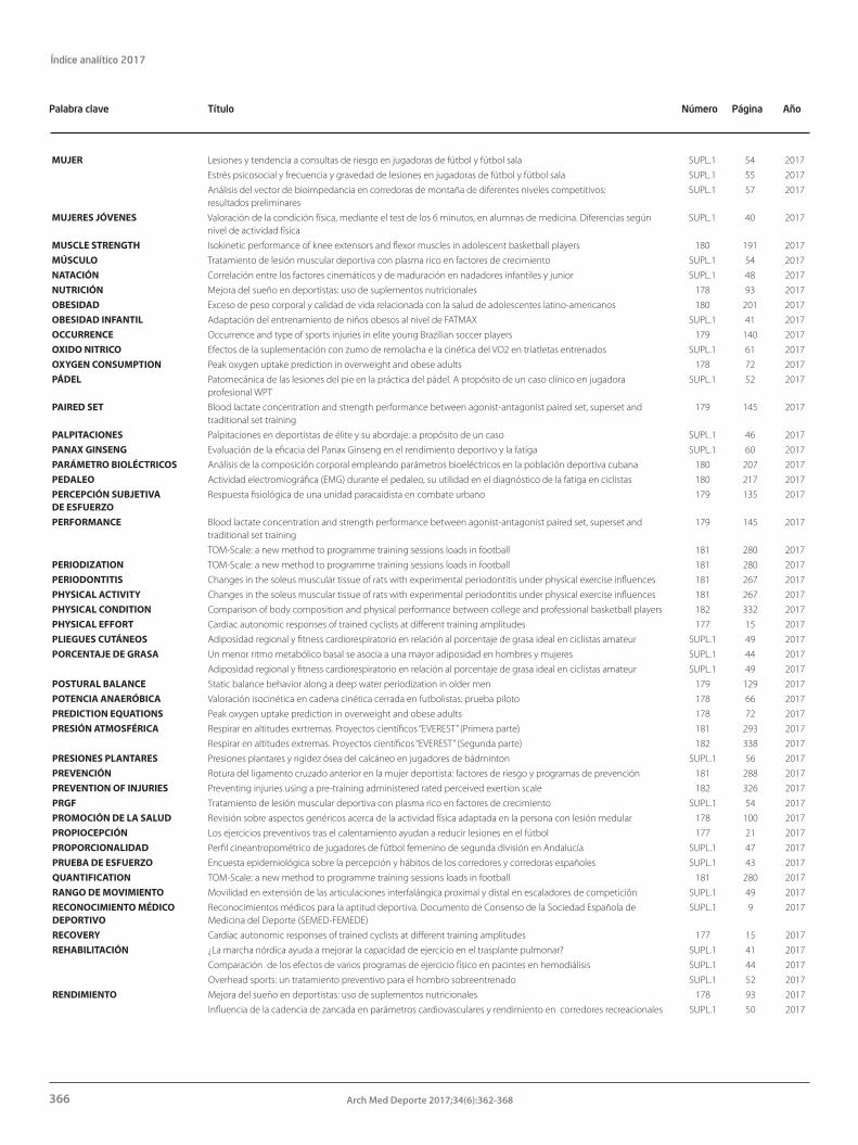
Índice analítico 2017
366 Arch Med Deporte 2017;34(6):362-368
MUJER Lesiones y tendencia a consultas de riesgo en jugadoras de fútbol y fútbol sala SUPL.1 54 2017 Estrés psicosocial y frecuencia y gravedad de lesiones en jugadoras de fútbol y fútbol sala SUPL.1 55 2017 Análisis del vector de bioimpedancia en corredoras de montaña de diferentes niveles competitivos: SUPL.1 57 2017 resultados preliminares MUJERES JÓVENES Valoración de la condición física, mediante el test de los 6 minutos, en alumnas de medicina. Diferencias según SUPL.1 40 2017 nivel de actividad física MUSCLE STRENGTH Isokinetic performance of knee extensors and flexor muscles in adolescent basketball players 180 191 2017MÚSCULO Tratamiento de lesión muscular deportiva con plasma rico en factores de crecimiento SUPL.1 54 2017NATACIÓN Correlación entre los factores cinemáticos y de maduración en nadadores infantiles y junior SUPL.1 48 2017NUTRICIÓN Mejora del sueño en deportistas: uso de suplementos nutricionales 178 93 2017OBESIDAD Exceso de peso corporal y calidad de vida relacionada con la salud de adolescentes latino-americanos 180 201 2017OBESIDAD INFANTIL Adaptación del entrenamiento de niños obesos al nivel de FATMAX SUPL.1 41 2017OCCURRENCE Occurrence and type of sports injuries in elite young Brazilian soccer players 179 140 2017OXIDO NITRICO Efectos de la suplementación con zumo de remolacha e la cinética del VO2 en triatletas entrenados SUPL.1 61 2017OXYGEN CONSUMPTION Peak oxygen uptake prediction in overweight and obese adults 178 72 2017PÁDEL Patomecánica de las lesiones del pie en la práctica del pádel. A propósito de un caso clínico en jugadora SUPL.1 52 2017 profesional WPT PAIRED SET Blood lactate concentration and strength performance between agonist-antagonist paired set, superset and 179 145 2017 traditional set training PALPITACIONES Palpitaciones en deportistas de élite y su abordaje: a propósito de un caso SUPL.1 46 2017PANAX GINSENG Evaluación de la eficacia del Panax Ginseng en el rendimiento deportivo y la fatiga SUPL.1 60 2017PARÁMETRO BIOLÉCTRICOS Análisis de la composición corporal empleando parámetros bioeléctricos en la población deportiva cubana 180 207 2017PEDALEO Actividad electromiográfica (EMG) durante el pedaleo, su utilidad en el diagnóstico de la fatiga en ciclistas 180 217 2017PERCEPCIÓN SUBJETIVA Respuesta fisiológica de una unidad paracaidista en combate urbano 179 135 2017 DE ESFUERZO PERFORMANCE Blood lactate concentration and strength performance between agonist-antagonist paired set, superset and 179 145 2017 traditional set training TOM-Scale: a new method to programme training sessions loads in football 181 280 2017PERIODIZATION TOM-Scale: a new method to programme training sessions loads in football 181 280 2017PERIODONTITIS Changes in the soleus muscular tissue of rats with experimental periodontitis under physical exercise influences 181 267 2017PHYSICAL ACTIVITY Changes in the soleus muscular tissue of rats with experimental periodontitis under physical exercise influences 181 267 2017PHYSICAL CONDITION Comparison of body composition and physical performance between college and professional basketball players 182 332 2017PHYSICAL EFFORT Cardiac autonomic responses of trained cyclists at different training amplitudes 177 15 2017PLIEGUES CUTÁNEOS Adiposidad regional y fitness cardiorespiratorio en relación al porcentaje de grasa ideal en ciclistas amateur SUPL.1 49 2017PORCENTAJE DE GRASA Un menor ritmo metabólico basal se asocia a una mayor adiposidad en hombres y mujeres SUPL.1 44 2017 Adiposidad regional y fitness cardiorespiratorio en relación al porcentaje de grasa ideal en ciclistas amateur SUPL.1 49 2017POSTURAL BALANCE Static balance behavior along a deep water periodization in older men 179 129 2017POTENCIA ANAERÓBICA Valoración isocinética en cadena cinética cerrada en futbolistas: prueba piloto 178 66 2017PREDICTION EQUATIONS Peak oxygen uptake prediction in overweight and obese adults 178 72 2017PRESIÓN ATMOSFÉRICA Respirar en altitudes exrtremas. Proyectos científicos “EVEREST” (Primera parte) 181 293 2017 Respirar en altitudes extremas. Proyectos científicos “EVEREST” (Segunda parte) 182 338 2017PRESIONES PLANTARES Presiones plantares y rigidez ósea del calcáneo en jugadores de bádminton SUPL.1 56 2017PREVENCIÓN Rotura del ligamento cruzado anterior en la mujer deportista: factores de riesgo y programas de prevención 181 288 2017PREVENTION OF INJURIES Preventing injuries using a pre-training administered rated perceived exertion scale 182 326 2017PRGF Tratamiento de lesión muscular deportiva con plasma rico en factores de crecimiento SUPL.1 54 2017PROMOCIÓN DE LA SALUD Revisión sobre aspectos genéricos acerca de la actividad física adaptada en la persona con lesión medular 178 100 2017PROPIOCEPCIÓN Los ejercicios preventivos tras el calentamiento ayudan a reducir lesiones en el fútbol 177 21 2017PROPORCIONALIDAD Perfil cineantropométrico de jugadores de fútbol femenino de segunda división en Andalucía SUPL.1 47 2017PRUEBA DE ESFUERZO Encuesta epidemiológica sobre la percepción y hábitos de los corredores y corredoras españoles SUPL.1 43 2017QUANTIFICATION TOM-Scale: a new method to programme training sessions loads in football 181 280 2017RANGO DE MOVIMIENTO Movilidad en extensión de las articulaciones interfalángica proximal y distal en escaladores de competición SUPL.1 49 2017RECONOCIMIENTO MÉDICO Reconocimientos médicos para la aptitud deportiva. Documento de Consenso de la Sociedad Española de SUPL.1 9 2017 DEPORTIVO Medicina del Deporte (SEMED-FEMEDE) RECOVERY Cardiac autonomic responses of trained cyclists at different training amplitudes 177 15 2017REHABILITACIÓN ¿La marcha nórdica ayuda a mejorar la capacidad de ejercicio en el trasplante pulmonar? SUPL.1 41 2017 Comparación de los efectos de varios programas de ejercicio físico en pacintes en hemodiálisis SUPL.1 44 2017 Overhead sports: un tratamiento preventivo para el hombro sobreentrenado SUPL.1 52 2017RENDIMIENTO Mejora del sueño en deportistas: uso de suplementos nutricionales 178 93 2017 Influencia de la cadencia de zancada en parámetros cardiovasculares y rendimiento en corredores recreacionales SUPL.1 50 2017
Palabra clave Título Número Página Año

Índice analítico 2017
367Arch Med Deporte 2017;34(6):362-368
Análisis de las diferencias físico-técnicas entre fútbol 7 y fútbol 8 en competición oficial SUPL.1 51 2017 Valores de vitamina D en deportistas de alto rendimiento españoles de distintas modalidades deportivas SUPL.1 59 2017RENDIMIENTO DEPORTIVO Efecto de la hipoxia intermitente (HI) sobre los indicadores renales del metabolismo proteico en atletas SUPL.1 49 2017 Evaluación de la eficacia del Panax Ginseng en el rendimiento deportivo y la fatiga SUPL.1 60 2017REPOLARIZACIÓN Comparación de hallazgos cardiológicos entre dos poblaciones de deportistas de élite SUPL.1 45 2017 Prevalencia de las alteraciones de la repolarización en el ECG de jugadores de baloncesto profesionales SUPL.1 45 2017 Deporte de élite en paciente con valvulopatía. A propósito de un caso SUPL.1 46 2017RESPIRACIÓN Respirar en altitudes extrremas. Proyectos científicos “EVEREST” (Primera parte) 181 293 2017 Respirar en altitudes extremas. Proyectos científicos “EVEREST” (Segunda parte) 182 338 2017RESPIRATORY GAS EXCHANGE Do the changes in acid-base status in respiratory gas exchange explain the voluntary termination of a test 178 80 2017 performed above the maximum lactate steady state? RESPUESTAS VENTILATORIAS Efectos de la suplementación con zumo de remolacha e la cinética del VO2 en triatletas entrenados SUPL.1 61 2017RETORNO AL DEPORTE Criterios para el retorno al deporte después de una lesión 177 40 2017REWARMING Frostbite: management update 182 345 2017ILOPROST Frostbite: management update 182 345 2017RIESGO LESIONAL Análisis de la fuerza y movilidad de la cadera como factores de riesgo de lesión en fútbol femenino amateur: 177 25 2017 un estudio piloto RITMO MÁXIMO DE OXIDACIÓN Un menor ritmo metabólico basal se asocia a una mayor adiposidad en hombres y mujeres SUPL.1 44 2017RITMO METABÓLICO BASAL Un menor ritmo metabólico basal se asocia a una mayor adiposidad en hombres y mujeres SUPL.1 44 2017ROTACIÓN EXTERNA DE Análisis de la fuerza y movilidad de la cadera como factores de riesgo de lesión en fútbol femenino amateur: 177 25 2017 LA CADERA un estudio piloto RUNNERS Encuesta epidemiológica sobre la percepción y hábitos de los corredores y corredoras españoles SUPL.1 43 2017SALUD DEL ADOLESCENTE Exceso de peso corporal y calidad de vida relacionada con la salud de adolescentes latino-americanos 180 201 2017SALUD SUBJETIVA Inactividad física en el tiempo libre y auto percepción del estado de salud de colombianos entre los 18 y 64 años 181 260 2017SEDENTARISMO Medición objetiva de la actividad física en centenarios españoles SUPL.1 43 2017SEDENTARY Peak oxygen uptake prediction in overweight and obese adults 178 72 2017SÍNCOPE Abordaje del síncope relacionado con el deporte 179 157 2017SOBREPESO Exceso de peso corporal y calidad de vida relacionada con la salud de adolescentes latino-americanos 180 201 2017SOCCER Occurrence and type of sports injuries in elite young Brazilian soccer players 179 140 2017SOLDADO Respuesta fisiológica de una unidad paracaidista en combate urbano 179 135 2017SOLEUS MUSCLE Changes in the soleus muscular tissue of rats with experimental periodontitis under physical exercise influences 181 267 2017SOMATOTIPO Perfil cineantropométrico de jugadores de fútbol femenino de segunda división en Andalucía SUPL.1 47 2017STATIC APNEA Influence of intermittent aerobic performance on the variables of static and dynamic apnea performances 181 274 2017STRENGTH TRAINING Hypertrophy training improves glycaemic and inflammatory parameters in men with risk factors 182 315 2017INFLAMMATION Hypertrophy training improves glycaemic and inflammatory parameters in men with risk factors 182 315 2017STRENGTH Blood lactate concentration and strength performance between agonist-antagonist paired set, superset and 179 145 2017 traditional set training SUBJECTIVE PERCEPTION Preventing injuries using a pre-training administered rated perceived exertion scale 182 326 2017SUEÑO Mejora del sueño en deportistas: uso de suplementos nutricionales 178 93 2017SUPLEMENTACIÓN Relevancia de la suplementación de hierro en una competición de ciclismo profesional: evaluación hematológica SUPL.1 60 2017SUPLEMENTO Mejora del sueño en deportistas: uso de suplementos nutricionales 178 93 2017SUPLEMENTOS ALIMENTICIOS Dopaje versus suplementos alimenticios en triatletas de élite SUPL.1 59 2017TAQUICARDIA Palpitaciones en deportistas de élite y su abordaje: a propósito de un caso SUPL.1 46 2017TENDENCIA AL RIESGO Lesiones y tendencia a consultas de riesgo en jugadoras de fútbol y fútbol sala SUPL.1 54 2017TENDÓN Valoración del sistema flexor de los dedos en escaladores de competición: ecografía como herramienta SUPL.1 54 2017 complementaria TERMOGRAFÍA Efectos agudos de la práctica del bádminton sobre la temperatura superficial de los miembros inferiores SUPL.1 56 2017TEST DE LOS 6 MINUTOS Valoración de la condición física, mediante el test de los 6 minutos, en alumnas de medicina. Diferencias según SUPL.1 40 2017 nivel de actividad física TOLERANCIA Tolerancia a los efectos ergogénicos de la cafeína con una ingesta continuada SUPL.1 61 2017TRABAJO TOTAL Características de los parámetros isocinéticos en personas con gonalgia leve SUPL.1 42 2017TRAINING LOAD Preventing injuries using a pre-training administered rated perceived exertion scale 182 326 2017TRASPLANTE PULMÓN ¿La marcha nórdica ayuda a mejorar la capacidad de ejercicio en el trasplante pulmonar? SUPL.1 41 2017TRATAMIENTO Patomecánica de las lesiones del pie en la práctica del pádel. A propósito de un caso clínico en jugadora SUPL.1 52 2017 ORTOPODOLÓGICO profesional WPT TRIATLÓN Dopaje versus suplementos alimenticios en triatletas de élite SUPL.1 59 2017ULTRASONIDO Variables antropométricas y ultrasonográficas en la determinación de la edad ósea. Estudio preliminar SUPL.1 58 2017VALIDEZ Estudio de la validez en la medición de los valores de lactato sanguíneo entre los dos modelos existentes de LactatePro 178 86 2017VALVULOPATÍA Deporte de élite en paciente con valvulopatía. A propósito de un caso SUPL.1 46 2017
Palabra clave Título Número Página Año
Índice analítico 2017
368 Arch Med Deporte 2017;34(6):362-368
VARIABILIDAD FRECUENCIA Diferentes temperaturas varían la potencia de alta frecuencia de la variabilidad de la frecuencia cardiaca SUPL.1 50 2017 CARDIACA VECTOR BIOELÉCTRICO Análisis del vector de bioimpedancia en corredoras de montaña de diferentes niveles competitivos: resultados SUPL.1 57 2017 preliminares VELOCIDAD AERÓBICA MÁXIMA Adaptaciones de la mecánica de carrera en tapiz rodante en velocidades incrementadas SUB y SUPRA VAM SUPL.1 51 2017VITAMINA D Valores de vitamina D en deportistas de alto rendimiento españoles de distintas modalidades deportivas SUPL.1 59 2017WEEKLY FREQUENCY The effect of weekly low frequency exercise on body composition and blood pressure of elderly women 177 9 2017WEIGHT LOSS Strategies to reduce pre-competition body weight in mixed martial arts 182 321 2017
Palabra clave Título Número Página Año
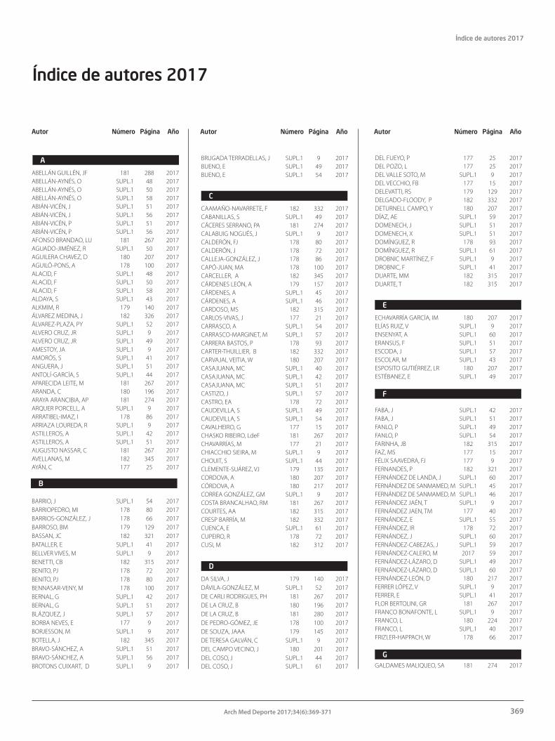
Índice de autores 2017
369Arch Med Deporte 2017;34(6):369-371
ABELLÁN GUILLÉN, JF 181 288 2017ABELLÁN-AYNÉS, O SUPL.1 48 2017ABELLÁN-AYNÉS, O SUPL.1 50 2017ABELLÁN-AYNÉS, O SUPL.1 58 2017ABIÁN-VICÉN, J SUPL.1 51 2017ABIÁN-VICÉN, J SUPL.1 56 2017ABIÁN-VICÉN, P SUPL.1 51 2017ABIÁN-VICÉN, P SUPL.1 56 2017AFONSO BRANDAO, LU 181 267 2017AGUADO-JIMÉNEZ, R SUPL.1 50 2017AGUILERA CHAVEZ, D 180 207 2017AGUILÓ-PONS, A 178 100 2017ALACID, F SUPL.1 48 2017ALACID, F SUPL.1 50 2017ALACID, F SUPL.1 58 2017ALDAYA, S SUPL.1 43 2017ALKMIM, R 179 140 2017ÁLVAREZ MEDINA, J 182 326 2017ÁLVAREZ-PLAZA, PY SUPL.1 52 2017ALVERO CRUZ, JR SUPL.1 9 2017ALVERO CRUZ, JR SUPL.1 49 2017AMESTOY, JA SUPL.1 9 2017AMORÓS, S SUPL.1 41 2017ANGUERA, J SUPL.1 51 2017ANTOLÍ-GARCÍA, S SUPL.1 44 2017APARECIDA LEITE, M 181 267 2017ARANDA, C 180 196 2017ARAYA ARANCIBIA, AP 181 274 2017ARQUER PORCELL, A SUPL.1 9 2017ARRATIBEL-IMAZ, I 178 86 2017ARRIAZA LOUREDA, R SUPL.1 9 2017ASTILLEROS, A SUPL.1 42 2017ASTILLEROS, A SUPL.1 51 2017AUGUSTO NASSAR, C 181 267 2017AVELLANAS, M 182 345 2017AYÁN, C 177 25 2017
BARRIO, J SUPL.1 54 2017BARRIOPEDRO, MI 178 80 2017BARRIOS-GONZÁLEZ, J 178 66 2017BARROSO, BM 179 129 2017BASSAN, JC 182 321 2017BATALLER, E SUPL.1 41 2017BELLVER VIVES, M SUPL.1 9 2017BENETTI, CB 182 315 2017BENITO, PJ 178 72 2017BENITO, PJ 178 80 2017BENNASAR-VENY, M 178 100 2017BERNAL, G SUPL.1 42 2017BERNAL, G SUPL.1 51 2017BLÁZQUEZ, J SUPL.1 57 2017BORBA NEVES, E 177 9 2017BORJESSON, M SUPL.1 9 2017BOTELLA, J 182 345 2017BRAVO-SÁNCHEZ, A SUPL.1 51 2017BRAVO-SÁNCHEZ, A SUPL.1 56 2017BROTONS CUIXART, D SUPL.1 9 2017
BRUGADA TERRADELLAS, J SUPL.1 9 2017BUENO, E SUPL.1 49 2017BUENO, E SUPL.1 54 2017
CAAMAÑO-NAVARRETE, F 182 332 2017CABANILLAS, S SUPL.1 49 2017CÁCERES SERRANO, PA 181 274 2017CALABUIG NOGUÉS, J SUPL.1 9 2017CALDERÓN, FJ 178 80 2017CALDERÓN, J 178 72 2017CALLEJA-GONZÁLEZ, J 178 86 2017CAPÓ-JUAN, MA 178 100 2017CARCELLER, A 182 345 2017CÁRDENES LEÓN, A 179 157 2017CÁRDENES, A SUPL.1 45 2017CÁRDENES, A SUPL.1 46 2017CARDOSO, MS 182 315 2017CARLOS-VIVAS, J 177 21 2017CARRASCO, A SUPL.1 54 2017CARRASCO-MARGINET, M SUPL.1 57 2017CARRERA BASTOS, P 178 93 2017CARTER-THUILLIER, B 182 332 2017CARVAJAL VEITIA, W 180 207 2017CASAJUANA, MC SUPL.1 40 2017CASAJUANA, MC SUPL.1 42 2017CASAJUANA, MC SUPL.1 51 2017CASTIZO, J SUPL.1 57 2017CASTRO, EA 178 72 2017CAUDEVILLA, S SUPL.1 49 2017CAUDEVILLA, S SUPL.1 54 2017CAVALHEIRO, G 177 15 2017CHASKO RIBEIRO, LdeF 181 267 2017CHAVARRÍAS, M 177 21 2017CHIACCHIO SIEIRA, M SUPL.1 9 2017CHOUIT, S SUPL.1 44 2017CLEMENTE-SUÁREZ, VJ 179 135 2017CORDOVA, A 180 207 2017CÓRDOVA, A 180 217 2017CORREA GONZÁLEZ, GM SUPL.1 9 2017COSTA BRANCALHAO, RM 181 267 2017COURTES, AA 182 315 2017CRESP BARRÍA, M 182 332 2017CUENCA, E SUPL.1 61 2017CUPEIRO, R 178 72 2017CUSI, M 182 312 2017
DA SILVA, J 179 140 2017DÁVILA-GONZÁLEZ, M SUPL.1 52 2017DE CARLI RODRIGUES, PH 181 267 2017DE LA CRUZ, B 180 196 2017DE LA CRUZ, B 181 280 2017DE PEDRO-GÓMEZ, JE 178 100 2017DE SOUZA, JAAA 179 145 2017DE TERESA GALVÁN, C SUPL.1 9 2017DEL CAMPO VECINO, J 180 201 2017DEL COSO, J SUPL.1 44 2017DEL COSO, J SUPL.1 61 2017
Autor Número Página Año
Índice de autores 2017
DEL FUEYO, P 177 25 2017DEL POZO, L 177 25 2017DEL VALLE SOTO, M SUPL.1 9 2017DEL VECCHIO, FB 177 15 2017DELEVATTI, RS 179 129 2017DELGADO-FLOODY, P 182 332 2017DETURNELL CAMPO, Y 180 207 2017DÍAZ, AE SUPL.1 59 2017DOMENECH, J SUPL.1 51 2017DOMENECH, X SUPL.1 51 2017DOMÍNGUEZ, R 178 93 2017DOMÍNGUEZ, R SUPL.1 61 2017DROBNIC MARTÍNEZ, F SUPL.1 9 2017DROBNIC, F SUPL.1 41 2017DUARTE, MM 182 315 2017DUARTE, T 182 315 2017
ECHAVARRÍA GARCÍA, IM 180 207 2017ELÍAS RUIZ, V SUPL.1 9 2017ENSENYAT, A SUPL.1 60 2017ERANSUS, F SUPL.1 51 2017ESCODA, J SUPL.1 57 2017ESCOLAR, M SUPL.1 43 2017ESPOSITO GUTIÉRREZ, LR 180 207 2017ESTÉBANEZ, E SUPL.1 49 2017
FABA, J SUPL.1 42 2017FABA, J SUPL.1 51 2017FANLO, P SUPL.1 49 2017FANLO, P SUPL.1 54 2017FARINHA, JB 182 315 2017FAZ, MS 177 15 2017FÉLIX SAAVEDRA, FJ 177 9 2017FERNANDES, P 182 321 2017FERNÁNDEZ DE LANDA, J SUPL.1 60 2017FERNÁNDEZ DE SANMAMED, M SUPL.1 45 2017FERNÁNDEZ DE SANMAMED, M SUPL.1 46 2017FERNÁNDEZ JAÉN, T SUPL.1 9 2017FERNÁNDEZ JAEN, TM 177 40 2017FERNÁNDEZ, E SUPL.1 55 2017FERNÁNDEZ, IR 178 72 2017FERNÁNDEZ, J SUPL.1 60 2017FERNÁNDEZ-CABEZAS, J SUPL.1 59 2017FERNÁNDEZ-CALERO, M 2017 59 2017FERNÁNDEZ-LÁZARO, D SUPL.1 49 2017FERNÁNDEZ-LÁZARO, D SUPL.1 60 2017FERNÁNDEZ-LEÓN, D 180 217 2017FERRER LÓPEZ, V SUPL.1 9 2017FERRER, E SUPL.1 41 2017FLOR BERTOLINI, GR 181 267 2017FRANCO BONAFONTE, L SUPL.1 9 2017FRANCO, L 180 224 2017FRANCO, L SUPL.1 40 2017FRIZLER-HAPPACH, W 178 66 2017
GALDAMES MALIQUEO, SA 181 274 2017
Autor Número Página Año Autor Número Página Año
A
B
C
D
E
F
G

Índice de autores 2017
370 Arch Med Deporte 2017;34(6):369-371
O
GALLARDO-FUENTES, F 182 332 2017GALVÁN, M SUPL.1 45 2017GALVÁN, M SUPL.1 46 2017GARACHATEA, N SUPL.1 43 2017GARCÍA GARCÍA, EM 181 288 2017GARCÍA SALVADOR, JJ 179 157 2017GARCÍA SALVADOR, JJ SUPL.1 45 2017GARCIA ZAPICO, P SUPL.1 9 2017GARCÍA, P SUPL.1 61 2017GARCÍA-MASET, R SUPL.1 44 2017GARCÍA-NIETO PORTABELLA, JN SUPL.1 9 2017GARCÍA-PASTOR, T SUPL.1 44 2017GARCÍA-PINILLOS, F 182 332 2017GARMILLA, I SUPL.1 54 2017GARNACHO, MA SUPL.1 61 2017GARNACHO, MV SUPL.1 61 2017GARRIDO, E 181 293 2017GARRIDO, E 182 338 2017GÁZQUEZ, O SUPL.1 41 2017GAZTAÑAGA AURREKOETXEA, T SUPL.1 9 2017GIANPIETRO BRANDAO, C 181 267 2017GIL, J SUPL.1 40 2017GIRALT, M SUPL.1 40 2017GIRALT, M SUPL.1 42 2017GODOY, E 179 140 2017GOMEÑUCA, NA 179 129 2017GÓMEZ-ESPEJO, V SUPL.1 54 2017GÓMEZ-GARRIDO, A SUPL.1 41 2017GÓMEZ-TRULLEN, EM SUPL.1 43 2017GOMÍS, M SUPL.1 61 2017GONÇALVES, D 179 140 2017GONZÁLEZ LAGO, L SUPL.1 9 2017GONZÁLEZ, E SUPL.1 43 2017GONZÁLEZ, R SUPL.1 42 2017GONZÁLEZ, R SUPL.1 51 2017GRAU, I SUPL.1 52 2017GRAZZIOLI, G SUPL.1 57 2017GUILLÉN GARCÍA, P 177 40 2017GUIRAO, L SUPL.1 61 2017GUISADO BARRILAO, R 181 274 2017GUTIÉRREZ ORTEGA, F SUPL.1 9 2017
HERDY, C 179 140 2017HERNÁNDEZ, D SUPL.1 60 2017HERNÁNDEZ, M SUPL.1 41 2017HERNÁNDEZ, O SUPL.1 57 2017HERNÁNDEZ-ABAD, F SUPL.1 52 2017HERNÁNDEZ-VICENTE, A SUPL.1 43 2017HERRERO, A SUPL.1 41 2017HIDALGO, C SUPL.1 54 2017HUELIN TRILLO, F SUPL.1 9 2017HUERTA OJEDA, AC 181 274 2017
IRURTIA, A SUPL.1 57 2017IZQUIERDO RODRÍGUEZ, E 182 321 2017
JANSA, M SUPL.1 41 2017JAVIERRE, C 182 345 2017JEREZ-MAYORGA, D 182 332 2017JIMÉNEZ MANGAS, R SUPL.1 9 2017JIMÉNEZ, S SUPL.1 60 2017
JIMÉNEZ-DÍAZ, F SUPL.1 56 2017JULIAO-CASTILLO, J 177 31 2017
KANITZ, AC 179 129 2017KRUEL, LFM 179 129 2017
LACLETA ALMOLDA, JJ SUPL.1 9 2017LAGO, J 177 25 2017LARA, B SUPL.1 61 2017LATASA, I 180 217 2017LATASA, I SUPL.1 49 2017LATASA, I SUPL.1 60 2017LATORRE-ROMÁN, P 182 332 2017LIEDTKE, GV 179 129 2017LIMA, V 179 140 2017LLOBET, M SUPL.1 41 2017LLORCA GARNERO, J SUPL.1 9 2017LLUNA LLORENS, AD 181 288 2017LOEZA MAGAÑA, P 178 66 2017LÓPEZ-LLUCH, G SUPL.1 47 2017LÓPEZ-PLAZA, D SUPL.1 48 2017LÓPEZ-PLAZA, D SUPL.1 50 2017LÓPEZ-PLAZA, D SUPL.1 58 2017LOZANO, E SUPL.1 41 2017LU, D SUPL.1 40 2017LUENGO FERNÁNDEZ, E SUPL.1 9 2017
MACHADO REIS, V 177 9 2017MAESTRO, A 177 25 2017MALO, M SUPL.1 49 2017MALO, M SUPL.1 54 2017MANONELLES MARQUETA, P SUPL.1 9 2017MANONELLES, P SUPL.1 43 2017MANONELLES, P SUPL.1 48 2017MANONELLES, P SUPL.1 50 2017MANONELLES, P SUPL.1 58 2017MANUZ GONZÁLEZ, B SUPL.1 9 2017MARGALEF, R SUPL.1 51 2017MARINI, C 180 191 2017MARRERO-GORDILLO, N SUPL.1 52 2017MARTÍN CASTELLANOS, A SUPL.1 9 2017MARTÍN, V 177 25 2017MARTINAZZO BORTOLINI, B 181 267 2017MARTÍNEZ-SALAZAR, C 182 332 2017MARTÍN-MARTÍNEZ, JP 177 21 2017MATA-ORDÓÑEZ F 178 93 2017MATÉ, JL SUPL.1 61 2017MAYOLAS-PI, C SUPL.1 43 2017MEDINA FDEZ. ACEYTUNO, A 179 157 2017MEDRANO MORTE, I 181 288 2017MELÉNDEZ-OLIVA, E SUPL.1 44 2017MENDOZA ROMERO, D 181 260 2017MIELGO AYUSO, J SUPL.1 49 2017MIELGO AYUSO, J SUPL.1 60 2017MINOBES, E SUPL.1 40 2017MIQUELEIZ, U SUPL.1 50 2017MIQUELEIZ, U SUPL.1 51 2017MIRANDA, H 179 145 2017MIRANDA, S SUPL.1 54 2017MONTALVO ZENARRUZABEITIA, Z SUPL.1 9 2017MORAGÓN ABADA, FJ SUPL.1 9 2017
MORENO, A SUPL.1 40 2017MORESCO, RN 182 315 2017MORILLAS MARTÍNEZ, JM SUPL.1 9 2017MOYA MORALES, JM 180 201 2017MUÑOZ LÓPEZ, A 181 280 2017MUÑOZ-GUERRA, JA 2017 59 2017MUÑOZ-PUENTE, I SUPL.1 44 2017MURILLO LORENTE, V 182 326 2017
NARANJO ORELLANA, J SUPL.1 9 2017NARANJO, J 180 196 2017NARANJO, J 181 280 2017NAUDÍ, A SUPL.1 60 2017NOGUÉS, R SUPL.1 40 2017NOVAES, J 179 140 2017NOVELLA MARÍA-FERNÁNDEZ, F SUPL.1 9 2017NUIN, I 180 217 2017
OCEJO VIÑALS, C SUPL.1 9 2017OEHLMEYER NASSAR, P 181 267 2017OJEDA, I SUPL.1 49 2017OJEDA, I SUPL.1 60 2017OLIVER, D 180 224 2017OLMEDILLA, A SUPL.1 54 2017OLMEDILLA, A SUPL.1 55 2017ORTEGA, E SUPL.1 54 2017ORTEGA, E SUPL.1 55 2017OYÓN, P SUPL.1 40 2017
PAIVA ALEGRE-MALLER, AC 181 267 2017PALACÍN, H SUPL.1 57 2017PALACIOS, N SUPL.1 59 2017PALACIOS, N SUPL.1 60 2017PALAZÓN, A SUPL.1 41 2017PAREJA-GALEANO, H SUPL.1 43 2017PAZ, GA 179 145 2017PEINADO, AB 178 72 2017PEINADO, AB 178 80 2017PÉREZ, L SUPL.1 42 2017PÉREZ, L SUPL.1 51 2017PÉREZ-DOMÍNGUEZ, B SUPL.1 44 2017PÉREZ-GÓMEZ, J 177 21 2017PERIS, F 180 224 2017PIAZZA, F 180 191 2017PICANÇO, LM 177 15 2017PIFARRÉ, F SUPL.1 57 2017PIGOZZI, F SUPL.1 9 2017PINTO GUEDES, D 180 201 2017PIRES JR, R 180 201 2017PIROLLI, PM 180 201 2017PLEGUEZUELOS, E SUPL.1 61 2017POHL, HH 177 9 2017POLO GALLARDO, R 177 31 2017PORTA, J SUPL.1 57 2017POVEDA, J 180 224 2017POZAS SÁNCHEZ, M SUPL.1 9 2017PRAT, JA SUPL.1 57 2017
QUERO, C SUPL.1 48 2017QUERO, C SUPL.1 50 2017
Autor Número Página Año Autor Número Página Año Autor Número Página Año
M
LN
P
Q
H
I
J
K

Índice de autores 2017
371Arch Med Deporte 2017;34(6):369-371
QUERO, C SUPL.1 58 2017QUINTANA CASANOVA, CA 179 157 2017QUINTANA, C SUPL.1 45 2017
RAMIREZ-CAMPILLO, R 182 332 2017RAMÓN, M SUPL.1 41 2017RANCHAL SÁNCHEZ, A 179 152 2017REBOLLEDO-COBOS, R 177 31 2017RECKZIEGEL, MB 177 9 2017REICHERT, T 179 129 2017REVUELTA, G 177 25 2017REYES-LAREDO, F SUPL.1 47 2017ROBLES-PALAZÓN, FJ SUPL.1 54 2017ROBLES-PÉREZ, JJ 179 135 2017ROCA, R SUPL.1 41 2017RODRÍGUEZ, FA SUPL.1 57 2017RODRÍGUEZ-BIES, E SUPL.1 47 2017RODRÍGUEZ-FALCES, J 180 217 2017ROMÁN, A SUPL.1 41 2017ROMANOVITCH RIBAS, M 182 321 2017ROMEU, M SUPL.1 40 2017ROSA, C 177 9 2017ROSSELLÓ, L SUPL.1 57 2017ROY, A SUPL.1 57 2017RUBIO PÉREZ, FJ SUPL.1 9 2017RUBIO, FJ 180 224 2017RUBIO, FJ SUPL.1 40 2017RUIZ-MORENO, C SUPL.1 61 2017
SACILOTO TADIELLO, G 180 191 2017SÁEZ, M SUPL.1 41 2017
SALGUEIRO, D SUPL.1 40 2017SALINERO, JJ SUPL.1 61 2017SALOM PORTELLA, F SUPL.1 9 2017SÁNCHEZ GUILLÉN, L 178 93 2017SÁNCHEZ LÓPEZ, S 181 288 2017SÁNCHEZ MARTÍNEZ, J SUPL.1 9 2017SÁNCHEZ OLIVER, AJ 178 93 2017SÁNCHEZ RAMOS, A SUPL.1 9 2017SÁNCHEZ SABATER, B 181 288 2017SÁNCHEZ-MOLINA, J 179 135 2017SÁNCHEZ-RAYA, J SUPL.1 41 2017SANTOS, DL 182 315 2017SANTOS-LOZANO, A SUPL.1 43 2017SANUY, X SUPL.1 57 2017SAURA, E SUPL.1 59 2017SCHEFFEL, M 182 321 2017SEGURA CASADO, L SUPL.1 9 2017SEGURA-ORTÍ, E SUPL.1 44 2017SELFE, J 179 140 2017SERRA, N SUPL.1 61 2017SERRANO, J SUPL.1 60 2017SETUAIN, I SUPL.1 51 2017SHIMA LUIZE, D 181 267 2017SIBILA, O 181 293 2017SIBILA, O 182 338 2017SILVA CORREA, C 177 31 2017SIMAO, R 179 140 2017SIMÓN DE LA TORRE, I SUPL.1 9 2017SINGH, M SUPL.1 45 2017SINGH, M SUPL.1 46 2017SIRONVALLE ARGANDOÑA, PD 181 274 2017SOARES, FA 182 315 2017SOLÁ, RM SUPL.1 42 2017SOLDATELLI ZARDO, B 180 191 2017STRUNK, R SUPL.1 60 2017
SUÁREZ LANDAZABAL, O 177 31 2017
TERRADOS CEPEDA, N SUPL.1 9 2017TERRADOS, N 178 86 2017TERREROS BLANCO, JL SUPL.1 9 2017TIL PÉREZ, LL SUPL.1 9 2017TORRIJOS-MONTALBÁN, A SUPL.1 56 2017
URBINA, A 181 260 2017USÁN SUPERVÍA, P 182 326 2017
VALE, R 179 140 2017VALERO, A 180 224 2017VARGAS, LS 182 315 2017VEIGA, P SUPL.1 61 2017VICO GUZMÁN, J SUPL.1 49 2017VIÇOSA BONETTI, L 180 191 2017VILAÇA-ALVES, E 177 9 2017VILLAGRA ASTUDILLO, HA 180 201 2017VISCOR, G 181 293 2017VISCOR, G 182 338 2017VISCOR, G 182 345 2017
YÁNEZ-CASTRO, P SUPL.1 52 2017
ZANINI, D 177 9 2017
Autor Número Página Año Autor Número Página Año Autor Número Página Año
T
U
V
S
R
Z
Y

Evaluadores
372 Arch Med Deporte 2017;34(6):372-373
Lista de revisores evaluadores externos de los artículos recibidos en 2017 en Archivos
Abellán Guillén, Juan Francisco (Hospital Morales Meseguer.
Murcia)
Alacid Cárceles, Fernando (Universidad Católica San Antonio de
Murcia)
Alegre de Miquel, Cayetano (Hospital Quirón. Barcelona)
Álvarez Herms, Jesús (Universidad de Barcelona)
Álvarez Medina, Javier (Universidad de Zaragoza)
Álvarez Rodríguez, Cesáreo (Hospital de Verín. Orense)
Alvear Órdenes, Ildefonso (Universidad de León)
Amani, Ali Reza (Shomal University. Iran)
Arias Poblete, Leónidas E (Universidad de las Américas, Santiago. Chile)
Arroyo Toledo, Juan Jaime (University of Leipzig. Alemania)
Avilés Villarroel, Carlos (Universidad Complutense. Madrid)
Ayán Pérez, Carlos Luis (Universidad de Vigo)
Baiget Vidal, Ernest (Universidad de Vic. Barcelona)
Bescós García, Raúl (Plymouth University. Reino Unido)
Blasco Redondo, Raquel (Universidad de Valladolid)
Brea Alejo, Lidia (Universidad Europea de Madrid)
Bresciani, Ghilheme (Pontificia Universidad Católica de Valparaíso
-PUCV- Chile)
Caballero Dorta, Eduardo (Hospital Universitario de Gran Canaria
Dr. Negrín. Las Palmas)
Calleja de Frutos, Carlos (Universidad Antonio de Nebrija. Madrid)
Campos Jara, Christian (Universidad Andrés Bello. Chile)
Cancino López, Jorge (Universidad Mayor Santiago. Chile)
Cappa, Darío Fernando (Universidad Memorial de Canadá. Argentina)
Carreño Zillmann, Gabriel (Universidad de Viña del Mar. Chile)
Carro Hevia, Amelia (Instituto Corvilud. Asturias)
Cejuela Anta, Roberto (Universidad de Alicante)
Chirosa Ríos, Luis Javier (Universidad de Granada)
Clemente Suarez, Vicente Javier (Universidad Europea de Madrid)
Coetzee, Ben (Northwest University. Potchefstroom. República de
Sudáfrica)
Cragno, Alejandro G (Universidad Nacional del Sur. Buenos Aires.
Argentina)
Cren Chimizazo, Joao Gilherme (Fac. de Jaguariúna, Campinas.
São Paulo. Brasil)
Cruz-Montesinos, Carlos (Universidad de Chile. Chile)
Danilla Dávila, Mario (Universidad Nacional Mayor de San Marcos.
Lima. Perú)
De la Vega Marcos, Ricardo (Universidad Autónoma de Madrid)
De León-Fierro, Lidia Guillermina (Universidad Autónoma de
Chihuahua. México)
Durán Agüero, Samuel (Universidad Autónoma de Chile)
Fernández de las Peñas, César (Universidad Rey Juan Carlos. Madrid)
Fradua Uriondo, Luis (Universidad de Granada)
García, Jorge Enrique (Universidad Nacional de Catamarca.
Argentina)
García Gómez, Susana (Universidad Complutense de Madrid)
García Suarez, Oliva (Universidad de Oviedo)
García del Moral Martín, Raimundo (Hospital Santa Ana. Motril.
Granada)
Garnacho Castaño, Manuel Vicente (Universidad Pompeu Fabra.
Barcelona)
Gil Agudo, Ángel (Hospital Nacional de Parapléjicos Toledo)
Gómez Cabrera, Mª Carmen (Universidad de Valencia)
González-Montesinos, José Luis (Universidad de Cádiz)
Gonzalo Skok, Oliver (Universidad de San Jorge. Zaragoza)
Gutiérrez Sánchez, Águeda (Universidad de Vigo)
Ibáñez Godoy, Sergio José (Universidad de Extremadura)
Irurtia Amigo, Alfredo (INEF Cataluña)
Jerez Mayorga, Daniel (Universidad Nacional Andrés Bello. Chile)
Jiménez Rejano, José Jesús (Universidad de Sevilla)
Jiménez-Reyes, Pedro (Universidad Católica San Antonio de Murcia)
Juarez Boa, Paloma (Clínica CEMTRO.Madrid)
Manuz González, Begoña (Centro de Medicina Deportiva.
Torrelavega. Cantabria)
Méndez Suárez, Blanca (FICYT. Asturias)
Nerín Rotger, Mª Antonia (Hospital Lannemezan. Francia)
Lentini, Nestor (Universidad de Buenos Aires)
Llana Benlloch, Salvador (Universidad de Valencia)
López-Alcorocho Sánchez, Juan Manuel (Amplicel. Madrid)
Luque Rubia, Antonio (Universidad Católica San Antonio de Murcia)
Marcos Pardo, Pablo Jorge (Universidad Católica San Antonio de
Murcia)
Menayo Antúnez, Ruperto (Universidad de Extremadura)
Nuviala Nuviala, Alberto (Universidad Pablo de Olavide. Sevilla)
Olmedillas Fernández, Hugo (Universidad de Oviedo)
Ortega Rincón, Eduardo (Universidad de Extremadura)
Pacheco-Soares, Cristina (Univesidade do Vale do Paraiba. Brasil)
Parra Patiño, Javier (Universidad de Antioquia-Univ. Industrial de
Santander. Colombia)

Lista de revisores evaluadores externos de los artículos recibidos en 2017 en Archivos
373Arch Med Deporte 2017;34(6):372-373
La dirección de Archivos de Medicina el Deporte desea agradecer a todos su desinteresada colaboración.
Pascual Esteban, José A (Institut Hospital del Mar d’Investigacions Mèdi-
ques. Barcelona)
Pastor Campos, Diego (Universidad Miguel Hernández. Elche)
Peirau Teres, Xavier (INEFC. Lleida)
Pons Sala, María Victoria (CAR. Barcelona)
Ramírez Villada, Jhon Fredy (Universidad de Antioquia. Medellín.
Colombia)
Ramos Campo, Domingo Jesús (Universidad Católica San Antonio
de Murcia)
Raya-González, Javier (Universidad Pablo de Olavide. Sevilla)
Rivas, Miriam (Universidad Central de Venezuela)
Richalet, Jean-Paul (Universidad de París. Francia)
Roi, Giulio Sergio (Miembro del Centro de Excelencia Médica de la FIFA.
Italia)
Rubio Arias, Jacobo (Universidad Católica San Antonio de Murcia)
Salgueirosa, Fabiano (Universidade Tuiuti do Parana. Brasil)
Sánchez-Sánchez, Javier (Universidad Europea de Madrid)
Segura Villalobos, Federico (Hospital Univ.de Insular de Gran Canaria.
Las Palmas)
Serra-Grima, José Ricardo (Hospital de la Santa Creu y Sant Pau.
Barcelona)
Sillero Quintana, Manuel (Universidad Politécnica de Madrid)
Suarez Arrones, Luis (Associazione Calcio Firenze Fiorentina. Italia)
Suárez de Lezo Herreros de Tejada, Javier (Hospital Reina Sofía.
Córdoba)
Tárrega Tarrero, Luis (Director medico ASISA. Valencia)
Varona López, Wenceslao (Hospital Royo Villanova. Zaragoza)
Verdugo, Francisco (Universidad Mayor. Chile)
Vicente Rodríguez, Germán (Universidad de Zaragoza)
Villa Vicente, José Gerardo (Universidad de León)
Wangüemert Pérez, Fernando (Centro médico cardiológico Cardiavant.
Las Palmas)

374 Arch Med Deporte 2017;34(6):374-375
Normas de publicación
The ARCHIVES OF SPORTS MEDICINE Journal (Arch Med Deporte) with ISSN 0212-8799 it is the official publication of the Spanish Federa-tion of Sports Medicine. It publishes original works on all of the aspects related to Medicine and Sports Sciences from 1984. It has been working uninterruptedly with a frequency of three months until 1995 and two months after then. It’s a Journal that uses fundamentally the system of external review by two experts (peerreview). It includes regularly arti-cles about clinical or basic investigation, reviews, articles or publishing commentaries, brief communications and letters to the publisher. The works may be published in SPANISH or in ENGLISH. The submission of papers in English will be particularly valued.
Occasionally communications accepted for presentation will be published in the Federation’s Congresses.
The Editorials will only be published after request by the Editor.The manuscripts admitted for publication will become property
of FEMEDE and their total or partial reproduction shall be properly authorized. All the authors of the works will have to send a written letter conceding these rights as soon as the article has been accepted.
Submit of manuscripts
1. The papers must be submitted, on the Editor Chief’s attention, written in double space in a DIN A4 sheet and numbered in the top right corner. It is recommended to use Word format, Times New Roman font size 12. They shall be sent by e-mail to FEMEDE's e-mail address: [email protected].
2. On the first page exclusively and by this order the following data will figure: work’s title (Spanish and English), authors’ name and surname by this order: first name, initial of the second name (in case there is), followed by the first surname and optionally by the second one; Main official and academic qualifications, workplace, full address and responsible for the work or first author’s e-mail address for the correspondence. Also supports received for the accomplishment of the study -by scholarships, equipments, medicaments, etc- will be included.
A letter in which the first author on behalf of all signatories to the study, the assignment of the rights of total or partial reproduction of the article, if accepted for publication shall be attached.
Furthermore, attachment, the consignor will propose up to four reviewers to the editor may be used if necessary. In the proposed, one at least shall be responsible for the different nationality work. Reviewers signatory institutions work will not be accepted.
Guidelines of publication Archives of Sports Medicine
3. On the second page the summary of the work will appear both in Spanish and English, and will have an extension of 250-300 words. It will include the intention of the work (motive and aims of the research), used methodology, the most out-standing results and the principal conclusions. It must be written in such a way that it allows understanding the essence of the article without reading it completely or partially. At the bottom of every summary from three to ten key words will be specified in Spanish and English (keyword), derived from the Medical Subject Headings (MeSH) of the National Library of Medicine (available in: http://www.nlm.nih.gov/mesh/MBrowser.html).
4. The extension of the text will change according to the section to which it is destined:a. Original report: maximum 5.000 words, 6 figures and 6 tables.b. Reviews articles: maximum 5.000 words, 5 figures and 4 tables.
In case of needing a wider extension it is recommended contact the journal Editor.
c. Editorials: they will be written by order of the Editorial Board.d. Letters to the Editor: maximum 1.000 words.
5. Structure of the text: it will change according to the section to which it is destined:a. ORIGINALS REPORTS: It will contain an introduction, which
will be brief and will contain the intention of the work, written in such a way that the reader can understand the following text.
Material and method: the material used in the work, human or of experimentation, will be exposed, as well as its characte-ristics, criteria of selection and used techniques, facilitating the necessary data, bibliographical or direct, in order to allow the reader to repeat the experience shown. The statistical methods will be described in detail.
Results: They report, not interpret, the observations made with the material and method used. This information can be published in detail in the text or by tables and figures. Information given in the tables or figures must not be repeated in the text.
Discussion: The authors will expose their opinions about the results, their possible interpretation, relating the observations to the results obtained by other authors in similar publications, suggestions for future works on the topic, etc. Connect the con-clusions with the aims of the study, avoiding free affirmations and conclusions not supported by the information of the work.
The acknowledgments will appear at the end of the text.
Guidelines of publication
Normas de publicación
375Arch Med Deporte 2017;34(6):374-375
b. REVIEWS ARTICLES: The text will be divided in as much para-graphs as the author considers necessary for a perfect compre-hension of the treated topic.
c. LETTERS TO THE EDITOR: Discussion of published papers in the last two issues, with the contribution of opinions and expe-riences briefed in a 3 DIN A4 size sheets, will have preference in this Section.
d. OTHERS: Specific sections commissioned by the Journal’s Edi-torial Board.
6. Bibliography: it will be presented on sheets apart and will be shown by order of appearance in the text, with a correlative nu-meration. In the article text the quote’s number will always figure between parentheses, followed or not by the authors’ name; if they are mentioned, in case the work was made by two authors both of them will figure, and if there are more than two authors only the first will figure, followed by “et al”.
There will not be included in the bibliographical appointments personal communications, manuscripts or any not published information.
The official citation for the journal Archives of Sports Medicine is Arch Med Sport.References will be exposed in the following way: - Journal: order number; surnames and name’s initial of the
article authors with no punctuation and separated between them with a comma (if the number of authors is higher than six, only the six first will figure, followed by “et al”); work’s title in its original language; abbreviated magazine name, segun the World Medical Periodical; year of publication; volume number; first and last page of the quoted extract. Example: Calbet JA, Radegran G, Boushel R and Saltin B. On the mechanisms that limit oxygen uptake during exercise in acute and chronic hypoxia: role of muscle mass. J Physiol. 2009;587:477-90.
- Book chapter: Authors, chapter title, editors, book title, city, publishing house, year and number of pages. Example: Iselin E. Maladie de Kienbock et Syndrome du canal carpien. En : Simon L, Alieu Y. Poignet et Medecine de Reeducation. Londres : Collection de Pathologie Locomotrice Masson; 1981. p162-6.
- Book. Authors, title, city, publishing house, year of publication, page of the quote. Example: Balius R. Ecografía muscular de la extremidad inferior. Sistemática de exploración y lesiones en el deporte. Barcelona. Editorial Masson; 2005. p 34.
- World Wide Web, online journal. Example: Morse SS. Factors in the emergence of infectious diseases. Emerg Infect Dis (revista electrónica) 1995 JanMar (consultado 0501/2004). Available in: http://www.cdc.gov/ncidod/EID/ eid.htm
7. Tables and figures. Tables and figures will be sent on separate files in JPEG format. Tables will also be sent in word format.
Tables shall be numbered according to the order of appearance in
the text, with the title on the top and the abbreviations described on the bottom. All nonstandard abbreviations which may be used in the tables shall be explained in footnotes.
Any kind of graphics, pictures and photographies will be denomi-nated figures. They must be numbered correlatively by order of appearance in the text and will be sent in black and white (except in those works in which colour is justified). Color printing is an economic cost that has to be consulted with the editor.
All tables as well as figures will be numbered with Arabic numbers by its order of appearance in the text.
At the end of the text document the tables and figures captions will be included on sheets apart.
8. The Archives of Sports Medicine Editorial Staff will communicate the reception of submitted works and will inform about its acceptance and possible date of publication.
9. Archives of Sports Medicine, after hearing the reviewers’ sugges-tions (journal uses peer correction system), may reject the works which doesn’t find suitable, or indicate the author the modifications which are thought to be necessary for its acceptance.
10. The Archives of Sports Medicine Editorial Boad is not responsible for the concepts, opinions or affirmations supported by the works authors.
11. Submissions of the papers: Archives of Sports Medicine. By e-mail to FEMEDE’S e-mail address: [email protected]. The submit will come with a presentation letter on which the work’s exam for its publication in the Journal will be requested, the sent article type will be specified, and it will be certified by all authors that the work is original and it has not been partially or totally published before.
Conflicts of interests
If there should be any relation between the work’s authors and any public or private entity, from which a conflict of interests could appear, it must be communicated to the Editor. Authors shall fulfil a specific document.
Ethics
All authors that sign the articles accept the responsibility defined by the World Association of Medical Editors.
The papers sent to the Archives of Sports Medicine Magazine for evaluation must have been elaborated respecting the international recommendations about clinical and laboratory animals’ researches, ratified in Helsinki and updated in 2008 by the American Physiology.
For the performance of controlled clinic essays the CONSORT nor-mative shall be followed, available at http://www.consort-statement.org/
Guidelines of publication


La Sociedad Española de Medicina del Deporte, en su incesante labor de expansión y consolidación de la Medicina del Deporte y, consciente de su vocación médica de preservar la salud de todas las personas, viene realizando diversas actuaciones en este ámbito desde los últimos años.
Se ha considerado el momento oportuno de lanzar la campaña de gran alcance, denominada CAMPAÑA DE APTITUD FÍSICA, DEPORTE Y SALUD relacionada con la promoción de la actividad física y depor-tiva para toda la población y que tendrá como lema SALUD – DEPORTE – DISFRÚTALOS, que aúna de la forma más clara y directa los tres pilares que se promueven desde la Medicina del Deporte que son el practicar deporte, con objetivos de salud y para la mejora de la aptitud física y de tal forma que se incorpore como un hábito permanente, y disfrutando, es la mejor manera de conseguirlo.
Campaña de aptitud física, deporte y salud

182 Volumen 34
November - December 2017
ISSN: 0212-8799
Volu
men
34
Nov
iem
bre
- Dic
iem
bre
2017
AR
CHIV
OS
DE
MED
ICIN
A D
EL D
EPO
RTE
1
82
ORIGINALS
Hypertrophy training improves glycaemic and inflammatory parameters in men with risk factors
Strategies to reduce pre-competition body weight in mixed martial arts
Preventing injuries using a pre-training administered rated perceived exertion scale
Comparison of body composition and physical performance between college and professional basketball players
REVIEWS
Breathing at extreme altitudes. Scientific projects “EVEREST” (Second part)
Frostbite: management update
UCAM Universidad Católica San Antonio de Murcia
Campus de los Jerónimos,Nº 135 Guadalupe 30107
(Murcia) - España
Tlf: (+34)968 27 88 01 · [email protected]