Original Article Bone Marrow-Derived Cells Participate in … · 2020-03-23 · Skeletal Muscle...
12
Folia Biologica (Praha) 64, 155-166 (2018) Original Article Bone Marrow-Derived Cells Participate in Composition of the Satellite Cell Niche in Intact and Regenerating Mouse Skeletal Muscle (satellite cells / bone marrow cells / satellite cell niche / transplantation / skeletal muscle regeneration) D. ČÍŽKOVÁ 1 , Z. KOMÁRKOVÁ 1 , A. BEZROUK 2 , L. MACHÁČKOVÁ 1 , J. VÁVROVÁ 3 , S. FILIP 4 , J. MOKRÝ 1 1 Department of Histology and Embryology, 2 Department of Medical Biophysics, 4 Department of Oncology and Radiotherapy, Faculty of Medicine in Hradec Králové, Charles University, Czech Republic 3 Department of Radiobiology, Faculty of Military Health Sciences in Hradec Králové, University of Defence, Czech Republic Received October 22, 2018. Accepted November 21, 2018. This work was supported by grant project No. 15-09161S from the Grant Agency of the Czech Republic and grant project PROGRES Q40/06 and Q40/09 from the Ministry of Education, Youth and Sports of the Czech Republic. Corresponding author: Dana Čížková, Department of Histology and Embryology, Faculty of Medicine in Hradec Králové, Charles University, Šimkova 870, 500 03 Hradec Králové, Czech Repub- lic. Phone: (+420) 495 816 375; e-mail: [email protected] Abbreviations: MSCs – multipotent mesenchymal stromal cells, PBS – Dulbecco’s phosphate-buffered saline. Abstract. The cellular components of the satellite cell niche participate in the regulation of skeletal muscle regeneration. Beside myogenic cells at different de- velopmental stages, this niche is formed by cells of the immune system, the interstitial connective tissue and the vascular system. Unambiguous determina- tion of the origin of these cell types could contribute to optimization of the cell-based therapy of skeletal muscle disorders. In our work, we intravenously transplanted mouse GFP + unseparated bone marrow cells into whole-body lethally irradiated immunocom- petent mice four weeks before cardiotoxin-induced injury of the recipients’ skeletal muscles. Seven and 28 days after the toxin injection, the injured regener- ating and contralateral intact muscles were exam- ined for identification of GFP + bone marrow-derived cells by direct fluorescence, protein immunohisto- chemistry and immunogold transmission electron microscopy. In both the intact and injured muscles, GFP positivity was determined in immune cells, mainly in macrophages, and in interstitial spindle- shaped cells. Moreover, in the injured muscles, rare GFP + endothelial cells of the blood vessels and newly formed myotubes and muscle fibres were present. Our results confirmed the ability of bone marrow- derived cells to contribute to the cellular component of the satellite cell niche in the intact and regenerat- ing skeletal muscle. These cells originated not only from haematopoietic stem cells, but obviously also from other stem or progenitor cells residing in the bone marrow, such as multipotent mesenchymal stro- mal cells and endothelial progenitors. Introduction The term niche denotes a specialized local environ- ment formed by cellular and acellular components where stem cells reside. It has a crucial role in maintaining the stem cells in undifferentiated and self-renewable state and in their production of differentiated progeny, i.e., in basic properties determining their stemness. The com- position of the niche is dynamically changing during various processes such as development, regeneration, aging, tumorigenesis or other pathological conditions and can be influenced by exogenous factors. The adult stem cells of the skeletal muscle called satellite cells are located on the surface of the muscle fibre, between the plasmalemma and basal lamina (Mauro, 1961). The cel- lular components of the unique satellite cell niche com- prise myogenic cells at various developmental stages, cells of the immune system, the interstitial tissue and the vascular system (Bentzinger et al., 2013; Yin et al., 2013; Mashinchian et al., 2018). The roles of these cell types that together participate in the regulation of adult skeletal muscle regeneration have been recently exten- sively studied; nevertheless, the origin of some of them remains to be elucidated. The bone marrow represents a remarkable heteroge- neous cell population composed by many cell types at different stages of maturity including haematopoietic stem cells and their progeny, multipotent mesenchymal stromal cells (MSCs; mesenchymal stem cells) and en-
Transcript of Original Article Bone Marrow-Derived Cells Participate in … · 2020-03-23 · Skeletal Muscle...
Folia Biologica (Praha) 64, 155-166 (2018)
Original Article
Bone Marrow-Derived Cells Participate in Composition of the Satellite Cell Niche in Intact and Regenerating Mouse Skeletal Muscle(satellite cells / bone marrow cells / satellite cell niche / transplantation / skeletal muscle regeneration)
D. ČÍŽKOVÁ1, Z. KOMÁRKOVÁ1, A. BEZROUK2, L. MACHÁČKOVÁ1, J. VÁVROVÁ3, S. FILIP4, J. MOKRÝ1
1Department of Histology and Embryology, 2Department of Medical Biophysics, 4Department of Oncology and Radiotherapy, Faculty of Medicine in Hradec Králové, Charles University, Czech Republic3Department of Radiobiology, Faculty of Military Health Sciences in Hradec Králové, University of Defence, Czech Republic
Received October 22, 2018. Accepted November 21, 2018.
This work was supported by grant project No. 15-09161S from the Grant Agency of the Czech Republic and grant project PROGRES Q40/06 and Q40/09 from the Ministry of Education, Youth and Sports of the Czech Republic.
Corresponding author: Dana Čížková, Department of Histology and Embryology, Faculty of Medicine in Hradec Králové, Charles University, Šimkova 870, 500 03 Hradec Králové, Czech Repub-lic. Phone: (+420) 495 816 375; e-mail: [email protected]
Abbreviations: MSCs – multipotent mesenchymal stromal cells, PBS – Dulbecco’s phosphate-buffered saline.
Abstract. The cellular components of the satellite cell niche participate in the regulation of skeletal muscle regeneration. Beside myogenic cells at different de-velopmental stages, this niche is formed by cells of the immune system, the interstitial connective tissue and the vascular system. Unambiguous determina-tion of the origin of these cell types could contribute to optimization of the cell-based therapy of skeletal muscle disorders. In our work, we intravenously transplanted mouse GFP+ unseparated bone marrow cells into whole-body lethally irradiated immunocom-petent mice four weeks before cardiotoxin-induced injury of the recipients’ skeletal muscles. Seven and 28 days after the toxin injection, the injured regener-ating and contralateral intact muscles were exam-ined for identification of GFP+ bone marrow-derived cells by direct fluorescence, protein immunohisto-chemistry and immunogold transmission electron microscopy. In both the intact and injured muscles, GFP positivity was determined in immune cells, mainly in macrophages, and in interstitial spindle-shaped cells. Moreover, in the injured muscles, rare GFP+ endothelial cells of the blood vessels and newly formed myotubes and muscle fibres were present.
Our results confirmed the ability of bone marrow-derived cells to contribute to the cellular component of the satellite cell niche in the intact and regenerat-ing skeletal muscle. These cells originated not only from haematopoietic stem cells, but obviously also from other stem or progenitor cells residing in the bone marrow, such as multipotent mesenchymal stro-mal cells and endothelial progenitors.
IntroductionThe term niche denotes a specialized local environ-
ment formed by cellular and acellular components where stem cells reside. It has a crucial role in maintaining the stem cells in undifferentiated and self-renewable state and in their production of differentiated progeny, i.e., in basic properties determining their stemness. The com-position of the niche is dynamically changing during various processes such as development, regeneration, aging, tumorigenesis or other pathological conditions and can be influenced by exogenous factors. The adult stem cells of the skeletal muscle called satellite cells are located on the surface of the muscle fibre, between the plasmalemma and basal lamina (Mauro, 1961). The cel-lular components of the unique satellite cell niche com-prise myogenic cells at various developmental stages, cells of the immune system, the interstitial tissue and the vascular system (Bentzinger et al., 2013; Yin et al., 2013; Mashinchian et al., 2018). The roles of these cell types that together participate in the regulation of adult skeletal muscle regeneration have been recently exten-sively studied; nevertheless, the origin of some of them remains to be elucidated.
The bone marrow represents a remarkable heteroge-neous cell population composed by many cell types at different stages of maturity including haematopoietic stem cells and their progeny, multipotent mesenchymal stromal cells (MSCs; mesenchymal stem cells) and en-
156 Vol. 64
dothelial progenitors. The haematopoietic stem cell-de-rived lymphoid progenitors differentiate into B and T lymphocytes, and myeloid progenitors give rise to granulocytes, monocytes, erythrocytes and megakaryo-cytes forming blood platelets. These cells circulate in the blood, but lymphocytes, granulocytes and monocytes are able to migrate into the connective tissue, where they play important roles in the immune system; for ex-ample, B lymphocytes can differentiate into antibody-producing plasma cells and monocytes can change into macrophages capable of phagocytosis, secretion of cy-tokines, and antigen processing and its presentation to other cells. Interestingly, mast cells and osteoclasts also originate from the haematopoietic stem cells. The already mentioned haematopoietic stem cell-derived immune cells are important cellular components of the skeletal muscle satellite cell niche. In resting conditions, mast cells and macrophages are the most abundant, but after muscle damage, circulating granulocytes, mainly neu-trophils, are the most numerous at early stages of the reaction (Chazaud et al., 2003; Dumont and Frenette, 2010; Bentzinger et al., 2013). Contribution of haemat-opoietic stem cells to skeletal muscle regeneration has been described (Ferrari et al., 1998; Palermo et al., 2005; LaBarge et al., 2002; Brazelton et al., 2003; Doyonnas et al., 2004; Sacco et al., 2005; Abedi et al., 2005, 2007; de la Garza-Rodea et al., 2011). Even following trans-plantation of a single haematopoietic stem cell, the progeny of this cell was detected in muscle fibres after regeneration (Camargo et al., 2003; Corbel et al., 2003). Nevertheless, this phenomenon occurs only rarely and by a mechanism in which myeloid precursors could be incorporated by fusion during the formation of new muscle fibres (Camargo et al., 2003; Doyonnas et al., 2004; Sacco et al., 2005).
Multipotent mesenchymal stromal cells (MSCs) are multipotent adult stem cells originally isolated from the bone marrow (Friedenstein et al., 1966; Tavassoli and Friedenstein, 1983; Caplan, 1991). They can also be ob-tained from other tissues, such as the fat tissue or umbili-cal cord, and can differentiate into osteoblasts, adipo-cytes and chondroblasts in vitro (Bianco and Gehron Robey, 2000; Ankrum et al., 2014). They have been exten-sively studied in vitro; however, their stem cell proper-ties have to be confirmed in the living organisms in vivo. In addition to the classical trilineage potential required as a minimal criterion for their defining (Dominici et al., 2006), they may also differentiate to other mesodermal cell types in vitro such as fibroblasts, and after induction by 5-azacytidine to myoblasts (Wakitani et al., 1995).
Dezawa et al. (2005) introduced a method of induc-tion of skeletal muscle lineage cells from adherent MSCs in vitro, which after their transplantation differ-entiated into muscle fibres in degenerated muscles or muscles of mdx-nude mice. Nevertheless, according to the recent knowledge, the ability of MSCs to directly differentiate into skeletal myoblasts under normal con-ditions in the adult organism has not been documented (Galli et al., 2014).
Endothelial progenitors represent a group of different cell types that contribute to endothelial cell formation in vitro and postnatal neovascularization in vivo, firstly isolated from the adult peripheral blood by Asahara et al. (1997). All the haematopoietic stem and progenitor cells, MSCs and vascular cells from the bone marrow have been proposed as sources of the endothelial pro-genitors (Chao and Hirschi, 2010).
The goal of our work was to unambiguously deter-mine the cell types of bone marrow origin contributing to the cellular component of the satellite cell niche in regenerating and intact skeletal muscles. Therefore, we intravenously transplanted mouse GFP+ freshly isolated unseparated bone marrow cells into whole-body lethally irradiated immunocompetent mice, and after four weeks during which the recipients had recovered, experimental injury was induced by intramuscular injection of cardio-toxin in the recipients’ skeletal muscles. In this skeletal muscle regeneration model, cardiotoxin causes necrosis of the muscle fibre sarcoplasm, but does not affect their basal laminae, the satellite cells, blood vessels and mus-cle nerves. Seven and 28 days after the cardiotoxin in-jection, the injured and contralateral intact recipients’ muscles were examined for the presence of GFP+ cells by direct fluorescence, protein immunohistochemistry and immunogold transmission electron microscopy. So far, the ultrastructure of the transplanted GFP+ bone mar-row-derived cells settled in the mouse intact and regen-erating skeletal muscle revealed in transmission elec-tron microscope has not been described in the literature. The results of our study could contribute to optimization of the cell-based therapy of skeletal muscle disorders.
Material and Methods
Animals
C57Bl6-Tg(CAG-EGFP)C14-Y01-FM131-Osb mice were purchased from Riken Laboratories (Saitama, Japan) and C57Bl/6 mice from the Animal House of Masaryk University (Brno, Czech Republic). The mice used for the experiments were housed in groups of two in a temperature- and humidity-controlled colony room that was maintained on a 12-h light/dark cycle. Food and water were available ad libitum throughout the ex-periment. For this study, in total 36 mice were used, out of which six were donors and 30 were recipients or mice of a control group. The investigation was approved by the Ethical Committee supervising procedures in exper-imental animals at the Faculty of Medicine in Hradec Králové, Charles University, Czech Republic.
Skeletal muscle injuryMuscle injury was induced in C57Bl/6 recipient mice
by injecting 75 µl cardiotoxin (Naja mossambica mos-sambica; 0.06 µg/µl diluted in 0.9% NaCl solution; Sigma-Aldrich, St. Louis, MO) into the right tibialis an-terior muscle. Before the cardiotoxin injection, the re-cipient mice were anesthetized by intraperitoneal ad-
D. Čížková et al.
Vol. 64 157Bone Marrow-Derived Cells in Satellite Cell Niche
ministration of ketamine (100 mg/kg; Narkamon 5%, Spofa, Prague, Czech Republic) and xylazine (10 mg/kg; Rometar 2%, Spofa). A short section of the skin above the right tibialis anterior muscle facilitated accu-rate injection of the cardiotoxin solution along the lon-gitudinal axis of the muscle, deeply from the ankle to the knee of the mouse leg, using a 27-gauge needle and a 1-ml syringe. After the cardiotoxin injection, the cut skin was sutured using Premilene (B/Braun, Tuttlingen, Germany). All surgical procedures were performed un-der aseptic conditions.
Bone marrow cell transplantationBone marrow cells were harvested from 8- to 10-week-
old C57Bl6-Tg(CAG-EGFP)C14-Y01-FM131-Osb do-nor mice. All cells with a nucleus including all bone marrow cells of these mice express the GFP protein. Single-cell suspension was prepared from the marrow contents of the femurs of mice in Dulbecco’s phosphate-buffered saline (PBS, Sigma-Aldrich) containing 2% foetal calf serum.
Recipient animals, 8- to 10-week-old C57Bl/6 mice, were exposed to 9 Gy of γ radiation from a 60Co source (Chisotron, Chirana, Prague, Czech Republic). Sub-sequently, three hours after whole-body irradiation, sus-pensions of 5 × 10⁶ unseparated freshly isolated GFP+ bone marrow cells/mouse were transplanted intrave-nously via the tail vein to the recipients.
The described method of bone marrow cell transplan-tation was used in our previous work (Čížková et al., 2011), where its efficacy was confirmed by analysis of the donor-specific gene DNA content in the bone mar-row of transplanted mice using the qPCR method.
Two types of control experiments were conducted. In the first type, unseparated bone marrow cells freshly iso-lated from donor mice were transplanted intravenously via the tail vein (5 × 10⁶ bone marrow cells/mouse) to recipient mice (C57Bl/6) 3 h after their whole-body le-thal irradiation, but the muscle injury was omitted. In the other type of control experiments, the cardiotoxin-induced injury of the recipients’ tibialis anterior muscles was the only intervention, since whole-body irradiation and bone marrow cell transplantation were not per-formed.
Histology and GFP fluorescenceThe recipient mice (N = 18) and the mice of control
experiments (N = 6) were sacrificed by CO2 inhalation and subsequent exsanguination and then perfused with 4% paraformaldehyde in PBS. Their right injured and left intact tibialis anterior muscles were excised and im-mersed in 4% paraformaldehyde in PBS either for 48 h at room temperature, dehydrated and embedded in par-affin, or for 24 h at 4 °C, soaked in 10% sucrose in PBS for 4 h and then in 30% sucrose overnight at 4 °C, em-bedded in OCT (Tissue-Tek, Sakura Finetek, Radnor, PA) and snap-frozen in liquid nitrogen-chilled isopen-tane (Sigma-Aldrich). Serial 10-µm-thick cryosections were cut using a cryostat (Leica Biosystems, Nussloch,
Germany) and every tenth slide was counterstained with 4,6-diamidino-2-phenylindole (DAPI; Sigma-Aldrich) and coverslipped with DAKO (Santa Clara, CA) Fluorescence Mounting Medium for GFP fluorescence observation. Six-µm-thick serial sections were cut from the paraffin blocks using a microtome and every tenth slide was stained with haematoxylin-eosin for histologi-cal examination.
Immunohistochemistry and immunofluorescenceImmunohistochemical detection of the GFP protein
was performed by the indirect three-step LSAB method in paraffin-embedded sections. After deparaffinization and rehydration of sections, HistoStation (Milestone, Sorisole, Italy) was applied for antigen retrieval. Endo-genous peroxidase was blocked in 5% H2O2 (3 × 10 min) and sections were then incubated in 5% normal donkey serum (Jackson ImmunoResearch Laboratories, West Grove, PA). Sections were incubated with primary rab-bit anti-GFP, clone D5.1 (Cell Signaling Technology, Danvers, MA; 1 : 400) antibody overnight at 4 °C and after washing in PBS, they were exposed to anti-rabbit secondary biotinylated antibody (Jackson ImmunoRe-search Laboratories, USA) for 45 min at room tempera-ture. After rinsing, sections were incubated with strepta-vidin conjugated to horseradish peroxidase (DAKO) for 45 min and the reaction was then developed with 3,3-di-aminobenzidine tetrahydrochloride (Sigma-Aldrich). Sections were dehydrated, counterstained with haema-toxylin and mounted in DPX (Sigma-Aldrich).
Immunofluorescent detection of desmin was per-formed by an indirect two-step method in cryosections. Five per cent normal donkey serum (Jackson Immuno-Research Laboratories, USA) was used for blocking, and cryosections were incubated with primary rabbit anti-desmin, clone Y66 (Abcam, Cambridge, UK) anti-body for 1 h at room temperature and then with anti-rabbit Cy3-conjugated secondary antibody (Jackson Im-munoResearch Laboratories, USA) for 45 min at room temperature. Cryosections were counterstained with DAPI (Sigma-Aldrich). To avoid false immunopositivi-ty, serial sections were processed according to the same protocol, but primary antibodies were omitted.
Tissue sections were examined in an Olympus BX51 microscope (Tokyo, Japan) equipped with epifluores-cence and a DP71 camera.
Immunogold transmission electron microscopyFor immunogold transmission electron microscopy,
the recipient mice (N = 6) were sacrificed by CO2 inha-lation and subsequent exsanguination and then perfused with 3% paraformaldehyde and 0.05% glutaraldehyde in 0.1 M SB (Sorensen buffer, pH 7.2–7.4, Sigma-Aldrich). Their right injured and left intact tibialis ante-rior muscles were excised, cut to small blocks and im-mersed in the same fixative solution for 4 h at room temperature. After rinsing in 0.1 M SB, the tissue blocks were dehydrated in graded alcohols (30%, 50%, 70%, 90%, 96%) at 4 °C and embedded in LR White resin
158 Vol. 64
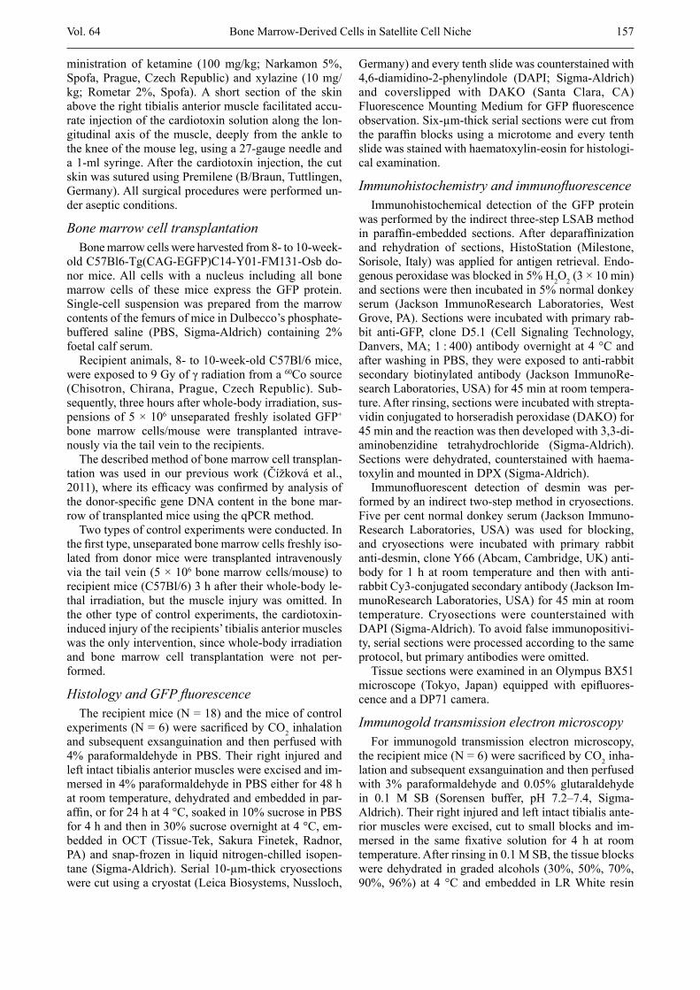
our experimental setting, myoblasts were not bone mar-row derived. Desmin was also expressed by myotubes and maturing muscle fibres, but only rarely; its signal had been co-localized with a weak GFP signal. GFP+ structures resembling muscle fibres were noticed in the cryosections using small magnifications (Fig. 1 C ar-row); nevertheless, using larger magnifications, it be-came apparent that they were degenerated muscle fibres packed with GFP+ macrophages phagocytosing the ne-crotic sarcoplasm. Surprisingly, the tendon of the in-jured muscles contained numerous GFP+ fibroblasts called tendinocytes. GFP positivity was infrequently observed in the wall of the blood vessels, mainly in en-dothelial cells (Fig. 1 E, F asterisks), but rarely also in some other cell types located in the deeper layers.
Bone marrow-derived cells in the regenerated skeletal muscle 28 days after injury
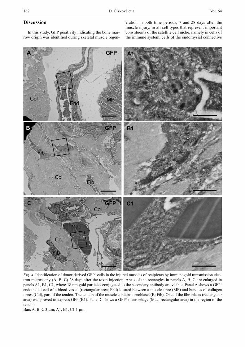
Twenty-eight days after the toxin injection, injured skeletal muscles of whole-body irradiated and BMC-transplanted mice were fully regenerated. The damaged area could be estimated according to the smaller diam-eters of the regenerated muscle fibres; however, their nuclei were located at the periphery, similarly as in the uninjured muscle fibres. The number of GFP+ immune cells substantially decreased in comparison with their number in the regenerating muscle examined 7 days af-ter the injury (Fig. 3 A, B arrowheads, Fig. 4 C, C1). The endomysial spindle-shaped cells represented the cell population that expressed GFP the most frequently (Fig. 3 A, B, C, D, F “empty” arrows). Both in cryosections and in deparaffinised sections, we succeeded in finding GFP+ muscle fibres whose frequency of occurrence fol-lowing BMC transplantation was described to be very low (Fig. 3 A, E arrows). GFP positivity was observed in several tendinocytes in the tendon (Fig. 4 B, B1) and rarely in endothelial cells of the blood vessels (Fig. 4 A, A1), similarly to regenerating muscles evaluated 7 days after the toxin injection.
Bone marrow-derived cells in the contralateral skeletal muscle
In contralateral skeletal muscles of whole-body irra-diated and BMC-transplanted mice, GFP positivity was observed in infrequent immune cells, namely in macro-phages and mast cells, and in occasional endomysial spindle-shaped cells in both time periods, 7 as well as 28 days after the toxin injury. In samples obtained from our experiments, we did not detect GFP in muscle fibres or in cells of the blood vessels.
Bone marrow-derived cells in the skeletal muscle in control experiments
In the first type of control experiments, the recipient mice were whole-body irradiated and BMC-transplanted, but the muscle injury was not induced. The recipients’
(Sigma-Aldrich; polymerized for 1 day at 50 °C). Semi-thin sections were stained with haematoxylin and eosin. Ultrathin sections were cut with Ultrotome Nova (LKB, Broma, Sweden), collected onto formvar carbon-coated nickel grids and processed for immunogold detection.
Immunohistochemical detection of the GFP protein was performed by an indirect two-step method in ul-trathin sections. Ten per cent normal goat serum (Jack-son ImmunoResearch Laboratories, USA) was used for blocking, and ultrathin sections were then incubated with primary rabbit anti-GFP, clone D5.1 (Cell Signaling Technology, USA; 1 : 40) or rabbit anti-desmin, clone Y66 (Abcam, 1:25) antibody for 3.5 h at room tempera-ture and after thorough washing, with anti-rabbit goat EM grade 18 nm colloidal gold-conjugated secondary antibody (Jackson ImmunoResearch Laboratories, USA, 1 : 20) for 2 h at room temperature.
Ultrathin sections counterstained with uranyl acetate and lead citrate were examined either under a Tesla BS500 transmission electron microscope (at 90 kV; Tesla, Czech Republic) equipped with a Megaview G2 digital camera and iTEM software (Olympus, Tokyo, Japan) or a JEOL JEM-1400Plus transmission electron microscope (at 120 kV; JEOL, Tokyo, Japan) equipped with an integrated 8Mpix CCD camera and software (JEOL).
Results
Bone marrow-derived cells in the regenerating skeletal muscle 7 days after injury
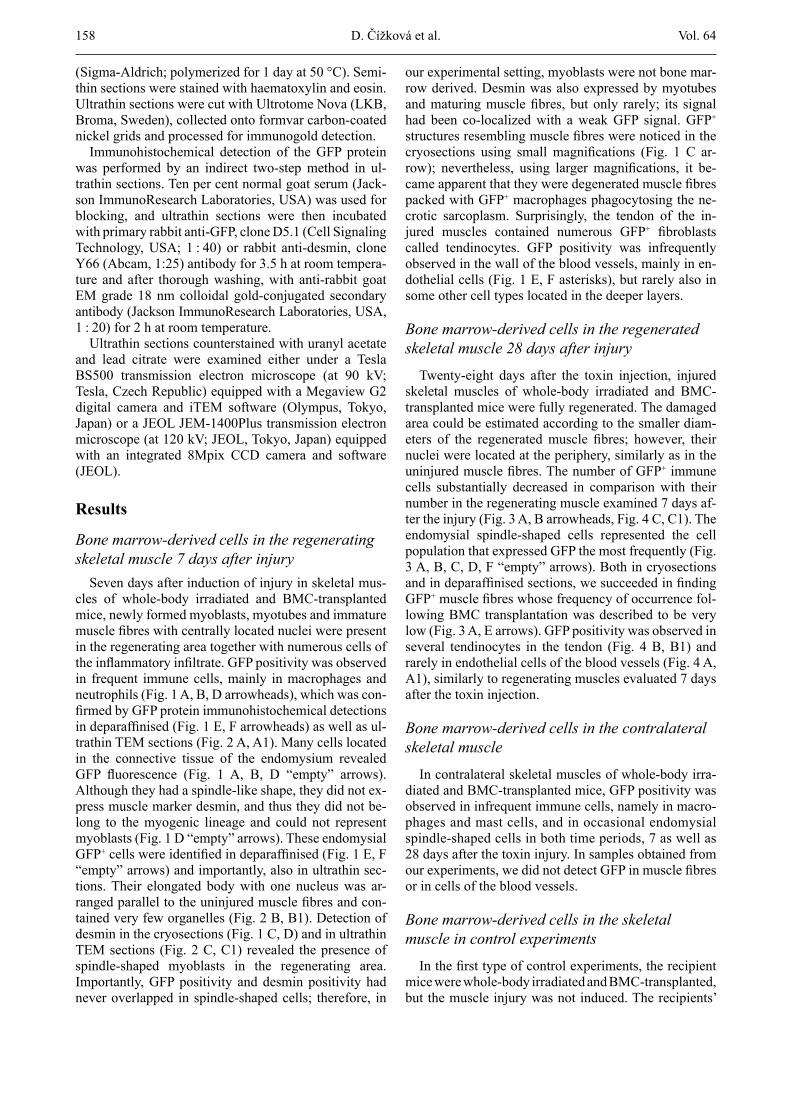
Seven days after induction of injury in skeletal mus-cles of whole-body irradiated and BMC-transplanted mice, newly formed myoblasts, myotubes and immature muscle fibres with centrally located nuclei were present in the regenerating area together with numerous cells of the inflammatory infiltrate. GFP positivity was observed in frequent immune cells, mainly in macrophages and neutrophils (Fig. 1 A, B, D arrowheads), which was con-firmed by GFP protein immunohistochemical detections in deparaffinised (Fig. 1 E, F arrowheads) as well as ul-trathin TEM sections (Fig. 2 A, A1). Many cells located in the connective tissue of the endomysium revealed GFP fluorescence (Fig. 1 A, B, D “empty” arrows). Although they had a spindle-like shape, they did not ex-press muscle marker desmin, and thus they did not be-long to the myogenic lineage and could not represent myoblasts (Fig. 1 D “empty” arrows). These endomysial GFP+ cells were identified in deparaffinised (Fig. 1 E, F “empty” arrows) and importantly, also in ultrathin sec-tions. Their elongated body with one nucleus was ar-ranged parallel to the uninjured muscle fibres and con-tained very few organelles (Fig. 2 B, B1). Detection of desmin in the cryosections (Fig. 1 C, D) and in ultrathin TEM sections (Fig. 2 C, C1) revealed the presence of spindle-shaped myoblasts in the regenerating area. Importantly, GFP positivity and desmin positivity had never overlapped in spindle-shaped cells; therefore, in
D. Čížková et al.
Vol. 64 159
uninjured skeletal muscles were excised at the same time as the injured skeletal muscles, e.g. 5 and 8 weeks after the whole-body irradiation and BMC transplanta-
tion; however, GFP positivity was only detected in in-frequent endomysial spindle-shaped cells and in sparse immune cells including macrophages and mast cells in
Bone Marrow-Derived Cells in Satellite Cell Niche
Fig. 1. Identification of donor-derived GFP+ cells in the injured muscles of recipients by GFP fluorescence (green; A, B, C, D) and GFP immunohistochemistry (brown; E, F). In the regenerating muscle 7 days after injury, GFP positivity was observed in numerous immune cells, mainly in macrophages (A, B, D, E, F arrowheads), in some spindle-shaped cells located in the endomysium (A, B, D, E, F “empty” arrows) and rarely in endothelial cells of the blood vessels (E, F aster-isks). The arrow in panel C points at a degenerated desmin+ (red) muscle fibre filled with GFP+ macrophages phagocytos-ing its necrotic sarcoplasm. The insert in panel E shows a negative control to GFP immunohistochemical detection when the primary antibody was omitted. A, B, C, D are counterstained with DAPI, E, F with haematoxylin. Bars A, B, E, F 200 µm; C 400 µm; D 50 µm.
160 Vol. 64
both time periods. We did not find the GFP signal in any muscle fibres or in the cells of the blood vessels in our samples.
In the other control experiments, the recipient mice were not whole-body irradiated nor BMC transplanted, but their skeletal muscles were injured. We examined
D. Čížková et al.
Fig. 2. Identification of donor-derived GFP+ cells in the injured muscles of recipients by immunogold transmission elec-tron microscopy (A, B) 7 days after the toxin injection. Areas of the rectangles in panels A, B, C are enlarged in panels A1, B1, C1, where 18 nm gold particles conjugated to the secondary antibody are visible. A shows a GFP+ macrophage (Mac; rectangular area). Elongated cells in the vicinity of a muscle fibre (MF) labelled with asterisks also express GFP (enlarged images not shown). Similar GFP+ elongated cells located next to a muscle fibre in the endomysium are captured in panel B (rectangular area and asterisk). Panel C shows immunogold detection of desmin. An elongated cell in the neighbourhood of a muscle fibre expresses this protein. In the insert, desmin immunopositivity in a muscle fibre is visible. Bars A, B, C 3 µm; A1, B1, C1 1 µm.

Vol. 64 161
the injured skeletal muscles both 7 and 28 days after the toxin injection and did not detect any GFP positivity.
The results of semi-quantitative evaluation of the amount of GFP+ cells in the injured, contralateral and control recipients’ skeletal muscles examined 5 (7D, 7
days after the toxin injection in the injured recipients’ muscles) and 8 (28D, 28 days after the toxin injection in the injured recipients’ muscles) weeks after the whole-body irradiation and BMC transplantation are summa-rized in Table 1.
Bone Marrow-Derived Cells in Satellite Cell Niche
Fig. 3. Identification of donor-derived GFP+ cells in the injured muscles of recipients by GFP fluorescence (green; A, B, C, D) and GFP immunohistochemistry (brown; E, F). In the regenerated muscle 28 days after injury, GFP+ immune cells (A, B arrowheads) and spindle-shaped cells in the endomysium (A, B, C, D “empty” arrows) became less numerous, whereas infrequent GFP+ endothelial cells (Fig. 4) and desmin+ GFP+ muscle fibres (A, E arrows) were still detected. A, B, C, D are counterstained with DAPI, E, F with haematoxylin. Bars A, B, E 200 µm; C 400 µm; D, F 50 µm.
162 Vol. 64
Discussion
In this study, GFP positivity indicating the bone mar-row origin was identified during skeletal muscle regen-
eration in both time periods, 7 and 28 days after the muscle injury, in all cell types that represent important constituents of the satellite cell niche, namely in cells of the immune system, cells of the endomysial connective
D. Čížková et al.
Fig. 4. Identification of donor-derived GFP+ cells in the injured muscles of recipients by immunogold transmission elec-tron microscopy (A, B, C) 28 days after the toxin injection. Areas of the rectangles in panels A, B, C are enlarged in panels A1, B1, C1, where 18 nm gold particles conjugated to the secondary antibody are visible. Panel A shows a GFP+ endothelial cell of a blood vessel (rectangular area; End) located between a muscle fibre (MF) and bundles of collagen fibres (Col), part of the tendon. The tendon of the muscle contains fibroblasts (B; Fib). One of the fibroblasts (rectangular area) was proved to express GFP (B1). Panel C shows a GFP+ macrophage (Mac; rectangular area) in the region of the tendon.Bars A, B, C 3 µm; A1, B1, C1 1 µm.

Vol. 64 163
tissue, blood vessel-associated cells and cells of the my-ogenic lineage. In contralateral and control uninjured muscles, GFP positivity was determined in less numer-ous immune cells and endomysial cells.
GFP+ immune cells, mainly macrophages, were abun-dant in the regenerating muscle 7 days after the injury and were still detected in the regenerated muscle 28 days after the cardiotoxin injection. In the contralateral and control uninjured skeletal muscles, GFP+ macrophages and sparse mast cells were sporadically observed. The cells of the immune system derived from haematopoi-etic stem cells are indispensable for the initial step of regeneration of the injured skeletal muscle (Lescaudron et al., 1999). Resident leukocytes activated by the injury secrete e.g. TNF-α and interleukin-6, which together with other factors promote activation and proliferation of the satellite cells and induce attraction of circulating granulocytes, mainly neutrophils. Neutrophils release pro-inflammatory signals necessary for recruitment of monocytes, which become very numerous and differen-tiate into macrophages. Initially, pro-inflammatory M1 macrophages phagocytose necrotic sarcoplasm of the degenerated muscle fibres and promote proliferation of myoblasts arisen from the activated dividing satellite cells. Subsequently, anti-inflammatory M2 macrophages regulate myogenic differentiation and stimulate myo-tube formation (Bentzinger et al., 2013; Yin et al., 2013; Mashinchian et al., 2018).
Thus, all types of the above-mentioned immune cells precisely participate in the regulation of skeletal muscle regeneration, and any changes in their appropriate pro-portions can lead to regeneration failure (Arnold et al., 2007; Segawa et al., 2008; Liu et al., 2017). This was also documented in our previous study in which the re-cipients were whole-body irradiated 4 h after the muscle injury to eradicate dividing satellite cells and suppress host haematopoiesis, and subsequently transplanted with donor bone marrow cells. Seven days after the car-diotoxin injection, no donor bone marrow-derived im-mune cells were found in the degeneration area, which led to persistence and even calcification of the necrotic sarcoplasm of the injured muscle fibres. Two and four
weeks following muscle damage, when in the course of proper regeneration new muscle fibres were already pre-sent, we found many donor-derived large multinucleat-ed cells resembling foreign body giant cells formed by fusion of activated macrophages in the close vicinity of the remaining necrotic muscle fibres (Čížková et al., 2011).
Interestingly, in this study the GFP signal was recog-nised in many desmin– spindle-shaped cells located in the endomysium of the injured muscles examined both 7 and 28 days after cardiotoxin injection. In the contralat-eral and control uninjured muscles, these cells were also noticed, but in lower quantity as estimated. In the inter-stitial tissue of endomysium, different cell types occur; however, their origin and functions in the muscle regen-eration have not been fully elucidated (Bentzinger et al., 2013; Yin et al., 2013; Mashinchian et al., 2018). Fibro-blasts proliferate after muscle injury very quickly and are indispensable for remodelling of the satellite cell niche during regeneration by secreting constituents of the extracellular matrix and basement membranes. More-over, they support expansion of the satellite cells, as documented by a study in which partial ablation of fi-broblasts during regeneration led to reduced numbers of satellite cells and their premature differentiation, and to a smaller size of formed muscle fibres (Murphy et al., 2011). Fibroblasts can arise from the resident mesen-chymal stem cells; nevertheless, e.g. in the tumour stro-ma, also from the donor bone marrow (Dikerze et al., 2004; Barcellos-de-Souza et al., 2013; Kurashige et al., 2018).
Fibroadipogenic progenitors are mesenchymal cells that express surface markers CD34, Sca-1, and PDGFRα and are capable of differentiation into fibroblasts, adipo-cytes, but not into muscle cells. After muscle injury, they are activated, proliferate, and together with fibro-blasts synthesize extracellular matrix proteins during regeneration. Furthermore, they facilitate myogenic dif-ferentiation, e.g. by secreting interleukin-6 (Joe et al., 2010; Heredia et al., 2013; Fiore et al., 2016). To date, their origin has not been unambiguously determined. Mitchell et al. (2010) described resident stem cells in the
Bone Marrow-Derived Cells in Satellite Cell Niche
Table 1. Summarized results of semi-quantitative evaluation of the amount of GFP+ cells in the injured, contralateral and control recipients’ skeletal muscles examined 5 (7D, 7 days after the toxin injection in the injured recipients’ muscles) and 8 (28D, 28 days after the toxin injection in the injured recipients’ muscles) weeks after whole-body irradiation and BMC transplantation
GFP+ cellsImmune cells Endomysial spindle-
shaped cellsMyotubes, muscle fibres
Endothelial cells of the blood vessels
Injured muscles7D +++ +++ + +28D ++ ++ + +
Contralateral muscles
7D + + - -28D + + - -
Control muscles7D (+) + - -28D (+) + - -
164 Vol. 64D. Čížková et al.
muscle interstitium which express stress mediator PW1, surface markers CD34, Sca-1, but not satellite cell markers Pax7 and PDGFRα, and have myogenic poten-tial when cultured in vitro, unlike fibroadipogenic pro-genitors (Mitchell et al., 2010; Pannérec et al., 2013; Yin et al., 2013). These PW1+ interstitial cells promote pro-liferation and differentiation of the satellite cells (Formicola et al., 2014; Mashinchian et al., 2018). Pax3 lineage tracing experiments indicate that PW1+ intersti-tial cells do not arise from embryonic Pax3+ myogenic progenitor cells; therefore, they are derived from a dis-tinct lineage than the satellite cells (Mitchell et al., 2010; Yin et al., 2013).
Telocytes represent another interstitial cell type of un-determined origin occurring in the skeletal muscle. They are characterized according to their appearance in the transmission electron microscope as cells with a small body and very long thin moniliform prolongations, di-chotomously branched, forming a 3D network (Popescu et al. 2011; Suciu et al., 2012; Yin et al., 2013). Unlike fibroblasts, they express c-kit and then also PDGFRβ and VEGF, but not Pax7. Their estimated functions are transmission of intercellular signalling by shedding or absorbing microvesicles enriched for proteins and RNAs and facilitation of vasculogenesis by secretion of VEGF (Popescu et al. 2011; Suciu et al., 2012; Yin et al., 2013). Dreyfus et al. (2004) identified donor bone mar-row-derived cells expressing CD34 or Sca1 in the inter-stitium of the intact muscle. Although immunopheno-typing of these cells is insufficient according to the recent knowledge, in accordance with our results this study shows the presence of bone marrow-derived cells in muscle interstitium. In conclusion, the interstitial cells of the endomysium are found to be a very intrigu-ing cellular component of the satellite cell niche and it will be important to study their exact functions and ori-gins. Which cell type the GFP+ desmin- cells observed in the endomysium of both intact and regenerating mus-cles in our experiments represent remains to be clarified and has become the goal of our upcoming study.
Infrequently, GFP positivity was noticed in the en-dothelial cells of the blood vessels of injured muscles both 7 and 28 days after the toxin injection. These find-ings give evidence of the presence of endothelial pro-genitors in the donor bone marrow. Blood vessel-associ-ated cells are an important component of the satellite cell niche. In both intact and regenerating muscle, capil-laries occur in close vicinity to the satellite cells (Christov et al., 2007). After muscle injury, their number initially increases, despite that they were not directly damaged (Ochoa et al., 2007). The endothelial cells se-crete a variety of mitogenic or anti-apoptotic factors, such as VEGF, that influence muscle cells (Abou-Khalil et al., 2010; Bentzinger et al., 2013) and vice versa, dif-ferentiating myogenic cells also produce VEGF and thus have a proangiogenic effect. Therefore, myogene-sis and angiogenesis are closely co-regulated (Bentzinger et al., 2013; Latroche et al., 2017).
We rarely detected GFP together with desmin in the myotubes of injured muscles 7 days and in the muscle fibres 28 days after the cardiotoxin injection. The contri-bution of haematopoietic stem cells to skeletal muscle regeneration has been described (Ferrari et al., 1998; Palermo et al., 2001; LaBarge et al., 2002; Brazelton et al., 2003; Camargo et al., 2003; Corbel et al., 2003; Doyonnas et al., 2004; Sacco et al., 2005; Abedi et al., 2005, 2007; de la Garza-Rodea et al., 2011) and our re-sults confirmed occurrence of this rare phenomenon. Importantly, we did not find any GFP+ desmin+ myo-blasts and the GFP signal appeared only in myotubes and muscle fibres, which supports the findings that hae-matopoietic stem cells are incapable of generating myo-genic progenitors and that myeloid cells are incorporat-ed by fusion during the formation of new myotubes (Camargo et al., 2003; Corbel et al., 2003). Myogenic cells at various developmental stages are important con-stituents of the satellite cell niche during reparative myo-genesis. They secrete growth factors, provide regulatory signals and are involved in intercellular interactions. Notch and Wnt7a signalling pathways belong to the most important and thus recently studied (Brack et al., 2008; Bentzinger et al., 2013; Yin et al., 2013).
To conclude, our results confirmed the ability of bone marrow-derived cells to contribute to the cellular com-ponent of the satellite cell niche in the intact and regen-erating skeletal muscle. These cells originated not only from the haematopoietic stem cells, but obviously also from other stem/progenitor cells residing in the bone marrow, such as MSCs and endothelial progenitors.
AcknowledgementsThe authors thank Mrs. Magda Voborníková, Ms. Petra
Hajzlerová, Mrs. Simona Vrchotová, Mrs. Jaroslava Pro-kešová, Mrs. Milada Hetešová and Mrs. Jana Hošková for their skilful technical assistance.
ReferencesAbedi, M., Greer, D. A., Foster, B. M., Colvin, G. A., Harpel,
J. A., Demers, D. A., Pimentel, J., Dooner, M. S., Quesen-berry, P. J. (2005) Critical variables in the conversion of marrow cells to skeletal muscle. Blood 106, 1488-1494.
Abedi, M., Foster, B. M., Wood, K. D., Colvin, G. A., McLean, S. D., Johnson, K. W., Greer, D. A. (2007) Haematopoietic stem cells participate in muscle regeneration. Br. J. Hae-matol. 138, 792-801.
Abou-Khalil, R., Mounier, R., Chazaud, B. (2010) Regulation of myogenic stem cell behavior by vessel cells: the “mé-nage à trois” of satellite cells, periendothelial cells and en-dothelial cells. Cell Cycle 9, 892-896.
Ankrum, J. A., Ong, J. F., Karp, J. M. (2014) Mesenchymal stem cells: immune evasive, not immune privileged. Nat. Biotechnol. 32, 252-260.
Arnold, L., Henry, A., Poron, F., Baba-Amer, Y., van Rooijen, N., Plonquet, A., Gherardi, R. K., Chazaud, B. (2007) In-flammatory monocytes recruited after skeletal muscle in-
Vol. 64 165Bone Marrow-Derived Cells in Satellite Cell Niche
jury switch into antiinflammatory macrophages to support myogenesis. J. Exp, Med. 204, 1057-1069.
Asahara, T., Murohara, T., Sullivan, A., Silver, M., van der Zee, R., Li, T., Witzenbichler, B., Schatteman, G., Isner, J. M. (1997). Isolation of putative progenitor endothelial cells for angiogenesis. Science 275, 964-967.
Barcellos-de-Souza, P., Gori, V., Bambi, F., Chiarugi, P. (2013) Tumor microenvironment: bone marrow-mesen-chymal stem cells as key players. Biochim. Biophys. Acta 1836, 321-335.
Bentzinger, C. F., Wang, Y. X., Dumont, N. A., Rudnicki, M.A. (2013) Cellular dynamics in the muscle satellite cell niche. EMBO Rep. 14, 1062-1072.
Bianco, P., Gehron Robey, P. (2000) Marrow stromal stem cells. J. Clin. Invest. 105, 1663-1668.
Brack, A. S., Conboy, I. M., Conboy, M. J., Shen, J., Rando, T. A. (2008) A temporal switch from notch to Wnt signaling in muscle stem cells is necessary for normal adult myogen-esis. Cell Stem Cell 2, 50-59.
Brazelton, T. R., Nystrom, M., Blau, H. M. (2003) Significant differences among skeletal muscles in the incorporation of bone marrow-derived cells. Dev. Biol. 262, 64-74.
Camargo, F. D., Green, R., Capetanaki, Y., Jackson, K. A., Goodell, M. A. (2003) Single hematopoietic stem cells generate skeletal muscle through myeloid intermediates. Nat. Med. 9, 1520-1527.
Caplan, A. I. (1991) Mesenchymal stem cells. J. Orthop. Res. 9, 641-650.
Chao, H., Hirschi, K. K. (2010) Hemato-vascular origins of endothelial progenitor cells? Microvasc. Res. 79, 169-173.
Chazaud, B., Sonnet, C., Lafuste, P., Bassez, G., Rimaniol, A. C., Poron, F., Authier, F. J., Dreyfus, P. A., Gherardi, R. K. (2003) Satellite cells attract monocytes and use mac-rophages as a support to escape apoptosis and enhance muscle growth. J. Cell. Biol. 163, 1133-1143.
Christov, C., Chrétien, F., Abou-Khalil, R., Bassez, G., Vallet, G., Authier, F. J., Bassaglia, Y., Shinin, V., Tajbakhsh, S., Chazaud, B., Gherardi, R. K. (2007) Muscle satellite cells and endothelial cells: close neighbors and privileged part-ners. Mol. Biol. Cell 18, 1397-1409.
Čížková, D., Vávrová, J., Mičuda, S., Filip, S., Brčáková, E., Brůčková, L., Mokrý, J. (2011) Role of transplanted bone marrow cells in response to skeletal muscle injury. Folia Biol. (Praha) 57, 232-241.
Corbel, S. Y., Lee, A., Yi, L., Duenas, J., Brazelton, T. R., Blau, H. M., Rossi F. M. (2003) Contribution of hemat-opoietic stem cells to skeletal muscle. Nat. Med. 9, 1528-1532.
de la Garza-Rodea, A. S., van der Velde, I., Boersma, H., Gon-çalves, M. A., van Bekkum, D. W., de Vries, A. A., Knaän-Shanzer, S. (2011) Long-term contribution of human bone marrow mesenchymal stromal cells to skeletal muscle re-generation in mice. Cell Transplant. 20, 217-231.
Dezawa, M., Ishikawa, H., Itokazu, Y., Yoshihara, T., Hoshi-no, M., Takeda, S., Ide, C., Nabeshima, Y. (2005) Bone marrow stromal cells generate muscle cells and repair mus-cle degeneration. Science 309, 314-317.
Direkze, N. C., Hodivala-Dilke, K., Jeffery, R., Hunt, T., Poul-som, R., Oukrif, D., Alison, M. R., Wright, N. A. (2004)
Bone marrow contribution to tumor-associated myofibro-blasts and fibroblasts. Cancer Res. 64, 8492-8495.
Dominici, M., Le Blanc, K., Mueller, I., Slaper-Cortenbach, I., Marini, F., Krause, D., Deans, R., Keating, A., Prockop, D. J., Horwitz. E. (2006) Minimal criteria for defining multipotent mesenchymal stromal cells. The International Society for Cellular Therapy position statement. Cytother-apy 8, 315-317.
Doyonnas, R., LaBarge, M. A., Sacco, A., Charlton, C., Blau, H. M. (2004) Hematopoietic contribution to skeletal mus-cle regeneration by myelomonocytic precursors. Proc. Natl. Acad. Sci. USA 101, 13507-13512.
Dreyfus, P. A., Chretien, F., Chazaud, B., Kirova, Y., Cara-melle, P., Garcia, L., Butler-Browne, G., Gherardi, R. K. (2004) Adult bone marrow-derived stem cells in muscle connective tissue and satellite cell niches. Am. J. Pathol. 164, 773-779.
Dumont, N., Frenette, J. (2010) Macrophages protect against muscle atrophy and promote muscle recovery in vivo and in vitro: a mechanism partly dependent on the insulin-like growth factor-1 signaling molecule. Am. J. Pathol. 176, 2228-2235.
Ferrari, G., Cusella-De Angelis, G., Coletta, M., Paolucci, E., Stornaiuolo, A., Cossu, G. Mavilio, F. (1998) Muscle re-generation by bone marrow-derived myogenic progenitors. Science 279, 1528-1530.
Fiore, D., Judson, R. N., Low, M., Lee, S., Zhang, E., Hop-kins, C., Xu, P., Lenzi, A., Rossi, F. M., Lemos, D. R. (2016) Pharmacological blockage of fibro/adipogenic pro-genitor expansion and suppression of regenerative fibro-genesis is associated with impaired skeletal muscle regen-eration. Stem Cell Res. 17, 161-169.
Formicola, L., Marazzi, G., Sassoon, D. A. (2014) The ex-traocular muscle stem cell niche is resistant to ageing and disease. Front. Aging Neurosci. 6, 328.
Friedenstein, A. J., Piatetzky-Shapiro, I. I., Petrakova, K. V. (1966) Osteogenesis in transplants of bone marrow cells. J. Embryol. Exp. Morphol. 16, 381-390.
Galli, D., Vitale, M., Vaccarezza, M. (2014) Bone marrow-derived mesenchymal cell differentiation toward myogenic lineages: facts and perspectives. Biomed Res. Int. 2014, 762695.
Heredia, J. E., Mukundan, L., Chen, F. M., Mueller, A. A., Deo, R. C., Locksley, R. M., Rando, T. A., Chawla, A. (2013) Type 2 innate signals stimulate fibro/adipogenic progenitors to facilitate muscle regeneration. Cell 153, 376-388.
Joe, A. W., Yi, L., Natarajan, A., Le Grand, F., So, L., Wang, J., Rudnicki, M. A., Rossi, F. M. (2010) Muscle injury ac-tivates resident fibro/adipogenic progenitors that facilitate myogenesis. Nat. Cell Biol. 12, 153-163.
Kurashige, M., Kohara, M., Ohshima, K., Tahara, S., Hori, Y., Nojima, S., Wada, N., Ikeda, J. I., Miyamura, K., Ito, M., Morii, E. (2018) Origin of cancer-associated fibroblasts and tumor-associated macrophages in humans after sex-mismatched bone marrow transplantation. Commun. Biol. 1, 131.
LaBarge, M. A., Blau, H. M. (2002) Biological progression from adult bone marrow to mononucleate muscle stem cell
166 Vol. 64
to multinucleate muscle fiber in response to injury. Cell 111, 589-601.
Latroche, C., Weiss-Gayet, M., Muller, L., Gitiaux, C., Leb-lanc, P., Liot, S., Ben-Larbi, S., Abou-Khalil, R., Verger, N., Bardot, P., Magnan, M., Chrétien, F., Mounier, R., Ger-main, S., Chazaud, B. (2017) Coupling between myogen-esis and angiogenesis during skeletal muscle regeneration is stimulated by restorative macrophages. Stem Cell Re-ports 12, 2018-2033.
Lescaudron, L., Peltékian, E., Fontaine-Pérus, J., Paulin, D., Zampieri, M., Garcia, L., Parrish, E. (1999) Blood borne macrophages are essential for the triggering of muscle re-generation following muscle transplant. Neuromuscul. Dis-ord. 9, 72-80.
Liu, X., Liu, Y., Zhao, L., Zeng, Z., Xiao, W., Chen, P. (2017) Macrophage depletion impairs skeletal muscle regenera-tion: The roles of regulatory factors for muscle regenera-tion. Cell Biol. Int. 41, 228-238.
Mashinchian, O., Pisconti, A., Le Moal E., Bentzinger C. F. (2018) The muscle stem cell niche in health and disease. Curr. Top. Dev. Biol. 126, 23-65.
Mauro, A. (1961) Satellite cell of skeletal muscle fibers. J. Biophys. Biochem. Cytol. 9, 493-495.
Mitchell, K. J., Pannérec, A., Cadot, B., Parlakian, A., Besson, V., Gomes, E. R., Marazzi, G., Sassoon D. A. (2010) Iden-tification and characterization of a non-satellite cell muscle resident progenitor during postnatal development. Nat. Cell Biol. 12, 257-266.
Murphy, M. M., Lawson, J. A., Mathew, S. J., Hutcheson, D. A., Kardon, G. (2011) Satellite cells, connective tissue fi-broblasts and their interactions are crucial for muscle re-generation. Development 138, 3625-3637.
Ochoa, O., Sun, D., Reyes-Reyna, S. M., Waite, L. L., Michalek, J. E., McManus, L. M., Shireman, P. K. (2007) Delayed angiogenesis and VEGF production in CCR2-/- mice during impaired skeletal muscle regeneration. Am. J. Physiol. Regul. Integr. Comp. Physiol. 293, 651-661.
D. Čížková et al.
Palermo, A. T., Labarge, M. A., Doyonnas, R., Pomerantz, J., Blau, H. M. (2005) Bone marrow contribution to skeletal muscle: a physiological response to stress. Dev. Biol. 279, 336-344.
Pannérec, A., Formicola, L., Besson, V., Marazzi, G., Sas-soon, D. A. (2013) Defining skeletal muscle resident pro-genitors and their cell fate potentials. Development 140, 2879-2891.
Popescu, L. M., Manole, E., Serboiu, C. S., Manole, C. G., Suciu, L. C., Gherghiceanu, M., Popescu, B. O. (2011) Identification of telocytes in skeletal muscle interstitium: implication for muscle regeneration. J. Cell. Mol. Med. 15, 1379-1392.
Sacco, A., Doyonnas, R., LaBarge, M. A., Hammer, M. M., Kraft, P., Blau, H.M. (2005) IGF-I increases bone marrow contribution to adult skeletal muscle and enhances the fu-sion of myelomonocytic precursors. J. Cell Biol. 171, 483-492.
Segawa, M., Fukada, S., Yamamoto, Y., Yahagi, H., Kanemat-su, M., Sato, M., Ito, T., Uezumi, A., Hayashi, S., Miyagoe-Suzuki, Y., Takeda, S., Tsujikawa, K., Yamamoto, H. (2008) Suppression of macrophage functions impairs skel-etal muscle regeneration with severe fibrosis. Exp. Cell. Res. 314, 3232-3244.
Suciu, L.C., Popescu, B. O., Kostin, S., Popescu, L. M. (2012) Platelet-derived growth factor receptor-β-positive telo-cytes in skeletal muscle interstitium. J. Cell. Mol. Med. 16, 701-707.
Tavassoli, M., Friedenstein, A. (1983) Hemopoietic stromal microenvironment. Am. J. Hematol. 15, 195-203.
Wakitani, S., Saito, T., Caplan, A. I. (1995) Myogenic cells derived from rat bone marrow mesenchymal stem cells ex-posed to 5-azacytidine. Muscle Nerve 18, 1417-1426.
Yin, H., Price, F., Rudnicki, M. A. (2013) Satellite cells and the muscle stem cell niche. Physiol. Rev. 93, 23-67.