Neoplasia lecture 10 - JU Medicine€¦ · Neoplasia 2018 lecture 9 Dr Heyam Awad MD, FRCPath. ILOS...
52
Neoplasia 2018 lecture 9 Dr Heyam Awad MD, FRCPath
Transcript of Neoplasia lecture 10 - JU Medicine€¦ · Neoplasia 2018 lecture 9 Dr Heyam Awad MD, FRCPath. ILOS...

Neoplasia 2018 lecture 9Dr Heyam Awad
MD, FRCPath

ILOS
• 1. understand the concept of immune surveillance.
• 2. list the most common tumor antigens and understand their origins.
• 3. understand the mechanisms through which tumor cells evade the immune system.
• 5. understand the role f inflammation as an enabler of malignancy.
• 6 list the most important DNA repair genes and understand their role in carcinogenesis.
8th hallmark of cancer
• Evading the immune system is an important tumor hallmark.
• Our immune system can destroy tumor cells, because tumor cells express antigens that can be recognized by the immune system as foreign.
• Once antigens are recognized the immune system can destroy the malignant cells.. This is called immune surveillance
• One of the promising treatments of cancer is immunotherapy: drugs that stimulate the immune system to attack cancer cells.

TUMOR IMMUNITY
• Tumor cells are recognized by the host ( the body) as non self.
• Once recognized, immunologic reactions are activated to destroy the tumor cells.
• This process is called immune surveillance
• However, immune surveillance is imperfect and that’s why tumors still occur i:e many of the tumor cells escape destruction by the immune system.
• Immune system recognizes cells by their antigens. (مولد الضد)مستضد )
• If cells express antigens that are perceived by the immune cells as non self , the immunologic reaction starts
• So: what are the antigens present on the cancer cells?

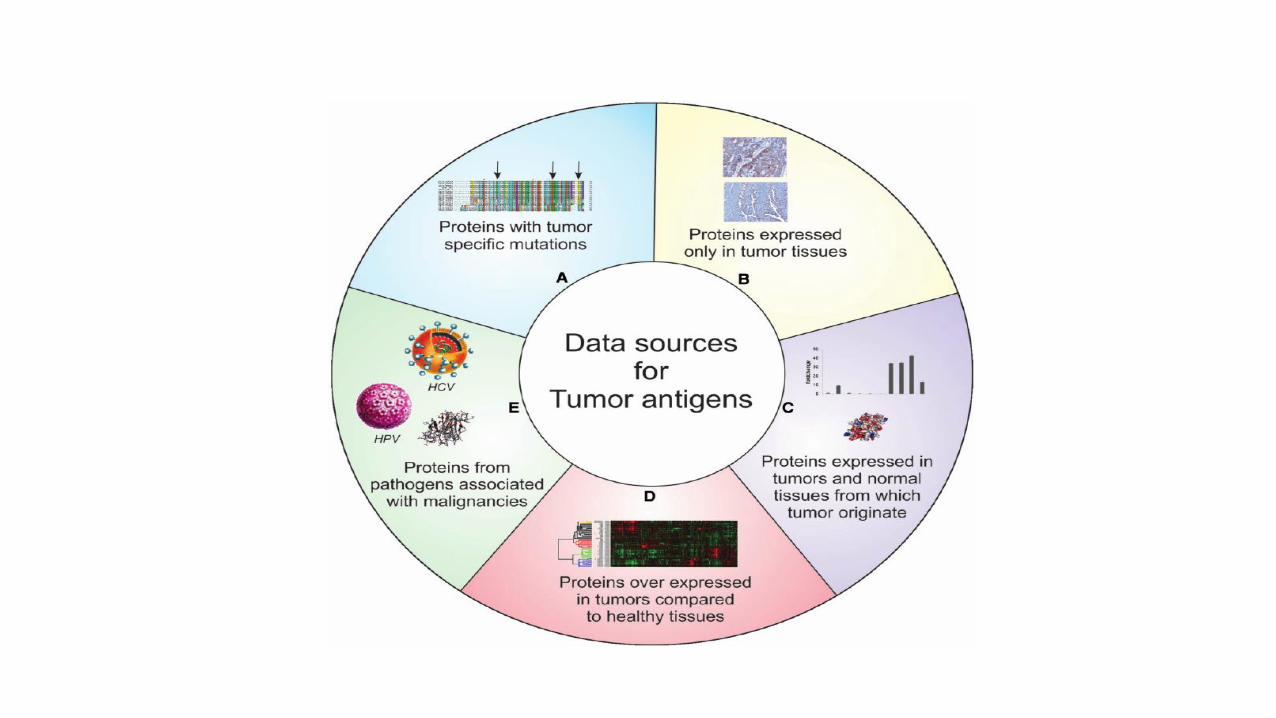
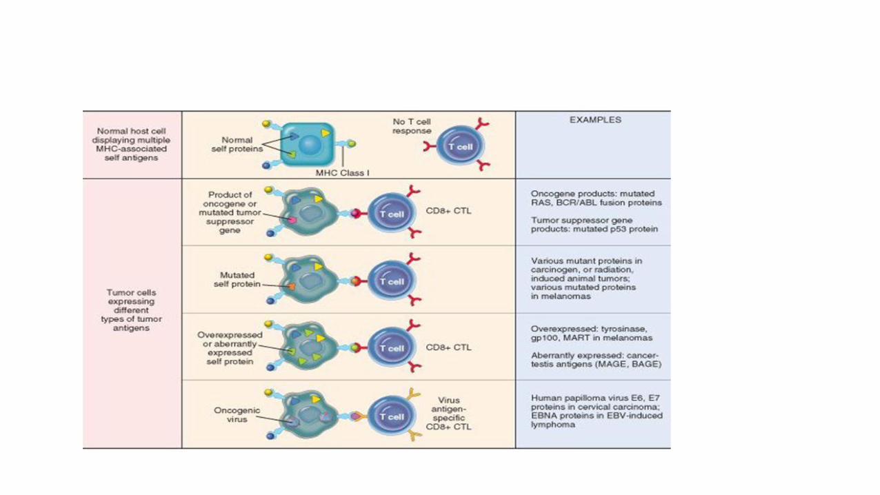
Tumor antigens
• Two types of tumor antigens: tumor specific and tumor associated antigens
• Tumor specific antigens: specific to the tumor and not seen in normal cells
• Tumor associated antigens: present on tumor cells and normal cells but are mutated or overexpressed in cancer cells

Products of mutated oncogenes or tumor suppressor genes• Mutated genes are translated to abnormal proteins that are non-self
• Examples: p53, RAS, B catenin
• Sometimes the gene product is not mutated but overexpressed like HER2/NEU.
• Anti HER 2 treatment = Herceptin is one of the earliest immunotherapies used ( we discussed HER 2 in detail previously)

Antigens produced by antigenic viruses
• HPV, EBV can produce proteins like E6 and E7
Oncofetal antigens
• These are proteins expressed only in embryos
• In some tumors ( mainly colon and liver) they are re-expressed
• examples: CEA= carcino-embryonic antigen and alpha fetoprotein
• These are important serum markers of cancer

Altered cell surface glycolipids and glycoproteins• Include: mucins.
• CA 125 and CA 19-9 are altered mucin proteins , mainly in ovarian carcinoma.

Anti-tumor mechanisms
• Our immune system has two mechanisms of attacking antigens: cellular and humoral immunity.
• Cellular immunity is mediated by T lymphocytes and natural killer cells.
• Humoral immunity is mediated by B lymphocytes that differentiate to plasma cells and secrete immunoglobulins.
• Cellular immunity plays a role in immune surveillance whereas humoral immunity doesn't play a role in vivo

Anti-tumor mechanisms
• The cells responsible for immune surveillance are:
• 1. cytotoxic T lymphocytes
• 2. Natural killer cells
• 3. macrophages

Cytotoxic T lymphocytes
• T lymphocytes have 2 main subtypes: cytotoxic T (CD 8) and helper T ( CD 4)
• Cytotoxic T are the most important cells involved in tumor immunity.
• We found that tumors can be infiltrated by T lymphocytes, mainly cytotoxic ones
• The tumors with more cytotoxic T cells have better prognosis than tumors with little T cells.
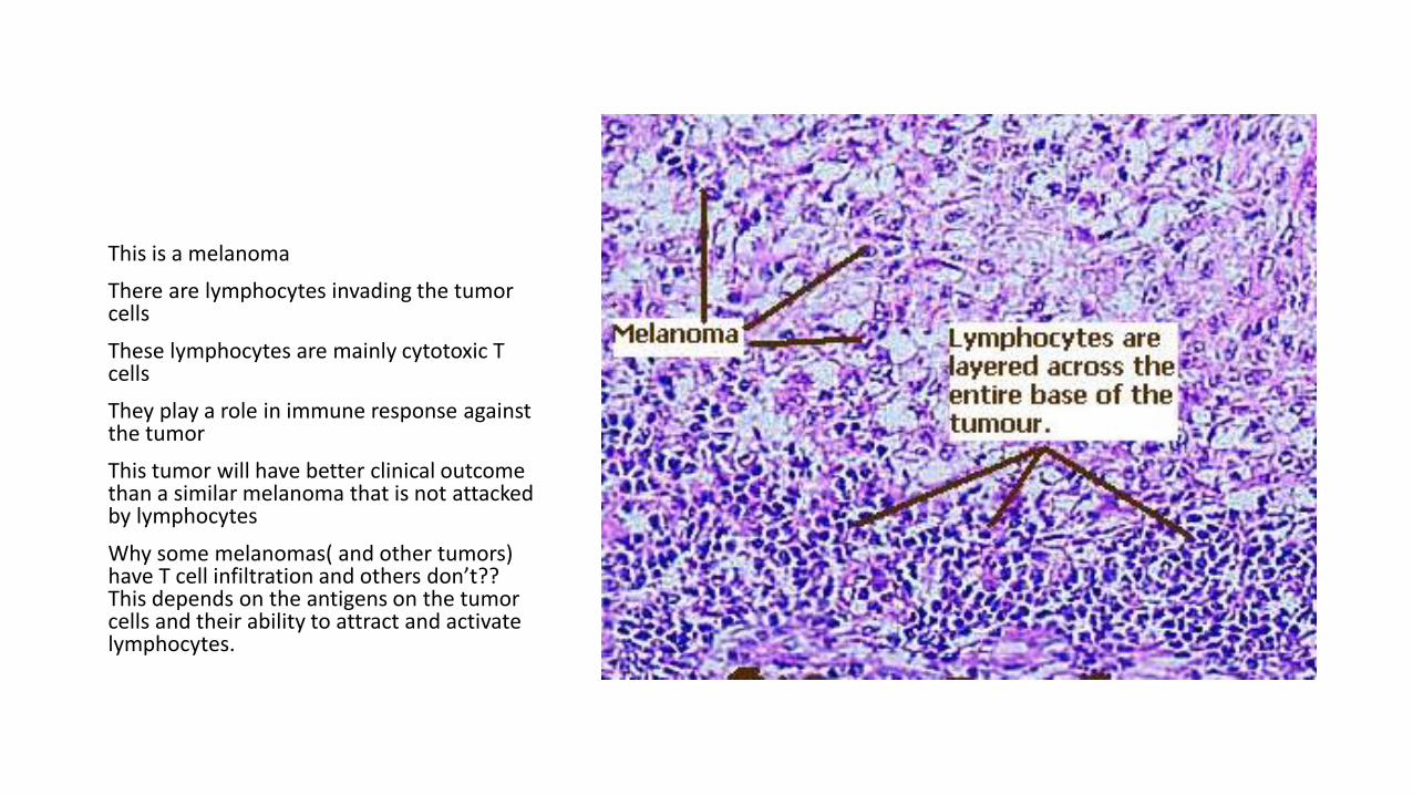
This is a melanoma
There are lymphocytes invading the tumor cells
These lymphocytes are mainly cytotoxic T cells
They play a role in immune response against the tumor
This tumor will have better clinical outcome than a similar melanoma that is not attacked by lymphocytes
Why some melanomas( and other tumors) have T cell infiltration and others don’t?? This depends on the antigens on the tumor cells and their ability to attract and activate lymphocytes.
Cytotoxic T cells ( CD8 positive lymphocytes)
• Need specific sensitization. So they cannot be stimulated unless thee are antigens on the tumor cells that need to be presented to these T cells.
• Antigens are usually presented to cytotoxic T cells via specialized ells called antigen presenting cells.
• For this presentation to happen, antigens from the tumor cell must be recognized by antigen presenting cells that attach them to MHC 1 molecules.
• The antigen- MHC 1 complex can then be recognized by the cytotoxic T cell.
• Co simulatory molecules are then needed, after the recognition, for T cells to be fully activated to kill the tumor cells.
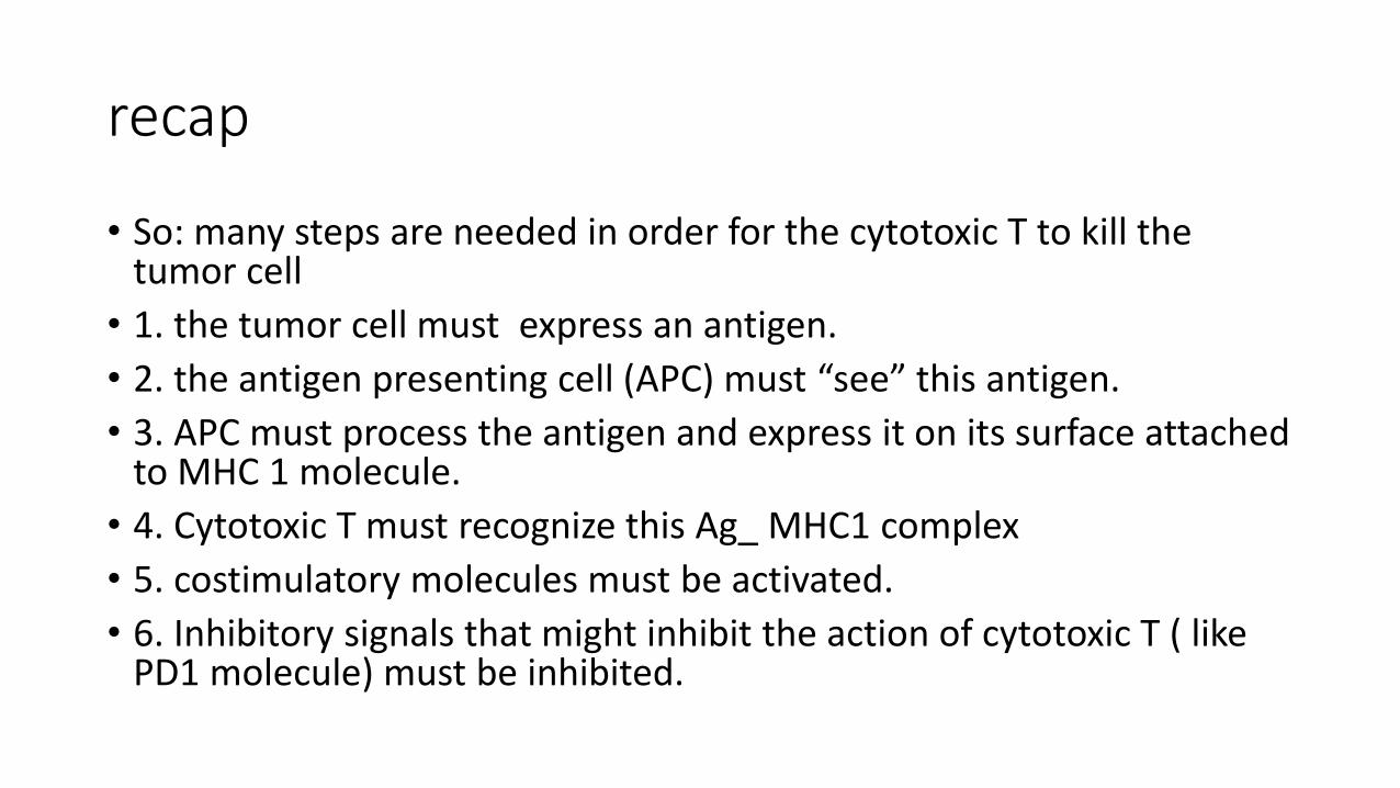
recap
• So: many steps are needed in order for the cytotoxic T to kill the tumor cell
• 1. the tumor cell must express an antigen.
• 2. the antigen presenting cell (APC) must “see” this antigen.
• 3. APC must process the antigen and express it on its surface attached to MHC 1 molecule.
• 4. Cytotoxic T must recognize this Ag_ MHC1 complex
• 5. costimulatory molecules must be activated.
• 6. Inhibitory signals that might inhibit the action of cytotoxic T ( like PD1 molecule) must be inhibited.
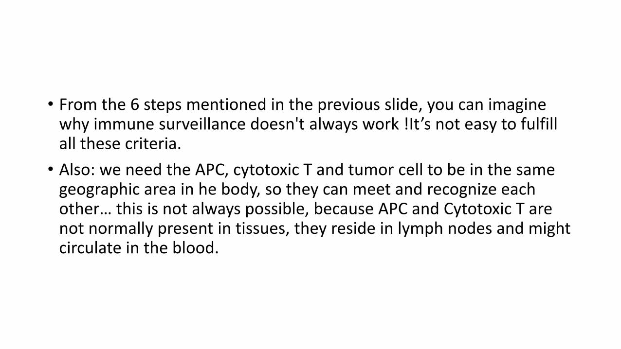
• From the 6 steps mentioned in the previous slide, you can imagine why immune surveillance doesn't always work !It’s not easy to fulfill all these criteria.
• Also: we need the APC, cytotoxic T and tumor cell to be in the same geographic area in he body, so they can meet and recognize each other… this is not always possible, because APC and Cytotoxic T are not normally present in tissues, they reside in lymph nodes and might circulate in the blood.
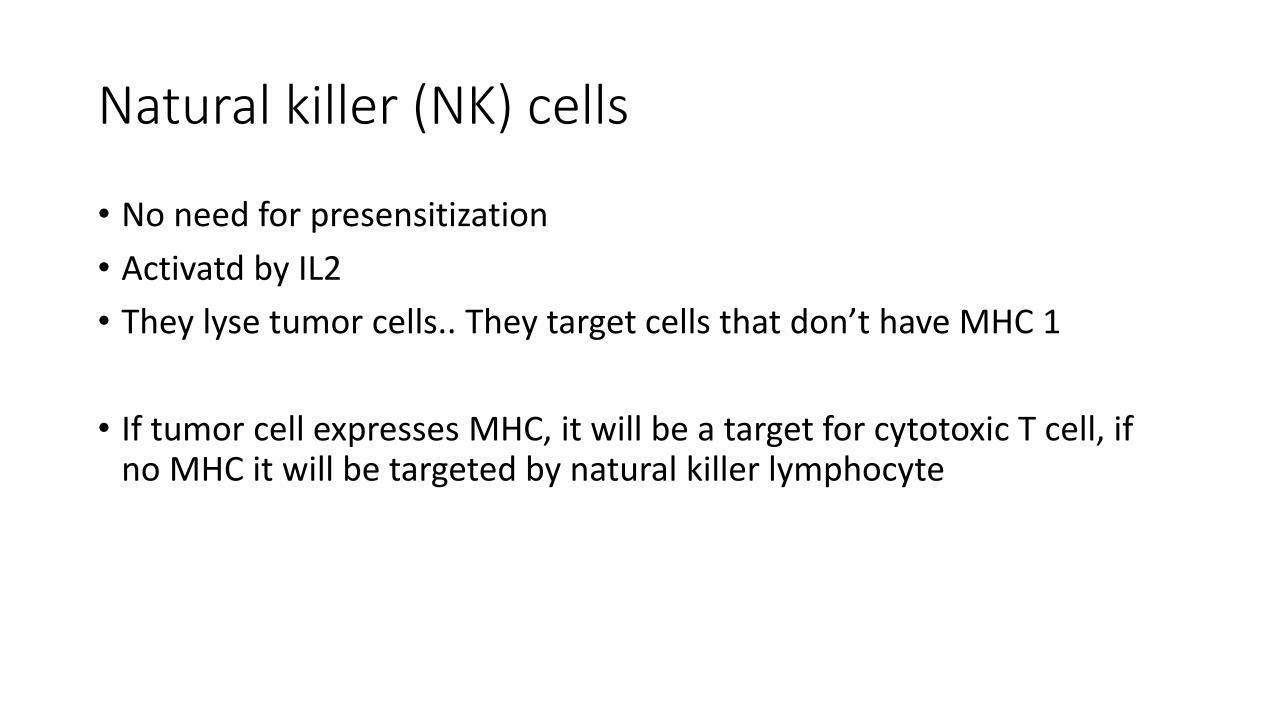
Natural killer (NK) cells
• No need for presensitization
• Activatd by IL2
• They lyse tumor cells.. They target cells that don’t have MHC 1
• If tumor cell expresses MHC, it will be a target for cytotoxic T cell, if no MHC it will be targeted by natural killer lymphocyte

macrophages
• M1 macrophages are cytotoxic to some cells
• They kill by: ROS or by TNF
• Cytotoxic T and Natural killers secrete interferon gamma that stimulates macrophages

Immune evasion
• Immune surveillance is important in protecting the host from cancer .
• Immune- compromised individuals have increased risk of developing cancer
• One of the hallmarks of cancer is evasion of destruction by the immune system
Mechanisms of evasion of the immune system• 1. selective growth of antigen negative variants ( subclones ). The
highly antigenic subclones are deleted from the tumor mass
• 2. loss or reduced expression of histocompatibility molecules.
• 3.downregulation of co-stimulatory molecules
• 4. antigen masking by producing a thick coat of external glycocalyxmolecules
• 5.immunosuppression ( see next slide)
immunosupression
• Tumor cells can suppress host immunity by:
• A. TGF beta production by tumor cells
• B. expression of fas ligand that binds to fas receptor on host lymphocytes causing apoptosis of these lymphocytes
• C. some oncogenic agents suppress host immunity, especially chemicals and ionizing radiation.


Clinical implications of tumor immunity: immunotherapy• US researcher James Allison and Japanese researcher Tasuku Honjo
have won the 2018 Nobel Prize for their work on manipulating the immune system to combat cancer.

An example of immunotherapy
• PD 1 ( programmed Death) is a negative regulator of the immune system.
• Tumors that express PD 1 ligand ( PDL 1), will escape being killed by cytotoxic T cells.( see pic on next slide)
• If we give PDL1 inhibitor to these patients we are removing the inhibition, thus stimulating T cells to attack cancer cells.


Although results are encouraging, immunotherapy has its own problems:• 1. inhibitory mechanisms are created to balance the strong action of
the immune system , so that our immune system doesn’t destroy our own cells… removing this inhibition has side effects and increase autoimmunity.
• 2. Another problem with immunotherapy is that it only works if the tumors express the ligand.
• 3. even tumors that respond to immunotherapy at the beginning can become non-responsive after some time, probably because subclonesthat don’t express the ligand are selected.
Role of pathology in immunotherapy
• We have stains that show if the tumor expresses the ligand or not. If they do express it the oncologist can consider treating with immunotherapy.
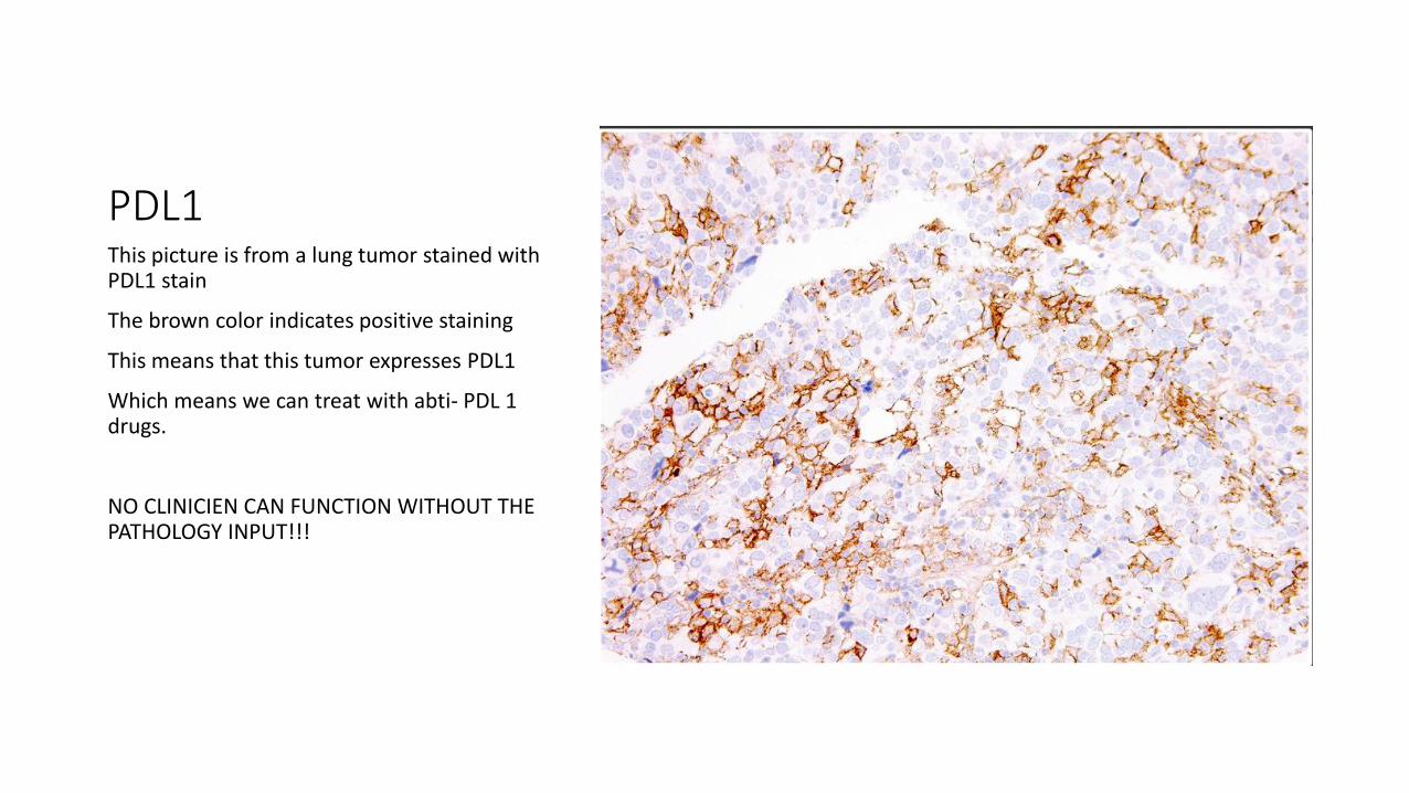
PDL1This picture is from a lung tumor stained with PDL1 stain
The brown color indicates positive staining
This means that this tumor expresses PDL1
Which means we can treat with abti- PDL 1 drugs.
NO CLINICIEN CAN FUNCTION WITHOUT THE PATHOLOGY INPUT!!!

Enablers of malignancy
• We said that there are 8 cancer hallmarks and 2 enablers.
• We discussed all hallmarks; let’s talk about the 2 enablers:
• 1. inflammation.
• 2. genomic instability.

Inflammation as an enabler of malignancy
• inflammatory cells modify the tumor microenvironment to enable many of the hallmarks of cancer.
• These effects may occur from direct interactions between inflammatory cells and tumor cells, or through indirect effects of inflammatory cells on other resident stromal cells.
Inflammation in response to tumors
• With any tumor there is associated inflammatory response, the aim of which is to protect tissue against cancer cells. However, inflammatory cells can enable malignant transformation.
• How do inflammatory cells help cancer cells to proliferate? By the variable chemical mediators and cytokines that are released from inflammatory cells.
• These mediators have several effects that enable growth, increase angiogenesis and even metastasis.. See next slide.

How do inflammatory cells affect tumor microenvironment??• 1. they secrete growth factors, such as EGF, and proteases that can liberate
growth factors from the extracellular matrix (ECM).
• 2. Removal of growth suppressors. growth of epithelial cells is suppressed by cell–cell and cell–ECM interactions. Proteases released by inflammatory cells can degrade the adhesion molecules that mediate these interactions, removing a barrier to growth.
• 3. Enhanced resistance to cell death. Detachment of epithelial cells from basement membranes and from cell–cell interactions can lead to a particular form of apoptosis. Any cell that looses attachment with other cells dies, this keeps coorct positioning of normal cells. However, tumor- associated macrophages may prevent apoptosis of the detached tumor cells by expressing adhesion molecules such as integrins that promote direct physical interactions with tumor cells.
• 4. Angiogenesis. Inflammatory cells release VEGF, that stimulate angiogenesis.
• 5. Invasion and metastasis. Proteases released from macrophages foster tissue invasion by remodeling the ECM, while factors such as TNF and EGF may directly stimulate tumor cell motility. TGF-β may promote epithelial-mesenchymal transition (EMT), which may be a key event in the process of invasion and metastasis.
• 6.Evasion of immune destruction. TGF-β and other factors favor the recruitment of immunosuppressive T regulatory cells or suppress the function of CD8+ cytotoxic T cells.
Role of M2 macrophages
• There is abundant evidence in cancer models and emerging evidence in human disease that advanced cancers contain mainly alternatively activated (M2) macrophages .
• M2 macrophages produce cytokines that promote angiogenesis, fibroblast proliferation, and collagen deposition.

note
• Colon cancer has high COX2 expression.
• Aspirin and other COX inbhibitors are used in the prevention of colon cancer.
Genomic instability as enabler of malignancy
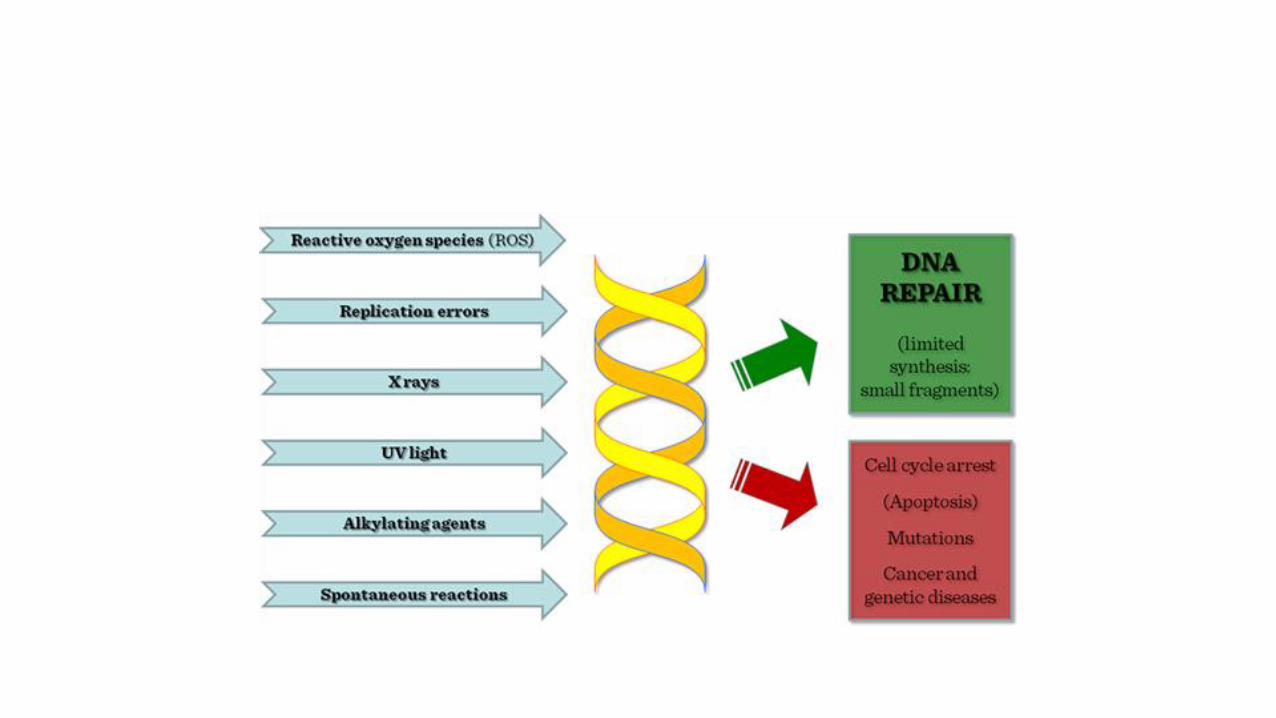
• Many mutations occur in normal individuals.. But are repaired by DNA repair genes
• If the DNA repair genes are inactivated… mutations can accumulate leading to cancer
• DNA repair genes are recessive.
• A cell with DNA repair gene mutated is not neoplastic yet but has the capacity to accumulate carcinogenic mutations. At this stage it is a “mutator phenotype”
• DNA repair genes can be inactivated by mutations or deletions in sporadic cancers and in some inherited diseases

DNA repair genes
• 1. mismatch repair gene… repairs nucleotide mismatch.. i:e makes sure that each A is paired with T and each C is paired with G ( not A or T) for example
• 2. nucleotide excision repair genes, repair nucleotide cross linking that results from UV exposure
• 3. recombination repair
Mismatch repair gene
• Mismatch repair gene is mutated in HNPCC = hereditary non-polyposis colorectal cancer syndrome
• People with the syndrome inherit one abnormal copy of the mismatch repair gene, and acquire the other mutation
• The syndrome causes familial colon cancer at a relatively young age, and mainly affecting the right side of the colon, mainly cecum.
• If the mismatch repair gene is defective there will microsatellite instability (MSI).
• Microsatellites are tandem repeats of 1-6 nucleotides in the genome.
Nucleotide excision repair gene
• This gene is mutated in xeroderma pigmentosum
• The nucleotide excision repair gene repairs nucleotide cross-linking occurring upon exposure to UV light
• People with the syndrome are predisposed to skin cancers

Recombination repair genes
• Certain DNA repair genes are important for repairing recombination errors
• Mutations in these genes occurs in several autosomal recessive diseases like
• 1. Fanconi anemia: there is predisposition to cancer and to anemia
• 2. Bloom’s syndrome : there is predisposition to cancer and developmental defects
• 3. Ataxia telangiectasia: cancer and gait imbalance

Ataxia telangiectasia
• The gene involved is ATM, which s important for DNA repair and for p53 activation
• If ATM mutated… no repair and no activation of p53… both lead to mutator phenotype and predispose to accumulation of mutations

Other DNA repair genes
• BRCA 1 and BRCA 2 also are important genes involved in DNA repair
• They are mutated in 50% of familial breast cancer… but rarely involved in sporadic breast cancer.
• BRCA 1 important for DNA repair and is linked to ATM protein
• BRCA 2 is one of the genes mutated in Fanconi anemia
summary
• Tumor cells express antigens, which makes them vulnerable to be recognized and destroyed by the immune system.
• These antigens can be protein products of the mutated ( p53) or overexpressed (HER2) genes. Antigens can also originate from oncoviral proteins, oncofetal (CEA) or abnormal mucins (CA125)
• Cellular immunity plays a role in immune surveillance whereas humoral immunity does not.
• Cytotoxic T cells can recognize tumor antigens expressed by APC. M1 macrophages and NK cells can destroy tumor cells.
• Tumors can evade this immunologic destruction through selective growth of antigen negative subclones, loss or reduced expression of histocompatibility molecules, downregulation of co-stimulatory molecules, antigen masking by producing a thick coat of external glycocalyx molecules or immunosuppression through production of TGF beta , expression of fasligand or as an effect of the oncogenic agent.
• Inflammation enables malignancy because inflammatory cells produce mediators and cytokines that increase growth, decrease growth inhibition, increase angiogenesis and help in metastatic spread.
• Mutation in DNA repair genes ( including mismatch repair, BRCA genes and others) cause genomic instability that allows accumulation of mutations which enables transformation.