IMPLICACIONES DE LA PROTEÍNA TAU Y LA CORTISTATINA EN …
222
UNIVERSIDAD AUTÓNOMA DE MADRID FACULTAD DE CIENCIAS Departamento de Biología Molecular IMPLICACIONES DE LA PROTEÍNA TAU Y LA CORTISTATINA EN LA PROGRESIÓN DE LA ENFERMEDAD DE ALZHEIMER TESIS DOCTORAL Alicia Rubio Garrido Madrid, 2008
Transcript of IMPLICACIONES DE LA PROTEÍNA TAU Y LA CORTISTATINA EN …

UNIVERSIDAD AUTÓNOMA DE MADRID
FACULTAD DE CIENCIAS
Departamento de Biología Molecular
IMPLICACIONES DE LA PROTEÍNA TAU Y LA
CORTISTATINA EN LA PROGRESIÓN DE LA
ENFERMEDAD DE ALZHEIMER
TESIS DOCTORAL
Alicia Rubio Garrido
Madrid, 2008

Madrid, 30 de Septiembre de 2008
Jesús Ávila de Grado, Profesor de Investigación del Consejo Superior de
Investigaciones Científicas y Mar Pérez, Contratada Ramón y Cajal de la
Universidad Autónoma de Madrid
INFORMAN QUE:
La presente tesis doctoral titulada “Implicaciones de la proteína tau y la
cortistatina en la progresión de la enfermedad de Alzheimer” ha sido
realizada bajo nuestra dirección por Alicia Rubio Garrido. Consideramos que el
trabajo reviste las características de originalidad y calidad científica requeridas
para ser defendido como Tesis Doctoral para optar al Grado de Doctor.
Los co-directores de la tesis,
Jesús Ávila Mar Pérez


A mis padres A mis hermanos


Agradecimientos


iii
Esta tesis doctoral no hubiese sido posible sin la ayuda y el apoyo de mucha gente. A
todos ellos, muchísimas gracias…
A Jesús Ávila, por la oportunidad de formar parte de su grupo de investigación y por sus
enseñanzas. También agradecer a Mar Pérez su paciencia y sus consejos de todo tipo. Muchas
gracias a los dos, por la dirección de esta tesis.
A Luis de Lecea, por acogerme en su laboratorio de Estados Unidos y por su inestimable
ayuda con el trabajo de cortistatina.
A José J Lucas y a Félix Hernández, muchas gracias por vuestras indicaciones y
disponibilidad. Gracias José por aceptarme en tu grupo cuando llegué.
A Isabel Fariñas, por haberme introducido en la neurobiología cuando aún era una
estudiante.
A todos aquellos que han compartido conmigo el día a día del laboratorio. A Nuria,
Almu, Thorsten, Elena G, Elena T, Pal, Alberto, Elenita, Esther, Raquel, Zahady, Isma y Laura.
Muchísimas gracias por los buenos momentos –que han sido muchos-, por vuestro cariño,
vuestro apoyo y por crear un ambiente tan agradable en el trabajo.
A la gente del laboratorio de José, del 119 y del antiguo 470, gracias por ser siempre tan
amables y ayudarnos con cualquier duda. Gracias también a Héctor y Olga que, en cierto modo,
forman parte de nuestro grupo. Gracias a Santi por resolverme siempre los problemas
informáticos. También quería recordar a la gente que se ha marchado y echamos de menos:
Tobías, Eva, Alfredo, Cristina y Marta. Quería agradecer también el entusiasmo de los
estudiantes de Valencia, Jorge y Jero. Gracias a María Teresa Miras por prestar su laboratorio y
a Miguel por su trabajo con la acetilcolina y el calcio y su optimismo.
Al personal del Centro de Biología Molecular, especialmente a los Servicios de
Microscopía, Genómica y Animalario, muchas gracias por vuestra contribución.
A mi familia, en especial a mis padres y hermanos, por su cariño incondicional. A mis
amigos, por lo que son.
Por último, querría mencionar que el desarrollo de este trabajo de investigación ha sido
posible gracias a la concesión de una beca de Formación de profesorado universitario del
Ministerio de Educación y Ciencia, una beca del Ayuntamiento de Madrid en la Residencia de
Estudiantes y un contrato de CIBERNED. Asimismo el laboratorio ha contado con la
financiación de los proyectos de la CAM SAL0202/2006, del MEC SAF2003/02697;
SAF2006/02424; SAF2006/27523-E; NAN2004/09183-C10-09; GEN2003-20235-C05-01, de
CIBERNED CB06/05/0035; CB06/247, del ISCIII FIS C03-06; PI04/0607 y de la Fundación
Botín.


Índice


vii
Agradecimientos Índice Abreviaturas Summary Introducción 1. GENERALIDADES SOBRE LA ENFERMEDAD DE ALZHEIMER
1.1. Aspectos genéticos y factores de riesgo de la EA 1.2. Características neuropatológicas de la EA
1.2.1. Degeneración del sistema colinérgico 1.2.2. Placas seniles 1.2.3. Ovillos neurofibrilares
2. LA PROTEÍNA TAU 2.1. Organización proteica 2.2. Distribución 2.3. Funciones fisiológicas de tau
2.3.1. Ensamblaje y estabilización de los microtúbulos 2.3.2. Transporte axonal 2.3.3. Otras interacciones
2.4. Agregación patológica de tau. Hiperfosforilación. 2.4.1. Quinasas 2.4.2. Fosfatasas
3. LA CORTISTATINA 3.1. Identificación 3.2. Estructura del péptido y del gen de la cortistatina 3.3. Expresión de la cortistatina 3.4. Receptores de somatostatina 3.5. Función
Objetivos Materiales y Métodos 1. MATERIALES
1.1. Reactivos 1.2. Anticuerpos
2. MÉTODOS 2.1. Mantenimiento de las distintas cepas de ratones 2.2. Cultivos celulares
2.2.1. Células SH-SY5Y, HEK-293 y COS-7: mantenimiento, transfección y tratamiento 2.2.2. Cultivos primarios de neuronas de corteza o de hipocampo de ratón
2.3. Western blot (WB) 2.4. Inmunofluorescencia 2.5. Internalización de Fluo-cortistatina en cultivos primarios de corteza murina 2.6. Reacción en cadena de la polimerasa (PCR) cuantitativa 2.7. Purificación de DNA genómico 2.8. Determinación fluorimétrica del Ca2+ intracelular con Fura-2 2.9. Purificación de proteínas 2.10. Ensayo de viabilidad 2.11. Test del reconocimiento de objeto 2.12. Análisis estadístico
i v
ix xiii
1 3 3 6 6 9 9
11 11 13 14 14 16 16 18 19 20 21 21 22 23 25 28
31
35 37 37 38 40 40 40 40 41 42 43 44 44 47 47 49 49 50 50

viii
Resultados 1. PAPEL DE TAU EN LA PROGRESIÓN DE LA EA
1.1. La proteína tau extracelular produce muerte celular de las células de neuroblastoma humano SH-SY5Y
1.2. La proteína tau altera la homeostasis de calcio intracelular de las SH-SY5Y a través de los receptores muscarínicos
1.3. Patrón de expresión de los receptores muscarínicos en las SH-SY5Y 1.4. Análisis farmacológico para determinar los receptores muscarínicos implicados en el
efecto de tau sobre los niveles de calcio intracelular en SH-SY5Y 1.5. Transfección transitoria con M1 y M3 en células COS-7 para confirmar la implicación
de estos receptores en la movilización de calcio inducida por tau 1.6. Identificación de la región de tau capaz de alterar la homeostasis intracelular de
calcio en células COS-7 que expresan M3 1.7. El calcio intracelular de las neuronas murinas de hipocampo en cultivo aumenta en
presencia de la proteína tau 2. EFECTO DE DISTINTOS AGONISTAS DE LOS RECEPTORES DE SOMATOSTATINA (sst) SOBRE LA FOSFORILACIÓN DE TAU EN LA Ser262
2.1. El nivel de fosforilación de la Ser262 de la proteína tau es menor en la corteza de los ratones que no expresan cortistatina
2.2. La cortistatina promueve la fosforilación de tau en la Ser262 de cultivos primarios de neuronas corticales murinas
2.3. La somatostatina también favorece la fosforilación de tau en la Ser262 pero con una cinética diferente a la de la cortistatina en neuronas corticales murinas en cultivo
2.4. El octreotide no afecta a la fosforilación de tau en la Ser262 de neuronas corticales murinas en cultivo
2.5. Las neuronas corticales murinas en cultivo presentan receptores que unen cortistatina
2.6. Las neuronas corticales en cultivo expresan los cinco subtipos de receptores de somatostatina
2.7. Los receptores sst2 y sst4 están implicados en el efecto de la cortistatina sobre la fosforilación de tau en la Ser262
2.8. La cortistatina incrementa la fosforilación de tau en la Ser262 probablemente a través de su unión a receptores heterodiméricos compuestos por sst2 y sst4
2.9. La PKC fosforila a la proteína tau en la Ser262 en presencia de cortistatina en neuronas corticales murinas
Discusión 1. PAPEL QUE JUEGA TAU EN EL AVANCE DE LA EA
1.2. La proteína tau extracelular induce un aumento en el calcio intracelular a través de M1 y M3
1.2. La controversia en torno a la posible toxicidad de la proteína tau 1.3. Modelo de propagación de la EA por toxicidad de la proteína tau extracelular
2. PAPEL DE LA CORTISTATINA EN EL PASO DE LA PATOLOGÍA DE TAU A LA CORTEZA CEREBRAL
2.1. La cortistatina induce la fosforilación de tau en la Ser262 2.2. Receptores de somatostatina e internalización 2.3. Receptores de somatostatina y heterodimerización 2.4. Receptores de somatostatina y señalización intracelular 2.5. La posible relación entre cortistatina y somatostatina con la EA
Conclusiones Bibliografía Anexo
51 53
53
55 58
59
59
63
65
67
67
70
73
74
76
78
79
81
84
85 88
88 89 92
93 93 94 96 98
100
103
107
143

Abreviaturas


xi
ACh: Acetylcholine / Acetilcolina AICD: APP Intracellular Domain / Dominio Intracelular del APP AMV: Avian Myeloblastosis Virus / Virus de la Mieloblastosis de Ave Ara-C: Arabinofuranosylcytosine / Arabinofuranosilcitosina ApoE/APOE: Apolipoprotein E / Apolipoproteína E (gen/proteína) app/APP: Amyloid Precursor Protein / Proteína Precursora del Amiloide (gen/proteína) BACE: β-site APP-cleaving enzyme / Enzima que procesa el APP en el sitio β BSA: Bovine Serum Albumin / Seroalbúmina Bovina CA: Cuerno de Ammon CaMKII: Calcium/calmodulin-dependent protein Kinase II / Proteína Quinasa dependiente de Calcio/calmodulina CBD: Corticobasal Degeneration / Degeneración Corticobasal CDK5: Cyclin-Dependent Kinase 5 / Quinasa Dependiente de Ciclina 5 cDNA: complementary Deoxyribonucleic Acid / Ácido Desoxirribonucleico complementario CST-14: Cortistatin-14 / Cortistatina-14 CSF: Cerebrospinal Fluid / Fluido Cerebroespinal CT: Threshold Cycle / Ciclo Umbral CKII: Casein Kinase II / Caseína Quinasa II 4-DAMP: 4-Diphenylacetoxy-N- methylpiperidine / 4-Difenil-acetoxi-N-metilpiperidina DAPI: 4',6-Diamidino-2-Phenylindole / 4´,6-Diamidino-2-fenilindol DMEM: Dulbecco’s Modified Eagle´s Medium / Medio Eagle modificado de Dulbecco DMSO: Dimethyl sulfoxide / Dimetilsulfóxido DNA: Deoxyribonucleic Acid / Ácido Desoxirribonucleico DIV: Days in vitro / Días in vitro EA: Enfermedad de Alzheimer ECL: Enhanced Chemiluminescent / Sustrato quimioluminiscente EDTA: Ethylenediaminetetraacetic acid / Ácido Etildiaminotetraacético EEG: Electroencephalography / Electroencefalografía eNFTs: extracellular Neurofibrillary Tangles / Ovillos Neurofibrilares extracelulares FTDP-17: Frontotemporal Dementia and Parkinsonism linked to Chromosome 17 / Demencia Frontotemporal con Parkinsonismo asociada al Cromosoma 17 FRET: Fluorescence Resonance Energy Transfer / Fluorescencia tras Transferencia de Energía por Resonancia Fura-2AM: Fura-2 acetoxymethyl ester / Fura-2 acetometil éster GABA: Gamma-Aminobutyric Acid / Ácido Gamma-Aminobutírico GHSR: Growth Hormone Secretagogue Receptor / Receptor Secretagogo de la Hormona de Crecimiento GIP: G protein-coupled receptor Interacting Proteins / Proteínas que Interaccionan con los receptores asociados a proteína G GRK: G-protein-coupled Receptor-Kinase / Receptor Quinasa acoplado a Proteína-G GSK-3: Glycogen Synthase Kinase-3 / Glucógeno Sintasa Quinasa-3 HA: Hemagglutinin / Hemaglutinina HEPES: N-(2-hydroxyethyl)-piperazine-N'-2-ethanesulfonic acid / N-(2-hidroxietil)-piperacina-N´-2-ácido etanolsulfónico Hsp70: Heat shock protein 70 / proteína de choque térmico 70 IF: Immunofluorescence / Inmunofluorescencia IP3: Inositol triphosphate / Inositol trifosfato JNK: c-Jun NH2-terminal Kinase / Quinasa c-Jun NH2-terminal kDa: kilo Dalton / kilo Dalton LTP: Long Term Potentiation / Potenciación a largo plazo M1-5: Muscarinic Receptor 1-5 / Receptor Muscarínico 1-5 MAP: Microtubule Associated Protein / Proteína Asociada a Microtúbulos MAPK: Mitogen Activated Protein Kinase / Proteína Quinasa Activada por Mitógenos

xii
MARK: MAP/microtubule Affinity-Regulating kinase / Quinasa Reguladora de Afinidad entre MAPs/microtúbulos MCI: Mild Cognitive Impairment / Deterioro Cognitivo Leve MI: Memory Index / Índice de Memoria MrgX2: Mas-related G-protein-coupled receptor member X2 / Receptor X2 acoplado a proteína G relacionado con Mas mRNA: messenger Ribonucleic Acid / Ácido Ribonucleico mensajero NB: Neurobasal Medium / Medio Neurobasal NFT: Neurofibrillary Tangles / Ovillos Neurofibrilares NMDA: N-methyl-D-Aspartate / N-Metil-D-Aspartato NPDPK: Non-Proline Directed Protein Kinases / Quinasas no Dirigidas por Prolina NREM: Non-Rapid Eye Movement / Movimientos oculares no rápidos (Sueño Lento) NT: Neuropil Threads / Hilos del Neuropilo OCT: Octreotide / Octreotide PAO: Phenylarsine Oxide / Óxido de Fenilarsina pb: pares de bases PBS: Phosphate-Buffered Saline / Solución Salina de Fosfato Tamponada PCR: Polymerase Chain Reaction / Reacción en Cadena de la Polimerasa PDPK: Proline Directed Protein Kinases / Quinasas Dirigidas por Prolina PHF: Paired Helical Filaments / Filamentos Apareados Helicoidales PKA: Protein Kinase A / Proteína Quinasa A PKC: Protein Kinase C / Proteína Quinasa C PLC: Phospholipase C / Fosfolipasa C PP: Protein Phosphatase / Proteína Fosfatasa ps / PS: Presenilin / Presenilina (gen/proteína) PS: Population Spikes / Respuestas Eléctricas Sinápticas PSP: Progressive Supranuclear Palsy / Parálisis Supranuclear Progresiva PTP: Phosphotyrosine phosphatases / Fosfotirosinas Fosfatasas RAMP: Receptor-activity-modifying protein / Proteína que modifica la actividad del receptor REM: Rapid Eye Movement / Movimiento Ocular Rápido (Sueño Paradójico) rpm: revoluciones por minuto RT-PCR: Reverse Transcription-Polymerase Chain Reaction / Transcripción Reversa-Reacción en Cadena de la Polimerasa SDS: Sodium Dodecyl Sulphate / Dodecil Sulfato Sódico SDS-PAGE: SDS-PolyAcrylamide Gel Electrophoresis / Gel de Electroforesis de Poliacrilamida con SDS Ser: Serine / Serina SF: Straight Filaments / Filamentos rectos SNC: Sistema Nervioso Central SNP: Sistema Nervioso Periférico SRIF-14: Somatotropin release-inhibiting factor-14 (Somatostatin-14) / Factor inhibidor de la liberación de somatotrofina (Somatostatina-14) sst 1-5: somatostatin receptor 1-5 / Receptor de somatostatina 1-5 SWS: Slow Wave Sleep / Sueño de Ondas Lentas TE: Tris EDTA / Tris EDTA TES: N-Tris(hydroxymethyl)methyl-2-aminoethanesulfonic acid / ácido N-Tris (hidroximetil) metil-2-aminoetanosulfónico Thr: Threonine / Treonina UV: Ultraviolet / Ultravioleta WB: Western Blot Los términos en inglés se han evitado en la medida de lo posible empleando tan sólo los que consideramos ampliamente aceptados como western blot o primer.

Summary

xv
Alzheimer disease (AD) is a senile dementia characterized by the presence of two aberrant
structures: senile plaques and neurofibrillary tangles (NFTs). NFTs are intra-neuronal lesions mainly
composed by an abnormal hyperphosphorylated form of the microtubule-associated tau protein. NFTs
have been shown to follow a characteristic distribution pattern in the brain related with the progression of
AD. NFTs are first detected in the entorhinal cortex and the hippocampus, and then, the pathology
spreads to the cortex. In this work, we try to find out different factors to explain how AD progresses. In
this process, neuronal degeneration occurs and dead neurons can release their intracellular content to the
extracellular space. We demonstrate that extracellular tau is toxic to neuroblastoma cells SH-SY5Y in
culture. We show that tau interacts with M1 and M3 muscarinic receptors leading to an increase in
intracellular calcium levels. This molecular mechanism could probably induce neuronal degeneration
explaining the spreading of AD from the entorhinal cortex and the hippocampus to the cortex in advanced
stages. The development of tau phosphorylation in the cortex seems critical in the onset of dementia in
patients with AD. Here, we suggest that cortistatin could contribute to tau hyperphosphorylation in the
cortex. Cortistatin, a neuropeptide related to somatostatin, is mainly expressed in the cerebral cortex. We
demonstrate that cortistatin-deficient mice show a decreased tau phosphorylation at Ser262 in the cortex,
but not in other brain regions tested. Then, we found that cortistatin induces tau phosphorylation at
Ser262 in cultures of murine cortical neurons. The effect of cortistatin is likely mediated by heteromeric
receptors composed of somatostatin receptor subtype 2 and 4 and also by protein kinase C signalling.
Therefore, our results suggest an important role for cortistatin in promoting tau phosphorylation at
Ser262 in the cortex of patients with AD.


Introducción


Implicaciones de la proteína tau y la cortistatina en la progresión de la enfermedad de Alzheimer Introducción
3
1. GENERALIDADES SOBRE LA ENFERMEDAD DE ALZHEIMER La enfermedad de Alzheimer (EA) es una enfermedad neurodegenerativa que afecta a las
personas de edad avanzada. Se caracteriza, en su forma típica, por una pérdida progresiva de la
capacidad cognitiva, acompañada de apatía emotiva y desconexión del entorno. En los estadios
más avanzados, las capacidades motoras llegan a deteriorarse, pudiendo, en última instancia,
desencadenar la muerte (Cummings, 2004). La duración de esta enfermedad puede variar entre
2 y 20 años pues difiere enormemente de un individuo a otro. Dicha progresión depende, en
gran medida, del grado de avance del deterioro cerebral por muerte neuronal. Los pacientes con
EA presentan una atrofia notable en las zonas del cerebro relacionadas con la memoria y las
emociones, como la corteza entorrinal, el hipocampo, la amígdala, el lóbulo frontal y la corteza
temporal y parietal (Clark et al., 2008). En cambio, el deterioro es menor en la corteza motora y
sensitiva (Damasio, 1989). De hecho, en las etapas finales de la EA, el número de neuronas del
hipocampo suele reducirse un 84% (Smith, 2002). En las zonas cerebrales más afectadas por la
EA, se han identificado dos estructuras aberrantes características: las placas seniles, formadas en
su mayoría por el péptido β-amiloide, y los ovillos neurofibrilares, compuestos principalmente
por la proteína tau hiperfosforilada.
1.1. Aspectos genéticos y factores de riesgo de la EA La EA puede clasificarse en dos tipos según su origen o etiología: la forma esporádica,
que es la predominante, y la forma familiar autosomal dominante con una incidencia, esta
última, de menos de 5% (Campion et al., 1999).
La EA familiar es de aparición temprana (alrededor de los 50 años de edad) y se debe a
mutaciones puntuales en los genes de la proteína precursora del amiloide (app, cromosoma 21),
presenilina-1 (ps-1, cromosoma 14) y presenilina-2 (ps-2, cromosoma 1) (Bird, 2008).
Las mutaciones del gen del app resultan en un aumento en la producción del péptido β-
amiloide o en la agregación de este péptido (Seubert et al., 1993). Muchas de las mutaciones
puntuales que causan la EA familiar se localizan en las zonas de procesamiento de la α, β y γ-
secretasas (Selkoe, 2001). Los ratones transgénicos que sobre-expresan la proteína APP normal o
la proteína APP mutada son modelos para el estudio de la EA y desarrollan, de forma
prematura, placas seniles en su cerebro (Games et al., 1995; Puig et al., 2004; Zhang et al., 1997).
Los ratones en los que se elimina la expresión del gen de app son viables y no sufren grandes
alteraciones salvo gliosis y pequeños cambios en el comportamiento (Zheng et al., 1995). Por
otro lado, la dosis génica elevada presente en el síndrome de Down conlleva una sobre-

Implicaciones de la proteína tau y la cortistatina en la progresión de la enfermedad de Alzheimer Introducción
4
expresión de APP, responsable, en la mayoría de los casos, de padecer EA prematura (Selkoe,
2001).
Se han identificado más de 100 mutaciones puntuales en los genes de la presenilina-1 y 2.
Las mutaciones en el gen de la presenilina-1 son especialmente abundantes puesto que están
asociadas a más de un tercio de los enfermos de Alzheimer familiar (Hartmann et al., 2007;
Selkoe, 2001). Existe un 67% de homología entre la presenilina-1 (46 kDa) y la 2 (55 kDa) en la
secuencia aminoacídica (Laudon et al., 2005; Oh & Turner, 2005; Ratovitski et al., 1997). Los
ratones en los que se elimina la expresión de los genes presenilina-1 y 2 pierden la actividad de
la γ-secretasa y no producen β−amiloide (De Strooper & Woodgett, 2003).
Curiosamente, no se han encontrado mutaciones del gen de tau en los enfermos de
Alzheimer apoyando así la “hipótesis de la cascada amiloide”, en la que se establece que la
acumulación de β-amiloide inicia una cascada de reacciones que originan la patología de tau y,
por último, la muerte neuronal (Hardy, 2002; Hardy et al., 1986). Sin embargo, los portadores de
mutaciones en el gen de tau padecen otro tipo de demencias conocidas como tauopatías. Estas
tauopatías se definen como enfermedades neurodegenerativas que presentan depósitos
filamentosos de tau hiperfosforilado en neuronas y glía y que cursan con aberraciones en el
procesamiento alternativo de tau, los niveles de tau y su grado de fosforilación. Dentro de las
tauopatías se incluyen la EA, las demencias frontotemporales, la degeneración corticobasal
(CBD), la demencia pugilística, la enfermedad de Niemann-Pick, la enfermedad de Creutzfeld-
Jacob, el síndrome de Down, la enfermedad de Pick y la parálisis supranuclear progresiva
(PSP). Algunas de éstas tienen origen esporádico, como la EA (salvo los casos de Alzheimer
familiar), y otras familiar o genético, como la demencia frontotemporal con parkinsonismo
asociada al cromosoma 17 (FTPD-17). Por el momento, se han identificado, al menos, 34
mutaciones en los exones 1, 9, 10, 11, 12 y 13 del gen de tau en las familias con FTDP-17
(revisadas recientemente en (Wang & Liu, 2008)). Muchas de estas mutaciones se localizan en la
región de unión a microtúbulos de tau (exón 10) y se ha visto que pueden provocar una
reducción en la interacción de tau con los microtúbulos (Hong et al., 1998; Hutton et al., 1998) o
con otras proteínas (Goedert & Spillantini, 2000). Algunas de estas mutaciones facilitan el
autoensamblaje de tau en filamentos anormales (Arrasate et al., 1999), reducen las capacidades
de tau para regular la inestabilidad dinámica de los microtúbulos (Bunker et al., 2006) o afectan
a la cantidad de tau 4R y 3R (Buee et al., 2000; Glatz et al., 2006; Goedert, 2005; Heutink, 2000;
Stanford et al., 2003; Yoshida, 2006). Como en la FTDP-17, también se han descrito otras
mutaciones sin sentido relacionadas con la PSP, la CBD y la enfermedad de Pick (Bugiani et al.,
1999; Delisle et al., 1999; Murrell et al., 1999).
Implicaciones de la proteína tau y la cortistatina en la progresión de la enfermedad de Alzheimer Introducción
5
La forma esporádica de la EA tiene, probablemente, un origen multifactorial y suele
aparecer a partir de los 60-65 años de edad. A pesar de que el envejecimiento normal no parece
ser la causa directa de esta enfermedad, se cree que el mayor factor de riesgo es la edad puesto
que la incidencia de la EA aumenta de forma exponencial a partir de los 65 años (Jicha et al.,
2008; Jorm & Jolley, 1998; Kawas & Corrada, 2006; Kukull & Bowen, 2002). Otros factores de
riesgo de la enfermedad son los antecedentes familiares (Evans et al., 1997; Moritz & Petitti,
1993), la exposición a distintos factores ambientales como pesticidas, metales, toxinas o virus
(Doty, 2008) y los factores de riesgo vascular (hipertensión, tabaquismo, aterosclerosis,
obesidad, hipercolesterolemia, diabetes mellitus tipo II, fibrilación auricular) (Ott et al., 1999).
Algunos trabajos sugieren que la ingesta de vitaminas B12 y folato, antioxidantes como las
vitaminas C y E, ácidos grasos insaturados y niveles moderados de alcohol podrían reducir el
riesgo de padecer EA (revisados en (Luchsinger & Mayeux, 2004)).
Numerosos genes, como los de la interleukina-1, la óxido nítrico sintasa 3 o el citocromo
CYP2D6, se han asociado con un mayor riesgo de padecer la EA (Hartmann et al., 2007). Entre
ellos, destaca el estudio del gen de la apolipoproteína E (ApoE). La proteína APOE es la
encargada del transporte del colesterol en sangre y entre neuronas y astrocitos. El gen de la
ApoE tiene tres alelos: E3, es el más común, E2, parece proteger a una persona de la EA y E4,
implica que la enfermedad se inicia a una edad más temprana (Corder et al., 1993; Poirier et al.,
1993; Polvikoski et al., 1995; Raber et al., 2004). La APOE4 parece ser menos eficiente en la
reutilización de los lípidos de membrana y la reparación neuronal y podría promover la
formación de placas amiloides alterando el procesamiento del APP (Holtzman et al., 2000;
Poirier, 1994; Prince et al., 2004). También parece que el gen para la proteína α2-macroglobulina
(Blacker et al., 1998), implicada en la degradación de β-amiloide, o el gen para SORL1 (Rogaeva
et al., 2007), que dirige el tráfico del APP en las vías de reciclaje, están relacionados con la
predisposición a padecer EA. Estudios recientes muestran que un polimorfismo del gen para
CALHM1 presenta una mayor susceptibilidad para sufrir la EA. La CALHM1 es un canal
cerebral de calcio que controla los niveles de β-amiloide (Dreses-Werringloer et al., 2008).
Los estudios genéticos asociados con la EA pueden consultarse en:
http://www.alzgene.org (Bertram et al., 2007).

Implicaciones de la proteína tau y la cortistatina en la progresión de la enfermedad de Alzheimer Introducción
6
1.2. Características neuropatológicas de la EA Como se mencionó anteriormente, las características histopatológicas más relevantes de la
EA avanzada son la pérdida sináptica y la presencia de placas seniles y ovillos neurofibrilares.
La aparición de placas seniles parece estar relacionada con la edad, mientras que los ovillos
neurofibrilares correlacionan con la gravedad de la demencia (Arriagada et al., 1992a; Arriagada
et al., 1992b).
1.2.1. Degeneración del sistema colinérgico Aunque la EA se ha asociado con el déficit de diferentes neurotransmisores, es la pérdida
de inervación colinérgica la que mejor correlaciona con el grado de deterioro cognitivo que
sufren estos pacientes (Francis et al., 1999; Geula, 1998; Perry et al., 1981). La degeneración de
las aferencias colinérgicas presentes en la corteza y el hipocampo se traduce en la reducción de
la recaptación de colina, la disminución de la liberación de acetilcolina y la atrofia de las
neuronas colinérgicas del septum y del núcleo basal de Meynert (Blusztajn & Berse, 2000;
Francis et al., 1999). Concretamente, en la corteza entorrinal, más de un 80% de la inervación
colinérgica puede perderse en los cerebros con EA (Geula & Mesulam, 1996). El deterioro de
estas estructuras cerebrales, implicadas en la memoria, unido a la disminución notable del nivel
de la colina acetiltransferasa en el hipocampo y en la corteza de los enfermos de Alzheimer
(Davies & Maloney, 1976) podrían explicar la pérdida de memoria explícita o declarativa de los
pacientes sentando así las bases de la “hipótesis colinérgica” (Coyle et al., 1983). De hecho, la
terapia actual frente a la EA se centra en la restauración de los niveles de acetilcolina mediante
la administración de inhibidores de la acetilcolinesterasa, como el donepezil, la rivastigmina, la
galantamina y la tacrina, durante los primeros estadios de la enfermedad, a pesar de que los
beneficios terapéuticos de la misma sobre las funciones cognitivas siguen siendo discutidos
(Cummings, 2004; Ladner & Lee, 1998; Raschetti et al., 2007; Trinh et al., 2003).
Los receptores colinérgicos pueden dividirse en muscarínicos y nicotínicos, basándose en
la actividad de dos alcaloides agonistas naturales conocidos como muscarina (aislada de la seta
Amanita muscaria) y nicotina (aislada, a su vez, de la hoja del tabaco Nicotina tabacum). Los
receptores muscarínicos forman una familia de receptores de membrana metabotrópicos que
incluye cinco subtipos diferentes (M1-5). Cada subtipo tiene su propia distribución y
farmacología, siendo capaces de modular respuestas tempranas y tardías (revisadas en
(Volpicelli & Levey, 2004)). Aunque los cinco subtipos se detectan en el sistema nervioso
central, la expresión del M5 es extremadamente baja. En la corteza y el hipocampo de humanos,
predomina la expresión de M1 (Piggott et al., 2002). En el caso del ratón adulto, M1 y M3

Implicaciones de la proteína tau y la cortistatina en la progresión de la enfermedad de Alzheimer Introducción
7
parecen ser los más abundantes en el hipocampo y la corteza entorrinal (Allen Brain Atlas;
http://www.brain-map.org/). Los receptores M1, M3 y M5, localizados principalmente en las
terminales colinérgicas postsinápticas, interaccionan de forma selectiva con las proteínas G de la
familia Gq/G11, mientras que M2 y M4, presinápticos en su mayoría, activan preferentemente a
las Gi/G0 (Wess et al., 2007). De este modo, la activación de los receptores M1, M3 y M5 induce
un incremento del calcio intracelular (Lanzafame et al., 2003). En los cerebros de individuos con
EA, la cantidad de M2 se ve reducida de forma significativa (Kar et al., 2004). También se ha
descrito que los enfermos de Alzheimer presentan una pérdida notable de la proteína G
asociada a los receptores M1 (Hellstrom-Lindahl, 2000; Pavia et al., 1998). Como mencionamos,
la acetilcolina también puede unirse a los receptores nicotínicos. A diferencia de los
muscarínicos, estos receptores forman canales ionotrópicos que, al activarse, permiten el paso
de sodio y calcio, causando una rápida despolarización de la membrana plasmática. Los
receptores nicotínicos son receptores heterólogos pentaméricos que se componen de dos
subunidades α y tres β. Existen 16 subtipos diferentes que difieren en su distribución cerebral,
su estructura y sus características electrofisiológicas y farmacológicas (revisados en (Vazquez-
Palacios & Bonilla-Jaime, 2004)). La corteza cerebral y el hipocampo de los cerebros afectados
por la EA presentan una reducción en el número de receptores nicotínicos comparados con los
cerebros control (Francis et al., 1999; Nordberg et al., 1995).
La acetilcolina ejerce un papel clave en la formación de la memoria declarativa. Durante
la vigilia, se produce la adquisición y el procesamiento inicial de la información sensorial que
llega del exterior a la corteza de asociación (lóbulos frontal, parietal y temporal). Más adelante,
esta información pasa a la corteza entorrinal y, de ahí, al hipocampo (figura 1). El sueño parece
ser crítico para la consolidación de la memoria (Gais et al., 2007; Stickgold, 2005). Durante el
sueño, el flujo de información se efectúa en las dos direcciones y en repetidas ocasiones según la
alternancia de las fases de sueño lento (NREM) y sueño paradójico (REM). En el sueño de ondas
lentas (SWS), se produce un tránsito de la información que va desde el hipocampo a la corteza
entorrinal y, de ahí a la corteza. Durante el sueño REM, el flujo de información se realiza en
sentido contrario. El SWS, que predomina en la primera parte de la noche, parece ser relevante
para la formación de la memoria declarativa (Gais & Born, 2004a; Gais & Born, 2004b).
Se ha demostrado que durante el sueño se reactivan las mismas poblaciones neuronales
del hipocampo que se activaron originalmente durante la adquisición de la memoria.
(Axmacher et al., 2008; Maquet, 2001; Stickgold et al., 2001). De este modo, la información se
integra y se almacena a largo plazo en la corteza, especialmente en la corteza prefrontal
(Frankland & Bontempi, 2005; Takashima et al., 2006; Wiltgen et al., 2004). En la corteza
entorrinal (Nakazawa, 2006; Yasuda & Mayford, 2006) y el hipocampo (Neves et al., 2008;
Squire et al., 2004; Suzuki, 2007) se almacena temporalmente la memoria. En apoyo a esta idea,

Implicaciones de la proteína tau y la cortistatina en la progresión de la enfermedad de Alzheimer Introducción
8
CORTEZADE ASOCIACIÓN
(lóbulos frontal, parietal,temporal)
CORTEZAENTORRINAL
Capas superficiales(I-III)
Capas profundas(IV-VI)
HIPOCAMPO
Consolidación de la memoria (SWS)
Adquisición y consolidación temprana (vigilia)
Estímulo externo
CORTEZADE ASOCIACIÓN
(lóbulos frontal, parietal,temporal)
CORTEZAENTORRINAL
Capas superficiales(I-III)
Capas profundas(IV-VI)
HIPOCAMPO
Consolidación de la memoria (SWS)
Adquisición y consolidación temprana (vigilia)
Estímulo externo
se ha visto que pacientes con daños restringidos a estas zonas muestran amnesia retrógrada o
incapacidad para formar nuevas memorias (Reed & Squire, 1998; Scoville & Milner, 2000; Squire
& Zola-Morgan, 1991). La corteza entorrinal es una de las primeras estructuras dañadas en la
EA (Braak & Braak, 1991). Esta zona cerebral es crítica para la formación de la memoria puesto
que representa el principal flujo de entrada de información al hipocampo a través de las capas
superficiales y el mayor flujo de salida del hipocampo a través de las capas profundas (Dolorfo
& Amaral, 1998; Witter et al., 1989). Se ha visto que los niveles de acetilcolina son altos durante
la vigilia y el sueño REM y bajos durante el SWS, por tanto se postula que la acetilcolina media
los cambios de dirección del flujo de información entre las tres estructuras cerebrales implicadas
en la formación de la memoria mediante la supresión de la transmisión sináptica glutamatérgica
excitatoria de CA3 a la corteza entorrinal (Buzsaki, 1996; Hasselmo, 1999; Steriade, 2004).
Figura 1. Representación esquemática del modelo de la adquisición y consolidación de la memoria.

Implicaciones de la proteína tau y la cortistatina en la progresión de la enfermedad de Alzheimer Introducción
9
1.2.2. Placas seniles
Las placas seniles (10-120 μm de diámetro) son depósitos amiloides extracelulares
(Glenner & Wong, 1984) que también pueden encontrarse en la corteza normal de personas de
edad avanzada (Maccioni et al., 2001). Estas estructuras pueden ser difusas o pre-amiloides con
depósitos amorfos de β−amiloide y evolucionar a placas neuríticas compuestas por depósitos
insolubles de β−amiloide rodeados por neuritas distróficas (axones y dendritas dañadas),
astrocitos reactivos y microglía activada (Selkoe, 2001). El péptido β-amiloide deriva de los
cortes proteolíticos secuenciales de la proteína APP realizados por varias proteasas (Kang et al.,
1987; Masters et al., 1985). La célula produce el β-amiloide 40, en mayor cantidad, que el β-
amiloide 42. Este último, al ser más hidrofóbico, puede agregarse y plegarse en hoja β-lámina
con más facilidad que el β-amiloide 40 e interaccionar con otras proteínas extracelulares que
podrían propiciar la formación de placas seniles (Citron et al., 1996; Neve et al., 2000). En un
primer momento, se pensó que las placas eran neurotóxicas pero más tarde se ha propuesto que
los oligómeros solubles de β−amiloide, capaces de inhibir el LTP hipocampal y dificultar la
plasticidad sináptica, podrían ser los responsables del inicio de los déficits cognitivos (Rowan et
al., 2007). También se ha visto que el péptido β-amiloide puede inducir apoptosis in vivo e in
vitro (Forloni et al., 1993; Selkoe, 2001).
1.2.3. Ovillos neurofibrilares Los ovillos neurofibrilares (NFT) consisten en inclusiones filamentosas formadas dentro
del citoplasma perinuclear y, ocasionalmente, presentes en las dendritas proximales de las
neuronas. También pueden detectarse NFT en las células gliales de algunas enfermedades
neurodegenerativas como, por ejemplo, la CBD y la PSP (Bergeron et al., 1997). Los hilos del
neuropilo (NT) se localizan en el interior de los segmentos dendríticos, sin implicar al axón, y
están formados por unas estructuras filamentosas dispersas. Tras la muerte celular, el material
insoluble se mantiene y es conocido como ovillos fantasma o también como ovillos
neurofibrilares extracelulares (eNFT) (Braak & Braak, 1991). Ultraestructuralmente, estas
alteraciones neurofibrilares contienen filamentos de morfología característica compuestos
mayoritariamente por la proteína tau hiperfosforilada (Grundke-Iqbal et al., 1986). La gran
mayoría se denominan filamentos apareados helicoidales (PHF) porque consisten en dos haces
entrecruzados uno sobre otro con una periodicidad de 65-80 nm y un grosor de 10-30 nm (Kurt
et al., 1997). También se encuentran, aunque en menor cantidad, otro tipo de filamentos que no
parecen tener una morfología helicoidal. Éstos son conocidos como filamentos rectos (SF) y
poseen un diámetro de unos 15 nm (Kurt et al., 1997).
Implicaciones de la proteína tau y la cortistatina en la progresión de la enfermedad de Alzheimer Introducción
10
Por otro lado, se ha visto que los enfermos de Alzheimer y de otras tauopatías también
pueden presentar otro tipo de agregados aberrantes de tau, conocidos como cuerpos de Hirano
(Galloway et al., 1987; Gibson & Tomlinson, 1977). La relevancia de estas estructuras en cuanto
a la patología de las enfermedades neurodegenerativas sigue sin establecerse. En los cuerpos de
Hirano, la proteína tau no se encuentra en forma fibrilar y está asociada a filamentos de actina
(Hirano, 1994).
En 1991, Braak y Braak (Braak & Braak, 1991) demostraron que la distribución de la
patología neurofibrilar en la EA sigue un patrón topográfico característico que empieza en la
corteza transentorrinal (propia de los estadios I y II), progresa hacia la corteza entorrinal, el
hipocampo, la amígdala y la corteza temporal adyacente (límbico, estadios III y IV) y finaliza en
el resto de la corteza (estadios V y VI). Este patrón correlaciona con el grado de demencia de
modo que, los estadios I y II son asintomáticos, los III y IV son propios de los pacientes con
Deterioro Cognitivo Leve (MCI) y, por último, los enfermos de Alzheimer se encuentran en los
V y VI (Lovell & Markesbery, 2007). Cabe destacar la importancia de la aparición de la patología
de tau en la corteza pues representa un momento crítico en el desarrollo de los síntomas
propios de la enfermedad (Braak & Braak, 1991; Delacourte et al., 1999). Este trabajo junto con el
que muestra que existe una correlación entre el número de NFT y el grado de demencia de los
pacientes con EA (Arriagada et al., 1992a; Arriagada et al., 1992b), relacionan, por primera vez,
la patología de tau con la progresión de la EA y la muerte neuronal. Este tema está siendo
discutido en la actualidad puesto que, por el momento, no se sabe si tau induce la muerte
neuronal debido a una pérdida de función del tau normal o a una ganancia de función de tau
debida a su toxicidad. Tampoco se sabe cómo se produce el avance de la patología de tau.
Ambos temas serán tratados a lo largo de esta tesis doctoral.

Implicaciones de la proteína tau y la cortistatina en la progresión de la enfermedad de Alzheimer Introducción
11
2. LA PROTEÍNA TAU
2.1. Organización proteica El gen de tau contiene 16 exones y el procesamiento alternativo de los exones 2, 3 y 10 da
lugar a seis isoformas distintas en el sistema nervioso central de humanos (figura 2) (Neve et al.,
1986). Las variantes de tau difieren unas de otras por la presencia de 3 o 4 repeticiones en la
parte carboxilo-terminal de la molécula, codificadas por los exones 9-12, y la presencia o
ausencia de uno o dos insertos en la parte amino-terminal, codificados por los exones 1 y 3
(Goedert et al., 1989a; Goedert et al., 1989b).
La proteína tau se divide en las cuatro regiones siguientes en base a su estructura y su
función que se detallarán más adelante (figura 2):
1. Dominio de proyección: Puede contener uno, dos o ninguno de los insertos
amino-terminales de 29 aminoácidos cada uno (Goedert et al., 1989a; Goedert et al., 1989b). El
nombre de dominio de proyección viene dado porque esta región se proyecta fuera de la
superficie del microtúbulo pudiendo interaccionar con otras estructuras y otras proteínas
(Brandt et al., 1995; Hirokawa et al., 1988). Otra de las funciones atribuidas a este dominio es la
de mantener el diámetro de los haces de microtúbulos (Chen et al., 1992; Frappier et al., 1994;
Georgieff et al., 1991).
2. Región rica en prolinas: Flanquea la región de las repeticiones. Está constituida
por serinas/treoninas seguidas de prolinas. Estas secuencias constituyen sitios de
reconocimiento de determinadas quinasas y están implicadas en la regulación funcional por
fosforilación.
3. Región de las repeticiones o dominio de unión: Comprende 3 o 4 repeticiones
casi idénticas de 31 aminoácidos en tándem. Cada repetición está formada por una secuencia
altamente conservada de 18 aminoácidos seguida de otra zona menos conservada de 13 o 14
aminoácidos conocida como dominio de inter-repetición (Goedert et al., 1989a). Diversos
experimentos demuestran que tau se une a la tubulina a través de esta región (Aizawa et al.,
1989; Lee et al., 1989).
4. Región carboxilo-terminal: Contiene varios residuos susceptibles de
fosforilación.
Aunque las seis isoformas presentes en el sistema nervioso central parecen ser
funcionalmente similares, es muy probable que la distinta composición exónica confiera
funciones fisiológicas diferentes. De hecho, las isoformas de tau pueden estar distribuidas de
forma diferencial en subpoblaciones neuronales. Por ejemplo, los mRNAs de tau que contienen
el exón 10 no se encuentran en células granulares del giro dentado (Goedert et al., 1989a). Los
Implicaciones de la proteína tau y la cortistatina en la progresión de la enfermedad de Alzheimer Introducción
12
Adu
ltoFe
tal
Dom
inio
de p
roye
cció
n
Dom
inio
de u
nión
a
mic
rotú
bulo
s Re
gión
car
boxi
lo-te
rmin
al
Regi
ón ri
ca e
n pr
olin
as
Regi
ón ri
ca e
n pr
olin
as
1 441
1 410
1 412
1 381
1 383
1 352
2N+3R
1N+4R
1N+3R
0N+4R
0N+3R
2N+4R1 441
1 410
1 412
1 381
1 383
1 352
2N+3R
1N+4R
1N+3R
0N+4R
0N+3R
2N+4R
factores que determinan el procesamiento del exón 10 se han estudiado con detalle (D'Souza &
Schellenberg, 2000; D'Souza & Schellenberg, 2002). Este proceso podría implicar mecanismos de
fosforilación de factores de transcripción como el SC35 tras la activación de la glucógeno
quinasa 3β (GSK3β) (Hernandez et al., 2004). Por otro lado, la expresión de las distintas
isoformas de tau varía durante el desarrollo: en estadios fetales, tan sólo la isoforma 3R sin
insertos está presente (Goedert & Jakes, 1990), sin embargo, durante la etapa adulta se detectan
las seis isoformas (Hong et al., 1998). Como las isoformas con 4 repeticiones se unen con mayor
afinidad a los microtúbulos que las de 3 (Lee et al., 1989), la presencia de 3 repeticiones en
estadios tempranos del desarrollo podría conferir al citoesqueleto la plasticidad necesaria para
la formación de sinapsis y la migración neuronal (Goedert & Jakes, 1990).
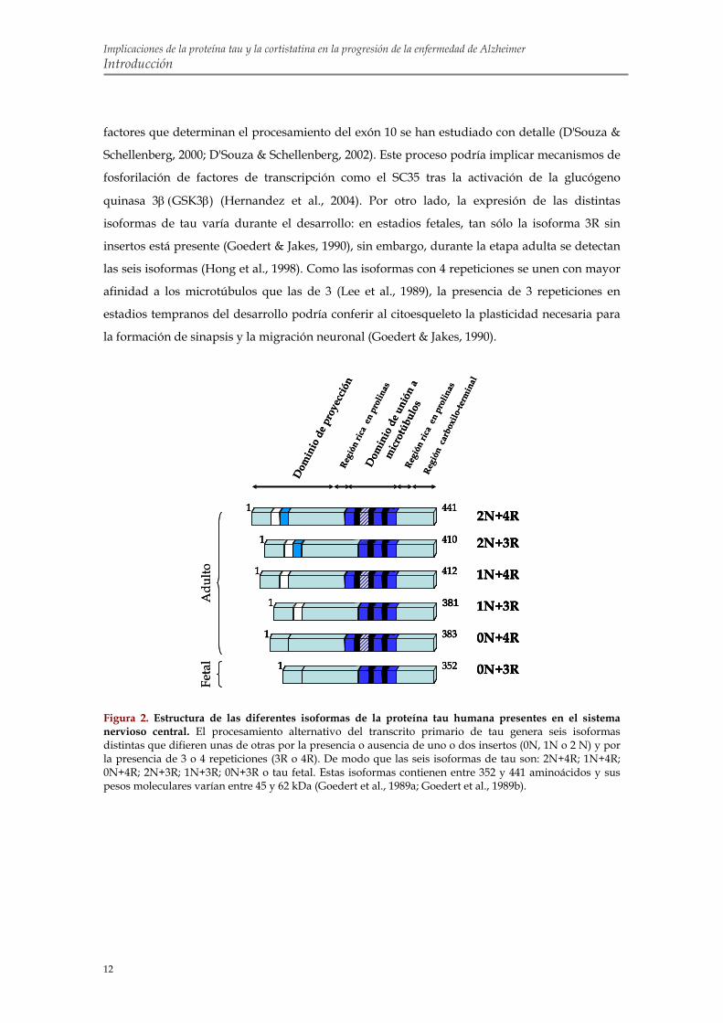
Figura 2. Estructura de las diferentes isoformas de la proteína tau humana presentes en el sistema nervioso central. El procesamiento alternativo del transcrito primario de tau genera seis isoformas distintas que difieren unas de otras por la presencia o ausencia de uno o dos insertos (0N, 1N o 2 N) y por la presencia de 3 o 4 repeticiones (3R o 4R). De modo que las seis isoformas de tau son: 2N+4R; 1N+4R; 0N+4R; 2N+3R; 1N+3R; 0N+3R o tau fetal. Estas isoformas contienen entre 352 y 441 aminoácidos y sus pesos moleculares varían entre 45 y 62 kDa (Goedert et al., 1989a; Goedert et al., 1989b).
Implicaciones de la proteína tau y la cortistatina en la progresión de la enfermedad de Alzheimer Introducción
13
La proteína tau sufre diversas modificaciones postraduccionales como la fosforilación, la
glicosilación, la glicación, la ubiquitinación, la deaminación, la oxidación, la sumoización, la
nitración o la truncación entre otras (revisadas detalladamente en (Avila et al., 2004; Gong et al.,
2005; Wang & Liu, 2008)). La glicación sólo se detecta en situaciones patológicas. El resto de
modificaciones postraduccionales pueden detectarse en condiciones normales pero en niveles
bajos. Los niveles de estas modificaciones se ven incrementados de forma notable en las
enfermedades neurodegenerativas (Khatoon et al., 1994). Debido a la complejidad de las
isoformas de tau es habitual visualizar en un western blot múltiples bandas inmunoreactivas con
distinto peso molecular aparente. En SDS-PAGE, los PHF purificados a partir de cerebros de EA
dan lugar a tres bandas mayoritarias a la altura de 60, 64 y 68 kDa y otra minoritaria de 72 kDa
(Greenberg & Davies, 1990; Greenberg et al., 1992; Grundke-Iqbal et al., 1986). Por el contrario,
cuando las muestras que contienen los PHF se tratan con fosfatasa alcalina y se analizan por
western blot se detectan seis bandas que se corresponden con las seis isoformas mayoritarias de
la proteína tau (Goedert & Jakes, 1990).
2.2. Distribución En cerebro humano adulto y mediante el uso de la técnica de hibridación in situ e
inmunohistoquímica, tau se detecta en distintas regiones que incluyen la corteza, el hipocampo,
el tálamo, el estriado y el cerebelo (Goedert et al., 1989b; Trojanowski et al., 1989) siendo más
abundantes, en todos los casos, las isoformas de 3 repeticiones frente a las de 4 (Goedert et al.,
1989b). Al parecer, los niveles de tau soluble disminuyen con la edad aproximadamente un 14%
por década a partir de los 20 años de edad (Mukaetova-Ladinska et al., 1993; Mukaetova-
Ladinska et al., 1996). Esta disminución es especialmente importante en la corteza frontal y el
hipocampo y es más pronunciada en los casos con EA (Mukaetova-Ladinska et al., 1993;
Mukaetova-Ladinska et al., 1996). En ratón, tau se expresa en la corteza y el hipocampo,
también en cerebelo aunque en menor cantidad (Santa-Maria et al., 2005). La distribución de tau
en la corteza cerebral humana se centra en las células piramidales predominantemente en las
capas más profundas (Goedert et al., 1989b; Kosik et al., 1989). En el hipocampo, las isoformas
de tau con 3 repeticiones se detectan en las células piramidales de CA y del subiculum y en las
granulares del giro dentado, en cambio, como se mencionó anteriormente, las variantes de 4
repeticiones sólo aparecen en las piramidales (Goedert et al., 1989b).
Implicaciones de la proteína tau y la cortistatina en la progresión de la enfermedad de Alzheimer Introducción
14
La proteína tau se encuentra mayoritariamente en las neuronas (Goedert et al., 1989b;
Kosik et al., 1989), aunque también puede estar presente en las células gliales en algunas
enfermedades neurodegenerativas (Arrasate et al., 1997; Chin & Goldman, 1996; Shin et al.,
1992). A nivel sub-celular, tau se localiza en el citoplasma, preferentemente en los axones
(Goedert et al., 1989b; Kosik et al., 1989), y puede estar asociado a las mitocondrias (Rendon et
al., 1990), a los ácidos nucleicos (Hua et al., 2003; Kampers et al., 1996) y a la membrana
plasmática (Brandt et al., 1995).
La fosforilación de tau afecta a su distribución celular en el cerebro adulto y en las
neuronas en desarrollo. Por ejemplo, tanto la unión de tau a los microtúbulos como su
interacción con la membrana plasmática están reguladas por fosforilación (Arrasate et al., 2000;
Lindwall & Cole, 1984). Además, en las neuronas en desarrollo se ha descrito que el tau
fosforilado en la región rica en prolinas se encuentra en su mayoría en el compartimento
somatodendrítico y el desfosforilado en la región distal del axón (Dotti & Banker, 1987). En el
cerebro de los pacientes con EA y otras tauopatías, la distribución mayoritaria de tau deja de ser
axonal, en este caso, la proteína tau hiperfosforilada forma agregados que se localizan en el
compartimento somatodendrítico (Buee et al., 2000).
2.3. Funciones fisiológicas de tau
2.3.1. Ensamblaje y estabilización de los microtúbulos
La proteína tau pertenece a la familia de proteínas asociadas a microtúbulos (MAP) y se
caracterizó por primera vez como un factor de ensamblaje de microtúbulos (Cleveland et al.,
1977; Fellous et al., 1977; Weingarten et al., 1975; Witman et al., 1976). El tau, que es
especialmente abundante en los axones de las neuronas, parece ser esencial en la iniciación y la
elongación de los microtúbulos y en su posterior estabilización (Caceres & Kosik, 1990; Drubin
& Kirschner, 1986). Estudios sucesivos de microinyección de tau en fibroblastos demostraron in
vivo que tau se asocia a la tubulina induciendo su polimerización y la estabilización de los
microtúbulos sin ser capaz de cambiar la morfología celular (Drubin & Kirschner, 1986). Sin
embargo, la expresión de tau en células Sf9 de ovario de insecto mediante infección con un
vector baculoviral sí altera la forma redondeada característica de estas células e induce la
formación de largas extensiones citoplasmáticas similares a los axones neuronales (Knops et al.,
1991; Knowles et al., 1994). Además, en este mismo modelo celular, se ha visto que tau
proporciona estabilidad a los microtúbulos y protege frente al nocodazol (Baas et al., 1994).

Implicaciones de la proteína tau y la cortistatina en la progresión de la enfermedad de Alzheimer Introducción
15
En relación con lo anterior, también cabe destacar el papel de tau en el crecimiento
neurítico (Drubin et al., 1985; Ferreira et al., 1989) y en la polaridad neuronal (Caceres & Kosik,
1990). En células PC12 y en neuronas de cerebelo se observó un aumento de la expresión de
tubulina y de tau durante la elongación axonal y dendrítica (Drubin et al., 1985; Ferreira et al.,
1989). Posteriormente, Cáceres et al. (1990) demostraron que la inhibición de la expresión de tau
en neuronas cerebelares mediante el uso de oligonucleótidos antisentido impedía la formación
del axón a pesar de la correcta extensión de las neuritas exploratorias (Caceres & Kosik, 1990).
Tau se une a los microtúbulos a través de las repeticiones del dominio de unión a
tubulina (Lee et al., 1989; Trinczek et al., 1999). En condiciones fisiológicas, tau está en constante
equilibrio dinámico de unión y desunión de los microtúbulos. Y se cree que este equilibrio está
controlado principalmente por el estado de fosforilación de tau. La afinidad de tau por los
microtúbulos es mayor si la región carboxilo-terminal de tau está fosforilada. En cambio, ocurre
lo contrario si la fosforilación de tau se localiza en el dominio de unión a microtúbulos o en la
región rica en prolinas, aunque el efecto de la fosforilación en esta última zona es menor
(Biernat et al., 1993; Biernat & Mandelkow, 1999; Drewes et al., 1995; Liu et al., 2007; Wang et al.,
2007). Por otro lado, se ha demostrado que, aunque una única repetición es capaz de unirse a la
tubulina (Lee et al., 1989), la secuencia con mayor capacidad de unión a los microtúbulos está
constituida por la primera repetición, la siguiente interrepetición y la segunda repetición
(región R1-R2) (Goode et al., 1997). Recientemente, distintos grupos postulan que las
repeticiones de tau se unen a bolsillos específicos de β-tubulina en la superficie interna de los
microtúbulos. Las regiones ricas en prolina, cargadas positivamente, parecen unirse
fuertemente a la superficie de los microtúbulos que están cargados negativamente y el dominio
de proyección, de carga negativa, forma “ramificaciones” en la superficie de los microtúbulos
debido posiblemente a repulsiones electrostáticas (Amos, 2004; Kar et al., 2003). De este modo,
el dominio de proyección podría mantener un espaciamiento entre los microtúbulos del axón
gracias a su interacción con otras proteínas, como las de los neurofilamentos (Chen et al., 1992;
Frappier et al., 1994; Georgieff et al., 1991). En apoyo a esta idea y teniendo en cuenta que la
isoforma de tau del sistema nervioso periférico es mayor (Couchie et al., 1992), se ha descrito
que el diámetro axonal en el sistema nervioso periférico es mayor respecto al del sistema
nervioso central (Georgieff et al., 1991).

Implicaciones de la proteína tau y la cortistatina en la progresión de la enfermedad de Alzheimer Introducción
16
2.3.2. Transporte axonal Diversos trabajos sostienen que tau podría tener un papel en el control del transporte
axonal de moléculas de señalización, factores tróficos e incluso orgánulos celulares, como
mitocondrias o vesículas (revisado en (Ballatore et al., 2007)). También se sugiere que ciclos
frecuentes de unión y desunión de tau de los microtúbulos (que se corresponde con
fosforilaciones y desfosforilaciones respectivamente) pueden ser necesarios para que el
transporte axonal sea efectivo. El dominio de unión a tubulina de tau parece competir con la
proteína motora quinesina en la unión a los microtúbulos provocando un descenso en el
transporte axonal anterógrado (Ebneth et al., 1998; Stamer et al., 2002; Trinczek et al., 1999). La
sobre-expresión de tau en células neuronales y no neuronales altera la morfología celular y la
distribución de varios orgánulos que necesitan el transporte axonal vía quinesina (Ebneth et al.,
1998; Trinczek et al., 1999). Este transporte anterógrado parece estar regulado por fosforilación
de tau mediada por GSK3β (Tatebayashi et al., 2004). También se ha descrito que la sobre-
expresión de tau inhibe el transporte del APP y que éste se acumula en el soma neuronal
(Stamer et al., 2002). Sin embargo, datos recientes ponen en duda el papel de tau en el
transporte axonal (Yuan et al., 2008). Este trabajo demuestra que el transporte axonal no se ve
afectado por la sobre-expresión o la falta de tau en la retina murina (Yuan et al., 2008).
2.3.3. Otras interacciones Al margen de la unión de tau a la tubulina, la proteína tau también puede interaccionar
con otras estructuras o proteínas diversas pudiendo llegar a tener un papel desconocido en una
gran variedad de funciones.
El dominio de unión a tubulina de tau interacciona con la PS-1 y ésta, a su vez, se une a
GSK3β. Estos datos sugiren que PS-1 facilita la fosforilación de tau favoreciendo la
aproximación del sustrato a la enzima (Takashima et al., 1998). También se ha visto que el
dominio de unión a tubulina de tau está implicado en la interacción con la histona desacetilasa 6
(Ding et al., 2008). Esta enzima, implicada en neurodegeneración (Olzmann et al., 2007; Pandey
et al., 2007), es clave en la regulación de la dinámica de la red citoesquelética (Gao et al., 2007;
Tran et al., 2007; Zhang et al., 2007) y en el control de varias respuestas celulares al estrés
(Boyault et al., 2007; Kawaguchi et al., 2003; Kwon et al., 2007).

Implicaciones de la proteína tau y la cortistatina en la progresión de la enfermedad de Alzheimer Introducción
17
El dominio de proyección de tau se proyecta fuera de la superficie del microtúbulo e
interacciona con otros elementos (Brandt et al., 1995; Hirokawa et al., 1988). De hecho, se ha
propuesto que los insertos de este dominio, ricos en residuos ácidos, podrían unir cationes tales
como el hierro (Arrasate et al., 1997). Otros motivos identificados en esta región incluyen la
secuencia KKXK que está implicada en la unión a heparina (Arrasate et al., 1997).
Las secuencias PXXP, presentes en la región rica en prolinas de tau, podrían estar
involucradas en la interacción de tau con dominios SH3 de proteínas como las familias de
tirosinas quinasas fyn o src (Jenkins & Johnson, 1998; Lee et al., 1998). Esta región podría estar
implicada en la unión de tau a proteínas asociadas a la membrana plasmática (Arrasate et al.,
2000; Brandt et al., 1995). La fosforilación de tau regula la unión de tau a las proteínas con SH3
puesto que estudios in vitro muestran que la unión de tau fosforilado a estas proteínas es menor
que en el caso del tau no fosforilado (Reynolds et al., 2008). También se ha visto que no se
produce unión en presencia de tau hiperfosforilado aislado de cerebros humanos con EA
(Reynolds et al., 2008). Estos datos sugieren que tau podría estar implicado en la transducción
celular de señales y que ésta podría verse afectada en la EA. En apoyo a lo anterior, se ha
descrito que un ratón deficiente en tau presenta un incremento en la expresión del factor de
transcripción c-fos (Oyama et al., 2004). La unión de tau a las proteínas de las familias fyn o src
podría resultar en la fosforilación de tau en tirosina (Lee, 2005; Lee et al., 1998).
Se sabe muy poco sobre el posible papel de tau como regulador de la expresión génica en
el núcleo. Antes de ser transportado al núcleo, la proteína tau se fosforila en el citosol
(Greenwood et al., 1997). El tau se ha detectado en el núcleo celular, principalmente en células
en división (Brady et al., 1995) y, se cree que, en el núcleo, el tau se une al DNA inhibiendo su
amplificación (Li et al., 2005). Esta unión depende del estado de agregación de tau y, sin
embargo, no depende de su nivel de fosforilación (Hua & He, 2002). La interacción de tau con
las mitocondrias, la membrana plasmática y los ácidos nucleicos sugiere que tau puede actuar
como un mediador entre los microtúbulos y estos orgánulos (Hua et al., 2003).
En definitiva, la proteína tau parece contribuir directa o indirectamente sobre la
modulación estructural y reguladora de funciones celulares siendo de vital importancia su
acción sobre la red de microtúbulos para mantener la morfología apropiada de las neuronas y
su correcto funcionamiento.
Implicaciones de la proteína tau y la cortistatina en la progresión de la enfermedad de Alzheimer Introducción
18
2.4. Agregación patológica de tau. Hiperfosforilación. En condiciones patológicas, como se ha explicado, se altera el equilibrio dinámico de la
unión de tau a los microtúbulos provocando un aumento anormal del nivel de tau libre en el
interior neuronal. A pesar de que tau es una proteína soluble debido a su alto contenido en
residuos hidrofílicos (Lee et al., 1988), el aumento de la concentración de tau citosólico podría
favorecer el cambio de conformación de tau a una conformación patogénica “mal plegada” que
predispone a la agregación (Ballatore et al., 2007). Además, dado que tau es bastante propenso a
interaccionar con otras estructuras –sobre todo, cuando no está unido a los microtúbulos-, ésto
también podría favorecer su mal plegamiento y su ensamblaje. Primero, se forman pequeños
agregados de tau no fibrilares (Maeda et al., 2007; Sahara et al., 2007), un material insoluble que
se infiltra progresivamente en el citoplasma y, ocasionalmente, en las dendritas proximales
formando, más adelante, las características inclusiones filamentosas de NFT (Galvan et al.,
2001).
Se han descrito diversas causas que podrían provocar o contribuir al aumento de la
concentración de tau libre. Estos factores incluyen un aumento de la fosforilación de tau en los
residuos de serinas y treoninas (Andersen, 2004; Moreira et al., 2005; Rapoport et al., 2002). Esta
fosforilación parece ser un paso previo al ensamblaje de la proteína si se localiza en el dominio
de unión a microtúbulos de tau (Perez et al., 2000; Schneider et al., 1999). Por el contrario, la
polimerización se ve impedida si la fosforilación de tau se sitúa en la región rica en prolinas o
en el extremo carboxilo-terminal (Alonso Adel et al., 2004; Liu et al., 2007).
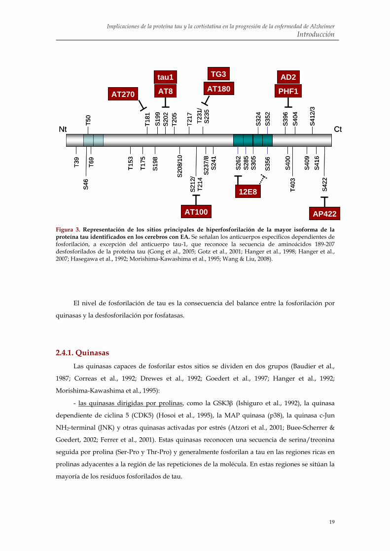
De entre los 79 residuos de serina o treonina susceptibles de fosforilación en la isoforma
humana de tau de mayor longitud, más de 30 han sido identificados en los cerebros con EA
utilizando anticuerpos frente a tau dependientes de fosforilación, espectrometría de masas y
secuenciación (figura 3) (Hanger et al., 1998; Hanger et al., 2007; Hasegawa et al., 1992;
Morishima-Kawashima et al., 1995; Wang & Liu, 2008). Aunque estos sitios se localizan
principalmente en la región rica en prolinas y la región carboxilo-terminal de tau, la
estabilización de los microtúbulos se ve afectada en mayor grado por la hiperfosforilación de
tau en los sitios situados en la región de unión a los microtúbulos como la Ser262, Ser285,
Ser305, Ser324, Ser352 y Ser356 (Cho & Johnson, 2003; Drewes et al., 1995; Sengupta et al., 1998;
Singh et al., 1996). La detección en el fluido cerebroespinal (CSF) de hiperfosforilación de tau o
de aumento en los niveles de tau total puede utilizarse como marcador de la
neurodegeneración. Concretamente, la fosforilación de tau en la Thr231 y la Thr181 detectada
en el CSF de los pacientes con MCI parece estar asociada con el futuro desarrollo de la EA (Arai
et al., 2000; Buerger et al., 2002a; Buerger et al., 2002b; Hansson et al., 2006). Estos datos sugieren
que la fosforilación en esos sitios está relacionada con el inicio del proceso patológico de la EA.
Implicaciones de la proteína tau y la cortistatina en la progresión de la enfermedad de Alzheimer Introducción
19
Figura 3. Representación de los sitios principales de hiperfosforilación de la mayor isoforma de la proteína tau identificados en los cerebros con EA. Se señalan los anticuerpos específicos dependientes de fosforilación, a excepción del anticuerpo tau-1, que reconoce la secuencia de aminoácidos 189-207 desfosforilados de la proteína tau (Gong et al., 2005; Gotz et al., 2001; Hanger et al., 1998; Hanger et al., 2007; Hasegawa et al., 1992; Morishima-Kawashima et al., 1995; Wang & Liu, 2008).
El nivel de fosforilación de tau es la consecuencia del balance entre la fosforilación por
quinasas y la desfosforilación por fosfatasas.
2.4.1. Quinasas Las quinasas capaces de fosforilar estos sitios se dividen en dos grupos (Baudier et al.,
1987; Correas et al., 1992; Drewes et al., 1992; Goedert et al., 1997; Hanger et al., 1992;
Morishima-Kawashima et al., 1995):
- las quinasas dirigidas por prolinas, como la GSK3β (Ishiguro et al., 1992), la quinasa
dependiente de ciclina 5 (CDK5) (Hosoi et al., 1995), la MAP quinasa (p38), la quinasa c-Jun
NH2-terminal (JNK) y otras quinasas activadas por estrés (Atzori et al., 2001; Buee-Scherrer &
Goedert, 2002; Ferrer et al., 2001). Estas quinasas reconocen una secuencia de serina/treonina
seguida por prolina (Ser-Pro y Thr-Pro) y generalmente fosforilan a tau en las regiones ricas en
prolinas adyacentes a la región de las repeticiones de la molécula. En estas regiones se sitúan la
mayoría de los residuos fosforilados de tau.
Nt Ct
T39
S46
T69
T50
T153
T175
AT270
S19
8
AT8
T181
S20
2
tau1
T205
S20
8/10
S21
2/T2
14
AT100
T217
T231
/S
235
S23
7/8
AT180
TG3
S24
1
S26
2
12E8
S28
5S
305
S32
4S
356
S35
2
S39
6S
404
PHF1
AD2
T403
S40
9S
412/
3S
416
S42
2
AP422
S19
9
S40
0
Nt Ct
T39
S46
T69
T50
T153
T175
AT270
S19
8
AT8
T181
S20
2
tau1
T205
S20
8/10
S21
2/T2
14
AT100
T217
T231
/S
235
S23
7/8
AT180
TG3
S24
1
S26
2
12E8
S28
5S
305
S32
4S
356
S35
2
S39
6S
404
PHF1
AD2
T403
S40
9S
412/
3S
416
S42
2
AP422
S19
9
S40
0
Implicaciones de la proteína tau y la cortistatina en la progresión de la enfermedad de Alzheimer Introducción
20
- las quinasas no dirigidas por prolinas, como la proteína quinasa A (PKA) (Scott et al.,
1993; Sironi et al., 1998), la proteína quinasa C (PKC) (Correas et al., 1992; Gomez-Ramos et al.,
2003), la calmodulina quinasa II (CaMKII) (Bennecib et al., 2001; Sironi et al., 1998), las quinasas
reguladoras de afinidad entre MAPs (MARK) (Drewes et al., 1997) o la caseína quinasa II (CKII)
(Correas et al., 1992). Estas quinasas fosforilan a tau en el dominio de unión a tubulina, salvo la
CKII que modifica residuos cercanos a los insertos del dominio de proyección (Greenwood et
al., 1994).
De entre las quinasas implicadas en la hiperfosforilación anómala de tau, cabe destacar el
papel relevante de la GSK3β y su importancia como posible diana terapéutica (Mazanetz &
Fischer, 2007). GSK3β fosforila a la proteína tau en sitios comunes a los PHF en células en
cultivo (Lovestone et al., 1996) y colocaliza con tau fosforilado en cerebros de pacientes con la
EA (Shiurba et al., 1996). Además, la relación entre la sobre-expresión de GSK3 y la
hiperfosforilación de tau ha sido establecida en distintos modelos animales (Engel et al., 2006a;
Engel et al., 2006b). También se ha visto que la inhibición de GSK3 con litio no sólo reduce la
fosforilación de tau in vivo, sino que también disminuye el nivel de agregación de tau,
comparado con los controles (Noble et al., 2005). Por otro lado, se ha establecido una conexión
entre el péptido β-amiloide y la fosforilación de tau. Al parecer, el efecto neurotóxico del
péptido β-amiloide podría deberse a una activación de GSK3 y el resultante aumento de la
fosforilación de tau (Alvarez et al., 1999; Ferrari et al., 2003). De hecho, el litio provoca una
reducción de la producción de β-amiloide en modelos murinos de la EA (Phiel et al., 2003).
2.4.2. Fosfatasas En los cerebros humanos con EA, muchos de los sitios fosforilados se desfosforilan más
lentamente durante el periodo post-mortem que en el caso de los cerebros control (Garver et al.,
1994; Matsuo et al., 1994). Estos resultados sugieren que, en la EA, la actividad de las fosfatasas
(PP) es menor. Esta hipótesis se comprobó posteriormente puesto que se vio que la expresión,
cantidad y actividad de la PP2A se reducen significativamente en las zonas cerebrales afectadas
por la EA, como la corteza y el hipocampo, a diferencia de lo que ocurre en las regiones no
afectadas, como el cerebelo, o en los individuos controles (Loring et al., 2001; Sontag et al., 2004;
Vogelsberg-Ragaglia et al., 2001).
A pesar de que se han identificado diversas fosfatasas (PP) en el cerebro (PP1, PP2A,
PP2B (o calcineurina), PP2C y PP5), la PP2A parece ser la más relevante en la regulación del
estado de fosforilación de tau en la EA (Bennecib et al., 2001; Goedert et al., 1995; Liu et al., 2005;
Saito et al., 1995; Sontag et al., 1996). Se ha comprobado que la inhibición de PP2A con ácido
Implicaciones de la proteína tau y la cortistatina en la progresión de la enfermedad de Alzheimer Introducción
21
okadaico provoca hiperfosforilación de tau en sitios comunes a los que se encuentran en la EA.
Ésto no ocurre cuando se inhibe selectivamente a la PP2B con ciclosporina A (Sontag et al.,
1999). Al parecer, la menor actividad de PP2A contribuye a la hiperfosforilación de tau no sólo
por una menor tasa de desfosforilación de tau sino también por la activación de distintas
quinasas como la CamKII, MAP quinasas y quinasas activadas por estrés (Bennecib et al., 2001;
Kins et al., 2003; Pei et al., 2003). La PP2A también parece estar implicada en ciertos casos de
FTDP-17. Las mutaciones de estos pacientes, localizadas en el dominio de unión de tau,
dificultan la unión de la PP2A a tau y, de este modo, se favorece la hiperfosforilación anómala
de tau (Goedert & Spillantini, 2000).
Como mencionamos, la EA se inicia en la región entorrinal (Braak & Braak, 1991)
extendiéndose posteriormente a la zona límbica y a la corteza, siendo la patología en la corteza
la que da lugar a la demencia. En la corteza, las señales (activación de quinasas y fosfatasas) que
podrían promover la patología de tau podrían tener una cierta especificidad. Por ello, se ha
pretendido analizar si péptidos presentes en la corteza, como la cortistatina, podrían jugar
algún papel en la patología de tau.
3. LA CORTISTATINA
3.1. Identificación En 1996, el grupo del Dr. de Lecea aisló un clon de cDNA de una nueva proteína a partir
de una librería de sustracción de hipocampo de rata (de Lecea et al., 1996). La secuencia
nucleotídica predecía una proteína putativa de 112 aminoácidos que llamaron
preprocortistatina debido a su patrón de expresión mayoritario en la corteza cerebral (de Lecea
et al., 1996). Esta proteína comparte homologías estructurales con la preprosomatostatina (de
116 residuos) y el extremo carboxilo-terminal es la única zona que comparte secuencia
aminoacídica (de Lecea et al., 1996). El corte de la secuencia señal (secuencia hidrofóbica de 27
residuos), en el extremo amino-terminal de la preprocortistatina, produce la procortistatina.
Esta proteína es sustrato de prohormona-convertasas que reconocen secuencias con un tándem
de residuos básicos (KR; KK) y que originan cortistatina-29, -14 o -13 (de Lecea et al., 1996; de
Lecea et al., 1997b). Cabe destacar que la cortistatina-13 no está relacionada con ningún
producto conocido. La cortistatina-29 y -14 se producen aproximadamente en cantidades
similares y, aunque la cortistatina-14 se libera preferentemente al exterior celular, por el

Implicaciones de la proteína tau y la cortistatina en la progresión de la enfermedad de Alzheimer Introducción
22
momento, aún no se han descrito funciones biológicas específicas para cada una de ellas
(Galanopoulou et al., 1993).
Posteriormente, se obtuvo el cDNA que codifica para la preprocortistatina de ratón, de
humano y de rana (de Lecea et al., 1997a; Fukusumi et al., 1997; Tostivint et al., 2006). En ratón,
se identificaron tan sólo dos sitios putativos de corte (R; KK) que originan la cortistatina-44 y la
cortistatina-14 (de Lecea et al., 1997b). La secuencia nucleotídica humana presenta un menor
grado de identidad con la de rata (71%). La preprocortistatina humana puede ser procesada en
varios sitios dando cortistatina-29, -17, -21 y -31 (de Lecea et al., 1997b; Fukusumi et al., 1997).
La cortistatina-21 y -31 no están conservadas en otras especies. Este proceso es análogo al de la
preprosomatostatina y la prosomatostatina que originan la somatostatina-28 y -14 (Brazeau et
al., 1973; Epelbaum et al., 1994; Spier & de Lecea, 2000). Más tarde, la presencia de cortistatina-
14 se demostró in vivo mediante el uso de la técnica de HPLC en los ratones modificados
genéticamente que no expresan somatostatina (Ramirez et al., 2002).
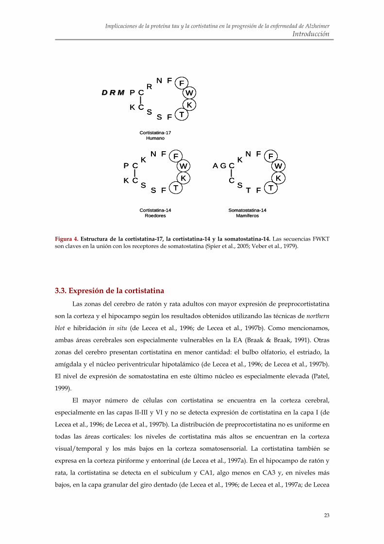
3.2. Estructura del péptido y del gen de la cortistatina La cortistatina-14 comparte 11 de sus 14 aminoácidos con la somatostatina-14 incluyendo
dos cisteínas que le proporcionan al péptido una estructura cíclica y la secuencia FWKT, crítica
en la unión con los receptores de somatostatina (figura 4) (Spier et al., 2005; Veber et al., 1979).
En rata y ratón, las secuencias aminoacídicas de la cortistatina están altamente conservadas, sin
embargo, el péptido humano contiene tres aminoácidos más en el extremo amino-terminal y
una arginina es sustituida por una lisina (cortistatina-17) (de Lecea et al., 1997b). La distribución
y las propiedades biológicas de la cortistatina humana son similares a las de roedores
(Fukusumi et al., 1997).
A pesar de las similitudes con la somatostatina, la secuencia nucleotídica y la localización
cromosómica (de Lecea et al., 1996; de Lecea et al., 1997b) indican que la cortistatina y la
somatostatina provienen de genes distintos. El gen de ratón para la cortistatina se sitúa en el
cromosoma 4 (se corresponde con la región 1p36 de humanos) y el de somatostatina en el 16
(3q28 de humanos) (Spier & de Lecea, 2000). El gen de preprocortistatina contiene dos exones y
un intrón, como el de preprosomatostatina (Spier & de Lecea, 2000). Se ha demostrado que los
genes de somatostatina y de cortistatina provienen de la duplicación de un mismo gen ancestral
de la familia de la urotensina (Tostivint et al., 2006).
Implicaciones de la proteína tau y la cortistatina en la progresión de la enfermedad de Alzheimer Introducción
23
P C
K C
KN F
FS
S
FW
KT
Cortistatina-14Roedores
A G C
C
KN F
FS
T
FW
KT
Somatostatina-14Mamíferos
Cortistatina-17Humano
P C
K C
RN F
FS
S
FW
KT
D R MD R M
P C
K C
KN F
FS
S
FW
KT
P C
K C
KN F
FS
S
FW
KT
Cortistatina-14Roedores
A G C
C
KN F
FS
T
FW
KT
A G C
C
KN F
FS
T
FFWW
KKTT
Somatostatina-14Mamíferos
Cortistatina-17Humano
P C
K C
RN F
FS
S
FW
KT
D R MD R M P C
K C
RN F
FS
S
FFWW
KKTT
D R MD R M
Figura 4. Estructura de la cortistatina-17, la cortistatina-14 y la somatostatina-14. Las secuencias FWKT son claves en la unión con los receptores de somatostatina (Spier et al., 2005; Veber et al., 1979).
3.3. Expresión de la cortistatina Las zonas del cerebro de ratón y rata adultos con mayor expresión de preprocortistatina
son la corteza y el hipocampo según los resultados obtenidos utilizando las técnicas de northern
blot e hibridación in situ (de Lecea et al., 1996; de Lecea et al., 1997b). Como mencionamos,
ambas áreas cerebrales son especialmente vulnerables en la EA (Braak & Braak, 1991). Otras
zonas del cerebro presentan cortistatina en menor cantidad: el bulbo olfatorio, el estriado, la
amígdala y el núcleo periventricular hipotalámico (de Lecea et al., 1996; de Lecea et al., 1997b).
El nivel de expresión de somatostatina en este último núcleo es especialmente elevada (Patel,
1999).
El mayor número de células con cortistatina se encuentra en la corteza cerebral,
especialmente en las capas II-III y VI y no se detecta expresión de cortistatina en la capa I (de
Lecea et al., 1996; de Lecea et al., 1997b). La distribución de preprocortistatina no es uniforme en
todas las áreas corticales: los niveles de cortistatina más altos se encuentran en la corteza
visual/temporal y los más bajos en la corteza somatosensorial. La cortistatina también se
expresa en la corteza piriforme y entorrinal (de Lecea et al., 1997a). En el hipocampo de ratón y
rata, la cortistatina se detecta en el subiculum y CA1, algo menos en CA3 y, en niveles más
bajos, en la capa granular del giro dentado (de Lecea et al., 1996; de Lecea et al., 1997a; de Lecea

Implicaciones de la proteína tau y la cortistatina en la progresión de la enfermedad de Alzheimer Introducción
24
et al., 1997b). La distribución de la preprocortistatina descrita mediante el uso de hibridación in
situ ha sido confirmada por inmunohistoquímica en cerebro de rata (de Lecea, 2007).
La preprosomatostatina se expresa en el stratum oriens y stratum radiatum de CA, el
subiculum y la corteza (Braun et al., 1998). Posteriormente, mediante PCR cuantitativa en
muestras de cerebro humano, se vio que la somatostatina se expresa en las mismas zonas
cerebrales (corteza frontal, hipocampo, puente y tronco cerebral, enumeradas de mayor a menor
abundancia) que la cortistatina (Dalm et al., 2004). Sin embargo, a diferencia de la cortistatina, la
somatostatina se detecta en todos los casos en mayor cantidad (Dalm et al., 2004).
Experimentos de doble hibridación in situ en ratón y rata muestran que la cortistatina sólo
se expresa en interneuronas GABAérgicas inhibitorias (de Lecea et al., 1996; de Lecea et al.,
1997a; de Lecea et al., 1997b). Las neuronas que expresan cortistatina pertenecen a la
subpoblación de interneuronas GABAérgicas que expresan calbindina y parvalbúmina
(proteínas de unión a calcio) (de Lecea et al., 1997a). A pesar de que la somatostatina también se
expresa en interneuronas GABAérgicas (Esclapez & Houser, 1995; Kosaka et al., 1988), menos
de la mitad de las neuronas que expresan cortistatina en la corteza y el hipocampo de rata
presentan también somatostatina, y aproximadamente un cuarto de las células con
somatostatina expresan cortistatina (de Lecea et al., 1997a). De modo que la somatostatina y la
cortistatina comparten un patrón de expresión parcialmente solapado (de Lecea et al., 1997a).
El patrón de expresión de la cortistatina parece ser más amplio de lo que en un principio
se pensó. Se ha demostrado que la cortistatina, como la somatostatina aunque en menor
cantidad, también se expresa en tejidos humanos periféricos como en: riñón, estómago, colon,
testículos, pulmón y distintos tumores (Dalm et al., 2004; Ejeskar et al., 2000). Cabe destacar que
la cortistatina, a diferencia de la somatostatina, se encuentra en células y tejidos humanos del
sistema inmune (Dalm et al., 2004; Dalm et al., 2003). En definitiva, estos datos sugieren que la
cortistatina también tiene una función en tejidos periféricos siendo notable su papel en el
sistema inmune (Dalm et al., 2004; Dalm et al., 2003).
La preprocortistatina comienza a expresarse en los cerebros de ratas de entre 5 y 10 días
de edad, alcanzando los máximos niveles de expresión en torno a los 15 días de edad, y,
posteriormente, la intensidad de la expresión se reduce progresivamente durante la edad adulta
(de Lecea et al., 1997a). La cortistatina podría estar modulando el establecimiento de las
conexiones sinápticas en la corteza y el hipocampo durante el desarrollo puesto que su patrón
de expresión correlaciona con la maduración de interneuronas corticales. La aplicación de
cortistatina-14 en rodajas de hipocampo adulto no parece afectar a las medidas de excitabilidad
de las células granulares, sin embargo, en ratas P15, la amplitud de las respuestas eléctricas
sinápticas (PS) se ve reducida (de Lecea et al., 1997a). De igual forma, se ha descrito una
expresión transitoria de la somatostatina en la corteza de rata en desarrollo sugiriendo también,

Implicaciones de la proteína tau y la cortistatina en la progresión de la enfermedad de Alzheimer Introducción
25
un papel relevante de la somatostatina en la diferenciación y la formación de sinapsis
(Cavanagh & Parnavelas, 1988; Parnavelas & Cavanagh, 1988). Los estudios de
inmunocitoquímica y autorradiografía muestran que la expresión máxima de somatostatina se
detecta a las dos semanas de edad y ésta disminuye progresivamente hasta alcanzar los niveles
del individuo adulto (Cavanagh & Parnavelas, 1988; Parnavelas & Cavanagh, 1988).
Durante el envejecimiento normal, los niveles de preprocortistatina en el hipocampo de
ratones aumentan (Winsky-Sommerer et al., 2004) mientras que la expresión de la somatostatina
parece disminuir en humanos y en primates (Hayashi et al., 1997; Lu et al., 2004). Estos datos
sugieren un papel diferencial de ambos neuropéptidos en el envejecimiento.
3.4. Receptores de somatostatina La cortistatina se une a los cinco subtipos conocidos de receptores para la somatostatina
(sst1-5 según la nomenclatura de la IUPHAR (Hoyer et al., 1995)) (de Lecea et al., 1996;
Fukusumi et al., 1997). La cortistatina-14 de roedores y la cortistatina-17 de humanos desplazan
la unión de la 125I-somatostatina-14 a los cinco receptores de somatostatina humanos clonados
en líneas de células transfectadas (Criado et al., 1999; Fukusumi et al., 1997) y en tejido humano
del timo (Dalm et al., 2003) con afinidades similares a las de la somatostatina en el rango del
nanomolar. Como la somatostatina y la cortistatina comparten un patrón de expresión
parcialmente solapado, en algunos casos, ambos neuropéptidos podrían competir por los
mismos receptores o ser liberados de distintas terminales nerviosas (de Lecea et al., 1997a).
Estos receptores, descritos por primera vez en células GH4C1 de pituitaria (Schonbrunn &
Tashjian, 1978), han sido clonados a partir de tejidos de humano, rata, ratón, cerdo y vaca y
pertenecen a la superfamilia de receptores acoplados a proteína G con siete dominios
hidrofóbicos α-hélice transmembrana, un extremo amino-terminal extracelular y carboxilo-
terminal intracelular (Patel, 1999; Weckbecker et al., 2003; Yamada et al., 1992).
En humanos, los receptores sst1-5 están codificados por cinco genes independientes no-
alélicos localizados en los cromosomas 14q13, 17q24, 22q13.1, 20p11.2 y 16p13.3,
respectivamente (Meyerhof, 1998; Patel, 1999). Cabe destacar que los genes de sst1, 3, 4, 5 no
contienen intrones, en cambio, el gen de sst2 de rata, ratón y humano contiene tres intrones y
genera, por procesamiento alternativo, dos isoformas proteicas de distinto tamaño en su
extremo carboxilo-terminal: sst2a y sst2b (Moller et al., 2003; Olias et al., 2004). La variante corta
(sst2b) no se expresa abundantemente en humanos (Moller et al., 2003; Olias et al., 2004). La
similitud de las secuencias entre los distintos subtipos es notable (39-57%) y altamente
conservada entre distintas especies (81-98% para ratón, humano y rata) (Meyerhof, 1998). Los

Implicaciones de la proteína tau y la cortistatina en la progresión de la enfermedad de Alzheimer Introducción
26
receptores sst1-5 humanos tienen de 1 a 4 sitios de N-glicosilación en el segmento amino-
terminal y el segundo bucle extracelular (Patel, 1999) (tabla 1). Los receptores de somatostatina
humanos presentan de 3 a 8 sitios putativos de fosforilación mediada por la PKA, la PKC y la
quinasa calmodulina II en el segmento carboxilo-terminal y en el segundo y tercer bucle
intracelular (Patel, 1999). En base a propiedades estructurales, filogénicas y farmacológicas, los
receptores de somatostatina pueden dividirse en dos grupos principales (Hoyer et al., 1995):
- SRIF1: sst2, sst3 y sst5 que presentan mayor afinidad por los análogos de
somatostatina siguientes: el octreotide (SMS-201-995), seglitide (MK-678) y lanreotide
(BIM-23014)
- SRIF2: sst1 y sst4
El sst5 es el único receptor que muestra cierta preferencia de unión por la somatostatina-
28 frente a la somatostatina-14, sin embargo, sst1-4 presentan una afinidad similar por ambos
péptidos (Moller et al., 2003).
Numerosos estudios, la mayoría realizados en tejido de rata y ratón, usando las técnicas
de hibridación in situ, RT-PCR, ensayos de unión y, más tarde, de inmunohistoquímica,
muestran que los receptores de somatostatina están ampliamente distribuidos en el cerebro y en
los tejidos periféricos (gastrointestinal, páncreas y sistema inmunitario) con distintos patrones
de expresión, comunes en algunos casos (Kreienkamp, 1999; Patel, 1999) (tabla 1). La expresión
de sst2, 3 y 4 se centra en distintas zonas del cerebro incluyendo corteza e hipocampo y su
expresión disminuye hacia la zona caudal. El receptor sst5 es el menos abundante y el menos
estudiado. En la corteza, la expresión de sst2 está más restringida a las capas profundas,
mientras que sst1 está presente en todas las capas (Moller et al., 2003). La expresión de sst2a y
sst2b es, en algunos casos, diferente, por ejemplo: mediante hibridación in situ se vio que la
expresión de sst2a es mucho mayor que la de sst2b en la corteza de ratón (Vanetti et al., 1994).
Los receptores de somatostatina también se expresan en tumores: sst2 preferentemente; sst1, 3, 5
menos frecuentemente y el sst4 se detecta raramente (Reubi et al., 2001). Estos estudios revelan
que la mayor parte de las células expresan no sólo uno sino distintos subtipos diferentes de
receptores de somatostatina.
Los receptores de somatostatina se expresan mayoritarimente en neuronas, también se
detectan en glía aunque en menor cantidad (Feindt et al., 1995; Heidet et al., 1990; Hosli & Hosli,
1994; Mentlein et al., 1990; van Calker et al., 1980). Su localización sub-celular y su función
pueden variar según el subtipo al que pertenezcan (tabla 1). Los receptores de somatostatina
con función presináptica modulan la liberación de la somatostatina y de otros
neurotransmisores (Schulz et al., 2000). Cabe destacar la localización de los sst3 en cilios
neuronales (9+0). Estos cilios no son motores (9+2) y se sostiene que los receptores de los cilios
puedan actuar como sensores químicos del medio (tipo olfativos) (Schulz et al., 2000).
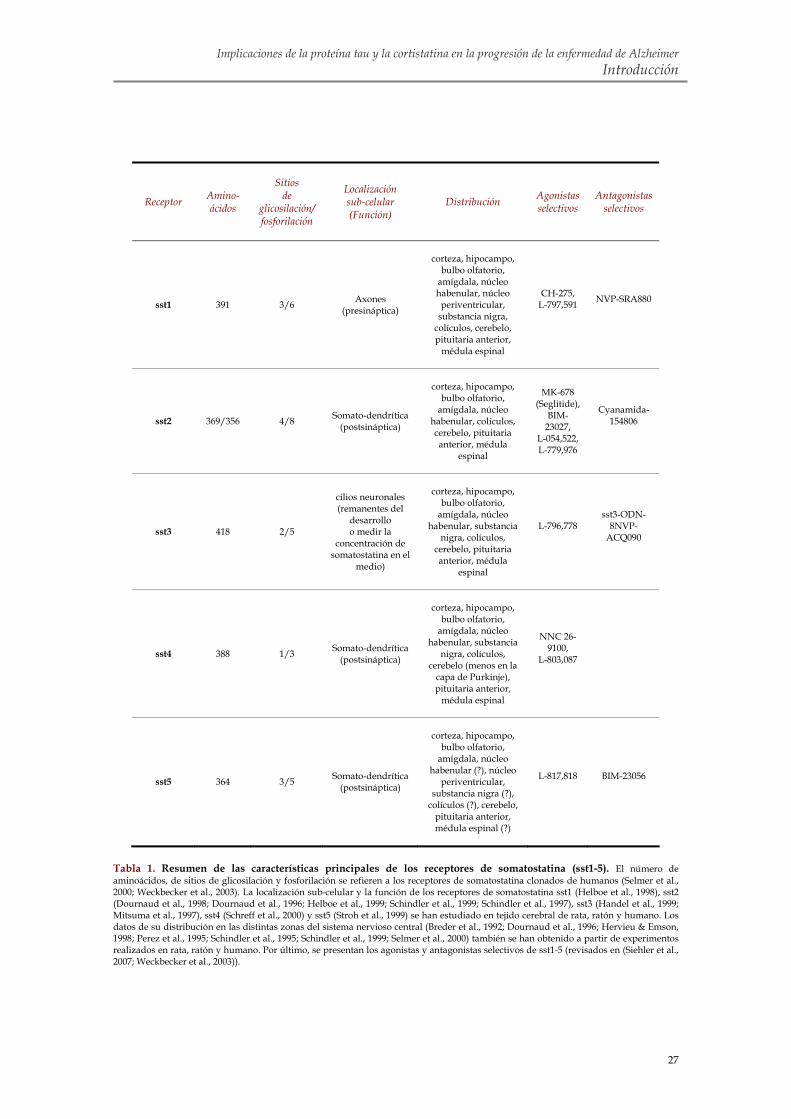
Implicaciones de la proteína tau y la cortistatina en la progresión de la enfermedad de Alzheimer Introducción
27
Receptor Amino- ácidos
Sitios
de glicosilación/ fosforilación
Localización sub-celular (Función)
Distribución Agonistas selectivos
Antagonistas selectivos
sst1 391 3/6 Axones (presináptica)
corteza, hipocampo,
bulbo olfatorio, amígdala, núcleo habenular, núcleo
periventricular, substancia nigra,
colículos, cerebelo, pituitaria anterior,
médula espinal
CH-275, L-797,591
NVP-SRA880
sst2 369/356 4/8 Somato-dendrítica (postsináptica)
corteza, hipocampo,
bulbo olfatorio, amígdala, núcleo
habenular, colículos, cerebelo, pituitaria anterior, médula
espinal
MK-678
(Seglitide), BIM-
23027, L-054,522, L-779,976
Cyanamida-154806
sst3 418 2/5
cilios neuronales (remanentes del
desarrollo o medir la
concentración de somatostatina en el
medio)
corteza, hipocampo,
bulbo olfatorio, amígdala, núcleo
habenular, substancia nigra, colículos,
cerebelo, pituitaria anterior, médula
espinal
L-796,778
sst3-ODN-8NVP-
ACQ090
sst4 388 1/3 Somato-dendrítica (postsináptica)
corteza, hipocampo,
bulbo olfatorio, amígdala, núcleo
habenular, substancia nigra, colículos,
cerebelo (menos en la capa de Purkinje), pituitaria anterior,
médula espinal
NNC 26-9100,
L-803,087
sst5 364 3/5 Somato-dendrítica (postsináptica)
corteza, hipocampo,
bulbo olfatorio, amígdala, núcleo
habenular (?), núcleo periventricular,
substancia nigra (?), colículos (?), cerebelo,
pituitaria anterior, médula espinal (?)
L-817,818
BIM-23056
Tabla 1. Resumen de las características principales de los receptores de somatostatina (sst1-5). El número de aminoácidos, de sitios de glicosilación y fosforilación se refieren a los receptores de somatostatina clonados de humanos (Selmer et al., 2000; Weckbecker et al., 2003). La localización sub-celular y la función de los receptores de somatostatina sst1 (Helboe et al., 1998), sst2 (Dournaud et al., 1998; Dournaud et al., 1996; Helboe et al., 1999; Schindler et al., 1999; Schindler et al., 1997), sst3 (Handel et al., 1999; Mitsuma et al., 1997), sst4 (Schreff et al., 2000) y sst5 (Stroh et al., 1999) se han estudiado en tejido cerebral de rata, ratón y humano. Los datos de su distribución en las distintas zonas del sistema nervioso central (Breder et al., 1992; Dournaud et al., 1996; Hervieu & Emson, 1998; Perez et al., 1995; Schindler et al., 1995; Schindler et al., 1999; Selmer et al., 2000) también se han obtenido a partir de experimentos realizados en rata, ratón y humano. Por último, se presentan los agonistas y antagonistas selectivos de sst1-5 (revisados en (Siehler et al., 2007; Weckbecker et al., 2003)).
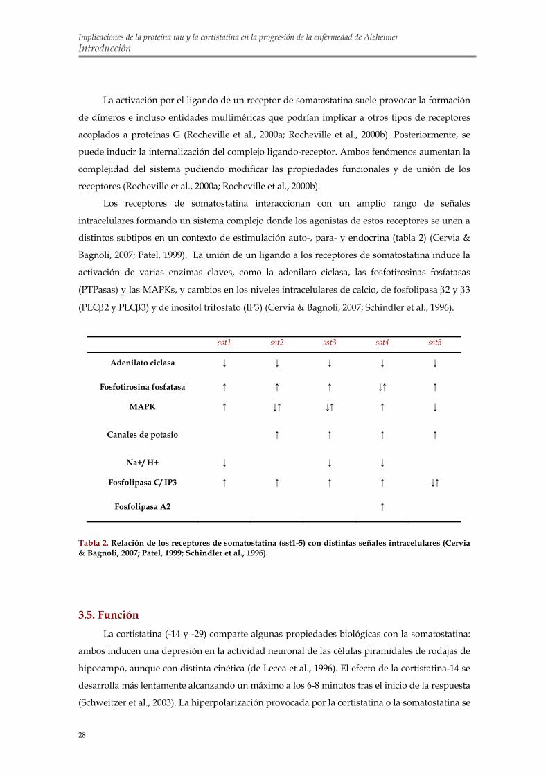
Implicaciones de la proteína tau y la cortistatina en la progresión de la enfermedad de Alzheimer Introducción
28
La activación por el ligando de un receptor de somatostatina suele provocar la formación
de dímeros e incluso entidades multiméricas que podrían implicar a otros tipos de receptores
acoplados a proteínas G (Rocheville et al., 2000a; Rocheville et al., 2000b). Posteriormente, se
puede inducir la internalización del complejo ligando-receptor. Ambos fenómenos aumentan la
complejidad del sistema pudiendo modificar las propiedades funcionales y de unión de los
receptores (Rocheville et al., 2000a; Rocheville et al., 2000b).
Los receptores de somatostatina interaccionan con un amplio rango de señales
intracelulares formando un sistema complejo donde los agonistas de estos receptores se unen a
distintos subtipos en un contexto de estimulación auto-, para- y endocrina (tabla 2) (Cervia &
Bagnoli, 2007; Patel, 1999). La unión de un ligando a los receptores de somatostatina induce la
activación de varias enzimas claves, como la adenilato ciclasa, las fosfotirosinas fosfatasas
(PTPasas) y las MAPKs, y cambios en los niveles intracelulares de calcio, de fosfolipasa β2 y β3
(PLCβ2 y PLCβ3) y de inositol trifosfato (IP3) (Cervia & Bagnoli, 2007; Schindler et al., 1996).
sst1 sst2 sst3 sst4 sst5
Adenilato ciclasa ↓ ↓ ↓ ↓ ↓
Fosfotirosina fosfatasa ↑ ↑ ↑ ↓↑ ↑
MAPK ↑ ↓↑ ↓↑ ↑ ↓
Canales de potasio ↑ ↑ ↑ ↑
Na+/ H+ ↓ ↓ ↓
Fosfolipasa C/ IP3 ↑ ↑ ↑ ↑ ↓↑
Fosfolipasa A2 ↑
Tabla 2. Relación de los receptores de somatostatina (sst1-5) con distintas señales intracelulares (Cervia & Bagnoli, 2007; Patel, 1999; Schindler et al., 1996).
3.5. Función La cortistatina (-14 y -29) comparte algunas propiedades biológicas con la somatostatina:
ambos inducen una depresión en la actividad neuronal de las células piramidales de rodajas de
hipocampo, aunque con distinta cinética (de Lecea et al., 1996). El efecto de la cortistatina-14 se
desarrolla más lentamente alcanzando un máximo a los 6-8 minutos tras el inicio de la respuesta
(Schweitzer et al., 2003). La hiperpolarización provocada por la cortistatina o la somatostatina se

Implicaciones de la proteína tau y la cortistatina en la progresión de la enfermedad de Alzheimer Introducción
29
debe probablemente al aumento de la amplitud de la corriente M a través de los canales de
potasio dependientes de voltaje (que pueden ser cerrados por la acetilcolina) (de Lecea et al.,
1996; Spier & de Lecea, 2000). Estos resultados parecen indicar que la cortistatina y la
somatostatina inducen, de forma similar, un aumento en la conductancia de potasio en las
neuronas del hipocampo mediada, muy probablemente, por los receptores de somatostatina (de
Lecea, 2007).
Tanto la cortistatina como la somatostatina parecen reducir la actividad de las neuronas
excitatorias glutamatérgicas. La administración de cortistatina-14 o de somatostatina-14
produce una reducción del potencial postsináptico excitatorio evocado mediado por los
receptores de glutamato en CA1, pero no afecta al potencial postsináptico inhibitorio (Tallent &
Siggins, 1997). La aplicación de cortistatina-14 en ratas produce una disminución en la actividad
de las células piramidales de CA1 inducida por pulsos de NMDA. En cultivos de neuronas de
hipotálamo, la cortistatina-14 y la somatostatina-14 inhiben las respuestas inducidas por el
glutamato, probablemente a través de la activación de sst2 (Vasilaki et al., 1999).
Sin embargo, y a pesar de las importantes homologías entre la cortistatina y la
somatostatina, algunos efectos de la cortistatina en el sistema nervioso central son diferentes a
los de la somatostatina.
(1) La cortistatina antagoniza los efectos de la acetilcolina en medidas de excitabilidad
cortical e hipocampal in vivo (de Lecea et al., 1996). Sin embargo, la somatostatina promueve la
liberación de la acetilcolina y potencia las respuestas frente a este neurotransmisor (Araujo et
al., 1990; de Lecea et al., 1996; Mancillas et al., 1986).
También se ha demostrado que la cortistatina está relacionada con la regulación del
sueño. La concentración de preprocortistatina es menor al final del periodo de luz y mayor
durante el periodo de oscuridad cuando los ratones son más activos (Bourgin et al., 2007). La
expresión de preprocortistatina aumenta en condiciones de privación de sueño pero la
privación de sueño REM no le afecta (Bourgin et al., 2007). La cortistatina (-14 y -17) inyectada
en el cerebro de ratas induce una reducción en la actividad locomotora (Criado et al., 1999) y un
aumento en el número de ondas de baja frecuencia en la corteza cerebral (1-4 Hz) y en la
duración del sueño SWS sin afectar significativamente al sueño REM (de Lecea et al., 1996;
Fukusumi et al., 1997). Sin embargo, se ha visto que la administración de somatostatina-14
favorece la hiperactividad (Rezek et al., 1976) y el sueño REM (Danguir, 1986).
Siguen sin conocerse los mecanismos exactos a través de los cuáles la cortistatina induce
el sueño. Una posibilidad sería que la cortistatina-14 potenciase la actividad intrínseca de las
neuronas corticales a través de la inhibición directa (hiperpolarización) de las neuronas
principales (de Lecea et al., 1996). Además, la liberación regulada de la cortistatina-14 en el
momento apropiado del ciclo circadiano podría antagonizar los efectos excitatorios de la

Implicaciones de la proteína tau y la cortistatina en la progresión de la enfermedad de Alzheimer Introducción
30
acetilcolina promoviendo el sueño (Bourgin et al., 2007). Por último, la cortistatina-14 podría
promover el sueño SWS induciendo la sincronización de los EEGs probablemente a través de la
activación de los canales H (Schweitzer et al., 2003). Estos canales, dependientes de voltaje, se
localizan en las interneuronas GABAérgicas y controlan el potencial de membrana en reposo
(Lupica et al., 2001; Maccaferri & McBain, 1996). La cortistatina-14, a diferencia de la
somatostatina-14, promueve la actividad de hiperpolarización de los canales H por
conductancia catiónica, en rodajas de hipocampo (Schweitzer et al., 2003). Se ha descrito que la
activación de estos canales induce una despolarización en las células tálamo-corticales que
causa un periodo refractario entre ondas (Luthi & McCormick, 1998). La activación y la
posterior desactivación de los canales H en las células tálamo-corticales parecen ser un
mecanismo clave para la inducción de oscilaciones sincronizadas. Por tanto, la activación de los
canales H por la cortistatina-14 en las redes neuronales de la corteza podría tener un papel
relevante en la regulación de la actividad sincrónica celular que caracteriza al sueño SWS.
(2) La cortistatina-14 presenta efectos neuroprotectores y anticonvulsivos. La
administración de cortistatina-14 parece proteger de la muerte neuronal inducida por el ácido
kaínico (Braun et al., 1998) o por la isquemia (Rauca et al., 1999). Su administración evita las
convulsiones inducidas por la inyección de ácido kaínico en cerebros de rata (Braun et al., 1998).
El ácido kaínico incrementa enormemente la actividad del hipocampo e induce muerte celular
en el hipocampo por excitotoxicidad. De igual forma, la administración de somatostatina-14 o
sus análogos (octreotide) induce una disminución en el número de convulsiones (Perez et al.,
1995; Vezzani et al., 1991). Además, se ha visto que la epilepsia en humanos y diversos modelos
animales está relacionada con la pérdida específica de interneuronas que expresan
somatostatina en el giro dentado (revisado en (Tallent & Qiu, 2007)).
(3) La cortistatina-14 está implicada en la plasticidad neuronal, puesto que se ha visto que
la sobre-expresión de la cortistatina-14 en ratones transgénicos produce alteraciones en la
formación del LTP hipocampal y el aprendizaje, debido a su efecto inhibitorio sobre las
neuronas del hipocampo (Tallent et al., 2005). Además, la inyección intraventricular de
cortistatina-14 en ratones justo después de un periodo de entrenamiento reduce la retención de
la memoria de una actividad aprendida (Flood et al., 1997). El aumento en la actividad
GABAérgica está asociado con problemas en la retención de la memoria y, por el contrario, el
aumento en el recambio de acetilcolina o de la actividad de sus receptores con una mejor
retención (Bartus et al., 1980; Brioni et al., 1990; Castellano & McGaugh, 1991; Chrobak et al.,
1989). El efecto de la cortistatina también podría estar mediado por la reducción de la función
post-sináptica del receptor de NMDA de glutamato (Tallent & Siggins, 1997).

Objetivos


Implicaciones de la proteína tau y la cortistatina en la progresión de la enfermedad de Alzheimer Objetivos
33
En esta tesis, nos hemos propuesto estudiar distintos factores que podrían contribuir
a la progresión de la EA. Para ello, nos planteamos los siguientes objetivos:
1. Estudio de la posible toxicidad de la proteína tau extracelular. Si se observa un
efecto, determinar el mecanismo molecular a través del cuál la proteína tau es
tóxica para las células e identificar la región de la molécula de tau responsable
de la muerte neuronal.
2. Efecto de la cortistatina en los niveles de fosforilación de tau utilizando los
ratones que no expresan cortistatina. Realizar este trabajo en ratones de distintas
edades.
3. Estudio del posible efecto de la cortistatina y de la somatostatina sobre la
fosforilación de tau en cultivos primarios de neuronas corticales murinas. Si
procede, identificación del/de los receptor/es responsable/s de dicho efecto y
de la/s proteina/s quinasa/s responsable/s.


Materiales y Métodos


Implicaciones de la proteína tau y la cortistatina en la progresión de la enfermedad de Alzheimer Materiales y Métodos
37
P C
K C
KN F
FS
S
FWK
T
Cortistatina 1-14Roedores
F C
T CW
FW
K
OctreotideSintético
AFINIDAD POR sst1-5
AFINIDAD POR sst2, 3, 5
Somatostatina 1-14Mamíferos
G C
C
KN F
FS T
AF
WK
T
P C
K C
KN F
FS
S
FWK
T
Cortistatina 1-14Roedores
F C
T CW
FW
K
OctreotideSintético
AFINIDAD POR sst1-5
AFINIDAD POR sst2, 3, 5
Somatostatina 1-14Mamíferos
G C
C
KN F
FS T
AF
WK
T
1. MATERIALES
1.1. Reactivos
Los proveedores de los reactivos y de los productos químicos de uso general empleados
en el desarrollo de este trabajo fueron: Merck (Barcelona, España), Sigma-Aldrich (San Louis,
MO, E.E.U.U.) y Gibco (Carlsbad, CA, E.E.U.U.).
La cortistatina (CST-14) acoplada a fluoresceína (Fluo-cortistatina) se sintetizó en SynPep
(Dublin, CA, E.E.U.U.). La fluoresceína está unida a la lisina 3 de la cortistatina porque la
prolina exocíclica situada en el extremo amino-terminal del péptido es esencial para su unión y
actividad específicas (Criado et al., 1999). La estructura y la integridad del péptido fueron
comprobadas por espectrometría de masas en el servicio de química de proteínas de The Scripps
Research Institute (La Jolla, CA, E.E.U.U.). La Fluo-cortistatina fue amablemente cedida por el Dr.
de Lecea (Universidad de Stanford, CA, E.E.U.U.).
La somatostatina (SRIF-14), el octreotide (OCT), el óxido de fenilarsina (PAO), la
acetilcolina (ACh) y los antagonistas de receptores colinérgicos (hexametonio, atropina,
pirencepina, galamina y 4-DAMP) provienen de Sigma-Aldrich (San Louis, MO, E.E.U.U.). La
cortistatina (CST-14) y el Ro 31-8220 son de Neomps (Strasbourg, Francia) y de Roche (Basel,
Suiza) respectivamente. El DAPI es de Calbiochem (San Diego, CA, E.E.U.U.). En la figura 5 se
detalla la estructura de la cortistatina, la somatostatina y el octreotide utilizados en esta tesis
doctoral.
Figura 5. Estructura cíclica de la cortistatina (1-14), la somatostatina (1-14) y el octreotide. La secuencia responsable de la unión a los receptores de somatostatina (sst1-5) aparece en rojo. La cortistatina y la somatostatina se unen con igual afinidad a sst1-5, en cambio, el octreotide se une preferentemente a sst2, 3 y 5.
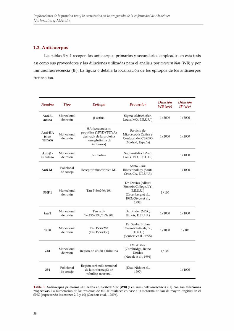
Implicaciones de la proteína tau y la cortistatina en la progresión de la enfermedad de Alzheimer Materiales y Métodos
38
1.2. Anticuerpos
Las tablas 3 y 4 recogen los anticuerpos primarios y secundarios empleados en esta tesis
así como sus proveedores y las diluciones utilizadas para el análisis por western blot (WB) y por
inmunofluorescencia (IF). La figura 6 detalla la localización de los epítopos de los anticuerpos
frente a tau.
Nombre Tipo Epítopo Proveedor
Dilución WB (v/v)
Dilución IF (v/v)
Anti-β-actina
Monoclonal de ratón β-actina Sigma-Aldrich (San
Louis, MO, E.E.U.U.) 1/5000 1/5000
Anti-HA (clon
12CA5)
Monoclonal de ratón
HA (secuencia no peptídica (YPYDVPDYA)
derivada de la proteína hemaglutinina de
influenza)
Servicio de Microscopía Óptica y Confocal del CBMSO
(Madrid, España)
1/2000 1/2000
Anti-β -tubulina
Monoclonal de ratón β-tubulina Sigma-Aldrich (San
Louis, MO, E.E.U.U.) 1/1000
Anti-M1 Policlonal de conejo Receptor muscarínico M1
Santa Cruz Biotechnology (Santa Cruz, CA, E.E.U.U.)
1/1000
PHF 1 Monoclonal de ratón
Tau P-Ser396/404
Dr. Davies (Albert Einstein College,NY,
E.E.U.U.) (Greenberg et al., 1992; Otvos et al.,
1994)
1/100
tau 1 Monoclonal de ratón
Tau noP-Ser195/198/199/202
Dr. Binder (MGC, Illinois, E.E.U.U.)
1/1000 1/1000
12E8 Monoclonal de ratón
Tau P-Ser262 (Tau P-Ser356)
Dr. Seubert (Elan Pharmaceuticals, SF,
E.E.U.U.) (Seubert et al., 1995)
1/1000 1/105
7.51 Monoclonal de ratón Región de unión a tubulina
Dr. Wishik (Cambridge, Reino
Unido) (Novak et al., 1991)
1/100
334 Policlonal de conejo
Región carboxilo terminal de la isoforma β3 de tubulina neuronal
(Diaz-Nido et al., 1990) 1/1000
Tabla 3. Anticuerpos primarios utilizados en western blot (WB) y en inmunofluorescencia (IF) con sus diluciones respectivas. La numeración de los residuos de tau se establece en base a la isoforma de tau de mayor longitud en el SNC (expresando los exones 2, 3 y 10) (Goedert et al., 1989b).

Implicaciones de la proteína tau y la cortistatina en la progresión de la enfermedad de Alzheimer Materiales y Métodos
39
7.51
Tau 1
C1 441
12E8 PHF 1
N
7.51
Tau 1
C1 441
12E8 PHF 1
N
Figura 6. Localización de los anticuerpos frente a tau utilizados. Se distinguen, en la parte de debajo de la figura, los anticuerpos dependientes del estado de fosforilación (tau-1, 12E8 y PHF1) y, en la parte de arriba, el anticuerpo independiente del estado de fosforilación (7.51).
Nombre Tipo Proveedor
Dilución WB (v/v)
Dilución IF (v/v)
Anti-conejo
Cabra-anti-conejo Dako (Glostrup, Dinamarca) 1/2000
Anti-ratón
Conejo-anti-ratón
Dako (Glostrup, Dinamarca) 1/2000
Alexa-488
Cabra-anti-conejo
Invitrogen (Carlsbad, CA, E.E.U.U.) 1/500
Alexa-555
Cabra-anti-conejo
Invitrogen (Carlsbad, CA, E.E.U.U.) 1/500
Tabla 4. Anticuerpos secundarios utilizados en western blot (WB) y en inmunofluorescencia (IF) con sus diluciones respectivas.
Implicaciones de la proteína tau y la cortistatina en la progresión de la enfermedad de Alzheimer Materiales y Métodos
40
2. MÉTODOS
2.1. Mantenimiento de las distintas cepas de ratones
En este trabajo, se utilizaron ratones silvestres C57BL6 de Harlan (Barcelona, España) y
distintas cepas de ratones modificados genéticamente que no expresan los siguientes genes:
cortistatina (CST -/-), sst2 (sst2 -/-), sst4 (sst4 -/-) y sst2/4 (sst2/4 -/-) (Videau et al., 2003). Las
diferentes líneas se cruzaron con ratones C57BL6 (N9).
Los ratones se estabularon en el animalario del Centro de Biología Molecular “Severo
Ochoa” (Madrid, España) en un ambiente de temperatura controlada con un ciclo
luz/oscuridad de 12/12 horas con comida y agua disponibles ad libitum.
Todos los procedimientos llevados a cabo fueron aprobados por el comité del Centro de
Biología Molecular “Severo Ochoa” (UAM-CSIC, Madrid, España).
2.2. Cultivos celulares
2.2.1. Células SH-SY5Y, HEK-293 y COS-7: mantenimiento, transfección y
tratamiento Durante el desarrollo de este trabajo se usaron las siguientes líneas celulares: las SH-SY5Y
de neuroblastoma humano (Biedler et al., 1978), las HEK-293 de riñón de embrión humano y las
COS-7 de fibroblastos de riñón de mono verde africano (Gluzman, 1981). Estas células crecieron
en un medio DMEM suplementado con 10% (v/v) de suero bovino fetal, 2 mM de glutamina, 1
mM de piruvato, 100 U/mL de penicilina y 100 U/mL de estreptomicina en una atmósfera
humedecida con 5% de CO2/95% aire a 37ºC.
Las células HEK-293 y COS-7 se transfectaron de forma transitoria con plásmidos
compuestos por el vector pcDNA 3.1 usando Lipofectamina 2000 (ambos de Invitrogen,
Carlsbad, CA, E.E.U.U.) según las instrucciones del fabricante. Las placas con las células control
se transfectaron, en las mismas condiciones, con el vector vacío pcDNA 3.1. Este vector se
expresa en células de mamíferos puesto que contiene el promotor de citomegalovirus (CMV).
En el caso de las HEK-293 se emplearon tres tipos de plásmidos con distintos insertos de
cDNA: la mayor isoforma humana de tau expresada en el SNC, los receptores murinos sst2 y
sst4. Las células HEK-293 se transfectaron con distintas combinaciones de plásmidos: (1) con
tau, (2) con tau y con sst2, (3) con tau y con sst4 y, por último, (4) con los tres plásmidos (tau,
sst2 y sst4).

Implicaciones de la proteína tau y la cortistatina en la progresión de la enfermedad de Alzheimer Materiales y Métodos
41
Para transfectar las COS-7 se sembraron en cubre-objetos tratados previamente con
polilisina (100 μg/mL). En este caso, se utilizaron dos tipos de plásmidos: unos que contienen el
cDNA de los receptores muscarínicos humanos M1 y otros, con M3 (UMR cDNA Resource
Center, Missouri, MO, E.E.U.U.). El epítopo de ambos, situado en la zona amino-terminal,
consiste en tres repeticiones de una secuencia no peptídica (YPYDVPDYA) derivada de la
proteína hemaglutinina de influenza (3 x HA). Las células se transfectaron con M1, con M3 o
con ambos plásmidos. Tras 24 horas, se fijaron para inmunofluorescencia o se procesaron para
estudiar los niveles de calcio intracelular.
Los tratamientos de las células HEK-293 se detallan a continuación. Primero, se lavaron
dos veces con PBS (Composición: 150 mM NaCl, 50 mM fosfato sódico, pH 7.4) y se incubaron
en presencia de medio DMEM sin suero durante 2 horas. Las células HEK-293, previamente
transfectadas de forma transitoria, se trataron por triplicado con distintas concentraciones (5, 50,
200 y 500 nM) de cortistatina durante 30 minutos o de somatostatina durante 5 minutos. Los
extractos proteicos se analizaron mediante western blot como se detalla más adelante. También
se purificó RNA.
Las SH-SY5Y se utilizaron para el estudio de la movilización intracelular de calcio,
extracción de RNA y viabilidad celular siguiendo los protocolos descritos en otras secciones de
del apartado de Métodos.
2.2.2. Cultivos primarios de neuronas de corteza o de hipocampo de ratón Siguiendo el protocolo descrito en Banker et al. (Banker & Cowan, 1977), el tejido
hipocampal o cortical obtenido a partir de crías de ratón C57BL6 recién nacidas (P0-P1) se
disoció con papaína (Worthington Biochemical Corporation, Lakewood, NJ, E.E.U.U.). Las
células se sembraron en placas recubiertas con 100 μg/mL de polilisina y 10 μg/mL de laminina
a una densidad de 1 x 10⁶ células/placa p35 y cultivadas en medio Neurobasal (NB; Gibco,
Carlsbad, CA, E.E.U.U.) con 10% (v/v) de suero de caballo, 2 mM de glutamina, 1 mM de
piruvato, 100 U/mL de penicilina y 100 U/mL de estreptomicina. Tres horas después, el medio
anterior se sustituyó por otro medio NB con 1% (v/v) de B27 (Gibco, Carlsbad, CA, E.E.U.U.), 2
mM de glutamina, 1 mM de piruvato, 100 U/mL de penicilina y 100 U/mL de estreptomicina.
Para las neuronas de hipocampo, se utilizó el medio anterior suplementado además con 1%
(v/v) de N2 (Gibco, Carlsbad, CA, E.E.U.U.). Las células se mantuvieron en una atmósfera
humedecida con 5% de CO2/95% aire a 37ºC y la mitad del medio de cultivo se reemplazó cada
2-3 días.

Implicaciones de la proteína tau y la cortistatina en la progresión de la enfermedad de Alzheimer Materiales y Métodos
42
Los cultivos de células hipocampales o corticales de 1, 2, 3 o 7 días in vitro (DIV) se
utilizaron para analizar la movilización intracelular de calcio.
Los experimentos para estudiar el efecto de los agonistas de receptores de somatostatina
se realizaron en cultivos corticales después de 8 DIV cuando estos receptores parecen ser
funcionales (Heidet et al., 1990; Stroh et al., 2000). Tras dos lavados con PBS, las células se
incubaron en un medio NB sin suero y, 2 o 6 horas después, se añadieron al medio de cultivo
los diferentes compuestos a las concentraciones y los tiempos indicados para cada tratamiento
realizado por triplicado: (1) 5, 50, 100 o 500 nM de cortistatina de 5 a 30 minutos, (2) 50 nM de
somatostatina de 5 a 30 minutos, (3) 5, 50, 100 o 500 nM de octreotide de 5 a 30 minutos, (4) 10
μM de Ro 31-8220 90 min, (5) 10 μM de Ro 31-8220 60 min + 50 nM de cortistatina 30 min. En los
experimentos control, se añadió el vehículo del tratamiento.
2.3. Western blot (WB) Los extractos de proteínas de tejido cerebral se prepararon a partir de disecciones de
corteza, hipocampo y resto de cerebro de ratones silvestres C57BL6 y CST -/-. El tejido se
homogeneizó en un homogeneizador (potter) de vidrio/vidrio con tampón de extracción en
hielo (Composición: 20 mM Hepes pH 7.4, 100 mM NaCl, 20 mM NaF, 5 mM EDTA y 1% (v/v)
Tritón TX-100 suplementado con inhibidores de fosfatasas (30 mM glicerofosfato, 5 M
pirofosfato, 1 μM ácido okadaico) y con inhibidores de proteasas (2 mM
fenilmetanelsulfonilfluoruro, 10 µg/ml leupeptina, 10 µg/ml aprotinina y 10 µg/ml pepstatina;
Roche, Basel, Suiza). Se centrifugaron las muestras a 12000 rpm durante 10 minutos a 4ºC. Se
recogió el sobrenadante descartando el sedimento. El contenido proteico se determinó por el
método de Bradford (Bradford, 1976).
Tras el tratamiento, la extracción de proteínas de las células de los cultivos se realizó
directamente en tampón de electroforesis (Composición: 50 mM Tris-HCl pH 6.8, 100 mM β-
mercaptoetanol, 2% (p/v) SDS, 0.1% (v/v) azul de bromofenol, 10% (v/v) glicerol)
suplementado con los inhibidores de fosfatasas y de proteasas anteriores).
En todos los casos, los extractos de proteínas se hirvieron durante 10 minutos en tampón
de electroforesis para después ser cargados en geles de poliacrilamida al 10% (p/v) en presencia
de SDS (SDS-PAGE). Una vez separadas las proteínas, se transfirieron a membranas de
nitrocelulosa (Schleicher & Schuell, Keene, NH, E.E.U.U.) según el método de Towbin (Towbin
et al., 1979). Para comprobar la eficiencia de la transferencia, las membranas se tiñeron con Rojo
Ponceau. Más tarde, las membranas se bloquearon para su unión no específica con 5% (p/v) de
leche en polvo en PBS con 0.05% (v/v) de Tween-20 durante 30 minutos. Se procedió a la

Implicaciones de la proteína tau y la cortistatina en la progresión de la enfermedad de Alzheimer Materiales y Métodos
43
incubación con el anticuerpo primario diluido en solución de bloqueo (tabla 3) durante 1 hora a
temperatura ambiente. Las membranas se lavaron tres veces con PBS Tween-20 (0.05% (v/v)) y
se incubaron con el anticuerpo secundario correspondiente (1/2000) acoplado a peroxidasa
(DAKO, Glostrup, Dinamarca) durante 45 minutos (tabla 4). Tras varios lavados con PBS
Tween-20 (0.05% (v/v)), la detección de las bandas inmunoreactivas se realizó con el sistema
ECL (Perkin Elmer Life Sciences, Boston, MA, E.E.U.U.).
Finalmente, la cuantificación del grado de inmunoreactividad de las proteínas, en
unidades arbitrarias, se realizó mediante escaneo y posterior densitometría de las películas
autorradiográficas usando el programa Quantity One (Bio-Rad Laboratories, Hercules, CA,
E.E.U.U.). Los niveles de fosforilación de la proteína tau se calcularon normalizando los datos
con los niveles de tau total determinados por el anticuerpo 7.51.
2.4. Inmunofluorescencia Esta técnica se utilizó en las células siguientes, crecidas sobre cubre-objetos:
- las COS-7 transfectadas con M1 o M3
- las HEK-293 transfectadas con combinaciones diferentes de los plásmidos de tau, sst2
y sst4
- los cultivos primarios de hipocampo y de corteza
Las células se fijaron con 4 % (p/v) de paraformaldehído en PBS suplementado con 5 mM
de MgCl2 durante 10 minutos a 37 °C. Tras tres lavados con PBS, los cubre-objetos se incubaron
con diferentes soluciones a temperatura ambiente: PBS con 0.1 % (v/v) de Tritón X-100 (15 min),
1M de glicina en PBS (30 min) y solución de bloqueo (1 % (p/v) de seroalbúmina bovina (BSA)
en PBS, 45 min). Los cultivos estuvieron en presencia del anticuerpo primario correspondiente
para cada caso durante 1 hora (tabla 3). Tras dos lavados de PBS, se añadió el anticuerpo
secundario en solución de bloqueo durante 45 minutos (tabla 4). En algunos experimentos, se
tiñeron los núcleos con DAPI (1/105, 5 min). Seguidamente, los cubre-objetos se montaron en
glicerol:PBS (1:1) o FluorSave (Calbiochem, San Diego, CA, E.E.U.U.). Las muestras se
visualizaron en distintas sesiones utilizando los microscopios confocales modelo Axiovert
200M, Axioskop 2 (Carl Zeiss International, Thornwood, NY, E.E.U.U.) y Leica SP2 AOBS
(Leica, Wetzlar, Alemania). Las imágenes se adquirieron de forma secuencial con ayuda del
programa informático asociado y se procesaron utilizando el programa Photoshop (Adobe
Systems, Mountain View, CA, E.E.U.U.).

Implicaciones de la proteína tau y la cortistatina en la progresión de la enfermedad de Alzheimer Materiales y Métodos
44
2.5. Internalización de Fluo-cortistatina en cultivos primarios de corteza
murina Los ensayos de internalización de Fluo-cortistatina se realizaron en cultivos corticales
después de 8 DIV. Las células se crecieron en un medio NB sin suero durante 2 horas y,
después, se realizaron los tratamientos siguientes: (1) 1-5 μM Fluo-cortistatina 1 o 30 min, (2)
100 μM cortistatina 10 min + 1 μM Fluo-cortistatina 30 min, (3) 100 μM somatostatina 10 min + 1
μM Fluo-cortistatina 30 min, (4) 1 μM PAO 30 min + 5 μM Fluo-cortistatina 30 min. En las
células control, se añadió el mismo volumen del vehículo del tratamiento en condiciones
experimentales similares. Los compuestos empleados se diluyeron en agua a excepción del
PAO que estaba diluido en dimetilsulfóxido (DMSO).
Después, se realizó una inmunofluorescencia con el anticuerpo primario 334 (tabla 3) y
los núcleos se tiñeron con DAPI siguiendo el protocolo que se detalla en el apartado anterior. El
recuento de las neuronas corticales en cultivo se realizó en campos seleccionados al azar a 40X
(un área aproximada de 0.2 mm²) utilizando un microcopio de fluorescencia modelo Zeiss (Carl
Zeiss International, Thornwood, NY, E.E.U.U.). Sólo se consideraron como células con Fluo-
cortistatina internalizada las células con intenso marcaje verde. La proporción de células
marcadas (vs. número total de células) se obtuvo contando 100-250 células totales por campo en
los cubre-objetos incubados en presencia de Fluo-cortistatina a una concentración de 5 μM
durante 30 minutos (n = 5 cubre-objetos). El número de células totales se obtuvo contando los
núcleos teñidos con DAPI. Para comparar los experimentos con Fluo-cortistatina (1 μM, 30 min)
con los de cortistatina o somatostatina (100 μM, 10 min) + Fluo-cortistatina (1 μM, 30 min), se
contaron las células marcadas con la fluorescencia verde en 20 campos distintos (n = 4 cubre-
objetos por condición).
2.6. Reacción en cadena de la polimerasa (PCR) cuantitativa La purificación del RNA, seguida de un tratamiento con DNasa, se realizó usando el RNA
Miniprep Kit (Stratagene, La Jolla, CA, E.E.U.U.) según el protocolo del fabricante. El RNA se
obtuvo a partir de las distintas líneas celulares (las HEK-293 transfectadas de forma transitoria,
las SH-SY5Y y las COS-7) y de los cultivos primarios de corteza de 8 DIV.
Para evitar la proliferación de células no-neuronales en los cultivos corticales, se añadió
2.5 μM de arabinofuranosilcitosina (Ara-C; Calbiochem, San Diego, CA, E.E.U.U.) tras 3 DIV.
Los receptores de somatostatina no sólo se expresan en las neuronas sino que también lo hacen,
aunque en menor medida, en las células gliales de roedores (Feindt et al., 1995; Heidet et al.,
1990; Hosli & Hosli, 1994; Mentlein et al., 1990; van Calker et al., 1980).

Implicaciones de la proteína tau y la cortistatina en la progresión de la enfermedad de Alzheimer Materiales y Métodos
45
Finalmente, la concentración y la pureza del RNA se comprobaron utilizando un
espectrofotómetro modelo Nanodrop ND-1000 (Nanodrop Technologies, Wilmington, DE,
E.E.U.U.).
El RNA (480 ng) se retro-transcribió como cDNA de simple cadena utilizando el kit de
síntesis de cDNA (Roche, Basel, Suiza) según las instrucciones del fabricante. La reacción incluía
20 unidades de transcriptasa inversa del Virus de la mieloblastosis de ave (AMV), 0.2 μg/μL de
primers hexanucleótidos y 0.08 μg/μL de primers oligonucleótidos (dT).
Usando el reactivo SYBR Green (n = 3), detectamos los niveles de expresión de distintos
genes:
- M1-5 en las SH-SY5Y y COS-7,
- sst1-5 en las neuronas corticales,
- tau, sst2 y sst4 en las células HEK-293 transfectadas de forma transitoria.
El SYBR Green, al igual que el bromuro de etidio, se une específicamente al DNA de doble
cadena, y al hacerlo incrementa fuertemente su fluorescencia. Al unirse al DNA de doble
cadena recién sintetizado en cada ciclo, este reactivo emite una fluorescencia proporcional a la
concentración de DNA. La reacción de 10 μL de volumen total contenía: 1 μL de molde (cDNA,
cDNA diluido (1:5), plásmido, DNA genómico o H2O), 5 μL de la mezcla de PCR de SYBR Green
(Applied Biosystems, Foster City, CA, E.E.U.U.) y 150 nM de cada par de primers. Las
secuencias de los primers utilizados en esta tesis y los genes específicos que amplifican aparecen
en la tabla 5 (Cristiani et al., 2000). Los niveles de expresión de los genes estudiados se
normalizaron con los de expresión de β-actina murina en el caso de los cultivos primarios y con
los de β-actina humana en el caso de las líneas celulares.
El protocolo de amplificación consistió en un paso de 10 minutos a 95°C (activación de la
Taq polimerasa) seguido de 50 ciclos de 15 segundos a 95°C y de 1 minuto a 60°C
(desnaturalización y anillamiento-extensión). Tras la amplificación, se obtuvo la curva de
disociación aumentando progresivamente la temperatura de 60°C a 95°C (figura 7). La señal
fluorescente se adquirió durante la fase de anillamiento por el sistema de detección de modelo
ABI PRISM 7900HT (Applied Biosystems, Foster City, CA, E.E.U.U.).
Para comprobar la especificidad de la reacción, se analizaron las curvas de disociación
correspondientes para cada caso. Además, usamos distintos cDNA como moldes de los
controles positivos: pituitaria de ratón para sst5, plásmidos para sst1-4 y DNA genómico de SH-
SY5Y y COS-7 para M1-5. Las reacciones sin molde para cada amplificación se usaron como
controles negativos. La media de los valores de los ciclos umbral (CT) de cada reacción,
realizada por triplicado, se calculó teniendo en cuenta las muestras diluidas y no diluidas y se
normalizó con los valores CT del gen de β-actina correspondiente.

Implicaciones de la proteína tau y la cortistatina en la progresión de la enfermedad de Alzheimer Materiales y Métodos
46
02
46
810
1214
16
62 67 72 77 82 87 92
Temperatura (ºC)
Fluo
resc
enci
a sst1sst2sst3sst4sst5
02468
10121416
0 10 20 30 40 50Ciclo
Fluo
resc
enci
a sst1sst2sst3sst4sst5
02
46
810
1214
16
62 67 72 77 82 87 92
Temperatura (ºC)
Fluo
resc
enci
a sst1sst2sst3sst4sst5
02468
10121416
0 10 20 30 40 50Ciclo
Fluo
resc
enci
a sst1sst2sst3sst4sst5
Gen
Secuencia “sentido” Secuencia “anti-sentido”
β-actina murina
GGCGCTTTTGACTCAGGATT GGGATGTTTGCTCCAACCAA
β-actina humana
CACACTGTGCCCATCTACGA CTCCTTAATGTCACGCACGA
M1
ACCTCTATACCACGTACCTG TGAGCAGCAGATTCATGACG
M2
ACCTGTGGTGTGTGACCTTT GCTTGACTGGGTAGGTCAGA
M3
CATCATGAATCGATGGGCCT GGCCTCGTGATGGAAAAGTA
M4
CAGTTTGTGGTGGGTAAGCG GTACAGCACCGTCATGATGA
M5
GATGAGTGCCAGATCCAGTT GACAGTAGAGGATGGTCATG
sst1
TATTGCTGATGAGCTGCTCATGCTC TCCACACTAAGCACAGTCAGACAGT
sst2
ATCAGCCCCACCCCAGCCCTGAA GCAAAGAACATTCTGGAAGCTCTTC
sst3
CAAGGAGATATACCAGGACTTGGGA TAGGACACTGGACTGTCCTGCAGAA
sst4
AACAACGGAGGCGCTCAGAGAAGAA TCGAGGCTGGTGACAAACAGGTTCA
sst5
GGGCTGGGGCACCTGCAACCTGA GACAGGACCACCACAAAGAAGTAGA
Tau
GCGGATCCATATGGGGGATCGCAGC CCGAATTCTTAGGGCACGGGGG
Tabla 5. Parejas de primers utilizados para amplificar los genes β-actina murina y humana, M1-5, sst1-5 y tau. Se detalla la secuencia (5´-3´) de cada primer utilizado.
Figura 7. Ejemplo de amplificación de los receptores de somatostatina (sst1-5) en el panel de la derecha. A la izquierda, curvas de disociación de los distintos genes.

Implicaciones de la proteína tau y la cortistatina en la progresión de la enfermedad de Alzheimer Materiales y Métodos
47
2.7. Purificación de DNA genómico El DNA genómico de las COS-7 se extrajo usando el sistema de purificación Wizard Plus
SV (Promega, Wisconsin, E.E.U.U.).
En el caso de las células SH-SY5Y, se siguió un protocolo de alta concentración de sales.
Primero, se añadió un tampón para lisis de núcleos (1% (v/v) TE pH 8.0, 0.4 M NaCl y 2 mM
EDTA) al precipitado celular y las células se centrifugaron durante 10 minutos a 4ºC (12000
rpm). El precipitado obtenido se resuspendió en el tampón de lisis anterior suplementado con
0.3 mg/mL de proteinasa K (Merck, Barcelona, España) y 1% (p/v) de SDS. Tras una incubación
a 45ºC durante toda la noche, se añadió a la muestra 6 M de NaCl y se centrifugó (15 min, 3000
rpm). El sobrenadante se centrifugó varias veces hasta que aparecía transparente (sin sales).
Para precipitar el DNA, se añadió al último sobrenadante 3 M de acetato sódico (pH 5.2) e
isopropanol frío. El DNA se transfirió a un nuevo tubo con 70% (v/v) de etanol y se centrifugó
(30 min, 14000 rpm). Finalmente, se resuspendió el precipitado en agua.
El DNA genómico sirvió de molde de la amplificación específica de M1-5 y la
concentración de los productos de PCR se midió en el espectrofotómetro modelo Nanodrop
ND-1000 (Nanodrop Technologies, Wilmington, DE, E.E.U.U.).
Los microgramos de DNA doble cadena se convirtieron a picomoles: pmol = μg x 106
pg/1 μg x 1 pmol/660 pg x 1/N (N, tamaño del fragmento amplificado en pares de bases (pb):
122, 128, 128, 132, 103 y 162 pb para M1-5 y β-actina humana respectivamente). Se realizaron las
distintas rectas patrones para M1-5 usando diluciones seriadas (diez veces) en tRNA de
levadura (110 ng/μL, Roche Applied Science, Basel, Suiza) de los productos de PCR
correspondientes a partir de 10-2 pmol.
2.8. Determinación fluorimétrica del Ca2+ intracelular con Fura-2 Se determinó el Ca2+ citosólico libre en distintos tipos celulares mediante un seguimiento
fluorimétrico continuo empleando Fura-2 como indicador fluorescente (Diaz-Hernandez et al.,
2001a; Diaz-Hernandez et al., 2001b). Este compuesto se une al Ca2+ específicamente y, al
hacerlo, aumenta su emisión de fluorescencia a 510 nm (figura 8) al mismo tiempo que se
desplaza su espectro de excitación hacia el UV, cambiando el máximo de excitación de 380
(solución de Fura-2 libre de calcio) a 340 nm (Fura-2 saturado de calcio).
Estas determinaciones se realizaron en cultivos de neuronas hipocampales y corticales, de
SH-SY5Y y de COS-7 transfectadas de forma transitoria. Las células se lavaron en tampón de
perfusión compuesto por: 122 mM NaCl, 3.1 mM KCl, 0.4 mM KH2PO4, 5.0 mM NaHCO3, 1.2
mM MgSO4, 10 mM glucosa y 20 mM TES a pH 7.4. Posteriormente, se incubaron con la sonda

Implicaciones de la proteína tau y la cortistatina en la progresión de la enfermedad de Alzheimer Materiales y Métodos
48
Fura-2 acetometil éster (Fura-2 AM) a una concentración de 7.5 μM durante 30 minutos a 37ºC.
Este periodo facilita la hidrólisis del Fura-2 AM. Es un compuesto esterificado que, al ser
liposoluble, atraviesa fácilmente la membrana plasmática. Una vez en el interior celular, el
Fura-2 AM es hidrolizado por esterasas celulares inespecíficas que liberan Fura-2 (ácido),
compuesto que al perder su liposolubilidad, queda retenido en la célula.
Tras varios lavados con medio fresco, los cubre-objetos se montaron en una cámara de
perfusión en un microscopio modelo Nikon Eclipse TE-2000 (Nikon Corporation, Japón). La
cámara se dotó de un flujo de entrada constante de tampón de perfusión termostatizado a 37ºC
(1.2 mL/min) y de una salida por aspiración mediante bomba de vacío. En todos los
experimentos, se añadió, primero, acetilcolina a una concentración de 50 μM para comprobar si
las células eran funcionales. Tras varios lavados, se detuvo el sistema de perfusión para realizar
los tratamientos añadiendo los distintos compuestos al medio de la cámara. Los ensayos con 50
μM de acetilcolina y 1 μM de tau, en distintas formas, eran de 10 minutos. Para ensayar los
antagonistas de los receptores muscarínicos, se pre-incubaron las células en presencia de los
compuestos siguientes durante 5 minutos a diferentes concentraciones: 10 μM de hexametonio,
100 μM de atropina, 10 μM de pirencepina, 2 μM de 4-DAMP y 2 μM de galamina.
Seguidamente, se añadió 50 μM de acetilcolina o 1 μM de distintas formas de tau durante 10
minutos. Al final de cada experimento, se aplicaron pulsos de acetilcolina a una concentración
de 50 μM para confirmar la viabilidad del cultivo estudiado. Las células se visualizaron
utilizando un objetivo de 40x.
Se excitaron las muestras alternativamente con luz de diferente longitud de onda (340 y
380 nm) mediante el uso de un monocromador de un ancho de banda de 10 nm (Optocan
monochromator, Cairn, Faversham, Reino Unido) y la fluorescencia se midió a 510 nm.
Sucesivamente, la cámara CCD Hamamatsu C-4880-80 (Hamamatsu, Japón) asociada al
programa informático Metafluor 6.3r6 PC (Universal Imaging Corp., Cambridge, Reino Unido)
tomó imágenes de 12 bit de las células. El tiempo de exposición fue de 150 ms para cada
longitud de onda y el tiempo de cambio <5 ms.
Los registros mostrados representan la media de la intensidad lumínica que se produce
en una región elíptica dibujada en el interior de cada célula. El valor de fluorescencia basal se
restó a los datos obtenidos para cada longitud de onda. En este trabajo, los resultados se
presentan como el ratio entre las señales de 340 y 380 nm (F340/F380) que se obtienen al dividir la
sucesión de imágenes obtenidas a las diferentes longitudes de onda. El cociente de fluorescencia
F340/F380 se calibró como [Ca2+]i empleando la ecuación descrita en (Grynkiewicz et al., 1985) y
se corrigió siguiendo el protocolo descrito en (Poenie, 1990).

Implicaciones de la proteína tau y la cortistatina en la progresión de la enfermedad de Alzheimer Materiales y Métodos
49
300 320 340 360 380 400 420
0
1000
2000
3000
4000N
ivel
es d
e G
rise
s
Longitud de onda (nm)
440
Con Ca2+
Sin Ca2+
Emisión: 510 nm
300 320 340 360 380 400 420
0
1000
2000
3000
4000N
ivel
es d
e G
rise
s
Longitud de onda (nm)
440
Con Ca2+
Sin Ca2+Con Ca2+
Sin Ca2+
Emisión: 510 nm
Figura 8. Espectro de excitación de la sonda fluorescente Fura-2 en presencia y en ausencia de calcio.
2.9. Purificación de proteínas En este trabajo, se utilizó la isoforma humana de tau 42 recombinante que contiene dos
insertos amino-terminal (N) y cuatro repeticiones de unión a microtúbulos (R) (Goedert & Jakes,
1990). También se emplearon los fragmentos de tau siguientes: tau 2R (residuos 250-335 con la
deleción de 274-304), 2N compuesto por la mitad amino-terminal de tau (residuos 1-251), 3R
(residuos 250-367 con la deleción de 274-304), 3RC (residuos 250-441 con la deleción de 274-304)
y tau391-407 que comprende la región carboxilo-terminal de la proteína y la siguiente secuencia
EIVYKSPVVSGDTSPRH (residuos 391-407) (Perez et al., 2001). La numeración de los residuos
de tau se define según la nomenclatura de la isoforma de tau más larga (Goedert et al., 1989a;
Goedert et al., 1989b). Los PHF se aislaron como detallan Greenberg y Davies (Greenberg &
Davies, 1990) y se visualizaron por el microscopio electrónico Jeol modelo JEM1010 (Hernandez
et al., 2002). Los fragmentos de tau se aislaron siguiendo el protocolo descrito en (Perez et al.,
1996) y el péptido tau391-407 se sintetizó y purificó según (Perez et al., 2001). Las proteínas
purificadas se separaron en un gel de electroforesis y se tiñeron con azul de Coomassie. El
contenido en tau se determinó mediante western blot usando el anticuerpo 7.51 frente a tau y
posterior cuantificación por densitometría.
2.10. Ensayo de viabilidad Se añadió al medio de cultivo de las SH-SY5Y el tau 42 a distintas concentraciones (1, 3, 5
μM) durante 24 y 48 horas. Las células control se incubaron con el mismo volumen de vehículo.
La viabilidad celular se midió usando el kit de la lactatodeshidrogenasa (LDH; Roche, Basel,
Suiza) según las instrucciones del fabricante.

Implicaciones de la proteína tau y la cortistatina en la progresión de la enfermedad de Alzheimer Materiales y Métodos
50
2.11. Test del reconocimiento de objeto Trabajamos con ratones machos de más de un año de edad (12-14 meses) silvestres
C57BL6 y CST -/-. Se siguió el protocolo del test de comportamiento descrito anteriormente
(Engel et al., 2006; Ennaceur et al., 1997; Woolley et al., 2003). En resumen, los ratones se
entrenaron durante 30 minutos en una caja de plástico con muros verticales opacos (73 x 33 x 25
cm). Al día siguiente, los animales se colocaron en la misma caja durante 10 minutos. Dos horas
después, cada ratón se dejó durante 4 minutos en la caja sin objetos. Más tarde, se situó al
animal durante 1 minuto en su jaula. La siguiente fase de familiarización fue en la caja durante
5 minutos en presencia de dos objetos idénticos (objetos A y B). Los dos objetos se colocaron en
cada extremo del eje más largo de la caja, cada uno a 7 cm de la pared de la caja. Tras cada
exposición, los objetos y la caja se limpiaron con etanol al 70% para eliminar las pistas olorosas.
Dos horas después de la familiarización, se realizó la prueba del reconocimiento: cada ratón se
dejó en la caja durante 5 minutos con un objeto antiguo (objeto A) y un objeto nuevo (objeto C).
La posición del objeto C era la misma que la del objeto B en la fase de familiarización. Se
consideró que los animales reconocían el objeto cuando su cabeza se situaba a menos de 2 cm
del objeto. Se anotaron los tiempos (tA y tC) que el ratón empleaba en explorar los dos objetos
(objetos A y B). El índice de memoria (MI) se define como el ratio entre el tiempo empleado en
explorar el nuevo objeto sobre el tiempo empleado en explorar ambos objetos, MI = [tC/(tA +
tC)] x 100. Ese índice se utilizó para medir la memoria no-espacial.
2.12. Análisis estadístico Los análisis estadísticos empleados en esta tesis doctoral fueron: ANOVA seguida por un
test Bonferroni post hoc (figura 20 A) y análisis de t Student (el resto de figuras).
Se utilizó el test de la chi cuadrado para comparar el número de células con Fluo-
cortistatina internalizada en distintas condiciones experimentales: (1) Fluo-cortistatina (1 μM, 30
min), (2) somatostatina (100 μM, 10 min) + Fluo-cortistatina (1 μM, 30 min) y (3) cortistatina (100
μM, 10 min) + Fluo-cortistatina (1 μM, 30 min). Se consideraron positivos los campos con 3 o
más células con Fluo-cortistatina internalizada (área aproximada de cada campo: 0.2 mm²).

Resultados


Implicaciones de la proteína tau y la cortistatina en la progresión de la enfermedad de Alzheimer Resultados
53
1. PAPEL DE TAU EN LA PROGRESIÓN DE LA EA 1.1. La proteína tau extracelular produce muerte celular de las células de
neuroblastoma humano SH-SY5Y Como se indicó en la introducción, durante el desarrollo de la EA, los NFT se van
formando según una secuencia invariable que correlaciona con el daño en estructuras concretas
del cerebro (Braak & Braak, 1991). En este trabajo, nos planteamos la posibilidad de que el
progreso de la EA pudiera depender de la toxicidad celular mediada por la presencia de tau
extracelular. La proteína tau presente en el interior celular puede pasar al medio extracelular en
el momento en que una neurona degenera. El tau extracelular podría ser tóxico para las células
vecinas de un modo similar al observado en el caso del péptido β-amiloide extracelular. El
péptido β-amiloide puede formar poros en las membranas de las células de neuroblastoma
humano provocando la entrada de calcio hasta niveles tóxicos para la célula (Demuro et al.,
2005).
Resultados previos del laboratorio indicaban que la proteína tau extracelular podría ser
tóxica, cuando se añadía a cultivos celulares, y que dicha toxicidad podría estar relacionada con
la presencia de receptores colinérgicos en las células cultivadas (Gomez-Ramos et al., 2006).
Primero, para comprobar la posible toxicidad de tau extracelular, se realizaron experimentos de
viabilidad añadiendo tau en forma monómerica y fibrilar (PHF) a células de neuroblastoma
humano SH-SY5Y en cultivo. Incubamos las células SH-SY5Y durante 24 horas en presencia de
distintas concentraciones de tau en forma monomérica (1, 3, 5 μM) o en PHF (1, 5 μM).
Medimos la muerte celular presente en las células SH-SY5Y tratadas y la comparamos con la de
células control tratadas con el vehículo. La figura 9 A muestra que la exposición a tau en
cualquiera de sus formas induce un aumento en la muerte celular y que este aumento se
acentúa con concentraciones crecientes de tau. Además, comprobamos que la proteína tau en
forma monomérica es más tóxica que en PHF.
También, estudiamos la toxicidad de tau en incubaciones de 48h cuando las células se
trataban con una concentración alta de 5 μM de tau en forma monomérica y en PHF.
Comprobamos que, en ambos casos, la toxicidad de tau es mayor si los tratamientos se
prolongan en el tiempo (figura 9 B-C).

Implicaciones de la proteína tau y la cortistatina en la progresión de la enfermedad de Alzheimer Resultados
54
B
*
*
0102030405060708090
100
24 h. 48 h.
% M
uert
e C
elul
ar
0102030405060708090
100
24 h. 48 h.
% M
uert
e C
elul
ar
C
*
0102030405060708090
100
24 h. 48 h.
% M
uert
e C
elul
ar
0102030405060708090
100
24 h. 48 h.
% M
uert
e C
elul
ar
0
10
20
30
40
50
60
70
0 1 2 3 4 5Concentraci ón (micromolar )
0
10
20
30
40
50
60
70
0 1 2 3 4 5
% M
uert
e C
elul
ar tauPHF
Concentración (micromolar)
A
*
**
B
*
*
0102030405060708090
100
24 h. 48 h.
% M
uert
e C
elul
ar
0102030405060708090
100
24 h. 48 h.
% M
uert
e C
elul
ar
C
*
0102030405060708090
100
24 h. 48 h.
% M
uert
e C
elul
ar
0102030405060708090
100
24 h. 48 h.
% M
uert
e C
elul
ar B
*
*
0102030405060708090
100
24 h. 48 h.
% M
uert
e C
elul
ar
0102030405060708090
100
24 h. 48 h.
% M
uert
e C
elul
ar
B
*
*
0102030405060708090
100
24 h. 48 h.
% M
uert
e C
elul
ar
0102030405060708090
100
24 h. 48 h.
% M
uert
e C
elul
ar
*
*
0102030405060708090
100
24 h. 48 h.
% M
uert
e C
elul
ar
0102030405060708090
100
24 h. 48 h.
% M
uert
e C
elul
ar
C
*
0102030405060708090
100
24 h. 48 h.
% M
uert
e C
elul
ar
0102030405060708090
100
24 h. 48 h.
% M
uert
e C
elul
ar C
*
0102030405060708090
100
24 h. 48 h.
% M
uert
e C
elul
ar
0102030405060708090
100
24 h. 48 h.
% M
uert
e C
elul
ar
*
0102030405060708090
100
24 h. 48 h.
% M
uert
e C
elul
ar
0102030405060708090
100
24 h. 48 h.
% M
uert
e C
elul
ar
0
10
20
30
40
50
60
70
0 1 2 3 4 5Concentraci ón (micromolar )
0
10
20
30
40
50
60
70
0 1 2 3 4 5
% M
uert
e C
elul
ar tauPHF
Concentración (micromolar)
A
0
10
20
30
40
50
60
70
0 1 2 3 4 5Concentraci ón (micromolar )
0
10
20
30
40
50
60
70
0 1 2 3 4 5
% M
uert
e C
elul
ar tauPHF
Concentración (micromolar)
0
10
20
30
40
50
60
70
0 1 2 3 4 5Concentraci ón (micromolar )
0
10
20
30
40
50
60
70
0 1 2 3 4 5
% M
uert
e C
elul
ar tauPHF
Concentración (micromolar)
A
*
**
Figura 9. La proteína tau extracelular es tóxica para las células de neuroblastoma humano SH-SY5Y en cultivo. (A) Efecto de la adición de distintas concentraciones de tau en forma monomérica (tau) y en forma fibrilar (PHF) sobre la viabilidad de las SH-SY5Y. Las incubaciones fueron de 24h. (B) Viabilidad celular en presencia de 5 μM de tau en forma monomérica durante 24h y 48h. Comparamos los resultados de las células control (tratadas con el vehículo) con los de las células con tau correspondientes a su respectivo periodo de incubación en cada caso (* p<0.05). (C) Como en (B), pero el tratamiento se realizó con tau en PHF. En ambos experimentos, la concentración creciente de tau y la prolongación en el tiempo de su exposición agravan la toxicidad sobre las células.
.
Implicaciones de la proteína tau y la cortistatina en la progresión de la enfermedad de Alzheimer Resultados
55
1.2. La proteína tau altera la homeostasis de calcio intracelular de las SH-
SY5Y a través de los receptores muscarínicos Para comprobar si la toxicidad de la proteína tau extracelular se debe a un aumento de
calcio intracelular como se ha descrito para el péptido β-amiloide (Demuro et al., 2005),
estudiamos el efecto de tau sobre los niveles de calcio intracelular de las células SH-SY5Y.
Centramos este estudio y los posteriores, en el análisis de tau en forma monomérica puesto que
vimos que era el más tóxico para las células en cultivo. La adición de 1 μM de tau al medio de
las SH-SY5Y, cargadas con Fura-2 AM, induce un aumento sostenido en los niveles de calcio
intracelular (figura 10). Usamos tratamientos de 50 μM de acetilcolina como control positivo de
aumento de calcio intracelular. La figura 10 muestra que el efecto de tau sobre los niveles de
calcio intracelular se prolonga más en el tiempo que el efecto de los tratamientos realizados con
acetilcolina. Estos datos sugieren que la toxicidad de tau podría deberse a la mayor duración de
su efecto sobre la movilización intracelular de calcio. Observamos que algunas de las células
que muestran un aumento en los niveles de calcio intracelular en presencia de acetilcolina no lo
hacen al añadir tau. Ésto se debe probablemente a que la composición de receptores no es la
misma en todas las células. Por tanto, quisimos identificar el/los receptor/es responsable/s del
efecto de tau sobre la movilización intracelular de calcio.
Nos planteamos que los receptores colinérgicos eran posibles mediadores de este efecto,
como previamente había sido propuesto (Gomez-Ramos et al., 2006). Para ello, realizamos
distintos experimentos en presencia de antagonistas de receptores colinérgicos. La figura 10
muestra que el incremento de los niveles de calcio intracelular inducido por tau también puede
observarse en presencia de 10 μM de hexametonio, un antagonista de los receptores nicotínicos.
Sin embargo, este efecto no aparece cuando las células se pre-incuban con 100 μM de atropina,
un antagonista de los receptores muscarínicos (figura 10). Estos resultados sugieren que el
aumento intracelular de calcio provocado por la proteína tau extracelular está mediado por los
receptores muscarínicos.

Implicaciones de la proteína tau y la cortistatina en la progresión de la enfermedad de Alzheimer Resultados
56
Tiempo240 (sec/div)
F340
/F38
0
ACh tau
0,2
0,4
0,6
0,8
1,0
1,2
1,4
F340
/F38
0
ACh tau
0,2
0,4
0,6
0,8
1,0
1,2
1,4
Tiempo240 (sec/div)
0,40
0,50
0,60
0,70
0,80
0,90
0,10
0,11
0,12
0,13ACh ACh Atau
Hexa
Rat
io 3
40/3
80
0,40
0,50
0,60
0,70
0,80
0,90
0,10
0,11
0,12
0,13ACh ACh Atau
Rat
io 3
40/3
80
Tiempo240 (sec/div)
Tiempo240 (sec/div)
0,40
0,50
0,60
0,70
0,80
0,90
0,10
0,11
0,12
0,13
0,40
0,50
0,60
0,70
0,80
0,90
0,10
0,11
0,12
0,13ACh ACh Atau
Hexa
Rat
io 3
40/3
80
0,40
0,50
0,60
0,70
0,80
0,90
0,10
0,11
0,12
0,13ACh ACh Atau
Rat
io 3
40/3
80
Tiempo240 (sec/div)
Atr
0,2
0,4
0,6
0,8
1,0
1,2
1,4
ACh ACh tautau
Rat
io 3
40/3
80
0,2
0,4
0,6
0,8
1,0
1,2
1,4
Atr
0,2
0,4
0,6
0,8
1,0
1,2
1,4
ACh ACh tautau
Rat
io 3
40/3
80
0,2
0,4
0,6
0,8
1,0
1,2
1,4
Tiempo240 (sec/div)
F340
/F38
0
ACh tau
0,2
0,4
0,6
0,8
1,0
1,2
1,4
F340
/F38
0
ACh tau
0,2
0,4
0,6
0,8
1,0
1,2
1,4
Tiempo240 (sec/div)
Tiempo240 (sec/div)
F340
/F38
0
ACh tau
0,2
0,4
0,6
0,8
1,0
1,2
1,4
F340
/F38
0
ACh tau
0,2
0,4
0,6
0,8
1,0
1,2
1,4
F340
/F38
0
ACh tau
0,2
0,4
0,6
0,8
1,0
1,2
1,4
F340
/F38
0
ACh tau
0,2
0,4
0,6
0,8
1,0
1,2
1,4
Tiempo240 (sec/div)
0,40
0,50
0,60
0,70
0,80
0,90
0,10
0,11
0,12
0,13ACh ACh Atau
Hexa
Rat
io 3
40/3
80
0,40
0,50
0,60
0,70
0,80
0,90
0,10
0,11
0,12
0,13ACh ACh Atau
Rat
io 3
40/3
80
Tiempo240 (sec/div)
Tiempo240 (sec/div)
0,40
0,50
0,60
0,70
0,80
0,90
0,10
0,11
0,12
0,13
0,40
0,50
0,60
0,70
0,80
0,90
0,10
0,11
0,12
0,13ACh ACh Atau
Hexa
Rat
io 3
40/3
80
0,40
0,50
0,60
0,70
0,80
0,90
0,10
0,11
0,12
0,13ACh ACh Atau
Rat
io 3
40/3
80
Tiempo240 (sec/div)
0,40
0,50
0,60
0,70
0,80
0,90
0,10
0,11
0,12
0,13ACh ACh Atau
Hexa
Rat
io 3
40/3
80
0,40
0,50
0,60
0,70
0,80
0,90
0,10
0,11
0,12
0,13ACh ACh Atau
Rat
io 3
40/3
80
Tiempo240 (sec/div)
Tiempo240 (sec/div)
0,40
0,50
0,60
0,70
0,80
0,90
0,10
0,11
0,12
0,13
0,40
0,50
0,60
0,70
0,80
0,90
0,10
0,11
0,12
0,13ACh ACh Atau
Hexa
Rat
io 3
40/3
80
0,40
0,50
0,60
0,70
0,80
0,90
0,10
0,11
0,12
0,13ACh ACh Atau
Rat
io 3
40/3
80
Tiempo240 (sec/div)
Atr
0,2
0,4
0,6
0,8
1,0
1,2
1,4
ACh ACh tautau
Rat
io 3
40/3
80
0,2
0,4
0,6
0,8
1,0
1,2
1,4
Atr
0,2
0,4
0,6
0,8
1,0
1,2
1,4
ACh ACh tautau
Rat
io 3
40/3
80
0,2
0,4
0,6
0,8
1,0
1,2
1,4
Tiempo240 (sec/div)
Tiempo240 (sec/div)
Atr
0,2
0,4
0,6
0,8
1,0
1,2
1,4
ACh ACh tautau
Rat
io 3
40/3
80
0,2
0,4
0,6
0,8
1,0
1,2
1,4
Atr
0,2
0,4
0,6
0,8
1,0
1,2
1,4
ACh ACh tautau
Rat
io 3
40/3
80
0,2
0,4
0,6
0,8
1,0
1,2
1,4
F 340/F
380
F 340/F
380
Tiempo (240 seg/div) Tiempo (240 seg/div) Tiempo (240 seg/div)
F 340/F
380
F 340/F
380
F 340/F
380
F 340/F
380
Tiempo240 (sec/div)
F340
/F38
0
ACh tau
0,2
0,4
0,6
0,8
1,0
1,2
1,4
F340
/F38
0
ACh tau
0,2
0,4
0,6
0,8
1,0
1,2
1,4
Tiempo240 (sec/div)
0,40
0,50
0,60
0,70
0,80
0,90
0,10
0,11
0,12
0,13ACh ACh Atau
Hexa
Rat
io 3
40/3
80
0,40
0,50
0,60
0,70
0,80
0,90
0,10
0,11
0,12
0,13ACh ACh Atau
Rat
io 3
40/3
80
Tiempo240 (sec/div)
Tiempo240 (sec/div)
0,40
0,50
0,60
0,70
0,80
0,90
0,10
0,11
0,12
0,13
0,40
0,50
0,60
0,70
0,80
0,90
0,10
0,11
0,12
0,13ACh ACh Atau
Hexa
Rat
io 3
40/3
80
0,40
0,50
0,60
0,70
0,80
0,90
0,10
0,11
0,12
0,13ACh ACh Atau
Rat
io 3
40/3
80
Tiempo240 (sec/div)
Atr
0,2
0,4
0,6
0,8
1,0
1,2
1,4
ACh ACh tautau
Rat
io 3
40/3
80
0,2
0,4
0,6
0,8
1,0
1,2
1,4
Atr
0,2
0,4
0,6
0,8
1,0
1,2
1,4
ACh ACh tautau
Rat
io 3
40/3
80
0,2
0,4
0,6
0,8
1,0
1,2
1,4
Tiempo240 (sec/div)
F340
/F38
0
ACh tau
0,2
0,4
0,6
0,8
1,0
1,2
1,4
F340
/F38
0
ACh tau
0,2
0,4
0,6
0,8
1,0
1,2
1,4
Tiempo240 (sec/div)
Tiempo240 (sec/div)
F340
/F38
0
ACh tau
0,2
0,4
0,6
0,8
1,0
1,2
1,4
F340
/F38
0
ACh tau
0,2
0,4
0,6
0,8
1,0
1,2
1,4
F340
/F38
0
ACh tau
0,2
0,4
0,6
0,8
1,0
1,2
1,4
F340
/F38
0
ACh tau
0,2
0,4
0,6
0,8
1,0
1,2
1,4
Tiempo240 (sec/div)
0,40
0,50
0,60
0,70
0,80
0,90
0,10
0,11
0,12
0,13ACh ACh Atau
Hexa
Rat
io 3
40/3
80
0,40
0,50
0,60
0,70
0,80
0,90
0,10
0,11
0,12
0,13ACh ACh Atau
Rat
io 3
40/3
80
Tiempo240 (sec/div)
Tiempo240 (sec/div)
0,40
0,50
0,60
0,70
0,80
0,90
0,10
0,11
0,12
0,13
0,40
0,50
0,60
0,70
0,80
0,90
0,10
0,11
0,12
0,13ACh ACh Atau
Hexa
Rat
io 3
40/3
80
0,40
0,50
0,60
0,70
0,80
0,90
0,10
0,11
0,12
0,13ACh ACh Atau
Rat
io 3
40/3
80
Tiempo240 (sec/div)
0,40
0,50
0,60
0,70
0,80
0,90
0,10
0,11
0,12
0,13ACh ACh Atau
Hexa
Rat
io 3
40/3
80
0,40
0,50
0,60
0,70
0,80
0,90
0,10
0,11
0,12
0,13ACh ACh Atau
Rat
io 3
40/3
80
Tiempo240 (sec/div)
Tiempo240 (sec/div)
0,40
0,50
0,60
0,70
0,80
0,90
0,10
0,11
0,12
0,13
0,40
0,50
0,60
0,70
0,80
0,90
0,10
0,11
0,12
0,13ACh ACh Atau
Hexa
Rat
io 3
40/3
80
0,40
0,50
0,60
0,70
0,80
0,90
0,10
0,11
0,12
0,13ACh ACh Atau
Rat
io 3
40/3
80
Tiempo240 (sec/div)
Atr
0,2
0,4
0,6
0,8
1,0
1,2
1,4
ACh ACh tautau
Rat
io 3
40/3
80
0,2
0,4
0,6
0,8
1,0
1,2
1,4
Atr
0,2
0,4
0,6
0,8
1,0
1,2
1,4
ACh ACh tautau
Rat
io 3
40/3
80
0,2
0,4
0,6
0,8
1,0
1,2
1,4
Tiempo240 (sec/div)
Tiempo240 (sec/div)
Atr
0,2
0,4
0,6
0,8
1,0
1,2
1,4
ACh ACh tautau
Rat
io 3
40/3
80
0,2
0,4
0,6
0,8
1,0
1,2
1,4
Atr
0,2
0,4
0,6
0,8
1,0
1,2
1,4
ACh ACh tautau
Rat
io 3
40/3
80
0,2
0,4
0,6
0,8
1,0
1,2
1,4
F 340/F
380
F 340/F
380
F 340/F
380
F 340/F
380
Tiempo (240 seg/div) Tiempo (240 seg/div) Tiempo (240 seg/div)
F 340/F
380
F 340/F
380
F 340/F
380
F 340/F
380
F 340/F
380
F 340/F
380
F 340/F
380
F 340/F
380
Figura 10. La proteína tau activa a los receptores muscarínicos de SH-SY5Y induciendo un incremento en la concentración intracelular de calcio. Estos datos representan los cambios en la fluorescencia de Fura-2 con el tiempo. En todos los experimentos, las SH-SY5Y cargadas con Fura-2 se estimularon primero, con 50 μM de acetilcolina (como control positivo) y 1 μM de tau y seguidamente, con 1 μM de tau y 50 μM de acetilcolina en presencia de distintos antagonistas para los receptores colinérgicos: hexametonio (Hexa; 10 μM; antagonista de los receptores nicotínicos) o atropina (Atr; 100 μM; antagonista de los receptores muscarínicos). Las rectas de la parte superior de la gráfica indican los periodos de estimulación y las barras de error, la desviación estándar. Los datos presentados son la media de la respuesta de ≥ 20 células. En rojo, se muestran los datos de las células que responden a acetilcolina pero que no responden a tau.
Más adelante, nos preguntamos si el número de pases de las células podría influir en el
efecto de tau sobre la homeostasis de calcio intracelular. Como los datos anteriores se
obtuvieron con SH-SY5Y de más de 10 pases (>10), repetimos el mismo experimento con SH-
SY5Y con menor número de pases (<10). En estas condiciones, comprobamos que la señal de
calcio intracelular provocada por tau es menos duradera que en el caso de las células de más
pases (figura 11). En las células de menos pases, el aumento en los niveles de calcio intracelular
es similar al observado en presencia de acetilcolina. Este incremento de calcio inducido por tau
no aparece cuando las células se incuban previamente con atropina (figura 11) sugiriendo, de
nuevo, que el aumento intracelular de calcio provocado por tau está mediado por los receptores
muscarínicos. Por tanto, la diferencia en los niveles de calcio en presencia de tau según la
cantidad de pases puede deberse a la expresión diferencial de los distintos subtipos de
receptores muscarínicos.

Implicaciones de la proteína tau y la cortistatina en la progresión de la enfermedad de Alzheimer Resultados
57
A
B
tau Atr + tau
tautau Atropin + tau
a) b)
tautau Atropin + tau
a) b)Atr + tau
Tiempo(240 seg/div)
F 340/F
380
F 340/F
380
Tiempo(240 seg/div)
Tiempo(240 seg/div)
Tiempo(240 seg/div)
F 340/F
380
F 340/F
380
F 340/F
380
F 340/F
380
F 340/F
380
F 340/F
380
tau
tauA
B
tau Atr + tau
tautau Atropin + tau
a) b)
tautau Atropin + tau
a) b)Atr + tau
Tiempo(240 seg/div)
F 340/F
380
F 340/F
380
F 340/F
380
F 340/F
380
Tiempo(240 seg/div)
Tiempo(240 seg/div)
Tiempo(240 seg/div)
F 340/F
380
F 340/F
380
F 340/F
380
F 340/F
380
F 340/F
380
F 340/F
380
F 340/F
380
F 340/F
380
F 340/F
380
F 340/F
380
F 340/F
380
F 340/F
380
tau
tau
Figura 11. El efecto de tau sobre el incremento intracelular de calcio varía según la cantidad de pases de las SH-SY5Y. (A) Cambios en la fluorescencia de Fura-2 con el tiempo en SH-SY5Y con bajo número de pases (<10) estimuladas con 1 μM de tau en ausencia o presencia de 100 μM de atropina. En (B) se presentan los datos de experimentos similares a los de (A) pero, en este caso, las células tienen un mayor número de pases (>10). Como en las figuras anteriores, las líneas de la parte superior de la gráfica representan los periodos de estimulación y los datos son la media de la respuesta de ≥ 20 células. Las barras de error indican la desviación estándar.
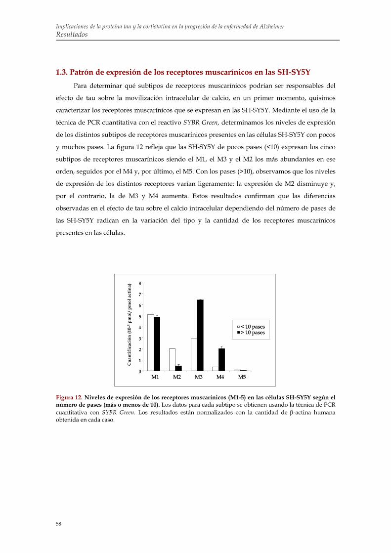
Implicaciones de la proteína tau y la cortistatina en la progresión de la enfermedad de Alzheimer Resultados
58
0
1
2
3
4
5
6
7
8
M1 M2 M3 M4 M5
Cua
ntif
icac
ión
(10-
4pm
ol/ p
mol
actin
a)
< 10 pases> 10 pases
0
1
2
3
4
5
6
7
8
M1 M2 M3 M4 M50
1
2
3
4
5
6
7
8
M1 M2 M3 M4 M5
Cua
ntif
icac
ión
(10-
4pm
ol/ p
mol
actin
a)
< 10 pases> 10 pases< 10 pases> 10 pases
1.3. Patrón de expresión de los receptores muscarínicos en las SH-SY5Y Para determinar qué subtipos de receptores muscarínicos podrían ser responsables del
efecto de tau sobre la movilización intracelular de calcio, en un primer momento, quisimos
caracterizar los receptores muscarínicos que se expresan en las SH-SY5Y. Mediante el uso de la
técnica de PCR cuantitativa con el reactivo SYBR Green, determinamos los niveles de expresión
de los distintos subtipos de receptores muscarínicos presentes en las células SH-SY5Y con pocos
y muchos pases. La figura 12 refleja que las SH-SY5Y de pocos pases (<10) expresan los cinco
subtipos de receptores muscarínicos siendo el M1, el M3 y el M2 los más abundantes en ese
orden, seguidos por el M4 y, por último, el M5. Con los pases (>10), observamos que los niveles
de expresión de los distintos receptores varían ligeramente: la expresión de M2 disminuye y,
por el contrario, la de M3 y M4 aumenta. Estos resultados confirman que las diferencias
observadas en el efecto de tau sobre el calcio intracelular dependiendo del número de pases de
las SH-SY5Y radican en la variación del tipo y la cantidad de los receptores muscarínicos
presentes en las células.
Figura 12. Niveles de expresión de los receptores muscarínicos (M1-5) en las células SH-SY5Y según el número de pases (más o menos de 10). Los datos para cada subtipo se obtienen usando la técnica de PCR cuantitativa con SYBR Green. Los resultados están normalizados con la cantidad de β-actina humana obtenida en cada caso.
Implicaciones de la proteína tau y la cortistatina en la progresión de la enfermedad de Alzheimer Resultados
59
1.4. Análisis farmacológico para determinar los receptores muscarínicos
implicados en el efecto de tau sobre los niveles de calcio intracelular en SH-
SY5Y En vista de que la movilización de calcio por tau parecía mediada por los receptores
muscarínicos, quisimos determinar cuáles eran los subtipos implicados mediante un estudio
farmacológico. En estos experimentos, usamos distintos antagonistas específicos de los
receptores muscarínicos: la pirencepina para bloquear el receptor M1 (Rosati et al., 1999), la
galamina para bloquear el M2 (Gnagey et al., 1999) y el 4-DAMP para bloquear el M3 (Fritz et
al., 2005). Los resultados obtenidos en estos estudios mostraron que, tanto la pirencepina (10
μM) como el 4-DAMP (2 μM), previenen la movilización intracelular de calcio en presencia de
tau (figura 13 A y C). Por el contrario, las incubaciones con galamina (2 μM) no parecen afectar
al efecto de tau sobre la desregulación de calcio (figura 13 B). Estos datos sugieren que los
receptores M1 y M3 median el efecto de tau sobre los niveles intracelulares de calcio.
1.5. Transfección transitoria con M1 y M3 en células COS-7 para confirmar la
implicación de estos receptores en la movilización de calcio inducida por tau Para confirmar las conclusiones de los estudios farmacológicos anteriores, realizamos un
nuevo abordaje experimental en células COS-7 transfectadas de forma transitoria con los
receptores muscarínicos M1 y M3.
Primero, quisimos comprobar si las células COS-7 eran un buen modelo celular para
estudiar el efecto de tau sobre los niveles de calcio. Usando la PCR cuantitativa con el reactivo
SYBR Green, comprobamos, que esta línea celular, en comparación con las SH-SY5Y, no expresa
los distintos subtipos de receptores muscarínicos (datos no mostrados). El siguiente paso fue
comprobar si las células COS-7 transfectadas de forma transitoria con M1 y M3 expresaban los
transgenes. Realizamos la transfección transitoria con plásmidos que contienen el cDNA de los
receptores muscarínicos humanos M1 o M3 unidos a un tag (3 x hemaglutinina o HA) en el
extremo amino-terminal. Como se muestra en la figura 14, usando el anticuerpo que reconoce el
HA, en western blot e inmunofluorescencia, comprobamos la expresión de M1 y M3 en las COS-7
transfectadas de forma transitoria con M1 y M3, respectivamente. Además, observamos,
mediante las dos técnicas, que las COS-7 control transfectadas con el vector vacío (pcDNA 3.1)
no presentan inmunorreactividad frente al anticuerpo HA.

Implicaciones de la proteína tau y la cortistatina en la progresión de la enfermedad de Alzheimer Resultados
60
C
4-DAMP
F 340
/F38
0
0 500 1000 1500 2000
0,4
0,6
0,8
1,0
1,2
1,4
1,6
1,8
2,0
2,2
F 340
/F38
0
0 500 1000 1500 2000
0,4
0,6
0,8
1,0
1,2
1,4
1,6
1,8
2,0
2,2
0 500 1000 1500 2000
0,4
0,6
0,8
1,0
1,2
1,4
1,6
1,8
2,0
2,2
0 500 1000 1500 2000
0,4
0,6
0,8
1,0
1,2
1,4
1,6
1,8
2,0
2,2
Tiempo (seg)
PirencepinaA
0 500 1000 1500 2000
0,4
0,6
0,8
1,0
1,2
1,4
1,6
1,8
2,0
2,2
F 340
/F38
0
ACh ACh AChTau Tau
0 500 1000 1500 2000
0,4
0,6
0,8
1,0
1,2
1,4
1,6
1,8
2,0
2,2
0 500 1000 1500 2000
0,4
0,6
0,8
1,0
1,2
1,4
1,6
1,8
2,0
2,2
0 500 1000 1500 2000
0,4
0,6
0,8
1,0
1,2
1,4
1,6
1,8
2,0
2,2
F 340
/F38
0
ACh ACh AChTau Tau
Tiempo (seg)
B
F 340
/F38
0
0 500 1000 1500 2000
0,4
0,6
0,8
1,0
1,2
1,4
1,6
1,8 ACh ACh AChTau Tau
Gallamine
F 340
/F38
0
0 500 1000 1500 2000
0,4
0,6
0,8
1,0
1,2
1,4
1,6
1,8 ACh ACh AChTau Tau
Gallamine
Tiempo (seg)
Galamina
ACh ACh AChTauTau
ACh ACh AChTauTau ACh ACh AChTauTau
Pirencepina
Tau
C
4-DAMP
F 340
/F38
0
0 500 1000 1500 2000
0,4
0,6
0,8
1,0
1,2
1,4
1,6
1,8
2,0
2,2
F 340
/F38
0
0 500 1000 1500 2000
0,4
0,6
0,8
1,0
1,2
1,4
1,6
1,8
2,0
2,2
0 500 1000 1500 2000
0,4
0,6
0,8
1,0
1,2
1,4
1,6
1,8
2,0
2,2
0 500 1000 1500 2000
0,4
0,6
0,8
1,0
1,2
1,4
1,6
1,8
2,0
2,2
Tiempo (seg)
PirencepinaA
0 500 1000 1500 2000
0,4
0,6
0,8
1,0
1,2
1,4
1,6
1,8
2,0
2,2
F 340
/F38
0
ACh ACh AChTau Tau
0 500 1000 1500 2000
0,4
0,6
0,8
1,0
1,2
1,4
1,6
1,8
2,0
2,2
0 500 1000 1500 2000
0,4
0,6
0,8
1,0
1,2
1,4
1,6
1,8
2,0
2,2
0 500 1000 1500 2000
0,4
0,6
0,8
1,0
1,2
1,4
1,6
1,8
2,0
2,2
F 340
/F38
0
ACh ACh AChTau Tau
Tiempo (seg)
B
F 340
/F38
0
0 500 1000 1500 2000
0,4
0,6
0,8
1,0
1,2
1,4
1,6
1,8 ACh ACh AChTau Tau
Gallamine
F 340
/F38
0
0 500 1000 1500 2000
0,4
0,6
0,8
1,0
1,2
1,4
1,6
1,8 ACh ACh AChTau Tau
Gallamine
Tiempo (seg)
Galamina
ACh ACh AChTauTauAChACh AChACh AChAChTauTauTauTau
AChACh AChACh AChAChTauTauTauTau ACh ACh AChTauTauAChACh AChACh AChAChTauTauTauTau
Pirencepina
TauTau
Figura 13. Identificación farmacológica de los receptores muscarínicos implicados en el efecto de tau sobre los niveles de calcio intracelular. Los datos mostrados representan los cambios en la fluorescencia de Fura-2 con el tiempo. En todos los experimentos, las SH-SY5Y, cargadas con Fura-2, se estimularon primero, con acetilcolina (50 μM) y tau (1 μM) y seguidamente, con tau (1 μM) y acetilcolina (50 μM) en presencia de distintos antagonistas para los receptores muscarínicos: pirencepina (antagonista de M1; 10 μM; A), galamina (de M2; 2 μM; B) y 4-DAMP (de M3; 2 μM; C). Finalmente, tras varios lavados, se añadió acetilcolina (50 μM) para confirmar la viabilidad de las células. Las líneas de la parte superior de la gráfica representan los periodos de estimulación y las barras de error, la desviación estándar. Los resultados presentados son la media de la respuesta de ≥ 20 células.
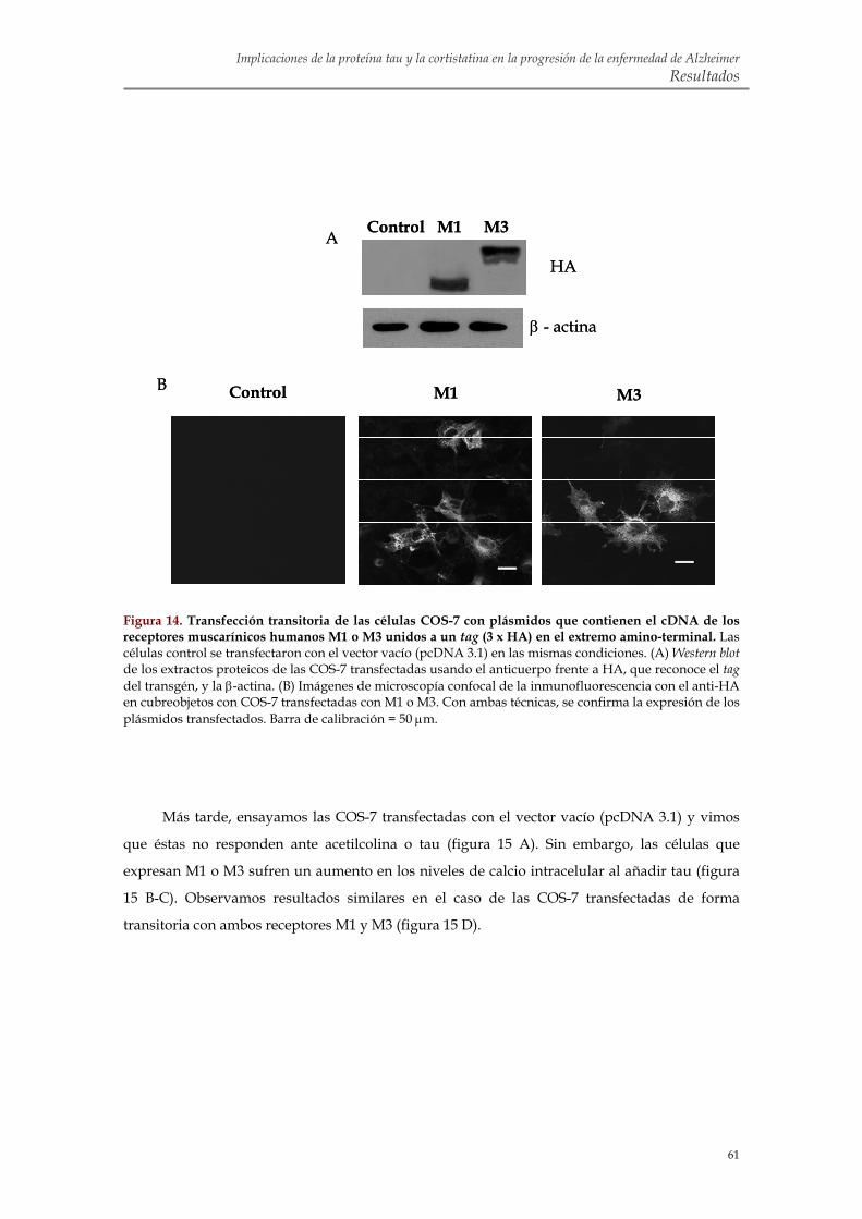
Implicaciones de la proteína tau y la cortistatina en la progresión de la enfermedad de Alzheimer Resultados
61
Control M1 M3B
A Control M1 M3
β - actina
HA
Control M1 M3B
A Control M1 M3
β - actina
HA
Figura 14. Transfección transitoria de las células COS-7 con plásmidos que contienen el cDNA de los receptores muscarínicos humanos M1 o M3 unidos a un tag (3 x HA) en el extremo amino-terminal. Las células control se transfectaron con el vector vacío (pcDNA 3.1) en las mismas condiciones. (A) Western blot de los extractos proteicos de las COS-7 transfectadas usando el anticuerpo frente a HA, que reconoce el tag del transgén, y la β-actina. (B) Imágenes de microscopía confocal de la inmunofluorescencia con el anti-HA en cubreobjetos con COS-7 transfectadas con M1 o M3. Con ambas técnicas, se confirma la expresión de los plásmidos transfectados. Barra de calibración = 50 μm.
Más tarde, ensayamos las COS-7 transfectadas con el vector vacío (pcDNA 3.1) y vimos
que éstas no responden ante acetilcolina o tau (figura 15 A). Sin embargo, las células que
expresan M1 o M3 sufren un aumento en los niveles de calcio intracelular al añadir tau (figura
15 B-C). Observamos resultados similares en el caso de las COS-7 transfectadas de forma
transitoria con ambos receptores M1 y M3 (figura 15 D).
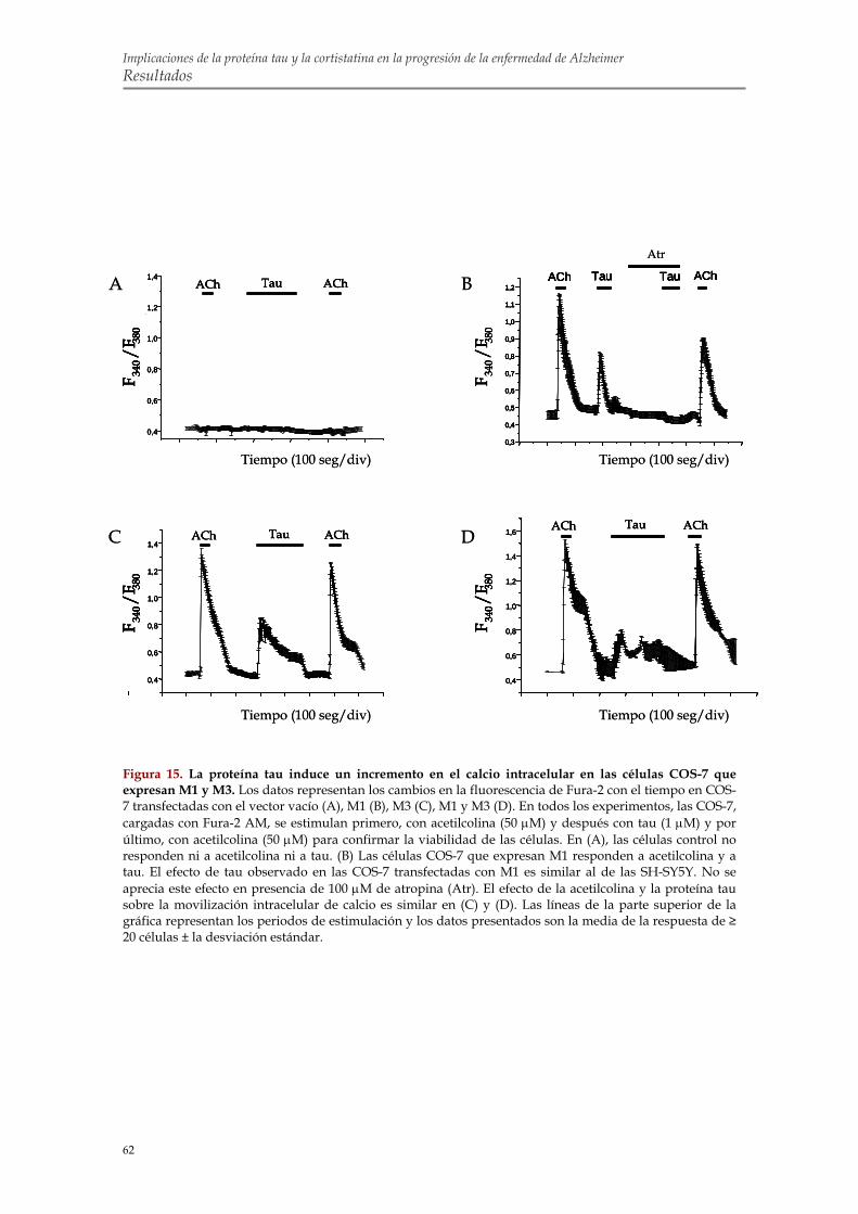
Implicaciones de la proteína tau y la cortistatina en la progresión de la enfermedad de Alzheimer Resultados
62
C
0,4
0,6
0,8
1,0
1,2
1,4ACh AChTau
0,4
0,6
0,8
1,0
1,2
1,4ACh AChTau D
0,4
0,6
0,8
1,0
1,2
1,4
1,6ACh AChTau
0,4
0,6
0,8
1,0
1,2
1,4
1,6ACh AChTau
0,3
0,4
0,5
0,6
0,7
0,8
0,9
1,0
1,1
1,2ACh AChTau Tau
Rat
io 3
40/3
80
0,3
0,4
0,5
0,6
0,7
0,8
0,9
1,0
1,1
1,2ACh AChTau Tau
Rat
io 3
40/3
80
0,3
0,4
0,5
0,6
0,7
0,8
0,9
1,0
1,1
1,2ACh AChTau Tau
Rat
io 3
40/3
80
B
Atr
A
A
0,4
0,6
0,8
1,0
1,2
1,4ACh AChTau
0,4
0,6
0,8
1,0
1,2
1,4ACh AChTau
F 340/F
380
F 340/F
380
Tiempo (100 seg/div)
F 340/F
380
F 340/F
380
F 340/F
380
F 340/F
380
F 340/F
380
F 340/F
380
Tiempo (100 seg/div)
Tiempo (100 seg/div)Tiempo (100 seg/div)
C
0,4
0,6
0,8
1,0
1,2
1,4ACh AChTau
0,4
0,6
0,8
1,0
1,2
1,4ACh AChTau D
0,4
0,6
0,8
1,0
1,2
1,4
1,6ACh AChTau
0,4
0,6
0,8
1,0
1,2
1,4
1,6ACh AChTau
0,3
0,4
0,5
0,6
0,7
0,8
0,9
1,0
1,1
1,2ACh AChTau Tau
Rat
io 3
40/3
80
0,3
0,4
0,5
0,6
0,7
0,8
0,9
1,0
1,1
1,2ACh AChTau Tau
Rat
io 3
40/3
80
0,3
0,4
0,5
0,6
0,7
0,8
0,9
1,0
1,1
1,2ACh AChTau Tau
Rat
io 3
40/3
80
B
Atr
A
A
0,4
0,6
0,8
1,0
1,2
1,4ACh AChTau
0,4
0,6
0,8
1,0
1,2
1,4ACh AChTau
F 340/F
380
F 340/F
380
F 340/F
380
F 340/F
380
Tiempo (100 seg/div)
F 340/F
380
F 340/F
380
F 340/F
380
F 340/F
380
F 340/F
380
F 340/F
380
F 340/F
380
F 340/F
380
F 340/F
380
F 340/F
380
F 340/F
380
F 340/F
380
Tiempo (100 seg/div)
Tiempo (100 seg/div)Tiempo (100 seg/div)
Figura 15. La proteína tau induce un incremento en el calcio intracelular en las células COS-7 que expresan M1 y M3. Los datos representan los cambios en la fluorescencia de Fura-2 con el tiempo en COS-7 transfectadas con el vector vacío (A), M1 (B), M3 (C), M1 y M3 (D). En todos los experimentos, las COS-7, cargadas con Fura-2 AM, se estimulan primero, con acetilcolina (50 μM) y después con tau (1 μM) y por último, con acetilcolina (50 μM) para confirmar la viabilidad de las células. En (A), las células control no responden ni a acetilcolina ni a tau. (B) Las células COS-7 que expresan M1 responden a acetilcolina y a tau. El efecto de tau observado en las COS-7 transfectadas con M1 es similar al de las SH-SY5Y. No se aprecia este efecto en presencia de 100 μM de atropina (Atr). El efecto de la acetilcolina y la proteína tau sobre la movilización intracelular de calcio es similar en (C) y (D). Las líneas de la parte superior de la gráfica representan los periodos de estimulación y los datos presentados son la media de la respuesta de ≥ 20 células ± la desviación estándar.

Implicaciones de la proteína tau y la cortistatina en la progresión de la enfermedad de Alzheimer Resultados
63
1.6. Identificación de la región de tau capaz de alterar la homeostasis
intracelular de calcio en células COS-7 que expresan M3 Para determinar qué región de la proteína tau está implicada en el aumento de la
concentración de calcio, incubamos las células COS-7 transfectadas transitoriamente con M3 en
presencia de tau y distintos fragmentos de tau (figura 16 A).
En este experimento, empleamos de nuevo la mayor isoforma de tau (compuesto por un
extremo amino-terminal, dos insertos amino-terminales (N), cuatro repeticiones de unión a
microtúbulos (R) y una zona carboxilo-terminal (C)) y los fragmentos de tau siguientes: 2R
(contiene la primera y tercera repetición), 2N (compuesto por la mitad amino-terminal de tau
incluyendo los dos insertos), 3R (con la primera, tercera y cuarta repetición) y 3RC (con la
primera, tercera y cuarta repetición y el extremo carboxilo-terminal de tau) (Perez et al., 1996).
De este modo, observamos que el fragmento de tau 3RC es el que induce una mayor
respuesta en la movilización de calcio intracelular de las células que expresan M3 (figura 16 B).
Estos datos sugieren que la zona carboxilo-terminal es responsable del efecto de tau sobre la
movilización de calcio.
Para acotar la zona de la proteína tau implicada en tal efecto, utilizamos un péptido de
tau que comprende la secuencia de residuos de 391 a 407 situados en la región carboxilo-
terminal. En este caso, comprobamos que las células responden a este péptido incrementando la
concentración de calcio en su interior (figura 16 C), confirmando, así, que esta zona de la
proteína tau es suficiente para inducir el efecto de tau observado sobre los niveles de calcio.
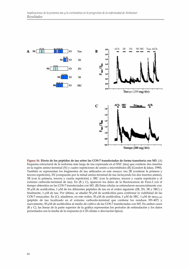
Implicaciones de la proteína tau y la cortistatina en la progresión de la enfermedad de Alzheimer Resultados
64
A B
C
Tiempo (200 seg/div)0,6
0,8
1,0
1,2
1,4
1,6
1,8
ACh ACh3RC tau393-423
0,6
0,8
1,0
1,2
1,4
1,6
1,8
ACh ACh3RC tau393-423
F 340/F
380
F 340/F
380
0,4
0,6
0,8
1,0
1,2
1,4
1,6
1,8
2,0 ACh AChTau3RC3R2N2R
Time (250 sec/div)
F 340
/F38
0
0,4
0,6
0,8
1,0
1,2
1,4
1,6
1,8
2,0 ACh AChTau3RC3R2N2R
Time (250 sec/div)
F 340
/F38
0
Tiempo (250 seg/div)
F 340/F
380
F 340/F
380
2R 2NACh 3R 3RC Tau AChTauNt Ct
R1 R2 R3 R4 N1 N2
2NCtNtN1 N2
Nt Ct 3RCNt
3RNt CtR1 R3 R4
2RNt CtR1 R2
R1 R3 R4
A B
C
Tiempo (200 seg/div)0,6
0,8
1,0
1,2
1,4
1,6
1,8
ACh ACh3RC tau393-423
0,6
0,8
1,0
1,2
1,4
1,6
1,8
ACh ACh3RC tau393-423
F 340/F
380
F 340/F
380
Tiempo (200 seg/div)0,6
0,8
1,0
1,2
1,4
1,6
1,8
ACh ACh3RC tau393-423
0,6
0,8
1,0
1,2
1,4
1,6
1,8
ACh ACh3RC tau393-423
F 340/F
380
F 340/F
380
F 340/F
380
F 340/F
380
0,4
0,6
0,8
1,0
1,2
1,4
1,6
1,8
2,0 ACh AChTau3RC3R2N2R
Time (250 sec/div)
F 340
/F38
0
0,4
0,6
0,8
1,0
1,2
1,4
1,6
1,8
2,0 ACh AChTau3RC3R2N2R
Time (250 sec/div)
F 340
/F38
0
Tiempo (250 seg/div)
F 340/F
380
F 340/F
380
2R 2NACh 3R 3RC Tau ACh
0,4
0,6
0,8
1,0
1,2
1,4
1,6
1,8
2,0 ACh AChTau3RC3R2N2R
Time (250 sec/div)
F 340
/F38
0
0,4
0,6
0,8
1,0
1,2
1,4
1,6
1,8
2,0 ACh AChTau3RC3R2N2R
Time (250 sec/div)
F 340
/F38
0
Tiempo (250 seg/div)
F 340/F
380
F 340/F
380
F 340/F
380
F 340/F
380
2R 2NACh 3R 3RC Tau AChTauNt Ct
R1 R2 R3 R4 N1 N2
2NCtNtN1 N2
Nt Ct 3RCNt
3RNt CtR1 R3 R4
2RNt CtR1 R2
R1 R3 R4
Figura 16. Efecto de los péptidos de tau sobre las COS-7 transfectadas de forma transitoria con M3. (A) Esquema estructural de la isoforma más larga de tau expresada en el SNC (tau) que contiene dos insertos en la región amino-terminal (N) y cuatro repeticiones de unión a microtúbulos (R) (Goedert & Jakes, 1990). También se representan los fragmentos de tau utilizados en este ensayo: tau 2R (contiene la primera y tercera repetición), 2N (compuesto por la mitad amino-terminal de tau incluyendo los dos insertos amino), 3R (con la primera, tercera y cuarta repetición) y 3RC (con la primera, tercera y cuarta repetición y el extremo carboxilo-terminal de tau). En (B y C), aparecen los datos de la fluorescencia de Fura-2 con el tiempo obtenidos en las COS-7 transfectadas con M3. (B) Estas células se estimularon secuencialmente con: 50 μM de acetilcolina, 1 μM de los diferentes péptidos de tau en el orden siguiente (2R, 2N, 3R y 3RC) y finalmente, 1 μM de tau. Por último, se añadió 50 μM de acetilcolina para confirmar la viabilidad de las COS-7 ensayadas. En (C), añadimos, en este orden, 50 μM de acetilcolina, 1 μM de 3RC, 1 μM de tau391-407 (péptido de tau localizado en el extremo carboxilo-terminal que contiene los residuos 391-407) y nuevamente, 50 μM de acetilcolina al medio de cultivo de las COS-7 transfectadas con M3. En ambos casos (B y C), las líneas de la parte superior de la gráfica representan los periodos de estimulación y los datos presentados son la media de la respuesta (n ≥ 20 células ± desviación típica).

Implicaciones de la proteína tau y la cortistatina en la progresión de la enfermedad de Alzheimer Resultados
65
1.7. El calcio intracelular de las neuronas murinas de hipocampo en cultivo
aumenta en presencia de la proteína tau Con el fin de corroborar nuestros resultados en un modelo celular más cercano al entorno
fisiológico del cerebro, estudiamos el efecto de tau y de la acetilcolina sobre la concentración de
calcio en cultivos de neuronas de hipocampo de 1, 2 o 7 días in vitro (DIV).
En los cultivos de 1 DIV, menos de un 20% de las células responden a la acetilcolina (50
μM) o a tau (1 μM). Sin embargo, tras 3 DIV, y, sobre todo tras 7 DIV, la proporción de células
sensibles a la acetilcolina y a tau aumenta (figura 17 A-B).
También observamos que la proporción de neuronas que expresan M1, detectadas por
inmunofluorescencia, aumenta con el número de DIV (figura 17 D).
La respuesta de tau se prolonga más en el tiempo que la de acetilcolina, y, quizá ahí
radique su mayor toxicidad para la célula. Como en los experimentos anteriores realizados con
las células SH-SY5Y y COS-7, el efecto característico de tau sobre el calcio intracelular
desaparece cuando incubamos previamente las células con 100 μM de atropina (figura 17 C).
Por último, comprobamos que las neuronas eran funcionales puesto que respondían a 60
μM de KCl, un agente despolarizante, añadido al cultivo. Resultados similares se observaron en
cultivos de neuronas corticales murinas (datos no mostrados).

Implicaciones de la proteína tau y la cortistatina en la progresión de la enfermedad de Alzheimer Resultados
66
F34
0/F 38
0
0,03
ACh Tau
Tiempo 100 seg
A
DIV 1
DIV 3
DIV 7
F34
0/F 38
0
ACh
D
DIV 1 DIV 3 DIV 7
0,08
0,10
0,12
0,14
0,16
0,18
Tiempo (100 seg/div)
ACh AChTau Tau K+
Atropina
0,08
0,10
0,12
0,14
0,16
0,18ACh AChTau K+
C
B
ACh TAU0
20
40
60
80
100
DIV1DIV3DIV7
Neu
rona
s hi
poca
mpa
les
(%)
ACh TAU0
20
40
60
80
100
0
20
40
60
80
100
DIV1DIV3DIV7
ACh TAU0
20
40
60
80
100
0
20
40
60
80
100
DIV1DIV3DIV7
ACh TAU0
20
40
60
80
100
0
20
40
60
80
100
DIV1DIV3DIV7
F34
0/F 38
0
0,03
ACh Tau
Tiempo 100 seg
A
DIV 1
DIV 3
DIV 7
F34
0/F 38
0
ACh
D
DIV 1 DIV 3 DIV 7
0,08
0,10
0,12
0,14
0,16
0,18
Tiempo (100 seg/div)
ACh AChTau Tau K+
Atropina
0,08
0,10
0,12
0,14
0,16
0,18ACh AChTau K+
C
B
ACh TAU0
20
40
60
80
100
DIV1DIV3DIV7
Neu
rona
s hi
poca
mpa
les
(%)
ACh TAU0
20
40
60
80
100
0
20
40
60
80
100
DIV1DIV3DIV7
ACh TAU0
20
40
60
80
100
0
20
40
60
80
100
DIV1DIV3DIV7
ACh TAU0
20
40
60
80
100
0
20
40
60
80
100
DIV1DIV3DIV7
B
ACh TAU0
20
40
60
80
100
DIV1DIV3DIV7
Neu
rona
s hi
poca
mpa
les
(%)
ACh TAU0
20
40
60
80
100
0
20
40
60
80
100
DIV1DIV3DIV7
ACh TAU0
20
40
60
80
100
0
20
40
60
80
100
DIV1DIV3DIV7
ACh TAU0
20
40
60
80
100
0
20
40
60
80
100
DIV1DIV3DIV7
F 340
/F38
0
F34
0/F 38
0
0,03
ACh Tau
Tiempo 100 seg
A
DIV 1
DIV 3
DIV 7
F34
0/F 38
0
ACh
D
DIV 1 DIV 3 DIV 7
0,08
0,10
0,12
0,14
0,16
0,18
Tiempo (100 seg/div)
ACh AChTau Tau K+
Atropina
0,08
0,10
0,12
0,14
0,16
0,18ACh AChTau K+
C
B
ACh TAU0
20
40
60
80
100
DIV1DIV3DIV7
Neu
rona
s hi
poca
mpa
les
(%)
ACh TAU0
20
40
60
80
100
0
20
40
60
80
100
DIV1DIV3DIV7
ACh TAU0
20
40
60
80
100
0
20
40
60
80
100
DIV1DIV3DIV7
ACh TAU0
20
40
60
80
100
0
20
40
60
80
100
DIV1DIV3DIV7
F34
0/F 38
0
0,03
ACh Tau
Tiempo 100 seg
A
DIV 1
DIV 3
DIV 7
F34
0/F 38
0
ACh
D
DIV 1 DIV 3 DIV 7
0,08
0,10
0,12
0,14
0,16
0,18
Tiempo (100 seg/div)
ACh AChTau Tau K+
Atropina
0,08
0,10
0,12
0,14
0,16
0,18ACh AChTau K+
C
B
ACh TAU0
20
40
60
80
100
DIV1DIV3DIV7
Neu
rona
s hi
poca
mpa
les
(%)
ACh TAU0
20
40
60
80
100
0
20
40
60
80
100
DIV1DIV3DIV7
ACh TAU0
20
40
60
80
100
0
20
40
60
80
100
DIV1DIV3DIV7
ACh TAU0
20
40
60
80
100
0
20
40
60
80
100
DIV1DIV3DIV7
B
ACh TAU0
20
40
60
80
100
DIV1DIV3DIV7
Neu
rona
s hi
poca
mpa
les
(%)
ACh TAU0
20
40
60
80
100
0
20
40
60
80
100
DIV1DIV3DIV7
ACh TAU0
20
40
60
80
100
0
20
40
60
80
100
DIV1DIV3DIV7
ACh TAU0
20
40
60
80
100
0
20
40
60
80
100
DIV1DIV3DIV7
F 340
/F38
0
Figura 17. La proteína tau altera la homeostasis de calcio de neuronas de hipocampo en cultivos primarios. (A) Datos de la fluorescencia de Fura-2 en células hipocampales de 1, 3 y 7 DIV tras la estimulación secuencial con acetilcolina (50 μM) y tau (1 μM). Las imágenes representan la respuesta de las neuronas frente a tau. En (B), se muestra el porcentaje de neuronas de hipocampo que responden a acetilcolina (50 μM) o a tau (1 μM) tras 1, 3 o 7 DIV. (C) Cambios en la fluorescencia de Fura-2 en las neuronas de 3 DIV estimuladas con acetilcolina (50 μM) y tau (1 μM) en presencia o ausencia de 100 μM de Atropina. Por último, las neuronas se despolarizaron con KCl (60 μM) para confirmar su viabilidad. Los periodos de estimulación se representan como líneas en la parte superior de la gráfica. Los datos presentados son la media de la respuesta de ≥ 20 células. (D) Inmunofluorescencia con el anticuerpo que reconoce el receptor M1 en las neuronas de hipocampo fijadas a 1, 3 o 7 DIV.
Implicaciones de la proteína tau y la cortistatina en la progresión de la enfermedad de Alzheimer Resultados
67
2. EFECTO DE DISTINTOS AGONISTAS DE LOS RECEPTORES DE
SOMATOSTATINA (sst) SOBRE LA FOSFORILACIÓN DE TAU EN LA
Ser262
2.1. El nivel de fosforilación de la Ser262 de tau es menor en la corteza de los
ratones que no expresan cortistatina La aparición de la patología de tau en la corteza cerebral de los pacientes con EA
representa un momento crítico en el desarrollo de la demencia (Delacourte et al., 1999). La
fosforilación de tau localizada en la corteza cerebral puede deberse a la presencia de un
neuropéptido cortical conocido como cortistatina. Para estudiar esta posibilidad, comparamos
los niveles de fosforilación de tau en los ratones modificados genéticamente que no expresan
cortistatina (CST -/-) y en los ratones control. Como vimos, aunque los sitios de fosforilación de
tau se localizan, en su mayoría, en la región rica en prolinas y carboxilo-terminal, la
fosforilación de los sitios situados en la región de unión a los microtúbulos afecta en mayor
grado a la unión de tau a los microtúbulos (Cho & Johnson, 2003; Drewes et al., 1995; Sengupta
et al., 1998; Singh et al., 1996). De entre estos sitios, por el momento, sólo se ha demostrado la
relevancia biológica de la fosforilación de la Ser262, por tanto, nuestro estudio se ha centrado en
este residuo de tau (Biernat et al., 1993; Drewes et al., 1997; Morishima-Kawashima et al., 1995;
Perez et al., 2005). Si se observa la figura 18 puede comprobarse que los niveles de fosforilación
de tau en la Ser262 en los extractos de corteza de los ratones control son unas dos veces mayores
a los obtenidos de la corteza de los ratones CST -/- de misma edad (p <0.05). Del mismo modo,
analizamos la fosforilación de tau en la Ser262, epítopo reconocido por el anticuerpo 12E8, en
los extractos obtenidos a partir del resto de tejido cerebral (sin corteza ni hipocampo) de los
animales anteriores. En este caso, sin embargo, no detectamos diferencias significativas entre los
ratones control y los CST -/- (figura 18). Tampoco encontramos diferencias significativas al
analizar la fosforilación de tau en el hipocampo (datos no mostrados). Estos datos sugieren que
la cortistatina parece modular la fosforilación de tau en la Ser262 específicamente en la corteza
siendo estas neuronas dianas para la cortistatina. También analizamos mediante western blot
estos mismos extractos proteicos usando, esta vez, el anticuerpo PHF1 que reconoce las Ser396 y
Ser404 fosforiladas de tau. En este caso, no encontramos diferencias significativas (datos no
mostrados).
Teniendo en cuenta que la edad representa el mayor factor de riesgo de la EA (Jicha et al.,
2008; Jorm & Jolley, 1998; Kawas & Corrada, 2006; Kukull & Bowen, 2002), quisimos comprobar
si los niveles de fosforilación de tau en la Ser262 varían con la edad en los ratones. Para ello,
estudiamos los niveles de fosforilación de tau en la Ser262 utilizando el anticuerpo 12E8 en las

Implicaciones de la proteína tau y la cortistatina en la progresión de la enfermedad de Alzheimer Resultados
68
12E8
7.51
control CST -/- control CST -/-
corteza - corteza
*
0
50
100
150
200
corteza -corteza
12E8
/ 7.5
1 (%
Con
trol
)
controlCST -/-
*
12E8
7.51
control CST -/- control CST -/-
corteza - corteza
12E8
7.51
control CST -/- control CST -/-
corteza - corteza
12E8
7.51
control CST -/- control CST -/-
corteza - corteza
*
0
50
100
150
200
corteza -corteza
12E8
/ 7.5
1 (%
Con
trol
)
controlCST -/-
**
0
50
100
150
200
corteza -corteza
12E8
/ 7.5
1 (%
Con
trol
)
controlCST -/-
*
0
50
100
150
200
corteza -corteza
12E8
/ 7.5
1 (%
Con
trol
)
controlCST -/-
*
cortezas de animales control y CST -/- de distintas edades (1, 6, 12 y 18 meses). Observamos
que la fosforilación de tau en la Ser262 aumenta con la edad en las cortezas de los ratones
control (figura 19). En cambio, este aumento no se aprecia cuando se analiza la fosforilación de
la Ser262 de tau en los extractos de corteza de los ratones que no expresan cortistatina.
Posteriormente, realizamos el test de comportamiento del reconocimiento de objeto a los
ratones control y CST -/- de más de un año de edad. Este sencillo test permite estudiar si existe
deterioro de la memoria declarativa (Winters et al., 2008). El reconocimiento de nuevos objetos
es una tarea difícil para muchos pacientes con enfermedades neurodegenerativas (Hajilou &
Done, 2007; Holdstock, 2005; Laatu et al., 2003; Lee et al., 2003; Purdy et al., 2002). En nuestro
trabajo, comprobamos que los ratones control y CST -/- de más de un año de edad no presentan
diferencias significativas en el test de reconocimiento de objeto (n = 9 control, n = 11 CST-/-;
datos no mostrados).
.
Figura 18. La fosforilación de tau en la Ser262 es menor en la corteza de ratones CST -/-. Se realizaron extractos proteicos a partir de la corteza y del resto de tejido cerebral, sin corteza ni hipocampo (-corteza), de ratones control y CST -/- de 3 meses de edad. Los datos presentados se corresponden con los western blots (panel superior) y la cuantificación por densitometría (panel inferior) de la fosforilación de tau en la Ser262 (usando el anticuerpo 12E8) normalizada con los niveles de proteína tau total (anticuerpo 7.51). Los datos mostrados son las medias en unidades arbitrarias ± la desviación típica (n = 3). *p<0.05 comparados con los datos control.

Implicaciones de la proteína tau y la cortistatina en la progresión de la enfermedad de Alzheimer Resultados
69
Figura 19. La fosforilación de tau en la Ser262 aumenta con la edad en las cortezas de los ratones control pero no en las de los ratones CST -/- . Se obtuvieron extractos proteicos de la corteza cerebral de ratones control y CST -/- de distintas edades (1, 6, 12 y 18 meses). Posteriormente, se analizaron por western blot (panel superior) utilizando los anticuerpos que detectan la Ser262 fosforilada de tau (12E8) y el tau total (7.51). Las gráficas representan la cuantificación por densitometría en unidades arbitrarias de la fosforilación de tau en la Ser262. Se normalizaron los niveles de fosforilación de tau en la Ser262 con la cantidad de tau total. El panel de la derecha muestra los resultados obtenidos con los extractos de los ratones control y, el de la izquierda, los de los ratones CST -/-. Los datos representan las medias ± la desviación típica (n = 2-4). *p<0.05 comparados con los ratones de 1 mes de edad.
7.51
cont
rol
CST
-/-
CST
-/-
CST
-/-
CST
-/-
cont
rol
cont
rol
cont
rol
1 mes 6 meses 12 meses 18 meses
12E8
0
100
200
300
400
500
600
700
800
1 6 12 18
Edad de los ratones control (en meses)
12E8
/ 7.5
1 (%
1 m
es)
*
**
0
100
200
300
400
500
600
700
800
1 6 12 18
Edad de los ratones CST -/- (meses)
12E8
/ 7.5
1 (%
1 m
es)
*
7.51
cont
rol
CST
-/-
CST
-/-
CST
-/-
CST
-/-
cont
rol
cont
rol
cont
rol
1 mes 6 meses 12 meses 18 meses
12E8
7.51
cont
rol
CST
-/-
CST
-/-
CST
-/-
CST
-/-
cont
rol
cont
rol
cont
rol
1 mes 6 meses 12 meses 18 meses
12E8
0
100
200
300
400
500
600
700
800
1 6 12 18
Edad de los ratones control (en meses)
12E8
/ 7.5
1 (%
1 m
es)
*
**
0
100
200
300
400
500
600
700
800
1 6 12 18
Edad de los ratones CST -/- (meses)
12E8
/ 7.5
1 (%
1 m
es)
*0
100
200
300
400
500
600
700
800
1 6 12 18
Edad de los ratones control (en meses)
12E8
/ 7.5
1 (%
1 m
es)
*
**
0
100
200
300
400
500
600
700
800
1 6 12 18
Edad de los ratones control (en meses)
12E8
/ 7.5
1 (%
1 m
es)
*
**
0
100
200
300
400
500
600
700
800
1 6 12 18
Edad de los ratones CST -/- (meses)
12E8
/ 7.5
1 (%
1 m
es)
*0
100
200
300
400
500
600
700
800
1 6 12 18
Edad de los ratones CST -/- (meses)
12E8
/ 7.5
1 (%
1 m
es)
*

Implicaciones de la proteína tau y la cortistatina en la progresión de la enfermedad de Alzheimer Resultados
70
2.2. La cortistatina promueve la fosforilación de tau en la Ser262 de cultivos
primarios de neuronas corticales murinas Para corroborar in vitro los resultados mencionados anteriormente obtenidos a partir de
experimentos in vivo, añadimos a cultivos primarios de neuronas corticales murinas 50 nM de
cortistatina durante distintos tiempos de incubación (5, 15 y 30 min). Más tarde, analizamos
mediante la técnica de western blot los niveles de fosforilación de tau en los distintos extractos
proteicos usando los anticuerpos frente a tau siguientes: el 12E8 que detecta la fosfoSer262 y el
PHF1 que reconoce ambas Ser396 y Ser404 fosforiladas. Observamos que las incubaciones de 30
minutos con cortistatina aumentan de forma significativa, unas dos veces respecto al control, los
niveles de fosforilación de la proteína tau en la Ser262 (p<0.01; figura 20 A).
En vista de que los niveles de fosforilación de los controles eran sorprendentemente
elevados, quisimos comprobar si algún factor presente en el medio del cultivo celular podía
inducir un aumento en la fosforilación de la Ser262 de tau. Para ello, aumentamos el tiempo de
incubación de las células corticales en presencia de medio neurobasal (NB) sin suero pasando
de estar 2 horas (como en el experimento anterior) a estar 6 horas. Esta vez observamos que
efectivamente los niveles de fosforilación de la proteína tau en la Ser262 reconocidos por el
anticuerpo 12E8 disminuyen cuando la incubación en NB es más larga (figura 20 B).
En este caso, repetimos el tratamiento anterior de 50 nM de cortistatina, pero esta vez en
células incubadas previamente en NB durante 6 horas y, de este modo, confirmamos
nuevamente que la cortistatina induce un aumento significativo en los niveles de fosforilación
de la proteína tau en la Ser262 y observamos que este aumento es algo mayor al del
experimento anterior en el que las células estuvieron 2 horas en presencia de NB (p<0.05; figura
20 C).
Comparamos las células tratadas con cortistatina con las tratadas con el vehículo
mediante el uso de inmunofluorescencia. Usando el anticuerpo 334, que reconoce la isoforma β3
neuronal de la tubulina, comprobamos que el aumento de la fosforilación de tau en la Ser262
inducido por la cortistatina no es suficiente para producir un cambio morfológico aparente en
las células (figura 20 D). Tanto en las células tratadas como en las tratadas con el vehiculo
encontramos neuronas con prolongaciones largas y otras con extensiones de menor longitud.
Tampoco observamos diferencias al analizar los cubre-objetos tratados y controles mediante
inmunofluorescencia con el anticuerpo 12E8 que reconoce la Ser262 fosforilada de tau (datos no
mostrados).
También analizamos por western blot los mismos ensayos de 50 nM de cortistatina usando
el anticuerpo PHF1 que reconoce las Ser396/404 fosforiladas. Sin embargo, en este caso, no se
encontraron diferencias en los niveles de fosforilación de este epítopo (datos no mostrados).
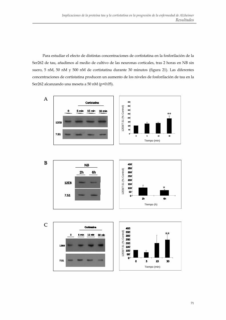
Implicaciones de la proteína tau y la cortistatina en la progresión de la enfermedad de Alzheimer Resultados
71
B
A
B
Cortistatina
0 5 min 15 min 30 min
12E8
7.51
Cortistatina
0 5 min 15 min 30 min
12E8
7.51
Cortistatina
0 5 min 15 min 30 min
12E8
7.51
Cortistatina
0 5 min 15 min 30 min
12E8
7.51
Cortistatina
0 5 min 15 min 30 min
12E8
7.51
Cortistatina
0 5 min 15 min 30 min
12E8
7.51
Cortistatina
0 5 min 15 min 30 min
12E8
7.51
Cortistatina
0 5 min 15 min 30 min
12E8
7.51
Cortistatina
0 5 min 15 min 30 min
12E8
7.51
Cortistatina
0 5 min 15 min 30 min
12E8
7.51
Cortistatina
0 5 min 15 min 30 min
12E8
7.51
Cortistatina
0 5 min 15 min 30 min
12E8
7.51
Cortistatina
0 5 min 15 min 30 min
12E8
7.51
Cortistatina
0 5 min 15 min 30 min
12E8
7.51
Cortistatina
0 5 min 15 min 30 min
12E8
7.51
2h 6h
NB
12E8
7.51
2h 6h
NB
12E8
7.51
2h 6h
NB
12E8
7.51
2h 6h
NB
12E8
7.51
2h 6h
NB
12E8
7.51
2h 6h
NB
12E8
7.51
2h 6h
NB
12E8
7.51
C
050
100150200250300350400
0 5 15 30Tiempo (min)
12E8
/ 7.5
1 (%
Con
trol
) **
050
100150200250300350400
0 5 15 30Tiempo (min)
12E8
/ 7.5
1 (%
Con
trol
)
050
100150200250300350400
0 5 15 30Tiempo (min)
12E8
/ 7.5
1 (%
Con
trol
) **
050
100150200250300350400
0 5 15 30Tiempo (min)
12E8
/ 7.5
1 (%
Con
trol
) **
050
100150200250300350400
0 5 15 30Tiempo (min)
12E8
/ 7.5
1 (%
Con
trol
)
050
100150200250300350400
0 5 15 30Tiempo (min)
12E8
/ 7.5
1 (%
Con
trol
) **
050
100150200250300350400
0 5 15 30Tiempo (min)
12E8
/ 7.5
1 (%
Con
trol
)
050
100150200250300350400
0 5 15 30Tiempo (min)
12E8
/ 7.5
1 (%
Con
trol
)
050
100150200250300350400
0 5 15 30Tiempo (min)
12E8
/ 7.5
1 (%
Con
trol
) **
Tiempo (min)
12E8
/ 7.5
1 (%
Con
trol
)
Tiempo (min)
12E8
/ 7.5
1 (%
Con
trol
)
Tiempo (min)
12E8
/ 7.5
1 (%
Con
trol
)
Tiempo (min)
12E8
/ 7.5
1 (%
Con
trol
)
Tiempo (min)
12E8
/ 7.5
1 (%
Con
trol
)
Tiempo (min)
12E8
/ 7.5
1 (%
Con
trol
)
050
100150200250300350400
0 5 15 30Tiempo (min)
12E8
/ 7.5
1 (%
Con
trol
)
050
100150200250300350400
0 5 15 30Tiempo (min)
12E8
/ 7.5
1 (%
Con
trol
)
050
100150200250300350400
0 5 15 30Tiempo (min)
12E8
/ 7.5
1 (%
Con
trol
) **
Tiempo (min)
12E8
/ 7.5
1 (%
Con
trol
)
Tiempo (min)
12E8
/ 7.5
1 (%
Con
trol
)
Tiempo (min)
12E8
/ 7.5
1 (%
Con
trol
)
Tiempo (min)
12E8
/ 7.5
1 (%
Con
trol
)
Tiempo (min)
12E8
/ 7.5
1 (%
Con
trol
) **
050
100150200250300350400
0 5 15 300
50100150200250300350400
0 5 15 30
**
050
100150200250300350400
0 5 15 300
50100150200250300350400
0 5 15 30
**
050
100150200250300350400
0 5 15 300
50100150200250300350400
0 5 15 30
****
050
100150200250300350400
0 5 15 300
50100150200250300350400
0 5 15 30
**
050
100150200250300350400
0 5 15 300
50100150200250300350400
0 5 15 30
**
050
100150200250300350400
0 5 15 300
50100150200250300350400
0 5 15 30
****
050
100150200250300350400
0 5 15 300
50100150200250300350400
0 5 15 30
**
050
100150200250300350400
0 5 15 300
50100150200250300350400
0 5 15 30
**
050
100150200250300350400
0 5 15 300
50100150200250300350400
0 5 15 30
****
050
100150200250300350400
0 5 15 300
50100150200250300350400
0 5 15 30
**
050
100150200250300350400
0 5 15 300
50100150200250300350400
0 5 15 30
**
050
100150200250300350400
0 5 15 300
50100150200250300350400
0 5 15 30
**
12E
8/7.
51 (%
Con
trol)
Tiempo (min)
0
50
100
150
200
250
300
350
400
0 5 15 30
Tiempo (min)
0
50
100
150
200
250
300
350
400
0 5 15 30
Tiempo (min)Tiempo (min)Tiempo (min)
**
0
50
100
150
200
250
300
350
400
0 5 15 300
50
100
150
200
250
300
350
400
0 5 15 30
**12
E8/
7.51
(% C
ontro
l)
0
50
100
150
200
250
300
350
400
0 5 15 30
Tiempo (min)
0
50
100
150
200
250
300
350
400
0 5 15 30
Tiempo (min)
0
50
100
150
200
250
300
350
400
0 5 15 30
Tiempo (min)
0
50
100
150
200
250
300
350
400
0 5 15 30
Tiempo (min)Tiempo (min)Tiempo (min)
**
0
50
100
150
200
250
300
350
400
0 5 15 300
50
100
150
200
250
300
350
400
0 5 15 30
****
0
50
100
150
200
250
300
350
400
0 5 15 300
50
100
150
200
250
300
350
400
0 5 15 30
**12
E8/
7.51
(% C
ontro
l)
0
50
100
150
200
250
300
350
400
2h 6h
12E8
/ 7.5
1 (%
Con
trol
)
*0
50
100
150
200
250
300
350
400
2h 6h
12E8
/ 7.5
1 (%
Con
trol
)
0
50
100
150
200
250
300
350
400
2h 6h
12E8
/ 7.5
1 (%
Con
trol
)
0
50
100
150
200
250
300
350
400
2h 6h
12E8
/ 7.5
1 (%
Con
trol
)12
E8/ 7
.51
(% C
ontr
ol)
12E8
/ 7.5
1 (%
Con
trol
)12
E8/ 7
.51
(% C
ontr
ol)
12E8
/ 7.5
1 (%
Con
trol
)12
E8/ 7
.51
(% C
ontr
ol)
***0
50
100
150
200
250300
350
400
2h 6h0
50
100
150
200
250300
350
400
2h 6h
*0
50
100
150
200
250300
350
400
2h 6h0
50
100
150
200
250300
350
400
2h 6h
*0
50
100
150
200
250300
350
400
2h 6h0
50
100
150
200
250300
350
400
2h 6h
*
12E8
/7.5
1 (%
Con
trol)
Tiempo (h)0
50
100
150
200
250
300
350
400
2h 6h
12E8
/ 7.5
1 (%
Con
trol
)
*0
50
100
150
200
250
300
350
400
2h 6h
12E8
/ 7.5
1 (%
Con
trol
)
0
50
100
150
200
250
300
350
400
2h 6h
12E8
/ 7.5
1 (%
Con
trol
)
*0
50
100
150
200
250
300
350
400
2h 6h
12E8
/ 7.5
1 (%
Con
trol
)
0
50
100
150
200
250
300
350
400
2h 6h
12E8
/ 7.5
1 (%
Con
trol
)
0
50
100
150
200
250
300
350
400
2h 6h
12E8
/ 7.5
1 (%
Con
trol
)
0
50
100
150
200
250
300
350
400
2h 6h
12E8
/ 7.5
1 (%
Con
trol
)
0
50
100
150
200
250
300
350
400
2h 6h
12E8
/ 7.5
1 (%
Con
trol
)
0
50
100
150
200
250
300
350
400
2h 6h
12E8
/ 7.5
1 (%
Con
trol
)12
E8/ 7
.51
(% C
ontr
ol)
12E8
/ 7.5
1 (%
Con
trol
)12
E8/ 7
.51
(% C
ontr
ol)
12E8
/ 7.5
1 (%
Con
trol
)12
E8/ 7
.51
(% C
ontr
ol)
***0
50
100
150
200
250300
350
400
2h 6h0
50
100
150
200
250300
350
400
2h 6h
*0
50
100
150
200
250300
350
400
2h 6h0
50
100
150
200
250300
350
400
2h 6h
*0
50
100
150
200
250300
350
400
2h 6h0
50
100
150
200
250300
350
400
2h 6h
****0
50
100
150
200
250300
350
400
2h 6h0
50
100
150
200
250300
350
400
2h 6h
*0
50
100
150
200
250300
350
400
2h 6h0
50
100
150
200
250300
350
400
2h 6h
*0
50
100
150
200
250300
350
400
2h 6h0
50
100
150
200
250300
350
400
2h 6h
*
12E8
/7.5
1 (%
Con
trol)
Tiempo (h)
B
A
B
Cortistatina
0 5 min 15 min 30 min
12E8
7.51
Cortistatina
0 5 min 15 min 30 min
12E8
7.51
Cortistatina
0 5 min 15 min 30 min
12E8
7.51
Cortistatina
0 5 min 15 min 30 min
12E8
7.51
Cortistatina
0 5 min 15 min 30 min
12E8
7.51
Cortistatina
0 5 min 15 min 30 min
12E8
7.51
Cortistatina
0 5 min 15 min 30 min
12E8
7.51
Cortistatina
0 5 min 15 min 30 min
12E8
7.51
Cortistatina
0 5 min 15 min 30 min
12E8
7.51
Cortistatina
0 5 min 15 min 30 min
12E8
7.51
Cortistatina
0 5 min 15 min 30 min
12E8
7.51
Cortistatina
0 5 min 15 min 30 min
12E8
7.51
Cortistatina
0 5 min 15 min 30 min
12E8
7.51
Cortistatina
0 5 min 15 min 30 min
12E8
7.51
Cortistatina
0 5 min 15 min 30 min
12E8
7.51
Cortistatina
0 5 min 15 min 30 min
12E8
7.51
Cortistatina
0 5 min 15 min 30 min
12E8
7.51
Cortistatina
0 5 min 15 min 30 min
12E8
7.51
Cortistatina
0 5 min 15 min 30 min
12E8
7.51
2h 6h
NB
12E8
7.51
2h 6h
NB
12E8
7.51
2h 6h
NB
12E8
7.51
2h 6h
NB
12E8
7.51
2h 6h
NB
12E8
7.51
2h 6h
NB
12E8
7.51
2h 6h
NB
12E8
7.51
C
050
100150200250300350400
0 5 15 30Tiempo (min)
12E8
/ 7.5
1 (%
Con
trol
) **
050
100150200250300350400
0 5 15 30Tiempo (min)
12E8
/ 7.5
1 (%
Con
trol
)
050
100150200250300350400
0 5 15 30Tiempo (min)
12E8
/ 7.5
1 (%
Con
trol
) **
050
100150200250300350400
0 5 15 30Tiempo (min)
12E8
/ 7.5
1 (%
Con
trol
) **
050
100150200250300350400
0 5 15 30Tiempo (min)
12E8
/ 7.5
1 (%
Con
trol
)
050
100150200250300350400
0 5 15 30Tiempo (min)
12E8
/ 7.5
1 (%
Con
trol
) **
050
100150200250300350400
0 5 15 30Tiempo (min)
12E8
/ 7.5
1 (%
Con
trol
)
050
100150200250300350400
0 5 15 30Tiempo (min)
12E8
/ 7.5
1 (%
Con
trol
)
050
100150200250300350400
0 5 15 30Tiempo (min)
12E8
/ 7.5
1 (%
Con
trol
) **
Tiempo (min)
12E8
/ 7.5
1 (%
Con
trol
)
Tiempo (min)
12E8
/ 7.5
1 (%
Con
trol
)
Tiempo (min)
12E8
/ 7.5
1 (%
Con
trol
)
Tiempo (min)
12E8
/ 7.5
1 (%
Con
trol
)
Tiempo (min)
12E8
/ 7.5
1 (%
Con
trol
)
Tiempo (min)
12E8
/ 7.5
1 (%
Con
trol
)
050
100150200250300350400
0 5 15 30Tiempo (min)
12E8
/ 7.5
1 (%
Con
trol
)
050
100150200250300350400
0 5 15 30Tiempo (min)
12E8
/ 7.5
1 (%
Con
trol
)
050
100150200250300350400
0 5 15 30Tiempo (min)
12E8
/ 7.5
1 (%
Con
trol
) **
Tiempo (min)
12E8
/ 7.5
1 (%
Con
trol
)
Tiempo (min)
12E8
/ 7.5
1 (%
Con
trol
)
Tiempo (min)
12E8
/ 7.5
1 (%
Con
trol
)
Tiempo (min)
12E8
/ 7.5
1 (%
Con
trol
)
Tiempo (min)
12E8
/ 7.5
1 (%
Con
trol
) **
050
100150200250300350400
0 5 15 300
50100150200250300350400
0 5 15 30
**
050
100150200250300350400
0 5 15 300
50100150200250300350400
0 5 15 30
**
050
100150200250300350400
0 5 15 300
50100150200250300350400
0 5 15 30
****
050
100150200250300350400
0 5 15 300
50100150200250300350400
0 5 15 30
**
050
100150200250300350400
0 5 15 300
50100150200250300350400
0 5 15 30
**
050
100150200250300350400
0 5 15 300
50100150200250300350400
0 5 15 30
****
050
100150200250300350400
0 5 15 300
50100150200250300350400
0 5 15 30
**
050
100150200250300350400
0 5 15 300
50100150200250300350400
0 5 15 30
**
050
100150200250300350400
0 5 15 300
50100150200250300350400
0 5 15 30
****
050
100150200250300350400
0 5 15 300
50100150200250300350400
0 5 15 30
**
050
100150200250300350400
0 5 15 300
50100150200250300350400
0 5 15 30
**
050
100150200250300350400
0 5 15 300
50100150200250300350400
0 5 15 30
**
12E
8/7.
51 (%
Con
trol)
Tiempo (min)0
50100150200250300350400
0 5 15 30Tiempo (min)
12E8
/ 7.5
1 (%
Con
trol
) **
050
100150200250300350400
0 5 15 30Tiempo (min)
12E8
/ 7.5
1 (%
Con
trol
)
050
100150200250300350400
0 5 15 30Tiempo (min)
12E8
/ 7.5
1 (%
Con
trol
) **
050
100150200250300350400
0 5 15 30Tiempo (min)
12E8
/ 7.5
1 (%
Con
trol
)
050
100150200250300350400
0 5 15 30Tiempo (min)
12E8
/ 7.5
1 (%
Con
trol
) **
050
100150200250300350400
0 5 15 30Tiempo (min)
12E8
/ 7.5
1 (%
Con
trol
)
050
100150200250300350400
0 5 15 30Tiempo (min)
12E8
/ 7.5
1 (%
Con
trol
) **
050
100150200250300350400
0 5 15 30Tiempo (min)
12E8
/ 7.5
1 (%
Con
trol
)
050
100150200250300350400
0 5 15 30Tiempo (min)
12E8
/ 7.5
1 (%
Con
trol
) **
050
100150200250300350400
0 5 15 30Tiempo (min)
12E8
/ 7.5
1 (%
Con
trol
) **
050
100150200250300350400
0 5 15 30Tiempo (min)
12E8
/ 7.5
1 (%
Con
trol
)
050
100150200250300350400
0 5 15 30Tiempo (min)
12E8
/ 7.5
1 (%
Con
trol
) **
050
100150200250300350400
0 5 15 30Tiempo (min)
12E8
/ 7.5
1 (%
Con
trol
)
050
100150200250300350400
0 5 15 30Tiempo (min)
12E8
/ 7.5
1 (%
Con
trol
) **
050
100150200250300350400
0 5 15 30Tiempo (min)
12E8
/ 7.5
1 (%
Con
trol
)
050
100150200250300350400
0 5 15 30Tiempo (min)
12E8
/ 7.5
1 (%
Con
trol
) **
050
100150200250300350400
0 5 15 30Tiempo (min)
12E8
/ 7.5
1 (%
Con
trol
)
050
100150200250300350400
0 5 15 30Tiempo (min)
12E8
/ 7.5
1 (%
Con
trol
) **
050
100150200250300350400
0 5 15 30Tiempo (min)
12E8
/ 7.5
1 (%
Con
trol
)
050
100150200250300350400
0 5 15 30Tiempo (min)
12E8
/ 7.5
1 (%
Con
trol
)
050
100150200250300350400
0 5 15 30Tiempo (min)
12E8
/ 7.5
1 (%
Con
trol
)
050
100150200250300350400
0 5 15 30Tiempo (min)
12E8
/ 7.5
1 (%
Con
trol
) **
050
100150200250300350400
0 5 15 30Tiempo (min)
12E8
/ 7.5
1 (%
Con
trol
)
050
100150200250300350400
0 5 15 30Tiempo (min)
12E8
/ 7.5
1 (%
Con
trol
) **
Tiempo (min)
12E8
/ 7.5
1 (%
Con
trol
)
Tiempo (min)
12E8
/ 7.5
1 (%
Con
trol
)
Tiempo (min)
12E8
/ 7.5
1 (%
Con
trol
)
Tiempo (min)
12E8
/ 7.5
1 (%
Con
trol
)
Tiempo (min)
12E8
/ 7.5
1 (%
Con
trol
)
Tiempo (min)
12E8
/ 7.5
1 (%
Con
trol
)
050
100150200250300350400
0 5 15 30Tiempo (min)
12E8
/ 7.5
1 (%
Con
trol
)
050
100150200250300350400
0 5 15 30Tiempo (min)
12E8
/ 7.5
1 (%
Con
trol
)
050
100150200250300350400
0 5 15 30Tiempo (min)
12E8
/ 7.5
1 (%
Con
trol
)
050
100150200250300350400
0 5 15 30Tiempo (min)
12E8
/ 7.5
1 (%
Con
trol
) **
050
100150200250300350400
0 5 15 30Tiempo (min)
12E8
/ 7.5
1 (%
Con
trol
)
050
100150200250300350400
0 5 15 30Tiempo (min)
12E8
/ 7.5
1 (%
Con
trol
) **
Tiempo (min)
12E8
/ 7.5
1 (%
Con
trol
)
Tiempo (min)
12E8
/ 7.5
1 (%
Con
trol
)
Tiempo (min)
12E8
/ 7.5
1 (%
Con
trol
)
Tiempo (min)
12E8
/ 7.5
1 (%
Con
trol
)
Tiempo (min)
12E8
/ 7.5
1 (%
Con
trol
) **
050
100150200250300350400
0 5 15 300
50100150200250300350400
0 5 15 30
**
050
100150200250300350400
0 5 15 300
50100150200250300350400
0 5 15 30
**
050
100150200250300350400
0 5 15 300
50100150200250300350400
0 5 15 30
****
050
100150200250300350400
0 5 15 300
50100150200250300350400
0 5 15 30
**
050
100150200250300350400
0 5 15 300
50100150200250300350400
0 5 15 30
**
050
100150200250300350400
0 5 15 300
50100150200250300350400
0 5 15 30
****
050
100150200250300350400
0 5 15 300
50100150200250300350400
0 5 15 30
**
050
100150200250300350400
0 5 15 300
50100150200250300350400
0 5 15 30
**
050
100150200250300350400
0 5 15 300
50100150200250300350400
0 5 15 30
****
050
100150200250300350400
0 5 15 300
50100150200250300350400
0 5 15 30
**
050
100150200250300350400
0 5 15 300
50100150200250300350400
0 5 15 30
**
050
100150200250300350400
0 5 15 300
50100150200250300350400
0 5 15 30
**
12E
8/7.
51 (%
Con
trol)
Tiempo (min)
0
50
100
150
200
250
300
350
400
0 5 15 30
Tiempo (min)
0
50
100
150
200
250
300
350
400
0 5 15 30
Tiempo (min)Tiempo (min)Tiempo (min)
**
0
50
100
150
200
250
300
350
400
0 5 15 300
50
100
150
200
250
300
350
400
0 5 15 30
**12
E8/
7.51
(% C
ontro
l)
0
50
100
150
200
250
300
350
400
0 5 15 30
Tiempo (min)
0
50
100
150
200
250
300
350
400
0 5 15 30
Tiempo (min)
0
50
100
150
200
250
300
350
400
0 5 15 30
Tiempo (min)
0
50
100
150
200
250
300
350
400
0 5 15 30
Tiempo (min)Tiempo (min)Tiempo (min)
**
0
50
100
150
200
250
300
350
400
0 5 15 300
50
100
150
200
250
300
350
400
0 5 15 30
****
0
50
100
150
200
250
300
350
400
0 5 15 300
50
100
150
200
250
300
350
400
0 5 15 30
**12
E8/
7.51
(% C
ontro
l)
0
50
100
150
200
250
300
350
400
2h 6h
12E8
/ 7.5
1 (%
Con
trol
)
*0
50
100
150
200
250
300
350
400
2h 6h
12E8
/ 7.5
1 (%
Con
trol
)
0
50
100
150
200
250
300
350
400
2h 6h
12E8
/ 7.5
1 (%
Con
trol
)
0
50
100
150
200
250
300
350
400
2h 6h
12E8
/ 7.5
1 (%
Con
trol
)12
E8/ 7
.51
(% C
ontr
ol)
12E8
/ 7.5
1 (%
Con
trol
)12
E8/ 7
.51
(% C
ontr
ol)
12E8
/ 7.5
1 (%
Con
trol
)12
E8/ 7
.51
(% C
ontr
ol)
***0
50
100
150
200
250300
350
400
2h 6h0
50
100
150
200
250300
350
400
2h 6h
*0
50
100
150
200
250300
350
400
2h 6h0
50
100
150
200
250300
350
400
2h 6h
*0
50
100
150
200
250300
350
400
2h 6h0
50
100
150
200
250300
350
400
2h 6h
*
12E8
/7.5
1 (%
Con
trol)
Tiempo (h)0
50
100
150
200
250
300
350
400
2h 6h
12E8
/ 7.5
1 (%
Con
trol
)
*0
50
100
150
200
250
300
350
400
2h 6h
12E8
/ 7.5
1 (%
Con
trol
)
0
50
100
150
200
250
300
350
400
2h 6h
12E8
/ 7.5
1 (%
Con
trol
)
*0
50
100
150
200
250
300
350
400
2h 6h
12E8
/ 7.5
1 (%
Con
trol
)
0
50
100
150
200
250
300
350
400
2h 6h
12E8
/ 7.5
1 (%
Con
trol
)
0
50
100
150
200
250
300
350
400
2h 6h
12E8
/ 7.5
1 (%
Con
trol
)
0
50
100
150
200
250
300
350
400
2h 6h
12E8
/ 7.5
1 (%
Con
trol
)
0
50
100
150
200
250
300
350
400
2h 6h
12E8
/ 7.5
1 (%
Con
trol
)
0
50
100
150
200
250
300
350
400
2h 6h
12E8
/ 7.5
1 (%
Con
trol
)12
E8/ 7
.51
(% C
ontr
ol)
12E8
/ 7.5
1 (%
Con
trol
)12
E8/ 7
.51
(% C
ontr
ol)
12E8
/ 7.5
1 (%
Con
trol
)12
E8/ 7
.51
(% C
ontr
ol)
***0
50
100
150
200
250300
350
400
2h 6h0
50
100
150
200
250300
350
400
2h 6h
*0
50
100
150
200
250300
350
400
2h 6h0
50
100
150
200
250300
350
400
2h 6h
*0
50
100
150
200
250300
350
400
2h 6h0
50
100
150
200
250300
350
400
2h 6h
****0
50
100
150
200
250300
350
400
2h 6h0
50
100
150
200
250300
350
400
2h 6h
*0
50
100
150
200
250300
350
400
2h 6h0
50
100
150
200
250300
350
400
2h 6h
*0
50
100
150
200
250300
350
400
2h 6h0
50
100
150
200
250300
350
400
2h 6h
*
12E8
/7.5
1 (%
Con
trol)
Tiempo (h)
Para estudiar el efecto de distintas concentraciones de cortistatina en la fosforilación de la
Ser262 de tau, añadimos al medio de cultivo de las neuronas corticales, tras 2 horas en NB sin
suero, 5 nM, 50 nM y 500 nM de cortistatina durante 30 minutos (figura 21). Las diferentes
concentraciones de cortistatina producen un aumento de los niveles de fosforilación de tau en la
Ser262 alcanzando una meseta a 50 nM (p<0.05).
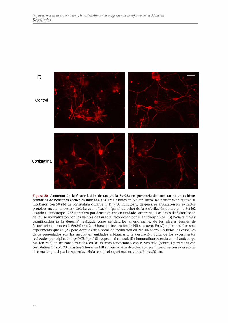
Implicaciones de la proteína tau y la cortistatina en la progresión de la enfermedad de Alzheimer Resultados
72
D
Cortistatina
Control
D
Cortistatina
Control
Cortistatina
Control
Figura 20. Aumento de la fosforilación de tau en la Ser262 en presencia de cortistatina en cultivos primarios de neuronas corticales murinas. (A) Tras 2 horas en NB sin suero, las neuronas en cultivo se incubaron con 50 nM de cortistatina durante 5, 15 y 30 minutos y, después, se analizaron los extractos proteicos mediante western blot. La cuantificación (panel derecho) de la fosforilación de tau en la Ser262 usando el anticuerpo 12E8 se realizó por densitometría en unidades arbitrarias. Los datos de fosforilación de tau se normalizaron con los valores de tau total reconocido por el anticuerpo 7.51. (B) Western blots y cuantificación (a la derecha) realizada como se describe anteriormente, de los niveles basales de fosforilación de tau en la Ser262 tras 2 o 6 horas de incubación en NB sin suero. En (C) repetimos el mismo experimento que en (A) pero después de 6 horas de incubación en NB sin suero. En todos los casos, los datos presentados son las medias en unidades arbitrarias ± la desviación típica de los experimentos realizados por triplicado. *p<0.05, **p<0.01 respecto al control. (D) Inmunofluorescencia con el anticuerpo 334 (en rojo) en neuronas tratadas, en las mismas condiciones, con el vehículo (control) y tratadas con cortistatina (50 nM, 30 min) tras 2 horas en NB sin suero. A la derecha, aparecen neuronas con extensiones de corta longitud y, a la izquierda, células con prolongaciones mayores. Barra, 50 μm.
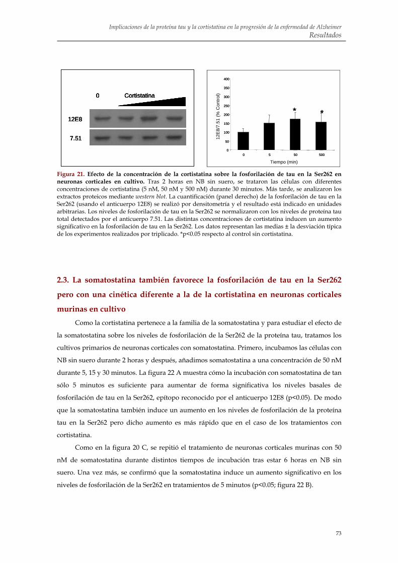
Implicaciones de la proteína tau y la cortistatina en la progresión de la enfermedad de Alzheimer Resultados
73
Figura 21. Efecto de la concentración de la cortistatina sobre la fosforilación de tau en la Ser262 en neuronas corticales en cultivo. Tras 2 horas en NB sin suero, se trataron las células con diferentes concentraciones de cortistatina (5 nM, 50 nM y 500 nM) durante 30 minutos. Más tarde, se analizaron los extractos proteicos mediante western blot. La cuantificación (panel derecho) de la fosforilación de tau en la Ser262 (usando el anticuerpo 12E8) se realizó por densitometría y el resultado está indicado en unidades arbitrarias. Los niveles de fosforilación de tau en la Ser262 se normalizaron con los niveles de proteína tau total detectados por el anticuerpo 7.51. Las distintas concentraciones de cortistatina inducen un aumento significativo en la fosforilación de tau en la Ser262. Los datos representan las medias ± la desviación típica de los experimentos realizados por triplicado. *p<0.05 respecto al control sin cortistatina.
2.3. La somatostatina también favorece la fosforilación de tau en la Ser262
pero con una cinética diferente a la de la cortistatina en neuronas corticales
murinas en cultivo Como la cortistatina pertenece a la familia de la somatostatina y para estudiar el efecto de
la somatostatina sobre los niveles de fosforilación de la Ser262 de la proteína tau, tratamos los
cultivos primarios de neuronas corticales con somatostatina. Primero, incubamos las células con
NB sin suero durante 2 horas y después, añadimos somatostatina a una concentración de 50 nM
durante 5, 15 y 30 minutos. La figura 22 A muestra cómo la incubación con somatostatina de tan
sólo 5 minutos es suficiente para aumentar de forma significativa los niveles basales de
fosforilación de tau en la Ser262, epítopo reconocido por el anticuerpo 12E8 (p<0.05). De modo
que la somatostatina también induce un aumento en los niveles de fosforilación de la proteína
tau en la Ser262 pero dicho aumento es más rápido que en el caso de los tratamientos con
cortistatina.
Como en la figura 20 C, se repitió el tratamiento de neuronas corticales murinas con 50
nM de somatostatina durante distintos tiempos de incubación tras estar 6 horas en NB sin
suero. Una vez más, se confirmó que la somatostatina induce un aumento significativo en los
niveles de fosforilación de la Ser262 en tratamientos de 5 minutos (p<0.05; figura 22 B).
12E8
7.51
0 Cortistatina
12E8
7.51
0 Cortistatina
0
50
100
150
200
250
300
350
400
0 5 50 500
* ** ** ** *
12E
8/7.
51 (%
Con
trol)
Tiempo (min)
12E8
7.51
0 Cortistatina
12E8
7.51
0 Cortistatina
0
50
100
150
200
250
300
350
400
0 5 50 500
* ** ** ** *
0
50
100
150
200
250
300
350
400
0 5 50 500
* ** ** ** *
12E
8/7.
51 (%
Con
trol)
Tiempo (min)

Implicaciones de la proteína tau y la cortistatina en la progresión de la enfermedad de Alzheimer Resultados
74
B
A Somatostatina
0 5 min 15 min 30 min
12E8
7.51
Somatostatina
0 5 min 15 min 30 min
12E8
7.51
0
50
100
150
200
250
300
350
400
0 5 15 30
Tiempo (min)
12E8
/ 7.5
1 (%
Con
trol
)
**
0
50
100
150
200
250
300
350
400
0 5 15 30
Tiempo (min)
12E8
/ 7.5
1 (%
Con
trol
)
**
050
100150200250300350400
0 5 15 30
Tiempo (min)
12E8
/ 7.5
1 (%
Con
trol
) **
050
100150200250300350400
0 5 15 30
Tiempo (min)
12E8
/ 7.5
1 (%
Con
trol
) **
B
A Somatostatina
0 5 min 15 min 30 min
12E8
7.51
Somatostatina
0 5 min 15 min 30 min
12E8
7.51
Somatostatina
0 5 min 15 min 30 min
12E8
7.51
Somatostatina
0 5 min 15 min 30 min
12E8
7.51
0
50
100
150
200
250
300
350
400
0 5 15 30
Tiempo (min)
12E8
/ 7.5
1 (%
Con
trol
)
**
0
50
100
150
200
250
300
350
400
0 5 15 30
Tiempo (min)
12E8
/ 7.5
1 (%
Con
trol
)
**
050
100150200250300350400
0 5 15 30
Tiempo (min)
12E8
/ 7.5
1 (%
Con
trol
) **
050
100150200250300350400
0 5 15 30
Tiempo (min)
12E8
/ 7.5
1 (%
Con
trol
) **
Además, comprobamos los niveles de fosforilación de tau en las Ser396/404 usando el
anticuerpo PHF1. En este caso, como en los tratamientos con cortistatina, los tratamientos con
somatostatina no parecían afectar a los niveles de fosforilación de la proteína tau en las
Ser396/404 (datos no mostrados).
Figura 22. La somatostatina induce un aumento en la fosforilación de tau en la Ser262 en cultivos primarios de neuronas corticales murinas. (A) Después de 2 horas en NB sin suero, se incubaron las neuronas en cultivo con 50 nM de somatostatina durante 5, 15 y 30 minutos. Se realizaron western blots de los extractos proteicos correspondientes usando los anticuerpos 12E8 (reconoce la Ser262 fosforilada de tau) y 7.51 (tau total). Los datos presentados en unidades arbitrarias representan la cuantificación por densitometría de los western blots anteriores. Los niveles de fosforilación de tau en la Ser262 se normalizaron con los de tau total. Aparecen las medias de los datos por triplicado en unidades arbitrarias ± la desviación típica. (B) Como en (A) pero la incubación en presencia de medio NB sin suero fue de 6 horas. *p<0.05 respecto al control sin somatostatina.
2.4. El octreotide no afecta a la fosforilación de tau en la Ser262 de neuronas
corticales murinas en cultivo Para continuar con el estudio de los distintos agonistas de los receptores de
somatostatina, nos interesamos por el efecto del octreotide, un octapéptido sintético análogo de
la somatostatina que se une preferentemente a sst2, sst3 y sst5, sobre la fosforilación de tau en la
Ser262. De forma similar a los ensayos realizados con cortistatina y somatostatina (figuras 20 A
y 22 A), incubamos los cultivos primarios de neuronas corticales en NB sin suero durante 2
horas y añadimos distintos tratamientos con 50 nM de octreotide durante 5, 15 y 30 minutos.

Implicaciones de la proteína tau y la cortistatina en la progresión de la enfermedad de Alzheimer Resultados
75
A
B Octreotide (nM)
0 5 50 500100
12E8
7.51
12E8
7.51
Octreotide
0 5 min 30 min15 min
0
50
100
150
200
250
300
350
400
0 5 15 30
Tiempo (min)
12E8
/ 7.5
1 (%
Con
trol)
0
50
100
150
200
250
300
350
400
0 5 15 30
Tiempo (min)
12E8
/ 7.5
1 (%
Con
trol)
0
50
100
150
200
250
300
350
400
0 5 50 100 500
OCT (nM)
12E8
/ 7.5
1 (%
Con
trol
)
Octreotide (nM)
0
50
100
150
200
250
300
350
400
0 5 50 100 500
OCT (nM)
12E8
/ 7.5
1 (%
Con
trol
)
Octreotide (nM)
A
B Octreotide (nM)
0 5 50 500100
12E8
7.51
12E8
7.51
Octreotide
0 5 min 30 min15 min
12E8
7.51
Octreotide
0 5 min 30 min15 min
0
50
100
150
200
250
300
350
400
0 5 15 30
Tiempo (min)
12E8
/ 7.5
1 (%
Con
trol)
0
50
100
150
200
250
300
350
400
0 5 15 30
Tiempo (min)
12E8
/ 7.5
1 (%
Con
trol)
0
50
100
150
200
250
300
350
400
0 5 50 100 500
OCT (nM)
12E8
/ 7.5
1 (%
Con
trol
)
Octreotide (nM)
0
50
100
150
200
250
300
350
400
0 5 50 100 500
OCT (nM)
12E8
/ 7.5
1 (%
Con
trol
)
Octreotide (nM)
Los resultados sugieren que el octreotide no produce un efecto significativo sobre la
fosforilación de tau en la Ser262 usando el anticuerpo 12E8 (figura 23 A).
Con objeto de analizar el efecto de distintas concentraciones de octreotide sobre la
fosforilación de tau en la Ser262, tratamos los cultivos primarios de neuronas corticales murinas
previamente incubadas con NB sin suero (2 h) con 5 nM, 50 nM, 100 nM y 500 nM de octreotide
durante 30 minutos. Una vez más, no observamos un efecto significativo del octreotide sobre la
fosforilación de tau en la Ser262 en ninguna de las condiciones experimentales anteriores (figura
23 B).
Figura 23. El octreotide no afecta a la fosforilación de tau en la Ser262 en neuronas corticales en cultivo. (A) Se incubaron las células en NB sin suero (2h) y se añadió 50 nM de octreotide durante 5, 15 y 30 minutos. Los extractos proteicos se analizaron mediante western blot utilizando los anticuerpos frente a tau que reconocen la fosfoSer262 (anticuerpo 12E8) y tau total (anticuerpo 7.51). Los datos obtenidos representan la cuantificación en unidades arbitrarias (panel de la derecha) por densitometría de los niveles de fosforilación de tau en la Ser262 normalizados con los de tau total (n = 3 ± la desviación típica). (B) En otro experimento, y tras 2 horas en NB sin suero, se añadió octreotide a las neuronas en distintas concentraciones (5 nM, 50 nM, 100 nM y 500 nM) durante 30 minutos. La cuantificación (a la derecha) de los niveles de inmunorreactividad obtenidos en los western blots se realizó como en (A).

Implicaciones de la proteína tau y la cortistatina en la progresión de la enfermedad de Alzheimer Resultados
76
2.5. Las neuronas corticales murinas en cultivo presentan receptores que unen
cortistatina Para comprobar si el efecto de la cortistatina sobre la fosforilación de tau en la Ser262 está
mediado por su unión a receptores específicos, estudiamos la unión y la internalización de un
compuesto de cortistatina acoplada a fluoresceína (Fluo-cortistatina) en cultivos primarios de
neuronas corticales murinas.
En un principio, observamos que algunas de las neuronas incubadas en presencia de
Fluo-cortistatina (1-5 μM) durante 30 minutos presentaban un intenso marcaje punteado
fluorescente en el citoplasma del soma neuronal indicando que se había producido una
internalización de los receptores unidos a cortistatina (figura 24 A). Para determinar el número
de células que mostraban internalización, realizamos una inmunofluorescencia de los cubre-
objetos usando el anticuerpo policlonal 334 que reconoce una isoforma de tubulina neuronal (la
β3) y DAPI para teñir los núcleos. El 24 ± 8% de las células tratadas con Fluo-cortistatina (5 μM,
30 min) mostraban internalización (figura 24 A).
Sin embargo, si se observa la figura 24 B, parece que una corta incubación de 1 minuto de
duración con Fluo-cortistatina no es suficiente para evidenciar un marcaje fluorescente en el
interior neuronal. La unión y la internalización de la Fluo-cortistatina se evita de forma
significativa en presencia de un exceso de cortistatina o somatostatina (100 μM, 10 min; p<0.05).
Estos resultados parecen sugerir que la unión de Fluo-cortistatina está mediada por receptores
de somatostatina.
En la figura 24 C, se muestra que la internalización de la Fluo-cortistatina también se ve
inhibida cuando se pre-incuban las células con PAO (1 μM, 30 min), un potente inhibidor de la
endocitosis. Por el contrario, observamos internalización de la Fluo-cortistatina al realizar, como
control, el experimento anterior añadiendo a las células el mismo volumen de DMSO, el
vehículo donde diluimos el PAO (datos no mostrados). Estos datos sugieren que la
internalización de la Fluo-cortistatina en las neuronas corticales murinas en cultivo está
mediada por la formación de vesículas de clatrina.
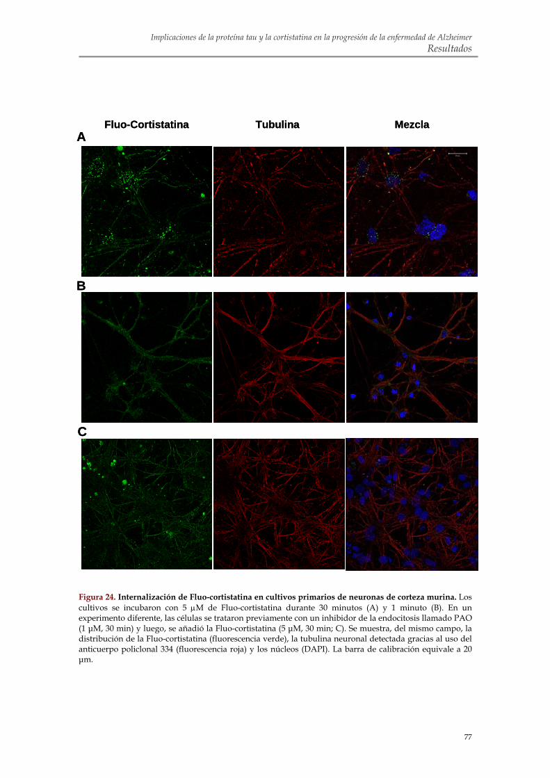
Implicaciones de la proteína tau y la cortistatina en la progresión de la enfermedad de Alzheimer Resultados
77
MezclaFluo-Cortistatina Tubulina A
C
B
MezclaFluo-Cortistatina Tubulina A
C
B
Figura 24. Internalización de Fluo-cortistatina en cultivos primarios de neuronas de corteza murina. Los cultivos se incubaron con 5 μM de Fluo-cortistatina durante 30 minutos (A) y 1 minuto (B). En un experimento diferente, las células se trataron previamente con un inhibidor de la endocitosis llamado PAO (1 μM, 30 min) y luego, se añadió la Fluo-cortistatina (5 μM, 30 min; C). Se muestra, del mismo campo, la distribución de la Fluo-cortistatina (fluorescencia verde), la tubulina neuronal detectada gracias al uso del anticuerpo policlonal 334 (fluorescencia roja) y los núcleos (DAPI). La barra de calibración equivale a 20 μm.

Implicaciones de la proteína tau y la cortistatina en la progresión de la enfermedad de Alzheimer Resultados
78
2.6. Las neuronas corticales en cultivo expresan los cinco subtipos de
receptores de somatostatina Como se ha visto en los experimentos anteriores, la somatostatina es capaz de evitar la
unión de la Fluo-cortistatina a los receptores presentes en las neuronas corticales en cultivo,
parece pues lógico pensar que la cortistatina se une a los receptores de somatostatina de estas
neuronas. De modo que para determinar qué subtipo de receptor de somatostatina es
responsable de la unión de cortistatina en las neuronas, realizamos una PCR cuantitativa
específica para cada subtipo de receptor de somatostatina (sst1-5). Utilizando el reactivo de
PCR SYBR Green, como viene descrito en el apartado de materiales y métodos, obtuvimos una
curva de amplificación para cada subtipo de receptor de somatostatina. Los datos normalizados
con los obtenidos a raíz de la amplificación de la β-actina determinaron el nivel de expresión de
los genes de interés.
Los resultados de la PCR cuantitativa, resumidos en la figura 25, muestran que las
neuronas corticales murinas expresan transcritos para los cinco subtipos de receptores de
somatostatina. El mRNA del receptor sst2 es el más abundante (aproximadamente diez veces
más que los mRNA de sst1 y sst4) seguido por el mRNA de sst1 y el de sst4. La cantidad de
receptor sst3 expresada en las neuronas corticales es menos relevante (representa alrededor de
un 1% respecto al de sst2). Finalmente, el receptor sst5 es el menos expresado puesto que sólo se
detecta después de 30 ciclos de amplificación (menos de un 0.1% respecto a sst3).
Como ya se ha indicado, la unión de cortistatina a los receptores celulares resulta en su
internalización (figura 24), de modo que parece improbable que el receptor sst1 medie este
efecto puesto que está descrito que la tasa de internalización de este receptor es baja (Hofland &
Lamberts, 2003). Dejando a un lado este receptor y centrándonos en los más abundantes,
quisimos comprobar si los receptores sst2 y sst4 eran responsables del efecto de cortistatina
sobre la fosforilación de tau en la Ser262.
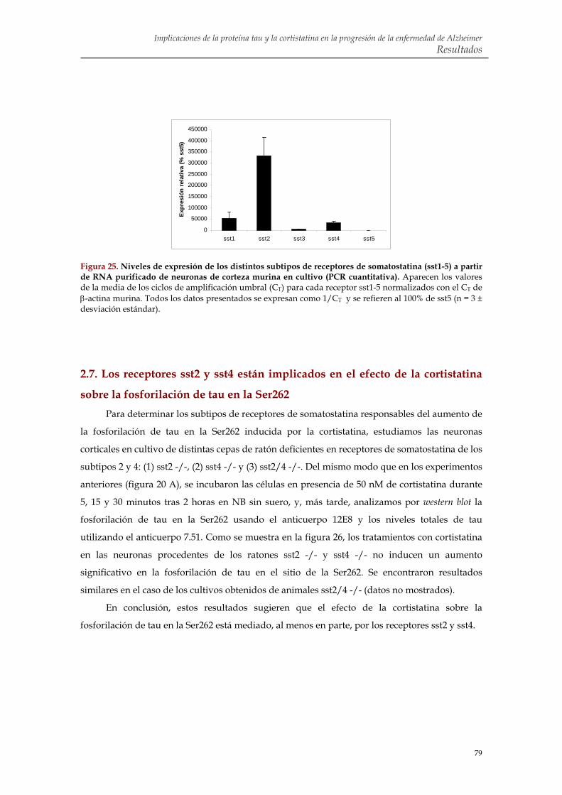
Implicaciones de la proteína tau y la cortistatina en la progresión de la enfermedad de Alzheimer Resultados
79
0
50000
100000
150000
200000
250000
300000
350000
400000
450000
sst1 sst2 sst3 sst4 sst5
Expr
esió
n re
lativ
a (%
sst
5)
Figura 25. Niveles de expresión de los distintos subtipos de receptores de somatostatina (sst1-5) a partir de RNA purificado de neuronas de corteza murina en cultivo (PCR cuantitativa). Aparecen los valores de la media de los ciclos de amplificación umbral (CT) para cada receptor sst1-5 normalizados con el CT de β-actina murina. Todos los datos presentados se expresan como 1/CT y se refieren al 100% de sst5 (n = 3 ± desviación estándar).
2.7. Los receptores sst2 y sst4 están implicados en el efecto de la cortistatina
sobre la fosforilación de tau en la Ser262 Para determinar los subtipos de receptores de somatostatina responsables del aumento de
la fosforilación de tau en la Ser262 inducida por la cortistatina, estudiamos las neuronas
corticales en cultivo de distintas cepas de ratón deficientes en receptores de somatostatina de los
subtipos 2 y 4: (1) sst2 -/-, (2) sst4 -/- y (3) sst2/4 -/-. Del mismo modo que en los experimentos
anteriores (figura 20 A), se incubaron las células en presencia de 50 nM de cortistatina durante
5, 15 y 30 minutos tras 2 horas en NB sin suero, y, más tarde, analizamos por western blot la
fosforilación de tau en la Ser262 usando el anticuerpo 12E8 y los niveles totales de tau
utilizando el anticuerpo 7.51. Como se muestra en la figura 26, los tratamientos con cortistatina
en las neuronas procedentes de los ratones sst2 -/- y sst4 -/- no inducen un aumento
significativo en la fosforilación de tau en el sitio de la Ser262. Se encontraron resultados
similares en el caso de los cultivos obtenidos de animales sst2/4 -/- (datos no mostrados).
En conclusión, estos resultados sugieren que el efecto de la cortistatina sobre la
fosforilación de tau en la Ser262 está mediado, al menos en parte, por los receptores sst2 y sst4.

Implicaciones de la proteína tau y la cortistatina en la progresión de la enfermedad de Alzheimer Resultados
80
A
B
Cortistatina
0 5 min 15 min 30 min
12E8
7.51
Cortistatina
0 5 min 15 min 30 min
12E8
7.51 0
50
100
150
200
250
300
350
400
0 5 15 30
Tiempo (min)
12E8
/ 7.5
1 (%
Con
trol
)
0
50
100
150
200
250
300
350
400
0 5 15 30
Tiempo (min)
12E8
/ 7.5
1 (%
Con
trol
)
0
50
100
150
200
250
300
350
400
0 5 15 30
Tiempo (min)
12E8
/ 7.5
1 (%
Con
trol
)
0
50
100
150
200
250
300
350
400
0 5 15 30
Tiempo (min)
12E8
/ 7.5
1 (%
Con
trol
)
0
50
100
150
200
250
300
350
400
0 5 15 30
Tiempo (min)
12E8
/ 7.5
1 (%
Con
trol
)
0
50
100
150
200
250
300
350
400
0 5 15 30
Tiempo (min)
12E8
/ 7.5
1 (%
Con
trol
)
0
50
100
150
200
250
300
350
400
0 5 15 30
Tiempo (min)
12E8
/ 7.5
1 (%
Con
trol
)
0
50
100
150
200
250
300
350
400
0 5 15 30
Tiempo (min)
12E8
/ 7.5
1 (%
Con
trol
)
A
B
Cortistatina
0 5 min 15 min 30 min
12E8
7.51
Cortistatina
0 5 min 15 min 30 min
12E8
7.51
Cortistatina
0 5 min 15 min 30 min
12E8
7.51
Cortistatina
0 5 min 15 min 30 min
12E8
7.51 0
50
100
150
200
250
300
350
400
0 5 15 30
Tiempo (min)
12E8
/ 7.5
1 (%
Con
trol
)
0
50
100
150
200
250
300
350
400
0 5 15 30
Tiempo (min)
12E8
/ 7.5
1 (%
Con
trol
)
0
50
100
150
200
250
300
350
400
0 5 15 30
Tiempo (min)
12E8
/ 7.5
1 (%
Con
trol
)
0
50
100
150
200
250
300
350
400
0 5 15 30
Tiempo (min)
12E8
/ 7.5
1 (%
Con
trol
)
0
50
100
150
200
250
300
350
400
0 5 15 30
Tiempo (min)
12E8
/ 7.5
1 (%
Con
trol
)
0
50
100
150
200
250
300
350
400
0 5 15 30
Tiempo (min)
12E8
/ 7.5
1 (%
Con
trol
)
0
50
100
150
200
250
300
350
400
0 5 15 30
Tiempo (min)
12E8
/ 7.5
1 (%
Con
trol
)
0
50
100
150
200
250
300
350
400
0 5 15 30
Tiempo (min)
12E8
/ 7.5
1 (%
Con
trol
)
Figura 26. La cortistatina no afecta a la fosforilación de tau en la Ser262 en cultivos de neuronas realizados a partir de cortezas de ratones sst2 -/- y sst4 -/-. (A) Se incubaron las células sst2 -/- en presencia de cortistatina (50 nM, 5, 15 y 30 min). Seguidamente, se realizaron western blots usando los anticuerpos 12E8 (reconoce la Ser262 fosforilada de tau) y 7.51 (tau total). La cuantificación en unidades arbitrarias (a la derecha) se realizó por densitometría de los western blots anteriores. Los niveles de fosforilación de tau en la Ser262 se normalizaron con los de tau total (n = 3 ± la desviación típica). (B) Se realizó un experimento similar al anterior pero, esta vez, con neuronas sst4 -/-.
Implicaciones de la proteína tau y la cortistatina en la progresión de la enfermedad de Alzheimer Resultados
81
2.8. La cortistatina incrementa la fosforilación de tau en la Ser262
probablemente a través de su unión a receptores heterodiméricos compuestos
por sst2 y sst4 Para desvelar qué subtipo/s de receptor/es de somatostatina es/son responsable/s del
efecto de la cortistatina sobre la fosforilación de tau en la Ser262, realizamos tratamientos con
cortistatina en células HEK-293 transfectadas de forma transitoria. Como se indica en la sección
de materiales y métodos, transfectamos con varias combinaciones de tres plásmidos distintos
compuestos por el vector pcDNA 3.1 y el inserto de cDNA de tau, sst2 o sst4 en cada caso. Las
transfecciones transitorias realizadas fueron las siguientes: (1) tau, (2) tau y sst2, (3) tau y sst4 y
(4) con los tres plásmidos (tau, sst2 y sst4).
En un primer momento, quisimos comprobar si las células HEK-293 transfectadas de
forma transitoria expresaban los transgenes. Por un lado, usando la técnica de PCR cuantitativa
con el reactivo SYBR Green, observamos que tau, sst2 y sst4 se expresan cuando se transfectan
(figura 27 A-C). Por otro lado, comprobamos que la proteína tau se expresa en las HEK-293
transfectadas con el cDNA de tau en experimentos de inmunofluorescencia con el anticuerpo
tau 1 que reconoce tau (figura 27 D). De igual forma confirmamos la expresión de tau en las
HEK-293 transfectadas con el cDNA de tau y con el de sst2, sst4 y con ambos (datos no
mostrados). La expresión de tau no se aprecia en células control transfectadas con el vector
pcDNA 3.1 (figura 27 D).
Las células HEK-293 transfectadas de forma transitoria con (1) tau, (2) tau y sst2, (3) tau y
sst4 y (4) tau, sst2 y sst4 se incubaron en presencia de distintas concentraciones de cortistatina
(5, 50, 200 y 500 nM) durante 30 minutos. Se analizaron los extractos proteicos correspondientes
mediante western blot utilizando el anticuerpo 12E8 que reconoce la Ser262 fosforilada de tau. La
cortistatina no parece afectar de forma significativa a la fosforilación de tau en la Ser262 cuando
se añadía a las HEK-293 transfectadas con sólo tau (datos no mostrados), con tau y sst2 o con
tau y sst4 (figura 28 A y B). Por el contrario, los tratamientos con 200 nM o 500 nM de
cortistatina producen un aumento significativo en la fosforilación de tau en la Ser262 en las
HEK-293 transfectadas con tau, sst2 y sst4 (figura 28 C; p<0.05). Estos experimentos sugieren
que ambos receptores, sst2 y sst4, son necesarios para mediar el efecto de la cortistatina sobre la
fosforilación de tau en la Ser262.

Implicaciones de la proteína tau y la cortistatina en la progresión de la enfermedad de Alzheimer Resultados
82
B
CA
D
Control Tau
0
2
4
6
8
10
12
Contro
ltau
tau+s
st2
tau+s
st4
tau+s
st2
+sst4
Expr
esió
n re
lativ
a de
sst
4 (1
/Ctv
s. c
ontr
ol)
Expr
esió
n re
lativ
a de
tau
(1/C
tvs.
con
trol
)
0
0,5
1
1,5
2
2,5
Contro
ltau
tau+s
st2
tau+s
st4
tau+s
st2
+sst4
Expr
esió
n re
lativ
a de
sst
2 (1
/Ctv
s. c
ontr
ol)
Contro
ltau
tau+s
st2
tau+s
st4
tau+s
st2
+sst4
0
0,5
1
1,5
2
2,5
3
3,5
4
4,5
5
0
0,5
1
1,5
2
2,5
3
3,5
4
4,5
5B
CA
D
Control Tau
0
2
4
6
8
10
12
Contro
ltau
tau+s
st2
tau+s
st4
tau+s
st2
+sst4
Expr
esió
n re
lativ
a de
sst
4 (1
/Ctv
s. c
ontr
ol)
0
2
4
6
8
10
12
Contro
ltau
tau+s
st2
tau+s
st4
tau+s
st2
+sst4
Expr
esió
n re
lativ
a de
sst
4 (1
/Ctv
s. c
ontr
ol)
Expr
esió
n re
lativ
a de
tau
(1/C
tvs.
con
trol
)
0
0,5
1
1,5
2
2,5
Contro
ltau
tau+s
st2
tau+s
st4
tau+s
st2
+sst4
Expr
esió
n re
lativ
a de
tau
(1/C
tvs.
con
trol
)
0
0,5
1
1,5
2
2,5
0
0,5
1
1,5
2
2,5
Contro
ltau
tau+s
st2
tau+s
st4
tau+s
st2
+sst4
Expr
esió
n re
lativ
a de
sst
2 (1
/Ctv
s. c
ontr
ol)
Contro
ltau
tau+s
st2
tau+s
st4
tau+s
st2
+sst4
0
0,5
1
1,5
2
2,5
3
3,5
4
4,5
5
0
0,5
1
1,5
2
2,5
3
3,5
4
4,5
5
Expr
esió
n re
lativ
a de
sst
2 (1
/Ctv
s. c
ontr
ol)
Contro
ltau
tau+s
st2
tau+s
st4
tau+s
st2
+sst4
0
0,5
1
1,5
2
2,5
3
3,5
4
4,5
5
0
0,5
1
1,5
2
2,5
3
3,5
4
4,5
5
De igual forma, tratamos las células HEK-293 transfectadas de forma transitoria con
diferentes concentraciones de somatostatina (de 5, 50, 200 y 500 nM) durante 5 minutos. En este
caso, a diferencia del tratamiento con cortistatina, la somatostatina no induce un efecto
significativo en la fosforilación de la Ser262 de tau (datos no mostrados) en las HEK-293
transfectadas con (1) tau, (2) tau y sst2, (3) tau y sst4 y, por último, (4) con los tres plásmidos
(tau, sst2 y sst4). Parece que la cortistatina y la somatostatina median su efecto sobre la
fosforilación de tau en la Ser262 por diferentes mecanismos.
Figura 27. Expresión de tau, sst2 y sst4 en las células HEK-293 transfectadas de forma transitoria con tau, sst2 y sst4. Datos de la PCR cuantitativa para detectar los niveles de expresión de tau (A), de sst2 (B) y de sst4 (C) en las transfecciones transitorias siguientes: (1) tau, (2) tau+sst2, (3) tau+sst4 y (4) tau+sst2+sst4. Como control, transfectamos en las mismas condiciones el vector vacío pcDNA 3.1. Los resultados, normalizados usando los datos de la β-actina humana, se presentan como 1/CT respecto al control (n = 3 ± desviación estándar). (D) Células HEK-293 transfectadas con pcDNA 3.1 (control, a la izquierda) o con tau (a la derecha) analizadas por inmunofluorescencia con un anticuerpo frente a tau (tau 1; fluorescencia roja) y tinción con DAPI (azul). La barra equivale a 20 μm.

Implicaciones de la proteína tau y la cortistatina en la progresión de la enfermedad de Alzheimer Resultados
83
A
B
C
0
50
100
150
200
250
300
350
400
450
0 5 50 200 500
Cortistatina (nM)
12E8
/ 7.5
1 (%
Con
trol
) 12E8
7.51
C Cortistatina-
12E8
7.51
C
0
50
100
150
200
250
300
350
400
450
0 5 50 200 500
Cortistatina (nM)
12E8
/ 7.5
1 (%
Con
trol
)
0
50
100
150
200
250
300
350
400
450
0 5 50 200 500
Cortistatina (nM)
12E8
/ 7.5
1 (%
Con
trol
) 12E8
7.51
C Cortistatina-
12E8
7.51
C
12E8
7.51
C Cortistatina-
12E8
7.51
C
050
100
150
200
250
300350
400450
0 5 50 200 500
CST-14 (nM)
12E8
/ 7.5
1 (%
Con
trol
)
12E8
7.51
C CST-14
12E8
7.51
C CST-14
050
100
150
200
250
300350
400450
0 5 50 200 500
Cortistatina (nM)
12E8
/ 7.5
1 (%
Con
trol
)
12E8
7.51
C -
12E8
7.51
C Cortistatina
050
100
150
200
250
300350
400450
0 5 50 200 500
CST-14 (nM)
12E8
/ 7.5
1 (%
Con
trol
)
050
100
150
200
250
300350
400450
0 5 50 200 500
CST-14 (nM)
12E8
/ 7.5
1 (%
Con
trol
)
12E8
7.51
C CST-14
12E8
7.51
C CST-14
12E8
7.51
C CST-14
12E8
7.51
C CST-14
12E8
7.51
C CST-14
12E8
7.51
C CST-14
050
100
150
200
250
300350
400450
0 5 50 200 500
Cortistatina (nM)
12E8
/ 7.5
1 (%
Con
trol
)
050
100
150
200
250
300350
400450
0 5 50 200 500
Cortistatina (nM)
12E8
/ 7.5
1 (%
Con
trol
)
12E8
7.51
C -
12E8
7.51
C Cortistatina
12E8
7.51
C -
12E8
7.51
C Cortistatina
0
50
100
150
200
250
300
350
400
450
0 5 50 200 500
CST-14 (nM)
12E8
/7.5
1 (%
Con
trol
)
**
*C CST-14
12E8
7.51
C CST-14
12E8
7.51
C CST-14
12E8
7.51
C CST-14
12E8
7.51
C CST-14
12E8
7.51
C CST-14
12E8
7.51
0
50
100
150
200
250
300
350
400
450
0 5 50 200 500
Cortistatina (nM)
12E8
/7.5
1 (%
Con
trol
)
**
*C Cortistatina-
12E8
7.51
C -
12E8
7.51
C -
12E8
7.51
C -
12E8
7.51
C -
12E8
7.51
C -
12E8
7.51
C -
12E8
7.51
C -
12E8
7.51
C -
12E8
7.51
C -
12E8
7.51
C -
12E8
7.51
C -
12E8
7.51
C
12E8
7.51
C
12E8
7.51
0
50
100
150
200
250
300
350
400
450
0 5 50 200 500
CST-14 (nM)
12E8
/7.5
1 (%
Con
trol
)
0
50
100
150
200
250
300
350
400
450
0 5 50 200 500
CST-14 (nM)
12E8
/7.5
1 (%
Con
trol
)
**
*C CST-14
12E8
7.51
C CST-14
12E8
7.51
C CST-14
12E8
7.51
C CST-14
12E8
7.51
C CST-14
12E8
7.51
C CST-14
12E8
7.51
C CST-14
12E8
7.51
C CST-14
12E8
7.51
C CST-14
12E8
7.51
C CST-14
12E8
7.51
C CST-14
12E8
7.51
C CST-14
12E8
7.51
C CST-14
12E8
7.51
C CST-14
12E8
7.51
0
50
100
150
200
250
300
350
400
450
0 5 50 200 500
Cortistatina (nM)
12E8
/7.5
1 (%
Con
trol
)
0
50
100
150
200
250
300
350
400
450
0 5 50 200 500
Cortistatina (nM)
12E8
/7.5
1 (%
Con
trol
)
**
*C Cortistatina-
12E8
7.51
C -
12E8
7.51
C Cortistatina-
12E8
7.51
C -
12E8
7.51
C -
12E8
7.51
C -
12E8
7.51
C -
12E8
7.51
C -
12E8
7.51
C -
12E8
7.51
C -
12E8
7.51
C -
12E8
7.51
C -
12E8
7.51
C -
12E8
7.51
C -
12E8
7.51
C
12E8
7.51
C
12E8
7.51
Figura 28. Fosforilación de la proteína tau en la Ser262 en las células HEK-293 transfectadas de forma transitoria con tau, sst2 y sst4. Tratamos las HEK-293 transfectadas con tau+sst2 (A), tau+sst4 (B) y tau+sst2+sst4 (C) con distintas concentraciones de cortistatina (5 nM, 50 nM, 200 nM y 500 nM) durante 30 minutos. Los extractos se analizaron por western blot para reconocer la Ser262 fosforilada de tau (anticuerpo 12E8) y tau total (7.51). La cuantificación por densitometría en unidades arbitrarias de los niveles de fosforilación de tau en la Ser262 se normalizó con la de tau total. Se representan las medias de los datos por triplicado ± la desviación típica. *p<0.05 respecto al control sin cortistatina.
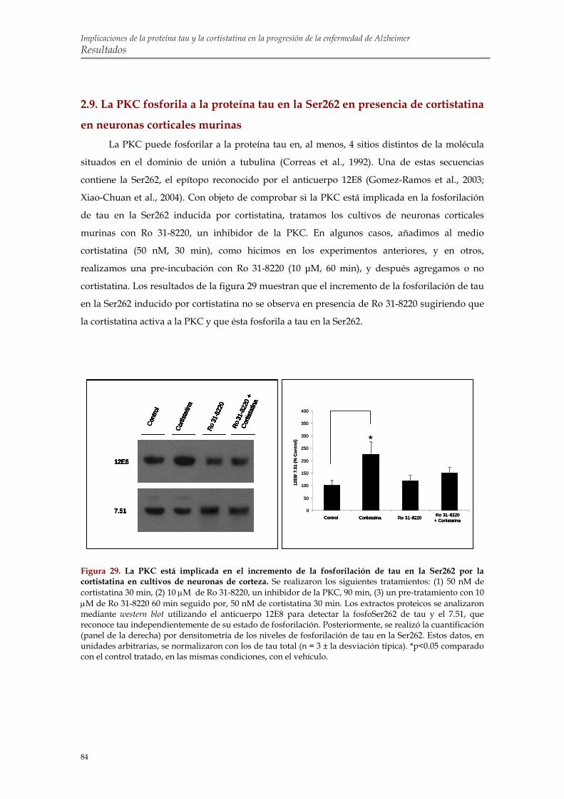
Implicaciones de la proteína tau y la cortistatina en la progresión de la enfermedad de Alzheimer Resultados
84
0
50
100
150
200
250
300
350
400
12E8
/ 7.5
1 (%
Con
trol)
Control Cortistatina Ro 31-8220 Ro 31-8220+ Cortistatina
*
12E8
7.51
Cont
rol
Corti
stat
ina
Ro 3
1-82
20
Ro 31
-822
0+
Corti
stat
ina
12E8
7.51
Cont
rol
Corti
stat
ina
Ro 3
1-82
20
Ro 31
-822
0+
Corti
stat
ina
*
Control Cortistatina Ro 31-8220 Ro 31-8220+ Cortistatina
12E8
7.51
Cont
rol
Corti
stat
ina
Ro 3
1-82
20
Ro 31
-822
0+
Corti
stat
ina
12E8
7.51
Cont
rol
Corti
stat
ina
Ro 3
1-82
20
Ro 31
-822
0+
Corti
stat
ina
0
50
100
150
200
250
300
350
400
12E8
/ 7.5
1 (%
Con
trol)
Control Cortistatina Ro 31-8220 Ro 31-8220+ Cortistatina
*
12E8
7.51
Cont
rol
Corti
stat
ina
Ro 3
1-82
20
Ro 31
-822
0+
Corti
stat
ina
12E8
7.51
Cont
rol
Corti
stat
ina
Ro 3
1-82
20
Ro 31
-822
0+
Corti
stat
ina
12E8
7.51
Cont
rol
Corti
stat
ina
Ro 3
1-82
20
Ro 31
-822
0+
Corti
stat
ina
12E8
7.51
Cont
rol
Corti
stat
ina
Ro 3
1-82
20
Ro 31
-822
0+
Corti
stat
ina
*
Control Cortistatina Ro 31-8220 Ro 31-8220+ Cortistatina
12E8
7.51
Cont
rol
Corti
stat
ina
Ro 3
1-82
20
Ro 31
-822
0+
Corti
stat
ina
12E8
7.51
Cont
rol
Corti
stat
ina
Ro 3
1-82
20
Ro 31
-822
0+
Corti
stat
ina
12E8
7.51
Cont
rol
Corti
stat
ina
Ro 3
1-82
20
Ro 31
-822
0+
Corti
stat
ina
12E8
7.51
Cont
rol
Corti
stat
ina
Ro 3
1-82
20
Ro 31
-822
0+
Corti
stat
ina
2.9. La PKC fosforila a la proteína tau en la Ser262 en presencia de cortistatina
en neuronas corticales murinas La PKC puede fosforilar a la proteína tau en, al menos, 4 sitios distintos de la molécula
situados en el dominio de unión a tubulina (Correas et al., 1992). Una de estas secuencias
contiene la Ser262, el epítopo reconocido por el anticuerpo 12E8 (Gomez-Ramos et al., 2003;
Xiao-Chuan et al., 2004). Con objeto de comprobar si la PKC está implicada en la fosforilación
de tau en la Ser262 inducida por cortistatina, tratamos los cultivos de neuronas corticales
murinas con Ro 31-8220, un inhibidor de la PKC. En algunos casos, añadimos al medio
cortistatina (50 nM, 30 min), como hicimos en los experimentos anteriores, y en otros,
realizamos una pre-incubación con Ro 31-8220 (10 μM, 60 min), y después agregamos o no
cortistatina. Los resultados de la figura 29 muestran que el incremento de la fosforilación de tau
en la Ser262 inducido por cortistatina no se observa en presencia de Ro 31-8220 sugiriendo que
la cortistatina activa a la PKC y que ésta fosforila a tau en la Ser262.
Figura 29. La PKC está implicada en el incremento de la fosforilación de tau en la Ser262 por la cortistatina en cultivos de neuronas de corteza. Se realizaron los siguientes tratamientos: (1) 50 nM de cortistatina 30 min, (2) 10 μM de Ro 31-8220, un inhibidor de la PKC, 90 min, (3) un pre-tratamiento con 10 μM de Ro 31-8220 60 min seguido por, 50 nM de cortistatina 30 min. Los extractos proteicos se analizaron mediante western blot utilizando el anticuerpo 12E8 para detectar la fosfoSer262 de tau y el 7.51, que reconoce tau independientemente de su estado de fosforilación. Posteriormente, se realizó la cuantificación (panel de la derecha) por densitometría de los niveles de fosforilación de tau en la Ser262. Estos datos, en unidades arbitrarias, se normalizaron con los de tau total (n = 3 ± la desviación típica). *p<0.05 comparado con el control tratado, en las mismas condiciones, con el vehículo.

Discusión


Implicaciones de la proteína tau y la cortistatina en la progresión de la enfermedad de Alzheimer Discusión
87
Numerosas evidencias avalan la relevancia de la proteína tau en el desarrollo de la EA y
de otras tauopatías. Por un lado, se considera que la presencia de NFT representa una marca
histopatológica de estas enfermedades, y que éstas, pueden deberse en algunos casos, como las
FTDP-17, a mutaciones puntuales en el gen que codifica para la proteína tau (Wang & Liu,
2008). Además, se ha visto que el número de NFT correlaciona con el grado de demencia de los
pacientes con EA (Arriagada et al., 1992a; Arriagada et al., 1992b) y que la distribución de la
patología neurofibrilar en la EA sigue un patrón anatómico característico que también se asocia
con el grado de demencia del enfermo (Braak & Braak, 1991). En personas de edad avanzada,
asintomáticos para la EA, se detecta la patología de tau en la corteza transentorrinal. Esta
patología, en pacientes con MCI que presentan pérdida de memoria, se extiende a otras
estructuras cerebrales como la corteza entorrinal y el hipocampo. En el caso de los enfermos de
Alzheimer, los NFT invaden también el resto de la corteza.
En definitiva, estos trabajos relacionan la patología de tau con la progresión de la EA. Por
tanto, uno de los objetivos fundamentales de esta tesis doctoral ha sido profundizar en los
mecanismos moleculares que podrían explicar dicha relación tratando de estudiar el papel que
juega tau en el avance de la enfermedad y en la atrofia cerebral debida a la muerte neuronal.
Por otra parte, también nos pareció importante estudiar la posible existencia de factores
que determinasen el desarrollo de la patología de tau en la corteza cerebral. Este momento es
clave en la progresión de la enfermedad puesto que determina el paso crítico del estado de
“pérdida de memoria”, propia del MCI, al de “demencia” que caracteriza al enfermo de
Alzheimer (Braak & Braak, 1991; Delacourte et al., 1999). El momento en el que la patología de
tau pasa de la corteza entorrinal y el hipocampo a la corteza cerebral se conoce como “punto de
no retorno”. En este trabajo, demostramos que la cortistatina, un neuropéptido que se expresa
principalmente en la corteza, podría jugar un papel relevante en el desarrollo de la patología de
tau en este área cerebral (de Lecea et al., 1996; de Lecea et al., 1997). La cortistatina podría
constituir una señal endógena capaz de modular las actividades de las quinasas y las fosfatasas
que afectan a la fosforilación de tau. A continuación discutiremos los puntos clave de esta tesis.

Implicaciones de la proteína tau y la cortistatina en la progresión de la enfermedad de Alzheimer Discusión
88
1. PAPEL QUE JUEGA TAU EN EL AVANCE DE LA EA
1.2. La proteína tau extracelular induce un aumento en el calcio intracelular a
través de M1 y M3 En nuestro trabajo, observamos que la proteína tau induce un aumento en el calcio
intracelular. Diversos estudios realizados previamente, indican que otras proteínas relacionadas
con enfermedades neurodegenerativas como la α-sinucleína (Danzer et al., 2007), el β-amiloide
o la proteína priónica (Demuro et al., 2005) alteran la homeostasis del calcio intracelular cuando
se añaden a cultivos de neuronas. Sin embargo, los mecanismos que regulan esta alteración del
calcio difieren según el tipo de proteína ensayada (Adamczyk & Strosznajder, 2006; Danzer et
al., 2007; Gomez-Ramos et al., 2006). Basándonos en nuestros resultados sugerimos que la
proteína tau induce cambios en el nivel intracelular de calcio a través de su interacción con los
receptores muscarínicos. La activación de estos receptores provoca la liberación de calcio de los
orgánulos intracelulares. De hecho, se han descrito otras proteínas capaces de interaccionar con
receptores muscarínicos como el caso reciente de una glicoproteína vírica (Kumar et al., 2007).
En primer lugar, para identificar qué receptor muscarínico sería capaz de unir tau, tuvimos que
determinar qué subtipos de receptores se expresan en las células de neuroblastoma humano
SH-SY5Y. Como se mencionó en el apartado de resultados, los receptores M1, M2 y M3 son los
más abundantes. Este patrón de expresión correlaciona bastante bien con lo que ocurre en el
cerebro ((Piggott et al., 2002); http://www.brain-map.org/). M1 se expresa en gran cantidad en
todo el cerebro, incluyendo las regiones más afectadas por la EA. El receptor M2 parece estar
relacionado con la neurodegeneración puesto que se ha descrito su menor densidad en cerebros
de pacientes con EA (Mash et al., 1985). Por último, el M3 se localiza en la corteza, el hipocampo
y la formación retrohipocampal. De modo que podemos considerar a las SH-SY5Y como un
buen modelo para estudiar el efecto de tau. Así que centramos nuestra búsqueda en los
receptores M1, M2 y M3. Tras realizar distintos estudios farmacológicos y análisis mediante
transfecciones transitorias, como se detalla en la sección de resultados, concluimos que M1 y M3
podrían ser responsables de la interacción con tau descartando así el papel de M2.
Por otra parte, diversas evidencias apuntan que existe una relación entre los receptores
muscarínicos y el procesamiento del APP. Este procesamiento, de función desconocida por el
momento, puede seguir dos vías. Se denomina vía no amiloidogénica a la ruta en la que actúan
secuencialmente, la α-secretasa primero y más tarde, la γ-secretasa. De este modo, se genera el
largo dominio extracelular del APP (αAPPs) que tiene efectos neuroprotectores, un péptido
pequeño (p3), que no forma agregados y el dominio intracelular del APP (AICD) (Hartmann et
al., 2007). Por otro lado, en la vía amiloidogénica, la activación de la β-secretasa seguida de la γ-
Implicaciones de la proteína tau y la cortistatina en la progresión de la enfermedad de Alzheimer Discusión
89
secretasa da lugar al dominio extracelular del APP (βAPPs), al péptido β-amiloide y al AICD. La
β-secretasa comprende una familia de aspartil proteasas con, al menos, dos miembros altamente
homólogos (BACE1, la mayoritaria en neuronas, y BACE2) (Hartmann et al., 2007). La γ-
secretasa corta el APP en dos lugares alternativos: la valina 711 dando β-amiloide 40 o la
alanina 713 generando β-amiloide 42 (Selkoe, 2001). Se ha visto que la activación de M1 y M3
promueve el procesamiento del APP por la vía no amiloidogénica (Nitsch et al., 1992). En
cuanto a la relación entre la estimulación de los receptores muscarínicos y la vía
amiloidogénica, se sabe que la activación de M1 conlleva un incremento en la expresión de
BACE, sin embargo, la estimulación de M2 produce una disminución de su expresión (Zuchner
et al., 2004; Zuchner et al., 2005). A pesar de estos datos, por el momento, en nuestro modelo, no
hemos encontrado un efecto de tau sobre el procesamiento del APP.
1.2. La controversia en torno a la posible toxicidad de la proteína tau Como se ha mencionado, la relevancia de la patología de tau en la progresión de la EA se
demostró en los trabajos en los que se descubrió que el número de NFT y la progresión
anatómica de la patología fibrilar correlacionan con el grado de demencia de los pacientes con
esta enfermedad (Arriagada et al., 1992a; Arriagada et al., 1992b; Braak & Braak, 1991).
Posteriormente, estudios de inmunohistoquímica mostraron que existe una relación inversa
entre el número de ovillos fantasma y el número de neuronas vivas (Bondareff et al., 1989; Cras
et al., 1995; Fukutani et al., 1995) sugiriendo que las neuronas que degeneran desarrollan
previamente agregados de tau. Por el momento, sigue sin saberse si la relación entre la
patología de tau y la muerte neuronal se debe a una pérdida de función de la proteína tau
normal o a una ganancia de función de tau debida a su toxicidad.
Diversos datos apoyan la idea de la pérdida de función de tau como posible causante de la
disfunción sináptica y la posterior neurodegeneración: la proteína tau, al separarse de los
microtúbulos, deja de estabilizarlos pudiendo comprometer la morfología normal de la célula y
otras funciones del citoesqueleto como el transporte axonal (Roy et al., 2005; Trojanowski et al.,
2005). Con el tiempo, se forman los NFT y éstos podrían secuestrar más tau, amplificando así, la
pérdida de la función normal de esta proteína. Se ha demostrado que el tau hiperfosforilado
inhibe el ensamblaje de los microtúbulos en presencia de cualquier isoforma de tau
desfosforilado y que causa la desestabilización de los microtúbulos previamente formados
(Alonso et al., 2001). De igual forma, el tau fibrilar en PHF compromete la estabilidad de los
microtúbulos resultando en un descenso del transporte axonal y dendrítico (Alonso et al., 1996;
Salehi et al., 2003). En relación a esta hipótesis, el grupo de Trojanowski (2005) ha demostrado

Implicaciones de la proteína tau y la cortistatina en la progresión de la enfermedad de Alzheimer Discusión
90
que la administración de una droga (paclitaxel) capaz de estabilizar los microtúbulos mejora el
deterioro motor de modelos murinos de tauopatías (Zhang et al., 2005). Este experimento
destaca la relevancia de la integridad de la red de microtúbulos para evitar los síntomas
asociados a la neurodegeneración.
Por otra parte, algunos trabajos realizados en ratones modificados genéticamente
sostienen que la proteína tau no es esencial para el correcto funcionamiento de la célula, ya que
su función podría verse complementada por otras proteínas. Por un lado, datos recientes
demuestran que el transporte axonal no se ve afectado por la sobre-expresión o la falta de tau en
la retina murina (Yuan et al., 2008). Además, conclusiones similares se han obtenido del estudio
de dos líneas independientes de ratones que no expresan tau (Dawson et al., 2001; Harada et al.,
1994). En ambos casos, observaron que los animales eran viables y que no presentaban grandes
deficiencias fenotípicas (Dawson et al., 2001; Harada et al., 1994). La falta de tau podría estar
compensada por una sobre-expresión de otras proteínas asociadas a microtúbulos como la
MAP1A (Harada et al., 1994). En concreto, las únicas modificaciones morfológicas descritas
fueron un retraso en la maduración axonal de neuronas en cultivo (Dawson et al., 2001) y, en la
otra línea independiente de ratones, axones neuronales de menor calibre e hiperactividad
(Harada et al., 1994; Ikegami et al., 2000). A pesar de esas diferencias, estudios recientes
sugieren incluso que la supresión de tau podría ser beneficiosa para la EA. Se ha demostrado
que los ratones modificados genéticamente que no expresan tau (Dawson et al., 2001) no
presentan déficits de memoria o aprendizaje (Roberson et al., 2007). Además, los ratones que no
expresan tau y sobre-expresan el APP humano mutado no muestran los déficits de memoria y
aprendizaje presentados por el modelo de EA (Roberson et al., 2007). También se ha visto que
las neuronas de hipocampo en cultivo obtenidas de ratones deficientes en tau (Dawson et al.,
2001) no son susceptibles a la toxicidad inducida por el β-amiloide (Rapoport et al., 2002).
Por otro lado, existen evidencias a favor de la ganancia de función tóxica de tau como
posible causa de la muerte neuronal. Es posible que los efectos tóxicos de los NFT puedan
deberse, al menos en parte, a la disrupción física de las funciones celulares por la acumulación
de agregados de proteína de gran tamaño en el interior celular (Ballatore et al., 2007). La
toxicidad de los NFT también podría deberse a su capacidad de interacción con otras proteínas
importantes para la función celular normal (Alonso et al., 1997).
Sin embargo, distintos trabajos sostienen que los NFT podrían ser agregados protectores
frente a la neurodegeneración. Por un lado, se ha visto que las neuronas con NFT pueden
sobrevivir durante décadas (Morsch et al., 1999). Y también se ha demostrado que no existe una
asociación entre la morfología apoptótica, como la fragmentación de DNA, y la acumulación de
tau (Broe et al., 2001). De igual forma, en otras enfermedades, como la enfermedad de
Huntington, se ha visto que los agregados protegen frente a la degeneración (Arrasate et al.,

Implicaciones de la proteína tau y la cortistatina en la progresión de la enfermedad de Alzheimer Discusión
91
2004). En un modelo celular de Huntington, se demostró que la formación de los cuerpos de
inclusión de huntingtina contribuía a la disminución de la muerte celular probablemente por
retener la huntingtina libre (Arrasate et al., 2004). En el caso de la EA y el resto de tauopatías, se
piensa que los agregados de tau podrían proteger a la neurona del ataque de las especies
reactivas derivadas del oxígeno (revisado en (Lee et al., 2005)). Otra posibilidad es que los NFT
sean protectores porque secuestran las especies tóxicas de tau soluble (Duff & Planel, 2005).
Diversos trabajos han demostrado la existencia de especies de tau pre-fibrilares (Maeda et al.,
2007; Maeda et al., 2006) que pueden encontrarse en el CSF de los enfermos de Alzheimer (Iqbal
et al., 2005). Se ha sugerido que las estructuras implicadas en la neurodegeneración son los
agregados intermedios de tau y no los PHF como se pensó en un principio (Duff & Planel,
2005). Primero, en un modelo de Drosophila melanogaster que sobre-expresa tau humano, se vio
que la neurodegeneración ocurría a pesar de que los NFT no se formaban (Wittmann et al.,
2001). Posteriormente, otro grupo confirmó esta idea en un modelo murino de tauopatía en el
que sobre-expresaban el tau mutado (P301L) de forma condicional gracias al uso del promotor
“tet-off” (Santacruz et al., 2005). En este trabajo, vieron que los déficits de memoria del ratón
transgénico mejoraban si la expresión del transgén se interrumpía mediante administración de
doxiciclina a pesar de que los NFT seguían acumulándose (Santacruz et al., 2005). Estos
resultados sugieren que los déficits de memoria no están relacionados con la presencia de NFT
y también se ven apoyados por las observaciones realizadas en individuos que padecen FTDP-
17 debido a la mutación P301L (Bird et al., 1999). En esta tesis doctoral, mostramos que la
presencia de la proteína tau causa la muerte de las células de neuroblastoma humano SH-SY5Y.
Su toxicidad podría deberse a que la interacción de tau con los receptores muscarínicos
promueve un aumento del calcio intracelular. Como se detalla en el apartado de resultados,
comprobamos que la proteína tau en PHF no altera los niveles de calcio intracelulares de las
SH-SY5Y. Por el contrario, la estimulación con tau en forma monomérica induce un aumento
sostenido en los niveles de calcio intracelular. El efecto de tau sobre los niveles de calcio
intracelular se prolonga más en el tiempo que en los tratamientos realizados con acetilcolina.
Por tanto, basándonos en estos resultados, postulamos que esa distinción cinética es clave para
la toxicidad celular. Pensamos que podría deberse a que la acetilcolina altera la homeostasis del
calcio intracelular debido a su interacción con los receptores muscarínicos y nicotínicos, en
cambio, en el caso de tau, se debe a la activación de los receptores muscarínicos, concretamente,
del M1 y del M3. Por último, mostramos que un fragmento de la zona carboxilo-terminal de tau,
que comprende los residuos 391-407, es suficiente para inducir el efecto de tau sobre la
movilización de calcio en las células COS-7 transfectadas transitoriamente con M3.
Aunque los agregados sean protectores para la célula en un principio, las consecuencias a
largo plazo de estas inclusiones intracelulares son probablemente perjudiciales porque pueden

Implicaciones de la proteína tau y la cortistatina en la progresión de la enfermedad de Alzheimer Discusión
92
llegar a ocupar un gran espacio en el interior celular. Por tanto, es muy probable que ambos
mecanismos -pérdida de función y, sobre todo, la ganancia de función tóxica de tau-
contribuyan a la neurodegeneración en el inicio y la progresión de la EA y otras tauopatías
especialmente en diferentes estadios de la enfermedad.
1.3. Modelo de propagación de la EA por toxicidad de la proteína tau
extracelular Los datos obtenidos en cuanto a la toxicidad de la proteína tau extracelular en forma
monomérica podrían ser relevantes para explicar la propagación de la EA.
En condiciones fisiológicas, la proteína tau se encuentra, en su mayoría, unida a los
microtúbulos (Gamblin et al., 2003; King et al., 1999; Reynolds et al., 2005). Sin embargo, en
condiciones patológicas, tau puede sufrir diversas modificaciones post-traduccionales como
hiperfosforilación, truncación o desamidación (Avila et al., 2004; Gong et al., 2005; Wang & Liu,
2008). Estas modificaciones pueden inducir una liberación de tau de los microtúbulos
acumulándose así tau monomérico. A partir de una concentración crítica, que según se ha visto
debe sobrepasar 0.5 μM, la proteína tau comienza a agregarse en el interior celular formando
PHF (Reynolds et al., 2005). En el momento en que estas neuronas degeneran, su contenido
celular queda libre en el espacio extracelular pudiendo encontrar tau en forma monomérica y
PHF. Como vimos, la proteína tau extracelular en forma monomérica puede resultar tóxica para
las células vecinas. A su vez, la muerte de estas células permitiría la liberación de tau
extracelular extendiéndose así el efecto tóxico de tau en las inmediaciones. Este modelo podría
explicar la propagación de la patología de tau que empieza en la estructura cerebral de la
corteza entorrinal (Braak & Braak, 1991) y luego avanza al resto de áreas.
Como hemos visto en este trabajo, la toxicidad de tau depende de su interacción con los
receptores muscarínicos M1 y M3. De hecho, en la EA, se ha descrito un incremento de M1 y M3
respecto a M2 (Mash et al., 1985). De este modo, la proteína tau podría actuar sobre M1 y M3
aunque las neuronas estén dañadas. La activación de estos receptores parece suponer una
alteración de la homeostasis de los niveles de calcio intracelular. Esta alteración podría resultar
en un aumento de la fosforilación de tau, modificación, que como hemos mencionado, está
relacionada con la EA (Braak & Braak, 1991; Delacourte et al., 1999). La fosforilación de tau
podría deberse a la activación de quinasas dependientes de calcio, como la PKC (Correas et al.,
1992; Gomez-Ramos et al., 2003) o CDK5 (Hosoi et al., 1995), y facilitaría la liberación de tau de
los microtúbulos (Biernat et al., 1993; Biernat & Mandelkow, 1999; Drewes et al., 1995; Liu et al.,
2007; Wang et al., 2007).

Implicaciones de la proteína tau y la cortistatina en la progresión de la enfermedad de Alzheimer Discusión
93
2. PAPEL DE LA CORTISTATINA EN EL PASO DE LA PATOLOGÍA DE
TAU A LA CORTEZA CEREBRAL
2.1. La cortistatina induce la fosforilación de tau en la Ser262 En este trabajo, realizado en cultivos primarios de neuronas corticales, demostramos que
la cortistatina promueve la fosforilación de tau en la Ser262 de un modo dependiente de la
concentración de cortistatina, con una saturación del efecto a 50 nM. Cabe destacar que en
estudios realizados con cortistatina fluorescente, aproximadamente un 24% de las neuronas en
cultivo mostraron internalización del ligando. De modo que el efecto de la cortistatina podría
ser, al menos, cuatro veces mayor que el observado en una población celular mixta. La
cortistatina induce un aumento en la fosforilación de tau en la Ser262, epítopo detectado por el
anticuerpo 12E8, un sitio de fosforilación no dirigido por prolinas (Morishima-Kawashima et
al., 1995). Sin embargo, no se encontraron diferencias en la fosforilación de un sitio dirigido por
prolinas: las Ser396 y Ser404 reconocidas por el anticuerpo PHF1 (Morishima-Kawashima et al.,
1995).
Algunos trabajos señalan la relevancia de la fosforilación de tau en la Ser262 en el
desarrollo de las enfermedades neurodegenerativas. La fosforilación anómala en este residuo se
detecta en los cerebros de pacientes con EA (Augustinack et al., 2002; Lauckner et al., 2003;
Morishima-Kawashima et al., 1995) y otras tauopatías (Ferrer et al., 2002). También, se ha
estudiado en modelos murinos de la EA (Perez et al., 2005; Puig et al., 2004). Las placas
amiloides de los ratones transgénicos que sobre-expresan el APP humano mutado (APPSwe) se
encuentran próximas a neuritas que contienen tau hiperfosforilado en la Ser262 (Puig et al.,
2004). Posteriormente, se generaron ratones dobles transgénicos derivados del cruce del ratón
anterior (APPSwe) con un ratón que sobre-expresa el tau humano mutado (tauVLW) (Perez et al.,
2005). En estos ratones dobles transgénicos (APPSwe/ tauVLW) que reproducen la patología de
tau y del β-amiloide se observó que la fosforilación de tau en la Ser262 correlaciona con un
incremento en la formación de agregados filamentosos de tau. Estas estructuras muestran un
diámetro similar al de los filamentos de tau descritos en la EA (Perez et al., 2005). Experimentos
in vitro, demuestran que la fosforilación en la Ser262, situada en el dominio de unión a tubulina,
reduce la unión de tau a los microtúbulos (Drewes et al., 1997; Drewes et al., 1995; Sengupta et
al., 1998; Singh et al., 1996). De este modo, el tau fosforilado en la Ser262 queda libre en el
citosol celular. En vez de favorecerse la formación de PHF, se ha visto que la fosforilación en la
Ser262 inhibe la agregación de tau (Schneider et al., 1999; Schneider & Mandelkow, 2008),
quedando el tau en forma monomérica, y dando lugar, tras la muerte neuronal, a la forma más
tóxica de tau extracelular.
Implicaciones de la proteína tau y la cortistatina en la progresión de la enfermedad de Alzheimer Discusión
94
2.2. Receptores de somatostatina e internalización Como indicamos, debido a la fuerte similitud estructural entre cortistatina y
somatostatina, la cortistatina se une in vitro a los cinco subtipos conocidos de receptores de
somatostatina (sst1-5 según la nomenclatura de la IUPHAR (Hoyer et al., 1995)) con igual
afinidad (Criado et al., 1999; de Lecea et al., 1996; Fukusumi et al., 1997; Siehler et al., 1998).
Distintos estudios han mostrado que la interacción de la somatostatina o sus análogos con los
receptores de somatostatina suele provocar la internalización del complejo ligando-receptor
induciendo una disminución de la respuesta del receptor gracias a un mecanismo conocido
como desensibilización (Csaba & Dournaud, 2001; Olias et al., 2004; Tulipano & Schulz, 2007).
Los receptores que pertenecen a la superfamilia de receptores acoplados a proteínas G sufren
este proceso de forma habitual. La internalización puede depender del tiempo de exposición, la
concentración del ligando y la regulación por distintas rutas de señalización intracelular
(Hofland & Lamberts, 2003). Tras la unión del ligando y la activación del receptor, la
señalización asociada se interrumpe: primero, los receptores quinasa acoplados a proteínas G
(GRK) fosforilan los dominios intracelulares del receptor y, más tarde, las arrestinas, proteínas
citoplásmicas, interrumpen la unión entre el receptor y la proteína G heterotrimérica (Gnagey et
al., 1999; Oakley et al., 2000). Este mecanismo suele ir seguido por la endocitosis o
internalización del complejo receptor/ligando mediante vesículas recubiertas de clatrina,
siendo esta molécula clave para la regulación negativa y resensibilización del receptor (Bohm et
al., 1997; Koenig & Edwardson, 1997). Las β-arrestinas, que también sirven de proteínas de
cargo, unen el receptor a los componentes de la maquinaria endocítica como AP-2 o clatrina
(Pierce et al., 2001). Sin embargo, las arrestinas no median la endocitosis de todos los receptores
acoplados a proteínas G (Zhang et al., 1996). Dentro del compartimento ácido endosomal, el
ligando se disocia del receptor y este último se desfosforila por fosfatasas específicas
permitiendo que el receptor sea reciclado de nuevo en la membrana plasmática (Shenoy &
Lefkowitz, 2003; Shenoy et al., 2001).
No todos los subtipos de receptores de somatostatina internalizan de la misma forma tras
la unión del agonista (Csaba & Dournaud, 2001; Olias et al., 2004; Tulipano & Schulz, 2007). En
resumen, la internalización provocada por la presencia del agonista parece ser menos eficiente
en el caso de sst1 según distintos experimentos realizados con células COS-7 y CHO-K1
transfectadas establemente con distintos subtipos de receptores de somatostatina humanos
(Hofland & Lamberts, 2003). Muchos estudios muestran que las dos isoformas de sst2 sufren
desensibilización e internalización tras la unión de un agonista en diversas líneas celulares y
tejidos (Boudin et al., 2000; Csaba & Dournaud, 2001; Hipkin et al., 1997; Liu et al., 2005; Vanetti
et al., 1993), pero no ocurre lo mismo en presencia de antagonistas de sst2 (Cescato et al., 2006).
Implicaciones de la proteína tau y la cortistatina en la progresión de la enfermedad de Alzheimer Discusión
95
El grado de fosforilación del receptor parece ser relevante en el proceso de internalización: el
receptor sst2 humano presente en tumores neuroendocrinos se encuentra preferentemente en la
superficie celular si está desfosforilado (Liu et al., 2003). Cabe destacar que distintos estudios
sugieren que el receptor sst2a no entra en la ruta de degradación y se recicla en la membrana
plasmática (Tulipano & Schulz, 2007). El receptor sst3 de humano y rata, como el sst2, se
internaliza de forma eficiente tras la estimulación del agonista aunque, a diferencia de sst2, sólo
se recicla en pequeña cantidad (Hukovic et al., 1996; Roosterman et al., 1997; Roth et al., 1997).
En el caso de sst5, la unión de las β-arrestinas podría ser menos estable (Cescato et al., 2006).
Nuestros datos parecen indicar que la cortistatina fluorescente internaliza en los cultivos
primarios de neuronas corticales. Este proceso se ve reducido de forma significativa en
presencia de somatostatina indicando que la cortistatina se une a los receptores de
somatostatina en estos cultivos neuronales y que el complejo ligando-receptor se internaliza en
la célula.
Al margen de los receptores de somatostatina, el receptor secretagogo de la hormona de
crecimiento (GHSR1a) (Deghenghi et al., 2001) y un receptor de la familia de los genes
relacionados con el oncogén mas (MrgX2) (Robas et al., 2003) han sido propuestos como
receptores selectivos de cortistatina. Por un lado, se ha demostrado que la cortistatina-14 y la -
17, pero no la somatostatina-14, se unen al receptor GHSR1a con afinidad similar a la grelina
(Deghenghi et al., 2001). Sin embargo, el efecto de la cortistatina-14 no ha podido ser detectado
cuando se inyectaba en ratas en las mismas condiciones en las que la grelina inhibe la secreción
de ácido gástrico por activación de GHSR1a (Sibilia et al., 2006). La distribución cerebral del
GHSR1a se centra en diversos núcleos hipotalámicos (Gnanapavan et al., 2002; Mitchell et al.,
2001; Zigman et al., 2006). Por otro lado, el receptor humano MrgX2 acoplado a proteína G ha
sido identificado como el primer receptor específico para cortistatina en humanos (Robas et al.,
2003) y monos rhesus (Burstein et al., 2006). La cortistatina-14 y la -17, pero no la somatostatina-
14, se unen a MrgX2 con alta afinidad (Criado et al., 1999). MrgX2 se encuentra
mayoritariamente en neuronas sensoriales (Siehler et al., 2007). También se expresa en:
hipotálamo, pituitaria, tiroides, pulmón, tracto gastroenteropancreático, testículos y ovarios
(Allia et al., 2005). Y se han detectado niveles moderados de MrgX2 en grupos de neuronas de
CA2, CA3 y CA4 del hipocampo. El problema es que este receptor no se observa en la corteza
cerebral, sugiriendo que otro receptor debe ser responsable del efecto de la cortistatina sobre el
sueño, y tampoco se detecta un ortólogo de este receptor en roedores (de Lecea & Castano,
2006). Además, se ha demostrado que MrgX2 puede unirse con alta afinidad a otros péptidos in
vivo, como los péptidos proadrenomedulina amino-terminal (Kamohara et al., 2005). Estos
péptidos y la cortistatina parecen unirse al receptor MrgX2 en el mismo sitio gracias a un
motivo estructural interno común centrado en torno a un octapéptido que alterna aminoácidos
Implicaciones de la proteína tau y la cortistatina en la progresión de la enfermedad de Alzheimer Discusión
96
aromáticos y básicos (Nothacker et al., 2005). En definitiva, la relevancia funcional de ambos
receptores en la corteza cerebral sigue siendo dudosa. De modo que, en esta tesis doctoral,
decidimos limitar nuestro estudio a la presencia de receptores de somatostatina. Demostramos
que las neuronas corticales murinas en cultivo expresan los cinco subtipos de receptores de
somatostatina, siendo sst2 el más abundante, seguido por sst1 y sst4. Como ya se ha indicado, la
unión de cortistatina a los receptores celulares provoca su internalización, de modo que parece
improbable que el receptor sst1 medie este efecto puesto que está descrito que la tasa de
internalización de este receptor es baja (Hofland & Lamberts, 2003). Dejando a un lado este
receptor, nos centramos en los más abundantes: sst2 y sst4. Por último, cabe destacar que los
receptores putativos de cortistatina podrían también encontrarse en la familia de receptores de
somatostatina. Estudios preliminares del grupo del Dr. Castaño indican que la cortistatina se
une con alta afinidad a una isoforma humana del receptor truncado de sst5 (de Lecea &
Castano, 2006).
2.3. Receptores de somatostatina y heterodimerización A pesar de que en un principio se pensó que los receptores acoplados a proteínas G
actuaban siempre como monómeros, más tarde se demostró que estos receptores, incluyendo
los receptores de somatostatina, interaccionan para formar dímeros e incluso entidades
multiméricas que presentan un grado mayor de complejidad. La dimerización de los receptores
parece ocurrir constitutivamente en el retículo endoplasmático justo después de la síntesis del
receptor mediante un mecanismo todavía por precisar que probablemente implica chaperonas
como la proteína de choque térmico 70 (Hsp70), la proteína que modifica la actividad del
receptor (RAMP) o la 14-3-3 (Bulenger et al., 2005). La formación de dímeros podría ser un
requisito para la localización sub-celular precisa del receptor (Bulenger et al., 2005).
El receptor sst2 es el receptor de somatostatina más abundante y también, el más
estudiado en distintas líneas celulares. Combinando las técnicas de inmunoprecipitación y de
FRET, se ha visto que el receptor humano sst2 transfectado en las líneas celulares CHO-K1 y
HEK-293 forma homodímeros constitutivamente con una eficiencia en torno al 12% (Grant et
al., 2004a). De igual forma, se ha demostrado la existencia de homodímeros del receptor sst2 en
rata (Pfeiffer et al., 2001) y en cerdo (Duran-Prado et al., 2007). La respuesta de los dímeros de
sst2 frente a la activación con somatostatina depende de la especie de origen del receptor: los
dímeros de sst2 de rata no se ven afectados (Pfeiffer et al., 2001), en cambio, los de humano
disminuyen de forma irreversible tras un tratamiento con somatostatina (Grant et al., 2004a) y
los porcinos presentan una disminución reversible que tan sólo dura unos segundos (Duran-
Prado et al., 2007). La homodimerización varía según el subtipo de receptor estudiado. En el
Implicaciones de la proteína tau y la cortistatina en la progresión de la enfermedad de Alzheimer Discusión
97
caso del receptor humano sst5 y, en contra de lo habitual para los receptores acoplados a
proteínas G, se sabe que sst5 no dimeriza de forma constitutiva tras su síntesis pero sí dimeriza
e incluso oligomeriza en presencia de somatostatina, al contrario de lo que ocurre en el caso del
receptor humano sst2 (Grant et al., 2004b). Diversos estudios usando técnicas de FRET,
inmunoprecipitación y western blot muestran que sst1 se sintetiza en forma monomérica y que
este estado no se altera por la unión del ligando (Grant et al., 2004b; Patel et al., 2002). La
dinámica de dimerización de sst3 de rata en las transfecciones de las HEK-293 parece ser similar
a la descrita en el caso de sst2 de rata (Pfeiffer et al., 2001).
Los procesos de heterodimerización entre los receptores de somatostatina adquieren
cierta relevancia fisiológica si se tiene en cuenta que distintos subtipos de receptores de
somatostatina suelen expresarse en el mismo tipo celular con relativa frecuencia (Kreienkamp,
1999; Patel, 1999). Se ha visto que los receptores de somatostatina pueden mostrar
heterodimerización en combinaciones específicas aumentando la complejidad del sistema y este
fenómeno puede modificar sus propiedades funcionales y de unión (Rocheville et al., 2000a;
Rocheville et al., 2000b). Por ejemplo, el receptor humano sst5 es capaz de formar heterodímeros
con sst1, pero no con sst4 (Grant et al., 2004b; Patel et al., 2002; Rocheville et al., 2000b). En el
caso de los receptores humanos sst1 y sst5 transfectados en células CHO-K1, se observó que la
formación de heterodímeros modifica las propiedades de internalización, siendo la
internalización de sst1 más eficiente si está formando un heterodímero con sst5 (Rocheville et
al., 2000b).
La heterodimerización en rata de sst2a y sst3 resulta en la formación de un complejo
proteico que mantiene las propiedades de unión de sst2 pero pierde la afinidad de los ligandos
y las funciones específicas de sst3 (Pfeiffer et al., 2001). Durante una incubación prolongada con
somatostatina-14, el heterodímero sst2a-sst3 se desensibiliza más lentamente que los
homodímeros de sst2a y de sst3 (Pfeiffer et al., 2001). En este estudio, el octreotide, un análogo
de somatostatina que se une preferentemente a sst2, sst3 y sst5, no induce un aumento
significativo en la fosforilación de la Ser262 de tau en los cultivos primarios de neuronas
corticales. Este dato sugiere que la activación de sst2, sst3 y sst5 probablemente no sea suficiente
como para incrementar la fosforilación de tau en la Ser262. Apoyando esta idea, mostramos que
la cortistatina no afecta a la fosforilación de tau en la Ser262 en las células HEK-293
transfectadas de forma transitoria con el cDNA que codifica para tau y sst2. Por el contrario, la
cortistatina promueve la fosforilación de tau en la Ser262 en las HEK-293 transfectadas de forma
transitoria con tau, sst2 y sst4 sugiriendo que el efecto de la cortistatina sobre la fosforilación de
tau está mediado a través de receptores heterodiméricos formados por sst2 y sst4.

Implicaciones de la proteína tau y la cortistatina en la progresión de la enfermedad de Alzheimer Discusión
98
Por último, resulta importante destacar que los receptores de somatostatina no sólo
interaccionan entre ellos sino que también pueden asociarse con otros receptores acoplados a
proteínas G. Se ha demostrado que el receptor murino sst2a y el receptor opioide µ pueden
heterodimerizar en las células HEK-293 (Pfeiffer et al., 2002). Esta interacción no parece afectar
a sus propiedades de unión al ligando pero sí afecta a la co-internalización y desensibilización
(Pfeiffer et al., 2002). Además, se mostró que el receptor humano sst5 y los receptores D2
pueden formar heterodímeros que influyen recíprocamente en su función (Rocheville et al.,
2000a). La unión de un ligando a cualquiera de los dos receptores que forman el heterodímero
produce una bajada en los niveles de AMPc mayor que en el caso de la activación de cualquiera
de los receptores por separado (Rocheville et al., 2000a). La asociación dinámica entre los dos
tipos de receptores puede inducirse al añadir dopamina o somatostatina. Ambos receptores co-
localizan en la corteza cerebral, el estriado y el sistema límbico y la formación de heterodímeros
con propiedades distintas a los homodímeros podría explicar las interacciones sinérgicas entre
la dopamina y la somatostatina (Ferone et al., 2001; Izquierdo-Claros et al., 1997; Marzullo et al.,
1999). Recientemente, se ha descrito que la presencia del ligando induce la interacción entre el
receptor humano sst2 y D2 (Baragli et al., 2007). El heterodímero resultante presenta una mayor
afinidad por la dopamina y sst2 muestra una mayor tasa endocítica (Baragli et al., 2007). Cabe
destacar que la dimerización de los receptores se ha observado por el momento en sistemas
recombinantes siendo necesaria la realización de experimentos en otro modelos (Siehler et al.,
2007; Weckbecker et al., 2003).
2.4. Receptores de somatostatina y señalización intracelular La somatostatina parece ejercer un efecto similar al de la cortistatina sobre la fosforilación
de tau en la Ser262 en cultivos primarios de neuronas corticales, pero este efecto parece tener
una cinética distinta. Las incubaciones de tiempos prolongados con somatostatina inducen una
disminución en la fosforilación de tau en la Ser262. Esta alteración podría deberse a la
activación de la PP2B o calcineurina, una fosfatasa capaz de desfosforilar específicamente el
sitio de la Ser262 (Rahman et al., 2006). Las diferencias cinéticas entre los efectos provocados
por la cortistatina y la somatostatina también han sido observadas en el efecto que inducen
ambos péptidos sobre la depresión en la actividad neuronal de las células hipocampales (de
Lecea et al., 1996). De hecho, como se detalló en la introducción de este trabajo, la cortistatina
parece ser funcionalmente distinta de la somatostatina. Se ha visto que la expresión de
cortistatina no aumenta en los ratones deficientes en somatostatina (Ramirez et al., 2002) y, sin
embargo, la expresión de los receptores de somatostatina aumenta en ese mismo modelo
(Cammalleri et al., 2006; Ramirez et al., 2002).

Implicaciones de la proteína tau y la cortistatina en la progresión de la enfermedad de Alzheimer Discusión
99
Estas diferencias no pueden explicarse por las distintas conformaciones de los péptidos
puesto que la cortistatina-14 y la somatostatina-14 no muestran una estructura secundaria
preferencial en solución, según los datos de dicroísmo circular y resonancia magnética (Criado
et al., 1999). Los experimentos realizados con varios péptidos sintéticos concluyen que los
aminoácidos exocíclicos de la cortistatina-14 (la Prolina amino-terminal y la Lisina carboxilo-
terminal), distintos en la somatostatina-14, son esenciales para ejercer las actividades biológicas
propias de la cortistatina (Criado et al., 1999). Otra posibilidad sería que la cortistatina activase
vías de señalización distintas a la somatostatina: por ejemplo, la cortistatina-14, a diferencia de
la somatostatina-14, activa la corriente H mediada por canales catiónicos activados por
hiperpolarización (Schweitzer et al., 2003). Alternativamente, la cortistatina podría unirse a
receptores con afinidad modificada por proteínas accesorias, como ocurre en el caso de las
RAMP y las proteínas que interaccionan con receptores acoplados a proteínas G (GIP) con los
receptores de adrenomedulina y calcitonina (Bockaert et al., 2004; Parameswaran & Spielman,
2006). También, podría modificar las propiedades de internalización de los receptores de
somatostatina (Spier & de Lecea, 2000) o unirse con alta afinidad a receptores heterodiméricos
(Agnati et al., 2003). Como hemos visto, nuestros datos parecen apoyar esta última posibilidad.
La proteína tau puede ser fosforilada en la Ser262 por diferentes quinasas como PKA
(Scott et al., 1993), MARK (Drewes et al., 1997), CaMKinasa II (Bennecib et al., 2001), fosforilasa
quinasa (Paudel, 1997), PK35/41 (Biernat et al., 1993) o PKC (Gomez-Ramos et al., 2003). Dado
que la unión de la somatostatina a los receptores de somatostatina induce una disminución en
la actividad de PKA (Schindler et al., 1996) pero activa la vía de la PLC-PKC (Ribalet &
Eddlestone, 1995), estudiamos si la PKC podría fosforilar tau en la Ser262 en presencia de
cortistatina. La PKC es responsable de la fosforilación de tau en los sitios no dirigidos por
prolinas (Correas et al., 1992; Morishima-Kawashima et al., 1995; Singh et al., 1994) como la
Ser262. Nuestros datos sugieren que la PKC media la fosforilación de tau en la Ser262 inducida
por la cortistatina puesto que la adición de un inhibidor de la PKC (Ro 31-8220) al medio de
cultivo de las neuronas corticales murinas evita el incremento en la fosforilación de la Ser262 en
presencia de cortistatina.

Implicaciones de la proteína tau y la cortistatina en la progresión de la enfermedad de Alzheimer Discusión
100
2.5. La posible relación entre cortistatina y somatostatina con la EA Diversos estudios describen cambios en la expresión de somatostatina y sus receptores en
la EA. Por un lado, se ha visto que la concentración de somatostatina disminuye en el cerebro y
en el CSF de pacientes con EA (Bissette et al., 1998; Davies et al., 1980; Nilsson et al., 2001). Por
el momento, no se han estudiado los niveles de cortistatina en los cerebros de EA. Además, la
expresión de los receptores de somatostatina sst4 y sst5 se ve reducida en la corteza cerebral de
los pacientes con EA (Kumar, 2005). Este dato parece estar en contradicción con nuestros
resultados, pero cabe recordar que la disminución en la densidad de estos receptores se estudió
en estadios avanzados de la enfermedad. En nuestro trabajo, nos interesamos por los estadios
tempranos donde la cantidad de sst4 debería estar poco afectada.
Por otro lado, la somatostatina podría estar relacionada con algunos aspectos de la
patología del β-amiloide puesto que parece elevar de forma significativa la actividad de la
neprilisina neuronal (Saito et al., 2005). Esta metaloproteasa es relevante en la degradación del
β−amiloide in vivo (Iwata et al., 2000; Shirotani et al., 2001; Takaki et al., 2000). Entre sus
sustratos, se incluyen tanto los oligómeros como los monómeros de β-amiloide 40 y 42
(Kanemitsu et al., 2003). De hecho, la actividad de la neprilisina podría tener un efecto
diferencial sobre el β-amiloide 40 y 42. Se ha sugerido que el β-amiloide 40 es degradado
mayoritariamente en el interior celular (pH de 5.0 en las vesículas de secreción) mientras que la
degradación del β-amiloide 42 ocurre en torno a la superficie celular (pH de 7.2) (Iwata et al.,
2001; Iwata et al., 2000).
Se ha visto que los niveles de expresión de la neprilisina se reducen de forma significativa
en áreas del cerebro de pacientes con EA en estadios tempranos (Estadio Braak II) (Yasojima et
al., 2001). Por tanto, se postula que, con la edad, la disminución de la expresión de
somatostatina (Hayashi et al., 1997; Lu et al., 2004) provoca una reducción gradual en la
actividad de la neprilisina que resulta en una elevación de los niveles de β-amiloide. De modo
que el incremento de la actividad de la neprilisina mediante sobre-expresión utilizando la
técnica de la terapia génica o mediante administración de análogos de somatostatina podría
mejorar la patología del β-amiloide de un paciente con EA (Saido & Iwata, 2006). Por el
momento, no se ha estudiado si la cortistatina es capaz de regular la actividad de la neprilisina.
Tampoco se sabe cuál será el efecto de un tratamiento con somatostatina o sus análogos sobre la
patología de tau.

Implicaciones de la proteína tau y la cortistatina en la progresión de la enfermedad de Alzheimer Discusión
101
Nuestros resultados sugieren que la cortistatina y la somatostatina promueven la
fosforilación de tau en la Ser262 de las neuronas murinas corticales en cultivo. Como vimos, la
EA está relacionada con la edad y se ha descrito que la expresión de la preprocortistatina
aumenta de forma significativa durante el envejecimiento (Winsky-Sommerer et al., 2004).
También se ha visto que los niveles de cortistatina aumentan progresivamente en la transición
vigilia/sueño y, sobre todo, en situaciones de privación de sueño (Bourgin et al., 2007). Por
tanto, el incremento de la cortistatina, con la edad o por falta de sueño, podría resultar en un
aumento en la fosforilación de tau en la Ser262 que podría contribuir a la patología de tau
descrita en la EA. El efecto de la somatostatina sobre la fosforilación de tau en la Ser262 parece
ser menos importante para la EA puesto que los niveles de somatostatina disminuyen con la
edad (Bissette et al., 1998; Davies et al., 1980; Nilsson et al., 2001), y la somatostatina induce un
aumento de la actividad de neprilisina (Saito et al., 2005).
En este trabajo, mostramos, por un lado, que la fosforilación de tau en la Ser262 se ve
reducida en la corteza de los ratones que no expresan cortistatina y, por otro, que la cortistatina
promueve la fosforilación de tau en ese mismo residuo en cultivos primarios de neuronas
corticales, probablemente a través de la activación de receptores de somatostatina
heterodímericos compuestos por sst2 y sst4. De hecho, experimentos de inmunohistoquímica en
cerebro de rata muestran un alto grado de co-localización entre sst2 y sst4 en la capa V de las
neuronas piramidales presentes en la corteza pero no ocurre lo mismo en el hipocampo (Schreff
et al., 2000). Este resultado apoya la idea de que el efecto de la cortistatina, mediado por sst2 y
sst4, sólo se observe en la corteza y no en el hipocampo.
Por tanto, nuestra hipótesis es que, en la corteza cerebral de individuos de edad avanzada
o con falta de sueño, la cortistatina podría inducir un aumento en la fosforilación de la Ser262
de tau. Esta modificación aberrante podría aumentar la concentración citosólica de tau libre en
forma monomérica y podría resultar en una desestabilización de los microtúbulos pudiendo
llegar a la degeneración neuronal. Como indicamos, el tau libre en forma monomérica quedaría
en el medio extracelular pudiendo ser tóxico para las células vecinas. En definitiva, el efecto de
la cortistatina sobre la fosforilación de tau en la Ser262 en la corteza cerebral podría jugar un
papel relevante en la aparición de la demencia propia de la EA puesto que afecta al paso de la
patología de tau desde la corteza entorrinal/hipocampo hasta la corteza cerebral (Braak &
Braak, 1991) (figura 30).
.

Implicaciones de la proteína tau y la cortistatina en la progresión de la enfermedad de Alzheimer Discusión
102
ESTA
DIO
S V-
VI
Dem
enci
a
TauUnido a MT
Tau Intracelular
libreDegeneración
neuronalTau
ExtracelularMuerteCelular
CORTEZA
CST
PSer262
Tau Intracelular
libre
ESTA
DIO
S I-I
VPé
rdid
a de
mem
oria
Degeneraciónneuronal
TauExtracelular
MuerteCelular
CORTEZA ENTORRINALHIPOCAMPOTau
Unido a MT
Tau Intracelular
libre
ESTA
DIO
S V-
VI
Dem
enci
aES
TAD
IOS
V-V
ID
emen
cia
TauUnido a MT
Tau Intracelular
libreDegeneración
neuronalTau
ExtracelularMuerteCelular
CORTEZA
CST
PSer262
Tau Intracelular
libre
ESTA
DIO
S I-I
VPé
rdid
a de
mem
oria
Degeneraciónneuronal
TauExtracelular
MuerteCelular
CORTEZA ENTORRINALHIPOCAMPOTau
Unido a MT
Tau Intracelular
libre
ESTA
DIO
S V-
VI
Dem
enci
a
Figura 30. Representación esquemática de nuestra hipótesis sobre la progresión de la EA. Se postula que la proteína tau extracelular en forma monomérica es tóxica e induce la degeneración neuronal en las primeras zonas afectadas por la EA (como la corteza entorrinal y el hipocampo). En las células, encontramos dos poblaciones distintas de tau: una en la que tau está unido a microtúbulos (MT) y otra en la que tau se encuentra fundamentalmente en forma monomérica. Esta última población es la más tóxica, tras degenerar la neurona y pasar al espacio extracelular. En la corteza, la cortistatina provoca un aumento en la fosforilación de tau en la Ser262, aumentando la proporción de tau libre en forma monomérica. Este proceso podría incrementar la toxicidad de la proteína tau extracelular en estadios más avanzados de la EA.

Conclusiones


Implicaciones de la proteína tau y la cortistatina en la progresión de la enfermedad de Alzheimer Conclusiones
105
1. La proteína tau extracelular en forma monomérica es tóxica para las células de
neuroblastoma humano SH-SY5Y. La toxicidad de tau depende de su
concentración y del tiempo de incubación en el medio celular.
2. La proteína tau altera la homeostasis de calcio intracelular a través de su unión a
los receptores muscarínicos M1 y M3. Proponemos que tau induce la muerte
neuronal a través de este mecanismo molecular.
3. Un fragmento del extremo carboxilo terminal de tau, que comprende los residuos
391-407, es suficiente para inducir el incremento de calcio intracelular.
4. El efecto de la cortistatina sobre los niveles de fosforilación de tau en la Ser262 se
limita a la corteza cerebral.
5. La cortistatina promueve la fosforilación de tau en la Ser262 en cultivos primarios
de neuronas corticales murinas con distinta cinética. El efecto de la cortistatina
podría deberse a su unión con receptores heterodiméricos compuestos por sst2 y
sst4 y la consiguiente activación de PKC.
6. Se postula que la toxicidad de la proteína tau extracelular en forma monomérica
podría explicar el avance de la enfermedad de Alzheimer y que el efecto de la
cortistatina sobre la fosforilación de tau en la Ser262 podría incrementar la
patología de tau en la corteza.


Bibliografía


Implicaciones de la proteína tau y la cortistatina en la progresión de la enfermedad de Alzheimer Bibliografía
109
Adamczyk, A. & Strosznajder, J.B. (2006) Alpha-synuclein potentiates Ca2+ influx through voltage-dependent Ca2+ channels. Neuroreport, 17, 1883-1886.
Agnati, L.F., Ferre, S., Lluis, C., Franco, R. & Fuxe, K. (2003) Molecular mechanisms and
therapeutical implications of intramembrane receptor/receptor interactions among heptahelical receptors with examples from the striatopallidal GABA neurons. Pharmacol Rev, 55, 509-550.
Aizawa, H., Kawasaki, H., Murofushi, H., Kotani, S., Suzuki, K. & Sakai, H. (1989) A common
amino acid sequence in 190-kDa microtubule-associated protein and tau for the promotion of microtubule assembly. J Biol Chem, 264, 5885-5890.
Alonso, A.C., Grundke-Iqbal, I. & Iqbal, K. (1996) Alzheimer's disease hyperphosphorylated tau
sequesters normal tau into tangles of filaments and disassembles microtubules. Nat Med, 2, 783-787.
Alonso, A.D., Grundke-Iqbal, I., Barra, H.S. & Iqbal, K. (1997) Abnormal phosphorylation of tau
and the mechanism of Alzheimer neurofibrillary degeneration: sequestration of microtubule-associated proteins 1 and 2 and the disassembly of microtubules by the abnormal tau. Proc Natl Acad Sci U S A, 94, 298-303.
Alonso, A.D., Zaidi, T., Novak, M., Barra, H.S., Grundke-Iqbal, I. & Iqbal, K. (2001) Interaction
of tau isoforms with Alzheimer's disease abnormally hyperphosphorylated tau and in vitro phosphorylation into the disease-like protein. J Biol Chem, 276, 37967-37973.
Alonso Adel, C., Mederlyova, A., Novak, M., Grundke-Iqbal, I. & Iqbal, K. (2004) Promotion of
hyperphosphorylation by frontotemporal dementia tau mutations. J Biol Chem, 279, 34873-34881.
Alvarez, G., Munoz-Montano, J.R., Satrustegui, J., Avila, J., Bogonez, E. & Diaz-Nido, J. (1999)
Lithium protects cultured neurons against beta-amyloid-induced neurodegeneration. FEBS Lett, 453, 260-264.
Allia, E., Tarabra, E., Volante, M., Cerrato, M., Ghigo, E., Muccioli, G. & Papotti, M. (2005)
Expression of cortistatin and MrgX2, a specific cortistatin receptor, in human neuroendocrine tissues and related tumours. J Pathol, 207, 336-345.
Amos, L.A. (2004) Microtubule structure and its stabilisation. Org Biomol Chem, 2, 2153-2160. Andersen, J.K. (2004) Oxidative stress in neurodegeneration: cause or consequence? Nat Med, 10
Suppl, S18-25. Arai, H., Ishiguro, K., Ohno, H., Moriyama, M., Itoh, N., Okamura, N., Matsui, T., Morikawa, Y.,
Horikawa, E., Kohno, H., Sasaki, H. & Imahori, K. (2000) CSF phosphorylated tau protein and mild cognitive impairment: a prospective study. Exp Neurol, 166, 201-203.
Araujo, D.M., Lapchak, P.A., Collier, B. & Quirion, R. (1990) Evidence that somatostatin
enhances endogenous acetylcholine release in the rat hippocampus. J Neurochem, 55, 1546-1555.
Arrasate, M., Mitra, S., Schweitzer, E.S., Segal, M.R. & Finkbeiner, S. (2004) Inclusion body
formation reduces levels of mutant huntingtin and the risk of neuronal death. Nature, 431, 805-810.

Implicaciones de la proteína tau y la cortistatina en la progresión de la enfermedad de Alzheimer Bibliografía
110
Arrasate, M., Perez, M., Armas-Portela, R. & Avila, J. (1999) Polymerization of tau peptides into
fibrillar structures. The effect of FTDP-17 mutations. FEBS Lett, 446, 199-202. Arrasate, M., Perez, M. & Avila, J. (2000) Tau dephosphorylation at tau-1 site correlates with its
association to cell membrane. Neurochem Res, 25, 43-50. Arrasate, M., Perez, M., Valpuesta, J.M. & Avila, J. (1997) Role of glycosaminoglycans in
determining the helicity of paired helical filaments. Am J Pathol, 151, 1115-1122. Arriagada, P.V., Growdon, J.H., Hedley-Whyte, E.T. & Hyman, B.T. (1992a) Neurofibrillary
tangles but not senile plaques parallel duration and severity of Alzheimer's disease. Neurology, 42, 631-639.
Arriagada, P.V., Marzloff, K. & Hyman, B.T. (1992b) Distribution of Alzheimer-type pathologic
changes in nondemented elderly individuals matches the pattern in Alzheimer's disease. Neurology, 42, 1681-1688.
Atzori, C., Ghetti, B., Piva, R., Srinivasan, A.N., Zolo, P., Delisle, M.B., Mirra, S.S. & Migheli, A.
(2001) Activation of the JNK/p38 pathway occurs in diseases characterized by tau protein pathology and is related to tau phosphorylation but not to apoptosis. J Neuropathol Exp Neurol, 60, 1190-1197.
Augustinack, J.C., Schneider, A., Mandelkow, E.M. & Hyman, B.T. (2002) Specific tau
phosphorylation sites correlate with severity of neuronal cytopathology in Alzheimer's disease. Acta Neuropathol, 103, 26-35.
Avila, J., Lucas, J.J., Perez, M. & Hernandez, F. (2004) Role of tau protein in both physiological
and pathological conditions. Physiol Rev, 84, 361-384. Axmacher, N., Elger, C.E. & Fell, J. (2008) Ripples in the medial temporal lobe are relevant for
human memory consolidation. Brain, 131, 1806-1817. Baas, P.W., Pienkowski, T.P., Cimbalnik, K.A., Toyama, K., Bakalis, S., Ahmad, F.J. & Kosik, K.S.
(1994) Tau confers drug stability but not cold stability to microtubules in living cells. J Cell Sci, 107 ( Pt 1), 135-143.
Ballatore, C., Lee, V.M. & Trojanowski, J.Q. (2007) Tau-mediated neurodegeneration in
Alzheimer's disease and related disorders. Nat Rev Neurosci, 8, 663-672. Banker, G.A. & Cowan, W.M. (1977) Rat hippocampal neurons in dispersed cell culture. Brain
Res, 126, 397-342. Baragli, A., Alturaihi, H., Watt, H.L., Abdallah, A. & Kumar, U. (2007) Heterooligomerization of
human dopamine receptor 2 and somatostatin receptor 2 Co-immunoprecipitation and fluorescence resonance energy transfer analysis. Cell Signal, 19, 2304-2316.
Bartus, R.T., Dean, R.L., Goas, J.A. & Lippa, A.S. (1980) Age-related changes in passive
avoidance retention: modulation with dietary choline. Science, 209, 301-303. Baudier, J., Lee, S.H. & Cole, R.D. (1987) Separation of the different microtubule-associated tau
protein species from bovine brain and their mode II phosphorylation by Ca2+/phospholipid-dependent protein kinase C. J Biol Chem, 262, 17584-17590.

Implicaciones de la proteína tau y la cortistatina en la progresión de la enfermedad de Alzheimer Bibliografía
111
Bennecib, M., Gong, C.X., Grundke-Iqbal, I. & Iqbal, K. (2001) Inhibition of PP-2A upregulates
CaMKII in rat forebrain and induces hyperphosphorylation of tau at Ser 262/356. FEBS Lett, 490, 15-22.
Bergeron, C., Pollanen, M.S., Weyer, L. & Lang, A.E. (1997) Cortical degeneration in progressive
supranuclear palsy. A comparison with cortical-basal ganglionic degeneration. J Neuropathol Exp Neurol, 56, 726-734.
Bertram, L., McQueen, M.B., Mullin, K., Blacker, D. & Tanzi, R.E. (2007) Systematic meta-
analyses of Alzheimer disease genetic association studies: the AlzGene database. Nat Genet, 39, 17-23.
Biedler, J.L., Roffler-Tarlov, S., Schachner, M. & Freedman, L.S. (1978) Multiple
neurotransmitter synthesis by human neuroblastoma cell lines and clones. Cancer Res, 38, 3751-3757.
Biernat, J., Gustke, N., Drewes, G., Mandelkow, E.M. & Mandelkow, E. (1993) Phosphorylation
of Ser262 strongly reduces binding of tau to microtubules: distinction between PHF-like immunoreactivity and microtubule binding. Neuron, 11, 153-163.
Biernat, J. & Mandelkow, E.M. (1999) The development of cell processes induced by tau protein
requires phosphorylation of serine 262 and 356 in the repeat domain and is inhibited by phosphorylation in the proline-rich domains. Mol Biol Cell, 10, 727-740.
Bird, T.D. (2008) Genetic aspects of Alzheimer disease. Genet Med, 10, 231-239. Bird, T.D., Nochlin, D., Poorkaj, P., Cherrier, M., Kaye, J., Payami, H., Peskind, E., Lampe, T.H.,
Nemens, E., Boyer, P.J. & Schellenberg, G.D. (1999) A clinical pathological comparison of three families with frontotemporal dementia and identical mutations in the tau gene (P301L). Brain, 122 ( Pt 4), 741-756.
Bissette, G., Cook, L., Smith, W., Dole, K.C., Crain, B. & Nemeroff, C.B. (1998) Regional
Neuropeptide Pathology in Alzheimer's Disease: Corticotropin-Releasing Factor and Somatostatin. J Alzheimers Dis, 1, 91-105.
Blacker, D., Wilcox, M.A., Laird, N.M., Rodes, L., Horvath, S.M., Go, R.C., Perry, R., Watson, B.,
Jr., Bassett, S.S., McInnis, M.G., Albert, M.S., Hyman, B.T. & Tanzi, R.E. (1998) Alpha-2 macroglobulin is genetically associated with Alzheimer disease. Nat Genet, 19, 357-360.
Blusztajn, J.K. & Berse, B. (2000) The cholinergic neuronal phenotype in Alzheimer's disease.
Metab Brain Dis, 15, 45-64. Bockaert, J., Fagni, L., Dumuis, A. & Marin, P. (2004) GPCR interacting proteins (GIP). Pharmacol
Ther, 103, 203-221. Bohm, S.K., Grady, E.F. & Bunnett, N.W. (1997) Regulatory mechanisms that modulate
signalling by G-protein-coupled receptors. Biochem J, 322 ( Pt 1), 1-18. Bondareff, W., Mountjoy, C.Q., Roth, M. & Hauser, D.L. (1989) Neurofibrillary degeneration
and neuronal loss in Alzheimer's disease. Neurobiol Aging, 10, 709-715.

Implicaciones de la proteína tau y la cortistatina en la progresión de la enfermedad de Alzheimer Bibliografía
112
Boudin, H., Sarret, P., Mazella, J., Schonbrunn, A. & Beaudet, A. (2000) Somatostatin-induced regulation of SST(2A) receptor expression and cellsurface availability in central neurons: role of receptor internalization. J Neurosci, 20, 5932-5939.
Bourgin, P., Fabre, V., Huitron-Resendiz, S., Henriksen, S.J., Prospero-Garcia, O., Criado, J.R. &
de Lecea, L. (2007) Cortistatin promotes and negatively correlates with slow-wave sleep. Eur J Neurosci, 26, 729-738.
Boyault, C., Zhang, Y., Fritah, S., Caron, C., Gilquin, B., Kwon, S.H., Garrido, C., Yao, T.P.,
Vourc'h, C., Matthias, P. & Khochbin, S. (2007) HDAC6 controls major cell response pathways to cytotoxic accumulation of protein aggregates. Genes Dev, 21, 2172-2181.
Braak, H. & Braak, E. (1991) Neuropathological stageing of Alzheimer-related changes. Acta
Neuropathol (Berl), 82, 239-259. Bradford, M.M. (1976) A rapid and sensitive method for the quantitation of microgram
quantities of protein utilizing the principle of protein-dye binding. Anal Biochem, 72, 248-254.
Brady, R.M., Zinkowski, R.P. & Binder, L.I. (1995) Presence of tau in isolated nuclei from human
brain. Neurobiol Aging, 16, 479-486. Brandt, R., Leger, J. & Lee, G. (1995) Interaction of tau with the neural plasma membrane
mediated by tau's amino-terminal projection domain. J Cell Biol, 131, 1327-1340. Braun, H., Schulz, S., Becker, A., Schroder, H. & Hollt, V. (1998) Protective effects of cortistatin
(CST-14) against kainate-induced neurotoxicity in rat brain. Brain Res, 803, 54-60. Brazeau, P., Vale, W., Burgus, R., Ling, N., Butcher, M., Rivier, J. & Guillemin, R. (1973)
Hypothalamic polypeptide that inhibits the secretion of immunoreactive pituitary growth hormone. Science, 179, 77-79.
Breder, C.D., Yamada, Y., Yasuda, K., Seino, S., Saper, C.B. & Bell, G.I. (1992) Differential
expression of somatostatin receptor subtypes in brain. J Neurosci, 12, 3920-3934. Brioni, J.D., Decker, M.W., Gamboa, L.P., Izquierdo, I. & McGaugh, J.L. (1990) Muscimol
injections in the medial septum impair spatial learning. Brain Res, 522, 227-234. Broe, M., Shepherd, C.E., Milward, E.A. & Halliday, G.M. (2001) Relationship between DNA
fragmentation, morphological changes and neuronal loss in Alzheimer's disease and dementia with Lewy bodies. Acta Neuropathol, 101, 616-624.
Buee-Scherrer, V. & Goedert, M. (2002) Phosphorylation of microtubule-associated protein tau
by stress-activated protein kinases in intact cells. FEBS Lett, 515, 151-154. Buee, L., Bussiere, T., Buee-Scherrer, V., Delacourte, A. & Hof, P.R. (2000) Tau protein isoforms,
phosphorylation and role in neurodegenerative disorders. Brain Res Brain Res Rev, 33, 95-130.
Buerger, K., Teipel, S.J., Zinkowski, R., Blennow, K., Arai, H., Engel, R., Hofmann-Kiefer, K.,
McCulloch, C., Ptok, U., Heun, R., Andreasen, N., DeBernardis, J., Kerkman, D., Moeller, H., Davies, P. & Hampel, H. (2002a) CSF tau protein phosphorylated at threonine 231 correlates with cognitive decline in MCI subjects. Neurology, 59, 627-629.

Implicaciones de la proteína tau y la cortistatina en la progresión de la enfermedad de Alzheimer Bibliografía
113
Buerger, K., Zinkowski, R., Teipel, S.J., Tapiola, T., Arai, H., Blennow, K., Andreasen, N.,
Hofmann-Kiefer, K., DeBernardis, J., Kerkman, D., McCulloch, C., Kohnken, R., Padberg, F., Pirttila, T., Schapiro, M.B., Rapoport, S.I., Moller, H.J., Davies, P. & Hampel, H. (2002b) Differential diagnosis of Alzheimer disease with cerebrospinal fluid levels of tau protein phosphorylated at threonine 231. Arch Neurol, 59, 1267-1272.
Bugiani, O., Murrell, J.R., Giaccone, G., Hasegawa, M., Ghigo, G., Tabaton, M., Morbin, M.,
Primavera, A., Carella, F., Solaro, C., Grisoli, M., Savoiardo, M., Spillantini, M.G., Tagliavini, F., Goedert, M. & Ghetti, B. (1999) Frontotemporal dementia and corticobasal degeneration in a family with a P301S mutation in tau. J Neuropathol Exp Neurol, 58, 667-677.
Bulenger, S., Marullo, S. & Bouvier, M. (2005) Emerging role of homo- and heterodimerization
in G-protein-coupled receptor biosynthesis and maturation. Trends Pharmacol Sci, 26, 131-137.
Bunker, J.M., Kamath, K., Wilson, L., Jordan, M.A. & Feinstein, S.C. (2006) FTDP-17 mutations
compromise the ability of tau to regulate microtubule dynamics in cells. J Biol Chem, 281, 11856-11863.
Burstein, E.S., Ott, T.R., Feddock, M., Ma, J.N., Fuhs, S., Wong, S., Schiffer, H.H., Brann, M.R. &
Nash, N.R. (2006) Characterization of the Mas-related gene family: structural and functional conservation of human and rhesus MrgX receptors. Br J Pharmacol, 147, 73-82.
Buzsaki, G. (1996) The hippocampo-neocortical dialogue. Cereb Cortex, 6, 81-92. Caceres, A. & Kosik, K.S. (1990) Inhibition of neurite polarity by tau antisense oligonucleotides
in primary cerebellar neurons. Nature, 343, 461-463. Cammalleri, M., Cervia, D., Dal Monte, M., Martini, D., Langenegger, D., Fehlmann, D.,
Feuerbach, D., Pavan, B., Hoyer, D. & Bagnoli, P. (2006) Compensatory changes in the hippocampus of somatostatin knockout mice: upregulation of somatostatin receptor 2 and its function in the control of bursting activity and synaptic transmission. Eur J Neurosci, 23, 2404-2422.
Campion, D., Dumanchin, C., Hannequin, D., Dubois, B., Belliard, S., Puel, M., Thomas-
Anterion, C., Michon, A., Martin, C., Charbonnier, F., Raux, G., Camuzat, A., Penet, C., Mesnage, V., Martinez, M., Clerget-Darpoux, F., Brice, A. & Frebourg, T. (1999) Early-onset autosomal dominant Alzheimer disease: prevalence, genetic heterogeneity, and mutation spectrum. Am J Hum Genet, 65, 664-670.
Castellano, C. & McGaugh, J.L. (1991) Oxotremorine attenuates retrograde amnesia induced by
post-training administration of the GABAergic agonists muscimol and baclofen. Behav Neural Biol, 56, 25-31.
Cavanagh, M.E. & Parnavelas, J.G. (1988) Development of somatostatin immunoreactive
neurons in the rat occipital cortex: a combined immunocytochemical-autoradiographic study. J Comp Neurol, 268, 1-12.
Cervia, D. & Bagnoli, P. (2007) An update on somatostatin receptor signaling in native systems
and new insights on their pathophysiology. Pharmacol Ther, 116, 322-341.

Implicaciones de la proteína tau y la cortistatina en la progresión de la enfermedad de Alzheimer Bibliografía
114
Cescato, R., Schulz, S., Waser, B., Eltschinger, V., Rivier, J.E., Wester, H.J., Culler, M., Ginj, M., Liu, Q., Schonbrunn, A. & Reubi, J.C. (2006) Internalization of sst2, sst3, and sst5 receptors: effects of somatostatin agonists and antagonists. J Nucl Med, 47, 502-511.
Citron, M., Diehl, T.S., Gordon, G., Biere, A.L., Seubert, P. & Selkoe, D.J. (1996) Evidence that
the 42- and 40-amino acid forms of amyloid beta protein are generated from the beta-amyloid precursor protein by different protease activities. Proc Natl Acad Sci U S A, 93, 13170-13175.
Clark, C.M., Davatzikos, C., Borthakur, A., Newberg, A., Leight, S., Lee, V.M. & Trojanowski,
J.Q. (2008) Biomarkers for early detection of Alzheimer pathology. Neurosignals, 16, 11-18.
Cleveland, D.W., Hwo, S.Y. & Kirschner, M.W. (1977) Purification of tau, a microtubule-associated protein that induces assembly of microtubules from purified tubulin. J Mol Biol, 116, 207-225.
Corder, E.H., Saunders, A.M., Strittmatter, W.J., Schmechel, D.E., Gaskell, P.C., Small, G.W.,
Roses, A.D., Haines, J.L. & Pericak-Vance, M.A. (1993) Gene dose of apolipoprotein E type 4 allele and the risk of Alzheimer's disease in late onset families. Science, 261, 921-923.
Correas, I., Diaz-Nido, J. & Avila, J. (1992) Microtubule-associated protein tau is phosphorylated
by protein kinase C on its tubulin binding domain. J Biol Chem, 267, 15721-15728. Couchie, D., Mavilia, C., Georgieff, I.S., Liem, R.K., Shelanski, M.L. & Nunez, J. (1992) Primary
structure of high molecular weight tau present in the peripheral nervous system. Proc Natl Acad Sci U S A, 89, 4378-4381.
Coyle, J.T., Price, D.L. & DeLong, M.R. (1983) Alzheimer's disease: a disorder of cortical
cholinergic innervation. Science, 219, 1184-1190. Cras, P., Smith, M.A., Richey, P.L., Siedlak, S.L., Mulvihill, P. & Perry, G. (1995) Extracellular
neurofibrillary tangles reflect neuronal loss and provide further evidence of extensive protein cross-linking in Alzheimer disease. Acta Neuropathol, 89, 291-295.
Criado, J.R., Li, H., Jiang, X., Spina, M., Huitron-Resendiz, S., Liapakis, G., Calbet, M., Siehler,
S., Henriksen, S.J., Koob, G., Hoyer, D., Sutcliffe, J.G., Goodman, M. & de Lecea, L. (1999) Structural and compositional determinants of cortistatin activity. J Neurosci Res, 56, 611-619.
Cristiani, R., Fontanesi, G., Casini, G., Petrucci, C., Viollet, C. & Bagnoli, P. (2000) Expression of
somatostatin subtype 1 receptor in the rabbit retina. Invest Ophthalmol Vis Sci, 41, 3191-3199.
Csaba, Z. & Dournaud, P. (2001) Cellular biology of somatostatin receptors. Neuropeptides, 35, 1-
23. Cummings, J.L. (2004) Alzheimer's disease. N Engl J Med, 351, 56-67. Chen, J., Kanai, Y., Cowan, N.J. & Hirokawa, N. (1992) Projection domains of MAP2 and tau
determine spacings between microtubules in dendrites and axons. Nature, 360, 674-677.

Implicaciones de la proteína tau y la cortistatina en la progresión de la enfermedad de Alzheimer Bibliografía
115
Chin, S.S. & Goldman, J.E. (1996) Glial inclusions in CNS degenerative diseases. J Neuropathol Exp Neurol, 55, 499-508.
Cho, J.H. & Johnson, G.V. (2003) Glycogen synthase kinase 3beta phosphorylates tau at both
primed and unprimed sites. Differential impact on microtubule binding. J Biol Chem, 278, 187-193.
Chrobak, J.J., Stackman, R.W. & Walsh, T.J. (1989) Intraseptal administration of muscimol
produces dose-dependent memory impairments in the rat. Behav Neural Biol, 52, 357-369.
D'Souza, I. & Schellenberg, G.D. (2000) Determinants of 4-repeat tau expression. Coordination
between enhancing and inhibitory splicing sequences for exon 10 inclusion. J Biol Chem, 275, 17700-17709.
D'Souza, I. & Schellenberg, G.D. (2002) tau Exon 10 expression involves a bipartite intron 10
regulatory sequence and weak 5' and 3' splice sites. J Biol Chem, 277, 26587-26599. Dalm, V.A., Van Hagen, P.M., de Krijger, R.R., Kros, J.M., Van Koetsveld, P.M., Van Der Lely,
A.J., Lamberts, S.W. & Hofland, L.J. (2004) Distribution pattern of somatostatin and cortistatin mRNA in human central and peripheral tissues. Clin Endocrinol (Oxf), 60, 625-629.
Dalm, V.A., van Hagen, P.M., van Koetsveld, P.M., Langerak, A.W., van der Lely, A.J.,
Lamberts, S.W. & Hofland, L.J. (2003) Cortistatin rather than somatostatin as a potential endogenous ligand for somatostatin receptors in the human immune system. J Clin Endocrinol Metab, 88, 270-276.
Damasio, A.R. (1989) Time-locked multiregional retroactivation: a systems-level proposal for
the neural substrates of recall and recognition. Cognition, 33, 25-62. Danguir, J. (1986) Intracerebroventricular infusion of somatostatin selectively increases
paradoxical sleep in rats. Brain Res, 367, 26-30. Danzer, K.M., Haasen, D., Karow, A.R., Moussaud, S., Habeck, M., Giese, A., Kretzschmar, H.,
Hengerer, B. & Kostka, M. (2007) Different species of alpha-synuclein oligomers induce calcium influx and seeding. J Neurosci, 27, 9220-9232.
Davies, P., Katzman, R. & Terry, R.D. (1980) Reduced somatostatin-like immunoreactivity in
cerebral cortex from cases of Alzheimer's disease and Alzheimer senile dementia. Nature, 288, 279-280.
Davies, P. & Maloney, A.J. (1976) Selective loss of central cholinergic neurons in Alzheimer's
disease. Lancet, 2, 1403. Dawson, H.N., Ferreira, A., Eyster, M.V., Ghoshal, N., Binder, L.I. & Vitek, M.P. (2001)
Inhibition of neuronal maturation in primary hippocampal neurons from tau deficient mice. J Cell Sci, 114, 1179-1187.
de Lecea, L. (2007) Cortistatin-functions in the Central Nervous System. Molecular and Cellular
Endocrinology.
Implicaciones de la proteína tau y la cortistatina en la progresión de la enfermedad de Alzheimer Bibliografía
116
de Lecea, L. & Castano, J.P. (2006) Cortistatin: not just another somatostatin analog. Nat Clin Pract Endocrinol Metab, 2, 356-357.
de Lecea, L., Criado, J.R., Prospero-Garcia, O., Gautvik, K.M., Schweitzer, P., Danielson, P.E.,
Dunlop, C.L., Siggins, G.R., Henriksen, S.J. & Sutcliffe, J.G. (1996) A cortical neuropeptide with neuronal depressant and sleep-modulating properties. Nature, 381, 242-245.
de Lecea, L., del Rio, J.A., Criado, J.R., Alcantara, S., Morales, M., Danielson, P.E., Henriksen,
S.J., Soriano, E. & Sutcliffe, J.G. (1997a) Cortistatin is expressed in a distinct subset of cortical interneurons. J Neurosci, 17, 5868-5880.
de Lecea, L., Ruiz-Lozano, P., Danielson, P.E., Peelle-Kirley, J., Foye, P.E., Frankel, W.N. &
Sutcliffe, J.G. (1997b) Cloning, mRNA expression, and chromosomal mapping of mouse and human preprocortistatin. Genomics, 42, 499-506.
De Strooper, B. & Woodgett, J. (2003) Alzheimer's disease: Mental plaque removal. Nature, 423,
392-393. Deghenghi, R., Papotti, M., Ghigo, E. & Muccioli, G. (2001) Cortistatin, but not somatostatin,
binds to growth hormone secretagogue (GHS) receptors of human pituitary gland. J Endocrinol Invest, 24, RC1-3.
Delacourte, A., David, J.P., Sergeant, N., Buee, L., Wattez, A., Vermersch, P., Ghozali, F., Fallet-
Bianco, C., Pasquier, F., Lebert, F., Petit, H. & Di Menza, C. (1999) The biochemical pathway of neurofibrillary degeneration in aging and Alzheimer's disease. Neurology, 52, 1158-1165.
Delisle, M.B., Murrell, J.R., Richardson, R., Trofatter, J.A., Rascol, O., Soulages, X., Mohr, M.,
Calvas, P. & Ghetti, B. (1999) A mutation at codon 279 (N279K) in exon 10 of the Tau gene causes a tauopathy with dementia and supranuclear palsy. Acta Neuropathol, 98, 62-77.
Demuro, A., Mina, E., Kayed, R., Milton, S.C., Parker, I. & Glabe, C.G. (2005) Calcium
dysregulation and membrane disruption as a ubiquitous neurotoxic mechanism of soluble amyloid oligomers. J Biol Chem, 280, 17294-17300.
Diaz-Hernandez, M., Gomez-Villafuertes, R., Hernando, F., Pintor, J. & Miras-Portugal, M.T.
(2001a) Presence of different ATP receptors on rat midbrain single synaptic terminals. Involvement of the P2X(3) subunits. Neurosci Lett, 301, 159-162.
Diaz-Hernandez, M., Pintor, J., Castro, E. & Miras-Portugal, M.T. (2001b) Independent receptors
for diadenosine pentaphosphate and ATP in rat midbrain single synaptic terminals. Eur J Neurosci, 14, 918-926.
Diaz-Nido, J., Serrano, L., Lopez-Otin, C., Vandekerckhove, J. & Avila, J. (1990) Phosphorylation
of a neuronal-specific beta-tubulin isotype. J Biol Chem, 265, 13949-13954. Ding, H., Dolan, P.J. & Johnson, G.V. (2008) Histone deacetylase 6 interacts with the
microtubule-associated protein tau. J Neurochem, 106, 2119-2130. Dolorfo, C.L. & Amaral, D.G. (1998) Entorhinal cortex of the rat: organization of intrinsic
connections. J Comp Neurol, 398, 49-82.

Implicaciones de la proteína tau y la cortistatina en la progresión de la enfermedad de Alzheimer Bibliografía
117
Dotti, C.G. & Banker, G.A. (1987) Experimentally induced alteration in the polarity of
developing neurons. Nature, 330, 254-256. Doty, R.L. (2008) The olfactory vector hypothesis of neurodegenerative disease: is it viable? Ann
Neurol, 63, 7-15. Dournaud, P., Boudin, H., Schonbrunn, A., Tannenbaum, G.S. & Beaudet, A. (1998)
Interrelationships between somatostatin sst2A receptors and somatostatin-containing axons in rat brain: evidence for regulation of cell surface receptors by endogenous somatostatin. J Neurosci, 18, 1056-1071.
Dournaud, P., Gu, Y.Z., Schonbrunn, A., Mazella, J., Tannenbaum, G.S. & Beaudet, A. (1996)
Localization of the somatostatin receptor SST2A in rat brain using a specific anti-peptide antibody. J Neurosci, 16, 4468-4478.
Dreses-Werringloer, U., Lambert, J.C., Vingtdeux, V., Zhao, H., Vais, H., Siebert, A., Jain, A.,
Koppel, J., Rovelet-Lecrux, A., Hannequin, D., Pasquier, F., Galimberti, D., Scarpini, E., Mann, D., Lendon, C., Campion, D., Amouyel, P., Davies, P., Foskett, J.K., Campagne, F. & Marambaud, P. (2008) A polymorphism in CALHM1 influences Ca2+ homeostasis, Abeta levels, and Alzheimer's disease risk. Cell, 133, 1149-1161.
Drewes, G., Ebneth, A., Preuss, U., Mandelkow, E.M. & Mandelkow, E. (1997) MARK, a novel
family of protein kinases that phosphorylate microtubule-associated proteins and trigger microtubule disruption. Cell, 89, 297-308.
Drewes, G., Lichtenberg-Kraag, B., Doring, F., Mandelkow, E.M., Biernat, J., Goris, J., Doree, M.
& Mandelkow, E. (1992) Mitogen activated protein (MAP) kinase transforms tau protein into an Alzheimer-like state. Embo J, 11, 2131-2138.
Drewes, G., Trinczek, B., Illenberger, S., Biernat, J., Schmitt-Ulms, G., Meyer, H.E., Mandelkow,
E.M. & Mandelkow, E. (1995) Microtubule-associated protein/microtubule affinity-regulating kinase (p110mark). A novel protein kinase that regulates tau-microtubule interactions and dynamic instability by phosphorylation at the Alzheimer-specific site serine 262. J Biol Chem, 270, 7679-7688.
Drubin, D.G., Feinstein, S.C., Shooter, E.M. & Kirschner, M.W. (1985) Nerve growth factor-
induced neurite outgrowth in PC12 cells involves the coordinate induction of microtubule assembly and assembly-promoting factors. J Cell Biol, 101, 1799-1807.
Drubin, D.G. & Kirschner, M.W. (1986) Tau protein function in living cells. J Cell Biol, 103, 2739-
2746. Duff, K. & Planel, E. (2005) Untangling memory deficits. Nat Med, 11, 826-827. Duran-Prado, M., Bucharles, C., Gonzalez, B.J., Vazquez-Martinez, R., Martinez-Fuentes, A.J.,
Garcia-Navarro, S., Rhodes, S.J., Vaudry, H., Malagon, M.M. & Castano, J.P. (2007) Porcine somatostatin receptor 2 displays typical pharmacological sst2 features but unique dynamics of homodimerization and internalization. Endocrinology, 148, 411-421.
Ebneth, A., Godemann, R., Stamer, K., Illenberger, S., Trinczek, B. & Mandelkow, E. (1998)
Overexpression of tau protein inhibits kinesin-dependent trafficking of vesicles,

Implicaciones de la proteína tau y la cortistatina en la progresión de la enfermedad de Alzheimer Bibliografía
118
mitochondria, and endoplasmic reticulum: implications for Alzheimer's disease. J Cell Biol, 143, 777-794.
Ejeskar, K., Abel, F., Sjoberg, R., Backstrom, J., Kogner, P. & Martinsson, T. (2000) Fine mapping
of the human preprocortistatin gene (CORT) to neuroblastoma consensus deletion region 1p36.3-->p36.2, but absence of mutations in primary tumors. Cytogenet Cell Genet, 89, 62-66.
Engel, T., Hernandez, F., Avila, J. & Lucas, J.J. (2006a) Full reversal of Alzheimer's disease-like
phenotype in a mouse model with conditional overexpression of glycogen synthase kinase-3. J Neurosci, 26, 5083-5090.
Engel, T., Lucas, J.J., Gomez-Ramos, P., Moran, M.A., Avila, J. & Hernandez, F. (2006b)
Cooexpression of FTDP-17 tau and GSK-3beta in transgenic mice induce tau polymerization and neurodegeneration. Neurobiol Aging, 27, 1258-1268.
Ennaceur, A., Neave, N. & Aggleton, J.P. (1997) Spontaneous object recognition and object
location memory in rats: the effects of lesions in the cingulate cortices, the medial prefrontal cortex, the cingulum bundle and the fornix. Exp Brain Res, 113, 509-519.
Epelbaum, J., Dournaud, P., Fodor, M. & Viollet, C. (1994) The neurobiology of somatostatin.
Crit Rev Neurobiol, 8, 25-44. Esclapez, M. & Houser, C.R. (1995) Somatostatin neurons are a subpopulation of GABA
neurons in the rat dentate gyrus: evidence from colocalization of pre-prosomatostatin and glutamate decarboxylase messenger RNAs. Neuroscience, 64, 339-355.
Evans, D.A., Hebert, L.E., Beckett, L.A., Scherr, P.A., Albert, M.S., Chown, M.J., Pilgrim, D.M. &
Taylor, J.O. (1997) Education and other measures of socioeconomic status and risk of incident Alzheimer disease in a defined population of older persons. Arch Neurol, 54, 1399-1405.
Feindt, J., Becker, I., Blomer, U., Hugo, H.H., Mehdorn, H.M., Krisch, B. & Mentlein, R. (1995)
Expression of somatostatin receptor subtypes in cultured astrocytes and gliomas. J Neurochem, 65, 1997-2005.
Fellous, A., Francon, J., Lennon, A.M. & Nunez, J. (1977) Microtubule assembly in vitro.
Purification of assembly-promoting factors. Eur J Biochem, 78, 167-174. Ferone, D., Pivonello, R., Lastoria, S., Faggiano, A., Del Basso de Caro, M.L., Cappabianca, P.,
Lombardi, G. & Colao, A. (2001) In vivo and in vitro effects of octreotide, quinagolide and cabergoline in four hyperprolactinaemic acromegalics: correlation with somatostatin and dopamine D2 receptor scintigraphy. Clin Endocrinol (Oxf), 54, 469-477.
Ferrari, A., Hoerndli, F., Baechi, T., Nitsch, R.M. & Gotz, J. (2003) beta-Amyloid induces paired
helical filament-like tau filaments in tissue culture. J Biol Chem, 278, 40162-40168. Ferreira, A., Busciglio, J. & Caceres, A. (1989) Microtubule formation and neurite growth in
cerebellar macroneurons which develop in vitro: evidence for the involvement of the microtubule-associated proteins, MAP-1a, HMW-MAP2 and Tau. Brain Res Dev Brain Res, 49, 215-228.
Implicaciones de la proteína tau y la cortistatina en la progresión de la enfermedad de Alzheimer Bibliografía
119
Ferrer, I., Barrachina, M. & Puig, B. (2002) Anti-tau phospho-specific Ser262 antibody recognizes a variety of abnormal hyper-phosphorylated tau deposits in tauopathies including Pick bodies and argyrophilic grains. Acta Neuropathol, 104, 658-664.
Ferrer, I., Blanco, R., Carmona, M. & Puig, B. (2001) Phosphorylated mitogen-activated protein
kinase (MAPK/ERK-P), protein kinase of 38 kDa (p38-P), stress-activated protein kinase (SAPK/JNK-P), and calcium/calmodulin-dependent kinase II (CaM kinase II) are differentially expressed in tau deposits in neurons and glial cells in tauopathies. J Neural Transm, 108, 1397-1415.
Flood, J.F., Uezu, K. & Morley, J.E. (1997) The cortical neuropeptide, cortistatin-14, impairs post-
training memory processing. Brain Res, 775, 250-252. Forloni, G., Chiesa, R., Smiroldo, S., Verga, L., Salmona, M., Tagliavini, F. & Angeretti, N. (1993)
Apoptosis mediated neurotoxicity induced by chronic application of beta amyloid fragment 25-35. Neuroreport, 4, 523-526.
Francis, P.T., Palmer, A.M., Snape, M. & Wilcock, G.K. (1999) The cholinergic hypothesis of
Alzheimer's disease: a review of progress. J Neurol Neurosurg Psychiatry, 66, 137-147. Frankland, P.W. & Bontempi, B. (2005) The organization of recent and remote memories. Nat
Rev Neurosci, 6, 119-130. Frappier, T.F., Georgieff, I.S., Brown, K. & Shelanski, M.L. (1994) tau Regulation of microtubule-
microtubule spacing and bundling. J Neurochem, 63, 2288-2294. Fritz, N., Macrez, N., Mironneau, J., Jeyakumar, L.H., Fleischer, S. & Morel, J.L. (2005)
Ryanodine receptor subtype 2 encodes Ca2+ oscillations activated by acetylcholine via the M2 muscarinic receptor/cADP-ribose signalling pathway in duodenum myocytes. J Cell Sci, 118, 2261-2270.
Fukusumi, S., Kitada, C., Takekawa, S., Kizawa, H., Sakamoto, J., Miyamoto, M., Hinuma, S.,
Kitano, K. & Fujino, M. (1997) Identification and characterization of a novel human cortistatin-like peptide. Biochem Biophys Res Commun, 232, 157-163.
Fukutani, Y., Kobayashi, K., Nakamura, I., Watanabe, K., Isaki, K. & Cairns, N.J. (1995)
Neurons, intracellular and extracellular neurofibrillary tangles in subdivisions of the hippocampal cortex in normal ageing and Alzheimer's disease. Neurosci Lett, 200, 57-60.
Gais, S., Albouy, G., Boly, M., Dang-Vu, T.T., Darsaud, A., Desseilles, M., Rauchs, G., Schabus,
M., Sterpenich, V., Vandewalle, G., Maquet, P. & Peigneux, P. (2007) Sleep transforms the cerebral trace of declarative memories. Proc Natl Acad Sci U S A, 104, 18778-18783.
Gais, S. & Born, J. (2004a) Declarative memory consolidation: mechanisms acting during human
sleep. Learn Mem, 11, 679-685. Gais, S. & Born, J. (2004b) Low acetylcholine during slow-wave sleep is critical for declarative
memory consolidation. Proc Natl Acad Sci U S A, 101, 2140-2144. Galanopoulou, A.S., Kent, G., Rabbani, S.N., Seidah, N.G. & Patel, Y.C. (1993) Heterologous
processing of prosomatostatin in constitutive and regulated secretory pathways. Putative role of the endoproteases furin, PC1, and PC2. J Biol Chem, 268, 6041-6049.

Implicaciones de la proteína tau y la cortistatina en la progresión de la enfermedad de Alzheimer Bibliografía
120
Galvan, M., David, J.P., Delacourte, A., Luna, J. & Mena, R. (2001) Sequence of neurofibrillary changes in aging and Alzheimer's disease: A confocal study with phospho-tau antibody, AD2. J Alzheimers Dis, 3, 417-425.
Galloway, P.G., Perry, G. & Gambetti, P. (1987) Hirano body filaments contain actin and actin-
associated proteins. J Neuropathol Exp Neurol, 46, 185-199. Gamblin, T.C., Berry, R.W. & Binder, L.I. (2003) Modeling tau polymerization in vitro: a review
and synthesis. Biochemistry, 42, 15009-15017. Games, D., Adams, D., Alessandrini, R., Barbour, R., Berthelette, P., Blackwell, C., Carr, T.,
Clemens, J., Donaldson, T., Gillespie, F. & et al. (1995) Alzheimer-type neuropathology in transgenic mice overexpressing V717F beta-amyloid precursor protein. Nature, 373, 523-527.
Gao, Y.S., Hubbert, C.C., Lu, J., Lee, Y.S., Lee, J.Y. & Yao, T.P. (2007) Histone deacetylase 6
regulates growth factor-induced actin remodeling and endocytosis. Mol Cell Biol, 27, 8637-8647.
Garver, T.D., Harris, K.A., Lehman, R.A., Lee, V.M., Trojanowski, J.Q. & Billingsley, M.L. (1994)
Tau phosphorylation in human, primate, and rat brain: evidence that a pool of tau is highly phosphorylated in vivo and is rapidly dephosphorylated in vitro. J Neurochem, 63, 2279-2287.
Georgieff, I.S., Liem, R.K., Mellado, W., Nunez, J. & Shelanski, M.L. (1991) High molecular
weight tau: preferential localization in the peripheral nervous system. J Cell Sci, 100 ( Pt 1), 55-60.
Geula, C. (1998) Abnormalities of neural circuitry in Alzheimer's disease: hippocampus and
cortical cholinergic innervation. Neurology, 51, S18-29; discussion S65-17. Geula, C. & Mesulam, M.M. (1996) Systematic regional variations in the loss of cortical
cholinergic fibers in Alzheimer's disease. Cereb Cortex, 6, 165-177. Gibson, P.H. & Tomlinson, B.E. (1977) Numbers of Hirano bodies in the hippocampus of
normal and demented people with Alzheimer's disease. J Neurol Sci, 33, 199-206. Glatz, D.C., Rujescu, D., Tang, Y., Berendt, F.J., Hartmann, A.M., Faltraco, F., Rosenberg, C.,
Hulette, C., Jellinger, K., Hampel, H., Riederer, P., Moller, H.J., Andreadis, A., Henkel, K. and Stamm, S. (2006) The alternative splicing of tau exon 10 and its regulatory proteins CLK2 and TRA2-BETA1 changes in sporadic Alzheimer's disease. J Neurochem, Vol. 96, pp. 635-644.
Glenner, G.G. & Wong, C.W. (1984) Alzheimer's disease: initial report of the purification and
characterization of a novel cerebrovascular amyloid protein. Biochem Biophys Res Commun, 120, 885-890.
Gluzman, Y. (1981) SV40-transformed simian cells support the replication of early SV40
mutants. Cell, 23, 175-182.

Implicaciones de la proteína tau y la cortistatina en la progresión de la enfermedad de Alzheimer Bibliografía
121
Gnagey, A.L., Seidenberg, M. & Ellis, J. (1999) Site-directed mutagenesis reveals two epitopes involved in the subtype selectivity of the allosteric interactions of gallamine at muscarinic acetylcholine receptors. Mol Pharmacol, 56, 1245-1253.
Gnanapavan, S., Kola, B., Bustin, S.A., Morris, D.G., McGee, P., Fairclough, P., Bhattacharya, S.,
Carpenter, R., Grossman, A.B. & Korbonits, M. (2002) The tissue distribution of the mRNA of ghrelin and subtypes of its receptor, GHS-R, in humans. J Clin Endocrinol Metab, 87, 2988.
Goedert, M. (2005) Tau gene mutations and their effects. Mov Disord, 20 Suppl 12, S45-52. Goedert, M., Hasegawa, M., Jakes, R., Lawler, S., Cuenda, A. & Cohen, P. (1997)
Phosphorylation of microtubule-associated protein tau by stress-activated protein kinases. FEBS Lett, 409, 57-62.
Goedert, M. & Jakes, R. (1990) Expression of separate isoforms of human tau protein: correlation
with the tau pattern in brain and effects on tubulin polymerization. Embo J, 9, 4225-4230. Goedert, M., Jakes, R., Qi, Z., Wang, J.H. & Cohen, P. (1995) Protein phosphatase 2A is the major
enzyme in brain that dephosphorylates tau protein phosphorylated by proline-directed protein kinases or cyclic AMP-dependent protein kinase. J Neurochem, 65, 2804-2807.
Goedert, M. & Spillantini, M.G. (2000) Tau mutations in frontotemporal dementia FTDP-17 and
their relevance for Alzheimer's disease. Biochim Biophys Acta, 1502, 110-121. Goedert, M., Spillantini, M.G., Jakes, R., Rutherford, D. & Crowther, R.A. (1989a) Multiple
isoforms of human microtubule-associated protein tau: sequences and localization in neurofibrillary tangles of Alzheimer's disease. Neuron, 3, 519-526.
Goedert, M., Spillantini, M.G., Potier, M.C., Ulrich, J. & Crowther, R.A. (1989b) Cloning and
sequencing of the cDNA encoding an isoform of microtubule-associated protein tau containing four tandem repeats: differential expression of tau protein mRNAs in human brain. Embo J, 8, 393-399.
Gomez-Ramos, A., Diaz-Hernandez, M., Cuadros, R., Hernandez, F. & Avila, J. (2006)
Extracellular tau is toxic to neuronal cells. FEBS Lett, 580, 4842-4850. Gomez-Ramos, A., Diaz-Nido, J., Smith, M.A., Perry, G. & Avila, J. (2003) Effect of the lipid
peroxidation product acrolein on tau phosphorylation in neural cells. J Neurosci Res, 71, 863-870.
Gong, C.X., Liu, F., Grundke-Iqbal, I. & Iqbal, K. (2005) Post-translational modifications of tau
protein in Alzheimer's disease. J Neural Transm, 112, 813-838. Goode, B.L., Denis, P.E., Panda, D., Radeke, M.J., Miller, H.P., Wilson, L. & Feinstein, S.C. (1997)
Functional interactions between the proline-rich and repeat regions of tau enhance microtubule binding and assembly. Mol Biol Cell, 8, 353-365.
Gotz, J., Chen, F., van Dorpe, J. & Nitsch, R.M. (2001) Formation of neurofibrillary tangles in
P301l tau transgenic mice induced by Abeta 42 fibrils. Science, 293, 1491-1495. Grant, M., Collier, B. & Kumar, U. (2004a) Agonist-dependent dissociation of human somatostatin receptor 2 dimers: a role in receptor trafficking. J Biol Chem, 279, 36179-36183.

Implicaciones de la proteína tau y la cortistatina en la progresión de la enfermedad de Alzheimer Bibliografía
122
Grant, M., Patel, R.C. & Kumar, U. (2004b) The role of subtype-specific ligand binding and the
C-tail domain in dimer formation of human somatostatin receptors. J Biol Chem, 279, 38636-38643.
Greenberg, S.G. & Davies, P. (1990) A preparation of Alzheimer paired helical filaments that
displays distinct tau proteins by polyacrylamide gel electrophoresis. Proc Natl Acad Sci U S A, 87, 5827-5831.
Greenberg, S.G., Davies, P., Schein, J.D. & Binder, L.I. (1992) Hydrofluoric acid-treated tau PHF
proteins display the same biochemical properties as normal tau. J Biol Chem, 267, 564-569.
Greenwood, I.A., Helliwell, R.M. & Large, W.A. (1997) Modulation of Ca(2+)-activated Cl-
currents in rabbit portal vein smooth muscle by an inhibitor of mitochondrial Ca2+ uptake. J Physiol, 505 ( Pt 1), 53-64.
Greenwood, J.A., Scott, C.W., Spreen, R.C., Caputo, C.B. & Johnson, G.V. (1994) Casein kinase II
preferentially phosphorylates human tau isoforms containing an amino-terminal insert. Identification of threonine 39 as the primary phosphate acceptor. J Biol Chem, 269, 4373-4380.
Grundke-Iqbal, I., Iqbal, K., Tung, Y.C., Quinlan, M., Wisniewski, H.M. & Binder, L.I. (1986)
Abnormal phosphorylation of the microtubule-associated protein tau (tau) in Alzheimer cytoskeletal pathology. Proc Natl Acad Sci U S A, 83, 4913-4917.
Grynkiewicz, G., Poenie, M. & Tsien, R.Y. (1985) A new generation of Ca2+ indicators with
greatly improved fluorescence properties. J Biol Chem, 260, 3440-3450. Hajilou, B.B. & Done, D.J. (2007) Evidence for a dissociation of structural and semantic
knowledge in dementia of the Alzheimer type (DAT). Neuropsychologia, 45, 810-816. Handel, M., Schulz, S., Stanarius, A., Schreff, M., Erdtmann-Vourliotis, M., Schmidt, H., Wolf,
G. & Hollt, V. (1999) Selective targeting of somatostatin receptor 3 to neuronal cilia. Neuroscience, 89, 909-926.
Hanger, D.P., Betts, J.C., Loviny, T.L., Blackstock, W.P. & Anderton, B.H. (1998) New
phosphorylation sites identified in hyperphosphorylated tau (paired helical filament-tau) from Alzheimer's disease brain using nanoelectrospray mass spectrometry. J Neurochem, 71, 2465-2476.
Hanger, D.P., Byers, H.L., Wray, S., Leung, K.Y., Saxton, M.J., Seereeram, A., Reynolds, C.H.,
Ward, M.A. & Anderton, B.H. (2007) Novel phosphorylation sites in tau from Alzheimer brain support a role for casein kinase 1 in disease pathogenesis. J Biol Chem, 282, 23645-23654.
Hanger, D.P., Hughes, K., Woodgett, J.R., Brion, J.P. & Anderton, B.H. (1992) Glycogen synthase
kinase-3 induces Alzheimer's disease-like phosphorylation of tau: generation of paired helical filament epitopes and neuronal localisation of the kinase. Neurosci Lett, 147, 58-62.
Implicaciones de la proteína tau y la cortistatina en la progresión de la enfermedad de Alzheimer Bibliografía
123
Hansson, O., Zetterberg, H., Buchhave, P., Londos, E., Blennow, K. & Minthon, L. (2006) Association between CSF biomarkers and incipient Alzheimer's disease in patients with mild cognitive impairment: a follow-up study. Lancet Neurol, 5, 228-234.
Harada, A., Oguchi, K., Okabe, S., Kuno, J., Terada, S., Ohshima, T., Sato-Yoshitake, R., Takei,
Y., Noda, T. & Hirokawa, N. (1994) Altered microtubule organization in small-calibre axons of mice lacking tau protein. Nature, 369, 488-491.
Hardy, J. (2002) Testing times for the "amyloid cascade hypothesis". Neurobiol Aging, 23, 1073-
1074. Hardy, J.A., Mann, D.M., Wester, P. & Winblad, B. (1986) An integrative hypothesis concerning
the pathogenesis and progression of Alzheimer's disease. Neurobiol Aging, 7, 489-502. Hartmann, T., Kuchenbecker, J. & Grimm, M.O. (2007) Alzheimer's disease: the lipid
connection. J Neurochem, 103 Suppl 1, 159-170. Hasegawa, M., Morishima-Kawashima, M., Takio, K., Suzuki, M., Titani, K. & Ihara, Y. (1992)
Protein sequence and mass spectrometric analyses of tau in the Alzheimer's disease brain. J Biol Chem, 267, 17047-17054.
Hasselmo, M.E. (1999) Neuromodulation: acetylcholine and memory consolidation. Trends Cogn
Sci, 3, 351-359. Hayashi, M., Yamashita, A. & Shimizu, K. (1997) Somatostatin and brain-derived neurotrophic
factor mRNA expression in the primate brain: decreased levels of mRNAs during aging. Brain Res, 749, 283-289.
Heidet, V., Faivre-Bauman, A., Kordon, C., Loudes, C., Rasolonjanahary, S. & Epelbaum, J.
(1990) Functional maturation of somatostatin neurons and somatostatin receptors during development of mouse hypothalamus in vivo and in vitro. Brain Res Dev Brain Res, 57, 85-92.
Helboe, L., Hay- Schmidt, A., Stidsen, C.E. & Moller, M. (1999) Immunohistochemical
localization of the somatostatin receptor subtype 2 (sst2) in the central nervous system of the golden hamster (Mesocricetus auratus). J Comp Neurol, 405, 247-261.
Helboe, L., Stidsen, C.E. & Moller, M. (1998) Immunohistochemical and cytochemical
localization of the somatostatin receptor subtype sst1 in the somatostatinergic parvocellular neuronal system of the rat hypothalamus. J Neurosci, 18, 4938-4945.
Hellstrom-Lindahl, E. (2000) Modulation of beta-amyloid precursor protein processing and tau
phosphorylation by acetylcholine receptors. Eur J Pharmacol, 393, 255-263. Hernandez, F., Perez, M., Lucas, J.J. & Avila, J. (2002) Sulfo-glycosaminoglycan content affects
PHF-tau solubility and allows the identification of different types of PHFs. Brain Res, 935, 65-72.
Hernandez, F., Perez, M., Lucas, J.J., Mata, A.M., Bhat, R. & Avila, J. (2004) Glycogen synthase
kinase-3 plays a crucial role in tau exon 10 splicing and intranuclear distribution of SC35. Implications for Alzheimer's disease. J Biol Chem, 279, 3801-3806.

Implicaciones de la proteína tau y la cortistatina en la progresión de la enfermedad de Alzheimer Bibliografía
124
Hervieu, G. & Emson, P.C. (1998) The localization of somatostatin receptor 1 (sst1) immunoreactivity in the rat brain using an N-terminal specific antibody. Neuroscience, 85, 1263-1284.
Heutink, P. (2000) Untangling tau-related dementia. Hum Mol Genet, 9, 979-986. Hipkin, R.W., Friedman, J., Clark, R.B., Eppler, C.M. & Schonbrunn, A. (1997) Agonist-induced
desensitization, internalization, and phosphorylation of the sst2A somatostatin receptor. J Biol Chem, 272, 13869-13876.
Hirano, A. (1994) Hirano bodies and related neuronal inclusions. Neuropathol Appl Neurobiol, 20,
3-11. Hirokawa, N., Shiomura, Y. & Okabe, S. (1988) Tau proteins: the molecular structure and mode
of binding on microtubules. J Cell Biol, 107, 1449-1459. Hofland, L.J. & Lamberts, S.W. (2003) The pathophysiological consequences of somatostatin
receptor internalization and resistance. Endocr Rev, 24, 28-47. Holdstock, J.S. (2005) The role of the human medial temporal lobe in object recognition and
object discrimination. Q J Exp Psychol B, 58, 326-339. Holtzman, D.M., Bales, K.R., Tenkova, T., Fagan, A.M., Parsadanian, M., Sartorius, L.J., Mackey,
B., Olney, J., McKeel, D., Wozniak, D. and Paul, S.M. (2000) Apolipoprotein E isoform-dependent amyloid deposition and neuritic degeneration in a mouse model of Alzheimer's disease. Proc Natl Acad Sci U S A, 97, 2892-2897.
Hong, M., Zhukareva, V., Vogelsberg-Ragaglia, V., Wszolek, Z., Reed, L., Miller, B.I.,
Geschwind, D.H., Bird, T.D., McKeel, D., Goate, A., Morris, J.C., Wilhelmsen, K.C., Schellenberg, G.D., Trojanowski, J.Q. and Lee, V.M. (1998) Mutation-specific functional impairments in distinct tau isoforms of hereditary FTDP-17. Science, 282, 1914-1917.
Hosli, E. & Hosli, L. (1994) Colocalization of binding sites for somatostatin, muscarine and
nicotine on cultured neurones of rat neocortex, cerebellum, brain stem and spinal cord: combined autoradiographic and immunohistochemical studies. Neurosci Lett, 173, 71-74.
Hosoi, T., Uchiyama, M., Okumura, E., Saito, T., Ishiguro, K., Uchida, T., Okuyama, A.,
Kishimoto, T. & Hisanaga, S. (1995) Evidence for cdk5 as a major activity phosphorylating tau protein in porcine brain extract. J Biochem, 117, 741-749.
Hoyer, D., Bell, G.I., Berelowitz, M., Epelbaum, J., Feniuk, W., Humphrey, P.P., O'Carroll, A.M.,
Patel, Y.C., Schonbrunn, A., Taylor, J.E. & et al. (1995) Classification and nomenclature of somatostatin receptors. Trends Pharmacol Sci, 16, 86-88.
Hua, Q. & He, R.Q. (2002) Effect of phosphorylation and aggregation on tau binding to DNA.
Protein Pept Lett, 9, 349-357. Hua, Q., He, R.Q., Haque, N., Qu, M.H., del Carmen Alonso, A., Grundke-Iqbal, I. & Iqbal, K.
(2003) Microtubule associated protein tau binds to double-stranded but not single-stranded DNA. Cell Mol Life Sci, 60, 413-421.

Implicaciones de la proteína tau y la cortistatina en la progresión de la enfermedad de Alzheimer Bibliografía
125
Hukovic, N., Panetta, R., Kumar, U. & Patel, Y.C. (1996) Agonist-dependent regulation of cloned human somatostatin receptor types 1-5 (hSSTR1-5): subtype selective internalization or upregulation. Endocrinology, 137, 4046-4049.
Hutton, M., Lendon, C.L., Rizzu, P., Baker, M., Froelich, S., Houlden, H., Pickering-Brown, S.,
Chakraverty, S., Isaacs, A., Grover, A., Hackett, J., Adamson, J., Lincoln, S., Dickson, D., Davies, P., Petersen, R.C., Stevens, M., de Graaff, E., Wauters, E., van Baren, J., Hillebrand, M., Joosse, M., Kwon, J.M., Nowotny, P., Che, L.K., Norton, J., Morris, J.C., Reed, L.A., Trojanowski, J., Basun, H., Lannfelt, L., Neystat, M., Fahn, S., Dark, F., Tannenberg, T., Dodd, P.R., Hayward, N., Kwok, J.B., Schofield, P.R., Andreadis, A., Snowden, J., Craufurd, D., Neary, D., Owen, F., Oostra, B.A., Hardy, J., Goate, A., van Swieten, J., Mann, D., Lynch, T. & Heutink, P. (1998) Association of missense and 5'-splice-site mutations in tau with the inherited dementia FTDP-17. Nature, 393, 702-705.
Ikegami, S., Harada, A. & Hirokawa, N. (2000) Muscle weakness, hyperactivity, and
impairment in fear conditioning in tau-deficient mice. Neurosci Lett, 279, 129-132. Iqbal, K., Flory, M., Khatoon, S., Soininen, H., Pirttila, T., Lehtovirta, M., Alafuzoff, I., Blennow,
K., Andreasen, N., Vanmechelen, E. & Grundke-Iqbal, I. (2005) Subgroups of Alzheimer's disease based on cerebrospinal fluid molecular markers. Ann Neurol, 58, 748-757.
Ishiguro, K., Omori, A., Takamatsu, M., Sato, K., Arioka, M., Uchida, T. & Imahori, K. (1992)
Phosphorylation sites on tau by tau protein kinase I, a bovine derived kinase generating an epitope of paired helical filaments. Neurosci Lett, 148, 202-206.
Iwata, N., Tsubuki, S., Takaki, Y., Shirotani, K., Lu, B., Gerard, N.P., Gerard, C., Hama, E., Lee,
H.J. & Saido, T.C. (2001) Metabolic regulation of brain Abeta by neprilysin. Science, 292, 1550-1552.
Iwata, N., Tsubuki, S., Takaki, Y., Watanabe, K., Sekiguchi, M., Hosoki, E., Kawashima-
Morishima, M., Lee, H.J., Hama, E., Sekine-Aizawa, Y. & Saido, T.C. (2000) Identification of the major Abeta1-42-degrading catabolic pathway in brain parenchyma: suppression leads to biochemical and pathological deposition. Nat Med, 6, 143-150.
Izquierdo-Claros, R.M., Boyano-Adanez, M.C., Larsson, C., Gustavsson, L. & Arilla, E. (1997)
Acute effects of D1- and D2-receptor agonist and antagonist drugs on somatostatin binding, inhibition of adenylyl cyclase activity and accumulation of inositol 1,4,5-trisphosphate in the rat striatum. Brain Res Mol Brain Res, 47, 99-107.
Jenkins, S.M. & Johnson, G.V. (1998) Tau complexes with phospholipase C-gamma in situ.
Neuroreport, 9, 67-71. Jicha, G.A., Parisi, J.E., Dickson, D.W., Cha, R.H., Johnson, K.A., Smith, G.E., Boeve, B.F.,
Petersen, R.C. & Knopman, D.S. (2008) Age and apoE associations with complex pathologic features in Alzheimer's disease. J Neurol Sci, 273, 34-39.
Jorm, A.F. & Jolley, D. (1998) The incidence of dementia: a meta-analysis. Neurology, 51, 728-733. Kamohara, M., Matsuo, A., Takasaki, J., Kohda, M., Matsumoto, M., Matsumoto, S., Soga, T.,
Hiyama, H., Kobori, M. & Katou, M. (2005) Identification of MrgX2 as a human G-

Implicaciones de la proteína tau y la cortistatina en la progresión de la enfermedad de Alzheimer Bibliografía
126
protein-coupled receptor for proadrenomedullin N-terminal peptides. Biochem Biophys Res Commun, 330, 1146-1152.
Kampers, T., Friedhoff, P., Biernat, J., Mandelkow, E.M. & Mandelkow, E. (1996) RNA
stimulates aggregation of microtubule-associated protein tau into Alzheimer-like paired helical filaments. FEBS Lett, 399, 344-349.
Kanemitsu, H., Tomiyama, T. & Mori, H. (2003) Human neprilysin is capable of degrading
amyloid beta peptide not only in the monomeric form but also the pathological oligomeric form. Neurosci Lett, 350, 113-116.
Kang, J., Lemaire, H.G., Unterbeck, A., Salbaum, J.M., Masters, C.L., Grzeschik, K.H., Multhaup,
G., Beyreuther, K. & Muller-Hill, B. (1987) The precursor of Alzheimer's disease amyloid A4 protein resembles a cell-surface receptor. Nature, 325, 733-736.
Kar, S., Fan, J., Smith, M.J., Goedert, M. & Amos, L.A. (2003) Repeat motifs of tau bind to the
insides of microtubules in the absence of taxol. Embo J, 22, 70-77. Kar, S., Slowikowski, S.P., Westaway, D. & Mount, H.T. (2004) Interactions between beta-
amyloid and central cholinergic neurons: implications for Alzheimer's disease. J Psychiatry Neurosci, 29, 427-441.
Kawaguchi, Y., Kovacs, J.J., McLaurin, A., Vance, J.M., Ito, A. & Yao, T.P. (2003) The deacetylase
HDAC6 regulates aggresome formation and cell viability in response to misfolded protein stress. Cell, 115, 727-738.
Kawas, C.H. & Corrada, M.M. (2006) Alzheimer's and dementia in the oldest-old: a century of
challenges. Curr Alzheimer Res, 3, 411-419. Khatoon, S., Grundke-Iqbal, I. & Iqbal, K. (1994) Levels of normal and abnormally
phosphorylated tau in different cellular and regional compartments of Alzheimer disease and control brains. FEBS Lett, 351, 80-84.
King, M.E., Ahuja, V., Binder, L.I. & Kuret, J. (1999) Ligand-dependent tau filament formation:
implications for Alzheimer's disease progression. Biochemistry, 38, 14851-14859. Kins, S., Kurosinski, P., Nitsch, R.M. & Gotz, J. (2003) Activation of the ERK and JNK signaling
pathways caused by neuron-specific inhibition of PP2A in transgenic mice. Am J Pathol, 163, 833-843.
Knops, J., Kosik, K.S., Lee, G., Pardee, J.D., Cohen-Gould, L. & McConlogue, L. (1991)
Overexpression of tau in a nonneuronal cell induces long cellular processes. J Cell Biol, 114, 725-733.
Knowles, R., LeClerc, N. & Kosik, K.S. (1994) Organization of actin and microtubules during
process formation in tau-expressing Sf9 cells. Cell Motil Cytoskeleton, 28, 256-264. Koenig, J.A. & Edwardson, J.M. (1997) Endocytosis and recycling of G protein-coupled
receptors. Trends Pharmacol Sci, 18, 276-287. Kosaka, T., Wu, J.Y. & Benoit, R. (1988) GABAergic neurons containing somatostatin-like
immunoreactivity in the rat hippocampus and dentate gyrus. Exp Brain Res, 71, 388-398.

Implicaciones de la proteína tau y la cortistatina en la progresión de la enfermedad de Alzheimer Bibliografía
127
Kosik, K.S., Crandall, J.E., Mufson, E.J. & Neve, R.L. (1989) Tau in situ hybridization in normal and Alzheimer brain: localization in the somatodendritic compartment. Ann Neurol, 26, 352-361.
Kreienkamp, H.J. (1999) Molecular biology of the receptors for somatostatin and cortistatin.
Results Probl Cell Differ, 26, 215-237. Kukull, W.A. & Bowen, J.D. (2002) Dementia epidemiology. Med Clin North Am, 86, 573-590. Kumar, P., Wu, H., McBride, J.L., Jung, K.E., Kim, M.H., Davidson, B.L., Lee, S.K., Shankar, P. &
Manjunath, N. (2007) Transvascular delivery of small interfering RNA to the central nervous system. Nature, 448, 39-43.
Kumar, U. (2005) Expression of somatostatin receptor subtypes (SSTR1-5) in Alzheimer's
disease brain: an immunohistochemical analysis. Neuroscience, 134, 525-538. Kurt, M.A., Davies, D.C. & Kidd, M. (1997) Paired helical filament morphology varies with
intracellular location in Alzheimer's disease brain. Neurosci Lett, 239, 41-44. Kwon, S., Zhang, Y. & Matthias, P. (2007) The deacetylase HDAC6 is a novel critical component
of stress granules involved in the stress response. Genes Dev, 21, 3381-3394. Laatu, S., Revonsuo, A., Jaykka, H., Portin, R. & Rinne, J.O. (2003) Visual object recognition in
early Alzheimer's disease: deficits in semantic processing. Acta Neurol Scand, 108, 82-89. Ladner, C.J. & Lee, J.M. (1998) Pharmacological drug treatment of Alzheimer disease: the
cholinergic hypothesis revisited. J Neuropathol Exp Neurol, 57, 719-731. Lanzafame, A.A., Christopoulos, A. & Mitchelson, F. (2003) Cellular signaling mechanisms for
muscarinic acetylcholine receptors. Receptors Channels, 9, 241-260. Lauckner, J., Frey, P. & Geula, C. (2003) Comparative distribution of tau phosphorylated at
Ser262 in pre-tangles and tangles. Neurobiol Aging, 24, 767-776. Laudon, H., Hansson, E.M., Melen, K., Bergman, A., Farmery, M.R., Winblad, B., Lendahl, U.,
von Heijne, G. & Naslund, J. (2005) A nine-transmembrane domain topology for presenilin 1. J Biol Chem, 280, 35352-35360.
Lee, A.C., Rahman, S., Hodges, J.R., Sahakian, B.J. & Graham, K.S. (2003) Associative and
recognition memory for novel objects in dementia: implications for diagnosis. Eur J Neurosci, 18, 1660-1670.
Lee, G. (2005) Tau and src family tyrosine kinases. Biochim Biophys Acta, 1739, 323-330. Lee, G., Cowan, N. & Kirschner, M. (1988) The primary structure and heterogeneity of tau
protein from mouse brain. Science, 239, 285-288. Lee, G., Neve, R.L. & Kosik, K.S. (1989) The microtubule binding domain of tau protein. Neuron,
2, 1615-1624. Lee, G., Newman, S.T., Gard, D.L., Band, H. & Panchamoorthy, G. (1998) Tau interacts with src-
family non-receptor tyrosine kinases. J Cell Sci, 111 ( Pt 21), 3167-3177.

Implicaciones de la proteína tau y la cortistatina en la progresión de la enfermedad de Alzheimer Bibliografía
128
Lee, H.G., Perry, G., Moreira, P.I., Garrett, M.R., Liu, Q., Zhu, X., Takeda, A., Nunomura, A. & Smith, M.A. (2005) Tau phosphorylation in Alzheimer's disease: pathogen or protector? Trends Mol Med, 11, 164-169.
Li, W., Wang, X.S., Qu, M.H., Liu, Y. & He, R.Q. (2005) Human protein tau represses DNA
replication in vitro. Biochim Biophys Acta, 1726, 280-286. Lindwall, G. & Cole, R.D. (1984) Phosphorylation affects the ability of tau protein to promote
microtubule assembly. J Biol Chem, 259, 5301-5305. Liu, F., Grundke-Iqbal, I., Iqbal, K. & Gong, C.X. (2005a) Contributions of protein phosphatases
PP1, PP2A, PP2B and PP5 to the regulation of tau phosphorylation. Eur J Neurosci, 22, 1942-1950.
Liu, F., Li, B., Tung, E.J., Grundke-Iqbal, I., Iqbal, K. & Gong, C.X. (2007) Site-specific effects of
tau phosphorylation on its microtubule assembly activity and self-aggregation. Eur J Neurosci, 26, 3429-3436.
Liu, Q., Cescato, R., Dewi, D.A., Rivier, J., Reubi, J.C. & Schonbrunn, A. (2005b) Receptor
signaling and endocytosis are differentially regulated by somatostatin analogs. Mol Pharmacol, 68, 90-101.
Liu, Q., Reubi, J.C., Wang, Y., Knoll, B.J. & Schonbrunn, A. (2003) In vivo phosphorylation of the
somatostatin 2A receptor in human tumors. J Clin Endocrinol Metab, 88, 6073-6079. Loring, J.F., Wen, X., Lee, J.M., Seilhamer, J. & Somogyi, R. (2001) A gene expression profile of
Alzheimer's disease. DNA Cell Biol, 20, 683-695. Lovell, M.A. & Markesbery, W.R. (2007) Oxidative DNA damage in mild cognitive impairment
and late-stage Alzheimer's disease. Nucleic Acids Res, 35, 7497-7504. Lovestone, S., Hartley, C.L., Pearce, J. & Anderton, B.H. (1996) Phosphorylation of tau by
glycogen synthase kinase-3 beta in intact mammalian cells: the effects on the organization and stability of microtubules. Neuroscience, 73, 1145-1157.
Lu, T., Pan, Y., Kao, S.Y., Li, C., Kohane, I., Chan, J. & Yankner, B.A. (2004) Gene regulation and
DNA damage in the ageing human brain. Nature, 429, 883-891. Luchsinger, J.A. & Mayeux, R. (2004) Dietary factors and Alzheimer's disease. Lancet Neurol, 3,
579-587. Lupica, C.R., Bell, J.A., Hoffman, A.F. & Watson, P.L. (2001) Contribution of the
hyperpolarization-activated current (I(h)) to membrane potential and GABA release in hippocampal interneurons. J Neurophysiol, 86, 261-268.
Luthi, A. & McCormick, D.A. (1998) H-current: properties of a neuronal and network
pacemaker. Neuron, 21, 9-12. Maccaferri, G. & McBain, C.J. (1996) The hyperpolarization-activated current (Ih) and its
contribution to pacemaker activity in rat CA1 hippocampal stratum oriens-alveus interneurones. J Physiol, 497 ( Pt 1), 119-130.

Implicaciones de la proteína tau y la cortistatina en la progresión de la enfermedad de Alzheimer Bibliografía
129
Maccioni, R.B., Munoz, J.P. & Barbeito, L. (2001) The molecular bases of Alzheimer's disease and other neurodegenerative disorders. Arch Med Res, 32, 367-381.
Maeda, S., Sahara, N., Saito, Y., Murayama, M., Yoshiike, Y., Kim, H., Miyasaka, T., Murayama,
S., Ikai, A. & Takashima, A. (2007) Granular tau oligomers as intermediates of tau filaments. Biochemistry, 46, 3856-3861.
Maeda, S., Sahara, N., Saito, Y., Murayama, S., Ikai, A. & Takashima, A. (2006) Increased levels
of granular tau oligomers: an early sign of brain aging and Alzheimer's disease. Neurosci Res, 54, 197-201.
Mancillas, J.R., Siggins, G.R. & Bloom, F.E. (1986) Somatostatin selectively enhances
acetylcholine-induced excitations in rat hippocampus and cortex. Proc Natl Acad Sci U S A, 83, 7518-7521.
Maquet, P. (2001) The role of sleep in learning and memory. Science, 294, 1048-1052. Marzullo, P., Ferone, D., Di Somma, C., Pivonello, R., Filippella, M., Lombardi, G. & Colao, A.
(1999) Efficacy of combined treatment with lanreotide and cabergoline in selected therapy-resistant acromegalic patients. Pituitary, 1, 115-120.
Mash, D.C., Flynn, D.D. & Potter, L.T. (1985) Loss of M2 muscarine receptors in the cerebral
cortex in Alzheimer's disease and experimental cholinergic denervation. Science, 228, 1115-1117.
Masters, C.L., Multhaup, G., Simms, G., Pottgiesser, J., Martins, R.N. & Beyreuther, K. (1985)
Neuronal origin of a cerebral amyloid: neurofibrillary tangles of Alzheimer's disease contain the same protein as the amyloid of plaque cores and blood vessels. Embo J, 4, 2757-2763.
Matsuo, E.S., Shin, R.W., Billingsley, M.L., Van deVoorde, A., O'Connor, M., Trojanowski, J.Q.
& Lee, V.M. (1994) Biopsy-derived adult human brain tau is phosphorylated at many of the same sites as Alzheimer's disease paired helical filament tau. Neuron, 13, 989-1002.
Mazanetz, M.P. & Fischer, P.M. (2007) Untangling tau hyperphosphorylation in drug design for
neurodegenerative diseases. Nat Rev Drug Discov, 6, 464-479. Mentlein, R., Buchholz, C. & Krisch, B. (1990) Somatostatin-binding sites on rat telencephalic
astrocytes. Light- and electron-microscopic studies in vitro and in vivo. Cell Tissue Res, 262, 431-443.
Meyerhof, W. (1998) The elucidation of somatostatin receptor functions: a current view. Rev
Physiol Biochem Pharmacol, 133, 55-108. Mitchell, V., Bouret, S., Beauvillain, J.C., Schilling, A., Perret, M., Kordon, C. & Epelbaum, J.
(2001) Comparative distribution of mRNA encoding the growth hormone secretagogue-receptor (GHS-R) in Microcebus murinus (Primate, lemurian) and rat forebrain and pituitary. J Comp Neurol, 429, 469-489.
Mitsuma, T., Rhue, N., Hirooka, Y., Kayama, M., Mori, Y., Wago, T., Takagi, J., Ping, J.,
Nogimori, T. & Sakai, J. (1997) Distribution of Somatostatin Receptor Type 3 in the Rat: Immunohistochemical Study. Endocr Regul, 31, 187-192.

Implicaciones de la proteína tau y la cortistatina en la progresión de la enfermedad de Alzheimer Bibliografía
130
Moller, L.N., Stidsen, C.E., Hartmann, B. & Holst, J.J. (2003) Somatostatin receptors. Biochim Biophys Acta, 1616, 1-84.
Moreira, P.I., Smith, M.A., Zhu, X., Nunomura, A., Castellani, R.J. & Perry, G. (2005) Oxidative
stress and neurodegeneration. Ann N Y Acad Sci, 1043, 545-552. Morishima-Kawashima, M., Hasegawa, M., Takio, K., Suzuki, M., Yoshida, H., Titani, K. &
Ihara, Y. (1995) Proline-directed and non-proline-directed phosphorylation of PHF-tau. J Biol Chem, 270, 823-829.
Moritz, D.J. & Petitti, D.B. (1993) Association of education with reported age of onset and
severity of Alzheimer's disease at presentation: implications for the use of clinical samples. Am J Epidemiol, 137, 456-462.
Morsch, R., Simon, W. & Coleman, P.D. (1999) Neurons may live for decades with
neurofibrillary tangles. J Neuropathol Exp Neurol, 58, 188-197. Mukaetova-Ladinska, E.B., Harrington, C.R., Roth, M. & Wischik, C.M. (1993) Biochemical and
anatomical redistribution of tau protein in Alzheimer's disease. Am J Pathol, 143, 565-578.
Mukaetova-Ladinska, E.B., Harrington, C.R., Roth, M. & Wischik, C.M. (1996) Alterations in tau
protein metabolism during normal aging. Dementia, 7, 95-103. Murrell, J.R., Spillantini, M.G., Zolo, P., Guazzelli, M., Smith, M.J., Hasegawa, M., Redi, F.,
Crowther, R.A., Pietrini, P., Ghetti, B. & Goedert, M. (1999) Tau gene mutation G389R causes a tauopathy with abundant pick body-like inclusions and axonal deposits. J Neuropathol Exp Neurol, 58, 1207-1226.
Nakazawa, K. (2006) Inducible and cell-type restricted manipulation in the entorhinal cortex.
Neuron, 50, 183-185. Neve, R.L., Harris, P., Kosik, K.S., Kurnit, D.M. & Donlon, T.A. (1986) Identification of cDNA
clones for the human microtubule-associated protein tau and chromosomal localization of the genes for tau and microtubule-associated protein 2. Brain Res, 387, 271-280.
Neve, R.L., McPhie, D.L. & Chen, Y. (2000) Alzheimer's disease: a dysfunction of the amyloid
precursor protein(1). Brain Res, 886, 54-66. Neves, G., Cooke, S.F. & Bliss, T.V. (2008) Synaptic plasticity, memory and the hippocampus: a
neural network approach to causality. Nat Rev Neurosci, 9, 65-75. Nilsson, C.L., Brinkmalm, A., Minthon, L., Blennow, K. & Ekman, R. (2001) Processing of
neuropeptide Y, galanin, and somatostatin in the cerebrospinal fluid of patients with Alzheimer's disease and frontotemporal dementia. Peptides, 22, 2105-2112.
Nitsch, R.M., Slack, B.E., Wurtman, R.J. & Growdon, J.H. (1992) Release of Alzheimer amyloid
precursor derivatives stimulated by activation of muscarinic acetylcholine receptors. Science, 258, 304-307.
Noble, W., Planel, E., Zehr, C., Olm, V., Meyerson, J., Suleman, F., Gaynor, K., Wang, L.,
LaFrancois, J., Feinstein, B., Burns, M., Krishnamurthy, P., Wen, Y., Bhat, R., Lewis, J., Dickson, D. & Duff, K. (2005) Inhibition of glycogen synthase kinase-3 by lithium

Implicaciones de la proteína tau y la cortistatina en la progresión de la enfermedad de Alzheimer Bibliografía
131
correlates with reduced tauopathy and degeneration in vivo. Proc Natl Acad Sci U S A, 102, 6990-6995.
Nordberg, A., Lundqvist, H., Hartvig, P., Lilja, A. & Langstrom, B. (1995) Kinetic analysis of
regional (S)(-)11C-nicotine binding in normal and Alzheimer brains--in vivo assessment using positron emission tomography. Alzheimer Dis Assoc Disord, 9, 21-27.
Nothacker, H.P., Wang, Z., Zeng, H., Mahata, S.K., O'Connor, D.T. & Civelli, O. (2005)
Proadrenomedullin N-terminal peptide and cortistatin activation of MrgX2 receptor is based on a common structural motif. Eur J Pharmacol, 519, 191-193.
Novak, M., Jakes, R., Edwards, P.C., Milstein, C. & Wischik, C.M. (1991) Difference between the
tau protein of Alzheimer paired helical filament core and normal tau revealed by epitope analysis of monoclonal antibodies 423 and 7.51. Proc Natl Acad Sci U S A, 88, 5837-5841.
Oakley, R.H., Laporte, S.A., Holt, J.A., Caron, M.G. & Barak, L.S. (2000) Differential affinities of
visual arrestin, beta arrestin1, and beta arrestin2 for G protein-coupled receptors delineate two major classes of receptors. J Biol Chem, 275, 17201-17210.
Oh, Y.S. & Turner, R.J. (2005) Topology of the C-terminal fragment of human presenilin 1.
Biochemistry, 44, 11821-11828. Olias, G., Viollet, C., Kusserow, H., Epelbaum, J. & Meyerhof, W. (2004) Regulation and
function of somatostatin receptors. J Neurochem, 89, 1057-1091. Olzmann, J.A., Li, L., Chudaev, M.V., Chen, J., Perez, F.A., Palmiter, R.D. & Chin, L.S. (2007)
Parkin-mediated K63-linked polyubiquitination targets misfolded DJ-1 to aggresomes via binding to HDAC6. J Cell Biol, 178, 1025-1038.
Ott, A., Stolk, R.P., van Harskamp, F., Pols, H.A., Hofman, A. & Breteler, M.M. (1999) Diabetes
mellitus and the risk of dementia: The Rotterdam Study. Neurology, 53, 1937-1942. Otvos, L., Jr., Feiner, L., Lang, E., Szendrei, G.I., Goedert, M. & Lee, V.M. (1994) Monoclonal
antibody PHF-1 recognizes tau protein phosphorylated at serine residues 396 and 404. J Neurosci Res, 39, 669-673.
Oyama, F., Kotliarova, S., Harada, A., Ito, M., Miyazaki, H., Ueyama, Y., Hirokawa, N., Nukina,
N. & Ihara, Y. (2004) Gem GTPase and tau: morphological changes induced by gem GTPase in cho cells are antagonized by tau. J Biol Chem, 279, 27272-27277.
Pandey, U.B., Nie, Z., Batlevi, Y., McCray, B.A., Ritson, G.P., Nedelsky, N.B., Schwartz, S.L.,
DiProspero, N.A., Knight, M.A., Schuldiner, O., Padmanabhan, R., Hild, M., Berry, D.L., Garza, D., Hubbert, C.C., Yao, T.P., Baehrecke, E.H. & Taylor, J.P. (2007) HDAC6 rescues neurodegeneration and provides an essential link between autophagy and the UPS. Nature, 447, 859-863.
Parameswaran, N. & Spielman, W.S. (2006) RAMPs: the past, present and future. Trends Biochem
Sci. Parnavelas, J.G. & Cavanagh, M.E. (1988) Transient expression of neurotransmitters in the
developing neocortex. Trends Neurosci, 11, 92-93.

Implicaciones de la proteína tau y la cortistatina en la progresión de la enfermedad de Alzheimer Bibliografía
132
Patel, R.C., Kumar, U., Lamb, D.C., Eid, J.S., Rocheville, M., Grant, M., Rani, A., Hazlett, T., Patel, S.C., Gratton, E. & Patel, Y.C. (2002) Ligand binding to somatostatin receptors induces receptor-specific oligomer formation in live cells. Proc Natl Acad Sci U S A, 99, 3294-3299.
Patel, Y.C. (1999) Somatostatin and its receptor family. Front Neuroendocrinol, 20, 157-198. Paudel, H.K. (1997) The regulatory Ser262 of microtubule-associated protein tau is
phosphorylated by phosphorylase kinase. J Biol Chem, 272, 1777-1785. Pavia, J., de Ceballos, M.L. & Sanchez de la Cuesta, F. (1998) Alzheimer's disease: relationship
between muscarinic cholinergic receptors, beta-amyloid and tau proteins. Fundam Clin Pharmacol, 12, 473-481.
Pei, J.J., Gong, C.X., An, W.L., Winblad, B., Cowburn, R.F., Grundke-Iqbal, I. & Iqbal, K. (2003)
Okadaic-acid-induced inhibition of protein phosphatase 2A produces activation of mitogen-activated protein kinases ERK1/2, MEK1/2, and p70 S6, similar to that in Alzheimer's disease. Am J Pathol, 163, 845-858.
Perez, J., Vezzani, A., Civenni, G., Tutka, P., Rizzi, M., Schupbach, E. & Hoyer, D. (1995)
Functional effects of D-Phe-c[Cys-Tyr-D-Trp-Lys-Val-Cys]-Trp-NH2 and differential changes in somatostatin receptor messenger RNAs, binding sites and somatostatin release in kainic acid-treated rats. Neuroscience, 65, 1087-1097.
Perez, M., Arrasate, M., Montejo De Garcini, E., Munoz, V. & Avila, J. (2001) In vitro assembly
of tau protein: mapping the regions involved in filament formation. Biochemistry, 40, 5983-5991.
Perez, M., Lim, F., Arrasate, M. & Avila, J. (2000) The FTDP-17-linked mutation R406W
abolishes the interaction of phosphorylated tau with microtubules. J Neurochem, 74, 2583-2589.
Perez, M., Ribe, E., Rubio, A., Lim, F., Moran, M.A., Ramos, P.G., Ferrer, I., Isla, M.T. & Avila, J.
(2005) Characterization of a double (amyloid precursor protein-tau) transgenic: tau phosphorylation and aggregation. Neuroscience, 130, 339-347.
Perez, M., Valpuesta, J.M., Medina, M., Montejo de Garcini, E. & Avila, J. (1996) Polymerization
of tau into filaments in the presence of heparin: the minimal sequence required for tau-tau interaction. J Neurochem, 67, 1183-1190.
Perry, E.K., Blessed, G., Tomlinson, B.E., Perry, R.H., Crow, T.J., Cross, A.J., Dockray, G.J.,
Dimaline, R. & Arregui, A. (1981) Neurochemical activities in human temporal lobe related to aging and Alzheimer-type changes. Neurobiol Aging, 2, 251-256.
Pfeiffer, M., Koch, T., Schroder, H., Klutzny, M., Kirscht, S., Kreienkamp, H.J., Hollt, V. &
Schulz, S. (2001) Homo- and heterodimerization of somatostatin receptor subtypes. Inactivation of sst(3) receptor function by heterodimerization with sst(2A). J Biol Chem, 276, 14027-14036.
Pfeiffer, M., Koch, T., Schroder, H., Laugsch, M., Hollt, V. & Schulz, S. (2002)
Heterodimerization of somatostatin and opioid receptors cross-modulates phosphorylation, internalization, and desensitization. J Biol Chem, 277, 19762-19772.

Implicaciones de la proteína tau y la cortistatina en la progresión de la enfermedad de Alzheimer Bibliografía
133
Phiel, C.J., Wilson, C.A., Lee, V.M. & Klein, P.S. (2003) GSK-3alpha regulates production of Alzheimer's disease amyloid-beta peptides. Nature, 423, 435-439.
Pierce, K.L., Luttrell, L.M. & Lefkowitz, R.J. (2001) New mechanisms in heptahelical receptor
signaling to mitogen activated protein kinase cascades. Oncogene, 20, 1532-1539. Piggott, M., Owens, J., O'Brien, J., Paling, S., Wyper, D., Fenwick, J., Johnson, M., Perry, R. &
Perry, E. (2002) Comparative distribution of binding of the muscarinic receptor ligands pirenzepine, AF-DX 384, (R,R)-I-QNB and (R,S)-I-QNB to human brain. J Chem Neuroanat, 24, 211-223.
Poenie, M. (1990) Alteration of intracellular Fura-2 fluorescence by viscosity: a simple
correction. Cell Calcium, 11, 85-91. Poirier, J. (1994) Apolipoprotein E in animal models of CNS injury and in Alzheimer's disease.
Trends Neurosci, 17, 525-530. Poirier, J., Davignon, J., Bouthillier, D., Kogan, S., Bertrand, P. & Gauthier, S. (1993)
Apolipoprotein E polymorphism and Alzheimer's disease. Lancet, 342, 697-699. Polvikoski, T., Sulkava, R., Haltia, M., Kainulainen, K., Vuorio, A., Verkkoniemi, A., Niinisto, L.,
Halonen, P. & Kontula, K. (1995) Apolipoprotein E, dementia, and cortical deposition of beta-amyloid protein. N Engl J Med, 333, 1242-1247.
Prince, J.A., Zetterberg, H., Andreasen, N., Marcusson, J. & Blennow, K. (2004) APOE epsilon4
allele is associated with reduced cerebrospinal fluid levels of Abeta42. Neurology, 62, 2116-2118.
Puig, B., Gomez-Isla, T., Ribe, E., Cuadrado, M., Torrejon-Escribano, B., Dalfo, E. & Ferrer, I.
(2004) Expression of stress-activated kinases c-Jun N-terminal kinase (SAPK/JNK-P) and p38 kinase (p38-P), and tau hyperphosphorylation in neurites surrounding betaA plaques in APP Tg2576 mice. Neuropathol Appl Neurobiol, 30, 491-502.
Purdy, K.S., McMullen, P.A. & Freedman, M. (2002) Changes to the object recognition system in
patients with dementia of the Alzheimer's type. Brain Cogn, 49, 213-216. Raber, J., Huang, Y. & Ashford, J.W. (2004) ApoE genotype accounts for the vast majority of AD
risk and AD pathology. Neurobiol Aging, 25, 641-650. Rahman, A., Grundke-Iqbal, I. & Iqbal, K. (2006) PP2B isolated from human brain preferentially
dephosphorylates Ser-262 and Ser-396 of the Alzheimer disease abnormally hyperphosphorylated tau. J Neural Transm, 113, 219-230.
Ramirez, J.L., Mouchantaf, R., Kumar, U., Otero Corchon, V., Rubinstein, M., Low, M.J. & Patel,
Y.C. (2002) Brain somatostatin receptors are up-regulated in somatostatin-deficient mice. Mol Endocrinol, 16, 1951-1963.
Rapoport, M., Dawson, H.N., Binder, L.I., Vitek, M.P. & Ferreira, A. (2002) Tau is essential to
beta -amyloid-induced neurotoxicity. Proc Natl Acad Sci U S A, 99, 6364-6369. Raschetti, R., Albanese, E., Vanacore, N. & Maggini, M. (2007) Cholinesterase inhibitors in mild
cognitive impairment: a systematic review of randomised trials. PLoS Med, 4, e338.

Implicaciones de la proteína tau y la cortistatina en la progresión de la enfermedad de Alzheimer Bibliografía
134
Ratovitski, T., Slunt, H.H., Thinakaran, G., Price, D.L., Sisodia, S.S. & Borchelt, D.R. (1997) Endoproteolytic processing and stabilization of wild-type and mutant presenilin. J Biol Chem, 272, 24536-24541.
Rauca, C., Schafer, K. & Hollt, V. (1999) Effects of somatostatin, octreotide and cortistatin on
ischaemic neuronal damage following permanent middle cerebral artery occlusion in the rat. Naunyn Schmiedebergs Arch Pharmacol, 360, 633-638.
Reed, J.M. & Squire, L.R. (1998) Retrograde amnesia for facts and events: findings from four
new cases. J Neurosci, 18, 3943-3954. Rendon, A., Jung, D. & Jancsik, V. (1990) Interaction of microtubules and microtubule-
associated proteins (MAPs) with rat brain mitochondria. Biochem J, 269, 555-556. Reubi, J.C., Waser, B., Schaer, J.C. & Laissue, J.A. (2001) Somatostatin receptor sst1-sst5
expression in normal and neoplastic human tissues using receptor autoradiography with subtype-selective ligands. Eur J Nucl Med, 28, 836-846.
Reynolds, C.H., Garwood, C.J., Wray, S., Price, C., Kellie, S., Perera, T., Zvelebil, M., Yang, A.,
Sheppard, P.W., Varndell, I.M., Hanger, D.P. & Anderton, B.H. (2008) Phosphorylation regulates tau interactions with Src homology 3 domains of phosphatidylinositol 3-kinase, phospholipase Cgamma1, Grb2, and Src family kinases. J Biol Chem, 283, 18177-18186.
Reynolds, M.R., Berry, R.W. & Binder, L.I. (2005) Site-specific nitration differentially influences
tau assembly in vitro. Biochemistry, 44, 13997-14009. Rezek, M., Havlicek, V., Hughes, K.R. & Friesen, H. (1976) Cortical administration of
somatostatin (SRIF): effect on sleep & motor behavior. Pharmacol Biochem Behav, 5, 73-77. Ribalet, B. & Eddlestone, G.T. (1995) Characterization of the G protein coupling of a
somatostatin receptor to the K+ATP channel in insulin-secreting mammalian HIT and RIN cell lines. J Physiol, 485 ( Pt 1), 73-86.
Robas, N., Mead, E. & Fidock, M. (2003) MrgX2 is a high potency cortistatin receptor expressed
in dorsal root ganglion. J Biol Chem, 278, 44400-44404. Roberson, E.D., Scearce-Levie, K., Palop, J.J., Yan, F., Cheng, I.H., Wu, T., Gerstein, H., Yu, G.Q.
& Mucke, L. (2007) Reducing endogenous tau ameliorates amyloid beta-induced deficits in an Alzheimer's disease mouse model. Science, 316, 750-754.
Rocheville, M., Lange, D.C., Kumar, U., Patel, S.C., Patel, R.C. & Patel, Y.C. (2000a) Receptors
for dopamine and somatostatin: formation of hetero-oligomers with enhanced functional activity. Science, 288, 154-157.
Rocheville, M., Lange, D.C., Kumar, U., Sasi, R., Patel, R.C. & Patel, Y.C. (2000b) Subtypes of the
somatostatin receptor assemble as functional homo- and heterodimers. J Biol Chem, 275, 7862-7869.
Rogaeva, E., Meng, Y., Lee, J.H., Gu, Y., Kawarai, T., Zou, F., Katayama, T., Baldwin, C.T.,
Cheng, R., Hasegawa, H., Chen, F., Shibata, N., Lunetta, K.L., Pardossi-Piquard, R., Bohm, C., Wakutani, Y., Cupples, L.A., Cuenco, K.T., Green, R.C., Pinessi, L., Rainero, I., Sorbi, S., Bruni, A., Duara, R., Friedland, R.P., Inzelberg, R., Hampe, W., Bujo, H.,

Implicaciones de la proteína tau y la cortistatina en la progresión de la enfermedad de Alzheimer Bibliografía
135
Song, Y.Q., Andersen, O.M., Willnow, T.E., Graff-Radford, N., Petersen, R.C., Dickson, D., Der, S.D., Fraser, P.E., Schmitt-Ulms, G., Younkin, S., Mayeux, R., Farrer, L.A. and St George-Hyslop, P. (2007) The neuronal sortilin-related receptor SORL1 is genetically associated with Alzheimer disease. Nat Genet, 39, 168-177.
Roosterman, D., Roth, A., Kreienkamp, H.J., Richter, D. & Meyerhof, W. (1997) Distinct agonist-
mediated endocytosis of cloned rat somatostatin receptor subtypes expressed in insulinoma cells. J Neuroendocrinol, 9, 741-751.
Rosati, A.M., Guarnieri, E., Avignone, E., Cherubini, E., Cattaneo, A. & Traversa, U. (1999)
Increased density of M1 receptors in the hippocampus of juvenile rats chronically deprived of NGF. Brain Res, 815, 185-191.
Roth, A., Kreienkamp, H.J., Nehring, R.B., Roosterman, D., Meyerhof, W. & Richter, D. (1997)
Endocytosis of the rat somatostatin receptors: subtype discrimination, ligand specificity, and delineation of carboxy-terminal positive and negative sequence motifs. DNA Cell Biol, 16, 111-119.
Rowan, M.J., Klyubin, I., Wang, Q., Hu, N.W. & Anwyl, R. (2007) Synaptic memory
mechanisms: Alzheimer's disease amyloid beta-peptide-induced dysfunction. Biochem Soc Trans, 35, 1219-1223.
Roy, S., Zhang, B., Lee, V.M. & Trojanowski, J.Q. (2005) Axonal transport defects: a common
theme in neurodegenerative diseases. Acta Neuropathol, 109, 5-13. Sahara, N., Maeda, S., Murayama, M., Suzuki, T., Dohmae, N., Yen, S.H. & Takashima, A. (2007)
Assembly of two distinct dimers and higher-order oligomers from full-length tau. Eur J Neurosci, 25, 3020-3029.
Saido, T.C. & Iwata, N. (2006) Metabolism of amyloid beta peptide and pathogenesis of
Alzheimer's disease. Towards presymptomatic diagnosis, prevention and therapy. Neurosci Res, 54, 235-253.
Saito, T., Ishiguro, K., Uchida, T., Miyamoto, E., Kishimoto, T. & Hisanaga, S. (1995) In situ
dephosphorylation of tau by protein phosphatase 2A and 2B in fetal rat primary cultured neurons. FEBS Lett, 376, 238-242.
Saito, T., Iwata, N., Tsubuki, S., Takaki, Y., Takano, J., Huang, S.M., Suemoto, T., Higuchi, M. &
Saido, T.C. (2005) Somatostatin regulates brain amyloid beta peptide Abeta42 through modulation of proteolytic degradation. Nat Med, 11, 434-439.
Salehi, A., Delcroix, J.D. & Mobley, W.C. (2003) Traffic at the intersection of neurotrophic factor
signaling and neurodegeneration. Trends Neurosci, 26, 73-80. Santa-Maria, I., Hernandez, F., Smith, M.A., Perry, G., Avila, J. & Moreno, F.J. (2005) Neurotoxic
dopamine quinone facilitates the assembly of tau into fibrillar polymers. Mol Cell Biochem, 278, 203-212.
Santacruz, K., Lewis, J., Spires, T., Paulson, J., Kotilinek, L., Ingelsson, M., Guimaraes, A.,
DeTure, M., Ramsden, M., McGowan, E., Forster, C., Yue, M., Orne, J., Janus, C., Mariash, A., Kuskowski, M., Hyman, B., Hutton, M. & Ashe, K.H. (2005) Tau suppression in a neurodegenerative mouse model improves memory function. Science, 309, 476-481.

Implicaciones de la proteína tau y la cortistatina en la progresión de la enfermedad de Alzheimer Bibliografía
136
Scott, C.W., Spreen, R.C., Herman, J.L., Chow, F.P., Davison, M.D., Young, J. & Caputo, C.B.
(1993) Phosphorylation of recombinant tau by cAMP-dependent protein kinase. Identification of phosphorylation sites and effect on microtubule assembly. J Biol Chem, 268, 1166-1173.
Scoville, W.B. & Milner, B. (2000) Loss of recent memory after bilateral hippocampal lesions.
1957. J Neuropsychiatry Clin Neurosci, 12, 103-113. Schindler, M., Harrington, K.A., Humphrey, P.P. & Emson, P.C. (1995) Cellular localisation and
co-expression of somatostatin receptor messenger RNAs in the human brain. Brain Res Mol Brain Res, 34, 321-326.
Schindler, M., Humphrey, P.P. & Emson, P.C. (1996) Somatostatin receptors in the central
nervous system. Prog Neurobiol, 50, 9-47. Schindler, M., Humphrey, P.P., Lohrke, S. & Friauf, E. (1999) Immunohistochemical localization
of the somatostatin sst2(b) receptor splice variant in the rat central nervous system. Neuroscience, 90, 859-874.
Schindler, M., Sellers, L.A., Humphrey, P.P. & Emson, P.C. (1997) Immunohistochemical
localization of the somatostatin SST2(A) receptor in the rat brain and spinal cord. Neuroscience, 76, 225-240.
Schneider, A., Biernat, J., von Bergen, M., Mandelkow, E. & Mandelkow, E.M. (1999)
Phosphorylation that detaches tau protein from microtubules (Ser262, Ser214) also protects it against aggregation into Alzheimer paired helical filaments. Biochemistry, 38, 3549-3558.
Schneider, A. & Mandelkow, E. (2008) Tau-based treatment strategies in neurodegenerative
diseases. Neurotherapeutics, 5, 443-457. Schonbrunn, A. & Tashjian, H., Jr. (1978) Characterization of functional receptors for
somatostatin in rat pituitary cells in culture. J Biol Chem, 253, 6473-6483. Schreff, M., Schulz, S., Handel, M., Keilhoff, G., Braun, H., Pereira, G., Klutzny, M., Schmidt, H.,
Wolf, G. & Hollt, V. (2000) Distribution, targeting, and internalization of the sst4 somatostatin receptor in rat brain. J Neurosci, 20, 3785-3797.
Schulz, S., Handel, M., Schreff, M., Schmidt, H. & Hollt, V. (2000) Localization of five
somatostatin receptors in the rat central nervous system using subtype-specific antibodies. J Physiol Paris, 94, 259-264.
Schweitzer, P., Madamba, S.G. & Siggins, G.R. (2003) The sleep-modulating peptide cortistatin
augments the h-current in hippocampal neurons. J Neurosci, 23, 10884-10891. Selkoe, D.J. (2001) Alzheimer's disease: genes, proteins, and therapy. Physiol Rev, 81, 741-766. Selmer, I., Schindler, M., Allen, J.P., Humphrey, P.P. & Emson, P.C. (2000) Advances in
understanding neuronal somatostatin receptors. Regul Pept, 90, 1-18.

Implicaciones de la proteína tau y la cortistatina en la progresión de la enfermedad de Alzheimer Bibliografía
137
Sengupta, A., Kabat, J., Novak, M., Wu, Q., Grundke-Iqbal, I. & Iqbal, K. (1998) Phosphorylation of tau at both Thr 231 and Ser 262 is required for maximal inhibition of its binding to microtubules. Arch Biochem Biophys, 357, 299-309.
Seubert, P., Mawal-Dewan, M., Barbour, R., Jakes, R., Goedert, M., Johnson, G.V., Litersky, J.M.,
Schenk, D., Lieberburg, I., Trojanowski, J.Q. & et al. (1995) Detection of phosphorylated Ser262 in fetal tau, adult tau, and paired helical filament tau. J Biol Chem, 270, 18917-18922.
Seubert, P., Oltersdorf, T., Lee, M.G., Barbour, R., Blomquist, C., Davis, D.L., Bryant, K., Fritz,
L.C., Galasko, D., Thal, L.J. & et al. (1993) Secretion of beta-amyloid precursor protein cleaved at the amino terminus of the beta-amyloid peptide. Nature, 361, 260-263.
Shenoy, S.K. & Lefkowitz, R.J. (2003) Trafficking patterns of beta-arrestin and G protein-coupled
receptors determined by the kinetics of beta-arrestin deubiquitination. J Biol Chem, 278, 14498-14506.
Shenoy, S.K., McDonald, P.H., Kohout, T.A. & Lefkowitz, R.J. (2001) Regulation of receptor fate
by ubiquitination of activated beta 2-adrenergic receptor and beta-arrestin. Science, 294, 1307-1313.
Shin, R.W., Iwaki, T., Kitamoto, T., Sato, Y. & Tateishi, J. (1992) Massive accumulation of
modified tau and severe depletion of normal tau characterize the cerebral cortex and white matter of Alzheimer's disease. Demonstration using the hydrated autoclaving method. Am J Pathol, 140, 937-945.
Shirotani, K., Tsubuki, S., Iwata, N., Takaki, Y., Harigaya, W., Maruyama, K., Kiryu-Seo, S.,
Kiyama, H., Iwata, H., Tomita, T., Iwatsubo, T. & Saido, T.C. (2001) Neprilysin degrades both amyloid beta peptides 1-40 and 1-42 most rapidly and efficiently among thiorphan- and phosphoramidon-sensitive endopeptidases. J Biol Chem, 276, 21895-21901.
Shiurba, R.A., Ishiguro, K., Takahashi, M., Sato, K., Spooner, E.T., Mercken, M., Yoshida, R.,
Wheelock, T.R., Yanagawa, H., Imahori, K. & Nixon, R.A. (1996) Immunocytochemistry of tau phosphoserine 413 and tau protein kinase I in Alzheimer pathology. Brain Res, 737, 119-132.
Sibilia, V., Muccioli, G., Deghenghi, R., Pagani, F., De Luca, V., Rapetti, D., Locatelli, V. & Netti,
C. (2006) Evidence for a role of the GHS-R1a receptors in ghrelin inhibition of gastric acid secretion in the rat. J Neuroendocrinol, 18, 122-128.
Siehler, S., Nunn, C., Hannon, J., Feuerbach, D. & Hoyer, D. (2007) Pharmacological profile of
somatostatin and cortistatin receptors. Mol Cell Endocrinol. Siehler, S., Seuwen, K. & Hoyer, D. (1998) [125I]Tyr10-cortistatin14 labels all five somatostatin
receptors. Naunyn Schmiedebergs Arch Pharmacol, 357, 483-489. Singh, T.J., Grundke-Iqbal, I., McDonald, B. & Iqbal, K. (1994) Comparison of the
phosphorylation of microtubule-associated protein tau by non-proline dependent protein kinases. Mol Cell Biochem, 131, 181-189.
Implicaciones de la proteína tau y la cortistatina en la progresión de la enfermedad de Alzheimer Bibliografía
138
Singh, T.J., Wang, J.Z., Novak, M., Kontzekova, E., Grundke-Iqbal, I. & Iqbal, K. (1996) Calcium/calmodulin-dependent protein kinase II phosphorylates tau at Ser-262 but only partially inhibits its binding to microtubules. FEBS Lett, 387, 145-148.
Sironi, J.J., Yen, S.H., Gondal, J.A., Wu, Q., Grundke-Iqbal, I. & Iqbal, K. (1998) Ser-262 in human
recombinant tau protein is a markedly more favorable site for phosphorylation by CaMKII than PKA or PhK. FEBS Lett, 436, 471-475.
Smith, A.D. (2002) Imaging the progression of Alzheimer pathology through the brain. Proc Natl
Acad Sci U S A, 99, 4135-4137. Sontag, E., Luangpirom, A., Hladik, C., Mudrak, I., Ogris, E., Speciale, S. & White, C.L., 3rd.
(2004) Altered expression levels of the protein phosphatase 2A ABalphaC enzyme are associated with Alzheimer disease pathology. J Neuropathol Exp Neurol, 63, 287-301.
Sontag, E., Nunbhakdi-Craig, V., Lee, G., Bloom, G.S. & Mumby, M.C. (1996) Regulation of the
phosphorylation state and microtubule-binding activity of Tau by protein phosphatase 2A. Neuron, 17, 1201-1207.
Sontag, E., Nunbhakdi-Craig, V., Lee, G., Brandt, R., Kamibayashi, C., Kuret, J., White, C.L., 3rd,
Mumby, M.C. & Bloom, G.S. (1999) Molecular interactions among protein phosphatase 2A, tau, and microtubules. Implications for the regulation of tau phosphorylation and the development of tauopathies. J Biol Chem, 274, 25490-25498.
Spier, A.D. & de Lecea, L. (2000) Cortistatin: a member of the somatostatin neuropeptide family
with distinct physiological functions. Brain Res Brain Res Rev, 33, 228-241. Spier, A.D., Fabre, V. & de Lecea, L. (2005) Cortistatin radioligand binding in wild-type and
somatostatin receptor-deficient mouse brain. Regul Pept, 124, 179-186. Squire, L.R., Stark, C.E. & Clark, R.E. (2004) The medial temporal lobe. Annu Rev Neurosci, 27,
279-306. Squire, L.R. & Zola-Morgan, S. (1991) The medial temporal lobe memory system. Science, 253,
1380-1386. Stamer, K., Vogel, R., Thies, E., Mandelkow, E. & Mandelkow, E.M. (2002) Tau blocks traffic of
organelles, neurofilaments, and APP vesicles in neurons and enhances oxidative stress. J Cell Biol, 156, 1051-1063.
Stanford, P.M., Shepherd, C.E., Halliday, G.M., Brooks, W.S., Schofield, P.W., Brodaty, H.,
Martins, R.N., Kwok, J.B. & Schofield, P.R. (2003) Mutations in the tau gene that cause an increase in three repeat tau and frontotemporal dementia. Brain, 126, 814-826.
Steriade, M. (2004) Acetylcholine systems and rhythmic activities during the waking--sleep
cycle. Prog Brain Res, 145, 179-196. Stickgold, R. (2005) Sleep-dependent memory consolidation. Nature, 437, 1272-1278. Stickgold, R., Hobson, J.A., Fosse, R. & Fosse, M. (2001) Sleep, learning, and dreams: off-line
memory reprocessing. Science, 294, 1052-1057.
Implicaciones de la proteína tau y la cortistatina en la progresión de la enfermedad de Alzheimer Bibliografía
139
Stroh, T., Jackson, A.C., Dal Farra, C., Schonbrunn, A., Vincent, J.P. & Beaudet, A. (2000) Receptor-mediated internalization of somatostatin in rat cortical and hippocampal neurons. Synapse, 38, 177-186.
Stroh, T., Kreienkamp, H.J. & Beaudet, A. (1999) Immunohistochemical distribution of the
somatostatin receptor subtype 5 in the adult rat brain: predominant expression in the basal forebrain. J Comp Neurol, 412, 69-82.
Suzuki, W.A. (2007) Making new memories: the role of the hippocampus in new associative
learning. Ann N Y Acad Sci, 1097, 1-11. Takaki, Y., Iwata, N., Tsubuki, S., Taniguchi, S., Toyoshima, S., Lu, B., Gerard, N.P., Gerard, C.,
Lee, H.J., Shirotani, K. & Saido, T.C. (2000) Biochemical identification of the neutral endopeptidase family member responsible for the catabolism of amyloid beta peptide in the brain. J Biochem, 128, 897-902.
Takashima, A., Murayama, M., Murayama, O., Kohno, T., Honda, T., Yasutake, K., Nihonmatsu,
N., Mercken, M., Yamaguchi, H., Sugihara, S. & Wolozin, B. (1998) Presenilin 1 associates with glycogen synthase kinase-3beta and its substrate tau. Proc Natl Acad Sci U S A, 95, 9637-9641.
Takashima, A., Petersson, K.M., Rutters, F., Tendolkar, I., Jensen, O., Zwarts, M.J., McNaughton,
B.L. & Fernandez, G. (2006) Declarative memory consolidation in humans: a prospective functional magnetic resonance imaging study. Proc Natl Acad Sci U S A, 103, 756-761.
Tallent, M.K., Fabre, V., Qiu, C., Calbet, M., Lamp, T., Baratta, M.V., Suzuki, C., Levy, C.L.,
Siggins, G.R., Henriksen, S.J., Criado, J.R., Roberts, A. & de Lecea, L. (2005) Cortistatin overexpression in transgenic mice produces deficits in synaptic plasticity and learning. Mol Cell Neurosci, 30, 465-475.
Tallent, M.K. & Qiu, C. (2007) Somatostatin: An endogenous antiepileptic. Mol Cell Endocrinol. Tallent, M.K. & Siggins, G.R. (1997) Somatostatin depresses excitatory but not inhibitory
neurotransmission in rat CA1 hippocampus. J Neurophysiol, 78, 3008-3018. Tatebayashi, Y., Haque, N., Tung, Y.C., Iqbal, K. & Grundke-Iqbal, I. (2004) Role of tau
phosphorylation by glycogen synthase kinase-3beta in the regulation of organelle transport. J Cell Sci, 117, 1653-1663.
Tostivint, H., Joly, L., Lihrmann, I., Parmentier, C., Lebon, A., Morisson, M., Calas, A., Ekker, M.
& Vaudry, H. (2006) Comparative genomics provides evidence for close evolutionary relationships between the urotensin II and somatostatin gene families. Proc Natl Acad Sci U S A, 103, 2237-2242.
Towbin, H., Staehelin, T. & Gordon, J. (1979) Electrophoretic transfer of proteins from
polyacrylamide gels to nitrocellulose sheets: procedure and some applications. Proc Natl Acad Sci U S A, 76, 4350-4354.
Tran, A.D., Marmo, T.P., Salam, A.A., Che, S., Finkelstein, E., Kabarriti, R., Xenias, H.S.,
Mazitschek, R., Hubbert, C., Kawaguchi, Y., Sheetz, M.P., Yao, T.P. & Bulinski, J.C. (2007) HDAC6 deacetylation of tubulin modulates dynamics of cellular adhesions. J Cell Sci, 120, 1469-1479.
Implicaciones de la proteína tau y la cortistatina en la progresión de la enfermedad de Alzheimer Bibliografía
140
Trinczek, B., Ebneth, A., Mandelkow, E.M. & Mandelkow, E. (1999) Tau regulates the
attachment/detachment but not the speed of motors in microtubule-dependent transport of single vesicles and organelles. J Cell Sci, 112 ( Pt 14), 2355-2367.
Trinh, N.H., Hoblyn, J., Mohanty, S. & Yaffe, K. (2003) Efficacy of cholinesterase inhibitors in
the treatment of neuropsychiatric symptoms and functional impairment in Alzheimer disease: a meta-analysis. Jama, 289, 210-216.
Trojanowski, J.Q., Schuck, T., Schmidt, M.L. & Lee, V.M. (1989) Distribution of tau proteins in
the normal human central and peripheral nervous system. J Histochem Cytochem, 37, 209-215.
Trojanowski, J.Q., Smith, A.B., Huryn, D. & Lee, V.M. (2005) Microtubule-stabilising drugs for
therapy of Alzheimer's disease and other neurodegenerative disorders with axonal transport impairments. Expert Opin Pharmacother, 6, 683-686.
Tulipano, G. & Schulz, S. (2007) Novel insights in somatostatin receptor physiology. Eur J
Endocrinol, 156 Suppl 1, S3-11. van Calker, D., Muller, M. & Hamprecht, B. (1980) Regulation by secretin, vasoactive intestinal
peptide, and somatostatin of cyclic AMP accumulation in cultured brain cells. Proc Natl Acad Sci U S A, 77, 6907-6911.
Vanetti, M., Vogt, G. & Hollt, V. (1993) The two isoforms of the mouse somatostatin receptor
(mSSTR2A and mSSTR2B) differ in coupling efficiency to adenylate cyclase and in agonist-induced receptor desensitization. FEBS Lett, 331, 260-266.
Vanetti, M., Ziolkowska, B., Wang, X., Horn, G. & Hollt, V. (1994) mRNA distribution of two
isoforms of somatostatin receptor 2 (mSSTR2A and mSSTR2B) in mouse brain. Brain Res Mol Brain Res, 27, 45-50.
Vasilaki, A., Lanneau, C., Dournaud, P., De Lecea, L., Gardette, R. & Epelbaum, J. (1999)
Cortistatin affects glutamate sensitivity in mouse hypothalamic neurons through activation of sst2 somatostatin receptor subtype. Neuroscience, 88, 359-364.
Vazquez-Palacios, G. & Bonilla-Jaime, H. (2004) [Nicotine acetylcholine receptors and
neuropsychiatric disorders]. Rev Neurol, 39, 1146-1160. Veber, D.F., Holly, F.W., Nutt, R.F., Bergstrand, S.J., Brady, S.F., Hirschmann, R., Glitzer, M.S. &
Saperstein, R. (1979) Highly active cyclic and bicyclic somatostatin analogues of reduced ring size. Nature, 280, 512-514.
Vezzani, A., Serafini, R., Stasi, M.A., Vigano, G., Rizzi, M. & Samanin, R. (1991) A peptidase-
resistant cyclic octapeptide analogue of somatostatin (SMS 201-995) modulates seizures induced by quinolinic and kainic acids differently in the rat hippocampus. Neuropharmacology, 30, 345-352.
Videau, C., Hochgeschwender, U., Kreienkamp, H.J., Brennan, M.B., Viollet, C., Richter, D. &
Epelbaum, J. (2003) Characterisation of [125I]-Tyr0DTrp8-somatostatin binding in sst1- to sst4- and SRIF-gene-invalidated mouse brain. Naunyn Schmiedebergs Arch Pharmacol, 367, 562-571.

Implicaciones de la proteína tau y la cortistatina en la progresión de la enfermedad de Alzheimer Bibliografía
141
Vogelsberg-Ragaglia, V., Schuck, T., Trojanowski, J.Q. & Lee, V.M. (2001) PP2A mRNA expression is quantitatively decreased in Alzheimer's disease hippocampus. Exp Neurol, 168, 402-412.
Volpicelli, L.A. & Levey, A.I. (2004) Muscarinic acetylcholine receptor subtypes in cerebral
cortex and hippocampus. Prog Brain Res, 145, 59-66. Wang, J.Z., Grundke-Iqbal, I. & Iqbal, K. (2007) Kinases and phosphatases and tau sites
involved in Alzheimer neurofibrillary degeneration. Eur J Neurosci, 25, 59-68. Wang, J.Z. & Liu, F. (2008) Microtubule-associated protein tau in development, degeneration
and protection of neurons. Prog Neurobiol. Weckbecker, G., Lewis, I., Albert, R., Schmid, H.A., Hoyer, D. & Bruns, C. (2003) Opportunities
in somatostatin research: biological, chemical and therapeutic aspects. Nat Rev Drug Discov, 2, 999-1017.
Weingarten, M.D., Lockwood, A.H., Hwo, S.Y. & Kirschner, M.W. (1975) A protein factor
essential for microtubule assembly. Proc Natl Acad Sci U S A, 72, 1858-1862. Wess, J., Eglen, R.M. & Gautam, D. (2007) Muscarinic acetylcholine receptors: mutant mice
provide new insights for drug development. Nat Rev Drug Discov, 6, 721-733. Wiltgen, B.J., Brown, R.A., Talton, L.E. & Silva, A.J. (2004) New circuits for old memories: the
role of the neocortex in consolidation. Neuron, 44, 101-108. Winsky-Sommerer, R., Spier, A.D., Fabre, V., de Lecea, L. & Criado, J.R. (2004) Overexpression
of the human beta-amyloid precursor protein downregulates cortistatin mRNA in PDAPP mice. Brain Res, 1023, 157-162.
Winters, B.D., Saksida, L.M. & Bussey, T.J. (2008) Object recognition memory: neurobiological
mechanisms of encoding, consolidation and retrieval. Neurosci Biobehav Rev, 32, 1055-1070.
Witman, G.B., Cleveland, D.W., Weingarten, M.D. & Kirschner, M.W. (1976) Tubulin requires
tau for growth onto microtubule initiating sites. Proc Natl Acad Sci U S A, 73, 4070-4074. Witter, M.P., Van Hoesen, G.W. & Amaral, D.G. (1989) Topographical organization of the
entorhinal projection to the dentate gyrus of the monkey. J Neurosci, 9, 216-228. Wittmann, C.W., Wszolek, M.F., Shulman, J.M., Salvaterra, P.M., Lewis, J., Hutton, M. & Feany,
M.B. (2001) Tauopathy in Drosophila: neurodegeneration without neurofibrillary tangles. Science, 293, 711-714.
Woolley, M.L., Marsden, C.A., Sleight, A.J. & Fone, K.C. (2003) Reversal of a cholinergic-
induced deficit in a rodent model of recognition memory by the selective 5-HT6 receptor antagonist, Ro 04-6790. Psychopharmacology (Berl), 170, 358-367.
Xiao-Chuan, W., Zheng-Hui, H., Zheng-Yu, F., Yue, F., Yun-Huang, Y., Qun, W., Xiao-Wei, T.,
Yi-Gen, W. & Jian-Zhi, W. (2004) Correlation of Alzheimer-like tau hyperphosphorylation and fMRI bold intensity. Curr Alzheimer Res, 1, 143-148.

Implicaciones de la proteína tau y la cortistatina en la progresión de la enfermedad de Alzheimer Bibliografía
142
Yamada, Y., Post, S.R., Wang, K., Tager, H.S., Bell, G.I. & Seino, S. (1992) Cloning and functional characterization of a family of human and mouse somatostatin receptors expressed in brain, gastrointestinal tract, and kidney. Proc Natl Acad Sci U S A, 89, 251-255.
Yasojima, K., Akiyama, H., McGeer, E.G. & McGeer, P.L. (2001) Reduced neprilysin in high
plaque areas of Alzheimer brain: a possible relationship to deficient degradation of beta-amyloid peptide. Neurosci Lett, 297, 97-100.
Yasuda, M. & Mayford, M.R. (2006) CaMKII activation in the entorhinal cortex disrupts
previously encoded spatial memory. Neuron, 50, 309-318. Yoshida, M. (2006) Cellular tau pathology and immunohistochemical study of tau isoforms in
sporadic tauopathies. Neuropathology, 26, 457-470. Yuan, A., Kumar, A., Peterhoff, C., Duff, K. & Nixon, R.A. (2008) Axonal transport rates in vivo
are unaffected by tau deletion or overexpression in mice. J Neurosci, 28, 1682-1687. Zhang, B., Maiti, A., Shively, S., Lakhani, F., McDonald-Jones, G., Bruce, J., Lee, E.B., Xie, S.X.,
Joyce, S., Li, C., Toleikis, P.M., Lee, V.M. & Trojanowski, J.Q. (2005) Microtubule-binding drugs offset tau sequestration by stabilizing microtubules and reversing fast axonal transport deficits in a tauopathy model. Proc Natl Acad Sci U S A, 102, 227-231.
Zhang, F., Eckman, C., Younkin, S., Hsiao, K.K. & Iadecola, C. (1997) Increased susceptibility to
ischemic brain damage in transgenic mice overexpressing the amyloid precursor protein. J Neurosci, 17, 7655-7661.
Zhang, J., Ferguson, S.S., Barak, L.S., Menard, L. & Caron, M.G. (1996) Dynamin and beta-
arrestin reveal distinct mechanisms for G protein-coupled receptor internalization. J Biol Chem, 271, 18302-18305.
Zhang, X., Yuan, Z., Zhang, Y., Yong, S., Salas-Burgos, A., Koomen, J., Olashaw, N., Parsons,
J.T., Yang, X.J., Dent, S.R., Yao, T.P., Lane, W.S. & Seto, E. (2007) HDAC6 modulates cell motility by altering the acetylation level of cortactin. Mol Cell, 27, 197-213.
Zheng, H., Jiang, M., Trumbauer, M.E., Sirinathsinghji, D.J., Hopkins, R., Smith, D.W., Heavens,
R.P., Dawson, G.R., Boyce, S., Conner, M.W., Stevens, K.A., Slunt, H.H., Sisoda, S.S., Chen, H.Y. & Van der Ploeg, L.H. (1995) beta-Amyloid precursor protein-deficient mice show reactive gliosis and decreased locomotor activity. Cell, 81, 525-531.
Zigman, J.M., Jones, J.E., Lee, C.E., Saper, C.B. & Elmquist, J.K. (2006) Expression of ghrelin
receptor mRNA in the rat and the mouse brain. J Comp Neurol, 494, 528-548. Zuchner, T., Perez-Polo, J.R. & Schliebs, R. (2004) Beta-secretase BACE1 is differentially
controlled through muscarinic acetylcholine receptor signaling. J Neurosci Res, 77, 250-257.
Zuchner, T., Schliebs, R. & Perez-Polo, J.R. (2005) Down-regulation of muscarinic acetylcholine
receptor M2 adversely affects the expression of Alzheimer's disease-relevant genes and proteins. J Neurochem, 95, 20-32.

Anexo

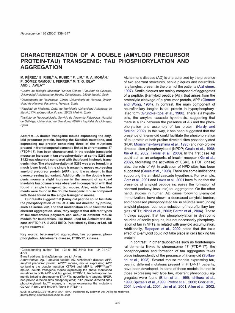
CHARACTERIZATION OF A DOUBLE (AMYLOID PRECURSORPROTEIN-TAU) TRANSGENIC: TAU PHOSPHORYLATION ANDAGGREGATION
M. PÉREZ,a E. RIBE,b A. RUBIO,a F. LIM,a M. A. MORÁN,c
P. GÓMEZ RAMOS,c I. FERRER,d M. T. G. ISLAb
AND J. AVILAa*aCentro de Biología Molecular “Severo Ochoa,” Facultad de Ciencias,Universidad Autónoma de Madrid, Cantoblanco, 28049 Madrid, SpainbDepartmento de Neurología, Clínica Universitaria de Navarra, Univer-sidad de Navarra, Pamplona, Navarra, SpaincFacultad de Medicina, Dpto. de Morfologıa Universidad Autónoma deMadrid, C/Arzobispo Morcillo s/n., 28029 Madrid, SpaindInstituto de Neuropatología, Servicio de Anatomía Patológica, Hospitalde Bellvitge, Universidad de Barcelona, 08907 Hospitalet de Llobregat,Spain
Abstract—A double transgenic mouse expressing the amy-loid precursor protein, bearing the Swedish mutations, andexpressing tau protein containing three of the mutationspresent in frontotemporal dementia linked to chromosome 17(FTDP-17), has been characterized. In the double transgenicmouse an increase in tau phosphorylation at serine S262 andS422 was observed compared with that found in simple trans-genic mice. The phosphorylation at S262 was also found, in amuch lower level, in the single transgenic mouse expressingamyloid precursor protein (APP), and it was absent in thatoverexpressing tau variant. Additionally, in the double trans-genic mouse a slight increase in the amount of sarkosylinsoluble tau polymers was observed in comparison with thatfound in single transgenic tau mouse. Also, wider tau fila-ments were found in the double transgenic mouse comparedwith those found in the single transgenic mouse.
Our results suggest that �-amyloid peptide could facilitatethe phosphorylation of tau at a site not directed by proline,such as serine 262, and that modification could facilitate tauaberrant aggregation. Also, they suggest that different typesof tau filamentous polymers can occur in different mousemodels for tauopathies, like those used for Alzheimer’s dis-ease or FTDP-17. © 2004 IBRO. Published by Elsevier Ltd. Allrights reserved.
Key words: beta-amyloid aggregates, tau polymers, phos-phorylation, Alzheimer’s disease, FTDP-17, kinases.
Alzheimer’s disease (AD) is characterized by the presenceof two aberrant structures, senile plaques and neurofibril-lary tangles, present in the brain of the patients (Alzheimer,1907). Senile plaques are mainly composed of aggregatesof a peptide, �-amyloid peptide (A�), that arises from theproteolytic cleavage of a precursor protein, APP (Glennerand Wong, 1984). In contrast, the main component ofneurofibrillary tangles is tau protein in hyperphosphory-lated form (Grundke-Iqbal et al., 1986). There is a hypoth-esis, the amyloid cascade hypothesis, suggesting thatthere is a link between the presence of A� and the phos-phorylation and assembly of tau protein (Hardy andSelkoe, 2002). In this way, it has been suggested that thepresence of �-amyloid could facilitate the phosphorylationof tau protein at both proline directed sites phosphorylated(PDP; Morishima-Kawashima et al., 1995) and non-prolinedirected sites phosphorylated (NPDP; Geula et al., 1998;Xie et al., 2002; Ferrari et al., 2003). In the first case A�could act as an antagonist of insulin receptor (Xie et al.,2002), facilitating the activation of GSK3, a PDP kinase;also, the role of A� in activation of NPD sites has beensuggested (Geula et al., 1998). There are some indicationssupporting the amyloid cascade hypothesis. For example,Gotz et al., 2001 and Lewis et al., 2001 have found that thepresence of amyloid peptide increases the formation ofaberrant (sarkosyl insoluble) tau aggregates. On the otherhand, studies in human AD cases following �-amyloidimmunization, have shown a decreased amyloid burden,and decreased phosphorylated tau in neurites surroundingamyloid plaques, but not a reduction of neurofibrillary tan-gles (NFTs; Nicoll et al., 2003; Ferrer et al., 2004). Thesefindings suggest that tau phosphorylation in dystrophicneurites of senile plaques, but not necessarily phosphory-lation of tau in NFTs, is related with amyloid accumulation.Additionally, Rapoport et al., 2002 noted that the toxiceffect of �-amyloid could not take place in cells lacking tauprotein.
In contrast, in other tauopathies such as frontotempo-ral dementia linked to chromosome 17 (FTDP-17), thephosphorylation and formation of tau aggregates takesplace independently of the presence of �-amyloid (Spillan-tini et al., 1998). Several mouse models expressing tau,bearing different mutations present in FTDP-17 patients,have been developed. In some of these models, but not inthose expressing wild type tau, aberrant phosphotau ag-gregates were found (Brion et al., 1999; Ishihara et al.,1999; Spittaels et al., 1999; Probst et al., 2000; Gotz et al.,2001; Lewis et al., 2001; Lim et al., 2001; Allen et al., 2002;
*Corresponding author. Tel: �34-91-497-8440; fax: �34-91-497-4799.E-mail address: [email protected] (J. Avila).Abbreviations: A�, �-amyloid peptide; AD, Alzheimer’s disease; APP,amyloid precursor protein; APPsw mouse, mouse expressing APPcontaining the double mutation K670N and M671L; APPsw/tauvlw
mouse, double transgenic mouse expressing the above mentionedmutations in both APP and tau genes; FTDP-17, frontotemporal de-mentia linked to chromosome 17; NFTs, neurofibrillary tangles; NPDP,non-proline directed sites phosphorylated; PDP, proline directed sitesphosphorylated; tauvlw mouse, a mouse expressing the mutationsG272V, P301L and R406W, found in FTDP-17.
Neuroscience 130 (2005) 339–347
0306-4522/05$30.00�0.00 © 2004 IBRO. Published by Elsevier Ltd. All rights reserved.doi:10.1016/j.neuroscience.2004.09.029
339

Tanemura et al., 2002; Tatebayashi et al., 2002; Bouta-jangout et al., 2004).
In this work, we have tested whether the presence ofA�, in aggregated form, may increase the phosphorylationand formation of sarkosyl insoluble tau aggregates. To dothis we have characterized a double transgenic mousegenerated from Tg2576, a mouse expressing APP contain-ing the double mutation K670N and M671L, crossed with amouse expressing the mutations G272V, P301L andR406W, found in FTDP-17. Our results indicate that thepresence of A� aggregates significantly facilitates thephosphorylation of tau at an NPD site like that of Ser 262and the formation of new sarkosyl insoluble aggregates.
EXPERIMENTAL PROCEDURES
Transgenic mice
A double Tg line was obtained by crossing Tg2576 and VLW lines.The progeny included double mutant APP (APPsw)/mutant tau(tauvlw) (hereafter termed APPsw-tauvlw), mutant APP (APPsw),mutant tau (tauvlw), and non-transgenic animals.
Detailed characterization of APPsw and tauvlw lines have beenpreviously published elsewhere (Mullan et al., 1992; Lim et al.,2001). In brief, the Tg2576 line overexpresses human APP con-taining the double Swedish mutation K670N-M671L (swe) underthe control of the hamster prion protein promoter in a C57Bl6jxSJLhybrid background. Tg2576 mice develop cerebral A� depositsand spatial memory deficits in an age-dependent manner (Mullanet al., 1992). The tauvlw line overexpresses three mutations linkedto frontotemporal dementia with Parkinsonism linked to chromo-some 17 (FTDP-17; G272V, P301L and R406W) driven by themouse Thy-1 promoter in a C57Bl6jxCBA hybrid background (Limet al., 2001). Tauvlw mice express human mutant tau at high levelsin cortex and hippocampus with minimal expression in the spinalcord. Immunohistochemical analysis reveals high transgene ex-pression in neuronal cell bodies and neurites in the cortex and thehippocampal formation. Ultrastructural analysis shows a pretangleappearance in neurons expressing mutant tau, with filaments oftau and increased numbers of lysosomes displaying aberrantmorphology similar to those found in AD (Lim et al., 2001). Nine-,16- and 25-month-old mice were used.
Antibodies
The antibodies employed to detect tau in soluble or aggregatedform were BR134 and 7.51, which is directed against themicrotubule-binding region (Novak et al., 1991; a kind gift fromDr. Wischik, UK); antibodies reacting with phosphoresidues 181,202, 262 and 422, were purchased from Calbiochem (Germany).Antibody AT8 was from Innogenetics (Belgium), this antibodyrecognizes phosphorylated tau at residue 202 (Goedert et al.,1995). Antibody 12E8 (Athena Laboratories), which recognizestau phosphorylated at serine 262, was a kind gift from Dr. SeubertFig. 1. Interaction of antibodies reacting with PDP and NPDP sites in
tau protein with tau from different sources. (A) Tau from wild type (wt),transgenic APPsw, transgenic tauvlw and double transgenic APPsw/tauvlw was isolated from a brain cell extract and its reaction with ab AT8was measured by Western blot, using the same amount of protein, asdetermined by measuring tubulin concentration by Western blot. Thebrain of each animal was divided in two halves, and the left half wasused to prepare the brain cell extract and to carry out the Western-blotanalysis. Male mice, 9 months of age were analyzed. For the analysisthe left half of the whole brain was used. Twelve mice were tested,three for each condition (wt, APPsw, tauvlw and APPsw/tauvlw). Theaverage of there determinants is shown. Similar results were found for16-month-old mice. (B) Quantitation of the results from (A). The quan-titation of tau reacting with AT8 was carried out by densitometricanalysis (OD) yielding arbitrary units (au). Since an increase in total
tau levels in transgenic tauvlw (21%�5) and in double transgenicAPPsw/tauvlw (23%�8) were observed, after measuring it by the reac-tion with ab 7.51; the tau phosphorylation data were normalized to thetotal (endogenous and exogenous) tau level in this panel and in panelD. (C) Tau from wt, transgenic APPsw, transgenic tauvlw and doubletransgenic APPsw/tauvlw was isolated from a cell brain extract and itsreaction with ab 12E8 was measured as indicated above. (D) Quan-titation of the results from (C). The age of the analyzed mice was 9months old. Similar results were found for 16-month-old mice. In thelower panel (E) the amount of �-tubulin used a control for proteincontent is shown.
M. Pérez et al. / Neuroscience 130 (2005) 339–347340

(Seubert et al., 1995). A monoclonal antibody directed against�-tubulin (Sigma) was used as an internal control for proteinquantity.
Immunoblots analysis
Samples were run on 10% SDS-PAGE and electrophoreticallytransferred to a nitrocellulose membrane (Schleicher & SchuellGmb). The membrane was blocked by incubation with 5% semifatdried milk in PBS and 0.1% Tween 20, followed by 1-h incubationat room temperature with the primary antibody in PBS. Primaryantibody dilutions were 7.51 (1/100), AT8 (1/100) and 12E8(1/1000). After three washes, the membrane was incubated with ahorseradish peroxidase-anti-rabbit or anti-mouse Ig conjugate(DAKO), followed by several washes in PBS-Tween 20. The mem-brane was then incubated for 1 min in Western Lightning reagents(PerkingElmer Life Sciences). The labeled protein was subjectedto densitometric analysis for quantification. The optical density ofthe stained protein bands, in arbitrary units, was measured. Sta-tistical analysis was performed using Student’s t-test. To deter-mine tau phosphorylation level (at different sites) using ab AT8 orab 12E8, the data were normalized to the total tau levels deter-mined by the interaction of tau protein with ab 7.51.
Isolation of sarkosyl-insoluble tau aggregates
Preparation of sarkosyl-insoluble extracts from mouse brain of16-month-old mice and electron microscopy of filaments was car-ried out as described previously (Greenberg and Davies, 1990).Rabbit anti-tau serum BR134 was used at a dilution of 1/30 forimmunoelectron microscopy.
Ultrastructural analysis in frontal cortex and hippocampuswere performed as indicated in Lim et al. (2001) in 25-month-olddouble transgenic mice and in single transgenic tauvlw mice.
Immunohistochemistry
Serial consecutive sections, 30 �m thick, were processed free-floating with the LSAB method (Dako LSAB� kit) following theinstructions of the supplier (Puig et al., 2004). Briefly, after block-ing endogenous peroxidases, the sections were incubated withnormal serum for 2 h and then incubated overnight at 4 °C withone of the primary antibodies. The phospho-specific tau Thr181and tau Ser202, Ser262, Ser396 and Ser422 polyclonal antibod-ies were incubated at dilutions of 1:500 and 1:100, respectively.The antibody AT8 (direct against phosphoserines 199/202) wasused at a dilution of 1/50. For A� immunohistochemistry, a slightvariation of the protocol was introduced. Before blocking endog-enous peroxidases, sections were incubated with 98% formic acidfor 3 min to enhance antigenicity. The � A1–40 and � A1–42 rabbitpolyclonal antibodies (kindly provided by Dr. M. Sarasa,Zaragoza) were used at a dilution of 1:500. The phosphorylation-dependent anti-SAPK/JNK (Thr183/Tyr185) rabbit polyclonal an-
tibody (SAPK/JNK-P; Cell Signaling) was used at a dilution of1:50. The phosphorylation-dependent rabbit polyclonal antibodyto p-38 (Thr180/Tr182; p38-P; Calbiochem) was used at a dilutionof 1:100. After washing, the sections were then incubated with linksolution (LSAB) and with streptavidin–peroxidase solution for15 min each at room temperature. The peroxidase reaction wasthen visualized, as a dark blue precipitate, with NH4NiSO4 (0.05M) in phosphate buffer (0.1 M), 0.05% diaminobenzidine, NH4Cland 0.01% hydrogen peroxide. Blank sections stained only withthe secondary antibodies were used as negative controls.
RESULTS
Tau phosphorylation in double transgenic mice
Control, transgenic APPsw, transgenic tauvlw, and doubletransgenic APPsw/tauvlw mice were analyzed. In this anal-ysis, the phosphorylation of tau at one proline directed site,that recognized by antibody AT8, and at one non-prolinedirected site, that recognized by ab 12E8, were studied.Fig. 1A shows a slight increase in tau phosphorylation atthe AT8 site in the different single APPsw and in the singletauvlw and double transgenic mice, compared with thecontrol mouse, determined by Western blot, using equalamounts of protein (determined by the measurement of�-tubulin content). Fig. 1B shows the quantitation of thatWestern blot. Also, in single transgenic tauvlw and in thedouble transgenic APPsw/tauvlw the presence of humantau (showing a lower mobility) can be found in phosphor-ylated form. When another antibody reacting with a PDPsite, that reacting with serine 422, was tested, some dif-ferences were found among both the single and the doubletransgenic mice in the reaction with that antibody. In con-trast, little, if any, reaction of tau protein was found in taufrom wild type mice (Table 1).
Tau phosphorylation at one NPDP site was determinedby the reaction with ab 12E8. In this case, a clear increasein tau phosphorylation was found when the control and thedouble transgenic APPsw/tauvlw mice were compared(Fig. 1C). Also, a slight increase in tau phosphorylation intransgenic APPsw, but less evident in transgenic tauvlw,was observed in different samples. The quantitation of thedata is indicated in Fig. 1D. These results suggest that thepresence of amyloid peptide may facilitate tau phosphor-ylation at this NPDP site. The age of the tested mice was9 months. At this age it was previously shown a tau hy-
Table 1. Phosphorylation sites of tau in neurites surrounding amyloid plaques and in neurons in wild type mice (�/�), in APPsw (�/�), tauvlw (�/�)and double transgenic APPsw/tauvlw (�/�) 16-month-old mice
�/� �/� �/� �/�
Plaques Neurons Plaques Neurons Plaques Neurons Plaques Neurons
Thr181 � � �� � � �� �� ��
Ser202 � � � � � �� � ��
Ser262 � � �� � � � �� �
Ser396 � � � � � � � �
Ser422 � � � � � � � �
AT8 � � � � � � � �
M. Pérez et al. / Neuroscience 130 (2005) 339–347 341
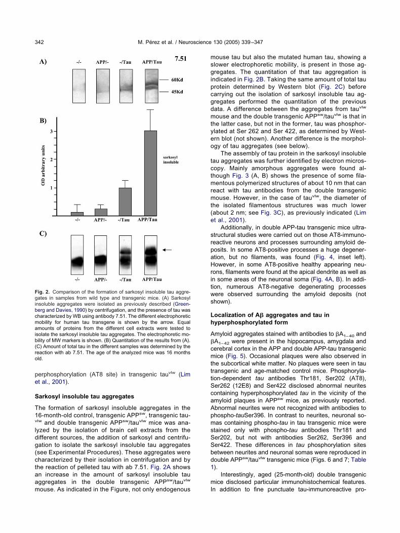
perphosphorylation (AT8 site) in transgenic tauvlw (Limet al., 2001).
Sarkosyl insoluble tau aggregates
The formation of sarkosyl insoluble aggregates in the16-month-old control, transgenic APPsw, transgenic tau-vlw and double transgenic APPsw/tauvlw mice was ana-lyzed by the isolation of brain cell extracts from thedifferent sources, the addition of sarkosyl and centrifu-gation to isolate the sarkosyl insoluble tau aggregates(see Experimental Procedures). These aggregates werecharacterized by their isolation in centrifugation and bythe reaction of pelleted tau with ab 7.51. Fig. 2A showsan increase in the amount of sarkosyl insoluble tauaggregates in the double transgenic APPsw/tauvlw
mouse. As indicated in the Figure, not only endogenous
mouse tau but also the mutated human tau, showing aslower electrophoretic mobility, is present in those ag-gregates. The quantitation of that tau aggregation isindicated in Fig. 2B. Taking the same amount of total tauprotein determined by Western blot (Fig. 2C) beforecarrying out the isolation of sarkosyl insoluble tau ag-gregates performed the quantitation of the previousdata. A difference between the aggregates from tauvlw
mouse and the double transgenic APPsw/tauvlw is that inthe latter case, but not in the former, tau was phosphor-ylated at Ser 262 and Ser 422, as determined by West-ern blot (not shown). Another difference is the morphol-ogy of tau aggregates (see below).
The assembly of tau protein in the sarkosyl insolubletau aggregates was further identified by electron micros-copy. Mainly amorphous aggregates were found al-though Fig. 3 (A, B) shows the presence of some fila-mentous polymerized structures of about 10 nm that canreact with tau antibodies from the double transgenicmouse. However, in the case of tauvlw, the diameter ofthe isolated filamentous structures was much lower(about 2 nm; see Fig. 3C), as previously indicated (Limet al., 2001).
Additionally, in double APP-tau transgenic mice ultra-structural studies were carried out on those AT8-immuno-reactive neurons and processes surrounding amyloid de-posits. In some AT8-positive processes a huge degener-ation, but no filaments, was found (Fig. 4, inset left).However, in some AT8-positive healthy appearing neu-rons, filaments were found at the apical dendrite as well asin some areas of the neuronal soma (Fig. 4A, B). In addi-tion, numerous AT8-negative degenerating processeswere observed surrounding the amyloid deposits (notshown).
Localization of A� aggregates and tau inhyperphosphorylated form
Amyloid aggregates stained with antibodies to �A1–40 and�A1–42 were present in the hippocampus, amygdala andcerebral cortex in the APP and double APP-tau transgenicmice (Fig. 5). Occasional plaques were also observed inthe subcortical white matter. No plaques were seen in tautransgenic and age-matched control mice. Phosphoryla-tion-dependent tau antibodies Thr181, Ser202 (AT8),Ser262 (12E8) and Ser422 disclosed abnormal neuritescontaining hyperphosphorylated tau in the vicinity of theamyloid plaques in APPsw mice, as previously reported.Abnormal neurites were not recognized with antibodies tophospho-tauSer396. In contrast to neurites, neuronal so-mas containing phospho-tau in tau transgenic mice werestained only with phospho-tau antibodies Thr181 andSer202, but not with antibodies Ser262, Ser396 andSer422. These differences in tau phosphorylation sitesbetween neurites and neuronal somas were reproduced indouble APPsw/tauvlw transgenic mice (Figs. 6 and 7; Table1).
Interestingly, aged (25-month-old) double transgenicmice disclosed particular immunohistochemical features.In addition to fine punctuate tau-immunoreactive pro-
Fig. 2. Comparison of the formation of sarkosyl insoluble tau aggre-gates in samples from wild type and transgenic mice. (A) Sarkosylinsoluble aggregates were isolated as previously described (Green-berg and Davies, 1990) by centrifugation, and the presence of tau wascharacterized by WB using antibody 7.51. The different electrophoreticmobility for human tau transgene is shown by the arrow. Equalamounts of proteins from the different cell extracts were tested toisolate the sarkosyl insoluble tau aggregates. The electrophoretic mo-bility of MW markers is shown. (B) Quantitation of the results from (A).(C) Amount of total tau in the different samples was determined by thereaction with ab 7.51. The age of the analyzed mice was 16 monthsold.
M. Pérez et al. / Neuroscience 130 (2005) 339–347342

cesses, varicose and coarse neurites were found sur-rounding many amyloid deposits. Moreover, very strongAT8-immunoreactive neurons were seen in the entorhinalcortex, perirhinal fissure and, rarely, the isocortex and CA1sector of the hippocampus (Fig. 7). These results suggestthat the presence of A� aggregates may facilitate not onlythe tau phosphorylation at a non-proline directed site likeserine 262, but also that at serine 202.
Colocalization of some tau kinases with tauaggregates
It has been already suggested a role for GSK3, in the taumodification observed in tau vlw (Pérez et al., 2003). Thus,we have tested for other additional kinases that may play arole in such modification using as criteria their colocaliza-tion with tau aggregates. These kinases are two membersof MAPkinase family: SAPK/JNK-P and p38-P.
Sections stained with antibodies to SAPK/JNK-P andp38-P permitted the study of the expression of phosphor-ylated (presumably active) kinases in double transgenic
Fig. 3. Tau filaments in sarkosyl insoluble aggregates. The presenceof tau filaments in double transgenic mice was tested in sarkosylinsoluble aggregates by electron microscopy (around 10 nm; A) andimmunoelectron microscopy (B). The diameter of these aggregates ismuch higher than that of the filaments assembled in single tau trans-genic mice (around 2 nm; C). Scale bar�200 nm in A and in B and100 nm in C. (D) No immunogold labeling was found in the absence ofadded tau antibody.
Fig. 4. Tau filaments in neurons of the double transgenic mice. (A)Electron microscopy of an AT8-immunoreactive neuron showing somefilaments immunolabeled with this antibody. Inset at the left shows aclear degenerating process of another neuron showing AT8 immuno-reactivity but without filaments. The white rectangle frames the apicaldendrite enlarged in (B). White arrows point to filaments immuno-stained with AT8 in the cerebral cortex of a double transgenic mouseof 25 months. Calibration bars are indicated in each figure.
M. Pérez et al. / Neuroscience 130 (2005) 339–347 343
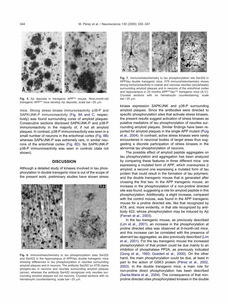
mice. Strong stress kinase immunoreactivity p38-P andSAPK/JNK-P immunoreactivity (Fig. 8A and C, respec-tively) was found surrounding cores of amyloid plaques.Consecutive sections disclosed SAPK/JNK-P and p38-Pimmunoreactivity in the majority of, if not all amyloidplaques. In contrast, p38-P immunoreactivity was seen in asmall number of neurons in the entorhinal cortex (Fig. 8B)whereas SAPk/JNK-P was extremely rare, in similar neu-rons of the entorhinal cortex (Fig. 8D). No SAPK/JNK-Pp38-P immunoreactivity was seen in controls (data notshown).
DISCUSSION
Although a detailed study of kinases involved in tau phos-phorylation in double transgenic mice is out of the scope ofthe present work, preliminary studies have shown stress
kinase expression SAPK/JNK and p38-P surroundingamyloid plaques. Since the antibodies were directed tospecific phosphorylation sites that activate stress kinases,the present results suggest activation of stress kinases asputative mediators of tau phosphorylation of neurites sur-rounding amyloid plaques. Similar findings have been re-ported for amyloid plaques in the single APP mutant (Puiget al., 2004). In contrast, active stress kinases were rarelyencountered in neuronal bodies of target areas thus sug-gesting a discrete participation of stress kinases in theabnormal tau phosphorylation of neurons.
The possible effect of amyloid peptide aggregates ontau phosphorylation and aggregation has been analyzedby comparing these features in three different mice: oneexpressing a mutated form of APP, which overexpress �amyloid; a second one expressing a mutated form of tauprotein that could result in the formation of tau polymers;and the double transgenic mouse that is generated aftercrossing the first two. In the APP transgenic mouse, anincrease in the phosphorylation of a non-proline directedsite was found, suggesting a role for amyloid peptide in thisphosphorylation. Additionally, a slight increase, comparedwith the control mouse, was found in the APP transgenicmouse for a proline directed site, like that recognized byAT8, and, more evidently, in that site recognized by anti-body 422, whose phosphorylation may be induced by A�(Ferrari et al., 2003).
In the tau transgenic mouse, as previously described(Lim et al., 2001), an increase in the phosphorylation atproline directed sites was observed at 9-month-old mice,and this increase can be correlated with the presence ofaberrant tau aggregates, as also previously described (Limet al., 2001). For the tau transgenic mouse the increasedphosphorylation of that protein could be due mainly to aninhibition of phosphatase PP2A, as previously indicated(Gong et al., 1993; Goedert et al., 2000). On the otherhand, the main phosphorylation could be due, at least inpart to the action of GSK3 protein (Pérez et al., 2002,2003). In the double transgenic mice, a main role fornon-proline direct phosphorylation has been described(Santa-Maria et al., 2004). The consequence of that non-proline directed sites phosphorylated kinases in the double
Fig. 5. A� deposits in transgenic APPsw mouse. Nine-month-oldtransgenic APPsw mice develop A� deposits; scale bar�25 �m.
Fig. 6. Immunohistochemistry to tau phosphorylation sites Ser202and Ser422 in the hippocampus of APP/tau double transgenic miceshowing differences in tau phosphorylation in neurites surroundingamyloid plaques and in neurons. The antibody Ser202 (or AT8) stainsphospho-tau in neurons and neurites surrounding amyloid plaques(arrow), whereas the antibody Ser422 recognizes only neurites sur-rounding amyloid plaques but not neurons. Cryostat sections with nohematoxylin counterstaining; scale bar�25 �m.
Fig. 7. Immunohistochemistry to tau phosphorylation site Ser202 inAPP/tau double transgenic mice. AT8 immunohistochemistry showsstrong immunoreactivity in coarse and varicose neurites (arrowheads)surrounding amyloid plaques and in neurons of the entorhinal cortexand hippocampus in 25 months APPsw/tauvlw transgenic mice (A–C).Cryostat sections with no hematoxylin counterstaining; scalebar�25 �m.
M. Pérez et al. / Neuroscience 130 (2005) 339–347344

transgenic mice correlates with an increase in tau aggre-gates that could be assembled by a possible mechanismdifferent from those found in tauvlw. In this way, differencesin the diameter of the filaments assembled in single(around 2 nm) and in double (around 10 nm) transgenicmice were observed. It is not known if the 2 nm filamentscould be the precursors that facilitate the assembly of the10 nm tau filaments.
Taking together our results we can suggest that in ourdouble transgenic mouse two different, and independent,types of tau phosphorylation could occur. In one typeamyloid peptide could facilitate non-proline directed phos-phorylation. It is know that � amyloid pathology is evidentat 6–8 months in APPsw mice, and it could proceed to tauphosphorylation. Also, tau phosphorylation could occur atother different sites, independently of the presence of A�.This type of phosphorylation is mainly a proline directedmodification. In the first type, it is likely that a mimicking of
the effect postulate in the amyloid cascade hypothesis(Selkoe, 1989; Hardy and Selkoe, 2002) occurs, whereasin the other case, the effect could be that occurring in othertype of tauopathies, such as FTDP-17, in which the A� isabsent.
The immunohistochemical analysis described in thiswork supports the earlier hypothesis, since in those placeswhere amyloid deposits are present, an increase in thephosphorylation at non-proline directed sites of tau proteinis found, whereas at those places where no amyloidplaques were found, only tau phosphorylation at prolinedirect sites was observed. When amyloid peptide ispresent, a new phosphorylation at the non-proline directedsite, identified by ab 12E8, occurs. In the tau transgenicmouse, in which amyloid peptide is not present, only anincrease in proline directed sites occurs.
Additionally, our data partially concur with the data ofGotz et al., 2001, and Lewis et al., 2001, since we found anincrease in tau aggregates when amyloid peptide ispresent, and our data further suggest a mechanism forsuch increase based on the phosphorylation of tau at itstubulin-binding region. This may result in an increase of tauthat is not bound to microtubules and which, therefore,could have a greater capacity for self-assembly intopolymers.
In contrast to this, tau phosphorylation at Ser 422,which does not occur in the wild type mouse, takes placein the APPsw transgenic mouse, and this phosphorylationcould be related to the toxic effects of A� (Ferrari et al.,2003). This phosphorylation at serine 422 (that could bemediated by a member of MAP kinase family; Morishima-Kawashima et al., 1995) does not occur in the tauvlw trans-genic mouse where aberrant 2 nm tau filaments are found,although recently Ferrari et al. (2003) have related thistype of phosphorylation with tau aberrant polymerizationinto 10 nm tau filaments.
Finally, serine 422 is also phosphorylated in the doubletransgenic mouse, probably as a consequence of the mod-ification found in APPsw transgenic mice.
In summary, our results suggest a role for A� in facil-itating the phosphorylation of tau at NPDP sites and in aslight increase in the amount of sarkosyl-insoluble taupolymers. The relation between these two features hasrecently been analyzed (Santa-Maria et al., 2004). Also,our results indicate that tau phosphorylation precedes itsassembly and it is compatible with a correlation betweenthose features (Pérez et al., 2003; Alonso et al., 2001).
Additionally, these results also support the data indi-cating that injections of fibrillar A� in the cortex of monkeyresult in tau phosphorylation at serine 262 (Geula et al.,1998). Also, they are consistent with the fact that in modelsfor tauopathies like that for FTDP-17 (Lim et al., 2001),fibrillar tau polymers can be assembled in the absence ofphosphorylation of ser 262, but when an overexpression ofA� takes place, that residue is modified and an increase oftau polymers is found, as shown in this work. Thus, amy-loid peptide could facilitate tau polymerization.
However, it has been shown that soluble amyloid�-protein is increased in frontotemporal dementia with
Fig. 8. p38-P (A, B) and SAP/JNK-P (C, D) immunoreactivity in dou-ble transgenic mice. p38-P and SAPK/JNK-P is observed surroundingcores of amyloid plaques (A, C, asterisks). p38-P immunoreactivity (C)is found in a few neurons of the entorhinal cortex, whereas SAPK/JNK-P immunoreactivity is rarely present, but for very isolated neu-rons, in double transgenic mice. Cryostat sections processed free-floating without counterstaining; scale bar�25 �m.
M. Pérez et al. / Neuroscience 130 (2005) 339–347 345

tau gene mutations (Vitali et al., 2004) and that intra-neuronal amyloid peptide could accumulate in the endo-somal-lysosomal vesicles (Planel et al., 2004), whichare affected in the transgenic tauvlw mice (Lim et al.,2001). Preliminary experimentation suggests that in thedouble transgenic mice there is an increased amyloidprotein aggregation compared with that for single APPtransgenic mice (Ribé et al., unpublished observations).In this way, not only can amyloid peptide facilitate taupolymerization; in addition, the expression of tauvlw maypromote amyloid aggregation.
Acknowledgments—This work was supported in part by FISgrants P1020004; C04-006 and G04-167 to IF; European Com-mission Grant DIADEM QLRT-2000-02362 to TG; and CICYT,C04-006, G04-167, NeuroPharma and Lilly Foundation grants toJ.A.
REFERENCES
Allen B, Ingram E, Takao M, Smith MJ, Jakes R, Virdee K, Yoshida H,Holzer M, Craxton M, Emson PC, Atzori C, Migheli A, Crowther RA,Ghetti B, Spillantini MG, Goedert M (2002) Abundant tau filamentsand nonapoptotic neurodegeneration in transgenic mice express-ing human P301S tau protein. J Neurosci 22:9340–9351.
Alonso A, Zaidi T, Novak M, Grundke-Iqbal I, Iqbal K (2001) Hyper-phosphorylation induces self-assembly of tau into tangles of pairedhelical filaments/straight filaments. Proc Natl Acad Sci USA98:6923–6928.
Alzheimer A (1907) Uber eine eigenartige Erkrankung der Hirninde.Z Psychiatr Psych Gerichtl Med 64:146–148.
Boutajangout A, Authelet M, Blanchard V, Touchet N, Tremp G,Pradier L, Brion JP (2004) Characterisation of cytoskeletal abnor-malities in mice transgenic for wild-type human tau and familialAlzheimer’s disease mutants of APP and presenilin-1. NeurobiolDis 15:47–60.
Brion JP, Tremp G, Octave JN (1999) Transgenic expression of theshortest human tau affects its compartmentalization and its phos-phorylation as in the pretangle stage of Alzheimer’s disease. Am JPathol 154:255–270.
Ferrari A, Hoerndli F, Baechi T, Nitsch RM, Gotz J (2003) beta-Amyloidinduces paired helical filament-like tau filaments in tissue culture.J Biol Chem 278:40162–40168.
Ferrer I, Boada Rovira M, Sanchez Guerra ML, Rey MJ, Costa-JussaF (2004) Neuropathology and pathogenesis of encephalitis follow-ing amyloid-beta immunization in Alzheimer’s disease. BrainPathol 14:11–20.
Geula C, Wu CK, Saroff D, Lorenzo A, Yuan M, Yankner BA (1998)Aging renders the brain vulnerable to amyloid beta-protein neuro-toxicity. Nat Med 4:827–831.
Glenner GG, Wong CW (1984) Alzheimer’s disease and Down’ssyndrome: sharing of a unique cerebrovascular amyloid fibril pro-tein. Biochem Biophys Res Commun 122:1131–1135.
Goedert M, Jakes R, Vanmechelen E (1995) Monoclonal antibody AT8recognises tau protein phosphorylated at both serine 202 andthreonine 205. Neurosci Lett 189:167–169.
Goedert M, Satumtira S, Jakes R, Smith MJ, Kamibayashi C, White CL3rd, Sontag E (2000) Reduced binding of protein phosphatase 2Ato tau protein with frontotemporal dementia and parkinsonismlinked to chromosome 17 mutations. J Neurochem 75:2155–2162.
Gong CX, Singh TJ, Grundke-Iqbal I, Iqbal K (1993) Phosphoproteinphosphatase activities in Alzheimer disease brain. J Neurochem61:921–927.
Gotz J, Chen F, van Dorpe J, Nitsch RM (2001) Formation of neuro-fibrillary tangles in P3011 tau transgenic mice induced by Abeta 42fibrils. Science 293:1491–1495.
Greenberg SG, Davies P (1990) A preparation of Alzheimer pairedhelical filaments that displays distinct tau proteins by polyacryl-amide gel electrophoresis. Proc Natl Acad Sci USA 87:5827–5831.
Grundke-Iqbal I, Iqbal K, Tung YC, Quinlan M, Wisniewski HM,Binder LI (1986) Abnormal phosphorylation of the microtubule-associated protein tau (tau) in Alzheimer cytoskeletal pathology.Proc Natl Acad Sci USA 83:4913–4917.
Hardy J, Selkoe DJ (2002) The amyloid hypothesis of Alzheimer’sdisease: progress and problems on the road to therapeutics. Sci-ence 297:353–356.
Ishihara T, Hong M, Zhang B, Nakagawa Y, Lee MK, Trojanowski JQ,Lee VM (1999) Age-dependent emergence and progression of atauopathy in transgenic mice overexpressing the shortest humantau isoform. Neuron 24:751–762.
Lewis J, Dickson DW, Lin WL, Chisholm L, Corral A, Jones G, Yen SH,Sahara N, Skipper L, Yager D, Eckman C, Hardy J, Hutton M,McGowan E (2001) Enhanced neurofibrillary degeneration intransgenic mice expressing mutant tau and APP. Science293:1487–1491.
Lim F, Hernandez F, Lucas JJ, Gomez-Ramos P, Moran MA, Avila J(2001) FTDP-17 mutations in tau transgenic mice provoke lysoso-mal abnormalities and Tau filaments in forebrain. Mol Cell Neurosci18:702–714.
Morishima-Kawashima M, Hasegawa M, Takio K, Suzuki M,Yoshida H, Titani K, Ihara Y (1995) Proline-directed and non-proline-directed phosphorylation of PHF-tau. J Biol Chem270:823–829.
Mullan M, Crawford F, Axelman K, Houlden H, Lilius L, Winblad B,Lannfelt L (1992) A pathogenic mutation for probable Alzheimer’sdisease in the APP gene at the N-terminus of beta-amyloid. NatGenet 1:345–347.
Nicoll JA, Wilkinson D, Holmes C, Steart P, Markham H, Weller RO(2003) Neuropathology of human Alzheimer disease after immu-nization with amyloid-beta peptide: a case report. Nat Med9:448–452.
Novak M, Jakes R, Edwards PC, Milstein C, Wischik CM (1991)Difference between the tau protein of Alzheimer paired helicalfilament core and normal tau revealed by epitope analysis ofmonoclonal antibodies 423 and 7.51. Proc Natl Acad Sci USA88:5837–5841.
Pérez M, Hernandez F, Gomez-Ramos A, Smith M, Perry G, Avila J(2002) Formation of aberrant phosphotau fibrillar polymers in neu-ral cultured cells. Eur J Biochem 269:1484–1489.
Pérez M, Hernandez F, Lim F, Diaz-Nido J, Avila J (2003) Chroniclithium treatment decreases mutant tau protein aggregation in atransgenic mouse model. J Alzheimers Dis 5:301–308.
Planel E, Miyasaka T, Launey T, Chui DH, Tanemura K, Sato S,Murayama O, Ishiguro K, Tatebayashi Y, Takashima A (2004)Alterations in glucose metabolism induce hypothermia leading totau hyperphosphorylation through differential inhibition of kinaseand phosphatase activities: implications for Alzheimer’s disease.J Neurosci 24:2401–2411.
Probst A, Gotz J, Wiederhold KH, Tolnay M, Mistl C, Jaton AL,Hong M, Ishihara T, Lee VM, Trojanowski JQ, Jakes R, CrowtherRA, Spillantini MG, Burki K, Goedert M (2000) Axonopathy andamyotrophy in mice transgenic for human four-repeat tau protein.Acta Neuropathol (Berl) 99:469–481.
Puig B, Gómez-Isla T, Ribé E, Cuadrado M, Torrejón Escribano B,Dalfó E, Ferrer I (2004) Expression of stress-activated kinasesc-Jun N-terminal kinase (SAPK/JNK-P) and p38 kinase (p38-P),and tau hyperphosphorylation in neurites surrounding A plaques inAPPTg2576 mice. Neuropathol Appl Neurobiol 3065:499–502.
Rapoport M, Dawson HN, Binder LI, Vitek MP, Ferreira A (2002) Tauis essential to beta-amyloid-induced neurotoxicity. Proc Natl AcadSci USA 99:6364–6369.
Santa-Maria I, Hernandez F, Martin CP, Avila J, Moreno FJ (2004)Quinones facilitate the self-assembly of the phosphorylated tubulin
M. Pérez et al. / Neuroscience 130 (2005) 339–347346

binding region of tau into fibrillar polymers. Biochemistry43:2888–2897.
Selkoe DJ (1989) The deposition of amyloid proteins in the agingmammalian brain: implications for Alzheimer’s disease. Ann Med21:73–76.
Seubert P, Mawal-Dewan M, Barbour R, Jakes R, Goedert M,Johnson GV, Litersky JM, Schenk D, Lieberburg I, TrojanowskiJQ, et al. (1995) Detection of phosphorylated Ser262 in fetaltau, adult tau, and paired helical filament tau. J Biol Chem.270:18917–18922.
Spillantini MG, Murrell JR, Goedert M, Farlow MR, Klug A, Ghetti B(1998) Mutation in the tau gene in familial multiple system tauopa-thy with presenile dementia. Proc Natl Acad Sci USA95:7737–7741.
Spittaels K, Van den Haute C, Van Dorpe J, Bruynseels K, Vandez-ande K, Laenen I, Geerts H, Mercken M, Sciot R, Van LommelA, Loos R, Van Leuven F (1999) Prominent axonopathy inthe brain and spinal cord of transgenic mice overexpress-
ing four-repeat human tau protein. Am J Pathol 155:2153–2165.
Tanemura K, Murayama M, Akagi T, Hashikawa T, Tominaga T,Ichikawa M, Yamaguchi H, Takashima A (2002) Neurodegenera-tion with tau accumulation in a transgenic mouse expressing V337M human tau. J Neurosci 22:133–141.
Tatebayashi Y, Miyasaka T, Chui DH, Akagi T, Mishima K, IwasakiK, Fujiwara M, Tanemura K, Murayama M, Ishiguro K, Planel E,Sato S, Hashikawa T, Takashima A (2002) Tau filament forma-tion and associative memory deficit in aged mice expressingmutant (R406W) human tau. Proc Natl Acad Sci USA99:13896 –13901.
Vitali A, Piccini A, Borghi R, Fornaro P, Siedlak SL, Smith MA,Gambetti P, Ghetti B, Tabaton M (2004) Soluble amyloid beta-protein is increased in frontotemporal dementia with tau genemutations. J Alzheimers Dis 6:45–51.
Xie L, Helmerhorst E, Taddei K, Plewright B, Van Bronswijk W,Martins R (2002) Alzheimer’s beta-amyloid peptides compete forinsulin binding to the insulin receptor. J Neurosci 22:RC221.
(Accepted 13 September 2004)(Available online 11 November 2004)
M. Pérez et al. / Neuroscience 130 (2005) 339–347 347


Current Molecular Medicine 2006, 6, 423-428 423
Acetylcholine Receptors and Tau Phosphorylation
A. Rubio, M. Pérez and J. Ávila*
Centro de Biología Molecular Severo Ochoa (CBMSO), Universidad Autónoma de Madrid, Madrid,Spain
Abstract: Alzheimer's disease (AD) is characterized by the presence, in the brain of the patients, oftwo aberrant structures: intracellular neurofibrillary tangles (NFTs), containing an abnormalhyperphosphorylated form of tau protein, and extracellular senile plaques (SPs), mainly composed byfibrillar amyloid β peptide. Another feature of AD is the neurodegeneration and dysfunction of basalforebrain cholinergic system. A possible connection among those AD characteristics could occur.Thus, the purpose of this short review is to summarize the involvement of nicotinic (nAChR) andmuscarinic (mAChR) receptors on tau phosphorylation, in a direct way, or through the previousinteraction of some of these receptors with amyloid β. Several studies have demonstrated that nAChRactivation results in a significantly increase of tau phosphorylation, whereas mAChR activation, mayprevent tau phosphorylation.
Keywords: Alzheimer's disease, tau phosphorylation, amyloid β peptide, nicotinic receptors, muscarinicreceptors, acetylcholine.
INTRODUCTION Another feature of AD is the neurodegenerationand dysfunction of basal forebrain cholinergicsystem. Long time ago, biochemical investigationsindicated that the disease was associated with a lossof acetylcholine in the cerebral cortex [8,9] and,afterwards, it was indicated that the severity ofdementia was correlated with the extent of thecholinergic loss [10] and the reduction in the activityof choline acetyltransferase in the cortex [11]. Inaddition, the involvement of cholinergic system in ADwas also supported by the use of inhibitors ofacetylcholinesterase (such as tacrine, rivastigmine,donepezil and galantamine) for the treatment of thedisease [12].
Alzheimer's disease (AD) is a senile dementiacharacterized by a progressive loss of memorytogether with cognitive and behavioural impairments.Neuropathologic hallmarks of AD include intracellularneurofibrillary tangles (NFTs) and senile plaques(SPs) [1].
NFTs are composed by paired helical filaments(PHF), containing an abnormal hyperphosphorylatedform of the microtubule-associated protein, tau (for areview see [2]). The modification of tau by phos-phorylation affects its interaction with microtubulesand facilitates tau assembly into PHF [3]. Tauphosphorylation depends on the balance of kinasesand phosphatases activities [4,5]. In the brain of anAD patient, NFTs are particularly abundant in theentorhinal cortex, the hippocampus and theassociation cortices. The number of cortical NFTscorrelates positively with the severity of dementia [6].
Acetylcholine is implicated in different cognitivefunctions such as learning and memory [13]. Oncereleased, acetylcholine can bind to two distinctreceptors subtypes: nicotinic acetylcholine receptors(nAChRs) and muscarinic acetylcholine receptors(mAChRs). Although nAChRs are ionotropicreceptors, mAChRs are metabotropic. nAChRs areligand-gated ion channels with a pentamericstructure formed by a combination of five differentsubunits (α, β, γ, δ and ε) (for a review see [14]).Each channel can be homomeric or heteromeric,when they are respectively formed by the samesubunit or different subunits combined. The α7 andα4β2 nAChRs subtypes are involved in thepathogenesis of AD since they are the mostabundant subtypes in brain regions affected by thisdementia [15, 16]. mAChRs are divided in fivesubtypes (M1-M5), and all of them are present in theCNS [17, 18]. Among the five different subtypes ofmAChRs, the M1 mAChR, highly expressed in thecerebral cortex and the hippocampus, seems to bespecifically implicated in memory processes relevantto AD [19].
SPs are extracellular lesions composed primarilyby a core of fibrillar amyloid β peptide surrounded byreactive astrocytes, microglial cells and dystrophicneurites. The major amyloid peptides in the plaquesare amyloid β (1-42) and amyloid β (1-40). Thesepeptides are generated by proteolytic cleavage ofthe amyloid precursor protein (APP). There are twodistinct α-secretase or β-secretase pathways.Cleavage of APP by α and γ -secretase preventsamyloid β formation, whereas β and γ -secretaseroute leads to a soluble APP and amyloid β (for areview see [7]).
*Address correspondence to this author at the Centro de BiologíaMolecular Severo Ochoa (CBMSO), Facultad de Ciencias, UniversidadAutónoma, 28049, Cantoblanco, Madrid, Spain; Tel: 913975070; Fax:913974799; E-mail: [email protected]
1566-5240/06 $50.00+.00 © 2006 Bentham Science Publishers Ltd.

424 Current Molecular Medicine, 2006, Vol. 6, No. 4 Rubio et al.
Since the molecular mechanism by whichcholinergic neurons dystrophy occurs is unclear andtaking into account the probable neurotoxicity ofamyloid β peptide and hyperphosphorylated tau, thisshort review will present the involvement of nAChRand mAChR on tau phosphorylation in a direct wayor through the previous interaction with amyloid β.
nicotine or epibatidine on up-regulation of nicotinicreceptors. In addition, since several cholinesteraseinhibitors have been shown to interact with anallosteric site on the nicotinic receptors different fromthat for acetylcholine [28-30], the effect ofcholinesterase inhibitors on tau phosphorylation wasalso studied. Similar to nicotine, treatments withtacrine, donepezil and galantamine all increased thelevels of phosphorylated tau [27]. Wang et al. [31]showed that nicotine and epibatidine stimulatetransient tau phosphorylation at three proline-directed sites (Ser202, Thr181 and Thr231) in SK-N-MC human neuroblastoma cells. So, both in vitrostudies demonstrate that nicotinic receptor activationincreases tau phosphorylation. Consistently, usingSH-SY5Y human neuroblastoma cells, we found thatnicotine treatments increased tau phosphorylation atSer 262 site (A. R. unpublished data).
NICOTINIC RECEPTORS AND TAU PROTEINPHOSPHORYLATION
A selective loss of nAChRs in the cortex and thehippocampus of AD brains had been extensivelystudied using different techniques: by immunohisto-chemistry and western-blot analysis [16, 20, 21], byquantifying the binding of specific radiolabeledligands [22, 23, 24] or by positron emission tomogra-phy (PET) [25]. This consistent result suggests apotential relationship between nAChRs and ADneuropathology. Interestingly, a possible association between
smoking and the number of NFTs has beeninvestigated in different post-mortem studies. In onehand, a positive correlation was found in men brainsbetween NFTs and the amount smoked [32]. Incontrast, neuropathologic data from the HonoluluHeart Program supported that the effects of smokingare specific for plaque more than NFTs. In this case,the number of neuritic plaques increased with thenumber of cigarettes consumed [33].
Nevertheless, few data are available about theeffects of nAChR activation and tau pathology(revised in [26]) (Table 1). Treatments with nicotineand epibatidine in SH-SY5Y human neuroblastomacells increased the levels of phosphorylated taurecognized by AT8 and AT270 antibodies anddecreased those of nonphosphorylated taurecognized by tau1 antibody [27]. This increase intau phosphorylation was prevented when theantagonists D-tubocurarine and mecamylaminewhere added, but failed to block the effect of
Moreover, recent findings indicated that chronicnicotine treatment exacerbates tau pathology in vivo
Table 1. Effect of Different Treatments on Tau Phosphorylation
Experimental models Treatments Tau phosphorylation (sites) References
nAChR agonists SH-SY5Y cells Nicotine and Epibatidine Increase (AT8, AT270) Hellström-Lindahl et al.2000 [27]
SH-SY5Y cells Tacrine, Donepezil,Galantamine
Increase (AT8, AT270) Hellström-Lindahl et al.2000 [27]
SK-N-MC cells Nicotine and Epibatidine Increase (Ser202, Thr181, Thr231) Wang et al. 2003 [31]
APP/tau mice Nicotine Increase (AT8, AT270) Oddo et al. 2005 [34]
mAChR agonists M1 transfected PC12 cells Carbachol and AF102B Decrease (AT8, PHF1) Sadot et al. 1996 [46]
M1 transfected CHO cells M1 agonists Decrease (AT8, tau1) Forlenza et al. 2000 [47]
Cortical neurons Carbachol Decrease (AT8, tau1) Forlenza et al. 2000 [47]
ApoE deficient mice AF150 (S) Decrease (AT8, AT270, SMI32, 37, 34,P262-7)
Genis et al. 1999 [48]
APP/tau mice AF267B Decrease (AT8) Caccamo et al. 2006 [50]
Amyloid Cultured neurons Amyloid β fibrils Increase (Ser202, Ser396/ Ser404) Busciglio et al. 1995 [69]
Hippocampal neurons Amyloid β (25-35) increase Takashima et al. 1996[70]
SH-SY5Y cells Amyloid β (25-35) Increase (PHF1) Shea et al. 1997 [71]
SN56 cells Amyloid β (1-40) Increase (AT8, PHF1) Le et al. 1997 [72]
SK-N-MC cells Amyloid β (1-42) Increase (Ser202, Thr181, Thr231) Wang et al. 2003 [31]
Hippocampal synaptosomes Amyloid β (1-42) Increase (Ser202, Thr181, Thr231) Wang et al. 2003 [31]

Acetylcholine Receptors and Tau Phosphorylation Current Molecular Medicine, 2006, Vol. 6, No. 4 425
[34]. A transgenic model of AD (3xTg-AD) expresseshuman APP with Swedish mutations and tau proteinbearing P301L mutation. The aged 3xTg-AD micepresent amyloid plaques and tangles andprogressive deficits in cognition and LTP [35-37].Nicotine administration to 3xTg-AD and nonTg miceinduces an up-regulation of nAChRs [34]. Thisfinding was consistent with previous reports showingthat the density of nAChR in human brain isincreased in smokers probably by the nicotinepresent in the tobacco smoke [38, 39]. In addition,chronic nicotine treatment to 3xTg-AD mice increasestau aggregation and tau phosphorylation at the sitesrecognized by AT8 and AT270 antibodies [34].
phosphorylation at sites recognized by AT8 and tau1antibodies was also demonstrated in primary corticalcultures treated with carbachol [47]. Furthermore, itwas shown that prolonged treatments ofapolipoprotein E (apoE)-deficient mice with the M1mAChR agonist AF150(S) reduces thehyperphosphorylation of tau protein associated withthese mice and localized N-terminally to themicrotubule binding domain of tau protein [48]. Inaddition, exposure of these mice to AF150(S)abolished their memory deficits and resulted inrecovery of their brain cholinergic markers [49].Moreover, our muscarine treatments in SH-SY5Yhuman neuroblastoma cells decreases tauphosphorylation at Ser 262 site (A. R. unpublisheddata). Thus, activation of M1 mAChR might provide anovel treatment strategy for AD due to its beneficialeffect on tau phosphorylation (Table 1).
The exact molecular mechanism by which nicotinicreceptor activation leads to changes in tauphosphorylation remains unclear. Nicotinic receptorsare permeable for Ca2+ and the consequentincrease in intracellular Ca2+ can activate differentspecific signalling cascades (reviewed in [40]). ManyCa2+ -dependent kinases are known to phos-phorylate tau in vivo such as cdk5, ERK1, ERK2,GSK3β and p38-MAP kinases and could beresponsible for tau phosphorylation-induced bynicotinic activation. Oddo et al. demonstrated thatthe activation of p38-MAP kinase seemed to causethe increase in tau phosphorylation due to thechronic nicotine intake in 3xTg-AD transgenic mice[34].
Interestingly, the therapeutic potential of selectiveM1 mAChR agonists has been recently supported byin vivo studies. Caccamo et al. [50] investigatedwhether the selective M1 muscarinic agonist AF267Bcan affect AD pathologies. 6-months old 3xTg-ADmice were treated chronically with AF267B. AF267Bintake by transgenic mice reduces tau phosphoryl-ation in the cortex and the hippocampus, asindicated by AT8 antibody. In contrast, the onset oftau pathology was accelerated in 3xTg-AD micetreated with dicyclomine, an M1 antagonist. In thiscase, tau phosphorylation was significantly increasedin the hippocampus using the AT8 antibody. Tauphosphorylation was also detected in the cortex andthe amygdala using AT8 and PHF1 antibodies,whereas no PHF1 or AT8-positive neurons weredetected at this age in these brain regions inuntreated-3xTg-AD. In agreement with previous invitro studies [47], this work concluded that thedecrease in tau phosphorylation induced by AF267Bwas mediated by the reduction of GSK3β activity.
MUSCARINIC RECEPTORS AND TAUPROTEIN PHOSPHORYLATION
Although most studies suggest that the mAChRdensity is not altered between AD and control brains[41, 42], functional studies of muscarinic M1 receptor-mediated signal transduction have shown a loss ofreceptor-G protein coupling [43, 44]. Since the M1subtype seems to be the most relevant in ADpathology, most of the studies presented to evaluatethe relationship between mAChRs and tauphosphorylation are focused on this receptor. Insummary, activation of M1 mAChR decreases tauprotein phosphorylation suggesting that the decrea-sed cholinergic activity associated with AD couldcontribute to a destabilization of the microtubulenetwork (revised in [45]). A stimulation of PC12 cells,stably transfected with the cDNA encoding M1mAChR, with two acetylcholine agonists, carbacholand AF102B, results in a decrease of tauphosphorylation, as indicated by the reaction of tauwith two specific phosphotau antibodies, AT8 andPHF1 [46]. The muscarinic effect was both time anddose dependent, and was blocked by atropine.Some work has been done later to understand themechanism of action of muscarinic agonists on tauphosphorylation. In non-neuronal cells expressingthe M1 mAChR, a range of M1 agonists reducedtransiently-expressed tau phosphorylation, via GSK-3β inhibition [47]. This inhibition could be due toprotein kinase C (PKC) activation, itself activatedfollowing muscarinic signalling. The reduction on tau
AMYLOID AND CHOLINERGIC RECEPTORSNicotinic Receptors
Different in vitro analysis reported that nicotinehas antiamyloidogenic effects. Based on circulardichroism and spectroscopic studies, nicotine seemsto inhibit amyloid β fibril formation from amyloid βpeptide [51, 52]. In addition, Ono et al. confirmedthat the inhibition of amyloid β fibril formation bynicotine was dose dependent and that nicotinedisrupted preformed amyloid β fibrils [53]. Zeng et al.suggested that nicotine inhibits amyloid β fibrilsformation by binding to a soluble β sheet aggregate[52]. The effect of nicotine could be also attributedto N-methylpyrrolidine moieties of nicotine [53].
Several studies have reported the effect ofnicotine on APP processing. Nicotine treatment toPC12 cells, that constitutively express APP andnAChRs [54], increased the release of soluble APPin a concentration-dependent manner [55]. Theeffect of nicotine was attenuated with the nAChR

426 Current Molecular Medicine, 2006, Vol. 6, No. 4 Rubio et al.
antagonist mecamylamine. Thus, nicotine couldenhance the release of APPs through the specificinteraction with nAChRs. The effect of nicotine onAPP was also observed in stably transfected PC12cells with the Swedish APP mutation [56]. Someexperiments in primary cultured neuron cellsindicated that nicotine protects from amyloid β andAPP metabolic fragments-induced neurotoxicity,probably through the increased release of APPs,which has neurotrophic and neuroprotective activities[57]. Other studies also support the therapeuticpotential of nicotine for AD. In this case, nicotinetreatment to rats reduces the secretion of APPcontaining amyloid β [58].
inflammatory disorders (revised in [64]). Acetylcholineand nicotine inhibit the production of pro-inflam-matory cytokines from macrophages through amechanism dependent on α7 nAChRs named the“nicotinic anti-inflammatory pathway” [64].
Muscarinic ReceptorsSome evidences presented so far demonstrate
that APP processing could be regulated bymuscarinic receptors. In different cell lines, musc-arinic agonists enhanced the release of soluble APP[65-67]. The use of selective M1 agonists with theconsequent M1 mAChRs activation increased thesecretion of soluble APP and decreased amyloid βproduction [68].
To date, several in vivo studies using transgenicmodels of AD have been carried out in order todetermine whether nicotine administration affects thepathology of the dementia. Nordberg et al. [59] havetreated with nicotine, transgenic mice that over-express human APP bearing a Swedish mutation[Tg(Hu.APP695.K670N-M671L)2576]. These trans-genic mice (APPsw) develop amyloid plaques andpresent cognitive impairments [60]. Nicotine treat-ment was administered in the drinking water of 9-month-old APPsw mice during 5.5 months. Thetreatment decreases significantly and dramatically(by more than 80%) amyloid plaque deposition and itreduces the levels of insoluble amyloid β 1−40 and 1−42 However, no significant change in soluble amyloidβ 1-40 or 1-42 levels was observed. The same groupdemonstrated how effective was a short-termnicotine treatment (for ten days) in reducing insolubleamyloid β 1-40 and 1-42 peptides in APPsw mice[61]. In addition, amyloid β associated with cerebralvessels was decreased in APPsw mice treatedchronically with nicotine. These results support thatnicotine may be used for the treatment of AD inorder to lower the amyloid β plaque burden in thebrain. In contrast, as previously reported in thisreview, Oddo et al. [34] suggested that nicotinetherapy in AD should be revaluated since chronicnicotine treatment (for 5 months) to 1 month old3xTg-AD mice increases tau pathology. Also, Oddoet al. [34] confirmed that chronic nicotine treatmentdid not change the soluble levels of amyloid βpeptide.
As previously discussed, the therapeutic benefitsof AF267B had been recently tested in a transgenicmodel of AD [50]. AF267B intake by 3xTg-AD micereduces amyloid β deposition in the cortex and thehippocampus, but not in the amygdala. AF267Baffects APP processing causing a selectiveactivation of ADAM 17 enzyme. This finding was inagreement with the amelioration in cognitive deficitsobserved in treated mice in a spatial task but not in acontextual fear conditioning [50]. Thus, the selectiveM1 muscarinic agonist AF267B have shownbeneficial effects on the major hallmarks of AD and itrepresents a potential candidate for AD therapy.
TAU PROTEIN AND AMYLOID INTERACTIONS
The relationship between amyloid β depositionand neurofibrillary tangle formation represents acentral issue in the pathogenesis of AD. Numerousstudies suggest that amyloid β enhances tauphosphorylation (Table 1). In primary culturedneurons, amyloid β fibrils, but not soluble oramorphous-aggregated, induces the phosphoryl-ation of tau protein at Ser 202 and Ser 396/ Ser 404and the subsequent accumulation of tau protein inthe somatodendritic cell compartment resulting in aloss of microtubule binding capacity [69]. Moreover,treatments of rat hippocampal neurons with amyloidβ peptide (25-35) increases tau phosphorylation andneuronal death [70]. Using SH-SY5Y humanneuroblastoma cells, Shea et al. demonstrated thatamyloid β peptide (25-35) exposure also causes anincrease in tau phosphorylation at the siterecognized by PHF1 antibody [71]. SN56 hybridseptal cell line treated with aggregated amyloid βpeptide (1-40) presents cell injury and enhances tauphosphorylation at AT8 and PHF1 sites as well astotal tau [72]. Interestingly, the contribution of tauphosphorylation in amyloid β-induced neurotoxicityhas been controversial. Ekinci et al. [73] hasreported that tau hyperphosphorylation, although aconsequence of amyloid β exposure, does notdirectly mediate the acute degeneration of culturedneurons. Unexpectedly, the healthiest-appearingmurine cortical neurons after amyloid β treatment
Others approaches have tried to elucidate theeffect of nicotine on AD. Although manyepidemiological studies focused on the associationbetween smoking and AD had been reported,conclusions reached remain contradictories (see [62]for a review). Briefly, these mixed results could beexplained by the analysis used: cohort studiesshowed that smoking increases the risk for ADwhereas case-control studies concluded thatsmoking was protective [63].
Finally, it is worthy to point out that the possibledeleterious effects of nicotine for AD treatment maybe due to its non selectivity for nAChRs. The designof selective nicotinic agonists would probably benefitthe AD treatment. Notably, similar strategies aresupported for treatments against infectious and
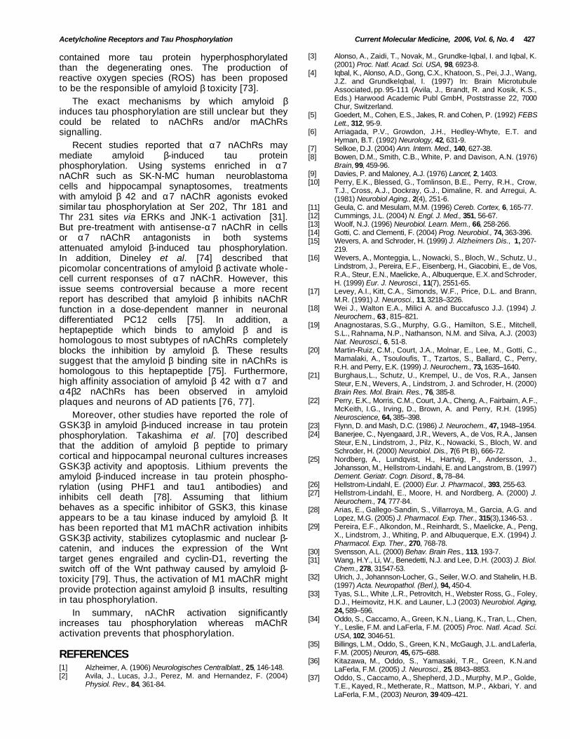
Acetylcholine Receptors and Tau Phosphorylation Current Molecular Medicine, 2006, Vol. 6, No. 4 427
contained more tau protein hyperphosphorylatedthan the degenerating ones. The production ofreactive oxygen species (ROS) has been proposedto be the responsible of amyloid β toxicity [73].
[3] Alonso, A., Zaidi, T., Novak, M., Grundke-Iqbal, I. and Iqbal, K.(2001) Proc. Natl. Acad. Sci. USA, 98, 6923-8.
[4] Iqbal, K., Alonso, A.D., Gong, C.X., Khatoon, S., Pei, J.J., Wang,J.Z. and GrundkeIqbal, I. (1997) In: Brain MicrotubuleAssociated, pp. 95-111 (Avila, J., Brandt, R. and Kosik, K.S.,Eds.) Harwood Academic Publ GmbH, Poststrasse 22, 7000Chur, Switzerland.The exact mechanisms by which amyloid β
induces tau phosphorylation are still unclear but theycould be related to nAChRs and/or mAChRssignalling.
[5] Goedert, M., Cohen, E.S., Jakes, R. and Cohen, P. (1992) FEBSLett., 312, 95-9.
[6] Arriagada, P.V., Growdon, J.H., Hedley-Whyte, E.T. andHyman, B.T. (1992) Neurology, 42, 631-9.Recent studies reported that α7 nAChRs may
mediate amyloid β-induced tau proteinphosphorylation. Using systems enriched in α7nAChR such as SK-N-MC human neuroblastomacells and hippocampal synaptosomes, treatmentswith amyloid β 42 and α7 nAChR agonists evokedsimilar tau phosphorylation at Ser 202, Thr 181 andThr 231 sites via ERKs and JNK-1 activation [31].But pre-treatment with antisense-α7 nAChR in cellsor α7 nAChR antagonists in both systemsattenuated amyloid β-induced tau phosphorylation.In addition, Dineley et al. [74] described thatpicomolar concentrations of amyloid β activate whole-cell current responses of α7 nAChR. However, thisissue seems controversial because a more recentreport has described that amyloid β inhibits nAChRfunction in a dose-dependent manner in neuronaldifferentiated PC12 cells [75]. In addition, aheptapeptide which binds to amyloid β and ishomologous to most subtypes of nAChRs completelyblocks the inhibition by amyloid β. These resultssuggest that the amyloid β binding site in nAChRs ishomologous to this heptapeptide [75]. Furthermore,high affinity association of amyloid β 42 with α7 andα4β2 nAChRs has been observed in amyloidplaques and neurons of AD patients [76, 77].
[7] Selkoe, D.J. (2004) Ann. Intern. Med., 140, 627-38.[8] Bowen, D.M., Smith, C.B., White, P. and Davison, A.N. (1976)
Brain, 99, 459-96.[9] Davies, P. and Maloney, A.J. (1976) Lancet, 2, 1403.[10] Perry, E.K., Blessed, G., Tomlinson, B.E., Perry, R.H., Crow,
T.J., Cross, A.J., Dockray, G.J., Dimaline, R. and Arregui, A.(1981) Neurobiol Aging., 2(4), 251-6.
[11] Geula, C. and Mesulam, M.M. (1996) Cereb. Cortex, 6, 165-77.[12] Cummings, J.L. (2004) N. Engl. J. Med., 351, 56-67.[13] Woolf, N.J. (1996) Neurobiol. Learn. Mem., 66, 258-266.[14] Gotti, C. and Clementi, F. (2004) Prog. Neurobiol., 74, 363-396.[15] Wevers, A. and Schroder, H. (1999) J. Alzheimers Dis., 1, 207-
219.[16] Wevers, A., Monteggia, L., Nowacki, S., Bloch, W., Schutz, U.,
Lindstrom, J., Pereira, E.F., Eisenberg, H., Giacobini, E., de Vos,R.A., Steur, E.N., Maelicke, A., Albuquerque, E.X. and Schroder,H. (1999) Eur. J. Neurosci., 11(7), 2551-65.
[17] Levey, A.I., Kitt, C.A., Simonds, W.F., Price, D.L. and Brann,M.R. (1991) J. Neurosci., 11, 3218–3226.
[18] Wei J., Walton E.A., Milici A. and Buccafusco J.J. (1994) J.Neurochem., 63 , 815–821.
[19] Anagnostaras, S.G., Murphy, G.G., Hamilton, S.E., Mitchell,S.L., Rahnama, N.P., Nathanson, N.M. and Silva, A.J. (2003)Nat. Neurosci., 6, 51-8.
[20] Martin-Ruiz, C.M., Court, J.A., Molnar, E., Lee, M., Gotti, C.,Mamalaki, A., Tsouloufis, T., Tzartos, S., Ballard, C., Perry,R.H. and Perry, E.K. (1999) J. Neurochem., 73, 1635–1640.
[21] Burghaus, L., Schutz, U., Krempel, U., de Vos, R.A., JansenSteur, E.N., Wevers, A., Lindstrom, J. and Schroder, H. (2000)Brain Res. Mol. Brain. Res., 76, 385-8.
[22] Perry, E.K., Morris, C.M., Court, J.A., Cheng, A., Fairbairn, A.F.,McKeith, I.G., Irving, D., Brown, A. and Perry, R.H. (1995)Neuroscience, 64, 385–398.Moreover, other studies have reported the role of
GSK3β in amyloid β-induced increase in tau proteinphosphorylation. Takashima et al. [70] describedthat the addition of amyloid β peptide to primarycortical and hippocampal neuronal cultures increasesGSK3β activity and apoptosis. Lithium prevents theamyloid β-induced increase in tau protein phospho-rylation (using PHF1 and tau1 antibodies) andinhibits cell death [78]. Assuming that lithiumbehaves as a specific inhibitor of GSK3, this kinaseappears to be a tau kinase induced by amyloid β. Ithas been reported that M1 mAChR activation inhibitsGSK3β activity, stabilizes cytoplasmic and nuclear β-catenin, and induces the expression of the Wnttarget genes engrailed and cyclin-D1, reverting theswitch off of the Wnt pathway caused by amyloid β-toxicity [79]. Thus, the activation of M1 mAChR mightprovide protection against amyloid β insults, resultingin tau phosphorylation.
[23] Flynn, D. and Mash, D.C. (1986) J. Neurochem., 47, 1948–1954.[24] Banerjee, C., Nyengaard, J.R., Wevers, A., de Vos, R.A., Jansen
Steur, E.N., Lindstrom, J., Pilz, K., Nowacki, S., Bloch, W. andSchroder, H. (2000) Neurobiol. Dis., 7(6 Pt B), 666-72.
[25] Nordberg, A., Lundqvist, H., Hartvig, P., Andersson, J.,Johansson, M., Hellstrom-Lindahi, E. and Langstrom, B. (1997)Dement. Geriatr. Cogn. Disord., 8, 78–84.
[26] Hellstrom-Lindahl, E. (2000) Eur. J. Pharmacol., 393, 255-63.[27] Hellstrom-Lindahl, E., Moore, H. and Nordberg, A. (2000) J.
Neurochem., 74, 777-84.[28] Arias, E., Gallego-Sandin, S., Villarroya, M., Garcia, A.G. and
Lopez, M.G. (2005) J. Pharmacol. Exp. Ther., 315(3),1346-53. .[29] Pereira, E.F., Alkondon, M., Reinhardt, S., Maelicke, A., Peng,
X., Lindstrom, J., Whiting, P. and Albuquerque, E.X. (1994) J.Pharmacol. Exp. Ther., 270, 768-78.
[30] Svensson, A.L. (2000) Behav. Brain Res., 113, 193-7.[31] Wang, H.Y., Li, W., Benedetti, N.J. and Lee, D.H. (2003) J. Biol.
Chem., 278, 31547-53.[32] Ulrich, J., Johannson-Locher, G., Seiler, W.O. and Stahelin, H.B.
(1997) Acta. Neuropathol. (Berl.), 94, 450-4.[33] Tyas, S.L., White ,L.R., Petrovitch, H., Webster Ross, G., Foley,
D.J., Heimovitz, H.K. and Launer, L.J (2003) Neurobiol. Aging,24, 589–596.In summary, nAChR activation significantly
increases tau phosphorylation whereas mAChRactivation prevents that phosphorylation.
[34] Oddo, S., Caccamo, A., Green, K.N., Liang, K., Tran, L., Chen,Y., Leslie, F.M. and LaFerla, F.M. (2005) Proc. Natl. Acad. Sci.USA, 102, 3046-51.
REFERENCES[35] Billings, L.M., Oddo, S., Green, K.N., McGaugh, J.L. and Laferla,
F.M. (2005) Neuron, 45, 675–688.[36] Kitazawa, M., Oddo, S., Yamasaki, T.R., Green, K.N.and
LaFerla, F.M. (2005) J. Neurosci., 25, 8843–8853.[1] Alzheimer, A. (1906) Neurologisches Centralblatt., 25, 146-148.[2] Avila, J., Lucas, J.J., Perez, M. and Hernandez, F. (2004)
Physiol. Rev., 84, 361-84.[37] Oddo, S., Caccamo, A., Shepherd, J.D., Murphy, M.P., Golde,
T.E., Kayed, R., Metherate, R., Mattson, M.P., Akbari, Y. andLaFerla, F.M., (2003) Neuron, 39 409–421.

428 Current Molecular Medicine, 2006, Vol. 6, No. 4 Rubio et al.
[38] Benwell, M., Balfour, D., Andersson, J. (1988) J. Neurochem.,50, 1243-7.
[59] Nordberg, A., Hellström-Lindahl, E., Lee, M., Johnson, M.,Mousavi, M., Hall, R., Perry, E., Bednar, I. and Court, J. (2002)J. Neurochem., 81, 655–658.[39] Court, J. A., Lloyd, S., Thomas, N., Piggott, M. A., Marshall, E.
F., Morris, C. M., Lamb, H., Perry, R. H., Johnson, M. andPerry, E. K. (1998) Neuroscience, 87, 63-78.
[60] Hsiao, K., Chapman, P., Nilsen, S., Eckman, C., Harigaya, Y.,Younkin, S., Yang, F. and Cole, G. (1996) Science, 274, 99–102.
[40] Dajas-Bailador, F. and Wonnacott, S. (2004) Trends Pharmacol.Sci., 25, 317-24.
[61] Hellstrom-Lindahl, E., Court, J., Keverne, J., Svedberg, M., Lee,M., Marutle, A., Thomas, A., Perry, E., Bednar, I. and Nordberg,A. (2004) Eur. J. Neurosci., 19(10),2703-10.[41] Sugaya, K., Giacobini, E. and Chiappinelli, V.A. (1990) J.
Neurosci. Res., 27, 349–359. [62] Oddo, S. and Laferla, F.M. (2006) J. Physiol. Paris., 99, 172-9.[42] Nordberg, A., Alafuzoff, I. and Winblad, B., (1992) J. Neurosci.
Res., 31, 103–111.[63] Almeida, O.P., Hulse, G.K., Lawrence, D. and Flicker, L. (2002)
Addiction, 97, 15-28.[43] Pavia, J., de Ceballos, M.L. and Sanchez de la Cuesta, F. (1998)
Fundam. Clin. Pharmacol., 12, 473–481.[64] Ulloa, L. (2005) Nat. Rev. Drug Discov., 4, 673-84.[65] Nitsch, R.M., Slack, B.E., Wurtman, R.J. and Growdon, J.H.,
(1992) Science, 258, 304–307.[44] Ladner, C.J. and Lee, J.M. (1998) J. Neuropathol. Exp. Neurol.,57, 719–731. [66] Buxbaum, J.D., Ruefli, A.A., Parker, C.A., Cypress, A.A. and
Greengard, P. (1994) Proc. Natl. Acad. Sci. USA, 91, 4489–4493.[45] Fisher, A. (2000) Jpn. J. Pharmacol., 84, 101-12.[46] Sadot, E., Gurwitz, D., Barg, J., Behar, L., Ginzburg, I. and
Fisher, A. (1996) J. Neurochem., 66, 877-80.[67] Wolf, B.A., Wertkin, A.M., Jolly, Y.C., Yasuda, R.P., Wolfe, B.B.,
Konrad, R.J., Manning, D., Ravi, S., Williamson, J.R. and Lee,V.M.-Y. (1995) J. Biol. Chem., 270, 4916–4922.[47] Forlenza, O.V., Spink, J.M., Dayanandan, R., Anderton, B.H.,
Olesen, O.F. and Lovestone, S. (2000) J. Neural. Transm., 107,1201-12.
[68] Fisher, A., Pittel, Z., Haring, R., Bar-Ner, N., Kliger-Spatz, M.,Natan, N., Egozi, I., Sonego, H., Marcovitch, I. and Brandeis, R.(2003) J. Mol. Neurosci., 20, 349-56.[48] Genis, I., Fisher, A. and Michaelson, D.M. (1999) J.
Neurochem., 72, 206-13. [69] Busciglio, J., Lorenzo, A., Yeh, J. and Yankner, B.A. (1995)Neuron., 14, 879-88.[49] Fisher, A., Brandeis, R., Haring, R., Eshhar, N., Heldman, E.,
Karton, Y., Eisenberg, O., Meshulam, H., Marciano, D., Bar-Ner,N. and Pittel, Z. (1998) J. Physiol. Paris., 92(5-6), 337-40.
[70] Takashima, A., Noguchi, K., Michel, G., Mercken, M., Hoshi, M.,Ishiguro, K. and Imahori, K. (1996) Neurosci. Lett., 203, 33-6.
[50] Caccamo, A., Oddo, S., Billings, L.M., Green, K.N., Martinez-Coria, H., Fisher, A., Laferla, F.M. (2006) Neuron., 2, 49, 671-82.
[71] Shea, T.B., Prabhakar, S. and Ekinci, F.J. (1997) J. Neurosci.Res., 49, 759-68.
[51] Salomon, A.R., Marcinowski, K.J., Friedland, R.P. and Zagorski,M.G. (1996) Biochemistry, 35, 13568–13578.
[72] Le, W.D., Xie, W.J., Kong, R. and Appel, S.H. (1997) J.Neurochem., 69, 978-85.
[52] Zeng, H., Zhang, Y., Peng, L.-J., Shao, H., Menon, N.K. andYang, J. (2001) Biol. Psychiatry, 49, 248–257.
[73] Ekinci, F.J. and Shea, T.B. (2000) J. Alzheimers. Dis., 2, 7-15.[74] Dineley, K.T., Bell, K.A., Bui, D. and Sweatt, J.D. (2002) J. Biol.
Chem., 277, 25056-61.[53] Ono, K., Hasegawa, K., Yamada, M. and Naiki, H. (2002) Biol.Psychiatry., 52, 880-6. [75] Magdesian, M.H., Nery, A.A., Martins, A.H., Juliano, M.A.,
Juliano, L., Ulrich, H. and Ferreira, S.T. (2005) J. Biol. Chem.,280, 31085-90.
[54] Sands, S.B. and Barish, M.E. (1992) J. Physiol., 447, 467–487.[55] Kim, S.H., Kim, Y.K., Jeong, S.J., Haass, C., Kim, Y.H. and Suh,
Y.H. (1997) Mol. Pharmacol., 52, 430–436. . [76] Wang, H.Y., Lee, D.H., D'Andrea, M.R., Peterson, P.A., Shank,R.P. and Reitz, A.B. (2000) J. Biol. Chem., 275, 5626-32.[56] Kuisak, J.W., Northon, D.D., Sisodia, S.S. and Zhao, B. (1999)
Soc. Neurosci. Abstr., 25, 598. [77] Nagele, R.G., D'Andrea, M.R., Anderson, W.J. and Wang, H.Y.(2002) Neuroscience, 110, 199-211.[57] Seo, J., Kim, S., Kim, H., Park, C.H., Jeong, S., Lee, J., Choi,
S.H., Chang, K., Rah, J., Koo, J., Kim, E. and Suh, Y. (2001) Biol.Psychiatry., 49(3), 240-7.
[78] Alvarez, G., Munoz-Montano, J.R., Satrustegui, J., Avila, J.,Bogonez, E. and Diaz-Nido, J. (1999) FEBS Lett., 453, 260-4.
[58] Lahiri, D.K., Utsuki, T., Chen, D., Farlow, M.R., Shoaib, M.,Ingram, D.K. and Greig, N.H. (2002) Ann. N. Y. Acad. Sci., 965,364-72.
[79] Farias, G.G., Godoy, J.A., Hernandez, F., Avila, J., Fisher, A.and Inestrosa, N.C. (2004) Neurobiol. Dis., 17, 337-48.

Journal of Molecular Neuroscience 185 Volume 30, 2006
*Author to whom all correspondence and reprint requests should be addressed. E-mail: [email protected]
Effect of Acetylcholine on Tau Phosphorylation in HumanNeuroblastoma Cells
Alicia Rubio, Jesús Ávila,* and Mar Pérez
Centro de Biología Molecular (CSIC-UAM), Universidad Autónoma de Madrid, 28048 Madrid, Spain
IntroductionAlzheimer’s disease (AD) is a senile dementia
characterized by a progressive loss of memory,together with cognitive and behavioral impairments.In the past it was indicated that the disease was asso-ciated with a loss of acetylcholine (ACh) in the cere-bral cortex (Bowen et al., 1976; Davies and Maloney,1976); afterward, it was indicated that the severityof dementia was correlated with the extent of cholin-ergic loss (Perry et al., 1981). Because one of the bio-chemical features of AD is modification byphosphorylation of the microtubule-associated pro-tein tau (for review, seeÁvila et al., 2004), in this workwe indicate the effect of ACh on tau phosphoryla-tion at specific sites recognized by 12E8 and PThr50antibodies in human neuroblastoma SH-SY5Y cells.Two sites in which modification might regulate thebinding of tau to microtubules (Novak et al., 1991;Feijoo et al., 2004).
Materials and MethodsAntibodies and Reagents
The antibody 7.51 (a gift from Dr. C. Wischik,Medical Research Council Center, Cambridge, UK)was used to detect total tau in immunoblot analysis(Novak et al., 1991). Antibody 12E8 (Athena Labo-ratories), which recognizes tau phosphorylated atserine 262, was a gift from Dr. P. Seubert (Seubert et al., 1995). Antibody PHF1, which recognizes tauphosphorylated at Ser396 and Ser404 was a gift fromDr. P. Davies (Albert Einstein College of Medicine,
Journal of Molecular NeuroscienceCopyright © 2006 Humana Press Inc.All rights of any nature whatsoever are reserved.ISSN0895-8696/06/30:185–188/$30.00JMN (Online)ISSN 1559-1166DOI 10.1385/JMN/30:1-2:185
ORIGINAL ARTICLE
Bronx, NY). Antibody tau-1 (Chemicon, Temecula,CA) recognizes tau dephosphorylated at Ser199 andSer202. Antibody PThr50 and unphosphorylatedpeptide (from residues 44–55 from human tau 40)were gifts from Dr. A. Cuenda (University of Dundee,UK). PThr50 antibody recognizes tau phosphorylatedat residue Thr50 (Feijoo et al., 2004). Acetylcholine(ACh) was purchased from Sigma.
Cell-Culture ConditionsSH-SY5Y cells (Biedler et al., 1978) were grown in
Dulbecco’s modified Eagle medium with 10% fetalbovine serum containing 2 mM glutamine and 100U/mLantibiotics in a humidified atmosphere at 37°Cwith 7% CO2. Cells were incubated in neurobasalmedium (GIBCO) for 2 h and treated with 10 MACh.
Protein Isolation and Immunoblot AnalysisProtein extraction and immunoblot analysis from
SH-SY5Y cells and brain samples from AD patients(supplied by R. Ravid, Netherlands Brain Bank) wasdone following the procedure of Pérez et al. (2002).Primary antibody dilutions were 7.51 (1/100), tau-1 (1/1000), PHF1 (1:100), and 12E8 (1/1000). Mem-brane for PThr50 recognition was incubated with 1 g/mL PThr50 antibody and100 g/ mL peptide.
Results and DiscussionACh Affects Phosphorylation Levels in Sites
Recognizes by 12E8SH-SY5Y cells were treated with 10 M ACh, and
tau phosphorylation at site 12E8 was tested. Figure 1
71_Avila 10/11/06 5:08 PM Page 185
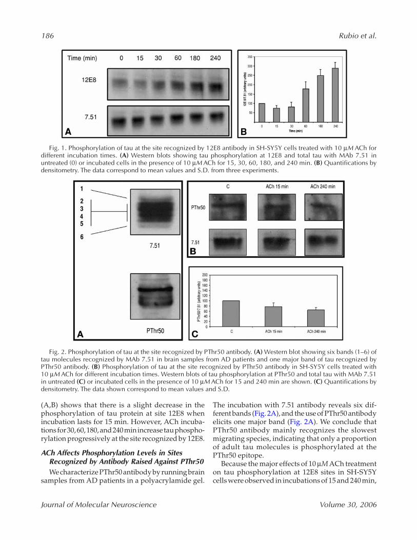
186 Rubio et al.
Journal of Molecular Neuroscience Volume 30, 2006
(A,B) shows that there is a slight decrease in thephosphorylation of tau protein at site 12E8 whenincubation lasts for 15 min. However, ACh incuba-tions for 30, 60, 180, and 240 min increase tau phospho-rylation progressively at the site recognized by 12E8.
ACh Affects Phosphorylation Levels in SitesRecognized by Antibody Raised Against PThr50We characterize PThr50 antibody by running brain
samples from AD patients in a polyacrylamide gel.
The incubation with 7.51 antibody reveals six dif-ferent bands (Fig. 2A), and the use of PThr50 antibodyelicits one major band (Fig. 2A). We conclude thatPThr50 antibody mainly recognizes the slowestmigrating species, indicating that only a proportionof adult tau molecules is phosphorylated at thePThr50 epitope.
Because the major effects of 10 M ACh treatmenton tau phosphorylation at 12E8 sites in SH-SY5Ycells were observed in incubations of 15 and 240 min,
Fig. 1. Phosphorylation of tau at the site recognized by 12E8 antibody in SH-SY5Y cells treated with 10 M ACh fordifferent incubation times. (A) Western blots showing tau phosphorylation at 12E8 and total tau with MAb 7.51 inuntreated (0) or incubated cells in the presence of 10 M ACh for 15, 30, 60, 180, and 240 min. (B) Quantifications bydensitometry. The data correspond to mean values and S.D. from three experiments.
Fig. 2. Phosphorylation of tau at the site recognized by PThr50 antibody. (A) Western blot showing six bands (1–6) oftau molecules recognized by MAb 7.51 in brain samples from AD patients and one major band of tau recognized byPThr50 antibody. (B) Phosphorylation of tau at the site recognized by PThr50 antibody in SH-SY5Y cells treated with 10 M ACh for different incubation times. Western blots of tau phosphorylation at PThr50 and total tau with MAb 7.51in untreated (C) or incubated cells in the presence of 10 M ACh for 15 and 240 min are shown. (C) Quantifications bydensitometry. The data shown correspond to mean values and S.D.
71_Avila 10/11/06 5:08 PM Page 186

ACh Effect on Tau Phosphorylation 187
Journal of Molecular Neuroscience Volume 30, 2006
we tested tau phosphorylation at other sites usingPThr50 antibody (Fig. 2B). In this case, data indicatethat short incubation time with 10 M ACh in SH-SY5Y cells does not modify PThr50 levels. However,long-lasting incubation with ACh decreases tauphosphorylation at the site recognized by PThr50antibody (Fig. 2C), decreasing tau microtubulebinding.
Immunoreactivity of PThr50 antibody in SH-SY5Yextracts shows that this human neuroblastoma cellline contains tau isoforms with the first N-terminalinsert, or exon 2. This evidence is in agreement withwork done by Feijoo et al. (2004).
In conclusion, our work in SH-SY5Y humanneuroblastoma cells treated with ACh reveals twodistinct effects in tau phosphorylation at 12E8 andPThr50 sites with different physiological conse-quences. No effect was observed in tau phospho-rylation at PHF1 and tau-1 sites (data not shown).Short incubation times with ACh promote taumicrotubule binding, evidenced by a slight decreasein 12E8 immunoreactivity (Fig. 3). However, long-lasting incubations with ACh increase phospho-rylation at the site recognized by 12E8 antibodyand decrease phosphorylation at the PThr50 site,suggesting that tau release from microtubules isfacilitated (Fig. 3).
ACh is characterized by its fast turnover due tothe rapid hydrolysis of the compound into acetateand choline (Ch). Based on this evidence, we suggestthat long lasting effects of ACh observed in tau phos-phorylation of SH-SY5Y cells may be produced byCh. Ch action could affect to an increase in calciumconcentration (Alkondon et al., 1997) that could regu-late some tau kinases which may modify Thr50 andSer 262 sites.
AcknowledgmentsWe thank Ana Cuenda (University of Dundee) for
comments and for providing PThr50 antibody. A. R.has been supported by a fellowship from Ministeriode Educación y Ciencia (Spain).
ReferencesAlkondon M., Pereira E. F., Cortes W. S., Maelicke A., and
Albuquerque E. X. (1997) Choline is a selective agonistof alpha7 nicotinic acetylcholine receptors in rat brainneurons. Eur. J. Neurosci. 9, 2734–2742.
Ávila J., Lucas J. J., Pérez M., and Hernández F. (2004)Role of tau protein in both physiological and patho-logical conditions, Physiol. Rev. 84, 361–384.
Biedler J. L., Roffler-Tarlw S., Schachner M., and Freed-man L. S. (1978) Multiple neurotransmitter synthesisby human neuroblastoma cell lines and clones. CancerRes. 38, 3751–3757.
Bowen D. M., Smith C. B., White P., and Davison A. N.(1976) Neurotransmitter-related enzymes and indicesof hypoxia in senile dementia and other abiotrophies.Brain 99, 459–496.
Davies P. and Maloney A. J. (1976) Selective loss of centralcholinergic neurons in Alzheimer’s disease. Lancet25,1403.
Feijoo C., Campbell D. G., Jakes R., Goedert M., andCuenda A. (2004) Evidence that phosphorylation of themicrotubule-associated protein Tau by SAPK4/p38delta at Thr50 promotes microtubule assembly. J. Cell Sci. 118, 387–408.
Novak M., Jakes R., Edwards P. C., Milstein C., and Wis-chik C. M. (1991) Difference between the tau protein ofAlzheimer paired helical filament core and normal taurevealed by epitope analysis of monoclonal antibodies423 and 7.51. Proc. Natl. Acad. Sci. U. S. A. 88, 5837–5841.
Pérez M., Hernández F., Gómez-Ramos A., Smith M., PerryG., and Ávila J. (2002) Formation of aberrant phos-photau polymers in neural cultured cells. Eur. J. Biochem.269, 1484–1489.
Fig. 3. Model showing the influence of ACh and choline (Ch) in tau microtubule binding. ACh has been pro-posed to enhance the binding of tau to the microtubule. However, choline action could desestabilize tau binding tomicrotubules.
71_Avila 10/11/06 5:08 PM Page 187

188 Rubio et al.
Journal of Molecular Neuroscience Volume 30, 2006
Perry E. K., Blessed G., Tomlinson B. E., Perry R. H., CrowT. J., Cross A. J., et al. (1981) Neurochemical activitiesin human temporal lobe related to aging and Alzheimer-type changes. Neurobiol. Aging 2, 251–256.
Seubert P., Mawal-Dewan M., Barbour R., Jakes R., Goe-dert M., Johnson G. V., et al. (1995) Detection of phos-phorylated Ser262 in fetal tau, adult tau, and pairedhelical filament tau. J. Biol. Chem. 270, 18,917–18,922.
71_Avila 10/11/06 5:08 PM Page 188


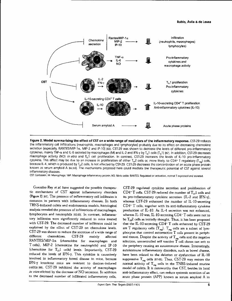


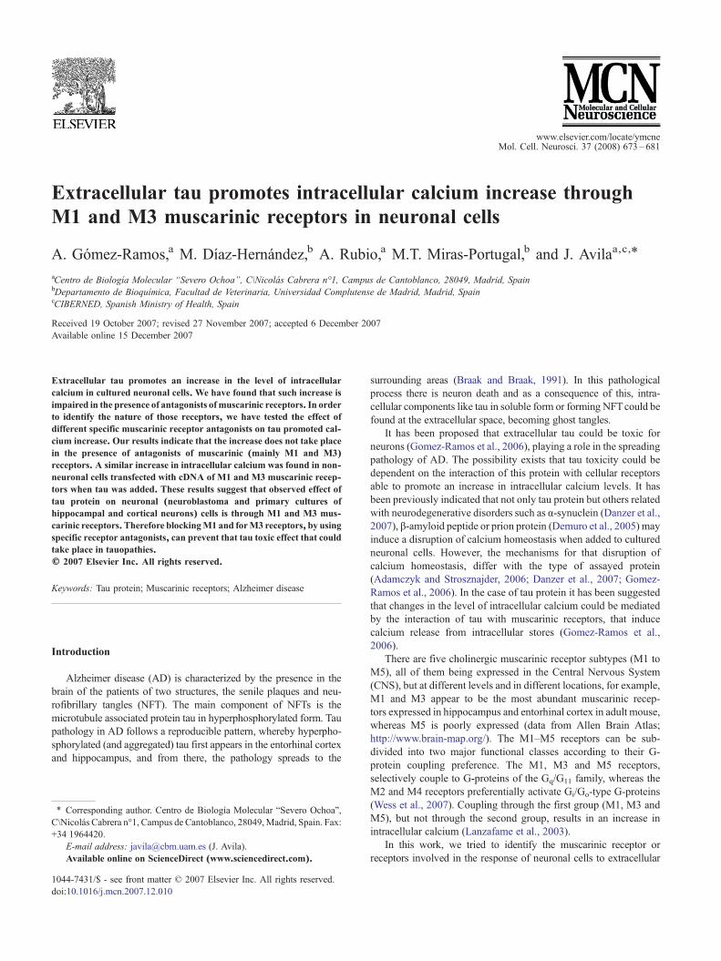
Extracellular tau promotes intracellular calcium increase throughM1 and M3 muscarinic receptors in neuronal cells
A. Gómez-Ramos,a M. Díaz-Hernández,b A. Rubio,a M.T. Miras-Portugal,b and J. Avilaa,c,⁎
aCentro de Biología Molecular “Severo Ochoa”, C\Nicolás Cabrera n°1, Campus de Cantoblanco, 28049, Madrid, SpainbDepartamento de Bioquímica, Facultad de Veterinaria, Universidad Complutense de Madrid, Madrid, SpaincCIBERNED, Spanish Ministry of Health, Spain
Received 19 October 2007; revised 27 November 2007; accepted 6 December 2007Available online 15 December 2007
Extracellular tau promotes an increase in the level of intracellularcalcium in cultured neuronal cells. We have found that such increase isimpaired in the presence of antagonists of muscarinic receptors. In orderto identify the nature of those receptors, we have tested the effect ofdifferent specific muscarinic receptor antagonists on tau promoted cal-cium increase. Our results indicate that the increase does not take placein the presence of antagonists of muscarinic (mainly M1 and M3)receptors. A similar increase in intracellular calcium was found in non-neuronal cells transfected with cDNA of M1 and M3 muscarinic recep-tors when tau was added. These results suggest that observed effect oftau protein on neuronal (neuroblastoma and primary cultures ofhippocampal and cortical neurons) cells is through M1 and M3 mus-carinic receptors. Therefore blockingM1 and forM3 receptors, by usingspecific receptor antagonists, can prevent that tau toxic effect that couldtake place in tauopathies.© 2007 Elsevier Inc. All rights reserved.
Keywords: Tau protein; Muscarinic receptors; Alzheimer disease
Introduction
Alzheimer disease (AD) is characterized by the presence in thebrain of the patients of two structures, the senile plaques and neu-rofibrillary tangles (NFT). The main component of NFTs is themicrotubule associated protein tau in hyperphosphorylated form. Taupathology in AD follows a reproducible pattern, whereby hyperpho-sphorylated (and aggregated) tau first appears in the entorhinal cortexand hippocampus, and from there, the pathology spreads to the
surrounding areas (Braak and Braak, 1991). In this pathologicalprocess there is neuron death and as a consequence of this, intra-cellular components like tau in soluble form or forming NFTcould befound at the extracellular space, becoming ghost tangles.
It has been proposed that extracellular tau could be toxic forneurons (Gomez-Ramos et al., 2006), playing a role in the spreadingpathology of AD. The possibility exists that tau toxicity could bedependent on the interaction of this protein with cellular receptorsable to promote an increase in intracellular calcium levels. It hasbeen previously indicated that not only tau protein but others relatedwith neurodegenerative disorders such as α-synuclein (Danzer et al.,2007), β-amyloid peptide or prion protein (Demuro et al., 2005) mayinduce a disruption of calcium homeostasis when added to culturedneuronal cells. However, the mechanisms for that disruption ofcalcium homeostasis, differ with the type of assayed protein(Adamczyk and Strosznajder, 2006; Danzer et al., 2007; Gomez-Ramos et al., 2006). In the case of tau protein it has been suggestedthat changes in the level of intracellular calcium could be mediatedby the interaction of tau with muscarinic receptors, that inducecalcium release from intracellular stores (Gomez-Ramos et al.,2006).
There are five cholinergic muscarinic receptor subtypes (M1 toM5), all of them being expressed in the Central Nervous System(CNS), but at different levels and in different locations, for example,M1 and M3 appear to be the most abundant muscarinic recep-tors expressed in hippocampus and entorhinal cortex in adult mouse,whereas M5 is poorly expressed (data from Allen Brain Atlas;http://www.brain-map.org/). The M1–M5 receptors can be sub-divided into two major functional classes according to their G-protein coupling preference. The M1, M3 and M5 receptors,selectively couple to G-proteins of the Gq/G11 family, whereas theM2 and M4 receptors preferentially activate Gi/Go-type G-proteins(Wess et al., 2007). Coupling through the first group (M1, M3 andM5), but not through the second group, results in an increase inintracellular calcium (Lanzafame et al., 2003).
In this work, we tried to identify the muscarinic receptor orreceptors involved in the response of neuronal cells to extracellular
www.elsevier.com/locate/ymcneMol. Cell. Neurosci. 37 (2008) 673–681
⁎ Corresponding author. Centro de Biología Molecular “Severo Ochoa”,C\Nicolás Cabrera n°1, Campus de Cantoblanco, 28049,Madrid, Spain. Fax:+34 1964420.
E-mail address: [email protected] (J. Avila).Available online on ScienceDirect (www.sciencedirect.com).
1044-7431/$ - see front matter © 2007 Elsevier Inc. All rights reserved.doi:10.1016/j.mcn.2007.12.010
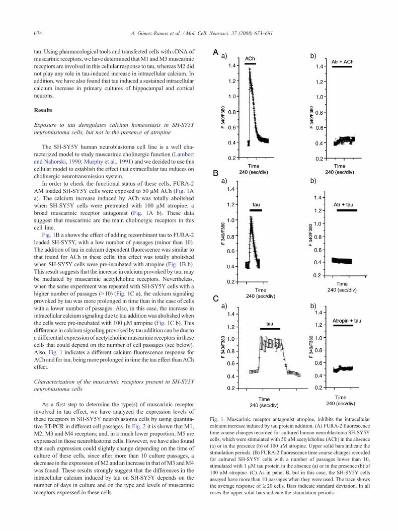
tau. Using pharmacological tools and transfected cells with cDNA ofmuscarinic receptors,we have determined thatM1 andM3muscarinicreceptors are involved in this cellular response to tau, whereasM2 didnot play any role in tau-induced increase in intracellular calcium. Inaddition, we have also found that tau induced a sustained intracellularcalcium increase in primary cultures of hippocampal and corticalneurons.
Results
Exposure to tau deregulates calcium homeostasis in SH-SY5Yneuroblastoma cells, but not in the presence of atropine
The SH-SY5Y human neuroblastoma cell line is a well cha-racterized model to study muscarinic cholinergic function (Lambertand Nahorski, 1990; Murphy et al., 1991) andwe decided to use thiscellular model to establish the effect that extracellular tau induces oncholinergic neurotransmission system.
In order to check the functional status of these cells, FURA-2AM loaded SH-SY5Y cells were exposed to 50 μM ACh (Fig. 1Aa). The calcium increase induced by ACh was totally abolishedwhen SH-SY5Y cells were pretreated with 100 μM atropine, abroad muscarinic receptor antagonist (Fig. 1A b). These datasuggest that muscarinic are the main cholinergic receptors in thiscell line.
Fig. 1B a shows the effect of adding recombinant tau to FURA-2loaded SH-SY5Y, with a low number of passages (minor than 10).The addition of tau in calcium dependent fluorescence was similar tothat found for ACh in these cells; this effect was totally abolishedwhen SH-SY5Y cells were pre-incubated with atropine (Fig. 1B b).This result suggests that the increase in calcium provoked by tau, maybe mediated by muscarinic acetylcholine receptors. Nevertheless,when the same experiment was repeated with SH-SY5Y cells with ahigher number of passages (N10) (Fig. 1C a), the calcium signalingprovoked by tau was more prolonged in time than in the case of cellswith a lower number of passages. Also, in this case, the increase inintracellular calcium signaling due to tau additionwas abolishedwhenthe cells were pre-incubated with 100 μM atropine (Fig. 1C b). Thisdifference in calcium signaling provoked by tau addition can be due toa differential expression of acetylcholinemuscarinic receptors in thesecells that could depend on the number of cell passages (see below).Also, Fig. 1 indicates a different calcium fluorescence response forACh and for tau, beingmore prolonged in time the tau effect thanACheffect.
Characterization of the muscarinic receptors present in SH-SY5Yneuroblastoma cells
As a first step to determine the type(s) of muscarinic receptorinvolved in tau effect, we have analyzed the expression levels ofthese receptors in SH-SY5Y neuroblastoma cells by using quantita-tive RT-PCR in different cell passages. In Fig. 2 it is shown that M1,M2, M3 and M4 receptors; and, in a much lower proportion, M5 areexpressed in those neuroblastoma cells. However, we have also foundthat such expression could slightly change depending on the time ofculture of these cells, since after more than 10 culture passages, adecrease in the expression ofM2 and an increase in that ofM3 andM4was found. These results strongly suggest that the differences in theintracellular calcium induced by tau on SH-SY5Y depends on thenumber of days in culture and on the type and levels of muscarinicreceptors expressed in these cells.
Fig. 1. Muscarinic receptor antagonist atropine, inhibits the intracellularcalcium increase induced by tau protein addition. (A) FURA-2 fluorescencetime course changes recorded for cultured human neuroblastoma SH-SY5Ycells, which were stimulated with 50 μM acetylcholine (ACh) in the absence(a) or in the presence (b) of 100 μM atropine. Upper solid bars indicate thestimulation periods. (B) FURA-2 fluorescence time course changes recordedfor cultured SH-SY5Y cells with a number of passages lower than 10,stimulated with 1 μM tau protein in the absence (a) or in the presence (b) of100 μM atropine. (C) As in panel B, but in this case, the SH-SY5Y cellsassayed have more than 10 passages when they were used. The trace showsthe average response of ≥20 cells. Bars indicate standard deviation. In allcases the upper solid bars indicate the stimulation periods.
674 A. Gómez-Ramos et al. / Mol. Cell. Neurosci. 37 (2008) 673–681
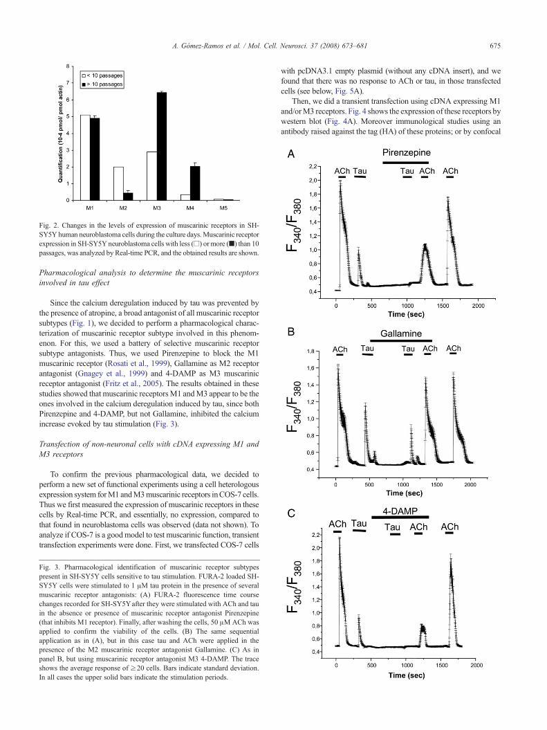
Pharmacological analysis to determine the muscarinic receptorsinvolved in tau effect
Since the calcium deregulation induced by tau was prevented bythe presence of atropine, a broad antagonist of all muscarinic receptorsubtypes (Fig. 1), we decided to perform a pharmacological charac-terization of muscarinic receptor subtype involved in this phenom-enon. For this, we used a battery of selective muscarinic receptorsubtype antagonists. Thus, we used Pirenzepine to block the M1muscarinic receptor (Rosati et al., 1999), Gallamine as M2 receptorantagonist (Gnagey et al., 1999) and 4-DAMP as M3 muscarinicreceptor antagonist (Fritz et al., 2005). The results obtained in thesestudies showed that muscarinic receptorsM1 andM3 appear to be theones involved in the calcium deregulation induced by tau, since bothPirenzepine and 4-DAMP, but not Gallamine, inhibited the calciumincrease evoked by tau stimulation (Fig. 3).
Transfection of non-neuronal cells with cDNA expressing M1 andM3 receptors
To confirm the previous pharmacological data, we decided toperform a new set of functional experiments using a cell heterologousexpression system forM1 andM3muscarinic receptors inCOS-7 cells.Thus we first measured the expression of muscarinic receptors in thesecells by Real-time PCR, and essentially, no expression, compared tothat found in neuroblastoma cells was observed (data not shown). Toanalyze if COS-7 is a goodmodel to test muscarinic function, transienttransfection experiments were done. First, we transfected COS-7 cells
with pcDNA3.1 empty plasmid (without any cDNA insert), and wefound that there was no response to ACh or tau, in those transfectedcells (see below, Fig. 5A).
Then, we did a transient transfection using cDNA expressing M1and/orM3 receptors. Fig. 4 shows the expression of these receptors bywestern blot (Fig. 4A). Moreover immunological studies using anantibody raised against the tag (HA) of these proteins; or by confocal
Fig. 3. Pharmacological identification of muscarinic receptor subtypespresent in SH-SY5Y cells sensitive to tau stimulation. FURA-2 loaded SH-SY5Y cells were stimulated to 1 μM tau protein in the presence of severalmuscarinic receptor antagonists: (A) FURA-2 fluorescence time coursechanges recorded for SH-SY5Yafter they were stimulated with ACh and tauin the absence or presence of muscarinic receptor antagonist Pirenzepine(that inhibits M1 receptor). Finally, after washing the cells, 50 μMACh wasapplied to confirm the viability of the cells. (B) The same sequentialapplication as in (A), but in this case tau and ACh were applied in thepresence of the M2 muscarinic receptor antagonist Gallamine. (C) As inpanel B, but using muscarinic receptor antagonist M3 4-DAMP. The traceshows the average response of ≥20 cells. Bars indicate standard deviation.In all cases the upper solid bars indicate the stimulation periods.
Fig. 2. Changes in the levels of expression of muscarinic receptors in SH-SY5Y human neuroblastoma cells during the culture days.Muscarinic receptorexpression in SH-SY5Yneuroblastoma cells with less (□) or more (■) than 10passages, was analyzed by Real-time PCR, and the obtained results are shown.
675A. Gómez-Ramos et al. / Mol. Cell. Neurosci. 37 (2008) 673–681
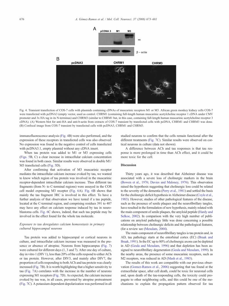
immunofluorescence analysis (Fig. 4B) were also performed, and theexpression of these receptors in transfected cells was also observed.No expression was found in the negative control of cells transfectedwith pcDNA3.1, empty plasmid without any cDNA insert.
When tau protein was added to M1 or M3 expressing cells(Figs. 5B, C) a clear increase in intracellular calcium concentrationwas found in both cases. Similar results were observed in double M1/M3 transfected cells (Fig. 5D).
After confirming that activation of M3 muscarinic receptormediates the intracellular calcium increase evoked by tau, we wantedto know which region of tau protein was involved in the muscarinicreceptor-dependent intracellular calcium increase. Thus different taufragments (from N- to C-terminal regions) were assayed in the COScell model expressing M3 receptor (Fig. 6A). Fig. 6B shows thatmainly the tau fragment 3RC is involved in this effect. To have afurther analysis of that observation we have tested if a tau peptide,located at the C-terminal region, and comprising residues 391 to 407may have any effect on calcium response induced by tau in neuro-blastoma cells. Fig. 6C shows, indeed, that such tau peptide may beinvolved in the effect found for the whole tau molecule.
Exposure to tau deregulates calcium homeostasis in primarycultured hippocampal neurons
Tau protein was added to hippocampal or cortical neurons inculture, and intracellular calcium increase was measured in the pre-sence or absence of atropine. Neurons from hippocampus (Fig. 7)were cultured for different days (1, 3 and 7). After one day of culture,day in vitro 1 (DIV1), less than 20%of the cells respond to eitherAChor tau protein. However, after DIV3, and mainly after DIV7, theproportion of cells responding to bothACh and tau proteinwas clearlyincreased (Fig. 7B). It is worth highlighting that a higher sensitivity totau (Fig. 7A) correlates with the increase in the number of neuronsexpressing M1 receptors (Fig. 7D). As expected, the calcium increaseevoked by tau was, in all cases, prevented by atropine pretreatment(Fig. 7C). A potassium dependent depolarization was performed in all
studied neurons to confirm that the cells remain functional after thedifferent treatments (Fig. 7C). Similar results were observed on cor-tical neurons in culture (data not shown).
A difference between ACh and tau responses is that tau res-ponse is more prolonged in time than ACh effect, and it could bemore toxic for the cell.
Discussion
Thirty years ago, it was described that Alzheimer disease wasassociated with a severe loss of cholinergic markers in the brain(Bowen et al., 1976; Davies and Maloney, 1976). This observationraised the hypothesis suggesting that cholinergic loss could be relatedto the severity of the dementia (Perry et al., 1981) and settled the basisfor the cholinergic deficit hypothesis inAlzheimer disease (Coyle et al.,1983). However, studies of other pathological features of the disease,such as the presence of senile plaques and the neurofibrillary tangles,have resulted in the formulation of new hypothesis, mainly related withthe main component of senile plaques, the amyloid peptide (Hardy andSelkoe, 2002). In comparison with the very high number of publi-cations on amyloid pathology little was done concerning a possiblerelationship between cholinergic deficits and the pathological features,(for a review see (Mesulam, 2004).
Themain component of neurofibrillary tangles is tau protein and, inAD, tau pathology starts at the entorhinal cortex (EC) (Braak andBraak, 1991). In the ECup to 80%of cholinergic axons can be depletedin AD (Geula and Mesulam, 1996) and that depletion has been as-signed to neurofibrillary degeneration (Geula and Mesulam, 1999). Inthe nearby areas, the presence of some muscarinic receptors, such asM2 receptors, was reduced in AD (Mash et al., 1985).
The results of this work are compatible with our previous obser-vation (Gomez-Ramos et al., 2006) suggesting that tau found at theextracellular space, after cell death, could be toxic for neuronal cellsand, upon death of the tau-responding cells, the toxicity could pro-pagate to other neighboring cells, and this could be one of the me-chanisms to explain the propagation pattern observed for tau
Fig. 4. Transient transfection of COS-7 cells with plasmids containing cDNAs of muscarinic receptors M1 or M3. African green monkey kidney cells COS-7were transfected with pcDNA3 (empty vector, used as control. CHRM1 (containing full-length human muscarinic acetylcholine receptor 1 cDNA under CMVpromoter and 3x HA tag in its N-terminus) and CHRM3 (similar to CHRM1 but, in this case, containing full-length human muscarinic acetylcholine receptor 3cDNA). (A) Western blot for anti-HA and anti-b-actin from extracts of COS-7 transient by transfected cells with pcDNA, CHRM1 and CHRM3 was done.(B) Confocal image from COS-7 transient by transfected cells with pcDNA3, CHRM1 and CHRM3.
676 A. Gómez-Ramos et al. / Mol. Cell. Neurosci. 37 (2008) 673–681

pathology in AD, if the process starts at EC. Indeed, working on SH-SY5Y human neuroblastoma cells, we have found that tau-inducedpathology involves a deregulation of calcium homeostasis, that takesplace in those cells bearing muscarinic receptors (Gomez-Ramoset al., 2006). The existence of proteins that could activate cholinergicreceptors is not a new idea since it has been reported that a rabies virusglycoprotein specifically binds to acetylcholine muscarinic receptors(Kumar et al., 2007).
As a first step to identify the muscarinic receptor able to bind tau,we have identified the muscarinic receptors expressed in SH-SY5Yneuroblastoma cells, and we found that mainly M1, M2 and M3 wereexpressed. Thus, we propose that SH-SY5Y cells can be a goodmodelfor studying tau effect, because M1 receptor is heavily expressed inwhole brain (http://www.brain-map.org/) including those regionsmainly affected inAD.Besides, a reduction ofM2 receptor expressionin AD has been described (Mash et al., 1985), and finally, M3receptors are mainly located on cortex, hippocampus, and retrohippo-campal formation (http://www.brain-map.org/). Thus, we have fo-cused our studies in these receptors.
After performing pharmacological studies and cDNA transfec-tion analyses, we found a possible role forM1 and forM3, but not forM2, in tau toxicity in neuroblastoma or non-neuronal transfectedcells.
Additionally, it has been shown that M1 and M3 mediated mus-carinic stimulation promotes the processing of amyloid precursorprotein (APP) by the α-secretase pathway (Nitsch et al., 1992).However,M1 stimulationmay also lead to an increase in BACE (APPβ-secretase) expression, whereas M2 stimulation decreases BACEexpression (Zuchner et al., 2004; Zuchner et al., 2005). In AD, anincrease of M1 and M3 receptors with respect to that of M2 has beenfound (Mash et al., 1985). Thus, tau protein acting through M1 andM3 but not through M2 receptor could act on those sick neurons. Inour studies we do not know, at the present, the possible difference fortau or ACh activation of muscarinic receptors and if they could berelated, or not, to any of the previous consequences that have beendescribed for muscarinic receptor activation. Nevertheless, sincechanges in α or β APP secretase activities could take place upon M1/M3 receptor activation that could result in the depletion of Aβproduction, we are currently testing if a possible overexpression of Aβdue to tau toxicitymay take place and comparing it to the effect ofM1/
M3 activation by ACh. However, our on-going research on M1/M3receptor and APP transfected cells, does not suggest any differencebetween the addition of ACh and that of tau to the cells.
Fig. 5. Tau protein induces intracellular calcium increase in COS-7 cellstransfected with CHRM1 and CHRM3 constructs. FURA-2 loaded COS-7cells transfected with CHRM1, CHRM3 constructs or with empty vectorpcDNA3.1 (Ctrl), were stimulated with 1 mM tau protein, and fluorescencetime course changes were measured. (A) Control COS-7 cells transfectedwith pcDNA3.1 were stimulated with a pulse of 50 μM ACh (in order toestablish functional cell status) followed by 1 μM tau. The cells do notrespond to acetylcholine neither to tau. (B) COS-7 cells transfected withCHRM1 construct were stimulated to ACh, followed by tau protein. The cellsrespond to tau protein in a similar magnification that occurs in SH-SY5Yhuman neuroblastoma cells. There was no response to tau when cells werepre-incubated with 100 μM atropine. Finally, after washing the cells withperfusion buffer (see Experimental methods) a pulse of 50 μM ACh wasapplied in order to confirm cell viability. C) COS-7 cells transfected withCHRM3 were stimulated with 50 μMACh, 1 μM tau protein and again with50 μMACh (after rinsingwith perfusion buffer). (D) As in panel C, but in thiscase the experiment was done with COS-7 cells transfected with bothCHRM1 and CHRM3 plasmids. The trace shows the average response of≥20 cells. Bars indicated standard deviation. In all cases the upper solid barsindicate the stimulation periods.
677A. Gómez-Ramos et al. / Mol. Cell. Neurosci. 37 (2008) 673–681

Finally, we like emphasize that our results on tau action, observedin non-neuronal transfected cells or in neuroblastoma cells, wereconfirmed in primary cultures of hippocampal and cortex neurons.
Experimental methods
Materials
Themuscarinic receptor antagonists: atropine, Pirenzepine,Gallamine and 4-DAMP were purchased from Sigma (St. Louis, MO, USA). Monoclonal mouseanti-HA antibody (clone 12CA5) was obtained from the “Optic and ConfocalMicroscopyService” (S.M.O.C., Centro deBiologíaMolecular “SeveroOchoa”,Madrid, Spain). Monoclonal mouse anti-β-actin antibody was obtained fromSigma (St. Louis, MO, USA). Polyclonal rabbit anti-muscarinic acetylcholinereceptor M1 was purchased from Santa Cruz Biotechnology (Santa Cruz, CA).Mammalian expression plasmids containing CMV promoter and the full-lengthcDNAs from human muscarinic receptors M1 (CHRM1) and M3 (CHRM3),tagged in its N-terminus with 3x HA epitope, were obtained from UMR cDNAResource Center (Missouri, MO). pcDNA3.1 plasmid used as control andLipofectamine 2000 transfection reagent, were obtained from Invitrogen(Carlsbad, Ca). Mammalian expression plasmid containing CMV promoterand cDNA from APP695 was a gift from Dr. Maria Recuero.
Tau peptide comprising the following sequence EIVYKSPVVSGDT-SPRH, residues 391 to 407, (nomenclature of the largest tau isoform), present atthe C-terminal region of the protein, was synthesized and purified, aspreviously described (Perez et al., 2001).
SH-SY5Y Cell culture
Human neuroblastoma SH-SY5Y (Biedler et al., 1978) cells obtained fromATTC (Rockville, Md) were grown in Dulbecco's modified Eagle's medium(DMEM) supplemented with 10% (vol/vol) fetal bovine serum (FBS) plus2 mM glutamine in a humidified atmosphere with 5% CO2. The day beforeperforming the experiment, the cells were seeded on polylysine-coated glasscoverslips at a density of 105 cells/coverslip to assay calcium dependentfluorescence analysis.
COS-7 cell culture and transfection
African green monkey kidney fibroblast (COS-7) cells (Gluzman, 1981)were grown in Dulbecco's modified Eagle's medium supplemented with
10% (vol/vol) FBS. The day before the transfection, the cells were seededon polylysine-coated glass coverslips at a density of 5×10⁴ cells/coverslip.Cells were transiently transfected with CHRM1, CHRM3 containing a HAtag, or with both plasmids using Lipofectamine 2000 reagent (Invitrogen)according to the manufacturer's instructions. Control cells were transfectedwith the empty vector (pcDNA3.1, Invitrogen) at the same conditions. After24 h of expression, transfected cells were fixed for immunofluorescenceanalysis or processed for calcium dependent fluorescence signal analysis.
Primary cultures of hippocampal neurons
Hippocampal neurons were cultured as described by Banker and Cowan(1977). Newborn wild-type mice (P0), were sacrificed, and the embryos wereremoved under sterile conditions. Dissociated hippocampal neurons from theembryos were plated on glass coverslips coated with 1 mg/mL poly (L-lysine)
Fig. 6. Effect of tau peptides in COS-7 cells transfected with CHRM3construct. (A) The whole tau protein and different tau peptides were assayed todeterminate the putative region of tau protein involved in deregulation ofcalcium homeostasis: Tau 42, indicates the longest human tau isoformexpressed in the SNC containing 2 inserts in its N-terminal region and 4 repeatsin its microtubule binding region (Goedert and Jakes, 1990); 2R, referred to astau peptide containing first and third repeats of Tau 42; 2N as tau peptidecontaining the N-terminal region of Tau 42 including its twoN-terminal inserts,but no the microtubule binding regions (aminoacids 1–243); 3R referred to astau peptide containing first, third and fourth repeats of its microtubule bindingregion and 3RC referred to as the C-terminal region of tau 42, including first,third and fourth microtubule binding repeats. (B) COS-7 cells transientlytransfected with CHRM3 plasmid, were stimulated with a sequential sequence,thus first it was assayed at 50 μM ACh (to check cell functionality), and afterthen were assayed differences in tau peptides at 1 μM: 2N, 2R, 3R, 3RC andfinally the full-length tau protein. After tau treatment, the cells were againstimulated with a pulse of 50 M ACh, The cells were washed with perfusionbuffer after every application. The trace shows the average response of ≥ 20cells. Bars indicated standard deviation. In all cases the upper solid bars indicatethe stimulation periods. (C) FURA-2 fluorescence time course changesrecorded for Cos-7 cells transiently transfected with CHRM3 plasmid, andsequentially stimulated with 50 μM ACh, 3RC peptide and C-terminal taupeptide comprising residues 391 to 407 (tau391–407). Again, the cell viability,after tau peptide treatment was tested by a pulse of ACh.
678 A. Gómez-Ramos et al. / Mol. Cell. Neurosci. 37 (2008) 673–681

and 20 μg/mL laminin.After incubation for 3 h inmedium containing 10% (v/v)horse serum (Gibco), the cells were transferred to N2- and B27-supplementedmedium (Gibco) and incubated for 1, 3 or 7 days.
Immunofluorescence analysis
COS-7 transiently transfected cells and primary cultures of neuronswere fixed with 4% paraformaldehyde for 20 min. After the fixation, thecoverslips were incubated with phosphate buffered saline (PBS) supple-
mented with 0.1% Triton X-100 for 10 min, then were incubated with 1%bovine serum albumin (BSA) in PBS/TX-100 for additional 10 min.Primary antibody anti-HA was diluted 1:2000 (vol/vol) in PBS/BSA andincubated in coverslips for 45 min at room temperature. After primaryantibody incubation, coverslips were rinsed with PBS and incubated withsecondary antibody (Alexa 488, Invitrogen) diluted 1:500 (vol/vol) in PBS/BSA. Finally, the coverslips were rinsed again with PBS and mounted inFluorSave (Calbiochem). Coverslips were analyzed by confocal micro-scopy in a Confocal MicroRadiance (BioRad) coupled with a verticalmicroscopy Axioskop 2 (Zeiss).
Fig. 7. Tau deregulates calcium homeostasis in primary cultured of hippocampal neurons. (A) Mouse hippocampal neurons cultured for 1, 3 and 7 days, were FURA-2loaded and the intracellular calcium levels were measured after a sequential application of 50 μMACh and 1 μM tau. Insets show images showing color representations ofthe FURA-2 loaded hippocampal neurons responding to tau. (B)Graphic shows the percentage ofmouse hippocampal neuronal cells that respond toAChor tau at 1 ( ),3 ( ) or 7( ) days in vitro. (C) Newborn mouse hippocampal neurons cultured for 3 days, were sequentially stimulated with 50 μM ACh and 1 μM tau in thepresence or absence of 100 mM atropine. Finally, the neurons, were depolarizated with 60 μMKCl to confirm their viability after the treatments. (D)Mouse hippocampalneurons were fixed at 1, 3 or 7 days in vitro and processed for immunofluorescence analysis with an antibody that recognizes muscarinic acetylcholine receptor 1.
679A. Gómez-Ramos et al. / Mol. Cell. Neurosci. 37 (2008) 673–681

Calcium dependent fluorescence signal analysis—imageacquisition and analysis of the Ca2+ response in different cell types
Primary cultures of hippocampal and cortical neurons, human neuro-blastoma SH-SY5Y and COS-7 transiently transfected cells, were washedwith perfusion buffer (122 mMNaCl, 3.1 mMKCl, 0.4 mMKH2PO4, 5 mMNaHCO3, 1.2 mM MgSO4, 10 mM glucose and 20 mM N–Tris(hydroxymethyl)methyl-2-aminoethanesulfonic acid (TES) buffer, pH 7.4),and they were loaded with FURA-2 AM solution (7.5 μM) for 30 min at37 °C. This period facilitated the intracellular hydrolysis of the FURA-2 AM.Subsequently, the coverslips were washedwith freshmedium andmounted ina superfusion chamber in a NIKON Eclipse TE-2000 microscope. In allexperiments the cells were superfused first at 1.2 mL/min with perfusionmedia and then, with 50 μM ACh in order to establish the functional cellstatus. When tau protein was assayed, the perfusion system was stopped.When the experiments were performed with muscarinic receptor antagonists:atropine (100 μM), Pirenzepine (10 μM), 4-DAMP (2 μM) and Gallamine(2 μM), the cells were incubated for 10 min before adding ACh or tau.
At the end of each experiment, 50 μMAch pulses were applied to confirmthe viability of the cells under study. Cells were visualized using a Nikonmicroscope containing a ×40 S Fluor 0.5–1.3 oil lens. The wavelength of theincoming light was filtered to 340 nm and 380 nm with the aid of amonochromator (10 nm bandwidth, Optoscan monocromator, Cairin). Thesewavelengths corresponded to the fluorescence peaks of Ca2+-saturated andCa2+-free FURA-2 solutions. The 12-bit images were acquired with an ORCA-ERC 47 42–98 CCD camera fromHamamatsu (Hamamatsu City, Japan) controlledby Metafluor 6.3r6 PC software (Universal Imaging Corp., Cambridge, UK).The exposure time was 150 ms for each wavelength and the changing timeb5ms. The images were acquired continuously and buffered in a fast SCSI disk.The time course data represent the average light intensity in a small ellipticalregion inside each cell. The background and autofluorescence components weresubtracted at eachwavelength, and the 340 over 380 nm ratiowas calibrated into[Ca2+]i values using Grynkiewicz's equation (Grynkiewicz et al., 1985). TheRmax, Rmin and β parameters were calculated from the spectra of small dropletsof FURA-2 in Ca2+-saturated and Ca2+-free solutions (100 mM KCl, 10 mMNaCl, 1 mMMgCl2, 10 mM Tris, 10 mMMOPS and 100 μM FURA-2. Ca2+-free: plus 2 mM EGTA. Ca2+-saturated: plus 2 mM CaCl2, pH 7.4) (Diaz-Hernandez et al., 2001) and the in vitro Rmax and Rmin were corrected for thedifferences with the cytosolic environment using the procedure described byPoenie (1990) and Baitinger et al. (1990).
Protein purification
The recombinant tau isoform (tau42) containing 2 N-terminal insertsand 4 microtubule binding repeats (Goedert and Jakes, 1990), and taufragments: 2N, containing the amino-terminal half of tau protein, (residues 1–251), tau 2R, containing residues 250–335 (deleted 274–304), tau 3R, containingresidues 250–367 (deleted 274–304), and tau 3RC, containing residues 250–441(deleted 274–304), was isolated as previously described (Perez et al., 1996). Theisolated proteins were characterized by gel electrophoresis followed by stainingwith Coomassie blue.
Gel electrophoresis and western blot
Cells were homogenized at 4 °C in a buffer containing 50mMHEPES, pH7.4, 10 mM EDTA, 0.1% Triton X-100, and including the phosphataseinhibitors 20 mM NaF, 0.1 mM sodium orthovanadate, and 1 mMphenylmethanesulfonyl fluoride. The cell lysates were centrifuged at10,000 g for 5 min at 4 °C and then heated at 100 °C for 5 min inelectrophoresis sample buffer. The protein concentration was determined byusing the BCA protein assay, at 37 °C (Pierce, Rockford, IL). Proteins werefractionated by sodium dodecyl sulfate-polyacrylamide gel electrophoresis(SDS-PAGE) on 10% gels and transferred to nitrocellulose membranes(Schleicher & Schuell Bioscience, Dassel, Germany). After blocking of non-specific protein binding to the membranes with 0.05% Tween 20 and 5% non-
fat dry milk in PBS, the membranes were incubated overnight with the primaryantibodies anti-HA (1:2000, vol/vol) or anti-β-actin (1:5000, vol/vol) inblocking buffer at 4 °C. The proteins recognized by the antibodies werevisualized by using enhanced chemiluminescence (Perkin Elmer Life Sciences,Boston, MA) after incubation with horseradish peroxidase (HRP)-linkedsecondary antibodies (Dako A/S, Glostrup, Denmark).
Quantitative RT-PCR
Total RNAwas purified from 35mm dishes of confluent SH-SY5Y cells orCOS-7 cells using an RNA Miniprep Kit (Stratagene) according to themanufacturer's recommended protocol. RNA extraction was followed byDNAse treatment to avoid genomic DNA contamination. Then, reversetranscription was performed with both 0.08 μg/μL oligo (dT) primers and0.2 μg/μL random primers using the first-strand cDNA synthesis kit for RT-PCR (AMV) (Roche Applied Science) protocol.
Genomic DNAwas purified from COS-7 cells using the Wizard Plus SVGenomic DNA purification system (Promega). And genomic DNA from SH-SY5Y cells was isolated using a high salt method as follows. First, nuclei lysisbuffer (1%Tris EDTA pH 8.0, 0.4MNaCl and 2 mMEDTA) was added to thecell pellet and cells were centrifuged for 10 min at 4 °C (1200 rpm). The pelletwas resuspended in the lysis buffer with 0.3 mg/mL proteinase K (Merck) and1% SDS and, then, it was incubated at 45 °C overnight. After, 6 M NaCl wasadded and centrifugation for 15 min (3000 rpm) was performed. Thesupernatant was centrifuged repeatedly until it was clear of salt. To precipitateDNA, 3M sodiumacetate pH5.2 and cold 100% isopropanolwere added to thefinal supernatant. DNAwas transferred into a new tube containing 70%Ethanoland it was centrifuged for 30 min (14,000 rpm). Finally, the pellet wasresuspended in H2O. Each amplification was performed using genomic DNAfrom SH-SY5Y as template. The concentrations of PCR products were mea-sured using Nanodrop ND-1000 (Nanodrop Technologies). Micrograms ofdouble stranded DNA (ds DNA) were converted in picomoles:pmol=μg×106 pg/1 μg×1 pmol/660 pg×1/N (length of the amplicon inbp). Ten-fold serial dilutions in brewer-yeast tRNA (110 ng/μL,RocheAppliedScience) of the PCR products starting from 10−2 pmol were used as standardsfor the calibration curves.
Real-time PCR analysis was performed in the ABI PRISM 7900HTSequenceDetection System (Applied Biosystems) in a 96-well optical plate. ThePCRmaster mix contained 5 μL of the SYBRGreen PCRMaster Mix (AppliedBiosystems), 1 μL of the template and 150 nM of each forward and reverseprimers in a total volume of 10 μL. Table 1 shows primer pairs designed amonghuman sequences for each muscarinic receptor subtype and β-actin used as anendogenous control. Each primer pair reaction for each template was assayed intriplicates.
Table 1Primers designed for each muscarinic receptor subtype and endogenous β-actin (used as control)
Gene name Orientation Sequence Productlength (bp)
Muscarinicreceptor 1
Forward ACCTCTATACCACGTACCTG 122Reverse TGAGCAGCAGATTCATGACG
Muscarinicreceptor 2
Forward ACCTGTGGTGTGTGACCTTT 128Reverse GCTTGACTGGGTAGGTCAGA
Muscarinicreceptor 3
Forward CATCATGAATCGATGGGCCT 128Reverse GGCCTCGTGATGGAAAAGTA
Muscarinicreceptor 4
Forward CAGTTTGTGGTGGGTAAGCG 132Reverse GTACAGCACCGTCATGATGA
Muscarinicreceptor 5
Forward GATGAGTGCCAGATCCAGTT 103Reverse GACAGTAGAGGATGGTCATG
β-actin Forward CACACTGTGCCCATCTACGA 162Reverse CTCCTTAATGTCACGCACGA
680 A. Gómez-Ramos et al. / Mol. Cell. Neurosci. 37 (2008) 673–681

Thermal cycling conditions were as follows: 10 min at 95 °C, then,40 cycles of 15 s at 95 °C and 1min at 60 °C. After the amplification, a meltingcurve was obtained increasing the temperature from 60 °C to 95 °C.
Different controls were carried out for each amplification: RT-PCR-template with no reverse transcriptase as a control of the genomic DNAcontamination, no-template reaction as negative control and genomic DNA-template as positive control.
Mean threshold cycle (CT) values for each amplification performed intriplicates were normalized and analyzed using ABI prism 7000 SequenceDetection System and SDS Software (Applied Biosystems). Melting curveswere studied to determine whether the primer pairs used were specific (seeTable 1).
Acknowledgments
This work was supported by grants from the Spanish PlanNacional, from the Spanish Ministry of Health, Neuropharma,Fundación M. Botín, Comunidad de Madrid and an institutional grantto CBMSO from the “Fundación R. Areces”. We thank Drs J. Lucasand F. Hernandez for the critical reading of the manuscript.
References
Adamczyk,A., Strosznajder, J.B., 2006.Alpha-synuclein potentiatesCa2+ influxthrough voltage-dependent Ca2+ channels. Neuroreport 17, 1883–1886.
Baitinger, C., Alderton, J., Poenie,M., Schulman, H., Steinhardt, R.A., 1990.Multifunctional Ca2+/calmodulin-dependent protein kinase is necessaryfor nuclear envelope breakdown. J. Cell Biol. 111, 1763–1773.
Banker, G.A., Cowan, W.M., 1977. Rat hippocampal neurons in dispersedcell culture. Brain Res. 126, 397–425.
Biedler, J.L., Roffler-Tarlov, S., Schachner, M., Freedman, L.S., 1978.Multiple neurotransmitter synthesis by human neuroblastoma cell linesand clones. Cancer Res. 38, 3751–3757.
Bowen, D.M., Smith, C.B., White, P., Davison, A.N., 1976. Neurotrans-mitter-related enzymes and indices of hypoxia in senile dementia andother abiotrophies. Brain 99, 459–496.
Braak, H., Braak, E., 1991. Neuropathological stageing of Alzheimer-relatedchanges. Acta. Neuropathol. (Berl). 82, 239–259.
Coyle, J.T., Price, D.L., DeLong, M.R., 1983. Alzheimer's disease: adisorder of cortical cholinergic innervation. Science 219, 1184–1190.
Danzer, K.M., Haasen, D., Karow, A.R., Moussaud, S., Habeck,M., Giese, A.,Kretzschmar, H., Hengerer, B., Kostka, M., 2007. Different species ofalpha-synuclein oligomers induce calcium influx and seeding. J. Neurosci.27, 9220–9232.
Davies, P., Maloney, A.J., 1976. Selective loss of central cholinergic neuronsin Alzheimer's disease. Lancet 2, 1403.
Demuro, A., Mina, E., Kayed, R., Milton, S.C., Parker, I., Glabe, C.G., 2005.Calcium dysregulation and membrane disruption as a ubiquitous neurotoxicmechanismof soluble amyloid oligomers. J. Biol. Chem. 280, 17294–17300.
Diaz-Hernandez, M., Gomez-Villafuertes, R., Hernando, F., Pintor, J.,Miras-Portugal, M.T., 2001. Presence of different ATP receptors on ratmidbrain single synaptic terminals. Involvement of the P2X(3) subunits.Neurosci. Lett. 301, 159–162.
Fritz, N., Macrez, N., Mironneau, J., Jeyakumar, L.H., Fleischer, S., Morel,J.L., 2005. Ryanodine receptor subtype 2 encodes Ca2+ oscillationsactivated by acetylcholine via the M2 muscarinic receptor/cADP-ribosesignalling pathway in duodenummyocytes. J. Cell Sci. 118, 2261–2270.
Geula, C., Mesulam, M.M., 1996. Systematic regional variations in the loss ofcortical cholinergic fibers inAlzheimer's disease. Cereb. Cortex 6, 165–177.
Geula, C., Mesulam, M.M., 1999. Cholinergic systems in Alzheimer'sdisease, Alzheimer Disease, 2nd Ed. Lippincott, Williams & Wilkins,Philadelphia, PA, pp. 69–292.
Gluzman, Y., 1981. SV40-transformed simian cells support the replication ofearly SV40 mutants. Cell 23, 175–182.
Gnagey, A.L., Seidenberg, M., Ellis, J., 1999. Site-directed mutagenesisreveals two epitopes involved in the subtype selectivity of the allostericinteractions of gallamine at muscarinic acetylcholine receptors. Mol.Pharmacol. 56, 1245–1253.
Goedert, M., Jakes, R., 1990. Expression of separate isoforms of human tauprotein: correlation with the tau pattern in brain and effects on tubulinpolymerization. Embo J. 9, 4225–4230.
Gomez-Ramos, A., Diaz-Hernandez, M., Cuadros, R., Hernandez, F., Avila, J.,2006. Extracellular tau is toxic to neuronal cells. FEBSLett. 580 4842–4850.
Grynkiewicz, G., Poenie, M., Tsien, R.Y., 1985. A new generation of Ca2+indicators with greatly improved fluorescence properties. J. Biol. Chem.260, 3440–3450.
Hardy, J., Selkoe, D.J., 2002. The amyloid hypothesis of Alzheimer'sdisease: progress and problems on the road to therapeutics. Science 297,353–356.
Kumar, P., Wu, H., McBride, J.L., Jung, K.E., Kim, M.H., Davidson, B.L.,Lee, S.K., Shankar, P., Manjunath, N., 2007. Transvascular delivery ofsmall interfering RNA to the central nervous system. Nature 448,39–43.
Lambert, D.G., Nahorski, S.R., 1990.Muscarinic-receptor-mediated changesin intracellular Ca2+ and inositol 1,4,5-trisphosphate mass in a humanneuroblastoma cell line, SH-SY5Y. Biochem. J. 265, 555–562.
Lanzafame, A.A., Christopoulos, A., Mitchelson, F., 2003. Cellular signalingmechanisms for muscarinic acetylcholine receptors. Receptors Channels9, 241–260.
Mash, D.C., Flynn, D.D., Potter, L.T., 1985. Loss of M2 muscarine receptorsin the cerebral cortex in Alzheimer's disease and experimentalcholinergic denervation. Science 228, 1115–1117.
Mesulam, M., 2004. The cholinergic lesion of Alzheimer's disease: pivotalfactor or side show. Learn. Mem. 11, 43–49.
Murphy, N.P., Vaughan, P.F., Ball, S.G., McCormack, J.G., 1991. Thecholinergic regulation of intracellular calcium in the human neuroblas-toma, SH-SY5Y. J. Neurochem. 57, 2116–2123.
Nitsch, R.M., Slack, B.E., Wurtman, R.J., Growdon, J.H., 1992. Release ofAlzheimer amyloid precursor derivatives stimulated by activation ofmuscarinic acetylcholine receptors. Science 258, 304–307.
Perez, M., Valpuesta, J.M., Medina, M., Montejo de Garcini, E., Avila, J.,1996. Polymerization of tau into filaments in the presence of heparin: theminimal sequence required for tau–tau interaction. J. Neurochem. 67,1183–1190.
Perez, M., Arrasate, M., Montejo de Garcini, E., Munoz, V., Avila, J., 2001.In vitro assembly of tau protein: mapping the regions involved infilament formation. Biochemistry 40, 5983–5991.
Perry, E.K., Blessed, G., Tomlinson, B.E., Perry, R.H., Crow, T.J., Cross, A.J.,Dockray, G.J., Dimaline, R., Arregui, A., 1981.Neurochemical activities inhuman temporal lobe related to aging and Alzheimer-type changes.Neurobiol. Aging 2, 251–256.
Poenie, M., 1990. Alteration of intracellular Fura-2 fluorescence by viscosity:a simple correction. Cell Calcium 11, 85–91.
Rosati, A.M., Guarnieri, E., Avignone, E., Cherubini, E., Cattaneo, A.,Traversa,U., 1999. Increased density ofM1 receptors in the hippocampus ofjuvenile rats chronically deprived of NGF. Brain Res. 815, 185–191.
Wess, J., Eglen, R.M., Gautam, D., 2007. Muscarinic acetylcholinereceptors: mutant mice provide new insights for drug development.Nat. Rev. Drug. Discov. 6, 721–733.
Zuchner, T., Perez-Polo, J.R., Schliebs, R., 2004. Beta-secretase BACE1 isdifferentially controlled through muscarinic acetylcholine receptorsignaling. J. Neurosci. Res. 77, 250–257.
Zuchner, T., Schliebs, R., Perez-Polo, J.R., 2005. Down-regulation ofmuscarinic acetylcholine receptor M2 adversely affects the expression ofAlzheimer's disease-relevant genes and proteins. J. Neurochem. 95,20–32.
681A. Gómez-Ramos et al. / Mol. Cell. Neurosci. 37 (2008) 673–681


Effect of Cortistatin on TauPhosphorylation at Ser262 Site
Alicia Rubio,1 Mar Perez,1,2 Luis de Lecea,3 and Jesus Avila1*1Centro de Biologıa Molecular ‘‘Severo Ochoa,’’ Consejo Superior de Investigaciones Cientıficas/Universidad Autonoma de Madrid (CSIC/UAM), Madrid, Spain2Department of Anatomy, Histology and Neuroscience, Faculty of Medicine, Universidad Autonoma deMadrid (UAM), Madrid, Spain3Department of Psychiatry and Behavioral Sciences, Stanford University, Stanford, California
The development of intraneuronal lesions as a result ofthe progressive deposition of hyperphosphorylated tauat specific brain regions (such as hippocampus andcortex) plays a key role in the pathological process ofAlzheimer’s disease. However, the mechanisms bywhich tau phosphorylation is regulated, mainly in thepathology found in the cortex, are still poorly under-stood. Here, we analyzed the effect of cortistatin, acortical neuropeptide related to somatostatin, on tauphosphorylation at Ser262 in cultures of murine corticalneurons. Both somatostatin and cortistatin induce tauphosphorylation at Ser262, a site modified in Alzhei-mer’s disease, although with different kinetics in cortex.The effect of cortistatin likely is mediated by heterodi-meric receptors composed of somatostatin receptorsubtypes 2 and 4 and also by protein kinase C signal-ing. Cortistatin-deficient mice show decreased tauphosphorylation at Ser262 in the cortex but not in otherbrain regions tested. Our results suggest an importantrole for cortistatin in the regulation of tau phosphoryla-tion that may be associated with the pathophysiologyof Alzheimer’s disease in regions such as the cerebralcortex. VVC 2008 Wiley-Liss, Inc.
Key words: Alzheimer’s disease; somatostatin;internalization; somatostatin receptors; real-time PCR
Alzheimer’s disease is a senile dementia character-ized by the presence of two aberrant structures: senileplaques and neurofibrillary tangles (Alzheimer, 1906).The number of cortical neurofibrillary tangles, whichmainly contain an abnormal hyperphosphorylated formof the microtubule-associated tau protein, correlates pos-itively with the severity of dementia (Arriagada et al.,1992).
Development of the disease is related to that of taupathology. First, the entorhinal cortex and hippocampusare affected, and then cortical neurons degenerate pro-gressively (Braak and Braak, 1991). In addition, the pro-gression of tau phosphorylation was shown to correlatewith the evolution of tau pathology (Delacourte et al.,1999), being a critical step for the development of thedisease when phosphorylation associated with tau pathol-ogy appears at the cortex (Delacourte et al., 1999).
Thus, the study of tau phosphorylation in this cerebralregion seems to be relevant. The modification of tau byphosphorylation affects its interaction with microtubules(for review see Avila et al., 2004). Tau phosphorylationdepends on the balance of protein kinase and phospha-tase activities (Goedert et al., 1992; Iqbal and Grundke-Iqbal, 1997). Signals that regulate these kinase/phospha-tase activities in different brain regions can have pro-found consequences on microtubule stability and in theonset of Alzheimer’s disease (Goedert et al., 1992; Iqbaland Grundke-Iqbal, 1997). Among those signals couldbe the peptides that belong to the somatostatin family.
Cortistatin is a neuropeptide of the somatostatinfamily expressed in g-aminobutyric acid (GABA)-ergicneurons in the cerebral cortex and hippocampus (deLecea et al., 1996, 1997), brain regions first affected byAlzheimer’s disease. Preprocortistatin mRNA expressionis significantly increased in the hippocampus during nor-mal aging but is attenuated in aged transgenic miceoverexpressing the human b-amyloid precursor protein(Winsky-Sommerer et al., 2004). Several studies provideevidence for a modulatory role of cortistatin in memoryprocesses. Intraventricular injection of cortistatin wasshown to impair memory consolidation in rats (Floodet al., 1997; Sanchez-Alavez et al., 2000). Cortistatin hasan inhibitory effect on the excitability of CA1 neurons
Supplementary Material for this article is available online at http://
www.mrw.interscience.wiley.com/suppmat/0360-4012/suppmat/ (www.
interscience.wiley.com).
Contract grant sponsor: Spanish Ministry of Education and Science and
Ministry of Health and CIBERNED, Spain; Contract grant sponsor:
Ministry of Education and Science, Spain (to A.R.); Contract grant num-
ber: MH58543 (to L.d.L.).
*Correspondence to: Jesus Avila, Centro de Biologıa Molecular ‘‘Severo
Ochoa,’’ Consejo Superior de Investigaciones Cientıficas/Universidad
Autonoma de Madrid (CSIC/UAM), Nicolas Cabrera 1, Universidad
Autonoma de Madrid Cantoblanco, 28049 Madrid, Spain.
E-mail: [email protected]
Received 13 November 2007; Revised 4 January 2008; Accepted 21
January 2008
Published online 25 April 2008 in Wiley InterScience (www.
interscience.wiley.com). DOI: 10.1002/jnr.21689
Journal of Neuroscience Research 86:2462–2475 (2008)
' 2008 Wiley-Liss, Inc.

(de Lecea et al., 1996; Schweitzer et al., 2003) andblocks long-term potentiation (LTP) in perforant path-dentate gyrus synapses. Transgenic mice overexpressingcortistatin also show deficits in LTP and in spatial mem-ory (Tallent et al., 2005).
Cortistatin could constitute an endogenous signalthat modulates protein kinase/phosphatase activity affect-ing tau phosphorylation in the brain regions critical forthe development of Alzheimer’s disease. The presentwork is focused on the effect of cortistatin on tau phos-phorylation at Ser262 site in cultures of murine corticalneurons. Our data point to a relevant role for cortistatinin tau phosphorylation at Ser262 in the cortex withpotential implications in the progression of tau pathologyin Alzheimer’s disease.
MATERIALS AND METHODS
Animals
Experimental animals were wild-type C57BL6, cortista-tin knockout, and somatostatin receptor (sst)-2 and -4 knock-out mice, and sst2/4 double-knockout mice (Videau et al.,2003). These strains were congenic in the C57BL6 back-ground (N9). Primary cortical neurons were obtained fromP0–P1 littermates of wild-type and sst2 and -4 knockout miceand from sst2/4 double-knockout mice. Wild-type and cortis-tatin knockout mice (3-month-old male) were used for pro-tein extracts (n 5 3 for each genotype). Mice were main-tained in a temperature-controlled environment on a 12/12-hr light-dark cycle, with food and water available ad libitum.All procedures were approved by the animal research commit-tee at the Centro de Biologıa Molecular ‘‘Severo Ochoa’’(UAM-CSIC, Madrid, Spain).
Antibodies and Reagents
The monoclonal antibodies against tau protein used forthis study were: 7.51 (a gift from Dr. C. Wischik, MRC,Cambridge, United Kingdom), 12E8 (a gift from Dr. P. Seu-bert, Elan Pharmaceuticals, South San Francisco, CA), andPHF1 (a gift from Dr. P. Davies, New York, NY). The 7.51antibody recognizes an epitope located in the last two repeatsof the tau protein microtubule-binding domain and reactswith all tau isoforms in a phosphorylation-independent man-ner (Novak et al., 1991). The 12E8 antibody detects tau phos-phorylated at Ser262 (Seubert et al., 1995), and the PHF1antibody recognizes tau protein when Ser396 and Ser404 arephosphorylated (Greenberg et al., 1992; Otvos et al., 1994).The numbering of the tau epitopes is given according to thelongest human tau isoform of 441 amino acids (Goedert et al.,1989). The 334 polyclonal antibody specifically detects thecarboxyl-terminal region of the neural b3 isoform of tubulin(Diaz-Nido et al., 1990).
Cortistatin coupled to fluorescein at Lys3 (Fluo-cortista-tin) was custom synthesized at SynPep (Dublin, CA). Fluores-cein was attached to the Lys3 residue because the exocyclicN-terminal proline residue is essential for its binding and spe-cific activities (Criado et al., 1999). The structure and integ-rity of the peptide were verified by mass spectroscopy atTSRIs protein chemistry facility. Cortistatin 1–14 and somato-
statin 1–14 were obtained from Neomps (Strasbourg, France)and from Sigma-Aldrich (St. Louis, MO), respectively (seeamino acid sequences of both compounds in Suppl. Fig. 1).Octreotide, a somatostatin analogue that binds preferentiallyto sst2, -3, and -5 receptors, and Ro 31-8220, a proteinkinase C (PKC) inhibitor, were purchased from Sigma-Aldrich and from Roche (Basel, Switzerland), respectively.
Cell Culture, Transfection, and Treatment
Human embryonic kidney cells (HEK-293) were grownin 12-well plates in Dulbecco’s modified Eagle’s medium(DMEM) supplemented with 10% (vol/vol) fetal calf serum,2 mM glutamine, 1 mM pyruvate, 100 U/ml penicillin, and100 U/ml streptomycin in a humidified atmosphere of 5%CO2/95% air at 378C. Cells were transiently transfected withdifferent combinations of plasmids containing cDNA of tau,sst2, or sst4 in pCDNA3.1 vector using Lipofectamine 2000reagent (Invitrogen, Carlsbad, CA) as described by the manu-facturer. Control cells were transfected with the empty vector(pCDNA3.1; Invitrogen) in the same conditions. One day af-ter the transfection, the medium was replaced by serum-freeDMEM for 2 hours, after which cortistatin was added at dif-ferent concentrations (5–500 nM) for 30 min. Similarly, after2 hr in serum-free DMEM, cells were treated with somatosta-tin (5–500 nM) for 5 min. Finally, protein extracts from cellswere analyzed by Western blot.
Primary Cortical Culture Procedure and Treatment
Cortical neurons were obtained from P0–P1 mouse lit-termates. Papain (Worthington Biochemical Corporation,Lakewood, NJ) was used to dissociate the cells (Banker andCowan, 1977). The cells were plated on poly-L-lysine(100 lg/ml)- plus laminin (10 lg/ml)-coated plastic dishes (1.83 105 cells/24-well plate) and cultured in Neurobasal medium(Gibco, Carlsbad, CA) supplemented with 10% (vol/vol) horseserum, 2 mM glutamine, 1 mM pyruvate, 100 U/ml penicillin,and 100 U/ml streptomycin. After 3 hours, the medium waschanged to Neurobasal medium supplemented with 1% (vol/vol) B27 (Gibco), 2 mM glutamine, 1 mM pyruvate, 100 U/ml, penicillin and 100 U/ml streptomycin. The cells weregrown in a humidified atmosphere of 5% CO2/95% air at378C, and half of the medium was replaced every 2–3 days.
The experiments were performed after 8 days in vitro,when functional somatostatin receptors are known to beexpressed in cultured primary neurons (Heidet et al., 1990;Stroh et al., 2000). After 2 hr in serum-free Neurobasal me-dium, the peptides or other compounds were added to thecultures. The primary cultures were incubated with or with-out 50 nM somatostatin for different times. In separate experi-ments, cortistatin or octreotide were also added at differentconcentrations and incubation times. In some cases, the cellswere incubated in the presence of 10 lM Ro 31-8220, aPKC inhibitor, for 90 min, or pretreated with 10 lM Ro 31-8220 for 60 min, followed by the addition of 50 nM cortista-tin for an additional 30 min. Thereafter, protein isolation andWestern blot analysis were performed.
Cortistatin and Tau Phosphorylation 2463
Journal of Neuroscience Research

Protein Isolation and Western Blot Analysis
Three-month-old mice wild-type and cortistatin knock-out mice were sacrificed by CO2 inhalation. Then, brain wasquickly dissected into cortex, hippocampus, and the remainingtissue on an ice-cold plate. These different samples were ho-mogenized separately in an ice-cold buffer containing 20 mMHepes pH 7.4, 100 mM NaCl, 20 mM NaF, 5 mM EDTA,and 1% (wt/vol) Triton X-100 and supplemented with 30mM glycerophosphate, 5 M pyrophosphate, 1 lM okadaicacid, and a cocktail of protease inhibitors (2 mM phenylme-thanesulfonyl fluoride, 10 lg/ml leupeptin, 10 lg/ml aproti-nin, and 10 lg/ml pepstatin; Roche, Basel, Switzerland).Lysates were centrifuged at 12,000g for 15 min at 48C, andpellets were discarded. The amount of protein in the superna-tants was quantified by using the Bradford assay (Bradford,1976).
Primary cortical neurons and transiently transfectedHEK-293 cells were lysed in electrophoresis sample buffer [50mM Tris-HCl, pH 6.8, 100 mM b-mercaptoethanol, 2% (wt/vol) sodium dodecyl sulfate (SDS), 0.1% (wt/vol) bromophe-nol blue, 10% (vol/vol) glycerol] supplemented with 30 mMglycerophosphate, 5 M pyrophosphate, 1 lM okadaic acid,and a cocktail of protease inhibitors (Roche). One well ofeach culture was homogenized according to the protein isola-tion protocol for brain tissue (indicated above) to determinethe protein content via Bradford assay (Bradford, 1976). Bothprotein extracts obtained from brain tissue and primary cul-tures were boiled for 10 min in the electrophoresis samplebuffer.
Fifteen micrograms of total protein were run on 10%SDS-PAGE gels and electrophoretically transferred to a nitro-cellulose membrane (Schleicher and Schuell, Keene, NH). Af-ter blocking of nonspecific protein binding to the membranewith 0.05% (vol/vol) Tween 20 and 5% (wt/vol) nonfat drymilk in phosphate-buffered saline (PBS), the filter was incu-bated for 1 hr at room temperature with the monoclonal pri-mary antibody. Primary antibodies used for this assay werediluted in the blocking solution as follows: 12E8 (1/1,000),PHF1 (1/100), and 7.51 (1/100). After three washes with0.05% (vol/vol) Tween 20 in PBS, the membrane was incu-bated with a horseradish peroxidase (HRP)-linked secondarygoat anti-mouse antibody (1/2,000; Dako, Glostrup, Den-mark), followed by several washes. To visualize the immuno-detection, Western Lightning reagents were used (PerkinElmer Life Sciences, Boston, MA).
Finally, the labelled protein was subjected to densito-metric analysis for quantification. The optical density of thestained protein bands, in arbitrary units, was measured in theQuantity One program (Bio-Rad Laboratories, Hercules,CA). To determine tau phosphorylation levels at Ser262 siteand Ser396/404 site using 12E8 and PHF1 antibodies, respec-tively, data were normalized to total tau levels determined bythe 7.51 antibody.
Immunofluorescence Microscopy
Medium of primary cortical neurons cultures grown oncoated coverslips (1 3 105 cells/24-well plate) for 8 days waschanged to serum-free Neurobasal medium for 2 hours, after
which several compounds were added to the cultures. In thisway, the cultures were incubated with 1–5 lM Fluo-cortistatinfor 1 or 30 min. Some of the cultures were preincubated ei-ther with 100 lM cortistatin or with 100 lM somatostatin for10 min, and then 1 lM Fluo-cortistatin was added for30 min. In another experiment, the primary cultures werepretreated with 1 lM phenylarsine oxide (PAO; Sigma-Aldrich), an endocytosis inhibitor, diluted in dimethyl sulfox-ide (DMSO; Sigma-Aldrich) for 30 min, and then 5 lMFluo-cortistatin was added for additional 30 min. A controlexperiment was also performed by adding the same volume ofDMSO instead of PAO for 30 min, followed by the additionof 5 lM Fluo-cortistatin for 30 min.
We fixed the primary cultures with 4% (wt/vol) para-formaldehyde in PBS plus 5 mM MgCl2 for 10 min at 378C.After three washes with PBS for 5 min each, the cells weresubsequently incubated with different solutions, in the dark, atroom temperature. Fixed cells were permeabilized with 0.1%(vol/vol) Triton X-100 in PBS for 15 min. Then, 1 M gly-cine in PBS was added for 30 min to the coverslips, followedby the blocking solution [1% (wt/vol) BSA in PBS] for45 min. To detect microtubules in neurons, we incubated thecultures with a 1/1,000 dilution of tubulin-specific primaryantibody (334) in 1% (wt/vol) BSA in PBS for 60 min. Aftertwo washes, the secondary antibody Alexa 555 goat anti-rabbitIgG (Invitrogen) diluted 1/1,000 in the blocking solution wasadded for 45 min, then rinsed three times with PBS. One mi-nute of incubation with DAPI (Calbiochem, San Diego, CA;1/105) was used for nuclear staining. Finally, the coverslipswere washed in PBS three times and mounted in glycerol:PBS(1:1). The cells were imaged in two sessions with a Zeiss Axi-overt 200M inverted microscope (Carl Zeiss International,Thornwood, NY) and a Leica Confocal SP2 AOBS (Leica,Wetzlar, Germany) using excitation at 405 nm, 488 nm, and543 nm. DAPI, fluorescein, and Alexa 555 were detectedusing bandpass filters at 435–485 nm, at 500–550 nm, and at565–615 nm, respectively. Images were acquired sequentiallyusing the associated software and processed in Photoshop(Adobe Systems, Mountain View, CA). Cells shown are rep-resentative of the majority of cells observed at a magnificationof 363 (Carl Zeiss International).
Cells were counted at 340 magnification in randomlyselected fields (approximate area of 0.2 mm2) with a Zeiss flu-orescent microscope. Cells with internalized Fluo-cortistatinwere considered only if intense green labeling was exhibited.Proportion of labeled cells (vs. total number of cells) wasobtained from counting 100–250 total cells per field in thecoverslips incubated in the presence of 5 lM Fluo-cortistatinfor 30 min (n 5 5 coverslips). Total number of cells repre-sents the number of nuclei stained with DAPI. To comparethe experiments with 1 lM Fluo-cortistatin for 30 min andwith 100 lM cortistatin or somatostatin for 10 min and then1 lM Fluo-cortistatin for 30 min, green-labeled cells werecounted in 20 fields (n 5 4 coverslips per condition).
RT-PCR Analysis
Numerous binding data support the idea that somatosta-tin receptors are expressed not only in neurons but also in
2464 Rubio et al.
Journal of Neuroscience Research

cultured glial cells obtained from rodent brains (van Calkeret al., 1980; Mentlein et al., 1990; Heidet et al., 1990; Hosliand Hosli, 1994; Feindt et al., 1995). Nevertheless, it is note-worthy that the expression of somatostatin receptors in cul-tured neurons is more abundant than in cultured glial cellsobtained from mice (Heidet et al., 1990). To focus on neuro-nal somatostatin receptors, we prevented glial proliferation byadding 5 lM arabinofuranosylcytosine (Ara-C; Calbiochem,San Diego, CA) 2 days after plating the primary cortical neu-rons (1 3 106 cells/P35-coated dish). After culture for 8 days,neurons were incubated for 2 hr in serum-free Neurobasalmedium, after which total RNA was extracted, followed byDNase treatment with an RNA Miniprep Kit (Stratagene, LaJolla, CA), as described by the manufacturer. Similarly, RNAfrom transiently transfected HEK-293 cells was purified. RNAconcentration and quality were checked using the NanodropND-1000 spectrophotometer (Nanodrop Technologies, Wil-mington, DE).
RNA (480 ng) was reverse transcribed into single-stranded cDNA for PCR with both 0.2 lg/ll random hexa-nucleotide primers and 0.08 lg/ll oligo-(dT) primers usingthe first-strand cDNA synthesis kit (AMV; Roche).
The amount of RNA for each murine somatostatin re-ceptor subtype expressed by cortical neurons in culture wasanalyzed by using the Sybr Green PCR method (n 5 3). Theexpression of tau protein and murine sst2 and sst4 by transi-ently transfected HEK-293 cells was also studied. Each 10-llreaction contained 1 ll of the template [either cDNA orcDNA diluted (1:5) or plasmid DNA], 5 ll of the Sybr GreenPCR Master Mix (Applied Biosystems, Foster City, CA), and150 nM each of sense and antisense primers. Primer pairsequences used for each gene-specific primer pair are shownin Table 1 (Cristiani et al., 2000). Murine beta-actin was usedas an endogenous control to normalize differences in totalRNA amounts from primary cortical cultures. Also, theamount of RNA from transiently transfected HEK-293 cellswas normalized using human beta-actin data.
Thermocycling parameters were as follows: 10 min at958C, then 50 cycles of 15 sec at 958C and 1 min at 608C.
After the amplification, a melting curve was obtained, increas-ing the temperature from 608C to 958C. Fluorescent signalwas acquired during the annealing step by the ABI Prism7900HT Sequence Detection System (Applied Biosystems).To determine whether the amplification was specific, eachdissociation curve was analyzed. In addition, mouse pituitarycDNA and different plasmids were used as positive controlsfor sst5 and sst1–4 respectively. No-template reactions, foreach primer pair, were used as negative controls. For eachamplicon, reactions were carried out in triplicate, from whichmean threshold cycle (CT) values plus SD were calculated andnormalized for both diluted and nondiluted templates. Primerand reaction specificity were assessed by dissociation curveanalysis for each primer set.
Statistical Analysis
Statistical analyses were performed using ANOVA, fol-lowed by a Bonferroni post hoc test in those results shown inFigure 2B and Student’s t-test in Figures 1, 2A, 3, and 5–7.Chi square was used to analyze the number of cells withinternalized Fluo-cortistatin in the experiments with 1 lMFluo-cortistatin for 30 min and with 100 lM cortistatin or so-matostatin for 10 min and then 1 lM Fluo-cortistatin for 30min. Fields of an approximate area of 0.2 mm2 with three ormore cells with green labeling were considered as positive.
RESULTS
Tau Phosphorylation at Ser262 Is Decreased in theCortex of Cortistatin-Deficient Mice
The appearance of tau pathology (phosphorylationand aggregation) in the cerebral cortex of patients withAlzheimer’s disease seems to represent a critical point inthe development of the disease (Delacourte et al., 1999).This specific tau phosphorylation in the cerebral cortexcould be due to the presence of a cortical neuropeptide,cortistatin. To test this possibility first, we compared tauphosphorylation between mice lacking cortistatin
TABLE I. Primers Used for the Amplification of Somatostatin Receptor Subtypes (Sst1–5), Beta-Actin and Tau (50–30)
Gene name Orientation Position Sequence
Sst1 Fw 734 TATTGCTGATGAGCTGCTCATGCTC
Re 862 TCCACACTAAGCACAGTCAGACAGT
Sst2 Fw 850 ATCAGCCCCACCCCAGCCCTGAA
Re 984 GCAAAGAACATTCTGGAAGCTCTTC
Sst3 Fw 2,176 CAAGGAGATATACCAGGACTTGGGA
Re 2,292 TAGGACACTGGACTGTCCTGCAGAA
Sst4 Fw 863 AACAACGGAGGCGCTCAGAGAAGAA
Re 962 TCGAGGCTGGTACAAACAGGTTCA
Sst5 Fw 856 GGGCTGGGGCACCTGCAACCTGA
Re 1,195 GACAGGACCACCACAAAGAAGTAGA
Mouse beta-actin Fw 1,379 GGCGTTTTGACTCAGGATT
Re 1,448 GGGATGTTTGCTCCAACCAA
Human beta- actin Fw 553 CACACTGTGCCCATCTACGA
Re 715 CTCCTTAATGTCACGCACGA
Tau Fw 573 GCGGATCCATATGGGGGATCGCAGC
Re 768 CCGAATTCTAGGGCACGGGGG
Cortistatin and Tau Phosphorylation 2465
Journal of Neuroscience Research

(CST–/–) and wild-type (wt) mice in the cortex and inother brain regions.
Our experiments show that tau phosphorylation atSer262 was decreased twofold in extracts obtained fromthe cortex of CST–/– mice compared with age-matchedwt mice (49% 6 16% in CST–/– vs. 100% 6 16% inwt; P < 0.05; Fig. 1).
Similarly, the levels of tau phosphorylation atSer262 were analyzed in protein extracts obtained frombrain tissue (depleted of cortex and hippocampus) of thesame animals. In contrast to the results obtained fromthe extracts of cortex, no differences were foundbetween wt and CST–/– mice (123% 6 17% in CST–/–
compared with 100% 6 21% in wt; Fig. 1). No differ-ences were found either when hippocampus from wtand CST–/– animals were compared (data not shown).Thus, cortistatin appears to modulate tau phosphoryla-tion specifically in the cortex, suggesting that these neu-
rons are targets of cortistatin. Therefore, we next studiedthe effect of cortistatin in cortical neurons in culture.
Cortistatin Promotes Tau Phosphorylationat Ser262
To measure the effect of different cortistatin concen-trations on tau phosphorylation, primary cortical neuronswere incubated in the presence of 5 nM, 50 nM, and 500nM cortistatin for 30 min (Fig. 2A). Different concentra-tions of cortistatin increase tau phosphorylation at Ser262,reaching a plateau at 50 nM cortistatin (183%6 35% com-pared with 100%6 20% in controls; P < 0.05).
We next incubated cortical neurons with 50 nMcortistatin during 5 min, 15 min, and 30 min and meas-ured tau phosphorylation at Ser262 by Western blot.The level of phosphorylated tau at Ser262 was increasedtwofold in the presence of cortistatin during 30 min(201% 6 24% vs. 100% 6 19% in control samples; P <0.01; Fig. 2B).
We also tested whether cortistatin induced tauphosphorylation at Ser396/404 recognized by PHF1antibody. However, no differences were found in thisepitope (data not shown).
Because cortistatin belongs to the somatostatin fam-ily and to elucidate the effect of somatostatin on tauphosphorylation at Ser262 site, we treated primary cul-tures of cortical neurons from mice with 50 nM somato-statin for 5, 15, and 30 min. Figure 3 shows that 5 minof incubation with somatostatin significantly increasestau phosphorylation at Ser262 (165% 6 21% vs. 100%6 19% in control samples; P < 0.05). Thus, somatosta-tin also induces an increase in tau phosphorylation, albeitat a faster rate than cortistatin.
We then tested whether somatostatin also promotestau phosphorylation at Ser396/404 recognized by PHF1antibody. As for cortistatin, incubation with somatostatinat different times did not appear to change the phospho-rylation found at Ser396/404 tau phospho-site (data notshown).
Murine Cortical Neurons in Culture HaveReceptors for Cortistatin
To determine whether the effect of cortistatin ontau phosphorylation at Ser262 was mediated through itsbinding to specific receptors, we tested binding andinternalization of cortistatin coupled to fluorescein(Fluo-cortistatin) in murine primary cultures of corticalneurons. Incubation with Fluo-cortistatin (1–5 lM) for30 min resulted in punctate fluorescence labeling distrib-uted throughout the soma, indicative of internalizationof cortistatin-bound receptors (Fig. 4A). The number ofcells showing internalization was assessed by labeling thecultures with antiserum against tubulin and DAPI, forvisualization of nuclei, and 24% 6 8% of the cellstreated with Fluo-cortistatin (5 lM) showed internaliza-tion (Fig. 4A). In contrast, a short incubation of Fluo-cortistatin during 1 min did not show any labeling (Fig.4B). Binding and internalization of Fluo-cortistatin were
Fig. 1. Tau phosphorylation at Ser262 is decreased in the cortex ofcortistatin-deficient mice (CST–/–). Protein extracts from the cortexand the brain tissue without cortex and hippocampus (–cortex) wereobtained from 3-month-old wild-type (wt) mice and CST–/– mice.Western blots and quantification (lower panel) by densitometry of tauphosphorylation at Ser262 (using 12E8 antibody) normalized with thelevels of total tau protein (7.51 antibody). Tau phosphorylation atSer262 was shown to be decreased twofold in extracts obtained fromthe cortex of CST–/– mice compared with age-matched wt mice (49%6 16% in CST–/– vs. 100% 6 16% in wt; P < 0.05). No differenceswere found in –cx between wt and CST–/– mice. Data are shown asmeans 6 SEM (n 5 3). *P < 0.05 vs. controls.
2466 Rubio et al.
Journal of Neuroscience Research

significantly competed in the presence of a molar excessof unlabeled 100 lM cortistatin or somatostatin (P <0.05), suggesting that binding was mediated through so-matostatin receptors. Fluo-cortistatin labeling was alsoinhibited by preincubation with 1 lM phenylarsine ox-ide (PAO), an endocytosis inhibitor, but not with thesame volume of vehicle (not shown), suggesting thatFluo-cortistatin internalization was mediated via clathrin-coated pits in cortical neurons (Fig. 4C).
Primary Cortical Cultures Express All FiveSomatostatin Receptor Subtypes
Because somatostatin displaces Fluo-cortistatinbinding in cultured neurons, it is likely that cortistatinbinds to somatostatin receptors in these neurons. Todetermine which somatostatin receptor was responsiblefor binding cortistatin in cortical cultures, we performedquantitative real-time PCR measurements of somatosta-
tin receptor mRNA concentrations. The Sybr GreenPCR method was used, and the normalized amplifica-tion curves (CT) of each somatostatin receptor subtypewas determined to measure the levels of expression.
Murine cortical neurons express transcripts for thefive somatostatin receptor subtypes according to RT-PCR results (Fig. 5A). Sst2 mRNA is the most abundant(almost 10 times more than sst1 and sst4 mRNAs), fol-lowed by sst1 and sst4 mRNAs. The amount of sst3expressed in cortical neurons is less relevant (about 1%of that of sst2), whereas sst5 mRNA could be detectedonly after 30 cycles of amplification (less than 0.1% ofthat of sst3) and is the least abundant.
Insofar as cortistatin binding to the cell receptorsresults in its internalization (Fig. 4), sst1 is unlikely tomediate its effects, because this receptor shows a lowinternalization rate (Hofland and Lamberts, 2003). Thus,we tested whether binding to sst2 and sst4 wouldincrease tau phosphorylation at the Ser262 site.
Fig. 2. Effect of cortistatin on tau phosphorylation at Ser262 in pri-mary cortical neurons. A: To test the effect of cortistatin concentra-tion, cells were incubated in the presence of 5 nM, 50 nM, and 500nM cortistatin for 30 min. Then, protein extracts were analyzed byWestern blot. Quantification (right panel) of tau phosphorylation atthe Ser262 site recognized by 12E8 antibody was performed by den-sitometric analysis, and the result is indicated in arbitrary units. Tauphosphorylation data were normalized with total tau level detectedby 7.51 antibody. Cortistatin increases significantly tau phosphoryla-tion at Ser262 site at different concentrations (183% 6 35% for
50 nM cortistatin compared with 100% 6 20% in controls). B: In aseparate experiment, primary cortical neurons in culture were incu-bated with 50 nM cortistatin for 5, 15, and 30 min. Then, Westernblots and quantification (right panel) were performed as describedabove to determine tau phosphorylation at Ser262 (12E8 antibody)normalized with total tau levels (7.51 antibody). The level of tau phos-phorylation at Ser262 site was increased twofold in the presence of50 nM cortistatin for 30 min (201% 6 24% vs. 100% 6 19% in con-trol samples; P < 0.01). In A and B, data are means 6 SEM of experi-ments carried out in triplicate. *P < 0.05, **P < 0.01 vs. control.
Cortistatin and Tau Phosphorylation 2467
Journal of Neuroscience Research
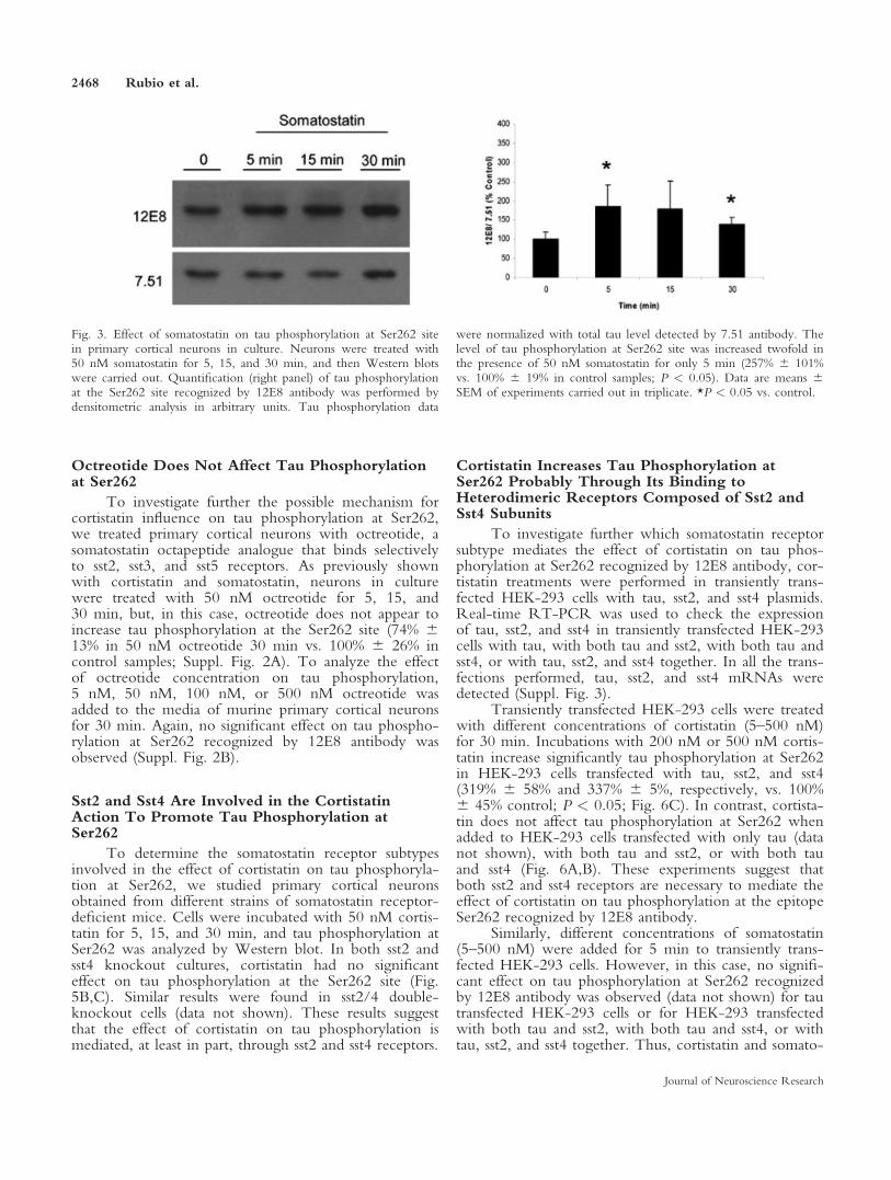
Octreotide Does Not Affect Tau Phosphorylationat Ser262
To investigate further the possible mechanism forcortistatin influence on tau phosphorylation at Ser262,we treated primary cortical neurons with octreotide, asomatostatin octapeptide analogue that binds selectivelyto sst2, sst3, and sst5 receptors. As previously shownwith cortistatin and somatostatin, neurons in culturewere treated with 50 nM octreotide for 5, 15, and30 min, but, in this case, octreotide does not appear toincrease tau phosphorylation at the Ser262 site (74% 613% in 50 nM octreotide 30 min vs. 100% 6 26% incontrol samples; Suppl. Fig. 2A). To analyze the effectof octreotide concentration on tau phosphorylation,5 nM, 50 nM, 100 nM, or 500 nM octreotide wasadded to the media of murine primary cortical neuronsfor 30 min. Again, no significant effect on tau phospho-rylation at Ser262 recognized by 12E8 antibody wasobserved (Suppl. Fig. 2B).
Sst2 and Sst4 Are Involved in the CortistatinAction To Promote Tau Phosphorylation atSer262
To determine the somatostatin receptor subtypesinvolved in the effect of cortistatin on tau phosphoryla-tion at Ser262, we studied primary cortical neuronsobtained from different strains of somatostatin receptor-deficient mice. Cells were incubated with 50 nM cortis-tatin for 5, 15, and 30 min, and tau phosphorylation atSer262 was analyzed by Western blot. In both sst2 andsst4 knockout cultures, cortistatin had no significanteffect on tau phosphorylation at the Ser262 site (Fig.5B,C). Similar results were found in sst2/4 double-knockout cells (data not shown). These results suggestthat the effect of cortistatin on tau phosphorylation ismediated, at least in part, through sst2 and sst4 receptors.
Cortistatin Increases Tau Phosphorylation atSer262 Probably Through Its Binding toHeterodimeric Receptors Composed of Sst2 andSst4 Subunits
To investigate further which somatostatin receptorsubtype mediates the effect of cortistatin on tau phos-phorylation at Ser262 recognized by 12E8 antibody, cor-tistatin treatments were performed in transiently trans-fected HEK-293 cells with tau, sst2, and sst4 plasmids.Real-time RT-PCR was used to check the expressionof tau, sst2, and sst4 in transiently transfected HEK-293cells with tau, with both tau and sst2, with both tau andsst4, or with tau, sst2, and sst4 together. In all the trans-fections performed, tau, sst2, and sst4 mRNAs weredetected (Suppl. Fig. 3).
Transiently transfected HEK-293 cells were treatedwith different concentrations of cortistatin (5–500 nM)for 30 min. Incubations with 200 nM or 500 nM cortis-tatin increase significantly tau phosphorylation at Ser262in HEK-293 cells transfected with tau, sst2, and sst4(319% 6 58% and 337% 6 5%, respectively, vs. 100%6 45% control; P < 0.05; Fig. 6C). In contrast, cortista-tin does not affect tau phosphorylation at Ser262 whenadded to HEK-293 cells transfected with only tau (datanot shown), with both tau and sst2, or with both tauand sst4 (Fig. 6A,B). These experiments suggest thatboth sst2 and sst4 receptors are necessary to mediate theeffect of cortistatin on tau phosphorylation at the epitopeSer262 recognized by 12E8 antibody.
Similarly, different concentrations of somatostatin(5–500 nM) were added for 5 min to transiently trans-fected HEK-293 cells. However, in this case, no signifi-cant effect on tau phosphorylation at Ser262 recognizedby 12E8 antibody was observed (data not shown) for tautransfected HEK-293 cells or for HEK-293 transfectedwith both tau and sst2, with both tau and sst4, or withtau, sst2, and sst4 together. Thus, cortistatin and somato-
Fig. 3. Effect of somatostatin on tau phosphorylation at Ser262 sitein primary cortical neurons in culture. Neurons were treated with50 nM somatostatin for 5, 15, and 30 min, and then Western blotswere carried out. Quantification (right panel) of tau phosphorylationat the Ser262 site recognized by 12E8 antibody was performed bydensitometric analysis in arbitrary units. Tau phosphorylation data
were normalized with total tau level detected by 7.51 antibody. Thelevel of tau phosphorylation at Ser262 site was increased twofold inthe presence of 50 nM somatostatin for only 5 min (257% 6 101%vs. 100% 6 19% in control samples; P < 0.05). Data are means 6SEM of experiments carried out in triplicate. *P < 0.05 vs. control.
2468 Rubio et al.
Journal of Neuroscience Research

Fig. 4. Internalization of Fluo-cortistatin in cultured cortical neurons.Cells were incubated with 5 lM Fluo-cortistatin for 30 min (A) andfor 1 min (B). In another experiment, the primary cultures were pre-treated with 1 lM phenylarsine oxide (PAO), an endocytosis inhibi-tor, for 30 min and then with 5 lM Fluo-cortistatin for 30 min (C).
The same cells are shown with green fluorescence corresponding toFluo-cortistatin distribution, red fluorescence showing the distribu-tion of tubulin using polyclonal 334 primary antibody, and blue fluo-rescence showing nuclei distribution (DAPI stain). Scale bar 520 lm.
Cortistatin and Tau Phosphorylation 2469
Journal of Neuroscience Research

Fig. 5. Expression of each murine somatostatin receptor subtype(sst1–5) in primary cortical neurons with the Sybr Green PCRmethod (A). The mean threshold cycle (CT) values for each somato-statin receptor subtype were normalized by the CT value of mousebeta-actin. Data are means 6 SEM of experiments in triplicate com-pared with 100% for sst5 (A). Effect of cortistatin on tau phosphoryl-ation at Ser262 in primary cortical neurons from sst2 and sst4 knock-out mice. Neurons in culture from sst2 knockout mice (B) and fromsst4 knockout mice (C) were incubated with 50 nM cortistatin for 5,
15, and 30 min, and then protein extracts were analyzed by Westernblot. Quantification (right panel) of tau phosphorylation at theSer262 site recognized by 12E8 antibody was performed by densito-metric analysis in arbitrary units. Tau phosphorylation data were nor-malized with total tau level detected by 7.51 antibody. Cortistatindoes not increase significantly tau phosphorylation at Ser262 in sst2-and sst4-deficient mice cultures. Experiments performed in duplicateare shown as means 6 SEM.
2470 Rubio et al.
Journal of Neuroscience Research

statin seem to mediate tau phosphorylation at Ser262 bya different mechanism.
PKC Mediates Tau Phosphorylation at Ser262Induced by Cortistatin in Murine Cortical Neurons
PLC-PKC signaling seems to be downstream ofthe somatostatin receptors (Ribalet and Eddlestone,1995), and PKC is one of the protein kinases that phos-phorylates tau at Ser262 (Gomez-Ramos et al., 2003;Xiao-Chuan et al., 2004). Thus, to determine the intra-cellular protein kinase pathway involved in the modifica-tion of tau Ser262 upon addition of cortistatin, we stud-ied primary cultures of cortical neurons treated with Ro31-8220, a PKC inhibitor.
Figure 7 shows that tau phosphorylation at Ser262induced by cortistatin was prevented in the presence of10 lM Ro 31-8220. Therefore, these results indicatethat PKC mediates the effect of cortistatin on tau phos-phorylation at Ser262 site in primary cortical neurons.
DISCUSSION
In the present study, we have demonstrated thatcortistatin promotes tau phosphorylation at Ser262 rec-ognized by 12E8 antibody in primary cultures of corticalneurons. P-Ser262 epitope, detected by 12E8 antibody,is an example of a nonproline-directed phosphorylationsite (NPDK) in tau protein (Morishima-Kawashimaet al., 1995). However, no differences were found whenthe phosphorylation at Ser396/404 site, recognized byPHF1 antibody, a proline-directed phosphorylation site(PDK; Morishima-Kawashima et al., 1995), was tested inthe absence or presence of cortistatin. Cortistatin isexpressed in GABAergic interneurons in the cortex (deLecea et al., 1997) and affects the excitability of corticalpyramidal neurons (Schweitzer et al., 2003). Cortistatininduces tau phosphorylation at Ser262 at different con-centrations in the nanomolar range, with saturation ofthe effect at 50 nM. It should be noted that, in studieswith fluorescent cortistatin, approximately 24% of cul-tured neurons showed ligand internalization. Therefore,the effect of cortistatin on the cortical neurons thatexpress cortistatin binding sites should be at least fourtimes higher than the effect observed in the mixed pop-ulation in primary cultures. Some studies report the rele-vance of tau phosphorylation at Ser262 in Alzheimer’sdisease (Morishima-Kawashima et al., 1995; Perez et al.,2005). It is noteworthy that phosphorylation at this resi-due is modified in the brain of patients with Alzheimer’sdisease (Morishima-Kawashima et al., 1995). In addition,in double transgenic mice (APPSwe/tauVLW) that repro-duce beta-amyloid and tau pathologies, tau phosphoryla-tion at Ser262 correlates with an increase in the forma-tion of filamentous tau aggregates showing a diametersimilar to that of tau filaments observed in Alzheimer’sdisease (Perez et al., 2005).
Fig. 6. Tau phosphorylation at Ser262 in HEK-293 cells transientlytransfected with tau, sst2, and sst4 cDNA. Transiently transfectedHEK-293 cells with both tau and sst2 (A), with both tau and sst4(B), and with tau, sst2, and sst4 together (C) in the presence of cor-tistatin at different concentrations (5 nM, 50 nM, 200 nM, and500 nM) for 30 min. Protein extracts were analyzed by Western blotand quantified by densitometry in arbitrary units. Data of tau phos-phorylation at Ser262 recognized by 12E8 antibody were normalizedwith total tau level detected by 7.51 antibody. Incubations with200 nM or 500 nM cortistatin increase significantly tau phosphoryla-tion at Ser262 in HEK-293 cells transfected with tau, sst2, and sst4(319% 6 58% and 337% 6 5%, respectively, vs. 100% 6 45% con-trol; P < 0.05), suggesting that both sst2 and sst4 receptors areinvolve in the effect of cortistatin on tau phosphorylation at Ser262.Data are means 6 SEM of experiments carried out in triplicate.*P < 0.05 vs. control.
Cortistatin and Tau Phosphorylation 2471
Journal of Neuroscience Research
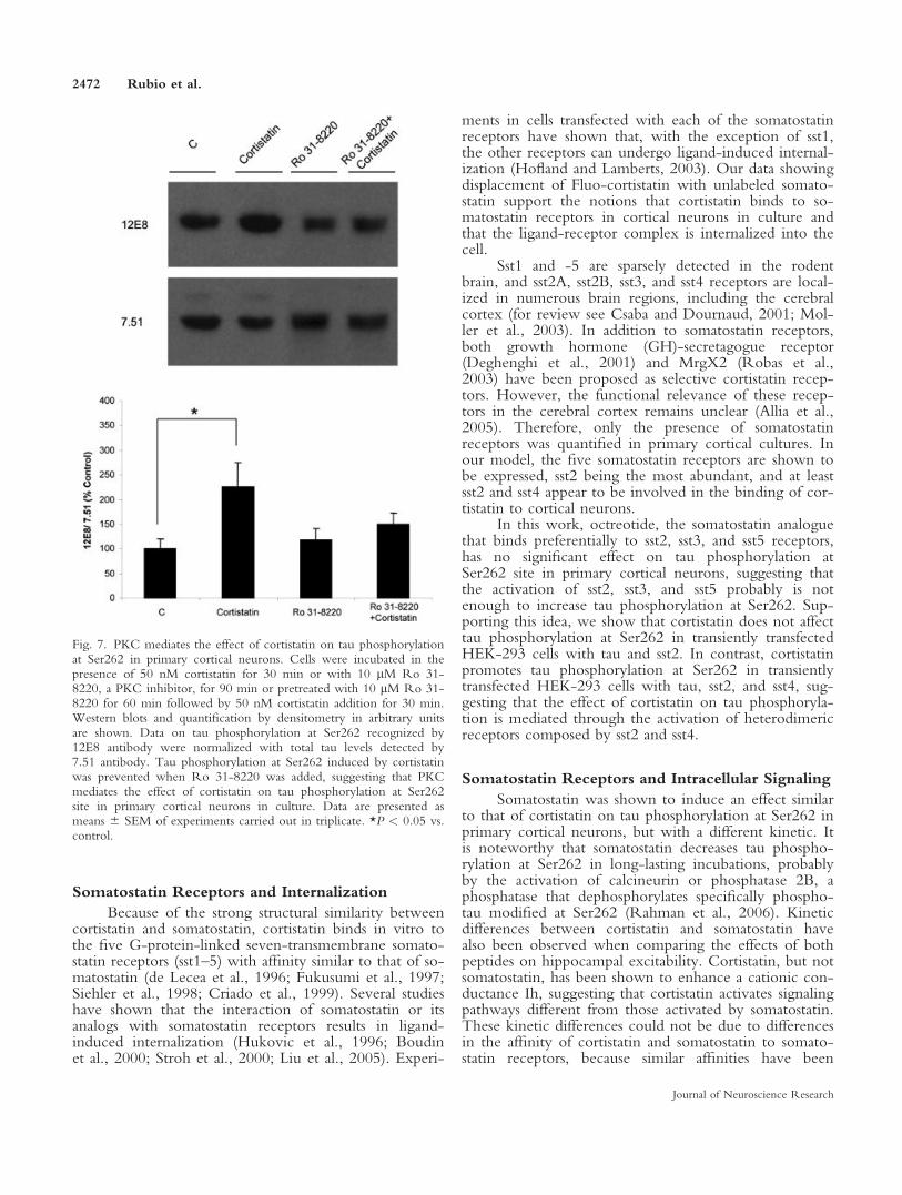
Somatostatin Receptors and Internalization
Because of the strong structural similarity betweencortistatin and somatostatin, cortistatin binds in vitro tothe five G-protein-linked seven-transmembrane somato-statin receptors (sst1–5) with affinity similar to that of so-matostatin (de Lecea et al., 1996; Fukusumi et al., 1997;Siehler et al., 1998; Criado et al., 1999). Several studieshave shown that the interaction of somatostatin or itsanalogs with somatostatin receptors results in ligand-induced internalization (Hukovic et al., 1996; Boudinet al., 2000; Stroh et al., 2000; Liu et al., 2005). Experi-
ments in cells transfected with each of the somatostatinreceptors have shown that, with the exception of sst1,the other receptors can undergo ligand-induced internal-ization (Hofland and Lamberts, 2003). Our data showingdisplacement of Fluo-cortistatin with unlabeled somato-statin support the notions that cortistatin binds to so-matostatin receptors in cortical neurons in culture andthat the ligand-receptor complex is internalized into thecell.
Sst1 and -5 are sparsely detected in the rodentbrain, and sst2A, sst2B, sst3, and sst4 receptors are local-ized in numerous brain regions, including the cerebralcortex (for review see Csaba and Dournaud, 2001; Mol-ler et al., 2003). In addition to somatostatin receptors,both growth hormone (GH)-secretagogue receptor(Deghenghi et al., 2001) and MrgX2 (Robas et al.,2003) have been proposed as selective cortistatin recep-tors. However, the functional relevance of these recep-tors in the cerebral cortex remains unclear (Allia et al.,2005). Therefore, only the presence of somatostatinreceptors was quantified in primary cortical cultures. Inour model, the five somatostatin receptors are shown tobe expressed, sst2 being the most abundant, and at leastsst2 and sst4 appear to be involved in the binding of cor-tistatin to cortical neurons.
In this work, octreotide, the somatostatin analoguethat binds preferentially to sst2, sst3, and sst5 receptors,has no significant effect on tau phosphorylation atSer262 site in primary cortical neurons, suggesting thatthe activation of sst2, sst3, and sst5 probably is notenough to increase tau phosphorylation at Ser262. Sup-porting this idea, we show that cortistatin does not affecttau phosphorylation at Ser262 in transiently transfectedHEK-293 cells with tau and sst2. In contrast, cortistatinpromotes tau phosphorylation at Ser262 in transientlytransfected HEK-293 cells with tau, sst2, and sst4, sug-gesting that the effect of cortistatin on tau phosphoryla-tion is mediated through the activation of heterodimericreceptors composed by sst2 and sst4.
Somatostatin Receptors and Intracellular Signaling
Somatostatin was shown to induce an effect similarto that of cortistatin on tau phosphorylation at Ser262 inprimary cortical neurons, but with a different kinetic. Itis noteworthy that somatostatin decreases tau phospho-rylation at Ser262 in long-lasting incubations, probablyby the activation of calcineurin or phosphatase 2B, aphosphatase that dephosphorylates specifically phospho-tau modified at Ser262 (Rahman et al., 2006). Kineticdifferences between cortistatin and somatostatin havealso been observed when comparing the effects of bothpeptides on hippocampal excitability. Cortistatin, but notsomatostatin, has been shown to enhance a cationic con-ductance Ih, suggesting that cortistatin activates signalingpathways different from those activated by somatostatin.These kinetic differences could not be due to differencesin the affinity of cortistatin and somatostatin to somato-statin receptors, because similar affinities have been
Fig. 7. PKC mediates the effect of cortistatin on tau phosphorylationat Ser262 in primary cortical neurons. Cells were incubated in thepresence of 50 nM cortistatin for 30 min or with 10 lM Ro 31-8220, a PKC inhibitor, for 90 min or pretreated with 10 lM Ro 31-8220 for 60 min followed by 50 nM cortistatin addition for 30 min.Western blots and quantification by densitometry in arbitrary unitsare shown. Data on tau phosphorylation at Ser262 recognized by12E8 antibody were normalized with total tau levels detected by7.51 antibody. Tau phosphorylation at Ser262 induced by cortistatinwas prevented when Ro 31-8220 was added, suggesting that PKCmediates the effect of cortistatin on tau phosphorylation at Ser262site in primary cortical neurons in culture. Data are presented asmeans 6 SEM of experiments carried out in triplicate. *P < 0.05 vs.control.
2472 Rubio et al.
Journal of Neuroscience Research

reported for both ligands (de Lecea et al., 1996; Fuku-sumi et al., 1997; Siehler et al., 1998; Criado et al.,1999). Kinetic differences between somatostatin and cor-tistatin may be explained by the notion that cortistatinmight bind receptors modified by accessory proteinssuch as RAMPS (Parameswaran and Spielman, 2006) orheterodimeric receptors (Agnati et al., 2003) essentiallydifferent from those for somatostatin. This later possibil-ity is supported by the data from this work.
Tau protein could be phosphorylated at Ser262 bydifferent protein kinases, such as PKA (Scott et al.,1993), MARK (Drewes et al., 1997), CaMKII (Ben-necib et al., 2001), phosphorylase kinase (Paudel, 1997),PK35/41 (Biernat et al., 1993), or PKC (Gomez-Ramoset al., 2003). Because somatostatin binding to somatosta-tin receptor has been shown to decrease PKA activity(Schindler et al., 1996) but also to activate the PLC-PKC pathway (Ribalet and Eddlestone, 1995), in thiswork we explored whether PKC could be involved inthe effect of cortistatin on tau phosphorylation atSer262. PKC is responsible for NPDP of tau protein(Correas et al., 1992; Singh et al., 1994; Morishima-Kawashima et al., 1995), and modification at the NPDKsites was shown to affect the interaction of tau withmicrotubules (Correas et al., 1992; Busciglio et al.,1995). In this work, we suggest that PKC mediates theincrease on tau phosphorylation induced by cortistatin,insofar as Ro 31-8220 prevents the increase of tau phos-phorylation observed upon the addition of cortistatin.
Possible Relation of Cortistatin and SomatostatinWith Alzheimer’s Disease
Significant changes in the expression of somatosta-tin and somatostatin receptors have been reported inAlzheimer’s disease. Somatostatin could be related tosome aspects of beta-amyloid pathology, insofar as it reg-ulates the degradation of beta-amyloid by neprilysin(Saito et al., 2005). Somatostatin concentration wasshown to be decreased in the brain and cerebrospinalfluid (CSF) of Alzheimer’s disease patients (Davies et al.,1980; Bissette et al., 1998; Nilsson et al., 2001). On theother hand, Kumar (2005) showed that the levels of so-matostatin receptors sst4 and sst5 are reduced in Alzhei-mer’s disease cortex. This result seems to contradict ourresults, but, in Kumar’s work, the decrease was observedat the last stage of the disease. Thus, at earlier stages(which we try to mimic), a higher amount of sst4 recep-tor should be present. In any case, the amount thatremains in the last stage of the disease may be sufficientto transmit cortistatin signal.
With regard to tau pathology, here we have shownthat cortistatin promotes tau phosphorylation at Ser262site in cortical neurons. The main risk for Alzheimer’sdisease is aging, and mRNA expression of preprocortis-tatin is significantly increased in the same brain regionsduring aging (Winsky-Sommerer et al., 2004). Thus, anincrease in preprocortistatin may result in an increase ofcortistatin that will result, as indicated in this work, in
an increase of tau phophorylation at Ser262. We havefound, first, that tau phosphorylation at Ser262 isreduced in the cortex of cortistatin-deficient mice and,second, that cortistatin increases tau phosphorylation atthe same site in primary cortical neurons in culture,probably through the binding to heterodimeric somato-statin receptors composed of sst2 and sst4. Interestingly,immunohistochemistry experiments in rat brain show ahigh degree of colocalization between sst2 and sst4 inlayer V of cortical pyramidal cells but not in hippocam-pal CA1 (Schreff et al., 2000). This result supports thefact that cortistatin effect, mediated through sst2 andsst4, was observed only in cortex but not in hippocam-pus. Indeed, our hypothesis is that, in the cortex of agedwild-type animals, cortistatin may induce phosphoryla-tion of tau at Ser262, and it may result in a decrease ofmicrotubule stability. The effect of cortistatin on tauphosphorylation at Ser262 is found mainly in the cortex,and it may play a role in the intermediate steps describedfor tau pathology in Alzheimer’s disease (Delacourteet al., 1999). This result could be relevant in explainingthe memory consolidation deficits related to Alzheimer’sdisease. In this disease, neuronal death, associated withtau and beta-amyloid pathologies, begins in the entorhi-nal cortex and then continues through the hippocampusand into the cortex (Braak and Braak, 1991), which isresponsible for memory consolidation (Hyman et al.,1984). In this process, a role for cortistatin has been sug-gested (de Lecea et al., 1996), and overexpression of cor-tistatin in transgenic mice has been shown to decreasecognitive performance (Tallent et al., 2005). Because ofits pattern of expression within the central nervous sys-tem at hippocampus and cortex, and its role in tau phos-phorylation at Ser262, the role of cortistatin in Alzhei-mer’s disease pathology should be studied further.
ACKNOWLEDGMENTS
We thank Dr. Patrice Bourgin for his generoushelp with real-time PCR.
REFERENCES
Agnati LF, Ferre S, Lluis C, Franco R, Fuxe K. 2003. Molecular mecha-
nisms and therapeutical implications of intramembrane receptor/recep-
tor interactions among heptahelical receptors with examples from the
striatopallidal GABA neurons. Pharmacol Rev 55:509–550.
Allia E, Tarabra E, Volante M, Cerrato M, Ghigo E, Muccioli G, Papotti
M. 2005. Expression of cortistatin and MrgX2, a specific cortistatin re-
ceptor, in human neuroendocrine tissues and related tumours. J Pathol
207:336–345.
Alzheimer A. 1906. Neurologisches Zentralblatt 25:146–148.
Arriagada PV, Growdon JH, Hedley-Whyte ET, Hyman BT. 1992.
Neurofibrillary tangles but not senile plaques parallel duration and se-
verity of Alzheimer’s disease. Neurology 42:631–639.
Avila J, Lucas JJ, Perez M, Hernandez F. 2004. Role of tau protein in
both physiological and pathological conditions. Physiol Rev 84:361–
384.
Banker GA, Cowan WM. 1977. Rat hippocampal neurons in dispersed
cell culture. Brain Res 126:397–342.
Cortistatin and Tau Phosphorylation 2473
Journal of Neuroscience Research

Bennecib M, Gong CX, Grundke-Iqbal I, Iqbal K. 2001. Inhibition of
PP-2A upregulates CaMKII in rat forebrain and induces hyperphos-
phorylation of tau at Ser 262/356. FEBS Lett 490:15–22.
Biernat J, Gustke N, Drewes G, Mandelkow EM, Mandelkow E. 1993.
Phosphorylation of Ser262 strongly reduces binding of tau to microtu-
bules: distinction between PHF-like immunoreactivity and microtubule
binding. Neuron 11:153–163.
Bissette G, Cook L, Smith W, Dole KC, Crain B, Nemeroff CB. 1998.
Regional neuropeptide pathology in Alzheimer’s disease: corticotropin-
releasing factor and somatostatin. J Alzheimers Dis 1:91–105.
Boudin H, Sarret P, Mazella J, Schonbrunn A, Beaudet A. 2000. So-
matostatin-induced regulation of SST(2A) receptor expression and cell-
surface availability in central neurons: role of receptor internalization. J
Neurosci 20:5932–5939.
Braak H, Braak E. 1991. Neuropathological stageing of Alzheimer-related
changes. Acta Neuropathol 82:239–259.
Bradford MM. 1976. A rapid and sensitive method for the quantitation
of microgram quantities of protein utilizing the principle of protein-dye
binding. Anal Biochem 72:248–254.
Busciglio J, Lorenzo A, Yeh J, Yankner BA. 1995. beta-amyloid fibrils
induce tau phosphorylation and loss of microtubule binding. Neuron
14:879–888.
Correas I, Diaz-Nido J, Avila J. 1992. Microtubule-associated protein tau
is phosphorylated by protein kinase C on its tubulin binding domain. J
Biol Chem 267:15721–15728.
Criado JR, Li H, Jiang X, Spina M, Huitron-Resendiz S, Liapakis G,
Calbet M, Siehler S, Henriksen SJ, Koob G, Hoyer D, Sutcliffe JG,
Goodman M, de Lecea L. 1999. Structural and compositional determi-
nants of cortistatin activity. J Neurosci Res 56:611–619.
Cristiani R, Fontanesi G, Casini G, Petrucci C, Viollet C, Bagnoli P.
2000. Expression of somatostatin subtype 1 receptor in the rabbit retina.
Invest Ophthalmol Vis Sci 41:3191–3199.
Csaba Z, Dournaud P. 2001. Cellular biology of somatostatin receptors.
Neuropeptides 35:1–23.
Davies P, Katzman R, Terry RD. 1980. Reduced somatostatin-like
immunoreactivity in cerebral cortex from cases of Alzheimer’s disease
and Alzheimer senile dementia. Nature 288:279–280.
de Lecea L, Criado JR, Prospero-Garcia O, Gautvik KM, Schweitzer P,
Danielson PE, Dunlop CL, Siggins GR, Henriksen SJ, Sutcliffe JG.
1996. A cortical neuropeptide with neuronal depressant and sleep-mod-
ulating properties. Nature 381:242–245.
de Lecea L, del Rio JA, Criado JR, Alcantara S, Morales M, Danielson
PE, Henriksen SJ, Soriano E, Sutcliffe JG. 1997. Cortistatin is expressed
in a distinct subset of cortical interneurons. J Neurosci 17:5868–5880.
Deghenghi R, Papotti M, Ghigo E, Muccioli G. 2001. Cortistatin, but
not somatostatin, binds to growth hormone secretagogue (GHS) recep-
tors of human pituitary gland. J Endocrinol Invest 24:RC1–RC3.
Delacourte A, David JP, Sergeant N, Buee L, Wattez A, Vermersch P,
Ghozali F, Fallet-Bianco C, Pasquier F, Lebert F, Petit H, Di Menza
C. 1999. The biochemical pathway of neurofibrillary degeneration in
aging and Alzheimer’s disease. Neurology 52:1158–1165.
Diaz-Nido J, Serrano L, Lopez-Otin C, Vandekerckhove J, Avila J.
1990. Phosphorylation of a neuronal-specific beta-tubulin isotype. J
Biol Chem 265:13949–13954.
Drewes G, Ebneth A, Preuss U, Mandelkow EM, Mandelkow E. 1997.
MARK, a novel family of protein kinases that phosphorylate microtu-
bule-associated proteins and trigger microtubule disruption. Cell
89:297–308.
Feindt J, Becker I, Blomer U, Hugo HH, Mehdorn HM, Krisch B,
Mentlein R. 1995. Expression of somatostatin receptor subtypes in cul-
tured astrocytes and gliomas. J Neurochem 65:1997–2005.
Flood JF, Uezu K, Morley JE. 1997. The cortical neuropeptide, cortista-
tin-14, impairs post-training memory processing. Brain Res 775:250–
252.
Fukusumi S, Kitada C, Takekawa S, Kizawa H, Sakamoto J, Miyamoto
M, Hinuma S, Kitano K, Fujino M. 1997. Identification and characteri-
zation of a novel human cortistatin-like peptide. Biochem Biophys Res
Commun 232:157–163.
Goedert M, Spillantini MG, Potier MC, Ulrich J, Crowther RA. 1989.
Cloning and sequencing of the cDNA encoding an isoform of microtu-
bule-associated protein tau containing four tandem repeats: differential
expression of tau protein mRNAs in human brain. EMBO J 8:393–
399.
Goedert M, Cohen ES, Jakes R, Cohen P. 1992. p42 MAP kinase phos-
phorylation sites in microtubule-associated protein tau are dephospho-
rylated by protein phosphatase 2A1. Implications for Alzheimer’s disease
[corrected]. FEBS Lett 312:95–99.
Gomez-Ramos A, Diaz-Nido J, Smith MA, Perry G, Avila J. 2003.
Effect of the lipid peroxidation product acrolein on tau phosphorylation
in neural cells. J Neurosci Res 71:863–870.
Greenberg SG, Davies P, Schein JD, Binder LI. 1992. Hydrofluoric acid-
treated tau PHF proteins display the same biochemical properties as
normal tau. J Biol Chem 267:564–569.
Heidet V, Faivre-Bauman A, Kordon C, Loudes C, Rasolonjanahary S,
Epelbaum J. 1990. Functional maturation of somatostatin neurons and
somatostatin receptors during development of mouse hypothalamus in
vivo and in vitro. Brain Res Dev Brain Res 57:85–92.
Hofland LJ, Lamberts SW. 2003. The pathophysiological consequences of
somatostatin receptor internalization and resistance. Endocrine Rev
24:28–47.
Hosli E, Hosli L. 1994. Colocalization of binding sites for somatostatin,
muscarine and nicotine on cultured neurones of rat neocortex, cerebel-
lum, brain stem and spinal cord: combined autoradiographic and immu-
nohistochemical studies. Neurosci Lett 173:71–74.
Hukovic N, Panetta R, Kumar U, Patel YC. 1996. Agonist-dependent
regulation of cloned human somatostatin receptor types 1–5 (hSSTR1–
5): subtype selective internalization or upregulation. Endocrinology
137:4046–4049.
Hyman BT, Van Hoesen GW, Damasio AR, Barnes CL. 1984. Alzhei-
mer’s disease: cell-specific pathology isolates the hippocampal forma-
tion. Science 225:1168–1170.
Iqbal K, Grundke-Iqbal I. 1997. Mechanism of Alzheimer neurofibrillary
degeneration and the formation of tangles. Mol Psychiatry 2:178–180.
Kumar U. 2005. Expression of somatostatin receptor subtypes (SSTR1–
5) in Alzheimer’s disease brain: an immunohistochemical analysis. Neu-
roscience 134:525–538.
Liu Q, Cescato R, Dewi DA, Rivier J, Reubi JC, Schonbrunn A. 2005.
Receptor signaling and endocytosis are differentially regulated by so-
matostatin analogs. Mol Pharmacol 68:90–101.
Mentlein R, Buchholz C, Krisch B. 1990. Somatostatin-binding sites on
rat telencephalic astrocytes. Light- and electron-microscopic studies in
vitro and in vivo. Cell Tissue Res 262:431–443.
Moller LN, Stidsen CE, Hartmann B, Holst JJ. 2003. Somatostatin recep-
tors. Biochim Biophys Acta 1616:1–84.
Morishima-Kawashima M, Hasegawa M, Takio K, Suzuki M, Yoshida
H, Titani K, Ihara Y. 1995. Proline-directed and non-proline-directed
phosphorylation of PHF-tau. J Biol Chem 270:823–829.
Nilsson CL, Brinkmalm A, Minthon L, Blennow K, Ekman R. 2001.
Processing of neuropeptide Y, galanin, and somatostatin in the cerebro-
spinal fluid of patients with Alzheimer’s disease and frontotemporal de-
mentia. Peptides 22:2105–2112.
Novak M, Jakes R, Edwards PC, Milstein C, Wischik CM. 1991. Differ-
ence between the tau protein of Alzheimer paired helical filament core
and normal tau revealed by epitope analysis of monoclonal antibodies
423 and 7.51. Proc Natl Acad Sci U S A 88:5837–5841.
Otvos L Jr, Feiner L, Lang E, Szendrei GI, Goedert M, Lee VM. 1994.
Monoclonal antibody PHF-1 recognizes tau protein phosphorylated at
serine residues 396 and 404. J Neurosci Res 39:669–673.
2474 Rubio et al.
Journal of Neuroscience Research

Parameswaran N, Spielman WS. 2006. RAMPs: the past, present and
future. Trends Biochem Sci (in press).
Paudel HK. 1997. The regulatory Ser262 of microtubule-associated pro-
tein tau is phosphorylated by phosphorylase kinase. J Biol Chem
272:1777–1785.
Perez M, Ribe E, Rubio A, Lim F, Moran MA, Ramos PG, Ferrer I,
Isla MT, Avila J. 2005. Characterization of a double (amyloid precursor
protein-tau) transgenic: tau phosphorylation and aggregation. Neuro-
science 130:339–347.
Rahman A, Grundke-Iqbal I, Iqbal K. 2006. PP2B isolated from human
brain preferentially dephosphorylates Ser-262 and Ser-396 of the Alz-
heimer disease abnormally hyperphosphorylated tau. J Neural Transm
113:219–230.
Ribalet B, Eddlestone GT. 1995. Characterization of the G protein cou-
pling of a somatostatin receptor to the K1ATP channel in insulin-
secreting mammalian HIT and RIN cell lines. J Physiol 485:73–86.
Robas N, Mead E, Fidock M. 2003. MrgX2 is a high potency cortistatin
receptor expressed in dorsal root ganglion. J Biol Chem 278:44400–
44404.
Saito T, Iwata N, Tsubuki S, Takaki Y, Takano J, Huang SM, Suemoto
T, Higuchi M, Saido TC. 2005. Somatostatin regulates brain amyloid
beta peptide Abeta42 through modulation of proteolytic degradation.
Nat Med 11:434–439.
Sanchez-Alavez M, Gomez-Chavarin M, Navarro L, Jimenez-Anguiano
A, Murillo-Rodriguez E, Prado-Alcala RA, Drucker-Colin R, Pros-
pero-Garcia O. 2000. Cortistatin modulates memory processes in rats.
Brain Res 858:78–83.
Scott CW, Spreen RC, Herman JL, Chow FP, Davison MD, Young J,
Caputo CB. 1993. Phosphorylation of recombinant tau by cAMP-
dependent protein kinase. Identification of phosphorylation sites and
effect on microtubule assembly. J Biol Chem 268:1166–1173.
Schindler M, Humphrey PP, Emson PC. 1996. Somatostatin receptors in
the central nervous system. Prog Neurobiol 50:9–47.
Schreff M, Schulz S, Handel M, Keilhoff G, Braun H, Pereira G,
Klutzny M, Schmidt H, Wolf G, Hollt V. 2000. Distribution, targeting,
and internalization of the sst4 somatostatin receptor in rat brain. J Neu-
rosci 20:3785–3797.
Seubert P, Mawal-Dewan M, Barbour R, Jakes R, Goedert M, Johnson
GV, Litersky JM, Schenk D, Lieberburg I, Trojanowski JQ, et al.
1995. Detection of phosphorylated Ser262 in fetal tau, adult tau, and
paired helical filament tau. J Biol Chem 270:18917–18922.
Siehler S, Seuwen K, Hoyer D. 1998. [125I]Tyr10-cortistatin14 labels all
five somatostatin receptors. Naunyn Schmiedebergs Arch Pharmacol
357:483–489.
Singh TJ, Grundke-Iqbal I, McDonald B, Iqbal K. 1994. Comparison of
the phosphorylation of microtubule-associated protein tau by non-
proline dependent protein kinases. Mol Cell Biochem 131:181–189.
Stroh T, Jackson AC, Dal Farra C, Schonbrunn A, Vincent JP, Beaudet
A. 2000. Receptor-mediated internalization of somatostatin in rat corti-
cal and hippocampal neurons. Synapse 38:177–186.
Tallent MK, Fabre V, Qiu C, Calbet M, Lamp T, Baratta MV, Suzuki
C, Levy CL, Siggins GR, Henriksen SJ, Criado JR, Roberts A,
de Lecea L. 2005. Cortistatin overexpression in transgenic mice pro-
duces deficits in synaptic plasticity and learning. Mol Cell Neurosci 30:
465–475.
van Calker D, Muller M, Hamprecht B. 1980. Regulation by secretin,
vasoactive intestinal peptide, and somatostatin of cyclic AMP accumula-
tion in cultured brain cells. Proc Natl Acad Sci U S A 77:6907–6911.
Videau C, Hochgeschwender U, Kreienkamp HJ, Brennan MB, Viollet
C, Richter D, Epelbaum J. 2003. Characterisation of [125I]Tyr0DTrp8-
somatostatin binding in sst1- to sst4- and SRIF-gene-invalidated mouse
brain. Naunyn Schmiedebergs Arch Pharmacol 367:562–571.
Winsky-Sommerer R, Spier AD, Fabre V, de Lecea L, Criado JR. 2004.
Overexpression of the human beta-amyloid precursor protein downre-
gulates cortistatin mRNA in PDAPP mice. Brain Res 1023:157–162.
Xiao-Chuan W, Zheng-Hui H, Zheng-Yu F, Yue F, Yun-Huang Y,
Qun W, Xiao-Wei T, Yi-Gen W, Jian-Zhi W. 2004. Correlation of
Alzheimer-like tau hyperphosphorylation and fMRI bold intensity.
Curr Alzheimer Res 1:143–148.
Cortistatin and Tau Phosphorylation 2475
Journal of Neuroscience Research