Donation of Gametes and Risk of...
137
Donation of Gametes and Risk of Preeclampsia Anna Blazquez Ventura Aquesta tesi doctoral està subjecta a la llicència Reconeixement- NoComercial – CompartirIgual 4.0. Espanya de Creative Commons. Esta tesis doctoral está sujeta a la licencia Reconocimiento - NoComercial – CompartirIgual 4.0. España de Creative Commons. This doctoral thesis is licensed under the Creative Commons Attribution-NonCommercial- ShareAlike 4.0. Spain License.
Transcript of Donation of Gametes and Risk of...
Donation of Gametes and Risk of Preeclampsia
Anna Blazquez Ventura
Aquesta tesi doctoral està subjecta a la llicència Reconeixement- NoComercial – CompartirIgual 4.0. Espanya de Creative Commons. Esta tesis doctoral está sujeta a la licencia Reconocimiento - NoComercial – CompartirIgual 4.0. España de Creative Commons. This doctoral thesis is licensed under the Creative Commons Attribution-NonCommercial-ShareAlike 4.0. Spain License.

PhDThesis
UNIVERSITAT DE BARCELONA
Facultat de Medicina i Ciències de la Salut
Departament d’ Obstetricia i Ginecologia, Pediatria, Radiologia i Anatomia
Donation of Gametes and Risk of Preeclampsia
Directors: Francesc Figueras Retuerta
Rita Vassena
Author: Anna Blazquez Ventura


ACKNOWLEDGEMENTS

Donation of gametes and Risk of Preeclampsia
5
ACKNOWLEDGEMENTS
6
A mis directores de tesis, Rita Vassena, quien me introdujo a la investigación, y
Francesc Figueras, con su impecable análisis de datos, por su tiempo (especialmente
durante sus vacaciones), sus consejos y su inmenso conocimiento.
A Désirée, por su guía y su constante motivación, y Sarai, por su indispensable ayuda
en todas las publicaciones.
A todos los coautores de las publicaciones presentadas, sin los cuales la realización de
esta tesis no sería posible. Al equipo de ATP, por su trabajo en la recogida de datos de
las pacientes.
A mis compañeros durante la residencia, junto con los cuales crecí personal y
profesionalmente, y a los compañeros de Eugin, médicos, enfermeras, biólogos y
técnicos de laboratorio, los cuales me han enseñado todo en reproducción asistida.
Finalment, una menció especial a la meva familia. Al meu pare, per tot el que en vaig
aprendre sobre la vida i referent pel qual avui sóc metge. A la meva mare, per ser un
exemple, pel seu infinit recolzament, i per ensenyar-me que el treball i la ilusió son
indispensables. Als meus germans, per viure cada èxit com si fossin seus, i per l’ajuda
a donar forma a aquesta tesis. I per últim, a l’Ariadna, per ensenyar-me coses que no
es troben als llibres, i sobretot al Xavi, pels ànims incondicionals, per recordar-me que
la fita no estava tant lluny, gràcies per fer sempre amb mi el camí.

Donation of gametes and Risk of Preeclampsia

ABBREVIATION LIST

Donation of gametes and Risk of Preeclampsia
9
ABBREVIATION LIST
10
ART: assisted reproductive technology
BMI: body mass index
CV: cardiovascular
DD: double donation of gametes
DET: double embryo transfer
ET: embryo transfer
IVF: in vitro fertilization
FP: women with female partner
GH: gestational hypertension
GnRHa: agonist of the Gonadotropin-releasing Hormone
HLA: Human Leukocyte Antigen
ICSI: intracytoplasmic sperm injection
KIR: killer immunoglobulin receptor
MP: women with male partner
OD: oocyte donation
PE: preeclampsia
sEng: soluble endoglin
SET: single embryo transfer
sFlt-1: soluble fms-like tyrosine kinase 1
SW: single Women
T-reg: regulatory T cells
TGF-β: transforming growth factor-β
uNK: uterine Natural Killer cell
VEGF: vascular endothelial growth factor
PGE: prostaglandin E
PlGF: placental growth factor

Donation of gametes and Risk of Preeclampsia
11
INDEX
12
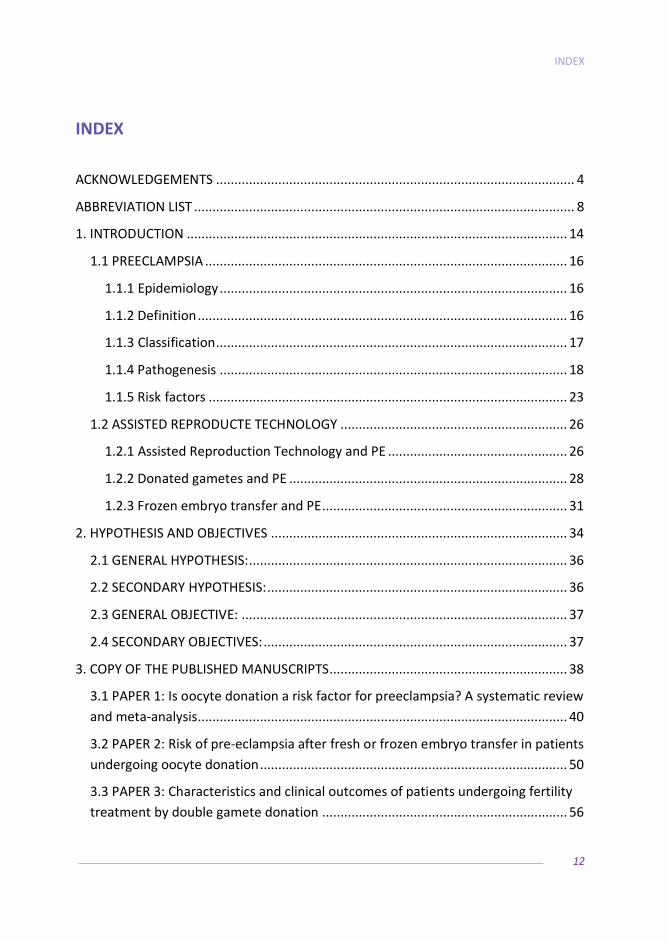
INDEX
ACKNOWLEDGEMENTS .................................................................................................. 4
ABBREVIATION LIST ........................................................................................................ 8
1. INTRODUCTION ........................................................................................................ 14
1.1 PREECLAMPSIA ................................................................................................... 16
1.1.1 Epidemiology ............................................................................................... 16
1.1.2 Definition ..................................................................................................... 16
1.1.3 Classification ................................................................................................ 17
1.1.4 Pathogenesis ............................................................................................... 18
1.1.5 Risk factors .................................................................................................. 23
1.2 ASSISTED REPRODUCTE TECHNOLOGY .............................................................. 26
1.2.1 Assisted Reproduction Technology and PE ................................................. 26
1.2.2 Donated gametes and PE ............................................................................ 28
1.2.3 Frozen embryo transfer and PE ................................................................... 31
2. HYPOTHESIS AND OBJECTIVES ................................................................................. 34
2.1 GENERAL HYPOTHESIS:....................................................................................... 36
2.2 SECONDARY HYPOTHESIS: .................................................................................. 36
2.3 GENERAL OBJECTIVE: ......................................................................................... 37
2.4 SECONDARY OBJECTIVES: ................................................................................... 37
3. COPY OF THE PUBLISHED MANUSCRIPTS................................................................. 38
3.1 PAPER 1: Is oocyte donation a risk factor for preeclampsia? A systematic review
and meta-analysis..................................................................................................... 40
3.2 PAPER 2: Risk of pre-eclampsia after fresh or frozen embryo transfer in patients
undergoing oocyte donation .................................................................................... 50
3.3 PAPER 3: Characteristics and clinical outcomes of patients undergoing fertility
treatment by double gamete donation ................................................................... 56

Donation of gametes and Risk of Preeclampsia
13
3.4 PAPER 4: Risk of preeclampsia in pregnancies resulting from double gamete
donation and from oocyte donation alone .............................................................. 62
4. DISCUSSION .............................................................................................................. 68
5. CONCLUSIONS .......................................................................................................... 82
6. BIBLIOGRAPHY .......................................................................................................... 86
7. SUMMARY IN SPANISH ........................................................................................... 112

INTRODUCTION

Donation of gametes and Risk of Preeclampsia
15
INTRODUCTION
16
PREECLAMPSIA
Epidemiology
Preeclampsia (PE) is a pregnancy hypertensive disorder which involves multiple
maternal organs, but characterized by the dysfunction of one, the placenta(1). It
complicates between 2 and 8% of all pregnancies, and while its mortality in developed
countries is decreasing, it is still one of the main causes of maternal death all over the
world, compounding 16% of pregnant women deaths(2). In the last decades, the
incidence of PE has been increasing in developed countries, because its risk factors,
such as hypertension, maternal age, diabetes mellitus, obesity, and multiple births
have also increased(3).
Definition
Pre-eclampsia is classically defined as new-onset hypertension (arterial tension
>140/90 mmHg in at least 2 determinations taken at least 6h apart) associated with
proteinuria (>300 mg protein in a 24h urine), diagnosed at or after 20 weeks of
pregnancy (4). Although proteinuria is present in the majority of cases, most current
guidelines do not require this sign in the presence of other organ-end signs or
symptoms. For instance, The American College of Obstetricians and Gynecologists
guidelines defines PE in the absence of proteinuria as hypertension with any of the
Donation of gametes and Risk of Preeclampsia
17
followings: new-onset thrombocytopenia, impaired liver function, renal insufficiency,
pulmonary edema, or visual or cerebral disturbances (5).
Classification
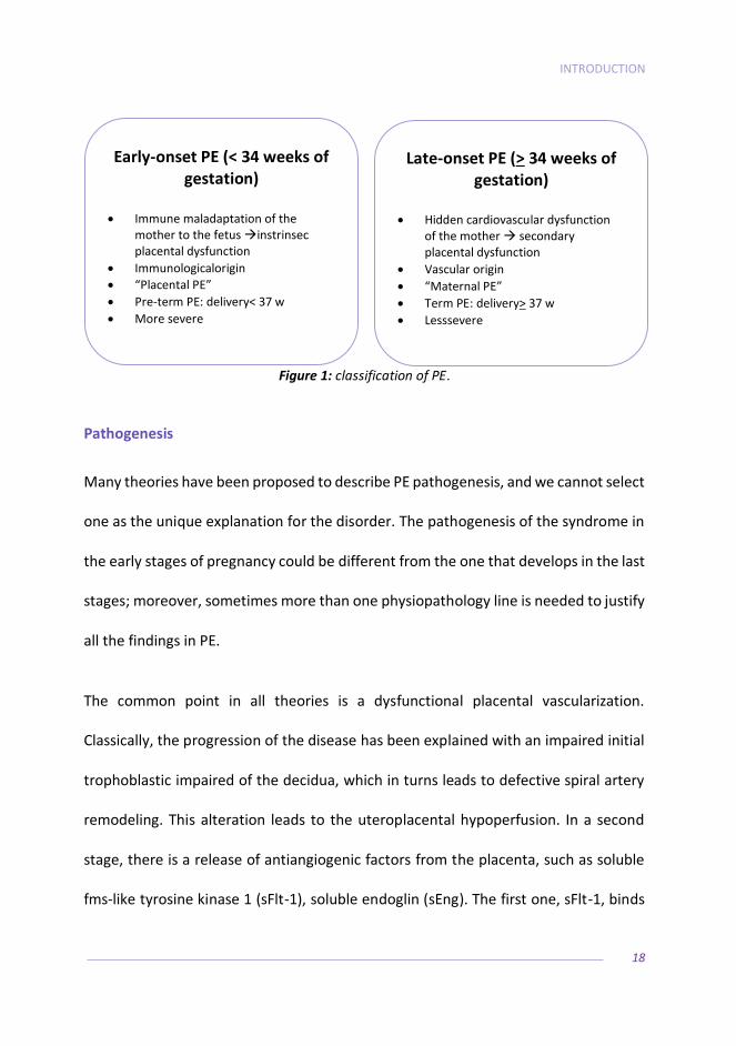
PE is classified in two subclasses: early-onset pre-eclampsia (< 34 weeks of gestation),
and late-onset (> 34 weeks of gestation). In the early-onset syndrome, it is believed
that the cause is an intrinsic placental dysfunction, triggered by an immune
maladaptation of the mother to the fetus (6). On the other hand, in the late-onset PE,
the origin must be found in a hidden cardiovascular dysfunction of the mother that,
due to the pregnancy increase in hemodynamic demands, leads to placental
dysfunction (7). The first one, with an immunological trigger, is also called “placental
PE”, and the late-onset, with a vascular origin, is termed “maternal PE” (1). This division
is relevant as the early-onset PE is more severe, with fetal growth restriction and
adverse maternal and neonatal consequences (8,9). On the other hand, despite late-
onset PE being milder, it is a large contributor to adverse perinatal outcomes due to
the fact that it is 5-10 times more frequent that early-onset PE.
INTRODUCTION
18
Figure 1: classification of PE.
Pathogenesis
Many theories have been proposed to describe PE pathogenesis, and we cannot select
one as the unique explanation for the disorder. The pathogenesis of the syndrome in
the early stages of pregnancy could be different from the one that develops in the last
stages; moreover, sometimes more than one physiopathology line is needed to justify
all the findings in PE.
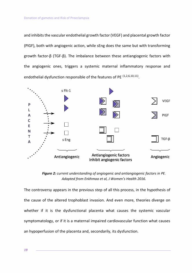
The common point in all theories is a dysfunctional placental vascularization.
Classically, the progression of the disease has been explained with an impaired initial
trophoblastic impaired of the decidua, which in turns leads to defective spiral artery
remodeling. This alteration leads to the uteroplacental hypoperfusion. In a second
stage, there is a release of antiangiogenic factors from the placenta, such as soluble
fms-like tyrosine kinase 1 (sFlt-1), soluble endoglin (sEng). The first one, sFlt-1, binds
Early-onset PE (< 34 weeks of gestation)
• Immune maladaptation of the mother to the fetus →instrinsec placental dysfunction
• Immunologicalorigin • “Placental PE”
• Pre-term PE: delivery< 37 w
• More severe
Late-onset PE (> 34 weeks of gestation)
• Hidden cardiovascular dysfunction of the mother → secondary placental dysfunction
• Vascular origin
• “Maternal PE”
• Term PE: delivery> 37 w
• Lesssevere
Donation of gametes and Risk of Preeclampsia
19
and inhibits the vascular endothelial growth factor (VEGF) and placental growth factor
(PlGF), both with angiogenic action, while sEng does the same but with transforming
growth factor-β (TGF-β). The imbalance between these antiangiogenic factors with
the angiogenic ones, triggers a systemic maternal inflammatory response and
endothelial dysfunction responsible of the features of PE (1,2,6,10,11).
Figure 2: current understanding of angiogenic and antiangiogenic factors in PE.
Adapted from Enkhmaa et al, J Women’s Health 2016.
The controversy appears in the previous step of all this process, in the hypothesis of
the cause of the altered trophoblast invasion. And even more, theories diverge on
whether if it is the dysfunctional placenta what causes the systemic vascular
symptomatology, or if it is a maternal impaired cardiovascular function what causes
an hypoperfusion of the placenta and, secondarily, its dysfunction.

INTRODUCTION
20
The immunological theory
The immunological theory of PE pathogenesis postulates that the vascular dysfunction
is the result of a maladaptation of the mother to fetal agents, and rests on the
observation that the trophoblastic cells invading the decidua during early pregnancy
express class I HLA-C antigens (both maternal and paternal), which are polymorphic,
unlike the non-classical HLA-G and HLA-E that are also expressed in trophoblastic cells.
The HLA-C is a strong ligand for the killer immunoglobulin receptor (KIR) which is
present on the surface of the uterine natural killer cells (uNK). The KIR receptor also
has a wide variability, as more than 350 different KIR genotypes have been described,
although they can be classified in 2 basic haplotypes: KIR A and KIR B.KIR A is inhibiting
and KIR B is activating, depending on their up-regulation or down-regulation of the
uNK function. Despite there are three different combinations (KIR AA, KIR AB and KIR
BB), if they have at least one haplotype of KIR B they act as activating, so in the practice
we still divide them in two groups(12). At the same time, HLA-C is also divided into two
subtypes: HLA-C1 and HLA-C2. In the trophoblastic cell, then, that express one
paternal and one maternal allotype of HLA-C, we can find three combinations: HLA-
C1/C1, HLA-C1/C2 and HLA-C2/C2.

Donation of gametes and Risk of Preeclampsia
21
Figure 3: possible combinations of trophoblastic HLA-C and maternal KIRs.
Adapted from Morin, Fert &Steril 2017.
The mode of action of uNK differs from that of systemic NK cells, as uNK modulate
proangiogenic and endothelial factors (PlGF, VEFG, TGF-β) which in turn stimulate
decidua neovascularitzation, promoting changes in the spiral arteries to supply proper
blood flow to the fetus.
INTRODUCTION
22
Figure 4: Placental angiogenesis during early pregnancy. From Maliqueo,
Frontiers in Physiology, 2016.
Some maternal KIR genotypes (especially the AA genotype) combined with certain
trophoblastic HLA-C allotypes (particularly HLA-C2, when the fetus has more C2 genes
than the mother or when fetal C2 is inherited paternally) can favor a dysfunction of
uNK, which is in turn associated with an altered maternal blood supply to the placenta,
ultimately inducing disorders like PE and fetal growth restriction(13,14).
This rationale supports the placental origin for early-onset PE, characterized by small
placenta with histological findings of hypoperfusion (15), and associated with fetal
growth restriction (16,17). But in the cases of late-onset PE, the most common
manifestation are proper of an hypertensive disorder, placental lesions of

Donation of gametes and Risk of Preeclampsia
23
hypoperfusion are less frequent or even absent (15), and usually fetuses have normal
weight for gestational age (18,19).
The cardiovascular theory
Several studies have shown increased cardiovascular risk factors in women after pre-
eclamptic pregnancies (20–22), but many others found that risk factors are present
before gestation and can predict PE. Furthermore, these factors are strongly
associated with future cardiovascular risk, more than PE itself (23,24). Pregnancy is a
state where the hemodynamic demands on the cardiovascular system are increased,
as well as other metabolic functions, as for example healthy pregnancies have certain
degree of insulin resistance. All these physiological changes can be excessive for
pregnancies with pre-gestational cardiovascular risk factors, which have a
predisposing endothelial dysfunction, and could lead secondary to PE (25).
Risk factors
Several risk factors have been identified for PE. Most of them are present before the
pregnancy and are used by healthcare professionals to assess the risk of each patient
at the beginning of the pregnancy, in order to schedule appropriate obstetrical follow
up and prophylactic measures.
INTRODUCTION
24
Not all the factors have the same significance in the risk for developing PE. The
following list ranks them by the relative risk that confer, from higher to lower:
1. History of PE in a previous pregnancy: having suffered PE in a previous
gestation confers a sevenfold risk for PE (26,27).
2. Underlying medical condition:
a. Autoimmune disease such as systemic lupus erythematosis,
or especially antiphospholipid syndrome, increases six times
the risk for PE (28,29).
b. Diabetes mellitus (DM) before pregnancy nearly quadruples
the risk for PE (30).
c. Pre-existing hypertension doubles the risk for developing PE
(28).
d. Renal disease(28).
3. Obesity: A pre-pregnancy Body Mass Index of >35 increases more
than three times the risk for PE (28,31).
4. Parity: nulliparity gives almost a threefold risk for developing PE (28).
5. Multiple pregnancy: twin pregnancies almost triples the risk for PE
compared with singleton pregnancies (32).

Donation of gametes and Risk of Preeclampsia
25
6. Family history of PE, hypertension and diabetes mellitus (in mother or
sisters) increases almost three times the risk for PE (33,34).
7. Advanced maternal age: pregnant women above 35 years, and
especially above 40 years, have twice the risk to develop PE (35).
8. Change in partner: changing the partner confers a higher risk to
develop PE (36,37).
9. Race: African-American pregnant women have higher risk than
Caucasian women(38).
10. Conception mode: assisted reproductive technology (ART) is
associated with more risk to develop PE compared to natural
gestations. The use of donated gametes confers an additional risk to
ART (39,40).
Noticeably, smoking has been found to act as a protective factor for PE (41).
INTRODUCTION
26
ASSISTED REPRODUCTE TECHNOLOGY
Since the first baby born by in vitro fertilization in 1978, ART has undergone many
changes, especially due to the advances in medicine and the steadily increase in the
number of patients demanding fertility treatments. The number of fertility clinics has
increased, making ART more accessible to people all over the world, despite important
differences among regions(42). Technology allows the culture of the embryo until
blastocyst stage, the use of donated gametes, embryo cryopreservation and
preimplantation genetic diagnosis, but outcomes of babies conceived by ART are still
a matter of study, as in many cases they are more adverse than in naturally conceived
babies.
Assisted Reproduction Technology and PE
ART is associated with a higher risk for PE compared to naturally conceived
pregnancies. While this association has been proved in several occasions, the reason
for it remains unexplained. The risk factors of the patients who needed fertility
treatments, the causes of the infertility itself, and the hormone therapy that they
received, are all plausible causes for the increased risk for PE in ART patients. To
complicate matter further, ART is also associated with an increased rate for multiple
gestation(42).

Donation of gametes and Risk of Preeclampsia
27
One study comparing singleton pregnancies conceived by ART (in vitro fertilization,
intracytoplasmic sperm injection and frozen embryo replacement) or spontaneous
conception, showed that, even after adjustment for maternal age and parity, the risk
of ART pregnancies and PE remained higher (43). Different theories have been
proposed to explain the relationship between ART and PE. One theory is based on the
infertility of these patients; infertility was found to be associated with anomalous DNA
methylation pattern in placentas from pregnancies conceived by IVF/ICSI compared
to naturally conception placentas (44). At the same time, epigenetic changes (changes
in DNA methylation, histone modifications and non-coding RNAs, in miR-210 and
genes as SERPINA3 or HI9, among others) have been observed in preeclampsia
placentas compared to normal placentas (45). Thus, infertility could be the cause of a
molecular impairment that predisposes to PE. However, ART per se could also explain
this association; the culture conditions used in the embryology laboratory (46), the
hormones that the patient needs to stimulate the ovary (47), ortheembryo transfer and
manipulations(48), all are possible causes for an altered trophoblast invasion.
INTRODUCTION
28
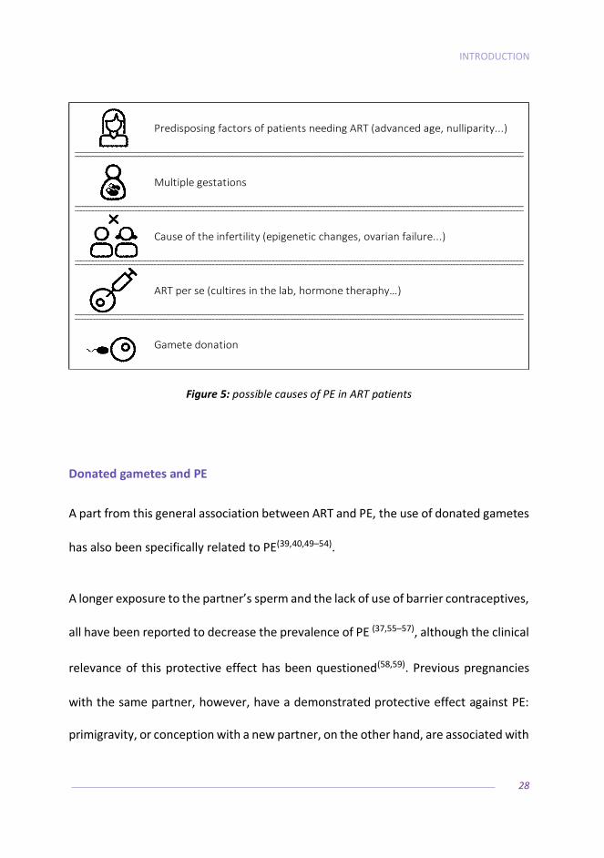
Predisposing factors of patients needing ART (advanced age, nulliparity...)
Multiple gestations
Cause of the infertility (epigenetic changes, ovarian failure...)
ART per se (cultires in the lab, hormone theraphy…)
Gamete donation
Figure 5: possible causes of PE in ART patients
Donated gametes and PE
A part from this general association between ART and PE, the use of donated gametes
has also been specifically related to PE(39,40,49–54).
A longer exposure to the partner’s sperm and the lack of use of barrier contraceptives,
all have been reported to decrease the prevalence of PE (37,55–57), although the clinical
relevance of this protective effect has been questioned(58,59). Previous pregnancies
with the same partner, however, have a demonstrated protective effect against PE:
primigravity, or conception with a new partner, on the other hand, are associated with
Donation of gametes and Risk of Preeclampsia
29
higher rates of PE, while multiparity or previous abortions with the same partner
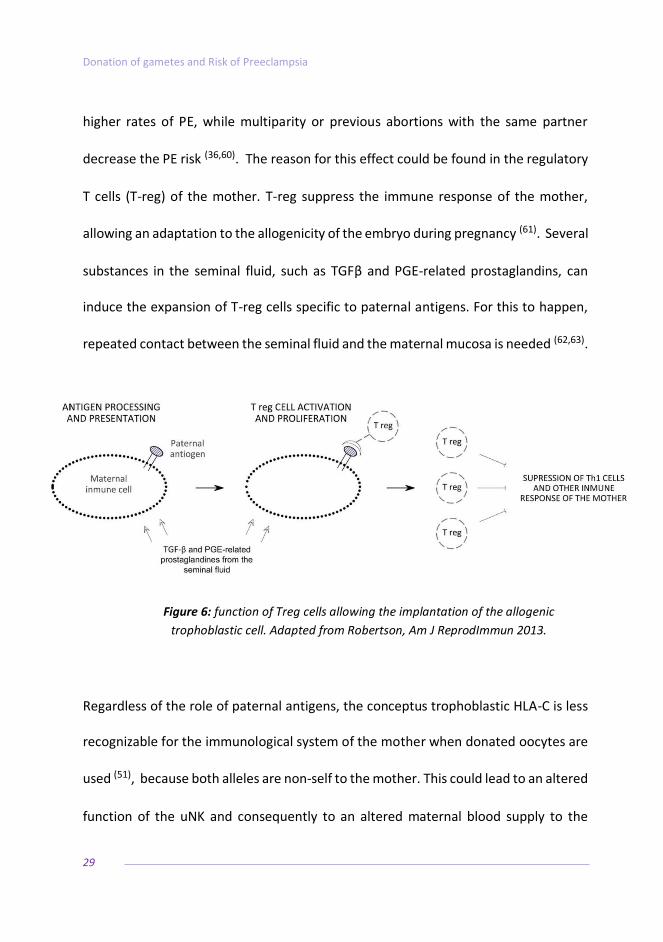
decrease the PE risk (36,60). The reason for this effect could be found in the regulatory
T cells (T-reg) of the mother. T-reg suppress the immune response of the mother,
allowing an adaptation to the allogenicity of the embryo during pregnancy (61). Several
substances in the seminal fluid, such as TGFβ and PGE-related prostaglandins, can
induce the expansion of T-reg cells specific to paternal antigens. For this to happen,
repeated contact between the seminal fluid and the maternal mucosa is needed (62,63).
Figure 6: function of Treg cells allowing the implantation of the allogenic
trophoblastic cell. Adapted from Robertson, Am J ReprodImmun 2013.
Regardless of the role of paternal antigens, the conceptus trophoblastic HLA-C is less
recognizable for the immunological system of the mother when donated oocytes are
used (51), because both alleles are non-self to the mother. This could lead to an altered
function of the uNK and consequently to an altered maternal blood supply to the

INTRODUCTION
30
placenta, facilitating disorders like PE and fetal growth restriction (13,14,51). Hibi shown
that the association between a maternal KIR AA with fetal C2, especially if the C2 gene
is inherited from the father, increases the risk for PE (12). In oocyte donation, the
oocyte HLA-C behaves as the paternal HLA-C, increasing the chances of finding the
combination of a KIR AA recipient with non-maternal HLA-C2 fetus. Additionally, in
ART, double embryo transfer (DET) increases the expression of trophoblastic HLA-C in
the decidua basalis, multiplying, in cases of oocyte donation, the number of external
HLA-C presented to the mother. A study analyzing reproductive outcomes in oocyte
donation cycles found that a decreased live birth rate per cycle after DET was
observed in mothers KIR AA compared to mothers with KIR AB and KIR BB, but not in
SET (64). They speculate that in DET cases, where 4 possible non-maternal HLA-C could
be present (one paternal and one from the oocyte donor per each one of the two
embryos), it was more likely to find an external HLA-C2 to the mother than in SET or
in own oocytes cycles.
Also, T cells have been involved in the immune down-regulation that allows the fetus
to develop in the maternal allogeneic environment (65). In a study of 26 placentas of
oocyte donation pregnancies, an infiltrate of macrophages was found in placentas of
pregnancies uncomplicated by PE, and not in the placentas of pregnancies with PE.
This lesion in the chorionic plate was associated with intervillositis, chronic deciduitis,
higher expression of CD14+ macrophages and fetal HLA-C2. All these data suggested
Donation of gametes and Risk of Preeclampsia
31
an immunological protection of the mother to the fetus, which was absent in PE
gestations (66).
Furthermore, some authors hypothesize an association between the need for oocyte
donation per se and PE, as circulating antibodies against granulosa cells and the
oocyte’s zona pellucida have been detected in patients presenting ovarian failure, a
classical indication for oocyte donation (OD) independently from sperm donation (67).
These authors postulate that the antibodies could cause injury to the trophoblastic
cells invading the decidua vessels as well (40,53).Moreover, it is not clear whether the
altered ovarian function of patients needing OD could be related to vascular or
immunological changes that could independently predispose to PE(53,68).
Frozen embryo transfer and PE
There is strong evidence suggesting that the supraphysiological hormonal levels
reached during controlled ovarian stimulation have deleterious effects on the
endometrium, causing placental dysfunction and alterations, likely affecting the
pregnancy course(69–71). Different studies relate abnormal levels of progesterone with
PE, although with opposite conclusions on whether a high or low level of this hormone
is found in preeclamptic pregnancies(72,73). The same controversy occurs with
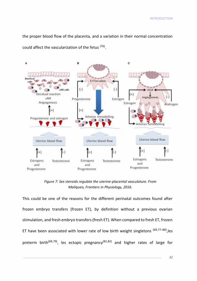
estradiol levels and pregnancy pathology (74,75). Sex steroids play an important role in
INTRODUCTION
32
the proper blood flow of the placenta, and a variation in their normal concentration
could affect the vascularization of the fetus (76).
Figure 7: Sex steroids regulate the uterine-placental vasculature. From
Maliqueo, Frontiers in Physiology, 2016.
This could be one of the reasons for the different perinatal outcomes found after
frozen embryo transfers (frozen ET), by definition without a previous ovarian
stimulation, and fresh embryo transfers (fresh ET). When compared to fresh ET, frozen
ET have been associated with lower rate of low birth weight singletons (69,77–80),les
preterm birth(69,79), les ectopic pregnancy(81,82) and higher rates of large for

Donation of gametes and Risk of Preeclampsia
33
gestational age singletons(69,77–79,83). Data are still inconsistent about the perinatal
mortality of frozen ET babies: while in some papers the risk is higher compared to
fresh ET (69,77),others do not find differences(79), or even report les risk(84).
The effect of frozen ET on the occurrence of placental alteration has been less studied
so far: the rate of placenta previa seems either lower(77), or notdifferent(78), while the
occurrence of placenta accreta seems higher after frozen ET (78).
Regarding PE, it is the only maternal effect consistently increased in frozen ET
compared to fresh ET (51,77,78,85,86). Some authors also suggest that the process of
freezing and thawing per se could bring about some metabolic or epigenetically
changes in the embryos, and thus be an adjuvant in the origin of PE in the frozen ET
group (87,88). Some cryoprotectants have been reported to interact with the main
enzyme involved in epigenetic reprogramming, methyltransferase (89). This interaction
during the initial embryonic developmental phases could be the cause of variations in
the epigenetic burden of the embryo and might affect developmental programming
of fetal and placental tissues (90).

HYPOTHESIS AND OBJECTIVES

Donation of gametes and Risk of Preeclampsia
35

HYPOTHESIS AND OBJECTIVES
36
GENERAL HYPOTHESIS:
• Assisted reproductive techniques are variably associated to preeclampsia, to
the extent of the allogenicity and exposure to paternal agents of each of the
techniques.
SECONDARY HYPOTHESIS:
• Project 1: In oocyte donation pregnancies, the risk for developing
preeclampsia is higher than in pregnancies achieved by IVF with patients own
oocytes.
• Project 2: In oocyte donation pregnancies, the risk for developing
preeclampsia is higher after frozen embryo transfer compared with fresh
embryo transfer.
• Project 3: Double-donation of oocytes and sperm is increasing worldwide, and
those patients have several risk factors for preeclampsia.
• Project 4: In double-donation of oocytes and sperm pregnancies, the risk for
developing preeclampsia is higher than in patients pregnant by oocyte
donation alone.

Donation of gametes and Risk of Preeclampsia
37
GENERAL OBJECTIVE:
• To evaluate the incidence of preeclampsia in pregnancies achieved by assisted
reproductive technology, especially in donation of gametes, and to define
novel risk factors.
SECONDARY OBJECTIVES:
• Project 1: to compare the risk for developing preeclampsia between patients
pregnant by oocyte donation and by IVF with own gametes.
• Project 2: to compare, in pregnancies achieved by oocyte donation, the risk
for developing preeclampsia between fresh embryo transfers and frozen
embryo transfers.
• Project 3: to evaluate patients undergoing double-donation of oocytes and
sperm, their characteristics and the trend of this treatment during the period
time of the study.
• Project 4: to compare the risk for developing preeclampsia between patients
pregnant by double-donation of oocytes and sperm and by oocyte donation.

COPY OF THE PUBLISHED MANUSCRIPTS

Donation of gametes and Risk of Preeclampsia
39

COPY OF THE PUBLISHED MANUSCRIPTS
40
PAPER 1: Is oocyte donation a risk factor for preeclampsia? A
systematic review and meta-analysis
Donation of gametes and Risk of Preeclampsia
41
COPY OF THE PUBLISHED MANUSCRIPTS
42

Donation of gametes and Risk of Preeclampsia
43

COPY OF THE PUBLISHED MANUSCRIPTS
44
Donation of gametes and Risk of Preeclampsia
45
COPY OF THE PUBLISHED MANUSCRIPTS
46

Donation of gametes and Risk of Preeclampsia
47

COPY OF THE PUBLISHED MANUSCRIPTS
48

Donation of gametes and Risk of Preeclampsia
49

COPY OF THE PUBLISHED MANUSCRIPTS
50
PAPER 2: Risk of pre-eclampsia after fresh or frozen embryo
transfer in patients undergoing oocyte donation
Donation of gametes and Risk of Preeclampsia
51

COPY OF THE PUBLISHED MANUSCRIPTS
52

Donation of gametes and Risk of Preeclampsia
53
COPY OF THE PUBLISHED MANUSCRIPTS
54

Donation of gametes and Risk of Preeclampsia
55

COPY OF THE PUBLISHED MANUSCRIPTS
56
PAPER 3: Characteristics and clinical outcomes of patients
undergoing fertility treatment by double gamete donation

Donation of gametes and Risk of Preeclampsia
57
COPY OF THE PUBLISHED MANUSCRIPTS
58

Donation of gametes and Risk of Preeclampsia
59
COPY OF THE PUBLISHED MANUSCRIPTS
60

Donation of gametes and Risk of Preeclampsia
61

COPY OF THE PUBLISHED MANUSCRIPTS
62
PAPER 4: Risk of preeclampsia in pregnancies resulting from
double gamete donation and from oocyte donation alone
Donation of gametes and Risk of Preeclampsia
63
COPY OF THE PUBLISHED MANUSCRIPTS
64
Donation of gametes and Risk of Preeclampsia
65

COPY OF THE PUBLISHED MANUSCRIPTS
66

Donation of gametes and Risk of Preeclampsia

DISCUSSION

Donation of gametes and Risk of Preeclampsia
69
DISCUSSION
70
This thesis aims to elucidate the relationship between preeclampsia and assisted
reproductive technology, especially with donated gametes.
From 2008 to 2010, around 4.5 million ART cycles were initiated around the world,
with an annual increase every year. The number of cycles with donated oocytes
reported increased a 35.8% in this triennium, with about 50,000 oocyte donation
embryo transfers performed in 2010 (42).Assisted reproductive care in general has
become more widespread and socially accepted, while technical advances allow for
the treatment of more people than ever before. The main factor for the increase in the
demand of ART in developed countries is the increasing age at which women have their
first child (91,92). More women are waiting to have children until they have completed
higher education degrees, and participation of women in the skilled workforce is
increasing; however, delaying motherhood can reduce the possibility of using one’s
own oocytes to achieve a pregnancy (93,94).
As a first insight into the relationship between gamete donation and PE, the design of
the meta-analysis focused specifically on PE and included solely cohort studies
comparing cycles of in vitro fertilization with OD vs. IVF. Since we avoided the
comparison with natural conception pregnancies, we excluded the bias of the assisted
reproductive technique per se, avoiding then the hormone therapy or the cultures in

Donation of gametes and Risk of Preeclampsia
71
the laboratory as an explanation of the difference in PE incidence. With 26.302 cases
analyzed, this meta-analysis shows a clear association between OD and PE.
A limitation of this review is the lack of information in the included studies about the
cause of infertility that has led to assisted reproductive technology (ART) with either
autologous or donor oocytes, because the underlying type of infertility leading to one
or the other treatment might itself contribute to the pathophysiology of PE. The
factors causing the need of ART certainly vary from patients that will require donated
oocytes from the ones that will perform an IVF with autologous oocytes (for instance,
tubal infertility is typically a cause of IVF with autologous oocytes, while a premature
ovarian failure will most likely lead to OD), thus making it difficult to determine if it is
the reception of the oocytes or the cause of its necessity that is associated with the
increase in PE incidence. Another limitation is the lack of detailed information in the
included studies on severity or gestational age at onset of PE: the association with OD
would be more clinically relevant in early onset PE, since it is more amenable to
prevention by aspirin than late-onset disease.
The etiological relationship between OD and PE remains unclear. Despite the fact that
the cause of developing PE seems to be multifactorial, OD is associated with an
increased incidence of this disorder. As with the IVF control group we avoid the bias of
the ART as a cause of the higher risk for PE, and with the meta regression procedure
of our review we account for the confounding factors of maternal age and multiple

DISCUSSION
72
pregnancy, the immunological theory reinforces. However, there is a need of an
increase in our understanding of the biochemical and immunological causes of PE, in
order to develop possible preventive strategies, such as the selection of the oocyte
donor immunologically matched to the HLA of the recipient (95).
Examining the studies reported so far regarding PE and frozen embryo transfers, we
realized that a common bias in all these papers is that they have only included embryo
transfers of treatments with patient’s own oocytes. Including only IVF with own
oocytes, it does not allow to understand more in detail the source of the hypothetical
effects of frozen ET, since in fresh ET the endometrium has been affected by the
controlled ovarian stimulation, and in frozen ET by hormonal preparation of the
endometrium for embryo reception. In this sense, oocyte donation offers a model to
isolate the effect of the frozen ET, since all endometrium are prepared, and no ovarian
stimulation is performed. So, when selecting this population, we avoided the possible
confounding effect of the hyperestrogenism of the ovarian stimulation in the genesis
of PE and GH(69,77,80).
To the best of our knowledge, this is the first study comparing the prevalence of PE in
fresh versus frozen ET after oocyte donation. We found no difference in preterm PE,
Donation of gametes and Risk of Preeclampsia
73
term PE, or gestational hypertension between fresh and frozen ET in recipients of
oocyte donation.
Our results seem to indicate that the clinical relevance of the epigenetic changes
supposed to be a possible etiology for PE in frozen ET, at least up to birth, is relatively
minor, given that, when the endometrial preparation and hormonal state is the same
in the recipients, the prevalence of preterm PE, term PE or GH does not change
between pregnancies achieved with fresh or frozen ETs. Our findings are in agreement
with the current PE etiological theory which assigns more weight to the defective
vascularization of the placenta, and not necessarily to the embryo characteristics and
manipulations (7,96). Female sex hormones play an important role in placental
vascularization, as they promote placental angiogenesis and decrease the resistance
of spiral uterine arteries (76,97). Studies about the relationship between female sex
hormones and PE report opposite results: while some authors report that low levels of
estrogens are correlated with PE development(97–99), others found no differences or
even increased estrogen levels in PE(100,101), as well as
withprogesteroneconcentrations(72,102).It seems then that certain ranges of estrogens
and progesterone are needed for normal placentation process. In the studies
published so far, frozen ET pregnancies carry an increased risk of PE compared to fresh
ET(51,77,78,85,86), possibly indicating that the protocols currently used for endometrial
DISCUSSION
74
reception of embryos have a deleterious effect on placentation, maybe due to the long
exposure of hormone replacement.
We recognize some limitations to the current study; a significant one is the collection
of data by means of a questionnaire rather than from a direct diagnosis, despite
patients were instructed to fill in the questionnaire with the help of their physician. A
recall bias is therefore possible, although unlikely, given the importance of PE/GH. In
any case, any potential selection bias would likely operate similarly between women
with fresh and frozen ET. The number of cases of PE in the frozen ET group is relatively
low, but the total number of patients included and the fact that it has never been
reported before in the literature, gives strength to this report. The embryo score in the
frozen ET group was likely lower than the fresh ET group, as embryos are vitrified after
a fresh ET, and transferred if the fresh ET has not been successful. Despite these frozen
embryos might present poorer morphology than the ones selected for the fresh ET,
the incidence of PE should not change. Oron showed that poor embryo quality is not
associated with adverse obstetric outcomes such as PE(103), as did other studies that
after comparing pregnancies from cleavage or blastocyst transfer, did not find
differences in PE incidence (84,104,105). The fact that are second embryo transfers of a
whole embryo cohort could represent a bias if the pregnancy rate was the outcome,
since the frozen ET group are by definition patients with worse prognosis when
Donation of gametes and Risk of Preeclampsia
75
pregnancy rate is the outcome, but once the pregnancy is ongoing, it should not
change the PE outcome.
One observation from our study of patients undergoing double-donation of gametes
treatment is that the shift in ART patients in general is not only due to an increase in
older women planning to have a child but also in single women and women with
female partner. The indications to perform an assisted reproductive technique with
donor oocytes have extended: to the classical ovarian failure (due to iatrogenic or
spontaneous menopause) we have to add the repeated IVF failures with own oocytes,
the genetic causes that discourage to use own’s oocytes, and maybe the most
common, ovarian aging. Although the number of MP couples needing DD has risen in
absolute numbers over the years, the relative proportion of MP couples in the total of
DD cycles has not. One reason for this shift might be ascribed to the fact that, while
DD is for the moment the only successful ART treatment available for SW or FP couples
experiencing ovarian failure, research in male factor infertility has advanced in such a
way that we are now able to offset the influence of a mild to moderate male factor to
a great extent (106,107), thus lowering their relative need to access DD. The proportion
of FP women and SW electing double gamete donation has increased over the years,
DISCUSSION
76
which may be due to the progressive acceptance in society of new structures such as
lesbian and single parent families.
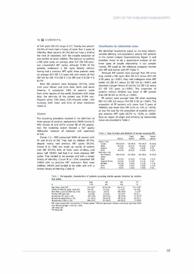
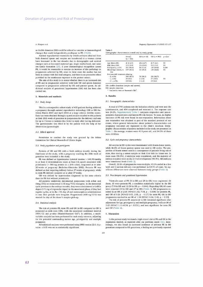
Our clustering analysis found that relationship status was the best variable to analyze
the groups accessing DD. On average, patients accessing DD were 42 years old, and
most of them (91.7%) did not have a child at the time of treatment. The reproductive
outcomes of DD treatments was no different among the three groups, with a
proportion of multiple pregnancies comparable with those reported from IVF and ICSI
cycles worldwide (25.5% for twins and 0.5% for triplets) (108), a rate of caesarean
section similar to that reported for spontaneous pregnancies in the European
population (109), and a spontaneous miscarriage/abortion rate (loss of a clinical
pregnancy that occurs before the 21st week of gestation) also comparable both with
the general population (110,111) or pregnancies achieved by ART taken as a whole in the
region(112).
Upon revision of the list of risk factors for PE, we found that DD patients have several
of them, like advanced maternal age, primigravity, the use of ART to achieve a
pregnancy, the use of donated gametes, and a high rate of multiple pregnancy. Despite
the fact that DD cycles are increasing worldwide (Rita Vassena personal inquiry, data
from USA in the 2000-2010 decade from the US Centers for Disease Control and
Prevention), there is an absence of publicly available literature on the matter. This is,

Donation of gametes and Risk of Preeclampsia
77
in fact, to the best of our knowledge, the first large study reporting patients’
characteristics and reproductive outcomes of DD cycles. So, in conclusion, DD cycles
are increasing, and it is a reasonable treatment option for selected patients, with
encouraging results. However, perinatal and neonatal outcomes should be reported
and monitored more widely, as has been done with the outcomes of ART cycles with
own or donor oocytes.
If poor immunological recognition causes PE, it would be reasonable to assume an
increase of this pathology in pregnancies achieved by DD, since in these cases the
mother has not been in contact with the fetal antigens, and there is no protective
effect provided by the continuous exposure to the partner semen. We tested this
hypothesis in the fourth project, and found an increased incidence of preterm PE in DD
gestations compared to OD gestations, a finding not previously reported in the medical
literature. So far there are two studies analyzing the incidence of PE in double gamete
donation: the first found an increased risk of PE in embryo donation pregnancies (39),
but unfortunately, those pregnancies were compared to women who became
pregnant naturally, thus introducing several unaccounted for variables in the
experimental design, such as the effect of hormonal manipulation, the laboratory
handling of gametes, or the need to resort to third party assisted reproduction itself.
DISCUSSION
78
The second study (113) did not found differences in the incidence of GH or PE between
DD and OD pregnancies, reporting only an increase in gestational diabetes mellitus
that could not be explained by the authors.
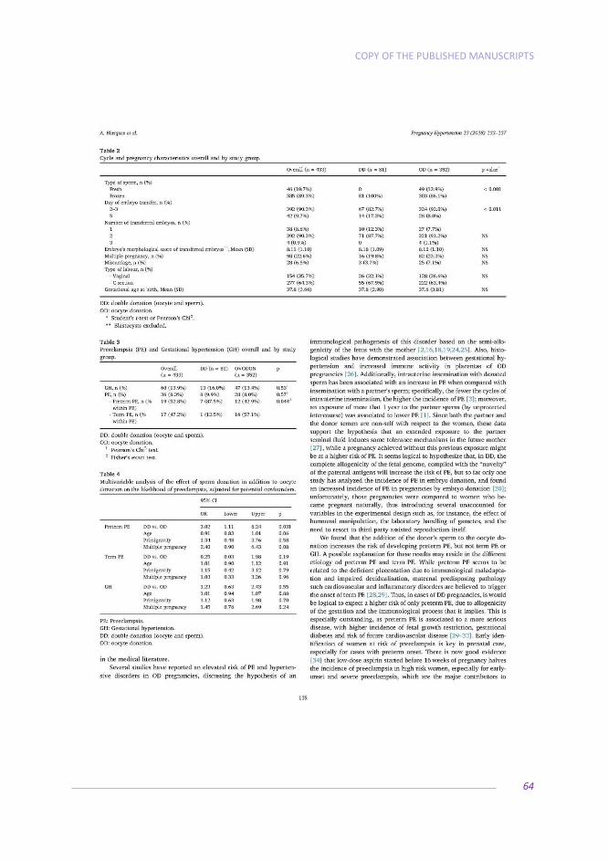
We found that the addition of the donor’s sperm to the oocyte donation increases the
risk of developing preterm PE, but not term PE or GH. A possible explanation for these
results may reside in the different etiology of preterm PE and term PE. While preterm
PE seems to be related to the deficient placentation due to immunological
maladaptation and impaired decidualization, maternal predisposing pathology such as
cardiovascular and inflammatory disorders are believed to trigger the onset of term PE
(6,114).Thus, in cases of DD pregnancies, it would be logical to expect a higher risk of
only preterm PE, due to allogenicity of the gestation and the immunological process
that it implies. This is especially outstanding, as preterm PE is associated to a more
serious disease, with higher incidence of fetal growth restriction, gestational diabetes
and risk of future cardiovascular disease(8,9,114–116). Nowadays studies about PE should
always report preterm PE and term PE separately, and probably this could be the cause
of the difference in the results found by Preaubert. Early identification of women at
risk of preeclampsia is key in prenatal care, and there is now good evidence (117) that
low-dose aspirin started before 16 weeks of pregnancy halves the incidence of
preeclampsia in high risk women, especially for early-onset and severe preeclampsia,
Donation of gametes and Risk of Preeclampsia
79
which are the major contributors to maternal and neonatal complications. Our study
indicates that the risk of preterm PE in DD triples the on in OD.
This study shares one limitation with the second project: data have been obtained
through a questionnaire, but again, giving the importance of the syndrome and the
specificity of the information asked, a mistake in the data provided by the patients is
unlikely, especially in the preterm group, and should be independent from the type of
treatment that generated the pregnancy. Despite the fact that we had a low response
rate in our questionnaire of pregnancy outcome, this is unlikely to result in a selection
bias since the baseline characteristics did not differ between responders and non-
responders. The elevated number of DD cases included, never before reported in the
scientific literature, together with the selection of only fresh embryo transfers, are two
important strengths of this report. Other studies reporting PE in ART included fresh
and frozen embryo transfers, and the lasts seem to confer and additional higher risk
of PE compared to the fresh embryo transfers(51,77,78,85,86).
In conclusion, women undergoing treatments with donated gametes should be made
aware of the higher risk of developing gestational hypertensive disorders, as should be
their attending physicians. Despite this high prevalence of PE, the freezing-thawing
process in the embryo does not seem to be adding more risk compared with fresh ET.

DISCUSSION
80
Particularly women pregnant by double gamete donation should receive strict
obstetrical surveillance, giving the shift in this reproductive treatment, as they have an
increased risk for developing preterm PE. These pregnancies should be identified early
in order to start prophylactic measures appropriately and decrease the risk of PE. Also,
as the endometrial hormonal environment can have a role in the pathogenesis of PE,
further studies are required to determine if levels of estrogens and progesterone could
be used as biomarkers for the early diagnosis of PE.
Reducing the prevalence of PE will not only improve maternal outcomes, but also
neonatal outcomes. There is evidence that the adverse perinatal outcomes reported
in OD pregnancies, specially preterm birth and low birth weight (118,119), improve after
adjustingfor PE(120–127). So, an important part of this prematurity is due to induced
labors that obstetricians perform in order to finish the pregnancy, the only curative
treatment for PE.
NICE guidelines recommend the prophylaxis with aspirin in those pregnant women
with 1 high risk factor or 2 moderate risk factors for PE (128). The U.S. Preventive
Services Task Force recommends the use of low-dose aspirin (81mg per day) as
preventive medication after 12 weeks of gestation in women who are at high risk of
preeclampsia(129). Age of 40 years or older, multiple pregnancy and primigravity are
some of these risk factors, but nowadays gamete donation is not included as an
Donation of gametes and Risk of Preeclampsia
81
independent risk factor. Probably, giving the increase in the risk for PE that oocyte
donation confers to the pregnancy, this ART treatment in itself should be included as
a risk factor and, thus, prophylaxis with aspirin might be warranted in these women.
Additionally, giving the role that hormonal environment also has in the pathogenesis
of PE, different hormonal replacement therapies could decrease the incidence of this
disorder.

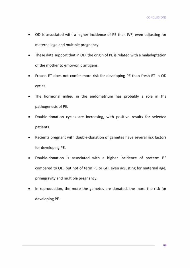
CONCLUSIONS

Donation of gametes and Risk of Preeclampsia
83
CONCLUSIONS
84
• OD is associated with a higher incidence of PE than IVF, even adjusting for
maternal age and multiple pregnancy.
• These data support that in OD, the origin of PE is related with a maladaptation
of the mother to embryonic antigens.
• Frozen ET does not confer more risk for developing PE than fresh ET in OD
cycles.
• The hormonal milieu in the endometrium has probably a role in the
pathogenesis of PE.
• Double-donation cycles are increasing, with positive results for selected
patients.
• Pacients pregnant with double-donation of gametes have several risk factors
for developing PE.
• Double-donation is associated with a higher incidence of preterm PE
compared to OD, but not of term PE or GH, even adjusting for maternal age,
primigravity and multiple pregnancy.
• In reproduction, the more the gametes are donated, the more the risk for
developing PE.

Donation of gametes and Risk of Preeclampsia

BIBLIOGRAPHY

Donation of gametes and Risk of Preeclampsia
87
BIBLIOGRAPHY
88
1. Phipps E, Prasanna D, Brima W, Jim B. Preeclampsia: Updates in pathogenesis,
definitions, and guidelines. Clinical Journal of the American Society of
Nephrology. 2016;11(6):1102-13.
2. Sibai B, Dekker G, Kupferminc M. Pre-eclampsia. Lancet. 2005;365(9461):785–
99.
3. Ventura SJ, Mosher WD, Curtin SC, Abma JC, Henshaw S. Trends in pregnancy
rates for the United States, 1976-97: an update. Natl Vital Stat Rep.
2001;49(4):1–9.
4. ACOG Committee on Practice Bulletins--Obstetrics. ACOG practice bulletin.
Diagnosis and management of preeclampsia and eclampsia. Number 33,
January 2002. Obstet Gynecol. 2002; 99(1):159-67.
5. American College of Obstetricians and Gynecologists, Task Force on
Hypertension in Pregnancy. Hypertension in pregnancy. Report of the American
College of Obstetricians and Gynecologists’ Task Force on Hypertension in
Pregnancy. Obstet Gynecol. 2013;122(5):1122-31.
6. Steegers EAP, Von Dadelszen P, Duvekot JJ, Pijnenborg R. Pre-eclampsia.
Lancet. 2010; 376(9741):631-44.
Donation of gametes and Risk of Preeclampsia
89
7. Thilaganathan B. Placental syndromes: getting to the heart of the matter.
Ultrasound Obstet Gynecol. 2017;49: 7–9.
8. Xiong X, Demianczuk NN, Saunders LD, Wang F-L, Fraser WD. Impact of
preeclampsia and gestational hypertension on birth weight by gestational age.
Am J Epidemiol. 2002;155(3):203-9.
9. Mongraw-Chaffin ML, Cirillo PM, Cohn BA. Preeclampsia and cardiovascular
disease death: Prospective evidence from the child health and development
studies cohort. Hypertension. 2010; 56(1):166-71.
10. Maynard SE, Min JY, Merchan J, Lim KH, Li J, Mondal S, et al. Excess placental
soluble fms-like tyrosine kinase 1 (sFlt1) may contribute to endothelial
dysfunction hypertension, and proteinuria in preeclampsia. J Clin Invest.
2003;111(5):649-58.
11. Venkatesha S, Toporsian M, Lam C, Hanai JI, Mammoto T, Kim YM, et al. Soluble
endoglin contributes to the pathogenesis of preeclampsia. Nat Med. 2006;
12(6):642-9.
12. Hiby SE, Apps R, Sharkey AM, Farrell LE, Gardner L, Mulder A, et al. Maternal
activating KIRs protect against human reproductive failure mediated by fetal
BIBLIOGRAPHY
90
HLA-C2. J Clin Invest. 2010; 120(11):4102-10.
13. Madeja Z, Yadi H, Apps R, Boulenouar S, Roper SJ, Gardner L, et al. Paternal
MHC expression on mouse trophoblast affects uterine vascularization and fetal
growth. Proc Natl Acad Sci. 2011; 108(10):4012-7.
14. Hiby SE, Walker JJ, O’Shaughnessy KM, Redman CWG, Carrington M, Trowsdale
J, et al. Combinations of Maternal KIR and Fetal HLA-C Genes Influence the Risk
of Preeclampsia and Reproductive Success. J Exp Med. 2004; 200(8):957-65.
15. Giovanna Ogge, Tinnakorn Chaiworapongsa, Roberto Romero, Youssef Hussein,
Juan Pedro Kusanovic, Lami Yeo, Chong Jai Kim, MD and SSH. Placental lesions
associated with maternal underperfusion are more frequent in early-Onset
than in late-onset preeclampsia. J Perinat Med. 2011;39(6):641–52.
16. Yu CKH, Khouri O, Onwudiwe N, Spiliopoulos Y, Nicolaides KH. Prediction of pre-
eclampsia by uterine artery Doppler imaging: Relationship to gestational age at
delivery and small-for-gestational age. Ultrasound Obstet Gynecol. 2008;
31(3):310-3.
17. Jelin AC, Cheng YW, Shaffer BL, Kaimal AJ, Little SE, Caughey AB. Early-onset
preeclampsia and neonatal outcomes. J Matern Neonatal Med Off J Eur Assoc

Donation of gametes and Risk of Preeclampsia
91
Perinat Med Fed Asia Ocean Perinat Soc Int Soc Perinat Obstet. 2010;23(5):389-
92.
18. Espinoza J, Lee W, Martin SR, Belfort MA. Customized growth curves for
identification of large-for-gestational age neonates in pre-eclamptic women.
Ultrasound Obstet Gynecol. 2014; 43(2):165-9.
19. Verlohren S, Melchiorre K, Khalil A, Thilaganathan B. Uterine artery Doppler,
birth weight and timing of onset of pre-eclampsia: providing insights into the
dual etiology of late-onset pre-eclampsia. Ultrasound Obstet Gynecol. 2014;
44(3):293-8.
20. Ray JG, Vermeulen MJ, Schull MJ, Redelmeier DA. Cardiovascular health after
maternal placental syndromes (CHAMPS): Population-based retrospective
cohort study. Lancet. 2005; 366(9499):1797-803.
21. Bellamy L, Casas J-P, Hingorani AD, Williams DJ. Pre-eclampsia and risk of
cardiovascular disease and cancer in later life: systematic review and meta-
analysis. BMJ. 2007; 335(7627):974.
22. Brown MC, Best KE, Pearce MS, Waugh J, Robson SC, Bell R. Cardiovascular
disease risk in women with pre-eclampsia: Systematic review and meta-

BIBLIOGRAPHY
92
analysis. European Journal of Epidemiology. 2013;28(1):1-19.
23. Magnussen EB, Vatten LJ, Lund-Nilsen TI, Salvesen KA, Davey Smith G,
Romundstad PR. Prepregnancy cardiovascular risk factors as predictors of pre-
eclampsia: population based cohort study. BMJ. 2007; ;335(7627):978.
24. Romundstad PR, Magnussen EB, Smith GD, Vatten LJ. Hypertension in
pregnancy and later cardiovascular risk: Common antecedents? Circulation.
2010; 122(6):579-84.
25. Enkhmaa D, Wall D, Mehta PK, Stuart JJ, Rich-Edwards JW, Merz CNB, et al.
Preeclampsia and Vascular Function: A Window to Future Cardiovascular
Disease Risk. J Women’s Heal. 2016; 25(3):284-91.
26. Sibai BM, El-Nazer A, Gonzalez-Ruiz A. Severe preeclampsia-eclampsia in young
primigravid women: Subsequent pregnancy outcome and remote prognosis.
Am J Obstet Gynecol. 1986; 155(5):1011-6.
27. Makkonen N, Heinonen S, Kirkinen P. Obstetric prognosis in second pregnancy
after preeclampsia in first pregnancy. Hypertens Pregnancy. 2000; 19(2):173-
81.
28. Duckitt K, Harrington D. Risk factors for pre-eclampsia at antenatal booking:

Donation of gametes and Risk of Preeclampsia
93
Systematic review of controlled studies. British Medical Journal.
2005;330(7491):565.
29. Skomsvoll JF, Ostensen M, Irgens LM, Baste V. Pregnancy complications and
delivery practice in women with connective tissue disease and inflammatory
rheumatic disease in Norway. Acta Obstet Gynecol Scand. 2000; 79(6):490-5.
30. Yogev Y, Xenakis EMJ, Langer O. The association between preeclampsia and the
severity of gestational diabetes: The impact of glycemic control. Am J Obstet
Gynecol. 2004;191(5):1655-60.
31. Baeten JM, Bukusi EA, Lambe M. Pregnancy complications and outcomes
among overweight and obese nulliparous women. Am J Public Health. 2001;
;91(3):436-40.
32. Coonrod D V, Hickok DE, Zhu K, Easterling TR, Daling JR. Risk factors for
preeclampsia in twin pregnancies: a population-based cohort study. Obstet
Gynecol. 1995; 85(5 Pt 1):645-50.
33. Qiu C, Williams MA, Leisenring WM, Sorensen TK, Frederick IO, Dempsey JC, et
al. Family history of hypertension and type 2 diabetes in relation to
preeclampsia risk. Hypertension. 2003; 41(3):408-13.
BIBLIOGRAPHY
94
34. Cincotta RB, Brennecke SP. Family history of pre-eclampsia as a predictor for
pre-eclampsia in primigravidas. Int J Gynecol Obstet. 1998; 60(1):23-7.
35. Bianco A, Stone J, Lynch L, Lapinski R, Berkowitz G, Berkowitz RL. Pregnancy
outcome at age 40 and older. Obstet Gynecol [Internet]. 1996 Jun;87(6):917–
22.
36. Saftlas AF, Levine RJ, Klebanoff MA, Martz KL, Ewell MG, Morris CD, et al.
Abortion, changed paternity, and risk of preeclampsia in nulliparous women.
Am J Epidemiol. 2003; 157(12):1108-14.
37. Einarsson JI, Sangi-Haghpeykar H, Gardner MO. Sperm exposure and
development of preeclampsia. Am J Obstet Gynecol. 2003; 188(5):1241-3.
38. Bibbins-Domingo K, Grossman DC, Curry SJ, Barry MJ, Davidson KW, Doubeni
CA, et al. Screening for Preeclampsia: US Preventive Services Task Force
Recommendation Statement. JAMA [Internet]. 2017 Apr 25;317(16):1661.
39. Salha O, Sharma V, Dada T, Nugent D, Rutherford AJ, Tomlinson AJ, et al. The
influence of donated gametes on the incidence of hypertensive disorders of
pregnancy. Hum Reprod. 1999; 14(9):2268-73.
40. Pecks U, Maass N, Neulen J. Oocyte donation: a risk factor for pregnancy-
Donation of gametes and Risk of Preeclampsia
95
induced hypertension: a meta-analysis and case series. Dtsch Arztebl Int. 2011;
108(3):23-31.
41. Cnattingius S, Lambe M. Trends in smoking and overweight during pregnancy:
Prevalence, risks of pregnancy complications, and adverse pregnancy
outcomes. Semin Perinatol. 2002; 26(4):286-95.
42. Dyer S, Chambers GM, De Mouzon J, Nygren KG, Zegers-Hochschild F, Mansour
R, et al. International committee for monitoring assisted reproductive
technologies world report: Assisted reproductive technology 2008, 2009 and
2010†. Hum Reprod. 2016;31(7):1588–609.
43. Tandberg A, Klungsøyr K, Romundstad LB, Skjærven R. Pre-eclampsia and
assisted reproductive technologies: Consequences of advanced maternal age,
interbirth intervals, new partner and smoking habits. BJOG An Int J Obstet
Gynaecol. 2015; 122(7):915-22.
44. Nelissen ECM, Dumoulin JCM, Daunay A, Evers JLH, Tost J, van Montfoort APA.
Placentas from pregnancies conceived by IVF/ICSI have a reduced DNA
methylation level at the H19 and MEST differentially methylated regions†. Hum
Reprod [Internet]. 2013 Apr;28(4):1117–26.

BIBLIOGRAPHY
96
45. Nelissen ECM, van Montfoort APA, Dumoulin JCM, Evers JLH. Epigenetics and
the placenta. Hum Reprod Update. 2011; 17(3):397-417.
46. Mann MRW. Selective loss of imprinting in the placenta following
preimplantation development in culture. Development. 2004; 131(15):3727-
35.
47. Fortier AL, Lopes FL, Darricarrère N, Martel J, Trasler JM. Superovulation alters
the expression of imprinted genes in the midgestation mouse placenta. Hum
Mol Genet. 2008; 17(11):1653-65.
48. Rivera RM, Stein P, Weaver JR, Mager J, Schultz RM, Bartolomei MS.
Manipulations of mouse embryos prior to implantation result in aberrant
expression of imprinted genes on day 9.5 of development. Hum Mol Genet.
2008; ;17(1):1-14.
49. Tarlatzi TB, Imbert R, Alvaro Mercadal B, Demeestere I, Venetis CA, Englert Y,
et al. Does oocyte donation compared with autologous oocyte IVF pregnancies
have a higher risk of preeclampsia? Reprod Biomed Online. 2017; 34(1):11-18.
50. Smith GN, Walker M, Tessier JL, Millar KG. Increased incidence of preeclampsia
in women conceiving by intrauterine insemination with donor versus partner

Donation of gametes and Risk of Preeclampsia
97
sperm for treatment of primary infertility. Am J Obstet Gynecol. 1997; 177(2)
455-8.
51. Klatsky PC, Delaney SS, Caughey AB, Tran ND, Schattman GL, Rosenwaks Z. The
role of embryonic origin in preeclampsia: A comparison of autologous in vitro
fertilization and ovum donor pregnancies. Obstet Gynecol. 2010; 116(6)1387-
92.
52. Kyrou D, Kolibianakis EM, Devroey P, Fatemi HM. Is the use of donor sperm
associated with a higher incidence of preeclampsia in women who achieve
pregnancy after intrauterine insemination? Fertil Steril. 2010;93(4) 1124-7.
53. Keegan DA, Krey LC, Chang HC, Noyes N. Increased risk of pregnancy-induced
hypertension in young recipients of donated oocytes. Fertil Steril. 2007;
87(4)776-81.
54. González-Comadran M, Avila JU, Tascón AS, Jimenéz R, Solà I, Brassesco M, et
al. The impact of donor insemination on the risk of preeclampsia: A systematic
review and meta-analysis. European Journal of Obstetrics Gynecology and
Reproductive Biology. 2014; 182:160-6.
55. Saftlas AF, Rubenstein L, Prater K, Harland KK, Field E, Triche EW. Cumulative
BIBLIOGRAPHY
98
exposure to paternal seminal fluid prior to conception and subsequent risk of
preeclampsia. J Reprod Immunol. 2014; 101-102:104-10.
56. Kho EM, McCowan LME, North RA, Roberts CT, Chan E, Black MA, et al. Duration
of sexual relationship and its effect on preeclampsia and small for gestational
age perinatal outcome. J Reprod Immunol. 2009; 82(1)66-73.
57. Sadat Z, Abedzadeh Kalahroudi M, Saberi F. The effect of short duration sperm
exposure on development of preeclampsia in primigravid women. Iran Red
Crescent Med J. 2012; 14(1)20-4.
58. Hall GH. Long-term sexual co-habitation offers no protection from hypertensive
disease of pregnancy. Hum Reprod. 2001; 16(2) 349-52.
59. Ness RB, Markovic N, Harger G, Day R. Barrier methods, length of
preconception intercourse, and preeclampsia. Hypertens Pregnancy. 2004;
23(3)227-35.
60. Robillard PY, Périanin J, Janky E, Miri EH, Hulsey TC, Papiernik E. Association of
pregnancy-induced hypertension with duration of sexual cohabitation before
conception. Lancet. 1994; 344(8928) 973-5.
61. Aluvihare VR, Kallikourdis M, Betz AG. Regulatory T cells mediate maternal

Donation of gametes and Risk of Preeclampsia
99
tolerance to the fetus. Nat Immunol. 2004; 5(3):266-71.
62. Robertson SA, Guerin LR, Moldenhauer LM, Hayball JD. Activating T regulatory
cells for tolerance in early pregnancy - the contribution of seminal fluid. J
Reprod Immunol. 2009; 83(1-2):109-16.
63. Johansson M, Bromfield JJ, Jasper MJ, Robertson SA. Semen activates the
female immune response during early pregnancy in mice. Immunology.
2004;112(2):290-300.
64. Alecsandru D, Garrido N, Vicario JL, Barrio A, Aparicio P, Requena A, et al.
Maternal KIR haplotype influences live birth rate after double embryo transfer
in IVF cycles in patients with recurrent miscarriages and implantation failure.
Hum Reprod. 2014; 29(12):2637-43.
65. Tilburgs T, Roelen DL, van der Mast BJ, de Groot-Swings GM, Kleijburg C,
Scherjon SA, et al. Evidence for a Selective Migration of Fetus-Specific
CD4+CD25bright Regulatory T Cells from the Peripheral Blood to the Decidua in
Human Pregnancy. J Immunol. 2008; 180(8):5737-45.
66. Schonkeren D, Swings G, Roberts D, Claas F, de Heer E, Scherjon S. Pregnancy
close to the edge: An immunosuppressive infiltrate in the chorionic plate of
BIBLIOGRAPHY
100
placentas from uncomplicated egg cell donation. PLoS One. 2012; 7(3):e32347.
67. Kelkar RL, Meherji PK, Kadam SS, Gupta SK, Nandedkar TD. Circulating auto-
antibodies against the zona pellucida and thyroid microsomal antigen in
women with premature ovarian failure. J Reprod Immunol. 2005; 66(1) 53-67.
68. Woldringh GH, Frunt MHA, Kremer JAM, Spaanderman MEA. Decreased
ovarian reserve relates to pre-eclampsia in IVF/ICSI pregnancies. Hum Reprod.
2006; 21(11)2948-54.
69. Wennerholm UB, Söderström-Anttila V, Bergh C, Aittomäki K, Hazekamp J,
Nygren KG, et al. Children born after cryopreservation of embryos or oocytes:
A systematic review of outcome data. Hum Reprod. 2009; 24(9):2158-72.
70. Söderström-Anttila V, Tiitinen A, Foudila T, Hovatta O. Obstetric and perinatal
outcome after oocyte donation: Comparison with in-vitro fertilization
pregnancies. Hum Reprod. 1998; 13(2):483-90.
71. Roque M, Lattes K, Serra S, Solà I, Geber S, Carreras R, et al. Fresh embryo
transfer versus frozen embryo transfer in in vitro fertilization cycles: A
systematic review and meta-analysis. Fertil Steril. 2013; 99(1):156-62.
72. Uddin MN, Horvat D, Jones RO, Beeram MR, Zawieja DC, Perger L, et al.

Donation of gametes and Risk of Preeclampsia
101
Suppression of aldosterone and progesterone in preeclampsia. J Matern
Neonatal Med. 2015;
73. He P, Chen Z, Sun Q, Li Y, Gu H, Ni X. Reduced Expression of 11β-Hydroxysteroid
Dehydrogenase Type 2 in Preeclamptic Placentas Is Associated With Decreased
PPARγ but Increased PPARα Expression. Endocrinology. 2014; 28:1-6.
74. Wuu J, Hellerstein S, Lipworth L, Wide L, Xu B, Yu GP, et al. Correlates of
pregnancy oestrogen, progesterone and Sex Hormone-binding Globulin in the
USA and China. European Journal of Cancer Prevention. 2002; 11(3):283-93.
75. Matsuura S, Itakura A, Ohno Y, Nakashima Y, Murata Y, Takeuchi M, et al.
Effects of estradiol administration on feto-placental growth in rat. Early Hum
Dev. 2004; 77(1-2):47-56.
76. Maliqueo M, Echiburú B, Crisosto N. Sex steroids modulate uterine-placental
vasculature: Implications for obstetrics and neonatal outcomes. Frontiers in
Physiology. 2016;26;7:152.
77. Sazonova A, Kllen K, Thurin-Kjellberg A, Wennerholm UB, Bergh C. Obstetric
outcome in singletons after in vitro fertilization with cryopreserved/thawed
embryos. Hum Reprod. 2012; 27(5):1343-50.
BIBLIOGRAPHY
102
78. Ishihara O, Araki R, Kuwahara A, Itakura A, Saito H, Adamson GD. Impact of
frozen-thawed single-blastocyst transfer on maternal and neonatal outcome:
An analysis of 277,042 single-embryo transfer cycles from 2008 to 2010 in
Japan. Fertility and Sterility. 2014;101(1):128-33.
79. Pelkonen S, Koivunen R, Gissler M, Nuojua-Huttunen S, Suikkari AM, Hydén-
Granskog C, et al. Perinatal outcome of children born after frozen and fresh
embryo transfer: The Finnish cohort study 1995-2006. Hum Reprod. 2010;
25(4):914-23.
80. Imudia AN, Awonuga AO, Kaimal AJ, Wright DL, Styer AK, Toth TL. Elective
cryopreservation of all embryos with subsequent cryothaw embryo transfer in
patients at risk for ovarian hyperstimulation syndrome reduces the risk of
adverse obstetric outcomes: A preliminary study. Fertil Steril. 2013; 99(1):168-
73.
81. Ishihara O, Kuwahara A, Saitoh H. Frozen-thawed blastocyst transfer reduces
ectopic pregnancy risk: An analysis of single embryo transfer cycles in Japan.
Fertil Steril. 2011; 95(6):1966-9.
82. Roque M. Freeze-all policy: is it time for that? J Assist Reprod Genet.
2015;32(2):171-6.
Donation of gametes and Risk of Preeclampsia
103
83. Pinborg A, Henningsen AA, Loft A, Malchau SS, Forman J, Andersen AN. Large
baby syndrome in singletons born after frozen embryo transfer (FET): Is it due
to maternal factors or the cryotechnique? Hum Reprod. 2014; 29(3):618-27.
84. Maheshwari A, Pandey S, Shetty A, Hamilton M, Bhattacharya S. Obstetric and
perinatal outcomes in singleton pregnancies resulting from the transfer of
frozen thawed versus fresh embryos generated through in vitro fertilization
treatment: A systematic review and meta-analysis. Fertility and Sterility.
2012;98(2):368-77 e1-9.
85. Barsky M, St. Marie P, Rahil T, Markenson GR, Sites CK. Are perinatal outcomes
affected by blastocyst vitrification and warming? In: American Journal of
Obstetrics and Gynecology. 2016;215(5):603 e1- e5.
86. Chen Z, Shi Y, Sun Y, et al. Fresh versus Frozen Embryos for Infertility in the
Polycystic Ovary Syndrome. New EnglandJ Medicine. 2016;11;375(6):523-33;
87. Shih W, Rushford DD, Bourne H, Garrett C, McBain JC, Healy DL, et al. Factors
affecting low birthweight after assisted reproduction technology: Difference
between transfer of fresh and cryopreserved embryos suggests an adverse
BIBLIOGRAPHY
104
effect of oocyte collection. Hum Reprod. 2008; 23(7):1644-53.
88. Young L. Large offspring syndrome in cattle and sheep. Rev Reprod. 1998;
3(3):155-63.
89. Yokochi T, Robertson KD. Dimethyl sulfoxide stimulates the catalytic activity of
de novo DNA methyltransferase 3a (Dnmt3a) in vitro. Bioorg Chem. 2004;
32(4):234-43.
90. De Geyter C, De Geyter M, Steimann S, Zhang H, Holzgreve W. Comparative
birth weights of singletons born after assisted reproduction and natural
conception in previously infertile women. Hum Reprod. 2006; 21(3):705-12.
91. De Graaff AA, Land JA, Kessels AGH, Evers JLH. Demographic age shift toward
later conception results in an increased age in the subfertile population and an
increased demand for medical care. Fertil Steril. 2011; 95(1):61-3.
92. Instituto Nacional de Estadísitica.
http://www.ine.es/jaxiT3/Tabla.htm?t=1579.
93. Blickstein I. Motherhood at or beyond the edge of reproductive age. Int J Fertil
Womens Med. 2003; 48(1):17-24.

Donation of gametes and Risk of Preeclampsia
105
94. Pal L, Santoro N. Age-related decline in fertility. Endocrinology and Metabolism
Clinics of North America. 2003;32(3):669-88.
95. van der Hoorn MLP, Lashley EELO, Bianchi DW, Claas FHJ, Schonkeren CMC,
Scherjon SA. Clinical and immunologic aspects of egg donation pregnancies: A
systematic review. Hum Reprod Update. 2010; 16(6):704-12.
96. Staff AC, Benton SJ, Von Dadelszen P, Roberts JM, Taylor RN, Powers RW, et al.
Redefining preeclampsia using placenta-derived biomarkers. Hypertension.
2013; 61(5):932-42..
97. Berkane N, Liere P, Oudinet J-P, Hertig A, Lefèvre G, Pluchino N, et al. From
Pregnancy to Preeclampsia: A Key Role for Estrogens. Endocr Rev. 2017;
38(2):123-44.
98. Hertig A, Fort J, Lefevre G, Chabbert-Buffet N, Uzan M, Rondeau E, et al. Soluble
endoglin in preeclamptic patients with or without HELLP syndrome. Am J Obstet
Gynecol. 2010; 202(6):594 e1-4.
99. Jobe SO, Ramadoss J, Wargin AJ, Magness RR. Estradiol-17 and its Cytochrome
P450- and Catechol-O-Methyltransferase-Derived Metabolites Selectively
Stimulate Production of Prostacyclin in Uterine Artery Endothelial Cells: Role of
BIBLIOGRAPHY
106
Estrogen Receptor- Versus Estrogen Receptor- . Hypertension. 2013;
61(2):509-18.
100. Bussen S, Bussen D. Influence of the vascular endothelial growth factor on the
development of severe pre-eclampsia or HELLP syndrome. Arch Gynecol
Obstet. 2011;284(3):551-7.
101. Su EJ, Ernst L, Abdallah N, Chatterton R, Xin H, Monsivais D, et al. Estrogen
receptor-β and fetoplacental endothelial prostanoid biosynthesis: A link to
clinically demonstrated fetal growth restriction. J Clin Endocrinol Metab.
2011;96(10):E1558-67.
102. Walsh SW, Coulter S. Increased placental progesterone may cause decreased
placental prostacyclin production in preeclampsia. Am J Obstet Gynecol.
1989;161(6 Pt 1):1586-92.
103. Oron G, Son WY, Buckett W, Tulandi T, Holzer H. The association between
embryo quality and perinatal outcome of singletons born after single embryo
transfers: A pilot study. Hum Reprod. 2014; 29(7):1444-51.
104. Oron G, Nayot D, Son WY, Holzer H, Buckett W, Tulandi T. Obstetric and
Donation of gametes and Risk of Preeclampsia
107
perinatal outcome from single cleavage transfer and single blastocyst transfer:
A matched case-control study. Gynecol Endocrinol. 2015; 31(6):469-72.
105. Oron G, Sokal-Arnon T, Son WY, Demirtas E, Buckett W, Zeadna A, et al.
Extended embryo culture is not associated with increased adverse obstetric or
perinatal outcome. Am J Obstet Gynecol. 2014; 211(2):165 e1-7.
106. Palermo GD, Cohen J, Rosenwaks Z. Intracytoplasmic sperm injection: a
powerful tool to overcome fertilization failure. Fertil Steril. 1996; 65(5):899-
908.
107. Nangia AK, Luke B, Smith JF, Mak W, Stern JE. National study of factors
influencing assisted reproductive technology outcomes with male factor
infertility. Fertil Steril. 2011; 96:609–614.
108. Sullivan EA, Zegers-Hochschild F, Mansour R, Ishihara O, De Mouzon J, Nygren
KG, et al. International Committee for Monitoring Assisted Reproductive
Technologies (ICMART) world report: Assisted reproductive technology 2004.
Hum Reprod. 2013; 28(5):1375-90.
109. OECD (2013), Health at a Glance 2013: OECD Indicators, OECD Publishing.
http://dx.doi.org/10.1787/health_glance-2013-en.

BIBLIOGRAPHY
108
110. Wang X, Chen C, Wang L, Chen D, Guang W, French J, et al. Conception, early
pregnancy loss, and time to clinical pregnancy: A population-based prospective
study. Fertil Steril. 2003; 79(3):577-84.
111. Wilcox AJ, Weinberg CR, O’Connor JF, Baird DD, Schlatterer JP, Canfield RE, et
al. Incidence of Early Loss of Pregnancy. N Engl J Med. 1988;28;319(4):189-94.
112. FIVCAT 2011.
http://www20.gencat.cat/docs/salut/Home/El%20Departament/Estadistiques
%20sanitaries/Dades%20de%20salut%20i%20serveis%20sanitaris/Reproducci
o%20humana%20assistida/documents/fivcat_2011_resum_resultats.pdf.
113. Preaubert L, Vincent-Rohfritsch A, Santulli P, Gayet V, Goffinet F, Le Ray C.
Outcomes of pregnancies achieved by double gamete donation: A comparison
with pregnancies obtained by oocyte donation alone. Eur J Obstet Gynecol
Reprod Biol. 2018; 222:1-6.
114. Phillips JK, Janowiak M, Badger GJ, Bernstein IM. Evidence for distinct preterm
and term phenotypes of preeclampsia. J Matern Neonatal Med. 2010;
;23(7)622-6.
115. Myatt L, Redman CW, Staff AC, Hansson S, Wilson ML, Laivuori H, et al. Strategy

Donation of gametes and Risk of Preeclampsia
109
for standardization of preeclampsia research study design. Hypertension.
2014;63(6):1293–301.
116. Manten GTR, Sikkema MJ, Voorbij HAM, Visser GHA, Bruinse HW, Franx A. Risk
factors for cardiovascular disease in women with a history of pregnancy
complicated by preeclampsia or intrauterine growth restriction. Hypertens
Pregnancy. 2007;26(1)39-50.
117. Roberge S, Nicolaides K, Demers S, Hyett J, Chaillet N, Bujold E. The role of
aspirin dose on the prevention of preeclampsia and fetal growth restriction:
systematic review and meta-analysis. In: American Journal of Obstetrics and
Gynecology. 2017; 216(2) 110-120 e6.
118. Storgaard M, Loft A, Bergh C, Wennerholm UB, Söderström-Anttila V,
Romundstad LB, et al. Obstetric and neonatal complications in pregnancies
conceived after oocyte donation: a systematic review and meta-analysis. BJOG
An Int J Obstet Gynaecol. 2017;124(4):561–72.
119. Adams DH,Clark RA, Davies MJ, De Lacey S. A meta-analysis of neonatal health
outcomes from oocyte donation. J Dev Orig Health Dis. 2016;7(3):257–72.
120. Malchau SS, Loft A, Larsen EC, Aaris Henningsen AK, Rasmussen S, Andersen
BIBLIOGRAPHY
110
AN, et al. Perinatal outcomes in 375 children born after oocyte donation: A
Danish national cohort study. Fertil Steril. 2013;99(6):1637–1643.e3.
121. Nejdet S, Bergh C, Källén K, Wennerholm UB, Thurin-Kjellberg A. High risks of
maternal and perinatal complications in singletons born after oocyte donation.
Acta Obstet Gynecol Scand. 2016;95(8):879–86.
122. Dildy, Jackson, Fowers, Oshiro, Varner, Clark. Very advanced maternal age:
Pregnancy after age 45. Am J Obstet Gynecol. 1996; 175(3 Pt 1):668-74.
123. Shrim A, Levin I, Mallozzi A, Brown R, Salama K, Gamzu R, et al. Does very
advanced maternal age, with or without egg donation, really increase obstetric
risk in a large tertiary center? J Perinat Med. 2010;38(6):645–50.
124. Clua E, Meler E, Rodríguez D, Coroleu B, Rodríguez I, Martínez F, et al. Obstetric
and perinatal complications in an oocyte donation programme. Is it time to limit
the number of embryos to transfer? Gynecol Endocrinol. 2016;32(4):267–71.
125. Guesdon E, Vincent-Rohfritsch A, Bydlowski S, Santulli P, Goffinet F, Le Ray C.
Oocyte donation recipients of very advanced age: perinatal complications for
singletons and twins. Fertil Steril. 2017;107(1):89–96.
126. Le Ray C, Scherier S, Anselem O, Marszalek A, Tsatsaris V, Cabrol D, et al.
Donation of gametes and Risk of Preeclampsia
111
Association between oocyte donation and maternal and perinatal outcomes in
women aged 43 years or older. Hum Reprod. 2012;27(3):896–901.
127. Tranquilli AL, Biondini V, Talebi Chahvar S, Corradetti A, Tranquilli D, Giannubilo
S. Perinatal outcomes in oocyte donor pregnancies. J Matern Neonatal Med.
2013;26(13):1263–7.
128. NICE Guideline in Hypertension in Pregnancy.
https://www.nice.org.uk/guidance/cg107/resources/hypertension-in-
pregnancy-diagnosis-and-management-pdf-35109334011877.
129. LeFevre ML. Low-dose aspirin use for the prevention of morbidity and mortality
from preeclampsia: U.S. Preventive Services Task Force recommendation
statement. Ann Intern Med. 2014; 161(11):819-26.

SUMMARY IN SPANISH

Donation of gametes and Risk of Preeclampsia
113

SUMMARY IN SPANISH
114
INTRODUCCIÓN:
PREECLAMPSIA:
Epidemiología:
La preeclampsia (PE) es un trastorno hipertensivo del embarazo que afecta entre un 2
y un 8% de las gestaciones, siendo además la causa del 16% de muertes en gestantes.
En países en desarrollo su incidencia está aumentando ya que sus factores de riesgo
también lo están haciendo, como se verá posteriormente.
Definición:
Clásicamente se define por hipertensión de nueva aparición (tensión arterial > 140/90
mmHG en al menos 2 determinaciones separadas 6h entre sí) asociada a proteinuria
(>300mg de proteína en orina de 24h), diagnosticado a o a partir de las 20 semanas de
gestación.
Clasificación:
Se clasifica en 2 subclases:
1. PE precoz o de debut temprano: se inicia antes de las 34 semanas de gestación.
Se debe a una mala adaptación inmunológica de la madre al feto que tiene
lugar en la placenta, por eso también es llamada “PE placentaria”.
Donation of gametes and Risk of Preeclampsia
115
Habitualmente es la cara más severa del síndrome, ya que se asocia a retraso
de crecimiento intrauterino y el parto suele ser antes de término (< 37
semanas).
2. PE de debut tardío: se inicia a partir de las 34 semanas y se postula que su
origen es por una disfunción cardiovascular materna que secundariamente da
lugar a una disfunción placentaria. Por ello se la llama “PE materna”. A pesar
que no es tan grave como la anterior, su incidencia supera en 5-20 veces la de
la PE precoz. El parto suele ser a término (> 37 semanas).
Patogénesis:
La etiología de la PE es una etiología de teorías, y probablemente en la mayoría de los
casos no existe una única vía causante del trastorno, sino que se solapan varias de
ellas.
El punto en común en todas las hipótesis es que existe una insuficiencia placentaria. El
trofoblasto invade la decídua de forma anómala y esto conlleva una deficiente
remodelación de las arterias espirales, hecho que deriva a una incorrecta
vascularización de la placenta y a su disfunción.
Las dos líneas etiológicas divergen en el desencadenante de la alteración en la invasión
del trofoblasto en las primeras fases de la gestación.
SUMMARY IN SPANISH
116
Teoría inmunológica:
Postula que la hipoperfusión placentaria es secundaria a una mala adaptación de la
madre a agentes fetales.
Las células trofoblásticas expresan HLA-C, muy polimórficos, y que presentan alelos
tanto paternos como maternos. El HLA-C es el ligando para el receptor Killer-
immonuglobulin (KIR) presente en las células Natural Killers uterinas (uNK). Estas
células tienen una función distinta a las NK sistémicas, ya que modulan factores
proangiogénicos y endoteliales (como PlGF, VEFG y TGF-β), que a su vez estimulan la
neovascularización, y que finalmente promueven los cambios en las arterias espirales
responsables de la correcta perfusión fetal.
La combinación de algunos genotipos de KIR maternos (especialmente KIR AA) con
algunos alotipos de HLA-C de las células trofoblásticas (particularmente HLA-C2)
ocasionan la disfunción de estas uNK, lo que deriva a una alteración de la perfusión
placentaria y, finalmente, a alteraciones durante el embarazo, como la PE o el retraso
de crecimiento intrauterino en el feto.
Esta teoría respalda la PE de debut temprano, con origen placentario, y que se
caracteriza por placentas de pequeño tamaño, con signos histológicos de
hipoperfusión, y asociado a restricción del crecimiento fetal. Pero en los numerosos
Donation of gametes and Risk of Preeclampsia
117
casos de PE de debut tardío las lesiones de hipoperfusión puedes ser ausentesen la
placenta, siendo ésta de tamaño normal con también fetos de peso normal.
Teoría cardiovascular:
Muchos estudios hallan factores de riesgo cardiovascular en pacientes que han sufrido
PE, creando una relación causal entre ambos hechos. Otros autores defienden que
estos factores de riesgo existen previos a la gestación, y que incluso se asocian a la
futura patología cardiovascular de la paciente con más solidez que la PE en sí misma.
Sabemos que el embarazo es un estado donde hay un incremento de las necesidades
metabólicas y hemodinámicas. Esta teoría sostiene que, en determinadas pacientes
con factores de riesgo cardiovascular, que a menudo son inadvertidos, estas demandas
pueden ser excesivas y, secundariamente, esto va a conllevar una hipoperfusión
placentaria causante de la PE.Esta disfunción cardiovascular es la etiología de la PE de
debut tardío.
Factores de riesgo:
La siguiente lista ordena los factores de riesgo según el riesgo relativo que suponen,
de mayor a menor:
1. Historia de PE en gestación previa. Incrementa 7 veces el riesgo de PE.
2. Patología médica de base:
SUMMARY IN SPANISH
118
a. Enfermedad autoinmune. Concretamente el síndrome antifosfolípido
aumenta 6 veces el riesgo de desarrollar PE.
b. Diabetes Mellitus. Incrementa en 4 el riesgo de PE.
c. Hipertensión arterial pre-existente. Dobla el riesgo de padecer PE.
d. Enfermedad renal
3. Obesidad: un IMC de> 35 triplica el riesgo de desarrollar PE.
4. Paridad: la nuliparidad incrementa casi 3 veces el riesgo de PE.
5. Embarazo múltiple: el embarazo gemelar confiere un riesgo 3 veces superior
de PE que el embarazo de feto único.
6. Historia en familiar de primer grado de PE, hipertensión o diabetes mellitus:
casi triplica el riesgo de PE.
7. Edad materna avanzada: sobre todo a partir de los 40 años el riesgo de PE se
dobla.
8. Cambio de pareja: el embarazo con una nueva pareja incrementa el riesgo de
desarrollar PE.
9. Raza: las gestantes afro-americanas tienen más riesgo que las mujeres blancas.
10. Modo de concepción: las embarazadas mediante técnicas de reproducción
asistida (TRA) tienen más riesgo que las embarazadas de forma natural.
Adicionalmente, los embarazos tras donación de gametos tienen un riesgo
incrementado respecto a los embarazos con gametos propios de la pareja.
Donation of gametes and Risk of Preeclampsia
119
TECNICAS DE REPRODUCCIÓN ASISTIDA I PREECLAMPSIA:
Está extensamente demostrada la asociación entre TRA y el riesgo de PE. La
controversia está en la justificación de esta asociación, ya que tanto los factores de
riesgo de las pacientes que necesitan TRA, la causa de la infertilidad o la
hormonoterapia que reciben, todos son posibles causas del aumento de este trastorno
hipertensivo. Además, en reproducción asistida encontramos un aumento de la tasa
de embarazo gemelar.
Tandberg comparó embarazos únicos tras TRA con embarazos únicos tras gestación
espontánea y concluyó que el aumento en el riesgo de PE tras TRA permanecía incluso
ajustando por edad materna y paridad. Una de las hipótesis que explica esta asociación
es la que se basa en la infertilidad de estas pacientes; la infertilidad se ha asociado a
patrones de metilación anómalos en ADN de placentas de embarazos obtenidos tras
TRA comparados con embarazos naturales y, por otro lado, cambios epigenéticos se
han asociado con placentas con PE también por patrones anómalos de metilación. La
infertilidad, por tanto, puede causar cambios moleculares y genéticos que finalmente
desencadenen la PE. Pero la TRA de por sí, por los cultivos embrionarios, los
tratamientos hormonales y la transferencia embrionaria, puede ser causa de una
alteración en la correcta invasión trofoblástica.

SUMMARY IN SPANISH
120
Donación de gametos y PE:
Además de la asociación entre TRA y PE, la donación de gametos también ha sido
relacionada con un incremento del riesgo de PE.
La exposición al semen de la pareja masculina o tener hijos con la misma pareja
protege de la PE; usar métodos anticonceptivos barrera o el cambio de pareja aumenta
la tasa de este síndrome. La explicación la encontramos en las células T regulatorias de
la madre: estas células suprimen la respuesta inmunológica materna, permitiendo la
adaptación a la alogenicidad del embrión. Diferentes factores en el líquido seminal,
como el TGFβ o prostaglandinas relacionadas con la PGE, son capaces de inducir la
expansión de las células T especificas a antígenos paternos, facilitando así la
implantación embrionaria. Para que esto ocurra debe haber contacto entre líquido
seminal y mucosa vaginal.
A parte de los antígenos paternos, el HLA-C fetal es menos reconocible por la madre
cuando se trata de donación de óvulos (OD), ya que ambos alelos son externos a ella,
tanto el del padre como el de la donante de óvulos. Esta alogenocidad conllevará una
disfunción de las uNK y finalmente una hipoperfusión placentaria responsable de la PE.
Además, se han halladoinfiltrados de macrófagos, signos de intervellositis y deciduitis
crónica en placentas no complicadas por PE tras donación de ovocitos, que no se han
Donation of gametes and Risk of Preeclampsia
121
observado en placentas de pacientes con PE. Estos hallazgos histológicos sugieren una
respuesta inmunológica protectora en la madre.
Sumado a lo anterior, algunos autores relacionan la necesidad de recibir óvulos de
donante con la PE; se han hallado autoanticuerpos contra la zona pellúcida y las células
de la granulosa en pacientes con fallo ovárico, que es una indicación clásica para la
recepción de ovocitos. La hipótesis es que estos autoanticuerpos pueden afectar
también las células trofoblásticas cuando invaden la decidua. Además, la alteración
ovárica de estas pacientes puede a su vez estar relacionada con cambios vasculares o
inmunológicos que independientemente predispongan a la PE.
Transferencia de embriones vitrificados y PE:
Son numerosos los estudios que muestran diferentes resultados obstétricos y
neonatales tras la transferencia de embriones en fresco comparados con la
transferencia de embriones vitrificados. La mayoría concluyen que la transferencia de
embriones que han sido criopreservados aumenta la tasa de PE. Algunos autores
sugieren que los criopreservantes pueden interactuar con enzimas responsables de la
metilación del ADN, alterando así la epigenética embrionaria y conduciendo a una
variación de la programación del desarrollo fetal y placentario.
Estos estudios se han realizado siempre con pacientes que realizan ciclos de
fecundación in vitro con ovocitos propios (FIV),por tanto se les transfiere el embrión

SUMMARY IN SPANISH
122
fresco tras la estimulación ovárica o, en caso de criotransfer, tras una preparación
endometrial. Secundario a la estimulación ovárica encontramos un hiperestrogenismo
a nivel endometrial. Este ambiente hormonal es, pues, distinto al que encontramos en
la transferencia de embriones vitrificados, donde no ha habido estimulación ovárica
sino preparación endometrial aislada. Los esteroides gonadales tienen un papel en la
correcta vascularización placentaria. Sabemos que hay relación entre variaciones en
los niveles adecuados de estrógenos y progesterona con la PE, se cree que
precisamente por la alteración de la perfusión corial.Esta distinción entre perfil
hormonal endometrial es, pues, un sesgo en estos estudios que concluyen que el
criotransfer incrementa la PE, ya que también ello puede justificar las diferencias
perinatales entre un tipo de transferencia y otra.
Donation of gametes and Risk of Preeclampsia
123
HIPOTESIS:
Hipótesis General:
• Las técnicas de reproducción asistida están relacionadas con la PE, en
relación al grado de alogenicidad y la exposición a antígenos paternos de
cada una de las técnicas.
Hipótesis secundarias:
• Proyecto 1: En embarazos con ovocitos de donante, el riesgo de
desarrollar PE es mayor que tras ciclos de FIV con óvulos propios.
• Proyecto 2: En embarazos con ovocitos de donante, el riesgo de
desarrollar PE es mayor tras la transferencia de embriones
criopreservados que tras la transferencia de embriones en fresco.
• Proyecto 3: La doble donación de gametos está en aumento en todo el
mundo, y las pacientes tiene varios factores de riesgo para PE.
• Proyecto 4:En embarazos con doble-donación de gametos, el riesgo de
desarrollar PE es mayor que tras solo donación de ovocitos.
SUMMARY IN SPANISH
124
OBJETIVOS:
Objetivo general:
• Evaluar la incidencia de PE en embarazos conseguidos tras TRA,
especialmente tras donación de gametos, y definir nuevos factores de
riesgo.
Objetivos secundarios:
• Proyecto 1:comparar el riesgo de desarrollar PE entre pacientes
embarazadas tras donación de ovocitos y tras FIV con ovocitos propios.
• Proyecto 2:comparar, en embarazos obtenidos con óvulos de donante, el
riesgo de desarrollar PE entre transferencia de embriones en fresco y
embriones vitrificados.
• Proyecto 3:evaluar pacientes que realizan doble donación de gametos, sus
características y la tendencia de este tipo de tratamiento durante el
periodo de tiempo del estudio.
• Proyecto 4:comparar el riesgo de desarrollar PE entre pacientes
embarazadas tras doble donación de gametos y tras donación de solo
ovocitos.
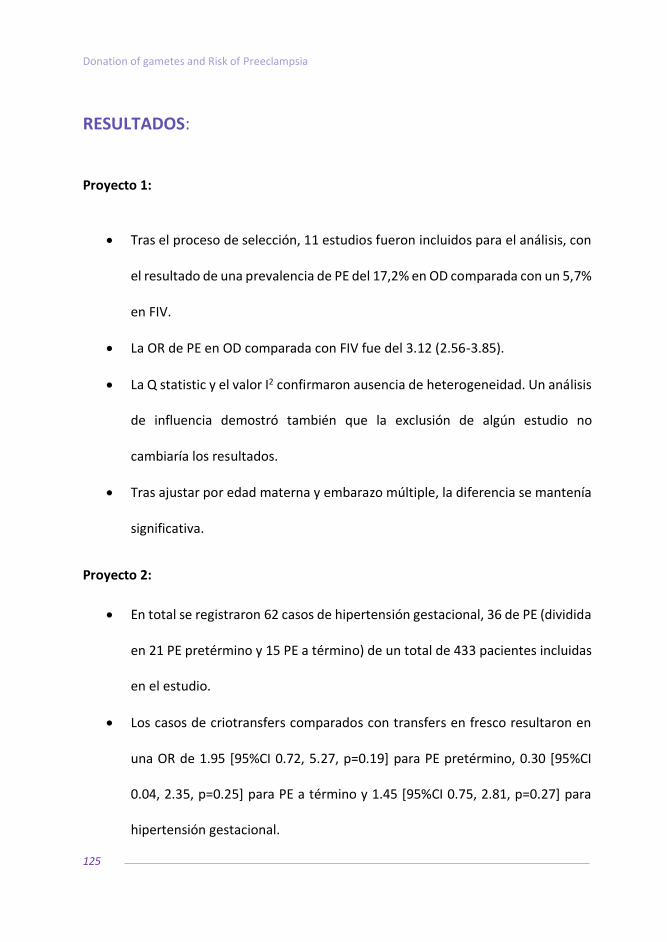
Donation of gametes and Risk of Preeclampsia
125
RESULTADOS:
Proyecto 1:
• Tras el proceso de selección, 11 estudios fueron incluidos para el análisis, con
el resultado de una prevalencia de PE del 17,2% en OD comparada con un 5,7%
en FIV.
• La OR de PE en OD comparada con FIV fue del 3.12 (2.56-3.85).
• La Q statistic y el valor I2 confirmaron ausencia de heterogeneidad. Un análisis
de influencia demostró también que la exclusión de algún estudio no
cambiaría los resultados.
• Tras ajustar por edad materna y embarazo múltiple, la diferencia se mantenía
significativa.
Proyecto 2:
• En total se registraron 62 casos de hipertensión gestacional, 36 de PE (dividida
en 21 PE pretérmino y 15 PE a término) de un total de 433 pacientes incluidas
en el estudio.
• Los casos de criotransfers comparados con transfers en fresco resultaron en
una OR de 1.95 [95%CI 0.72, 5.27, p=0.19] para PE pretérmino, 0.30 [95%CI
0.04, 2.35, p=0.25] para PE a término y 1.45 [95%CI 0.75, 2.81, p=0.27] para
hipertensión gestacional.

SUMMARY IN SPANISH
126
• Tras ajustar por edad materna, primigravidad, IMC y embarazo múltiple, el
análisis continuó sin demostrar diferencias significativas entre grupos para
todos los resultados analizados.
Proyecto 3:
• Los tratamientos de doble donación de gametos (DD) aumentaron
considerablemente durante la década del estudio.
• La media de edad de las pacientes era de 42,2 años, con un 91,7% de pacientes
sin hijos. La media de ciclos de TRA previos sin éxito era de 3.21.
• Tras el proceso de “cluster”, se definieron 3 grupos según el estado de relación
de pareja: mujeres con pareja masculina, mujeres con pareja femenina y
mujeres sin pareja.
• Las tasas de embarazo bioquímico, clínico y embarazo en curso fueron del
58,7%, 50,2% y 49,4% respectivamente, con un 26% de embarazo múltiple.
Proyecto 4:
• En total se registraron 60 casos de hipertensión gestacional, 36 de PE (dividida
en 19 PE pretérmino y 17 PE a término) de un total de 433 pacientes incluidas
en el estudio.
• Los casos de DD comparados con OD resultaron en una OR de 2.68 [95%CI
1.02, 7.04, p=0.038] para PE pretérmino, 0.26 [95%CI 0.03, 2.01, p=0.17] para
PE a término y 1.24 [95%CI 0.64, 2.42, p=0.53] para hipertensión gestacional.

Donation of gametes and Risk of Preeclampsia
127
• Tras ajustar por edad materna, primigravidad y embarazo múltiple, la OR para
preterm PE permaneció significativa con 3.02 (95%CI 1.11-8.24; p=0.031), y no
significativa para PE a término o hipertensión gestacional.

SUMMARY IN SPANISH
128
DISCUSIÓN:
Esta tesis estudia la relación entre PE y la TRA, especialmente con gametos donados.
Las TRA son cada vez más accesibles en todo el mundo, como indican los 4,5 millones
de ciclos realizados entre 2008 y 2010 a nivel mundial. Especialmente la donación de
ovocitos ha incrementado su necesidad, puesto que actualmente las mujeres tienen
los hijos en unas edades cada vez más avanzadas, impidiendo en muchos casos la
utilización de los ovocitos propios.
Para iniciar el estudio de la relación entre PE y donación de gametos, el metaanálisis
se diseñó de forma que se comparó embarazos tras OD con embarazos tras FIV.
Excluyendo embarazos espontáneos evitamos el sesgo de la TRA como causa de PE.
El metaanálisis nos confirma que la OD triplica el riesgo de la FIV para desarrollar PE.
Habiendo descartado la TRA como causa de PE en estas pacientes, y también habiendo
descartado la edad materna y el embarazo múltiple tras el análisis multivariado, la
teoría inmunológica gana peso en la etiología de la PE en donación de ovocitos.
Las dos limitaciones del metaanálisis son el no saber la causa de la infertilidad, ya que
diferentes etiologías podrían contribuir por sí solas a la fisiopatología de la PE, y no

Donation of gametes and Risk of Preeclampsia
129
disponer de la información acerca del momento del debut de la PE, ya que la PE precoz
tiene más relevancia a nivel clínico por disponer de tratamientos preventivos como la
aspirina.
Examinando los estudios publicados hasta el momento sobre PE y transferencia de
embriones vitrificados, nos dimos cuenta que un sesgo común en todos es la
comparación de ciclos de FIV con ovocitos propios. En estas pacientes, el ambiente
hormonal del endometrio es distinto en TE en fresco y con embriones previamente
congelados. Seleccionando esta población no podemos analizar con detalle el efecto
de la vitrificación en los embriones ya que, a parte de esta diferencia, los ciclos en
fresco tienen un endometrio influido por la hiperestimulación ovárica que es diferente
a la preparación endometrial que encontramos en los ciclos donde se transfieren
embriones vitrificados. En este sentido la donación de ovocitos es un modelo ideal ya
que la preparación endometrial es la misma cuando hacemos TE de embriones frescos
o congelados.
Hasta el momento, y que nosotros sepamos, es el primer estudio que compara los
trastornos hipertensivos del embarazo en OD tras TE en fresco y congelado, sin hallar
diferencias entre grupos en ninguno de los resultados analizados.

SUMMARY IN SPANISH
130
Nuestro estudio va en acorde con la actual teoría etiológica de la PE que asigna más
peso a la vascularización de la placenta que a las características del embrión. Hemos
comprobado que, con el mismo ambiente hormonal a nivel endometrial, las
probabilidades de desarrollar PE son iguales cuando transferimos embriones
previamente congelados o no. Consecuentemente podemos concluir que un adecuado
nivel hormonal en el endometrio es imprescindible para una correcta placentación, y
que los protocolos que actualmente usamos para preparar el endometrio para la
transferencia de embriones tienen un efecto deletéreo en este proceso, de allí los
resultados encontrados en los estudios publicados hasta el momento con pacientes
que realizan FIV con óvulos propios.
La principal limitación de este estudio es el uso de un cuestionario para la recogida de
datos. Aunque se pidió a las pacientes que lo rellenaran con ayuda de su obstetra, no
podemos negar un posible sesgo de recuerdo, aunque dada la importancia de la PE y
la hipertensión gestacional, sería poco probable, e independiente del tipo de
tratamiento.
Una observación de nuestro estudio sobre pacientes realizando doble-donación de
gametos fue que a pesar que la incidencia de este tratamiento aumentó durante la
década del estudio, la proporción de mujeres con pareja masculina se mantuvo
Donation of gametes and Risk of Preeclampsia
131
constante, y en cambio la de las mujeres sin pareja o con pareja femenina aumentó
considerablemente. El aumento relativo de estos dos grupos se puede deber a que,
estas pacientes, a partir de cierta edad y con el fallo ovárico asociado, van a requerir
siempre de una doble-donación para conseguir embarazo. En cambio, la ICSI es una
herramienta que solventa un porcentaje muy alto de infertilidad masculina, de forma
que cuando existe pareja masculina la donación de semen puede no ser necesaria.
Como ya se ha comentado, el aumento de la edad en la maternidad, junto con una
mayor aceptación social de la familia monoparental o con madres homosexuales, ha
conllevado un incremento absoluto y relativo de estas pacientes llevando a cabo
tratamientos con doble-donación de gametos.
Revisando los factores de riesgo para PE, observamos que las pacientes de doble-
donación tienen varios de ellos, como puede ser una edad materna avanzada,
primigravidad, el uso de TRA para conseguir la gestación, el uso de gametos donados
y una tasa alta de embarazo gemelar. A pesar que este tipo de tratamientos está
aumentando a nivel mundial, falta literatura acerca de los resultados obstétricos en
estas pacientes. Este es, hasta el momento y que nosotros sepamos, el primer estudio
amplio que describe las características de las pacientes y los resultados de la doble-
donación.

SUMMARY IN SPANISH
132
Basándonos en que eldeficiente reconocimiento inmunológico es causa de PE, sería
entonces razonable asumir un aumento de esta patología en la DD, puesto que en
estos casos la madre no ha estado expuesta a ninguno de los antígenos fetales ni ha
estado en contacto con el esperma del donante. En el cuarto proyecto hallamos un
aumento de PE precoz en DD respecto a la OD, un hallazgo nunca antes descrito en la
literatura, pero no de PE a término ni de hipertensión gestacional. La explicación sobre
estos resultados la podemos encontrar en la diferente etiología de la PE precoz y
tardía: mientras que la PE tardía tiene el origen en un sistema cardiovascular materno
alterado, la precoz se debe a una mala implantación corial secundaria a una mala
adaptación inmunológica de la madre al embrión. Por tanto, en los embarazos tras
doble donación de gametos, es lógico que sea la PE precoz la que incremente. Esto es
especialmente relevante ya que las consecuencias de la PE precoz son más graves,
tanto para la gestante como para el feto, pero además su diagnóstico precoz puede
permitir iniciar medidas preventivas. El inicio de la aspirina a dosis bajas antes de la
semana 16 de embarazo se ha demostrado eficaz en la reducción de la incidencia de
la PE precoz.
Este estudio comparte la misma limitación que el proyecto 2, que es que los datos han
sido recogidos mediante un cuestionario enviado a las pacientes, pero por el mismo
motivo que anteriormente, no debería alterar los resultados hallados. Además, nunca
antes se habían descrito tantos casos de DD, y como segundo punto fuerte solo se
Donation of gametes and Risk of Preeclampsia
133
incluyeron transferencias de embriones en fresco, evitando el posible sesgo de incluir
TE en fresco y vitrificados.
En conclusión, las pacientes embarazadas tras donación de gametos, y sobre todo tras
doble donación, deben ser informadas y conscientes del alto riesgo que tienen de
desarrollar PE, al igual que sus médicos. Tras la transferencia de embriones vitrificados,
estas pacientes que han recibido óvulos donados siguen teniendo un riesgo elevado,
pero no mayor que las que han recibido embriones frescos. Estas embarazadas deben
recibir un control gestacional estricto para poder ser identificadas, iniciar medidas
preventivas cuanto antes y, en caso de desarrollo del síndrome hipertensivo, recibir un
tratamiento adecuado. Varios estudios demuestran que la prematuridad y el bajo peso
al nacer en embarazos tras donación de ovocitos mejoran al ajustar por PE. Esto
significa que parte de estas complicaciones neonatales son consecuencia de la
inducción prematura del parto que el obstetra lleva a cabo para tratar de la única
forma definitiva que hay la PE, que es finalizando la gestación. Identificando a las
pacientes de riesgo e iniciando medidas preventivas como la administración de
aspirina antes de las 16 semanas, pues, podríamos evitar resultados perinatales
adversos para madre y para el neonato. Además, sabiendo que el ambiente hormonal

SUMMARY IN SPANISH
134
tiene un papel en la PE, el desarrollo de protocolos con diferentes terapias hormonales
sustitutivas para el endometrio podría disminuir la incidencia de este síndrome.

Donation of gametes and Risk of Preeclampsia
135
CONCLUSIONES:
• La OD está asociada a una mayor incidencia de PE que la FIV, incluso ajustando
por edad materna y embarazo múltiple.
• Estos datos sugieren que en OD, el origen de la PE está relacionada con una
mala adaptación de la madre a antígenos embrionarios.
• En OD, la transferencia de embriones previamente vitrificados no confiere más
riesgo de desarrollar PE que la transferencia de embriones frescos.
• El ambiente hormonal en el endometrio probablemente tiene un papel en la
patogénesis de la PE en TRA.
• Los tratamientos de doble donación de gametos están incrementando, con
resultados positivos para pacientes seleccionadas
• Las pacientes embarazadas tras doble donación de gametos tienen múltiples
factortes de riesgo para desarrollar PE.
• La DD está asociada a una mayor incidencia de PE pretérmino que la OD, pero
no de PE a término o hipertensión gestacional, incluso ajustando por edad
materna, primigravidad y embarazo múltiple.
• En reproducción asistida, cuantos más gametos donados se reciben, más
riesgo de desarrollar PE.

SUMMARY IN SPANISH
136