CIRUGÍA y CIRUJANOS - CORE · CIRUGÍA y CIRUJANOS ... de se˜nalización intracelular en cáncer...
10
Cirugía y Cirujanos. 2016;84(5):434---443 www.amc.org.mx www.elsevier.es/circir CIRUGÍA y CIRUJANOS Órgano de difusión científica de la Academia Mexicana de Cirugía Fundada en 1933 GENERAL INFORMATION Intracellular signalling mechanisms in thyroid cancer Paul Mondragón-Terán a,e,∗ , Luz Berenice López-Hernández b,e , José Gutiérrez-Salinas c,e , Juan Antonio Suárez-Cuenca d,e , Rosa Isela Luna-Ceballos a,e , Aura Erazo Valle-Solís d,e a Laboratorio de Medicina Regenerativa e Ingeniería de Tejidos, Centro Médico Nacional 20 de Noviembre, Instituto de Seguridad y Servicios Sociales de los Trabajadores del Estado, Mexico City, Mexico b División de Investigación Biomédica, Centro Médico Nacional 20 de Noviembre, Instituto de Seguridad y Servicios Sociales de los Trabajadores del Estado, Mexico City, Mexico c Laboratorio de Bioquímica y Medicina Experimental, Centro Médico Nacional 20 de Noviembre, Instituto de Seguridad y Servicios Sociales de los Trabajadores del Estado, Mexico City, Mexico d Laboratorio de Metabolismo Experimental e Investigación Clínica, Centro Médico Nacional 20 de Noviembre, Instituto de Seguridad y Servicios Sociales de los Trabajadores del Estado, Mexico City, Mexico e Subdirección de Investigación y Ense˜ nanza, Centro Médico Nacional 20 de Noviembre, Instituto de Seguridad y Servicios Sociales de los Trabajadores del Estado, Mexico City, Mexico Received 22 July 2015; accepted 27 May 2016 Available online 30 August 2016 KEYWORDS Thyroid cancer; Mitogenactivated protein kinase MAPK; BRAF mutations Abstract Background: Thyroid cancer is the most common malignancy of the endocrine system, the papil- lary variant accounts for 80---90% of all diagnosed cases. In the development of papillary thyroid cancer, BRAF and RAS genes are mainly affected, resulting in a modification of the system of intracellular signalling proteins known as ‘‘protein kinase mitogen-activated’’ (MAPK) which consist of ‘‘modules’’ of internal signalling proteins (Receptor/Ras/Raf/MEK/ERK) from the cell membrane to the nucleus. In thyroid cancer, these signanling proteins regulate diverse cellular processes such as differentiation, growth, development and apoptosis. MAPK play an impor- tant role in the pathogenesis of thyroid cancer as they are used as molecular biomarkers for diagnostic, prognostic and as possible therapeutic molecular targets. Mutations in BRAF gene have been correlated with poor response to treatment with traditional chemotherapy and as an indicator of poor prognosis. Please cite this article as: Mondragón-Terán P, López-Hernández LB, Gutiérrez-Salinas J, Suárez-Cuenca JA, Luna-Ceballos RI, Erazo Valle-Solís A. Mecanismos de se˜ nalización intracelular en cáncer de tiroides. Cir Cir. 2016;84:434---443. ∗ Corresponding author at: Laboratorio de Medicina Regenerativa e Ingeniería de Tejidos, División de Investigación Biomédica, Centro Médico Nacional 20 de Noviembre, Instituto de Seguridad y Servicios Sociales de los Trabajadores del Estado, San Lorenzo 502, 3. er piso, Colonia del Valle, C.P. 03100, Delegación Benito Juárez, Mexico City, Mexico. Tel.: +52 (55) 5200 5003 ext. 50162. E-mail address: [email protected] (P. Mondragón-Terán). 2444-0507/© 2016 Academia Mexicana de Cirug´ ıa A.C. Published by Masson Doyma M´ exico S.A. This is an open access article under the CC BY-NC-ND license (http://creativecommons.org/licenses/by-nc-nd/4.0/).
Transcript of CIRUGÍA y CIRUJANOS - CORE · CIRUGÍA y CIRUJANOS ... de se˜nalización intracelular en cáncer...

C
G
I
PJR
a
yb
Tc
Sd
Se
d
RA
V
MC
2B
irugía y Cirujanos. 2016;84(5):434---443
www.amc.org.mx www.elsevier.es/circir
CIRUGÍA y CIRUJANOSÓrgano de difusión científica de la Academia Mexicana de Cirugía
Fundada en 1933
ENERAL INFORMATION
ntracellular signalling mechanisms in thyroid cancer�
aul Mondragón-Terána,e,∗, Luz Berenice López-Hernándezb,e,osé Gutiérrez-Salinasc,e, Juan Antonio Suárez-Cuencad,e,osa Isela Luna-Ceballosa,e, Aura Erazo Valle-Solísd,e
Laboratorio de Medicina Regenerativa e Ingeniería de Tejidos, Centro Médico Nacional 20 de Noviembre, Instituto de Seguridad Servicios Sociales de los Trabajadores del Estado, Mexico City, MexicoDivisión de Investigación Biomédica, Centro Médico Nacional 20 de Noviembre, Instituto de Seguridad y Servicios Sociales de losrabajadores del Estado, Mexico City, MexicoLaboratorio de Bioquímica y Medicina Experimental, Centro Médico Nacional 20 de Noviembre, Instituto de Seguridad yervicios Sociales de los Trabajadores del Estado, Mexico City, MexicoLaboratorio de Metabolismo Experimental e Investigación Clínica, Centro Médico Nacional 20 de Noviembre, Instituto deeguridad y Servicios Sociales de los Trabajadores del Estado, Mexico City, MexicoSubdirección de Investigación y Ensenanza, Centro Médico Nacional 20 de Noviembre, Instituto de Seguridad y Servicios Socialese los Trabajadores del Estado, Mexico City, Mexico
eceived 22 July 2015; accepted 27 May 2016vailable online 30 August 2016
KEYWORDSThyroid cancer;Mitogenactivatedprotein kinase MAPK;BRAF mutations
AbstractBackground: Thyroid cancer is the most common malignancy of the endocrine system, the papil-lary variant accounts for 80---90% of all diagnosed cases. In the development of papillary thyroidcancer, BRAF and RAS genes are mainly affected, resulting in a modification of the systemof intracellular signalling proteins known as ‘‘protein kinase mitogen-activated’’ (MAPK) whichconsist of ‘‘modules’’ of internal signalling proteins (Receptor/Ras/Raf/MEK/ERK) from the cellmembrane to the nucleus. In thyroid cancer, these signanling proteins regulate diverse cellularprocesses such as differentiation, growth, development and apoptosis. MAPK play an impor-
tant role in the pathogenesis of thyroid cancer as they are used as molecular biomarkers for diagnostic, prognostic and as possible therapeutic molecular targets. Mutations in BRAF genehave been correlated with poor response to treatment with traditional chemotherapy and asan indicator of poor prognosis.� Please cite this article as: Mondragón-Terán P, López-Hernández LB, Gutiérrez-Salinas J, Suárez-Cuenca JA, Luna-Ceballos RI, Erazoalle-Solís A. Mecanismos de senalización intracelular en cáncer de tiroides. Cir Cir. 2016;84:434---443.
∗ Corresponding author at: Laboratorio de Medicina Regenerativa e Ingeniería de Tejidos, División de Investigación Biomédica, Centroédico Nacional 20 de Noviembre, Instituto de Seguridad y Servicios Sociales de los Trabajadores del Estado, San Lorenzo 502, 3.er piso,olonia del Valle, C.P. 03100, Delegación Benito Juárez, Mexico City, Mexico. Tel.: +52 (55) 5200 5003 ext. 50162.E-mail address: [email protected] (P. Mondragón-Terán).
444-0507/© 2016 Academia Mexicana de Cirugıa A.C. Published by Masson Doyma Mexico S.A. This is an open access article under the CCY-NC-ND license (http://creativecommons.org/licenses/by-nc-nd/4.0/).

Intracellular signalling mechanisms in thyroid cancer 435
Objective: To review the molecular mechanisms involved in intracellular signalling of BRAF andRAS genes in thyroid cancer.Conclusions: Molecular therapy research is in progress for this type of cancer as new moleculeshave been developed in order to inhibit any of the components of the signalling pathway(RET/PTC)/Ras/Raf/MEK/ERK; with special emphasis on the (RET/PTC)/Ras/Raf section, whichis a major effector of ERK pathway.© 2016 Academia Mexicana de Cirugıa A.C. Published by Masson Doyma Mexico S.A. This is anopen access article under the CC BY-NC-ND license (http://creativecommons.org/licenses/by-nc-nd/4.0/).
PALABRAS CLAVECáncer de tiroides;Cinasas de proteínasactivadas pormitógenos MAPK;Mutaciones BRAF
Mecanismos de senalización intracelular en cáncer de tiroides
ResumenAntecedentes: El cáncer de tiroides es el tumour maligno más frecuente del sistema endocrino;la variante papilar representa entre el 80 y el 90% de todos los casos diagnosticados. En eldesarrollo del cáncer papilar de tiroides están afectados principalmente 2 genes llamados BRAFy RAS, que alteran el sistema de senalización intracelular de las proteínas conocidas como«cinasas de proteínas activadas por mitógenos» (MAPK, por sus siglas en inglés para mitogen-activated protein kinase) y que se componen de «módulos» de proteínas de senalización interna(Receptor/Ras/Raf/MEK/ERK), que van de la membrana celular al núcleo y que en el cáncer detiroides regulan diversos procesos celulares, como la diferenciación, crecimiento, desarrolloy apoptosis. Tienen un papel importante en la patogénesis del cáncer de tiroides, debido aque son usados como biomarcadores moleculares, como elementos de diagnóstico, pronósticoy como posibles blancos terapéuticos moleculares.Objetivo: Revisar los mecanismos moleculares que intervienen en las vías de senalización, enlas que están involucradas las proteínas de los genes BRAF y RAS, en el cáncer de tiroides.Conclusiones: Las mutaciones en el gen BRAF han sido correlacionadas con una pobre respuestaal tratamiento con quimioterapia tradicional, además de ser un índice de mal pronóstico.La terapia molecular es de gran interés en este tipo de cáncer, ya que se han desarrolladomedicamentos que actúan inhibiendo alguno de los componentes de la vía de senalización(RET/PTC)/Ras/Raf/MEK/ERK, con especial énfasis en la sección (RET/PTC)/Ras/Raf, que con-stituye un efector principal de la vía ERK.© 2016 Academia Mexicana de Cirugıa A.C. Publicado por Masson Doyma Mexico S.A. Este es unartıculo Open Access bajo la licencia CC BY-NC-ND (http://creativecommons.org/licenses/by-nc-nd/4.0/).
a3tmclct
i2eiam
Background
Thyroid cancer is the most common malignancy of theendocrine system, and represents approximately 1% ofcancer cases diagnosed worldwide. Differentiated thyroidcancer includes papillary and follicular types. The mostfrequent is papillary carcinoma, which accounts for approx-imately 80---90% of all cases diagnosed and most frequentlypresents in women rather than men, at a 2:1 ratio. Itsincidence worldwide has significantly increased during thelast 3 decades, probably due to more advanced systems ofdiagnosis, which has resulted in more timely diagnoses andincreasingly more targeted treatments.1---5 Recent reportsindicate that there is a similar prevalence in diagnostic ages,
regardless of ethnic group. However, there are differencesin prevalence by gender and ethnicity. It has been reportedthat Caucasian men and women present a prevalence of 6.3%and 7.1%; in the English population prevalence is reportedcca
t 4.3% and 8.4%; Hispanic at 4.2% and 6.7% and Asian at.4% and 6.4%, respectively.6 It is believed that genetic fac-ors, environmental influences and access to health servicesay be factors which determine the incidence of thyroid
ancer in any one particular region. However, during theast decade a sustained increase in the rate of thyroid can-er worldwide has been observed, mainly of the papillaryype.6
3195 cases of thyroid cancer were reported in Mexicon 2008 (1351 male and 1844 female), which represented.5% of total malignancies, with an incidence of 3 out ofvery 100,000 inhabitants and a mortality of 0.6 per 100,000nhabitants, becoming the sixth causes of death in womennd the thirteenth cause of death in men, and with maxi-um frequency of between 41 and 50 years of age: 60% of
ases occur between 31 and 60. With regards to thyroid can-ers, papillary carcinoma and its variants represent 80.3%nd follicular cancer and its variants, 2.4%.7---9
4
qsktdttt
G
Ir(gnpag
ridceotgcitotg
ctrpwtepsaiwm
sColotidtbatcpooig
FbS
36
On a molecular level, papillary thyroid cancer fre-uently presents with metabolic changes in the intracellularignalling systems, which involve the activation of proteinsnown as mitogen-activated protein kinase (MAPK). The lat-er have, in recent years, been the purpose of many studiesue to their role in the pathogenesis of thyroid cancer andheir use as molecular biomarkers, in addition to their poten-ial usage for diagnostic, prognostic purposes and as possibleherapeutic molecular targets.10---12
enetic changes in cancer
t is a known fact that changes in 3 types of genes may beesponsible for the start and continuation of the cancer:a) oncogenes, (b) tumour suppressor genes and (c) stabilityenes. It is also well known that the cell has several mecha-isms to safeguard and protect the organism from a lethal,otentially carcinogenic effect such as a genetic mutationnd it is therefore only when there are changes in severalenes that cancer may develop.12---14
A mutation in an oncogene leads to a gene which shouldemain constituitively active or inactive normally becomingnactive or active. The activation of an oncogene may beue to: chromosomal translocation, abnormal gene amplifi-ation or intragenic mutation which affects a vital residualxpression for the activity of this gene. A mutation of anncogene allele generally suffices to confer lack of con-rol in cellular growth. For their part, tumour suppressorenes act against what may be a genetic mutation; in theirase, they reduce the gene’s activity and its product. Thisnactivation is a product of the mutations which lead to
he loss of essential residual expressions for the activityf a protein coded by this gene, which in turn results inruncated proteins, albeit due to deletion, insertion or epi-enetic alterations. The third type of cancer genes arefcpg
Insensinh
Potential for limited
replication
o
Invasive nature and ability to produce
metastasis
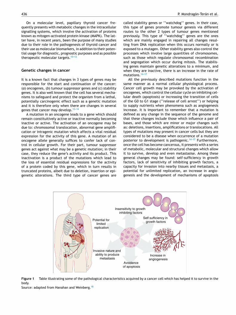
igure 1 Table illustrating some of the pathological characteristicody.ource: adapted from Hanahan and Weinberg.18
P. Mondragón-Terán et al.
alled stability genes or ‘‘watchdog’’ genes. In their case,his type of genes promote tumour genesis via differentoutes to the other 2 types of tumour genes mentionedreviously. This type of ‘‘watchdog’’ genes are the oneshich are mainly engaged in repairing all changes resul-
ing from DNA replication when this occurs normally or isxposed to a mutagen. Other stability genes also control therocesses which involve large quantities of chromosomes,uch as those which regulate chromosomal recombinationnd segregation which occur during mitosis. The stabilis-ng genes maintain genetic alterations to a minimum, andhen they are inactive, there is an increase in the rate ofutations.14---17
All the previously described mutations function in theame manner as a normal cellular physiological process.ancer cell growth may be provoked by the activation ofncogenes, which control the cellular cycle on inhibiting cel-ular death (apoptosis) or increasing the transition of cellsf the G0 to G1 stage (‘‘release of cell arrest’’) or helpingo supply nutrients when phenomena such as angiogenesisncrease. It is important to remember that a mutation isefined as any change in the sequence of the genome andhat these changes include those which influence a pair ofases, and those which are minor or major changes suchs: deletions, insertions, amplifications or translocations. Allypes of mutations may present in cancer cells but they areonsidered to be a disease when occurrence of a mutationosterior to development is pathogenic.14---17 Furthermore,nce the cell has become cancerous, it presents with a seriesf metabolic, molecular and structural changes which allowt to survive, develop and even metastasise. Among theseeneral changes may be found: self-sufficiency in growth
actors, lack of sensitivity of inhibiting growth factors, aapacity for invasion into nearby tissues and metastasis, aotential for unlimited replication, an increase in angio-enesis and the development of mechanisms of apoptosisitivity to growth ibiting factors
Self sufficiency in growth factors
Increase in angiogenesis
Avoidance f apoptosis
s acquired by a cancer cell which has helped it to survive in the
437
StimulusA B
Receptor
Activation of intermediaries
MAPKKK
MAPKK
MAPK
Activation of transcription
factors
Celluar responseProliferation, differentiation,
survival, migration, development
NF-kB/ c-Jun/Elk-1/ Sap1
ERK1/2
MEK1/MEK2
BRaf
Ras
RET/PTC receptor
Growth factors,
cytokines
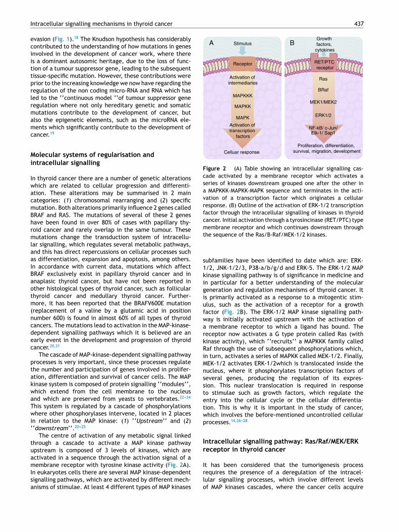
Figure 2 (A) Table showing an intracellular signalling cas-cade activated by a membrane receptor which activates aseries of kinases downstream grouped one after the other ina MAPKKK-MAPKK-MAPK sequence and terminates in the acti-vation of a transcription factor which originates a cellularresponse. (B) Outline of the activation of ERK-1/2 transcriptionfactor through the intracellular signalling of kinases in thyroidcancer. Initial activation through a tyrosincinase (RET/PTC) typemembrane receptor and which continues downstream throught
s1kigiufwarkRiMnsstetwp
Ir
Intracellular signalling mechanisms in thyroid cancer
evasion (Fig. 1).18 The Knudson hypothesis has considerablycontributed to the understanding of how mutations in genesinvolved in the development of cancer work, where thereis a dominant autosomic heritage, due to the loss of func-tion of a tumour suppressor gene, leading to the subsequenttissue-specific mutation. However, these contributions wereprior to the increasing knowledge we now have regarding theregulation of the non coding micro-RNA and RNA which hasled to the ‘‘continuous model ‘‘of tumour suppressor generegulation where not only hereditary genetic and somaticmutations contribute to the development of cancer, butalso the epigenetic elements, such as the microRNA ele-ments which significantly contribute to the development ofcancer.19
Molecular systems of regularisation andintracellular signalling
In thyroid cancer there are a number of genetic alterationswhich are related to cellular progression and differenti-ation. These alterations may be summarised in 2 maincategories: (1) chromosomal rearranging and (2) specificmutation. Both alterations primarily influence 2 genes calledBRAF and RAS. The mutations of several of these 2 geneshave been found in over 80% of cases with papillary thy-roid cancer and rarely overlap in the same tumour. Thesemutations change the transduction system of intracellu-lar signalling, which regulates several metabolic pathways,and this has direct repercussions on cellular processes suchas differentiation, expansion and apoptosis, among others.In accordance with current data, mutations which affectBRAF exclusively exist in papillary thyroid cancer and inanaplasic thyroid cancer, but have not been reported inother histological types of thyroid cancer, such as follicularthyroid cancer and medullary thyroid cancer. Further-more, it has been reported that the BRAFV600E mutation(replacement of a valine by a glutamic acid in positionnumber 600) is found in almost 60% of all types of thyroidcancers. The mutations lead to activation in the MAP-kinase-dependent signalling pathways which it is believed are anearly event in the development and progression of thyroidcancer.20,21
The cascade of MAP-kinase-dependent signalling pathwayprocesses is very important, since these processes regulatethe number and participation of genes involved in prolifer-ation, differentiation and survival of cancer cells. The MAPkinase system is composed of protein signalling ‘‘modules’’,which extend from the cell membrane to the nucleusand which are preserved from yeasts to vertebrates.22---24
This system is regulated by a cascade of phosphorylationswhere other phosphorylases intervene, located in 2 placesin relation to the MAP kinase: (1) ‘‘Upstream’’ and (2)‘‘downstream’’.20---25
The centre of activation of any metabolic signal linkedthrough a cascade to activate a MAP kinase pathwayupstream is composed of 3 levels of kinases, which areactivated in a sequence through the activation signal of a
membrane receptor with tyrosine kinase activity (Fig. 2A).In eukaryotes cells there are several MAP kinase-dependentsignalling pathways, which are activated by different mech-anisms of stimulae. At least 4 different types of MAP kinasesIrlo
he sequence of the Ras/B-Raf/MEK-1/2 kinases.
ubfamilies have been identified to date which are: ERK-/2, JNK-1/2/3, P38-a/b/g/d and ERK-5. The ERK-1/2 MAPinase signalling pathway is of significance in medicine andn particular for a better understanding of the moleculareneration and regulation mechanisms of thyroid cancer. Its primarily activated as a response to a mitogentic stim-lus, such as the activation of a receptor for a growthactor (Fig. 2B). The ERK-1/2 MAP kinase signalling path-ay is initially activated upstream with the activation of
membrane receptor to which a ligand has bound. Theeceptor now activates a G type protein called Ras (withinase activity), which ‘‘recruits’’ a MAPKKK family calledaf through the use of subsequent phosphorylations which,
n turn, activates a series of MAPKK called MEK-1/2. Finally,EK-1/2 activates ERK-1/2which is translocated inside theucleus, where it phosphorylates transcription factors ofeveral genes, producing the regulation of its expres-ion. This nuclear translocation is required in responseo stimulae such as growth factors, which regulate thentry into the cellular cycle or the cellular differentia-ion. This is why it is important in the study of cancer,hich involves the before-mentioned uncontrolled cellularrocesses.14,26---28
ntracellular signalling pathway: Ras/Raf/MEK/ERKeceptor in thyroid cancer
t has been considered that the tumorigenesis processequires the presence of a deregulation of the intracel-ular signalling processes, which involve different levelsf MAP kinases cascades, where the cancer cells acquire
4
deatov
uwocufrktirwittcRwinIccsaspaboiec
BiBrwidtda
r(RaaewcTf
knrfiigeRt
stdcaw
Rawbb1tppaiibvmtaide
irdiphEeoceigo
Ic
38
ifferent capacities such as: (1) ignoring signals to prolif-rate; (2) avoiding apoptosis; (3) becoming insensitive tontiproliferation signals; (4) acquiring an unlimited poten-ial to replicate; (5) invading and producing metastasis tother tissues and (6) producing angioigenesis, which pro-ides them with nutrients and vital support.18,29
It has been reported that different malignancies showp as an alteration in the intracellular signalling path-ay, comprising of the cascade of activation in sequencef the Ras/Raf/MEK/ERK kinases, in which the last cas-ade effector (which is the ERK-kinase protein) is activatedpstream by genetic mutations which may affect the dif-erent kinasees located in some of the levels of the chaineactions, starting with an over-expression of the tyrosineinase type receptor and most frequently with mutations inhe Ras and Raf proteins. The intracellular signalling cascaden a cancer cell begins with the activation of a membraneeceptor (with tyrosine kinase activity) when the latter bindsith its ligand or through an external stimulus (e.g. change
n osmosis, oxidative stress, lack of nutrients, etc.). Oncehe receptor has been activated, it recruits the first effec-or, by means of specific phyosphylations, of a signallingascade which is a MAPKKK which may be A-Raf, B-Raf oraf-1. These kinases phosphylate to the following MAPKKhich are MEK-1/2 and finally phosphylate to a MAPK, which
s ERK-1 or ERK-2, which are in turn translocated to theucleus to initiate activation/inhibition of specific genes.f this signalling cascade is found constitutively in all normalells of the body, in the cancer cell each component of thehain may have mutations which provoke excessive down-tream activation of their kinase, and this increases cellularctivity exponentially through the activation/inhibition ofpecific genes. It has therefore been reported, for exam-le, that the mutations in the Ras and Raf kinases (whichre the components of the intracellular pathway composedy the Ras/Raf/MEK/ERK receptor), are found in all typesf thyroid cancer, primarily in papillary thyroid cancer. Thisndicates that a single alteration in these molecules may benough to lead to a malignant transformation of the thyroidells.
It is thus known that the presence of the mutation calledRAFV600E (consistent in the presence of a valine residual,
nstead of a glutamic acid residual, in position 600 of the-Raf protein kinase), the prevalence of which has beeneported between 27% and 80% in different patient groupsith thyroid cancer, leads to an increase of over 400 times
n the activity of B-Raf, which produces an increase in theownstream activity of the ERk effectors, which increaseheir phosphorylant capacity and therefore the activity ofiverse key genes30 for cellular metabolism, proliferationnd apoptosis.
The most frequently reported mutations in papillary thy-oid cancer are: re-arrangements in the RET/PTC genemembrane receptor with tyrosine kinase activity) and B-af and Ras mutations (proteins with kinase activity). Theyre all involved in the intracellular signalling pathway whichctivates the nuclear ERK effector. These alterations arexclusively found in patients with papillary thyroid cancer,
hich proves that every separate alteration may be suffi-ient to lead to a malignant transformation of thyroid cells.he proto-oncogene called RET (rearranged during trans-ection), encodes for a membrane receptor with tyrosineIt(a
P. Mondragón-Terán et al.
inase activity, which is expressed in a particular man-er in type C parafollicuar cells in thyroid cancer, whichesults in aberrant proteins with different chimeric formsrom the receptor. However, their expression is very lown the follicular cells. The name RET/PTC (rearranged dur-ng transfection/papillar tyroid cancer associate) has beeniven to these chimeric forms. To date, more than 11 differ-nt types of RET/PTC proteins have been described, whereET/PTC1 and RET/PTC3 are the most frequently found inhyroid cancer.31,32
In the cancer cell, RET/PTC leads to constituitive down-tream activation of a kinase called Ras. It is now knownhat Ras activates a large number of molecules which act asownstream intracellular signalling systems and induce can-er cell invasion properties. It has been reported that Raslso activates reactive proteins to stress, such as Raf kinase,hich is the key downstream cytosolic Ras effector.
Several isoforms of Raf have been reported: A-Raf, B-af and Raf-1 (also called C-Raf). Raf activation includes
series of highly regulated metabolic steps, which startith their recruitment of the internal cellular membraney the Ras protein. Once Raf has been activated it mayind downstream with other kinases, among which are MEK-
and MEK-2. For activation, as well as Ras being activated,he Raf isoforms require that an enzyme with protein phos-hatise activity known as Src-kinase be activated. Thisrotein phosphorylates or dephosphorylates certain aminocid residues, located within the Raf areas, which regulatets inactive/active state. Of the 3 known isoforms for Raf,soform B-Raf is the one which presents with the highestasal level activity compared with A-Raf or Raf-1. This isery significant, since in thyroid cancer a large number ofutations in protein B-Raf have been reported, compared
o that of A-Raf or Raf-1. This has led to the use of B-Rafs a tumour marker for several types of cancer, and specif-cally thyroid cancer. Moreover, B-Raf is the most powerfulownstream activator of MEK-1 kinase, which is the ERKffector.
The MEK-1 protein is a protein kinase with dual activ-ty, since it is able to phosphorylate serine and tyrosineesidues. Once MEK-1 has been activated by B-Raf, theownstream activation of the ERK effector occurs, whichs a serine/threonine kinase which phosphorylates diverseroteins, both cytosolic and nuclear. In this way, theyperactivation of the signalling cascade which regulatesRK may lead to the arrest of the cellular cycle. How-ver, if there is an aberration in any of the sequencesf the signalling cascade, a tumour transformation of theell may be induced. The response kinetics and theirxtension to diverse ligands or extracellular stimulae maynduce the ERK to regulate specific diverse biological pro-rammes such as cellular differentiation and proliferationr apoptosis.32
ntracellular PI3K/Akt/mTOR signalling in thyroidancer
n addition to the above mentioned intracellular signalling,here is also the phosphatidylinositol -3 kinase/protein BAtk) pathway/mammalian target, for rapamicin (mTOR),lso expressed as PI3K/Akt/mTOR. This signalling pathway

aclifAaaitvc
M
Asdcwmfd
vbpigmhrt
Bmcoeistota1abTatcscti
Intracellular signalling mechanisms in thyroid cancer
is involved in process such as: cellular differentiation andgrowth, progression of cellular cycle, endocitosis, motility,apoptosis and intermediate metabolism (related primarily toglucose uptake).33---35 In the last few years, alterations in thesaid pathway have been reported in several types of cancersuch as thyroid, gastric, neuro-endocrine and ovarian. Fur-thermore, it has been observed that the activation of thispathway in cancer cells may increase resistance to cisplatin,cabo-platin and paclitaxel treatment, and as a result theirstudy may help to understand the mechanisms of genesis andprogression of the cancer, in addition to the mechanisms oftumour resistance for being a possible target, for moleculartherapy and the management of thyroid cancer.33---37
Phosphatidylinositol-3 kinase (PI3K) phosphorylatesgroups 3-OH of the inositol ring of phosphatidylinositolgroups. The sub-products of the reaction of PI3K measurethe reversible nature of the cytoplasm membrane locationof proteins, which contain complex lipid binding commandsand an above standard increase in their activity associatedwith an oncogenic cellular transformation.36,37
In terms of intracellular signalling the class I PI3K maybe a downstream effector of either a membrane recep-tor with tyrosincinase activity or a G protein attached toa receptor.37,38 However, the protein known as Akt has anapproximate molecular weight of 57 kDa, which is a ser-ine/threonine kinase that belongs to the kinase family wherethey following may be found: protein kinase A, proteinkinase G and protein kinase C. The Akt is also known asprotein kinase B (PKB).37---41
The protein called mTOR is a kinase serine/threoninecoded by the FRAP 1 gene and is the primary downstreameffector of the complex PI3K/Akt. There are 2 differentisoforms of mTOR, called mTORC1 and mTORC2. Each oneis a protein multicomplex formed by several components,where the centre is the protein mTOR itself, to which sev-eral sub-units have been added. The complex mTORC1 isformed by the protein mTOR associated with the proteinRAPTOR, which works as a link and positively regulatesmTOR. It is also associated with 2 negative regulators whichare PRAS40 and the protein DEPTOR, whilst the sub-unitmLST8, combined with the complex mTORC1, regulatesits activity.39---42 The intracellular communication pathwaymeasured by mTORC1 is involved in cellular growth anddifferentiation whilst the complex mTORC2 confers insen-sitivity to rapamicine.41,42 De-regulation of mTOR activityis correlated with the presence of hamartomas, tuberoussclerosis, Peutz---Jeghers syndrome and with several types ofcancer in which the genetic changes to the regulating genesof mTOR, such as TSC1-TSC2 and LKB1, present diverse typesof mutations.42---44
During bone formation, the RUNX2 gene is key for thedifferentiation of the osteoblastoma, and for the prolifera-tion of chondrocytes and endochondral differentiation andhypertrophy. Its participation as the promoter of tumour inbreast and prostate cancer has been reported, as well as itsassociation with genes which control tumour invasion andmetastasis, since it promotes the expression of the metal-loproteinases MMP2, MMP13, MMP14. In this way, RUNX2 has
been defined as a promigratory transcription, pro-invasive,and pro-angiogenic factor, in addition to participating in theinitial events of tumorogenesis and leading the spread ofbone metastasis.43---45ttot
439
The expression and activity of RUNX2 is induced directlynd indirectly by Akt. In vitro experiments made with cancerells have shown that the activation of the PI3K/Akt intracel-ular communication pathway leads directly to an increasen affinity to DNA by the RUNX2 and RUNX2-dependentactors of transcription and that the mutations affectingkt lead to a decrease in the affinity of RUNX2, whichffects the progression of the cellular cycle. Indirectly,ctivation of the PI3K/Akt pathway regulates RUNX2 activ-ty, since it increases its protein stability either by actinghrough a nuclear transcription factor called FoxO, an acti-ator or a specific gene transcriptional repressor and theytoplasm.46,47
utations and thyroid cancer
mutation of any protein involved in the intracellularignalling cascade measured by a tyrosine type receptor mayetermine that a normal cell becomes a cancer cell. In thisontext, it has been reported that around 70% of patientsho present with papillary thyroid cancer have at least oneutation which affects the ERK activation cascade; the most
requent are those affecting Ras and B-Raf and, to a lesseregree, RET/PTC.
The prevalence of RET/PTC mutations in thyroid cancer isaried, since it depends on the geographic region, but it maye stated that approximately 20% of cases of thyroid cancerresent these alterations, and also that it is more frequentn young people or those with a background of having under-one radiation treatment. Furthermore, the mutations in aember of the gene family which code for the protein Ras
ave been primarily described in follicular adenoma and thy-oid carcinoma, but only described in around 10% of papillaryhyroid cancer cases.33
In the case of B-Raf (which may also be expressed asRaf) it has been observed that it is expressed in a nor-al manner in: blood forming cells, neurons, testicular
ells and in thyroid follicular cells. Contrary to that whichccurs with the mutations in A-Raf and Raf-1 which arextremely rare, mutations in B-Raf are the most frequentn papillary thyroid cancer, as well as being the secondomatic mutation found in all types of cancer which affecthe human being. Thus, for example, over 45% of all typesf cancer which affect the human being present a muta-ion in B-Raf. Close to 90% of these mutations consist of
replacement of a timine for an adenine in the exon5 of the neucleotide 1799 (c.1799T>A), which results in
mutation of B-Raf, where a valine residual is replacedy a glutamic acid residual in the protein position 600.his mutation is expressed as BRAFV600E (also expresseds B-Raf-V600E or BRAF-V600E). It has been reported thathe prevalence of this mutation in papillary thyroid can-er may vary from 29% to 83%, depending on the sampletudied, and has not been described in follicular thyroidancer.48 Research studies carried out in vitro have shownhat the replacement of the valine for a glutamic acidn 600 position adjacent to a threonine residual in posi-
ion 599 within protein B-Raf induces similar behaviouro a phosphorylation, which makes the tertiary structuref the protein affected where the hydrophobic interac-ions break between segment P (the activation site of the
4
pm4satnlp
iefigwmwatdcp
R
Trtcs
cdPoaPatcTp
M
Twpbfrtmcf
cest
srkchwdtngbptic
GicpiBoasaitowltpatraBr
aePcmctsacPrituawt
40
rotein by phosphorylation) and the kinase activity seg-ent, resulting in the BRAFV600E kinase activity being
60 times higher than the native B-Raf. This raised con-tituitive activity of BRAFV600E produces a downstreamctivation of all effectors of the signalling cascade effec-ors up to ERK,which results in the transformation oformal cells into cancer cells, and their unnecessary pro-iferation of Ras protein, for activation of the signallingathway.
It has been proven that the presence of BRAFV600Es linked to genes involved in the metabolism of iodine,specially those involved in the uptake of iodine by cellsor thyroid hormone synthesis. The presence of BRAFV600Enduces the thyroid cells to present changes in several of theenes involved in this process, which have been associatedith a greater aggressive tumour pattern. The presence ofutations in the BRAF gene has therefore been correlatedith a poor response to standard chemotherapy treatmentnd also is an indication of poor prognosis, since the muta-ion in BRAF was found both in anaplasic cells and in poorlyifferentiated carcinomas, which means that the molecularhanges in B-Raf are produced early on in the tumorogenesishenomenon.48
e-arrangements in PAX8-PPAR� in thyroid cancer
he PAX8 gene is a specific thyroid transcription factor whichegulates development and differentiation. Furthermore,he PPAR� gene is involved in the control of the cellularycle, apoptosis and carcinogenesis,49 as well in adipogene-is and sensitivity to insulin.
Together PAX8 and PPAR� have a key role in thyroidancer. The translocation between these 2 genes pro-uces a new gene, the overexpression of the resultingAX8/PPAR� or PPFP fusion protein alters the PPAR� functionf the downstream inhibitor of the cellular proliferation andpoptosis inductor. The chromosomal translocation PAX8-PAR� was detected in 35% of follicular thyroid cancernd anaplasic thyroid cancer.49 Furthermore, in follicularhyroid cancer the overexpression of PAX8/PPAR� is asso-iated with the activation of a MAP kinases depending onGFß receptors, which is closely linked to the tumorogeneisrocess.49,50
olecular therapy in thyroid cancer
hyroid cancer, whether it be papillary or follicular, which isell differentiated, has a fairly promising clinical behaviourattern since it may be treated with surgery, followedy radiotherapy. However, the tumours which are undif-erentiated or those which lose their capacity to captureadioactive iodine cannot be surgically removed, and areherefore tumours of poor prognosis. This type of tumoursay be treated with molecular therapy, where the intra-
ellular signalling chain (RET/PTC)/Ras/Raf/MEK/ERK is arequent target for molecular therapy studies.
Molecular therapy is of great interest in this type of
ancer as drugs have been developed which inhibit sev-ral of the components of the (RET/PTC)/Ras/Raf/MEK/ERKignalling pathway. Particular emphasis has been place onhe (RET/PTC)/Ras/Raf section, as this segment of thetpoc
P. Mondragón-Terán et al.
ignalling cascade is the main ERK effector. It has beeneported that the ZD6474 compound, which is a RET/PTCinase activity, has been effective in in vitro and pre-linical trials to induce arrest of the cellular cycle inuman papillary carcinoma, and this impedes its growthhen injected in mice.51 The compounds pyrazolopyrimi-ine (PP1 y PP2) and sunitinib (SU12248) have also beenested in in vitro, in vivo and preclinical trials. Theseullify the RET/PTC signal for eliminating the tumori-enic effect in test animals. Promising results have alsoeen reported in phase II clinical trials ii in treatingatients who do not respond to treatment with radioac-ive iodine or who have unresectable tumours, and alson those patients who present with medullary thyroidancer.52---54
Furthermore, it has been reported that the compoundsDC-0879 and PLX4720 selectively inhibit the kinase activ-
ty of BRAFV600E in vitro, which results in the tumourells reducing their proliferation. However, these have onlyroven effective in preclinical trials. On the other hand,t has been observed that patients who present with theRAFV600E mutation may develop resistance to this typef compounds, since they increase the Raf-1 expression byctivating Ras (generally over expressed) and thus the down-tream signalling cascade towards ERK may continue beingctive. This event proves the need for a precise genotyp-ng for each type of tumour, since it is necessary to ensurehat the possible mutations have been detected in a fewr several components of the signalling chain componentshich activate ERK, since, although one component is nul-
ified, another may be activated and continue with theumour proliferation process. That previously stated alsoroves that treatment should be include several compoundst the same time to ensure a better inhibitory outcome ofhe signalling cascade, and all the more so when tumouresistance to chemotherapy alone has been demonstratednd in the case of those who present with the mutationRAFV600E, this type of combined treatment is the mostecommended.53,54
The PI3K/Akt/mTOR signalling pathway is anotherppealing molecular target, due to its therapeutic ben-fit reported for different malignant tumours. TheI3K/Akt/mTOR pathway regulates critical cellular pro-esses such as proliferation, apoptosis, the cellular cycle,etabolism and angiogenesis, whilst several preclinical and
linical studies support that the pharmacological inhibi-ion of PI3K/Akt/mTOR represents a well tolerated anduccessful strategy in malignant tumour treatment, suchs prostate, breast, colon, ovarian, lung and melanomaancer.55 Apparently, the reduction in the activity ofI3K/Akt/mTOR is related to the induction of tumouradiosensibility effects,56 which are pro-apoptotic effectsn tumour cells related to lower autophagia, and tohe loss conservation in cancer cells.57 Regarding follic-lar type thyroid cancer, advanced or non differentiatednaplasia, resistant to radioactive iodine and thereforeith a poor prognosis, for its response to convention
reatment, several pre-clinical and clinical studies showhat inhibition of PI3K/Akt/mTOR pathway may be aromising strategic target, whether with monotherapy
r combined therapy, in this type of advanced thyroidancer.58
Rd
C
T
A
Dn‘IÁeRCit
R
1
Intracellular signalling mechanisms in thyroid cancer
Conclusions
Major progress has been made during the last 30 years in thefield of molecular biology aimed at discerning the mech-anisms involved in the process of tumour transformation,primarily in studies intended to understand the intracellularsystems which involve Ras/Raf/MEK/ERK kinase cascades.This signalling cascade controls cellular proliferation in nor-mal cells and cancer cells. Moreover, they are importantpathways in the development of resistance to the drugs usedin chemotherapy.
Current studies have proven that the Ras/Raf/MEK/ERKsignalling cascade induces malignant proliferation, since itstimulates cellular growth and simultaneously inhibits apo-ptosis (programmed cell death). Furthermore, the discoverythat mutations in the BRAF gene may alone lead to cel-lular transformation and proliferation in several types ofcancer, mainly thyroid cancer, has highlighted the needfor further studies to be conducted on the control mech-anisms of this type of genes and their products. This wouldinclude the development of effective chemical agents tonullify this type of protein and thus control the growth anddevelopment of cancer cells.59 That expressed above alsounderlines the need for greater study of tumour genotyp-ing, due to their significant in molecular therapy usage.The description of mutations in BRAF may predict the sen-sitivity of B-Raf to several anti-cancer agents and inhibitthe downstream to ERK and therefore inhibit growth anddevelopment of tumours. Genotyping of the tumour is alsouseful for predicting clinical behaviour of the tumour, toensure correct administration of drugs and avoid confusionregarding the type of drugs to be applied when there isthe possibility of resistance from tumours. Worldwide clini-cal research indicates that molecular therapy aimed againstmutations in the BRAF may be promising. However, there isstill much research to be carried out since each anti-cancercompound aimed at any component of the Ras/Raf/MEK/ERKchain must prove its clinical usage in patients with can-cer. However, the application at the same time of severaltypes of drugs for inhibiting the Ras/Raf/MEK/ERK chainand the combination with standard chemotherapy appearsto be the most effective plan for thyroid cancer treat-ment. Notwithstanding, the risk of developing resistance isalways latent, and the development of further and improvedproducts for cancer control treatment is an absolute pri-ority, especially if the development of these drugs led tomore generalised usage and their availability in hospitalsbecame standard, thus promoting wellbeing for the cancerpatient.
Ethical disclosure
Protection of people and animals. The authors declare thatfor this research no experiments on humans or animals have
been conducted.Data confidentiality. The authors declare that the protocolsof the centre of work have been adhered to regarding thepublication of patient data.
1
1
441
ight to privacy and informed consent. The authorseclare that no patient data appears in this article.
onflict of interests
he authors have no conflicts of interest to declare.
cknowledgements
r. Paul Mondragón Terán anbd Dr. José Gutiérrez Sali-as would like to give thanks to the support from the‘Programa de Investigación Científica y Tecnológica delSSSTE’’ (Key E015). The authors thank the biologist Miguelngel Juárez Mancera for his support in reviewing the ref-rences. They would like to thank Mr. Sergio Hernandezodriguez for his support in editing the figures and to Miss.ynthia Santiago Nicolas (Division de Investigation Biomed-
cal, CAN 20 de November, ISSUE) for her help in secretarialasks.
eferences
1. Parkin DM, Bray F, Ferlay J, Pisani P. Estimating the worldcancer burden: GLOBOCAN 2000. Int J Cancer. 2001;94:153---6.
2. Global cancer, facts and figures. 2nd ed. Atlanta: AmericanCancer Society; 2011. Available from: http://globocan.iarc.fr[accessed 02.06.15].
3. Siegel R, Ma J, Zou Z, Jema A. Cancer statistics, 2014. CA CancerJ Clin. 2014;64:9---29.
4. Cancer facts and figures for hispanics/latinos 2012---2014.Atlanta: American Cancer Society; 2012. Available from:http://globocan.iarc.fr [accessed 02.06.15].
5. Pellegriti G, Frasca F, Regalbuto C, Squatrito S, Vigneri R.Worldwide increasing incidence of thyroid cancer: update onepidemiology and risk factors. J Cancer Epidemiol. 2013:10.Article ID 965212, Available from: http://dx.doi.org/10.1155/2013/965212 [accessed 06.04.15].
6. Jemal A, Siegel R, Xu J, Ward E. Cancer statistics, 2010. CACancer J Clin. 2010;60:277---300.
7. Granados-García M, León-Takahashi AM, Guerrero-Huerta FJ,Taissoun-Aslan ZA. Cáncer diferenciado de tiroides: unaantigua enfermedad con nuevos conocimientos. Gac Med Mex.2014;150:65---77.
8. Secretaría de Salud. Sistema Nacional de Vigilancia Epi-demiológica. Bases de datos del Perfil Epidemiológico de losTumores Malignos en México; 2015. Available from: http://www.epidemiologia.salud.gob.mx/doctos/infoepid/publicaciones/2011/monografias/P EPI DE LOS TUMORES MALIGNOS M%C3%A9xico.pdf [accessed 10.06.15].
9. INEGI. Estadística de defunciones generales. Descrip-ción de la base de datos 2012; 2015. Available from:http://www.inegi.org.mx/inegi/default.aspx?s=est&c=11094[accessed 10.06.15].
0. Nikiforov YE. Thyroid carcinoma: molecular pathways
and therapeutic targets. Mod Pathol. 2008;21 Suppl. 2:S37---43.1. Xing M. Prognostic utility of BRAF mutation in papillary thyroidcancer. Mol Cell Endo. 2010;321:86---93.
2. Kam-Tsun T, Chen-Hsen L. BRAF mutation in papillary thyroidcarcinoma: pathogenic role and clinical implications. J Chin MedAssoc. 2010;73:113---28.
4
1
1
1
1
1
1
1
2
2
2
2
2
2
2
2
2
2
3
3
3
3
3
3
3
3
3
3
4
4
4
4
4
4
4
4
4
4
5
5
5
5
5
5
42
3. Li X, Abdel-Mageed AB, Kandil E. BRAF mutation in papillarythyroid carcinoma. Int J Clin Exp Med. 2012;5:310---5.
4. Vogelstein B, Kinzler KW. Cancer genes and the pathways theycontrol. Nat Med. 2004;10:789---99.
5. Knudson AG. Cancer genetics. Am J Med Genet. 2002;111:96---102.
6. Friedberg EC. DNA damage and repair. Nature. 2003;421:436---40.
7. Nowell PC. Tumor progression: a brief historical perspective.Semin Cancer Biol. 2002;12:261---6.
8. Hanahan D, Weinberg RA. The hallmarks of cancer. Cell.2000;100:57---70.
9. Berger AH, Knudson AG, Pandolfi PP. A continuum model fortumour supression. Nature. 2011;476:163---9.
0. Imajo M, Tsuchiya Y, Nishida E. Regulatory mechanisms andfunctions of MAP kinase signaling pathways. IUBMB Life.2006;58:312---7.
1. Pearson G, Robinson F, Gibson TB, Xu BE, Karandikar M,Berman K, et al. Mitogen Activated Protein (MAP) kinase path-ways: regulation and physiological functions. Endocrine Rev.2001;22:153---83.
2. Roskoski R Jr. RAF protein-serine/threonine kinases: struc-ture and regulation. Biochem Biophys Res Commun. 2010;399:313---7.
3. Takekawa M, Kubota Y, Nakamura T, Ichikawa K. Regulation ofstress-actived MAP kinase pathways during cell fate decisions.Nagoya J Med Sci. 2011;73:1---14.
4. Johnson B, Dohlman HG, Graves LM. MAPK kinase kinases(MKKKs) as a target class for small-molecule inhibition to mod-ulate signaling networks and gene expression. Curr Opin ChemBiol. 2005;9:325---31.
5. Randolph SF, Thane GM, Grant NP. Mitogen activated pro-tein kinase at the nuclear pore complex. J Cell Mol Med.2011;15:928---37.
6. Roskoski R Jr. ERK1/2 MAP kinases: structure, function, andregulation. Pharmacol Res. 2012;66:105---43.
7. McCubrey JA, Steelman LS, Chappell WH, Abrams SL, Wong EW,Chang F, et al. Roles of the Raf/MEK/ERK pathway in cell growth,malignant transformation and drug resistance. Biochim BiophysActa. 2007;1773:1263---84.
8. Cargnello M, Roux PP. Activation and function of the MAPKs andtheir substrates, the MAPK-activated protein kinases. MicrobiolMol Biol Rev. 2011;75:50---83.
9. Dhillon AS, Hagan S, Rath O, Kolch W. MAP kinase signallingpathways in cancer. Oncogene. 2007;26:3279---90.
0. Hall RD, Kudchadkar RR. BRAF mutations: signaling, epidemiol-ogy, and clinical experience in multiple malignancies. CancerControl. 2014;21:221---30.
1. Vincenzo Marotta V, Guerra A, Sapio MR, Vitale M. RET/PTCrearrangement in benign and malignant thyroid diseases: a clin-ical standpoint. Eur J Endocrinol. 2011;165:499---507.
2. Wells SA Jr, Santoro M. Targeting the RET pathway in thyroidcancer. Clin Cancer Res. 2009;15:7119---23.
3. Xing M. Genetic alterations in the phosphatidylinositol-3kinase/Akt pathway in thyroid cancer. Thyroid. 2010;20:697---706.
4. Martelli AM, Evangelisti C, Chiarini F, Grimaldi C, McCubreyJA. The emerging role of the phosphatidylinositol 3-kinase/Akt/mammalian target of rapamycin signaling networkin cancer stem cell biology. Cancers. 2010;2:1576---96.
5. Manfredi GI, Dicitore A, Gaudenzi G, Caraglia M, Persani L,Vitale G. PI3K/Akt/mTOR signaling in medullary thyroid can-cer: a promising molecular target for cancer therapy. Endocrine.2015;48:363---70.
6. Chiganer G, Ghersevich S, Sánchez A, Novelli JL. Biologíamolecular en el cáncer de tiroides. Rev Med Rosario. 2011;77:147---56.
5
P. Mondragón-Terán et al.
7. Manning BD, Cantley LC. AKT/PKB signaling: navigating down-stream. Cell. 2007;129:1261---74.
8. Gaikwad SM, Ray P. Non-invasive imaging of PI3K/Akt/mTORsignalling in cancer. Am J Nucl Med Mol Imaging. 2012;2:418---31.
9. Pinzón CE, Serrano ML, Sanabria MC. Papel de la vía fosfatidili-nositol 3 cinasa (PI3K/Akt) en humanos. Rev Cienc Salud Bogotá.2009;7:47---66.
0. Romitti M, Ceolin L, Rodriguez DS, Vaz CF, Magagnin SW, Maia AL.Signaling pathways in follicular cell-derived thyroid carcinomas(Review). Int J Oncol. 2013;42:19---28.
1. Saji M, Ringel MD. The PI3K-AKT-mTOR pathway in initia-tion and progression of thyroid tumors. Mol Cell Endocrinol.2010;321:20---8.
2. Briest F, Grabowski P. PI3K-AKT-mTOR-signaling and beyond:the complex network in gastroenteropancreatic neuroendocrineneoplasms. Theranostics. 2014;4:336---65.
3. Cohen-Solal KA, Boregowda RK, Lasfar A. RUNX2 and thePI3K/AKT axis reciprocal activation as a driving force for tumorprogression. Mol Cancer. 2015;14:137.
4. Nikiforov YE. Molecular analysis of thyroid tumors. Mod Pathol.2011;24:S34---43.
5. Baniwal SK, Khalid O, Gabet Y, Shah RR, Purcell DJ, MavD. Runx2 transcriptome of prostate cancer cells: insightsinto invasiveness and bone metastasis. Mol Cancer. 2010;9:258.
6. Fu Z, Tindall1 DJ. FoxOs, cancer and regulation of apoptosis.Oncogene. 2008;27:2312---9.
7. Xinbo Zhang X, Tang N, Hadden TJ, Rishi AK. Akt, FoxO andregulation of apoptosis. Biochim Biophys Acta. 2011;1813:1978---86.
8. Cantwell-Dorris E, O’Leary J, Sheils O. BRAFV600E: implicationsfor carcinogenesis and molecular therapy. Mol Cancer Ther.2011;10:385---94.
9. Placzkowski KA, Reddi HV, Grebe SK, Eberhardt NL, McIverB. The role of the PAX8/PPARgamma fusion oncogene in thy-roid cancer. PPAR Res. 2008;2008:672829, http://dx.doi.org/10.1155/2008/672829 [Epub 29.10.08].
0. Raman P, Koening RJ. Pax-8-PPAR-� fusion protein in thyroidcarcinoma. Nat Rev Endocrinol. 2014;10:616---23.
1. Herbst RS, Heymach JV, O’Reilly MS. Vandetanib (ZD6474):an orally available receptor tyrosine kinase inhibitor thatselectively targets pathways critical for tumor growthand angiogenesis. Expert Opin Invest Drugs. 2007;16:239---49.
2. Carlomagno F, Vitagliano D, Guida T, Napolitano M, VecchioG, Fusco A, et al. The kinase inhibitor PP1 blocks tumor-ogenesis induced by RET oncogenes. Cancer Res. 2002;62:1077---82.
3. Carlomagno F, Vitagliano D, Guida T, Basolo F, Castellone MD,Melillo RM, et al. Efficient inhibition of RET/papillary thyroidcarcinoma oncogenic kinases by 4-amino-5-(4-chloro-phenyl)-7-(t-butyl)pyrazolo[3,4-d]pyrimidine (PP2). J Clin EndocrinolMetab. 2003;88:1897---902.
4. Kim DW, Jo YS, Jung HS, Chung HK, Song JH, Park SH, et al.An orally administered multitarget tyrosine kinase inhibitor,SU11248, is a novel potent inhibitor of thyroid oncogenicRET/papillary thyroid cancer kinases. J Clin Endocrinol Metab.2006;91:4070---6.
5. Argiles G, Munster PN, Hollebecque A, Dajani O, Cheng J,Wang R, et al. A phase I trial of combined ridaforolimusand MK-2206 in patients with advanced malignancies. ClinCancer Res. 2015;21:5235---44, http://dx.doi.org/10.1158/1078-0432.CCR-15-0180 [Epub 17.07.15].
6. Chang L, Graham PH, Ni J, Hao J, Bucci J, Cozzi PJ,et al. Targeting PI3K/Akt/mTOR signaling pathway in thetreatment of prostate cancer radioresistance. Crit Rev

5
Intracellular signalling mechanisms in thyroid cancer
Oncol Hematol. 2015;96:507---17, http://dx.doi.org/10.1016/j.critrevonc.2015.07.005 [Epub 18.07.15].
57. Pal I, Parida S, Prashanth Kumar BN, Banik P, Kumar Dey K,Chakraborty S, et al. Blockade of autophagy enhances proapo-ptotic potential of BI-69A11, a novel Akt inhibitor, in coloncarcinoma. Eur J Pharmacol. 2015;765:217---27.
5
443
8. Petrulea MS, Plantinga TS, Smit JW, Georgescu CE, Netea-Maier RT. PI3K/Akt/mTOR: a promising therapeutic target for
non-medullary thyroid carcinoma. Cancer Treat Rev. 2015;41:707---13.9. Garnett MJ, Marais R. Guilty as charged: B-RAF is a human onco-gene. Cancer Cell. 2004;6:313---9.