2º Curso de Osteoporosis y Enfermedades€¦ · 10-10:30 Epidemiología de la osteoporosis en...
52
Transcript of 2º Curso de Osteoporosis y Enfermedades€¦ · 10-10:30 Epidemiología de la osteoporosis en...


2º Curso de Osteoporosis y EnfermedadesMetabólicas Óseas para Médicos Residentes
PONENTES
Dr. Jose Luis Pérez CastrillónMedicina Interna. Hospital Rio Hortega. ValladolidDr. Javier Del PinoReumatología. Hospital Clínico. SalamancaDr. Alejandro OrtegaAnestesiología. Unidad del dolor. Asepeyo, Coslada. MadridDr. Guillermo Martínez Díaz-GuerraEndocrinología. Hospital 12 de Octubre. Madrid
Dr. Ricardo LarrainzarTraumatología. Hospital Infanta Leonor-Virgen de la TorreMadridDra. Elena MartinezRehabilitación. Hospital Ramón y Cajal. MadridDra. Mª Jesús MoroMedicina Interna. Hospital Infanta Leonor-Virgen de La TorreMadrid
HOSPITAL UNIVERSITARIO INFANTA LEONORGran Vía del Este, 80 - 28031 Madrid
Introducción y formación básica en el área de las enfermedades metabólicas óseas. Tanto la osteoporosis como otras enfermedades metabólicas óseas requieren de un manejo multidisciplinar con lo que este curso tiene como objetivo dar
a conocer aspectos básicos de la epidemiología, diagnostico, manejo clínico y farmacológico de estas enfermedades
PROGRAMA
Preincripción en el siguiente enlace: https://goo.gl/forms/WUbbEjDorTAxGL0C3
Para más información:[email protected] - Móvil: 625 680 737
INSCRIPCIÓN GRATUITAAFORO LIMITADO A 40 PLAZASINSCRIPCIÓN
Fecha límite de inscripción: 20 de mayo. El día 31 de mayo se comunicará los aceptados por correo electrónico.La inscripción será por orden de registro, priorizando a los socios de la SEIOMM y, en caso sobrepasarse el número depreinscripciones, se seleccionaran dos por servicio o centro de trabajo.La inscripción es gratuita e incluye sesiones de trabajo, cafés y comida de trabajo. Los traslados no se incluyen.
* Pendientes de fijar las fechas de celebración en otras sedes de Barcelona, Sevilla y Santiago de Compostela
13-13:30 Hiperparatiroidismo primario, hiperparatiroidismo normocalcémico, hipoparatiroidismo postquirúrgico e idiopático Dr. Guillermo Martínez Díaz-Guerra
13:30-15 COMIDA
BLOQUE 4: FORMAS DE PRESENTACIÓN CLÍNICA DE LA OSTEOPOROSIS
FRACTURA OSTEOPORÓTICA
15-15.30 Fractura no vertebral (humero, radio y cadera) Dr. Ricardo Larrainzar
15.30-16 Fractura vertebral Dra. Elena Martínez
BLOQUE 5: TRATAMIENTO NO FARMACOLOGICO Y FAMACOLÓGICO
DE LA OSTEOPOROSIS. GUÍA CLINICA DE LA SEIOMM16-16:30 Tratamiento de la osteoporosis.
Guia clínica SEIOMM Dr. Javier del Pino
16.30-17 Efectos adversos de los tratamientosDra. Elena Martínez
17-17.30 Tratamiento del dolor en la osteoporosis (analgésicos, vertebroplastia y cifoplastia, bloqueos anestésicos) Dr. Alejandro Ortega
17.30-18 CAFÉ
18-19 Osteotrivial: taller casos clínicos. Dra. Mª Jesús Moro
PRESENTACIÓN DE LA JORNADA
9-9:15 Bienvenida/Entrega de la documentación9:15-9:30 Inauguración del curso
BLOQUE 1: FISIOPATOLOGIA Y EPIDEMIOLOGÍA DE LA OSTEOPOROSIS
9:30-10 Nuevos datos de la fisiopatología de la osteoporosis (el remodelado óseo a nivel celular y molecular. El sistema rankl. La vía Wnt-catenina)Dr. Guillermo Martínez Díaz-Guerra
10-10:30 Epidemiología de la osteoporosis en España Dr. Ricardo Larrainzar
BLOQUE 2: FACTORES DE RIESGO Y DIAGNÓSTICO DE LA OSTEOPOROSIS
10:30-11 Pruebas complementarias en el abordaje inicial. (Escalas de riesgo. Cuándo solicitar y cómo interpretar una densitometría. Analítica para estudio de secundarias.Marcadores remodelado óseo. Otras exploraciones)Dra. Mª Jesús Moro
11-11:30 CAFÉ
BLOQUE 3: ENFERMEDADES METABÓLICAS ÓSEAS DE MAYOR PREVALENCIA
11:30-12 Osteoporosis (postmenopáusica, del varón, corticoidea osteoporosis secundaria) Dr. José Luis Pérez Castrillón
12.-12:30 Enfermedad de Paget Dr. Javier del Pino
12:30-13 Efectos sobre el metabolismo óseo de la cirugía bariátrica. Osteomalacia. Osteodistrofia renalDr. José Luis Pérez Castrillón
MADRID, JUEVES 15 DE JUNIO 2017 *
EDITORIALDivergent effects of vascular endothelial growth factor,VEGF and the N-terminal fragment of the parathormo-ne-related protein, PTHrP on human adipose derivedfrom mesenchymal stem cellsPlotkin LI
ORIGINALSVascular endothelial growth factor (VEGF) and theN-terminal portion of parathyroid hormone-relatedprotein (PTHrP) regulate the proliferation of humanmesenchymal stem cellsBravo B, Fernández de Castro L, Buendía I, Santos X,Gortázar A
Effects of the catalase antioxidant enzyme in vascularcalcification and bone demineralizationMartínez Arias L, Panizo García S, Carrillo López N,Barrio Vázquez S, Quirós González I, Román García P,Mora Valenciano I, Miguel Fernández D, Añón Álvarez E,Fernández Martín JL, Ruiz Torres MP, Cannata Andía JB,Naves Díaz M
Influence of obesity on microarchitecture and bio-mechanical properties in patients with hip fractureGiner M, Montoya MJ, Miranda C, Vázquez MA, Miranda MJ,Pérez‐Cano R
Identification of genetic variants associated withbone mineral density (BMD) in the FLJ42280 geneRoca‐Ayats N, Cozar Morillo M, Gerousi M, Czwan E,Urreizti R, Martínez‐Gil N, García‐Giralt N, Mellibovsky L,Nogués X, Díez‐Pérez A, Balcells S, Grinberg D
CLINICAL NOTEMesenteric panniculitis associated with the use ofbisphosphonates: are these more proinflammatorythan we know?Torregrosa Suau O, Guilló Quiles E, Mora Rufete A
REVIEWOsteoporosis in rheumatic diseases and glucocorticoidinduced Maldonado G, Messina O, Moreno M, Ríos C
3
5
13
20
28
35
38
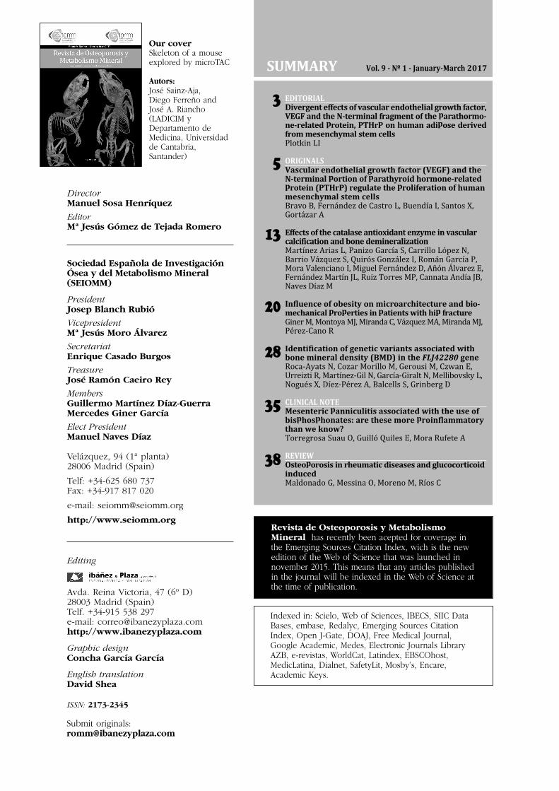
SUMMARY Vol. 9 - Nº 1 - January-March 2017
Submit originals:[email protected]
Our coverSkeleton of a mouseexplored by microTAC
Autors:José Sainz-Aja,Diego Ferreño andJosé A. Riancho(LADICIM yDepartamento deMedicina, Universidadde Cantabria,Santander)
Sociedad Española de InvestigaciónÓsea y del Metabolismo Mineral(SEIOMM)
PresidentJosep Blanch Rubió
VicepresidentMª Jesús Moro Álvarez
SecretariatEnrique Casado Burgos
TreasureJosé Ramón Caeiro Rey
MembersGuillermo Martínez Díaz-GuerraMercedes Giner García
Elect PresidentManuel Naves Díaz
Velázquez, 94 (1ª planta)28006 Madrid (Spain)
Telf: +34-625 680 737Fax: +34-917 817 020
e-mail: [email protected]
http://www.seiomm.org
Editing
Avda. Reina Victoria, 47 (6º D)28003 Madrid (Spain)Telf. +34-915 538 297 e-mail: [email protected]://www.ibanezyplaza.com
Graphic designConcha García García
English translationDavid Shea
ISSN: 2173-2345
DirectorManuel Sosa Henríquez
Editor Mª Jesús Gómez de Tejada Romero
Indexed in: Scielo, Web of Sciences, IBECS, SIIC DataBases, embase, Redalyc, Emerging Sources CitationIndex, Open J-Gate, DOAJ, Free Medical Journal,Google Academic, Medes, Electronic Journals LibraryAZB, e-revistas, WorldCat, Latindex, EBSCOhost,MedicLatina, Dialnet, SafetyLit, Mosby’s, Encare,Academic Keys.
Revista de Osteoporosis y MetabolismoMineral has recently been acepted for coverage inthe Emerging Sources Citation Index, wich is the newedition of the Web of Science that was launched innovember 2015. This means that any articles publishedin the journal will be indexed in the Web of Science atthe time of publication.
Pilar Aguado AcínMaría José Amérigo GarcíaMiguel Arias PacienciaChesús Beltrán AuderaPere Benito RuizSantiago Benito UrbinaMiguel Bernard PinedaJosep Blanch i RubióJosé Antonio Blázquez CabreraJosé Ramón Caeiro ReyJavier Calvo CataláMª Jesús Cancelo HidalgoJorge Cannata AndíaAntonio Cano SánchezCristina Carbonell AbellaPedro Carpintero BenítezEnrique Casado BurgosSantos Castañeda SanzJesús Delgado CalleBernardino Díaz LópezCasimira Domínguez CabreraFernando Escobar JiménezJosé Filgueira RubioJordi Fiter AresteJuan José García BorrásJuan Alberto García VadilloEduardo Girona QuesadaCarlos Gómez AlonsoMilagros González Béjar
Jesús González MacíasEmilio González ReimersJenaro Graña GilSilvana di GregorioDaniel Grinberg VaismanNuria Guañabens GayRoberto Güerri FernándezFederico Hawkins CarranzaDiego Hernández HernándezJosé Luis Hernández HernándezGabriel Herrero-Beaumont CuencaEsteban Jódar GimenoPau Lluch MezquidaMª Luisa Mariñoso BarbaGuillermo Martínez Díaz-GuerraMaría Elena Martínez RodríguezLeonardo Mellivobsky SaldierManuel Mesa RamosAna Monegal BrancosJosefa Montoya GarcíaMaría Jesús Moro ÁlvarezManuel Muñoz TorresLaura Navarro CasadoManuel Naves GarcíaJosé Luis Neyro BilbaoXavier Nogués SolánJoan Miquel Nolla SoléJosé Antonio Olmos MartínezNorberto Ortego Centeno
Santiago Palacios Gil-AntuñanoEsteban Pérez AlonsoRamón Pérez CanoJosé Luis Pérez CastrillónPilar Peris BernalConcepción de la Piedra GordoJosé Manuel Quesada GómezEnrique Raya ÁlvarezRebeca Reyes GarcíaJosé Antonio Riancho MoralLuis de Río BarqueroLuis Rodríguez ArboleyaArancha Rodríguez de Gortázar
Alonso-Villalobos Minerva Rodríguez GarcíaAntonia Rodríguez HernándezManuel Rodríguez PérezInmaculada Ros VillamajóRafael Sánchez BorregoOscar Torregrosa SuauAntonio Torrijos EslavaCarmen Valdés y LlorcaCarmen Valero Díaz de Lamadrid
METHODOLOGY AND DESIGN OF DATA
Pedro Saavedra SantanaJosé María Limiñana Cañal
Committee of experts
Editorial Committee
Teresita Bellido. PhDDepartment of Medicine, Division of Endocrinology.Indiana University School of Medicine. Indianapolis,Indiana. Estados Unidos
Ernesto Canalis. MD, PhDDirector, Center for Skeletal Research. Professor ofOrthopedic Surgery and Medicine New EnglandMusculoskeletal Institute University of ConnecticutHealth Center. Farmington, CT. Estados Unidos
Dr. Oswaldo Daniel MessinaFacultad de Medicina. Universidad de Buenos Aires.Hospital Cosme Argerich. Buenos Aires. Argentina
Patricia Clark Peralta. MD, PhDFacultad de Medicina, UNAM. Unidad ClínicaEpidemiológica. Hospital Infantil Federico Gómez.México DF. México
Dr. Carlos MautalenProfesor Consultor Titular de la Facultad de Medicina.Universidad de Buenos Aires. Director de "Mautalen,Salud e Investigación". Buenos Aires. Argentina.
Lilian I Plotkin. PhDAnatomy and Cell Biology. Indiana University Schoolof Medicine. Indianapolis, Indiana. Estados Unidos
Dr. Manuel Díaz CurielUniversidad Autónoma de Madrid. Unidad deMetabolismo Óseo. Hospital Fundación Jiménez Díaz.Instituto de Investigación FJD. Fundación Hispana deOsteoporosis y Metabolismo Mineral (FHOEMO).Madrid. España
Dr. Adolfo Díez PérezUniversidad de Barcelona. Servicio de Medicina Interna.Instituto Municipal de Investigación Médica. (IMIM).Hospital del Mar. Barcelona. España
Dr. Josep Blanch RubióServicio de Reumatología. Hospital del Mar, Barcelona.Instituto Municipal de Investigaciones Médicas deBarcelona. Parque de Investigación Biomédica deBarcelona. España
Dr. Manuel Sosa Henríquez (Director)Universidad de Las Palmas de Gran Canaria. Grupo deInvestigación en Osteoporosis y Metabolismo Mineral.Hospital Universitario Insular. Servicio de MedicinaInterna. Unidad Metabólica Ósea.Las Palmas de Gran Canaria. España
Dra. María Jesús Gómez de Tejada Romero (Editor)Universidad de Sevilla. Departamento de Medicina.Sevilla. España
2COMMITTEESS / Rev Osteoporos Metab Miner. 2017;9(1):2

3EDITORIAL / Rev Osteoporos Metab Miner. 2017;9(1):3-4
Divergent effects of vascular endothelial growthfactor, VEGF and the N-terminal fragment of theparathormone-related protein, PTHrP on humanadipose derived from mesenchymal stem cells
he possibility of obtaining stem cellsfrom adult tissues is highly attractive asthey can potentially generate a varietyof differentiated cells and be used intissue regeneration. The study of stemcells obtained from adult organisms
began some 50 years ago, when hematopoieticstem cells were described, which give rise to allblood cells1. Later, researchers describedmesenchymal lineage stem cells and differentiatedthem into adipocyte, osteoblastic and chondrocy-tic cells. Mesenchymal stem cells were originallydiscovered in bone marrow, but were later foundin other adult tissues, including peripheral adipo-se tissue. As Bravo et al.2 report in their study,mesenchymal cells are characterized by theexpression of surface markers, including CD90,and the use of others, such as CD45 and CD34.The advantages of adipose tissue as a source ofstem cells are its abundance in adults and the factthat it can be obtained by minimally invasive pro-cedures such as liposuction. Once adipose tissueis obtained and treated enzymatically to removeextracellular proteins, stem cells can be separatedfrom mature adipocytes by centrifugation, takingadvantage of the low density of the adipocytes flo-ating in the isolation medium. The cells at the bot-tom of the centrifuge tube, called the vascularstromal fraction, contain the so-called ASC (adi-pocyte stem cell or adipocyte stem cells). In theappropriate medium, ASCs can be differentiatedinto adipocytes, osteoblasts/osteocytes and chon-drocytes or even into glial and neuronal cells1.The beneficial effect of parathyroid hormone(PTH) on the bone is widely recognized3,4. Whenthe hormone is administered intermittently, it canactivate the parathormone 1 receptor (PTH1R),triggering an increase in the number of osteo-blasts, which leads to increased bone formationand bone mass. Its administration in humans is theonly anabolic treatment approved by the Foodand Drug Administration (FDA). Parathyroid hor-
mone-related protein (PTHrP) is an analog of PTHcapable of activating PTH1R through its N-termi-nal region5. Similar to PTH, clinical studies haveshown that fragments containing the N-terminalregion of PTHrP increase bone mass in postmeno-pausal women with osteoporosis.Vascular endothelial growth factor (VEGF) is acytokine produced by cells that are part of, ordirectly associated with, blood vessels6. VEGF isalso produced by osteoblasts and participates inthe development of endochondral, intramembra-nous bone and bone repair.The research group that carried out this study2
previously demonstrated the role of receptors forPTH and VEGF in the response of osteocytes tomechanical impulses7,8. These studies establishedthe involvement of PTH1R and VEGF receptor 2 inthe anti-apoptotic effect of mechanical stimulationexerted by fluid flow. In the present study byBravo et al.2 ASCs are shown to respond diffe-rently to VEGF and PTHrP (1-36). Treatment withpro-differentiating media leads to the productionof alkaline phosphatase and mineral accumula-tion, along with the expression of osteoprotegerinand Runx2 in the ASCs. In contrast to the similareffect that receptors for PTH and VEGF exert onthe viability of osteocytes subjected to mechanicalstimulation, PTHrP (1-36) and VEGF do not havethe same effect on the proliferation of ASCs. Inparticular, VEGF stimulates the proliferation ofundifferentiated cells, whereas PTHrP (1-36) hasno effect on growth medium. On the other hand,PTHrP (1-36), but not VEGF, stimulates the proli-feration of ASCs maintained in medium supple-mented with ascorbic acid and β-glycerophospha-te to induce differentiation of cells into the osteo-blastic lineage. The authors suggest that VEGFwould be more effective in increasing the numberof cells that remain undifferentiated in the vicinityof blood vessels, particularly in the presence ofendothelial cells. On the other hand, PTHrP stimu-lates the proliferation of cells involved in the osteo-
T
Plotkin LIDepartamento de Anatomía y Biología Celular, Facultad de Medicina de Indiana. Centro Médico de la Administración de Veteranos Roudebush. Centrode Indiana para la Salud Musculoesquelética, Indianapolis, IN (EE.UU.)
e-mail: [email protected]
DOI: http://dx.doi.org/10.4321/S1889-836X2017000100001

4EDITORIAL / Rev Osteoporos Metab Miner. 2017;9(1):3-4
blastic lineage in the proximity of more maturecells. These studies suggest the possibility of treat-ments combining the 2 agents to increase theamount of cells in cultures of ASCs that can beused to promote bone regeneration, for example,in individuals with fractures that cannot spontane-ously weld.
Bibliography
1. Lo Furno D, Mannino G, Cardile V, Parenti R, Giuffrida R.Potential therapeutic applications of adipose-derivedmesenchymal stem cells. Stem Cells Dev. 2016 Sep 22.[Epub ahead of print].
2. Bravo B, Fernández de Castro L, Buendía I, Santos X,Gortázar A. El factor de crecimiento endotelial vascu-lar (VEGF) y el fragmento N-terminal de la proteínarelacionada con la parathormona (PTHrP) regulan la
proliferación de células madres mesenquimales huma-nas. Rev Osteoporos Metab Miner. 2017;9(1):5-12.
3. Jilka RL. Molecular and cellular mechanisms of theanabolic effect of intermittent PTH. Bone.2007;40:1434-46.
4. Bellido T, Saini V, Divieti Pajevic P. Effects of PTH onosteocyte function. Bone. 2013;54:250-7.
5. Esbrit P, Herrera S, Portal-Nunez S, Nogues X, Diez-Perez A. Parathyroid Hormone-Related ProteinAnalogs as Osteoporosis Therapies. Calcif Tissue Int.2016;98:359-69.
6. Hu K, Olsen BR. Vascular endothelial growth factorcontrol mechanisms in skeletal growth and repair.Dev Dyn. 2016 Oct 17.[Epub ahead of print].
7. de Castro LF, Maycas M, Bravo B, Esbrit P, Gortazar A.VEGF Receptor 2 (VEGFR2) activation is essential forosteocyte survival induced by mechanotransduction. JCell Physiol. 2015;230:278-85.
8. Maycas M, Ardura JA, de Castro LF, Bravo B, GortazarAR, Esbrit P. Role of the parathyroid hormone type 1receptor (PTH1R) as a mechanosensor in osteocytesurvival. J Bone Miner Res. 2015;30:1231-44.

ORIGINALS / Rev Osteoporos Metab Miner. 2017;9(1):5-125
Bravo B1, Fernández de Castro L1, Buendía I1, Santos X2, Gortázar A1
1 Facultad de Medicina. Instituto de Medicina Molecular Aplicada (IMMA). Universidad CEU San Pablo. Madrid (España)2 Fundación HM Hospitales. Madrid (España)
Vascular endothelial growth factor (VEGF)and the N-terminal portion of parathyroidhormone-related protein (PTHrP) regulatethe proliferation of human mesenchymalstem cells
Correspondence: Arancha Gortázar - Facultad de Medicina - Universidad CEU San Pablo - Avda. Montepríncipe, s/n -28668 Boadilla del Monte - Madrid (Spain)e-mail: [email protected]
Date of receipt: 16/09/2016Date of acceptance: 20/12/2016
Work submitted as presentation FEIOMM scholarship received to attend the 36th Congress of the ASBMR.(Houston, 2014).
SummaryAdipose tissue contains a large number of mesenchymal stem cells (ASCs) residing in their vascular stro-ma. Although there is controversy regarding the ability to generate bone tissue from these cells in vivo,the in vitro cells offer a good model of osteogenic differentiation due to its phenotypic similarity withthe bone marrow stromal cells (BMSCs) in culture. The differentiation of osteo-progenitor populationsof bone marrow is intensely regulated by local factors, such as vascular endothelial growth factor (VEGF)and parathyroid hormone-related protein (PTHrP), which modulate these populations' proliferation indifferent stages of differentiation. Both the VEGF and the N-terminal fragment of the PTHrP exert osteo-genic effects. In this study, we posited that its effects on proliferation of osteo-progenitors are stagedependent of osteoblastic differentiation. After confirming its capacity to in vitro differentiation byRunx2 gene expression and accumulation of calcium, the proliferative response to stimuli was analyzedwith VEGF or PTHrP (1-36) of ASCs submitted or not to osteogenic induction. VEGF, but not PTHrP (1-36), stimulated the proliferative capacity of uninduced ASCs, whereas BMSCs, but not VEGF, stimulatedthe proliferation of induced ASCs, corroborating the differential role of this growth in different stages ofdifferentiation.
Key words: adipose mesenchymal stem cells (ASCs), PTHrP, VEGF, osteogenic differentiation.
DOI: http://dx.doi.org/10.4321/S1889-836X2017000100002

ORIGINALS / Rev Osteoporos Metab Miner. 2017;9(1):5-126
IntroductionIn the late 1960s, Friedenstein first described BoneMarrow Stromal Cells (BMSCs) as fibroblastic cellswith adhesion to the plastic and tri-linear differen-tiation capacity, generating osteoblasts, chondro-blasts and adipocytes. A fraction of these cells alsodemonstrated clonogenic capacity when theywere cultured in very low density (ColonyForming Units-Fibroblast, CFU-F)1. It was laternoted that this multipotentiality is only inherent ina small part of this heterogeneous cell population,corresponding to Skeletal Stem Cells (SSCs, lessthan 0.1% of BMSCs and about 12% of CFU-F),which are also the only ones able to produce thestromal necessary for generating hematopoieticniches in bone marrow2,3.
Adipose tissue contains a large number of adhe-rent cells, capable of forming CFU-F and manyother features that liken them to BMSCs. Comparedto BMSCs, this tissue is easily accessible, such asliposuction discarded in liposuction operations, andisolation of stromal cells is relatively simple4,5.Similar to the SSCs among BMSCs, the stromal vas-cular fraction (SVF) of adipose tissue is a heteroge-neous population of cells, that include the ASCs,which presumably are similarly associated withSSCs to the microvasculature of fat6, in consonancewith other tissues in which the adult stem cells areassociated with the microvasculature in the form ofpericytes7. Although several authors claim that ASCshave the inherent ability to differentiate into bone-derived cells, to date no research group hasdemonstrated that this is possible except after trans-differentiating these cells after intensive treatmentwith BMPs, which has been shown to be a potentosteo-inductor of various cell types8. Although theclinical utility of ASCs for bone regeneration has notbeen demonstrated, these cells may be a conve-nient model for studying osteoblastic differentiationin vitro, because of its easy access and responsesimilar to that of BMSCs to in vitro tri-linear diffe-rentiation factors9.
In the presence of ascorbic acid, dexamethaso-ne and β-glycerol-phosphate the ASCs expressmarkers of osteoblastic differentiation in vitro.This is the case of the system consisting of osteo-protegerin (OPG) and activator receptor ligand forthe nuclear factor kB (RANKL), proteins involvedin bone remodeling. RANKL is a protein of thetumor necrosis factor α (TNF-α) family that isexpressed on the membrane of osteoblasts, whichin turn can be secreted by them10. It binds to theactivator receptor for the nuclear factor kB(RANK) that is present in osteoclast precursorsactivating its differentiation and maturation toosteoclasts11. OPG is a decoy receptor secreted byosteoblasts that binds to RANKL, preventing itfrom binding to RANK, thereby blocking the acti-vation of osteoclasts. On the other hand, Runx2 isa key transcription factor in the differentiation ofosteoblastic cells12.
Vascular endothelial growth factor (VEGF) is akey molecule in the regulation of endothelial cellproliferation. It promotes the proliferation, migra-
tion and survival of these cells, as well as theirvascular permeability13. The expression of VEGFand its receptors in BMSCs has been demonstratedby various studies in cell cultures13,14. In addition,the role of the VEGFR2 receptor as key in osteo-blastic differentiation and survival has been shownin vitro15. VEGF induces differentiation in cell cul-tures of preosteoblasts14 and stimulates theirmigration and proliferation13,14. Thus, VEGF seemsto be involved in the early stages of bone differen-tiation, both in skeletogenesis –an important fac-tor in endochondral and intramembranous boneformation– and in adult homeostasis, promotingosteoblastic differentiation and reducing adipoge-nic differentiation of BMSCs16,17.
Parathormone-related protein (PTH), PTHrP, isa pleiotropic cytokine with important functions inbone tissue15. It is considered a modulator of boneremodeling and a stimulator of bone formationthat promotes osteoblastic differentiation and itssurvival15. It is, therefore, an important maturationfactor. The post-translational processing of thePTHrP gene generates different fragments, inclu-ding an N-terminal fragment, PTHrP (1-36), whichshows great homology with PTH and acts throughthe common 1 receptor for PTH/PTHrP, PTH1R12.The expression of PTHrP in the osteoblastic linea-ge decreases as it differentiates20, but PTH1R playsa fundamental role in mature osteoblasts and oste-ocytes, decreasing its apoptosis and increasing itsnumber in periosteum osteoblasts in vivo18,19.Previous studies indicate that PTHrP could increa-se the proliferation of immature osteoblaststhrough the regulation of Cyclin D1 (promoter)and p27 (inhibitor), both regulators of the cellcycle12.
In this paper, we hypothesize that the effects ofVEGF and PTHrP on cell proliferation of osteopro-genitors are dependent on the osteoblastic diffe-rentiation stage. Thus, more undifferentiatedpopulations would be more sensitive to VEGFwhile the progenitors already committed to osteo-blastic differentiation would respond better toPTHrP (1-36). For this purpose, human ASCs fromliposuction were used as an in vitro model ofosteogenic differentiation. After confirming themodel’s in vitro differentiation ability by Runx2gene expression and calcium accumulation, theproliferative response to stimuli with VEGF orPTHrP (1-36) of ASCs subjected to or not under-going osteogenic induction was analyzed. VEGF,but not PTHrP (1-36), induced proliferative capa-city of undifferentiated ASCs, whereas PTHrP (1-36), but not VEGF, induced proliferation of ASCspreviously treated with osteogenic differentiationmedium, confirming the differential role of thesegrowth factors in different stages of differentiation.
Materials and methodsIsolation, primary culture and functionalstudy of ASCsAbdominal subcutaneous adipose tissue wasobtained during the surgery of healthy patientsusing the liposuction technique. Six women were

ORIGINALS / Rev Osteoporos Metab Miner. 2017;9(1):5-127
included in this study with an average age of 40years (range 25-60 years). All donors gave theirinformed consent, according to the appropriateclinical protocol. Patients were operated in theDepartment of Plastic Surgery of HM Hospitals(Madrid, Spain), and tissue sample collection wasapproved by the Institutional Review Board/ClinicalResearch Ethics Committee of HM Hospitals(Madrid, Spain). Adipose tissue was digested in0.075% collagenase solution type I (Invitrogen,Life Technologies, New York, USA) for 30 minutesat 37°C, following the protocol described pre-viously4. The cells were then plated on plastic sur-face (Corning, New York, USA) for 24 hours inDMAX Growth Medium (Invitrogen, LifeTechnologies, New York, USA) with 10% FBS(Sigma St. Louis, Missouri, USA) and supplemen-ted with antibiotics: penicillin (100 IU/ml) andstreptomycin (100 mg/ml) (Sigma-Aldrich, St.Louis, Missouri, USA). Non-adherent cells wereremoved, and fresh medium was added for theprimary culture of the adherent cell fraction for 7days. The culture medium was replaced every 3days. At that time, ASCs were functionally phe-notyped by flow cytometry and their potential forosteogenic differentiation was analyzed.
Flow cytometryFor flow cytometry analysis, ASCs were re-suspen-ded in PBS (saline phosphate buffer) at a densityof 1x10 6 cells/ml, fixed with 2% paraformaldehy-de (PFA) and incubated with conjugated mousemonoclonal antibodies (FITC or PE) with anti-CD90, CD29, CD34, CD45, CD106 CD44, CD144,CD31 and KDR (BioLegend, San Diego, CA, USA)for 30 min at room temperature. Cells were was-hed 3 times with PBS and analyzed by Accuri's C6Flow System cytometer at 488 nm and 15 mW.Frontal dispersion (FSC), lateral dispersion (SSC)and specific fluorescent marker (LGFL) at 488 nmand 540 nm were automatically obtained for eachcell. Data were digitally collected over a dynamicrange of 16 million digital data channels.Amplification and logarithmic analysis of fluores-cence was performed using BD Accuri™ c6Analysis Software.
Assays of osteogenic differentiationCell lineage osteogenic differentiation mediumwas used to evaluate the potential for osteogenicdifferentiation of ASC. For this differentiation,ASCs were cultured for 14 days in the presence ofDMEM (Dulbecco's Modified Eagle medium) with10% FBS, 10-8 M dexamethasone, 0.5 mg/ml ascor-bic acid and 0.1 M β-glycerolphosphate Sigma-Aldrich, St. Louis, Missouri, USA). The mediumwas replaced every 3 days, and at the end of the14-day period, the histochemical red alizarin stai-ning was performed to reveal and quantify thenumber of cells per microscopic field surroundedby mineralized extracellular matrix stained withalizarin red. A check of osteoblastic differentiationwas also performed by means of alkaline phos-phatase (ALP) assay, which consisted of the iden-
tification of the red stained deposits indicatingalkaline phosphatase activity by the Sigma Aldrich86R kit. ASCs from six different patients were usedin each test.
Cellular proliferation studiesThe response related to the proliferation of ASCsto factors such as VEGF (160 pM) and PTHrP (1-36) (100 nM) in the cultures were studied for 24hours in both growth medium and osteogenic dif-ferentiation medium. Measuring the proliferationof these assays, the xCelligent System (RocheDiagnostics, Basel, Switzerland) was used to mea-sure cell proliferation in real time. The xCelligentsystem provided real-time and end-point prolifera-tion measurements based on readings of culturedplates with electrodes that detected changes in cellmorphology, providing a parameter called CellIndex extrapolable to cell proliferation.
Extraction of total RNA and quantitative PCRin real timeASCs, both at baseline and at differentiated condi-tions to osteoblasts, were subjected to extractionof total RNA from a cell homogenate with a stan-dard method with guanidyl-phenol-chloroformthiocyanate (Tri-Reagent©, MCR; Cincinnati, Ohio,USA). The purity and quantification of total RNAextracted was determined by spectrophotometryA260/A280 (Nanodrop 2000/2000c Spectrophotometer,Thermo Scientific). Synthesis of cDNA was perfor-med using random oligonucleotides and a reversetranscriptase (High capacity RNA to cDNA Kit,Applied Byosystem; Foster City, Calif., USA). Geneexpression analysis by real-time RT-PCR was per-formed with resulting cDNA, using a heat-activa-ted polymerase, TaqDNA (Taqman gene expres-sion master mix, Applied Byosystem, Foster City,California, USA) and human-specific primers ForRUNX2, (Hs00231692 m1) OPG (TNFRSF1 Hs00171068), VEGF-A (Hs 00173626 m1) andVEGFR2 (Hs 00176676 m1) (Applied Byosystem;Foster City, California, USA). After an initial incu-bation of 10 minutes at 25°C and another of 2hours at 37°C, the samples were cycled at 4°C.The results were expressed as expression levels ofeach gene (once normalized to the 18s RNA asconstitutive gene) in each experimental condition,relative to its corresponding control: 2 -ΔCt, whereCt represents the PCR threshold cycle in theWhich the program detects for the first time anappreciable increase of fluorescence on the basalsignal. All determinations were performed induplicate. (ΔCt = Ct (gene of interest) - Ct (18Sendogenous control).
Statistical analysisResults were expressed as mean +/- StandardDeviation (SD). The non-parametric comparisonbetween the means of two samples was perfor-med by the Mann Whitney test. Non-parametricANOVA was used to compare several samples(Kruskal-Wallis). All values with p <0.05 were con-sidered significant.

ORIGINALS / Rev Osteoporos Metab Miner. 2017;9(1):5-128
ResultsFirstly, the characterization of ASCs from the pri-mary culture of liposuction was carried out.Phenotypic analysis of ASC by flow cytometryrevealed that 99.6% were CD90+, 91,6% CD44+,90,4% CD 29+, 4,2% CD34+, 2,2% CD45+. In addi-tion, the markers CD106, CD155, KDR and CD31,all negative for ASCs (Table 1), were analyzed.
To evaluate the potential for osteogenic diffe-rentiation of ASC, they were subjected to osteo-blastic differentiation in vitro. As shown in Figure1A, ALP activity revealed that ASCs in osteogenicdifferentiation medium had undergone such diffe-rentiation. The red cytochemical staining of aliza-rin (Figure 1B) also revealed the positive result ofosteogenic differentiation.
Gene expression was evaluated for some ofthe markers of osteogenic differentiation such asOPG and Runx212,13,24. The expression of these twomarkers was significantly increased (p <0.05) after14 days of culture with the osteogenic differentia-tion medium (Figure 2A-B).
The gene expression of the VEGFA system andits receptor induced by osteogenic differentiationwas evaluated. As can be seen in Figure 3, after 7days of differentiation the cells significantly decrea-se the expression of VEGF and its type 2 receptor.
Cell proliferation assays with media supple-mented with PTHrP and VEGF showed differentresults. A proliferation study of the ASCs in res-ponse to VEGF (160 pM) was performed for 24hours under basal conditions (growth medium) orwith osteogenic differentiation medium.
As shown in Figure 4, VEGF significantly incre-ased the proliferation of ASCs in growth medium,but had no significant effect on proliferation ofcultured cells in osteogenic differentiationmedium (p <0.05). The results indicated that VEGFincreased the proliferation of ASCs, but providedthey did not begin their differentiation process. In
response to PTHrP (1-36) (100 nM) for 24 hours,ASCs did not significantly alter their proliferationindex (Figure 4A). When the above-described pro-liferation assay was performed on osteogenic dif-ferentiation medium, it was found to change theASC phenotype, and altered its responsiveness tothese factors. Thus, in this case, PTHrP increasedproliferation index in cells already committed toosteoblastic differentiation (Figure 4B).
DiscussionIn the present study, ASCs from healthy womenwere exposed to short treatments with PTHrP andVEGF under different differentiation conditionsand their proliferative capacity was analyzed. Ourresults indicate that the more undifferentiatedpopulations would be more sensitive to VEGFwhile the progenitors already committed to osteo-blastic differentiation respond better to PTHrP (1-36), showing a greater expression of PTH/PTHrR(PTH1R) receptor 1. Human ASCs derived fromliposuction as an in vitro model of osteogenic dif-ferentiation were used. During this study the cha-racterization of the ASCs was carried out by astudy of the cell surface markers. The data obtai-ned were those expected according to previousstudies21,22. The capacity of osteoblastic differentia-tion of ASCs was also studied. Such differentiationwas verified by the ALP activity of these cells after14 days in osteogenic differentiation medium.Similarly, histochemical staining of alizarin redwas used to verify such differentiation therebycomplementing the analysis of ALP activity. Inaddition, the gene expression of some markerssuch as OPG and Runx2,16,23 was evaluated. Runx2is essential for osteoblastic differentiation, leadingASCs to the osteoblastic lineage and inhibitingtheir differentiation into adipogenic or chondroge-nic lineage23. In addition, Runx2 has been descri-bed as a factor that keeps osteoblasts in the imma-
Table 1. Characterization by flow cytometry of ASCs isolated from adipose tissue obtained by liposuction
Surface marker Expected expression % mean expressionobtained
CD 90 (+) for mesenchymal cell 94.6%
CD 44 (+) for mesenchymal cell 91.6%
CD 29 (+) for mesenchymal cell 90.4%
CD 45 (-) for mesenchymal cell 2.2%
CD 34 (-) for mesenchymal cell 4.2%
CD 106 (+) for endothelial cells 7.6%
(-) for mesenchymal cell
CD 144 (+) for endothelial cells 0.6%
KDR (+) for endothelial cells 0.5%
CD 31 (+) for endothelial cells 0.2%

ORIGINALS / Rev Osteoporos Metab Miner. 2017;9(1):5-129
Figure 1. Primary ASC culture response to specific means of cell line differentiation. (A) Detection of alkalinephosphatase activity (ALP) and (B) detection of 14-day osteogenic differentiation by alizarin red staining todetect mineralization
A Activity ALP
Osteogenic mean
Basal mean
Osteogenic mean
Basal mean
B Mineralization (red alizarin)
ture state without differentiating into osteocytes23.Likewise, previous studies consider OPG as a mar-ker of early osteogenic differentiation in humanmesenchymal cells24. As expected, our data showthat osteogenic differentiation induces an increasein the expression of these two markers, OPG andRunx2, with respect to the undifferentiated state.
Gene expression of the VEGFA system and itsVEGFR2 receptor were also evaluated under condi-tions of osteogenic differentiation. After seven daysin the presence of the osteogenic differentiationmedium, a decrease in the expression of VEGF andits type 2 receptor was observed. In our study, wehypothesized that the effects of VEGF and PTHrP oncell proliferation of osteoprogenitors are dependenton the stage of differentiation Osteoblast. Thus, themore undifferentiated populations would be moresensitive to VEGF due to their proximity to theendothelial niche in vivo, whereas the already com-promised progenitors towards osteoblastic differen-tiation would respond better to PTHrP (1-36) byshowing the PTH1R receptor a more important rolein osteoblasts Mature and osteocytes. After confir-ming the model’s in vitro differentiation ability byRunx2 gene expression and mineralization, the pro-
liferative response to VEGF or PTHrP (1-36) stimuliof ASCs subjected to or not undergoing osteogenicinduction was analyzed. VEGF, but not PTHrP (1-36), favored the proliferative capacity of uninducedASCs, whereas PTHrP (1-36), but not VEGF, favoredthe proliferation of previously differentiated ASCs,confirming the differential role of these growth fac-tors in different stages of differentiation. Previousstudies indicate that secreted VEGF is critical in thedifferentiation of BMSCs into osteoblasts, hinderingtheir differentiation to other cell lineages such asadipogenic16,17. Likewise, Alonso et al.15 have shownhow VEGF and PTHrP modulate the differentiationand survival of osteoblasts. Our results from the pro-liferation study of ASCs in response to VEGF for 24hours in growth medium or with osteogenic diffe-rentiation medium show an increase in the prolife-ration of ASCs grown in normal medium butwithout significant effects on cell proliferation cultu-red in the middle of osteogenic differentiation, sup-porting the notion that VEGF exerts a preponderantrole on the regulation of proliferation in early stagesof differentiation, although previous studies alsopoint to an implication of VEGF in the survival ofosteoblasts15.

ORIGINALS / Rev Osteoporos Metab Miner. 2017;9(1):5-1210
Figure 2. Changes in gene expression (analyzed by real-time PCR) of bone differentiation factors: (A) OPG, (B)Runx2, at different times of osteoblastic differentiation
*
*
OPG
CNT
7 days 14 days
OST CNT OST
Leve
ls e
xpre
ssio
n
2.5x10⁻⁵
2.0x10⁻⁵
1.5x10⁻⁵
1.0x10⁻⁵
5.0x10⁻⁵
0.0
A
*
*
RUNX2
CNT
7 days 14 days
OST CNT OST
Leve
ls e
xpre
ssio
n
2.5x10⁻⁵
2.0x10⁻⁵
1.5x10⁻⁵
1.0x10⁻⁵
5.0x10⁻⁵
0.0
B
(*) p<0.05 vs. corresponding basal value.
Figure 3. Changes in gene expression after 7 days of differentiation (analyzed by real-time PCR) of angiogene-sis factors: (A) VEGF-A and (B) VEGFR2
(*) p<0.05 vs. corresponding basal value.
Figure 4. Cell proliferation index (103) in ASC cultures undergoing stimuli with 160 pM VEGF165 and 100 nMPTHrP for 24 hours. (A) Proliferation in growth medium. (B) Proliferation in the middle of osteogenic differen-tiation
(**) p<0.05 vs. corresponding basal value.
VEGF-A
CNT
7 days
OST
Leve
ls e
xpre
ssio
n
8x10⁻⁵
6x10⁻⁵
4x10⁻⁵
2x10⁻⁵
0
A
*
VEGFR2
CNT
7 days
OST
Leve
ls e
xpre
ssio
n
4x10⁻⁷
3x10⁻⁷
2x10⁻⁷
1x10⁻⁷
0
B
*
Growth medium
CB VEGF PTHrP
Pro
lifer
atio
n (
1/h)
40
30
20
10
0
A
**
Middle differentiation
CB VEGF PTHrP
40
30
20
10
0
B
**

ORIGINALS / Rev Osteoporos Metab Miner. 2017;9(1):5-1211
After 24 hours of treatment with PTHrP (1-36),undifferentiated ASCs did not alter their prolifera-tion index. However, in the presence of osteogenicdifferentiation medium, PTHrP (1-36) significantlyincreased proliferation, pointing to the implicationof this factor in the proliferation of BMSCs involvedin osteogenic differentiation in vivo.
Thus, we may conclude that, although previousstudies have shown that VEGF and PTHrP modu-late the differentiation and survival of osteoblasts15,these factors could regulate the proliferation ofosteoprogenitors depending on their commitmentor not to the osteoblastic differentiation, withVEGF more involved in the proliferation of moreundifferentiated progenitors close to the perivascu-lar niche, whereas PTHrP (1-36) would stimulatecells more involved in osteoblastogenesis.
Conflict of interest: The authors declare theyhave no conflict of interest regarding this work.
Bibliography
1. Friedenstein AY, Lalykina KS. Lymphoid cell popula-tions are competent systems for induced osteogenesis.Calcif Tissue Res. 1970;Suppl:105-6.
2. Bianco P, Robey PG, Simmons PJ. Mesenchymal stemcells: revisiting history, concepts, and assays. Cell StemCell. 2008;2(4):313-9.
3. Sworder BJ, Yoshizawa S, Mishra PJ, Cherman N,Kuznetsov SA, Merlino G, et al. Molecular profile of clo-nal strains of human skeletal stem/progenitor cells withdifferent potencies. Stem Cell Res. 2015;14:297-306.
4. Van Harmelen V, Röhrig K, Hauner H. Comparison ofproliferation and differentiation capacity of human adi-pocyte precursor cells from the omental and subcuta-neous adipose tissue depot of obese subjects.Metabolism. 2004;53:632-7.
5. Prunet-Marcassus B, Cousin B, Caton D, André M,Pénicaud L, Casteilla L. From heterogeneity to plasti-city in adipose tissues: site-specific differences. ExpCell Res. 2006;312:727-36.
6. Su X, Lyu Y, Wang W, Zhang Y, Li D, Wei S, et al.Fascia Origin of Adipose Cells. Stem Cells. 2016;34:1407-19.
7. Nakagomi T, Kubo S, Nakano-Doi A, Sakuma R, Lu S,Narita A, et al. Brain vascular pericytes followingischemia have multipotential stem cell activity to diffe-rentiate into neural and vascular lineage cells. StemCells. 2015;33:1962-74.
8. Bianco P, Robey PG. Skeletal stem cells. Development.2015;142:1023-7.
9. Gimble JM, Katz AJ, Bunnell BA. Adipose-derived stemcells for regenerative medicine. Circ Res. 2007;100:1249-60.
10. Khosla S. Minireview: the OPG/RANKL/RANK system.Endocrinology. 2001;142:5050-5.
11. Cowan CM, Shi YY, Aalami OO, Chou YF, Mari C,Thomas R, et al. Adipose-derived adult stromal cellsheal critical-size mouse calvarial defects. NatBiotechnol. 2004;22:560-7.
12. Esbrit P, Herrera S, Portal-Núñez S, Nogués X, Díez-Pérez A. Parathyroid Hormone-Related ProteinAnalogs as Osteoporosis. Calcif Tissue Int. 2016;98:359-69.
13. Ferrara N, Gerber HP, LeCouter J. The biology of VEGFand its receptors. Nat Med. 2003;9:669-76.
14. Deckers MM, Karperien M, van der Bent C, Yamashita T,Papapoulos SE, Löwik CW. Expression of vascularendothelial growth factors and their receptors duringosteoblast differentiation. Endocrinology. 2000;141:1667-74.
15. Alonso V, Gortázar AR, Ardura JA, Andrade-Zapata I,Alvarez Arroyo MV, Esbrit P. Parathyroid homone-rela-ted protein (107-139) increases human osteoblastic cellesurvival by activation os vascular endothelial growthfactor receptor-2. J Cell Physiol. 2008;217:717-27.
16. Liu Y, Olsen BR. Distinct VEGF functions during bonedevelopment and homeostasis. Arch Immunol TherExp (Warsz). 2014;62:363-8.
17. Liu Y, Berendsen AD, Jia S, Lotinun S, Baron R, Ferrara N,et al. Intracellular VEGF regulates the balance betwe-en osteoblast and adipocyte differentiation. J ClinInvest. 2012;122:3101-13.
18. Jilka RL, Weinstein RS, Bellido T, Roberson P, Parfitt AM,Manolagas SC. Increased bone formation by preven-tion of osteoblast apoptosis with parathyroid hormo-ne. J Clin Invest. 1999;104:439-46.
19. Jilka RL, O'Brien CA, Ali AA, Roberson PK, Weinstein RS,Manolagas SC. Intermittent PTH stimulates periostealbone formation by actions on post-mitotic preosteo-blasts. Bone. 2009;44:275-86.
20. Suda N, Gillespie MT, Traianedes K, Zhou H, Ho PW,Hards DK, et al. Expression of parathyroid hormone-related protein in cells of osteoblast lineage. J CellPhysiol. 1996;166:94-104.
21. Gimble J, Guilak F. Adipose-derived adult stem cells:isolation, characterization, and differentiation poten-tial. Cytotherapy. 2003,5:362-9.
22. Dominici M, Le Blanc K, Mueller I, Slaper-Cortenbach I,Marini F, Krause D, et al. Minimal criteria for definingmultipotent mesenchymal stromal cells; TheInternational Society for Cellular Therapy position sta-tement. Cytotherapy. 2006;8:315-7.
23. Komori T. Regulation of osteoblast differentiation bytranscription factors. J Cell Biochem. 2006;99(5):1233-9.
24. Song SJ, Jeon O, Yang HS, Han DK, Kim BS. Effects ofculture conditions on osteogenic differentiation inhuman mesenchymal stem cells. J MicrobiolBiotechnol. 2007;17:1113-9.

ORIGINALS / Rev Osteoporos Metab Miner. 2017;9(1):5-1212

ORIGINALS / Rev Osteoporos Metab Miner. 2017;9(1):13-1913
Martínez Arias L1, Panizo García S1, Carrillo López N1, Barrio Vázquez S1, Quirós González I3, Román García P3, Mora Valenciano I4,Miguel Fernández D2, Añón Álvarez E2, Fernández Martín JL1, Ruiz Torres MP4, Cannata Andía JB1,5, Naves Díaz M1
1 Servicio de Metabolismo Óseo y Mineral - Instituto Reina Sofía de Investigación Nefrológica - REDinREN del ISCIII - Hospital Universitario Central deAsturias - Oviedo (España)2 Laboratorio de Medicina - Hospital Universitario Central de Asturias - Oviedo (España)3 Universidad/ Instituto Sanger - Cambridge (Reino Unido) 4 Departamento de Biología de Sistemas - Universidad de Alcalá de Henares - REDinREN del ISCIII - Alcalá de Henares - Madrid (España) 5 Departamento de Medicina - Universidad de Oviedo - Oviedo (España)
Effects of the catalase antioxidantenzyme in vascular calcificationand bone demineralization
Correspondence: Manuel Naves Díaz - Servicio de Metabolismo Óseo y Mineral - Instituto Reina Sofía de InvestigaciónNefrológica - REDinREN del ISCIII - Hospital Universitario Central de Asturias - Edificio FINBA, 1ª planta - Avenida deRoma, s/n - 33011 Oviedo (Spain)e-mail: [email protected]
Date of receipt: 23/05/2016Date of acceptance: 12/07/2016
Work awarded a scholarship Research Molecular Biology Bone FEIOMM 2012.
SummaryObjetives: Assess the role of the catalase antioxidant enzyme in the vascular calcification process associa-ted with chronic renal failure (CRF) and its effect on bone mass.Material and methods: Wild type C57/BL6J mice (WT) and transgenic mice (TG) were used, that overex-press the catalase enzyme, to which CRF was induced. Control WT and TG mice were used in simulatedintervention. After 16 weeks, the mice were sacrificed, with serum samples taken for biochemical markersas well as residual pieces of kidney, aorta and tibias. An in vitro model of primary culture of smooth vas-cular muscle cells (SVMC) taken from the WT and TG aorta which underwent eight days of 3 mM phos-phorus and 2 mM calcium calcifying medium.Results: A significant increase in Runx2 gene expression, calcium renal deposit and bone structure dete-rioration at trabecular level was only detected in WT mice with CRF. This was not observed in TG micewith CRF.Only in the case of WT mice SVMC, did added calcification medium raise calcium levels, proteic Runx2expression and the reactive oxygen species of mitochondria with low catalase enzyme.Conclusions: Calcifying catalase over-expression was observed in both in vivo and in vitro, with in vivoshowing that this reduction was accompanied by an improvement in bone parameters under study.
Key words: vascular calcification, bone, antioxidants, oxidative stress, catalase, µCT, chronic renal failure.
DOI: http://dx.doi.org/10.4321/S1889-836X2017000100003

ORIGINALS / Rev Osteoporos Metab Miner. 2017;9(1):13-1914
IntroductionCardiovascular disease currently represents the lea-ding cause of death in the developed world. It isexpected to continue rising in the coming decadesdue to our aging population. One factor that con-tributes to cardiovascular risk is oxidative stress1.Different stimuli are associated with the develop-ment of cardiovascular disease including macro-phage activation, hyperglycemia, LDL oxidationand even angiotensin II which exert their harmfuleffects, at least partially, through the local synthesisof reactive oxygen species2-5.
On the other hand, there is evidence of a positi-ve relationship between oxidative stress and vascu-lar calcifications6. Vascular smooth muscle cells(VSMC) subjected to oxidative stress increase theactivity of alkaline phosphatase and calcium depo-sition, indicating their transdifferentiation towardcells capable of mineralizing (osteoblast-like/chon-drocyte)7. In VSMC in primary culture of mouseaorta, the oxidative stress induced by hydrogenperoxide or glucose oxidase promotes calcificationand overexpression of Runx2 (Cbfa1), transcriptionfactor related bone osteogenic differentiation,mediated AKT8. Interestingly, these same stimulihave the opposite effect on osteoblast precursorcells, demonstrating the importance of hydrogenperoxide or oxygenated water (H2O2) in the processof differentiation of mesenchymal cells lineage9.
The main antioxidant enzymes involved in thecatalytic removal of hydrogen peroxide is catalase,glutathione peroxidases and thyroiodin peroxidase.Of these, the catalase is the most efficient in remo-ving the hydrogen peroxide enzyme.
One of the factors affecting aging is the progres-sive accumulation of oxidative damage. This dama-ge may be due to exposure to normal intracellularoxidative stress or, in pathological situations, anincrease in this stress due to inflammation or othercauses can actually cause accelerated aging.Therefore, our objective was to evaluate the roleplayed by overexpression of catalase on the pro-cess of vascular calcification associated with mode-rate kidney disease and its effect on bone mass10.For this purpose, a transgenic mouse model ove-rexpressing catalase enzyme, subjected to chronicrenal failure was used.
Material and methodsExperimental model in vivoEstablishment of chronic renal failure (CRF)C57/BL6J wild (WT) and C57/BL6J transgenic (TG)overexpressing the antioxidant enzyme catalasewere used. To induce CRF, mice underwent 3months old to a first intervention, which involvedthe lateral opening of the animal on the right sidewhere the two poles of the kidney is cauterized.Isoflurane anesthesia was used (1-2%) by inhala-tion. A week after the first operation, the animalunderwent a second intervention, which consistedof opening the left side and complete removal ofthe kidney. After 16 weeks of the last operation, theanimals were sacrificed by exsanguination, anesthe-tized with CO2. In slaughter animal serum he was
obtained to analyze and general biochemical mar-kers of bone metabolism: urea, calcium, phospho-rus, Ca-P product, iPTH and FGF23. the remainingpiece of kidney, aorta and tibias were also extrac-ted.
The left tibia, which was preserved in ethanol70º, was analyzed by computerized microtomo-graphy (µCT) with a SkyScan 1174, Bruker µCT(Kontich, Belgium) equipment. The 2D and 3Dmorphometric analysis was carried out using theCTAn software. The region of interest (ROI) wasdefined manually in each sample. For the trabecu-lar region, 150 cuts were selected and thresholdlevels used in the gray scale between 78 and 250.The morphometric analyses were based on internalplug-ins CTAN in 2D and 3D. Morphometric para-meters were measured trabecular bone volume(BV/TV,%), trabecular spacing (TbSp, µm), trabecu-lar number (ToNb, mm-1) and trabecular porosity(TbPo, µm).
All studies were approved and authorized bythe Committee on Animal Experimentation of theUniversity of Oviedo.
In vitro experimental modelPrimary cultures of aortic VSMCs from C57/BLJ6WT and TG were used. To do this, the aortas of ani-mals chopped and put explants to grow in culturedishes coated with fibronectin to promote adhe-sion.
Cells were cultured in DMEM medium supple-mented with fetal bovine serum at 10%. When cellsreached 60-70% confluency was replaced withDMEM-F12 supplemented medium with 0.1% bovi-ne albumin (control medium) and calcifyingmedium supplemented medium consisting of con-trol phosphorus and calcium at concentrations of 3mM and 2 mM, respectively. Cells were incubatedunder these conditions for 8 days.
The basal activity of catalase was measured inVSMC from WT and TG mice using the commercialkit "catalase assay kit" (Cayman Chemical, 707002),following the protocol established by the manufac-turer.
Levels of reactive oxygen species in VSMC cul-tured WT and TG mice with control medium andcalcifying were measured with a fluorochrome spe-cific mitochondria, dihydrorhodamine 123 (DHR123).
Proteins of cell cultures were extracted with abuffer composition 50 mM Tris-HCl, 150 mM NaCl,1% NP-40, sodium deoxycholate 0.5%, 1.0 mMEDTA and 0.1% SDS inhibitor proteases. the samplewas sonicated in cold 10 minutes to prevent thebreakdown of proteins and centrifuged at 10,000rpm for 5 minutes at 4°C proteins of the superna-tant was collected, quantified by the Bio-Rad DCmethod and stored at -80.
20 µg of protein were electrophoresed on acry-lamide gels of 0.75 mm thickness under denaturingconditions (SDS-PAGE), for identification usingknown molecular weight markers (MolecularWeight Markers RainbowTM, GE Healthcare, UnitedKingdom). Proteins were transferred to a nitrocellu-

ORIGINALS / Rev Osteoporos Metab Miner. 2017;9(1):13-1915
lose membrane (Hybond AmershamTM 0.45 µmPVDF, GE Healthcare, UK). The transfer wascarried out in the cold for one hour at 100 v. Afterthe same, the membrane was blocked for onehour with 5% milk in phosphate buffered saline(PBS). Subsequently, membranes were kept over-night with the primary antibody (monoclonal anti-body of catalase in 1: 5000 dilution in BSA, Runx21: 500 in BSA against glyceraldehyde 3-phosphatedehydrogenase (GAPDH) diluted 1: 5000 in BSA,all from Santa Cruz Biotechnology, Inc., SantaCruz, California, USA). The next day, the membra-ne was washed three times at intervals of 10 minu-tes each with wash solution composed of PBS andTween-20 (Sigma-Aldrich), and subsequently incu-bated with antibody or goat against rabbit. Themembrane was washed with the washing solutiondescribed above and the protein was detected byECL Western ClarityTM Substrate (BIO-RAD, USA)commercial kit. The relative quantification of theintensity of the bands obtained in the Westernblotting was performed with Image LabTM Softwareand Molecular Imager scanner ChemiDocTM XRS+(both from BIO-RAD, USA).
Common techniques employedqRT-PCRThe analysis by qRT-PCR was performed with thecDNA obtained from different experimental condi-tions using the High Capacity Reverse TranscriptionKit (Applied Biosystems) and phenol RNA extrac-tion. For normalization of results constitutiveexpression of genes (rRNA Runx2 and GAPDH)and ΔΔCT relative quantification method was used.
Study of vascular calcificationAfter addition of 0.6 N HCl for 24 hours to extractthe cell or tissue calcium, the calcium content wasquantified by ortho-cresolphthalein method com-plexone. Calcium levels were relativized to totalprotein content.
Statistical analysisFor the statistical analysis of the results the SPSS17.0 software was used. Comparison of the treat-ment groups was performed using chi-square in thecase of categorical variables and by Student's t testfor numerical variables.
Resultsa) In vivo studies:Biochemical markersThe CRF in WT and TG mice showed increasedlevels of urea, Ca-P product, and iPTH FGF23. Inthe case of FGF23 increase it was much higher (10times) in WT mice with CRF regarding their Shamcontrol.
Moderate CRF effect on vascular calcification andbone level changesAlthough the C57/BL6J mouse strain used for thecalcification studies did not allow us to obtain vas-cular calcification in the aortas (Table 1), a signifi-cant increase in gene expression of Runx2, osteo-
blast differentiation marker, was observed only inthe WT group with CRF (Figure 1). This increasewas not observed in the TG animals with CRF. Theeffect of CRF on the calcification process was stu-died in other soft tissue such as the kidney, with anincrease of calcium content in CRF mice with a hig-her increase in WT than in TG mice (Table 1).
Vascular level changes were also observed atbone level. In WT animals with CKD deteriorationof bone structure it was about its Sham controlgroup with a statistically significant decrease in tra-becular bone volume (Figure 2A) and the numberof trabeculae (Figure 2C), and increased trabecularseparation (Figure 2B) and trabecular porosity(Figure 2D). The group TG mice with CRF did notdiffer with respect to their Sham control group attrabecular level.
b) In vitro studies:Effect of overexpression of catalase in the hydrogenperoxide purification and Runx2 expression inVSMCsThe VSMCs from the TG mice had increased acti-vity and protein expression of the catalase enzymeat baseline compared with those of WT mice(Figure 3 A and B).
When calcifying medium was added to VSMCWT mice there was increased calcium content. Thiseffect was not observed in VSMC TG mice (Figure4A). In parallel, the protein expression of Runx2was studied in VSMC of WT and TG mice with cal-cifying medium for 8 days and an increase in Runx2expression in VSMC of WT mice was observedwhich was not observed in TG (Figure 4B).
Study of the levels of markers of oxidative stressand protein levels of catalaseAn increased level of fluorescence of the probeDHR123 was observed in the VSMC of WT micecultured with calcifying medium, indicating anincrease in mitochondrial oxygen reactive species.This effect was not observed in VSMC of TG mice,but there was a decrease of reactive oxygen species(Figure 5). Lower protein expression was alsoobserved of the enzyme catalase by culturing theVSMC of WT mice for 8 days with calcifyingmedium relative to those from TG mice (Figure 6).
DiscussionIn this work we have been able to confirm thatoverexpression of catalase antioxidant enzyme in amouse model protects the process of vascular cal-cification and bone deterioration. In the VSMC ofthese same mice, a decrease was observed in thelevels of reactive oxygen species, but also osteoge-nic proteins such as Runx2.
At the biochemical level, increased FGF23 wasparticularly noticeable in WT animals with CRFregarding the Sham group (10 times). This effectwas less marked in the TG with CRF whose incre-ase was twice that of the control group (Sham).Some authors have postulated the role of FGF23 asa calcification inducer11,12. It has even been associa-ted with high levels of renal mortality in patients13.

ORIGINALS / Rev Osteoporos Metab Miner. 2017;9(1):13-1916
On the other hand, it inhibits FGF23 expression ofCYP27B1 gene encoding the alpha 1-hydroxylasesuppressing renal calcitriol synthesis from its pre-cursor 25-hydroxyvitamin D314. Furthermore, FGF23activates CYP24 gene expression that encodes 24hydroxylase, the enzyme that hydrolyzes and inac-tivates calcitriol15. This contributes to the decline inthe synthesis of calcitriol, but also to degradationwhich leads to decreased levels of vitamin D, a fac-tor that could induce vascular calcification as hasbeen shown in epidemiological studies16,17.
The calcium content in the aorta of TG micewith CRF was similar to WT mice with CRF. Whilethis may seem paradoxical and contradictory toother observed results, Giachelli et al. have confir-med the absence of calcification in the aorta in thesame strain of mice used for transgenic generation18.However, the protective effect of the overexpressionof catalase preventing calcium accumulation wasobserved in other soft tissue such as the kidney.
Moreover, high levels of FGF23 have been associa-ted with suppression of osteoblast differentiation andmineralization of bone matrix in vitro19. This couldperhaps explain the negative effect of CRF on bonedeterioration in WT mice and to a much lesser extentin the TG mice. Recent studies by our group haveshown bone loss in a rat model with CRF and highlevels of phosphorus, PTH and FGF23. In the samestudy, gene silencing studies have confirmed thatFGF23 only has a direct effect activating Dkk120, inhi-bitor of Wnt pathway which is involved in pathwayinactivation. Thus, high levels of FGF23, as observedin our experimental model in WT mice with CRF,could have contributed to the decrease in bone mine-ral density through inactivation of the Wnt pathway.
The increase in reactive oxygen species contribu-tes to increased osteogenic protein, being a stimulusfor the start of the calcification process7,8. This effecthas been observed in VSMC of WT mice subjected tocalcifying stimuli. However, in VSMC of TG mice sub-jected to a calcifying stimulus, not only did theexpression of reactive oxygen species not increasebut it decreased, as with the protein expression ofRunx2, osteogenic protein and early marker processcalcification. In fact, the importance of hydrogenperoxide has noted as a second messenger involvedin intracellular signaling21 regulated by oxidativestress. A drop of hydrogen peroxide by catalase ove-rexpression contributes to lower oxidative stress anddecreased vascular calcification process through inhi-bition of Runx2.
In view of the results obtained, studies are nee-ded to ascertain the mechanisms by which decre-ased oxidative stress confers an advantage at bothvascular and bone level. The reduction of theinflammatory process, maintaining protein levelsof renal Klotho, the main molecule involved inaging, or regulation of the Wnt pathway may helpexplain the different behavior, so further researchinto the field is required.
Table 1. Values of biochemical WT and TG mice with and without CRF
WT Sham(n=7)
WT CRF(n=9)
TG Sham(n=10)
TG CRF(n=10)
Urea (mg/dL) 47±3 83±13* 41±4 94±19*
Ca-P product (mg2/dL2) 69±7 81±12* 60±14 74±13*
iPTH (pg/mL) 316 (0.834) 3,941 (3,649-4,499)* 338 (35-1,836) 3,411 (1,204-3,868)*
FGF23 (pg/mL) 111 (102-125) 1,103 (773-1,143)* 224 (204-268) 437 (153-988)
Calcium aorta (µg/mg) 92±7 95±6 82±11 93±5
Calcium kidney (µg/mg) 3.0 (1.9-4.0) 562.8 (200.2-636.3)* 0 (0-3.3) 20.7 (3.7-123.8)*
WT: wild mouse; TG: transgenic mouse; CRF: chronic renal failure. *p<0.05 with respect to their respectiveSham.
Figure 1. Levels of gene expression of Runx2 (relativeunits) in aorta in different groups. WT: wild mouse;TG: transgenic mouse; CRF: chronic renal failure
*
UR
54.5
43.5
32.5
21.5
10.5
0
Runx2
WT Sham WT CRF TG Sham TG CRF
*p<0.05 with respect to their respective Sham.

ORIGINALS / Rev Osteoporos Metab Miner. 2017;9(1):13-1917
Figure 2. Changes to bone level; WT: wild mouse; TG: transgenic mouse; CRF: chronic renal failure. A) Trabecularbone volume (BV/TV) in different groups. *p<0.05 relative to its respective Sham. B) Trabecular separation (TbSp) inthe different groups. *p<0.001 relative to its respective Sham. C) Number of trabeculae (TbN) in the different groups.*p<0.05 relative to its respective Sham. D) Porosity trabecular (PoTb) in the different groups. *p<0.05 relative to itsrespective Sham
Figure 3. A) Basal Activity of catalase enzyme in WT and TG VSMC mice. B) Protein expression in relative units ofcatalase enzyme in VSMC WT and TG mice by Western blotting; WT: wild mouse; TG: transgenic mouse. *p<0.001compared to WT group
Figure 4. Effect of the addition of calcifying medium (Ca and P) in VSMC WT and TG mice on: A) The calcium con-tent for 8 days. B) The protein expression of Runx2 for 8 days (the values of the densitometry vs GAPDH) are shownby Western Blot. WT: wild mouse; TG: transgenic mouse. *p<0.05 vs control
*
%
200
160
120
80
40
0
Trabecular bone volume (BV/TV)
WT Sham WT CRF TG Sham TG CRF
A
*
%%
200
160
120
80
40
0
Trabecular separation (TbSp)
WT Sham WT CRF TG Sham TG CRF
B
*
%
200
160
120
80
40
0
Number of trabeculae (TbN)
WT Sham WT CRF TG Sham TG CRF
C
*
200
160
120
80
40
0
Porosity trabecular (TbPo)
WT Sham WT CRF TG Sham TG CRF
D
*
UR
800
700
600
500
400
300
200
100
0
Enzymatic activity
WT TG
A B
B
*
ug
Ca/
mg
pro
t
400
350
300
250
200
150
100
50
0
Calcium content to 8 days
ControlCa and P
WT TG
A
Catalase
GAPDH
WT TG
1 15
Runx2
GAPDH
8 days
WT TG
Con ConCaP CaP
1 11.95 0.69

ORIGINALS / Rev Osteoporos Metab Miner. 2017;9(1):13-1918
In summary, the overexpression of catalaseenzyme reduced the calcification process both invivo and in vitro, showing in vivo that this declinewas accompanied by an improvement in boneparameters studied.
Declaration of interest: The authors declare noconflicts of interest.
Acknowledgements: This work has been possiblethanks to funding obtained by the FEIOMM 2012scholarship to promote research in the area ofMARROW MOLECULAR BIOLOGY. This work hasalso been partially funded with help from theNational R+D+I 2008-2011 State Plan for R & D 2013-2016 Carlos III Health Institute (ISCIII) - EuropeanRegional Development Fund PI11/00667 andPI13/00014), Plan for Science, Technology andInnovation 2013-2017 of the Principality of Asturias(GRUPIN14-028), Foundation for the Promotion inAsturias of Applied Scientific Research andTechnology (FICYT), Reina Sofía Research Institutenephrology, Renal Foundation Iñigo Alvarez deToledo, Renal Research Network-REDinREN ISCIII(RD06/0016/1013, RD12/0021/1023 and RD16/0009),by the Metabolic Research Development CompanyAsturiana.
Bibliography
1. Stocker R, Keaney JJ. Role of oxidative modificationsin atherosclerosis. Physiol Rev. 2004;84:1381-478.
2. Rajagopalan S, Meng XP, Ramasamy S, Harrison DG,Galis ZS. Reactive oxygen species produced by macro-phage-derived foam cells regulate the activity of vas-cular matrix metalloproteinases in vitro. Implicationsfor atherosclerotic plaque stability. J Clin Invest.1996;98:2572-9.
3. Ushio-Fukai M, Alexander RW, Akers M, Yin Q, FujioY, Walsh K, et al. Reactive oxygen species mediate theactivation of Akt/Protein kinase B by angiotensin II invascular smooth muscle cells. J Biol Chem.1999;274:22699-704.
4. Uemura S, Matsushita H, Li W, Glassford AJ, AsagamiT, Lee KH, et al. Diabetes mellitus enhances vascularmatrix metalloproteinase activity: role of oxidativestress. Circ Res. 2001;88:1291-8.
5. Zmijewski JW, Moellering DR, Le Goffe C, Landar A,Ramachandran A, Darley-Usmar VM. Oxidized LDLinduces mitochondrially associated reactiveoxygen/nitrogen species formation in endothelial cells.Am Physiol Heart Circ Physiol. 2005;2:852-61.
6. Sutra T, Morena M, Bargnoux AS, Caporiccio B,Canaud B, Cristol JP. Superoxide production: a procal-cifying cell signalling event in osteoblastic differentia-tion of vascular smooth muscle cells exposed to calci-fication media. Free Radic Res. 2008;42:789-97.
7. Mody N, Parhami F, Sarafian TA, Demer LL. Oxidativestress modulates osteoblastic differentiation of vascularand bone cells. Free Radic Biol Med. 2001;31:509-19.
8. Byon CH, Javed A, Dai Q, Kappes JC, Clemens TL,Darley-Usmar VM, et al. Oxidative stress induces vas-cular calcification through modulation of the osteoge-nic transcription factor Runx2 by AKT signalling. J BiolChem. 2008;283:15319-27.
9. Wang L, Zhang YG, Wang XM, Ma LF, Zhang YM.Narigin products human adipose-derived mesenchy-mal stem cells against hydrogen peroxide-inducedinhibition of osteogenic differentiation. Chem BiolInteract. 2015;242:255-61.
10. Roman-Garcia P, Carrillo-Lopez N, Fernandez-MartinJL, Naves-Diaz M, Ruiz-Torres MP, Cannata-Andia JB.High phosphorus diet induces vascular calcification, arelated decrease in bone mass and changes in the aor-tic gene expression. Bone. 2010;46:121-8.
11. Fang Y, Ginsberg C, Sugatani T, Monier-Faugere MC,Malluche H, Hruska KA. Early chronic kidney disease-mineral bone disorder stimulates vascular calcification.Kidney Int. 2014;85:142-50.
12. Jimbo R, Kawakami-Mori F, Mu S, Hirohama D, Majtan B,Shimizu Y, et al. Fibroblast growth factor 23 acceleratesphosphate-induced vascular calcification in the absenceof Klotho deficiency. Kidney Int. 2014;85:1103-11.
13. Gutiérrez OM, Mannstadt M, Isakova T, Rauh-Hain JA,Tamez H, Shah A, et al. Fibroblast growth factor 23and mortality among patients undergoing hemodialy-sis. N Engl J Med. 2008;359:584-92.
14. Shimada T, Hasegawa H, Yamazaki Y, Muto T, Hino R,Takeuchi Y, et al. FGF-23 is a potent regulator of vita-min D metabolism and phosphate homeostasis. J BoneMiner Res. 2004;19:429-35.
15. Liu S, Tang W, Zhou J, Stubbs JR, Luo Q, Pi M, et al.Fibroblast growth factor 23 is a counter-regulatoryphosphaturic hormone for vitamin D. J Am SocNephrol. 2006;17:1305-15.
16. Dobnig H, Pilz S, Scharnagl H, Renner W, Seelhorst U,Wellnitz B, et al. Independent association of lowserum 25-hydroxyvitamin d and 1, 25-dihydroxyvita-min d levels with all-cause and cardiovascular morta-lity. Arch Intern Med. 2008;168:1340–9.
17. Naves-Díaz M, Cabezas-Rodríguez I, Barrio-Vázquez S,Fernández E, Díaz-López JB, Cannata-Andía JB. Lowcalcidiol levels and risk of progression of aortic calci-fication. Osteoporos Int. 2012;23:1177-82.
18. Lau WL, Linnes M, Chu EY, Foster BL, Bartley BA,
Figure 5. Effect of addition of calcifying medium (Caand P) on the levels of reactive oxygen species inmitochondrial VSMC WT and TG mice. UR: relativeunits DHR123 fluorescence probe. WT: wild mouse;TG: transgenic mouse. *p<0.05 compared to control
Figure 6. Effect of the addition of calcifying medium(Ca and P) in VSMC WT and TG mice on proteinexpression (relative units) of catalase at 8 days ofculture by Western blotting. WT: wild mouse; TG:transgenic mouse
*
*
UR
21.81.61.41.2
10.80.60.40.2
0
8 days
ControlCa and P
VSMC mice WT VSMC mice TG
8 days
Catalase
GAPDH
Con ConCaP CaP
WT TG
1 11.44 4.02

ORIGINALS / Rev Osteoporos Metab Miner. 2017;9(1):13-1919
Somerman MJ, et al. High phosphate feeding promo-tes mineral and bone abnormalities in mice with chro-nic kidney disease. Nephrol Dial Transplant. 2013;28:62-9.
19. Wang H, Yoshiko Y, Yamamoto R, Minamizaki T, KozaiK, Tanne K, et al. Overexpression of fibroblast growthfactor 23 suppresses osteoblast differentiation andmatrix mineralization in vitro. J Bone Miner Res.2008;23:939-48.
20. Carrillo-López N, Panizo S, Alonso-Montes C, Román-García P, Rodríguez I, Martínez-Salgado C, et al. Directinhibition of osteoblastic Wnt pathway by fibroblastgrowth factor 23 contributes to bone loss in chronickidney disease. Kidney Int. 2016;90:77-89.
21. Cozzolino M, Brenna I, Ciceri P, Volpi E, Cusi D,Brancaccio D. Vascular calcification in chronic kidneydisease: a changing scenario. J Nephrol. 2011;24(Suppl18):S3-S10.

ORIGINALS / Rev Osteoporos Metab Miner. 2017;9(1):20-2720
Giner M1,2, Montoya MJ3, Miranda C1, Vázquez MA3, Miranda MJ1, Pérez-Cano R1,3
1 Unidad de Osteoporosis - Servicio Medicina Interna - Hospital Universitario Virgen Macarena - Sevilla (España)2 Departamento de Citología Normal y Patológica - Facultad de Medicina - Universidad de Sevilla - Sevilla (España)3 Departamento de Medicina - Facultad de Medicina - Universidad de Sevilla - Sevilla (España)
Influence of obesity on microarchitectureand biomechanical properties in patientswith hip fracture
Correspondence: Mercedes Giner - Avda. Dr. Fedriani, s/n - 41009 Sevilla (Spain)e-mail: [email protected]
Dae of receipt: 18/07/2016Date of acceptance: 15/10/2016
Paper submitted as FEIOMM-funded research received to deliver at the 37th ASBMR Congress (Seattle, 2015).
SummaryIntroduction: Obesity and osteoporosis (OP) are two very prevalent diseases in our society today. Theeffect of obesity on bone quality is currently a subject under discussion.Objective: To assess the effect of body weight on the microstructure and biomechanical properties of tra-becular bone biopsies from the proximal end of the femur in patients with hip fracture fragility.Material and Methods: Cross-sectional study of 16 patients with hip fracture. The 2 groups are divided accor-ding to their BMI: (A) normal weight individuals and (B) those with obesity. We collected biopsies of can-cellous bone from the femoral head and assessed biochemical determinations (PTH, 25 (OH) vitamin D andIGF-1), bone remodeling markers (PINP, CTX), bone mass (BMD neck and total hip), bone microstructureand biomechanical study (µCt). Statistical analysis: Student's t test (SPSS 22.0) significance p<0.05.Results: All patients had hip BMD in osteoporotic range. The obese group had higher levels of PTH and lowerIGF-1, vitamin D and PINP. We found no differences in the parameters related to bone metabolism. Theobese group showed better indices reaching microstructural significance: increased bone volume (BV/TV:36.6±12.7 vs 19.4±11.4%, BS/TV: 5.5±1.1 vs 3.9±1.3%), higher trabecular number (Tb.N: 1.6±0.4 vs 1,01±0,4),greater trabecular width (Tb.Th: 0.22±0.003 vs 0.17±0.05) and lower trabecular separation (Tb.Sp: 0.51±0.12vs 0.66±0.16). Biomechanical parameters confirm greater strength of trabecular bone in obese patients.Conclusion: Obesity may be a protective factor of bone quality in the femoral region and has less effecton bone mineral density.
Key words: osteoporosis, obesity, microstructure trabecular bone, biomechanics, bone mineral density, bone turnover markers.
DOI: http://dx.doi.org/10.4321/S1889-836X2017000100004

21ORIGINALS / Rev Osteoporos Metab Miner. 2017;9(1):20-27
IntroductionObesity and osteoporosis (OP) are diseases thathave presented in epidemic form in recent deca-des. Both are of multifactorial etiology and chro-nic. They have a significant rate of morbimortalityin developed countries1,2. The relationship betwe-en them has been studied extensively from diffe-rent points of view, including epidemiological, cli-nical and basic research, and different links havebeen proposed, such as: 1) both are influenced bygenetic and environmental factors, or the interac-tion between both; 2) aging is associated, with ahigh incidence of bone loss and adiposity in thebone marrow; 3) both bone remodeling and obe-sity are regulated by a complex of adipocytokinesand hormones; 4) physical activity improves thesetwo diseases; and 5) adipocytes and osteoblastsare derived from common progenitors3.
Obesity is determined by an imbalance inwhich caloric intake exceeds consumption over aprolonged period3,4 and constitutes a risk factor forhypertension, dyslipidemia, diabetes mellitus, car-diovascular disease and some forms of tumors5.
Osteoporosis is a metabolic bone disease cha-racterized by decreased bone strength due to areduction in bone quantity and/or quality, whichleads to an increased risk of spontaneous andtraumatic fractures6.
Traditionally, obesity has been considered a pro-tective factor of bone loss and osteoporosis, becau-se of the positive relationship between body weight,or body mass index (BMI), with bone mineral den-sity (BMD), which is one of the main determinantsof osteoporotic fracture risk7. This beneficial effectof body weight on BMD has been mainly related toincreased bone formation due to mechanical loa-ding, as well as the contribution of hormones deri-ved from fatty tissue and its action on bone metabo-lism6,8. It has been shown that the incidence of hipfractures is decreased in subjects with a high BMI9.Likewise, low BMI (below 18 kg/m2) reportedly pre-sents a risk factor for osteoporotic fracture10. Morerecent evidence, however, indicates that while over-weight (BMI between 25-29 kg/m2) protects againstOP, obesity (BMI >30) interferes with bone health11.Thus, fractures in children have been associatedwith alterations in body composition, such as anincrease in adiposity and bone structure12 and a riskof osteoporosis and non-vertebral fractures hasbeen reported in subjects with the highest propor-tion of body fat, regardless of weight11. This has ledresearchers to suggest that the relationship betweenbody mass index and obesity and the risk of frailtyfracture and BMD is complex. A meta-analysispublished in 2014 showed that more than 80% ofosteoporosis fractures (including the hip) werefound in women with a BMI <30 kg/m2 13 and inobese women, a higher prevalence of fractures ofthe proximal humerus and ankle14,15.
Changes in parameters related to bone metabo-lism, such as insufficient levels of vitamin D and ele-vated PTH, have also been reported in obesity,along with markers of bone reshaping of formationand resorption16, which points once again to a nega-
tive effect of fatty tissue on the bone. In addition,energy metabolism is closely linked to the osteo-blastic response to insulin regulating homeostasisand bone remodeling. In stages of normaglycemia,insulin stimulates osteoblastogenesis and the pro-duction of RANKL inducing an increase in bone tur-nover. On the other hand, the release to the envi-ronment of decarboxylated osteocalcin regulatesinsulin production in an endocrine manner17.
The effect of obesity on bone quality iscurrently under debate and very few studies haveevaluated the microstructure and properties ofbone in this condition. Thus, an inverse relations-hip between fat mass and the Trabecular BoneScore (TBS), assessed in the lumbar spine, wasobserved in premenopausal women with obesity,whereas there was no relationship with the struc-tural analysis of the hip (HSA)18. A reduction ofcortical bone has also been reported, due to a gre-ater porosity, and yet an increase in volumetrictrabecular BMD analyzed by peripheral computedtomography19 and a negative correlation of corticalbone properties, resistance indexes (by micro-indentation), with BMI and total fat mass20.
Our aim was to assess the effect of bodyweight on the microstructure and biomechanicalproperties of trabecular bone from femoral proxi-mal extremity biopsies of obese patients versussubjects with normal weight who present fragilityhip fracture.
Material and methods 1. Study design and subjects includedThe present research is an experimental, analyticaland transversal study of cases and controls. It wasapproved by the Virgen Macarena UniversityHospital Ethics Committee and informed, writtenconsent was obtained from all participants. Allincluded patients agreed to donate their bonesamples for the study.
Patients have been included randomly. Theyentered our hospital’s Unit of Clinical Managementof Traumatology to be operated on for hip arthro-plasty due to fracture or osteoporosis due to fragilityor fall in height below that of the individual withoutacceleration mechanism, all were over 65 years ofage. We included 16 patients divided into twogroups according to BMI: 7 subjects formed group Awith BMI <25, and 9 had group B with BMI >30.
We excluded patients taking medication withinfluence on bone metabolism (antiresorptive,osteoformers, corticoids and anticonvulsants)and/or vitamin D or calcium supplementation.The clinical history, densitometric, istomorphome-tric and biomechanical determinations werecarried out.
2. Clinical dataA medical history was carried out that includeddata related to parentage; birthdate; anthropome-tric measures: weight (kg); height (cm) and bodymass index (BMI) (weight in kg/height in m2).History of osteoporosis and previous fractureswere also included.
22ORIGINALS / Rev Osteoporos Metab Miner. 2017;9(1):20-27
3. Biochemical determinationsBiochemical determinations, based on the serumsamples, performed at the Biochemistry Service ofthe Virgen Macarena University Hospital (Seville).
Parameters included were: glucose, glycosyla-ted hemoglobin (HbA1c), calcium, phosphorus,insulin-like growth factor 1 (IGF-I), total alkalinephosphatase (FAT), carboxyl terminal telopeptideof type I collagen (β-CrossLaps) and amino-termi-nal propeptide of collagen type I (P1NP) in auto-antibody ADVIA 2400 (Siemens). Vitamin D [25(OH) D3] and PTH were determined by chemilu-minescent immunoassay (CLIA) on the CP ADVIACentaur Immunoassay (Siemens).
4. Assessment of bone massWe measured bone mineral density (BMD) of thelumbar spine (L2-L4) and hip (total hip and femoralneck) by Dual Absorptiometry X-Ray (DXA, Hologic-Discovery, Hologic Inc., Waltham, Massachusetts,USA). The CV in vivo was 1.40% (L2-L4 column),2.9% (femoral neck) and 2.5% (total hip).
5. Bone histomorphometry and biomechanicalstudyMicrostructural analysis of the biopsies was carriedout using computerized microtomography (micro-CT), with SkyScan 1172, 100 kV, 1.3 MPixels. Theentire sample was scanned to reconstruct the imagesand used for the quantitative and qualitative analysisof the trabecular bone microstructure. To analyze thismicrostructure, microtomography equipment softwa-re (CTAn 1.7.0.5) was used. The quantitative variableswere: bone volume fraction (BV/TV), specific bonesurface (BS/BV), bone surface density (BS/TV), trabe-cular thickness (Tb.Th), trabecular number),Trabecular pattern factor (Tb.Sp), trabecular patternfactor (Tb.Sp), trabecular pattern factor (Tb.Sp), ani-sotropy degree (DA), and structure-index structures.
The samples were subjected to a mechanicalmono-axial compression test until rupture so as toevaluate the elastic-plastic mechanical propertiesof the biopsy (Microtest EM1/10/FR/m) at a cons-tant loading speed and using a load cell of 1 kNor 10 kN, once the force-displacement curve wasobtained Young's elastic modulus (E), the hard-ness (u), the maximum supported voltage (σ), themaximum force reached (F), the stiffness (S) andthe energy required to fracture (U).
6. Statistical AnalysisThe variables were analyzed for normal distribu-tion by the Kolmogorov-Smirnov test. Student's t-test was performed to determine statistically signi-ficant differences between the two groups.
The SPSS version 22.0 package for Windows(IBM Corp., Armonk, New York, USA) was usedfor the statistical management of results. In allcases, the level significance was considered as p<0.05. Data are expressed as mean ± SD.
ResultsThe anthropometric characteristics and BMD ofthe hip, femoral neck and spine are shown in
Table 1. Both groups were similar in age, weightand lifestyle. Absolutely expressed BMD valuesand T-score of hip, femoral neck and spine werelower in the normal weight group, obtaining thegreatest difference in hip T-score of -2.87±0,84 insubjects with normal weight and -1.67±1.07 insubjects with obesity although these differenceswere not statistically significant.
The FRAX® 10-year risk of major fracture andhip fracture was lower in obese patients than inpatients with a BMI <25 kg/m2, although it was notstatistically significant.
The biochemical analysis of parameters relatedto bone metabolism is shown in Table 2. No diffe-rences were observed between the two studygroups. It should be noted that vitamin D levelswere found below 20 ng/mL in almost all patientsstudied independent of patients’ BMI.
Microstructural indices show differences in themicroarchitecture of spongy bone between bothgroups (Figure 1). The group of obese subjectspresented higher BV/TV (p=0.015), BS/TV(p=0.015), Tb.Th (p=0.04) and Tb.N (p=0.007). Inaddition, they have less trabecular separationTb.Sp (p=0.038) and lower values of Tb.Pf(p=0.015) and SMI (p=0.012). Indicating all this abetter bone microstructure in the obese osteopo-rotic subjects compared to those who presentednormal weight.
The biomechanical parameters studied (Figure2) confirm a higher resistance of the trabecularbone in obese patients compared to subjects withnormal weight. The obese group showed a grea-ter rigidity, both in the stiffness due to the structu-ral characteristics (p=0.029), and due to bonematerial properties: Young's modulus (p=0.01),maximum tension (p=0.036) and maximum forcereached (p=0.034). In addition, the energy requi-red to fracture the obese osteoporotic bone istwice that in subjects with normal weight,although this difference did not reach statisticalsignificance.
Discussion The effect of obesity on bone tissue is still uncle-ar. Although it is known that obese women havereduced the volume of cortical bone and increa-sed the volume of the trabecular bone19, there arelittle data on the repercussion in the microstructu-re and/or biomechanics of the bone of obese peo-ple.
Our results indicate a positive effect of bodyweight on parameters of microarchitecture andbiomechanics in trabecular bone. Although bothstudy groups have presented hip fracture, withsimilar BMD values, bone quality characteristicsare better in the obese group than in normalweight individuals.
The micro-CT study of the trabecular bonebiopsies of the femoral head from obese fractu-red patients indicates that they present a greateramount of bone, in relation to the total bodyvolume, greater bone density and greater numberof trabeculae, and that these are wider. At the

ORIGINALS / Rev Osteoporos Metab Miner. 2017;9(1):20-2723
same time, we also noted that the trabeculaehave less separation between them. All thesemicrostructural values correlate with the valuesobtained in the biomechanical studies in whichwe observe how the trabecular bone of the obesepatients present a greater rigidity and a greatermodel of Young than the group of subjects witha BMI <25 kg/m2.
We consider that this effect on the microarchi-tecture may be due to two fundamental facts: hor-monal and/or mechanical factors. At the hormonallevel, the increase in the aromatization of estro-gens to androgens in adipose tissue leads to adecrease in sex hormones bound to globulin, agreater transformation of the adrenal hormones toperipheral estrones and hyperinsulinemia, whichhas a mitogenic effect on osteoblasts21. In addition,these patients, when carrying greater weight, havea greater mechanical activity on bone that mayalso stimulate osteogenesis19,21-23.
The positive effect of body weight on bone tis-sue also leads us to question whether fatty tissue,muscle or both, to a greater or lesser extent, areresponsible for these results. If it is the greateramount of fat or muscle tissue that is responsiblefor this skeletal beneficial effect, we cannot besure, as we do not have data on hormone compo-sition or serum levels of adipokines and myokinesfor these subjects.
Our results do not agree with those of otherauthors that indicate a worse bone microstructureof the femur in obese subjects24. However, theseauthors do not present results of bone biopsiesbut the microstructural values are evaluated by
DXA. Recently, Shen et al., in a similar study, havenot found such differences concluding that theadipose tissue can interfere in the values obtainedsince by DXA, the soft tissues that surround thebone, can give an erroneous reading in the mea-surement of the area Bone and therefore theamount of bone mineral content25,26.
Obesity is associated with increased bone massand a reduced risk of hip fractures. However,other fractures such as those of the ankle orhumerus have a higher incidence in obese per-sons27. In our case, BMD was comparable in bothstudy groups, the likelihood of fracture accordingto FRAX® was also similar between subjects withnormal weight and obese, but we must considerthat in all our hip fracture patients, there was nohigher incidence of previous fractures in obesepeople than those of normal weight.
Our obese patients have slight increases inPTH levels and lower vitamin D and P1NP levels.In healthy post-menopausal women, PTH values correlate positively with BMI19 and low levels ofvitamin D are described. These lower levels ofvitamin D have been attributed to a greaterabsorption of this hormone by the adipocytes28
and to lower solar exposure, due to the more limi-ted mobility of obese subjects29. On the otherhand, it has been reported that obese patientstend to have less bone remodeling activity. It isunknown whether this is due to the effect of otherdiseases associated with obesity such as type 2diabetes mellitus, among others, and/or by theeffect of adipokines and myokines on remode-ling21,29.
Table 1. Anthropometric characteristics, FRAX® and BMD (mean ± standard deviation)
Normal weight Obese p
Gender (♂/♀�) 3/4 2/7
Age (years) 78±9 79±7
Height (cm) 157±9 153±8
Weight (kg) 55.07±9.7 81.53±12.4
BMI (kg/m2) 22.1±2.4 33.8±3.6 0.000
10 year risk of major fracture (FRAX®) 16.5±9.8 13.8±12.0
10 year risk of hip fracture (FRAX®) 11.8±8.3 7.6±9.8
BMD femur neck (gHA/cm2) 0.456±0.16 0.52±0.09
BMD hip (gHA/cm2) 0.589±0.10 0.759±0.17
T-score neck -3.47±1.36 -3.02±0.64
T-score hip -2.87±0.84 -1.67±1.07
BMD column (gHA/cm2) 0.81±0.05 0.86±0.85
T-score column -2.57±0.67 -2.17±0.64

ORIGINALS / Rev Osteoporos Metab Miner. 2017;9(1):20-2724
Our study has several limitations: the main one isthe sample size, which is relatively small, but thedata obtained from microstructure and biomecha-nics, which were the objectives of our study, arequite forceful and statistically strong. We do not haveserum levels of adipokines and hormones derivedfrom the fatty tissue in order to relate them to themicrostructural and biomechanical parameters.
In conclusion, we can say that measurementsof trabecular bone biopsies from the femoral headindicate that obese patients have better biomecha-nical properties and better bone microarchitecturethan patients with normal weight, showing abeneficial effect of body weight on bone quality.
Acknowledgments: Our sincere thanks to allthose who took part in this study and to the staffof the Virgin Macarena University Hospital’s Unitof Clinical Management of Traumatology. Thiswork was financed by the Research Project of theAndalusian Government (Spain) (CTS-5360/09)and by the ISCIII 2013-RED 12-0043 (RETICEF).
Conflict of interests: The authors declare thatthere is no conflict of interests regarding this rese-arch paper.
Ethics: This study was approved by theInstitutional Ethics Committee (HUV Macarena,Seville, Spain) and informed consent was obtainedfrom each participant.
Bibliography
1. Mitchell NS, Catenacci VA, Wyatt HR, Hill JO. Obesity:overview of an epidemic. Psychiatr Clin North Am.2011;34(4):717-32.
2. Hernlund E, Svedbom A, Ivergård M, Compston J,Cooper C, Stenmark J, et al. Osteoporosis in theEuropean Union: medical management, epidemiologyand economic burden. A report prepared in collaborationwith the International Osteoporosis Foundation (IOF)and the European Federation of Pharmaceutical IndustryAssociations (EFPIA). Arch Osteoporos. 2013;8:136.
3. Migliaccio S, Greco EA, Fornari R, Donini LM, Lenzi A.Is obesity in women protective against osteoporosis?Diabetes Metab Syndr Obes. 2011;4:273-82.
4. Rössner S. Obesity: the disease of the twenty-first century.Int J Obes Relat Metab Disord. 2002;26(Suppl 4):S2-4.
5. Sowers JR. Obesity as a cardiovascular risk factor. AmJ Med. 2003;115 Suppl:37S-41S.
6. Consensus development conference: prophylaxis andtreatment of osteoporosis. Am J Med. 1991;90(1):107-10.
7. Ravn P, Cizza G, Bjarnason NH, Thompson D, Daley M,Wasnich RD, et al. Low body mass index is an importantrisk factor for low bone mass and increased bone loss inearly postmenopausal women. Early PostmenopausalIntervention Cohort (EPIC) study group. J Bone MinerRes. 1999;14(9):1622-7.
8. Colaianni G, Brunetti G, Faienza MF, Colucci S, Grano M.Osteoporosis and obesity: Role of Wnt pathway in humanand murine models. World J Orthop. 2014;5(3):242-6.
9. De Laet C, Kanis JA, Odén A, Johanson H, Johnell O,Delmas P, et al. Body mass index as a predictor of frac-ture risk: a meta-analysis. Osteoporos Int. 2005;16(11):1330-8.
10. Mafi Golchin M, Heidari L, Ghaderian SM, Akhavan-NiakiH. Osteoporosis: A Silent Disease with Complex GeneticContribution. J Genet Genomics. 2016;43(2):49-61.
11. Zhao L-J, Jiang H, Papasian CJ, Maulik D, Drees B,Hamilton J, et al. Correlation of obesity and osteopo-rosis: effect of fat mass on the determination of osteo-porosis. J Bone Miner Res. 2008;23(1):17-29.
12. Migliaccio S, Falcone S, Spera G. Bone modeling andremodeling: from biology to clinical application. AgingClin Exp Res. 2004;16 (Suppl 3):20-2.
13. Yu EW, Thomas BJ, Brown JK, Finkelstein JS.Simulated increases in body fat and errors in bonemineral density measurements by DXA and QCT. JBone Miner Res. 2012;27(1):119-24.
14. Compston JE, Flahive J, Hosmer DW, Watts NB, Siris ES,Silverman S, et al. Relationship of weight, height, and
Table 2. Biochemical values (mean ± SD)
Normal weight Obese
Glucose (mg/dL) 98.33±33.5 129.5±32.5
HbA1c (%) 5.7±1.4 6.0±0.8
IGF-1 (ng/mL) 75.16±42.7 42.37±12.51
PTH (pg/mL) 46.5±32.3 76.4±46.7
β-CrossLaps (µg/mL) 0.8±0.5 0.7±0.3
P1NP (ng/mL) 89.4±57.5 54.1±36.5
Calcium (mg/dL) 9.6±0.5 9.1±0.1
Phosphorus (mg/dL) 3.6±1.1 2.9±0.4
Total alkaline phosphatase (U/L) 229.8±28.2 211.37±67.5
25-hydroxivitamin D (ng/mL) 12.9±9.1 9.3±4.6

ORIGINALS / Rev Osteoporos Metab Miner. 2017;9(1):20-2725
body mass index with fracture risk at different sites inpostmenopausal women: the Global Longitudinalstudy of Osteoporosis in Women (GLOW). J BoneMiner Res. 2014;29(2):487-93.
15. Johansson H, Kanis JA, Odén A, McCloskey E,Chapurlat RD, Christiansen C, et al. A meta-analysis ofthe association of fracture risk and body mass index inwomen. J Bone Miner Res. 2014;29(1):223-33.
16. Schollenberger AE, Heinze JM, Meile T, Peter A,Königsrainer A, Bischoff SC. Markers of bone metabo-lism in obese individuals undergoing laparoscopic sle-eve gastrectomy. Obes Surg. 2015;25(8):1439-45.
17. Lecka-Czernik B, Stechschulte LA. Bone and fat: a rela-tionship of different shades. Arch Biochem Biophys.2014;561:124-9.
18. Schorr M, Dichtel LE, Gerweck A V, Torriani M, Miller
KK, Bredella MA. Body composition predictors of ske-letal integrity in obesity. Skeletal Radiol. 2016;45(6):813-9.
19. Sukumar D, Schlussel Y, Riedt CS, Gordon C, Stahl T,Shapses SA. Obesity alters cortical and trabecular bonedensity and geometry in women. Osteoporos Int.2011;22(2):635-45.
20. Sundh D, Rudäng R, Zoulakis M, Nilsson AG, Darelid A,Lorentzon M. A. High amount of local adipose tissue isassociated with high cortical porosity and low bonematerial strength in older women. J Bone Miner Res.2016;31(4):749-57.
21. Palermo A, Tuccinardi D, Defeudis G, Watanabe M,D'Onofrio L, Lauria Pantano A, et al. BMI and BMD: Thepotential interplay between obesity and bone fragility.Int J Environ Res Public Health. 2016;13(6). pii: E544.
Figure 1. Comparison of structural parameters between patients with normal weight (A) and obese (B)
BV/TV: bone volume fraction; BS/BV: bone specific surface; BS/TV: bone surface density; Tb.Th: trabecularthickness; Tb.N: trabecular number; Tb.Sp: trabecular separation; DA: degree of anisotropy; SMI: Structuralmodel index; Tb.Pf: trabecular connectivity.Values are expressed as mean ± SD. *statistically significant values.
*
BV/T
V (
%)
50
40
30
20
10
0A B
*
Tb.N
(1/
mm
)
2
1.5
1
0.5
0A B
A B
*
Tb.S
p (
mm
)
0.8
0.6
0.4
0.2
0
A B
*
SMI
2
1.5
1
0.5
0
*
BS/
TV (
1/m
m)
8
6
4
2
0A B
*
Tb.T
h(m
m)
0.4
0.2
0A B
*
Tb.P
f (1
/mm
)
8
6
4
2
0A B
DA
4
3A B

ORIGINALS / Rev Osteoporos Metab Miner. 2017;9(1):20-2726
Figure 2. Comparison of biomechanical parameters between the two study groups: normal weight subjects (A)and obese (B)
The parameters represented are: Young's elastic modulus (E); hardness (u); maximum supported voltage (σ);maximum force reached (F); stiffness (S); energy needed to fracture (U).
*
E (
MPa)
600
400
200
0A B
*
σ(M
Pa)
12
9
6
3
0A B
*
S (N
/mm
)
1000
800
600
400
200
0A B
u (
MPa)
0.3
0.2A B
*
F (N
)
150
100
50
0A B
U (
mJ)
25
20
15
10
5
0A B
22. Evans AL, Paggiosi MA, Eastell R, Walsh JS. Bone den-sity, microstructure and strength in obese and normalweight men and women in younger and older adul-thood. J Bone Miner Res. 2015;30(5):920-8.
23. Lecka-Czernik B, Stechschulte LA, Czernik PJ, Dowling AR.High bone mass in adult mice with diet-induced obe-sity results from a combination of initial increase inbone mass followed by attenuation in bone formation;implications for high bone mass and decreased bonequality in obesity. Mol Cell Endocrinol. 2015;410:35-41.
24. Shen J, Leslie WD, Nielson CM, Majumdar SR, Morin SN,Orwoll ES. Associations of body mass index with incidentfractures and hip structural parameters in a large cana-dian cohort. J Clin Endocrinol Metab. 2016;101(2):476-84.
25. Yu EW, Thomas BJ, Brown JK, Finkelstein JS. Simulatedincreases in body fat and errors in bone mineral den-
sity measurements by DXA and QCT. J Bone Miner Res.2012;27(1):119-24.
26. Nelson L, Gulenchyn KY, Atthey M, Webber CE. Is afixed value for the least significant change appropria-te? J Clin Densitom. 2010;13(1):18-23.
27. Prieto-Alhambra D, Premaor MO, Fina Avilés F,Hermosilla E, Martinez-Laguna D,Carbonell-Abella C, etal. The association between fracture and obesity is site-dependent: a population-based study in postmenopau-sal women. J Bone Miner Res. 2012;27(2):294-300.
28. Wortsman J, Matsuoka LY, Chen TC, Lu Z, Holick MF.Decreased bioavailability of vitamin D in obesity. AmJ Clin Nutr. 2000;72(3):690-3.
29. Mpalaris V, Anagnostis P, Goulis DG, Iakovou I. Complexassociation between body weight and fracture risk inpostmenopausal women. Obes Rev. 2015;16(3):225-33.

27ORIGINALS / Rev Osteoporos Metab Miner. 2017;9(1):20-27

ORIGINALS / Rev Osteoporos Metab Miner. 2017;9(1):28-3428
Roca-Ayats N1, Cozar Morillo M1, Gerousi M1, Czwan E2, Urreizti R1, Martínez-Gil N1, García-Giralt N3, Mellibovsky L3, Nogués X3,Díez-Pérez A3, Balcells S1, Grinberg D1
1 Departamento de Genética, Microbiología y Estadística - Facultad de Biología - Universidad de Barcelona - CIBERER (Centro de Investigación en Redde Enfermedades Raras) - IBUB (Instituto de Biomedicina de la Universidad de Barcelona) - Barcelona (España)2 Laboratorios Roche Diagnostics Deutschland GmbH - Mannheim (Alemania)3 Unidad de Investigación de Patología Ósea y Articular (URFOA) - IMIM (Instituto Hospital del Mar de Investigaciones Médicas) - Parque de SaludMar - RETICEF (Red Temática de Investigación Cooperativa en Envejecimiento y Fragilidad) - Barcelona (España)
Identification of genetic variantsassociated with bone mineral density(BMD) in the FLJ42280 gene
Correspondence: Daniel Grinberg - Departamento de Genética, Microbiología y Estadística - Facultad de Biología -Avda. Diagonal, 643 - 08028 Barcelona (Spain)e-mail: [email protected]
Date of receipt: 15/10/2016Date of acceptance: 15/12/2016
Work awarded a scholarship Research FEIOMM 2014.
SummaryFLJ42280 is a possible gene for susceptibility to osteoporosis. Different studies of GWAs have identified4 non-coding SNPs in this gene associated with bone mineral density (BMD) and fracture risk.In order to ascertain the cause of the association between these SNPs and osteoporosis, we searched forgenetic variants by resequencing the 28-kb gene, in a truncated selection of women with very low (n=50)or very high BMD (N=50) of the BARCOS cohort (Barcelona Cohort Osteoporosis, cohort of postmenopau-sal women in Barcelona). The variants found were filtered and their frequency analyzed in each group.The overlap of the variants with functional elements of the ENCODE project was calculated. Finally, aneQTL analysis of the 4 SNPs-coding was performed on the expression levels of FLJ42280 neighbor genesin lymphoblasts.In all, 110 variants were selected. The differences in their frequencies between the two groups were belowthe statistical power of the experimental design. However, three variants overlapped with possible enhancersand one overlapped with an active enhancer in osteoblasts (rs4613908). A strong linkage disequilibrium wasobserved between the 4 non-coding SNPs and the SNP rs4613908, which belong to a block spanning the genealmost completely. None of the non-coding SNPs showed association with the expression levels of FLJ42280neighbor genes.In conclusion, the SNP rs4613908 could be involved functionally in determining BMD. Tangible experi-ments will be required to confirm this.
Key words: FLJ42280, bone mineral density, genetic variants, eQTLs, enhancers.
DOI: http://dx.doi.org/10.4321/S1889-836X2017000100005

ORIGINALS / Rev Osteoporos Metab Miner. 2017;9(1):28-3429
IntroductionOsteoporosis is a complex disease characterized bylow bone mass and deterioration of the bone tis-sue’s microarchitecture which raises the risk of frac-ture. In the US, for example, there are 1.5 millionnew cases of fracture each year, thus incurring ahuge economic burden for health care services.Osteoporosis is defined clinically by measuring bonemineral density (BMD), which remains the bestmeans of predicting fracture1,2. Studies of heritabilityusing twins or families have shown that 50-85% ofthe variation in bone mineral density is geneticallydetermined3. Osteoporotic fractures also show inde-pendent heritability of bone mineral density4.
Genome-wide association studies (GWAS) havegreatly expanded our understanding of the geneticarchitecture of common and complex diseases5.This genomic approach is providing key informa-tion on disease mechanisms, with perspectives fordesigning more effective strategies for assessingdisease risk and developing new therapeutic inter-ventions6. However, genetic variants that are identi-fied in GWAs are often found in non-coding regionsof the genome whose possible function is less wellknown and in many cases these signals may be inlinkage disequilibrium with non genotyped causalvariants. The GWA meta-analysis for BMD and oste-oporotic fracture of Estrada et al.7 identified up to56 genomic loci associated with BMD, 14 of whichwere also associated with osteoporotic fractures.One of the SNPs whose association with both phe-notypes showed a more robust significance(rs4727338) was found in an intronic region of theFLJ42280 gene, marking it as a locus of osteoporo-tic susceptibility (Figure 1). Other GWA studies sho-wed that other intronic SNPs of the same gene(rs7781370, rs10429035 and rs4729260) were alsoassociated with BMD8,9. FLJ42280 is a gene whichhas not been studied extensively and its relationwith bone biology is not known.
In this context, the aim of our work was to givemeaning to this association by determining whatthe causal variant is. Is rs4727338 the causal SNPor is there another SNP in linkage disequilibriumwith it that is the true functional SNP? To answerthis question, we have explored the genetic varia-bility of the genomic region where the FLJ42280gene is found and have addressed the functiona-lity of these variants by different approaches. First,by resequencing the region in women with extre-mely high or extremely low BMD to look forvariants with an unbalanced distribution betweenthe two groups; Secondly, a bioinformatic study ofthe overlapping of the variants found with functio-nal signals defined in the ENCODE project (TheEncyclopedia of DNA Elements) and finally eva-luate the possible role as eQTLs of some of thevariants found.
Material and methodsSelection of study sampleThe sample of this study consists of 100 womenfrom the BARCOS10 cohort. This cohort is compo-sed of about 1,500 Spanish postmenopausal
women monitored at the Hospital del Mar deBarcelona. Women diagnosed with osteomalacia,Paget's disease, some metabolic or endocrinedisorder, or those undergoing hormone replace-ment therapy or drug treatment that could affectbone mass were excluded from the cohort.Women with early menopause (before age 40)were also excluded. The data collected for eachsample were BMD, age, age of first menstruation,age of menopause, years since menopause,weight and height. Blood samples and informed,written consents were obtained from each patientaccording to the regulations of the Marital HealthPark Clinical Research Ethics Committee. BMD(g/cm2) was measured at the femoral neck andlumbar spine. A dual energy X-ray densitometerwas used to carry out the measurements.Two groups of 50 samples with extreme BMDvalues were selected according to the Z-scorevalue. Specifically, the groups consisted of the 50samples with the highest Z-score (range: 2.98 to0.73) and the 50 samples with the lowest Z-score(range: -2.41 to -4.26) of the BOATS cohort.
Preparation of genomic samplesEach woman's DNA was extracted from peripheralblood samples. The concentration and quality ofthe DNA samples (260/280 and 260/230 ratios)were measured by spectrophotometry on aNanoDrop ND-1000 (NanoDrop Products) instru-ment. To determine DNA integrity, 5 µl of eachsample was analyzed by 1% agarose gel electro-phoresis. Finally, the samples were normalized toa concentration of 100 ng/µl.
Long-Range PCR (LR-PCR)A 28 kb genomic region (containing the FLJ42280gene [22 kb] along with 3.8 kb of flanking regionat 5 'and 2 kb of flanking region at 3') was dividedinto 7 overlapping fragments (Figure 2). The sizesand coordinates of these 7 fragments and the pri-mer pairs used to amplify them are shown inTable 1. The fragments, 2 to 5 kb, were amplifiedby LR-PCR. Each LR-PCR reaction included: 100 ngof genomic DNA, 5 µl of "Magnesium +" Ex Taqbuffer (20 mM Mg++; Takara) x10, mixture ofdNTPs (2.5 mM each), Ex Taq polymerase 5 U/µl)and primers (20 µM), in a final volume of 50 µl.The reactions were carried out in a GeneAmp®
PCR System 2700 (Applied Biosystems) thermocy-cler. Each fragment required distinct elongationtime and hybridization temperature conditions.The total number of amplicons was 700 (100 sam-ples x 7 fragments). The quantity and quality of allamplicons were checked by 1% w/v agarose gelelectrophoresis in TBE x1 buffer.
Purification and quantification of samplesTo remove the residues from the PCR reagents, thePCR products were purified using 96-well filter pla-tes with a suitable pore size (Pall Corporation). Thevacuum (Vacuum Manifold, Merck Millipore) wasapplied and the DNA retained in the filter was resus-pended in 35 µl of milliQ water. The PCR products

ORIGINALS / Rev Osteoporos Metab Miner. 2017;9(1):28-3430
were then quantified using the Quant-iT PicoGreendsDNA Reagent Kit (Life Technologies) followingmanufacturer's instructions. Briefly, a standard curveof concentrations was constructed by fluorescenceemission measurements at 520 nm after the DNAwas excited at 480 nm. The curve was then used tocalculate the samples’ DNA concentration.
Standardization of sample concentrationsand poolingThe samples on the plates were normalized to aconcentration of 5 ng/µl and then 5 µl of eachsample of one plate (one tube per plate) wasmixed in a single tube using the epMotion® 5075Liquid Handling Workstation (Eppendorf). Thus,14 tubes with 250 µl each were obtained, two foreach PCR fragment (high BMD and low BMD).The 14 pools were pooled up to 5 times using theGenevac EZ-2 evaporator (Genevac SP Scientific)and each tube was quantified using Qubit® 2.0Fluorometer (Life Technologies). Finally, the PCRfragments were mixed equimolarly in two tubes,one for high BMD and one for low BMD.
Massively parallel sequencingThe mass sequencing of the samples was carriedout at the Genomics Service of the Scientific and
Technological Centers of theUniversity of Barcelona, usingthe GS 454 Junior System(Roche). Briefly, the DNA wasfragmented by nebulization,two labeled libraries were pre-pared with adapters (sequencesof 10 nucleotides), one for eachgroup, which were mixed in asingle tube. The mixture wasthen amplified by emulsion PCRand the final library was loadedonto a picotiter plate for pyrose-quencing. Four sequencing runswere carried out, correspondingto 140 Mb of final data (35Mb/stroke). This volume of dataprovides a theoretical coverageof 40x for each initial sample.
Processing of sequencingdata and variant selectionThe readings obtained from thesequencing were preprocessedbased on their quality and alig-ned against the reference geno-me (GRCh37) using the GSMapper program (Roche). Thereadings were indexed and filte-red using SAMtools. Thevariants present in the twogroups were detected by GATKusing standard filtering parame-ters11. The variants found wereprioritized according to thefollowing criteria: variants wereselected with a coverage of at
least 1,000 readings, present in 1% of readings andwith a low bias strand. The number of readings ofthe variants that passed the filters was normalizedby the coverage and the variants were classifiedbetween common (with a frequency greater than5%) and rare or low frequency (with frequencyless than 5%).
Functional and statistical analysis of variantsThe frequencies of each variant were comparedbetween the two groups using an exact Fisher'stest, applying the Bonferroni correction for multiplecomparisons. The functional analysis of variantsconsisted in looking at whether they were descri-bed in databases such as dbSNP and 1000Genomes and, if so, searching for their MAF in theEuropean and Iberian population. In addition, forthe exonic variants it was observed what aminoacid change they assumed and their severity predic-ted by SIFT, PolyPhen and Provean. For the intro-nic variants, we analyzed the region containing thevariant: sites of hypersensitivity to DNAse, bindingof transcription factors, DNA methylation, histonemodifications and regulatory regions. All these datawere obtained from databases and repositoriessuch as Ensembl, UCSC Genome Browser, ENCO-DE, BioMart, MatInspector. HaploReg was also
Figure 1. 7q21.3 genomic region with the BMD association signals ofthe SNPs tested by Estrada et al.7. The coordinate between the SNPgenomic position rs4727338 (x-axis) and the -log10 p-value (y-axis) ofits association with BMD (y-axis) is marked with a red line box. ThisSNP shows the greatest significance in this region. The colored pointsare the coordinates of the rest of the SNPs studied in the region. Eachcolor indicates a different degree of linkage disequilibrium betweeneach SNP and the SNP rs4727338. The FLJ42280 gene is not shownbecause, at the time the GWAs meta-analysis was performed, this genewas not yet annotated in the genome. Its location between SLC25A13and SHFM1 is indicated by a red oval. This figure is a modification ofwhat is presented in Estrada et al.7
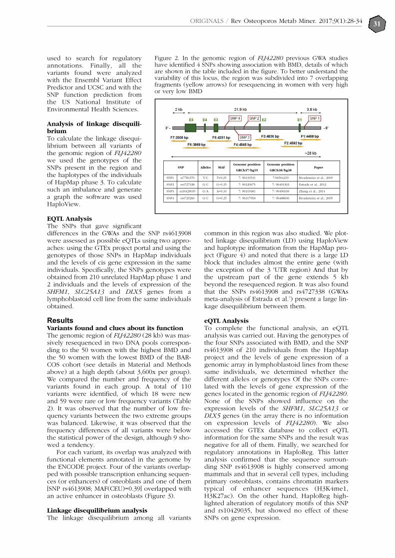
ORIGINALS / Rev Osteoporos Metab Miner. 2017;9(1):28-3431
used to search for regulatoryannotations. Finally, all thevariants found were analyzedwith the Ensembl Variant EffectPredictor and UCSC and with theSNP function prediction fromthe US National Institute ofEnvironmental Health Sciences.
Analysis of linkage disequili-briumTo calculate the linkage disequi-librium between all variants ofthe genomic region of FLJ42280we used the genotypes of theSNPs present in the region andthe haplotypes of the individualsof HapMap phase 3. To calculatesuch an imbalance and generatea graph the software was usedHaploView.
EQTL AnalysisThe SNPs that gave significantdifferences in the GWAs and the SNP rs4613908were assessed as possible eQTLs using two appro-aches: using the GTEx project portal and using thegenotypes of those SNPs in HapMap individualsand the levels of cis gene expression in the sameindividuals. Specifically, the SNPs genotypes wereobtained from 210 unrelated HapMap phase 1 and2 individuals and the levels of expression of theSHFM1, SLC25A13 and DLX5 genes from alymphoblastoid cell line from the same individualsobtained.
Results Variants found and clues about its functionThe genomic region of FLJ42280 (28 kb) was mas-sively resequenced in two DNA pools correspon-ding to the 50 women with the highest BMD andthe 50 women with the lowest BMD of the BAR-COS cohort (see details in Material and Methodsabove) at a high depth (about 3,600x per group).We compared the number and frequency of thevariants found in each group. A total of 110variants were identified, of which 18 were newand 59 were rare or low frequency variants (Table2). It was observed that the number of low fre-quency variants between the two extreme groupswas balanced. Likewise, it was observed that thefrequency differences of all variants were belowthe statistical power of the design, although 9 sho-wed a tendency.
For each variant, its overlap was analyzed withfunctional elements annotated in the genome bythe ENCODE project. Four of the variants overlap-ped with possible transcription enhancing sequen-ces (or enhancers) of osteoblasts and one of them[SNP rs4613908; MAF(CEU)=0.39] overlapped withan active enhancer in osteoblasts (Figure 3).
Linkage disequilibrium analysisThe linkage disequilibrium among all variants
common in this region was also studied. We plot-ted linkage disequilibrium (LD) using HaploViewand haplotype information from the HapMap pro-ject (Figure 4) and noted that there is a large LDblock that includes almost the entire gene (withthe exception of the 3 'UTR region) And that bythe upstream part of the gene extends 5 kbbeyond the resequenced region. It was also foundthat the SNPs rs4613908 and rs4727338 (GWAsmeta-analysis of Estrada et al.7) present a large lin-kage disequilibrium between them.
eQTL AnalysisTo complete the functional analysis, an eQTLanalysis was carried out. Having the genotypes ofthe four SNPs associated with BMD, and the SNPrs4613908 of 210 individuals from the HapMapproject and the levels of gene expression of agenomic array in lymphoblastoid lines from thesesame individuals, we determined whether thedifferent alleles or genotypes Of the SNPs corre-lated with the levels of gene expression of thegenes located in the genomic region of FLJ42280.None of the SNPs showed influence on theexpression levels of the SHFM1, SLC25A13 orDLX5 genes (in the array there is no informationon expression levels of FLJ42280). We alsoaccessed the GTEx database to collect eQTLinformation for the same SNPs and the result wasnegative for all of them. Finally, we searched forregulatory annotations in HaploReg. This latteranalysis confirmed that the sequence surroun-ding SNP rs4613908 is highly conserved amongmammals and that in several cell types, includingprimary osteoblasts, contains chromatin markerstypical of enhancer sequences (H3K4me1,H3K27ac). On the other hand, HaploReg high-lighted alteration of regulatory motifs of this SNPand rs10429035, but showed no effect of theseSNPs on gene expression.
Figure 2. In the genomic region of FLJ42280 previous GWA studieshave identified 4 SNPs showing association with BMD, details of whichare shown in the table included in the figure. To better understand thevariability of this locus, the region was subdivided into 7 overlappingfragments (yellow arrows) for resequencing in women with very highor very low BMD
SNP Alelos MAFPosición genoma
GRCh37/hg19
Posición genoma
GRCh38/hg38Paper
SNP1 rs7781370 T/C T=0.25 7: 96133531 7:96504219 Rivadeneira et al., 2009
SNP2 rs4727338 G/C G=0.25 7: 96120675 7: 96491363 Estrada et al., 2012
SNP3 rs10429035 G/A A=0.33 7: 96119481 7: 96490169 Zhang et al., 2014
SNP4 rs4729260 G/C G=0.25 7: 96117918 7: 96488606 Rivadeneira et al., 2009
Genome position Genome positionAlleles
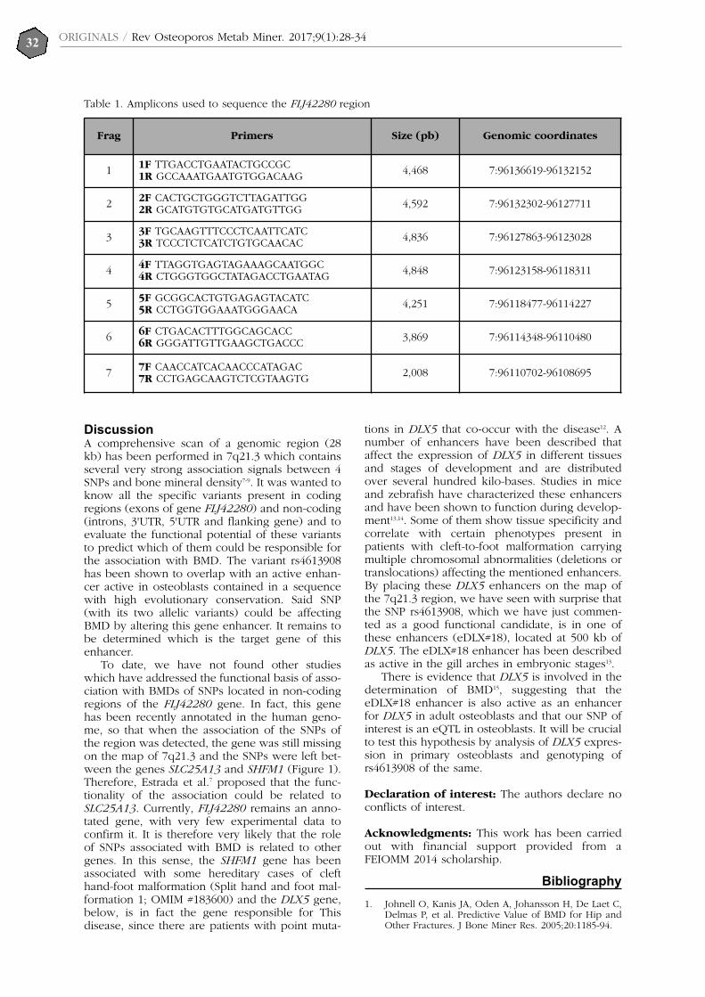
ORIGINALS / Rev Osteoporos Metab Miner. 2017;9(1):28-3432
DiscussionA comprehensive scan of a genomic region (28kb) has been performed in 7q21.3 which containsseveral very strong association signals between 4SNPs and bone mineral density7-9. It was wanted toknow all the specific variants present in codingregions (exons of gene FLJ42280) and non-coding(introns, 3'UTR, 5'UTR and flanking gene) and toevaluate the functional potential of these variantsto predict which of them could be responsible forthe association with BMD. The variant rs4613908has been shown to overlap with an active enhan-cer active in osteoblasts contained in a sequencewith high evolutionary conservation. Said SNP(with its two allelic variants) could be affectingBMD by altering this gene enhancer. It remains tobe determined which is the target gene of thisenhancer.
To date, we have not found other studieswhich have addressed the functional basis of asso-ciation with BMDs of SNPs located in non-codingregions of the FLJ42280 gene. In fact, this genehas been recently annotated in the human geno-me, so that when the association of the SNPs ofthe region was detected, the gene was still missingon the map of 7q21.3 and the SNPs were left bet-ween the genes SLC25A13 and SHFM1 (Figure 1).Therefore, Estrada et al.7 proposed that the func-tionality of the association could be related toSLC25A13. Currently, FLJ42280 remains an anno-tated gene, with very few experimental data toconfirm it. It is therefore very likely that the roleof SNPs associated with BMD is related to othergenes. In this sense, the SHFM1 gene has beenassociated with some hereditary cases of clefthand-foot malformation (Split hand and foot mal-formation 1; OMIM #183600) and the DLX5 gene,below, is in fact the gene responsible for Thisdisease, since there are patients with point muta-
tions in DLX5 that co-occur with the disease12. Anumber of enhancers have been described thataffect the expression of DLX5 in different tissuesand stages of development and are distributedover several hundred kilo-bases. Studies in miceand zebrafish have characterized these enhancersand have been shown to function during develop-ment13,14. Some of them show tissue specificity andcorrelate with certain phenotypes present inpatients with cleft-to-foot malformation carryingmultiple chromosomal abnormalities (deletions ortranslocations) affecting the mentioned enhancers.By placing these DLX5 enhancers on the map ofthe 7q21.3 region, we have seen with surprise thatthe SNP rs4613908, which we have just commen-ted as a good functional candidate, is in one ofthese enhancers (eDLX#18), located at 500 kb ofDLX5. The eDLX#18 enhancer has been describedas active in the gill arches in embryonic stages13.
There is evidence that DLX5 is involved in thedetermination of BMD15, suggesting that theeDLX#18 enhancer is also active as an enhancerfor DLX5 in adult osteoblasts and that our SNP ofinterest is an eQTL in osteoblasts. It will be crucialto test this hypothesis by analysis of DLX5 expres-sion in primary osteoblasts and genotyping ofrs4613908 of the same.
Declaration of interest: The authors declare noconflicts of interest.
Acknowledgments: This work has been carriedout with financial support provided from aFEIOMM 2014 scholarship.
Bibliography
1. Johnell O, Kanis JA, Oden A, Johansson H, De Laet C,Delmas P, et al. Predictive Value of BMD for Hip andOther Fractures. J Bone Miner Res. 2005;20:1185-94.
Table 1. Amplicons used to sequence the FLJ42280 region
Frag Primers Size (pb) Genomic coordinates
1 1F TTGACCTGAATACTGCCGC1R GCCAAATGAATGTGGACAAG 4,468 7:96136619-96132152
2 2F CACTGCTGGGTCTTAGATTGG2R GCATGTGTGCATGATGTTGG 4,592 7:96132302-96127711
3 3F TGCAAGTTTCCCTCAATTCATC3R TCCCTCTCATCTGTGCAACAC 4,836 7:96127863-96123028
4 4F TTAGGTGAGTAGAAAGCAATGGC4R CTGGGTGGCTATAGACCTGAATAG 4,848 7:96123158-96118311
5 5F GCGGCACTGTGAGAGTACATC5R CCTGGTGGAAATGGGAACA 4,251 7:96118477-96114227
6 6F CTGACACTTTGGCAGCACC6R GGGATTGTTGAAGCTGACCC 3,869 7:96114348-96110480
7 7F CAACCATCACAACCCATAGAC7R CCTGAGCAAGTCTCGTAAGTG 2,008 7:96110702-96108695

33ORIGINALS / Rev Osteoporos Metab Miner. 2017;9(1):28-34
bone-mineral-density loci identified by large-scalemeta-analysis of genome-wide association studies. NatGenet. 2009;41:1199-206.
9. Zhang LL, Choi HJ, Estrada K, Leo PJ, Li J, Pei YF, etal. Multistage genome-wide association meta-analysesidentified two new loci for bone mineral density. HumMol Genet. 2014;23:1923-33.
10. Bustamante M, Nogués X, Enjuanes A, Elosua R, García-Giralt N, Pérez-Edo L, et al. COL1A1, ESR1, VDR andTGFB1 polymorphisms and haplotypes in relation toBMD in Spanish postmenopausal women. OsteoporosInt. 2007;18:235-43.
11. DePristo MA, Banks E, Poplin R, Garimella KV,Maguire JR, Hartl C, et al. A framework for variationdiscovery and genotyping using next-generation DNAsequencing data. Nat Genet. 2011;43:491-8.
12. Shamseldin HE, Faden MA, Alashram W, Alkuraya FS.Identification of a novel DLX5 mutation in a family withautosomal recessive split hand and foot malformation. JMed Genet. 2012;49:16-20.
13. Birnbaum RY, Everman DB, Murphy KK, Gurrieri F,Schwartz CE, Ahituv N. Functional characterization oftissue-specific enhancers in the DLX5/6 locus. HumMol Genet. 2012;21:4930-8.
14. Rasmussen MB, Kreiborg S, Jensen P, Bak M, Mang Y,Lodahl M, et al. Phenotypic subregions within thesplit-hand/foot malformation 1 locus. Hum Genet.2016;135:345-57.
15. Ulsamer A, Ortuño MJ, Ruiz S, Susperregui AR, OssesN, Rosa JL, et al. BMP-2 Induces Osterix Expressionthrough Up-regulation of Dlx5 and Its Phosphorylationby p38. J Biol Chem. 2008;283:3816-26.
Table 2. Number and location of single nucleotide variants found in this study
Raw Filtered Coding Regulatoryregion*
Putativeosteoblastenhancer
Activeenhancer inosteoblast
Commonvariants 96 51 0 12 3 1
LFV 24.243 59 1 16 1 0
Total 24.339 110 1 28 4 1
*includes flanking regions, 5'UTR, 3'UTR and introns; LFV: low frequency variant (MAF <5%).
Figure 3. A) Schematic of the genomic region of FLJ42280 and location ofsome promising variants. Common variants are indicated in red and lowfrequency variants (LFV) or rare in gray. Blue squares represent enhancersof osteoblasts; The darker represents an active enhancer of the osteoblasts.B) Detail of active enhancer in osteoblasts. The sequence conservationprofile in vertebrates, transcription factor binding sites and histone modifi-cations are shown. These data were extracted from UCSC GenomeBrowser -GRCh37- and from ENCODE (data referred to osteoblasts)
2. Kanis JA, Oden A, Johnell O,Johansson H, De Laet C, Brown J, etal. The use of clinical risk factorsenhances the performance of BMDin the prediction of hip and osteopo-rotic fractures in men and women.Osteoporos Int. 2007; 18:1033-46.
3. Peacock M, Turner CH, Econs MJ,Foroud T. Genetics of Osteoporosis.Endocr Rev. 2002;23:303-26.
4. Deng HW, Chen WM, Recker S,Stegman MR, Li JL, Davies KM, et al.Genetic determination of Colles'fracture and differential bone massin women with and without Colles'fracture. J Bone Miner Res.2000;15:1243-52.
5. Hardy J, Singleton A. GenomewideAssociation Studies and HumanDisease. N Engl J Med. 2009;360:1759-68.
6. Manolio TA. Genomewide AssociationStudies and Assessment of theRisk of Disease. N Engl J Med.2010;363:166-76.
7. Estrada K, Styrkarsdottir U,Evangelou E, Hsu YH, Duncan EL,Ntzani E, et al. Genome-widemeta-analysis identifies 56 bonemineral density loci and reveals14 loci associated with risk of frac-ture. Nat Genet. 2012;44,491-501.
8. Rivadeneira F, Styrkársdottir U,Estrada K, Halldórsson BV, HsuYH, Richards JB, et al. Twenty

34ORIGINALS / Rev Osteoporos Metab Miner. 2017;9(1):28-34
Figure 4. Graph of linkage disequilibrium present in the genomicregion of FLJ42280. A large block (24 kb) is seen that includes muchof the gene except its 3'UTR region. The 4 SNPs that have been asso-ciated with BMD in the various GWAs are marked with green ovals.The SNP overlapping with the active enhancer in osteoblasts is markedwith a purple oval. The blue horizontal line indicates the resequencedgenomic region in this study
35CLINICAL NOTE / Rev Osteoporos Metab Miner. 2017;9(1):35-37
Torregrosa Suau O1, Guilló Quiles E2, Mora Rufete A3
1 Unidad de Metabolismo Óseo. Servicio de Medicina Interna 2 Medicina Familiar y Comunitaria. Departamento de Salud Elche 3 Servicio de Medicina InternaHospital General Universitario de Elche. Elche. Alicante (España)
Mesenteric panniculitis associated withthe use of bisphosphonates: are thesemore proinflammatory than we know?
Correspondence: Óscar Torregrosa Suau - Hospital General Universitario de Elche - Camino de la Almazara, s/n -03203 Elche - Alicante (Spain) e-mail: [email protected]
Date of receipt: 07/11/2016Date of acceptance: 13/12/2016
SummaryMesenteric panniculitis is characterized by chronic inflammation of the adipose tissue of the intestinalmesentery, and its etiology is unknown. It has been associated with malignancy, vasculitis, rheumaticdiseases and the use of certain drugs. We present a case of panniculitis associated with bisphosphonateuse, not previously described in the literature, thus suggesting its potential secondary proinflammatoryeffects.
Key words: mesenteric panniculitis, bisphosphonates, proinflammatory.
DOI: http://dx.doi.org/10.4321/S1889-836X2017000100006

36CLINICAL NOTE / Rev Osteoporos Metab Miner. 2017;9(1):35-37
IntroductionMesenteric panniculitis is a rare disorder characteri-zed by chronic nonspecific inflammation of adipo-se tissue of the intestinal mesentery of unknownorigin1. It may develop independently or in associa-tion with other alterations, which has been the sub-ject of much discussion. It has been linked tovarious conditions, such as vasculitis, granuloma-tous diseases, rheumatic diseases, malignant disea-ses, pancreatitis2, smoking and the use of certaindrugs (beta blockers, methyldopa, sulfonamides,salicylates and oral contraceptives).
This entity has been found to present severalstages of development. It is classified into differenttypes depending on the radiological characteris-tics: type I (42%), diffuse thickening of the mesen-tery from root to the edges of the small intestine;Type II (32%), isolated nodular mass in mesente-ric root; Type III (20%), the mesentery containsmultiple nodules which vary in size3. Diagnosticanatomopathological criteria include: presence oflarge quantities of foamy macrophages in theinitial phase of mesenteric lipodystrophy;Infiltrated plasma cells, giant foreign body cells,and foamy macrophages in mesenteric panniculi-tis. In the final stage, the deposition of collagenand the fibrosis in the tissues are noteworthy4.
Although different factors have been recogni-zed that may trigger the disease, its origin in cer-tain cases remains uncertain. Once diagnosed,there is no specific treatment. Corticosteroids arerecommended, although cases of spontaneousremission have been reported. Surgical resection isconsidered for cases in which there is intestinalobstruction due to inflammation and secondaryscarring. Antibiotics, radiation therapy and cyclo-phosphamide have also been reported as possibleresponses.
Clinical Case ReportWe present the case of a 67-year-old woman,without toxic habits, with no medical history ofinterest except cervico-artrosis and spondylolisthe-sis in rehabilitation and postmenopausal osteopo-rosis for 3 years following bisphosphonate treat-ment (BF). Mesenteric panniculitis was not justi-fied by other causes.
Our patient, after 3 years of treatment withrisedronate, began to present diffuse abdominalpain on a global basis, not related to meals andwith episodes of frequent exacerbations over aperiod of about 2-3 months.
In one of the exacerbations, the patient wasadmitted and an abdominal computed tomo-graphy (CT) carried out, among other tests. Inthe CT, mesenteric panniculitis was revealed(Figure 1).
After reasonably ruling out other clinical etio-logies of mesenteric panniculitis (Table 1)5, theBFs that were taken and reevaluated at 6 monthswith a new abdominal control CT was taken, sho-wing a complete resolution of the abdominalinflammatory lesions which presented in the pre-vious CT scan (Figure 2).
DiscussionThere are no similar cases described in the litera-ture, nor have we found clinical trials of bisphos-phonates, FIT, VERT, BONE, HORIZON6-9, in casesof mesenteric panniculitis, although its use hasbeen associated with other inflammatory proces-ses. Cases of ocular side effects have been repor-ted in this group of mostly inflammatory drugssuch as uveitis, scleritis and conjunctivitis10.Conjunctivitis is the most frequent adverse ocularcondition, although its actual incidence is verylow. In general, it responds quickly to topical tre-atment, even if we maintain the drug, although itis more prudent to suspend it, at least temporarily.Another complication, less frequent but potentiallymore serious, is uveitis. Its incidence is very low,between 2 and 5 cases per 10,000 patients treated.In general, its location is anterior, although someisolated cases of posterior uveitis have been des-cribed. Its occurrence is quite variable, with amedian number of days after the start of the 70-day drug (between 1 and 146) and its incidence ishigher in patients treated with intravenous BF.
An increase in the susceptibility to uveitis inpatients with associated diseases such as spondy-loarthropathies, Behçet's syndrome, Wegener'sgranulomatosis or sarcoidosis and/or treatmentwith certain drugs has also been observed inwhich BP will act as a precipitating factor11.
Bisphosphonates are potent inhibitors of osteo-clasts by exerting a strong interaction with thesecells, resulting in a marked decrease in boneresorption12.
A number of adverse effects have been repor-ted with bisphosphonate administration, whichappear to correspond to "class effects." Theseeffects may vary from bisphosphonates to othersdepending, among other factors, on the route ofadministration. Upper gastrointestinal tract such asesophagitis, gastric ulcers and even hemorrhages,musculoskeletal pain, influenza-like manifesta-tions, atrial fibrillation, renal failure, hypocalce-mia, maxillary osteonecrosis, atypical femoral frac-tures and ocular adverse effects such as conjuncti-vitis, uveitis, iritis, Episcleritis, scleritis or keratitis13.There has been no association with alterations ofmesenteric panniculitis type adipose tissue in theliterature.
ConclusionsMesenteric panniculitis continues to be a nosolo-gical entity which is, in many cases, incidental andcontroversial. The retrospective analysis of manyseries has identified possible risk factors and amultitude of etiologic agents. Further studies andmore described cases like ours are required toassociate the use of BF more strongly with areasof inflammation of mesenteric adipose tissue.Although they have demonstrated their "proin-flammatory" effect in other organs, especially atthe ocular level, as we have developed previously.
Declaration of interest: The authors declare noconflicts of interest.

37CLINICAL NOTE / Rev Osteoporos Metab Miner. 2017;9(1):35-37
Figure 1. Abdominal CT scan showing area of panni-culitis at the root of the mesentery
Figure 2. Abdominal CT scan 6 months later. Thereare no areas of mesenteric panniculitis
Table 1. Differential diagnosis of mesenteric panniculitis
Carcinoides tumors Lymphomas
Reaction to adjacent cancer or chronic abscess Lymphomas
Amyloidosis Liposarcoma
Desmoid tumors Peritoneal carcinomatosis
Infectious diseases (tuberculosis and histoplasmosis) Peritoneal fibrosis
Peritoneal mesothelioma Histoplasmosis
Lymphosarcomas Retroperitoneal sarcoma
Chronic foreign body inflammation Lymphangioma
Whipple's disease
Bibliography
1. Ruiz Tovar J, Alonso Hernández N, Sanjuanbenito A,Martinez E. Paniculitis mesentérica: presentación de 10casos. Rev Esp Enferm Dig. 2007;99:240-7.
2. Daskalogiannaki M, Voloudaki A, Prassopoulos P,Magkanas M, Stefanaki K, Apostolaki E, et al. Evaluationof mesenteric panniculitis: prevalence and associateddiseases. Am J Roentgenol. 2000;174:427-31.
3. Colomer E, Blanes A, Carbonell C, Biot A, Villar H,Tomas A. Paniculitis Mesentérica con afectación retro-peritoneal resuelta tras tratamiento con pulsos deciclofosfamida endovenosa. An Med Intern. 2003;20:31-3.
4. Delgado Plasencia L, Rodríguez Ballester L, López-Tomassetti Fernández EM, Hernández Morales A,Carillo Pallares A, Hernández Siverio N. Mesentericpanniculitis: experience in our center. Rev Esp EnfermDig. 2007;99:291-7.
5. Viscido G, Napolitano D, Rivoira G, Parodi M, Barotto M,Picón H, et al. Paniculitis mesentérica asociada conabsceso intraabdominal. Rev Chilena de Cirugía.2008(3):452-6.
6. Harris S, Watts N, Genant H. Effects of risedronate tre-atment on vertebral and nonvertebral fractures inwomenwith postmenopausal osteoporosis: a randomi-zed controlled trial. JAMA. 1999;282:1344-52.
7. Black D, Thompson D, Bauer D, Ensrud K, MuslinerMC, Hochberg MC, et al. Fracture risk reduction withalendronate in women with osteoporosis: the FractureIntervention Trial. FIT Research Group. J ClinEndocrinol Metab. 2000;85:4118-24.
8. Chesnut III CH, Skag A, Christiansen C. Recker R,Stakkestad JA, Hioseth A, et al. Effects of oral ibandro-nate administered daily or intermittently on fracturerisk in postmenopausal osteoporosis. J Bone MinerRes. 2004;19:1241-9.
9. Black DM, Delmas PD, Eastell R, Reid IR, Boonen S,Cauley JA, et al. Once-yearly zoledronic acid for treat-ment of postmenopausal osteoporosis. N Engl J Med.2007;356:1809-22.
10. Cano Parra J, Díaz-Llopis M. Uveitis Inducida por fár-macos. Arch Soc Esp Oftalmol. 2005;80:1-12.
11. Arboleya L, Alperi M, Alonso A. Efectos adversos debifosfonatos. Reumatol Clin. 2011;07:189-97.
12. Jaimes M, Chaves Netto HD, Olate S, Chaves MMGA,Barbosa AJR. Bifosfonatos y osteonecrosis de los maxi-lares. Consideraciones sobre su tratamiento. Int JMorphol. 2008;26(3):681-8.
13. González-Macías J, del Pino-Montes J, Olmos JM,Nogués X. Guías de práctica clínica en la osteoporosisposmenopáusica, glucocorticoidea y del varón.Sociedad Española de Investigación Ósea y delMetabolismo Mineral. (3ª versión actualizada 2014).Rev Clin Esp. 2015;215:515-26.

REVIEW / Rev Osteoporos Metab Miner. 2017;9(1):38-4938
Maldonado G1, Messina O2, Moreno M3, Ríos C4
1 Universidad de Especialidades Espíritu Santo - Samborondón (Ecuador) 2 Hospital Cosme Argerich - Buenos Aires (Argentina) 3 Hospital Luis Vernaza. Guayaquil (Ecuador) 4 Centro de Reumatología y Rehabilitación - Guayaquil (Ecuador)
Osteoporosis in rheumatic diseases andglucocorticoid induced
Correspondence: Génessis Maldonado Vélez - Universidad de Especialidades Espíritu Santo km 2.5 - Vía la Puntilla -Samborondón (Ecuador)e-mail: [email protected]
SummaryOsteoporosis is a systemic skeletal disease characterized by low bone mineral density, changes in bonemicroarchitecture and increased risk of fracture. It has been shown that depends on physiological pro-cesses and secondary to other pathologies and associated with the use of glucocorticoids, the latest beingthe most common cause of osteoporosis associated to drugs, this may be represent a great magnitudepublic health issue. This review is presented in order to emphasize the clinical importance of osteoporo-sis in rheumatic diseases and glucocorticoid-induced osteoporosis.
Key words: osteoporosis, rheumatic diseases, glucocorticoids.
DOI: http://dx.doi.org/10.4321/S1889-836X2017000100007

REVIEW / Rev Osteoporos Metab Miner. 2017;9(1):38-4939
Figure 1. Regulation and formation of osteoclasts
Inflammation Estrogens
TH17 Cel. T activated
Cel. T
Cel. of stroma
Spendingthymic
Release of TGFβof the bone matrix
Formation and activity of osteoclasts
Feedback of the osteoclasts of the cel. T
IntroductionOsteoporosis is a systemic skeletal disease characteri-zed by decreased bone mineral density with altera-tions in bone microarchitecture and increased risk offracture, involving genetic and environmental factors.It is classified as primary when it depends on physio-logical processes, as in the case of menopause andnormal aging, and secondary when conditioned byother pathologies or in relation to medications, as inosteoporosis secondary to rheumatic diseases andglucocorticoid-induced osteoporosis (GIO).
The current trend of widespread corticoid use hasled to GIO being the most common cause of drug-induced osteoporosis, constituting a worldwide pro-blem. Approximately 0.5% of the general populationand 1.7% of women over 55 years of age receive oralglucocorticoids and, despite the existence of diagnos-tic methods and adequate measures to prevent GIO,less than 14% receive some type of treatment to avoidloss of bone mass due to glucocorticoids1-3.
In patients with rheumatic diseases such as rheu-matoid arthritis (RA), systemic lupus erythematosus(SLE), ankylosing spondylitis (AS), polymyalgiarheumatica (PMR), and vasculitis, among others,osteoporosis is an associated comorbidity. This maybe due to the use of glucocorticoids over longperiods of time as part of the accepted therapy ofthese diseases, or it may also be associated with theinflammatory activity of the condition and its impacton the bone. In any case, these patients are morelikely to suffer fractures due to fragility, which leadsto a decrease in the quality of life3,4.
The clinical relevance of osteoporosis in rheu-matic diseases is underestimated. Studies haveshown that patients with RA receiving oral gluco-corticoids who underwent a control bone densito-metry had osteopenia/osteoporosis in 23% of thecases, of which 42% were prescribed at least onemedication to reduce bone loss3.
This indicates an insufficient appreciation ofthe clinical problem and a lack of consensus onosteoporosis detection and treatment in rheumaticdiseases.
Effects of inflammation on bone turnoverThe determinants of inflammation on the bone areproinflammatory cytokines. Thus, the alpha (TNF-α) tumor necrosis factor, interleukins (IL) 1 and 6(IL-1 and IL-6) favor bone resorption by directly orindirectly promoting osteoclastogenesis. IL-17enhances activator receptor ligand expression fornuclear factor kappa B (RANK-L), which in turn is akey part of the bone resorption process (Figure 1).
TNF acts on proinflammatory signaling pathwayswithin the joints, influencing bone turnover, at thesame time as it seems to be involved in developingbone erosions and the progression of osteoporosis.In addition, TNF has been shown to promote pre-osteoclastic cell formation expressing the activatorreceptor for nuclear factor kappa B (RANK), pro-moting overexpression of RANK-L osteo-protege-rin (OPG) and inhibiting the maturation and func-tion of the osteoblasts, because it increases theexpression of the synovial fibroblasts5.

REVIEW / Rev Osteoporos Metab Miner. 2017;9(1):38-4940
The relationship between proinflammatory cyto-kines and resorption is well established. However,there is a lack of evidence on the relationship bet-ween cytokines and bone formation. IL-4, IL-12, IL-8 and interferon are known to promote bone for-mation due to increased OPG/RANKL ratio, thusinhibiting osteoclastogenesis6.
On the other hand, the osteoblast produces cyto-kines and in its immature state promotes the forma-tion of RANK-L and in its mature state the produc-tion of OPG, showing that the osteoblast maturationprocess is essential to balance the relationship bet-ween the inflammatory process and bone mass6.
Rifas and Weitzmann discovered a cytokine andcalled it a secreted osteoclastogenic factor of activa-ted T cells (SOFAT)and demonstrated that it inducesthe osteoblastic production of IL-6 and the forma-tion of osteoclasts in the absence of osteoblasts orRANKL, and that it is insensitive to OPG effects. Thisdemonstrates that SOFAT is a potent inducer of IL-6production, playing a key role in the local inflam-matory response, and may indirectly exacerbatebone destruction in rheumatoid arthritis throughmultiple IL-6 mediated processes7.
Acute and chronic inflammatory processes inrheumatic diseases cause bone damage becauseinflammation increases the number and activity ofosteoclasts and, at the same time, the activation ofT cells. During infectious processes increasesRANKL production, which, in turn, stimulates oste-oclastogenesis. It has been demonstrated that theapplication of OPG can reverse this effect in dise-ases such as RA and periodontitis8.
Osteoporosis in specific rheumatic diseasesIt is known that subjects with a history of chronicautoimmune diseases have a higher risk of deve-loping osteoporosis due to different causes, suchas specific alterations that affect bone metabolism,induce loss and sometimes inhibit formation, or tomeet factors such as: sex (predominance inwomen), low physical activity, underlying disease,smoking, low body mass index (BMI), vitamin Ddeficiency, etc. (Table 1).
The Multidisciplinary Forum on Managementof Patients with High Risk of OsteoporoticFracture (HRF) has suggested classifying risk fac-tors as follows9:
- Keys: age over 70 years, history of previousfrail fracture (vertebral or hip), glucocorticoid inta-ke (7.5 mg/day for 3 months or more) and BMD(T-score ≤3).
- Important: maternal history of hip fracture,BMI ≤20, frequent falls in the elderly, low measu-rements in activity and physical function.
- Moderate: levels of 25(OH)-vitamin D ≤30ng/dl, some lifestyle-related harmful factors (smo-king, excessive alcohol intake, sedentary lifestyleor excessive coffee consumption).
Rheumatoid arthritis (RA)RA is an autoimmune inflammatory disease of unk-nown origin that is more common in young women,between the third and fifth decades of life [4]. It is a
deforming and disabling disease that directly affectsthe joint causing bone erosions, which end up inju-ring the joint and resulting in its deformity.
Osteoporosis is a common complication of RAand is classified as follows10:
- Yuxta-articular: early and secondary to localfactors.
- Generalized: late and multi-causal in origin(rest, drugs, disease activity, degree of functionalalteration and deterioration of muscle mass).
The frequency of osteoporosis in RA is varia-ble. Sinigaglia et al reported an incidence of oste-oporosis of 10-56% in patients with RA3. Forsbladadded that this range will depend on the popula-tion studied, and stressed the importance of jointdamage in RA and generalized osteoporosis11.
Patients with RA have a higher risk of fracturecompared to the general population. A retrospec-tive study of 30,262 patients showed a higher riskof hip fracture (RR=2.0) and spine (RR=2.4) vs thecontrol group, and the risk increased due to pro-longed use of corticosteroids (RR=3.4)12.
The high risk of fracture in RA patients has alsobeen associated with disease-specific factors13 (Table 2).
Corticosteroid use is an independent predictor ofthe decrease in bone mass at the lumbar and femo-ral levels. A low dose steroid regimen (5 mg pred-nisone) is associated with a 50% increase in the riskof osteoporosis14. This demonstrates that treatmentwith oral corticosteroids of more than 5 mg of pred-nisone is able to reduce bone mass with a rapidincrease in the risk of fracture during the treatmentperiod15,16. Taking into account this risk factor, anAmerican College of Rheumatology (ACR) commit-tee recommends screening for osteoporosis usingBMD and initiating treatment in patients with T-score ≤1.0, preferably with bisphosphonates andcalcium supplements and Vitamin D. These patientsshould be monitored annually and treatment shouldcontinue while receiving glucocorticoids17.
Longitudinal and cross-sectional studies haveshown that disease modifying anti-rheumatic drugs(DMARDs), such as methotrexate, do not haveadverse effects on bone mass18. Anti-TNF therapy(infliximab) has shown positive effects on bonemass19. It is believed that the bone protective effectis not only due to the decreased disease activity, butalso to the role played by the cytokine TNF in oste-oclastogenesis20,21. Regarding tocilizumab (inhibitorof IL-6), its role is fundamental, since IL-6 intervenesin the inflammatory and osteoclastogenic process13.
The CAMERA II study analyzed patients withearly RA who were on methotrexate and 10 mgprednisone and were administered 500 mg of cal-cium, 400 IU of vitamin D and alendronate/risedro-nate over two years. BMD showed a 2.6% increasein lumbar spine bone mass during the first year ofprophylactic treatment regardless of glucocorticoiduse, demonstrating that the addition of 10 mg ofprednisone daily in a methotrexate-based treatmentdoes not lead to bone loss in patients with early RA22.
The quality of life of these patients is obviouslyaffected. A study by Riggs and Melton showed that ahip fracture has a mortality rate of 10-20% in the sub-

REVIEW / Rev Osteoporos Metab Miner. 2017;9(1):38-4941
sequent 6 months, 50% ofthese patients will not be ableto walk without externalobjects such as walking sticks.Also 25% will need homehelp for long periods23.
Systemic Lupus Erythematosus(SLE)SLE is an autoimmune disea-se of unknown origin thatoccurs more frequently inyoung women4. Osteoporosisis also an associated disease,and probably caused by treat-ments that have a negativeeffect on the bone or by ova-rian dysfunction induced bysome immunosuppressants24.
The loss of bone mass inSLE could be the result ofseveral mechanisms, thosethat depend on the diseaseitself and those that are tre-atment related (Table 3).
The mechanism responsi-ble for the loss of bone massin patients with SLE receivingcorticosteroids is the markedincrease in bone resorptionand the maturation deficit ofthe osteoblast, as well asbone mineralization. BMDbegins to fall from the thirdmonth of glucocorticoid useand progresses rapidly to sixmonths. From this period, thecurve gradually declines25.
A study by Houssiau et alin premenopausal womenwith a definitive diagnosis ofSLE, showed that patientswho had not received corti-costeroids had lower BMDcompared to healthy controls,indicating that the diseasealone is a bone loss factor26.
A systematic review byWang et al showed that SLEpatients have lower levels ofBMD than the general popula-tion, and that not only occursin a specific place, but there isloss of bone mass in all the places studied (femoralneck, lumbar spine and hip). In addition, an increa-sed risk of fracture was demonstrated (RR=1.97)27.
Jacobs et al, in a prospective 6-year follow-upstudy in patients with SLE, found an associationbetween loss of BMD in the lumbar spine and theuse of corticosteroids at high doses. In addition, aloss of bone mass was demonstrated with the useof immune-suppressants and base antimalarials28.
Regarding the association between lower serumvitamin D levels in these patients and the loss of
bone mass, the deficiency is believed to be due toreduced sun exposure by the photosensitivity ofthese patients, in addition to the use of corticoste-roids and renal insufficiency29. The mechanism bywhich these patients have a lower calcium absorp-tion results in a decrease in the conversion of25(OH)-vitamin D to 1,25(OH)2-vitamin D (calci-triol). This association has been demonstrated incross-sectional studies29-31, hence the importance ofdetermining levels of 25(OH)-vitamin D in patientswith SLE.
Table 1. Profile of high risk of fracture (HRF). Group of Rheumatology
Factors associated with HRF
✓ Age >70 years✓ Fracture due to previous fragility (symptomatic or asymptomatic)✓ Low BMD (T-score <3) ✓ Maternal history of hip fracture✓ Administered corticoids ≤7,5 mg/day over more than 3 months✓ Low weight (BMI <19 kg/m2)
Special and common situations in rheumatologic patients
✓ Chronic inflammatory disease with persistent activity✓ Rheumatic polymyalgia and/or giant cell arteritis✓ Transplants (distinguishing between pre-transplant and post-transplant)✓ Frequent use of treatments that induce osteoporosis
HRF in patients with rheumatologic diseases
Osteoporosis induced by glucocorticoids✓ Daily dose of corticosteroids greater than 15 mg✓ Treatment time greater than one yearRheumatoid arthritis:✓ Postmenopausal women (especially those over 65 years of age)✓ DMO similar to the risk of postmenopausal osteoporosis✓ Treated with corticosteroids at doses higher than 15 mg/day✓ High disability rate✓ Extended disease✓ Low physical activityAnkylosing spondylitis:✓ Patients with a disease of more than 10 years of evolution✓ Male >30 years old, treated with corticosteroids✓ Accused loss of BMD in the first 5 years, with an extended disease✓ Back pain episodes in the last 6 months✓ Associated inflammatory bowel diseaseSystemic lupus erythematosus:✓ Postmenopausal woman, with a long-term disease✓ Start after age 30✓ Low sun exposure✓ Uses sunscreens✓ Low hip BMDSystemic sclerosis:✓ Age >50 years ✓ Female✓ Early menopause✓ Body mass index <25✓ Use of systemic corticosteroidsRheumatic polymyalgia/giant cell arteritis:✓ Age >60 years ✓ Functional limitation (low physical activity)✓ Use of systemic corticosteroids (high cumulative dose)✓ Decrease in strength✓ Reduced BMD

REVIEW / Rev Osteoporos Metab Miner. 2017;9(1):38-4942
Patients with SLE and neurological involve-ment (epilepsy, stroke, etc.) comprise the group athighest risk of fractures as they are more prone tofalls and to the adverse effects of antiepileptics32.
Ankylosing Spondylitis (AS)AS is part of the group of seronegative spondylo-arthropathies (SpA), a group of inflammatory jointdisorders that also includes psoriatic arthritis,inflammatory bowel disease and reactive arthritis.AS is the typical form of SpA with symptoms rela-ted to arthritis, enthesitis, sacroiliitis, amongothers. The location of the primary disease isbelieved to be enthesis, i.e. the area where thetendons and ligaments are inserted into the bone4.
Axial involvement generates a negative boneimpact and increases the risk of vertebral fracturedue to continuous inflammation, which results ina decrease in bone mass and progressive ankylo-sis, together with bone proliferation33.
Osteoporosis can occur due to physical inacti-vity, reduced mobility of the spine related to pain,stiffness and ankylosis, in addition to subclinicalinvolvement of intestinal disease34.
The prevalence of osteoporosis in AS is appro-ximately 14-27% in the spine and 4-14% in thehip35,36. Using the visual and morphometric defini-tions of vertebral size, the prevalence of fracturesin this group of patients is 10-30%37,38.
Prieto et al demonstrated a strong associationbetween AS and vertebral fractures. These patientshave a 5-fold higher risk of fracture compared toa control group. In addition, they observed that
the first 2.5 years of evolu-tion of the disease are criti-cal because the peak fractu-re risk increases and is belie-ved to be secondary to theinflammatory33. These dataare similar to those reportedby Van der Wijden et al, sug-gesting that fractures in ASare associated with exacer-bations of the disease. Inaddition, they showed thatvertebral fractures weremore frequent in men39.
It is important to empha-size that this group ofpatients has a high risk ofvertebral fractures. However,they do not present an incre-ased risk of non-vertebralfractures.
Psoriatic Arthritis (PA)PAs are also part of serone-gative spondyloarthropa-thies. Studies on bone invol-vement in these patients arescarce, probably because thefrequency of the disease islower than that of rheuma-toid arthritis and systemic
lupus erythematosus. However, a relationship bet-ween inflammatory markers (TNF, IL-6, interferon-gamma or IFN-γ) and loss of bone mass have beendemonstrated40.
Keller et al showed that the group of patientswith severe PAs have lower bone mass loss valuesthan those with mild to moderate PA, showing thatinflammatory factors play a fundamental role inbone resorption40.
Dreiher et al analyzed 7,939 patients with pso-riasis. The prevalence of osteoporosis was signifi-cantly higher compared to the control group andmore frequent in women. However, it was obser-ved that in the male group osteoporosis was theresult of systemic disease (PAs), whereas inwomen it was shown to be due to an estrogendeficiency. In addition, women tended to be diag-nosed before men because, usually, women areroutinely referred for bone evaluation41.
Osteoarthrosis (OA)Osteoarthrosis is a degenerative disease thatusually occurs over 45 years and whose origin ismultifactorial, with joint disease being more fre-quent4.
Arthrosis, together with osteoporosis, representthe two etiologies that affect the bone and jointstructure in the elderly population and in turn arethe primary cause of the deterioration of their qua-lity of life42.
Yoshimura et al carried out a 10-year follow-upstudy in patients with lumbar osteoarthritis andosteoporosis. The cumulative incidence of lumbar
Table 2. Profile of high risk of fracture (ARF). Rheumatoid arthritis
Table 3. Possible mechanisms of loss of bone mass in SLE
Very relevant
✓ Age i>70 years ✓ Low BMD (T-score <3)✓ The previous existence of at least 2 vertebral fractures or hip fracture
Relevant
✓ More than 2-3 falls per year✓ Use of oral corticosteroids at a dose of 7.5 mg/day for at least 3 months✓ BMI <19 kg/m✓ Family history of hip fracture✓ Tobacco consumption >10 cigarettes/day
Dependent on disease Dependent on treatment
✓ Reduced mobility ✓ Long-term corticosteroids✓ Renal insufficiency ✓ Immunosuppressive drugs✓ Endocrine factors Azathioprine✓ Amenorrhea Cyclophosphamide✓ Premature menopause Cyclosporine✓ Low plasma levels of ✓ Chronic anticoagulation
androgens ✓ Lack of sun exposure✓ Hyperprolactinemia✓ Induction of cytokines
bone resorption

REVIEW / Rev Osteoporos Metab Miner. 2017;9(1):38-4943
osteoarthritis in 10 years within the age range of40-79 years was 25.8% in men and 45.2% inwomen. In turn, there was a significant relations-hip between the presence of lumbar osteoporosisand the incidence of lumbar osteoarthritis42.
The inverse relationship of arthrosis and oste-oporosis has been debated for years. Zhang et aldemonstrated the mechanical and microstructuralstructures of the subchondral trabecular bone ofpostmenopausal women with arthrosis and osteo-porosis, through the determination of fractionalbone volume and bone mineral density. Thegroup of patients with osteoarthritis showed a gre-ater fractional bone volume compared to thegroup of patients with osteoporosis. However, therelationship with bone density was not signifi-cant43.
Regarding the production of fractures in thesepatients, the causal factor is believed to be falls. Alongitudinal global study of osteoporosis foundthat approximately 40% of the population studiedhad osteoarthritis, and the direct relationship withfractures was not significant after adjusting forincidental falls. However, the adjusted relative riskfor osteoarthritis as a predictor of falls was 1.24(95% CI, 1.22 - 1.26, p<0.0001). In addition, pos-tmenopausal women with osteoarthritis were 25%more likely to fall than women without osteoarth-ritis33.
Glucocorticoid-induced Osteoporosis (GIO)GIO is the most common cause of secondary oste-oporosis. Prolonged treatment with 2.5-5 mg dailyof prednisolone has been shown to increase therisk of vertebral and hip fracture16,44.
GIO most commonly affects the areas of trabe-cular bone (lumbar spine and proximal femur).Fractures can occur in approximately 30-50% ofpatients receiving corticosteroids on an extendedbasis44.
Table 4 describes some risk factors for GIO.
PathophysiologyGlucocorticoids have a direct and indirect effecton metabolism by blocking the actions of vitaminD and calcium absorption, leading to a decreasein serum calcium and an increase in parathormo-ne (PTH) levels32. However, raised PTH does notfully account for bone loss from glucocorticoidexposure45.
During the initial period of treatment with cor-ticosteroids, bone resorption increases, resultingin a sudden decrease in bone density and increa-sed risk of fracture45.
The specific mechanism by which corticoste-roids induce bone resorption is by activating theRANKL receptor and the macrophage colony sti-mulating factor (MCSF). These two componentsare a fundamental part of osteoclastogenesis,along with decreased OPG receptor46.
As for bone formation, it is affected due to theinhibition of osteoblast precursor cells and anincrease in osteoblastic apoptosis45. In addition,there is inhibition of the function of mature oste-
oblasts and suppression of growth factors (IGF1)in bone cells, together with increased apoptosis ofosteocytes47,48 (Table 5).
EvaluationPatients with osteoporosis should be investigatedfor possible causes of bone loss (Table 6). Duringphysical examination, it is important to measurethe patient's height and compare with previousmeasurements, in order to evaluate possibleasymptomatic vertebral fractures.
TreatmentIt is common to find suboptimal levels of vitaminD and calcium in these patients. Because of this, acorrection treatment of these levels should beimplemented.
In patients who begin treatment with glucocor-ticoids for more than 3 months at doses >5mg/day, it is necessary to establish preventive tre-atment for bone mass loss, and in those patientswho are already being treated, a bone densito-metry should be carried out to assess the reductionof bone mass and the possible risk of fracture17,49.
Due to increased use of corticosteroids and theprevalence of secondary osteoporosis, severalinternational societies have described treatmentguidelines. However, there is no global consensuson this matter50-53. In 2010 the ACR (AmericanCollege of Rheumatology) published guidelinesfor the management of GIO, according to FRAX®
(Figures 2 and 3)54.
Non-pharmacological treatmentThe management and prevention of GIO in non-pharmacological treatment is similar to that of pri-mary osteoporosis and consists in eliminatingmodifiable risk factors55:
- Smoking.- Alcohol consumption (≥3 unit/day).- Sedentarism (energy consumption ≤1,682
kcal/day).- Diet high in sodium (mainly in the presence
of hypercalciuria).- Decreased body mass index.Patients with advanced age require special
help to avoid falls, which are the main risk factorfor fractures.
CalciumCalcium and vitamin D are nutritional elementsconsidered essential in any therapeutic option forosteoporosis.
The use of calcium carbonate at a dose of1,000 mg/day has not been shown to preventbone loss or reduce the risk of fracture in patientsinitiating prolonged corticosteroid therapy, parti-cularly in postmenopausal women. Because ofthis, it is not indicated for primary prevention56;however, secondary prevention has shown thatBMD levels remain constant in the lumbar spine inpostmenopausal women with the use of 500mg/day of calcium carbonate accompanied by0.25 µg/day of calcitriol55.
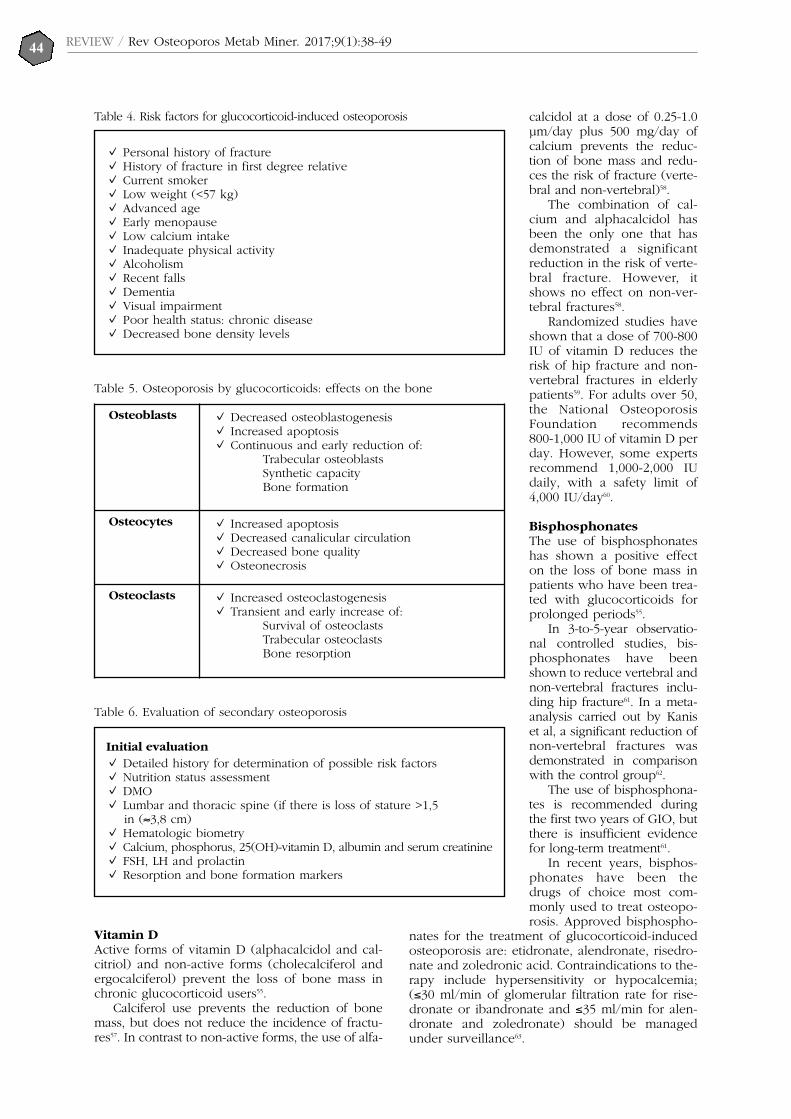
REVIEW / Rev Osteoporos Metab Miner. 2017;9(1):38-4944
Vitamin DActive forms of vitamin D (alphacalcidol and cal-citriol) and non-active forms (cholecalciferol andergocalciferol) prevent the loss of bone mass inchronic glucocorticoid users55.
Calciferol use prevents the reduction of bonemass, but does not reduce the incidence of fractu-res57. In contrast to non-active forms, the use of alfa-
calcidol at a dose of 0.25-1.0µm/day plus 500 mg/day ofcalcium prevents the reduc-tion of bone mass and redu-ces the risk of fracture (verte-bral and non-vertebral)58.
The combination of cal-cium and alphacalcidol hasbeen the only one that hasdemonstrated a significantreduction in the risk of verte-bral fracture. However, itshows no effect on non-ver-tebral fractures58.
Randomized studies haveshown that a dose of 700-800IU of vitamin D reduces therisk of hip fracture and non-vertebral fractures in elderlypatients59. For adults over 50,the National OsteoporosisFoundation recommends800-1,000 IU of vitamin D perday. However, some expertsrecommend 1,000-2,000 IUdaily, with a safety limit of4,000 IU/day60.
BisphosphonatesThe use of bisphosphonateshas shown a positive effecton the loss of bone mass inpatients who have been trea-ted with glucocorticoids forprolonged periods55.
In 3-to-5-year observatio-nal controlled studies, bis-phosphonates have beenshown to reduce vertebral andnon-vertebral fractures inclu-ding hip fracture61. In a meta-analysis carried out by Kaniset al, a significant reduction ofnon-vertebral fractures wasdemonstrated in comparisonwith the control group62.
The use of bisphosphona-tes is recommended duringthe first two years of GIO, butthere is insufficient evidencefor long-term treatment61.
In recent years, bisphos-phonates have been thedrugs of choice most com-monly used to treat osteopo-rosis. Approved bisphospho-
nates for the treatment of glucocorticoid-inducedosteoporosis are: etidronate, alendronate, risedro-nate and zoledronic acid. Contraindications to the-rapy include hypersensitivity or hypocalcemia;(≤30 ml/min of glomerular filtration rate for rise-dronate or ibandronate and ≤35 ml/min for alen-dronate and zoledronate) should be managedunder surveillance63.
Table 4. Risk factors for glucocorticoid-induced osteoporosis
Table 5. Osteoporosis by glucocorticoids: effects on the bone
✓ Personal history of fracture✓ History of fracture in first degree relative✓ Current smoker✓ Low weight (<57 kg)✓ Advanced age✓ Early menopause✓ Low calcium intake✓ Inadequate physical activity✓ Alcoholism✓ Recent falls✓ Dementia✓ Visual impairment✓ Poor health status: chronic disease✓ Decreased bone density levels
Table 6. Evaluation of secondary osteoporosis
Initial evaluation✓ Detailed history for determination of possible risk factors✓ Nutrition status assessment✓ DMO✓ Lumbar and thoracic spine (if there is loss of stature >1,5
in (≈3,8 cm)✓ Hematologic biometry✓ Calcium, phosphorus, 25(OH)-vitamin D, albumin and serum creatinine✓ FSH, LH and prolactin✓ Resorption and bone formation markers
Osteoblasts ✓ Decreased osteoblastogenesis✓ Increased apoptosis✓ Continuous and early reduction of:
Trabecular osteoblastsSynthetic capacityBone formation
Osteocytes ✓ Increased apoptosis✓ Decreased canalicular circulation✓ Decreased bone quality✓ Osteonecrosis
Osteoclasts ✓ Increased osteoclastogenesis✓ Transient and early increase of:
Survival of osteoclastsTrabecular osteoclastsBone resorption

REVIEW / Rev Osteoporos Metab Miner. 2017;9(1):38-4945
Figure 2. Management of patients of both genders> 50 years who are initiating or receiving glucocorticoid (GC)therapy
Assess possible risk factors for OIG
Determine the risk of fracture (FRAX)
To evaluate these patients periodically
Low risk✓ GC dosage <7.5 mg/day:
treatment notpharmacological
✓ Dosage >7.5 mg/day:bisphosphonates(alendronate, risendronate,zoledronic acid)
Moderate risk✓ GC dosage <7.5 mg/day:
alendronate or risendronate✓ Dosage >7.5 mg/day:
alendronate, risendronateor zoledronic acid
High risk✓ GC dosage <5 mg/day
for ≤1 month: alendronateor zoledronic acid
✓ GC dose >5 mgfor ≥1 month: alendronate,risendronate, zoledronic acid or teriparatide
The use of alendronate 5-10 mg/day for 48weeks has been shown to increase bone mass64,65.A study by Adachi et al demonstrated an increasein lumbar spine bone mineral density by 2.8% (5mg/day) and 3.9% (10 mg/day) in patients withprolonged glucocorticoid therapy66. A dose of 5mg/day of risedronate increases bone mass andalso reduces the risk of fracture67.
Zoledronic acid is approved by the Food andDrug Administration for the treating and preven-ting osteoporosis in men and postmenopausalwomen as well as glucocorticoid-induced osteo-porosis. The appropriate dose of zoledronic acidis 5 mg intravenously infused once a year, whichhas been shown to reduce the risk of spine frac-ture, non-vertebral fracture and hip fracture inpostmenopausal women with osteoporosis68.
TeriparatideTeriparatide is an analog of PTH obtained by therecombinant DNA technique (PTH-1-34). Thisanalogous agent increases osteoblastic functionand decreases the apoptosis of osteocytes55.
The use of teriparatide at a dose of 20 mg/daysubcutaneously should be considered as a treat-ment for GIO because it significantly increasesbone mineral density in this group of patients, inaddition to reducing vertebral fractures. However,it has no effect on non-vertebral fractures69.
Because of the high cost of teriparatide, its useis recommended when bisphosphonates fail69, thatis, when despite treatment with bisphosphonates,the bone mass continues to decrease, against frac-tures in the presence of bisphosphonates, or ifthey are contraindicated55.
DenosumabDenosumab is an antibody against RANKL, whichis used to treat primary osteoporosis. However,the study by Dore et al in patients with rheuma-toid arthritis under treatment with GC, demonstra-ted an increase in BMD and reduced resorption,compared to placebo70.
As denosumab is not filtered by the kidneys, itmay be a therapeutic option for patients with renaldysfunction who do not tolerate bisphosphonates70.
The use of denosumab for the treatment ofosteoporosis induced by GC has not been appro-ved yet, and its study is in stage III71. More evi-dence is needed to prove its utility in GIO61.
OdanacatibOdanacatib is a protease cathepsin-K inhibitorwhich induces bone deterioration by the osteo-clast72. A phase II study by Bone et al, using odana-catib 50 mg once weekly, demonstrated an increasein bone mineral density in the lumbar spine (5.5%vs. 0.2% in the control group) and hip 5.5% (3.2%vs 0.9% in the control group)73. However, there arenot enough controlled studies to determine the pro-per use of this drug in GIO, in addition to not beingapproved by the FDA so far.
Sclerostin InhibitorsThe mechanism responsible for the inhibition ofbone formation in patients with prolonged gluco-corticoid therapy is the reduction of the half-lifeand activity of osteoclasts. Glucocorticoids havebeen shown to alter bone cell formation by redu-cing the proliferation of osteoblasts and suppres-sing growth factors49. Because of this, new thera-

REVIEW / Rev Osteoporos Metab Miner. 2017;9(1):38-4946
Figura 3. Management of patients of both sexes <50 years who are initiating or receiving glucocorticoid therapy (GC)
Little evidencefor
recommendationsThere is no evidence offracture due to fragility
Evaluation ofrisk factors forpatients underGC therapy
Evidence of fracturefor fragility
Women (without proba-bility of pregnancy) or
women <50 years
Women (with probabilityof pregnancy)
GC 1-3 months✓Prednisone ≥5 mg/day:
alendronate or risendronate or/✓Prednisone ≥7 mg/day:
zoledronic acid
GC >3 months✓Alendronate or/✓Risendronate or/✓Zoledrónico acid or/✓Teriparatide
GC >3 months✓Prednisone ≥7.5 mg/day:
Alendronate or/✓Risendronate/zoledrónico
acid/teriparatide✓Prednisone <7.5 mg/day:
no consensus
GC 1-3 months✓There is no consensus
To evalu
ate these p
atients p
eriodically
peutic approaches are based on maintaining theviability of osteoblasts and osteocytes in the pre-sence of glucocorticoids.
Sclerostin (Sost) is a protein produced by oste-ocytes and its main function is to inhibit the matura-tion of osteoblasts74-76. A study by Yao et al demons-trated an increase in sclerostin gene expression inpatients exposed to GC therapy for 28 days77.
Monoclonal antibodies to sclerostin (Scl-Ab)currently known as romosozumab (AMG 785),blosozumab and BPS804 have been developedwhich inhibit the activity of sclerostin and thus sti-mulate bone formation76,78.
Yao et al used Scl-Ab in mice exposed to 4mg/kg/day of methylpredonoxone, and showedbone volumes of trabecular bone (Tb-BV/TV) inlower lumbar spine and neck of femur, less masscortical bone in the middle third of the femur andlower cortical bone resistance compared to the pla-cebo group. In addition, models receiving 25 mg/kgof Scl-Ab had a 60-125% increase in Tb-BV/TV andan increase in vertebral resistance of 30-70%79.
Studies to date have shown that sclerostin inhi-bitors may be a future therapeutic option formanaging patients with severe osteoporosis80.
Conclusion Osteoporosis is a systemic skeletal disease charac-terized by decreased bone mineral density withalterations in bone microarchitecture and increa-sed risk of fracture.
Glucocorticoids are the primary cause ofsecondary osteoporosis, being an independentfactor of morbidity and mortality in these patients,because the progressive loss of bone mass andincreased risk of fracture begins shortly after thestart of treatment with glucocorticoids.
It is important to identify, and, if possible, tocorrect risk factors and comorbidities in this groupof patients, initiate preventive measures and pro-vide health promotion tips such as change ofhabits, and give calcium and vitamin D supple-ments, in addition to treatment specific.
Conflict of interest: The authors declare no con-flict of interest.
Bibliography
1. Khosla S, Luftkin E, Hodgson S, Fitzpatrick L, Melton LJ3rd. Epidemiology and clinical features of osteoporo-sis in young individuals. Bone. 1994;15(5):551-5.
2. Walsh L, Wong C, Pringle M, Tattersfield A. Use of oralcorticosteroids in the community and the preventionof secondary osteoporosis: a cross sectional study.BMJ. 1996;313:344-6.
3. Sinigaglia L. Epidemiology of osteoporosis in rheuma-tic diseases. Rheum Dis Clin North Am. 2006;631-58.
4. Firestein G, Budd R, Gabriel S, McIness I, O’Dell J. Kelley’sTextbook of Rheumatology. 9th ed. Elsevier; 2013. 2292 p.
5. Baker-LePain J, Nakumura M, Lane N. Effects ofInflammation on bone: an update. Curr Opin Rheumatol.2011;23(4):389-95.

REVIEW / Rev Osteoporos Metab Miner. 2017;9(1):38-4947
6. Walsh N, Crotti T, Goldring S, Gravallese EM.Rheumatic diseases: the effect of inflammation onbone. Immunol Rev. 2005;208:228-51.
7. Rifas L, Weitzmann M. A novel T cell cytokine, secre-ted osteoclastogenic factor of activated T cells, inducesosteoclast formation in a RANKL-independent manner.Arthritis Rheum. 2009;60:3324-35.
8. Kong Y, Feige U, Sarosi I, Bolon B, Tafuri A, Morony S,et al. Activated T cells regulate bone loss and joint des-truction in adjuvant arthritis through osteoprotegerinligand. Nature. 1999;402:304-9.
9. Jódar Gimeno E. Conclusiones consensuadas del IForo Multidisciplinar en el Manejo del Paciente conAlto Riesgo de Fractura (ARF) Osteoporótica. RevOsteoporos Metab Miner. 2010;2(2):79-86.
10. Moon SJ, Ahn IE, Kwok SK, Park KS, Min JK, Park SH,et al. Periarticular osteoporosis is a prominent featurein early rheumatoid arthritis: estimation using shaft toperiarticular bone mineral density ratio. Korean MedSci. 2013;28:287-94.
11. Forsblad D, Larsen A, Waltbrand E, Kvist G, Mellstrom D,Saxne T, et al. Radiographic joint destruction in pos-tmenopausal rheumatoid arthritis is strongly associatedwith generalised osteoporosis. Ann Rheum Dis.2003;62:617-623.
12. Van Staa T, Geusens P, Bijlsma J, Leufkens H, Cooper C.Clinical assessment of the long-term risk of fracture inpatients with rheumatoid arthritis. Arthritis Rheum.2006;54(10):3104-12.
13. Edwards CJ, Williams E. The role of interleukin-6 inrheumatoid arthritis-associated osteoporosis. OsteoporosInt. 2010;21(8):1287-93.
14. Sinigaglia L, Nevertti A, Mela Q. A multicenter cross-sectional study on bone mineral density in rheumatoidarthritis. J Rheumatol. 2000;27:2528-9.
15. Stafford L, Bleasel J, Giles A. Androgen deficiency andbone mineral density in men with rheumatoid arthritis.J Rheumatol. 2000;27:2786-90.
16. Van Staa T, Leufkens H, Cooper C. The epidemiologyof corticosteroid-induced osteoporosis: a meta-analy-sis. Osteoporos Int. 2002;13:777-87.
17. Deal C. Recent recommendations on steroid-inducedosteoporosis: more targeted but more complicated.Clevel Clin J Med. 2013;80(2):117-25.
18. Di Munno O, Mazzantini M, Sinigaglia L, Bianchi G,Minisola G, Muratore M, et al. Effect of low-dosemethotrexate on bone density in women with rheuma-toid arthritis: results from a multicenter cross-sectionalstudy. J Rheumatol. 2004;31:1305-9.
19. Seriolo B, Paolino S, Sulli A, Ferretti V, Cutolo M. Bonemetabolism changes during anti-TNF-alpha therapy inpatients with active rheumatoid arthritis. Ann NY AcadSci. 2006;1069:420-7.
20. Marotte H, Pallot-Prades B, Grange L, Gaudin P,Alexandre C, Miossec P. A 1-year case–control study inpatients with rheumatoid arthritis indicates prevention ofloss of bone mineral density in both responders and non-responders to infliximab. Arthritis Res Ther. 2007;9(3):R61.
21. Lange U, Teichmann J, Muller-Ladner U, Strunk J. Increasein bone mineral density of patients with rheumatoid arthri-tis treated with anti-TNF-alpha antibody: a prospectiveopen-label pilot study. Rheumatol. 2005;44:1546-8.
22. Van der Goes M, Jacobs G, Jurgens M, Bakker MF, vander Veen MJ, van der Werf JH, et al. Are changes inbone mineral density different between groupsof earlyrheumatoid arthritis patients treated according to atight control strategy with or without prednisone ifosteoporosis prophylaxis is applied? Osteoporos Int.2013;24:1429-36.
23. Riggs B, Melton LJ 3rd. The worldwide problem ofosteoporosis: insights afforded by epidemiology.Bone. 1995;115:505S-11S.
24. Mok C, Mak A, Ma K. Bone mineral density in postmenopausal Chinese patients with Systemic LupusErythematosus. Lupus. 2005;14:106-12.
25. Lai CC, Chen WS, Chang DM, Tsao YP, Wu TH, Chou CT,et al. Increased serum fibroblast growth factor-23 anddecreased bone turnover in patients with systemic
lupus erythematosus under treatment with cyclospori-ne and steroid but not steroid only. Osteoporos Int.2015;26(2):601-10.
26. Houssiau F, Lefebvre C, Depresseux G, Lambert M,Devogelaer JP, Nagant de Deuxchaisnes C. Trabecularand cortical bone loss in systemic lupus erythemato-sus. Br J Rheumatol. 1996;35:244-7.
27. Wang X, Yan S, Liu C, Xu Y, Wan L, Wang Y, et al.Fracture risk and bone mineral density levels inpatients with systemic lupus erythematosus: a systema-tic review and meta-analysis. Osteoporos Int.2016;4:1413-23.
28. Jacobs J, Korswagen LA, Schilder AM, Van Tuyl LH,Dijkmans BAC, Lems WF, et al. Six-year follow-up studyof bone mineral density in patients with systemic lupuserythematosus. Osteoporos Int. 2013;24(6):1827-33.
29. Bultink E, Lems W, Kostense P, Dijkmans BA, Voskuyl AE.Prevalence and risk factors for low bone mineral densityand vertebral fractures in patients with systemic lupus ery-thematosus. Arthritis Rheum. 2005;54:2044-50.
30. Toloza S, Cole D, Gladman D, Ibanez D, Urowitz M.Vitamin D insufficiency in a large female SLE cohort.Lupus. 2010;19:13-9.
31. Ruiz-Irastorza G, Egurbide M, Olivares N, Martinez-Berriotxoa A, Aguirre C. Vitamin D deficiency in syste-mic lupus erythematosus: prevalence, predictors andclinical consequences. Rheumatology. 2008;47:920-3.
32. Mazziotti G, Canalis E, Giustina A. Drug-induced oste-oporosis: mechanisms and clinical implications. Am JMed. 2010;123:877-84.
33. Prieto-Alhambra D, Nogues X, Javaid K, Wyman A,Arden NK, Azagra R, et al. An increased rate of fallingleads to a rise in fracture risk in postmenopausalwomen with self-reported osteoarthritis: a prospectivemultinational cohort study (GLOW). Ann Rheum Dis.2013;72:911-7.
34. Ralston S, Urquhart G, Brezeski M, Sturrock RD.Prevalence of vertebral compression fractures due to oste-oporosis in ankylosing spondylitis. BMJ. 1990;300:563-5.
35. Lange U, Kluge A, Strunk J, Teichmann J, Bachmann G.Ankylosing spondylitis and bone mineral density.What is the ideal tool for measurement? Rheumatol Int.2005;26(2):115-20.
36. Cooper C, Carbone L, Michet C, Atkinson EJ, O'Fallon WM,Melton LJ 3rd. Fracture risk in patients with ankylosingspondylitis:a population based study. J Rheumatol.1994;21:1877-82.
37. Mullaji A, Upadhyay S, Ho E. Bone mineral density inankylosing spondylitis. DEXA comparison of controlsubjects with mild and advanced cases. J Bone Jt SurBr. 1994;76:660-5.
38. Toussirot R, Michel F, Wendling D. Bone density, ultra-sound measurements and body composition in earlyankylosing spondylitis. Rheumatol. 2001;40:882-8.
39. van der Weijden M, van der Horst-Bruinsma I, vanDenderen J, Dijkmans B, Heymans M, Lems W. Highfrequency of vertebral fractures in early spondylarthro-pathies. Osteoporos Int. 2012;23:1683-90.
40. Keller J, Kang J, Lin H. Association between osteopo-rosis and psoriasis: results from the LongitudinalHealth Insurance Database in Taiwan. Osteoporos Int.2013;24:1835-41.
41. Dreiher J, Weirzman D, Cohen A. Psoriasis and osteo-porosis: a sex-specific association? J Inves Dermatol.2009;129:1643-9.
42. Yoshimura N, Muraki S, Oka H, Mabuchi A, Kinoshita H,Yosihda M, et al. Epidemiology of lumbar osteoporosisand osteoarthritis and their causal relationship-is osteo-arthritis a predictor for osteoporosis or vice versa?: theMiyama study. Osteoporos Int. 2009;20(6):999-1008.
43. Zhang ZM, Li ZC, Jiang LS, Jiang SD, Dai LY. Micro-CTand mechanical evaluation of subchondral trabecularbone structure between postmenopausal women withosteoarthritis and osteoporosis. Osteoporos Int.2010;21(8):1383-90.
44. Pereira RM, Carvalho JF, Paula AP, Zerbini C,Domiciano DS, Gonçalves H, et al. Guidelines for theprevention and treatment of glucocorticoid-induced

REVIEW / Rev Osteoporos Metab Miner. 2017;9(1):38-4948
osteoporosis. Rev Bras Reum. 2012;52(4):569-93. 45. Mirza F, Canalis E. Secondary osteoporosis: Pathophysiology
and management. Eur J Endocrinol. 2015;173(3):R131-51. 46. Hofbauer L, Gori F, Riggs B, Lacey D, Dunstan C,
Spelsberg T, et al. Stimulation of osteoprotegerin ligandand inhibition of osteoprotegerin production by gluco-corticoids in human osteoblastic lineage cells: potentialparacrine mechanisms of glucocorticoid- induced osteo-porosis. Endocrinology. 1999;140:4382-9.
47. Dallas S, Prideaux M, Bonewald L. The osteocyte: anendocrine cell... and more. Endocr Rev. 2013;34:658-90.
48. Delany A, Durant D, Canalis E. Glucocorticoid sup-pression of IGF I transcription in osteoblasts. MolEndocrinol. 2001;15(10):1781-9.
49. Weinstein R. Glucocorticoid-Induced Bone Disease.New Eng J Med. 2011;365:62-70.
50. American College of Rheumatology. Recommendationsfor the prevention and treatment of glucocorticoid-indu-ced osteoporosis: 2001 update. American College ofRheumatology Ad Hoc Committee on Glucocorticoid-Induced Osteoporosis. Arthritis Rheum. 2001;44:1496-503.
51. Adler R, Hochberg M. Suggested guidelines for evalua-tion and treatment of glucocorticoid-induced osteopo-rosis for the Department of Veterans Affairs. ArchIntern Med. 2003;163(21):2619-24.
52. Devogelaer J, Goemaere S, Boonen S, Body J,Kaufman J, Reginster JY, et al. Evidence-based guideli-nes for the prevention and treatment of glucocorticoid-induced osteoporosis: a consensus document of theBelgian Bone Club. Osteoporos Int. 2006;17(1):8-19.
53. Messina O, Somma L, Tamborenea M, Castelli G,Riopedre A, Lancioni G, et al. Guías para el diagnósti-co, la prevención y el tratamiento de la osteoporosisinducida por glucocorticoides en el adulto. ActualOsteol. 2016;12(2):107-25.
54. Grossman J, Gordon R, Ranganath V, Deal C, Caplan L,Chen W, et al. American College of Rheumatology2010 recommendations for the prevention and treat-ment of glucocorticoid-induced osteoporosis. ArthritisCare Res. 2010;62(11):1515-26.
55. Rodrigues Pereira R, Freire de Carvalho J, Paula A,Zerbini C, Domiciano D, Gonçalves H, et al. Guidelinesfor the prevention and treatment of glucocorticoid-indu-ced osteoporosis. Rev Bras Reum. 2012;52(4):569-93.
56. Sambrook P, Birmingham J, Kelly P, Kempler S,Nguyen T, Pocock N, et al. Prevention of corticosteroidosteoporosis. A comparison of calcium, calcitriol andcalcitonin. N Eng J Med. 1993;328(24):1747-52.
57. Richy F, Ethgen O, Bruyere O, Regunster J. Efficacy ofalfacalcidol and calcitriol in primary and corticoste-roid-induced osteoporosis: a meta-analysis of theireffects on bone mineral density and fracture rate.Osteoporos Int. 2004;14(5):301-10.
58. Ringe JD, Dorst A, Faber H, Schacht E, Rahlfs V.Superiority of alfacalcidol over plain vitamin D in thetreatment of glucocorticoid- induced osteoporosis.Rheumatol Int. 2004;24(2):63-70.
59. Bischoff-Ferrari H, Willett W, Wong JB, Giovannucci E,Dietrich T, Dawson-Hughes B. Fracture preventionwith vitamin D supplementation: a meta-analysis ofrandomized controlled trials. JAMA. 2005;293:2257-64.
60. Ross A, Manson J, Abrams S, Aloia JF, Brannon PM,Clinton SK, et al. The 2011 report on dietary referenceintakes for calcium and vitamin D from the Institute ofMedicine: what clinicians need to know. J ClinEndocrinol Metab. 2011;96(1):53-8
61. Lems W, Saag K. Bisphosphonates and glucocorticoid-induced osteoporosis: cons. Endocrine. 2015;49(3):628-34.
62. Kanis JA, Stevenson M, McCloskey EV, Davis S, Lloyd-Jones M. Glucocorticoid-induced osteoporosis: a syste-matic review and cost-utility analysis. Health TechnolAssess. 2007;11(7):1-231.
63. Kidney Disease: Improving Global Outcomes (KDIGO)CKD-MBD Work Group. KDIGO clinical practice guideli-
ne for the diagnosis, evaluation, prevention, and treat-ment of chronic kidney disease-mineral and bone disor-ders (CKD-MBD). Kidney Int Suppl. 2009;76(113):S1-130.
64. Saag K, Emkey R, Schnitzer T, Brown J, Hawkins F,Goemacre S, et al. Alendronate for the prevention andtreatment of glucocorticoid- induced osteoporosis. NEng J Med. 1998;339(5):292-9.
65. de Nijs R, Jacobs J, Lems W, Laan R, Algra A, Huisman A,et al. Alendronate or alfacalcidol in glucocorticoid-indu-ced osteoporosis. N Engl J Med. 2006;355(7):675-84.
66. Adachi JD, Saag KG, Delmas PD, Liberman UA, Emkey RD,Seeman E, et al. Two-year effects of alendronate onbone mineral density and vertebral fracture in patientsreceiving glucocorticoids: a randomized, double-blind,placebo-controlled extension trial. Arthritis Rheum.2001;44(1):202-11.
67. Cohen S, Levy R, Keller M, Boling E, Emkey R,Greenwald M, et al. Risedronate therapy prevents corti-costeroid-induced bone loss: a twelve-month, multicen-ter, randomized, double-blind, placebo-controlled, para-llel-group study. Arthritis Rheum. 1999;42(11):2309-18.
68. Watts N, Bilezikian J, Camacho P, Greenspan S, Harris S,Hodgson S, et al. American Association Of ClinicalEndocrinologists Medical Guidelines for clinical practi-ce for the diagnosis and treatment of postmenopausalosteoporosis: executive summary of recommenda-tions. Endocr Pr. 2010;16(6):1016-9.
69. Saag K, Shane E, Boonen S, Marin F, Donley D, Taylor K,et al. Teriparatide or alendronate in glucocorticoid-indu-ced osteoporosis. N Engl J Med. 2007;357(20):2028-39.
70. Dore RK, Cohen SB, Lane NE, Palmer W, Shergy W,Zhou L, et al. Effects of denosumab on bone mineraldensity and bone turnover in patients with rheumatoidarthritis receiving concurrent glucocorticoids or bis-phosphonates. Ann Rheum Dis. 2010;69(5):872-5.
71. Whittier X, Saag K. Glucocorticoid-induced Osteoporosis.Rheum Dis Clin N Am. 2016;42(1):177-89.
72. Boonen S, Rosenberg F, Claessens F, Vanderschueren D,Papapoulos S. Inhibition of cathepsin K for treatmentof osteoporosis. Curr Osteoporos Rep. 2012;10(1):73-9.
73. Bone H, McClung MR, Roux C, Recker RR, Eisman JA,Verbruggen N, et al. Odanacatib, a cathepsin K inhibi-tor for osteoporosis: a two-year study in post- meno-pausal women with low bone density. J Bone MinerRes. 2010;25:937-47.
74. Paszty C, Turner C, Robinson M. Sclerostin: a gemfrom the genome leads to bone-building antibodies. JBone Min Res. 2010;25(9):1897-904.
75. van Bezooijen R, Roelen B, Visser A, van der Wee-Pals L,de Wilt E, Karperien M, et al. Sclerostin is an osteocyte-expressed negative regulator of bone formation, but not aclassical BMP antagonist. J Exp Med. 2004;199(6):805-14.
76. Li X, Warmington K, Niu Q, Asuncion F, Barrero M,Grisanti M, et al. Inhibition of sclerostin by monoclo-nal antibody increases bone formation, bone mass andbone strength in aged male rats. J Bone Min Res.2010;25(12):2647-56.
77. Yao W, Cheng Z, Busse C, Pham A, Nakamura M, Lane N.Glucocorticoid excess in mice results in early activation ofosteoclastogenesis and adipogenesis and prolonged sup-pression of osteogenesis: a longitudinal study of geneexpression in bone tissue from glucocorticoid-treatedmice. Arthritis Rheum. 2008;58(6):1674-86.
78. McClung M, Grauer A, Boonen S, Bolognese M, Brown J,Diez-Perez A, et al. Romosozumab in postmenopausalwomen with low bone mineral density. New Eng JMed. 2014;370(5):412-20.
79. Yao W, Dai W, Jiang L, Lay EY, Zhong Z, Ritchie R, etal. Sclerostin-antibody treatment of glucocorticoid-induced osteoporosis maintained bone mass andstrength. Osteoporos Int. 2016;27:283-94.
80. Appelman-Dijkstra N, Papapoulos S. Sclerostin inhibi-tion in the management of osteoporosis. Calcif TissueInt. 2016;98:370-80.

REVIEW / Rev Osteoporos Metab Miner. 2017;9(1):38-4949