


Idiomas
Páginas
Jurídico

7/30/2019 Espectro Autista - II
http://slidepdf.com/reader/full/espectro-autista-ii 1/12
S83
1. Associate director, Dan Marino Child Nett, Dan Marino Center, MiamiChildren’s Hospital. Assistant professor, Department of Neurology, Schoolof Medicine, University of Miami.
2. Medical director, Dan Marino Center, Miami Children’s Hospital. Assistantprofessor, Department of Neurology, School of Medicine, University ofMiami.
3. Associate professor, Department of Pediatrics, School of Medicine,Universidade Federal do Rio Grande do Sul (UFRGS). Chief of the Unitof Children’s Neurology, Service of Pediatrics, Hospital de Clínicas dePorto Alegre (HCPA), Porto Alegre, RS, Brazil.
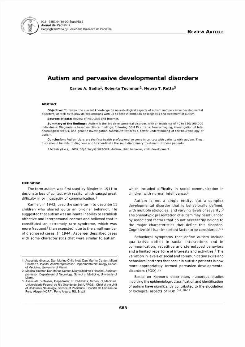
Abstract
Objective: To review the current knowledge on neurobiological aspects of autism and pervasive developmental
disorders, as well as to provide pediatricians with up to date information on diagnosis and treatment of autism.
Sources of data: Review of MEDLINE and Internet.
Summary of the findings: Autism is the 3rd developmental disorder, with an incidence of 40 to 130/100,000
individuals. Diagnosis is based on clinical findings, following DSM IV criteria. Neuroimaging, investigation of fetal
neurological status, and genetic investigation contribute towards a better understanding of the neurobiology of
autism.
Conclusion: Pediatricians are the first health professional to come in contact with patients with autism. Thus,
they should be able to diagnose and to coordinate the multidisciplinary treatment of these patients.
J Pediatr (Rio J). 2004;80(2 Suppl):S83-S94: Autism, child behavior, child development.
Autism and pervasive developmental disorders
Carlos A. Gadia1, Roberto Tuchman2, Newra T. Rotta3
0021-7557/04/80-02-Suppl/S83
Jornal de Pediatria
Copyright © 2004 by Sociedade Brasileira de Pediatria
Definition
The term autism was first used by Bleuler in 1911 to
designate loss of contact with reality, which caused great
difficulty in or incapacity of communication.1
Kanner, in 1943, used the same term to describe 11
children who shared quite an original behavior. He
suggested that autism was an innate inability to establish
affective and interpersonal contact and believed that it
constituted an extremely rare syndrome, which was
more frequent2 than expected, due to the small number
of diagnosed cases. In 1944, Asperger described cases
with some characteristics that were similar to autism,
which included difficulty in social communication in
children with normal intelligence.3
Autism is not a single entity, but a complex
developmental disorder that is behaviorally defined,
with multiple etiologies, and varying levels of severity.3
The phenotypic presentation of autism may be influenced
by associated factors that do not necessarily belong to
the major characteristics that define this disorder.
Cognitive skill is an important factor to be considered.4-9
Behavioral symptoms that define autism include
qualitative deficit in social interactions and in
communication, repetitive and stereotyped behaviors
and a limited repertoire of interests and activities.1 The
variation in levels of social and communication skills and
behavioral patterns that occur in autistic patients is now
more appropriately termed pervasive developmental
disorders (PDD).10
Based on Kanners description, numerous studies
involving the epidemiology, classification and identification
of autism have significantly contributed to the elucidation
of biological aspects of PDD.3-7,10-12
R EVIEW ARTICLE
7/30/2019 Espectro Autista - II
http://slidepdf.com/reader/full/espectro-autista-ii 2/12
S84 Jornal de Pediatria - Vol. 80, No.2(Suppl), 2004 Autism and pervasive developmental disorders Gadia CA et alii
Difficulties in social interaction in PDD may be
characterized by withdrawal or improper social behavior;
poor eye contact; difficulty in participating in group
activities; blunting of affect, or inappropriate demonstration
of emotions; lack of social or emotional empathy. As these
individuals reach adulthood, social withdrawal is reduced,
but poor social skills and difficulty making friends still
persist.
Adolescents and adults with autism misinterpret how
they are perceived by others, and autistic adults, even
with appropriate cognitive skills, tend to alienate
themselves.
Communication disorders occur in varying degrees,
compromising both verbal and nonverbal ability to share
information with others. Some children do not develop
communication skills, whereas others show immature
language characterized by jargon, echolalia, pronoun
reversal, abnormal prosody, monotonous intonation, etc.
Those with appropriate capacity to express themselves
may be unable to start or maintain a conversation.Language and communication disorders persist in
adulthood, and a significant number of autistic patients
remain nonverbal. Those who acquire verbal skills may
have persistent difficulty in establishing a conversation,
such as lack of reciprocity, difficulty in understanding
language subtleties, jokes or sarcasm, as well as problems
with interpreting body language and facial expressions.
Repetitive and stereotyped behaviors, characteristic
of autism, include reluctance to changes, insistence on
certain routines, excessive attachment to objects,
fascination with spinning objects (e.g.: wheels or
propellers). Although some children seem to play, they aremore concerned about aligning or handling toys than using
them for a symbolic purpose. Motor and verbal stereotypies
such as swinging, clapping repeatedly, walking in circles
or repeating certain words, sentences or songs also are
frequent among autistic individuals. Autistic adults show
improved adaptation to changes, but limited interests
persist, and those with appropriate cognitive skills tend to
concentrate their interests in restricted topics, such as
train/airplane timetables, maps or historical facts, which
rule their lives.
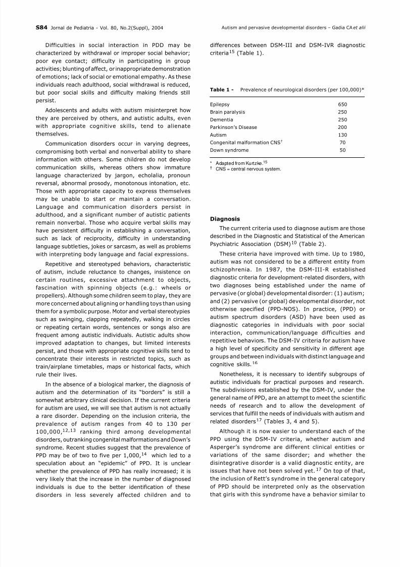
In the absence of a biological marker, the diagnosis of
autism and the determination of its borders is still a
somewhat arbitrary clinical decision. If the current criteria
for autism are used, we will see that autism is not actually
a rare disorder. Depending on the inclusion criteria, the
prevalence of autism ranges from 40 to 130 per
100,000,12,13 ranking third among developmental
disorders, outranking congenital malformations and Downs
syndrome. Recent studies suggest that the prevalence of
PPD may be of two to five per 1,000,14 which led to a
speculation about an epidemic of PPD. It is unclear
whether the prevalence of PPD has really increased; it is
very likely that the increase in the number of diagnosed
individuals is due to the better identification of these
disorders in less severely affected children and to
differences between DSM-III and DSM-IVR diagnostic
criteria15 (Table 1).
* Adapted from Kurtzke.15
† CNS = central nervous system.
Table 1 - Prevalence of neurological disorders (per 100,000)*
Epilepsy 650
Brain paralysis 250
Dementia 250
Parkinsons Disease 200
Autism 130
Congenital malformation CNS 70
Down syndrome 50
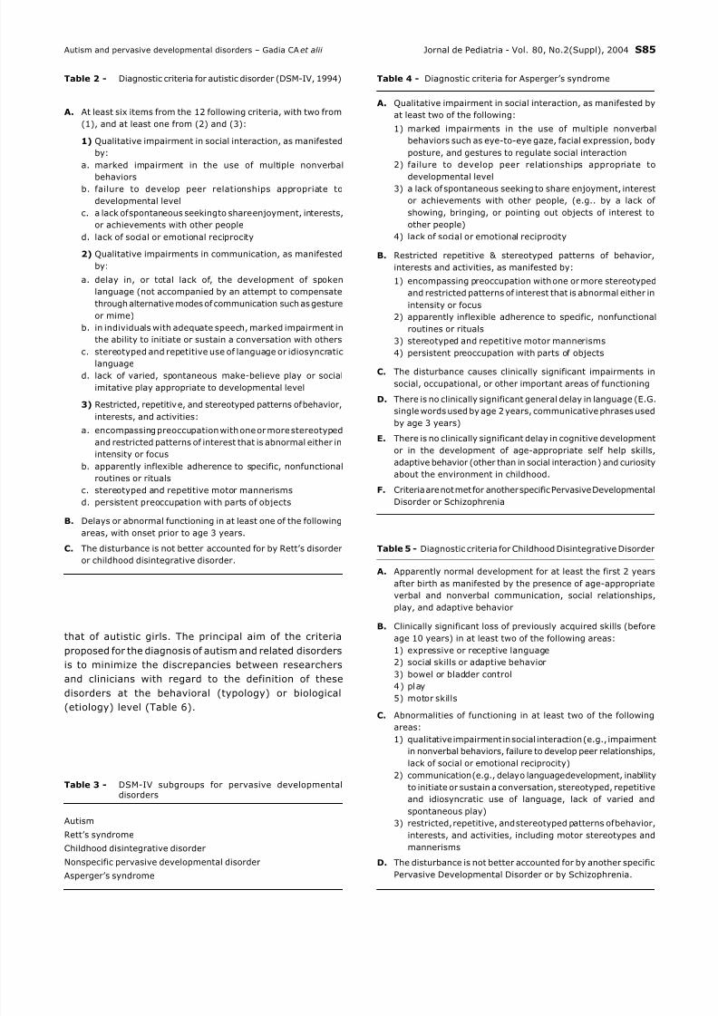
Diagnosis
The current criteria used to diagnose autism are those
described in the Diagnostic and Statistical of the American
Psychiatric Association (DSM)10 (Table 2).
These criteria have improved with time. Up to 1980,
autism was not considered to be a different entity from
schizophrenia. In 1987, the DSM-III-R established
diagnostic criteria for development-related disorders, with
two diagnoses being established under the name of
pervasive (or global) developmental disorder: (1) autism;
and (2) pervasive (or global) developmental disorder, not
otherwise specified (PPD-NOS). In practice, (PPD) or
autism spectrum disorders (ASD) have been used as
diagnostic categories in individuals with poor social
interaction, communication/language difficulties and
repetitive behaviors. The DSM-IV criteria for autism have
a high level of specificity and sensitivity in different age
groups and between individuals with distinct language and
cognitive skills.16
Nonetheless, it is necessary to identify subgroups of
autistic individuals for practical purposes and research.
The subdivisions established by the DSM-IV, under the
general name of PPD, are an attempt to meet the scientificneeds of research and to allow the development of
services that fulfill the needs of individuals with autism and
related disorders17 (Tables 3, 4 and 5).
Although it is now easier to understand each of the
PPD using the DSM-IV criteria, whether autism and
Aspergers syndrome are different clinical entities or
variations of the same disorder; and whether the
disintegrative disorder is a valid diagnostic entity, are
issues that have not been solved yet.17 On top of that,
the inclusion of Retts syndrome in the general category
of PPD should be interpreted only as the observation
that girls with this syndrome have a behavior similar to
7/30/2019 Espectro Autista - II
http://slidepdf.com/reader/full/espectro-autista-ii 3/12
Jornal de Pediatria - Vol. 80, No.2(Suppl), 2004 S85
that of autistic girls. The principal aim of the criteria
proposed for the diagnosis of autism and related disorders
is to minimize the discrepancies between researchers
and clinicians with regard to the definition of these
disorders at the behavioral (typology) or biological
(etiology) level (Table 6).
Table 2 - Diagnostic criteria for autistic disorder (DSM-IV, 1994)
A. At least six items from the 12 following criteria, with two from
(1), and at least one from (2) and (3):
1) Qualitative impairment in social interaction, as manifested
by:
a. marked impairment in the use of multiple nonverbal
behaviorsb. failure to develop peer relationships appropriate to
developmental level
c. a lack of spontaneous seeking to share enjoyment, interests,
or achievements with other people
d. lack of social or emotional reciprocity
2) Qualitative impairments in communication, as manifested
by:
a. delay in, or total lack of, the development of spoken
language (not accompanied by an attempt to compensate
through alternative modes of communication such as gesture
or mime)
b. in individuals with adequate speech, marked impairment in
the ability to initiate or sustain a conversation with others
c. stereotyped and repetitive use of language or idiosyncraticlanguage
d. lack of varied, spontaneous make-believe play or social
imitative play appropriate to developmental level
3) Restricted, repetitive, and stereotyped patterns of behavior,
interests, and activities:
a. encompassing preoccupation with one or more stereotyped
and restricted patterns of interest that is abnormal either in
intensity or focus
b. apparently inflexible adherence to specific, nonfunctional
routines or rituals
c. stereotyped and repetitive motor mannerisms
d. persistent preoccupation with parts of objects
B. Delays or abnormal functioning in at least one of the following
areas, with onset prior to age 3 years.C. The disturbance is not better accounted for by Retts disorder
or childhood disintegrative disorder.
Autism
Retts syndrome
Childhood disintegrative disorder
Nonspecific pervasive developmental disorder
Aspergers syndrome
Table 3 - DSM-IV subgroups for pervasive developmentaldisorders
Table 4 - Diagnostic criteria for Aspergers syndrome
A. Qualitative impairment in social interaction, as manifested by
at least two of the following:
1) marked impairments in the use of multiple nonverbal
behaviors such as eye-to-eye gaze, facial expression, body
posture, and gestures to regulate social interaction
2) failure to develop peer relationships appropriate to
developmental level3) a lack of spontaneous seeking to share enjoyment, interest
or achievements with other people, (e.g.. by a lack of
showing, bringing, or pointing out objects of interest to
other people)
4) lack of social or emotional reciprocity
B. Restricted repetitive & stereotyped patterns of behavior,
interests and activities, as manifested by:
1) encompassing preoccupation with one or more stereotyped
and restricted patterns of interest that is abnormal either in
intensity or focus
2) apparently inflexible adherence to specific, nonfunctional
routines or rituals
3) stereotyped and repetitive motor mannerisms
4) persistent preoccupation with parts of objects
C. The disturbance causes clinically significant impairments in
social, occupational, or other important areas of functioning
D. There is no clinically significant general delay in language (E.G.
single words used by age 2 years, communicative phrases used
by age 3 years)
E. There is no clinically significant delay in cognitive development
or in the development of age-appropriate self help skills,
adaptive behavior (other than in social interaction) and curiosity
about the environment in childhood.
F. Criteria are not met for another specific Pervasive Developmental
Disorder or Schizophrenia
Table 5 - Diagnostic criteria for Childhood Disintegrative Disorder
A. Apparently normal development for at least the first 2 years
after birth as manifested by the presence of age-appropriate
verbal and nonverbal communication, social relationships,
play, and adaptive behavior
B. Clinically significant loss of previously acquired skills (before
age 10 years) in at least two of the following areas:
1) expressive or receptive language
2) social skills or adaptive behavior
3) bowel or bladder control
4) play
5) motor skills
C. Abnormalities of functioning in at least two of the following
areas:
1) qualitative impairment in social interaction (e.g., impairment
in nonverbal behaviors, failure to develop peer relationships,
lack of social or emotional reciprocity)
2) communication (e.g., delay o language development, inability
to initiate or sustain a conversation, stereotyped, repetitive
and idiosyncratic use of language, lack of varied and
spontaneous play)
3) restricted, repetitive, and stereotyped patterns of behavior,
interests, and activities, including motor stereotypes and
mannerisms
D. The disturbance is not better accounted for by another specific
Pervasive Developmental Disorder or by Schizophrenia.
Autism and pervasive developmental disorders Gadia CA et alii
7/30/2019 Espectro Autista - II
http://slidepdf.com/reader/full/espectro-autista-ii 4/12
S86 Jornal de Pediatria - Vol. 80, No.2(Suppl), 2004
interesting. The prevalence of TE in autistic individuals
is of 1-4% (significantly higher than that of the fragile
X syndrome and autism), while 25% of TE patients are
autistic and 40-50% meet the criteria for PDD. This
association is probably due to abnormal organization of
the brain related to TE genes - TSC1 on chromosome
9q34 and TSC2 on 16p13.3 and/or to complications of
TE, such as mental retardation and severe epilepsy inthe first year of life (West syndrome).19
Even when autistic disorders are properly diagnosed
using appropriate diagnostic criteria, the symptomatic
profile varies considerably, depending on the underlying
etiology.20 The diagnosis of autism requires a careful
clinical assessment: language and neuropsychological
evaluation, in addition to additional exams (e.g.:
chromosome studies including DNA for fragile X syndrome
and neuroimaging or neurophysiological studies, whenever
necessary), may be useful in specific cases to identify
more homogeneous subgroups, according to the behavioral
phenotype and etiology. This will allow us to understandthe pathophysiology of these disorders and to establish
more specific interventions and prognoses.
The screening of autistic individuals requires a
multidisciplinary team and the use of objective scales.
Structured techniques should be used to assess childrens
social behavior (joint attention, eye contact, facial
expression, and affect) and their capacity to imitate.
The Childhood Autism Rating Scale (CARS)21 is one of
the most commonly used scales, consisting of a structured
15-item questionnaire (applicable in 30-45 minutes)
answered by the parents or surrogates of an autistic
child older than two years. A seven-point score is used
for each of the 15 items, which allows the classification
of autism into mild/moderate or severe.
The Vineland adaptive behavior scale is also commonly
used. It is used to measure social development in a
healthy population, which may be compared to that of
autistic individuals.22
The two most comprehensive batter ies of
psychological tests used for the diagnosis of autism,
especially in research, are the autism diagnostic
observation schedule (ADOS) and autism diagnostic
interview (ADI). Together, they represent a complete
structured interview and an observation method for the
objective assessment of social skills, communication
skills, and behavior of autistic individuals, ranging from
speechless children to adults who are able to
communicate relatively well.23-25 Its use became a
standard in research studies on autism in the 1990s. A
recent study investigated specific deficits in social
communication in ASD children using ADOS, and three
factors were identified: joint attention, affective
rec iproc i ty, and mind theory.26 These three
communication domains are central to social growth in
typical children, and if not properly developed, they are
responsible for basic deficits in the spectrum of social
communication disorders.
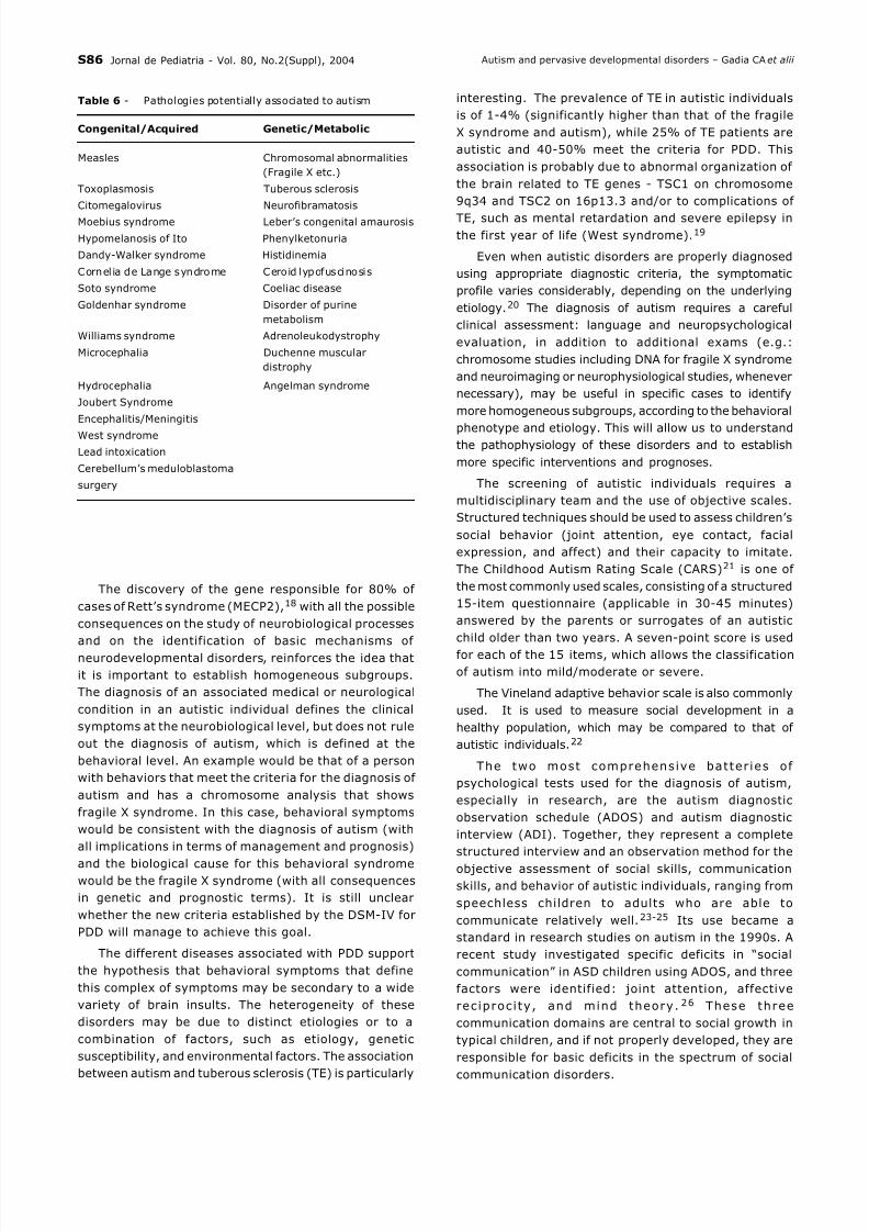
Congenital/Acquired Genetic/Metabolic
Measles Chromosomal abnormalities
(Fragile X etc.)
Toxoplasmosis Tuberous sclerosis
Citomegalovirus Neurofibramatosis
Moebius syndrome Lebers congenital amaurosis
Hypomelanosis of Ito Phenylketonuria
Dandy-Walker syndrome Histidinemia
Cornelia de Lange syndrome Ceroid lypofuscinosis
Soto syndrome Coeliac disease
Goldenhar syndrome Disorder of purine
metabolism
Williams syndrome Adrenoleukodystrophy
Microcephalia Duchenne muscular
distrophy
Hydrocephalia Angelman syndrome
Joubert Syndrome
Encephalitis/Meningitis
West syndrome
Lead intoxication
Cerebellums meduloblastoma
surgery
Table 6 - Pathologies potentially associated to autism
The discovery of the gene responsible for 80% of
cases of Retts syndrome (MECP2),18 with all the possible
consequences on the study of neurobiological processes
and on the identification of basic mechanisms of neurodevelopmental disorders, reinforces the idea that
it is important to establish homogeneous subgroups.
The diagnosis of an associated medical or neurological
condition in an autistic individual defines the clinical
symptoms at the neurobiological level, but does not rule
out the diagnosis of autism, which is defined at the
behavioral level. An example would be that of a person
with behaviors that meet the criteria for the diagnosis of
autism and has a chromosome analysis that shows
fragile X syndrome. In this case, behavioral symptoms
would be consistent with the diagnosis of autism (with
all implications in terms of management and prognosis)
and the biological cause for this behavioral syndromewould be the fragile X syndrome (with all consequences
in genetic and prognostic terms). It is still unclear
whether the new criteria established by the DSM-IV for
PDD will manage to achieve this goal.
The different diseases associated with PDD support
the hypothesis that behavioral symptoms that define
this complex of symptoms may be secondary to a wide
variety of brain insults. The heterogeneity of these
disorders may be due to distinct etiologies or to a
combination of factors, such as etiology, genetic
susceptibility, and environmental factors. The association
between autism and tuberous sclerosis (TE) is particularly
Autism and pervasive developmental disorders Gadia CA et alii

7/30/2019 Espectro Autista - II
http://slidepdf.com/reader/full/espectro-autista-ii 5/12
Jornal de Pediatria - Vol. 80, No.2(Suppl), 2004 S87
Neuropathology and neuroimaging
Currently, the neuropathology of autism is based on
Bauman & Kemper,27-29 who obtained consistent
neuropathological findings in the limbic system and in
cerebellar circuits of eleven brains investigated so far.
The cells of the limbic system (hippocampus, amygdala,
mamillary bodies, cingulate gyrus and septal nuclei) are
small in size, but large in number per unit of volume
(increased cell density) compared to controls. This led
to the hypothesis of a delay in the maturational
development of limbic system circuits. The investigated
cerebellums revealed a low number of Purkinje cells,
especially in the posterolateral neocerebellum and in
the adjacent archicerebellar cortex (posterior and inferior
portions of the cerebellum). The inferior olivary nucleus
in the investigated brains did not show expected
retrograde neuronal loss (secondary to the loss of
Purkinje cells). This suggests that abnormal findings in
the brains of autistic individuals occurred at around 30
weeks gestation, before the connection between the
olive and Purkinje cells was established.
Recent studies suggest that minicolumnar
organization of the brain is abnormal in autistic
individuals. Minicolumns are very thin radial structures
(30-60µ) that represent the lowest level of vertical
cortical organization. In autistic individuals, a larger
number of minicolumns, smaller and less compact than
expected, has been described. These findings suggest
that abnormalities in the proliferation of neuronal
precursor cells or changes in the minicolumnar
architecture due to diverse causes may be related to the
neuropathology of autism and of other developmental
disorders.29-31
Neuroimaging studies in autistic individuals yielded
different results as expected, given the clinical
heterogeneity of ASD. Cortical abnormalities include
enlarged volume of left lateral ventricle or of both
ventricles, cortical malformations such as polymicrogyria,
schizencephaly and macrogyria.30-33 None of these
findings is consistent with or specific to autism. Abnormal
findings in the posterior fossa structures described in
autistic patients include hypoplasia of lobules VI and VII
of the cerebellar vermis and brainstem hypoplasia.34-36
Abnormal cerebellar findings were not properly
reproduced and some researchers believe they may berelated to technical and methodological factors.37,38
Courchesne et al. performed a meta-analysis including
data from various laboratories and suggested a bimodal
distribution of the measures of cerebellar vermis in
investigated autistic patients. They found two subgroups,
one with hypoplasia and another one with hyperplasia of
lobules VI and VII of the vermis. Over 80% of the
patients belonged to the group with hypoplasia. Besides
inter-group differences, both groups were significantly
different from controls.39,40
There is some clear discrepancy between neuro-
pathological and neuroimaging studies in autism.
Neuropathological studies showed that the most
remarkable anatomical abnormalities are observed in
the posterior and inferior portions of cerebellar
hemispheres and involve cell loss. This cell loss has been
observed throughout the cerebellum, affecting the vermis
evenly. However, neuroimaging studies showed that
volume loss was concentrated on lobules VI and VII.
Therefore, the vermis may become the best in vivo
indicative sign that cerebellum as a whole is abnormal
in autistic individuals, and this emphasis of neuroimaging
studies on lobules I-IV and VI-VII may simply represent
the ease and reliability with which these structures can
be measured.41 Other studies used the above data to
show that the level of hypoplasia may correlate with
slower attention responses to visual stimuli, when a
spatial paradigm of attention is used, in children with
cerebellar hypoplasia and autism. This is consistent with
the literature that suggests that cerebellum plays a key
role in autism and in an array of other disorders that
involve higher cognitive functions.42
Recent data show that memory deficits, and procedural
learning deficits are important in autism and could be
related to cerebellar dysfunction.43
Several morphometric analyses using magnetic
resonance have been published, showing the relation
between head circumference, brain volume, and
autism.44,45
The head size in autistic individuals tends to be similar
to that of healthy children at birth.50,51 Nevertheless,
between the ages of two and four years, 90% of autistic
children have a brain volume that is larger than the
average for same-aged children, and 37% havemacrocephaly.46
Neuroimaging studies suggest an abnormal brain
development pattern in autistic patients, with an
accelerated growth in the first years of life followed by
deceleration in some brain regions, whereas growth
arrest is noted in other areas.
A study with a group of autistic individuals aged
between 8 and 46 years, compared with a control group,
revealed increased brain volume in autistic children
aged from 8 to 12 years, but no increase in those older
than 12 years.47 Courchesne et al. reported that 90% of
autistic boys aged between two and four years had alarger volume of cerebral and cerebellar white matter
and of gray matter compared to controls, but this was
not observed among older autistic children.48 Larger
brain volume in very young autistic children seems to
follow an anteroposterior gradient: frontal lobes show a
larger volume, while occipital lobes have a smaller
increase in volume.49,50
Recent studies have used functional magnetic
resonance (fMRI) to investigate areas of social processing
in autism. Usually, during fMRI, there is a marked
activation of the fusiform gyrus (facial fusiform area) in
response to pictures of faces, which is remarkably
Autism and pervasive developmental disorders Gadia CA et alii

7/30/2019 Espectro Autista - II
http://slidepdf.com/reader/full/espectro-autista-ii 6/12
S88 Jornal de Pediatria - Vol. 80, No.2(Suppl), 2004
reduced in autistic individuals, who tend to activate
other regions (frontal, occipital). Hypoactivation of the
facial fusiform area is not reliant on age or IQ, but seems
to be related to the level of social deficit and may be
used as a biological marker that could be replicated in
autistic patients. This area of research on autism
reinforces the idea of a social circuit involving the
fusiform gyrus (recognition of faces), amygdala(meaning assignment/emotional value of what is seen),
superior and medial temporal gyri (distinction of facial
expressions), as well as mesial prefrontal cortex,
hypothalamus and pulvinar.51-54
Neurochemistry
The elevation of serotonin levels in platelets is the
most consistent finding in autistic patients. It has been
suggested that the elevation of serotonin levels in
autistic patients may be heterogeneous, with a subgroup
where there is an increase in 5-HT uptake and another
subgroup with a decrease in 5-HT2 receptor binding.55
Only recently, possible relations between serotonin,
neurodevelopment and autism have been explored. A
too early serotonin depletion in rat fetuses leads to
permanent reduction in the number of neurons in adult
rats.56 On the other hand, persistently high levels of
serotonin could indicate a deficit in synaptic release in
brains of autistic individuals and may contribute to the
increase in the number of cortical minicolumns.57
Chugani et al. reported a series of studies using PET
scan with alpha-methyl-tryptophan. A study showed
abnormal serotonin synthesis in dentate-thalamo-cortical
tracts of male autistic individuals.58 Moreover, the period
of elevated serotonin synthesis in the brain of typical
children up to the age of five years (synthesis capacity
200% greater than in adults) does not seem to occur in
autistic children. In the latter, the capacity of serotonin
synthesis gradually increases between the ages of two and
11 years, resulting in values that are 1.5 times greater
than those observed in typical adults.59,60
Electrophysiology
Abnormal EEGs are obtained in 13% to 83% of
autistic children.
61
The varying percentage rates betweenthese studies may probably be explained by the different
criteria used for the clinical diagnosis of autism, by
associated diseases, and by different evaluation methods
and distinct ways of interpreting results. Long EEG
recordings are significantly more likely to identify
abnormal findings than routine exams, at least in children
with ASD and with a history of regression. Twenty-
three-hour Video-EEGs, in children with ASD and
regression, but without a history of seizures, showed
epileptiform activities in 46% of these children.62,63
Magnetoelectroencephalography in children with ASD
and regression (and suspected of having seizures)
revealed epileptiform activity in 82% of investigated
children.64 The high incidence of seizures and epileptiform
activities in children with ASD is particularly interesting
owing to findings about the role of amygdala in autism,
since it is a highly epileptogenic region.
Studies including auditory evoked potentials or middle-
latency auditory-evoked response did not show consistent
findings in autistic patients without mental retardation.65
Klin66 reviewed the literature on ASD and autism and
found quite contradictory results, with some studies showing
prolongation, others showing reduction and some showing
no abnormality in central conduction latency. Hearing
problems may coexist with autism and this has to be taken
into account clinically and in evoked potential studies.67
Abnormal findings in endogenous or event-related
potentials have been reported and suggest abnormal
cortical processing.68,69
GeneticsGenetic studies have demonstrated an increased risk
of autism recurrence of approximately 3 % to 8% in
families with one autistic child.75,76 The concordance for
the diagnosis of autism in monozygous twins is of at least
60%, if strict criteria for autism (DSM-IV) are used, and
of 71% for ASD and of 92% for a broader spectrum of
language/social interaction disorders.70,71
Whole genome analysis has revealed strongly positive
signs of correlation on chromosomes 2, 7, 1 and 17,
especially on 2q and 7q, and to a lesser extent, on
chromosomes 1, 9, 13,15, 19, 22 and X.72-74 The link
between chromosomes 2 and 7 and autism is particularlyrobust when only autistic patients with severe language
disabilities are studied.
The International Molecular Genetic Study of Autism
Consortium, in 1998, found evidence of susceptibility on
the long arm of chromosome 7 (7q31), in a region
previously associated with a severe familial language
disability, but only in the subgroup of 56 families from
the United Kingdom.75 Other studies found little evidence
of this susceptibility.77,78 The gene responsible for this
severe language disorder was identified as a putative
transcription factor (FOXP2).79 Another gene located on
chromosome 7 with possible association with autism
and the gene that encodes reelin (RELN). This
extracellular protein guides neuronal migration during
brain development, especially of the cerebral cortex,
cerebellum, hippocampus and brainstem.80,81
Ingram et al. showed that there is statistical
significance in the frequency of allelic variations of
HOXA1 in a population of autistic patients comparatively
to two groups of non-autistic patients.82 HOXA1 and
HOXB1 are critical for the development of caudal
medullary structures of the fetus and are only expressed
on the third week after conception, when the neural tube
is being formed, and seem to be particularly involved in
Autism and pervasive developmental disorders Gadia CA et alii

7/30/2019 Espectro Autista - II
http://slidepdf.com/reader/full/espectro-autista-ii 7/12
Jornal de Pediatria - Vol. 80, No.2(Suppl), 2004 S89
the formation of the superior olivary nucleus and of the
facial and abducens nuclei. This study suggests a role
for HOXA1 in the susceptibility for autism and implies a
relation between the earliest phase of brainstem
development in the etiology of ASD. Despite the great
interest aroused by original studies, the data on a
possible association between autism, reelin and HOXA1
have been inconsistent.83-85
Several studies have described a possible association
between autism and cytogenetic duplications of the
proximal arm of chromosome 15.86-90 In this region, we
also have the deletions responsible for Prader-Willi and
Angelmans syndromes. A high association between
autism and Angelmans syndrome91 has been reported.
However, none of the autistic children with inverse
duplication of 15q11-q13 had clinical characteristics of
Angelmans or Prader-Willi syndromes. An association
has been described with 15q11-q13 in a large group of
autistic individuals92 and genetic polymorphism involving
chromosome 15, with a marker in a GABAa receptorsubunit.93 Nevertheless, among four collaborative
studies, only the French study confirmed this finding.
A relation between preferential transmission of alleles
of genetic markers of two serotonin transporter genes and
autism has been suggested.94 These findings, however,
were not replicated in a later study.95
The relation between genetic factors in the expression
of an autism spectrum disorder and the role of non-
genetic events in determining the severity of these
disorders needs to be further investigated.96 Autism is
a complex genetic disorder and, based on low scores
obtained from genome collaborative studies, it has beensuggested that approximately 5 to 100 loci may be
involved in the susceptibility for ASD. Although multiple
chromosomes have been implicated in autism, no
definitive answer has been found.97-99
Therapeutic interventions
The treatment of autistic patients requires a
multidisciplinary approach. Basic treatment consists of
behavioral changes, educational or work programs, and
speech therapy. It is crucial to work with psychologists
or educators who are experienced in functional behavioral
analysis and in behavioral change techniques. In addition
to social and cognitive deficits, behavioral disorders are
of enormous concern since they represent the difficulties
that most frequently interfere with childrens integration
into their families and school, and with adolescents and
adults integration into the community. In children,
these disorders include hyperactivity, inattention,
aggressiveness, and self-injurious behaviors. Behavioral
disorders persist in a significant number of adolescents
and adults and aggressiveness and self-injurious
behaviors may increase during adolescence. Abnormal
responses to sensory stimuli, such as loud sounds,
tactile hypersensitivity, fascination with some visual
stimuli and high tolerance of pain, also contribute to
behavioral disorders in autistic patients. Mood and
affective disorders are common. These disorders may
be characterized by laughing or crying fits for no
apparent reason, unawareness of danger or, excessive
fear, generalized anxiety, fits of anger, self-injurious
behavior or absent or reduced emotional reactions.100
Abnormal movements are common in autistic
individuals and include stereotypies (repeated hand
movements, repetitive swinging of the body or complex
body movements), as well as abnormal posture and a
wide variety of other involuntary movements.101
Stereotypies persist in a significant number of autistic
adults (even in high-functioning ones), but sometimes
become miniaturized. 102.
In adolescents and adults, the possibility that
abnormal movements may result from the use of
neuroleptics should be considered. A study showed that
typical stereotypies observed in autistic patients cannot
be certainly distinguished from dyskinesis.103 This findingdraws attention to the importance of characterizing and
quantifying abnormal movements before prescribing
medications.
Seizures occur in 16% to 35% of autistic children.
The variation in prevalence is due to the differences
between the studied populations as to associated
diseases. The major risk factors for epilepsy are severe
mental retardation and combination of severe mental
retardation with motor deficit (in this case, 40% of the
children had epilepsy).104 If cognitive and motor deficits
are ruled out, the only factor associated with an increased
risk for epilepsy in autistic children is the type of language disability. Any type of seizure may occur in
autistic children. The association between autism and
infantile spasm (West syndrome) is an interesting finding.
Various studies have suggested a bimodal distribution
of the risk of epilepsy in autistic children: a peak
incidence in the first year of life and another one in
adolescence.105-107 This second peak in adolescence,
which reaches its maximum between the ages of 17 and
18 years, gradually decreases after this age and seems
to be associated with the severity of cognitive deficit.108
The management of seizures in autistic patients is not
different from that used in non-autistic individuals, but
the risk of seizures should be considered when selectingthe drugs to treat behavioral disorders.
A considerable number of autistic patients have sleep-
related problems, but there is a paucity of studies on sleep
disorders in autistic individuals. A recent study with non-
autistic children who suffer from other developmental
disorders suggested a narrow and quantifiable relationship
between changes in sleep architecture and the results of
neuropsychological tests that evaluate attention,
concentration, psychomotor speed, and higher cognitive
functions.109 The association between sleep disorders and
behavioral and cognitive symptoms of autism needs to be
further investigated.
Autism and pervasive developmental disorders Gadia CA et alii
7/30/2019 Espectro Autista - II
http://slidepdf.com/reader/full/espectro-autista-ii 8/12
S90 Jornal de Pediatria - Vol. 80, No.2(Suppl), 2004
Pharmacotherapy
The use of drugs to treat autism is still recent.
Neuroleptics, especially haloperidol, have been widely
used to treat behavioral disorders in autistic patients.
However, possible side effects restrict their use in chronic
disorders, such as autism. Haloperidol has proved to
remarkably reduce aggressiveness, stereotypies and self-
injurious behaviors in autistic individuals.110-112 Atypical
antipsychotics seem to have positive effects on target
symptoms, such as irritability, aggressiveness, and
hyperactivity in ASD patients. In a controlled multicenter
study, which was regarded as a pioneering study due to
the number of autistic patients (101) and to the selection
of well-defined target symptoms, a group specifically
formed to investigate the use of psychotropic drugs in
pediatrics (Research Unit in Pediatric Psychopharmacology
or RUPP), showed a clear improvement in aggressiveness
and irritability in patients treated with risperidone (0.5 to
3.5 mg/day). The number of stereotypies also decreased
significantly.113 Side effects, sedation and weight gain
were relatively mild. Unpublished data with a four-month
follow-up, including patients that responded to risperidone
and those who did not respond to placebo, suggest that
medication was maintained during this time period.
Uncontrolled studies with quite a small number of
patients using olanzapine, quetiapine and ziprasidone
suggest that these atypical antipsychotics might have
similar effects to that of risperidone.114-116 Possibly
significant side effects, such as elevation of prolactin
and triglyceride (risperidone, quetiapine and olanzapine)
levels, a greater risk for type 2 diabetes (olanzapine and
possibly other atypical antipsychotics), and long QT
syndrome (ziprasidone) require that these patients becarefully monitored.117-119
Clomipramine (tricyclic antidepressant and nonselective
serotonin reuptake blocker) proved to be efficient in the
treatment of obsessive-compulsive behavior and, more
recently, in obsessive-compulsive symptoms, in the
minimization of stereotypies and self-injurious behavior in
autistic patients. The risk of cardiac arrhythmias, among
others, has restricted its use.120,121
Selective serotonin reuptake inhibitors, such as
fluoxetine, fluvoxamine, paroxetine, sertraline and
citalopram have been used in autistic individuals in an
attempt to reduce obsessive behaviors, rituals andstereotypies with variable efficacy, being usually well
tolerated.122 Two controlled studies with adult autistic
patients (one with fluoxetine and one with fluvoxamine)
showed improvement of repetitive behaviors, compared
to placebo.123 Akathisia or excessive activation seems
to be a relatively frequent dose-dependent side effect.
Drugs with glutamate modulating effects have been
considered for autistic patients. A controlled study using
amantidine in 39 autistic patients between 5 and 15
years suggests a positive effect on irritability and
hyperactivity, but the sample size might have been
extremely small.124 Lamotrigine, even at high serum
levels, did not show significant differences comparatively
to placebo.125
No data are available that support the use of naltrexone
to reduce self-injurious behaviors.
Buspirone, 5HT receptor agonist, may have a positive
effect by reducing anxiety and, in a second moment,
reducing stereotyped or self-injurious behaviors.126
Clonidine seems to be useful in the treatment of
hyperactivity, impulsivity, and aggressive behavior,
although very few studies have been conducted to confirm
this clinical impression.127
It has been reported that pyridoxine (vitamin B6) and
magnesium may increase the state of alertness and
minimize self-injurious behaviors. Most of these studies
had methodological problems and their results have not
been confirmed by controlled studies.128,129
In 1998, Horvath et al.130 described improvement of
social and language skills after the intravenous
administration of secretin (a peptide hormone with 27amino acids) in three autistic patients with gastrointestinal
symptoms (secretin is used as part of an endoscopic
diagnostic test). Since then, a large number of autistic
children have received this treatment. Subsequently,
thirteen controlled and randomized studies were carried
out with more than 550 patients. Eleven of these studies
(±520 patients) did not show significant differences
between secretin-treated patients and the control group
as to basic symptoms of autism or to abnormal behavior.
Children with autism spectrum disorders, speech
regression (auditory verbal agnosia) and abnormal
epileptiform activities on EEG, with no history of seizures,have been described as having autistic epileptiform
regression (AER). Treatments such as those used in
patients with Landau-Kleffner syndrome were tested in a
limited number of studies with children in this ASD
subgroup. Four of these studies described cases in which
valproic acid was used in children with ASD without history
of seizures, but with epileptiform activities on EEG.131,132
Another study describes the use of steroids in an autistic
child with auditory verbal agnosia and regression, but with
normal EEG findings.133 In the literature, there are many
abstracts and case reports about the use of valproic acid
and steroids in children with AER, but controlled studies
are still necessary. A small number of studies in childrenwith autistic regression and epilepsy (clinical history of
seizures) has suggested the possible use of epilepsy
surgery techniques with positive results.134,135 In these
cases, children suffered from intractable epilepsy and this
was the indication for surgical treatment. In a postoperative
study, there was improved seizure control but no
improvement in autistic symptoms.136 On the other hand,
Lewine et al.137 described improvement in behavior and
language in 12 out of 18 children with ASD, speech
regression, abnormal multifocal epileptiform activities
and possible subclinical seizures (blank stare, repetitive
blinking, etc), but with no clinical history of seizures after
Autism and pervasive developmental disorders Gadia CA et alii

7/30/2019 Espectro Autista - II
http://slidepdf.com/reader/full/espectro-autista-ii 9/12
Jornal de Pediatria - Vol. 80, No.2(Suppl), 2004 S91
multiple subpial transections. The results of this study are
controversial and show the necessity of controlled studies
in order to avoid inappropriate and irreversible
interventions. If we consider that the indication of surgical
treatment to treat behavioral symptoms in children with
Landau-Kleffner syndrome is still controversial and has to
be validated,138 its use in children with ASD is currently
unacceptable.
Prognosis
In 1978, Lotter139 reviewed articles on autism until the
mid-1970s. Eight studies from the United Kingdom, three
from the USA, and one from Belgium were analyzed. The
conclusion of these studies is that the prognosis of autism
is variable and often tends to be poor, with 66% of
individuals suffering from severe disabilities with no social
improvement or unable to lead an independent life.
Gillberg & Steffenburg140 obtained similar results in a
population-based study. In general, the prognosis of autism is variable and probably relies on the severity of
underlying etiologies.105 Studies that have followed autistic
children up to adulthood revealed that the prognosis is
related to their skills, which is shown in cognitive and
language tests. Approximately 5 to 10% of the studied
children became independent adults (1% to 2% with
normal cognitive and language tests) and around 25%
made a considerable progress, showing some degree of
independence. The remaining 65% to 70% still have quite
significant deficits and require extensive care.141,142 A
study performed in Japan143 suggested that the prognosis
of individuals with autism could be improving: 54 of 197
(27.4%) autistic adults had attained a reasonable social
improvement (they were employed and living independently
or almost independently). The reasons for this better
result included the fact that these individuals received
early and intensive intervention; that this study included
high-functioning individuals; and that the good economic
situation of Japan favored jog openings.
Early intervention programs can make an enormous
difference and result in significant and long-lasting gains.
It is reasonable to suppose that individuals with autism
and other associated diseases, such as tuberous sclerosis,
will have a different prognosis from those without severe
disorders associated, but this has not been clearlydemonstrated. It is common knowledge that better and
more widely available educational and community services
will be able to change the long-term prognosis of autistic
patients.143,144
Autism is a complex disorder that affects social and
cognitive development and as such gives us the opportunity
to understand and identify the neuronal systems that
determine social interaction and communication. The
spectrum of clinical presentation and symptoms suggests
neurobiological heterogeneity. The identification of specific
subgroups of individuals within the autism spectrum is
essential for improved understanding of its neurobiological
References
1. Ajuriahuerra J. Las Psicosis Infantiles. In Manual de PsiquiatríaInfantil. 4th ed. Barcelona: Toray-Masson; 1977. p. 673-731.
2. Kanner L. Autistic disturbances of affective contact. NervousChild. 1943;2:217-50.
3. Rutter M, Schopler E. Classification of pervasive developmentaldisorders: some concepts and practical considerations. J AutismDev Disord. 1992;22:459-82.
4. Minshew NJ, Payton JB. New perspectives in autism, Part I: theclinical spectrum of autism. Curr Probl Pediatr. 1988;18:561-610.
5. Minshew NJ, Payton JB. New perspectives in autism, Part II: thedifferential diagnosis and neurobiology of autism. Curr ProblPediatr. 1988;18:613-94.
6. Rapin I. Disorders of higher cerebral function in preschoolchildren. Part I. AJDC. 1988;142:1119-24.
7. Rapin I. Disorders of higher cerebral function in preschoolchildren. Part II. AJDC. 1988;142:1178-82.
8. Tuchman R, Rapin I, Shinnar S. Autistic and dysphasic children:I Clinical characteristics. Pediatrics. 1991;88:1211-18.
9. Schopler E. Convergence of learning disability, higher levelautism and Aspergers syndrome. J Autism Dev Disord.1985;15:359-60.
10. DSM-IV. Pervasive Developmental Disorders. In: Diagnosticand Statistical Manual of Mental Disorders. 4th ed. Washington,DC: American Psychiatric Association; 1994. p. 65-78.
11. Rapin I. Autistic children: diagnosis and clinical features.Pediatrics. 1991;87:751-60.
12. Gillberg C, Coleman M. Prevalence of autism and autistic-likeconditions. The Biology of the Autistic Syndromes. New York:Mac Keith Press; 1992. p. 85-95.
13. Fombonne E. Epidemiological trends in rates of autism. MolPsychiatry. 2002;7 Suppl 2:4.
14. Wing L, Potter D. The epidemiology of autistic spectrum disorders:is the prevalence rising? Ment Retard Dev Disabil Res Rev.2002;8:151.
15. Kurtzke J. Neuroepidemiology. In: Bradley W, Daroff R, FenichelG, Marsden C, editors. Neurology in Clinical Practice. Stoneham:Butterworth-Heinemann; 1991. p. 545-560.
16. Cohen DJ, Volkmar F, Anderson G, Klin A. Integrating biologicaland behavioral perspectives in the study and care of autisticindividuals: the future. Isr J Psychiatry Relat Sci. 1993;30:15-32.
17. Rutter M, Schopler E. Classification of pervasive developmentaldisorders: some concepts and practical considerations. J AutismDev Disord. 1992;22:459-82.
18. Percy A. Genetics of Rett syndrome: properties of the newlydiscovered gene and pathobiology of the disorder. Curr OpinPediatr. 2000;12:589-95.
19. Wiznitzer M. Autism and Tuberous Sclerosis. 3rd Neurobiologyof Disease in Children Symposium. 32nd Annual Child NeurologySociety Meeting, Miami, 2003.
20. Gillberg C, Coleman M. Theoretical considerations: CNSmechanisms underlying the autistic syndromes. In: The Biologyof the Autistic Syndromes. New York: MacKeith Press; 1992. p.283-295.
21. Schopler E, Reichler R, Renner B. Childhood Autism Rating Scale(CARS). Los Angeles: Western Psychological Services; 1986.
22. Volkmar FR, Carter A, Sparrow SS, Cicchetti DV. Quantifyingsocial development in autism. J Am Acad Child Adolesc Psychiatry.1993;32:627-32.
23. LeCouteur A, Rutter M, Lord C, Rios P, Robertson S, HoldgraferM, et al. Autism diagnostic interview: a standardized investigator-based instrument. J Aut Dev Dis. 1989;19:363-87.
24. Lord C. Methods and measures of behavior in the diagnosis of autism and related disorders. Psychiatr Clin North Am. 1991;14:69-80.
bases. Cooperation between neurologists, psychiatrists,
neuroscientists, psychologists, speech therapists,
occupational therapists and educators is crucial for the
elucidation of autism spectrum disorders, for a more
appropriate management of patients, and for a clearer
view of social being as a whole.
Autism and pervasive developmental disorders Gadia CA et alii

7/30/2019 Espectro Autista - II
http://slidepdf.com/reader/full/espectro-autista-ii 10/12
S92 Jornal de Pediatria - Vol. 80, No.2(Suppl), 2004
25. Lord C, Pickles A, McLennan J, Rutter M, Bregman J, Folstein S,et al. Diagnosing autism: analyses of data from the AutismDiagnostic Interview. J Autism Dev Disord. 1997;27:501-17.
26. Robertson JM, Tanguay PE, LEcuyer S, Sims A, Waltrip C.Domains of social communication handicap in autism spectrumdisorder. J Am Acad Child Adolesc Psychiatry. 1999;38:738-45.
27. Bauman M. Microscopic neuroanatomic abnormalities in autism.Pediatrics. 1991;87 Suppl 5:791-5.
28. Casanova M, Buxhoeveden D, Brown C. Clinical and macroscopiccorrelates of minicolumnar pathology in autism. J Child Neurol.
2002;17:692.29. Casanova MF, Buxhoeveden DP, Switala AE, Roy E. Minicolumnar
pathology in autism. Neurology. 2002;58:428-32.
30. Courchesne E. Neuroanatomic imaging in autism. Pediatrics.1991;87:781-90.
31. Berthier ML, Bayes A, Tolosa ES. Magnetic resonance imaging inpatients with concurrent Tourettes disorder and Aspergerssyndrome. J Am Acad Child Adolesc Psychiatry. 1993;32:633-9.
32. Piven J, Berhier M, Starkstein S, Nehme E, Pearlson G, FolsteinS. Magnetic resonance imaging evidence for a defect of cerebralcortical development in autism. Am J Psychiatry. 1990;147:734-9.
33. Nowell M, Hackney D, Muraki A, Coleman M. Varied MRappearance of autism: Fifty-three pediatric patients having thefull autistic syndrome. Magn Reson Imaging. 1990;8:811-16.
34. Courchesne E, Yeung-Courchesne BA, Press GA, Hesselink JR,Jernigan TL. Hypoplasia of cerebellar vermal lobules VI and VII
in autism. N Engl J Med. 1988;318:1349-54.35. Kleiman MD, Neff S, Rosman NP. The brain in infantile autism:
are posterior fossa structures abnormal? Neurology.1992;42:753-60.
36. Hashimoto T, Tayama M, Miyazaki M, Murakawa K, Kuroda Y.Brainstem and cerebellar vermis involvement in autistic children.J Child Neurol. 1993;8:149-53.
37. Holttum JR, Minshew NJ, Sanders RS, Phillips NE. Magneticresonance imaging of the posterior fossa in autism. BiolPsychiatry. 1992;32:1091-101.
38. Courchesne E, Saitoh O, Yeung-Courchesne R, Press GA, LincolnAJ, Haas RH, et al. Abnormality of cerebellar vermian lobules VIand VII in patients with infantile autism: identification of hypoplastic and hyperplastic subgroups with MR imaging. AjrAm J Roentgenol. 1994;162:123-30.
39. Courchesne E. New evidence of cerebellar and brainstemhypoplasia in autistic infants, children and adolescents: the MRimaging study by Hashimoto and colleagues. J Autism DevDisord. 1995;25:19-22.
40. Filipek PA. Quantitative magnetic resonance imaging in autism:the cerebellar vermis. Curr Opin Neurol. 1995;8:134-8.
41. Harris NS, Courchesne E, Townsend J, Carper RA, Lord C.Neuroanatomic contributions to slowed orienting of attention inchildren with autism. Brain Res Cogn Brain Res. 1999;8:61-71.
42. Schmahmann J. The cerebellum in autism: clinical and anatomicperspectives. In: Bauman M, Kemper T, editors. The neurobiologyof autism. Baltimore: John Hopkins University Press; 1994. p.195-226.
43. Mostofsky SH, Goldberg MC, Landa RJ, Denckla MB. Evidencefor a deficit in procedural learning in children and adolescentswith autism: implications for cerebellar contribution. J. IntNeuropsychol Soc. 2000;6:752-9.
44. Fidler D, Bailey J, Smalley S. Macrocephaly in autism and otherpervasive developmental disorders. Dev Med Child Neurol.2000;42:737-40.
45. Fombonne E, Roge B, Claverie J, Courty S, Fremolle J.Microcephaly and macrocephaly in autism. J Autism Dev Disord.1999;29:113-9.
46. Lainhart JE, Piven J, Wzorek M, Landa R, Santangelo SL, CoonH, et al. Macrocephaly in children and adults with autism. J AmAcad Child Adolesc Psychiatry. 1997;36:282-90.
47. Aylward EH, Minshew NJ, Field K, Sparks BF, Singh N. Effects of age on brain volume and head circumference in autism.Neurology. 2002;59:351-4.
48. Courchesne E, Karns CM, Davis HR, Ziccardi R, Carper RA, TigueZD, et al. Unusual brain growth patterns in early life in patientswith autistic disorder: an MRI study. Neurology. 2001;57:245-54.
49. Carper R, Courchesne E. Inverse correlation between frontallobe and cerebellum sizes in children with autism. Brain.2000;123:836-44.
50. Carper R, Moses P, Tigue Z, Courchesne E. Cerebral lobes inautism: early hyperplasia and abnormal age effects. Neuroimage.2002;16:1038.
51. Klin A, Lones W, Schultz R, Volkmar F, Cohen D. Visual fixationpatterns during viewing of naturalistic social situations aspredictors of social competence in individuals with autism. ArchGen Psychiatry. 2002;59:809-16.
52. Schultz R, Gauthier I, Klin A, Fulbright R, Anderson A, VolkmarF, et al. Abnormal ventral temporal cortical activity during facediscrimination among individuals with autism and Aspergersyndrome. Arch Gen Psychiatry. 2000;57:331-40.
53. Schultz R, Grelotti D, Klin A, Kleinman J, van der Gaag C, MarolsR, et al. The role of the fusiform face area in social cognition:implications for the pathobiology of autism. Philos Trans R SocLond B Biol Sci. 2003;358:415-27.
54. Pierce K, Muller R, Ambrose J, Allen G, Courchesne E. Faceprocessing occurs outside the fusiform face area in autism:evidence from functional MRI. Brain. 2001;124:2059-73.
55. Anderson G. Genetics of childhood disorders: XLV. Autism, part4: serotonin in autism. J Am Acad Child Adolesc Psychiatry.2002;41:1513.
56. Whitaker-Azmitia P. Serotonin and brain development: role inhuman developmental diseases. Brain Res Bull. 2001;56:479-85.
57. Keller F, Persico A. The neurobiological context of autism. MolNeurobiol. 2003;28:1-22.
58. Chugani DC, Muzik O, Behen M, Rothermel R, Janisse JJ, Lee J,et al. Developmental changes in brain serotonin synthesiscapacity in autistic and nonautistic children. Ann Neurol.1999;45:287-95.
59. Muller RA, Chugani DC, Behen ME, Rothermel RD, Muzik O,Chakraborty PK, et al. Impairment of dentato-thalamo-corticalpathway in autistic men: language activation data from positronemission tomography. Neurosci Lett. 1998;245:1-4.
60. Chugani DC, Muzik O, Rothermel R, Behen M, Chakraborty P,Mangner T, et al. Altered serotonin synthesis in thedentatothalamocortical pathway in autistic boys. Ann Neurol.1997;42:666-9.
61. Chugani DC. Role of altered brain serotonin mechanisms inautism. Mol Psychiatry. 2002;7 Suppl 2:16-7.
62. Tuchman R, Rapin I. Regression in pervasive developmentaldisorders: seizures and epileptiform electroencephalogramcorrelates. Pediatrics. 1997;99:560.
63. Tuchman R, Jayakar P, Yaylali I, Villalobos R. Seizures and EEGfindings in children with autism spectrum disorders. CNSSpectrums. 1997;3:61-70.
64. Lewine JD, Andrews R, Chez M, Patil AA, Devinsky O, Smith M,et al. Magnetoencephalographic patterns of epileptiform activityin children with regressive autism spectrum disorders. Pediatrics.1999;104:405-18.
65. Grillon C, Courchesne E, Akshoomoff N. Brainstem and middlelatency auditory evoked potentials in autism and developmentallanguage disorder. J Autism Dev Dis. 1989;19:255-69.
66. Klin A. Auditory brainstem responses in autism: brainstemdysfunction or peripheral hearing loss? J Autism Dev Disord.1993;23:15-35.
67. Jure R, Rapin I, Tuchman RF. Hearing-impaired autistic children.Dev Med Child Neurol. 1991;33:1062-72.
68. Lincoln AJ, Courchesne E, Harms L, Allen M. Contextual probabilityevaluation in autistic, receptive developmental language disorder,and control children: event-related brain potential evidence. JAutism Dev Disord. 1993;23:37-58.
69. Lotspeich LJ, Ciaranello RD. The neurobiology and genetics of infantile autism. Int Rev Neurobiol. 1993;35:87-129.
70. Folstein S, Piven J. Etiology of autism: genetic influences.Pediatrics. 1991;87:767-73.
71. Folstein S, Rutter M. Infantile autism: a genetic study of 21 twinpairs. J Child Psychol Psychiatry. 1977;18:29-321.
72. Bailey A, Le Couteur A, Gottesman I, Bolton P, Simonoff E, YuzdaE, et al. Autism as a strongly genetic disorder: evidence from aBritish twin study. Psychol Med. 1995;25:63-77.
73. Petit E, Herault J, Martineau J, Perrot A, Barthelemy C, HameuryL, et al. Association study with two markers of a humanhomeogene in infantile autism. J Med Genet. 1995;32:269-74.
74. Gutknecht I. Full-genome scans with autistic disorders: areview. Behav Genet. 2001;31:113-23.
75. Shao Y, Wolpert CM, Raiford KL, Menold MM, Donnelly SL, RavanSA, et al. Genomic screen and follow-up analysis for autisticdisorders. Am J Med Genet. 2002;114:99-105.
76. Fisher S, Vargha-Kadem F, Watkins K, Monaco A, Pembrey M.Localization of a gene implicated in a severe speech and
language disorder. Nat Genetics. 1998;18:16-170.
Autism and pervasive developmental disorders Gadia CA et alii
7/30/2019 Espectro Autista - II
http://slidepdf.com/reader/full/espectro-autista-ii 11/12
Jornal de Pediatria - Vol. 80, No.2(Suppl), 2004 S93
77. Philippe A, Martinez M, Guilloud-Bataille M, Gillberg C, RastamM, Sponheim E, et al. Genome-wide scan for autism susceptibilitygenes. Paris Autism Research International Sibpair Study. HumMol Genet. 1999;8:805-12.
78. Risch N, Spiker D, Lotspeich L, Nouri N, Hinds D, Hallmayer J,et al. A genomic screen of autism: evidence for a multilocusetiology. Am J Hum Genet. 1999;65:493-507.
79. Collaborative Linkage Study of Autism. An autosomal genomicscreen for autism. Am J Med Genet. 1999;88:600-15.
80. Lai CS, Fisher SE, Hurst JA, Vargha-Khadem F, Monaco AP. Afork-head domain gene is mutated in a severe speech andlanguage disorder. Nature. 2001;413:519-23.
81. Persico AM, DAgruma L, Maiorano N, Totaro A, Militerni R,Bravaccio C, et al. Reelin gene alleles and haplotypes as a factorpredisposing to autistic disorder. Mol Psychiatry. 2001;6:150-9.
82. Fatemi S, Stary J, Halt A, Realmutto G. Dysregulation of reelinand Rel-2 proteins in autistic cerebellum. J Autism Dev Disord.2001;31:529-35.
83. Ingram J, Stodgell C, Hyman S, Figlewics D, Weitkamp L,Rodier P. Discovery of allelic variants of HOXA1 and HOXB1:genetic susceptibility to autism spectrum disorders. Teratology.2000;62:393-405.
84. Krebs MO, Betancur C, Leroy S, Bourdel MC, Gillberg C, LeboyerM, et al. Absence of association between a polymorphic GGCrepeat in the 5 untranslated region of the reelin gene andautism. Mol Psychiatry. 2002;7:801-4.
85. Li J, Tabor HK, Nguyen L, Gleason C, Lotspeich LJ, Spiker D, etal. Lack of association between HoxA1 amd HoxB1 genevariants and autism in 110 multiplex families. Am J Med Genet.2002;114:24-30.
86. Talebizadeh Z, Bittel DC, Miles JH, Takahashi N, Wang CH,Kibiryeva N, et al. No association between HOXA1 and HOXAB1genes and autistic spectrum disorders (ASD). J Med Genet.2002;39:e70.
87. Baker P, Piven J, Schwartz S, Patil S. Brief report: duplicationof chromosome 15q11-13 in two individuals with autisticdisorder. Autism Dev Disord. 1994;24:529-35.
88. Bundey S, Hardy C, Vickers S, Kilpatrick MW, Corbett JA.Duplication of the 15q11-13 region in a patient with autism,epilepsy and ataxia. Dev Med Child Neurol. 1994;36:736-42.
89. Flejter WL, Bennett-Baker PE, Ghaziuddin M, McDonald M,Sheldon S, Gorski JL. Cytogenetic and molecular analysis of invdup(15) chromosomes observed in two patients with autisticdisorder and mental retardation. Am J Med Genet. 1996;61:
182-7.90. Gillberg C, Steffenburg S, Wahlstrom J, Gillberg IC, Sjostedt A,
Martinsson T, et al. Autism associated with marker chromosome.J Am Acad Child Adolesc Psychiatry. 1991;30:489-94.
91. Hotopf M, Bolton P. A case of autism associated with partialtetrasomy 15. J Autism Dev Disord. 1995;25:41-9.
92. Steffenburg S, Gillberg CL, Steffenburg U, Kyllerman M. Autismin Angelman Syndrome: a population based study. PediatrNeurol. 1996;14:131-6.
93. Pericak-Vance MA, Wolpert CM, Menold MM, Bass MP, DeLongGR, Beaty LM, Zimmerman A, et al. Linkage evidence supportsthe involvement of chromosome 15 in autistic disorder. Am JHum Genet. 1997;61(4):A40.
94. Cook EH Jr, Courchesne RY, Cox NJ, Lord C, Gonen D, Guter SJ,et al. Linkage disequilibrium mapping with 15q-13 markers inautistic disorder. Am J Hum Genet. 1998;62:1077-83.
95. Cook EH Jr, Courchesne R, Lord C, Cox NJ, Yan S, Lincoln A, et
al. Evidence of linkage between the serotonin transporter andautistic disorder. Mol Psychiatry. 1997;2:247-50.
96. Klauck SM, Poustka F, Benner A, Lesch KP, Poustka A. Serotonintransporter (5-HTT) gene variants associated with autism?Hum Mol Genet. 1997;6:2233-8.
97. Vukicevic J, Siegel B. Pervasive developmental disorder inmonozygotic twins. J Am Acad Child Adolesc Psychiatry.1990;29:897-900.
98. Szatmari P. Heterogeneity and the genetics of autism. JPsychiatry Neurosci. 1999;24:159-65.
99. Salmon B, Hallmayer J, Rogers T, Kalaydjieva L, Petersen P,Nicholas P, et al. Absence of linkage and linkage disequilibriumto chromosome 15q11-q13 markers in 139 multiplex familieswith autism. Am J Med Genet. 1999;88:551-6.
100.Konstantareas MM, Homatidis S. Chromosomal abnormalitiesin a series of children with autistic disorder. J Autism DevDisord. 1999;29:275-85.
101.Mesibov G. Current perspectives and issues in autism andadolescence. In: Schopler E, Mesibov G, editors. Autism inAdolescents and Adults. New York and London: Plenum Press;1983. p. 37-53.
102.Vilensky J, Damasio A, Maurer R. Gait disturbances in patientswith autistic behavior. Arch Neur. 1981;38:646-9.
103.Hallet M, Lebiedowska M, Thomas S, Stanhope S, Denckla M,Rumsey J. Locomotion of autistic adults. Arch Neurol.1993;50:1304-8.
104. Meiselas K, Spencer E, Oberfield R. Differentiation of stereotypesfrom neuroleptic-related dyskenisias in autistic children. J ClinPsychopharmacol. 1989;9:207-9.
105. Tuchman RF, Rapin I, Shinnar S. Autistic and dysphasicchildren. II: Epilepsy. Pediatrics. 1991;88:1219-25.
106.Volkmar F, Nelson D. Seizure disorders in autism. J Am AcadChild Adolesc Psychiatry. 1990;29:127-9.
107.Gillberg C. Outcome in autism and autistic-like conditions. J AmAcad Child Adolesc Psychiatry. 1991;30:375-82.
108.Deykin E, MacMahon B. The incidence of seizures amongchildren with autistic symptoms. Am J Psychiatry.1979;136:1310-12.
109.Ballaban-Gil K, Rapin I, Tuchman R, Freeman K, Shinnar S. Therisk of seizures in autistic individuals: occurrence of a secondarypeak in adolescence. Epilepsia. 1991;32 Suppl 3:84.
110.Goulding P, Mendez S, Gibbons V, Hansen D, Kotagal S. Therelationship between alterations in sleep architecture and
daytime neuropsychological functions. Ann Neur. 1993;34:504.111.Anderson LT, Campbell M, Grega DM, Perry R, Small AM, Green
WH. Haloperidol in the treatment of infantile autism: effects onlearning and behavioral symptoms. Am J Psychiatry.1984;141:1195-202.
112.Anderson LT, Campbell M, Adams P, Small AM, Perry R, ShellJ. The effects of haloperidol on discrimination learning andbehavioral symptoms in autistic children. J Autism Dev Disord.1989;19:227-39.
113. McCracken JT, McGough J, Shah B, Cronin P, Hong D, Aman MG,et al. Risperidone in children with autism and serious behavioralproblems. N Engl J Med. 2002;347:314-21.
114.McDougle C, Kem D, Posey D. Case series: use of ziprazidonefor maladaptive symptoms in youths with autism. J Am AcadChild Adolesc Psychiatry. 2002;41:921-7.
115.Malone RP, Cater J, Sheikh RM, Choudhury MS, Delaney MA.Olanzapine versus haloperidol in children with autistic disorder:
an open pilot study. J Am Acad Child Adolesc Psychiatry.2001;40:887-94.
116.Kemner C, Willemsen-Swinkels SH, de Jonge M, Tuynman-QuaH, van Engeland H. Open-label study of olanzapine in childrenwith pervasive developmental disorder. J Clin Psychopharmacol.2002;22:455-60.
117.Allison D, Casey D. Antipsychotic-induced weight gain: areview of the literature. J Clin Psychiatry. 2001;62:22-31.
118.Ratzoni G, Gothelf D, Brand-Gothelf A, Reidman J, Kikinzon L,Gal G, et al. Weight gain associated with olanzapine andrisperidone in adolescent patients: a comparative prospectivestudy. J Am Acad Child Adolesc Psychiatry. 2002;41:337-43.
119.Correll C, Parikh U, Kane J, Malhotra A. Atypical antipsychotic-induced nutritional and metabolic effects during development.AACAP Scientific Proceedings; 2003. p. 129-30.
120.Brasic JR, Barnett JY, Sheitman BB, Tsaltas MO. Adverse effectsof clomipramine [letter]. J Am Acad Child Adolesc Psychiatr.1997;36:1165-6.
121. Gordon CT, State RC, Nelson JE, Hamburger SD, Rapoport JL.A double-blind comparison of clomipramine, desipramine, andplacebo in the treatment of autistic disorder. Arch Gen Psychiatry.1993;50:441-7.
122.Buchsbaum MS, Hollander E, Haznedar MM, Tang C, Spiegel-Cohen J, Wei TC, et al. Effect of fluoxetine on regional cerebralmetabolism in autistic spectrum disorders: a pilot study. Int JNeuropsychopharmacol. 2001;4:119-25.
123.McDougle C, Kresch L, Posey D. Repetitive thoughts andbehavior in pervasive developmental disorders: treatment withserotonin reuptake inhibitors. J Autism Dev Disorders.2000;30:427-35.
124.King BH, Wright DM, Handen BL, Sikich L, Zimmerman AW,McMahon W, et al. Double-blind, placebo-controlled study of amantidine hydrochloride in the treatment of children withautistic disorder. J Am Acad Child Adolesc Psychiatry.2001;40;658-65.
Autism and pervasive developmental disorders Gadia CA et alii

7/30/2019 Espectro Autista - II
http://slidepdf.com/reader/full/espectro-autista-ii 12/12
S94 Jornal de Pediatria - Vol. 80, No.2(Suppl), 2004
Corresponding author:Newra T. RottaRua Luiz Gonzaga, 650CEP 90470-280 - Porto Alegre, RS, BrazilTel.: +55 (51) 3328.7541E-mail: [email protected]
125.Belsito KM, Law PA, Kirk KS, Landa RJ, Zimmerman AW.Lamotrigine therapy for autistic disorder: a randomized, double-blind, placebo-controlled trial. J Autism Dev Disord.2001;31:175-81.
126.McCormick L. Treatment with buspirone in a patient withautism. Arch Fam Med. 1997;6:368-70.
127. Gilman J, Tuchman R. Autism and associated behavioraldisorders: pharmacotherapeutic intervention. AnnPharmacother. 1995;29:47-56.
128.Pfeiffer SI, Norton J, Nelson L, Shott S. Efficacy of vitamin B6and magnesium in the treatment of autism: a methodologyreview and summary of outcomes. J Autism Dev Disord.1995;25:481-93.
129.Findling RL, Maxwell K, Scotese-Wojtila L, Huang J, YamashitaT, Wiznitzer M. High-dose pyridoxine and magnesiumadministration in children with autistic disorder: an absence of salutary effects in a double-blind, placebo-controlled study. JAutism Dev Disord. 1997;27:467-78.
130.Horvath K, Stefanatos G, Sokolski KN, Wachtel R, Nabors L,Tildon JT. Improved social and language skills after secretinadministration in patients with autistic spectrum disorders. JAssoc Acad Minor Phys. 1998;9:9-15.
131.Nass R, Petrucha D. Acquired aphasia with convulsive disorder:a pervasive developmental disorder variant. J Child Neurol.1990;5:327-8.
132.Plioplys A. Autism: electroencephalogram abnormalities andclinical improvement with valproic acid. Arch Pediatr Adolesc
Med. 1994;148:220-2.133.Stefanos GA, Grover W, Geller E. Case study: corticosteroid
treatment of language regression in pervasive developmentaldisorder. J Am Acad Child Adolesc Psychiatry. 1995;34:1107-11.
134.Nass R, Gross A, Wisoff J, Devinsky O. Outcome of multiplesubpial transections for autistic epileptiform regression. PediatrNeurol. 1999;21:464-70.
135.Neville BG, Harkness WF, Cross JH, Cass HC, Burch VC, Lees JA,et al. Surgical treatment of severe autistic regression inchildhood epilepsy. Pediatr Neurol. 1997;16:137-40.
136.Szabo CA, Wyllie E, Dolske M, Stanford LD, Kotagal P, ComairYG. Epilepsy surgery in children with pervasive developmentaldisorder. Pediatr Neurol. 1999;20:349-53.
137. Lewine JD, Andrews R, Chez M, Patil AA, Devinsky O, Smith M,et al. Magnetoencephalographic patterns of epileptiform activityin children with regressive autism spectrum disorders. Pediatrics.1999;104:405-18.
138.Tuchman R, Gilman J. Pharmacotherapy of pervasivedevelopmental disorders. Int Pediatr. 1993;8:211-18.
139. Lotter V. Follow-up studies. In: Rutter M, Schopler E, editors.Autism. A Reappraisal of Concepts and Treatment. New York:Plenum Press; 1978. p. 475-495.
140.Gillberg C, Steffenburg S. Outcome and prognostic factors ininfantile autism and similar conditions: A population-basedstudy of 46 cases followed through puberty. J Autism DevDisord. 1987;17:273-87.
141.Wing L. Autistic adults. In: Gilberg C, editor. Diagnosis andTreatment of Autism. New York: Plenum Press; 1989. p.419-32.
142.Paul R. Natural history. In: Cohen D, Donellan A, editors.Handbook of Autism and Pervasive Developmental Disorders.New York: John Wiley; 1987. p. 121-130.
143.Kobayashi R, Murata T, Yoshinaga K. A follow-up study of 201children with autism in Kyushu and Yamaguchi areas, Japan. JAutism Dev Disord. 1992;22:395-411.
144. McEachin JJ, Smith T, Lovaas OI. Long-term outcome forchildren with autism who received early intensive behavioraltreatment. Am J Ment Retard. 1993;97:359-91.
Autism and pervasive developmental disorders Gadia CA et alii
Top Related