The Role of Tumor Necrosis Factor Receptor Super-Family ...
112
The Role of Tumor Necrosis Factor Receptor Super-Family Member CD30 and its Cognate Ligand CD30L for the Interplay of Immune Effector Cells Inaugural-Dissertation zur Erlangung des Doktorgrades der Mathematisch-Naturwissenschaftlichen Fakultät der Universität zu Köln vorgelegt von Vijaya Lakshmi Simhadri aus Prakasam, AP, Indien Köln, March 2009
Transcript of The Role of Tumor Necrosis Factor Receptor Super-Family ...

The Role of Tumor Necrosis Factor Receptor Super-Family Member CD30
and its Cognate Ligand CD30L for the Interplay of Immune Effector Cells
Inaugural-Dissertation
zur
Erlangung des Doktorgrades
der Mathematisch-Naturwissenschaftlichen Fakultät
der Universität zu Köln
vorgelegt von
Vijaya Lakshmi Simhadri
aus Prakasam, AP, Indien
Köln, March 2009
1. Berichterstatter:
PD. Dr. Roswitha Nischt
2. Berichterstatter:
Prof. Dr. Helmut W. Klein
3. Berichterstatter:
Prof. Dr. Elke Pogge von Strandmann
4. Berichterstatter:
Prof. Dr. Matthias Hammerschmidt
Tag der mündlichen Prüfung: 12 May, 2009.
CONTENTS
ABBREVIATIONS............................................................................................................................................ 1
ZUSAMMENFASSUNG................................................................................................................................... 4
......................................................................................................................................................... 9
................................................................................................................................................ 7
............................................................................................... 7
......................................................................................... 11 ............................................................................................................ 11
.......................................................................................................... 13 .......................................................................................................................... 13
............................................................................................................ 14
.............................................................................................................. 16 ....................................................................................... 17
................................................................. 18 ..................................................................................................... 20
........................................................................................................................................ 21
......................................................................................................................... 23
................................................................................................................................................... 23 .............................................................................................................................................. 23
......................................................................................................... 23 ...................................................................................................... 23
........................................................................................................................................... 24 .............................................................................................................................................. 27
.................................................................................................................... 29 .............................................................................................................................. 32
INTERACTION OF CD153 WITH CD30 (PULL-DOWN ASSAY).............................................................. 35
.......................................................................................... 36 ........................................................................................................................... 43
........................................................................................................................................... 43
......................................................................................................................... 44
........................................................................................................................................................... 46
........................................................................................................................................................................... 46 ................... 47
.............. 50
................................................................................................... 52 8.4.1. FACS Analysis: ................................................................................................................................... 52 8.4.2. Pull-down Assay.................................................................................................................................. 52
................................................................. 54 ...................... 56
......................................... 57
........................................ 59

................................................................................................................................... 59 .......................................................................................................................... 60
............................................................. 61
.......................................................................................... 62 ..................................................................................... 64
.............................................................................................................................. 64 ......................................................... 65
.................................................................................................................... 68 ...................................................................................................................... 70
............................................................................................. 71 ....................................................................................................... 72
............................................................... 76 .................................................................... 78
............................................................................................................. 79
..................................................................................................................................................... 81
........................................................ 81 .............................................................. 82
......................................................... 82 ................................................ 85
................................................................ 85
............................................................... 86 ................................................................................................. 87
...................................... 88
...................................................................................................... 88 .................................................................................................. 89
................................................................................................................................................. 90
............................................................................................................................. 103
.......................................................................................................... 104
:.......................................................................................................................................... 105
............................................................................................................................................... 106

Abbreviations
Abbreviations
ALCL anaplastic large cell lymphoma
APC antigen presenting cell
APRIL A proliferation-inducing ligand
ATCC American Type Culture Collection
ATL adult T-cell leukemia
Apc allophycocyanin
bp base pair
BAT3 HLA-B-associated transcript 3
BAFF B-cell activating factor
BCMA B-cell maturation antigen
BSA bovine serum albumin
CCL chemokine (c-c motif) ligand
CD cluster of differentiation
CFSE carboxy fluorescein diacetate succinimidyl ester
cDNA complementary DNA
DCFHDA 2’,7’-Dichlorofluorescein diacetate
DNA deoxy ribonucleic acid
EBV Epstein-Barr virus
ELISA enzyme-linked immunosorbent assay
ERK extracellular-regulated kinase
FACS fluorescence-activated cell sorting
FADD Fas-associated protein death domain
FITC fluorescein isothiocyanate
GAPDH glycerolaldehyde-3-phosphate dehydrogenase
GMCSF granulocyte macrophage colony stimulating factor
X g relative centrifugal force
GST glutathione S-transferase
HIV human immunodeficiency virus

Abbreviations
HL Hodgkin Lymphoma
HRP horseradish peroxidase
HTLV human T-lymphotropic virus
IMDM Iscove’s modified Dulbecco’s medium
IL interleukin
IS immunological synapse
IFN interferon
iDC immature dendritic cells
JNK c-Jun-n-terminal kinase
kDa kilo Dalton
LPS lipopolysaccharide
MAPK mitogen-activated protein kinase
MAb monoclonal antibody
MFI mean fluorescence intensity
Mo-iDC monocyte-derived immature dendritic cells
MOPS 3-N-morpholino propane sulfonic acid
NK natural killer
NF- B nuclear factor kappa-light –chain-enhancer of activated B cells
O.D optical density
PBS phosphate buffered saline
PCR polymerase chain reaction
PE phycoerythrin
PerCP peridinin-chlorophyll protein
pH negative log of hydrogen ion concentration
PHA phyto-hemagglutinin
PVA polyvinylalcohol
RA rheumatoid arthritis
ROS reactive oxygen species
RT room temperature
RPM rotations per minute
sCD30 soluble CD30
Abbreviations
SLE systemic lupus erythematosus
TACI transmembrane activator and calcium-modulator and cytophilin ligand
TBS tris-buffered saline
TLR toll-like receptor
TNF tumor necrosis factor
TNFR tumor necrosis factor receptor
TRADD TNF receptor death domain
TWEAK TNF-like weak inducer of apoptosis
VEGI vascular endothelial cell-derived growth inhibitor

Zusammenfassung
Zusammenfassung
Der CD30 Rezeptor und der korrespondierende Ligand (CD30L) sind membranständige
Glykoproteine der tumor necrosis factor receptor (TNFR) beziehungsweise der tumor necrosis factor
receptor ligand Superfamilie. CD30 wurde ursprünglich als lymphoid activation antigen (Ki-1) in
Hodgkin Lymphom (HL) Patienten identifiziert. Der Serumspiegel des löslichen Rezeptors, der von
der Zelloberfläche nach proteolytischer Spaltung freigesetzt wird, gilt als prognostischer Marker bei
Patienten mit HL. Die Rolle der CD30-CD30L Interaktion für inflammatorische Prozesse ist in
verschiedenen Modellen beobachtet worden, wobei die molekularen Grundlagen vielfach noch nicht
gut verstanden sind. So sind Daten zur Expression und Funktion von CD3
0/CD30L auf Zellen des angeborenen Immunsystems wie Granulozyten, Natürlichen Killer (NK)-
Zellen und dendritischen Zellen unvollständig und zum Teil widersprüchlich.
Im Rahmen dieser Arbeit wird gezeigt, dass unreife dendritische Zellen (iDCs) und Granulozyten
CD30L exprimieren. Die Aktivierung von CD30L auf iDCs mit immobilisiertem CD30 Rezeptor führt
zur Reifung der iDCs und zur Sekretion pro-inflammatorischer Zytokine. CD30-stimulierte DCs sind
wie professionelle antigenpräsentierende Zellen in der Lage, eine Polarisation und Proliferation von
T-Zellen auszulösen. Es wurde nachgewiesen, dass die Aktivierung von CD30L zur Bildung reaktiver
Sauerstoffspezies führt (ROS) und den MAP-Kinase Signalweg und die Zytokin-Ausschüttung
aktiviert.
Die Reifung von NK Zellen und iDCs in der frühen Phase der Immunantwort beruht auf der
gegenseitigen Aktivierung beider Zelltypen. In diesem Zusammenhang wurde gezeigt, dass CD30
Rezeptor exprimierende NK Zellen die Reifung von iDCS in einer CD30-CD30L abhängigen Weise
unterstützen. Somit könnten CD30-positive NK Zellen für die CD30L-vermittelte Reifung von iDCs
in der frühen Phase der Immunantwort verantwortlich sein. Der lösliche CD30 Rezeptor stimuliert im
Gegensatz zum membranständigen oder zum immobilisierten Rezeptor die Reifung von iDCs nicht.
Werden jedoch CD30L-exprimierende Granulozyten mit dem löslichen CD30 Rezeptor inkubiert, so
führt dies zur Aktivierung ihrer Migration und zur Sekretion des pro-angionetischen Faktors IL-8.
Die Daten belegen, dass CD30 und CD30L wichtige immun-regulatorische Faktoren für Immunzellen
des angeborenen Immunsystems sind. So wird diskutiert, dass die Zell-Zell Kontakt vermittelte CD30-
CD30L Interaktion die akute inflammatorische Immunantwort über die Steuerung der NK-DC
Kommunikation beeinflusst. Der lösliche CD30 Rezeptor spielt hingegen eher bei der chronischen
Entzündung über die Mobilisierung von Granulozyten und die IL-8 Sekretion eine wichtige Rolle.

Abstract
CD30 and CD30 ligand (CD30L) are cell surface glycoproteins of the tumor necrosis factor receptor
(TNFR) and the tumor necrosis factor receptor ligand super-family, respectively. CD30 was originally
identified as the lymphoid activation antigen (Ki-1) in Hodgkin Lymphoma (HL) patients and the
serum level of the soluble shed receptor is considered as a prognostic marker. The role of the CD30-
CD30L interaction for inflammatory processes has been observed in several models. However, the
participating cells and the molecular mechanisms of the cross-talk are not well understood. In
particular, data on the expression and function of both membrane proteins in innate immune cells such
as granulocytes, natural killer (NK) cells and dendritic cells (DCs) are incomplete and discussed
controversially.
This study demonstrates that immature DCs (iDCs) and granulocytes express CD30L. Moreover, the
physiological function of CD30L on iDCs is shown: in vitro the engagement of the membrane-
anchored molecule using immobilized CD30 caused reverse signaling leading to iDC maturation. The
CD30-maturated DCs were functional, since they were able to directly cause polarization and
proliferation of allogenic T cells. Furthermore, the engagement of CD30L induced the generation of
reactive oxygen species (ROS) and activation of the MAP-kinase pathway in iDCs, consequently
leading to a specific release of pro-inflammatory cytokines.
The stimulation of NK cells and iDCs in the early phase of an immune response is dependent on the
reciprocal activation of both cell types. Here, it was demonstrated that activated CD30 receptor-
expressing NK cells promoted iDC maturation and this was inhibited upon abrogation of the CD30-
CD30L interaction. Thus, CD30-positive NK cells might be the cell type that engages CD30L on
iDCs to initiate the immune response. In contrast, stimulation of iDCs with soluble CD30 (sCD30) did
not promote iDC maturation but induced the release of the anti-proinflammatory cytokine IL-10.
Interestingly, sCD30 triggers also the migration capacity of CD30L-expressing granulocytes and the
release of IL-8. This indicates a broader immunomodulatory effect and suggests a pro-angiogeneic
role for sCD30.
In conclusion, both membrane-anchored CD30 and sCD30 are effective immune regulators. Whereas
cell contact-dependent CD30-CD30L interaction might support acute inflammation through NK-DC
cross-talk, sCD30 rather plays a role in chronic inflammation through IL-8 release and mobilization of
granulocytes.

Introduction
The immune system protects against diseases by identifying and eliminating pathogens and
suppressing the growth of tumor cells. The functions are achieved by two parts, the so called
innate immunity and adaptive immunity. The innate immune system consists of molecules
and cells that distinguish host cells from infected ones, in part by recognizing conserved
constituents of microorganisms and it is activated within hours. The efficacy of primary
immune response is not significantly increased by previous exposure. In contrast, the
lymphocytes of the adaptive immune system, and the antibodies they produce, can recognize
essentially an unlimited number of different targets but become effective only after a delay of
two to four days on first encounter with a given microorganism.
The communication between innate and adaptive immune cells is responsible for the
generation of an adequate immune response. This communication is dependent on cell-cell
contacts and soluble factors. Many of the effective cytokines and their receptors belong to the
Tumor Necrosis Factor (TNF) and Tumor Necrosis Factor Receptor (TNFR) super-families,
respectively.
O’Malley and coworkers showed that the effects of tumor inflammation due to
bacterial lipopolysaccharide (LPS) is dependent on a soluble factor in the serum, which was
termed tumor-necrotizing factor and renamed by L. Old’s group as tumor necrosis factor
(TNF) (Carswell et al. 1975). Later, a second molecule was discovered as lymphotoxin (LT),
a protein that is produced by lymphocytes and kills tumor cells (Williams and Granger 1968).
Then, the cDNAs for LT and TNF were isolated and their protein sequence was deciphered
(Gray et al. 1984; Pennica et al. 1984). The sequence homology between these two proteins
(30% amino acid identity) and the existence of common cell surface receptors led to the
renaming of TNF and LT to TNF-alpha and TNF-beta, respectively. These two cytokines
(TNF-alpha and TNF-beta) were the first members of a family of cytokines, now known as
TNF super-family. Considerable advances have been made during past two decades in
understanding the biology and the clinical role of the TNF super-family.

Introduction
Tumor necrosis factors and its receptors are structurally related to an increasing
number of molecules, therefore belonging to two super-families: the above mentioned TNF
super-family (TNF-SF) and the TNF receptor super-family (TNFR-SF). To date, 19 different
ligands and 32 receptors have been identified. Although the majority of the TNF family
members consist of ligand/receptor pairs, some ligands have more than one receptor, and
some receptors are shared between more than one ligand. These receptor-ligand pairs of
molecules play diverse roles in inflammation, in the immune response, in organogenesis of
lymphoid and bone tissues and other body structures, and in apoptosis (Locksley et al. 2001;
Croft 2003).
Identification of these structurally related proteins was performed by large-scale
sequencing of “expressed sequenced tags” (ESTs) (Smith et al. 1994). Unlike TNF-alpha and
LT, the ligands for receptors Fas (CD95), CD27, CD30, CD40, 4-1BB, OX40 and herpes-
virus entry mediator (HVEM) were identified by direct expression-cloning strategies
(Yonehara et al. 1989; Itoh et al. 1991; Smith et al. 1994; Locksley et al. 2001). The
description of the amino acid sequences of several ligands and receptors of the super-family
led to the identification of certain regions of homology. Instead of an expression-cloning
strategy, the availability of human genome sequences also led scientists to use homology
searches to identify several additional members of the TNF super- family. TNF-related
apoptosis-inducing ligand (TRAIL) (Wiley et al. 1995), followed by the identification of
receptor activator of nuclear factor- B (NF- B) ligand (RANKL), also known as TRANCE
(TNF-related activation-induced cytokine) or OPGL (osteoprotegenin ligand) has pronounced
a major advancement in the understanding of apoptosis (Anderson et al. 1997; Wong et al.
1997; Lacey et al. 1998).
Most of the TNF family ligands are mainly expressed by Antigen Presenting Cells
(APCs) including B cells, T cells, NK cells, dendritic cells, granulocytes and monocytes
(Aggarwal 2003). CD70 is mainly expressed by B cells (Hintzen et al. 1994), whereas
OX40L, 4-1BBL and CD30L are more often expressed by broad range of professional APCs.
By contrast, LIGHT is expressed by iDCs, with no reports of B-cell expression and is down-
regulated during the process of maturation (Tamada et al. 2000). The expression of these

Introduction
ligands can be induced individually by various factors like CD40, LPS or B-cell receptor and
cause activation/maturation of APCs (Pollok et al. 1994; Ohshima et al. 1997).
The biologically active forms of TNF ligands and receptors are self-assembled trimer
molecules. The trimers do not share any sequence homology at receptor-binding site, but they
do share 25-30 sequence homologies at tri-merization sites. The ligands are either type-11
trans-membrane proteins (N-terminal inside the cell and C-terminal outside the cell) or
soluble forms (Kwon et al. 1999). Similarly, the receptors can exist in either trans-membrane
(Type 1) or soluble forms.
The functional outcome of the interactions between TNF/TNFR-SF members has been
consistent with the notion that most of the TNF molecules receive and deliver signals upon
specific interaction with their receptors. Two possible mechanisms might lead to the specific
function: (i) direct cell-to-cell contact and (ii) soluble factor-dependent interaction with
receptors. In general, most of the TNF family proteins are expressed in soluble form or
released from the cell surface due to the cleavage through specific metalloproteases. The
function of most ligands has been widely investigated. An intriguing feature of these ligands
is that when certain ligands are shed, they inhibit the function of ligand-receptor complex. For
example, membrane–bound CD95 ligand (CD95L) kills human peripheral blood T cells,
soluble CD95L blocks this killing (Suda et al. 1997). The fact that some soluble ligands act as
agonists and others as antagonists is still not solved on the molecular level.
Members of the TNF super-family proteins have versatile functions during the
immune response. The outcome of the interaction is apoptosis (TNF, LT, TRAIL, VEGI,
TWEAK and LIGHT), survival (RANKL and BAFF), differentiation (TNF, RANKL and
DR6) or proliferation (TNF, CD27L, CD30L, CD40L, OX40L, 4-1BBL, APRIL and BAFF).
The activation pathways involve NK- B, c-Jun N-terminal kinase (JNK), p42/p44 and p38
MAPK. Additionally, the co-stimulatory TNFR-TNF family members like, OX40-OX40L
(CD134), 4-1 BB (CD137)-4-1BBL, CD27-CD70, CD30-CD30L (CD153), CD40-CD40L
(CD154) and HVEM (herpes-virus entry mediator)-LIGHT play a major role as positive
regulators of T-cell function (Fig. 1).

Introduction
Figure 1: Structural Organisation of the co-stimulatory TNFR/TNF-family members
The tumor necrosis factor (TNF) receptor ligands (top) are shown as homotrimeric type II transmembrane
proteins. The TNF receptor (TNFR) family molecules (bottom) are depicted as type I transmembrane monomers
that are thought to associate in trimers when interacting with their ligands. The total amino acid length and
number of intra-cellular amino acids (parantheses) are indicated.
Croft, 2003

Introduction
CD30 and CD30L are one of the major receptor-ligand pairs of TNFR/TNF-SF proteins
involved in inflammatory responses. Several reports describe that CD30L activates and
stimulates CD30 to induce NF- B pathway leading to cell survival. Apart from the basic
stimulatory function on CD30, CD30L has another characteristic feature in the inflammatory
immune response by increasing the cytokine pool that is responsible for leukocyte migration
(IL-8 release from granulocytes) (Wiley et al. 1996). The above two functions, combined
with the published data concerning other ligands in the TNF family (Stuber et al. 1995; van
Essen et al. 1995) suggest that TNF family members and their cognate receptors signal bi-
directionally.
CD30 is a type I trans-membrane glycosylated protein of 105 to 120 kDa. The proteomic
analysis of human CD30 demonstrates that this molecule has an 18-residue leader peptide,
followed by a 362 amino acid extra-cytoplasmic domain, a 24-amino acid trans-membrane
region, and a cytoplasmic domain of 188 amino acids. By contrast, murine and rat CD30 have
498 and 493 amino acids, respectively (Fig. 2). The extra-cellular domain of human CD30
has six cysteine-rich regions in a duplicated structure (Durkop et al. 1992), whereas murine
and rat lack the second cluster. This region shows significant homology to those of other
TNFR super-family members. The human CD30 gene is mapped to chromosome 1p36
(Fonatsch et al. 1992), like other members of this super-family, such as the human TNFR2
and OX40 (Kemper et al. 1991), (Latza et al. 1994). It is noteworthy that a variant CD30
transcript encoding a truncated CD30 protein that lacks the extracellular, transmembrane and
part of the cytoplasmic domain is expressed in some myeloid and lymphoid cells (Horie et al.
1996; Horie et al. 1999).
CD30L is a type II, 40 kDa membrane glycoprotein. The human CD30L gene is
mapped to chromosome 9q33. Human CD30L protein has an extracellular domain comprising
C- terminal 172 amino acids, and a cytoplasmic domain of N-terminal 40 residues (Fig. 2).
The extracellular domain shows significant homology to TNF-alpha, TNF-beta and the
CD40L. Like other TNF family proteins, CD30L also forms a trimer, which is considered to

Introduction
be the functional form. It was shown that only the immobilized recombinant CD30L appears
to be functional (Smith et al. 1993; Gruss et al. 1994) and furthermore, it is presently unclear
whether CD30L also exists as a soluble form like TNF-alpha and FasL (Smith et al. 1993).
Figure 2: Schematic organization of human, murine and rat CD30 and human CD30L. N: N-terminus of
the protein and C: C-terminus of the protein.
Introduction
CD30 was originally identified as a surface marker in Hodgkin and Reed Sternberg (H-RS)
cells of HL using anti-CD30 monoclonal antibody (MAb) Ki-1 (Schwab et al. 1982; Stein et
al. 1985). However, over-expression of CD30 is not an exclusive feature of HL, but also of
some non-Hodgkin lymphomas (NHL) including Burkitt’s lymphomas (Jones et al. 1995),
mediastinal large B-cell lymphomas (Higgins and Warnke 1999) and especially anaplastic
large cell lymphomas (ALCL) (Stein et al. 2000; Morris et al. 2001). CD30 expression can
also be induced by various stimuli. In vitro, expression of the receptor on PBLs is caused by
mitogens like phytohemagglutinin (PHA), IL-2 or viral infections such as HTLV-1, -2, HIV,
and EBV (Stein et al. 1985; Schwarting et al. 1989; Falini et al. 1995). Preferentially, CD30
expression is restricted mainly to activated lymphoid cells – B, T and NK cells (Andreesen et
al. 1984; Cambiaggi et al. 1993).
CD30 ligand (CD30L) was identified using a CD30-Fc fusion protein on the surface
of anti-CD3 stimulated human PBMC and the murine T cell line 7B9 (Smith et al. 1993).
Under physiological conditions the expression of CD30L was evident on activated T cells,
granulocytes, monocytes/macrophages (Smith et al. 1993; Armitage 1994) and the medulla of
the thymus (epithelial cells and Hassals corpuscles) (Romagnani et al. 1998) as well as on
resting and malignant B lymphocytes (Younes et al. 1996). CD30L is also expressed in
various diseases such as Burkitt lymphoma, acute myeloid lymphoma, and B cells in
lymphoproliferative disorders (Gruss 1996; Pinto et al. 1996; Gattei et al. 1997). In tissue
sections of ALCL, CD30L expression was not detected. In contrast, the majority of HL tissue
sections were positive for CD30L staining, revealing a co-expression of CD30 receptor and
ligand (Hsu and Hsu 2000).
The extra-cellular part of the membrane-bound CD30 can be proteolytically cleaved by the
action of zinc-metalloproteases (Hansen et al. 1995). This produces a soluble form of CD30
(sCD30) with a molecular mass of 85-90 kDa (Josimovic-Alasevic et al. 1989). Shedding of
CD30 occurs as an active process of viable CD30-positive cells. Increased serum sCD30
levels have been noted in various conditions like infections (HIV-1, EBV, HTLV-1, and
hepatitis B virus), autoimmune disorders (RA, SLE, systemic sclerosis, atopic dermatitis,

Introduction
Grave’s disease, Wegener’s granulomatosis, and Omen’s syndrome) and neoplasms (HL,
ALCL, ATL, ALID like T-cell lymphoma) and correlated with disease activity, in most
subjects (Pfreundschuh et al. 1990; Pizzolo et al. 1990; Pizzolo et al. 1990; Nadali et al. 1994;
Pizzolo et al. 1994; Caligaris-Cappio et al. 1995; Fattovich et al. 1996). In HL, it is suggested
that levels of sCD30 can be used as a prognostic factor. It is of particular interest that
CD30L+ cells (neutrophils, mast cells and eosinophils) could play a major role in
pathogenesis of lymphoma (Kuppers 2009). The relevance of CD30 activation in HL
pathology is still uncertain and only a limited number of studies have addressed the
contribution of CD30L expressing cells. The activation of HRS cells by CD30L results in a
proliferative response in the tumor cells, and this might be one reason for the negative
prognostic impact associated with the presence of large numbers of eosinophils and mast cells
in HL (Gruss et al. 1996; Pinto et al. 1996; Molin et al. 2002). The above studies were
performed with recombinant immobilized CD30 protein. However, the impact of CD30 in the
Hodgkin environment has been still a puzzle to understand the pathogenesis of the disease.
Because CD30 expression on normal cells is restricted to activated T, B, NK cells and
eosinophils, it has been proposed as a target for antibody-based immunotherapy of HL. Few
studies have demonstrated that anti-CD30 monoclonal antibodies (mAbs) can be effective in
inhibiting the growth of HL cells in severe combined immunodeficiency mice (Wahl et al.
2002; Borchmann et al. 2003). Interestingly, these antibodies inhibited the growth of HL cells
in vitro in the absence of immune effector cells, suggesting direct cytotoxic potential for the
antibodies.
Recent advances in studies of signal transduction pathways of the TNFR super-family
revealed that signals of many, if not all members, are mediated through interaction with two
groups of signal transducers, the TNF receptor-associated factors (TRAFs) and proteins with
a death domain (FADD and TRADD). CD30 mediated signaling is transmitted by interaction
with TRAFs, which are attached to the cytoplasmic tail of CD30 after receptor stimulation
(Wajant et al. 1999). Upon CD30 stimulation, the adapter molecules TRAFs induce different
signaling mechanisms promoting various biological responses such as cell survival and cell
death.

Introduction
CD30 lacks the death domain of TNF receptor 1 and Fas antigen, which is required for
transduction of an apoptotic signal. CD30-CD30L associated cell death relies on interaction
of the cytoplasmic domain of CD30 with TRAF-1, 2, 3 and 5 (Ansieau et al. 1996; Gedrich et
al. 1996; Hsu et al. 1996; Aizawa et al. 1997; Boucher et al. 1997; Duckett et al. 1997;
Duckett and Thompson 1997). A previous study indicates that cytotoxic effects, induced by
CD30, are mediated by endogenous production of TNF and autocrine or paracrine activation
of TNF receptor 1 (Lee et al. 1996). Later, this was explained that CD30 signaling led to the
recruitment and degradation of intracellular TRAF2 limiting the ability to induce NF- B and
increasing the sensitivity for TNFR1-induced apoptosis (Duckett and Thompson 1997).
Like other TNFR super-family members, CD30 has been shown to activate the NF- B
pathway as well as MAPKs i.e extracellular-regulated kinase (ERK), Jun N-terminal kinase
(JNK) and p38. Activation of JNK and p38 require the recruitment of TNF receptor-
associated factors (TRAFs) to the CD30 cytoplasmic domain (Duckett et al. 1997; Harlin et
al. 2002; Zheng et al. 2003). It is described that these adapter molecules lead to activation of
I- B kinases and NF- B kinase (NIK), which phosphorylate I- Bs and thereby activate NF-
B (Kucharczak et al. 2003). Activation of NF- B is mostly regulated by TRAF2, TRAF5
and TRAF6 (Aizawa et al. 1997; Horie et al. 1998). This activation is reflected by the
expression of several NF- B regulated targets, e.g, A20, cellular inhibitor of apoptosis
protein 1-2 (cIAP), cellular FLICE inhibitor protein (c-FLIP), and TRAF1 (Durkop et al.
2003; Mathas et al. 2004; Durkop et al. 2006).
NF- B activation by CD30L cross-linking was also observed in the HL cell line L540
and promotes cell survival (McDonald et al. 1995). In contrast, stimulation of the ALCL-
derived cell line Karpas299 resulted in drastic decrease of cell growth by CD30 mediated cell
death (Smith et al. 1993) leading to the hypothesis that activation of CD30 might induce
opposite effects in HL and ALCL cells. Furthermore, a recent report demonstrated that
CD30-induced signaling is absent in classical HL but present in ALCL (Hirsch et al. 2008).

Introduction
Restricted expression of CD30 on activated T cells indicates a role of CD30 in the T-cell
dependent immune response, a characteristic function shared with some other members of
TNFR super-family (Horie and Watanabe 1998). CD30 stimulates proliferation of peripheral
blood T cells in the presence of TCR stimulation (Gilfillan et al. 1998). CD30 can also
activate T cells to produce cytokines. The production of IL-2, TNF-alpha and IFN-gamma by
human peripheral blood T cells activated by phytohemagglutinin (PHA), was enhanced by
CD30 signals (Gruss and Herrmann 1996). It was also shown that IL-5 release by cytotoxic T
lymphocytes (CTLs) was induced by CD30 stimulation (Bowen et al. 1996). These results
provided evidence that CD30 can function as a co-stimulatory receptor.
The interaction between CD30 and its ligand constitutes a bidirectional interaction, whereby
not only CD30+ cells can be activated by CD30L but also the CD30L
+ cells can be activated
by CD30. This mechanism is described as reverse signaling (Fig. 3). CD30L has a
cytoplasmic tail consisting of 37 amino acids, conserved between species, a feature that
makes signal transduction feasible. Examples of such reverse signaling have been shown for
various TNF family members, including CD40 ligand, OX40 ligand, and Fas ligand (Stuber
et al. 1995; van Essen et al. 1995; Suzuki and Fink 1998). Signaling downstream of CD30L is
reported to induce proliferation of T cells and IL-6 production by T cells, IL-8 production and
a strong, rapid oxidative burst in neutrophils (Wiley et al. 1996), impaired immunoglobulin
isotype switching in B cells (Cerutti et al. 2000) and IL-8 release by mast cells (Fischer et al.
2006). The mechanism of CD30L mediated reverse signaling with other cell types of the
immune system such as DCs and granulocytes is still unclear. Thus, this project focuses on
the possible role of CD30L-CD30 signaling in dendritic cells and granulocytes.

Introduction
Figure 3: Bidirectional Signaling of CD30-CD30L
Engagement of CD30 receptor on H-RS cells by CD30L on other immune cells leads to NF- B activation or
apoptosis. Ligand-expressing cells were also activated, leading to generation of reactive oxygen species (ROS)
and pro-inflammatory cytokines.
Mature dendritic cells are antigen-presenting cells (APCs) that activate T cells and induce
antigen-specific T-cell responses.
Upon inflammation, the progenitor cells or DC precursors (monocytes) are
transformed into immature dendritic cells (iDC). These cells are characterized by high
endocytic activity and low T-cell activation potential. Immature DCs present antigens to T
cells, which in the absence of appropriate co-stimulation lead to tolerance. This mechanism is
one of many that permit to control auto-reactive T cells. In contrast, DCs undergo maturation,
when stimulated by microbes, toll-like receptor (TLR) ligands, activated T and NKT cells
(through CD40L), or NK cells (through NKp30) and pro-inflammatory cytokines. Then, DCs
are geared towards the launching of antigen-specific immunity finally leading to T-cell
proliferation and differentiation into helper and effector profiles (Cella et al. 1996; McLellan
et al. 1996; Bereta et al. 2004; Schmitz et al. 2006; Steinman 2007; Steinman and Banchereau
2007). Therefore, the complex mechanism of DC maturation is a key event for the generation
Introduction
of an immune response. A better understanding will expand our basic knowledge and is also
important for the development of novel immunotherapeutic strategies.
The major cells of the lymphoid lineage that interact with DCs to shape the immune response
are NK cell. NK cells lack antigen-specific receptors but are capable of killing virus infected
cells or tumor transformed cells without antigen-specific prior activation. These are generally
regarded as part of the innate immune system, but also play a role in DC maturation leading
to an enhanced/controlled adaptive immune response (Gerosa et al. 2002; Piccioli et al. 2002;
Gerosa et al. 2005). Recently, there has been emerging evidence for the importance of the
interaction between human NK cells and DCs. The NK-DC interaction is a reciprocal
activation that functions as an important control switch for amplifying or attenuating innate
immune responses (Ferlazzo et al. 2002; Gerosa et al. 2002; Zitvogel 2002). Thus, these two
cell types can potentially activate each other in their maturation process; DCs activate NK
cells during the process of priming the innate response and in turn the NK cells promote the
DC maturation by cytokine production. This bidirectional signaling between NK-DC might
take place at different stages of the innate and adaptive immune responses indicating that this
cross-talk links the innate and adaptive immunity (Moretta 2002).
The bidirectional cross-talk begins with the recruitment of these cells to the site of
inflammation. Upon inflammation or infection there is a release of type-I IFNs and
chemokines (Biron et al. 1999). This stimulus recruits the NK cells from the blood stream.
Both the resident and simultaneously recruited DCs are then able to promote NK-cell
activation. The cytotoxic effect of these activated NK cells on the surrounding iDCs is
dependent on natural cytotoxicity receptor, NKp30 and its exosomal ligand BAT3 (Ferlazzo
et al. 2002; Simhadri et al. 2008).
Introduction
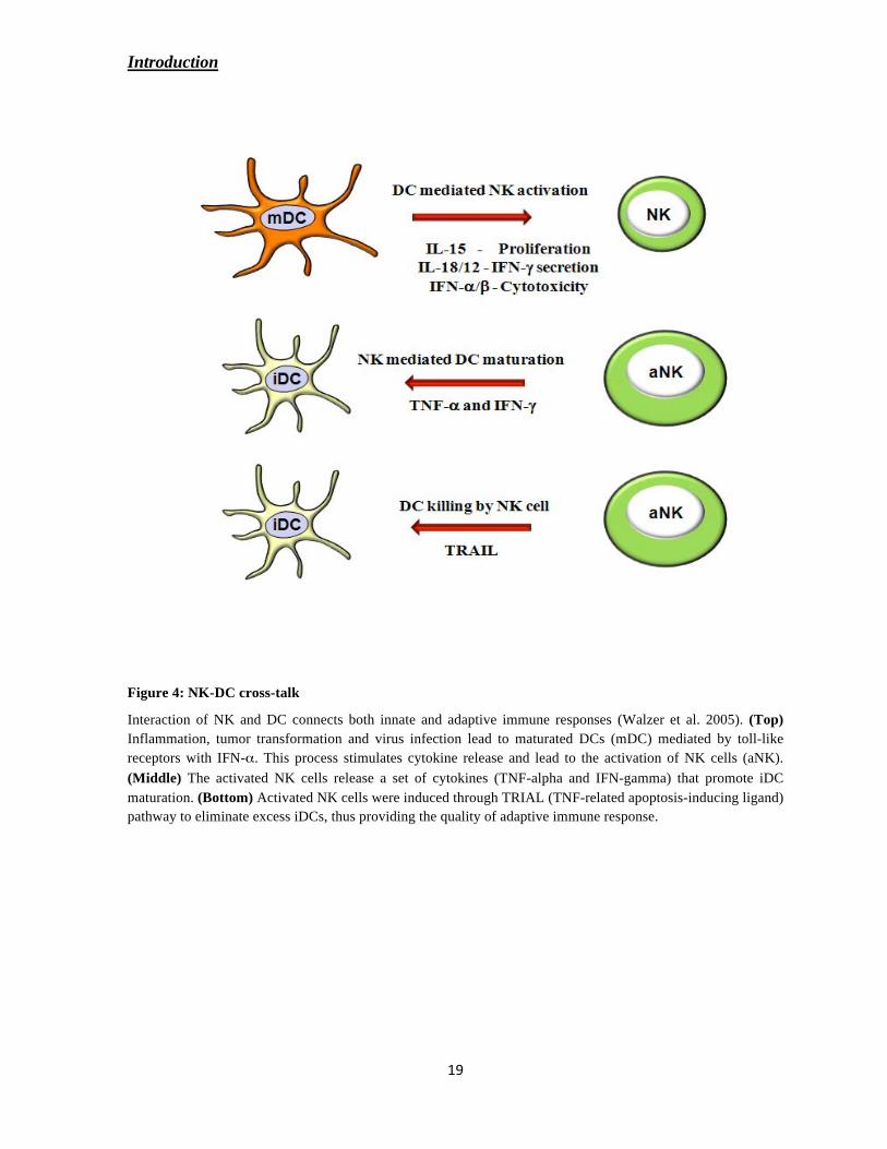
Figure 4: NK-DC cross-talk
Interaction of NK and DC connects both innate and adaptive immune responses (Walzer et al. 2005). (Top)
Inflammation, tumor transformation and virus infection lead to maturated DCs (mDC) mediated by toll-like
receptors with IFN- . This process stimulates cytokine release and lead to the activation of NK cells (aNK).
(Middle) The activated NK cells release a set of cytokines (TNF-alpha and IFN-gamma) that promote iDC
maturation. (Bottom) Activated NK cells were induced through TRIAL (TNF-related apoptosis-inducing ligand)
pathway to eliminate excess iDCs, thus providing the quality of adaptive immune response.
Introduction
NK-cell activation by tumor cells has been shown to promote the elicitation of cognate and
protective T-cell responses against the tumor. In some cases, T cell-mediated tumor rejection
was shown dependent on DC activation by NK cells (Mocikat et al. 2003). IFN-gamma
secreted during NK cell-mediated tumor rejection is critical for cytotoxic T lymphocytes
(CTL) generation, particularly when tumors express CD70 or CD80 and CD86 (Kelly et al.
2002). Furthermore, Adam et al. reported that NK-cell-DC crosstalk may by-pass the T
helper arm in CTL induction against tumors expressing NKG2D ligands (Adam et al. 2005).
Mutual activation of NK cells with other immune regulatory cells is mediated by several
receptor-ligand interactions particularly NKp30 with its ligand BAT3, NKG2D with its
ligands ULBP1-3 and MICA/B, DNAM-1 with PVR and Nectin-2 and NKp80 with AICL
(Newman and Riley 2007; Pogge von Strandmann et al. 2007; Simhadri et al. 2008).
The Vujanovic group has shown that DC and NK cells constitutively express several
TNF family ligands and corresponding TNF family receptors, these receptor-ligand pairs
greatly impaired DC-NK cell abilities to reciprocally mediate the increases in cytokines
(Makarenkova et al. 2005). Recent reports described that the cognate interaction of TNF-
TNFR2 is essential for mouse DC-NK cell cross-talk (Xu et al. 2007). These finding indicate
TNF and some other members of TNF family ligands might be important mediators of DC-
NK interaction and reciprocal stimulation. Therefore, this study extends the above
observations on the function of NK-DC cross-talk with the involvement of CD30-CD30L
interaction.
Aim of the study
Ligands of the tumor necrosis factor super-family (TNF-SF) and the corresponding receptors
have been the subject of extensive investigation during the last decades. Although CD30, a
member of TNF receptor super-family and CD30L, member of TNF ligand super-family were
identified and cloned long ago, it is still challenging to define the consequences of CD30-
CD30L interactions at both molecular and cellular levels.
CD30 is differentially expressed on almost all activated immune effector cells and over-
expressed in some malignant lymphomas. Previous studies describe that engagement of CD30
on CD30+ lymphoma results in H-RS cell survival in HL, cell-growth arrest in ALCL or Th1-
mediated responses in RA. In these diseases, the ectodomain of CD30 is shed in the
inflammatory region. The effect of sCD30 on CD30L+ cells is not clearly elucidated.
Moreover, the exact function of surface-expressed CD30 on normal activated NK cells was
not yet addressed. On the other hand, CD30L shows greater homology to other TNF ligand
members and is expressed on stimulated hematopoietic cells during the inflammatory
response. It is well described that interaction between CD30 and CD30L effects not only the
receptor bearing cells but also induce specific downstream signaling to ligand-bearing cells.
With respect to CD30L, definition of its biological role remained elusive because its
expression on different immune cells (granulocytes and DCs) is controversially discussed.
In recent years, there has been emerging evidence that NK cells interact with other immune
cells such as DCs, mast cells, eosinophils, basophils and neutrophils during inflammation.
Among these interactions, cross-talk between NK cells and monocyte-derived DCs has been
the major focus. Recent studies indicate that NK cell activation induced by DCs requires the
synergistic action of several cytokines and direct contact between DCs and NK cells. The
requirement for cell-cell contact is likely to reflect (i) the implication of membrane bound
receptor-ligand pairs; and/or (ii) the necessity for local delivery of cytokines at high
concentration at the interface between DCs and NK cells.

Aim of the study
The goal of the study is to elucidate the role of CD30-CD30 ligand-dependent signaling for
the communication among immune effector cells that is mediated either through cell-cell
contact or through the soluble CD30. For this the following issues will be addressed:
(i) Establishment of the expression pattern and functional properties of CD30L on
dendritic cells
(ii) Functional investigation of the CD30-CD30L interaction in NK-DC cross-talk
(iii) Elucidation of the expression profile of CD30L on granulocytes
(iv) Characterization of the CD30L-mediated cellular response of granulocytes to
soluble CD30

Materials and Methods
293T Human Fibroblast kidney cell line (ATCC, Manassas, USA)
L540 Human Hodgkin’s Lymphoma cell line (a gift from Prof. V. Diehl, Koeln)
Table 1:
Vector Fusion Selection marker
(Bacteria)
Selection marker
(Mammalian cells)
pcDNA 3.1 No tag Ampicillin Zeocin
pFuse 2
Fc-fragment of
human IgG1
Zeocin Zeocin
pIG (pCDM8)
Fc-fragment of
human IgG1
Ampicillin No resistance
Different bacterial strains Escherichia coli (E.coli) used for molecular biology:
DH5 , XL-1 blue, TG-1, BL-21 (DE3) and MC-1061.

Materials and Methods
Table 2:
Antigen
Fluoro-
chrome Company
Against
species
From
species
Iso-
type clone
CD3 Purified BD Biosciences human mouse IgG2a HIT3a
CD3 FITC BD Biosciences human mouse
CD3 PE Immuno Tools human mouse IgG1 UCHT1
CD3 APC Immuno Tools human mouse IgG2a MEM57
CD3 percp BD Biosciences human mouse
CD4 APC BD Biosciences human mouse
CD9 purified BD Biosciences human
CD14 FITC BD Biosciences human
CD14 PE BD Biosciences human mouse
CD16 purified BD Biosciences human
CD16 PE BD Biosciences human mouse
CD30 purified Laboratory of
Immune therapy human human 5F11
CD30 (Ki-1) Purified Dr. Lemke, Kiel
human mouse IgG3
CD30 (Ki-2) Purified Dr. Lemke, Kiel
human mouse IgG1

Materials and Methods
CD30 (Ki-3) Purified Dr. Lemke, Kiel
human mouse IgG2b
CD30-BerH2 Purified Dr. H. Stein,
Berlin human mouse IgG1
CD30-HeFi-1 Purified Dr. T. Ellis,
Chicago Human Mouse IgG1
CD30 PE BD Biosciences human mouse
CD30L purified R&D Systems human mouse IgG2b 210845
CD56 FITC Immuno Tools human mouse
CD56 PE BD Biosciences human mouse
CD56 APC BD Biosciences human mouse
CD1a PE BD Biosciences human mouse
CD1a APC Bio legends human mouse
CD83 FITC BD Biosciences human mouse
CD80 FITC BD Biosciences human mouse
CD80 PE Bio Legends human mouse
CD86 purified Immuno Tools human mouse
CD86 FITC BD Biosciences human mouse
CD86 PE BD Biosciences human mouse
NKp30 PE Beckman Coulter
human mouse IgG1 Z25
NKp44 PE Beckman Coulter
human mouse IgG1
NKp46 purified BD Pharmingen human mouse 9E/2
NKp46 PE Beckman Coulter
human mouse IgG1 BAB281

Materials and Methods
Kp46 APC BD Biosciences human mouse IgG2b
hNKG2D PE BD Biosciences human mouse IgG1
NKG2D Fitc Abcam human mouse IgG1 1D11
hNKG2A PE BD Biosciences human mouse IgG2a
HLA-ABC purified BD Biosciences human mouse IgG1 G46-2.6
HLA-DR purified BD Biosciences human mouse
HLA-A,B,C PE BD Biosciences human mouse
HLA-DR FITC BD Biosciences human mouse
IgG Fc purified Dianova human goat F(ab)2
IgG purified Dianova goat IgG
IgG H+L purified Dianova mouse goat F(ab)2
IgG PE BD Biosciences human mouse
human IgG1 PE BD Biosciences mouse goat
IgG APC BD Biosciences human mouse
7-AAD 7-AAD BD Biosciences
Annexin V PE BD Biosciences
anti-mouse FITC BD Biosciences mouse goat poly
anti-mouse PE Dako mouse goat poly

Materials and Methods
The following reagents were used: Human recombinant GM-CSF, IL-4 and TNF-alpha
(Immunotools, Germany), 8-chamber microscopy slides (Nalgen, Nunc International Corp,
Naperville, USA), 2’,7
’-Dichlorofluorescein diacetate (DCFH-DA), Phorbol myristate acetate
(PMA), Lipopolysaccharide (LPS), and Carboxy-fluorescein diacetate succinimidyl ester
(CFSE) (Sigma Aldrich Inc, St. Louis, USA), MAP kinase inhibitors PD98059, SB203580,
and SP600125 (Calbiochem-Novabiochem, San Diego, USA), Protein A sepharose beads
(GE Healthcare, Freiburg). NK cell, Pan T cell and Blood DC isolation Kits were purchased
from Miltenyi, Bergisch Gladbach, Germany).
All the laboratory chemicals and other reagents were purchased from Roth Chemicals and
Sigma Life Sciences.
All the oligo-nucleotides used for amplifying the desired gene products were obtained from
MWG-Biotech, Ebersberg, Germany.
Synthetic Oligonulceotides
Table 3:
Primer Name Oligonulceotide Sequence (5'-------------- 3') Restriction
Endonuclease
CD30 D1-2 for gcgagaagcttatgcgcgtcctcctcgccgcg HindIII
CD30 D1-2 rev ccggaattcgcaggtgcgggaggagttc EcoRI
CD30 stalk for ccggaattcaaccccaccccagagaatggcgag EcoR1
CD30 stalk rev gacggatccacttacctgtttcgcaggtgcgggaggagttcca BamH1
CD30 D1 for gcgagaagcttatgcgcgtcctcctcgccgcg HindIII
CD30 D1 rev gatggaattcgcagtcagtaggcctctgagggcac EcoR1
CD30 D6 for acccctcgagtgtcgacctggcatgatctgtgccac Xho1
CD30 D6 rev gacggatccacttacctgtttcgcaggtgcgggaggagttcca BamH1
Leader seq for gcgagaagcttatgcgcgtcctcctcgccgcg HindIII
Leader seq rev gacactcgaggggtcgatcctgtgggaaggctcgt Xho1

Materials and Methods
Construction of desired cDNA into expression vectors
The construction of pcDNA3.1-CD30 (wt) (CD30 full length), a kind gift from Dr. Hinrich
Hansen was described previously (Hansen et al. 2004). The ectodomain of CD30 is
comprised of six domains where in the last three domains are exactly the pictures of the first
three domains. The constructs pIgG1-LCD30-Ig (all domains), pIgG1-MCD30-Ig
(Domain-1, 2/5 and 6) and pIgG1-SCD30-Ig (Domain-1 and 6) were a kind gift from Dr.
Hinrich Hansen (Hansen et al. 2004; Eichenauer et al. 2007). The other deleted CD30
variants were constructed using the oligonucleotides mentioned in the Table 2.
pIgG1-D1-2 CD30-Ig (Domain-1 and 2/5)
The construct was generated using multiple cloning steps.
A) D1-2 CD30 (domain1 and 2/5) was amplified using pIgG1-CD30M as a template with
primers CD30 D1-2 for and CD30 D1-2 rev. Subsequently, the amplified product was
digested with HindIII and EcoRI restriction enzymes and sub-cloned into pEGFP-C1 (used as
a cloning vector to maintain the frame with pIg vector) named as pEGFP-C1 CD30 D1-2/5.
B) The stalk domain of CD30 was amplified using pIgG1-CD30M as template with primers
CD30 stalk for and CD30 stalk rev. The amplified stalk domain was inserted into the above
construct pEGFP-C1 CD30 D1-2/5 using EcoR1 and BamH1 restriction sites.
C) As a final step, CD30 D1-2/5 with stalk domain was digested from pEGFP-C1 using
HindIII and BamH1 and sub-cloned into pIgG1 CD30, replacing the full-length ectodomain
of CD30 to be in frame with human-IgG.
pIgG1- D1CD30-Ig (Domain-1)
A CD30 variant only with Domain-1 was amplified using pIgG1-CD30M as template with
primers CD30 D1 for and CD30 D1 rev. The amplified products were subsequently digested
with HindIII and EcoR1 and inserted into the expression vector (pIgG1) in frame with
human-IgG.

Materials and Methods
pIgG1- D6CD30-Ig (Domain-6)
The construct was generated in two steps.
(A) Domain-6 of the ectodomain was amplified using pIgG1-MCD30 as template with
primers CD30 D6 for and CD30 D6 rev. The amplified PCR product was digested with Xho1
and BamH1 and inserted into the expression vector (pIgG1) in frame with the Fc-fragment of
human-IgG1.
(B) Next, the leader sequence was generated using pIgG1-MCD30 as a template with primers
leader seq for and leader seq rev. The PCR product was sub-cloned into above construct to
enable the synthesized protein to be released into the cell culture supernatants.
pGEX-3T CD153 (CD30 ligand)
The cDNA for CD153 flanked by BamHI and EcoR1sites was amplified and ligated into the
pGEX-3T vector (GST- as a tag expressing in bacteria).
Isolation of plasmid DNA from E.coli
Isolation of plasmid was performed using Qiagen kit according to the manufacturer’s
protocol. In brief, bacteria (E.coli strains DH5 , MC-1061, XL1-Blue) were cultivated for at
least 12-16 hours in LB-Medium with respective antibiotics (e.g.100 g/ml Ampicillin) at
37°C. The cells were harvested, and were subjected to alkaline lysis and the plasmid-DNA
was isolated. The purified plasmid was quantified and confirmed using restriction enzyme
analysis.
Quantification of DNA and RNA
DNA and RNA samples were diluted in ddH2O before they were measured in Bio-Rad Smart
Spec 3000 Spectrophotometer. The measured wavelength is 260 nm; concentrations are
calculated by the conversion factors 50 μg/ml for double stranded DNA and 40 μg/ml for
RNA. The purity of the DNA preparation is given by the ratio of Abs 260 nm/280 nm. While

Materials and Methods
1.8 is ideal, lower values point to contaminations with proteins and aromatic substances
whereas higher ratios indicate possible contaminations with RNA.
Polymerase Chain Reaction (PCR)
PCR can be used for in vitro amplification of DNA fragments. A double stranded DNA
(dsDNA) serving as a template, two oligonucleotides (primers) complementary to the
template DNA, deoxyribonucleotides (dNTPs) and heat resistant Taq-DNA-polymerase (from
Thermus aquaticus) are required for this reaction. When a proofreading activity was
necessary, Pfu-DNA-polymerase (Pyranococcus furiosus) was used.
Primers may be designed having non-complementary ends with sites for restriction enzymes.
First step in PCR reactions is the denaturing of dsDNA at 94°C. Second, the reaction mix was
incubated at different annealing temperatures, depending on the G/C content of the primers.
Different programs provide an accurate calculation of the annealing temperature and other
primer properties. The specific annealing temperature for the primers was obtained using the
formula: TA = TM – 5oC where, {TM = 4 (G + C) + 2 (A + T)}. The third step with a
temperature of 72°C allows elongation of the new strand of DNA by the polymerase. The
time for this step depends on the fidelity of the polymerase. In general a high fidelity
polymerase, such as Taq, requires shorter elongation time, whereas Pfu requires longer
elongation times.
Agarose Gel Electrophoresis.
Agarose gel electrophoresis was performed to analyze the length of DNA fragments after
restriction enzyme digests and PCR, as well as for the purification of PCR products and DNA
fragments. DNA fragments of different molecular weight show different electrophoretic
mobility in an agarose gel matrix. Optimal separation results were obtained using 0.8-1 %
(w/v) agarose gels in TAE buffer at 15 V/cm. Horizontal gel electrophoresis apparatus of
different sizes were used. Before loading the gel, the DNA sample was mixed with 1/6
volume of the 6x DNA-loading buffer. For examination of the DNA fragments under UV-
light, agarose gels were stained with 0.1 g/ml Ethidium bromide. In order to define the size
of the DNA fragments, DNA molecular standard markers were also loaded on the gel.

Materials and Methods
Gel Extraction of DNA
Elution of DNA fragments from agarose gels was performed after excising the band of
interest from the agarose gel and using the Qiagen gel extraction kit according to the
application manual. Eluted DNA was dissolved in appropriate volume of 10 mM Tris-HCL,
pH 8.5 or water.
Ligation of DNA
T4-DNA-ligase (Fermentas) catalyzed the ligation of isolated DNA fragments and linearized
vector DNA. The desired DNA fragment (s) were ligated with respective vector (s) at ratio
(insert: vector = 3: 1) in 20 μl ligation reaction (0.5 U T4-DNA-Ligase; 1 l T4-Ligation
buffer) for 5-10 minutes at room temperature.
Preparation and transformation of competent bacteria
Preparation of Competent Bacteria
Single bacterial colony of the specific strain of E.coli was picked from LB-agar plate and
grown over night in LB media at 37oC. Next day the bacterial culture was diluted 1:100 and
grown till an optical density (OD) at 0.6-0.8. Bacterial culture was cooled down for 10
minutes in an ice-cold water bath before pelleting the bacteria 10 min, 800 X g at 4oC. The
pellet was re-suspended in 15 ml TFB1 buffer (buffer section) and incubated 10 minutes at
4oC. After centrifuging the bacteria (10 min, 800 X g, 4
oC, they were re-suspended in 2 ml
TFB II buffer. 200 ml aliquots of the chemo-competent bacteria were shock-frosted in liquid
nitrogen and stored at -80oC.
Transformation of competent E.coli cells
100 μl competent E.coli were incubated with the ligation reaction (usually 7 μl) for 30
minutes on ice. Afterwards the transformation mix was heat shocked at 42oC for 50 sec and
immediately placed on ice for 2 minutes. The mix was re-suspended in 900 ml LB media
without antibiotic, and incubated at 37oC with shaking for at least 45 minutes. Cells were
collected by centrifugation in table-top centrifuge 2000 X g for 5 minutes and plated on LB-
agar plates supplemented with appropriate antibiotics (e.g.100 μg/ml ampicillin). The plates
were incubated at 37oC for overnight.

Materials and Methods
Estimation of Protein Concentration
Protein concentration was estimated by colorimetric analysis using the Bi-Cinchoninic Acid
(BCA) Protein Assay (Thermo Scientific, Pierce). Protein standards dilutions of BSA were
used to construct a standard curve (five concentrations: 2 mg/ml, 1 mg/ml, 0.5 mg/ml, 0.25
mg/ml, and 0.125 mg/ml). Several dilutions of the protein sample were mixed and measured
at 562 nm. The concentration was automatically correlated to the data for the standards and
the protein concentration was recorded as mg/ml.
SDS-Poly-Acrylamide Gel-Electrophoresis (SDS-PAGE)
In presence of Sodium Dodecyl Sulfate (SDS), proteins with different molecular weight show
different electrophoretic mobility in a denaturative polyacrilamide gel. Protein samples mixed
with SDS-sample buffer were heated, when necessary, for 5 minutes at 95°C prior to loading
on the gel. Biorad Mini-Protean gel running system was used for running the gels. At the
completion of the run, gels were either stained with Coomassie-blue or transferred onto a
PVDF or nitrocellulose membrane for Western blot analysis.
Western Blotting (Transfer of Proteins)
Western Blotting is a technique that allows the transfer of proteins from a poly-acrylamide
gel onto the membrane. In this work the wet blotting method was used: Two layers of
Whatmann paper, soaked in Blotting buffer, were placed on the blotting chamber. Then the
membrane and the gel all soaked in Transfer buffer were overlaid. Finally, two layers of
Whatmann paper, in Blotting buffer, where placed on top. The transfer was performed at 250
mA for 90 minutes. Membranes were stained with Ponceau S (Bio-Rad) for assessment of
blotting efficiency and marker detection prior to immuno-detection.
Protein detection on membrane
After protein transfer on membrane, the latter was incubated in blocking solution (5% w/v
milk powder in TBS-Tween buffer or using Roti-Block) for 60 minutes. Antibodies (serum
diluted in blocking solution) were incubated for 60 minutes at RT or for over-night at 4oC,
followed by 3 times washing with TBS-Tween buffer. Peroxidase conjugated secondary
antibodies were applied for 60 minutes at RT. Membranes were washed 3 times for 10

Materials and Methods
minutes with TBS-T buffer and treated with Luminol 1 and Luminol 2 solutions. Thus
treated, the membranes were exposed to X-ray film for 5 seconds to 1 hour depending on the
strength of the signal.
Stripping of nitrocellulose membranes
When membranes had to be stained with another antibody, they were treated for 15-30
minutes with 100 mM Glycin-HCL, pH 2.5 and washed for 10 minutes with TBS-T buffer.
Purification of monoclonal antibody from hybridoma culture Supernatant
Step1: Hyridoma cell culture
• A frozen aliquot of mouse monoclonal (HeFi-1) that express monoclonal antibody
(subtype IgG1) against CD30 epitope was thawed and cutltured in RPMI-1640
medium containing 10% FBS and incubated at 37oC with 5% CO2.
• Once the cells were grown to 90% confluent stage, the medium was replaced by
serum-free hybridoma medium.
• The supernatant was collected for every two days (3 times) and stored at -20oC.
Step2: Affinity purification of antibody
• Appropriate volume of protein-A sepharose column was set according to the general
principle: culture supernatant contain 20-50 μg/ml of antibody, 1 ml of wet beads
(slurry) bind approximately 10-20 mg of antibody. The column was washed with
50mM Tris buffer pH 8.0.
• The filtered culture supernatant was adjusted to pH 8.0 by adding 1/10 volume of 1.0
M Tris buffer pH 8.0, and allowed to pass through the column at the flow-rate of 1ml
per minute.
• The column was washed thoroughly with at least 10 column volumes of 50 mM Tris
buffer.
• The antibody was eluted with 100 mM glycine (pH 2.7), and immediately the pH was
adjusted to pH 8.0 with Tris buffer.

Materials and Methods
• The eluted antibody was dialyzed against PBS (pH 7.4) and the concentration was
determined by BCA-protein assay.
Horse Radish Peroxidase (HRP) labeling of antibody
The procedure has three major steps
• Activation of HRP: To activate HRP, 4 mg of HRP was dissolved in 1 ml of distilled
water and 0.2 ml of freshly prepared 0.1 M Sodium Per-iodate (NaIO4) was added and
incubated for 20 minutes at 25oC, followed by a dialysis step with 1mM Sodium
Acetate (pH 4.4) at 4oC.
• Preparation of antibody solution: 1 ml of antibody solution (2 mg/ml) was dialyzed
overnight at 4oC in buffer containing 10 mM Potassium Phosphate (pH 8.0) and 50
mM Sodium chloride for overnight at 4oC.
• Labeling of antibody: The dialyzed HRP and antibody solutions were mixed. The
reaction was started by addition of 40 μl of 0.5 M Sodium Carbonate buffer (pH 9.5).
The antibody was incubated for 2 hr at 25oC, and further incubated 2 hr at 4
oC with
0.1 ml of Sodium borohydride (4 mg/ml in H2O). Finally, the antibody was dialyzed
against PBS. The antibody was stabilized with BSA (0.1 mg/ml) and few grains of
thymol and stored at -80oC.
Purification of CD30 Ig and human Ig
The truncated CD30-Ig was gifted by Dr. Hinrich Hansen (Eichenauer et al. 2007). CD30-Ig
and human-Ig (pFuse-hFc2 purchased from Invitogen) were transfected to 293T cells a
human fibroblast kidney cell line. After 48 hr of transfection supernatants were collected and
purified using Protein A sepharose (GE Healthcare). The purity was analyzed by SDS-PAGE
and the concentration was determined by BCA-protein assay.
Soluble CD30 (sCD30) purification
Approximately 4x10⁶/ml L540 cells were washed once with serum free cold media, incubated
for 90 minutes at 37oC in serum free media. Supernatant was collected and removed the cells
and filtered with 0.2 m, followed by ultracentrifugation for 60 minutes at 40,000 RPM. Run
the supernatant in NHS activated (anti CD30) Ki-2 column or isotype matched column. Wash

Materials and Methods
the column with 75 mM Tris with pH 8 and eluted with 100 mM Glycine at pH 2.7. The
sample was dialyzed against PBS at 4oC.
RT-PCR Analysis of CD30L
To evaluate the expression of CD30L mRNA, RNA was isolated from iDCs (1x10⁶) and used
1 ng of RNA was converted to cDNA by reverse transcriptase at 37o C for 1 hr. PCR reaction
was performed using the following primers;
Forward primer: 5 GAGGAATTCGGTTCAGAGGACGGACTCCATTCCCAAC 3
Reverse primer: 5 GTGAAGATCTTCAGTCTGAATTACTGTATAAGAAGATGGAC 3
Binding of CD30 to iDC
Nearly 5 μg/ml of recombinant CD30-Ig and its variants were incubated with 1x10⁵ cells each
FACS tube, incubated on ice for 40 min and washed the cells twice with FACS buffer and
followed by detected with FITC conjugated to anti-human Ig (gamma specific) antibody and
analyzed by flow cytometry.
Interaction of CD153 with CD30 (Pull-Down Assay)
For pull down assays GST fusion proteins (GST alone and GST-CD153) were expressed in E.
coli BL21-D3 cells and were immobilized on glutathione-sepharose 4B beads (Amersham).
The purified CD30-Ig constructs were expressed in mammalian cells (293T) and purified
from the supernatants using Protein-A column. In a typical pull down reaction ~20 μg of
recombinant protein-coated beads were incubated with 10 g/ml of purified CD30 protein for
30 minutes with end-over-end rotation at 4ºC. Beads were washed with buffer (TBS buffer +
0.1% BSA + 0.1% Triton X-100) and boiled in 5X SDS sample buffer and resolved on 10%
SDS-PAGE. The gel was processed for Western transfer and the membranes were probed
with anti-CD30 antibodies and developed using ECL (Amersham).

Materials and Methods
Freezing and thawing cells
Cells were harvested and pelleted before re-suspending them in 1 ml freezing solution
containing 90% FCS and 10% DMSO. Immediately, the suspension was put on ice and then
stored at -20oC for 1 day subsequently at -80
oC for one week, finally stored in liquid nitrogen.
For thawing cells, the freezing vial was taken out from the liquid nitrogen tank and
transported on ice. Carefully, the suspension was thawed in a water bath at 37oC. The cells
were transferred into a 15 ml plastic tube containing pre-warmed medium before pelleting the
cells at 300 X g for 5 min at room temperature in order to remove toxic DMSO. After re-
suspension in fresh medium, the cells were plated in culture flasks.
Generation of Dendritic cells
The sources of cells were buffy coats acquired from healthy donors. PBMCs were separated
by Ficoll-Plaque gradient centrifugation with Leucosep columns from Greiner bio-one
(Solingen, Germany) and were incubated with RPMI media without FBS for 60 minutes in
plastic culture flasks (Nunc) at 37oC. Suspension cells were washed out and the adherent
monocytes were cultured in IMDM media containing 10% FBS, IL-4 (20 ng/ml) and GM-
CSF (50 ng/ml) for differentiation. The medium was changed after 3 days and the purity of
immature dendritic cells was assessed after 5 days culture by flow cytometry (FACS analysis)
using fluorescence conjugated antibodies CD14, CD1a, CD80, CD86 and CD83.
Purification of Blood Dendritic Cells
Blood dendritic cells were isolated from PBMCs by using blood dendritic cell isolation kit
11. Isolation of dendritic cells is performed in a two-step procedure. First, PBMCs are labeled
with the Non-DC depletion cocktail, later the flow-through fraction with pre-enriched
dendritic cells were incubated with DC enrichment cocktail, upon magnetic separation, the
magnetically labeled dendritic cells are retained on the column and are eluted after removal of

Materials and Methods
column from the magnetic field. The cell purity was assessed by using following antibodies
monocytes (CD14-), B cells (CD19
-), NK cells (CD56
-), and Dendritic cells (CD1a
+).
Maturation of dendritic cells
In this report for in-vitro maturation, monocytes-derived immature dendritic cells were
stimulated with different CD30-Ig fusion constructs that were immobilized as a mimic for
membrane anchored CD30. When mentioned soluble factors (e.g. soluble CD30) were added
into the media directly. TNF-alpha and LPS were used as positive controls. The incubation
period for maturation was 48 hours. Subsequently, the maturation markers were assessed by
flow cytometry. The mature dendritic cells were determined by the up-regulation of co-
stimulatory molecules like CD83, CD80 and CD86. For NK-DC co-culture, cells were
cultured at a ratio of 1:5 (NK: DC) for 48 hours and analyzed by flow cytometry.
Purification and Activation of NK Cells
PBMCs were isolated as described above, and non-NK cells were depleted using the NK cell
Isolation Kit and vario MACS. Separated polyclonal NK cells were cultivated for 2-3 days in
the presence of RPMI supplemented with antibiotics, 10% FBS, 5 μg/ml PHA and
recombinant IL-2 100 U/ml. The purity was analyzed with NK cell markers like NKp46,
NKp30, CD56, CD3, NKG2D and CD30. For the preparation of NK cell clones, PHA-
activated NK cells were plated in 96-well plate as 3 cells per well and cultured with 100 U/ml
IL-2 and irradiated PBMCs (allogenic) for 5 days and monitored for the expression of NK
cell markers. The medium was replaced for every 2 days and the selected clones were
cultured and analyzed further.
Purification of T cells
MACS
PBMCs were obtained from buffycoats as described above, and Pan T cells were purified by
negative selection according to the manufacturs protocol (MACS Pan T cell isolation Kit). To
verify the purity of the Pan T cell preparations, cells were stained with antibodies to CD3,
CD4, CD8, CD14, CD56 and CD19 (BD pharmingen). To generate the activated T cells were
cultured in RPMI1640 supplemented with 10% FBS, 20 mM glutamine, 100 units/ml
Materials and Methods
pencillin and streptomycin and 1 μg/ml phytohemagglutinin (PHA) for 2-4 days. For resting
T cells were purified just before doing the experiment.
Rosettesep Procedure
For some experiments we have used Rosettesep Kit to prepare human T cells from stemcell
technologies. Briefly, fresh blood was collected from normal healthy donors in EDTA
containing tubes. Human T cell enrichment cocktail was added at 50 μl/ml of blood and
incubated 20 minutes at room temperature, then diluted the blood sample with equal volumes
of PBS+2% FBS. Diluted sample was overlayed on Ficoll-Paque and centrifuge for 20 min at
1200 X g, collected the enriched T cells in-between Ficoll-Paque and plasma. Finally wash
the T cells with PBS+2% FBS. Purity of T cells was measured by flow cytometry after
staining with antibodies to CD3, CD4, CD8, CD14, CD56 and CD19 (BD Biosciences).
Isolation of Granulocytes
Whole blood obtained from healthy donors was collected in EDTA- containing tubes and
diluted with equal volumes of RPMI without FCS. The diluted blood was carefully placed on
Ficoll slowly through the walls of the falcon tube and centrifuged for 35 minutes at 350 X g
at room temperature (RT) without brake. Supernatant was discarded and the erythrocyte
pellet was re-suspended in the double volume of 1% polyvinyl alcohol (PVA) and allowed to
stand for 20 minutes at RT without any agitation. During the incubation period, the
erythrocytes settle at the bottom. The granulocytes were collected from the supernatant and
diluted with double the volume of ice cold PBS and spun for 10 minutes at 250 X g at 4oC.
The supernatant was discarded and the pellet was re-suspended and left in ice-cold water for
12 seconds and finally washed with ice-cold PBS. The cells were re-suspended with PBS.
Pappenheim staining of Granulocytes
Cytospins were performed to fix the granulocytes onto glass slides. The fixed cells were
stained for 3 minutes in May-Grunewald stain. The slides were washed using the buffer
containing of 2.6 mM KH2PO4 and 2.6 mM Na2HPO4. And the slides were further incubated
for 20 minutes with Giemsa stain diluted with 9 volumes of distilled water. Then, slides were
washed and dried enough before proceeding to light microscopy.

Materials and Methods
Flow cytometry
Cells were washed with FACS buffer (PBS+0.2% BSA+0.2%NaN3) and incubated with
primary Ab for 40 min on ice, washed twice and followed by 20-30 minutes with secondary
Ab incubation either by FITC or PE conjugated on ice in the dark place. After the incubation,
cells were washed two more times and measured in FACS Calibur. Some experiments
directly conjugated Abs was used.
Immunoflorescence of dendritic cells
Monocyte-derived immature dendritic cells were placed on poly-L-lysine coated 8 well
chamber slides for overnight in the presence of IMDM media. The cells were washed with
PBS and fixed with methanol for 10 minutes at -20oC. After the cells were washed with PBS,
blocked with 10% FBS in PBS for 1hr at room temperature and further incubated with
primary antibody (monoclonal CD30L Ab from R&D systems) for overnight at 4oC. Later
cells were washed and stained with Alexa 594 diluted in blocking buffer. The nucleus was
detected with Hoechst 33342 (Invitrogen). Finally washed with water and then mounted the
slide with mounting medium (aqua poly/mount – Polysciences, Inc). The slides were
examined in Nikon ECLIPSE E800 florescence microscope with an objective lens of 20X.
The images were obtained at 590 nm excitation and 617 nm emissions. The images were
processed using Adobe photoshop.
Reactive oxygen Species Signaling (oxidative burst)
Approximately 0.5x10⁶ DC/ml were stimulated with immobilized constructs at a final
concentration of 5 μg/ml for various periods, and an oxidative sensitive dye, 2’,7
’,- DCFH-
DA was added at 5 μM during the last 15 minutes of incubation. DCFH-DA is nonfluorescent
until oxidized by ROS. An increased DCF fluorescence indicates oxidation by peroxides,
including hydrogen peroxide, hydroxyl radical and peroxynitrite. Samples were washed with
FACS buffer and analyzed by FACS for fluorescence signals within the PI- populations.
Enzyme-Linked-Immuno-Sorbent-Assay (ELISA)
1.5x10⁵/ each 24 well iDCs were incubated for 5-6 hr with respective immobilized wells and
supernatants were analyzed using specific ELISA detection Kits (BD biosciences). In brief,
Materials and Methods
100 μl of diluted capture antibody was added in maxisorb 96 well plate and incubated
overnight at 4oC, after washings plates were blocked with 200 μl of block buffer
(PBS+10%FBS) for 1 hr at RT. After adequate washings either standard or samples were
incubated for 2 hr at RT. Finally the plate was washed and detected with the specific
detection antibody conjugated to HRP. The absorbance of the plates was measured using
ELISA-reader μ-Quant (Bio-Tek, Bad Friedrichshall, and Germany) in parallel with the
measurement of the corresponding standards.
Fluorescent Bead Immuno assay
The bender med systems human Th1/Th2 11 plex assay was carried out according to
manufacturer’s protocol.
Soluble CD30 ELISA
Flexible 96-well micro titer plates were coated with 100 μl of Ki-2 mAb (50 μg/ml in sodium
carbonate buffer 50 mM, pH 9.2) for overnight incubation at 4oC. The plates were washed 3
times with PBS + 0.05% Tween 20 (wash buffer) and subsequently blocked with PBS with
10% FCS for 1 hr at RT. After triplicate washings, serial dilutions of sCD30 standard and test
samples were added and incubated for 1 hr at room temperature. Plates were washed for 3
times and incubated with 100 μl of peroxidase-coupled Ki-3 mAb for 1 hr at RT. The plates
were further washed and detected with 100 μl of OPD substrate solution (Sigma Aldrich, St
Louis, USA). After incubation the plates were evaluated at 492 nm using ELISA reader.
CD30-Dependent Maturation of Dendritic cells
DCs were collected on day 6 and cultured around 3x 10⁵ cells in 12 well coated with 5 μg/ml
concentrations of different CD30-Ig constructs, in the presence of IMDM media for 36-48
hours. The cells were harvested and checked for the surface expression of co-stimulatory
molecules in FACS analysis.
For the NK-DC co culture, iDCs were collected on day 5 and incubated with activated NK
cells at 5:1 for 36-48 hours in medium (RPMI+5% FBS) at 37o
C and 5% Co2 .DCs were
analyzed for maturation markers in flow cytometric analysis. Dendritic cells alone were used
as the control for the co-culture experiments.

Materials and Methods
Confocal Microscopy
Mo-DC (iDC or CD30 stimulated mature DC) and T cells were gently mixed at a ratio at 1:3
and spread onto a slide coated with poly-D-lysine. Slides were incubated for 25 minutes a
37oC. After incubation cells were washed with 1X PBS and fixed in 4% paraformaldehyde
and permeabilized with PBS+0.5% Saponin (Wash buffer) for 30 minutes at room
temperature. The cells were blocked for further 30 minutes in blocking buffer (wash buffer +
3% BSA) and washed 3 times before immuno staining. Cells were stained with appropriate
primary antibodies (e.g. anti-human CD3) diluted in wash buffer +1% BSA for overnight at
4oC. Later, the slides were extensively washed and incubated with the specific secondary
antibodies (e.g. Alexa 594) for 1 hour at room temperature. The nucleus was stained with
Hoechst 33342 for 10 minutes at room temperature. The cells were finally washed with water
and then mounted the slide with mounting medium (aqua poly/mount – Polysciences, Inc).
Confocal stack images were obtained with a Zeises LSM 150 laser scanning confocal
microscope using a 60X W 1.4 NA apochromat plan objective. Z-projection of slices and
image analyses were performed using Zeises LSM image examiner software and Image J.
The pictures were finally made using Adobe Photoshop.
Proliferation assay by CFSE
Principle: This method has been useful in determining cell division in B and T cells. The
carboxyfluorescein diacetate succinimidyl ester (CFSE) passively diffuses into cells. It is
colourless and non-fluorescent until the acetate groups are cleaved by intracellular esterases
to yield highly fluorescent carboxyfluorescein succinmidyl ester. The succinmidyl ester group
reacts with intracellular amines, forming fluorescent conjugates that are well retained and can
be fixed with aldehyde fixatives.
The dye-protein adducts that are formed in labelled cells are retained throughout
development and meiosis. The label is inherited by daughter cells after either cell division or
cell fusion, and is not transferred to adjacent cells in a population.
Method: CFSE stock solution was prepared in DMSO according manufacture’s protocol
(Molecular probes, Invitrogen). Resting T cells were resuspended with PBS+0.1%BSA at a
final concentration of 1x10⁶/ml. The CFSE at 10 M final concentration was added to cells

Materials and Methods
and incubated at 37oC for 10 minutes. The cells were further incubated on ice for 5 min by
addition of 5 volumes of ice-cold media. Cells were washed 3 times with fresh media and co-
cultured in different ratios with either iDC or CD30 stimulated DC and incubated at 37oC for
5-6 days and measured for proliferation by flow cytometry at 488 nm (FL-1 staining).
Stimulation of Granulocytes
Freshly isolated granulocytes 1x10⁶/ml were incubated in the presence of sCD30 transfected
or mock transfected or untransfected supernatants in the presence or absence of antiCD30
antibody (HeFi-1) in serum free RPMI medium. After over-night incubation, supernatants
were harvested and analyzed in IL-8 ELISA according to manufactures (BD Bio-Sciences)
protocol.
Time-lapsed video microscopy data analysis and cell tracking
24 well plates were pre-coated with fibronectin at concentration of 10 μg/ml diluted in PBS
for 1 hr at room temperature and washed twice with serum free medium. Freshly isolated
granulocytes were seeded in wells and incubated for 30 minutes at 37oC to allow them to bind
to the coated plates. Then, non-adherent cells were carefully washed out and 200 μl of 0.1%
BSA+ 200 μl transfected supernatant was added. Blocking experiments were performed with
either anti-CD30 antibody (HeFi-1) or isotype antibody at concentration of 3μg/ml. The
surface of the medium was overlaid with 400 μl mineral oil to avoid evaporation. The
migration of cells was recorded for 60 minutes in an incubator chamber attached to 1X81
microscopes (Olympus Europa GmbH, Hamburg, Germany) at 37o
C, 5% Co2. For tracking,
20-40 cells within the microscopic field of approximately 800 600 m were randomly
selected in each experiment and were analyzed using a computer-assisted cell tracking
system. Frames were taken for every two minutes using an OBS CCD FV2T camera
(Olympus). For visualization and quantification of cell movements, the computer programs
OBS cell R (Olympus) and DIAS (Solltech.Oakdale, IA) were used. Individual cells are
tracked based on similarities in brightness and shape. The directionality of an individual cell
is defined as the distance between the start and end position of the cell. The locomotive
pattern and moving speed were analyzed by drawing the trajectory of each cell using
digitizer.

Materials and Methods
Trans-well assay
24 well plates (3 μm pore size) were pretreated with 0.1% BSA and washed twice with RPMI
media. The lower chamber was filled with 300 μl of 0.2% BSA+ 300 μl of transfected
supernatants and the upper chamber contains 106 granulocytes in 150 μl of RPMI+0.1% BSA.
Plates were incubated at 37oC for 50 minutes and measured by trypan blue staining for the
migratory cells in the lower chamber.
The results of all the cytokine release assays are indicated as means + standard deviation.
Significance was calculated with Graph Pad Prism software (San Diego, CA) using student t-
test. Other software used are Microsoft Office, Adobe Photoshop, Win-MDI, Cyflogic and
Image J.
Primer design and analysis, elementary DNA and protein sequence analysis were performed
using DNA star.

Composition of Buffers
10X PBS: 1.37 M NaCl
27 mM KCl
100 mM Na2HPO4 x 2 H2O
20 mM KH2 PO4
10X TBS: 1.37 M NaCl
27 mM KCl
248 mM Tris Base
FACS Buffer: 1x PBS +0.2% BSA+0.2% sodium azide.
ELISA Buffers: Coating Buffer: 8.4 g NaHCO3 +3.4 g Na2CO3 in 1 liter, pH 9.5
Dilution Buffer: 1xPBS+10%FBS
Wash Buffer: 1xPBS+0.05% Tween 20
FPLC column Buffers:
Binding buffer: 50 mM Tris Base, pH 8.0
Wash Buffer: 50 mM Tris Base, pH 8.0
Elution Buffer: 100 mM Glycine+100 mM NaCl, pH 2.7
Blot Buffers:
PVDF Membrane: 3 g Tris Base
14.4 g Glycine
200 ml Methanol
800 ml H2O
Nitrocellulose Membrane: 250 mM Na2HPO4
Wash Buffer: 1xTBS+ 0.1% Tween 20.

Composition of Buffers
50X TAE Buffer: 2 M Tris acetate
50 mM EDTA (pH 8.0)
LB Medium: 1% Tryptone
1% NaCl
0.5% Yeast extract
LB Medium: 1% Tryptone
(Low Salt) 0.5% NaCl
0.5% Yeast extract
Zeocin concentration 25 μg/ml
LB agar: LB medium + 1.5% Agar
Transformation Buffers:
TFB1 30mM CH3COO Na
50mM MgCl2x6H2O
10mM CaCl2x2H2O
100mM NaCl
15% glycerol pH: 5.8
TFB2 10mM Mops
75mM CaCl2x2H2O
10mM NaCl
15% glycerol pH 7.0

Results
The sources of cells were buffy coats acquired from healthy donors. PBMCs were separated
by Ficoll-Plaque gradient centrifugation with Leucosep columns and were incubated 45
minutes in plastic culture flasks (Nunc) at 37oC. Suspension cells were washed out and the
adherent monocytes were allowed for differentiation in IMDM media containing IL-4 (20
ng/ml) and GM-CSF (50 ng/ml) (Fig. 5A).
The purity of immature dendritic cells was assessed after 5 days culture using FACS
analysis. Immature DC were CD14-, CD1a
+, CD80
+, CD86
+ and CD83
-/very low. As shown in
Fig. 5B, only the cells were used that were 90-95% negative for CD14 and CD83, which are
markers for monocytes and mature DCs, respectively.
Figure 5: Generation of monocytes-derived dendritic cells. (A) Schematic representation of dendritic cell
purification. (B) Surface phenotype of monocytes-derived immature dendritic cells. Non-adherent cells were
stained for CD14, CD83, CD1a, CD80 and CD86 conjugated to fluorescent antibodies and analyzed by FACS.
The relative cell count was kept constant on the Y-axis and the fluorescence was shown as histograms. The grey

Results
histograms represent the antibody controls and the opened color histograms determine the surface expression of
different markers.
The expression of CD30L is restricted to immune cells and tightly regulated. Previous studies
showed that human immature dendritic cells express on their cell surface cytotoxic TNF
family ligands (Lu et al. 2002) and recent studies also demonstrated that CD30L is up
regulated on myeloid and plasmacytoid dendritic cells in mice upon inflammation (Zeiser et
al. 2007). It was therefore tested whether human dendritic cells might express CD30L
(CD153). The classical immature dendritic cells were generated from adherent monocytes,
from PBMCs of healthy donors. Stimulation of immature dendritic cells with TNF-alpha for 2
days, led to the maturation of DCs (mDCs). The CD30L expression on human DCs during
differentiation was examined by RT-PCR and FACS analysis. The mRNA expression of
CD30L was partially detectable during 3rd
day of monocyte-differentiation and was up
regulated on 6th
day of differentiation (iDCs). In contrast, the CD30L mRNA expression was
reduced in mDCs stimulated by TNF-alpha (Fig. 6A). The RT-PCR products correspond to
the same size as the positive control (recombinant CD30L construct). The housekeeping gene
GAPDH served as a control and confirmed equal synthesis of cDNA in all the cell types. In
line with the mRNA expression, the surface expression of CD30L protein on Mo-iDCs is
present and reduced on TNF alpha-mDCs as demonstrated by mean fluorescence intensity
(MFI) using flow cytometry (Fig. 6B). Consistent to the previous studies the expression level
of co-stimulatory receptors CD80, CD83 and CD86 was up regulated upon maturation and
the detection level of the controls (antibody and CD14) was not affected (Fig. 6B).
In addition, a representative histogram of CD30L expression on the surface of iDCs
was obtained by FACS analysis, where cells were stained with isotype and CD30-ligand
antibody and detected with PE-conjugated goat anti-mouse antibody, measured in FACS
calibur (BD Biosciences) (Fig. 6E). Moreover, the surface expression was also determined by
microscopy. For microscopy analysis, Mo-iDCs were stained with monoclonal antibodies
anti-human CD30L and corresponding isotype followed by detection with the specific
secondary antibody rabbit anti-mouse conjugated to Alexa-594. All the nuclei were stained

Results
with Hoechst 33342 (Fig. 6C). Analysis of the cell lysates by western blotting with specific
antibody also revealed the protein expression (Fig. 6D).
Figure 6: Expression of CD30L on monocyte-derived DCs. (A) Human iDCs were differentiated from
adherent monocytes in presence of GM-CSF and IL-4 for 6 days. Mature DCs were generated from iDCs by
stimulating with (20 ng/ml) TNF-alpha for additional 2 days. The expression of CD30L mRNA was assessed by
standard RT-PCR. GAPDH was used as a control over the same period. M: marker, +: positive control for
CD30L, 3: 3rd
day, 6: 6th
day (iDC), Mat: mature DCs (TNF-alpha). (B) iDCs (shaded bars) and mature DCs
(squared bars) were analyzed for surface expression of CD14, CD83, CD80, CD86 with conjugated antibodies
and un-conjugated CD30L mAb followed by staining with PE goat anti-mouse Fab. Neg: cells alone, Control:
Isotype antibody for CD30L. The mean fluorescence intensity (MFI) on the Y-axis was represented in two

Results
segments and the segment 65-130 represents for only CD86. (C) CD30L surface expression was shown by
histogram analysis. Gray shaded histogram represents the isotype antibody; and open histogram represents
staining of CD30L. (D) iDCs were stained with isotype antibody (left), or anti-CD30L (right). Blue color
represents the nucleus staining and red color indicates the CD30L specific antibody detection. (E) iDC lysate
was checked for protein expression by western blot analysis. 10 μg/ml of iDC lysate was loaded in 10% SDS
PAGE gel and detected with mAb for CD30L.
Next it was investigated whether freshly isolated peripheral blood DCs express
CD30L. Blood DCs were purified and isolated from PBMCs of healthy donors using positive
selection by MACS separation. The expression of CD30L on human blood DCs was
determined by RT-PCR and FACS analysis. The purity of the cells was assessed by flow
cytometry as CD14-, CD1a
+. As shown in Fig. 7A, specific amplified products were clearly
detectable demonstrating the presence of CD30L mRNA in blood DCs. To determine whether
transcription of the CD30L was accompanied by specific protein expression in blood DCs,
the cells were stained with anti-CD30L and were analyzed by flow cytometry (Fig. 7B).
Taken together, CD30L is expressed on DCs.
Figure 7: Expression of CD30L on blood-derived DCs (A) Blood DCs were purified from PBMCs using auto
MACS and the expression of CD30L mRNA was determined by RT-PCR. Lane 1: CD30L construct, positive
control, lane 2 is CD30L expression, lane 3: GAPDH control and last lane M is Marker. (B) Surface expression
of CD30L on blood iDC was analyzed by flow cytometry. Shaded grey region is isotype control and opened
histogram (blue) shows the CD30L expression.

Results
The ectodomain of CD30 and deleted portions thereof were generated as human-Ig fusion
proteins. The DNA constructs were analyzed by sequencing either with T7 promoter or gene-
specific forward primers. In this study, the constructs were labeled as LCD30 (entire
ectodomain containing domain 1 to 6 and stalk), MCD30 (domain 1-2-6 and stalk), SCD30
(domain 1 and 6 and stalk), D1-2CD30 (domain 1-2 and stalk), D1CD30 (domain 1 and stalk)
and D6CD30 (domain 6 and stalk). Figure 8A shows the schematic representation of the
constructs. The CD30-Ig constructs were expressed in 293T cells by transfection with
specific cDNAs using lipofectamine (Invitrogen). After transfection the supernatants were
collected twice for every 48 hours. An affinity column with Protein-A sepharose beads (GE)
was used to purify the Ig-proteins from the harvested supernatant on HPLC instrument (Bio-
Rad). The purified proteins were dialyzed against PBS and sterilized by filtration. The
concentrations of proteins were determined by bicinchoninic acid-based protein assay
(Pierce). The purified CD30-Ig proteins were also analyzed by Coomassie brilliant blue
staining after SDS/PAGE analysis (Fig. 8B). The specificity of the human-Ig was
demonstrated by western blotting using anti-human Ig antibody (gamma-specific antibody)
and followed by HRP-conjugated secondary antibody (Fig. 8C). This showed the expression
of CD30 ectodomain variants fused with the Fc-portion of human-Ig. Additional bands of
lower molecular weight were observed in Coomassie as well as in western blot; they probably
represent traces of the degraded proteins. In summary recombinant-Ig constructs were
expressed, purified and feasible for the further functional experiments.
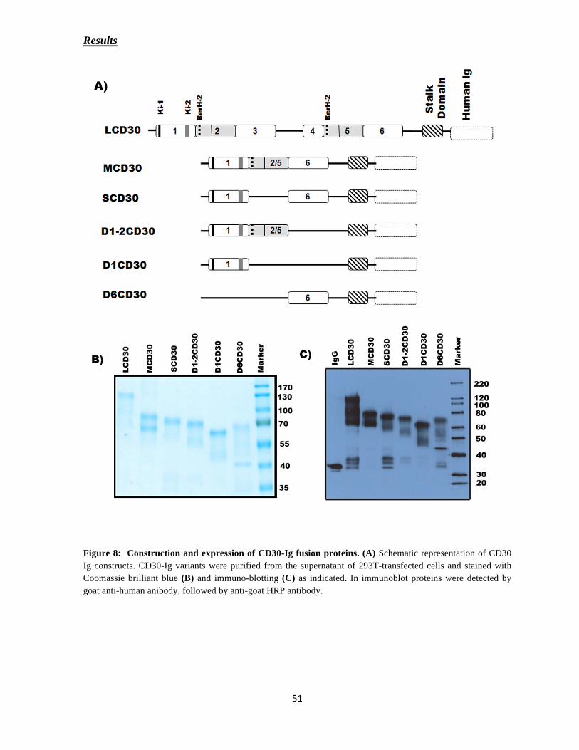
Results
Figure 8: Construction and expression of CD30-Ig fusion proteins. (A) Schematic representation of CD30
Ig constructs. CD30-Ig variants were purified from the supernatant of 293T-transfected cells and stained with
Coomassie brilliant blue (B) and immuno-blotting (C) as indicated. In immunoblot proteins were detected by
goat anti-human anibody, followed by anti-goat HRP antibody.

Results
8.4.1. FACS Analysis:
To study the effect of CD30 on DCs, monocyte-derived iDCs were stained with CD30-Ig. To
identify the specific domain that binds to iDCs, all tested proteins, were dialyzed against
PBS. Proteins were incubated on ice at 10-μg/ml for 40 minutes to allow binding to CD30L
positive iDCs and the fusion constructs were detected with FITC-conjugated anti-human Ig
(Ig-gamma specific) using FACS analysis. The mean fluorescence intensities determined by
FACS analysis, indicated that the constructs LCD30, MCD30, SCD30 and D1-2CD30
interact with DCs albeit with different intensity. Staining with the constructs D1CD30
(domain 1 alone) and D6CD30 (domain 6 alone) did not notably interact with Mo-iDCs (Fig.
9), this demonstrates specificity of the binding of CD30 constructs L, M, S and D1-2.
Figure 9: Interaction of CD30-Ig variants to iDCs. The mean fluorescent intensity (MFI) on Y-axis
determines the strength of binding.
8.4.2. Pull-down Assay
To demonstrate binding of CD30L to recombinant CD30-Ig, the in-vitro interaction was
further investigated in an independent assay using pull-down experiments. CD30-Ig
constructs were purified from mammalian cells and used as soluble proteins. CD30L was
produced as GST fusion protein. GST alone (control protein) and GST-CD30L was expressed
in E. coli and purified as recombinant proteins possessing an N-terminal Glutathione-S-

Results
Transferase (GST) tag. The GST fusion proteins were immobilized on glutathione-Sepharose
beads to pull down the specifically interacting proteins. As shown in the Fig. 10, immobilized
GST-CD30L was able to pull down LCD30, MCD30 and SCD30 whereas the control protein,
immobilized GST did not pull-down any of the CD30-Ig constructs. GST-CD30L pull-down
of CD30-Ig was detected when performed under non-reducing conditions. The above data
indicate that under non-reducing conditions the disulphide bridges were intact between the
cysteine residues present in the extra-cellular domain of CD30. This further suggests that the
interaction between CD30-CD30L also depends on the cysteine-rich domains (CRD).
Probably as mentioned for other TNF receptors like TNFR1, the CRDs in the ectodomain of
CD30 could also enable the formation of trimeric structure that binds to its cognate ligand.
The above interaction data imply that at least two CRD domains of CD30 (TNF-
receptor) are required for binding to CD30L (TNF ligand). Therefore, CD30L could bind to
LCD30-Ig, MCD30-Ig, SCD30-Ig and D1-2CD30-Ig but not to the constructs with only one
CRD domain (D1CD30-Ig and D6CD30-Ig).
Figure 10: Pull-down assay. GST-CD30L and GST were immobilized on Glutathione Sepharose beads and
incubated at 4°C with 10μg/ml of LCD30-Ig, (15 minutes), MCD30-Ig (15 minutes) and SCD30-Ig (90 minutes)
to assess their ability to pull down CD30 variants. Beads were thoroughly washed with buffer and were boiled in
SDS-PAGE non-reducing sample buffer and the solubilized proteins were resolved on 10% polyacrylamide gels.
The pulled-down of LCD30-Ig and MCD30-Ig was detected using Ber-H2 and SCD30-Ig was detected by Ki-2
antibody, followed by incubation with anti-mouse HRP and then analyzed by immuno-blotting. 200 ng of
purified protein was loaded as input. Interaction of CD30L is seen with CD30 variants, whereas GST alone does
not show any interaction.
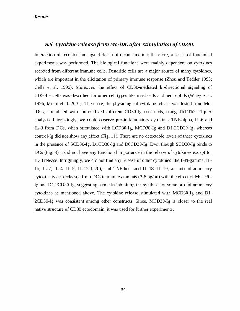
Results
Interaction of receptor and ligand does not mean function; therefore, a series of functional
experiments was performed. The biological functions were mainly dependent on cytokines
secreted from different immune cells. Dendritic cells are a major source of many cytokines,
which are important in the elicitation of primary immune response (Zhou and Tedder 1995;
Cella et al. 1996). Moreover, the effect of CD30-mediated bi-directional signaling of
CD30L+ cells was described for other cell types like mast cells and neutrophils (Wiley et al.
1996; Molin et al. 2001). Therefore, the physiological cytokine release was tested from Mo-
iDCs, stimulated with immobilized different CD30-Ig constructs, using Th1/Th2 11-plex
analysis. Interestingly, we could observe pro-inflammatory cytokines TNF-alpha, IL-6 and
IL-8 from DCs, when stimulated with LCD30-Ig, MCD30-Ig and D1-2CD30-Ig, whereas
control-Ig did not show any effect (Fig. 11). There are no detectable levels of these cytokines
in the presence of SCD30-Ig, D1CD30-Ig and D6CD30-Ig. Even though SCD30-Ig binds to
DCs (Fig. 9) it did not have any functional importance in the release of cytokines except for
IL-8 release. Intriguingly, we did not find any release of other cytokines like IFN-gamma, IL-
1b, IL-2, IL-4, IL-5, IL-12 (p70), and TNF-beta and IL-18. IL-10, an anti-inflammatory
cytokine is also released from DCs in minute amounts (2-8 pg/ml) with the effect of MCD30-
Ig and D1-2CD30-Ig, suggesting a role in inhibiting the synthesis of some pro-inflammatory
cytokines as mentioned above. The cytokine release stimulated with MCD30-Ig and D1-
2CD30-Ig was consistent among other constructs. Since, MCD30-Ig is closer to the real
native structure of CD30 ectodomain; it was used for further experiments.
Results
Figure 11: Pro-inflammatory
cytokine release from iDCs upon
stimulation with CD30-Ig variants.
Mo-iDCs were stimulated with 5
μg/ml of either CD30-Ig variants or
control-Ig for 2 days at 37oC and the
supernatants were collected. Specific
inflammatory cytokines like TNF-
alpha, IL-6, IL-8 and IL-10 were
analyzed in the supernatants using
Th1/Th2 11-plex kit (Bender
MedSystems). This method was
performed using flow cytometry. The
final concentrations were calculated
in pg/ml using software designed for
this assay (Flow Cytomix Pro 2.2).
Results
With relevance to the function of MCD30-Ig function, a striking phenomenon was observed
that MCD30-Ig is better stimulant than LCD30-Ig. Probably the antibody construct LCD30-Ig
(the entire extra-cellular domain), do not represent the physiological CD30 at the cell surface
due to dimerization, forming a complex structure with human Ig. This could bind and
stimulate CD30L cells (iDCs), but in an in-consistent manner. In contrast, MCD30 is
comprised of three CRDs of the extra-cellular domain, resembling to other TNF receptors. It
is also similar to the functional CD30 homologues of mouse and rat in domain structure.
Thus, MCD30 when fused with human-Ig and immobilized would better represent the native
structure of CD30. Therefore and because of the best activity of this construct, it was
dominantly used in subsequent experiments.
To obtain a better insight for the future experiments, the other important aspect to be
mentioned is the correlation of CD30-specific antibody binding sites with the knowledge of
CD30L binding sites. Several groups have taken up the task to characterize the relationships
among epitopes recognized by different anti-CD30 MAb using cross-blocking approaches.
Based on patterns of mutual inhibition, anti-CD30 MAbs have been assigned to one of the
three cluster groups (A, B, and C). Franke and his group determined in detail that MAbs
belonging to cluster C (e.g. HeFi-1 and M44) effects the biological effects on CD30 by
blocking CD30L mediated effect and to a little extent with MAbs of cluster A (e.g. Ki-4 and
Ber-H2). The antibodies of cluster B (Ki-1) do not block the effect of CD30L (Franke et al.
2000). Therefore, in subsequent related experiments, HeFi-1 (cluster C) was used for
blocking the cross-linking effect of CD30-CD30L on immature dendritic cells and Ki-1
(cluster B) was used as control antibody.

Results
In addition to the previous binding experiments, interaction of MCD30-Ig to immature
dendritic cells was further confirmed by staining with a specific anti-CD30 antibody (Ki-1)
conjugated to FITC. It was observed that MCD30-Ig binds strongly to Mo-iDC surface
compared to the control-Ig (Fig. 12A). Since, the cytokine release stimulated with MCD30-Ig
was consistent; a detailed study of this functional interaction was performed. A robust
secretion of the pro-inflammatory cytokines TNF-alpha, IL-6 and IL-8 was observed in
response to MCD30-Ig stimulation (Fig. 12B). Control-Ig was used as a negative control.
This stimulation was specific for CD30L-CD30 interaction, since it was inhibited by anti-
CD30 antibody HeFi-1 (cluster C), known to disrupt the CD30-CD30L interaction (Franke et
al. 2000). Ki-1 (cluster B) is a specific antibody against CD30 does not have any effect on
functional interaction. This was used as control and did not have any inhibitory effect.
Figure 12: MCD30-Ig specifically induces the cytokine release from Mo-iDCs. (A) The histogram shows
binding of MCD30-Ig on iDC surface as detected by CD30 specific antibody. Shaded grey region shows the
anti-CD30 antibody alone (Ki-1 conjugated to FITC). Green color overlay indicates binding of control-Ig and
red color overlay determines the MCD30-Ig binding as detected by Ki-1 FITC. (B) Blocking of CD30-CD30L
interaction inhibits the release of pro-inflammatory cytokines. Immobilized MCD30-Ig was blocked using 10

Results
μg/ml concentration of anti-CD30 antibodies either Ki-1 or HeFi-1 and simulated with Mo-iDC for 6 hr at 37oC.
Supernatants were analyzed for specific cytokine specific ELISAs. Neg: DC alone, Ig: Human-Ig control. The
quantity of cytokines released was determined as pg/ml. The data shown is for one representative experiment
among three experiments.
Next, the specific cytokine release was further determined by checking the dose-
dependent production of pro-inflammatory cytokines by DC in response to MCD30-Ig. Mo-
iDC were incubated with immobilized MCD30-Ig at concentrations ranging from 10 μg/ml –
0.312 μg/ml. Culture supernatants were assessed for the production of TNF-alpha, IL-6 and
IL-8 by cytokine specific ELISA. Stimulation of Mo-iDCs with MCD30-Ig resulted in a
dose-dependent increase in the production of the cytokines TNF-alpha, IL-6 and IL-8 (Fig.
13). The optimal induction of TNF-alpha, IL-6 and IL-8 by MCD30-Ig was observed at a
concentration of 5 μg/ml, whereas the control human-Ig did not show any dose-dependent
increase effect. Taken together, the results demonstrate a functional pro-inflammatory
response of iDCs mediated via CD30-CD30L interaction.
Figure 13: Dose-dependent release of cytokines by iDCs in response to MCD30-Ig. Monocyte-derived iDC
(3x10⁵/ml) were stimulated with various concentrations of immobilized MCD30-Ig and human-Ig. Culture
supernatants were harvested after 20 hours and assessed for TNF-alpha, IL-6 and IL-8 levels by ELISA. Results
are expressed as the mean +/- SD of duplicate cultures from one of three representative experiments.

Results
The up-stream mechanisms or signaling events were further analyzed to elucidate the release
of different pro-inflammatory cytokines from iDCs upon stimulation with CD30 on the
molecular level. ROS is a key element of the innate immunity that acts as an intracellular
secondary messenger and activates MAP-kinase pathway which in turn initiates specific
biological functions such as proliferation, differentiation, apoptosis and cytokine production
(Kim and Ro 2005). Previously, it has been described that upon CD30-Ig stimulation, ROS
was generated in neutrophils and also enhanced the release of IL-8 (Wiley et al. 1996). It was
therefore tested; with the stimulation of CD30 whether iDCs produce oxidative burst (ROS)
and has a role in MAP kinase-dependent cytokine release.
To test for the generation of ROS, control human-Ig and MCD30-Ig were immobilized as a
mimic of membrane anchored proteins and phorbol 12-myristate 13-acetate (PMA) was used
as positive control to stimulate monocyte-derived iDCs at different time intervals. Using flow
cytometric analysis, the intracellular ROS levels were monitored by adding 2’, 7’-dichloro
fluorescin diacetate (DCFH-DA) at the end of the incubation period. This cell permeable
probe shows intracellular deacetylation and becomes highly fluorescent upon oxidation by
hydrogen peroxide. Fig. 14 demonstrates that small amounts of ROS were generated after 3
minutes; they increased after 7 minutes and decreased at later time points for both positive
control (PMA) and with MCD30-Ig. The generation of ROS was absent with the negative
control human-Ig demonstrating a CD30 specific ROS in iDC. This proves that engagement
of CD30L with MCD30-Ig activates ROS signaling in DCs.

Results
Figure 14: ROS from Mo-iDC induced by cross-linking of CD30L in a time-dependent manner. Monocyte-
derived DCs were stimulated with immobilized MCD30-Ig or control human-Ig for indicated times. After end of
incubation period, cells were loaded with 5 mM 2’,7’ Dichlorofluorescin-diacetate (DCFH-DA) for further 15
minutes. Cells were washed and immediately the oxidation of DCFH-DA dye was measured by FACS. PMA
(30 ng/ml) was used as a positive control. Gray background: stimulation with control Ig, Red: MCD30-Ig, Blue:
PMA stimulation. The generation of ROS was determined by counting 10,000 cells, and the histograms were
processed by Win-MDI program. One representative experiment of three is shown.
Previous reports suggest that the MAP-kinase pathway is stimulated in response to ROS
generation and could finally result in cytokine expression. To unravel the contribution of the
MAP-kinase pathway in CD30-specific cell activation, Mo-iDCs were stimulated with
MCD30-Ig in the presence of specific MAP kinase inhibitors. The pro-inflammatory cytokine
TNF-alpha level was specifically up regulated by CD30-Ig and failed after triggering with
control human Ig. Not only CD30-dependent but also the constitutive release of TNF-alpha
was inhibited by PD98059 (50 M), SB203580 (10 M) and SP100625 (10 M) targeting
extra-cellular signal-regulated kinase (ERK), p38 and c-Jun N-terminal kinase (JNK),
respectively (Fig. 15). TNF-alpha release from Mo-iDCs also involved the MAP kinase
pathway, even when stimulated with bacterial LPS or PMA. The data demonstrate that
ligation of CD30L on iDCs generates oxidative burst and stimulates pro-inflammatory
response mediated by MAP-kinase pathway.

Results
Figure 15: Effects of specific MAPK inhibitors on the production of TNF-alpha. Mo-iDCs were stimulated
with 5 μg/ml human-Ig (control), or MCD30-Ig, for positive control 30 ng/ml of PMA and 1 μg/ml of LPS was
used. MAP kinase inhibitors (SB203580 for p38; SP600125 for c-Jun-N-terminal kinase-c-JNK; PD98059 for
extracellular signal -regulated kinase-ERK) were added 10 minutes before the simulation. Supernatants were
collected after 6-hr incubation and analyzed in TNF-alpha ELISA. Cells were not stimulated: Neg.
Recently, several studies have shown that interactions between DCs and NK cells result in
cytokine secretion and maturation of iDCs leading to a proper immune response. Previous
studies describe an important role for TNF-alpha in inducing maturation of DCs upon co-
culturing with activated NK cells (Ferlazzo et al. 2002; Gerosa et al. 2002). Upon stimulation
with CD30-Ig a specific release of TNF-alpha was observed. Therefore, I could hypothesize
that CD30 on NK cells also might have a physiological significance on CD30L-mediated DC
maturation. Primarily, the expression of CD30 receptor was analyzed by FACS analysis on
polyclonal NK-cell populations or clones after limiting dilution NK cell clones.

Results
To obtain polyclonal populations, NK cells were purified from PBMCs of healthy donors and
cultured for 7-10 days in presence of recombinant IL-2 (150 U/ml) and 5 μg/ml phyto-
hemagglutinin (PHA). The NK-cell populations were assessed for purity, and only those
homogenously expressing NKp46 of classical NK cells were gated to analyze the expression
of CD30. The NK-cell phenotype was determined using different NK cell markers CD56,
NKp46 and NKp30. It was described that NKp44; a natural cytotoxicity receptor, is
expressed exclusively on activated NK cells. CD30 is also expressed on polyclonal activated
NK cells as demonstrated by staining with CD30 specific antibody (Ki-1) conjugated to
phycoerythrin (Fig. 16).
Figure 17: Surface expression of cultured activated NK cell markers. Activated NK cells were stained with
indicated anti-NK cell antibodies (PE-coupled anti-NKp46, FITC-coupled anti-CD56, PE-coupled anti-NKp30,
PE-coupled anti-NKp44 and PE-coupled anti-CD30), then gated for CD3- NKp46
+ (NK cells) and analyzed by
flow cytometry. Cells, not stained with antibody are demonstrated in filled histograms. The opened overlaid
histogram shows the specific antibody staining.

Results
Primary NK cells were purified as described and activated with rIL-2 and PHA for 3-4 days.
NK-cell clones were obtained by performing limiting dilutions of the activated NK cells in
presence of high concentrations of IL-2 (100-200 U/ml) for 2 weeks co-cultured with
irradiated PBMCs (2000 Rad). NK-cell clones obtained were NKp46 positive (90%) and very
few clones are CD3+ (5-10%). As described earlier, CD30 expression was analyzed on the
cells with NKp46 phenotype by FACS analysis. Expression of CD30 on such clones was not
uniform but differentially expressed as demonstrated by the scattered distribution of cell
fluorescence intensity (Fig. 17). The expression of CD30 ranged from 18% to 55% and few
clones being completely negative. Intriguingly, even though all the clones were 85-95%
NKp46+, it is still unclear why there is a difference in CD30 expression. For further
experiments, I have used polyclonal NK cell populations and recombinant CD30 constructs to
understand its role in NK-DC interaction.
Figure 17: Distinct expression of CD30 on NK clones. Freshly isolated NK cells were purified and cultured
for 3 weeks in the presence of the irradiated PBMCs. The obtained NK clones were analyzed by two-color
immunofluorescence for the expression of NKp46 and CD30. Anti-NKp46 antibody was APC-labeled and anti-
CD30 antibody was labeled with PE. Dot plot shows the percentage of CD30++
NK cells for four different NK
clones.

Results
As a proof of principle, co-culture of CD30-positive NK cells and iDCs (ratio 1:5) resulted in
the up-regulation of the co-stimulatory molecules CD80 (Fig. 18A) as determined by flow
cytometry. This induction declined upon addition of anti-NKp30 antibody, since it was
described as the major triggering receptor for DC maturation (Vitale et al. 2005). Interstingly,
a similar inhibitory effect was also observed with anti-CD30 antibody (HeFi-1), indicating a
physiological role of CD30/CD30L-signaling for the maturation of iDCs. Supporting, the up-
regulation of co-stimulatory receptors CD80 and CD86 on mature DCs was also estimated by
the median values obtained by FACS analysis. The data represents the involvement of CD30
in maturation of iDCs as shown by blocking with anti-CD30 Ab (HeFi-1) compared to
control antibody (Fig. 18B). However, the expression was not completely abolished, probably
reflecting to the contribution of receptors distinct from CD30 such as NKp30 and NKG2D on
NK cells (Moretta 2002; Vitale et al. 2005).

Results
Figure 18: Blocking of CD30 inhibits the up-regulation of co-stimulatory markers in NK-DC co-culture.
(A) Immature DCs were co-cultured with allogenic polyclonal activated NK cells for 2 days in the absence or
presence of with anti-NKp30 mAb or anti-CD30 mAb (HeFi-1). DCs were analyzed for expression of CD80.
Filled grey histogram indicated the CD80 expression of immature dendritic cells, and green color open
histogram shows the iDC co-cultured with activated NK cells. Blue color open histogram represents the
inhibition of maturation by anti-NKp30 antibody where as anti-CD30 antibody is red color histogram. (B) NK-
mediated DC maturation was also analyzed for the expression of CD80 and CD86. Monocyte-derived DCs were
co-cultured with activated NK cells for 2 days in the presence or absence anti-CD30 antibody (HeFi-1) or
isotype control (IgG1) at a concentration of 10 μg/ml. The data are expressed as the median values of CD80 and
CD86 positive cells. ** P < 0.005, *** p < 0.0005 compared with isotype control.
The physiological state of DCs are believed to be strictly regulated by extra-cellular stimuli,
including cytokines, adhesion/co-stimulatory molecules, and bacterial products. The
activation and maturation state of DCs are correlated with morphological, phenotypical, and
functional changes including up-regulation of cell surface molecules, migratory capacity,
antigen uptake, processing capacity and T-cell stimulatory capacity.
To analyze the effect of maturation more specifically, I directly tested whether engagement of
CD30L was able to induce maturation of Mo-iDCs. Mo-iDCs were stimulated with
immobilized MCD30-Ig (5 μg/ml), human-Ig as a negative control or TNF-alpha (20 ng/ml)
as a positive control. After 48 hours, the cell surface expression of co-stimulatory receptors,
indicating the maturation state of DCs, was analyzed by flow cytometry. Stimulation with
TNF-alpha and MCD30-Ig induced an increase of the co-stimulatory molecules CD83, CD80
and CD86, whereas a control-Ig did not affect Mo-iDC maturation (Fig. 19A). Thus, the data

Results
are in line with CD30-dependent cytokine release (TNF-alpha and IL-6) from iDCs and the
cells undergo the process of maturation by an autocrine mechanism, mediated specifically by
MCD30-Ig.
In another set of experiments, the maturation of Mo-iDCs was also performed by
immobilized CD30-Ig variants. As shown in Figure 19B, the median values of the CD83,
CD80 and CD86 expression were demonstrated. As expected, the up-regulation of CD83, and
co-stimulatory molecules CD80 and CD86 were observed in the presence of LCD30,
MCD30, and D1-2CD30, whereas SCD30 and D1CD30 and D6CD30 had no effect.
Interestingly, CD86 expression was increased even when stimulated with SCD30, D1CD30
and D6CD30. Control-Ig was always equal to the value of iDC alone. Thus, the specific
function on maturation of iDCs with different CD30-Ig variants was consistent with the
release of pro-inflammatory cytokines.
Previous studies showed that DC maturation, induced by TNF-alpha, is correlated
with cluster formation (Sato et al. 1999). Therefore, I examined the relationship between
maturation of monocytes-derived DCs and their cluster formation. Results from light
microscopy revealed that stimulation of DCs by different immobilized CD30-Ig constructs
showed variation in their morphology when compared to iDC alone and control-Ig protein
(Fig. 19C). The aggregation of DCs, upon CD30 stimulation directly correlated to the
maturation of DCs. Thus these results indicate that CD30 stimulation changes the properties
of monocyte-derived DCs in terms of morphology and phenotypic features.
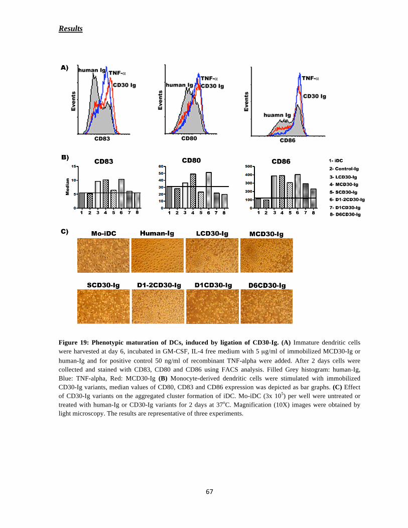
Results
Figure 19: Phenotypic maturation of DCs, induced by ligation of CD30-Ig. (A) Immature dendritic cells
were harvested at day 6, incubated in GM-CSF, IL-4 free medium with 5 μg/ml of immobilized MCD30-Ig or
human-Ig and for positive control 50 ng/ml of recombinant TNF-alpha were added. After 2 days cells were
collected and stained with CD83, CD80 and CD86 using FACS analysis. Filled Grey histogram: human-Ig,
Blue: TNF-alpha, Red: MCD30-Ig (B) Monocyte-derived dendritic cells were stimulated with immobilized
CD30-Ig variants, median values of CD80, CD83 and CD86 expression was depicted as bar graphs. (C) Effect
of CD30-Ig variants on the aggregated cluster formation of iDC. Mo-iDC (3x 10⁵) per well were untreated or
treated with human-Ig or CD30-Ig variants for 2 days at 37oC. Magnification (10X) images were obtained by
light microscopy. The results are representative of three experiments.

Results
Encounter of cognate MHC complexes on the surface of antigen presenting cells (APCs) by T
cells triggers the assembly of specialized structure at the APC-T cell interface, known as the
immunological synapse (IS). This process involves the reorganization of membrane and
intracellular signaling molecules in the T cell, leading to polarization of its secretory
machinery toward the APC (Revy et al. 2001; Muller et al. 2006). This event is necessary for
efficient T cell activation and function. An important morphological parameter associated
with IS assembly is the polarization of the surface molecules in T cells. To this end, I
investigated whether CD30 stimulated dendritic cells have some effect in formation of
immunological synapse using confocal microscopy. When activated T cells were co-cultured
with either allogenic iDC or MCD30-Ig stimulated DC (Mature DC), the CD3 expression on
T-cells was not clustered to one region in the case of iDC alone, stating that polarization did
not occur, whereas in the case of mature DC, CD3 expression was concentrated to one point
at the site of IS, proving the point that the T-cell is polarized for a proper immune response
(Fig. 20). But this effect was not observed with resting T cell and DC co-culture experiments.
CD30-CD30L interaction contributes to DC-maturation and is sufficient to trigger their T-cell
activation potential.

Results
Figure 20: MCD30-Ig simulated-DCs are involved in allogenic DC-T cell IS. Monocyte- derived immature
DC (iDC) or MCD30 stimulated mature DCs (mDC CD30) were co-cultured with allogenic T cells either resting
or activated (1 μg/ml PHA for 24 hr) for 25 minutes. Cells were settled on to poly-L-lysine-coated slides, fixed,
and stained for CD3, followed by goat anti-mouse Alexa 594-labeled secondary antibody (red color), Nucleus
was detected by Hochest 33342 (blue colour). Polarization of T-cell was determined by confocal microscopy.
Corresponding Nomarski merged images are shown on the right. Confocal Z-stacks images 0.4 μM were taken
using 60X objective lense.
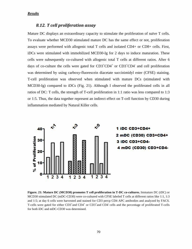
Results
Mature DC displays an extraordinary capacity to stimulate the proliferation of naïve T cells.
To evaluate whether MCD30 stimulated mature DC has the same effect or not, proliferation
assays were performed with allogenic total T cells and isolated CD4+ or CD8+ cells. First,
iDCs were stimulated with immobilized MCD30-Ig for 2 days to induce maturation. These
cells were subsequently co-cultured with allogenic total T cells at different ratios. After 6
days of co-culture the cells were gated for CD3+CD4
+ or CD3
+CD4
- and cell proliferation
was determined by using carboxy-fluorescein diacetate succinimidyl ester (CFSE) staining.
T-cell proliferation was observed when stimulated with mature DCs (stimulated with
MCD30-Ig) compared to iDCs (Fig. 21). Although I observed the proliferated cells in all
ratios of DC: T cells, the strength of T-cell proliferation in 1:1 ratio was less compared to 1:3
or 1:5. Thus, the data together represent an indirect effect on T-cell function by CD30 during
inflammation mediated by Natural Killer cells.
Figure. 21: Mature DC (MCD30) promotes T cell proliferation in T-DC co-cultures. Immature DC (iDC) or
MCD30 stimulated DC (mDC-CD30) were co-cultured with CFSE labeled T cells at different ratios like 1:1, 1:3
and 1:5; at day 6 cells were harvested and stained for CD3 percp CD4 APC antibodies and analyzed by FACS.
T-cells were gated for either CD3+and CD4
+ or CD3
+and CD4
- cells and the percentage of proliferated T-cells
for both iDC and mDC-CD30 was determined.
Results
As introduced, the function of the interaction of TNFR-TNFR ligand family members in
immune regulation is not only regulated by cell-cell contact but also mediated by soluble
factors. This includes cytokines (chemokines, lymphokines or monokines) but also soluble
counterparts of TNFR and TNFRL family members (Armitage 1994; Croft 2003). However,
in this context, the physiological function of CD30-CD30L interaction and a possible role of
the soluble molecules are far from being understood. Following this interaction there is not
only signaling through CD30 receptor but also through membrane anchored CD30L. It was
previously described that contact-dependent reverse signaling of CD30L+ cells by
recombinant CD30 (immobilized) results in the release of inflammatory cytokines like IL-8
from mast cells, eosinophils and neutrophils (Wiley et al. 1996; Molin 2004; Fischer et al.
2006). The previous sections describe the importance of CD30-CD30L interaction in the case
of normal acute single-phase inflammation, playing a major role in the release of pro-
inflammatory cytokines and in NK-DC cross talk that leads to the maturation of DCs.
The extra-cellular domain of CD30 is cleaved from the surface of CD30+ cells by the
action of cell surface metalloproteinase TNF-alpha converting enzyme (Hansen et al. 2000),
and binds to CD30L+ cells (Hargreaves and Al-Shamkhani 2002). Several reports
demonstrated elevated levels of sCD30 in chronic inflammatory cases such as HL and ALCL,
autoimmune disorder RA and skin disorder AD. Elevated serum levels of sCD30 in the
majority of HL patients were associated with a poor clinical outcome. Therefore, it was very
interesting to question whether sCD30 might also play a role in reverse signaling of
CD30L+ + cells.
In my preliminary experiments, the stimulation of iDCs with sCD30 resulted in the
elevated levels of IL-10, an anti-inflammatory cytokine (Fig. 22). This result supports the
observation that in RA, there is a direct correlation to the elevated levels of sCD30 and IL-10
(Gerli et al. 2001). Moreover, sCD30 stimulated iDCs do not undergo the process of
maturation, in contrast to the data shown for immobilized CD30. Since granulocytes are the
most abundant immune cells in the milieu of Hodgkin’s tissue, this section describes the
effects of sCD30 on granulocytes (eosinophils and neutrophils).

Results
Figure 22: sCD30 induces higher levels of IL-10: Mo-iDCs were stimulated with the purified sCD30 (5000
units/ml) using NHS-activated column loaded with anti-CD30 (Ki-2) column or by isotype column as a control
(used same volume) and the supernatants were subjected to IL-10 ELISA. Neg: DCs alone.
CD30L expression has been reported on different immune cells and granulocytes are the
major cells that play crucial role in innate system. It has been reviewed controversially that
the expression analysis of CD30L on these cells was due to non-specific binding of antibodies
and lack of controls (Kennedy et al. 2006). Thus, as one step behind, the cell surface
expression of CD30L on granulocytes was analyzed on the mRNA and protein level.
Granulocytes (polymorph nuclear leukocytes-PMN) were purified from whole blood obtained
from healthy donors. The schematic representation of granulocytes isolation is shown in Fig.
23. The isolation and purity of the granulocytes were confirmed by Pappenheim-staining
method (Fig. 23). Staining of multi-lobed nucleus determines the characteristic feature of
granulocytes.

Results
Figure 23: Schematic representation of granulocyte isolation. The purple color multi-lobulated nuclei of
granulocytes were determined by Pappenheim staining. PVA: Polyvinyl alcohol.
CD30 and CD30L mRNA expression was determined in purified granulocytes of
healthy donors by RT-PCR. Analysis of the PCR products showed clear CD30L but no CD30
expression in granulocytes. As a control, RT-PCR of CD30 and CD30L mRNA expression
was analyzed in CD30+ HL cell line L540. These cells in contrast, showed CD30 and no
detectable CD30L expression (Fig. 24A). GAPDH served as a control housekeeping gene for
both granulocytes and L540 cells. In line with the mRNA expression of CD30L in
granulocytes, the cell surface protein expression was demonstrated by immunofluorescence.
Cytospins of freshly purified granulocytes were stained with specific antibodies against CD30
and CD30L. Isotype-matched antibodies served as controls. As determined by alexa-594

Results
fluorochrome-labeled secondary antibody, red staining reveals surface expression of CD30L
and in contrast, CD30 expression is completely absent. Hoechst 33342 stains, the multi-
lobulated nucleus in blue (Fig. 24B).
To further confirm that granulocytes express CD30L, antibody competition
experiments using CD30L protein were performed by FACS analysis. Freshly purified PMN
were stained with monoclonal anti-human CD30L and corresponding isotype as primary
antibodies followed by detection with its specific secondary antibody. For the competition
experiments, primary antibodies were pre-incubated with purified CD30L protein (10 μg/ml)
on ice and later the subsequent staining of granulocytes was performed using antibodies
blocked with CD30L protein and untreated ones by flow cytometry. The surface expression
of CD30L is clearly demonstrated and the staining of CD30L antibody to cells was
considerably decreased, when pre-incubated with recombinant CD30L protein (Fig. 24C).
This effect was not observed with the isotype-matched antibodies. Taken together, the
analysis confirms the CD30L expression on granulocytes.

Results
Figure 24: Expression of CD30L on granulocytes. (A) RT-PCR analysis of CD30L from granulocytes
obtained from healthy donor CD30 from L540 cells. GAPDH was used as a control. (B) Granulocytes were
stained with anti-CD30 antibody (left), isotype antibody (middle), or anti-CD30L antibody (right). The multi-
lobulated nucleus staining was stained with Hoechst 33342 (blue) and the CD30L-specific antibody detection
determines the expression of CD30 ligand (red staining). (C) Antibody competition was performed to show the
specific CD30 ligand expression on granulocytes. The background grey color indicates the staining of secondary
antibody alone. The staining of CD30L was noted by the open histogram (pink) and could be blocked using
CD30L protein (orange). The isotype control has a background staining but no specificity against CD30L
protein (green and blue).

Results
Different reports had evidenced that HRS cells secrete different chemokines and cytokines
that are able to actively attract immune cells. For example, IL-5, CCL5, CCL22 and GM-CSF
presumably attract eosinophils, CCL-5 attracts mast cells and IL-8 recruits neutrophils
(Skinnider and Mak 2002). It has been well reviewed that these cytokines not only attract
other cells but keep the integrity of HRS cell survival and proliferation (Aldinucci et al.
2008). As mentioned earlier, the interesting characteristic feature of this micromilieu is the
presence of sCD30, able to bind most of the CD30L+ immune cells in the environment.
Making these points into context, the migration of granulocytes was tested upon stimulation
with soluble CD30 using Time Lapse Video Microscopy (TLVM).
An extra-cellular matrix glycoprotein, fibronectin (10 μg/ml) was coated on 24-well
plates to facilitate the process of migration. Granulocytes were isolated from whole blood,
plated in fibronectin-coated plates and stimulated with soluble supernatants derived from
293T cells either transfected with vector alone (mock) or with full-length cDNA of CD30
(LCD30). Granulocyte movements were measured by time-lapse microscopy under tissue
culture conditions (37oC, 5% CO2) for 60 minutes. The cells were tracked down to observe
their trajectory path for every minute of approximately 20-40 cells. The specific trajectory
movements, when stimulated with sCD30 supernatants showed a significantly increased
random migration of granulocytes in comparison with un-transfected and mock-transfected
supernatants (Fig. 25). In addition, blocking the functional interaction of sCD30 and CD30L
using anti-CD30 antibody (HeFi-1 at 3 μg/ml) inhibited the stimulation of sCD30-specific
migration. On the other hand, migration was not inhibited when blocked with the
corresponding isotype control. The data represents that sCD30 has an impact on granulocyte
migration and might act as a chemo-attractant. Thus, the recruitment of inflammatory cells
into the lymphoma microenvironment is also dependent on sCD30 and essential for the
survival of HRS cells in HL.

Results
Figure 25: Migration of granulocytes was measured using Time-Lapse Video Microscopy. Granulocyte
migration was tested after stimulation with supernatants of untreated (neg), CD30-transfected (LCD30) and
empty vector transfected (mock) 293T cells. All three stimulants were applied in the absence (Alone), or in
presence of anti-CD30 antibody (HeFi-1) and control Ab (Isotype). Each dot shows the distance covered by an
individual cell (path length in mm). Representative trajectories were randomly selected for 20-40 cells traced by
using digitizer during 60 frames. Using Graph-pad prism software the statistical analysis was performed by
student’s unpaired t- test for LCD30 (Alone) verses mock SN (Alone), LCD30 (Alone) versus LCD30 (HeFi-1)
and LCD30 (HeFi-1) versus LCD30 (Isotype). ** P < 0.005, *** P < 0.0005.
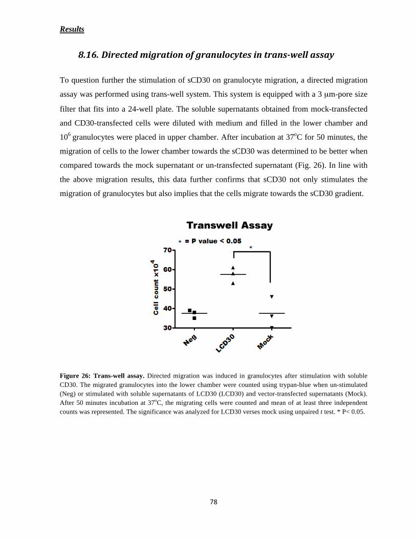
Results
To question further the stimulation of sCD30 on granulocyte migration, a directed migration
assay was performed using trans-well system. This system is equipped with a 3 μm-pore size
filter that fits into a 24-well plate. The soluble supernatants obtained from mock-transfected
and CD30-transfected cells were diluted with medium and filled in the lower chamber and
106
granulocytes were placed in upper chamber. After incubation at 37oC for 50 minutes, the
migration of cells to the lower chamber towards the sCD30 was determined to be better when
compared towards the mock supernatant or un-transfected supernatant (Fig. 26). In line with
the above migration results, this data further confirms that sCD30 not only stimulates the
migration of granulocytes but also implies that the cells migrate towards the sCD30 gradient.
Figure 26: Trans-well assay. Directed migration was induced in granulocytes after stimulation with soluble
CD30. The migrated granulocytes into the lower chamber were counted using trypan-blue when un-stimulated
(Neg) or stimulated with soluble supernatants of LCD30 (LCD30) and vector-transfected supernatants (Mock).
After 50 minutes incubation at 37oC, the migrating cells were counted and mean of at least three independent
counts was represented. The significance was analyzed for LCD30 verses mock using unpaired t test. * P< 0.05.

Results
HL is characterized with less than 1% malignant cells in the tumor tissue and
abundant granulocytes in the reactive infiltrate. Elevated levels of IL-8 showed an association
with B symptoms in Hodgkin patients. IL-8 expression in HL is largely confined to reactive
cells and association with infiltration by neutrophils (Foss et al. 1996). It is well described
that mast cells and granulocytes release IL-8 upon stimulation of immobilized CD30. Since
HRS cells release sCD30, it has been an interesting point to check whether soluble CD30
stimulates the IL-8 release from granulocytes. Similar to previous experiments, sCD30
supernatant was obtained from 293T cells either un-transfected, or transfected with vector
alone and CD30. Here, the cells were incubated with supernatants in serum free media for
over-night at 37oC. The cell free supernatants were collected and were subjected to IL-8
specific ELISA. As shown in Fig. 27, CD30 in soluble supernatant enhanced the IL-8 release
from granulocytes in contrast to un-transfected or mock-transfected supernatants. The
enhanced IL-8 release was blocked using anti-CD30 antibody (HeFi-1) at final concentration
of 1 μg/ml. These results indicate that sCD30 also stimulates granulocytes to release IL-8
resulting in an indirect effect on the angiogenesis of the tumor.

Results
Figure 27: IL-8 secretion. Levels of IL-8 present in supernatants from granulocytes (5x10⁵/ 24 well) after
overnight treatment with soluble CD30 (2000 units/ml) or control supernatants were used at equal volumes.
Soluble supernatants were pre-incubated with anti-CD30 mAb (HeFi-1) for one hour at 37oC. Neg-cells alone,
UT: un-transfected, Mock: vector transfected and LCD30: CD30 transfected. Minus (-) and plus (+) symbols
indicate the absence and presence of antibody respectively. Error bars represent the standard deviation.

Discussion
CD30 was first described in 1982 and recognized be selectively expressed on the
malignant cells of Hodgkin lymphoma and a few activated lymphocytes. A few years
later, in 1992, after molecular cloning, it was grouped as a member of the TNFR super
family. Although expression and function of other family members were published soon
after their first description, the physiological role of CD30 remained long time obscure.
Even today, the function of CD30 is not fully understood. It is widely accepted that CD30
is strongly expressed on activated B and T lymphocytes. In the present study we
confirmed the expression of CD30 on activated NK cells as shown previously (Cambiaggi
et al. 1993). To understand its physiological function, it is necessary to consider the
expression of CD30 ligand too. Generally, the expression is weaker and less restricted in
comparison to CD30. While CD30L expression is established on B cells and activated T
cells as well as on monocytes/macrophages and mast cells (Smith et al. 1993; Armitage
1994; Younes et al. 1996), the CD30L expression on other cells of the innate immune
system needs confirmation. There is evidence about a highly restricted and regulated
expression of CD30L on immune regulatory cells (Sun and Fink 2007). There are reports
about an up-regulation of CD30L on myeloid and plasmacytoid dendritic cells in mice
upon inflammation (Zeiser et al. 2007). Here, it is shown that human iDCs that were
differentiated from monocytes show distinct CD30L expression, whereas it is down
regulated upon maturation of iDCs. Similarly, in vitro-generated immature DCs, freshly
isolated blood DCs, also express cell surface CD30 ligand. Polymorpho nuclear
leukocytes, such as neutrophils, are cells that have also been suggested to express CD30L
(Wiley). Although others challenged this finding, we clearly confirmed that CD30L is
expressed on peripheral blood-derived granulocytes, both on the mRNA and on the
protein level. Taken together, the following profile emerges: CD30 is selectively
expressed on B, T and NK cells upon cell activation. CD30 ligand, by contrast, is broadly
distributed on immune cells, such as activated T cells, granulocytes, mast cells as well as
monocytes, macrophages and DCs.
Discussion
NK cells are a part of the innate immune system that is directly activated by cytokines and
bacterial, viral or pathogen encounter. Since NK cells express CD30 only upon activation and
highest CD30L expression in DCs was observed in immature status, we hypothesize that NK-
iDC interaction through CD30-CD30L might contribute to induce DC maturation. In this
respect we showed in this work that i) cross-linking of CD30L caused signaling in iDCs. We
found that stimulation of iDCs by CD30 receptor caused a transient cytoplasmic liberation of
ROS followed by secretion of pro-inflammatory cytokines. ii) This DC stimulation used the
MAP kinase pathway as demonstrated by selective pathway inhibitors. iii) We further showed
that CD30L ligation caused DC maturation as demonstrated by measuring maturation marker
at the cell surface. This effect was not only observed after direct stimulation of DCs with
recombinant CD30 receptor but also in co-culture experiments using activated CD30+ NK
cells. The cell-cell effect was CD30-CD30L-dependent because anti-CD30 antibodies caused
significant reduction of DC maturation. Thus, we showed for the first time that DCs can
maturate through reverse signaling, i.e. CD30 receptor is able to activate DCs through
ligation of membrane-anchored CD30 ligand.
Reactive Oxygen Species (ROS) have long been considered as harmful by-products of
intrinsic oxygen metabolism or cellular responses to hazardous stimuli. ROS has been
implicated in various pathogenesis of various disorders, including cancers, aging, diabetes,
atherosclerosis, chronic inflammation, HIV infection and injury (Droge 2002). But increasing
evidence suggests that ROS serve as essential secondary messengers mediating cellular
responses to many physiological stimuli (Reth 2002). ROS affect the maturation state, the
production and secretion of cytokines, and the antigen-presenting capacity
of DC (Alderman
et al. 2002; Matsue et al. 2003; Vulcano et al. 2004; Tan et al. 2005). Like monocytes and
neutrophils, for example, DC are equipped with a membrane-localized electron
transport
system, NADPH-oxidase, that reduces molecular oxygen to superoxide anions at the expense
of NADPH (Matsue et al. 2003; Elsen et al. 2004; Werling et al. 2004). Activation of DC may
thus initiate/include ROS-mediated autocrine/paracrine regulatory functions.

Discussion
The majority of studies of DC-generated ROS production have been performed using
in vitro-generated dendritic cells differentiated from human (Verhasselt et al. 1998; Rutault et
al. 1999; Alderman et al. 2002; Kantengwa et al. 2003). Recently, quite a few data suggest
that diverse receptors stimulate ROS production. These are not cytotoxic, but rather required
for the MAPK family member activation, gene expression and/or cell proliferation (Lo and
Cruz 1995; Sundaresan et al. 1995; Lo et al. 1996; Lee and Koretzky 1998; Sattler et al. 1999;
Devadas et al. 2002). Wiley et al observed ROS generation in neutrophils upon cross linking
CD30L with immobilized CD30 and activating the synthesis of cytokine release (Wiley et al.
1996). In line with the above findings my results demonstrated that Mo-iDCs generate ROS
(oxidative burst) upon activation with immobilized CD30. This oxidative burst was very
rapid and a decrease immediately suggests that the ROS concentration was very low. Thus,
the results support the current concept that ROS at low concentrations play physiological
roles in cellular signaling via regulating the redox states of transcription factors and various
enzymes involved in signal transduction (e.g. protein tyrosines phosphatases, receptor
tyrosine kinases, mitogen-activated protein kinases (MAPK), protein kinase C) (Abate et al.
1990; Droge 2002).
The relationship of cytokine production and reactive oxygen species generated from
DCs was known during antigen presentation (Matsue et al. 2003), whereas, this study aimed
to examine the production of cytokines from iDCs upon CD30 stimulation. We showed that
iDCs were able to release the proinflammatory cytokines TNF-alpha, IL-6, and IL-8. As a
consequence of the oxidative stress, which is often associated with tissue injury, DCs could
thus contribute to the local inflammatory response via their enhanced production of
cytokines. The proinflammatory cytokines like IL-8 aids in the recruitment of immune cells
whereas TNF-alpha and IL-6 synergizes in the activation of polymorph nuclear leukocytes
(PMN).
TNF-alpha has versatile functions in inflammatory responses particularly involved in
systemic inflammation and stimulates the acute phase reaction. It appears to have profound
effects on DC functions, as it contributes to their activation (Rieser et al. 1997), maturation
(Brunner et al. 2000), and migration to, and accumulation within, draining lymph nodes
(Cumberbatch and Kimber 1995). It significantly reduces IL-10 mediated inhibition of DC
Discussion
development and function (Brossart et al. 2000). Recently, it has been shown that TNF-alpha
trans-gene expressing DCs underwent cellular maturation and induced efficient CTL
responses and subsequent anti-tumor immunity (Zhang et al. 2003). Interleukin-6 (IL-6) acts
as both a proinflammatory and anti-inflammatory cytokine. It is well known that IL-6 is
secreted by macrophages in response to specific microbial molecules, referred to as pathogen
associated molecular patterns (PAMPs). Similar to TNF alpha, IL-6 also stimulates the acute
phase reaction to generate inflammatory responses. These cytokines acts as autocrine
regulators in inducing DC maturation and enhance the immuno-stimulatory capacity (Jonuleit
et al. 1997). Hence, it would be possible that the secretion of TNF alpha and IL-6 from iDCs
upon CD30 stimulation might contribute for the maturation of dendritic cells.
In addition to TNF alpha and IL-6, IL-8 is another major pro-inflammatory cytokine
that is released by CD30 stimulation from dendritic cells. Unlike TNF alpha and IL-6, IL-8 is
not involved directly in activation of immune cells but rather acts a chemo-attractant to
recruit granulocytes. IL-8 is a chemokine of the CXC family (CXCL8) whose major function
is to attract and activate PMN particularly neutrophils to the site of inflammation (Baggiolini
et al. 1997). On the other hand, enhanced production of this cytokine has been found in
inflammatory disorders such as rheumatoid arthritis and ulcerative colitis, infectious diseases
and inflammatory skin diseases. Furthermore, elevated levels of IL-8 have been also detected
in sera of Hodgkin patients in about 30-50% of the cases. Thus it is tempting to speculate that
IL-8 released from dendritic cells upon CD30 stimulation could possibly have a positive role
in normal acute-inflammation, in contrast to the chronic inflammation where, elevated levels
of IL-8 possibly have a negative effect.
Production of lower concentrations of IL-10 from iDCs upon CD30 stimulation may
be responsible for a proper control of the immune response during inflammation. It was
demonstrated that IL-10 plays an important regulatory role on monocyte function and on DC
maturation (de Waal Malefyt et al. 1991; Buelens et al. 1995). Dendritic cells secreting IL-10
exhibit minimal or no stimulatory properties in primary MLRs and are markedly inhibitory to
T cell proliferation induced by polyclonal activator (Ding and Shevach 1992). Thus, IL-10
producing DCs are functionally and phenotypically inhibitory accessory cells and putatively

Discussion
tolerogenic. However, so far it remains unclear, whether such lower concentrations reflect the
efforts by the immune system to control inflammation.
MAPK signaling pathway is a highly conserved pathway that is involved in diverse cellular
functions, including cell proliferation, cell differentiation, and apoptosis. The fundamental
role of MAP kinases in innate immunity (inflammation) is closely related with in cytokine
production by cells involved in the innate response (e.g. macrophages and dendritic cells). It
was well documented that cytokine production is regulated mainly by three MAPK-pathways
such as JNK, ERK, and p38 MAPKs. All share the common property of being activated via
three- module phosphorylation cascade, which in turn allows them to phosphorylate a wide
range of substrates, including other protein kinases, phospholipases and transcription factors
(Chang and Karin 2001; Johnson and Lapadat 2002). Although there is some cross-talk and
cell-type specificity, the JNK and p38 MAPK cascades are strongly activated by stress stimuli
and inflammatory cytokines, where as ERK pathway is strongly activated by polypeptide
growth factors through receptors for tyrosine kinase (Kyriakis and Avruch 2001). The
findings of TNF-alpha ELISA show that three MAP kinases (p38, JNK and ERK) were
involved in TNF-alpha production from CD30-stimulated iDCs. Specific inhibitors of three
MAP kinases blocked the synthesis and release of TNF-alpha. This is in line with previous
reports demonstrating that the TNF-alpha release upon MAP kinase inhibition was blocked in
LPS and PMA-stimulated iDCs. From these results, it can be inferred or rather postulated that
MAPK pathways play a major role in pro-inflammatory cytokine production by ROS
generated from CD30-stimulated dendritic cells.
During inflammation, recruitment, activation and maturation of almost all immune regulatory
cells occur primarily at the inflammatory site with a strong burst of cytokines and chemokines
from different cell types. Here it was shown, that maturation of iDCs is triggered by
immobilized CD30-Ig (mimic of membrane anchored receptor) or upon co-culture with
activated NK cells in a CD30-CD30L-dependent manner. NK-DC cross-talk leads to a
Discussion
profound activation of both cell types, finally leading to the induction of signals for iDCs
maturation. During this process it was well documented that engagement of NKp30 (NCR3)
with DCs stimulates the release of pro-inflammatory cytokines (TNF-alpha and IFN-gamma)
from NK cells, which contributes to the maturation of iDCs via paracrine mechanism. In
contrast to these observations, this study in particular focused in explaining the effects of
CD30 during iDC maturation with the release of TNF-alpha from iDCs itself describing an
autocrine mechanism. Furthermore, inhibition of maturation process was not completely
abolished when blocked with CD30-specific antibodies in NK-DC co-culture, bringing the
fact that other receptors on NK cells like NKp30 or DNAM-1 were also involved. Thus, these
results together infer that TNF-alpha-mediated maturation of iDCs in the bi-directional cross-
talk with NK cells could be induced by both autocrine and paracrine mechanisms.
Moreover, consistent to the results obtained for the cytokine release (TNF-alpha and
IL-6), immobilized CD30-Ig deletion constructs correspondingly induced iDC maturation.
The degree of maturation is determined by the up-regulation of co-stimulatory molecules
(CD80, CD83 and CD86). Previous reports observed that TNF-alpha or RANTES dependent
DC maturation led to the aggregation of DCs (Sato et al. 1999; Enomoto et al. 2001).
Differences in the maturation process within the CD30-Ig constructs directly correlated to the
aggregation of dendritic cells. Cell clustering, which is a characteristic feature of mature DC
cultures, probably results from the up-regulation of several surface molecules mediating cell-
cell interactions. Although the precise molecular mechanisms underlying DC aggregation is
not clear, it was postulated that ICAM-1 (CD54), a cell adhesion molecule is involved in the
formation of DC aggregates (H. Haegel-Kronenberger, unpublished data).
As a consequence of CD30-dependent DC maturation, the study also describes the function of
mature DCs in the formation of activated T-cell synapse and proliferation of naïve T-cells. It
is known that maturation of DC is critical in inducing effective, naïve T cell priming and
robust proliferation (Guermonprez et al. 2002). One of the major pre-requiste for a proper
DC-T-cell contact is the expression of co-stimulatory and adhesion molecules on both cell-
types. Here it is demonstrated iDCs upon CD30L-engagement are able to form a proper
immunological synapse (IS) with the clustering of CD3 receptor at the synapse. Interestingly,
Discussion
unlike the formation of the IS with naïve T cells, CD30 derived mature DCs were more
specific to activated T cells, probably correlating with a specific function for cytotoxic T-
lymphocytes. In fact, this mechanism further supports that NK-DC cross talk play an
important role in shaping the adaptive immune response. In addition to the function of T-cell
polarization, CD30 derived mature DCs enhance the proliferation of either with CD3+ CD4
+
or CD3+ CD4
- T cells.
CD30 has been well documented to have a role in the pathogenesis of hematopoietic
malignancies. CD30 is cleaved and the ectodomain (sCD30) is released in the
microenvironment of the malignant cells. The tumor microenvironment in malignancies such
as HL is characterized by an infiltration of many different types of immune cells including T
cells, B cells, plasma cells, neutrophils, eosinophils and mast cells. Depending on the
cytokines released different immune cells were recruited to the site of inflammatory tissue
(Kuppers 2009). It is anticipated that the chronic inflammation in HL contributes to the tumor
initiation, progression and survival of the malignant cells (HRS). Elevated sCD30 serum
levels in HL patients correlate to the progression of the disease. Soluble CD30 binds to the
CD30L+ cells and blocks the direct interaction with surface-expressed CD30 on HRS cells or
ALCL, thereby inhibiting the biological effects of CD30L. The results shown in this study
collectively uncover novel functions for sCD30 engaging CD30L on granulocytes that are
usually present either in the HL microenvironment or in malignant lymphoma like ALCL,
and also in RA.
The study shows that, human granulocytes express CD30L and HL cells release the
soluble ectodomain of CD30 (sCD30). We found that sCD30 stimulates chemotaxis of
granulocytes and elevated concentrations induced them to release IL-8. Therefore, it is
conceivable that recruitment of inflammatory cells to the lymphoma microenvironment is due
to the presence of sCD30. Previous studies have shown that some of the survival signals that
are provided by inflammatory cells to the HRS cells are triggered by CD40 signaling by
CD40L expressing rosetting T-cells and also by activation of TACI and BCMA through
production of their ligand APRIL by neutrophils (Kuppers 2009). Similar to this fashion
perhaps sCD30-CD30L interaction could also play a role in HRS survival. In contrast to the

Discussion
data shown with soluble form, it has been previously described that immobilized CD30
stimulate mast cells and neutrophils to secrete IL-8. Therefore, the content of IL-8 in the
tumor microenvironment could be at elevated levels correlating to a role in the pathogenesis
of HL (Trumper et al. 1994). However, sCD30-dependent release of IL-8 from granulocytes
may affect angiogenesis positively and thus promote tumor progression.
The complex immune regulatory mechanisms by immune cells are modulated by various
factors depending on the type of inflammation. Generally, inflammation is broadly classified
as acute and chronic inflammation. Although, the mechanisms involved in acute and chronic
inflammation differ a lot in inducing different immune responses, the effect of specific
interactions under molecular level does not change. This study provides evidence for a crucial
role of CD30-CD30L interaction in both the inflammatory responses. Based on the results I
propose a model that early cell-cell dependent CD30-CD30L signaling is involved in acute
inflammation whereas elevated levels of soluble sCD30 may contribute to chronic
inflammation.
Acute inflammation corresponds to infection of pathogens or viruses. NK cells play a major
role in inducing primary immune responses, but they require specific activatory mechanisms.
Until now, it has been suggested that the early phases of an inflammatory response involves
the activation of toll-like receptors (TLRs) by pathogen-associated molecular patterns
(PAMPs) and result in NK-DC interaction. But recent reports strongly suggest that NK cells
need to be primed before their activation to dendritic cells, and this priming is mediated by
various cytokines like IL-4, IFN- or IL-18 released from other innate cells like eosinophils,
neutrophils, monocytes and mast cells. This could be termed as “primary activation of NK
cells”. This primary activation results in expression of CD30 on NK cells. Moreover, it has
been shown that CD30 expression is sustained in presence of IL-4. Thus we could
hypothesize that CD30-expressing NK cells might then interact and activate CD30L+
Discussion
dendritic cells to induce pro-inflammatory responses leading to the maturation of dendritic
cells and finally elicit the T-cell mediated responses. Thus mostly, the function of CD30-
CD30L interaction in acute inflammation is dependent on cell-cell contact (Fig. 28).
Prolonged inflammation, known as chronic inflammation, leads to a progressive shift in the
type of cells which are present at the site of inflammation and is characterized by
simultaneous destruction and healing of the tissue from the inflammatory process. The best
known examples are the lymphomas or autoimmune disorders. However, chronic
inflammation is generally linked to tumor initiation and progression. The chronic lymphomas
and in particular HL are best characterized by the recruitment of large number of
inflammatory cells and elevated levels of CD30 in its soluble form (sCD30). In contrast to the
acute inflammation the immune effector cells (NK or T cells) were suppressed or absent. In
this study it was shown that sCD30 has a direct function on migration of granulocytes and
release of IL-8. Thus it is tempting to speculate that sCD30 mediated directed migration,
results in the accumulation of inflammatory granulocytes in the Hodgkin tissue. Their release
of IL-8 is probably responsible for tumor maintenance and progression (Fig. 28). The release
of IL-10 from DCs upon stimulation with sCD30 may act as an anti-inflammatory cytokine
that provide immune-tolerance in the tumor microenvironment. This could result in the
suppression of T or NK cell function.

Discussion
Figure 28: Hypothetical Model of CD30-CD30L function in inflammation

References
Abate, C., L. Patel, F. J. Rauscher, 3rd and T. Curran (1990). "Redox regulation of fos and jun DNA-
binding activity in vitro." Science 249(4973): 1157-61.
Adam, C., S. King, T. Allgeier, H. Braumuller, C. Luking, J. Mysliwietz, A. Kriegeskorte, D. H.
Busch, M. Rocken and R. Mocikat (2005). "DC-NK cell cross talk as a novel CD4+ T-cell-
independent pathway for antitumor CTL induction." Blood 106(1): 338-44.
Aggarwal, B. B. (2003). "Signalling pathways of the TNF superfamily: a double-edged sword." Nat
Rev Immunol 3(9): 745-56.
Aizawa, S., H. Nakano, T. Ishida, R. Horie, M. Nagai, K. Ito, H. Yagita, K. Okumura, J. Inoue and T.
Watanabe (1997). "Tumor necrosis factor receptor-associated factor (TRAF) 5 and TRAF2 are
involved in CD30-mediated NFkappaB activation." J Biol Chem 272(4): 2042-5.
Alderman, C. J., S. Shah, J. C. Foreman, B. M. Chain and D. R. Katz (2002). "The role of advanced
oxidation protein products in regulation of dendritic cell function." Free Radic Biol Med 32(5): 377-
85.
Aldinucci, D., D. Lorenzon, L. Cattaruzza, A. Pinto, A. Gloghini, A. Carbone and A. Colombatti
(2008). "Expression of CCR5 receptors on Reed-Sternberg cells and Hodgkin lymphoma cell lines:
involvement of CCL5/Rantes in tumor cell growth and microenvironmental interactions." Int J Cancer
122(4): 769-76.
Anderson, D. M., E. Maraskovsky, W. L. Billingsley, W. C. Dougall, M. E. Tometsko, E. R. Roux,
M. C. Teepe, R. F. DuBose, D. Cosman and L. Galibert (1997). "A homologue of the TNF receptor
and its ligand enhance T-cell growth and dendritic-cell function." Nature 390(6656): 175-9.
Andreesen, R., J. Osterholz, G. W. Lohr and K. J. Bross (1984). "A Hodgkin cell-specific antigen is
expressed on a subset of auto- and alloactivated T (helper) lymphoblasts." Blood 63(6): 1299-302.
Ansieau, S., I. Scheffrahn, G. Mosialos, H. Brand, J. Duyster, K. Kaye, J. Harada, B. Dougall, G.
Hubinger, E. Kieff, F. Herrmann, A. Leutz and H. J. Gruss (1996). "Tumor necrosis factor receptor-
associated factor (TRAF)-1, TRAF-2, and TRAF-3 interact in vivo with the CD30 cytoplasmic
domain; TRAF-2 mediates CD30-induced nuclear factor kappa B activation." Proc Natl Acad Sci U S
A 93(24): 14053-8.
Armitage, R. J. (1994). "Tumor necrosis factor receptor superfamily members and their ligands." Curr
Opin Immunol 6(3): 407-13.
Baggiolini, M., B. Dewald and B. Moser (1997). "Human chemokines: an update." Annu Rev
Immunol 15: 675-705.
Bereta, M., J. Bereta, J. Park, F. Medina, H. Kwak and H. L. Kaufman (2004). "Immune properties of
recombinant vaccinia virus encoding CD154 (CD40L) are determined by expression of virally

References
encoded CD40L and the presence of CD40L protein in viral particles." Cancer Gene Ther 11(12):
808-18.
Biron, C. A., K. B. Nguyen, G. C. Pien, L. P. Cousens and T. P. Salazar-Mather (1999). "Natural
killer cells in antiviral defense: function and regulation by innate cytokines." Annu Rev Immunol 17:
189-220.
Borchmann, P., J. F. Treml, H. Hansen, C. Gottstein, R. Schnell, O. Staak, H. F. Zhang, T. Davis, T.
Keler, V. Diehl, R. F. Graziano and A. Engert (2003). "The human anti-CD30 antibody 5F11 shows in
vitro and in vivo activity against malignant lymphoma." Blood 102(10): 3737-42.
Boucher, L. M., L. E. Marengere, Y. Lu, S. Thukral and T. W. Mak (1997). "Binding sites of
cytoplasmic effectors TRAF1, 2, and 3 on CD30 and other members of the TNF receptor
superfamily." Biochem Biophys Res Commun 233(3): 592-600.
Bowen, M. A., R. K. Lee, G. Miragliotta, S. Y. Nam and E. R. Podack (1996). "Structure and
expression of murine CD30 and its role in cytokine production." J Immunol 156(2): 442-9.
Brossart, P., A. Zobywalski, F. Grunebach, L. Behnke, G. Stuhler, V. L. Reichardt, L. Kanz and W.
Brugger (2000). "Tumor necrosis factor alpha and CD40 ligand antagonize the inhibitory effects of
interleukin 10 on T-cell stimulatory capacity of dendritic cells." Cancer Res 60(16): 4485-92.
Brunner, C., J. Seiderer, A. Schlamp, M. Bidlingmaier, A. Eigler, W. Haimerl, H. A. Lehr, A. M.
Krieg, G. Hartmann and S. Endres (2000). "Enhanced dendritic cell maturation by TNF-alpha or
cytidine-phosphate-guanosine DNA drives T cell activation in vitro and therapeutic anti-tumor
immune responses in vivo." J Immunol 165(11): 6278-86.
Buelens, C., F. Willems, A. Delvaux, G. Pierard, J. P. Delville, T. Velu and M. Goldman (1995).
"Interleukin-10 differentially regulates B7-1 (CD80) and B7-2 (CD86) expression on human
peripheral blood dendritic cells." Eur J Immunol 25(9): 2668-72.
Caligaris-Cappio, F., M. T. Bertero, M. Converso, A. Stacchini, F. Vinante, S. Romagnani and G.
Pizzolo (1995). "Circulating levels of soluble CD30, a marker of cells producing Th2-type cytokines,
are increased in patients with systemic lupus erythematosus and correlate with disease activity." Clin
Exp Rheumatol 13(3): 339-43.
Cambiaggi, A., C. Cantoni, S. Marciano, D. De Totero, S. Pileri, P. L. Tazzari, H. Stein and S. Ferrini
(1993). "Cultured human NK cells express the Ki-1/CD30 antigen." Br J Haematol 85(2): 270-6.
Carswell, E. A., L. J. Old, R. L. Kassel, S. Green, N. Fiore and B. Williamson (1975). "An endotoxin-
induced serum factor that causes necrosis of tumors." Proc Natl Acad Sci U S A 72(9): 3666-70.
Cella, M., D. Scheidegger, K. Palmer-Lehmann, P. Lane, A. Lanzavecchia and G. Alber (1996).
"Ligation of CD40 on dendritic cells triggers production of high levels of interleukin-12 and enhances
T cell stimulatory capacity: T-T help via APC activation." J Exp Med 184(2): 747-52.

References
Cerutti, A., A. Schaffer, R. G. Goodwin, S. Shah, H. Zan, S. Ely and P. Casali (2000). "Engagement
of CD153 (CD30 ligand) by CD30+ T cells inhibits class switch DNA recombination and antibody
production in human IgD+ IgM+ B cells." J Immunol 165(2): 786-94.
Chang, L. and M. Karin (2001). "Mammalian MAP kinase signalling cascades." Nature 410(6824):
37-40.
Croft, M. (2003). "Co-stimulatory members of the TNFR family: keys to effective T-cell immunity?"
Nat Rev Immunol 3(8): 609-20.
Cumberbatch, M. and I. Kimber (1995). "Tumour necrosis factor-alpha is required for accumulation
of dendritic cells in draining lymph nodes and for optimal contact sensitization." Immunology 84(1):
31-5.
de Waal Malefyt, R., J. Abrams, B. Bennett, C. G. Figdor and J. E. de Vries (1991). "Interleukin
10(IL-10) inhibits cytokine synthesis by human monocytes: an autoregulatory role of IL-10 produced
by monocytes." J Exp Med 174(5): 1209-20.
Devadas, S., L. Zaritskaya, S. G. Rhee, L. Oberley and M. S. Williams (2002). "Discrete generation of
superoxide and hydrogen peroxide by T cell receptor stimulation: selective regulation of mitogen-
activated protein kinase activation and fas ligand expression." J Exp Med 195(1): 59-70.
Ding, L. and E. M. Shevach (1992). "IL-10 inhibits mitogen-induced T cell proliferation by
selectively inhibiting macrophage costimulatory function." J Immunol 148(10): 3133-9.
Droge, W. (2002). "Free radicals in the physiological control of cell function." Physiol Rev 82(1): 47-
95.
Duckett, C. S., R. W. Gedrich, M. C. Gilfillan and C. B. Thompson (1997). "Induction of nuclear
factor kappaB by the CD30 receptor is mediated by TRAF1 and TRAF2." Mol Cell Biol 17(3): 1535-
42.
Duckett, C. S. and C. B. Thompson (1997). "CD30-dependent degradation of TRAF2: implications for
negative regulation of TRAF signaling and the control of cell survival." Genes Dev 11(21): 2810-21.
Durkop, H., B. Hirsch, C. Hahn, H. D. Foss and H. Stein (2003). "Differential expression and function
of A20 and TRAF1 in Hodgkin lymphoma and anaplastic large cell lymphoma and their induction by
CD30 stimulation." J Pathol 200(2): 229-39.
Durkop, H., B. Hirsch, C. Hahn and H. Stein (2006). "cIAP2 is highly expressed in Hodgkin-Reed-
Sternberg cells and inhibits apoptosis by interfering with constitutively active caspase-3." J Mol Med
84(2): 132-41.
Durkop, H., U. Latza, M. Hummel, F. Eitelbach, B. Seed and H. Stein (1992). "Molecular cloning and
expression of a new member of the nerve growth factor receptor family that is characteristic for
Hodgkin's disease." Cell 68(3): 421-7.

References
Eichenauer, D. A., V. L. Simhadri, E. P. von Strandmann, A. Ludwig, V. Matthews, K. S. Reiners, B.
von Tresckow, P. Saftig, S. Rose-John, A. Engert and H. P. Hansen (2007). "ADAM10 inhibition of
human CD30 shedding increases specificity of targeted immunotherapy in vitro." Cancer Res 67(1):
332-8.
Elsen, S., J. Doussiere, C. L. Villiers, M. Faure, R. Berthier, A. Papaioannou, N. Grandvaux, P. N.
Marche and P. V. Vignais (2004). "Cryptic O2- -generating NADPH oxidase in dendritic cells." J Cell
Sci 117(Pt 11): 2215-26.
Enomoto, M., H. Nagayama and T. A. Takahashi (2001). "Enhancement of migratory and aggregate
activities of human peripheral blood monocyte-derived dendritic cells by stimulation with RANTES."
Microbiol Immunol 45(9): 639-47.
Falini, B., S. Pileri, G. Pizzolo, H. Durkop, L. Flenghi, F. Stirpe, M. F. Martelli and H. Stein (1995).
"CD30 (Ki-1) molecule: a new cytokine receptor of the tumor necrosis factor receptor superfamily as
a tool for diagnosis and immunotherapy." Blood 85(1): 1-14.
Fattovich, G., F. Vinante, G. Giustina, L. Morosato, A. Alberti, A. Ruol and G. Pizzolo (1996).
"Serum levels of soluble CD30 in chronic hepatitis B virus infection." Clin Exp Immunol 103(1): 105-
10.
Ferlazzo, G., M. L. Tsang, L. Moretta, G. Melioli, R. M. Steinman and C. Munz (2002). "Human
dendritic cells activate resting natural killer (NK) cells and are recognized via the NKp30 receptor by
activated NK cells." J Exp Med 195(3): 343-51.
Fischer, M., I. T. Harvima, R. F. Carvalho, C. Moller, A. Naukkarinen, G. Enblad and G. Nilsson
(2006). "Mast cell CD30 ligand is upregulated in cutaneous inflammation and mediates degranulation-
independent chemokine secretion." J Clin Invest 116(10): 2748-56.
Fonatsch, C., U. Latza, H. Durkop, H. Rieder and H. Stein (1992). "Assignment of the human CD30
(Ki-1) gene to 1p36." Genomics 14(3): 825-6.
Foss, H. D., H. Herbst, S. Gottstein, G. Demel, I. Araujo and H. Stein (1996). "Interleukin-8 in
Hodgkin's disease. Preferential expression by reactive cells and association with neutrophil density."
Am J Pathol 148(4): 1229-36.
Franke, A. C., D. Jung and T. M. Ellis (2000). "Characterization of the CD30L binding domain on the
human CD30 molecule using anti-CD30 antibodies." Hybridoma 19(1): 43-8.
Gattei, V., M. Degan, A. De Iuliis, F. M. Rossi, D. Aldinucci and A. Pinto (1997). "Competitive
reverse-transcriptase PCR: a useful alternative to northern blotting for quantitative estimation of
relative abundances of specific mRNAs in precious samples." Biochem J 325 ( Pt 2): 565-7.
Gedrich, R. W., M. C. Gilfillan, C. S. Duckett, J. L. Van Dongen and C. B. Thompson (1996). "CD30
contains two binding sites with different specificities for members of the tumor necrosis factor
receptor-associated factor family of signal transducing proteins." J Biol Chem 271(22): 12852-8.

References
Gerli, R., C. Lunardi, F. Vinante, O. Bistoni, G. Pizzolo and C. Pitzalis (2001). "Role of CD30+ T
cells in rheumatoid arthritis: a counter-regulatory paradigm for Th1-driven diseases." Trends Immunol
22(2): 72-7.
Gerosa, F., B. Baldani-Guerra, C. Nisii, V. Marchesini, G. Carra and G. Trinchieri (2002).
"Reciprocal activating interaction between natural killer cells and dendritic cells." J Exp Med 195(3):
327-33.
Gerosa, F., A. Gobbi, P. Zorzi, S. Burg, F. Briere, G. Carra and G. Trinchieri (2005). "The reciprocal
interaction of NK cells with plasmacytoid or myeloid dendritic cells profoundly affects innate
resistance functions." J Immunol 174(2): 727-34.
Gilfillan, M. C., P. J. Noel, E. R. Podack, S. L. Reiner and C. B. Thompson (1998). "Expression of the
costimulatory receptor CD30 is regulated by both CD28 and cytokines." J Immunol 160(5): 2180-7.
Gray, P. W., B. B. Aggarwal, C. V. Benton, T. S. Bringman, W. J. Henzel, J. A. Jarrett, D. W. Leung,
B. Moffat, P. Ng, L. P. Svedersky and et al. (1984). "Cloning and expression of cDNA for human
lymphotoxin, a lymphokine with tumour necrosis activity." Nature 312(5996): 721-4.
Gruss, H. J. (1996). "Molecular, structural, and biological characteristics of the tumor necrosis factor
ligand superfamily." Int J Clin Lab Res 26(3): 143-59.
Gruss, H. J., N. Boiani, D. E. Williams, R. J. Armitage, C. A. Smith and R. G. Goodwin (1994).
"Pleiotropic effects of the CD30 ligand on CD30-expressing cells and lymphoma cell lines." Blood
83(8): 2045-56.
Gruss, H. J. and F. Herrmann (1996). "CD30 ligand, a member of the TNF ligand superfamily, with
growth and activation control CD30+ lymphoid and lymphoma cells." Leuk Lymphoma 20(5-6): 397-
409.
Gruss, H. J., A. Pinto, A. Gloghini, E. Wehnes, B. Wright, N. Boiani, D. Aldinucci, V. Gattei, V.
Zagonel, C. A. Smith, M. E. Kadin, C. von Schilling, R. G. Goodwin, F. Herrmann and A. Carbone
(1996). "CD30 ligand expression in nonmalignant and Hodgkin's disease-involved lymphoid tissues."
Am J Pathol 149(2): 469-81.
Guermonprez, P., J. Valladeau, L. Zitvogel, C. Thery and S. Amigorena (2002). "Antigen presentation
and T cell stimulation by dendritic cells." Annu Rev Immunol 20: 621-67.
Hansen, H. P., S. Dietrich, T. Kisseleva, T. Mokros, R. Mentlein, H. H. Lange, G. Murphy and H.
Lemke (2000). "CD30 shedding from Karpas 299 lymphoma cells is mediated by TNF-alpha-
converting enzyme." J Immunol 165(12): 6703-9.
Hansen, H. P., T. Kisseleva, J. Kobarg, O. Horn-Lohrens, B. Havsteen and H. Lemke (1995). "A zinc
metalloproteinase is responsible for the release of CD30 on human tumor cell lines." Int J Cancer
63(5): 750-6.
References
Hansen, H. P., A. Recke, U. Reineke, B. Von Tresckow, P. Borchmann, E. P. Von Strandmann, H.
Lange, H. Lemke and A. Engert (2004). "The ectodomain shedding of CD30 is specifically regulated
by peptide motifs in its cysteine-rich domains 2 and 5." Faseb J 18(7): 893-5.
Hargreaves, P. G. and A. Al-Shamkhani (2002). "Soluble CD30 binds to CD153 with high affinity
and blocks transmembrane signaling by CD30." Eur J Immunol 32(1): 163-73.
Harlin, H., E. Podack, M. Boothby and M. L. Alegre (2002). "TCR-independent CD30 signaling
selectively induces IL-13 production via a TNF receptor-associated factor/p38 mitogen-activated
protein kinase-dependent mechanism." J Immunol 169(5): 2451-9.
Higgins, J. P. and R. A. Warnke (1999). "CD30 expression is common in mediastinal large B-cell
lymphoma." Am J Clin Pathol 112(2): 241-7.
Hintzen, R. Q., R. de Jong, S. M. Lens and R. A. van Lier (1994). "CD27: marker and mediator of T-
cell activation?" Immunol Today 15(7): 307-11.
Hirsch, B., M. Hummel, S. Bentink, F. Fouladi, R. Spang, R. Zollinger, H. Stein and H. Durkop
(2008). "CD30-induced signaling is absent in Hodgkin's cells but present in anaplastic large cell
lymphoma cells." Am J Pathol 172(2): 510-20.
Horie, R., S. Aizawa, M. Nagai, K. Ito, M. Higashihara, T. Ishida, J. Inoue and T. Watanabe (1998).
"A novel domain in the CD30 cytoplasmic tail mediates NFkappaB activation." Int Immunol 10(2):
203-10.
Horie, R., V. Gattei, K. Ito, S. Imajo-Ohmi, T. Tange, J. Miyauchi, A. Pinto, M. Degan, A. De Iuliis,
F. Tassan Mazzocco, F. M. Rossi, M. Higashihara and T. Watanabe (1999). "Frequent expression of
the variant CD30 in human malignant myeloid and lymphoid neoplasms." Am J Pathol 155(6): 2029-
41.
Horie, R., K. Ito, M. Tatewaki, M. Nagai, S. Aizawa, M. Higashihara, T. Ishida, J. Inoue, H. Takizawa
and T. Watanabe (1996). "A variant CD30 protein lacking extracellular and transmembrane domains
is induced in HL-60 by tetradecanoylphorbol acetate and is expressed in alveolar macrophages."
Blood 88(7): 2422-32.
Horie, R. and T. Watanabe (1998). "CD30: expression and function in health and disease." Semin
Immunol 10(6): 457-70.
Hsu, H., H. B. Shu, M. G. Pan and D. V. Goeddel (1996). "TRADD-TRAF2 and TRADD-FADD
interactions define two distinct TNF receptor 1 signal transduction pathways." Cell 84(2): 299-308.
Hsu, P. L. and S. M. Hsu (2000). "Autocrine growth regulation of CD30 ligand in CD30-expressing
Reed-Sternberg cells: distinction between Hodgkin's disease and anaplastic large cell lymphoma." Lab
Invest 80(7): 1111-9.
Itoh, N., S. Yonehara, A. Ishii, M. Yonehara, S. Mizushima, M. Sameshima, A. Hase, Y. Seto and S.
Nagata (1991). "The polypeptide encoded by the cDNA for human cell surface antigen Fas can
mediate apoptosis." Cell 66(2): 233-43.

References
Johnson, G. L. and R. Lapadat (2002). "Mitogen-activated protein kinase pathways mediated by ERK,
JNK, and p38 protein kinases." Science 298(5600): 1911-2.
Jones, T. J., N. A. Coad, K. R. Muir, S. E. Parkes, C. D. Evans and J. R. Mann (1995).
"Immunophenotypic analysis of childhood Burkitt's lymphoma in the West Midlands 1957-1986." J
Clin Pathol 48(1): 22-5.
Jonuleit, H., U. Kuhn, G. Muller, K. Steinbrink, L. Paragnik, E. Schmitt, J. Knop and A. H. Enk
(1997). "Pro-inflammatory cytokines and prostaglandins induce maturation of potent
immunostimulatory dendritic cells under fetal calf serum-free conditions." Eur J Immunol 27(12):
3135-42.
Josimovic-Alasevic, O., H. Durkop, R. Schwarting, E. Backe, H. Stein and T. Diamantstein (1989).
"Ki-1 (CD30) antigen is released by Ki-1-positive tumor cells in vitro and in vivo. I. Partial
characterization of soluble Ki-1 antigen and detection of the antigen in cell culture supernatants and in
serum by an enzyme-linked immunosorbent assay." Eur J Immunol 19(1): 157-62.
Kantengwa, S., L. Jornot, C. Devenoges and L. P. Nicod (2003). "Superoxide anions induce the
maturation of human dendritic cells." Am J Respir Crit Care Med 167(3): 431-7.
Kelly, J. M., P. K. Darcy, J. L. Markby, D. I. Godfrey, K. Takeda, H. Yagita and M. J. Smyth (2002).
"Induction of tumor-specific T cell memory by NK cell-mediated tumor rejection." Nat Immunol 3(1):
83-90.
Kemper, O., J. Derre, D. Cherif, H. Engelmann, D. Wallach and R. Berger (1991). "The gene for the
type II (p75) tumor necrosis factor receptor (TNF-RII) is localized on band 1p36.2-p36.3." Hum
Genet 87(5): 623-4.
Kennedy, M. K., C. R. Willis and R. J. Armitage (2006). "Deciphering CD30 ligand biology and its
role in humoral immunity." Immunology 118(2): 143-52.
Kim, J. Y. and J. Y. Ro (2005). "Signal pathway of cytokines produced by reactive oxygen species
generated from phorbol myristate acetate-stimulated HMC-1 cells." Scand J Immunol 62(1): 25-35.
Kucharczak, J., M. J. Simmons, Y. Fan and C. Gelinas (2003). "To be, or not to be: NF-kappaB is the
answer--role of Rel/NF-kappaB in the regulation of apoptosis." Oncogene 22(56): 8961-82.
Kuppers, R. (2009). "The biology of Hodgkin's lymphoma." Nat Rev Cancer 9(1): 15-27.
Kwon, B., B. S. Youn and B. S. Kwon (1999). "Functions of newly identified members of the tumor
necrosis factor receptor/ligand superfamilies in lymphocytes." Curr Opin Immunol 11(3): 340-5.
Kyriakis, J. M. and J. Avruch (2001). "Mammalian mitogen-activated protein kinase signal
transduction pathways activated by stress and inflammation." Physiol Rev 81(2): 807-69.
Lacey, D. L., E. Timms, H. L. Tan, M. J. Kelley, C. R. Dunstan, T. Burgess, R. Elliott, A. Colombero,
G. Elliott, S. Scully, H. Hsu, J. Sullivan, N. Hawkins, E. Davy, C. Capparelli, A. Eli, Y. X. Qian, S.
Kaufman, I. Sarosi, V. Shalhoub, G. Senaldi, J. Guo, J. Delaney and W. J. Boyle (1998).

References
"Osteoprotegerin ligand is a cytokine that regulates osteoclast differentiation and activation." Cell
93(2): 165-76.
Latza, U., H. Durkop, S. Schnittger, J. Ringeling, F. Eitelbach, M. Hummel, C. Fonatsch and H. Stein
(1994). "The human OX40 homolog: cDNA structure, expression and chromosomal assignment of the
ACT35 antigen." Eur J Immunol 24(3): 677-83.
Lee, J. R. and G. A. Koretzky (1998). "Production of reactive oxygen intermediates following CD40
ligation correlates with c-Jun N-terminal kinase activation and IL-6 secretion in murine B
lymphocytes." Eur J Immunol 28(12): 4188-97.
Lee, S. Y., C. G. Park and Y. Choi (1996). "T cell receptor-dependent cell death of T cell hybridomas
mediated by the CD30 cytoplasmic domain in association with tumor necrosis factor receptor-
associated factors." J Exp Med 183(2): 669-74.
Lo, Y. Y. and T. F. Cruz (1995). "Involvement of reactive oxygen species in cytokine and growth
factor induction of c-fos expression in chondrocytes." J Biol Chem 270(20): 11727-30.
Lo, Y. Y., J. M. Wong and T. F. Cruz (1996). "Reactive oxygen species mediate cytokine activation
of c-Jun NH2-terminal kinases." J Biol Chem 271(26): 15703-7.
Locksley, R. M., N. Killeen and M. J. Lenardo (2001). "The TNF and TNF receptor superfamilies:
integrating mammalian biology." Cell 104(4): 487-501.
Lu, G., B. M. Janjic, J. Janjic, T. L. Whiteside, W. J. Storkus and N. L. Vujanovic (2002). "Innate
direct anticancer effector function of human immature dendritic cells. II. Role of TNF, lymphotoxin-
alpha(1)beta(2), Fas ligand, and TNF-related apoptosis-inducing ligand." J Immunol 168(4): 1831-9.
Makarenkova, V., A. K. Chakrabarti, J. A. Liberatore, P. Popovic, G. Lu, S. Watkins and N. L.
Vujanovic (2005). "Dendritic cells and natural killer cells interact via multiple TNF family
molecules." J Leukoc Biol 77(3): 408-13.
Mathas, S., A. Lietz, I. Anagnostopoulos, F. Hummel, B. Wiesner, M. Janz, F. Jundt, B. Hirsch, K.
Johrens-Leder, H. P. Vornlocher, K. Bommert, H. Stein and B. Dorken (2004). "c-FLIP mediates
resistance of Hodgkin/Reed-Sternberg cells to death receptor-induced apoptosis." J Exp Med 199(8):
1041-52.
Matsue, H., D. Edelbaum, D. Shalhevet, N. Mizumoto, C. Yang, M. E. Mummert, J. Oeda, H.
Masayasu and A. Takashima (2003). "Generation and function of reactive oxygen species in dendritic
cells during antigen presentation." J Immunol 171(6): 3010-8.
McDonald, P. P., M. A. Cassatella, A. Bald, E. Maggi, S. Romagnani, H. J. Gruss and G. Pizzolo
(1995). "CD30 ligation induces nuclear factor-kappa B activation in human T cell lines." Eur J
Immunol 25(10): 2870-6.
McLellan, A. D., R. V. Sorg, L. A. Williams and D. N. Hart (1996). "Human dendritic cells activate T
lymphocytes via a CD40: CD40 ligand-dependent pathway." Eur J Immunol 26(6): 1204-10.

References
Mocikat, R., H. Braumuller, A. Gumy, O. Egeter, H. Ziegler, U. Reusch, A. Bubeck, J. Louis, R.
Mailhammer, G. Riethmuller, U. Koszinowski and M. Rocken (2003). "Natural killer cells activated
by MHC class I(low) targets prime dendritic cells to induce protective CD8 T cell responses."
Immunity 19(4): 561-9.
Molin, D. (2004). "Bystander cells and prognosis in Hodgkin lymphoma. Review based on a doctoral
thesis." Ups J Med Sci 109(3): 179-228.
Molin, D., A. Edstrom, I. Glimelius, B. Glimelius, G. Nilsson, C. Sundstrom and G. Enblad (2002).
"Mast cell infiltration correlates with poor prognosis in Hodgkin's lymphoma." Br J Haematol 119(1):
122-4.
Molin, D., M. Fischer, Z. Xiang, U. Larsson, I. Harvima, P. Venge, K. Nilsson, C. Sundstrom, G.
Enblad and G. Nilsson (2001). "Mast cells express functional CD30 ligand and are the predominant
CD30L-positive cells in Hodgkin's disease." Br J Haematol 114(3): 616-23.
Moretta, A. (2002). "Natural killer cells and dendritic cells: rendezvous in abused tissues." Nat Rev
Immunol 2(12): 957-64.
Morris, S. W., L. Xue, Z. Ma and M. C. Kinney (2001). "Alk+ CD30+ lymphomas: a distinct
molecular genetic subtype of non-Hodgkin's lymphoma." Br J Haematol 113(2): 275-95.
Muller, N., E. Avota, J. Schneider-Schaulies, H. Harms, G. Krohne and S. Schneider-Schaulies
(2006). "Measles virus contact with T cells impedes cytoskeletal remodeling associated with
spreading, polarization, and CD3 clustering." Traffic 7(7): 849-58.
Nadali, G., F. Vinante, A. Ambrosetti, G. Todeschini, D. Veneri, R. Zanotti, V. Meneghini, M. M.
Ricetti, F. Benedetti, A. Vassanelli and et al. (1994). "Serum levels of soluble CD30 are elevated in
the majority of untreated patients with Hodgkin's disease and correlate with clinical features and
prognosis." J Clin Oncol 12(4): 793-7.
Newman, K. C. and E. M. Riley (2007). "Whatever turns you on: accessory-cell-dependent activation
of NK cells by pathogens." Nat Rev Immunol 7(4): 279-91.
Ohshima, Y., Y. Tanaka, H. Tozawa, Y. Takahashi, C. Maliszewski and G. Delespesse (1997).
"Expression and function of OX40 ligand on human dendritic cells." J Immunol 159(8): 3838-48.
Pennica, D., G. E. Nedwin, J. S. Hayflick, P. H. Seeburg, R. Derynck, M. A. Palladino, W. J. Kohr, B.
B. Aggarwal and D. V. Goeddel (1984). "Human tumour necrosis factor: precursor structure,
expression and homology to lymphotoxin." Nature 312(5996): 724-9.
Pfreundschuh, M., C. Pohl, C. Berenbeck, J. Schroeder, W. Jung, R. Schmits, A. Tschiersch, V. Diehl
and A. Gause (1990). "Detection of a soluble form of the CD30 antigen in sera of patients with
lymphoma, adult T-cell leukemia and infectious mononucleosis." Int J Cancer 45(5): 869-74.
Piccioli, D., S. Sbrana, E. Melandri and N. M. Valiante (2002). "Contact-dependent stimulation and
inhibition of dendritic cells by natural killer cells." J Exp Med 195(3): 335-41.

References
Pinto, A., D. Aldinucci, A. Gloghini, V. Zagonel, M. Degan, S. Improta, S. Juzbasic, M. Todesco, V.
Perin, V. Gattei, F. Herrmann, H. J. Gruss and A. Carbone (1996). "Human eosinophils express
functional CD30 ligand and stimulate proliferation of a Hodgkin's disease cell line." Blood 88(9):
3299-305.
Pizzolo, G., H. Stein, O. Josimovic-Alasevic, F. Vinante, R. Zanotti, M. Chilosi, A. C. Feller and T.
Diamantstein (1990). "Increased serum levels of soluble IL-2 receptor, CD30 and CD8 molecules, and
gamma-interferon in angioimmunoblastic lymphadenopathy: possible pathogenetic role of
immunoactivation mechanisms." Br J Haematol 75(4): 485-8.
Pizzolo, G., F. Vinante, M. Chilosi, F. Dallenbach, O. Josimovic-Alasevic, T. Diamantstein and H.
Stein (1990). "Serum levels of soluble CD30 molecule (Ki-1 antigen) in Hodgkin's disease:
relationship with disease activity and clinical stage." Br J Haematol 75(2): 282-4.
Pizzolo, G., F. Vinante, M. Chilosi, S. Romagnani and G. Del Prete (1994). "CD30 antigen and
cellular biology of Reed-Sternberg cells." Blood 84(11): 3983-4.
Pogge von Strandmann, E., V. R. Simhadri, B. von Tresckow, S. Sasse, K. S. Reiners, H. P. Hansen,
A. Rothe, B. Boll, V. L. Simhadri, P. Borchmann, P. J. McKinnon, M. Hallek and A. Engert (2007).
"Human leukocyte antigen-B-associated transcript 3 is released from tumor cells and engages the
NKp30 receptor on natural killer cells." Immunity 27(6): 965-74.
Pollok, K. E., Y. J. Kim, J. Hurtado, Z. Zhou, K. K. Kim and B. S. Kwon (1994). "4-1BB T-cell
antigen binds to mature B cells and macrophages, and costimulates anti-mu-primed splenic B cells."
Eur J Immunol 24(2): 367-74.
Reth, M. (2002). "Hydrogen peroxide as second messenger in lymphocyte activation." Nat Immunol
3(12): 1129-34.
Revy, P., M. Sospedra, B. Barbour and A. Trautmann (2001). "Functional antigen-independent
synapses formed between T cells and dendritic cells." Nat Immunol 2(10): 925-31.
Rieser, C., G. Bock, H. Klocker, G. Bartsch and M. Thurnher (1997). "Prostaglandin E2 and tumor
necrosis factor alpha cooperate to activate human dendritic cells: synergistic activation of interleukin
12 production." J Exp Med 186(9): 1603-8.
Romagnani, P., F. Annunziato, R. Manetti, C. Mavilia, L. Lasagni, C. Manuelli, G. B. Vannelli, V.
Vanini, E. Maggi, C. Pupilli and S. Romagnani (1998). "High CD30 ligand expression by epithelial
cells and Hassal's corpuscles in the medulla of human thymus." Blood 91(9): 3323-32.
Rutault, K., C. Alderman, B. M. Chain and D. R. Katz (1999). "Reactive oxygen species activate
human peripheral blood dendritic cells." Free Radic Biol Med 26(1-2): 232-8.
Sato, K., H. Nagayama, K. Tadokoro, T. Juji and T. A. Takahashi (1999). "Extracellular signal-
regulated kinase, stress-activated protein kinase/c-Jun N-terminal kinase, and p38mapk are involved
in IL-10-mediated selective repression of TNF-alpha-induced activation and maturation of human
peripheral blood monocyte-derived dendritic cells." J Immunol 162(7): 3865-72.
References
Sattler, M., T. Winkler, S. Verma, C. H. Byrne, G. Shrikhande, R. Salgia and J. D. Griffin (1999).
"Hematopoietic growth factors signal through the formation of reactive oxygen species." Blood 93(9):
2928-35.
Schmitz, V., F. Dombrowski, J. Prieto, C. Qian, L. Diehl, P. Knolle, T. Sauerbruch, W. H. Caselmann,
U. Spengler and L. Leifeld (2006). "Induction of murine liver damage by overexpression of CD40
ligand provides an experimental model to study fulminant hepatic failure." Hepatology 44(2): 430-9.
Schwab, U., H. Stein, J. Gerdes, H. Lemke, H. Kirchner, M. Schaadt and V. Diehl (1982). "Production
of a monoclonal antibody specific for Hodgkin and Sternberg-Reed cells of Hodgkin's disease and a
subset of normal lymphoid cells." Nature 299(5878): 65-7.
Schwarting, R., J. Gerdes, H. Durkop, B. Falini, S. Pileri and H. Stein (1989). "BER-H2: a new anti-
Ki-1 (CD30) monoclonal antibody directed at a formol-resistant epitope." Blood 74(5): 1678-89.
Simhadri, V. R., K. S. Reiners, H. P. Hansen, D. Topolar, V. L. Simhadri, K. Nohroudi, T. A. Kufer,
A. Engert and E. Pogge von Strandmann (2008). "Dendritic cells release HLA-B-associated transcript-
3 positive exosomes to regulate natural killer function." PLoS ONE 3(10): e3377.
Skinnider, B. F. and T. W. Mak (2002). "The role of cytokines in classical Hodgkin lymphoma."
Blood 99(12): 4283-97.
Smith, C. A., T. Farrah and R. G. Goodwin (1994). "The TNF receptor superfamily of cellular and
viral proteins: activation, costimulation, and death." Cell 76(6): 959-62.
Smith, C. A., H. J. Gruss, T. Davis, D. Anderson, T. Farrah, E. Baker, G. R. Sutherland, C. I.
Brannan, N. G. Copeland, N. A. Jenkins and et al. (1993). "CD30 antigen, a marker for Hodgkin's
lymphoma, is a receptor whose ligand defines an emerging family of cytokines with homology to
TNF." Cell 73(7): 1349-60.
Stein, H., H. D. Foss, H. Durkop, T. Marafioti, G. Delsol, K. Pulford, S. Pileri and B. Falini (2000).
"CD30(+) anaplastic large cell lymphoma: a review of its histopathologic, genetic, and clinical
features." Blood 96(12): 3681-95.
Stein, H., D. Y. Mason, J. Gerdes, N. O'Connor, J. Wainscoat, G. Pallesen, K. Gatter, B. Falini, G.
Delsol, H. Lemke and et al. (1985). "The expression of the Hodgkin's disease associated antigen Ki-1
in reactive and neoplastic lymphoid tissue: evidence that Reed-Sternberg cells and histiocytic
malignancies are derived from activated lymphoid cells." Blood 66(4): 848-58.
Steinman, R. M. (2007). "Dendritic cells: understanding immunogenicity." Eur J Immunol 37 Suppl 1:
S53-60.
Steinman, R. M. and J. Banchereau (2007). "Taking dendritic cells into medicine." Nature 449(7161):
419-26.
Stuber, E., M. Neurath, D. Calderhead, H. P. Fell and W. Strober (1995). "Cross-linking of OX40
ligand, a member of the TNF/NGF cytokine family, induces proliferation and differentiation in murine
splenic B cells." Immunity 2(5): 507-21.

References
Suda, T., H. Hashimoto, M. Tanaka, T. Ochi and S. Nagata (1997). "Membrane Fas ligand kills
human peripheral blood T lymphocytes, and soluble Fas ligand blocks the killing." J Exp Med
186(12): 2045-50.
Sun, M. and P. J. Fink (2007). "A new class of reverse signaling costimulators belongs to the TNF
family." J Immunol 179(7): 4307-12.
Sundaresan, M., Z. X. Yu, V. J. Ferrans, K. Irani and T. Finkel (1995). "Requirement for generation of
H2O2 for platelet-derived growth factor signal transduction." Science 270(5234): 296-9.
Suzuki, I. and P. J. Fink (1998). "Maximal proliferation of cytotoxic T lymphocytes requires reverse
signaling through Fas ligand." J Exp Med 187(1): 123-8.
Tamada, K., K. Shimozaki, A. I. Chapoval, Y. Zhai, J. Su, S. F. Chen, S. L. Hsieh, S. Nagata, J. Ni
and L. Chen (2000). "LIGHT, a TNF-like molecule, costimulates T cell proliferation and is required
for dendritic cell-mediated allogeneic T cell response." J Immunol 164(8): 4105-10.
Tan, P. H., P. Sagoo, C. Chan, J. B. Yates, J. Campbell, S. C. Beutelspacher, B. M. Foxwell, G.
Lombardi and A. J. George (2005). "Inhibition of NF-kappa B and oxidative pathways in human
dendritic cells by antioxidative vitamins generates regulatory T cells." J Immunol 174(12): 7633-44.
Trumper, L., W. Jung, G. Dahl, V. Diehl, A. Gause and M. Pfreundschuh (1994). "Interleukin-7,
interleukin-8, soluble TNF receptor, and p53 protein levels are elevated in the serum of patients with
Hodgkin's disease." Ann Oncol 5 Suppl 1: 93-6.
van Essen, D., H. Kikutani and D. Gray (1995). "CD40 ligand-transduced co-stimulation of T cells in
the development of helper function." Nature 378(6557): 620-3.
Verhasselt, V., M. Goldman and F. Willems (1998). "Oxidative stress up-regulates IL-8 and TNF-
alpha synthesis by human dendritic cells." Eur J Immunol 28(11): 3886-90.
Vitale, M., M. Della Chiesa, S. Carlomagno, D. Pende, M. Arico, L. Moretta and A. Moretta (2005).
"NK-dependent DC maturation is mediated by TNFalpha and IFNgamma released upon engagement
of the NKp30 triggering receptor." Blood 106(2): 566-71.
Vulcano, M., S. Dusi, D. Lissandrini, R. Badolato, P. Mazzi, E. Riboldi, E. Borroni, A. Calleri, M.
Donini, A. Plebani, L. Notarangelo, T. Musso and S. Sozzani (2004). "Toll receptor-mediated
regulation of NADPH oxidase in human dendritic cells." J Immunol 173(9): 5749-56.
Wahl, A. F., K. Klussman, J. D. Thompson, J. H. Chen, L. V. Francisco, G. Risdon, D. F. Chace, C.
B. Siegall and J. A. Francisco (2002). "The anti-CD30 monoclonal antibody SGN-30 promotes growth
arrest and DNA fragmentation in vitro and affects antitumor activity in models of Hodgkin's disease."
Cancer Res 62(13): 3736-42.
Wajant, H., M. Grell and P. Scheurich (1999). "TNF receptor associated factors in cytokine
signaling." Cytokine Growth Factor Rev 10(1): 15-26.

References
Walzer, T., M. Dalod, S. H. Robbins, L. Zitvogel and E. Vivier (2005). "Natural-killer cells and
dendritic cells: "l'union fait la force"." Blood 106(7): 2252-8.
Werling, D., J. C. Hope, C. J. Howard and T. W. Jungi (2004). "Differential production of cytokines,
reactive oxygen and nitrogen by bovine macrophages and dendritic cells stimulated with Toll-like
receptor agonists." Immunology 111(1): 41-52.
Wiley, S. R., R. G. Goodwin and C. A. Smith (1996). "Reverse signaling via CD30 ligand." J
Immunol 157(8): 3635-9.
Wiley, S. R., K. Schooley, P. J. Smolak, W. S. Din, C. P. Huang, J. K. Nicholl, G. R. Sutherland, T.
D. Smith, C. Rauch, C. A. Smith and et al. (1995). "Identification and characterization of a new
member of the TNF family that induces apoptosis." Immunity 3(6): 673-82.
Williams, T. W. and G. A. Granger (1968). "Lymphocyte in vitro cytotoxicity: lymphotoxins of
several mammalian species." Nature 219(5158): 1076-7.
Wong, B. R., R. Josien, S. Y. Lee, B. Sauter, H. L. Li, R. M. Steinman and Y. Choi (1997).
"TRANCE (tumor necrosis factor [TNF]-related activation-induced cytokine), a new TNF family
member predominantly expressed in T cells, is a dendritic cell-specific survival factor." J Exp Med
186(12): 2075-80.
Xu, J., A. K. Chakrabarti, J. L. Tan, L. Ge, A. Gambotto and N. L. Vujanovic (2007). "Essential role
of the TNF-TNFR2 cognate interaction in mouse dendritic cell-natural killer cell crosstalk." Blood
109(8): 3333-41.
Yonehara, S., A. Ishii and M. Yonehara (1989). "A cell-killing monoclonal antibody (anti-Fas) to a
cell surface antigen co-downregulated with the receptor of tumor necrosis factor." J Exp Med 169(5):
1747-56.
Younes, A., U. Consoli, S. Zhao, V. Snell, E. Thomas, H. J. Gruss, F. Cabanillas and M. Andreeff
(1996). "CD30 ligand is expressed on resting normal and malignant human B lymphocytes." Br J
Haematol 93(3): 569-71.
Zeiser, R., V. H. Nguyen, J. Z. Hou, A. Beilhack, E. Zambricki, M. Buess, C. H. Contag and R. S.
Negrin (2007). "Early CD30 signaling is critical for adoptively transferred CD4+CD25+ regulatory T
cells in prevention of acute graft-versus-host disease." Blood 109(5): 2225-33.
Zhang, W., Z. Chen, F. Li, H. Kamencic, B. Juurlink, J. R. Gordon and J. Xiang (2003). "Tumour
necrosis factor-alpha (TNF-alpha) transgene-expressing dendritic cells (DCs) undergo augmented
cellular maturation and induce more robust T-cell activation and anti-tumour immunity than DCs
generated in recombinant TNF-alpha." Immunology 108(2): 177-88.
Zheng, B., P. Fiumara, Y. V. Li, G. Georgakis, V. Snell, M. Younes, J. N. Vauthey, A. Carbone and
A. Younes (2003). "MEK/ERK pathway is aberrantly active in Hodgkin disease: a signaling pathway
shared by CD30, CD40, and RANK that regulates cell proliferation and survival." Blood 102(3):
1019-27.

References
Zhou, L. J. and T. F. Tedder (1995). "A distinct pattern of cytokine gene expression by human CD83+
blood dendritic cells." Blood 86(9): 3295-301.
Zitvogel, L. (2002). "Dendritic and natural killer cells cooperate in the control/switch of innate
immunity." J Exp Med 195(3): F9-14.
Acknowledgements
Firstly and most importantly, I convey my gratitude to my supervisor, Prof. Dr. Elke Pogge
von Strandmann, for giving me the opportunity to be a part of her interesting research. I am
especially grateful for her support, generosity and for providing an excellent balance between
scientific guidance and independence.
I would also like to thank my co-supervisor, PD Dr. Hinrich P. Hansen, for
introducing me to the biology of CD30-CD30L interaction, his support and guidance
althrough my studies has been outstanding for me to develop as a scientist. I really appreciate
his kind cooperation in strengthening my work, especially with granulocyte experiments.
My stay at University Hospital of Cologne has been a great learning experience for
me. I appreciate the academic advantages I had here during my tenure. It all started from the
day one in Laboratory of Immune Therapy with lots of fun and joy, both academic and
personal, in Engert’s Lab. I never regretted for choosing this lab and I thank Prof. Dr.
Andreas Engert for affiliation myself in the lab.
I am grateful to PD. Dr. Roswitha Nischt, Prof. Dr. Helmut W. Klein, Prof. Dr.
Matthias Hammerschmidt and Dr. Matthias Cramer for accepting to be part of my thesis
committee.
My special thanks to Venkateswara Rao Simhadri and Katrin S. Reiners for their
contribution to this work. I thank Venkat in his involvement in NK-DC co-culture
experiments and in his input while writing thesis and Katrin for her assistance in FACS
analysis. Special thanks to the excellent assistance provided by Gisela Schoen for the sCD30
experiments and in many other ways.
I thank Daniela Topolar and Joerg Kessler for all the fun I had in the lab including
excellent scientific discussions. I would also be grateful to Frank Oberhaeuser, Anne
Kruessmann, Herman Straub and Christelle Briere who helped me a lot in many ways. I want
to thank all former and present lab mates for their friendly help whenever I needed.
Unforgettable gratitude goes to all the people who are behind the scene like kitchen
staff who takes care of autoclaving, cleaning and many other small aspects that are very
important in science.
Finally, I thank my parents and friends for their moral support and of course there is
no comparison for their love and affection that keeps me motivated.

Eidesstattliche Erklaerung
Ich versichere, daß ich die von mir vorgelegte Dissertation selbständig angefertigt, die
benutzten Quellen und Hilfsmittel vollständig angegeben und die Stellen der Arbeit -
einschließlich Tabellen, Karten und Abbildungen -, die anderen Werken im Wortlaut oder
dem Sinn nach entnommen sind, in jedem Einzelfall als Entlehnung kenntlich gemacht habe;
daß diese Dissertation noch keiner anderen Fakultät oder Universität zur Prüfung vorgelegen
hat; daß sie - abgesehen von unten angegebenen Teilpublikationen - noch nicht veröffentlicht
worden ist sowie, daß ich eine solche Veröffentlichung vor Abschluß des
Promotionsverfahrens nicht vornehmen werde. Die Bestimmungen dieser Promotionsordnung
sind mir bekannt.
Köln,
March, 2009. Vijaya Lakshmi Simhadri

Publications
:
Dennis A. Eichenauer, Vijaya Lakshmi Simhadri, Elke Pogge von Strandmann, Andreas
Ludwig, Vance Matthews, Katrin S Reiners, Bastian von Tresckow, Paul Saftig, Stefan Rose-
john, Andreas Engert and Hinrich P. Hansen.
ADAM10 Inhibition of Human CD30 Shedding Increases Specificity of Targeted
Immunotherapy In vitro, Cancer research, 2007.
Elke Pogge von Strandmann, Venkateswara Rao Simhadri, Bastian von Tresckow, Stephanie
Sasse, Katrin S. Reiners, Hinrich P. Hansen, Achim Rothe, Vijaya Lakshmi Simhadri, Boris
Böll, Peter Borchmann, Peter J. McKinnon*, Michael Hallek & Andreas Engert
HLA-B associated transcript-3 is a novel danger signal engaging the triggering receptor on
NK cell. Immunity, 2007.
Venkateswara Rao Simhadri, Katrin S Reiners, Hinrich P Hansen, Daniela Topolar, Vijaya L
Simhadri, Klaus Nohroudi, Thomas A Kufer, Andreas Engert and Elke Pogge von
Strandmann
Exosomal derived HLA-B-Associated Transcript-3 activates natural killer cells and
regulates its function in both innate and adaptive immune responses, PLoS One, 2008.
Vijaya L Simhadri, Hinrich P Hansen, Venkateswara Rao Simhadri, Katrin S Reiners,
Andreas Engert and Elke Pogge von Strandmann
CD30/CD30L mediated signaling in the interaction of NK cells and Dendritic cells
(Submitted)

Lebenslauf
Name: Vijaya Lakshmi, Simhadri
Geburtsdatum: 05.03.1980
Geburtsort: Prakasam, A.P, Indien
Staatsangehörigkeit: Indisch
Familienstand: verheiratet, 1 Kind
Schulbildung
1993-1995 Gymanasium (SSLC), Andhra Pradesh, Indien
1995-1997 Abitur (intermediate), Andhra Prakdesh, Indien
Studium
1997-2000 Bachelor of Science (B. Sc), Andhra University, Indien
2000-2002 (Diplom) Master of Science (M. Sc), Bharatidasan University,
Indien
Berufstätigkeit
2003-2004 Research assistant
Dr. Renuka P Munshi’s Lab
Nair Hospitals
Mumbai, Indien
03/2005-12/2005 Studentische Hilfskraft im Labor für Immuntherapie bei Professor Dr.
A. Engert an der Klinik I für Innere Medizin der Universitätsklinik
Köln
Lebenslauf
2006-2009 Wissenschaftliche Mitarbeiterin im Labor für Immuntherapie bei
Professor. Dr. A. Engert an der Klinik I für Innere Medizin der
Universitätsklinik Köln
Conferences attended:
Poster Presentation: Experimental Biology 2008 at San Diego, USA.
Role of CD30/CD30L Mediated Signaling in the Cross-talk of Natural Killer Cells and
Dendritic Cells
Vijaya Lakshmi Simhadri, Hinrich P Hansen, Venkateswara Rao Simhadri, Katrin Reiners,
Andreas Engert and Elke Pogge Von Strandmann
Dissertation
Im Labor für Immuntherapie unter der Leitung von Prof. Dr. Elke Pogge vonStrandmann
Title: The Role of Tumor Necrosis Factor Receptor Super-Family Member CD30 and its
Cognate Ligand CD30L for the Interplay of Immune Effector Cells.
Köln
March, 2009. Unterschrift