PSCA is a target of chimeric antigen receptor T cells in ...
11
RESEARCH Open Access PSCA is a target of chimeric antigen receptor T cells in gastric cancer Di Wu 1,2,3 , Jiang Lv 2,3,4 , Ruocong Zhao 2,3,5 , Zhiping Wu 2,3,4 , Diwei Zheng 2,3,4 , Jingxuan Shi 2,3,4 , Simiao Lin 2,3 , Suna Wang 2,3 , Qiting Wu 2,3 , Youguo Long 2,3 , Peng Li 2,3,6* and Yao Yao 2,3* Abstract Background: Gastric cancer is a deadly malignancy and is a prognostically unfavorable entity with restricted therapeutic strategies available. Prostate stem cell antigen (PSCA) is a glycosylphosphatidylinositol (GPI)-anchored cell surface protein widely expressed in bladder, prostate, and pancreatic cancers. Existing studies have thoroughly recognized the availability of utilizing anti-PSCA CAR-T cells in the treatment of metastatic prostate cancer and non- small-cell lung cancer. However, no previous study has investigated the feasibility of using anti-PSCA CAR-T cells to treat gastric cancer, irrespective of the proven expression of PSCA on the gastric cancer cell surface. Methods: We determined the expression of PSCA in several primary tumor tissues and constructed third- generation anti-PSCA CAR-T cells. We then incubated anti-PSCA CAR-T cells and GFP-T cells with target tumor cell lines at E:T ratios of 2:1, 1:1, 1:2, and 1:4 to evaluate the therapeutic efficacy of anti-PSCA CAR-T cells in vitro. We also assayed canonical T cell activation markers after coculturing anti-PSCA CAR-T cells with target cell lines by flow cytometry. The detection of a functional cytokine profile was carried out via enzyme-linked immunosorbent assays. We then evaluated the antitumor activity of anti-PSCA CAR-T cells in vivo by establishing two different xenograft GC mouse models. Results: Anti-PSCA CAR-T cells exhibited upregulated activation markers and increased cytokine production profiles related to T cell cytotoxicity in an antigen-dependent manner. Moreover, anti-PSCA CAR-T cells exhibited robust anti-tumor cytotoxicity in vitro. Importantly, we demonstrated that anti-PSCA CAR-T cells delivered by peritumoral injection successfully stunted tumor progression in vivo. However, intravenous administration of anti-PSCA CAR-T cells failed to reveal any therapeutic improvements. Conclusions: Our findings corroborated the feasibility of anti-PSCA CAR-T cells and their efficacy against gastric cancer, implicating the potential of applying anti-PSCA CAR-T cells to treat GC patients in the clinic. Keywords: Chimeric antigen receptor T cells, Gastric cancer, Prostate stem cell antigen, Immunotherapy Introduction Gastric cancer is a significant public health issue, as the fourth most common cancer and the third leading cause of cancer death worldwide [1]. The overall patient sur- vival is significantly poor because of late diagnosis and suboptimal therapies. For early diagnosed patients, com- binations of chemotherapy, radiation therapy, or target therapy may be recommended, and many cases can be cure [2]. Unfortunately, the disease is usually diagnosed in an advanced stage, with a reduced response rate to preceding therapies, which underscores the urgency of discovering new treatment modalities. Chimeric antigen receptors (CARs) are genetically modified receptors that redirect T cells to various tumor surface antigens [3]. First generated in the late 1980s and later developed in the early 2010s, a CAR molecule now generally contains an extracellular antigen-binding do- main, an intracellular signaling domain, and one or two additional intracellular costimulatory signaling domains [4, 5]. For clinical use, T cells are harvested from either patients or healthy donors, manipulated to express a spe- cific receptor protein, and then infused into patients after © The Author(s). 2020 Open Access This article is distributed under the terms of the Creative Commons Attribution 4.0 International License (http://creativecommons.org/licenses/by/4.0/), which permits unrestricted use, distribution, and reproduction in any medium, provided you give appropriate credit to the original author(s) and the source, provide a link to the Creative Commons license, and indicate if changes were made. The Creative Commons Public Domain Dedication waiver (http://creativecommons.org/publicdomain/zero/1.0/) applies to the data made available in this article, unless otherwise stated. * Correspondence: [email protected]; [email protected] 2 Key Laboratory of Regenerative Biology, South China Institute for Stem Cell Biology and Regenerative Medicine, Guangzhou Institutes of Biomedicine and Health, Chinese Academy of Sciences, Guangzhou 510530, China Full list of author information is available at the end of the article Wu et al. Biomarker Research (2020) 8:3 https://doi.org/10.1186/s40364-020-0183-x
Transcript of PSCA is a target of chimeric antigen receptor T cells in ...
RESEARCH Open Access
PSCA is a target of chimeric antigenreceptor T cells in gastric cancerDi Wu1,2,3, Jiang Lv2,3,4, Ruocong Zhao2,3,5, Zhiping Wu2,3,4, Diwei Zheng2,3,4, Jingxuan Shi2,3,4, Simiao Lin2,3,Suna Wang2,3, Qiting Wu2,3, Youguo Long2,3, Peng Li2,3,6* and Yao Yao2,3*
Abstract
Background: Gastric cancer is a deadly malignancy and is a prognostically unfavorable entity with restrictedtherapeutic strategies available. Prostate stem cell antigen (PSCA) is a glycosylphosphatidylinositol (GPI)-anchoredcell surface protein widely expressed in bladder, prostate, and pancreatic cancers. Existing studies have thoroughlyrecognized the availability of utilizing anti-PSCA CAR-T cells in the treatment of metastatic prostate cancer and non-small-cell lung cancer. However, no previous study has investigated the feasibility of using anti-PSCA CAR-T cells totreat gastric cancer, irrespective of the proven expression of PSCA on the gastric cancer cell surface.
Methods: We determined the expression of PSCA in several primary tumor tissues and constructed third-generation anti-PSCA CAR-T cells. We then incubated anti-PSCA CAR-T cells and GFP-T cells with target tumor celllines at E:T ratios of 2:1, 1:1, 1:2, and 1:4 to evaluate the therapeutic efficacy of anti-PSCA CAR-T cells in vitro. Wealso assayed canonical T cell activation markers after coculturing anti-PSCA CAR-T cells with target cell lines by flowcytometry. The detection of a functional cytokine profile was carried out via enzyme-linked immunosorbent assays.We then evaluated the antitumor activity of anti-PSCA CAR-T cells in vivo by establishing two different xenograftGC mouse models.
Results: Anti-PSCA CAR-T cells exhibited upregulated activation markers and increased cytokine production profilesrelated to T cell cytotoxicity in an antigen-dependent manner. Moreover, anti-PSCA CAR-T cells exhibited robustanti-tumor cytotoxicity in vitro. Importantly, we demonstrated that anti-PSCA CAR-T cells delivered by peritumoralinjection successfully stunted tumor progression in vivo. However, intravenous administration of anti-PSCA CAR-Tcells failed to reveal any therapeutic improvements.
Conclusions: Our findings corroborated the feasibility of anti-PSCA CAR-T cells and their efficacy against gastriccancer, implicating the potential of applying anti-PSCA CAR-T cells to treat GC patients in the clinic.
Keywords: Chimeric antigen receptor T cells, Gastric cancer, Prostate stem cell antigen, Immunotherapy
IntroductionGastric cancer is a significant public health issue, as thefourth most common cancer and the third leading causeof cancer death worldwide [1]. The overall patient sur-vival is significantly poor because of late diagnosis andsuboptimal therapies. For early diagnosed patients, com-binations of chemotherapy, radiation therapy, or targettherapy may be recommended, and many cases can becure [2]. Unfortunately, the disease is usually diagnosed
in an advanced stage, with a reduced response rate topreceding therapies, which underscores the urgency ofdiscovering new treatment modalities.Chimeric antigen receptors (CARs) are genetically
modified receptors that redirect T cells to various tumorsurface antigens [3]. First generated in the late 1980s andlater developed in the early 2010s, a CAR molecule nowgenerally contains an extracellular antigen-binding do-main, an intracellular signaling domain, and one or twoadditional intracellular costimulatory signaling domains[4, 5]. For clinical use, T cells are harvested from eitherpatients or healthy donors, manipulated to express a spe-cific receptor protein, and then infused into patients after
© The Author(s). 2020 Open Access This article is distributed under the terms of the Creative Commons Attribution 4.0International License (http://creativecommons.org/licenses/by/4.0/), which permits unrestricted use, distribution, andreproduction in any medium, provided you give appropriate credit to the original author(s) and the source, provide a link tothe Creative Commons license, and indicate if changes were made. The Creative Commons Public Domain Dedication waiver(http://creativecommons.org/publicdomain/zero/1.0/) applies to the data made available in this article, unless otherwise stated.
* Correspondence: [email protected]; [email protected] Laboratory of Regenerative Biology, South China Institute for Stem CellBiology and Regenerative Medicine, Guangzhou Institutes of Biomedicineand Health, Chinese Academy of Sciences, Guangzhou 510530, ChinaFull list of author information is available at the end of the article
Wu et al. Biomarker Research (2020) 8:3 https://doi.org/10.1186/s40364-020-0183-x
expansion. Acting as a living drug, CAR-T cell therapy israpidly emerging as a promising new treatment forhematological and nonhematological malignancies [6]. In2017, the FDA approved Novartis’s tisagenlecleucel forpediatric B cell precursor acute lymphoblastic leukemia(ALL) and Kite’s axicabtagene ciloleucel for adult diffuselarge B cell lymphoma [7, 8], thus pushing forward the de-velopment of CAR-T cell therapy. Interests have been in-creased around the possibility that similar success couldbe achieved in solid tumors. A growing number of preclin-ical and clinical trials have been conducted ever since [9–11]. Despite the characterization of EpCAM and claudinas new target antigens in gastric cancer [12, 13], diseaseheterogeneity remains a substantial obstacle for solidtumor immunotherapy [14], necessitating the identifica-tion of alternative antigens.PSCA, formerly denoted as prostate stem cell antigen,
is a glycosylphosphatidylinositol (GPI)-anchored cell sur-face protein belonging to the Thy-1/Ly-6 family. Thisantigen has been recognized as a critical marker in sev-eral primary cancers, including bladder, prostate, andpancreatic cancers [15]. Existing studies have thoroughlyacknowledged the availability of utilizing anti-PSCACAR-T cells in the treatment of metastatic prostate can-cer and non-small-cell lung cancer [16, 17]. Specifically,a phase I study employing anti-PSCA CAR-T cells forthe treatment of patients with metastatic castration-resistant prostate cancer has recently been initiated. Al-though extensive research applying this target in mul-tiple malignancies has been carried out, no single studyhas verified the applicability of targeting PSCA in gastriccancer, irrespective of its proven expression on the gas-tric cancer cell surface [18].To address the feasibility of using anti-PSCA CAR-T
cells to treat GC, we first confirmed PSCA expression innumerous primary GC tissues and multiple cell lines. Tcells encoding a PSCA-specific CAR exhibited upregu-lated activation markers and secreted abundant cyto-kines critical for T cell immunity upon coculture withBGC-823 cells. Anti-PSCA CAR-T cells also exerted in-creasing cytotoxicity from low to high E:T ratios. In par-ticular, we showed that the peritumoral application ofanti-PSCA CAR-T cells in xenograft GC mouse modelsimposed efficient tumor control. Altogether, we pro-vided proof-of-principle data for the use of anti-PSCACAR-T cells in the treatment of gastric cancer.
MethodsGeneration of the anti-PSCA lentiviral vectorThe anti-PSCA scFv fragment was derived from the hu-manized IG8 anti-PSCA antibody [19] and was synthe-sized by Genscript Co., Ltd. (NanJing, China) aftercodon optimization. This fragment was then cloned into
a previously reported lentiviral vector containing bothCD28 and DAP10 intracellular domains.
Lentivirus productionTo produce lentivirus particles, 293 T cells cultured inDMEM (Gibco, Life Technologies) were co-transfectedwith the aforementioned anti-PSCA plasmid togetherwith the packaging constructs psPAX2 and pMD2gusing polyethyleneimine (Sigma-Aldrich, St Louis, MO,USA). The supernatant was collected 24 h, 48 h, and 72h post-transfection and filtered through a 0.45 μm filter.
Generation and expansion of CAR-T cellsPBMCs were extracted from whole blood obtained fromhealthy donors through Ficoll-Hypaque density gradientcentrifugation (Fresenius Kabi Norge, AS, Berg i Østfold,Norway), while primary human T cells were isolatedfrom PBMCs by means of negative selection with thePan-T Isolation Kit (Miltenyi Biotec, Germany) and acti-vated by microbeads coated with anti-human CD3, anti-human CD2, and anti-human CD28 antibodies (MiltenyiBiotec, Germany) at a 1:1 ratio for 48 h. Afterwards, Tcells were transfected with 293 T cell supernatant con-taining lentivirus particles for 6–8 h in the presence of8 μg/ml polybrene (Sigma). Two rounds of transductionwere conducted, after which T cells were maintained inRPMI-1640 medium supplemented with 10% heat-inactivated fetal bovine serum (FBS), 300 IU/mlinterleukin-2 (IL-2), and 1% penicillin/streptomycin.Peripheral blood mononuclear cells (PBMCs) were
generated from healthy donors following informedconsent and approved by the Research Ethics Boardof Guangzhou Institutes of Biomedicine and Health(GIBH).
Cell lines and reagents293 T cells were used for lentivirus production and weremaintained in DMEM (Gibco, Life Technologies) supple-mented with 10% fetal bovine serum (FBS), 2mM l-glutam-ine, 50 μM β-mercaptoethanol, 100 IU/ml penicillin, and100 IU/ml streptomycin. BGC-823 (human gastric adeno-carcinoma), KATO III (human gastric carcinoma), andMKN-28 (human gastric carcinoma) cell lines were ob-tained from IBCB (Shanghai, China) and were cultured inRPMI-1640 complete medium. These cell lines were thentransduced with a lentivirus vector coexpressing greenfluorescent protein (GFP) and luciferase to generate GL-labeled cells.
Flow cytometryCells were harvested and suspended in 50 μl 1 x PBSand primary labeled antibodies were added subsequently.The antibodies used included anti-human PSCA-APCantibody (clone 75) from Santa Cruz Biotechnology
Wu et al. Biomarker Research (2020) 8:3 Page 2 of 11
(Dallas, TX, USA), anti-human CCR7-APC (clone3D12), anti-human CD62L-PE (clone DREG-56), anti-human CD45RA-APC (clone HI100), anti-humanCD45RO-PE (clone UCHL1), anti-human CD27-PE(clone M-T271), anti-human CD25-PE (clone BC96),anti-human CD69-APC (clone FN50), anti-humanCD107a-APC (clone H4A3), anti-human CD3-APC(clone UCHT1), anti-human CD4-PerCP/Cy5.5 (cloneOKT4), anti-human CD8-PE (clone OKT8), mouseIgG2a isotype control-APC (clone RMG2a-62), andmouse IgG1 kappa isotype control-PE (Biolegend, SanDiego, CA, USA). The samples were incubated at 4 °Cfor 30 min before analyzed by NovoCyteTM (ACEA Bio-sciences), and data were analyzed using FlowJo software(FlowJo, LLC, Ashland, OR, USA).
Cytotoxicity assaysThe cytolytic activity of anti-PSCA CAR-T cells againstBGC-823-GL, KATO III-GL, and MKN-28-GL cells wasevaluated by incubating the target cells with anti-PSCA-expressing T cells at the indicated ratio in triplicate wellsof white 96-well plates. Target cell viability was moni-tored 24 h later by adding 100 μl/well D-luciferin(potassium salt) (Yeasen, China) at 100 μg/ml. The back-ground luminescence was negligible (< 1% of the signalfrom wells containing only target cells). The percentageviability was calculated with the following formula: ex-perimental signal/maximal signal × 100, and the percent-age of lysis were normalized to 100% viability.
Cytokine release assaysEnzyme-linked immunosorbent assay (ELISA) kits forIL-2, interferon-γ (IFN-γ), TNFα, and granulocyte-macrophage colony-stimulating factor (GM-CSF) wereobtained from eBioscience (San Diego, CA, USA), andall ELISAs were performed following the operation man-ual. T cells were co-cultured with target cells at an E:Tratio of 1:2 for 24 h. The culture supernatant was thencollected and analyzed for the secretion of IL-2, IFN-γ,GM-CSF, and TNFα by using ELISA kits.
CDX models for CAR-T cell treatmentAnimal experiments were performed in the LaboratoryAnimal Center of the Guangzhou Institutes of Biomedi-cine and Health (GIBH). All procedures were under-taken under the approval of the Institutional AnimalCare and Use Committee of GIBH. NOD-SCID-IL2Rg−/−mice were generated as previously described [20]. Todevelop the cancer xenograft models, NSI mice agedfrom 6 to 10 weeks were used. All mice were maintainedin specific pathogen-free (SPF)-grade cages and providedwith autoclaved food and water.For the BGC-823 cell line-based GC subcutaneous
(s.c.) xenograft models, 5*105 tumor cells suspended in
150 μl PBS were injected subcutaneously into the leftflanks of NSI mice. When the tumor nodules were palp-able, the mice were treated with 2.5*106 GFP-T cells oranti-PSCA CAR-T cells both by intravenous injectionand peritumoral injection. Tumor volume was measuredtwice a week and was on the track record for over 3weeks by a vernier caliper. For the MKN-28 subcutane-ously transplanted xenograft models, 2.5*106 tumor cellswere injected, and T cells were delivered at a more chal-lenging time point when tumor size went up to around100 mm3, except which other procedures were followedby the same instructions as described above.
StatisticsStatistical analysis was performed using GraphPadPrism, Version 7(GraphPad, Inc., San Diego, CA, USA);p values were calculated by unpaired t-test, * indicatesp < 0.05, ** indicates p < 0.01, and *** indicates p < 0.001.
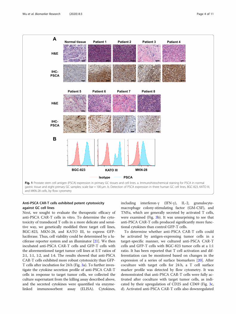
ResultsPSCA expression in patient tissues and gastric cancer celllinesTo evaluate the potential of the tumor antigen PSCA asan immunotherapeutic target, we immunohistochemi-cally detected its presence and abundance in eight pri-mary gastric cancer samples (Fig. 1a). Most of theanalyzed gastric cancer samples expressed PSCA at vari-ous frequencies compared to normal tissues. We alsoperformed flow cytometry in several gastric cancer celllines. The cell types employed in this experiment in-cluded BGC-823, MKN-28, and KATO III cells. Uniformexpression of PSCA was detected on the surface of thesecells (Fig. 1b). Altogether, these data revealed PSCA as apossible novel target for CAR-T cell therapy in GC.
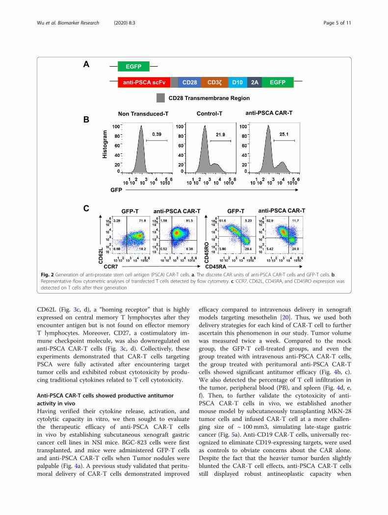
Generation and characterization of anti-PSCA CAR-T cellsWe then constructed a third-generation CAR using ahumanized single-chain variable fragment (scFv) derivedfrom a mouse anti-human PSCA antibody and a third-generation lentivirus vector composed of a CD3ζ intra-cellular domain and two costimulatory domains, thoseof CD28 and DAP10, as previously described [20](Fig. 2a). T cells transfected with only enhanced greenfluorescent protein (eGFP) served as the control for un-specific tonic CAR signaling. Primary human T cellswere isolated from peripheral blood mononuclear cells(PBMCs) by magnetic selection and were then activatedfor 48 h using CD3/CD28/CD2 beads. CAR expressionwas detected 48 h after lentivirus transduction by flowcytometry according to the GFP-positive proportion(Fig. 2b). Transduced T cells were cultured for 10 daysand achieved a final CD45RO+CCR7+CD62Lhigh pheno-type (Fig. 2c), implicating their presumed sustainable an-titumor potential in vivo.
Wu et al. Biomarker Research (2020) 8:3 Page 3 of 11
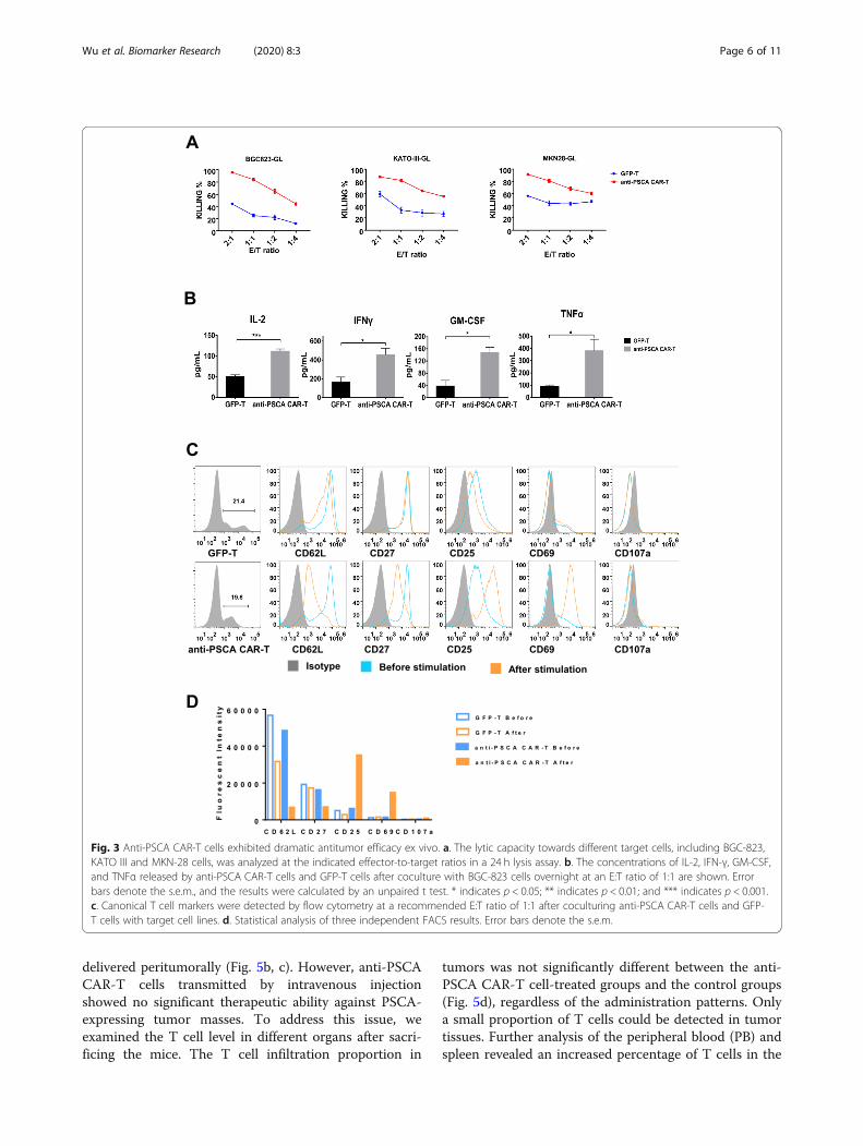
Anti-PSCA CAR-T cells exhibited potent cytotoxicityagainst GC cell linesNext, we sought to evaluate the therapeutic efficacy ofanti-PSCA CAR-T cells in vitro. To determine the cyto-toxicity of transduced T cells in a more delicate and sensi-tive way, we genetically modified three target cell lines,BGC-823, MKN-28, and KATO III, to express GFP-luciferase. Thus, cell viability could be determined by a lu-ciferase reporter system and an illuminator [21]. We thenincubated anti-PSCA CAR-T cells and GFP-T cells withthe aforementioned target tumor cell lines at E:T ratios of2:1, 1:1, 1:2, and 1:4. The results showed that anti-PSCACAR-T cells exhibited more robust cytotoxicity than GFP-T cells after incubation for 24 h (Fig. 3a). To further inves-tigate the cytokine secretion profile of anti-PSCA CAR-Tcells in response to target tumor cells, we collected theculture supernatant from the killing assay described above,and the secreted cytokines were quantified via enzyme-linked immunosorbent assay (ELISA). Cytokines,
including interferon-γ (IFN-γ), IL-2, granulocyte-macrophage colony-stimulating factor (GM-CSF), andTNFα, which are generally secreted by activated T cells,were examined (Fig. 3b). It was unsurprising to see thatanti-PSCA CAR-T cells produced significantly more func-tional cytokines than control GFP-T cells.To determine whether anti-PSCA CAR-T cells could
be activated by antigen-expressing tumor cells in atarget-specific manner, we cultured anti-PSCA CAR-Tcells and GFP-T cells with BGC-823 tumor cells at a 1:1ratio. It has been reported that T cell activation and dif-ferentiation can be monitored based on changes in theexpression of a series of surface biomarkers [20]. Aftercoculture with target cells for 24 h, a T cell surfacemarker profile was detected by flow cytometry. It wasdemonstrated that anti-PSCA CAR-T cells were fully ac-tivated after coculture with target tumor cells, as indi-cated by their upregulation of CD25 and CD69 (Fig. 3c,d). Activated anti-PSCA CAR-T cells also downregulated
Fig. 1 Prostate stem cell antigen (PSCA) expression in primary GC tissues and cell lines. a. Immunohistochemical staining for PSCA in normalgastric tissue and eight primary GC samples; scale bar = 100 μm. b. Detection of PSCA expression in three human GC cell lines, BGC-823, KATO III,and MKN-28 cells, by flow cytometry
Wu et al. Biomarker Research (2020) 8:3 Page 4 of 11
CD62L (Fig. 3c, d), a “homing receptor” that is highlyexpressed on central memory T lymphocytes after theyencounter antigen but is not found on effector memoryT lymphocytes. Moreover, CD27, a costimulatory im-mune checkpoint molecule, was also downregulated onanti-PSCA CAR-T cells (Fig. 3c, d). Collectively, theseexperiments demonstrated that CAR-T cells targetingPSCA were fully activated after encountering targettumor cells and exhibited robust cytotoxicity by produ-cing traditional cytokines related to T cell cytotoxicity.
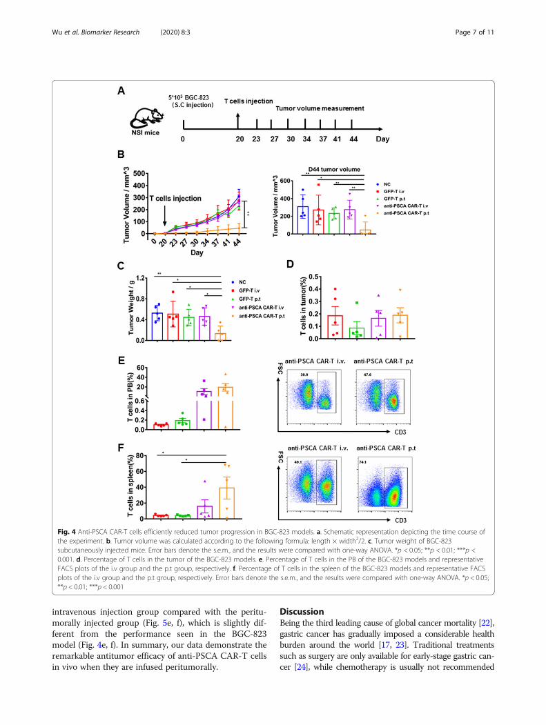
Anti-PSCA CAR-T cells showed productive antitumoractivity in vivoHaving verified their cytokine release, activation, andcytolytic capacity in vitro, we then sought to evaluatethe therapeutic efficacy of anti-PSCA CAR-T cellsin vivo by establishing subcutaneous xenograft gastriccancer cell lines in NSI mice. BGC-823 cells were firsttransplanted, and mice were administered GFP-T cellsand anti-PSCA CAR-T cells when Tumor nodules werepalpable (Fig. 4a). A previous study validated that peritu-moral delivery of CAR-T cells demonstrated improved
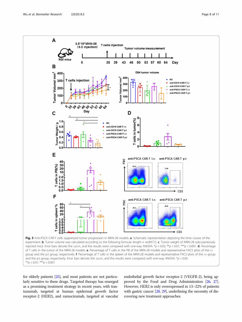
efficacy compared to intravenous delivery in xenograftmodels targeting mesothelin [20]. Thus, we used bothdelivery strategies for each kind of CAR-T cell to furtherascertain this phenomenon in our study. Tumor volumewas measured twice a week. Compared to the mockgroup, the GFP-T cell-treated groups, and even thegroup treated with intravenous anti-PSCA CAR-T cells,the group treated with peritumoral anti-PSCA CAR-Tcells showed significant antitumor efficacy (Fig. 4b, c).We also detected the percentage of T cell infiltration inthe tumor, peripheral blood (PB), and spleen (Fig. 4d, e,f). Then, to further validate the cytotoxicity of anti-PSCA CAR-T cells in vivo, we established anothermouse model by subcutaneously transplanting MKN-28tumor cells and infused CAR-T cell at a more challen-ging size of ~ 100 mm3, simulating late-stage gastriccancer (Fig. 5a). Anti-CD19 CAR-T cells, universally rec-ognized to eliminate CD19-expressing targets, were usedas controls to obviate concerns about the CAR alone.Despite the fact that the heavier tumor burden slightlyblunted the CAR-T cell effects, anti-PSCA CAR-T cellsstill displayed robust antineoplastic capacity when
Fig. 2 Generation of anti-prostate stem cell antigen (PSCA) CAR-T cells. a. The discrete CAR units of anti-PSCA CAR-T cells and GFP-T cells. b.Representative flow cytometric analyses of transfected T cells detected by flow cytometry. c. CCR7, CD62L, CD45RA, and CD45RO expression wasdetected on T cells after their generation
Wu et al. Biomarker Research (2020) 8:3 Page 5 of 11
delivered peritumorally (Fig. 5b, c). However, anti-PSCACAR-T cells transmitted by intravenous injectionshowed no significant therapeutic ability against PSCA-expressing tumor masses. To address this issue, weexamined the T cell level in different organs after sacri-ficing the mice. The T cell infiltration proportion in
tumors was not significantly different between the anti-PSCA CAR-T cell-treated groups and the control groups(Fig. 5d), regardless of the administration patterns. Onlya small proportion of T cells could be detected in tumortissues. Further analysis of the peripheral blood (PB) andspleen revealed an increased percentage of T cells in the
Fig. 3 Anti-PSCA CAR-T cells exhibited dramatic antitumor efficacy ex vivo. a. The lytic capacity towards different target cells, including BGC-823,KATO III and MKN-28 cells, was analyzed at the indicated effector-to-target ratios in a 24 h lysis assay. b. The concentrations of IL-2, IFN-γ, GM-CSF,and TNFα released by anti-PSCA CAR-T cells and GFP-T cells after coculture with BGC-823 cells overnight at an E:T ratio of 1:1 are shown. Errorbars denote the s.e.m., and the results were calculated by an unpaired t test. * indicates p < 0.05; ** indicates p < 0.01; and *** indicates p < 0.001.c. Canonical T cell markers were detected by flow cytometry at a recommended E:T ratio of 1:1 after coculturing anti-PSCA CAR-T cells and GFP-T cells with target cell lines. d. Statistical analysis of three independent FACS results. Error bars denote the s.e.m.
Wu et al. Biomarker Research (2020) 8:3 Page 6 of 11
intravenous injection group compared with the peritu-morally injected group (Fig. 5e, f), which is slightly dif-ferent from the performance seen in the BGC-823model (Fig. 4e, f). In summary, our data demonstrate theremarkable antitumor efficacy of anti-PSCA CAR-T cellsin vivo when they are infused peritumorally.
DiscussionBeing the third leading cause of global cancer mortality [22],gastric cancer has gradually imposed a considerable healthburden around the world [17, 23]. Traditional treatmentssuch as surgery are only available for early-stage gastric can-cer [24], while chemotherapy is usually not recommended
Fig. 4 Anti-PSCA CAR-T cells efficiently reduced tumor progression in BGC-823 models. a. Schematic representation depicting the time course ofthe experiment. b. Tumor volume was calculated according to the following formula: length × width2/2. c. Tumor weight of BGC-823subcutaneously injected mice. Error bars denote the s.e.m., and the results were compared with one-way ANOVA. *p < 0.05; **p < 0.01; ***p <0.001. d. Percentage of T cells in the tumor of the BGC-823 models. e. Percentage of T cells in the PB of the BGC-823 models and representativeFACS plots of the i.v group and the p.t group, respectively. f. Percentage of T cells in the spleen of the BGC-823 models and representative FACSplots of the i.v group and the p.t group, respectively. Error bars denote the s.e.m., and the results were compared with one-way ANOVA. *p < 0.05;**p < 0.01; ***p < 0.001
Wu et al. Biomarker Research (2020) 8:3 Page 7 of 11
for elderly patients [25], and most patients are not particu-larly sensitive to these drugs. Targeted therapy has emergedas a promising treatment strategy in recent years, with tras-tuzumab, targeted at human epidermal growth factorreceptor-2 (HER2), and ramucirumab, targeted at vascular
endothelial growth factor receptor-2 (VEGFR-2), being ap-proved by the Food and Drug Administration [26, 27].However, HER2 is only overexpressed in 13–22% of patientswith gastric cancer [28, 29], underlining the necessity of dis-covering new treatment approaches.
Fig. 5 Anti-PSCA CAR-T cells suppressed tumor progression in MKN-28 models. a. Schematic representation depicting the time course of theexperiment. b. Tumor volume was calculated according to the following formula: length × width2/2. c. Tumor weight of MKN-28 subcutaneouslyinjected mice. Error bars denote the s.e.m., and the results were compared with one-way ANOVA. *p < 0.05; **p < 0.01; ***p < 0.001. d. Percentageof T cells in the tumor of the MKN-28 models. e. Percentage of T cells in the PB of the MKN-28 models and representative FACS plots of the i.vgroup and the p.t group, respectively. f. Percentage of T cells in the spleen of the MKN-28 models and representative FACS plots of the i.v groupand the p.t group, respectively. Error bars denote the s.e.m., and the results were compared with one-way ANOVA. *p < 0.05;**p < 0.01; ***p < 0.001
Wu et al. Biomarker Research (2020) 8:3 Page 8 of 11
The advent of CAR-T cell therapy triggered a paradigmshift in cancer immunotherapy [30]. The application ofCAR-T cells in hematological malignancies has achievedcompelling success [31], sparking considerable interest inin-depth research for this field. Until now, the effects ob-tained for the treatment of solid tumors have been rela-tively less encouraging [32]. A potential caveat is posed bythe fact that solid tumors are heterogeneous, especiallygastric cancer [33]. The lack of truly tumor-specific targetantigens is one of the major obstacles. To provide morepossibilities for clinical efficacy, we conducted a tentativeexperiment targeting PSCA in gastric cancer. We con-structed a third-generation CAR composed of two costi-mulatory molecules as previously described [20] andconfirmed the potential of PSCA as an ideal antigen forCAR-T cell therapy in gastric cancer. The third-generation CAR we applied herein composed of two costi-mulatory molecules, CD28 and DAP10. It has beencorroborated by our lab that DAP10, the NKG2D associ-ated adaptor protein, is a perfect costimulatory moleculefor CAR-T therapy. Incorporation DAP10 in the second-generation CAR-T cells could enhance anti-tumor cap-acity both in vitro and in vivo [34]. We assayed canonicalT cell activation and memory related markers after cocul-turing anti-PSCA CAR-T cells with target cell lines. Anti-PSCA CAR-T cells exhibited upregulated activationmarkers and cytokine production profiles in an antigen-dependent manner. Importantly, we demonstrated thatanti-PSCA CAR-T cells delivered by peritumoral injectionsuccessfully stunted tumor progression in vivo. However,anti-PSCA CAR-T cells given intravenously failed to haveany therapeutic effects. Although CAR-T cells given intra-venously demonstrated slightly augmented T cell level inthe tumor, peripheral blood, and spleen in MKN-28 cellline based mouse model, there were no statistically signifi-cant differences. The absolute number of CAR-T cells re-mains low, especially within the tumor. Hence, we did notpay much attention to this phenomenon. We then ana-lyzed T cell population in the tumor, the data showed thatCAR positive T cell percentage moderately increasedcompared to that of the pre-infused CAR-T cells. It is alsopossible that there were free PSCA in the blood thatmasked the CAR-T cells. However, it has been previouslyreported that CARs can dissociate from dying tumor cellseven more rapidly than TCRs [35]. Hence, we did notmake it the primary cause of the impaired CAR-T cellcytotoxicity. Much more effort might be taken to investi-gate whether it is a general phenomenon or a rare caseand the mechanism hidden behind. Statistical significancewas observed in the tumor weight between the peritu-moral injection group and the intravenous injection groupin the BGC-823 model, while no differences were seen inthe MKN-28 model. Presumably, the augmented periph-eral blood and spleen T cell levels in the peritumoral
injection group might have contributed to this deviation.Consistently, these data unveiled another challenge im-peding CAR-T cell application in solid tumors, that is, thedifficulty of T cells in migrating to and physically infiltrat-ing into the tumor [36]. As previously reported, much ofthe current literature on CAR-T cell therapy for solid tu-mors pays particular attention to T cell trafficking. KhengNewick (2016) noted that blocking protein kinase A(PKA) localization successfully augmented CAR-T celltrafficking and antitumor efficacy [37]. Along the samelines, Qun Gao (2019) found that DOC induced CD8+ Tcell recruitment to the tumor microenvironment by en-hancing the secretion of HMGB1 and CXCL11 [38],underlining another view that successful trafficking relieson an appropriate match between the chemokine recep-tors on T cells and the chemokines secreted by the tumors[39]. The evidence reviewed here seems to suggest a com-bination strategy, including anti-PSCA CAR-T cells andchemical drugs.Altogether, our findings corroborated the feasibility
and efficacy of anti-PSCA CAR-T cells against gastriccancer, thus providing a new solution for interpatientand intratumor heterogeneity.
ConclusionThis study identified PSCA as an ideal target for CAR-Tcell therapy in GC and achieved impressive efficacywhen using third-generation CAR-T cells for GC treat-ment. The results of this research reveal that anti-PSCACAR-T cells can specifically eliminate target cells, bothex vivo and in vivo, implicating the potential of anti-PSCA CAR-T cells in treating GC patients in the clinic.
AbbreviationsALL: Acute lymphoblastic leukemia; CAR: Chimeric antigen receptor;E:T: Effector to target; ELISA: Enzyme-linked immune absorbance assay;GC: Gastric cancer; GM-CSF: Granulocyte-macrophage colony-stimulating fac-tor; i.p.: Intraperitoneally; i.v.: Intravenous; IFNγ: Interferon-γ; IL-2: Interleukin-2;p.t.: Peritumoral; p.t.: Peritumoral; PB: Peripheral blood; PBMC: Peripheralblood mononuclear cells mononuclear cells; PBMCs: Peripheral blood;PSCA: Prostate stem cell antigen; s.c.: Subcutaneous; scFv: Single-chainvariable fragment
AcknowledgmentsWe would like to thank all the members in the list for experimentalmaterials, technical assistance, helpful discussions, and comments.
Authors’ contributionsConception and design: DW, PL, YY. Development of methodology: DW, JL,RZ, ZW, DZ, JS. Acquisition of data (provided animals, acquired and managedpatients, provided facilities, etc.): DW, JL, RZ, QW, SL, YL, SW, PL, YY. Analysisand interpretation of data (e.g., statistical analysis, biostatistics, computationalanalysis): DW, JL, RZ, YL, PL, YY. Writing, review, and/or revision of themanuscript: DW, JL, RZ, PL, YY. Administrative, technical, or material support(i.e., reporting or organizing data, constructing databases): YY, PL. Studysupervision: PL, YY. All authors read and approved the final manuscript.
FundingThis study is supported by the National Natural Science Foundation of China(NSFC), No. 81961128003; The Strategic Priority Research Program of theChinese Academy of Sciences, Grant No. XDB19030205; Guangdong
Wu et al. Biomarker Research (2020) 8:3 Page 9 of 11
Provincial Significant New Drugs Development, No. 2019B020202003; TheNational Major Scientific and Technological Special Project for “SignificantNew Drugs Development”, No. SQ2018ZX090201; Guangdong ProvincialApplied Science and Technology Research& Development Program, No.2016B020237006; Frontier Research Program of Guangzhou RegenerativeMedicine and Health Guangdong Laboratory, No. 2018GZR110105003;Science and Technology Planning Project of Guangdong Province, China(2017B030314056).
Availability of data and materialsAll data generated or analyzed are available from the corresponding authoron reasonable request.
Ethics approval and consent to participateHealthy PBMCs providers, donors provided gastric cancer tissues and normaltissues, have given consent. All animal experiments were performed at theLaboratory Animal Center of the Guangzhou Institutes of Biomedicine andHealth (GIBH). All procedures were approved by the Research Ethics Board ofGuangzhou Institutes of Biomedicine and Health (GIBH).
Consent for publicationNot applicable.
Competing interestsThe authors declare that they have no competing interests.
Author details1School of Life Sciences, University of Science and Technology of China,Hefei 230027, China. 2Key Laboratory of Regenerative Biology, South ChinaInstitute for Stem Cell Biology and Regenerative Medicine, GuangzhouInstitutes of Biomedicine and Health, Chinese Academy of Sciences,Guangzhou 510530, China. 3Guangdong Provincial Key Laboratory of StemCell and Regenerative Medicine, South China Institute for Stem Cell Biologyand Regenerative Medicine, Guangzhou Institutes of Biomedicine andHealth, Chinese Academy of Sciences, Guangzhou 510530, China. 4Universityof Chinese Academy of Sciences, Shijingshan District, Beijing 100049, China.5Institute of Hematology, Medical College, Jinan University, Guangzhou510632, China. 6Hefei Institute of Stem Cell and Regenerative Medicine,Guangzhou Institutes of Biomedicine and Health, Chinese Academy ofSciences, Guangzhou 510530, China.
Received: 5 November 2019 Accepted: 2 January 2020
References1. Crew KD, Neugut AI. Epidemiology of gastric cancer. World J Gastroenterol:
WJG. 2006;12(3):354.2. Correa P, Piazuelo MB, Camargo MC. The future of gastric cancer
prevention. Gastric Cancer. 2004;7(1):9–16.3. Zhao J, Lin Q, Song Y, Liu D. Universal CARs, universal T cells, and universal
CAR T cells. J Hematol Oncol. 2018;11(1):132.4. Chang ZL, Chen YY. CARs: synthetic immunoreceptors for cancer therapy
and beyond. Trends Mol Med. 2017;23(5):430–50.5. Zhang C, Liu J, Zhong JF, Zhang X. Engineering car-t cells. Biomark Res.
2017;5(1):22.6. Newick K, Moon E, Albelda SM. Chimeric antigen receptor T-cell therapy for
solid tumors. Mol Ther-Oncol. 2016;3:16006.7. Shum T, Kruse RL, Rooney CM. Strategies for enhancing adoptive T-cell
immunotherapy against solid tumors using engineered cytokine signalingand other modalities. Expert Opin Biol Ther. 2018;18(6):653–64.
8. Wang Z, Han W. Biomarkers of cytokine release syndrome and neurotoxicityrelated to CAR-T cell therapy. Biomark Res. 2018;6:4.
9. Yu S, Li A, Liu Q, Li T, Yuan X, Han X, Wu K. Chimeric antigen receptor Tcells: a novel therapy for solid tumors. J Hematol Oncol. 2017;10(1):78.
10. Li J, Li W, Huang K, Zhang Y, Kupfer G, Zhao Q. Chimeric antigen receptor Tcell (CAR-T) immunotherapy for solid tumors: lessons learned and strategiesfor moving forward. J Hematol Oncol. 2018;11(1):22.
11. Li Z, Song W, Rubinstein M, Liu D. Recent updates in cancerimmunotherapy: a comprehensive review and perspective of the 2018China Cancer immunotherapy workshop in Beijing. J Hematol Oncol. 2018;11(1):142.
12. Wenqi D, Li W, Shanshan C, Bei C, Yafei Z, Feihu B, Jie L, Daiming F. EpCAMis overexpressed in gastric cancer and its downregulation suppressesproliferation of gastric cancer. J Cancer Res Clin Oncol. 2009;135(9):1277–85.
13. Jung H, Jun KH, Jung JH, Chin HM, Park WB. The expression of claudin-1,claudin-2, claudin-3, and claudin-4 in gastric cancer tissue. J Surg Res. 2011;167(2):e185–91.
14. Padmanabhan N, Ushijima T, Tan P. How to stomach an epigenetic insult: thegastric cancer epigenome. Nat Rev Gastroenterol Hepatol. 2017;14(8):467.
15. Saeki N, Gu J, Yoshida T, Wu X. Prostate stem cell antigen: a Jekyll and Hydemolecule? Clin Cancer Res. 2010;16(14):3533–8.
16. Priceman SJ, Gerdts EA, Tilakawardane D, Kennewick KT, Murad JP, Park AK,Jeang B, Yamaguchi Y, Yang X, Urak R. Co-stimulatory signaling determinestumor antigen sensitivity and persistence of CAR T cells targeting PSCA+metastatic prostate cancer. Oncoimmunology. 2018;7(2):e1380764.
17. Wei X, Lai Y, Li J, Qin L, Xu Y, Zhao R, Li B, Lin S, Wang S, Wu Q. PSCA andMUC1 in non-small-cell lung cancer as targets of chimeric antigen receptorT cells. Oncoimmunology. 2017;6(3):e1284722.
18. Zhao X, Wang F, Hou M. Expression of stem cell markers nanog and PSCAin gastric cancer and its significance. Oncol Lett. 2016;11(1):442–8.
19. Olafsen T, Gu Z, Sherman MA, Leyton JV, Witkosky ME, Shively JE,Raubitschek AA, Morrison SL, Wu AM, Reiter RE. Targeting, imaging, andtherapy using a humanized antiprostate stem cell antigen (PSCA) antibody.J Immunother. 2007;30(4):396–405.
20. Lv J, Zhao R, Wu D, Zheng D, Wu Z, Shi J, Wei X, Wu Q, Long Y, Lin S.Mesothelin is a target of chimeric antigen receptor T cells for treatinggastric cancer. J Hematol Oncol. 2019;12(1):18.
21. Corso S, Isella C, Bellomo SE, Apicella M, Durando S, Migliore C, Ughetto S,D'Errico L, Menegon S, Moya-Rull D. A comprehensive PDX gastric cancercollection captures cancer cell intrinsic transcriptional MSI traits. Cancer Res.2019;2019:1166.
22. Wadhwa R, Song S, Lee J-S, Yao Y, Wei Q, Ajani JA. Gastric cancer—molecularand clinical dimensions. Nat Rev Clin Oncol. 2013;10(11):643.
23. Liu D, Mehta D, Kaur S, Kumar A, Parikh K, Chawla L, Patel S, Devi A, Saha A.Decreasing mortality and hospitalizations with rising costs related to gastriccancer in the USA: an epidemiological perspective. J Hematol Oncol. 2018;11(1):138.
24. Jeong O, Park Y-K. Clinicopathological features and surgical treatment ofgastric cancer in South Korea: the results of 2009 nationwide survey onsurgically treated gastric cancer patients. J Gastric Cancer. 2011;11(2):69–77.
25. Kim HS, Kim JH, Kim JW, Kim BC. Chemotherapy in elderly patients withgastric cancer. J Cancer. 2016;7(1):88.
26. Roukos DH. Targeting gastric cancer with trastuzumab: new clinical practiceand innovative developments to overcome resistance. Ann Surg Oncol.2010;17(1):14–7.
27. Casak SJ, Fashoyin-Aje I, Lemery SJ, Zhang L, Jin R, Li H, Zhao L, Zhao H,Zhang H, Chen H. FDA approval summary: ramucirumab for gastric cancer.Clin Cancer Res. 2015;21(15):3372–6.
28. Meza-Junco J, Au H-J, Sawyer MB. Critical appraisal of trastuzumab intreatment of advanced stomach cancer. Cancer Manag Res. 2011;3:57.
29. Fusco N, Rocco EG, Del Conte C, Pellegrini C, Bulfamante G, Di Nuovo F,Romagnoli S, Bosari S. HER2 in gastric cancer: a digital image analysis inpre-neoplastic, primary and metastatic lesions. Mod Pathol. 2013;26(6):816.
30. Liu D, Zhao J, Song Y. Engineering switchable and programmable universalCARs for CAR T therapy. J Hematol Oncol. 2019;12(1):69.
31. Zhao J, Song Y, Liu D. Clinical trials of dual-target CAR T cells, donor-derivedCAR T cells, and universal CAR T cells for acute lymphoid leukemia. JHematol Oncol. 2019;12(1):17.
32. Li D, Li X, Zhou W-L, Huang Y, Liang X, Jiang L, Yang X, Sun J, Li Z, Han W-D. Genetically engineered T cells for cancer immunotherapy. SignalTransduction Targeted Ther. 2019;4(1):1–17.
33. Patil DT, Rubin BP. Genetics of gastrointestinal stromal tumors: aheterogeneous family of tumors? Surg Pathol Clin. 2015;8(3):515–24.
34. Zhao R, Cheng L, Jiang Z, Wei X, Li B, Wu Q, Wang S, Lin S, Long Y, ZhangX, Wu Y, Du X, Pei D, Liu P, Li Y, Cui S, Yao Y, Li P. DNAX-activating protein10 co-stimulation enhances the anti-tumor efficacy of chimeric antigenreceptor T cells. Oncoimmunology. 2019;8(1):e1509173.
35. Watanabe K, Kuramitsu S, Posey AD Jr, June CH. Expanding the therapeuticwindow for CAR T cell therapy in solid tumors: the Knowns and unknownsof CAR T cell biology. Front Immunol. 2018;9:2486.
36. Castellarin M, Watanabe K, June CH, Kloss CC, Posey AD. Driving cars to theclinic for solid tumors. Gene Ther. 2018;25(3):165.
Wu et al. Biomarker Research (2020) 8:3 Page 10 of 11
37. Newick K, O'Brien S, Sun J, Kapoor V, Maceyko S, Lo A, Puré E, Moon E,Albelda SM. Augmentation of CAR T-cell trafficking and antitumorefficacy by blocking protein kinase a localization. Cancer Immunol Res.2016;4(6):541–51.
38. Gao Q, Wang S, Chen X, Cheng S, Zhang Z, Li F, Huang L, Yang Y, Zhou B,Yue D. Cancer-cell-secreted CXCL11 promoted CD8+ T cells infiltrationthrough docetaxel-induced-release of HMGB1 in NSCLC. J ImmunotherCancer. 2019;7(1):42.
39. Newick K, O'Brien S, Moon E, Albelda SM. CAR T cell therapy for solidtumors. Annu Rev Med. 2017;68:139–52.
Publisher’s NoteSpringer Nature remains neutral with regard to jurisdictional claims inpublished maps and institutional affiliations.
Wu et al. Biomarker Research (2020) 8:3 Page 11 of 11