OULU 2014 D 1249 ACTA UNIVERSITATIS OULUENSIS …jultika.oulu.fi/files/isbn9789526204666.pdf ·...
102
UNIVERSITATIS OULUENSIS MEDICA ACTA D D 1249 ACTA Irina I. Nagy OULU 2014 D 1249 Irina I. Nagy WNT-11 SIGNALING ROLES DURING HEART AND KIDNEY DEVELOPMENT UNIVERSITY OF OULU GRADUATE SCHOOL; UNIVERSITY OF OULU, FACULTY OF BIOCHEMISTRY AND MOLECULAR MEDICINE; BIOCENTER OULU; OULU CENTER FOR CELL-MATRIX RESEARCH; NORTHERN FINLAND LABORATORY CENTRE
Transcript of OULU 2014 D 1249 ACTA UNIVERSITATIS OULUENSIS …jultika.oulu.fi/files/isbn9789526204666.pdf ·...
ABCDEFG
UNIVERSITY OF OULU P .O. B 00 F I -90014 UNIVERSITY OF OULU FINLAND
A C T A U N I V E R S I T A T I S O U L U E N S I S
S E R I E S E D I T O R S
SCIENTIAE RERUM NATURALIUM
HUMANIORA
TECHNICA
MEDICA
SCIENTIAE RERUM SOCIALIUM
SCRIPTA ACADEMICA
OECONOMICA
EDITOR IN CHIEF
PUBLICATIONS EDITOR
Professor Esa Hohtola
University Lecturer Santeri Palviainen
Postdoctoral research fellow Sanna Taskila
Professor Olli Vuolteenaho
University Lecturer Veli-Matti Ulvinen
Director Sinikka Eskelinen
Professor Jari Juga
Professor Olli Vuolteenaho
Publications Editor Kirsti Nurkkala
ISBN 978-952-62-0465-9 (Paperback)ISBN 978-952-62-0466-6 (PDF)ISSN 0355-3221 (Print)ISSN 1796-2234 (Online)
U N I V E R S I TAT I S O U L U E N S I S
MEDICA
ACTAD
D 1249
ACTA
Irina I. Nagy
OULU 2014
D 1249
Irina I. Nagy
WNT-11 SIGNALING ROLES DURING HEART AND KIDNEY DEVELOPMENT
UNIVERSITY OF OULU GRADUATE SCHOOL;UNIVERSITY OF OULU,FACULTY OF BIOCHEMISTRY AND MOLECULAR MEDICINE;BIOCENTER OULU;OULU CENTER FOR CELL-MATRIX RESEARCH;NORTHERN FINLAND LABORATORY CENTRE

A C T A U N I V E R S I T A T I S O U L U E N S I SD M e d i c a 1 2 4 9
IRINA I. NAGY
WNT-11 SIGNALING ROLES DURING HEART AND KIDNEY DEVELOPMENT
Academic dissertation to be presented with the assentof the Doctoral Training Committee of Health andBiosciences of the University of Oulu for public defencein the Leena Palotie Auditorium (101A) of the Faculty ofMedicine (Aapistie 5 A), on 6 June 2014, at 12 noon
UNIVERSITY OF OULU, OULU 2014

Copyright © 2014Acta Univ. Oul. D 1249, 2014
Supervised byProfessor Seppo Vainio
Reviewed byProfessor Kersti K. LinaskAssociate Professor Ovidiu I. Sirbu
ISBN 978-952-62-0465-9 (Paperback)ISBN 978-952-62-0466-6 (PDF)
ISSN 0355-3221 (Printed)ISSN 1796-2234 (Online)
Cover DesignRaimo Ahonen
JUVENES PRINTTAMPERE 2014
OpponentProfessor Stefan Hoppler
Nagy, Irina I., Wnt-11 signaling roles during heart and kidney development. University of Oulu Graduate School; University of Oulu, Faculty of Biochemistry andMolecular Medicine; Biocenter Oulu; Oulu Center for Cell-Matrix research; Northern FinlandLaboratory CentreActa Univ. Oul. D 1249, 2014University of Oulu, P.O. Box 8000, FI-90014 University of Oulu, Finland
Abstract
Organogenesis involves precursor cells proliferation, differentiation along with their coordinatedorganization into precise multicellular arrangements by planar cell polarity (PCP) pathways. Thebeta-catenin independent/non-canonical type of Wnt-11 signaling has been known as a PCPmodulator during development. In this thesis were analyzed the roles of Wnt-11 in heart andkidney development by using in vivo functional genomics technologies.
We show that the Wnt-11 gene is important for murine ventricular myocardium development,since Wnt-11 deficiency in early cardiogenesis leads to impaired organization and maturation ofmouse ventricular cardiomyocytes, causing primary cardiomyopathy with in utero lethality. Wnt-11 coordinates the co-localized expression of the cell adhesion molecules N-cadherin and β-catenin, which are critical for the spatially specific organization of cardiomyocytes. We show thatWnt-11 deficiency causes primary hypertrophic and noncompaction cardiomyopathy in adultmice, with consequences for regional myocardium function.
The Wnt family of secreted signals has been implicated in kidney tubule development andtubular cystic diseases such as polycystic kidney disease. We show here that Wnt-11 is expressedin mature nephrons and is involved in late steps of nephrogenesis, since the kidney tubuleorganization is deregulated in Wnt-11 deficient kidneys, to enlarged lumen with increasedconvolution. These tubule abnormalities are associated with glomerular microcyst formation andkidney failure. Wnt-11 deficiency reduced significantly Wnt-9b expression, a critical signal forPCP-mediated kidney tubule elongation. In the cortical region this associated with reducedexpression of nephron and stromal progenitor cell marker.
The results in this thesis point out that Wnt-11 function is required for proper myocardiumorganization and maturation as well as proper morphogenesis of the kidney tubules during theembryonic and postnatal developmental stages. Wnt-11 knockout phenotypes depend on thegenetic background, similarly to human congenital disease. This data may be relevant for humancongenital cardiomyopathy and glomerulocystic kidney disease studies.
Keywords: cardiomyopathy, convolution, glomerular cyst, human congenital diseases,kidney tubule, lumen, myocardium, organogenesis, planar cell polarity, Wnt-11

Nagy, Irina I., Wnt-11 signalointi sydämen ja munuaisten kehityksessä. Oulun yliopiston tutkijakoulu; Oulun yliopisto, Biokemian ja molekyylilääketieteen tiedekunta;Biocenter Oulu; Oulu Center for Cell-Matrix research; Pohjois-Suomen laboratoriokeskusActa Univ. Oul. D 1249, 2014Oulun yliopisto, PL 8000, 90014 Oulun yliopisto
Tiivistelmä
Alkion sisäelinten kehityksen aikana esisolut lisääntyvät ja erilaistuvat muodostaen tarkoin mää-riteltyjä monisoluisia rakenteita. Muodostuvan kudosrakenteen määrittelyssä erilaiset solusig-naalit ovat keskeisessä asemassa. Yksi näistä on nk. Wnt signaali perhe. Wnt perheeen jäsenWnt-11 tehtävät on huonosti tunnettu. Wnt-11 viestittää ilmeisesti nk. planaaristen solupola-riteettireittien (PCP) avulla, joka on beeta-kateniinista riippumattoman nk. ei-kanonisen Wntsignaali. Väitöskirjatyössä selvitettiin Wnt-11:n vaikutuksia sydämen ja munuaisten kehityk-seen in vivo funktionaalisten genomisten menetelmien avulla.
Ihmisen synnynnäiset kardiomyopatiat ovat sydänlihaksen ensisijaisia vaurioita, joiden taus-talla on sydänlihaksen kehityshäiriö. Tutkimuksessa osoitetaan, että Wnt-11-geenillä on tärkeämerkitys hiiren sydänkammion kehitykselle, koska Wnt-11-geenin puute sydämen varhaisenkehityksen vaiheessa johtaa sydänlihassolujen järjestäytymisen ja kypsymisen häiriintymiseen,jolloin seurauksena on ensisijaisesta kardiomyopatiasta johtuva sikiökuolema. Wnt-11 koordinoikahden solukiinnitysmolekyylin, N-kadheriinin ja β-kateniinin, samanaikasta ilmentymistä.Kyseiset molekyylit ovat keskeisen tärkeitä sydänlihasssolujen spatiaalisen järjestäytymisenkannalta. Tutkimuksessa osoitetaan, että Wnt-11-puutos aiheuttaa aikuisilla hiirillä ensisijaistasydänlihaksen liikakasvua ja trabekuloivaa kardiomyopatiaa, mikä vaikuttaa sydänlihaksen toi-mintaan. Tuloksilla voi olla merkitystä tutkittaessa ihmisen synnynnäisiä kardiomyopatioita.
Wnt-signaaliperheen on osoitettu olevan yhteydessä munuaisputken kehitykseen ja sen saira-uksiin, kuten munuaisten monirakkulatautiin. Väitöstutkimuksessa osoitetaan, että Wnt-11ilmentyy kypsissä nefroneissa ja että se osallistuu nefrogeneesiin myöhempiin vaiheisiin, koskamunuaisputken kehityksen säätely on poikkeavaa niissä munuaisissa, joista Wnt-11 puuttuu.Seurauksena on laajentunut, normaalia poimuttuneempi luumen. Munuaisputken poikkeavuuk-silla oli yhteyttä munuaiskerästen mikrokystien muodostumiseen sekä munuaisten vajaatoimin-taan. Wnt-11 -puute vähensi huomattavasti Wnt-9b-ilmentymistä, joka on PCP-välitteisen munu-aisputken pidentymisen kannalta keskeisen tärkeä signaali. Kortikaalialueella Wnt9b vaimennus-säätely liittyi poikkeavaan solujen lisääntymiseen, apoptoosiin ja kypsymiseen sekä vähentynee-seen nefroni- ja stroomakantasolujen merkkiaineen ilmentymiseen.
Väitöskirjatutkimuksen tulokset viittaavat siihen, että Wnt-11 -toiminto on välttämätönsydänlihaksen normaalin muodostumisen ja kypsymisen sekä munuaisputken normaalin morfo-geneesin kannalta sikiövaiheen ja syntymän jälkeisen kehityksen aikana. Wnt-11 -poistogeeni-sen hiiren fenotyypi riippuu geneettisestä tausta, samaan tapaan kuin ihmisen synnynnäisissäsairauksissa. Väitöstutkimuksesta saatavalla tiedolla voi olla merkitystä tutkittaessa ihmisen syn-nynnnäistä kardiomyopatiaa ja munuaisten monirakkulatautia.
Asiasanat: glomerulaarinen kysta, ihmisen synnynnäiset sairaudet, kardiomyopatia,luumen, munuaisputki, organogeneesi, planaarinen solupolariteetti, poimuuntuminen,sydänlihas, Wnt-11


To my family

8
9
Acknowledgements
The present thesis was carried out in the research group of the Professor Seppo
Vainio, at Biocenter Oulu, Faculty of Biochemistry and Molecular Medicine,
former Department of medical biochemistry and molecular biology, Faculty of
Medicine, University of Oulu, Finland.
First of all, I would like to express my sincere gratitude to my supervisor,
professor Seppo Vainio, for giving me the opportunity to work in his
developmental biology research group and introducing me to the fascinating
world of science.
I am grateful to Professor Kersti Linask and Associate Professor Ovidiu Sirbu
for their careful review and valuable comments on this thesis. I wish to thank
Anna Vuolteenaho, Msc for her careful revision of the language.
I am also sincerely grateful to Professor Juha Räsänen and Dr. Pekka
Kilpeläinen for their valuable guidance especially in the beginning of my research
carrier.
I owe sincere gratitude to the chairman of my thesis committee Docent Olavi
Ukkola, Docent Ritva Heljasvaara, PhD Reetta Vuolteenaho, PhD Kaija Autio
and Biocenter Doctoral Programme Coordinator Docent Ritva Saastamoinen for
their encouragement to finalize this thesis.
I would like to extend my gratitude to the leaders of our faculty Professor
Taina Pihlajaniemi, Pofessor Kalervo Hiltunen, Professor Johanna Myllyharju,
and Professor Peppi Karppinen and to all of the faculty and staff for creating the
excellent research infrastructure and encouraging academic growth.
I wish to sincerely thank to all my co-authors for their valuable contribution
to my research projects.
My warmest thanks to my colleagues, past and present, SV-group members.
Special thanks to Johanna Kekolahti-Liias, Hannele Härkman and Jaana Kujala
for their kind help and technical assistance.
I would like to express my gratitude to the NordLab academic members for
their sincere encouragements and support during the last years to finalize this
thesis. Special thanks to my residency colleagues Professor Sohvi Hörkkö and
Docent Päivi Myllynen.
My warmest thanks to all my friends for encouraging my personal growth,
you teach me every day to be a better person.

10
I am deeply grateful to my parents and my brother Tibi for their love and
support to find my own way in life, even if this turned out to be far away from
home.
Finally, there are no words to adequately express my gratitude to my better
half in life Atti and our beloved son Robi, I hope I can make you proud.
Oulu, april 2014 Irina I. Nagy
11
Abbreviations
AO aorta
AV atrioventricular
ANP atrial natriuretic peptide
Bmp bone morphogenetic protein
Brg LysR family transcriptional regulator
BUN blood urea nitrogen
Ccr creatinine clearance
CD collecting duct
CHD congenital heart disease
Cl chlorine
CM cap mesenchyme
CMP cardiomyopathy
cCMP congenital cardiomyopathy
cRet receptor tyrosine kinase to Gdnf
DCM dilated cardiomyopathy
DORV double outlet right ventricle
Dkk dickkopf
Dvl disheveled
EB embryonic body
Egf epithelial growth factor
Erbb v-erb-b2 avian erythroblastic leukemia viral oncogene homolog
Ffg fibroblast growth factor
Fox forkhead box
EMT epithelial to mesenchymal transformation
FHF first heart field
Fz Frizzled homolog receptor
Gata Gata-Zinc finger DNA binding factor
GCK glomerular cystic kidney
Gdnf glial cell-derived neurotrophic factor
GFR glomerular filtration rate
GTP guanidine triphosphate
HCM hypertrophic cardiomyopathy
HL Henle loop
Hnf hepatocyte nuclear factor
Hox homeobox factor
12
HR heart rate
ICD intercalated disc structure
IRT isovolumetric relaxation time
Isl Lim-type homeobox gene islet factor
JNK c-Jun NH2-termianl kinase
K potassium
LV left ventricle
LVNC left ventricular noncompaction
Mef myocyte enhancer factor
Mesp mesoderm posterior homologue factor
MET mesenchyme-to-epithelial transformation
Nkx Nk homeobox-containing transcription factor
MM metanephric mesenchyme
Na natrium
Notch neurogenic locus notch homolog factor
OFT outflow tract
PA for kidney section - pretubular aggregate
PA for heart section - pulmonary artery
PEO proepicardial organ
Pitx paired-like homeodomain factor
PT proximal tubule
PTA persistent truncus arteriousus
PCNA proliferation cell nucleic antigen
PCP planar cell polarity
PKD polycystic kidney disease
Pkd polycystin
RA retinoic acid
Rac Ras-related C3 botulinum toxin substrate protein
Rho Ras homologue factor
RV for kidney section - renal vesicle
RV for heart section - right ventricle
Shh sonic hedgehog
SHF second heart field
Scribble Scribbled homolog (Drosophila) protein-Circletail homolog
Spry Sprouty
UB ureteric bud
Vangl Van Gogh/strabismus homolog (Drosophila)-Loop-Tail homolog
13
Vegf vascular endothelial growth factor
VNC ventricular noncompaction
VSD ventricular septum defect
Wnt wingless-related MMTV integration site factor
Tbx t-box factor
TGA transposition of big arteries
Tgf transforming growth factor

14
15
List of original articles
The thesis is based on the following articles, which are referred to in the text by
their roman numerals:
I Nagy II, Railo A, Rapila R, Hast T, Sormunen R, Tavi P, Räsänen J & Vainio SJ (2010) Wnt-11 signaling controls ventricular myocardium development by patterning N-cadherin and beta-catenin expression. Cardiovasc Res 85(1): 100–109.
II Nagy II, Szabo Z, Kerkelä R & Vainio SJ (2014) Wnt-11 deficiency causes primary cardiomyopathy in C57Bl6 mice. Manuscript.
III Nagy II, Naillat F, Jokela T, Miinalainen I, Sormunen R & Vainio SJ (2014) Wnt-11 signaling contributes to kidney tubular system development during nephrogenesis. Manuscript.

16
17
Table of contents
Abstract
Tiivistelmä
Acknowledgements 9
Abbreviations 11
List of original articles 15
Table of contents 17
1 Introduction 19
2 Review of the literature 21
2.1 Heart organogenesis ................................................................................ 21
2.1.1 Cell behavior in cardiac mesoderm – cardiac heart fields
and linear heart tube ..................................................................... 21
2.1.2 Development of the ventricles, looping and ballooning ............... 25
2.1.3 Myocardium patterning during development ............................... 26
2.1.4 Abnormal heart organogenesis and consequences for
cardiac functional development .................................................... 32
2.1.5 Developmental cardiac defects: Congenital heart diseases
and Cardiomyopathies .................................................................. 36
2.2 Kidney organogenesis ............................................................................. 37
2.2.1 Cortical nephrogenesis and ureter branching ............................... 37
2.2.2 Medulla formation and kidney tubule maturation and
elongation ..................................................................................... 41
2.2.3 Functional evaluation of kidney disease ....................................... 44
2.2.4 Congenital kidney disease, cystogenesis and impaired
prenatal and postnatal kidney development.................................. 46
2.3 Wnt-11 gene, protein and signaling pathways in mammalian
organogenesis with emphasis on heart and kidney development ............ 47
2.3.1 Wnt-11 during heart development ................................................ 49
2.3.2 Wnt-11 during kidney development ............................................. 55
3 Outline of the present study 57
4 Material and methods 59
5 Results 61
5.1 Wnt-11 deficiency in 129SV background leads to primary
cardiomyopathy causing heart failure with severe lethality in
midgestation (I) ....................................................................................... 61
18
5.2 Loss of Wnt-11 in ventricular cardiomyocytes is linked to
impaired cell adhesion and cytoarchitecture associated with
delayed differentiation (I) ........................................................................ 63
5.3 Wnt-11 deficiency abnormalities in ventricular myocardium
associate with changes in genes implicated in heart development
(I) ............................................................................................................. 66
5.4 Wnt-11 is expressed in late myocardium development and adult
C57Bl6 Wnt-11 knockout mice present CMP phenotype (II) .................. 66
5.5 Phenotypic characterization of Wnt-11-/- hearts - Functional assay
telemetry, standard Doppler and 2D strain imaging (STE) (II) ............... 68
5.6 Wnt-11 loss in C57Bl6 background leads to glomerulocystic
kidney with abnormal tubular morphology and impaired kidney
function (III) ............................................................................................ 69
5.7 Wnt-11 is expressed in tubular epithelial cells from cortex to tip
of papilla and throughout kidney organogenesis (III) ............................. 70
5.8 Abnormal Wnt-11-/- kidney tubule morphology suggests impaired
cell polarity processes (III) ...................................................................... 71
5.9 Wnt-11 deficiency alters cell proliferation and cell survival in the
cortex, correlating with changes in nephron progenitor gene
expression (III) ........................................................................................ 72
6 Discussion 73
6.1 Wnt-11 signaling controls development of the early myocardium
and is essential for normal cardiac functional development (I) ............... 73
6.2 Cardiac phenotypes in Wnt-11 knock out models and their
relevance to human inherited cardiac diseases (I and II) ........................ 75
6.3 Kidney phenotype in Wnt-11-/- C57Bl6 mice suggests novel roles
for Wnt-11 in late nephrogenesis (III) ..................................................... 79
6.3.1 Wnt-11 knockout as a disease model for human
glomerulocystic kidney disease studies ........................................ 79
6.3.2 Wnt-11 signaling fine-tunes nephrogenesis .................................. 80
6.3.3 Wnt-11 and cell polarity pathway control of kidney tubule
morphogenesis .............................................................................. 81
6.4 Wnt-11 signaling commonalities between heart and kidney
development ............................................................................................ 82
References 83
Original articles 97
19
1 Introduction
In development, organogenesis is the process by which precursor cells form
mature functional organs. This is achieved by complex cellular inductive
interactions (Glibert 2006). Organ development involves not only the
proliferation and differentiation of cells, but also their coordinated organization
into precise multicellular arrangements by cell polarity pathways. This precise
cell arrangement in an organ cannot be disturbed without impairing its function.
The ability to regenerate damaged human organs constitutes the “holy grail” of
medicine and researchers are attempting to find ways of awakening in the adult
developmental programs that were used during organogenesis (Gilbert 2006).
Congenital malformations are defects in organogenesis “present at birth”. For
most congenital anomalies (50–60%) the etiology is unknown; gene mutations are
thought to account for 7–8% of human birth defects, while 20–15% are caused by
a combination of genetic and environmental factors (Moon & Persaud 2003).
The Wnt gene family encodes secreted signaling molecules and is critical for
the developmental process of organogenesis, including specifically cardiac and
kidney development.
The mammalian heart develops through a series of sequential morphogenetic
steps during which the cardiogenic precursor cells in the mesoderm differentiate
into cardiomyocytes. During the early stages of cardiogenesis the ventricular
cardiomyocytes become polarized, although the cues that regulate the spatial
organization of the cardiomyocytes during heart ontogeny are still poorly
characterized. Moreover, very little is known about possible consequences for
heart function in utero or later adult life in case of impaired processes of
embryonic cardiomyocyte polarization.
The Wnts family of secreted signals and Wnt signal transduction pathways
have been implicated in kidney development and in kidney tubular diseases,
which include cystogenesis. The underlying mechanism correlates with the main
roles of the Wnts in controlling tubular diameter and length via the cell polarity
pathways (Merkel et al. 2007, Karner et al. 2009, Yu et al. 2009). The latter
Wnt/PCP signaling has risen as a novel participant in PKD etiology and
pathogenesis, as shown by human and mouse studies (Karner et al. 2009, Yu et al.
2009, Bagherie-Lachidan & McNeill 2010, Lancaster & Gleeson 2010, Luyten et
al. 2010, Wuebken & Schmidt-Ott 2011). Consistent with these, Wnt pathway-
related proteins are involved in human and mouse PKD (Romaker et al. 2009,
Torres & Harris 2009).
20
Based on previous experiments, it was shown that Wnt-11 has an important
role in outflow tract during cardiac development (Zhou et al. 2008) and in ureter
bud branching during kidney development (Majumdar et al. 2003).
The overall goal of this thesis is to gain an understanding of how Wnt-11
participates in cardiomyocyte polarization during myocardium development and
epithelium organization during kidney tubule morphogenesis, including
identifying Wnt-11 downstream effectors. By using a Wnt-11 knockout model,
this thesis aimed to understand the consequences of Wnt-11 loss of function for
heart and kidney function during the embryonic stage but also later in adult life.
21
2 Review of the literature
2.1 Heart organogenesis
The heart is the first organ to form in the embryo and its pumping function is
essential from early embryonic life, permitting the exchange of gases, nutrients
and waste between tissue and vessel circulation. The majority of the heart cells
have their origin in mesodermal cardiac progenitor cells, migrating from the
middle ventral region of the primitive streak, during embryonic gastrulation (see
Fig 1 A) (Vincent & Buckingham 2010). Specific events lead to demarcation and
separation of cardiac progenitor cells as a separate cellular compartment within
the homogenous population of the mesoderm. Formation of the pericardial
coelom is a visible event in the mesoderm splitting, with subsequent cellular
sorting into ventral-cardiac and dorsal-somatic layers. Mesodermal splitting
involves N-cadherin/β-catenin complex patterning (Linask et al. 1997).
2.1.1 Cell behavior in cardiac mesoderm – cardiac heart fields and
linear heart tube
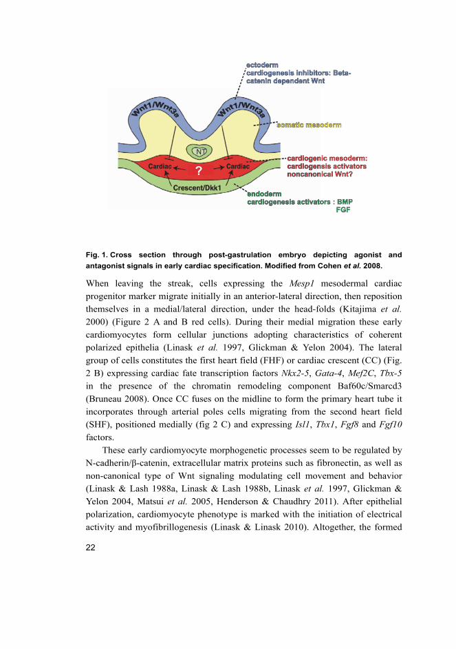
Mesodermal cardiac progenitor cell specification is modeled by a complex
network of positive and negative intercellular signal events (Evans et al. 2010).
Pro-differentiating factors, including bone morphogenetic protein and fibroblasts
growth factors, come from the endoderm, whereas negative signals arrive from
the neural tube as beta-catenin dependent-Wnt, which is modulated by
endodermal procardiogenic Wnt antagonists, such as dickkopf (Fig. 1) (Cohen et
al. 2008, Vincent & Buckingham 2010).
22
Fig. 1. Cross section through post-gastrulation embryo depicting agonist and
antagonist signals in early cardiac specification. Modified from Cohen et al. 2008.
When leaving the streak, cells expressing the Mesp1 mesodermal cardiac
progenitor marker migrate initially in an anterior-lateral direction, then reposition
themselves in a medial/lateral direction, under the head-folds (Kitajima et al.
2000) (Figure 2 A and B red cells). During their medial migration these early
cardiomyocytes form cellular junctions adopting characteristics of coherent
polarized epithelia (Linask et al. 1997, Glickman & Yelon 2004). The lateral
group of cells constitutes the first heart field (FHF) or cardiac crescent (CC) (Fig.
2 B) expressing cardiac fate transcription factors Nkx2-5, Gata-4, Mef2C, Tbx-5
in the presence of the chromatin remodeling component Baf60c/Smarcd3
(Bruneau 2008). Once CC fuses on the midline to form the primary heart tube it
incorporates through arterial poles cells migrating from the second heart field
(SHF), positioned medially (fig 2 C) and expressing Isl1, Tbx1, Fgf8 and Fgf10
factors.
These early cardiomyocyte morphogenetic processes seem to be regulated by
N-cadherin/β-catenin, extracellular matrix proteins such as fibronectin, as well as
non-canonical type of Wnt signaling modulating cell movement and behavior
(Linask & Lash 1988a, Linask & Lash 1988b, Linask et al. 1997, Glickman &
Yelon 2004, Matsui et al. 2005, Henderson & Chaudhry 2011). After epithelial
polarization, cardiomyocyte phenotype is marked with the initiation of electrical
activity and myofibrillogenesis (Linask & Linask 2010). Altogether, the formed
23
primitive heart tube provides an essential structural scaffold (van de Schans et al.
2007, van de Schans et al. 2007) for the subsequent cardiac progenitor
incorporation and chamber morphogenetic processes of looping, septation and
myocardium growth. (For reviews see (Sirbu et al. 2008, Gessert & Kuhl 2010,
Vincent & Buckingham 2010, Miquerol & Kelly 2013, Pandur et al. 2013).
The primary heart tube thus formed has a simple structure, with one or two
outer layers of cardiomyocytes and a luminal epithelial layer, the endocardium,
(Figure 3 A), emerging from a hematopoietic vascular lineage. Close to the
venous pole of the heart tube, a group of cells named the proepicardial organ
(PEO), generates the epicardium, the outer epithelial layer, and the smooth
muscles cells of the coronary vasculature (Fig. 2 E). Although considered to be of
SHF origin, marker analysis suggested that endocardium and PEO contain cells
originating from both heart fields. In mouse embryo, retrospective clonal analysis
showed that the FHF generates most cells which form the left ventricle (LV),
while SHF contributes mainly to outflow tract (OFT) and right ventricle (RV)
cells. Atria have contributions from both lineages. The OFT structure also
incorporates a population of neuroectodermal origin, neural crest cells migrating
from the dorsal neural tube (Fig. 2 D-F) (Vincent & Buckingham 2010).
24
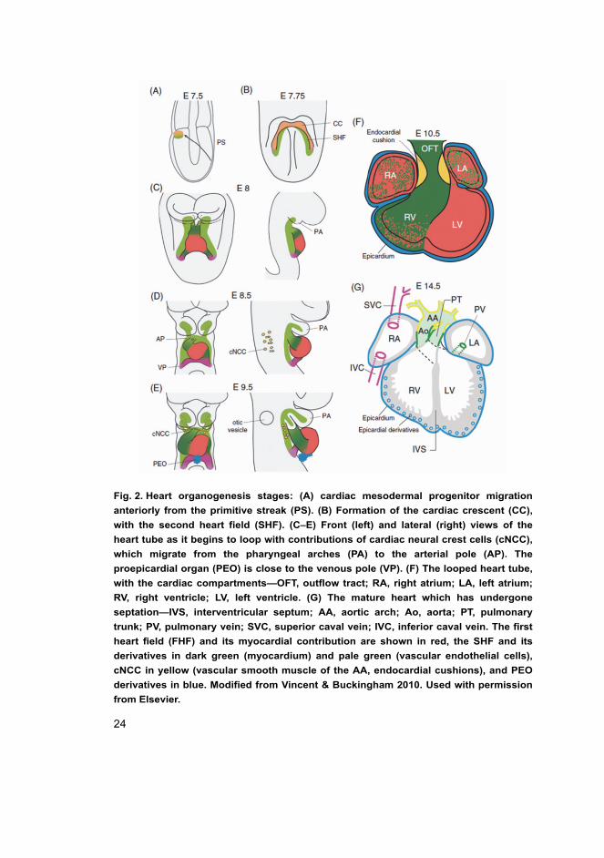
Fig. 2. Heart organogenesis stages: (A) cardiac mesodermal progenitor migration
anteriorly from the primitive streak (PS). (B) Formation of the cardiac crescent (CC),
with the second heart field (SHF). (C–E) Front (left) and lateral (right) views of the
heart tube as it begins to loop with contributions of cardiac neural crest cells (cNCC),
which migrate from the pharyngeal arches (PA) to the arterial pole (AP). The
proepicardial organ (PEO) is close to the venous pole (VP). (F) The looped heart tube,
with the cardiac compartments—OFT, outflow tract; RA, right atrium; LA, left atrium;
RV, right ventricle; LV, left ventricle. (G) The mature heart which has undergone
septation—IVS, interventricular septum; AA, aortic arch; Ao, aorta; PT, pulmonary
trunk; PV, pulmonary vein; SVC, superior caval vein; IVC, inferior caval vein. The first
heart field (FHF) and its myocardial contribution are shown in red, the SHF and its
derivatives in dark green (myocardium) and pale green (vascular endothelial cells),
cNCC in yellow (vascular smooth muscle of the AA, endocardial cushions), and PEO
derivatives in blue. Modified from Vincent & Buckingham 2010. Used with permission
from Elsevier.
25
Our understanding of heart progenitor origins has changed dramatically with the
novel finding of the SHF and its complex cell behavior regulation (Buckingham
et al. 2005). Initially, it was thought that certain transcription factors are specific
to certain heart field progenitors; however, later retrospective clonal analysis
showed that several transcription factors mark cells from both FHF and SHF
(Nkx2-5, Gata-4, Mef2C, Isl-1, Tbx1), with the two heart fields being clearly
juxtaposed in the mouse at E7.5 (Abu-Issa & Kirby 2007, Sirbu et al. 2008,
Pandur et al. 2013). Furthermore, cell tracing experiments were able to
characterize subdomains in SHF, but their spatiotemporal significance is poorly
understood (Vincent & Buckingham 2010). Mutant phenotypes indicate that many
of these cardiac regulators are mainly functional in the SHF progenitor population
although also present in cells of FHF (Buckingham et al. 2005, Cohen et al. 2012).
These cardiac factors might be redundant in their functions, which would explain
why myocardial cell differentiation is not completely abolished by any single
mutation in a cardiac regulatory gene. The network of interacting cardiac factors
has continued to grow in complexity; for example, Tbx5, Gata-4, and Nkx2-5
associate physically to promote genes of cardiomyocyte differentiation, while
Tbx5 and Sall4 interact to either promote or repress these genes. These factors are
all dose-sensitive regulators of cardiogenesis, where the level required for
progenitor survival differs from the level required for progenitor differentiation.
SHF size and anterior to posterior axis patterning is controlled by retinoic
acid, (Sirbu et al. 2008), while left to right axis is patterned by Sonic hedgehog
(Hildreth et al. 2009) and Nodal-Pitx2 (Linask et al. 2002, Tessari et al. 2008).
Regarding heart field progenitor proliferation and maintenance, SHF seems to
maintain more active cell proliferation and lower differentiation than primary
heart tube cells. Early heart field cell proliferation involves also a complex spatial
control in clonal proliferation, but cell field expansion is also achieved by
changes in cell size (Meilhac et al. 2003). Maintenance and proliferation in SHF
is controlled through Fgf, Shh and beta-catenin-dependent Wnt signaling, while
differentiation of SHF is controlled through Shh, Notch, Bmp and non-canonical
Wnt signaling (Cohen et al. 2008). For reviews see (Henderson & Anderson 2009,
Vincent & Buckingham 2010, Miquerol & Kelly 2013).
2.1.2 Development of the ventricles, looping and ballooning
To shape the heart into its final form the primary heart tube follows complex
processes of looping and ballooning.
26
At first the looping process generates an S-shape heart configuration, with
definition of the inflow and outflow-ventricular segments (fig 2 D-F), mouse
embryonic day 9–10 and human Carnegie 12/26 days (Henderson & Anderson
2009). During cardiac looping the cardiac jelly disappears from the atria and
ventricle walls, while accumulating in the junction between the chambers and
developing OFT, forming the endocardial cushions, the primary structure which
generates the cardiac septum and valve (Figure 2 F) (Snarr et al. 2008).
Endocardial cushion morphogenesis is mediated by epithelial-to-mesenchymal
transformation, regulated by intrinsic and extrinsic factors secreted from the
nearby myocardium, such as Gata-4, EGF, Tgfβ and Notch (Wessels & Sedmera
2003, Niessen & Karsan 2008, Snarr et al. 2008).
Next, the heart chamber atria and ventricles are formed by serial ballooning
processes from the cavity of the primary heart tube. The process involves regional
differences in the rate of proliferation, with lower levels in the heart loop inner
curvature and complex proliferation patterns of clonal growth (Meilhac et al.
2003, Sedmera & Thompson 2011). Each ventricular chamber is defined by its
own inlet/inflow, outlet/outflow and apex components, and the interventricular
communication is remodeled as well, bringing in direct communication right
chambers (RA and RV) and left chambers (LA and LV), changes essential in
generating the AV conduction axis (Moorman & Christoffels 2003). Finally,
twisting of OFT accompanied by fusion and muscularization of the proximal
outflow cushions permits separation of pulmonary and aortic-systemic
circulations, by closing interventricular communication and connecting the aorta
to LV and the pulmonary artery to RV (fig 2 G). Heart gross morphology is
complete in mouse by embryonic day 14.5, human Carnegie stage 23/end of week
7), although the heart continues to mature throughout the remainder of in utero
life (Meilhac et al. 2003, Henderson & Anderson 2009, Savolainen et al. 2009,
Sedmera 2011).
2.1.3 Myocardium patterning during development
During cardiac development the ventricular myocardium follows complex
patterning with specification into compact and trabeculated type of myocardium.
Cytoarchitectural changes involve distinct arrangement of cardiomyocytes layers
within the ventricular wall, with increasing cardiomyocytes differentiation levels
from the epicardium towards the endocardium. In regulation of patterning
27
processes a prime candidate has arisen, the non-canonical type of Wnt signaling
through planar cell polarity pathway (Henderson & Chaudhry 2011)
Before the looping stage there is quite homogenous cellular architecture
along the cardiac tube, with one or two layers of cuboidal-shaped cardiomyocytes
and circumferential arrangement of undifferentiated myofibrils (fig 3 A) (Hirschy
et al. 2006). The early patterning of cytoskeleton, myofibrils and cellular
junctional N-cadherin seems to be play a crucial role in heart looping
morphogenesis of different species (Linask et al. 1997, Linask et al. 2002, Linask
& Vanauker 2007). However, the degree of organelle differentiation in these early
cardiomyocytes is dependent on genetic background, for example in mice strain
(Kastner et al. 1997).
Clear regional differences in myocardial cell myofibrillar pattern were
observed after looping stage, when trabeculations emerge next to the maximum
curvature areas (Fig. 3 B)(Hirschy et al. 2006). The trabeculation morphogenesis
includes important exchange of inductive and patterning intercellular signals
between the endocardium, myocardium and the myocardium secreted cardiac jelly.
These signals include endocardial neuregulin binding on myocardial receptor
Erbb, endocardial Brg1 repression on endocardial matrix metalloproteinases,
secreted Vegf and angiopoietin from the myocardium acting on the endocardium
(Stankunas et al. 2008).
Before heart septation, compact myocardium proliferative activity is higher
and its differentiation is lower than in trabeculated myocardium, while the latter
has the main responsibility for the heart pumping activity at this stage (Sedmera
& Thompson 2011). The patterns of early trabeculations are similar in RV and LV,
but by the completion of septation (mouse embryo day 14.5 and human Carnegie
22/8 weeks) ventricles obtain a more specific morphological patterning, with
certain differences between species. Through these processes the radial trabecular
myocardium is rearranged into apico-basal orientation. Coalescence of the
trabecular myocardium leads to formation of the papillary muscles supporting the
leaflets of the AV valves (Sedmera et al. 2000).
Once heart septation is complete, the compact myocardium increases in
proportion and thickness over the trabeculated myocardium, with its contribution
to the total myocardial mass and pressure pump function becoming significant
especially in the LV (Sedmera et al. 2000). This process called myocardial
compaction is at present poorly understood (Henderson & Anderson 2009). It has
been hypothesized to be generated by compression/consolidation of trabeculations
within the ventricular wall (Sedmera et al. 2000); however, no evidence for this
28
exists so far except for the formation of papillary muscles. Major thickening of
ventricular wall compact myocardium is currently thought to be brought on by its
proliferation and myoarchitecture remodeling (Henderson & Anderson 2009). In
utero proliferation and differentiation of the compact myocardium are regulated
by epicardial-derived signals such as Fgf, retinoic acid, erythropoietin and Wnts
(Sedmera et al. 2000, Henderson & Anderson 2009). Cardiomyocyte
differentiation involves cell size increase and shape changes, together with
increasing abundance in mature specialized cardiomyocytes organelles such as Z
bands defining sarcomeres in myofibrillar bundles and intercalated disc (ICD)
cell-cell contacts (Kastner et al. 1997, Du et al. 2003). ICD are specific to
cardiomyocytes and their complex structure distinguishes three types of cell
contact: adherens junctions anchoring actin filaments (cadherins, catenins,
vinculin, alpha-actinin), desmosomes anchoring intermediate filaments
(desmoplakins) and gap junctions mediating ion transfer (connexins). In adult
myocardium ICD become exclusively located at bipolar ends of rod-shaped
cardiomyocytes; however, this is not the case in utero when these structures are
found surrounding the cardiomyocytes’ lateral membranes (Peters et al. 1994,
Hirschy et al. 2006). Maturation of cardiomyocytes cell-cell contacts is a slow
process coordinated intimately to the changes in cellular dimensions and
cardiomyocyte-polarized orientation into the growing myocardial wall. At
neonatal stage cardiomyocyte adherens junctions no longer surround the whole
cells and become restricted to myofibrillar attachment sites; however, electrical
and mechanical coupling still continues to be fine-tuned and remodeled in
humans up to about 6 years of age. An essential adaptation to the almost 16-fold
increase in heart weight in the first years of life, conduction velocities increase
through increased association between gap junctions and adherens junctions
(Peters et al. 1994, Hirschy et al. 2006).
Substantial further myocardial growth and compaction continues postnatally,
and this process involves further improvement in muscle organization complexity.
The adult ventricular myocardium has a three-layered spiral/helical system of
myocardial fibers, correlating to the twisting pattern of contraction existing from
early heart development, after initiation of trabeculation (fig 4 D). This complex
adult architecture is behind the observed regional heterogeneity in myocardium
shortening and stretching during cardiac cycle (Sedmera et al. 2000, Henderson &
Anderson 2009, Sedmera 2011).
29
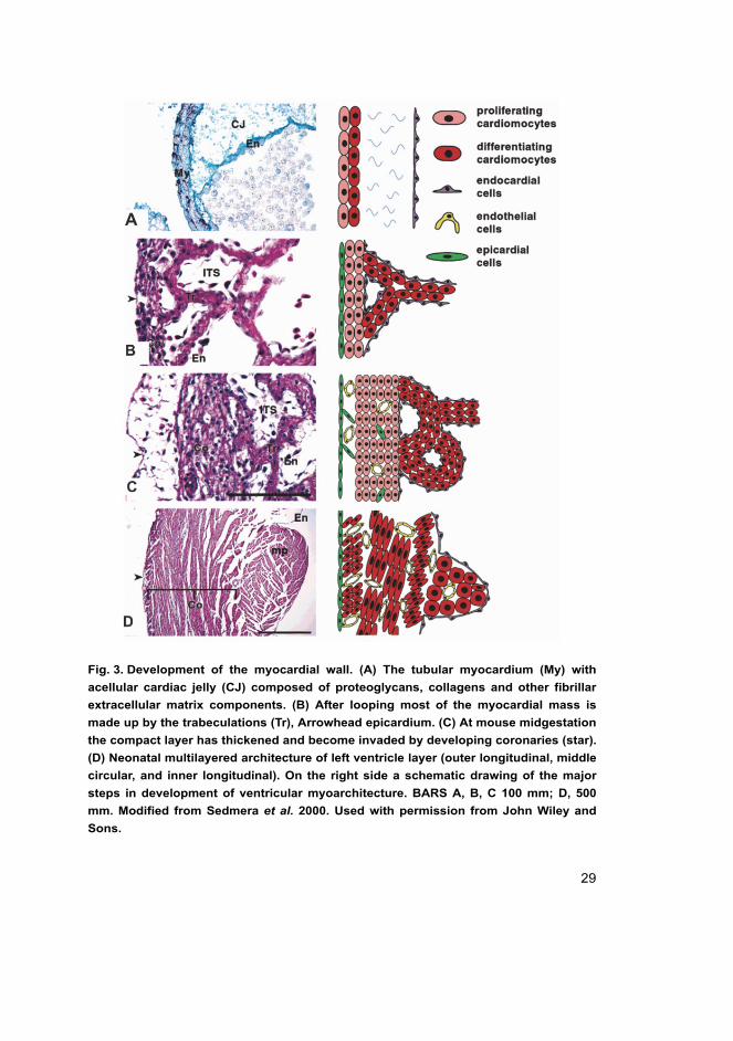
Fig. 3. Development of the myocardial wall. (A) The tubular myocardium (My) with
acellular cardiac jelly (CJ) composed of proteoglycans, collagens and other fibrillar
extracellular matrix components. (B) After looping most of the myocardial mass is
made up by the trabeculations (Tr), Arrowhead epicardium. (C) At mouse midgestation
the compact layer has thickened and become invaded by developing coronaries (star).
(D) Neonatal multilayered architecture of left ventricle layer (outer longitudinal, middle
circular, and inner longitudinal). On the right side a schematic drawing of the major
steps in development of ventricular myoarchitecture. BARS A, B, C 100 mm; D, 500
mm. Modified from Sedmera et al. 2000. Used with permission from John Wiley and
Sons.
30
Cell polarity pathways and myocardium patterning
The dynamics of organogenesis does not involve only proliferation and
differentiation. Organs shape and dimensions are modeled in a particular axis
though regulation of cell division orientation (OCD) and convergent extension
(CE-cellular rearrangement through intercalation with each other in order to
narrow and extend a tissue), both regulated by the planar cell polarity pathways
(PCP). In these dynamic processes cells are only transiently polarized in a certain
plane, and afterwards move and/or divide in another orientation. Many of the
components of PCP pathways are conserved between flies and vertebrates, but the
degree of mechanistic conservation is still unclear. However, certain similarities
have been found between gastrulation convergent extension-like movements and
the behavior of special migrating cell types, such as neural crest cells and
epithelial cells in kidney tubule elongation. (Goodrich & Strutt 2011).
One compelling model for cell polarity during organogenesis holds that cells
regulate their proliferation, orientation (polarization) and movement (cell
intercalation) in response to a specific morphogen gradient. Beta-catenin
independent Wnt ligands, including Wnt-11, which bind to frizzled (Fz) receptors,
are involved in PCP pathway activation. However, there is poor evidence to
clarify whether these signals are permissive or inductive. Fz receptors are part of
the so-called core group in PCP signaling molecules, together with Dishevelled,
Prickle, Van Gogh/strabismus, Diego, Flamingo. These molecules are
characterized by asymmetric cell membrane or subcellular organization, which
act to amplify and stabilize polarization signal by communicating to the boundary
of adjacent cells. Their asymmetric cellular organization is thought to result from
intracellular dynamic interactions between cellular proximal and distal
components. Recruited cellular effectors such as JNK kinase and small GTP-ases
(RhoA and Rac) modulate the cell shape and behavior by affecting cytoskeleton
and possibly also cell-to-cell junctional molecules (For reviews see (Karner et al.
2006, Simons & Mlodzik 2008, Goodrich & Strutt 2011).
Disruption of PCP may play a causal role in several human congenital
diseases. The original PCP pathways have been expanded to play morphogenetic
roles in tissues that do not show obvious planar organizations, which is why in
these contexts PCP organization becomes harder to understand (Goodrich & Strutt
2011).
For normal morphogenesis of the primitive heart tube (E8.5) establishing an
apical-basal cell polarity as well as planar cell polarity seems to be important.
31
Disruption in these processes causes cardia bifida in Diversin and dominant Dsh
in zebrafish mutants in addition to disrupted organization of cardiomyocytes in
mouse Scrib and Vangl2 mutants (Phillips et al. 2005, Phillips et al. 2007,
Henderson & Chaudhry 2011). Perturbation in the early epithelialization of
cardiomyocytes affects their coherent migration and may disrupt severely heart
formation at this developmental stage, resulting in cardiac phenotypes such as
abnormal heart tube formation (Phillips et al. 2007, Henderson & Chaudhry 2011),
cardia bifida (Pandur et al. 2002, Garriock et al. 2005, Matsui et al. 2005) or
abolishment of heart formation in chick (Linask et al. 1997). These phenotypes
associated with abnormal apico-basal polarity and improper junctional complexes
as shown by antibody blocking N-cadherin function in avians (Linask et al. 1997),
fibronectin mutation disrupting interaction with extracellular matrix in chick
(Linask & Lash 1988a, Linask & Lash 1988b), zebrafish and mouse (Glickman &
Yelon 2004) and also impaired Wnt-11 signaling by morpholino in Xenopus
(Pandur et al. 2002, Garriock et al. 2005) and zebrafish (Matsui et al. 2005).
Impaired PCP with causal role in cardiac anomalies is linked to defects in
early heart tube looping, outflow tract morphogenesis and septation. Little data is
available on cardiomyocyte cell polarity in ventricular walls, due to the fact that
aberrations from normal ventricular development do not resemble classic PCP
phenotypes (Davis & Katsanis 2007, Simons & Mlodzik 2008, Henderson &
Chaudhry 2011). In the tubular heart shape cardiomyocytes are round with no
polarized cell-cell junctional proteins, junctional components are distributed
around the membrane, and first polarization behavior can be observed in mice in
midgestation when cardiomyocytes change their shape and elongate (Hirschy et al.
2006, Henderson & Chaudhry 2011). Adult cardiomyocytes have a rod-like shape
and junctional components are tightly polarized as ICD on the cardiomyocyte
long axis membranes while gap junctions, ion channels, beta –adrenergic
receptors become polarized on lateral membranes (Peters et al. 1994, Hirschy et
al. 2006). In mouse, mutation in PCP core proteins causes congenital heart
disease with OFT defects (Vangl2-Lp mutant, Scrib-Crc mutant), but these mice
have also a clear myocardium phenotype with defects in cell shape, proliferation
and patterning of myocardium (Phillips et al. 2005, Henderson et al. 2006,
Phillips et al. 2007). By late gestation myocardial defects in core PCP mutants
resemble ventricular noncompaction (VNC) observed in humans (Henderson &
Chaudhry 2011). Wnt-11 stands as a good candidate for PCP control of heart
organogenesis; mutation in homologous Wnt-11 R zebrafish causes possibly a
similar phenotype of disorganized cells in ventricular myocardium, and a line of
32
evidence that Wnt-11 acts as a directional cue to organize muscle has been shown
in Xenopus elongation of early skeletal muscle fibers (Garriock et al. 2005, Gros
et al. 2008).
Many of the core proteins are expressed in the myocardium from early
embryo to adulthood, suggesting a role in myocardium assembly but also
maintenance. Altogether PCP pathway components have been shown to cause
changes in mammalian cardiomyocyte shape and junctional organization, but
these might not be analogous to classic PCP pathway, pointing out that
myocardium cell polarity is at present poorly understood (Henderson & Chaudhry
2011).
2.1.4 Abnormal heart organogenesis and consequences for cardiac
functional development
Although extremely simple and lacking a differentiated cellular structure, the
primitive tubular heart becomes a functional organ. First signs of electrical
activity generate peristaltic-like movements and have been recorded as early as
mouse embryonic day 8.5, equivalent to human Carnegie stage 10, 21–23 days of
gestation (Sedmera et al. 2000). As the embryo grows, there is a developmental
relationship between heart regional architecture, myofiber alignment and cardiac
function (Tobita et al. 2005). Improvement in ventricular function, especially
diastolic function, correlates with changes in ventricular myoarchitecture during
cardiac developmental (Ishiwata et al. 2003). However, changing from normal
hemodynamic conditions (pressure and volume load) affected normal
morphogenesis in experimental chick embryos, by accelerating development in
increased loading conditions and delaying development in decreased loading
conditions (Tobita et al. 2005).
The heart exhibits remarkable adaptive responses to a wide array of genetic
and extrinsic factors to maintain cardiac function; however, persistent activation
of these pathways leads eventually to cardiac dysfunction and apoptotic loss of
cardiomyocytes (Harvey & Leinwand 2011). After apoptosis the ability of mature
adult myocardium to regenerate is reduced, adaptive response to maintain cardiac
workload is achieved mainly by ventricular hypertrophy or dilation, with changes
in extracellular matrix composition, individual cardiomyocyte volume and
organelle content (Fogel et al. 2005, Harvey & Leinwand 2011). Recent evidence
suggests that cardiomyocyte proliferation may play an unrecognized role during
the period of developmental heart growth between birth and adolescence; this
33
evidence came combined with proof for cardiomyocyte turnover in adult humans
(Mollova et al. 2012). In contrast, during development the heart can also respond
robustly to changes in loading conditions by cardiomyocyte hyperplasia, or to
decreased load by hypoplasia, as shown in chick models (Sedmera et al. 1999,
Tobita et al. 2005). In the neonatal period there is a shift from a fetal isoform
gene-expression profile to adult isoforms, especially in genes concerning
contractile apparatus components (Epstein 2010). Both hyperplastic and
hypertrophic responses to an increased pressure load are normal characteristics of
fetal to postnatal heart development; however, the exact time point and factors
involved in switching from hyperplastic to pure hypertrophic response are not yet
fully understood. Epigenetic control of chromatin structure is recently believed to
regulate the transition from fetal gene program to adult gene program (Epstein
2010).
Cardiac functional evaluation – global cardiac function by conventional
Doppler echography
Cardiovascular functions involve broad aspects of cardiac physiology, including
contraction and relaxation during cardiac cycle systole and diastole, and
distribution of blood flow over specific vascular beds. The conventional approach
is to assess the global cardiac function by (2D) Doppler ultrasonography, and so
far multiparameter analysis is required because of the limitations of any single
parameter.
Fetal echocardiography is a well-established tool for prenatal diagnosis of
structural, but also functional heart disease. Studies of human fetuses and mouse
models agree that developmental changes in cardiac function must meet the
requirements of the growing embryo, in particular diastolic function parameters,
which may provide a key prognostic factor in cardiomyopathies (Pedra et al. 2002,
Ishiwata et al. 2003). In human fetal cardiomyopathies the diastolic dysfunction
causes secondarily an increase in systemic venous pressure and associates with
greatest risk in fetal mortality (Pedra et al. 2002).
Pulsatility index values of vessels stands for PI = peak systolic velocity-end-
diastolic velocity/time-averaged maximum velocity in cardiac cycle. This
correlate to intrinsic vascular wall properties and downstream vascular resistance
resistance/impedance = pressure/volume. Pulsatility index values of vessels
proved to be relevant for both systolic and diastolic cardiac functions (Pedra et al.
2002. Indeed, increased PIs in umbilical artery and aorta are observed in human
34
placental insufficiency, and increased PI in ductus venosus implies an increase in
systemic venous pressure as a sign of diastolic or congestive heart failure. A
specific diastolic parameter is ventricular isovolumetric relaxation time (IRT%),
which describes the time needed for the ventricle to drop its pressure from a
systemic to an atrial level (Makikallio et al. 2005). IRT% is sensitive to the rate of
ventricular relaxation, which depends on the passive properties of the
myocardium, but is also load-dependent by aortic diastolic pressure and atrial
pressure, and mouse studies showed that improvement in active relaxation
correlates directly to maturation in compact myocardium architecture (Ishiwata et
al. 2003). Improvement in cardiac diastole in early development might be
important for the fetal heart to adapt to the increased volume of blood flow
(Makikallio et al. 2005).
Systolic function is evaluated by isovolumetric contraction time (ICT%),
which describes the time needed for the ventricle to increase its pressure to the
systemic blood flow level, directly correlating with ventricular contractility and
pressure generation (Makikallio et al. 2005). ICT% decreases in early embryonic
development according to the improvement in ventricular pressure generation.
Inflow and outflow velocities are proportional to volume blood flows
(Vmean = fetal heart rate HR × time velocity integrals), and umbilical artery
velocity correlates directly to umbilico-placental blood flow. The cardiac output
parameter describes the cardiac hemodynamics at one point in time, being the
volume of blood being pumped by the heart in one minute. Cardiac output
depends on heart rate and correlates with the conventional systolic parameter in
adult cardiac echocardiography: ejection fraction (EF%). EF is the fraction of
blood ejected by the ventricle during the contraction or ejection phase of the
Systole in cardiac cycle: EF% = (end diastolic volume-end systolic volume)/end
diastolic volume) × 100%. In practice, however, ejection fraction has weak inter-
assay reproducibility, being dependent on preload, afterload and heart rate. EF
assesses systolic global ventricular performance and does not take into
consideration myocardial regional contractile dysfunction (Abraham et al. 2007,
Hoit 2011).
Cardiac functional evaluation - regional myocardial function by Speckle
tracking echography
Speckle tracking echography (STE) as tissue strain imaging is a relatively new
echocardiography method, yet proving to be difficult to standardize. The
35
mechanical properties of tissue have long been recognized as a useful indicator of
disease and STE looks directly into the myocardium and measures myocardial
deformation in strain (Abraham et al. 2007, Hoit 2011). While standard Doppler
echography captures global changes in blood flow showing global cardiac
function, tissue strain is a non-Doppler technology that evaluates cardiac regional
function by detecting complex myocardial line motion (Thomas 2004). Imaging
tracking technology is fast advancing and numerous studies are clarifying the
limitations and clinical applicability of the methods. STE is automated, angle-
independent, less noise sensitive with better intra- and inter-observed variability
than standard Doppler technique (Hoit 2010).
STE measures velocity and strain parameters of myocardium performance in
2D longitudinal, radial and circumferential directions during systole and diastole.
These may reflect closely the regional functional impact of any changes in
regional myocardium architecture caused by cardiac diseases. Strain technology
uses continuum mechanism Lagrangian strain principles, where strain represents
the magnitude and rate of deformation between two points in the myocardium,
and the rate of deformation is referred to as strain rate (SR; in seconds−1). SR is
noisier, less reproducible, but less dependent on load conditions compared to
strain.
In measuring the tissue motion, for displacement and velocity parameters a
reference point from the transducer is used; this is biased by translational
movement and tethering, as it cannot distinguish well between passive or active
tissue movements. The myocardium deformations are complex; these occur not
only perpendicular to a given plane (normal strain) but also in between planes, or
as shear strain. 3D ultrasound techniques are expected to improve and expand the
2D ecography diagnostic capabilities.
Mathematically, strain is the integral of SR, with compression expressed in a
negative value and expansion in a positive value (Dandel et al. 2009, Hoit 2011).
Lower values in tissue velocities and SR have been reported in ischemic
myocardium, where changes in SR may even precede changes in velocity
(Abraham et al. 2007, Hoit 2011). Lower longitudinal strain and SR have been
observed in hypertrophic cardiomyopathy versus physiological athletic
hypertrophic cardiomyopthay; moreover, in a study of patients with suspected
congestive heart failure of different etiologies it was also shown that mean
longitudinal LV strain is closely related to plasma brain-type natriuretic peptide
(BNP) levels in patients with both systolic and diastolic heart failure. (Dandel et
36
al. 2009, Wang & Nagueh 2009, Hoit 2011) These techniques are promising in
terms of potential clinical value especially in sub-clinical cases.
2.1.5 Developmental cardiac defects: Congenital heart diseases and
Cardiomyopathies
Congenital heart defects (CHD) have worldwide prevalence approaching 1:100
births and refer to structural or functional cardiac abnormalities that arise before
birth (Bruneau 2008, Houyel et al. 2011, Garne et al. 2012). Remarkably,
excluded from this number are congenital cardiomyopathies (cCMP), inherited
primary defects of the myocardium, which are defined as an isolated group of
cardiac anomalies in the absence of congenital structural heart disease (Maron et
al. 2006, Elliott et al. 2008), although it has long been known that these
phenotypes can coexist (Somerville & Becu 1978, Eidem et al. 2000). A more
exact prevalence of primary or congenital cardiomyopathies is currently unknown,
varying from 1:500 for the hypertrophic form of cardiomyopathy to 1:2000 for
dilated cardiomyopathy, and to 1:5000 for the more uncommon unclassified
cardiomyopathy, ventricular noncompaction (VNC) (Seidman & Seidman 2001,
Maron et al. 2006, Elliott et al. 2008). The separation of the developmental heart
defects into the two groups, CHD and CMP, has been sustained through
differences in clinical presentation and treatment approaches (Bruneau 2008). It is
increasingly realized that the current nomenclature fails to adequately describe a
substantial overlap between the classic cardiomyopathy syndromes (Klaassen et
al. 2008), or fails to describe how the same/similar developmental abnormalities
can underlie anatomically distinct heart defects (Epstein 2010). It is also
important for understanding heart morphogenesis in the context of heart
functional development, as heart function is required for life from very early heart
morphogenesis on; hemodynamics alterations may have consequences for heart
morphogenesis. This may explain why defects in myosin contractile proteins
encoding genes cause inherited atrial septum defects (for review see (Bruneau
2008).
Among the major forms of cardiomyopathies hypertrophic CMP (HCM) is
characterized by increased ventricular chamber size, increased contractility but
impaired diastolic relaxation, while dilated cardiomyopathy (DCM) is
characterized by increased ventricular chamber size and reduced contractility
(Seidman & Seidman 2001). Ventricular noncompaction (VNC), a distinct form
of CMP, has only recently been added to the primary CMP group with genetic
37
origins. It is often found associated with CHD and other forms of CMP (Tsai et al.
2009, Oechslin & Jenni 2011). VNC is characterized by abnormal trabeculations
associated with deep intertrabecular recesses and thin compact myocardium.
Based on elucidations of its genetic causes, the human inherited cardiomyopathies
show mutations in sarcomeric proteins and sarcomeric-related proteins, although
50% of the HCM patients have no mutations in these proteins (Seidman &
Seidman 2001, Alcalai et al. 2008).
Cardiac disease studies raise the question whether each CMP form is a
distinct program or just a different morphologic phenotype reflecting gradation
steps in a single disease model (Seidman & Seidman 2001, Ahmad et al. 2005).
Important to remember here, all CMP forms are affected by modifier genes: the
prime candidates are variants in renin-angiotensin-aldosterone system,
transforming growth factor and insulin-like growth factor, endothelin-1 and tumor
necrosis factor, calcium regulations and homeostasis (Seidman & Seidman 2001,
Towbin & Bowles 2002, Alcalai et al. 2008).
2.2 Kidney organogenesis
The kidney derives from the intermediate mesoderm, which forms in temporal
and spatial sequences transient structures on the right and left side of the body:
first forming is the pronephros, second the mesonephros and third and last, the
metanephros. In higher vertebrates only metanephros differentiates into
permanent kidney, while the others participate in adrenal gland and gonad
organogenesis.
2.2.1 Cortical nephrogenesis and ureter branching
Kidney organogenesis from the metanephros comprises complex reciprocal
inductive interactions between two primordial tissues: epithelial structured
ureteric bud (UB) and surrounding metanephric mesenchyme (MM). While we
have been aware of these reciprocal inductive interactions for almost 50 years
since their discovery by Grobstein (Grobstein 1956, Saxen & Sariola 1987), the
clues towards understanding morphogenesis and its molecular basis in detail have
increased dramatically during the last decade (Little & McMahon 2012).
In vitro experiments proved that these tissues must be in close contact to each
other to activate reciprocal signals for kidney induction (Saxen & Sariola 1987).
The ureteric bud grows and branches in a certain bifurcation or trifurcation
38
pattern in response to mesenchymal signals, while mesenchymal tissue forms new
early nephron structures, renal vesicles (RV), through mesenchyme-to-epithelial
transformation (MET) in response to signals from branching ureter tips. Before
committing towards RV formation, the mesenchymal cells go through two
intermediary phases, cap mesenchyme (CM) and pretubular aggregate (PA). The
formation of RV represents the “birth” of a single nephron, RV being the
precursor for most segments of the nephron from the glomerular podocytes to the
connecting segment to collecting ducts system, while collecting duct cells
originate from the ureteric bud. Eventually, these small renal vesicles begin to
pattern as they proliferate and elongate to form tubular nephrons. In addition to
the nephrogenic MM and ureter epithelium, a third cell type has been described in
the nephrogenic cortex, i.e., cortical stromal cells, which surround the CM
domain (Cullen-McEwen et al. 2005, Yallowitz et al. 2011). For reviews see
(Hendry et al. 2011, Little 2011, Little & McMahon 2012).
Self-renewal and progenitor survival within MM, UB and stroma are
essential for generating normal nephron number. This is defined by complex
reiterative positive loops between these cells. A subpopulation of MM/CM
expressing Six2 represents a self-renewing multipotent nephron progenitor
population throughout kidney organogenesis (Kobayashi et al. 2008). Survival of
these MM progenitor cells seems to be a separate attribute to self-renewing and
depends on ureteric, not fully known UB signals, but also on intrinsic MM signals
including WT1, Eya1, Hox11 paralogs. Signaling from stromal cells Hox10
paralog, Foxd1, is important for maintaining a stroma progenitor population
forming the kidney capsule, but also seems to play critical roles in nephron
differentiation, ureter branching and mesenchyme renewal (Yallowitz et al. 2011,
Little & McMahon 2012). Mesenchymal Six2 cooperates closely with epithelial
Wnt-9b, which seems to be critical in balancing this mesenchymal progenitor
self-renewal versus differentiation (Carroll et al. 2005, Karner et al. 2011). This
could be explained by different levels in Wnt-9b signaling reaching the different
cellular compartments of CM (Little & McMahon 2012). Besides this important
role in maintaining mesenchymal Six2+ progenitor population, Wnt-9b has a
crucial inductive role in the subsequent requirement for Wnt-4 expression in
initiation of MET and RV formation (Thiagarajan et al. 2011, Hendry & Little
2012, Little & McMahon 2012).
Wnt-9b localizes in the ureter epithelium together with other Wnts, Wnt-11
and Wnt-7b, although in different subdomains, Wnt-11 more proximal at the tips,
Wnt-9b and Wnt-7b more distal in the stalk (Kispert et al. 1996, Carroll et al.
39
2005, Yu et al. 2009). Ureter bud also expresses Pax2, which is essential for
ureter bud survival and growth. The Wnt-11 expression at the tips of the
branching UB is required in a positive feedback loop that promotes mesenchymal
secreted glial-derived growth factor (Gdnf) essential for survival and branching of
the ureter bud, to bind its receptor cRet receptor in UB (Kispert et al. 1996,
Majumdar et al. 2003). Gdnf/cRet signaling lies at the signaling core of epithelial
branching and determines the kidney nephron number as shown in Wnt-11 and
cRet deficient hypoplastic kidneys (Majumdar et al. 2003), while Gdnf null mice
show a more extreme phenotype as renal agenesis due to lack of ureter induction.
The UB branching is maintained by multiple and complex positive and negative
reiterative loops between the mesenchyme, epithelium and also stroma
maintaining optimal signaling in the Wnt-11/Gdnf/cRet loop. Stromal Fgf7
promotes UB branching (Cullen-McEwen et al. 2005) having possibly redundant
function to the upper loop. Stromal Bmp4 has inhibitory function on UB
branching (Raatikainen-Ahokas et al. 2000), but is finely tuned by its inhibitor
Gremlin (Michos 2009), both acting downstream of cRet. Epithelial Wnt-9b and
mesenchymal Hox11 are reinforcing levels of mesenchymal Six2 upstream of
Gdnf (Kobayashi et al. 2008, Karner et al. 2011). GLI3 repressor restricts
hedgehog signaling maintaining normal levels of Wnt-11 and cRet in UB (Cain et
al. 2009); stroma Foxd1 and retinoic acid-related molecule (Batourina et al. 2001)
and epithelial Gata3 cooperate with Pax2/8 to maintain optimal levels and correct
topography of cRet and Wnt-11 in the epithelium. For reviews see (Cullen-
McEwen et al. 2005, Grote et al. 2006, Costantini & Kopan 2010, Costantini
2010, Hendry et al. 2011, Hendry & Little 2012).
Downstream of Gdnf/cRet signaling lies, among others, Etv4/5 from the Pea3
family of ETS transcription factors, which control genes involved in cell
proliferation, migration and extracellular matrix during UB branching (Kuure et
al. 2010a, Kuure et al. 2010b). Within important feedback inhibitor of ureter
branching lies Sprouty 1 (Spry 1), also downstream of Gdnf/cRet. MM marker
Fgf10 is able to rescue ureter branching in the combined absence of Gdnf/cRet
signaling and Spry1 negative regulation; its pattern of branching is, however,
severely perturbed, pointing to a unique function of Gdnf in ureteric bud
patterning (Michos et al. 2010).
40
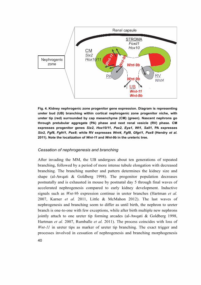
Fig. 4. Kidney nephrogenic zone progenitor gene expression. Diagram is representing
ureter bud (UB) branching within cortical nephrogenic zone progenitor niche, with
ureter tip (red) surrounded by cap mesenchyme (CM) (green). Nascent nephrons go
through pretubular aggregate (PA) phase and next renal vesicle (RV) phase. CM
expresses progenitor genes Six2, Hox10/11, Pax2, Eya1, Wt1, Sall1, PA expresses
Six2, Fgf8, Fgfrl1, Pax8; while RV expresses Wnt4, Fgf8, Gfgrl1, Pax8 (Hendry et al.
2011). Note the localization of Wnt-11 and Wnt-9b in the ureteric tree.
Cessation of nephrogenesis and branching
After invading the MM, the UB undergoes about ten generations of repeated
branching, followed by a period of more intense tubule elongation with decreased
branching. The branching number and pattern determines the kidney size and
shape (al-Awqati & Goldberg 1998). The progenitor population decreases
postnatally and is exhausted in mouse by postnatal day 5 through final waves of
accelerated nephrogenesis compared to early kidney development. Inductive
signals such as Wnt-9b expression continue in ureter branches (Hartman et al.
2007, Karner et al. 2011, Little & McMahon 2012). The last waves of
nephrogenesis and branching seem to differ as until birth, the nephron to ureter
branch is one-to-one with few exceptions, while after birth multiple new nephrons
jointly attach to one ureter tip forming arcades (al-Awqati & Goldberg 1998,
Hartman et al. 2007, Rumballe et al. 2011). The process coincides with loss of
Wnt-11 in ureter tips as marker of ureter tip branching. The exact trigger and
processes involved in cessation of nephrogenesis and branching morphogenesis
41
are poorly known. It is thought to be initiated by a shift in mesenchyme fate
through presumptive increased inductive signals, possibly in combination with
decreased sensitivity to growth factors and inhibitor of differentiation. As a result
Six2 progenitor gene expression in cap mesenchyme is lost together with Foxd1
progenitor gene expression in stroma, but surprisingly, some mesenchymal cells
continue to proliferate with no dramatic change in apoptosis (Hartman et al. 2007,
Karner et al. 2011). In humans, nephron formation is regarded as complete by 36
weeks of gestation while in mice it is not regarded as complete until postnatal day
3–4 (Rumballe et al. 2011). Once nephrogenesis has ceased renal development
concentrates around tubule elongation and nephron segment maturation
(Costantini 2010).
2.2.2 Medulla formation and kidney tubule maturation and elongation
Kidney tubule maturation and elongation are essential to kidney medulla
formation during prenatal, but also postnatal kidney development; after cessation
of glomerulogenesis, these processes seem to be governed by PCP pathways and
Wnts (Yu 2011).
Epithelial RV representing stage I nephron undergoes shape changes through
stage II-S shape and III-capillary loop until the final stage IV, the mature nephron.
The nephron fuses immediately after MET event to ureter tip and proximal and
distal gene expression becomes polarized. Proximal RV gives rise to glomerulus
podocytes and Bowman capsule while distal RV generates from a specific subset
of cells the proximal tubule (PT) together with Henle’s loop, then distal tubules
and respectively connecting tubules. Each nephron forms a spatial cortico-
medullary pyramid, named also the malpighian pyramid, which is well conserved
in mammals, the broad base adjacent to the renal cortex and the narrow apex as
kidney papilla. However, the number of these structures and therefore the number
of papillae differs from species to species: humans have eight papillae while mice
only have one (Yu 2011, Song & Yosypiv 2012).
Collecting ducts (CD), which form from ureter epithelium, undergo distinct
morphogenesis through dichotomous branching, tubular growth and elongation
within a fan-like linear array patterning leading to formation of kidney medullary
collecting ducts. The latter converge centrally also in a dichotomous fashion to
form larger tubules, papillary tubules whose terminal merger gives rise to the
ducts of Bellini (Dwyer & Schmidt-Nielsen 2003, Costantini & Kopan 2010,
Song & Yosypiv 2012) (Fig. 5).
42
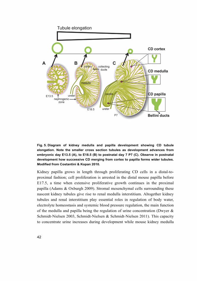
Fig. 5. Diagram of kidney medulla and papilla development showing CD tubule
elongation. Note the smaller cross section tubules as development advances from
embryonic day E13.5 (A), to E18.5 (B) to postnatal day 7 P7 (C). Observe in postnatal
development how successive CD merging from cortex to papilla forms wider tubules.
Modified from Costantini & Kopan 2010.
Kidney papilla grows in length through proliferating CD cells in a distal-to-
proximal fashion; cell proliferation is arrested in the distal mouse papilla before
E17.5, a time when extensive proliferative growth continues in the proximal
papilla (Adams & Oxburgh 2009). Stromal mesenchymal cells surrounding these
nascent kidney tubules give rise to renal medulla interstitium. Altogether kidney
tubules and renal interstitium play essential roles in regulation of body water,
electrolyte homeostasis and systemic blood pressure regulation, the main function
of the medulla and papilla being the regulation of urine concentration (Dwyer &
Schmidt-Nielsen 2003, Schmidt-Nielsen & Schmidt-Nielsen 2011). This capacity
to concentrate urine increases during development while mouse kidney medulla
43
becomes identifiable morphologically only at E15.5, increases 4.5-fold up to birth
and continues to grow during postnatal development (Song & Yosypiv 2012).
The main process in medulla and papilla growth seems to be tubular
elongation, which generates narrower tubular diameter as development advances
(see Fig. 5) (Yu 2011, Song & Yosypiv 2012). Elongation of tubules seems to
happen through two planar cell polarity processes: orientated cell division (OCD),
which aligns the cells on the long axis of the tube, and orientated cell migration or
convergent extension-like processes, which drive specific migration of polarized
cells (Karner et al. 2009) on cell polarity see also section 2.1.3). It was recently
shown by live imaging technique that the PCP convergent extension-like
movement during tubule elongation is driven in large part by a myosin-dependent,
multicellular rosette-based mechanism for mediolateral cell intercalation, a deeply
conserved cellular engine for epithelial morphogenesis. Disturbance in any of
these two processes has been shown to cause renal cyst formation; increased
rosette-based intercalation can restore normal tubule elongation if OCD is
impaired (Lienkamp et al. 2012).
These planar cell polarity processes start in embryonic kidney development
but are mainly responsible for the significant medullary and papillary postnatal
growth. Two Wnts have been described so far to modulate these processes; Wnt-
9b and Wnt-7b (Karner et al. 2009, Yu et al. 2009). Wnt-9b seems to regulate both
tubular elongation processes but at different stages: in embryonic stage
influencing the convergent extension-like movements, and postnatally regulating
OCD. In hypomorph Wnt-9b kidney the tubules’ cellular orientation is
randomized, resulting in larger diameter tubules which consequently develop into
tubular cysts. OCD is more tightly regulated during postnatal tubular elongation
but also seems to be of crucial importance in the embryo during initiation of renal
medulla at E15.5, as Wnt-7b mutants orientate their cells in a plane opposite to
wild type and undergo increased apoptosis, which leads to failure in medulla
formation. Moreover, Wnt-7b also seems to have survival properties on tubular
cells as inactivation of Dickkopf1 (Dkk1), a Wnt inhibitor on UB lineage, leads to
a phenotype with overgrown kidney papilla with increased Wnt-7b signal (Pietila
et al. 2011). These two Wnts seem to act in both autocrine and paracrine fashion,
autocrine in collecting ducts (Wnt-9b and Wnt-7b) and paracrine in proximal
tubules (Wnt-9b) and Henle’s loop (Wnt-7b), the paracrine processes taking place
in close relationship to medullary and papillary stromal cells (Karner et al. 2009,
Yu et al. 2009, Costantini & Kopan 2010). Both of these Wnt signals implicated
in cortical UB branching and determining nephron number endowment are also
44
important for tubule elongation in the medulla and papilla. Loss of these Wnts
causes phenotypes of hypoplasia and dysplasia, hypodysplasia. Medulla and
papilla hypodysplasia defects may indeed be secondary also to UB branching
defects (Song et al. 2012), but in most mouse models these seem to be caused by
an intrinsic role of these signals in medulla-papilla morphogenesis (Song &
Yosypiv 2012).
Tubule maturation is achieved through establishing specialized cell types
with a specific segmental cell type ratio (Costantini & Kopan 2010). CD include
intercalated cells, which regulate pH homeostasis by secretion of H+ and HCO3-
into the urine, and principal cells, which concentrate the urine by absorbing water
and regulating Na+ homeostasis. Towards the medulla there is a gradual increase
in principal cells in relationship to the number of intercalated cells. Loss of
principal cells and consequently their marker water channel aquaporin 2 (Aqp2)
causes impairment in urine concentration, diabetes insipidus, while loss of
intercalated cells causes distal renal tubular acidosis (Costantini & Kopan 2010).
Cells in medulla, but mostly papilla, are able to sustain extreme osmolarity stress;
this is mainly achieved through osmoprotective control in the ability to change
cell volume through cytoskeleton rearrangement, aquaporin water channel control
and osmolyte transport (Dwyer & Schmidt-Nielsen 2003, Gabert & Kultz 2011).
If our current understanding of tubule elongation is new and limited, even
less is known about the complex tri-dimensional kidney tissue architecture within
different organ axis, about tubular shape patterning, how a kidney tubular
segment is confined to be straight or convoluted, or why the nephrons in the
rostro-caudal poles of the kidney have more extensive curvature than the central
ones (al-Awqati & Goldberg 1998, Yu 2011).
2.2.3 Functional evaluation of kidney disease
The role of a healthy kidney is to maintain the fluid and electrolyte composition,
but it also plays key roles in several hormonal systems. This is accomplished by
glomerular filtration, tubular reabsorption and secretion of electrolytes, solutes
and water mediated by various hormones. In evaluation of kidney function urine
analysis remains the most important screening tool for renal disease. In the mouse,
daily 24-hour urine collections are possible in metabolic cages; however, they are
still prone to bias, which is why calculation of concentration ratio of the analytes
to urinary creatinine is necessary. Renal function and urinary excretion rate
should also be related to body size, surface area or caloric expenditure.
45
Glomerular filtration rate (GFR) remains the single most useful quantitative
measure of renal function in both health and disease and creatinine the filtration
marker of choice for estimating GFR. Creatinine is an endogenous product of
skeletal muscle metabolism, and the rate of production may vary depending on
the muscle. It is freely filtered by the glomeruli but approximately 10–30% is
secreted by the proximal tubule (PT). Under normal circumstances the rates of
creatinine production and excretion are fairly constant.
Creatinine clearance (Ccr) can be used to estimate GFR, even though it
slightly overestimates the result because of PT secretion of creatinine. It is an
excellent late marker for renal damage but not useful in acute kidney injury when
tubular secretion of creatinine can increase twofold with a very slow increase in
plasma creatinine, both underestimating the severity of renal injury. The formula
used to calculate Ccr (mL/min) = (Ucr × V)/Pcr, where Ucr is urine creatinine, V
is the urine volume in mL per minute and Pcr is the plasma creatinine
concentration.
Blood urea nitrogen (BUN) is another marker used in conjunction with
creatinine and evaluates the capacity of the kidney to excrete toxic nitrogen
recycled as urea BUN (mg/dL) = urea (mmol/L) × 2.8. Urea is synthetized in the
liver cells from ammonia by the urea cycle and more than 90% is excreted by
kidney. Because of some tubular reabsorption, especially in the presence of
oliguria, measurement of BUN underestimates GFR. Increased BUN also
suggests obstructive renal disease with stagnation of urine in proximal tubules
and increased passive reabsorption of urea. BUN over plasma creatinine ratio
gives an important direction in renal disease diagnostics (prerenal, renal,
postrenal-obstructive); however, interpretation must be careful as creatinine
values might be lower due to lower body mass. A complex situation is chronic
obstructive disease, which would be characterized by postrenal azotemia as
elevated BUN and plasma creatinine. The obstruction leads to slow renal flow,
and therefore to increased passive reabsorption of urea, but also to increased
pressure in urinary space and therefore decreased glomerular filtration. Chronic
obstruction will lead to gradual loss of nephrons (glomerular and tubular function
loss) and to renal azotemia features (Dennis J. Dietzen and Michael J. Bennett, 4th
edition).
46
2.2.4 Congenital kidney disease, cystogenesis and impaired prenatal
and postnatal kidney development
The term glomerulocystic kidney (GCK) is not currently in frequent use and it is
mainly accepted as a phenotypic characterization of the two major forms of
congenital polycystic kidney diseases (PKD), autosomal dominant and recessive.
The GCK is defined as a kidney with more than 5% cystic glomeruli, glomerular
cysts comprising Bowman space dilatations greater than 2 to 3 times normal size.
GCK has variable clinical presentation; however, even though kidney function
can be stable for years, most cases progress eventually to end-stage kidney failure
(Torres & Harris 2009, Bissler et al. 2010, Lennerz et al. 2010, Torres & Harris
2011). For a long time, the glomerular cyst formation was seen as a secondary
consequence to gross urine reflux into kidney parenchyma, caused by lower
urinary tract obstructions. A few mouse models have shown that primary defects
in glomerulus can cause glomerulocystic phenotypes, but there is little evidence
from human disease genetic studies. Recent investigations postulate that kidney
tubular abnormalities may play a role in glomerular cystogenesis; however, this
aspect has been poorly investigated so far.
Disturbances in kidney tubule patterning play a major the role in the etiology
of polycystic kidney disease (PKD) in general. Human disease studies and their
targeted genetic models showed that PKDs are mainly ciliopathies, caused by
mutations in proteins located in primary cilia and associated with abnormal rates
in tubular epithelial proliferation and apoptosis (Torres & Harris 2009, Bagherie-
Lachidan & McNeill 2010, Luyten et al. 2010). Advanced research studies in
ciliary protein mouse models introduced planar cell polarity (PCP) as
fundamental to the pathogenesis of PKD (Bagherie-Lachidan & McNeill 2010,
Luyten et al. 2010). An earlier study in a Xenopus laevis PKD model proposed
that the ciliary protein inversin regulates the switch between canonical and
noncanonical Wnt in kidney tubulogenesis (Simons et al. 2005, Simons &
Mlodzik 2008). However, the inversin mouse study which failed to support the
cystogenesis Wnt switching hypothesis showed a loss of orientated mitotic
spindle, considered to be managed by non-canonical Wnt/PCP signaling
(Sugiyama et al. 2011). In recent years the Wnt and Wnt pathway related proteins,
well known as cell polarity tissue organizers, have emerged as stronger candidates
in mouse PKD pathogenesis through PCP control in kidney tubule lumen size and
elongation (Karner et al. 2009, Yu et al. 2009, Bagherie-Lachidan & McNeill
2010, Lancaster & Gleeson 2010, Wuebken & Schmidt-Ott 2011).
47
Changes in Wnt pathway related proteins have now been noted in studies of
human PKD disease and different mouse models of PKD (Romaker et al. 2009,
Torres & Harris 2009). Cilia proteins, PCP signaling proteins and Wnts are
suggested to be part of a complex network that controls and maintains tubule
diameter and elongation; if impaired, any of these may initiate cystogenesis
(Wallingford & Mitchell 2011).
Renal hypoplasia is a common, yet often misused term describing congenital
kidney diseases. (Cain et al. 2010). Renal hypoplasia is defined as smaller kidney
with reduced nephron number but otherwise normal morphology. The exact
incidence of pure renal hypoplasia is unknown because congenital kidney
diseases exhibit evidence of kidney tissue abnormality, defined as dysplasia.
Confusion and term misuse is mostly due to limitation of noninvasive tools
(ultrasound) to discriminate between pure hypoplasia and hypodysplasia. Renal
hypoplasia/dysplasia are the leading cause for childhood end-stage renal disease.
Subtle changes in normal nephron number, such as mild bilateral hypoplasia, are
associated with adult onset hypertension disease and chronic kidney disease.
Mouse mutant models, but also studies of human congenital diseases, provide
insights into molecular pathogenesis of renal hypoplasia. Any of the
nephrogenesis processes involving MM, UB and stroma can cause kidney
hypoplasia with a lower number of nephrons: defects in induction, growth and
branching, survival and differentiation (Cain et al. 2010).
2.3 Wnt-11 gene, protein and signaling pathways in mammalian
organogenesis with emphasis on heart and kidney
development
Wnt-11 is a unique member of the Wnt family, with multiple expression sites
during embryonic development and possible action in specific tissue and cellular
context through several distinct pathway networks (Uysal-Onganer & Kypta
2012).
Initially, Wnt-11 was classified as a non-canonical Wnt, a subclass of Wnts
such as Wnt-4, and Wnt-5, which are not able to stabilize intracellular β-catenin,
but activate RhoA, JNK or calcium-dependent pathways (Fig 6). However, the
initial classification became extremely controversial as further studies showed
that most Wnts, including Wnt-11, have the potential to activate more than one
type of Wnt pathway in what might be a complex Wnt signaling network (Kestler
& Kuhl 2008). This fresh view considers that specific Wnt activity is conferred by
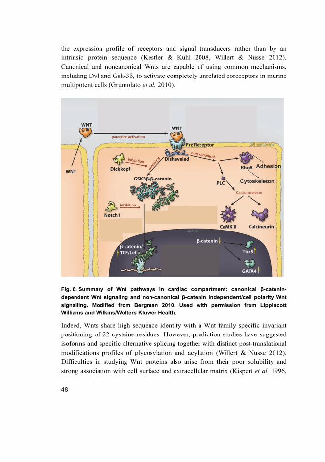
48
the expression profile of receptors and signal transducers rather than by an
intrinsic protein sequence (Kestler & Kuhl 2008, Willert & Nusse 2012).
Canonical and noncanonical Wnts are capable of using common mechanisms,
including Dvl and Gsk-3β, to activate completely unrelated coreceptors in murine
multipotent cells (Grumolato et al. 2010).
Fig. 6. Summary of Wnt pathways in cardiac compartment: canonical β-catenin-
dependent Wnt signaling and non-canonical β-catenin independent/cell polarity Wnt
signalling. Modified from Bergman 2010. Used with permission from Lippincott
Williams and Wilkins/Wolters Kluwer Health.
Indeed, Wnts share high sequence identity with a Wnt family-specific invariant
positioning of 22 cysteine residues. However, prediction studies have suggested
isoforms and specific alternative splicing together with distinct post-translational
modifications profiles of glycosylation and acylation (Willert & Nusse 2012).
Difficulties in studying Wnt proteins also arise from their poor solubility and
strong association with cell surface and extracellular matrix (Kispert et al. 1996,

49
Eisenberg & Eisenberg 1999). Particularly Wnt-11 has more predicted
glycosylation sites than other Wnt members, and in certain biological system it is
subjected to additional post-transcriptional modifications. During Xenopus axis
formation this involves tyrosine sulphation hetero-oligomerization with Wnt-5a
forming a multimer Wnt with higher Wnt signaling activity than either individual
(Cha et al. 2009). Wnt-5a and Wnt-11 cooperation seem to go beyond lower
vertebrates in heart development in mammals (Cohen et al. 2012).
The Wnt-11 gene is highly conserved during evolution; however, some
differences have been discovered between mammalian Wnt-11 and other
vertebrates’ Wnt-11. In many vertebrate species there exists more than one Wnt-11
gene, also called Wnt-11-related genes, but Xenopus (Wnt-11R), zebrafish (Wnt-
11r) and chick (Wnt-11b) are true orthologs to human Wnt-11, sharing higher
sequence identity than their Wnt-11 relatives. Therefore care should be taken
when interpreting studies from Xenopus and zebrafish relative to mammals
(Uysal-Onganer & Kypta 2012).
Expression patterns have been reported for Wnt-11 during mammalian
development in morula-blastocyst cells, kidney ureter buds and other areas of the
urogenital system, somite, limb, perichondrium of developing skeleton, lung
mesenchyme, cortex of adrenal gland, somites, limb, skin, development, digestive
tract and pancreatic epithelium (Uysal-Onganer & Kypta 2012). Among these,
maybe the most commonly known have been the roles of Wnt-11 in heart and
kidney development.
2.3.1 Wnt-11 during heart development
Wnts are required throughout cardiogenesis and seem to play temporally distinct
roles (Fig. 3). In the first phase of cardiogenesis, β-catenin-dependent canonical
Wnt (Wnt/β-catenin) signaling promotes formation of mesoderm and proliferation
of cardiac progenitors in precardiac mesodermal cells, while inhibiting their
cardiac specification. In later stages, Wnt/β-catenin inhibits cardiomyocyte
differentiation (Fig. 7, Naito et al. 2006, Ueno et al. 2007, Gessert & Kuhl 2010).
In contrast, cardiogenesis requires β-catenin-independent (non-canonical) Wnt
activity of Wnt-5a and Wnt-11, which have the dual role of promoting
cardiomyocyte specification and terminal differentiation while restraining the
inhibitory effect of canonical Wnt (Abdul-Ghani et al. 2011, Cohen et al. 2012).
Besides promoting cardiomyocyte differentiation, β-catenin-independent Wnts
seem to modulate cardiomyocyte proliferation/survival, an overexpression of
50
Wnt-11 signaling resulting in a bigger heart through hyperplasic mechanisms
(Abdul-Ghani et al. 2011).
The postnatal roles of Wnt signaling have mainly been studied in the context
of adult heart remodeling. Loss of β-catenin or inhibition of GSK3β/ β-catenin in
Dvl knockout mouse attenuated stress-induced hypertrophic response, while in
vitro or in vivo stabilization of β-catenin in cardiomyocytes triggered hypertrophic
response, with or without stimuli (Bergmann 2010). Dvl lies upstream of both β-
catenin and β-catenin-independent Wnt signaling (Grumolato et al. 2010). These
strongly suggest that Wnt pathways have a potential role in adult cardiac
hypertrophy and remodeling, a role that needs to be investigated in more detail.
For reviews see (Brade et al. 2006, Gessert & Kuhl 2010, Vincent & Buckingham
2010, Miquerol & Kelly 2013).
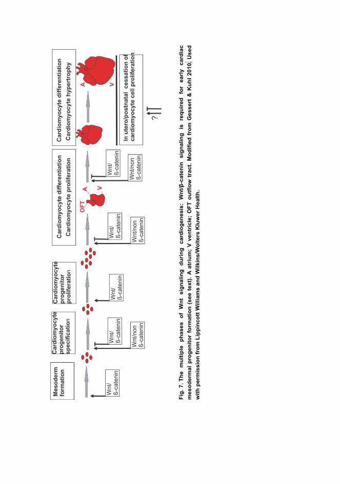
Fig
. 7. T
he
m
ult
iple
p
ha
se
s
of
Wn
t s
ign
ali
ng
d
uri
ng
c
ard
iog
en
es
is:
Wn
t/β
-ca
ten
in
sig
na
lin
g
is
req
uir
ed
fo
r e
arl
y
ca
rdia
c
me
so
de
rma
l p
rog
en
ito
r fo
rmati
on
(s
ee
te
xt)
. A
atr
ium
; V
ve
ntr
icle
; O
FT
ou
tflo
w t
rac
t. M
od
ifie
d f
rom
Ge
ss
ert
& K
uh
l 2
01
0.
Us
ed
wit
h p
erm
iss
ion
fro
m L
ipp
inco
tt W
illi
am
s a
nd
Wil
kin
s/W
olt
ers
Klu
we
r H
ea
lth
.
52
Wnt-11 expression during heart development
Wnt-11 activity during mammalian gastrulation within or in close proximity to the
cardiogenic mesoderm remains poorly understood. In zebrafish, Xenopus and
avians Wnt-11 is active in the cardiogenic mesoderm where it seeems to be
important in early cardiomyocyte coherent migration (Pandur et al. 2002,
Garriock et al. 2005, Matsui et al. 2005). These mutants show phenotypes of
cardia bifida with abnormal apico-basal polarity and improper junctional
complexes, suggesting improper epithelialization in early cardiomyocytes (Pandur
et al. 2002, Garriock et al. 2005, Matsui et al. 2005). Similar phenotypes are
reported in N-cadherin blocking studies in chick (Linask et al. 1997) and
fibronectin mutants in zebrafish and mouse (Glickman & Yelon 2004). Altogether,
these suggest that cardiomyocyte coherent migration is regulated by Wnt-11
through cadherin-mediated signaling and by interaction with extracellular matrix
proteins.
Indeed, Wnt-11 signaling requires binding with extracellular matrix proteins,
heparan sulfate proteoglycans (HSPG), during mouse kidney morphogenesis
(Kispert et al. 1996). Moreover, during gastrulation both fish embryos with
mutated glypican - an HSPG - and glypican-depleted Xenopus laevis embryos
exhibit defects in Wnt-11 signaling (Ohkawara et al. 2003). Taken together, these
suggest that Wnt-11 interaction with extracellular matrix proteins is a preserved
mechanism for Wnt-11 signaling between diverse biological contexts.
Although expression of Wnt-11 in mouse cardiogenic mesoderm has not been
clearly demonstrated (Cohen et al. 2012), Wnt-11 may be present in this region
and dramatically upregulated upon fusion of the cardiac crescent with formation
of the early heart tube from E8.0 onwards (Kispert et al. 1996, Terami et al. 2004,
Cohen et al. 2012). Mouse Wnt-11 knockouts do not show severe phenotype of
cardiac bifida; therefore if Wnt-11 is important in early cardiac epithelialization,
its function may be redundant with another Wnt, such as Wnt-5a.
Later on in the heart looping stage, Wnt-11 expression has been detected in
the outflow tract (Kispert et al. 1996, Zhou et al. 2007) with roles in OFT
assembly. The results presented in this thesis were the first to show that mouse
Wnt-11 gene and protein are active in ventricular myocardium from E8.0 onwards,
participating in ventricular myocardium development and maturation in looped
and pre-septated heart E10.5-E12.5. We showed that Wnt-11 signaling regulation
involves cardiomyocyte N-cadherin and β-catenin protein expression and protein
-protein interaction, in line with the previous studies in the cardiogenic mesoderm
53
of lower vertebrates (Garriock et al. 2005). Our finding of active Wnt-11
signaling at this stage of ventricular development was later confirmed by others
(Abdul-Ghani et al. 2011, Cohen et al. 2012). The latter Wnt-11 studies revealed
novel roles for Wnt-11 in cardiomyocyte survival and cardiac progenitor
maintenance. Cohen et al. show an unexpected co-requirement for Wnt-11 and
another non-canonical Wnt, Wnt-5a, in regulation of SHF progenitor population
size in mice (Cohen et al. 2012). Moreover, in the same publication in
differentiating mouse embryonic bodies Wnt-5a and Wnt-11 are co-required to
promote both FH and SHF fates by their attenuating effect on repressor canonical
Wnt, therefore activating progenitor terminal differentiation (Cohen et al. 2012)
(Fig. 7).
Wnt-11 and cardiac transcription factor regulation
Wnt-11 is essential for heart development, being required for in vivo cardiac
marker gene expression (Pandur et al. 2002, Afouda et al. 2008). The first line of
evidence was produced by Eisenberg et al. from an in vitro model system
showing the ability of Wnt-11 to trigger cardiomyogenic faith in noncardiac
mesoderm of quail embryonic explants (Eisenberg et al. 1997, Eisenberg &
Eisenberg 1999). Subsequent studies addressed the issue of cardiac “induction” in
numerous in vitro model systems of embryonic pluripotent cells (Flaherty et al.
2008, Flaherty et al. 2012), quail mesodermal cells (Eisenberg & Eisenberg 1999,
Terami et al. 2004), murine ES cells (Ueno et al. 2007), but also adult progenitors
such as human endothelial progenitor cells (Koyanagi et al. 2005b) or murine
bone marrow-derived multipotent cells (Flaherty et al. 2008, Flaherty et al. 2012).
Expression of Wnt-11 in all these pluripotent cells triggers expression of major
cardiac transcription factors: Nkx2.5, Mef2c, and Gata-4 defining their
cardiomyocyte fate.
Our results during early cardiogenesis are in line with this showing impaired
cardiac transcription factor expression due to loss of Wnt-11 function in
ventricular myocardium. This primary mechanism might be behind the
cardiomyopathy seen in our adult mouse C57Bl6 Wnt-11 knockout study. Adult
onset cardiomyopathies have been reported in human patients with
haploinsufficiency mutations in these cardiac transcription factors (Costa et al.
2013, Li et al. 2013)
54
Wnt-11 and Gata-4
Human Wnt-11 integrative genomics analysis shows TCF/LEF-binding sites
within Wnt-11 proximal promoter region as well as double Gata-binding sites
within intron 2, conserved in other mammalian orthologs (Katoh & Katoh 2005),
suggesting that both canonical-Wnt and Gata factors can activate Wnt-11
expression. The interaction between Gata factors and Wnt-11 is reciprocal since
Gata-4/6 are required for Wnt-11 expression during Xenopus cardiogenesis, and
Wnt-11 promotes Gata-4/6 differentiation program in maturing cardiomyocytes
(Afouda et al. 2008). Gata factors link canonical and non-canonical Wnt
signaling in cardiogenesis; while canonical Wnt inhibits Gata factors expression,
canonical Wnt is also capable of upregulating non-canonical Wnt-11 activity in in
vitro and mouse model (Lin et al. 2007, Ueno et al. 2007) and consequently
promoting Gata-induced cardiomyocyte differentiating program. These findings
evoke the biphasic roles of canonical Wnt during the cardiogenic program, first
promoting the precardiac mesodermal progenitors and later repressing the
cardiomyocyte progenitor terminal differentiation (in Zebrafish and ES cells)
(Gessert & Kuhl 2010). Although Wnt-11 alone is sufficient to activate Gata-4
cardiomyogenic program in pluripotent cells, synergistic and perhaps sequential
signaling by other cardiogenic factors is critical for optimal terminal
differentiation of cardiomyocytes (Cohen et al. 2012).
Wnt-11 and calcium signaling
During early heart tube formation in Xenopus Wnt-11 signals are required for
genesis of the myocardial electrical gradient through L-type calcium channels, a
mechanism which is not dependent on connexins or cell shape (Panakova et al.
2010). This study revealed a previously unrecognized role for Wnt-11/Ca
signaling in establishing a physiological electrical gradient in the plane of the
developing cardiac epithelium through modulation of ion channel function
(Panakova 2010). Furthermore, this data demonstrated that Wnt-11 is capable of
regulating subcellular calcium domains. This data is intriguing in relationship to
an avian study showing that calcium signaling has a regulatory role in cardiac
precursor cells during their commitment towards cardiomyocyte fate (Linask &
Linask 2010).
55
In avians, Wnt-11 expression has also been detected in conduction system
elements (Bond et al. 2003), but with no evidence on conduction system function
later in heart development or heart disease.
It remains unclear whether the same type of Wnt-11 /Ca signaling is required
in mammalian early or later heart formation. In our mouse Wnt-11 mutants Cx-40
showed a normal expression pattern during ventricular development in cardiac
development E10.5-E12.5, in addition to normal action potential and calcium
fluxes in isolated embryonic cardiomyocytes (E12.5). No arrhythmias were
observed in our mutants during echography investigations.
2.3.2 Wnt-11 during kidney development
Wnt-11 is expressed at the distal tips of the branching ureter throughout
embryonic development but it is not essential for induction of nephrogenesis in
vitro or in vivo, compared to other ureter bud-expressed Wnts as Wnt-6, Wnt-7b,
Wnt-9b able to induce nephrogenesis in in vitro or in vivo conditions (Carroll et al.
2005). At the tips of the branching ureter Wnt-11 expression takes place in an
autoregulatory feed-back loop with mesenchymal ligand glial-cell derived
neurotrophic factor Gdnf and its epithelial cRet tyrosine kinase receptor (Kispert
et al. 1996, Majumdar et al. 2003) (Fig 4). Gdnf is decreased in 129 SV Wnt-11
mutants and the lower number nephron phenotype seen in these mutants seems to
correlate to possibly decreased trifurcation type of branching which is observed in
later kidney development (Majumdar et al. 2003, Costantini 2010). For more
information on Wnt signaling in the cortex nephrogenesis, see section 2.2.1.
Optimal ureter bud branching does indeed seem to determine an optimal
nephron number. This has important clinical relevance as kidneys with a low
number of nephrons are at high risk of developing hypertension since nephrons
are naturally gradually lost during the aging process; in addition, it may also lead
to childhood end stage renal failure (Cain et al. 2010).
Besides ureter branching no other role has been clearly proposed or
demonstrated for Wnt-11 signaling in kidney development, although Wnt-11 gene
activity has been noted, for example, in the medulla around medulla tubules
together with Wnts which signal through both canonical and noncanonical Wnt
pathways Wnt-4, Wnt-9b and Wnt-7b. So far clear roles in programming kidney
tubule diameter and elongation through cell polarity pathways have been
demonstrated for Wnt-9b and Wnt-7b in mouse models through coordinated cell
migration and orientated cell division check (Merkel et al. 2007, Karner et al.
56
2009, Yu et al. 2009, Yu 2011). Final nephron endowment is completed during
embryogenesis week 36 in humans and postnatal day 5 in mice, although the
kidney tubules continue to mature long into the postnatal period. Later Wnt-11
roles during these developmental stages have not been addressed until the C57Bl6
Wnt-11 knockout model in the present study. Our results from this mice study
suggest that Wnt-11 might have an additional role during kidney development,
such as regulation of tubule elongation and convolution, with involvement of
Wnt-9b signaling, which was severely reduced in these kidney tubules.
57
3 Outline of the present study
Of the mammalian Wnts, Wnt-11 has mainly been classified as non-canonical,
triggering a β-catenin-independent signaling pathway. Wnt-11 has proven to be
involved in kidney ureteric tip branching (Kispert et al. 1996, Majumdar et al.
2003) and heart outflow tract morphogenesis (Kispert et al. 1996, Zhou et al.
2007). Possible connections between Wnt-11 and cadherin-mediated cell adhesion
and migration have been drawn in in vivo studies performed on lower vertebrates
and quail models (Eisenberg et al. 1997, Ulrich et al. 2005). In vitro
investigations have revealed its ability to promote cardiogenesis in several types
of progenitor cells (Pandur et al. 2002, Terami et al. 2004, Koyanagi et al. 2005a,
Koyanagi et al. 2005b, Flaherty et al. 2008). The goal of the present study was to
reveal novel Wnt-11 roles during heart and kidney development with relevance to
human congenital diseases.
Specific aims of this study were:
1. To uncover new roles of Wnt-11 during myocardium development and to
study the impact of its loss on heart function.
2. To reveal additional roles of Wnt-11 during tubule morphogenesis and study
the consequences of Wnt-11 loss on kidney function.

58
59
4 Material and methods
Detailed description of the material and methods used in this study are found in
the original article I and manuscripts II-III.
Method Original article
Mouse line and genotyping I, II, III
Tissue preparation I, II, III
Cardiomyocyte primary cell culture I
Histology staining (Hematoxylin Eosin) I, II, III
Histology staining (Masson Trichrome) I, II, III
Histology staining (Pass-Schiff) II, III
Immunohistochemistry I, II, III
Morphometry studies I, II, III
Western Blotting I
Immunoprecipitation I
Tissue RNA extraction and real time PCR I, II
In situ hybridization I, III
Plasma and urine sampling and analysis III
Cardiomyocyte cellular electrophysiology I
Doppler echocardiography I, II
Myocardial Strain analysis II
Telemetry II
Transmission electron microscopy I, III
Statistical analysis I, II, III

60
61
5 Results
5.1 Wnt-11 deficiency in 129SV background leads to primary
cardiomyopathy causing heart failure with severe lethality in
midgestation (I)
It has previously been shown that Wnt-11 is strongly expressed in the primitive
heart tube OFT segment (Kispert et al. 1996), from E8.5-E8.75, and plays an
important role in the morphogenesis of this heart segment (Zhou et al. 2007). In
addition to this, low levels of Wnt-11 have been reported in the early ventricle
myocardium (Eisenberg & Eisenberg 2006). Our Wnt-11 expression assay shows
Wnt-11 transcripts and protein in ventricular and atrial myocardium in the looped
heart at E10.5 and E12.5 (I Fig. 1 and online Supplementary Fig 1, and 2). These
findings suggest a specific role for Wnt-11 in myocardium development.
To investigate putative functions of Wnt-11 in myocardium morphogenesis
we studied myocardium morphology and function in Wnt-11-/- mice in 129Sv
background, with a previously reported severe lethality during in utero
development (Majumdar et al. 2003, Zhou et al. 2007). We investigated in more
detail the Wnt-11-/- lethality curve and noted that these mice were still recovered
in normal Mendelian ratio at E12.5, but in less than 50% from the expected ratio
at E13.5. Although it has previously been shown that OFT morphogenesis and
septation are impaired in Wnt-11-/- (Zhou et al. 2007), at E12.5 heart septation is
not completed; therefore we hypothesize that it cannot explain the severe mid-
gestation lethality.
Our close inspection revealed that Wnt-11-/- mice did indeed have a
myocardium defect in the form of a thinner ventricular wall already at E10.5 (I
Fig.). Moreover, this associated with defects in LV and RV specific ventricular
myocardium patterning, shown by compact and trabeculated myocardium
morphometry analysis at E10.5 and E12.5 (I Fig 2). These were not caused by
impaired apoptosis or proliferation processes (data not shown), confirming
previous findings (Zhou et al. 2007). Signs of heart failure were observed in
conjunction with the Wnt-11-/- myocardium defects, such as dilated atrial
appendices, pleural and pericardial effusions (Supplemental fig 3 online). Later
gestation embryos had hypoplastic hearts with thinner compact myocardium and
less dense trabeculations with abnormal morphology (Fig. 8).
62
Furthermore, non-invasive Doppler echocardiography assay on E12.5
embryos Wnt-11-/- showed impaired IRT (p < 0.001, Table 1 in I and
Supplementary Fig. 4 online), indicating abnormal embryonic myocardium
relaxation in early diastole with preservation of the systolic function. This has
probably as consequence increased arterial and venous pressure, seen as increased
pulsatility indices for umbilical artery, descending aorta and ductus venosus
(p < 0.001, Table 1 in I and Supplementary Fig. 4 online). As increased PIs in
umbilical artery and aorta are observed in human placental insufficiency, we
analyzed the mutant placenta and found normal histology, excluding placental
insufficiency from causes of cardiac failure. Impaired relaxation seemed not to be
caused by changes in cardiomyocyte excitability or calcium signaling as isolated
ventricular cardiomyocytes from both study groups showed no significant
differences in electrophysiology assay in vitro investigating action potential,
calcium transient amplitude, duration, oscillation rate or pacing (Supplementary
Fig. 6 online). In addition, we found that 92% of the E12.5 Wnt-11-/- embryos had
holosystolic AV valve regurgitation compared to 8% in the control group
(p < 0.001); however, no defects in AV valvular cushions were noted. This is most
probably a functional type of regurgitation due to the observed dilatation of the
ventricular chambers (I Fig. 2).
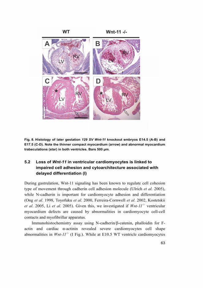
63
Fig. 8. Histology of later gestation 129 SV Wnt-11 knockout embryos E14.5 (A-B) and
E17.5 (C-D). Note the thinner compact myocardium (arrow) and abnormal myocardium
trabeculations (star) in both ventricles. Bars 500 µm.
5.2 Loss of Wnt-11 in ventricular cardiomyocytes is linked to
impaired cell adhesion and cytoarchitecture associated with
delayed differentiation (I)
During gastrulation, Wnt-11 signaling has been known to regulate cell cohesion
type of movement through cadherin cell adhesion molecule (Ulrich et al. 2005),
while N-cadherin is important for cardiomyocyte adhesion and differentiation
(Ong et al. 1998, Toyofuku et al. 2000, Ferreira-Cornwell et al. 2002, Kostetskii
et al. 2005, Li et al. 2005). Given this, we investigated if Wnt-11-/- ventricular
myocardium defects are caused by abnormalities in cardiomyocyte cell-cell
contacts and myofibrillar apparatus.
Immunohistochemistry assay using N-cadherin/β-catenin, phalloidin for F-
actin and cardiac α-actinin revealed severe cardiomyocytes cell shape
abnormalities in Wnt-11-/- (I Fig.). While at E10.5 WT ventricle cardiomyocytes
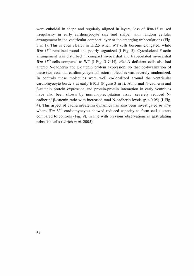
64
were cuboidal in shape and regularly aligned in layers, loss of Wnt-11 caused
irregularity in early cardiomyocyte size and shape, with random cellular
arrangement in the ventricular compact layer or the emerging trabeculations (Fig.
3 in I). This is even clearer in E12.5 when WT cells become elongated, while
Wnt-11-/- remained round and poorly organized (I Fig. 3). Cytoskeletal F-actin
arrangement was disturbed in compact myocardial and trabeculated myocardial
Wnt-11-/- cells compared to WT (I Fig. 3 G-H). Wnt-11-deficient cells also had
altered N-cadherin and β-catenin protein expression, so that co-localization of
these two essential cardiomyocyte adhesion molecules was severely randomized.
In controls these molecules were well co-localized around the ventricular
cardiomyocyte borders at early E10.5 (Figure 3 in I). Abnormal N-cadherin and
β-catenin protein expression and protein-protein interaction in early ventricles
have also been shown by immunoprecipitation assay: severely reduced N-
cadherin/ β-catenin ratio with increased total N-cadherin levels (p < 0.05) (I Fig.
4). This aspect of cadherin/catenin dynamics has also been investigated in vitro
where Wnt-11-/- cardiomyocytes showed reduced capacity to form cell clusters
compared to controls (Fig. 9), in line with previous observations in gastrulating
zebrafish cells (Ulrich et al. 2005).
65
Fig. 9. Confocal images of primary cardiomyocyte cultures from E12.5 WT and
Wnt-11-/- ventricles. (A-B) Staining for F-actin with Phalloidin (green) and (C-D) N-
Cadherin (green) and α-actinin (red). In all images DAPI (blue) for nucleus. Note
smaller cluster formation in Wnt-11-/- cardiomyocyte cultures. Bars A-B 100 µm, C-D
50µm.
Immunohistochemistry on E12.5 cells shows poor sarcomeric organization in
Wnt-11-/-, as demonstrated by cardiac α-actinin (Supplemental Fig. 7 online). This
has been evaluated in detail by our TEM analysis in line with our abnormal
cardiomyocyte morphological findings from immunohistochemistry. Wnt-11-/-
cardiomyocytes are poorly differentiated, as shown by their low myofibrillar
content, poor spatial arrangement in immature myofibrillar bundles and immature
cell-cell junction ultrastructure (I Fig. 5 and Supplementary Fig. 8 online).
In summary, these observations support the conclusion that Wnt-11 signaling
has a specific role in the control of cardiomyocyte architecture in developing
myocardium.
66
5.3 Wnt-11 deficiency abnormalities in ventricular myocardium
associate with changes in genes implicated in heart
development (I)
The Gata-4, Nkx2.5, and Mef2C genes encode important cardiac transcription
factors upstream of other essential signals for cardiogenesis, which include
natriuretic proteins ANP and BNP. These are prime candidate targets for Wnt-11
signal transduction in mammalian ventricular development, given that Wnt-11
regulates their expression in cell culture and frog mesoderm explants (Eisenberg
& Eisenberg 1999, Pandur et al. 2002, Terami et al. 2004, Koyanagi et al. 2005a,
Cohen et al. 2008, Flaherty et al. 2008). In situ hybridization and qRT-PCR
analysis revealed that the Gata-4, Nkx2.5, Mef2C, and ANP (atrial natriuretic
peptide) transcripts were reduced in Wnt-11-deficient heart compared with
controls both at E10.5 and E12.5 (Fig. 6 in I). In contrast, B-type NP (BNP)
expression was induced at E10.5, coinciding with increased phosphorylated-JNK
activity in ventricular myocardium, as shown by Western blotting. These latter
changes are possibly in response to heart dysfunction (Cameron & Ellmers 2003,
Lubbers & Eghtesady 2007).
Level expression changes in heart transcription factors could be part of the
pathogenesis mechanism of the Wnt-11-/- embryonic heart defects, in line with the
findings that a heterozygous knock-out or a hypomorphic allele for any of these
genes can lead to similar heart malformations (Bruneau 2008). Our results suggest
that these genes important for ventricular myocardium development are
modulated by Wnt-11 signaling.
5.4 Wnt-11 is expressed in late myocardium development and adult
C57Bl6 Wnt-11 knockout mice present CMP phenotype (II)
The next aim was to investigate putative roles of Wnt-11 in late myocardium
development; however, 129SV Wnt-11 knockout has severe lethality during
midgestation and no mice survive to adulthood. Given that genetic modifiers are
known to modulate the penetrance or severity of cardiac disease in the mouse
(Barrick et al. 2007), we have crossed the Wnt-11 knockout allele to another
mouse genetic background, the C57Bl6. Compared to the severe in utero lethality
observed in the previous Wnt-11 knockout mouse investigations (Zhou et al. 2007,
Bergmann 2010, Nagy et al. 2010, Cohen et al. 2012), the C57Bl6 Wnt-11-/- mice
from this study show normal viability until the first weeks of postnatal stage (see
67
viability data in III) when around 50% of the mutant mice die. This suggests that
in utero lethality of the Wnt-11 knockout mice caused by the cardiac phenotype is
modulated by genetic background.
Histology of 12- to 16-week-old mouse survivor hearts revealed notably
enlarged hearts in C57Bl6 Wnt-11-/- compared to WT, including a few individuals
with a gross cardiomegaly phenotype (1/10) (II Fig 1 C). We noted abnormal
asymmetrical thickness in LV ventricular walls with significant LV papillary
muscle hypertrophy (II Fig 1 B, E). Wnt-11-/- desiccated heart mass relative to
total body mass was significantly increased compared to controls (p < 0.001,
n = 6) and in line with this, cardiomyocytes had a larger cross section area as a
sign of cellular hypertrophy (II Fig. 1 J). Altogether, these findings suggest that
the Wnt-11-/- hearts have a hypertrophic type of cardiomyopathy.
In addition to the notable hypertrophy phenotype, we noted ventricular
noncompaction segments with thin compact myocardium and abnormal clubbed
trabeculations located in the apex and mid-anterior wall of LV and RV (II note
apical LV noncompaction Fig. 1F). We investigated capillary formation by
endothelial PECAM and noted no significant difference compared to the control
group, suggesting normal capillary network formation in mutant myocardium (II
Fig. 1 L).
Some inherited hypertrophic cardiomyopathy models involve cardiomyocyte
hyperplasia; however, this was not the case in Wnt-11-/- neonatal myocardium (II
Fig 2). The PCNA signal was weaker in certain areas of Wnt-11-/- myocardium,
including noncompaction areas. The proliferating cell nuclear antigen (PCNA) is
required for DNA replication preceding mitosis as well as DNA nucleotide
excision repair, labeling cells that recently completed karyokinesis or cytokinesis.
This issue needs further investigation with a specific marker for the mitotic phase
(for example, phospho-Histone H3 or bromodeoxyuridine) to elucidate the
significance of this finding.
We reported previously that in 129SV mouse background Wnt-11 is expressed
in the ventricular myocardium of the tubular heart, looped heart and before heart
septation is completed at E12.5. In line with this, Wnt-11-deficient hearts showed
defects in myocardium morphogenesis, leading to heart failure and severe
lethality in midgestation. This indicated the importance of Wnt-11 signaling in
pre-septated heart early myocardium development. Our Wnt-11 qRT-PCR results
showed that the Wnt-11 gene is active as well in late ventricular myocardium, at
E16.5 and neonatal stage (II Fig. 3), in line with previous findings in other genetic
backgrounds (Abdul-Ghani et al. 2011, Cohen et al. 2012). This suggests a role
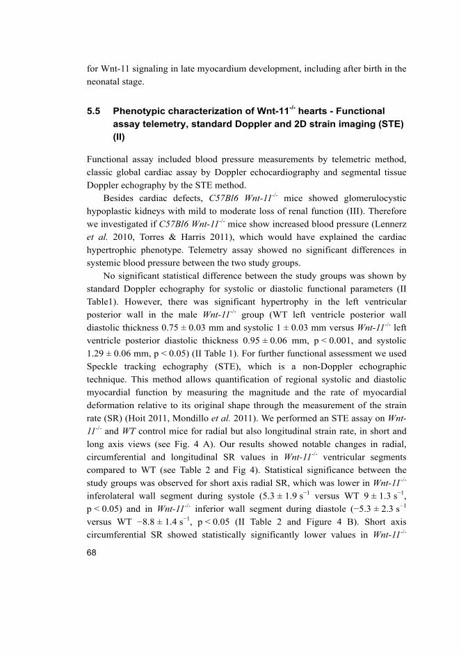
68
for Wnt-11 signaling in late myocardium development, including after birth in the
neonatal stage.
5.5 Phenotypic characterization of Wnt-11-/- hearts - Functional
assay telemetry, standard Doppler and 2D strain imaging (STE)
(II)
Functional assay included blood pressure measurements by telemetric method,
classic global cardiac assay by Doppler echocardiography and segmental tissue
Doppler echography by the STE method.
Besides cardiac defects, C57Bl6 Wnt-11-/- mice showed glomerulocystic
hypoplastic kidneys with mild to moderate loss of renal function (III). Therefore
we investigated if C57Bl6 Wnt-11-/- mice show increased blood pressure (Lennerz
et al. 2010, Torres & Harris 2011), which would have explained the cardiac
hypertrophic phenotype. Telemetry assay showed no significant differences in
systemic blood pressure between the two study groups.
No significant statistical difference between the study groups was shown by
standard Doppler echography for systolic or diastolic functional parameters (II
Table1). However, there was significant hypertrophy in the left ventricular
posterior wall in the male Wnt-11-/- group (WT left ventricle posterior wall
diastolic thickness 0.75 ± 0.03 mm and systolic 1 ± 0.03 mm versus Wnt-11-/- left
ventricle posterior diastolic thickness 0.95 ± 0.06 mm, p < 0.001, and systolic
1.29 ± 0.06 mm, p < 0.05) (II Table 1). For further functional assessment we used
Speckle tracking echography (STE), which is a non-Doppler echographic
technique. This method allows quantification of regional systolic and diastolic
myocardial function by measuring the magnitude and the rate of myocardial
deformation relative to its original shape through the measurement of the strain
rate (SR) (Hoit 2011, Mondillo et al. 2011). We performed an STE assay on Wnt-
11-/- and WT control mice for radial but also longitudinal strain rate, in short and
long axis views (see Fig. 4 A). Our results showed notable changes in radial,
circumferential and longitudinal SR values in Wnt-11-/- ventricular segments
compared to WT (see Table 2 and Fig 4). Statistical significance between the
study groups was observed for short axis radial SR, which was lower in Wnt-11-/-
inferolateral wall segment during systole (5.3 ± 1.9 s−1 versus WT 9 ± 1.3 s−1,
p < 0.05) and in Wnt-11-/- inferior wall segment during diastole (−5.3 ± 2.3 s−1
versus WT −8.8 ± 1.4 s−1, p < 0.05 (II Table 2 and Figure 4 B). Short axis
circumferential SR showed statistically significantly lower values in Wnt-11-/-
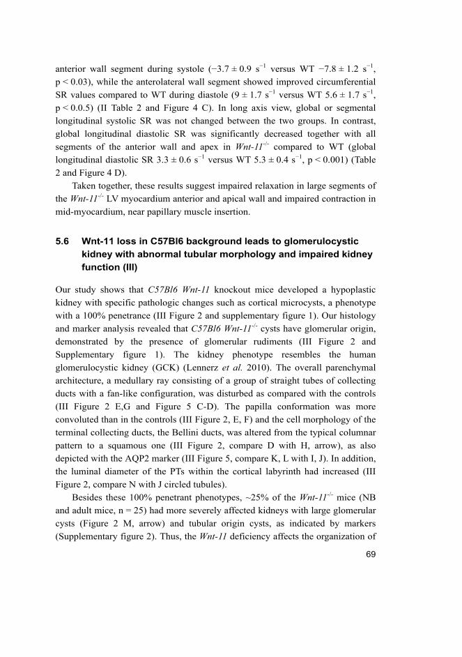
69
anterior wall segment during systole (−3.7 ± 0.9 s−1 versus WT −7.8 ± 1.2 s−1,
p < 0.03), while the anterolateral wall segment showed improved circumferential
SR values compared to WT during diastole (9 ± 1.7 s−1 versus WT 5.6 ± 1.7 s−1,
p < 0.0.5) (II Table 2 and Figure 4 C). In long axis view, global or segmental
longitudinal systolic SR was not changed between the two groups. In contrast,
global longitudinal diastolic SR was significantly decreased together with all
segments of the anterior wall and apex in Wnt-11-/- compared to WT (global
longitudinal diastolic SR 3.3 ± 0.6 s−1 versus WT 5.3 ± 0.4 s−1, p < 0.001) (Table
2 and Figure 4 D).
Taken together, these results suggest impaired relaxation in large segments of
the Wnt-11-/- LV myocardium anterior and apical wall and impaired contraction in
mid-myocardium, near papillary muscle insertion.
5.6 Wnt-11 loss in C57Bl6 background leads to glomerulocystic
kidney with abnormal tubular morphology and impaired kidney
function (III)
Our study shows that C57Bl6 Wnt-11 knockout mice developed a hypoplastic
kidney with specific pathologic changes such as cortical microcysts, a phenotype
with a 100% penetrance (III Figure 2 and supplementary figure 1). Our histology
and marker analysis revealed that C57Bl6 Wnt-11-/- cysts have glomerular origin,
demonstrated by the presence of glomerular rudiments (III Figure 2 and
Supplementary figure 1). The kidney phenotype resembles the human
glomerulocystic kidney (GCK) (Lennerz et al. 2010). The overall parenchymal
architecture, a medullary ray consisting of a group of straight tubes of collecting
ducts with a fan-like configuration, was disturbed as compared with the controls
(III Figure 2 E,G and Figure 5 C-D). The papilla conformation was more
convoluted than in the controls (III Figure 2, E, F) and the cell morphology of the
terminal collecting ducts, the Bellini ducts, was altered from the typical columnar
pattern to a squamous one (III Figure 2, compare D with H, arrow), as also
depicted with the AQP2 marker (III Figure 5, compare K, L with I, J). In addition,
the luminal diameter of the PTs within the cortical labyrinth had increased (III
Figure 2, compare N with J circled tubules).
Besides these 100% penetrant phenotypes, ~25% of the Wnt-11-/- mice (NB
and adult mice, n = 25) had more severely affected kidneys with large glomerular
cysts (Figure 2 M, arrow) and tubular origin cysts, as indicated by markers
(Supplementary figure 2). Thus, the Wnt-11 deficiency affects the organization of
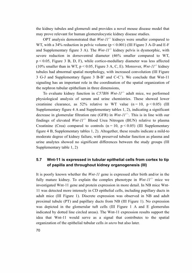
70
the kidney tubules and glomeruli and provides a novel mouse disease model that
may prove relevant for human glomerulocystic kidney disease studies.
OPT analysis demonstrated that Wnt-11-/- kidneys were smaller compared to
WT, with a 34% reduction in pelvic volume (p < 0.001) (III Figure 3 A-D and E-F
and Supplementary figure 3 A). The Wnt-11-/- kidney pelvis is dysmorphic, with
severe reduction in dorsoventral diameter (46% smaller compared to WT,
p < 0.05, Figure 3 B, D, F), while cortico-medullary diameter was less affected
(10% smaller than in WT, p < 0.05, Figure 3 A, C, E). Moreover, Wnt-11-/- kidney
tubules had abnormal spatial morphology, with increased convolution (III Figure
3 G-J and Supplementary figure 3 B-B’ and C-C’). We conclude that Wnt-11
signaling has an important role in the coordination of the spatial organization of
the nephron tubular epithelium in three dimensions,
To evaluate kidney function in C57Bl6 Wnt-11-/- adult mice, we performed
physiological analysis of serum and urine chemistries. These showed lower
creatinine clearance, as 52% relative to WT value (n = 10, p < 0.05) (III
Supplementary figure 4 A and Supplementary tables 1, 2), indicating a significant
decrease in glomerular filtration rate (GFR) in Wnt-11-/-.. This is in line with our
findings of elevated Wnt-11-/- Blood Urea Nitrogen (BUN) relative to plasma
Creatinine (Crea) compared to controls (n = 10, p < 0.05) (III Supplementary
figure 4 B, Supplementary tables 1, 2). Altogether, these results indicate a mild-to
moderate degree of kidney failure, with preserved tubular function as plasma and
urine analytes showed no significant differences between the study groups (III
Supplementary table 1, 2)
5.7 Wnt-11 is expressed in tubular epithelial cells from cortex to tip
of papilla and throughout kidney organogenesis (III)
It is poorly known whether the Wnt-11 gene is expressed after birth and/or in the
fully mature kidney. To explain the complex phenotype in Wnt-11-/- mice we
investigated Wnt-11 gene and protein expression in more detail. In NB mice Wnt-
11 was detected more intensely in CD epithelial cells, including papillary ducts in
adult mice (III Figure 1). Discrete expression was observed in NB and adult
proximal tubule (PT) and papillary ducts from NB (III Figure 1). No expression
was depicted in the glomerular tuft cells (III Figure 1 A and E glomerulus
indicated by dotted line circled areas). The Wnt-11 expression results support the
idea that Wnt-11 would serve as a signal that contributes to the spatial
organization of the epithelial tubular cells in utero but also later.
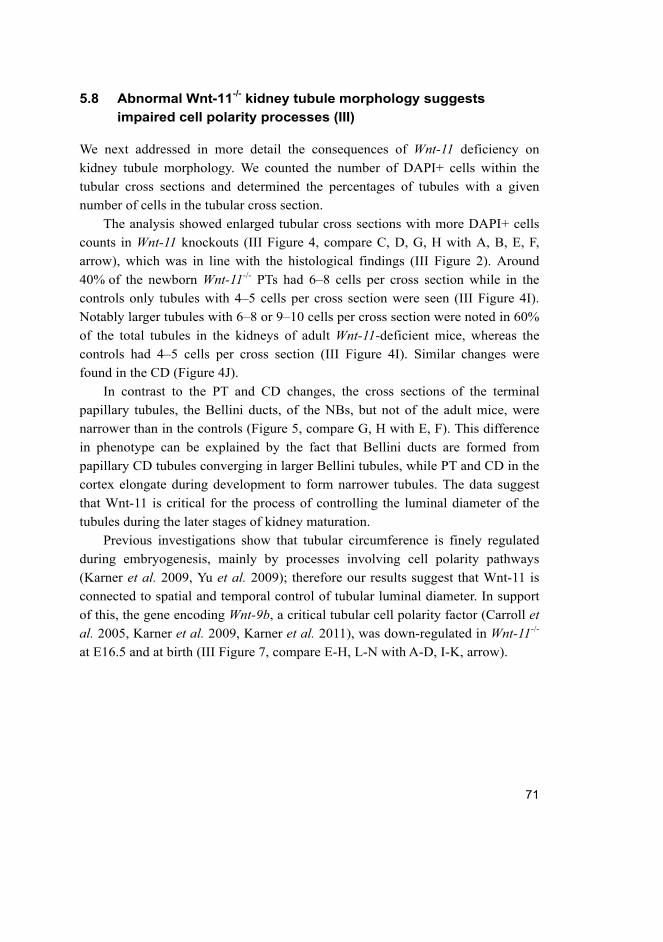
71
5.8 Abnormal Wnt-11-/- kidney tubule morphology suggests
impaired cell polarity processes (III)
We next addressed in more detail the consequences of Wnt-11 deficiency on
kidney tubule morphology. We counted the number of DAPI+ cells within the
tubular cross sections and determined the percentages of tubules with a given
number of cells in the tubular cross section.
The analysis showed enlarged tubular cross sections with more DAPI+ cells
counts in Wnt-11 knockouts (III Figure 4, compare C, D, G, H with A, B, E, F,
arrow), which was in line with the histological findings (III Figure 2). Around
40% of the newborn Wnt-11-/- PTs had 6–8 cells per cross section while in the
controls only tubules with 4–5 cells per cross section were seen (III Figure 4I).
Notably larger tubules with 6–8 or 9–10 cells per cross section were noted in 60%
of the total tubules in the kidneys of adult Wnt-11-deficient mice, whereas the
controls had 4–5 cells per cross section (III Figure 4I). Similar changes were
found in the CD (Figure 4J).
In contrast to the PT and CD changes, the cross sections of the terminal
papillary tubules, the Bellini ducts, of the NBs, but not of the adult mice, were
narrower than in the controls (Figure 5, compare G, H with E, F). This difference
in phenotype can be explained by the fact that Bellini ducts are formed from
papillary CD tubules converging in larger Bellini tubules, while PT and CD in the
cortex elongate during development to form narrower tubules. The data suggest
that Wnt-11 is critical for the process of controlling the luminal diameter of the
tubules during the later stages of kidney maturation.
Previous investigations show that tubular circumference is finely regulated
during embryogenesis, mainly by processes involving cell polarity pathways
(Karner et al. 2009, Yu et al. 2009); therefore our results suggest that Wnt-11 is
connected to spatial and temporal control of tubular luminal diameter. In support
of this, the gene encoding Wnt-9b, a critical tubular cell polarity factor (Carroll et
al. 2005, Karner et al. 2009, Karner et al. 2011), was down-regulated in Wnt-11-/-
at E16.5 and at birth (III Figure 7, compare E-H, L-N with A-D, I-K, arrow).
72
5.9 Wnt-11 deficiency alters cell proliferation and cell survival in
the cortex, correlating with changes in nephron progenitor
gene expression (III)
To obtain a better view of the cellular and molecular mechanisms of kidney
hypoplasia and tubular abnormalities in Wnt-11-deficient mice, we next addressed
potential changes in cell proliferation (PCNA, P-H3), apoptosis (TUNEL) and
progenitor cell marker genes.
Proliferation (P-H3) of the cortical cells was reduced in Wnt-11-/- (III Figure 6,
compare B with A, K); however, it did not correlate with the cystogenesis (data
not shown). The PCNA marker, also coupled to DNA repair and apoptosis
(Brenner et al. 2003), depicted an increase in staining in the kidneys of the NB
mice, but not at E16.5 (data not shown). Consistent with this, there was TUNEL+
overlapping in some of the PCNA+ cells in the cortex of the Wnt-11-/- kidneys (III
Figure 6, compare D, F with C, E, arrow; K). No significant differences were
noted in the medulla or papilla, except that the distribution of proliferating cells
within the tubules was different, especially at the tip of the papilla (III Figure 6 G-
J).
The progenitor cells within cortical domain Six2, Hox10 cluster genes and the
Foxd1 gene were altered in Wnt-11 knockout (III Figure 8 A-L). The failure in
Wnt-11 signaling was thus reflected in changes in nephron progenitor and stromal
marker expression. The data suggest that Wnt-11 function contributes to the
coordination of nephrogenesis alongside Wnt-9b. The decrease in proliferation
and increase in apoptosis in the Wnt-11-/- cortical progenitor domain may lead to
cessation of nephrogenesis via control of the genes that are important for this
process.
73
6 Discussion
6.1 Wnt-11 signaling controls development of the early
myocardium and is essential for normal cardiac functional
development (I)
Our study points to an additional critical function for Wnt-11 in heart
development, namely development of the ventricular myocardium. Wnt-11 is
expressed by the pre-septated heart ventricular cardiomyocytes and is important
in shaping their spatial organization and differentiation during the development of
the four-chamber heart. The ventricular cardiomyocytes in the Wnt-11-/- embryos
were disorganized in relation to each other; the compact ventricular wall was
hypoplastic with poorly developed trabecular processes. It should be noted that
septation of the OFT in the mouse embryo becomes complete at E14.5 (Bruneau
2002) and human fetuses with TGA may have undisturbed cardiac and placental
functional development (unpublished observations) (Bruneau 2008). Given these,
the OFT defect does not fully explain the early lethality of the Wnt-11-/- before
heart septation is completed; it is therefore likely that the ventricular myocardial
developmental defects are the primary factor contributing to the failure of the
Wnt-11-deficient embryonic heart as both the properly formed compact and
trabecular compartments are essential for normal heart function. Altogether, these
suggest a critical function for Wnt-11 in controlling ventricular myocardium
development.
Our Doppler ultrasound analysis indicated changes in several functional
parameters in the Wnt-11-/- heart at midgestation, during which the mouse embryo
is growing fast and should increase its cardiac output to maintain adequate
placental and fetal perfusion. We find that the IRT% was significantly greater in
the Wnt-11-/- embryos, and this could lead to abnormal filling of the ventricles
causing an increase in atrial and venous pressures (Makikallio et al. 2005). Indeed,
we observed an increase in the pulsatility of the ductus venosus blood velocity
waveform and also identified holosystolic AV valve regurgitation in the Wnt-11-/-
embryonic hearts. As no abnormalities were noted in the developing AV cushions,
the regurgitation was most probably functional, caused by dilatation of the
ventricles. No structural abnormality was noted in the mutant placenta, excluding
it from causes of heart failure.
74
Our study indicates that Wnt-11 is important for myocardium development
and heart function. Previous in vitro studies show that increase expression in Wnt-
11 marks the terminal differentiation of the cardiomyocytes and by adding Wnt-11
to progenitor cells increases formation of beating foci (Ueno et al. 2007). In line
with these and in vivo Xenopus studies (Garriock et al. 2005, Afouda et al. 2008,
Cohen et al. 2012), our 129Sv Wnt-11-/- mouse investigation showed poor
cardiomyocyte differentiation altogether with low level of cardiac transcription
factors essential for this process. The sensitivity of the heart development to the
dosage of Nkx2.5, Mef2C or Gata-4 was recapitulated in mouse models of human
mutations (Pu et al. 2004, Jay et al. 2005, Bruneau 2008). Among these the
decreased activity of the Gata-4 cardiac transcription factor has a notable
significance for the Wnt-11-/- heart pathology. Gata-4 and Wnt-11 have reciprocal
positive regulatory properties (Afouda et al. 2008, Afouda et al. 2008, Afouda &
Hoppler 2011), share cardiogenic promoting activity (Eisenberg & Eisenberg
1999, Pandur et al. 2002, Terami et al. 2004, Jay et al. 2005, Afouda et al. 2008)
and are co-required for an optimal cardiomyocyte terminal differentiation.
We showed that Wnt-11 regulates myocardium development by coordinating
the co-localized expression of the cell adhesion molecules N-cadherin and β-
catenin, which are critical for the spatially specific organization of
cardiomyocytes (Linask et al. 1997, Zuppinger et al. 2000, Sedmera 2005,
Hirschy et al. 2006, Phillips et al. 2007). Wnt-11 is known to control forces of
cohesion and cell adhesion in some other systems by regulating endocytosis of
another cadherin superfamily member, E-cadherin (Ulrich et al. 2005). Similarly,
Wnt-11 signaling may control the localization of N-cadherin and β-catenin to
specific cellular domains of the embryonic cardiomyocytes. This mechanism
promotes cell adhesion and is important for the organization of the cells in a
specific orientation during development of the ventricular wall. By affecting
cardiomyocyte organization Wnt-11 signaling might also influence processes
dependent on it, such as orientation of cell division affecting the myocardium
patterning or specific response to a polarized morphogen. In the working
myocardium abnormal cardiomyocyte organization may especially affect the
structural integrity with direct consequences on force transmission.
In other systems Wnt-11 signaling has been involved in the planar cell
polarity (PCP) pathway controlling the convergent extension (CE) movements
that take place during gastrulation and during organogenesis, including formation
of the early heart (Ulrich et al. 2005, Brade et al. 2006, Karner et al. 2006).
Several PCP pathway components regulate heart development, e.g. Scribble and
75
Vangl2 are known to control outflow tract and ventricular myocardium
development, as Wnt-11 is shown to do here. Mutations in Scribble and Vangl2
cause a similar cardiac phenotype to that seen in Wnt-11 mutants. PCP proteins
are thought to operate at the level of a putative frizzled receptor and upstream of
the small cytoplasmic GTPases Rac and Rho, which link the PCP pathway to the
dynamics of the cytoskeleton. In line with this, we noted a correlation between
the changes in cytoskeletal organization and the organization of the
cardiomyocytes, similarly to that observed in chick muscle (Gros et al. 2008).
Hence, as a non-canonical Wnt, Wnt-11 may control the directional movements
of the cells in the assembling ventricular wall by modulating cell adhesion in
connection with changes in the cytoskeleton.
In summary, Wnt-11 is implicated in heart development, as it controls the
differentiation and organization of the cardiomyocytes in the ventricular wall. The
reason for the failure of the Wnt-11-deficient heart probably lies in a reduced
capacity to improve its function, especially the diastole, during the early
developmental stages. This is caused primarily by disturbed adhesion between the
ventricular cardiomyocytes, mediated by impaired co-localized expression of the
N-cadherin/ß-catenin adhesion complex with a simultaneous change in the
organization of the cytoskeleton. Wnt-11 is a critical signal for myocardium
development and may function as a PCP pathway signal in the ventricular wall,
regulating the convergent extension type of cell organization by controlling
cellular adhesion.
6.2 Cardiac phenotypes in Wnt-11 knock out models and their
relevance to human inherited cardiac diseases (I and II)
In clinical work human heart malformations are mainly evaluated in the old frame
of the segmental model of heart development, with congenital diseases of big
arteries and valves separated from congenital diseases of the myocardium
(Buckingham et al. 2005). However, myocardium shares cellular lineage with all
the other heart structures mentioned above, shown by the recent discovery of first
and second heart field (SHF) (Bruneau 2008, Sirbu et al. 2008, Pandur et al.
2013). The Wnt-11 gene has been shown in our studies and by others to be
expressed in early heart tube (Kispert et al. 1996, Nagy et al. 2010, Abdul-Ghani
et al. 2011, Cohen et al. 2012) and to be important for cardiomyocyte terminal
differentiation, especially for the cells derived from SHF (Cohen et al. 2012). In
76
line with these Wnt-11 mutants shows primary defects in ventricular myocardium
but also OFT structures (I, II,(Zhou et al. 2007))
We have shown previously that Wnt-11 loss causes primary defects in utero in
myocardium maturation and patterning, in pre-septated heart (Nagy et al. 2010),
and OFT defects in septated heart stages (Zhou et al. 2007). Such OFT defects
may directly alter hemodynamic conditions, which may secondarily influence
myocardium development (Sedmera 2005). This most probably occurs in late
gestation when septation is complete, and at birth together with the complex
circulatory changes during the transition from fetal to neonatal states (Aiello &
Binotto 2007, Blyth et al. 2008). The association of these cardiac structural
disease and primary cardiomyopathy results in complete lethality of 129SV Wnt-
11-/- mice from midgestation up to neonatal stage.
In contrast, C57Bl6 Wnt-11-/- shows improved survival. Our analysis of adult
C57Bl6 Wnt-11-/- show no OFT defects, which were detected in this background
with low penetrance (~10%, n = 10) and not later than neonatal stage. This is in
contrast with 129Sv background where OFT defects have 100% penetrance (Zhou
et al. 2007). We hypothesize that the cardiomyopathy phenotype in C57Bl6 Wnt-
11-/- is a primary consequence of abnormal myocardium development and not
secondary to other cardiac structural defects.
The myocardium phenotypes observed in Wnt-11-/- C57Bl6 mice show
similarities to human cardiomyopathy reports, including overlapping congenital
cardiomyopathy phenotypes in human patients (Klaassen et al. 2008, Kelley-
Hedgepeth et al. 2009, Tsai et al. 2009). Moreover, each of the cardiomyopathies
in Wnt-11-/- C57Bl6, the hypertrophic and noncompaction forms, are found in
human patients as isolated forms which share genetic origin (Klaassen et al. 2008,
Kelley-Hedgepeth et al. 2009, Tsai et al. 2009).
Hypertrophic cardiomyopathy is maybe the most common primary form of
inherited cardiomyopathies (1:500) and diagnosis must exclude other identifiable
causes, such as hypertension (Elliott et al. 2008). Ventricular noncompaction has
a reported lower prevalence; however, the true prevalence is unclear and it has
been postulated that is has been underdiagnosed or even wrongly diagnosed as
HCM (Pignatelli et al. 2003, Elliott et al. 2008). The cardiomyopathy in Wnt-11-/-
C57Bl6 mice is not likely to be secondary to a general condition like arterial
hypertension, as mean systemic blood pressures did not differ in Wnt-11-/-
compared to controls.
It has recently been shown that cardiomyocyte hyperplasia does not cease at
neonatal stage, but is responsible for heart growth much longer than previously
77
thought, until adolescence (Mollova et al. 2013). It has also has been defined that
the rate and pattern of cardiomyocyte proliferation are an important regulator of
cardiac compact and trabeculated myocardium architecture, determining the ratio
of compact to trabeculated myocardium tissue, which is specifically defined for
each ventricular segment and closely related to functional parameters (Sedmera et
al. 2000, Henderson & Anderson 2009, Sedmera & Thompson 2011, Sedmera
2011). Proliferation was not affected in pre-septated heart myocardium of
Wnt-11-/- mutants (I and (Cohen et al. 2012); however, C57Bl6 Wnt-11-/- adult
mice ventricular myocardium abnormal architecture may be linked to the
abnormal proliferation, and this issue is worthy of further investigation.
Recent discoveries show new insights into ventricular noncompaction
pathogenesis, which involves developmental arrest in trabecular maturation and
myocardial compaction in a mouse model of human mutation of Notch pathway
regulator (Luxan et al. 2013). This mouse model presents mostly thin
myocardium phenotypes, while in human patients with this mutation either a thin
myocardium or hypertrophic myocardium with noncompaction segments has been
reported. Follow-up of human cases of neonatal primary hypertrophy reveals the
dynamic character of the congenital cardiomyopathy forms during postnatal
myocardium development, where the initial isolated neonatal finding of
hypertrophic cardiomyopathy evolved to ventricular noncompaction and
associated hypertrophy later in life (Betrian Blasco et al. 2010). In line with these,
our previous findings indicated arrest in trabecular and compact myocardium
maturation in Wnt-11-/- pre-septated embryonic heart; this also associated with
poor cardiomyocyte organization and impaired N-cadherin/β-catenin adhesion
(Nagy et al. 2010). Similar phenotypes with thinner myocardium and poorly
polarized cardiomyocytes in early development have been reported in mouse
models of cell polarity pathway core components Vangl2 and Scribble, with
resemblance to myocardium noncompaction-like phenotypes in late gestation
(Phillips et al. 2005, Phillips et al. 2007, Henderson & Chaudhry 2011).
Moreover, altered cardiomyocyte cell cycling and ventricular noncompaction was
associated with ventricular septal defects in a mouse model of human Noonan
syndrome which includes an array of cardiomyopathy phenotypes. This mouse
model holds a gain of function mutation in tyrosine phosphatase Src homology
region 2 leading to overactive MAPK- ERK1/2 pathway during cardiac
development, with consequent abnormal modulation of involving cell
proliferation, differentiation, migration, adhesion and survival (Nakamura et al.
2007). Wnt-11 has been shown to modulate these processes in in vivo and in vitro
78
models, suggesting a common link between Wnt-11, cell polarity core
components and downstream kinase effectors controlling cardiomyocyte
proliferation and survival, differentiation and organized migration and adhesion
(Nagy et al. 2010, Abdul-Ghani et al. 2011, Cohen et al. 2012, Flaherty et al.
2012, Uysal-Onganer & Kypta 2012). It is worth investigating whether any cell
polarity core genes are affected in C57Bl6 Wnt-11-/- mice, or whether
cardiomyocyte polarization is affected in utero or during neonatal/postnatal
development. It also remains under question whether the array of these
hypertrophic changes involves altered gene expression of important cardiac
transcription factors involved in cardiac development and maturation but also
adult heart hypertrophic response Gata-4, Nkx2.5, Mef2C, ANP, BNP in line with
our previous results (Nagy et al. 2010) and others (Cohen et al. 2012).
Global cardiac systolic and diastolic function is preserved in Wnt-11-/- C57Bl6
as shown by standard echographics parameters in male and female groups.
However, our speckle tracking myocardial strain rate assay detected especially
lower ventricular wall longitudinal regional relaxation in the male group in
contrast with the female group, which was not statistically significantly different
from controls despite similar severity in cardiomyopathy histological phenotypes.
This cardiac regional dysfunction has previously been well correlated with
the severity of global diastolic dysfunction in human patients presenting with
HCM associated with preserved ejection fraction/systolic global function (Goto et
al. 2006, Wang & Nagueh 2009, Kasner et al. 2010). In these studies lower
longitudinal diastolic SR correlated with poor LV filling pressure, and in similar
patients lower diastolic SR was found to be significantly decreased, also in
myocardial segments without apparent hypertrophy (Goto et al. 2006). Although
strain rate imaging provides support and additional details to standard echography
on diastolic ventricular function, the true clinical value of diastolic strain and
strain rate parameters has not yet been defined and it is not ready for standard
practice (Hoit 2011). Nevertheless, STE functional evaluation of mouse models of
human CMP is extremely useful for understanding the pathogenesis and
functional course of the human disease.
An array of identified mutations is available for diagnosing the human
inherited cardiomyopathies, although a significant proportion of the “idiopathic”
cardiomyopathy human cases have no unidentified genetic cause (Seidman &
Seidman 2001, Alcalai et al. 2008, Kimura 2010). Our findings show here that the
Wnt-11 gene is required for normal mouse ventricular myocardium development
while disturbed Wnt-11 signaling during ventricular development leads in adult
79
mouse to noncompaction cardiomyopathy and hypertrophic remodeling of the
myocardium, associated with regional myocardium dysfunction. Altogether our
data propose Wnt-11 gene as a new candidate to be investigated in human
congenital cardiomyopathy studies.
6.3 Kidney phenotype in Wnt-11-/- C57Bl6 mice suggests novel
roles for Wnt-11 in late nephrogenesis (III)
Our study shows that C57Bl6 Wnt-11 knockout mice developed a hypoplastic
kidney, in line with the previous findings from a 129SV Wnt-11 study (Majumdar
et al. 2003). In the previous study a normal kidney histology was described; in
contrast, the kidneys of the C57Bl6 Wnt-11-/- had clear pathologic changes with a
specific cortical microcystic phenotype with a 100% penetrance (III Figure 2).
This suggests that kidney Wnt-11 knockout phenotype is sensitive to genetic
background modulators.
6.3.1 Wnt-11 knockout as a disease model for human
glomerulocystic kidney disease studies
Although the clinical manifestations of glomerular cystic kidneys are variable and
kidney function can be stable for years, most cases do progress to end-stage
kidney failure (Torres & Harris 2009, Bissler et al. 2010, Torres & Harris 2011).
Our findings with the Wnt-11 knockout models are consistent with the hypothesis
that the genetic background with specific genetic modifiers may modulate kidney
diseases that involve cystogenesis based on studies in human and rat model
(Woolf et al. 2002, Bissler et al. 2010, Lennerz et al. 2010, O'Meara et al. 2012).
Hence, Wnt-11 knockout mice could serve as a model for identifying genetic
modifiers of potential significance in relation to the risk of glomerular cystic
kidney diseases in humans.
The fact that C57Bl6 Wnt-11 knockout mice survive to adulthood provided a
unique model for studying the roles of Wnt-11 at later stages in the life cycle.
Wnt-11 is expressed in kidney tubular cells and its loss causes abnormal tubule
morphology by the impairment of tubular diameter and convolution control.
Interestingly, the primary tubular abnormalities in the Wnt-11-deficient kidneys
did not cause tubular cysts, unlike in the Wnt-7b/9b knockouts (Karner et al. 2009,
Yu et al. 2009), but they did secondarily impair the glomerular morphology. Most
importantly, this led to glomerular dysfunction and kidney failure. The Wnt-11
80
knockout shows a number of similarities to some forms of human
glomerulocystic kidney disease and their mouse models (Woolf et al. 2002,
Bissler et al. 2010, Lennerz et al. 2010, O'Meara et al. 2012). Among these, two
of the well-known human mutation causing polycystic kidney disease associated
with glomerular cysts affect the Hnf-1β and Pkd1 genes, which are not expressed
in glomerular structures. During embryonic development the Hnf-1β gene is
strongly expressed in branching ureteric bud that gives rise to CD system and
discretely in comma/S-shape body forming the nephron proper, while Pkd1 is
intensely expressed in differentiating PT (Igarashi et al. 2005, Ahrabi et al. 2010).
While Hnf-1β mutations cause a specific hypoplastic glomerulocystic kidney
phenotype with involvement of cortex glomeruli, Pkd1 mutations may cause as
early manifestation glomerular cysts prior to developing massive tubular cysts
(Igarashi et al. 2005, Ahrabi et al. 2010). Mouse studies show that these genes
seem not to be essential in early induction of nephrogenesis but rather in late
tubule nephrogenesis, similarly to Wnt-11. Altogether, this suggests that any
degree of primary morphological defects in the kidney tubules is sufficient to
compromise glomerular structure and function secondarily.
6.3.2 Wnt-11 signaling fine-tunes nephrogenesis
Besides possessing abnormal tubule morphology, the Wnt-11-/- kidneys were
hypoplastic and we found severely decreased expression of Wnt-9b, a critical UB
signal that triggers nephrogenesis. This raises the possibility that Wnt-11
signaling in ureteric bud tip may regulate Wnt-9b expression and suggests a
mechanism by which Wnt-11 modulates the nephrogenesis process. Wnt-11-
mediated control of Wnt-9b expression appears to be reciprocal, since Wnt-11
expression is also reduced in the absence of Wnt-9b signaling in the kidney
(Carroll et al. 2005, Karner et al. 2009, Gessert & Kuhl 2010, Karner et al. 2011,
Uysal-Onganer & Kypta 2012).
Significantly, the Six2 gene expression was down-regulated in both the Wnt-
9b and Wnt-11 knockouts. It has been suggested that the concentration of active
Wnt-9b may make a critical contribution to either the self-renewal of the Six2+
progenitor cells or to the induction of their transition to form the nephron
epithelium (Hendry et al. 2011, Karner et al. 2011, Rumballe et al. 2011, Little &
McMahon 2012, Park et al. 2012). Based on this, we propose that Wnt-11 may
fine-tune the output of Wnt-9b signaling to coordinate the self-renewal and
commitment of the Six2-positive nephron progenitor cells. In conclusion, our
81
results indicate that Wnt-11 not only serves as an early ureteric bud signal, but
also has a later function in fine-tuning nephrogenesis.
Wnt-11 signaling also has a notable influence on the stromal progenitor cells,
since the expression of Hox10 and Foxd1 was down-regulated in the Wnt-11-/-
cortex, a finding that points to a role for Wnt-11 as an epithelial signal in
controlling the compartmentalization of the kidney cortical mesenchyme
(Levinson et al. 2005, Yallowitz et al. 2011). Moreover, Six2+ cells’ self-renewal
depends on positive regulatory feedback from Foxd1 (Levinson et al. 2005,
Hendry et al. 2011, Park et al. 2012).
Given all these, we consider that Wnt-11 contributes in some way to the
segregation and maintenance of the Six2+ progenitor populations. It may exercise
its influence on the Six2+ cells through the control of Wnt-9b or via regulation of
stromal Hox10-Foxd1 expression, a matter which warrants further investigation.
6.3.3 Wnt-11 and cell polarity pathway control of kidney tubule
morphogenesis
Besides these early inductive events, Wnt-11 is expressed in the collecting tubules
in a manner that correlates with Wnt-9b expression, a signal that mediates tubule
morphogenesis by the PCP pathway (Karner et al. 2009). Wnt-11 knockout
increases the luminal diameter of the PT and CD as is the case with the Wnt-9b
hypomorph (Karner et al. 2009). The results presented here demonstrate that the
loss of Wnt-11 function affected Wnt-9b expression and the tubular abnormalities
caused by Wnt-11 deficiency correlated with Wnt-9b knockout ones, especially
the impairment of tubular luminal diameter development.
Besides having an influence on the luminal diameter, Wnt-11 deficiency also
affected the convolution of the tubules, as revealed by OPT. Elongation and
expansion of the tubules is critically controlled by the convergent extension (CE)
movement and oriented cell divisions (OCD). These processes may dictate the
pattern and degree of tubular convolution. The Wnt-11 signal regulates CE
movements during gastrulation (Ulrich et al. 2005) and also later in some other
systems (Wallingford et al. 2002, Wallingford & Mitchell 2011). Given this, we
speculate that Wnt-11 may control tubulogenesis through a similar function.
Taken together, Wnt-11 may be a novel Wnt contributing to kidney tubulogenesis
via the PCP pathway, but this remains to be seen.
In summary, Wnt-11 C57BL6 knockout has provided a unique developmental
Wnt model. In the present work we show that besides having a role in UB
82
branching and the CD derived from the process, Wnt-11 signaling contributes to
the spatial organization of the tubular epithelial cells in the nephron, since
convolution of the nephron is deregulated in its absence. This study suggests a
novel concept worthy of further investigation, namely that Wnt-11 might have
some organizational signaling roles operating via the Six2, Wnt-9b, and
Foxd1/Hox10 genes to coordinate nephrogenesis in general. The finding that
impaired Wnt-11 function leads to glomerular cysts with significant loss of
glomerular filtration indicates that the mouse model generated here is useful for
gaining a better molecular understanding of the pathogenesis of human
glomerulocystic kidney diseases (Woolf et al. 2002, Lennerz et al. 2010).
6.4 Wnt-11 signaling commonalities between heart and kidney
development
Although it is difficult to draw clear commonalities between the roles of Wnt-11
in these two distinct organ developments, this work supports the existing concept
in literature that Wnt-11 acts in general as a modulator of convergent extension-
like movements during development. Similar to human congenital diseases, both
kidney and heart Wnt-11 knockout phenotypic manifestation depends on genetic
background modifiers.
The present study shows specifically that loss of Wnt-11 during mammalian
cardiac organogenesis leads to poor organization of cardiomyocytes within the
ventricular myocardium, which can result in two possible outcomes: a severe
early onset outcome, with heart failure leading to embryonic or neonatal lethality,
or to a late onset outcome causing cardiomyopathy. Loss of Wnt-11 during
mammalian kidney organogenesis causes kidney hypoplasia with glomerular
cystic formation caused by primary abnormalities in tubule convolution and
lumen formation.
The array of data presented in this thesis is in line with previous studies
showing that Wnt-11 signaling plays significant roles in mammalian heart and
kidney development, with relevance to human congenital disease studies.
83
References
Abdul-Ghani M, Dufort D, Stiles R, De Repentigny Y, Kothary R & Megeney LA (2011) Wnt11 promotes cardiomyocyte development by caspase-mediated suppression of canonical Wnt signals. Mol Cell Biol 31(1): 163–178.
Abraham TP, Dimaano VL & Liang HY (2007) Role of tissue Doppler and strain echocardiography in current clinical practice. Circulation 116(22): 2597–2609.
Abu-Issa R & Kirby ML (2007) Heart field: from mesoderm to heart tube. Annu Rev Cell Dev Biol 23: 45–68.
Adams DC & Oxburgh L (2009) The long-term label retaining population of the renal papilla arises through divergent regional growth of the kidney. Am J Physiol Renal Physiol 297(3): F809–15.
Afouda BA & Hoppler S (2011) Different requirements for GATA factors in cardiogenesis are mediated by non-canonical Wnt signaling. Dev Dyn 240(3): 649–662.
Afouda BA, Martin J, Liu F, Ciau-Uitz A, Patient R & Hoppler S (2008) GATA transcription factors integrate Wnt signalling during heart development. Development 135(19): 3185–3190.
Ahmad F, Seidman JG & Seidman CE (2005) The genetic basis for cardiac remodeling. Annu Rev Genomics Hum Genet 6: 185–216.
Ahrabi AK, Jouret F, Marbaix E, Delporte C, Horie S, Mulroy S, Boulter C, Sandford R & Devuyst O (2010) Glomerular and proximal tubule cysts as early manifestations of Pkd1 deletion. Nephrol Dial Transplant 25(4): 1067–1078.
Aiello VD & Binotto MA (2007) Myocardial remodeling in congenital heart disease. Arq Bras Cardiol 88(6): e185–6.
al-Awqati Q & Goldberg MR (1998) Architectural patterns in branching morphogenesis in the kidney. Kidney Int 54(6): 1832–1842.
Alcalai R, Seidman JG & Seidman CE (2008) Genetic basis of hypertrophic cardiomyopathy: from bench to the clinics. J Cardiovasc Electrophysiol 19(1): 104–110.
Bagherie-Lachidan M & McNeill H (2010) What drives cyst formation in PKD? J Am Soc Nephrol 21(2): 200–202.
Batourina E, Gim S, Bello N, Shy M, Clagett-Dame M, Srinivas S, Costantini F & Mendelsohn C (2001) Vitamin A controls epithelial/mesenchymal interactions through Ret expression. Nat Genet 27(1): 74–78.
Bergmann MW (2010) WNT signaling in adult cardiac hypertrophy and remodeling: lessons learned from cardiac development. Circ Res 107(10): 1198–1208.
Betrian Blasco P, Albert Brotons DC, Menduna QF, Noguer FR & Garcia GG (2010) Severe foetal hypertrophic cardiomyopathy evolving to left ventricular non-compaction. Eur J Echocardiogr 11(10): E36.
Bissler JJ, Siroky BJ & Yin H (2010) Glomerulocystic kidney disease. Pediatr Nephrol 25(10): 2049–56; quiz 2056–9.
84
Blyth M, Howe D, Gnanapragasam J & Wellesley D (2008) The hidden mortality of transposition of the great arteries and survival advantage provided by prenatal diagnosis. BJOG 115(9): 1096–1100.
Bond J, Sedmera D, Jourdan J, Zhang Y, Eisenberg CA, Eisenberg LM & Gourdie RG (2003) Wnt11 and Wnt7a are up-regulated in association with differentiation of cardiac conduction cells in vitro and in vivo. Dev Dyn 227(4): 536–543.
Brade T, Manner J & Kuhl M (2006) The role of Wnt signalling in cardiac development and tissue remodelling in the mature heart. Cardiovasc Res 72(2): 198–209.
Brenner RM, Slayden OD, Rodgers WH, Critchley HO, Carroll R, Nie XJ & Mah K (2003) Immunocytochemical assessment of mitotic activity with an antibody to phosphorylated histone H3 in the macaque and human endometrium. Hum Reprod 18(6): 1185–1193.
Bruneau BG (2002) Transcriptional regulation of vertebrate cardiac morphogenesis. Circ Res 90(5): 509–519.
Bruneau BG (2008) The developmental genetics of congenital heart disease. Nature 451(7181): 943–948.
Buckingham M, Meilhac S & Zaffran S (2005) Building the mammalian heart from two sources of myocardial cells. Nat Rev Genet 6(11): 826–835.
Cain JE, Di Giovanni V, Smeeton J & Rosenblum ND (2010) Genetics of renal hypoplasia: insights into the mechanisms controlling nephron endowment. Pediatr Res 68(2): 91–98.
Cain JE, Islam E, Haxho F, Chen L, Bridgewater D, Nieuwenhuis E, Hui CC & Rosenblum ND (2009) GLI3 repressor controls nephron number via regulation of Wnt11 and Ret in ureteric tip cells. PLoS One 4(10): e7313.
Cameron VA & Ellmers LJ (2003) Minireview: natriuretic peptides during development of the fetal heart and circulation. Endocrinology 144(6): 2191–2194.
Carroll TJ, Park JS, Hayashi S, Majumdar A & McMahon AP (2005) Wnt9b plays a central role in the regulation of mesenchymal to epithelial transitions underlying organogenesis of the mammalian urogenital system. Dev Cell 9(2): 283–292.
Cha SW, Tadjuidje E, White J, Wells J, Mayhew C, Wylie C & Heasman J (2009) Wnt11/5a complex formation caused by tyrosine sulfation increases canonical signaling activity. Curr Biol 19(18): 1573–1580.
Cohen ED, Miller MF, Wang Z, Moon RT & Morrisey EE (2012) Wnt5a and Wnt11 are essential for second heart field progenitor development. Development 139(11): 1931–1940.
Cohen ED, Tian Y & Morrisey EE (2008) Wnt signaling: an essential regulator of cardiovascular differentiation, morphogenesis and progenitor self-renewal. Development 135(5): 789–798.
85
Costa MW, Guo G, Wolstein O, Vale M, Castro ML, Wang L, Otway R, Riek P, Cochrane N, Furtado M, Semsarian C, Weintraub RG, Yeoh T, Hayward C, Keogh A, Macdonald P, Feneley M, Graham RM, Seidman JG, Seidman CE, Rosenthal N, Fatkin D & Harvey RP (2013) Functional characterization of a novel mutation in NKX2–5 associated with congenital heart disease and adult-onset cardiomyopathy. Circ Cardiovasc Genet 6(3): 238–247.
Costantini F (2010) GDNF/Ret signaling and renal branching morphogenesis: From mesenchymal signals to epithelial cell behaviors. Organogenesis 6(4): 252–262.
Costantini F & Kopan R (2010) Patterning a complex organ: branching morphogenesis and nephron segmentation in kidney development. Dev Cell 18(5): 698–712.
Cullen-McEwen LA, Caruana G & Bertram JF (2005) The where, what and why of the developing renal stroma. Nephron Exp Nephrol 99(1): e1–8.
Dandel M, Lehmkuhl H, Knosalla C, Suramelashvili N & Hetzer R (2009) Strain and strain rate imaging by echocardiography - basic concepts and clinical applicability. Curr Cardiol Rev 5(2): 133–148.
Davis EE & Katsanis N (2007) Cell polarization defects in early heart development. Circ Res 101(2): 122–124.
Dietzen DJ, Bennett MJ & Wong ECC (2010) Biochemical and Molecular Basis of Pediatric disease, 4th edition AACC press: 65–104
Du A, Sanger JM, Linask KK & Sanger JW (2003) Myofibrillogenesis in the first cardiomyocytes formed from isolated quail precardiac mesoderm. Dev Biol 257(2): 382–394.
Dwyer TM & Schmidt-Nielsen B (2003) The renal pelvis: machinery that concentrates urine in the papilla. News Physiol Sci 18: 1–6.
Eidem BW, Jones C & Cetta F (2000) Unusual association of hypertrophic cardiomyopathy with complete atrioventricular canal defect and Down syndrome. Tex Heart Inst J 27(3): 289–291.
Eisenberg CA & Eisenberg LM (1999) WNT11 promotes cardiac tissue formation of early mesoderm. Dev Dyn 216(1): 45–58.
Eisenberg CA, Gourdie RG & Eisenberg LM (1997) Wnt-11 is expressed in early avian mesoderm and required for the differentiation of the quail mesoderm cell line QCE-6. Development 124(2): 525–536.
Eisenberg LM & Eisenberg CA (2006) Wnt signal transduction and the formation of the myocardium. Dev Biol 293(2): 305–315.
Elliott P, Andersson B, Arbustini E, Bilinska Z, Cecchi F, Charron P, Dubourg O, Kuhl U, Maisch B, McKenna WJ, Monserrat L, Pankuweit S, Rapezzi C, Seferovic P, Tavazzi L & Keren A (2008) Classification of the cardiomyopathies: a position statement from the European Society Of Cardiology Working Group on Myocardial and Pericardial Diseases. Eur Heart J 29(2): 270–276.
Epstein JA (2010) Franklin H. Epstein Lecture. Cardiac development and implications for heart disease. N Engl J Med 363(17): 1638–1647.
Evans SM, Yelon D, Conlon FL & Kirby ML (2010) Myocardial lineage development. Circ Res 107(12): 1428–1444.
86
Ferreira-Cornwell MC, Luo Y, Narula N, Lenox JM, Lieberman M & Radice GL (2002) Remodeling the intercalated disc leads to cardiomyopathy in mice misexpressing cadherins in the heart. J Cell Sci 115(Pt 8): 1623–1634.
Flaherty MP, Abdel-Latif A, Li Q, Hunt G, Ranjan S, Ou Q, Tang XL, Johnson RK, Bolli R & Dawn B (2008) Noncanonical Wnt11 signaling is sufficient to induce cardiomyogenic differentiation in unfractionated bone marrow mononuclear cells. Circulation 117(17): 2241–2252.
Flaherty MP, Kamerzell TJ & Dawn B (2012) Wnt signaling and cardiac differentiation. Prog Mol Biol Transl Sci 111: 153–174.
Fogel MA (2005) Ventricular Function and Blood Flow in Congenital Heart Disease, Blackwell edition: 187–205
Gabert BJ & Kultz D (2011) Osmoprotective proteome adjustments in mouse kidney papilla. Biochim Biophys Acta 1814(3): 435–448.
Garne E, Olsen MS, Johnsen SP, Hjortdal V, Andersen HO, Nissen H, Sondergaard L, Videbaek J & Danish Register of Congenital Heart Disease (2012) How do we define congenital heart defects for scientific studies? Congenit Heart Dis 7(1): 46–49.
Garriock RJ, D'Agostino SL, Pilcher KC & Krieg PA (2005) Wnt11-R, a protein closely related to mammalian Wnt11, is required for heart morphogenesis in Xenopus. Dev Biol 279(1): 179–192.
Gessert S & Kuhl M (2010) The multiple phases and faces of wnt signaling during cardiac differentiation and development. Circ Res 107(2): 186–199.
Glickman NS & Yelon D (2004) Coordinating morphogenesis: epithelial integrity during heart tube assembly. Dev Cell 6(3): 311–312.
Gilbert SF (2006) Developmental Biology. Sinauer Associates, 6th edition. Goodrich LV & Strutt D (2011) Principles of planar polarity in animal development.
Development 138(10): 1877–1892. Goto K, Mikami T, Onozuka H, Kaga S, Inoue M, Komatsu H, Komuro K, Yamada S,
Tsutsui H & Kitabatake A (2006) Role of left ventricular regional diastolic abnormalities for global diastolic dysfunction in patients with hypertrophic cardiomyopathy. J Am Soc Echocardiogr 19(7): 857–864.
Grobstein C (1956) Inductive tissue interaction in development. Adv Cancer Res 4: 187–236.
Gros J, Serralbo O & Marcelle C (2008) WNT11 acts as a directional cue to organize the elongation of early muscle fibres. Nature.
Grote D, Souabni A, Busslinger M & Bouchard M (2006) Pax 2/8-regulated Gata 3 expression is necessary for morphogenesis and guidance of the nephric duct in the developing kidney. Development 133(1): 53–61.
Grumolato L, Liu G, Mong P, Mudbhary R, Biswas R, Arroyave R, Vijayakumar S, Economides AN & Aaronson SA (2010) Canonical and noncanonical Wnts use a common mechanism to activate completely unrelated coreceptors. Genes Dev 24(22): 2517–2530.
Harris PC & Torres VE (2009) Polycystic kidney disease. Annu Rev Med 60: 321–337.
87
Hartman HA, Lai HL & Patterson LT (2007) Cessation of renal morphogenesis in mice. Dev Biol 310(2): 379–387.
Harvey PA & Leinwand LA (2011) The cell biology of disease: cellular mechanisms of cardiomyopathy. J Cell Biol 194(3): 355–365.
Henderson DJ & Anderson RH (2009) The development and structure of the ventricles in the human heart. Pediatr Cardiol 30(5): 588–596.
Henderson DJ & Chaudhry B (2011) Getting to the heart of planar cell polarity signaling. Birth Defects Res A Clin Mol Teratol 91(6): 460–467.
Henderson DJ, Phillips HM & Chaudhry B (2006) Vang-like 2 and noncanonical Wnt signaling in outflow tract development. Trends Cardiovasc Med 16(2): 38–45.
Hendry C, Rumballe B, Moritz K & Little MH (2011) Defining and redefining the nephron progenitor population. Pediatr Nephrol 26(9): 1395–1406.
Hendry CE & Little MH (2012) Reprogramming the kidney: a novel approach for regeneration. Kidney Int 82(2): 138–146.
Hildreth V, Webb S, Chaudhry B, Peat JD, Phillips HM, Brown N, Anderson RH & Henderson DJ (2009) Left cardiac isomerism in the Sonic hedgehog null mouse. J Anat 214(6): 894–904.
Hirschy A, Schatzmann F, Ehler E & Perriard JC (2006) Establishment of cardiac cytoarchitecture in the developing mouse heart. Dev Biol 289(2): 430–441.
Hoit BD (2011) Strain and strain rate echocardiography and coronary artery disease. Circ Cardiovasc Imaging 4(2): 179–190.
Houyel L, Khoshnood B, Anderson RH, Lelong N, Thieulin AC, Goffinet F, Bonnet D & EPICARD Study group (2011) Population-based evaluation of a suggested anatomic and clinical classification of congenital heart defects based on the International Paediatric and Congenital Cardiac Code. Orphanet J Rare Dis 6.
Igarashi P, Shao X, McNally BT & Hiesberger T (2005) Roles of HNF-1beta in kidney development and congenital cystic diseases. Kidney Int 68(5): 1944–1947.
Ishiwata T, Nakazawa M, Pu WT, Tevosian SG & Izumo S (2003) Developmental changes in ventricular diastolic function correlate with changes in ventricular myoarchitecture in normal mouse embryos. Circ Res 93(9): 857–865.
Jay PY, Rozhitskaya O, Tarnavski O, Sherwood MC, Dorfman AL, Lu Y, Ueyama T & Izumo S (2005) Haploinsufficiency of the cardiac transcription factor Nkx2-5 variably affects the expression of putative target genes. FASEB J 19(11): 1495–1497.
Karner C, Wharton KA,Jr & Carroll TJ (2006) Planar cell polarity and vertebrate organogenesis. Semin Cell Dev Biol 17(2): 194–203.
Karner CM, Chirumamilla R, Aoki S, Igarashi P, Wallingford JB & Carroll TJ (2009) Wnt9b signaling regulates planar cell polarity and kidney tubule morphogenesis. Nat Genet 41(7): 793–799.
Karner CM, Das A, Ma Z, Self M, Chen C, Lum L, Oliver G & Carroll TJ (2011) Canonical Wnt9b signaling balances progenitor cell expansion and differentiation during kidney development. Development 138(7): 1247–1257.
88
Kasner M, Gaub R, Sinning D, Westermann D, Steendijk P, Hoffmann W, Schultheiss HP & Tschope C (2010) Global strain rate imaging for the estimation of diastolic function in HFNEF compared with pressure-volume loop analysis. Eur J Echocardiogr 11(9): 743–751.
Kastner P, Messaddeq N, Mark M, Wendling O, Grondona JM, Ward S, Ghyselinck N & Chambon P (1997) Vitamin A deficiency and mutations of RXRalpha, RXRbeta and RARalpha lead to early differentiation of embryonic ventricular cardiomyocytes. Development 124(23): 4749–4758.
Katoh Y & Katoh M (2005) Comparative genomics on Wnt11 gene. Int J Mol Med 15(5): 879–883.
Kelley-Hedgepeth A, Towbin JA & Maron MS (2009) Images in cardiovascular medicine. Overlapping phenotypes: left ventricular noncompaction and hypertrophic cardiomyopathy. Circulation 119(23): e588–9.
Kestler HA & Kuhl M (2008) From individual Wnt pathways towards a Wnt signalling network. Philos Trans R Soc Lond B Biol Sci 363(1495): 1333–1347.
Kimura A (2010) Molecular basis of hereditary cardiomyopathy: abnormalities in calcium sensitivity, stretch response, stress response and beyond. J Hum Genet 55(2): 81–90.
Kispert A, Vainio S, Shen L, Rowitch DH & McMahon AP (1996) Proteoglycans are required for maintenance of Wnt-11 expression in the ureter tips. Development 122(11): 3627–3637.
Kitajima S, Takagi A, Inoue T & Saga Y (2000) MesP1 and MesP2 are essential for the development of cardiac mesoderm. Development 127(15): 3215–3226.
Klaassen S, Probst S, Oechslin E, Gerull B, Krings G, Schuler P, Greutmann M, Hurlimann D, Yegitbasi M, Pons L, Gramlich M, Drenckhahn JD, Heuser A, Berger F, Jenni R & Thierfelder L (2008) Mutations in sarcomere protein genes in left ventricular noncompaction. Circulation 117(22): 2893–2901.
Kobayashi A, Valerius MT, Mugford JW, Carroll TJ, Self M, Oliver G & McMahon AP (2008) Six2 defines and regulates a multipotent self-renewing nephron progenitor population throughout mammalian kidney development. Cell Stem Cell 3(2): 169–181.
Kostetskii I, Li J, Xiong Y, Zhou R, Ferrari VA, Patel VV, Molkentin JD & Radice GL (2005) Induced deletion of the N-cadherin gene in the heart leads to dissolution of the intercalated disc structure. Circ Res 96(3): 346–354.
Koyanagi M, Haendeler J, Badorff C, Brandes RP, Hoffmann J, Pandur P, Zeiher AM, Kuhl M & Dimmeler S (2005a) Non-canonical Wnt signaling enhances differentiation of human circulating progenitor cells to cardiomyogenic cells. J Biol Chem 280(17): 16838–16842.
Koyanagi M, Urbich C, Chavakis E, Hoffmann J, Rupp S, Badorff C, Zeiher AM, Starzinski-Powitz A, Haendeler J & Dimmeler S (2005b) Differentiation of circulating endothelial progenitor cells to a cardiomyogenic phenotype depends on E-cadherin. FEBS Lett 579(27): 6060–6066.
Kuure S, Cebrian C, Machingo Q, Lu BC, Chi X, Hyink D, D'Agati V, Gurniak C, Witke W & Costantini F (2010a) Actin depolymerizing factors cofilin1 and destrin are required for ureteric bud branching morphogenesis. PLoS Genet 6(10): e1001176.
89
Kuure S, Chi X, Lu B & Costantini F (2010b) The transcription factors Etv4 and Etv5 mediate formation of the ureteric bud tip domain during kidney development. Development 137(12): 1975–1979.
Lancaster MA & Gleeson JG (2010) Cystic kidney disease: the role of Wnt signaling. Trends Mol Med 16(8): 349–360.
Lennerz JK, Spence DC, Iskandar SS, Dehner LP & Liapis H (2010) Glomerulocystic kidney: one hundred-year perspective. Arch Pathol Lab Med 134(4): 583–605.
Levinson RS, Batourina E, Choi C, Vorontchikhina M, Kitajewski J & Mendelsohn CL (2005) Foxd1-dependent signals control cellularity in the renal capsule, a structure required for normal renal development. Development 132(3): 529–539.
Li J, Patel VV, Kostetskii I, Xiong Y, Chu AF, Jacobson JT, Yu C, Morley GE, Molkentin JD & Radice GL (2005) Cardiac-specific loss of N-cadherin leads to alteration in connexins with conduction slowing and arrhythmogenesis. Circ Res 97(5): 474–481.
Li RG, Li L, Qiu XB, Yuan F, Xu L, Li X, Xu YJ, Jiang WF, Jiang JQ, Liu X, Fang WY, Zhang M, Peng LY, Qu XK & Yang YQ (2013) GATA4 loss-of-function mutation underlies familial dilated cardiomyopathy. Biochem Biophys Res Commun 439(4): 591–596.
Lienkamp SS, Liu K, Karner CM, Carroll TJ, Ronneberger O, Wallingford JB & Walz G (2012) Vertebrate kidney tubules elongate using a planar cell polarity-dependent, rosette-based mechanism of convergent extension. Nat Genet 44(12): 1382–1387.
Lin L, Cui L, Zhou W, Dufort D, Zhang X, Cai CL, Bu L, Yang L, Martin J, Kemler R, Rosenfeld MG, Chen J & Evans SM (2007) Beta-catenin directly regulates Islet1 expression in cardiovascular progenitors and is required for multiple aspects of cardiogenesis. Proc Natl Acad Sci USA 104(22): 9313–9318.
Linask KK, Knudsen KA & Gui YH (1997) N-cadherin-catenin interaction: necessary component of cardiac cell compartmentalization during early vertebrate heart development. Dev Biol 185(2): 148–164.
Linask KK & Lash JW (1988a) A role for fibronectin in the migration of avian precardiac cells. I. Dose-dependent effects of fibronectin antibody. Dev Biol 129(2): 315–323.
Linask KK & Lash JW (1988b) A role for fibronectin in the migration of avian precardiac cells. II. Rotation of the heart-forming region during different stages and its effects. Dev Biol 129(2): 324–329.
Linask KK & Vanauker M (2007) A role for the cytoskeleton in heart looping. ScientificWorldJournal 7: 280–298.
Linask KK, Yu X, Chen Y & Han MD (2002) Directionality of heart looping: effects of Pitx2c misexpression on flectin asymmetry and midline structures. Dev Biol 246(2): 407–417.
Linask KL & Linask KK (2010) Calcium channel blockade in embryonic cardiac progenitor cells disrupts normal cardiac cell differentiation. Stem Cells Dev 19(12): 1959–1965.
Little MH (2011) Renal organogenesis: what can it tell us about renal repair and regeneration? Organogenesis 7(4): 229–241.
90
Little MH & McMahon AP (2012) Mammalian kidney development: principles, progress, and projections. Cold Spring Harb Perspect Biol 4(5): 10.1101/cshperspect.a008300.
Lubbers WC & Eghtesady P (2007) Fetal aortic stenosis and changes in amniotic fluid natriuretic peptides. Am J Obstet Gynecol 196(3): 253.e1–253.e6.
Luxan G, Casanova JC, Martinez-Poveda B, Prados B, D'Amato G, MacGrogan D, Gonzalez-Rajal A, Dobarro D, Torroja C, Martinez F, Izquierdo-Garcia JL, Fernandez-Friera L, Sabater-Molina M, Kong YY, Pizarro G, Ibanez B, Medrano C, Garcia-Pavia P, Gimeno JR, Monserrat L, Jimenez-Borreguero LJ & de la Pompa JL (2013) Mutations in the NOTCH pathway regulator MIB1 cause left ventricular noncompaction cardiomyopathy. Nat Med 19(2): 193–201.
Luyten A, Su X, Gondela S, Chen Y, Rompani S, Takakura A & Zhou J (2010) Aberrant regulation of planar cell polarity in polycystic kidney disease. J Am Soc Nephrol 21(9): 1521–1532.
Majumdar A, Vainio S, Kispert A, McMahon J & McMahon AP (2003) Wnt11 and Ret/Gdnf pathways cooperate in regulating ureteric branching during metanephric kidney development. Development 130(14): 3175–3185.
Makikallio K, Jouppila P & Rasanen J (2005) Human fetal cardiac function during the first trimester of pregnancy. Heart 91(3): 334–338.
Maron BJ, Towbin JA, Thiene G, Antzelevitch C, Corrado D, Arnett D, Moss AJ, Seidman CE, Young JB, American Heart Association, Council on Clinical Cardiology, Heart Failure and Transplantation Committee, Quality of Care and Outcomes Research and Functional Genomics and Translational Biology Interdisciplinary Working Groups & Council on Epidemiology and Prevention (2006) Contemporary definitions and classification of the cardiomyopathies: an American Heart Association Scientific Statement from the Council on Clinical Cardiology, Heart Failure and Transplantation Committee; Quality of Care and Outcomes Research and Functional Genomics and Translational Biology Interdisciplinary Working Groups; and Council on Epidemiology and Prevention. Circulation 113(14): 1807–1816.
Matsui T, Raya A, Kawakami Y, Callol-Massot C, Capdevila J, Rodriguez-Esteban C & Izpisua Belmonte JC (2005) Noncanonical Wnt signaling regulates midline convergence of organ primordia during zebrafish development. Genes Dev 19(1): 164–175.
Meilhac SM, Kelly RG, Rocancourt D, Eloy-Trinquet S, Nicolas JF & Buckingham ME (2003) A retrospective clonal analysis of the myocardium reveals two phases of clonal growth in the developing mouse heart. Development 130(16): 3877–3889.
Merkel CE, Karner CM & Carroll TJ (2007) Molecular regulation of kidney development: is the answer blowing in the Wnt? Pediatr Nephrol 22(11): 1825–1838.
Michos O (2009) Kidney development: from ureteric bud formation to branching morphogenesis. Curr Opin Genet Dev 19(5): 484–490.
Michos O, Cebrian C, Hyink D, Grieshammer U, Williams L, D'Agati V, Licht JD, Martin GR & Costantini F (2010) Kidney development in the absence of Gdnf and Spry1 requires Fgf10. PLoS Genet 6(1): e1000809.
91
Miquerol L & Kelly RG (2013) Organogenesis of the vertebrate heart. Wiley Interdiscip Rev Dev Biol 2(1): 17–29.
Mollova M, Bersell K, Walsh S, Savla J, Das LT, Park SY, Silberstein LE, Dos Remedios CG, Graham D, Colan S & Kuhn B (2013) Cardiomyocyte proliferation contributes to heart growth in young humans. Proc Natl Acad Sci USA 110(4): 1446–1451.
Mondillo S, Galderisi M, Mele D, Cameli M, Lomoriello VS, Zaca V, Ballo P, D'Andrea A, Muraru D, Losi M, Agricola E, D'Errico A, Buralli S, Sciomer S, Nistri S, Badano L & Echocardiography Study Group Of The Italian Society Of Cardiology (Rome, Italy) (2011) Speckle-tracking echocardiography: a new technique for assessing myocardial function. J Ultrasound Med 30(1): 71–83.
Moon KL & Persaud V (2003) The Developing human. 7th edition: 159. Moorman AF & Christoffels VM (2003) Cardiac chamber formation: development, genes,
and evolution. Physiol Rev 83(4): 1223–1267. Nagy II, Railo A, Rapila R, Hast T, Sormunen R, Tavi P, Rasanen J & Vainio SJ (2010)
Wnt-11 signalling controls ventricular myocardium development by patterning N-cadherin and beta-catenin expression. Cardiovasc Res 85(1): 100–109.
Naito AT, Shiojima I, Akazawa H, Hidaka K, Morisaki T, Kikuchi A & Komuro I (2006) Developmental stage-specific biphasic roles of Wnt/beta-catenin signaling in cardiomyogenesis and hematopoiesis. Proc Natl Acad Sci USA 103(52): 19812–19817.
Nakamura T, Colbert M, Krenz M, Molkentin JD, Hahn HS, Dorn GW,2nd & Robbins J (2007) Mediating ERK 1/2 signaling rescues congenital heart defects in a mouse model of Noonan syndrome. J Clin Invest 117(8): 2123–2132.
Niessen K & Karsan A (2008) Notch signaling in cardiac development. Circ Res 102(10): 1169–1181.
Oechslin E & Jenni R (2011) Left ventricular non-compaction revisited: a distinct phenotype with genetic heterogeneity? Eur Heart J 32(12): 1446–1456.
Ohkawara B, Yamamoto TS, Tada M & Ueno N (2003) Role of glypican 4 in the regulation of convergent extension movements during gastrulation in Xenopus laevis. Development 130(10): 2129–2138.
O'Meara CC, Hoffman M, Sweeney WE,Jr, Tsaih SW, Xiao B, Jacob HJ, Avner ED & Moreno C (2012) Role of genetic modifiers in an orthologous rat model of ARPKD. Physiol Genomics 44(15): 741–753.
Ong LL, Kim N, Mima T, Cohen-Gould L & Mikawa T (1998) Trabecular myocytes of the embryonic heart require N-cadherin for migratory unit identity. Dev Biol 193(1): 1–9.
Panakova D, Werdich AA & Macrae CA (2010) Wnt11 patterns a myocardial electrical gradient through regulation of the L-type Ca(2+) channel. Nature 466(7308): 874–878.
Pandur P, Lasche M, Eisenberg LM & Kuhl M (2002) Wnt-11 activation of a non-canonical Wnt signalling pathway is required for cardiogenesis. Nature 418(6898): 636–641.
Pandur P, Sirbu IO, Kuhl SJ, Philipp M & Kuhl M (2013) Islet1-expressing cardiac progenitor cells: a comparison across species. Dev Genes Evol 223(1–2): 117–129.
92
Park JS, Ma W, O'Brien LL, Chung E, Guo JJ, Cheng JG, Valerius MT, McMahon JA, Wong WH & McMahon AP (2012) Six2 and Wnt Regulate Self-Renewal and Commitment of Nephron Progenitors through Shared Gene Regulatory Networks. Dev Cell.
Pedra SR, Smallhorn JF, Ryan G, Chitayat D, Taylor GP, Khan R, Abdolell M & Hornberger LK (2002) Fetal cardiomyopathies: pathogenic mechanisms, hemodynamic findings, and clinical outcome. Circulation 106(5): 585–591.
Peters NS, Severs NJ, Rothery SM, Lincoln C, Yacoub MH & Green CR (1994) Spatiotemporal relation between gap junctions and fascia adherens junctions during postnatal development of human ventricular myocardium. Circulation 90(2): 713–725.
Phillips HM, Murdoch JN, Chaudhry B, Copp AJ & Henderson DJ (2005) Vangl2 acts via RhoA signaling to regulate polarized cell movements during development of the proximal outflow tract. Circ Res 96(3): 292–299.
Phillips HM, Rhee HJ, Murdoch JN, Hildreth V, Peat JD, Anderson RH, Copp AJ, Chaudhry B & Henderson DJ (2007) Disruption of planar cell polarity signaling results in congenital heart defects and cardiomyopathy attributable to early cardiomyocyte disorganization. Circ Res 101(2): 137–145.
Pietila I, Ellwanger K, Railo A, Jokela T, Barrantes Idel B, Shan J, Niehrs C & Vainio SJ (2011) Secreted Wnt antagonist Dickkopf-1 controls kidney papilla development coordinated by Wnt-7b signalling. Dev Biol 353(1): 50–60.
Pignatelli RH, McMahon CJ, Dreyer WJ, Denfield SW, Price J, Belmont JW, Craigen WJ, Wu J, El Said H, Bezold LI, Clunie S, Fernbach S, Bowles NE & Towbin JA (2003) Clinical characterization of left ventricular noncompaction in children: a relatively common form of cardiomyopathy. Circulation 108(21): 2672–2678.
Pu WT, Ishiwata T, Juraszek AL, Ma Q & Izumo S (2004) GATA4 is a dosage-sensitive regulator of cardiac morphogenesis. Dev Biol 275(1): 235–244.
Raatikainen-Ahokas A, Hytonen M, Tenhunen A, Sainio K & Sariola H (2000) BMP-4 affects the differentiation of metanephric mesenchyme and reveals an early anterior-posterior axis of the embryonic kidney. Dev Dyn 217(2): 146–158.
Romaker D, Puetz M, Teschner S, Donauer J, Geyer M, Gerke P, Rumberger B, Dworniczak B, Pennekamp P, Buchholz B, Neumann HP, Kumar R, Gloy J, Eckardt KU & Walz G (2009) Increased expression of secreted frizzled-related protein 4 in polycystic kidneys. J Am Soc Nephrol 20(1): 48–56.
Rumballe BA, Georgas KM, Combes AN, Ju AL, Gilbert T & Little MH (2011) Nephron formation adopts a novel spatial topology at cessation of nephrogenesis. Dev Biol 360(1): 110–122.
Savolainen SM, Foley JF & Elmore SA (2009) Histology atlas of the developing mouse heart with emphasis on E11.5 to E18.5. Toxicol Pathol 37(4): 395–414.
Saxen L & Sariola H (1987) Early organogenesis of the kidney. Pediatr Nephrol 1(3): 385–392.
Schmidt-Nielsen B & Schmidt-Nielsen B (2011) On the function of the mammalian renal papilla and the peristalsis of the surrounding pelvis. Acta Physiol (Oxf) 202(3): 379–385.

93
Sedmera D (2005) Form follows function: developmental and physiological view on ventricular myocardial architecture. Eur J Cardiothorac Surg 28(4): 526–528.
Sedmera D (2011) Function and form in the developing cardiovascular system. Cardiovasc Res 91(2): 252–259.
Sedmera D, Pexieder T, Rychterova V, Hu N & Clark EB (1999) Remodeling of chick embryonic ventricular myoarchitecture under experimentally changed loading conditions. Anat Rec 254(2): 238–252.
Sedmera D, Pexieder T, Vuillemin M, Thompson RP & Anderson RH (2000) Developmental patterning of the myocardium. Anat Rec 258(4): 319–337.
Sedmera D & Thompson RP (2011) Myocyte proliferation in the developing heart. Dev Dyn 240(6): 1322–1334.
Seidman JG & Seidman C (2001) The genetic basis for cardiomyopathy: from mutation identification to mechanistic paradigms. Cell 104(4): 557–567.
Simons M, Gloy J, Ganner A, Bullerkotte A, Bashkurov M, Kronig C, Schermer B, Benzing T, Cabello OA, Jenny A, Mlodzik M, Polok B, Driever W, Obara T & Walz G (2005) Inversin, the gene product mutated in nephronophthisis type II, functions as a molecular switch between Wnt signaling pathways. Nat Genet 37(5): 537–543.
Simons M & Mlodzik M (2008) Planar cell polarity signaling: from fly development to human disease. Annu Rev Genet 42: 517–540.
Sirbu IO, Zhao X & Duester G (2008) Retinoic acid controls heart anteroposterior patterning by down-regulating Isl1 through the Fgf8 pathway. Dev Dyn 237(6): 1627–1635.
Snarr BS, Kern CB & Wessels A (2008) Origin and fate of cardiac mesenchyme. Dev Dyn 237(10): 2804–2819.
Somerville J & Becu L (1978) Congenital heart disease associated with hypertrophic cardiomyopathy. Br Heart J 40(9): 1034–1039.
Song R, Preston G, Khalili A, El-Dahr SS & Yosypiv IV (2012) Angiotensin II regulates growth of the developing papillas ex vivo. Am J Physiol Renal Physiol 302(9): F1112–20.
Song R & Yosypiv IV (2012) Development of the kidney medulla. Organogenesis 8(1): 10–17.
Stankunas K, Hang CT, Tsun ZY, Chen H, Lee NV, Wu JI, Shang C, Bayle JH, Shou W, Iruela-Arispe ML & Chang CP (2008) Endocardial Brg1 represses ADAMTS1 to maintain the microenvironment for myocardial morphogenesis. Dev Cell 14(2): 298–311.
Sugiyama N, Tsukiyama T, Yamaguchi TP & Yokoyama T (2011) The canonical Wnt signaling pathway is not involved in renal cyst development in the kidneys of inv mutant mice. Kidney Int 79(9): 957–965.
Terami H, Hidaka K, Katsumata T, Iio A & Morisaki T (2004) Wnt11 facilitates embryonic stem cell differentiation to Nkx2.5-positive cardiomyocytes. Biochem Biophys Res Commun 325(3): 968–975.
94
Tessari A, Pietrobon M, Notte A, Cifelli G, Gage PJ, Schneider MD, Lembo G & Campione M (2008) Myocardial Pitx2 differentially regulates the left atrial identity and ventricular asymmetric remodeling programs. Circ Res 102(7): 813–822.
Thiagarajan RD, Georgas KM, Rumballe BA, Lesieur E, Chiu HS, Taylor D, Tang DT, Grimmond SM & Little MH (2011) Identification of anchor genes during kidney development defines ontological relationships, molecular subcompartments and regulatory pathways. PLoS One 6(2): e17286.
Thomas G (2004) Tissue Doppler echocardiography - a case of right tool, wrong use. Cardiovasc Ultrasound 2: 12.
Tobita K, Garrison JB, Liu LJ, Tinney JP & Keller BB (2005) Three-dimensional myofiber architecture of the embryonic left ventricle during normal development and altered mechanical loads. Anat Rec A Discov Mol Cell Evol Biol 283(1): 193–201.
Torres VE & Harris PC (2009) Autosomal dominant polycystic kidney disease: the last 3 years. Kidney Int 76(2): 149–168.
Torres VE & Harris PC (2011) Polycystic kidney disease in 2011: Connecting the dots toward a polycystic kidney disease therapy. Nat Rev Nephrol 8(2): 66–68.
Towbin JA & Bowles NE (2002) The failing heart. Nature 415(6868): 227–233. Toyofuku T, Hong Z, Kuzuya T, Tada M & Hori M (2000) Wnt/frizzled-2 signaling
induces aggregation and adhesion among cardiac myocytes by increased cadherin-beta-catenin complex. J Cell Biol 150(1): 225–241.
Tsai SF, Ebenroth ES, Hurwitz RA, Cordes TM, Schamberger MS & Batra AS (2009) Is left ventricular noncompaction in children truly an isolated lesion? Pediatr Cardiol 30(5): 597–602.
Ueno S, Weidinger G, Osugi T, Kohn AD, Golob JL, Pabon L, Reinecke H, Moon RT & Murry CE (2007) Biphasic role for Wnt/beta-catenin signaling in cardiac specification in zebrafish and embryonic stem cells. Proc Natl Acad Sci USA 104(23): 9685–9690.
Ulrich F, Krieg M, Schotz EM, Link V, Castanon I, Schnabel V, Taubenberger A, Mueller D, Puech PH & Heisenberg CP (2005) Wnt11 functions in gastrulation by controlling cell cohesion through Rab5c and E-cadherin. Dev Cell 9(4): 555–564.
Uysal-Onganer P & Kypta RM (2012) Wnt11 in 2011 - the regulation and function of a non-canonical Wnt. Acta Physiol (Oxf) 204(1): 52–64.
van de Schans VA, van den Borne SW, Strzelecka AE, Janssen BJ, van der Velden JL, Langen RC, Wynshaw-Boris A, Smits JF & Blankesteijn WM (2007) Interruption of Wnt signaling attenuates the onset of pressure overload-induced cardiac hypertrophy. Hypertension 49(3): 473–480.
Vincent SD & Buckingham ME (2010) How to make a heart: the origin and regulation of cardiac progenitor cells. Curr Top Dev Biol 90: 1–41.
Wallingford JB, Fraser SE & Harland RM (2002) Convergent extension: the molecular control of polarized cell movement during embryonic development. Dev Cell 2(6): 695–706.
Wallingford JB & Mitchell B (2011) Strange as it may seem: the many links between Wnt signaling, planar cell polarity, and cilia. Genes Dev 25(3): 201–213.
95
Wang J & Nagueh SF (2009) Current perspectives on cardiac function in patients with diastolic heart failure. Circulation 119(8): 1146–1157.
Wessels A & Sedmera D (2003) Developmental anatomy of the heart: a tale of mice and man. Physiol Genomics 15(3): 165–176.
Willert K & Nusse R (2012) Wnt proteins. Cold Spring Harb Perspect Biol 4(9): a007864. Woolf AS, Feather SA & Bingham C (2002) Recent insights into kidney diseases
associated with glomerular cysts. Pediatr Nephrol 17(4): 229–235. Wuebken A & Schmidt-Ott KM (2011) WNT/beta-catenin signaling in polycystic kidney
disease. Kidney Int 80(2): 135–138. Yallowitz AR, Hrycaj SM, Short KM, Smyth IM & Wellik DM (2011) Hox10 genes
function in kidney development in the differentiation and integration of the cortical stroma. PLoS One 6(8): e23410.
Yu J (2011) Wnt signaling and renal medulla formation. Pediatr Nephrol 26(9): 1553–1557. Yu J, Carroll TJ, Rajagopal J, Kobayashi A, Ren Q & McMahon AP (2009) A Wnt7b-
dependent pathway regulates the orientation of epithelial cell division and establishes the cortico-medullary axis of the mammalian kidney. Development 136(1): 161–171.
Zhou W, Lin L, Majumdar A, Li X, Zhang X, Liu W, Etheridge L, Shi Y, Martin J, Van de Ven W, Kaartinen V, Wynshaw-Boris A, McMahon AP, Rosenfeld MG & Evans SM (2007) Modulation of morphogenesis by noncanonical Wnt signaling requires ATF/CREB family-mediated transcriptional activation of TGFbeta2. Nat Genet 39(10): 1225–1234.
Zuppinger C, Eppenberger-Eberhardt M & Eppenberger HM (2000) N-Cadherin: structure, function and importance in the formation of new intercalated disc-like cell contacts in cardiomyocytes. Heart Fail Rev 5(3): 251–257.

96
97
Original articles
I Nagy II, Railo A, Rapila R, Hast T, Sormunen R, Tavi P, Räsänen J & Vainio SJ (2010) Wnt-11 signaling controls ventricular myocardium development by patterning N-cadherin and beta-catenin expression. Cardiovasc Res 85(1): 100–109.
II Nagy II, Szabo Z, Kerkelä R & Vainio SJ (2014) Wnt-11 deficiency causes primary cardiomyopathy in C57Bl6 mice. Manuscript.
III Nagy II, Naillat F, Jokela T, Miinalainen I, Sormunen R & Vainio SJ (2014) Wnt-11 signaling contributes to kidney tubular system development during nephrogenesis. Manuscript.
Reprinted with permission from Oxford University Press (I).
Original publications are not included in the electronic version of the dissertation.

98
A C T A U N I V E R S I T A T I S O U L U E N S I S
Book orders:Granum: Virtual book storehttp://granum.uta.fi/granum/
S E R I E S D M E D I C A
1230. Vaaramo, Kalle (2013) Alcohol affects the outcome after head trauma
1231. Moilanen, Riina (2013) Heterotopic ossification in skin : special focus on multiplemiliary osteoma cutis and the role of bone morphogenetic proteins
1232. Vatjus, Ritva (2014) Kohti suhdekeskeisyyttä lääkärin ja potilaan kohtaamisessa :laadullinen tutkimus potilas-lääkärisuhteen hahmottumisesta yleislääkäreidenkoulutuksessa
1233. Mankinen, Katariina (2014) Neuropsychological performance and functional MRIfindings in children with non-lesional temporal lobe epilepsy
1234. Karvonen, Henna (2014) Ultrastructural and functional characterization ofmyofibroblasts in lung diseases
1235. Sipola, Seija (2014) Colectomy in an ICU patient population : clinical andhistological evaluation
1236. Lipponen, Kaija (2014) Potilasohjauksen toimintaedellytykset
1237. Jansson, Miia (2014) The effectiveness of education on critical care nurses'knowledge and skills in adhering to guidelines to prevent ventilator-associatedpneumonia
1238. Turunen, Sanna (2014) Protein-bound citrulline and homocitrulline in rheumatoidarthritis : confounding features arising from structural homology
1239. Kuorilehto, Ritva (2014) Moniasiantuntijuus sosiaali- ja terveydenhuollonperhetyössä : monitahoarviointi Q-metodologialla
1240. Sova, Henri (2014) Oxidative stress in breast and gynaecological carcinogenesis
1241. Tiirinki, Hanna (2014) Näkyvien ja piilotettujen merkitysten rajapinnoilla :terveyskeskukseen liittyvät kulttuurimallit asiakkaan näkökulmasta
1242. Syrjänen, Riikka (2014) TIM family molecules in hematopoiesis
1243. Kauppila, Joonas (2014) Toll-like receptor 9 in alimentary tract cancers
1244. Honkavuori-Toivola, Maria (2014) The prognostic role of matrixmetalloproteinase-2 and -9 and their tissue inhibitor-1 and -2 in endometrialcarcinoma
1245. Pienimäki, Tuula (2014) Factors, complications and health-related quality of lifeassociated with diabetes mellitus developed after midlife in men
1246. Kallio, Miki (2014) Muuttuuko lääketieteen opiskelijoiden käsitys terveydestäperuskoulutuksen aikana : kuusivuotinen seurantatutkimus
ABCDEFG
UNIVERSITY OF OULU P .O. B 00 F I -90014 UNIVERSITY OF OULU FINLAND
A C T A U N I V E R S I T A T I S O U L U E N S I S
S E R I E S E D I T O R S
SCIENTIAE RERUM NATURALIUM
HUMANIORA
TECHNICA
MEDICA
SCIENTIAE RERUM SOCIALIUM
SCRIPTA ACADEMICA
OECONOMICA
EDITOR IN CHIEF
PUBLICATIONS EDITOR
Professor Esa Hohtola
University Lecturer Santeri Palviainen
Postdoctoral research fellow Sanna Taskila
Professor Olli Vuolteenaho
University Lecturer Veli-Matti Ulvinen
Director Sinikka Eskelinen
Professor Jari Juga
Professor Olli Vuolteenaho
Publications Editor Kirsti Nurkkala
ISBN 978-952-62-0465-9 (Paperback)ISBN 978-952-62-0466-6 (PDF)ISSN 0355-3221 (Print)ISSN 1796-2234 (Online)
U N I V E R S I TAT I S O U L U E N S I S
MEDICA
ACTAD
D 1249
ACTA
Irina I. Nagy
OULU 2014
D 1249
Irina I. Nagy
WNT-11 SIGNALING ROLES DURING HEART AND KIDNEY DEVELOPMENT
UNIVERSITY OF OULU GRADUATE SCHOOL;UNIVERSITY OF OULU,FACULTY OF BIOCHEMISTRY AND MOLECULAR MEDICINE;BIOCENTER OULU;OULU CENTER FOR CELL-MATRIX RESEARCH;NORTHERN FINLAND LABORATORY CENTRE