ORIGINAL ARTICLE MED12 is recurrently mutated in Middle ... · ORIGINAL ARTICLE MED12 is...
9
ORIGINAL ARTICLE MED12 is recurrently mutated in Middle Eastern colorectal cancer Abdul K Siraj, 1 Tariq Masoodi, 1 Rong Bu, 1 Poyil Pratheeshkumar, 1 Nasser Al-Sanea, 2 Luai H Ashari, 2 Alaa Abduljabbar, 2 Samar Alhomoud, 2 Fouad Al-Dayel, 3 Fowzan S Alkuraya, 4,5 Khawla S Al-Kuraya 1 ABSTRACT Objective Colorectal cancer (CRC) is a common cancer and a leading cause of cancer deaths. Previous studies have identified a number of key steps in the evolution of CRC but our knowledge of driver mutations in CRC remains incomplete. Recognising the potential of studying different human populations to reveal novel insights in disease pathogenesis, we conducted genomic analysis of CRC in Saudi patients. Design In the discovery phase of the study, we conducted whole genome sequencing of tumour and corresponding germline DNA in 27 patients with CRC. In addition to known driver mutations, we identified three MED12 somatic mutations. In the replication phase, we employed a next-generation sequencing approach to capture and sequence MED12 and other candidate genes in a larger sample of 400 patients with CRC and confirmed the enrichment for recurrent MED12 mutations. Results In order to gain insight into a plausible biological mechanism for the potential role of MED12 mutations in CRC, we studied CRC cell lines that differ substantially in the expression level of MED12, and found the latter to be correlated inversely with transforming growth factor (TGF)-β signalling and directly with apoptosis in response to chemotherapeutic agents. Importantly, these correlations were replicated when MED12 expression was experimentally manipulated. Conclusions Our data expand the recently described role of MED12 as a tumour suppressor in other cancers to include CRC, and suggest TGF-β signalling as a potential mediator of this effect. INTRODUCTION Colorectal cancer (CRC) is a very common cancer that is only second to breast cancer in women, and lung and prostate cancer in men, and is the second leading cause of cancer death in Western coun- tries. 1 2 In the USA alone, it is estimated that 134 000 persons will be found to have CRC and that 49 000 will die from it. These alarming figures, however, are at least 40% lower than the mid-1980s thanks in large part to the implementa- tion of preventive screening programmes that were conceived on the basis of improved understanding of CRC pathogenesis. 34 The seminal proposal by Vogelstein et al in 1988 laid the ground for the contemporary view of the transformation of intestinal epithelium into invasive adenocarcinoma through a slow multistage muta- tional process. 5 6 Efforts to identify mutations in tumour-suppressor genes and oncogenes that play a key role in the initial stages of CRC tumourigenesis were limited in scope due to technical challenges, although early attempts to sequence large sets of genes using Sanger sequencing have provided valu- able insights. 7–9 Recent advances in sequencing technology have propelled the implementation of genomic analysis of CRC, which greatly expanded the repertoire of driver mutations, both at the base pair (bp) and chromosomal scale. 10 MED12 encodes a member of Mediator, a multi- protein complex involved in the transcriptional regulation of many genes by mediating the inter- action of RNA polymerase with various transcrip- tional factors. 11 Germline mutations in this X linked gene are known to cause intellectual disabil- ity in various syndromic forms. 12 This was corro- borated by mouse data that show a strict requirement for MED12 level in normal gastrula- tion and neurulation. 13 More recently, somatic mutations in MED12 have been found to play a sig- nificant role in the aetiology of uterine leiomyoma, a finding that was reproduced in several cohorts. 14 Significance of this study What is already known on this subject? ▸ Colorectal cancer is known to develop as a multistep process involving driver mutations. ▸ MED12 mutations are known to drive several tumour types. What are the new findings? ▸ We show that MED12 is recurrently mutated in colorectal cancer, uniformly in the active (unmethylated) X chromosome. ▸ We also show that MED12 deficiency confers resistance to chemotherapy at the tissue culture level. How might it impact on clinical practice in the foreseeable future? ▸ MED12 mutations identified through genomic sequencing of colorectal cancer may predict resistance to chemotherapy. 663 Siraj AK, et al. Gut 2018;67:663–671. doi:10.1136/gutjnl-2016-313334 Colon To cite: Siraj AK, Masoodi T, Bu R, et al. Gut 2018;67:663–671. ► Additional material is published online only. To view please visit the journal online (http://dx.doi.org/10.1136/ gutjnl-2016-313334). 1 Human Cancer Genomic Research, King Faisal Specialist Hospital and Research Centre, Riyadh, Saudi Arabia 2 Department of Surgery and Colorectal Section, King Faisal Specialist Hospital and Research Centre, Riyadh, Saudi Arabia 3 Department of Pathology and Laboratory Medicine, King Faisal Specialist Hospital and Research Centre, Riyadh, Saudi Arabia 4 Department of Genetics, King Faisal Specialist Hospital and Research Centre, Riyadh, Saudi Arabia 5 Department of Anatomy and Cell Biology, College of Medicine, Alfaisal University, Riyadh, Saudi Arabia Correspondence to Dr Khawla S Al-Kuraya, Human Cancer Genomic Research, King Faisal Specialist Hospital and Research Centre, Research Centre at KFNCCC, MBC#98-16, P.O. Box 3354, Riyadh 11211, Saudi Arabia; [email protected] Received 31 October 2016 Revised 05 January 2017 Accepted 12 January 2017 Published Online First 31 January 2017 on May 17, 2020 by guest. Protected by copyright. http://gut.bmj.com/ Gut: first published as 10.1136/gutjnl-2016-313334 on 9 February 2017. Downloaded from
Transcript of ORIGINAL ARTICLE MED12 is recurrently mutated in Middle ... · ORIGINAL ARTICLE MED12 is...
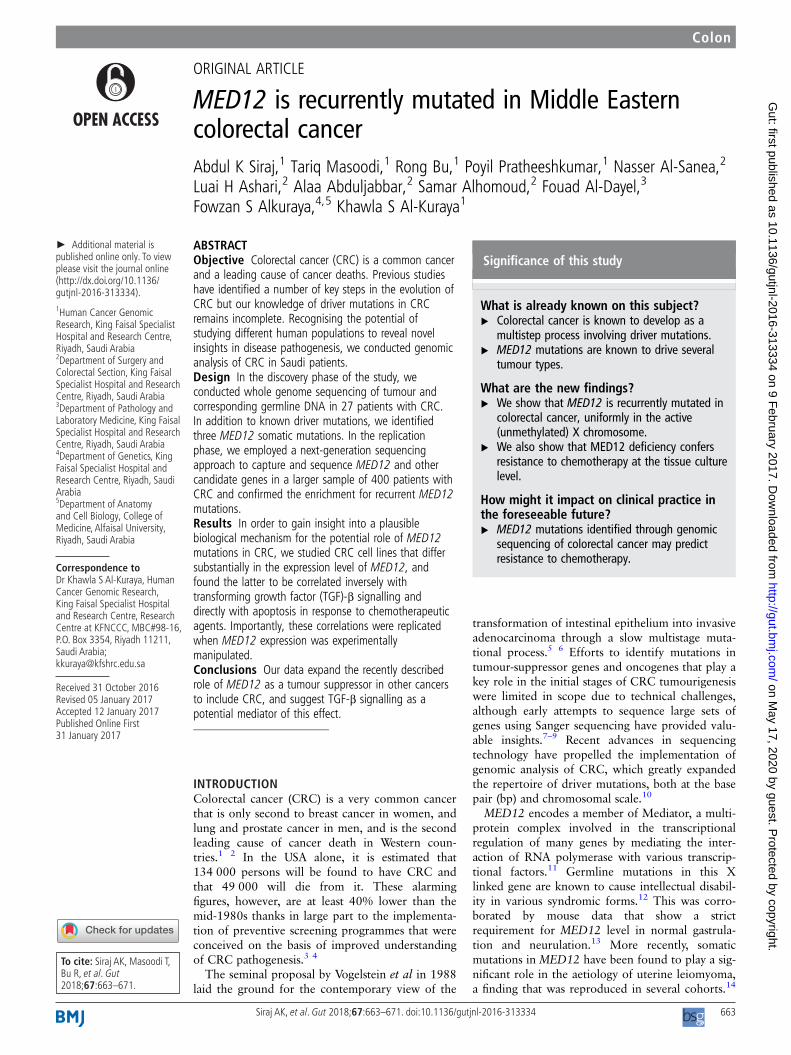
ORIGINAL ARTICLE
MED12 is recurrently mutated in Middle Easterncolorectal cancerAbdul K Siraj,1 Tariq Masoodi,1 Rong Bu,1 Poyil Pratheeshkumar,1 Nasser Al-Sanea,2
Luai H Ashari,2 Alaa Abduljabbar,2 Samar Alhomoud,2 Fouad Al-Dayel,3
Fowzan S Alkuraya,4,5 Khawla S Al-Kuraya1
ABSTRACTObjective Colorectal cancer (CRC) is a common cancerand a leading cause of cancer deaths. Previous studieshave identified a number of key steps in the evolution ofCRC but our knowledge of driver mutations in CRCremains incomplete. Recognising the potential ofstudying different human populations to reveal novelinsights in disease pathogenesis, we conducted genomicanalysis of CRC in Saudi patients.Design In the discovery phase of the study, weconducted whole genome sequencing of tumour andcorresponding germline DNA in 27 patients with CRC.In addition to known driver mutations, we identifiedthree MED12 somatic mutations. In the replicationphase, we employed a next-generation sequencingapproach to capture and sequence MED12 and othercandidate genes in a larger sample of 400 patients withCRC and confirmed the enrichment for recurrent MED12mutations.Results In order to gain insight into a plausiblebiological mechanism for the potential role of MED12mutations in CRC, we studied CRC cell lines that differsubstantially in the expression level of MED12, andfound the latter to be correlated inversely withtransforming growth factor (TGF)-β signalling anddirectly with apoptosis in response to chemotherapeuticagents. Importantly, these correlations were replicatedwhen MED12 expression was experimentallymanipulated.Conclusions Our data expand the recently describedrole of MED12 as a tumour suppressor in other cancersto include CRC, and suggest TGF-β signalling as apotential mediator of this effect.
INTRODUCTIONColorectal cancer (CRC) is a very common cancerthat is only second to breast cancer in women, andlung and prostate cancer in men, and is the secondleading cause of cancer death in Western coun-tries.1 2 In the USA alone, it is estimated that134 000 persons will be found to have CRC andthat 49 000 will die from it. These alarmingfigures, however, are at least 40% lower than themid-1980s thanks in large part to the implementa-tion of preventive screening programmes that wereconceived on the basis of improved understandingof CRC pathogenesis.3 4
The seminal proposal by Vogelstein et al in 1988laid the ground for the contemporary view of the
transformation of intestinal epithelium into invasiveadenocarcinoma through a slow multistage muta-tional process.5 6 Efforts to identify mutations intumour-suppressor genes and oncogenes that play akey role in the initial stages of CRC tumourigenesiswere limited in scope due to technical challenges,although early attempts to sequence large sets ofgenes using Sanger sequencing have provided valu-able insights.7–9 Recent advances in sequencingtechnology have propelled the implementation ofgenomic analysis of CRC, which greatly expandedthe repertoire of driver mutations, both at the basepair (bp) and chromosomal scale.10
MED12 encodes a member of Mediator, a multi-protein complex involved in the transcriptionalregulation of many genes by mediating the inter-action of RNA polymerase with various transcrip-tional factors.11 Germline mutations in this Xlinked gene are known to cause intellectual disabil-ity in various syndromic forms.12 This was corro-borated by mouse data that show a strictrequirement for MED12 level in normal gastrula-tion and neurulation.13 More recently, somaticmutations in MED12 have been found to play a sig-nificant role in the aetiology of uterine leiomyoma,a finding that was reproduced in several cohorts.14
Significance of this study
What is already known on this subject?▸ Colorectal cancer is known to develop as a
multistep process involving driver mutations.▸ MED12 mutations are known to drive several
tumour types.
What are the new findings?▸ We show that MED12 is recurrently mutated in
colorectal cancer, uniformly in the active(unmethylated) X chromosome.
▸ We also show that MED12 deficiency confersresistance to chemotherapy at the tissue culturelevel.
How might it impact on clinical practice inthe foreseeable future?▸ MED12 mutations identified through genomic
sequencing of colorectal cancer may predictresistance to chemotherapy.
663Siraj AK, et al. Gut 2018;67:663–671. doi:10.1136/gutjnl-2016-313334
Colon
To cite: Siraj AK, Masoodi T, Bu R, et al. Gut 2018;67:663–671.
► Additional material is published online only. To view please visit the journal online (http:// dx. doi. org/ 10. 1136/ gutjnl- 2016- 313334).1Human Cancer Genomic Research, King Faisal Specialist Hospital and Research Centre, Riyadh, Saudi Arabia2Department of Surgery and Colorectal Section, King Faisal Specialist Hospital and Research Centre, Riyadh, Saudi Arabia3Department of Pathology and Laboratory Medicine, King Faisal Specialist Hospital and Research Centre, Riyadh, Saudi Arabia4Department of Genetics, King Faisal Specialist Hospital and Research Centre, Riyadh, Saudi Arabia5Department of Anatomy and Cell Biology, College of Medicine, Alfaisal University, Riyadh, Saudi Arabia
Correspondence toDr Khawla S Al-Kuraya, Human Cancer Genomic Research, King Faisal Specialist Hospital and Research Centre, Research Centre at KFNCCC, MBC#98-16, P.O. Box 3354, Riyadh 11211, Saudi Arabia; kkuraya@ kfshrc. edu. sa
Received 31 October 2016Revised 05 January 2017Accepted 12 January 2017Published Online First 31 January 2017
on May 17, 2020 by guest. P
rotected by copyright.http://gut.bm
j.com/
Gut: first published as 10.1136/gutjnl-2016-313334 on 9 F
ebruary 2017. Dow
nloaded from

Subsequently, driver MED12 mutations were identified in othertumours including adrenocortical carcinoma, prostate cancerand breast fibroadenoma.15–17 Interestingly, the distribution ofMED12 mutations differs in different types of tumour. Inuterine leiomyoma, most of MED12 mutations were identifiedin exon 2 and lead to the activation of WNT pathway genesthrough interaction with β-catenin, while in prostate cancerMED12 mutations were mainly detected in the non-N-terminalregion and seem to influence p53 and androgen signallingpathway.16 18–21
In this study, we implemented a hypothesis-free genomicsapproach in our search for relevant somatic mutations in CRCin Saudi Arabia, a country with a particularly high incidence ofCRC with an unusually young age of onset.22 This allowed usto identify recurrent somatic MED12 mutations suggesting apotential role in CRC pathogenesis. Furthermore, functionalanalysis of relevant cell lines supports a previously reported roleof MED12 deficiency in mediating drug resistance.23
MATERIALS AND METHODSHuman subjectsA total of 427 patient samples diagnosed with CRC at the KingFaisal Specialist Hospital and Research Centre were collectedfrom Department of Pathology. Detailed clinicopathologicaldata were noted from case records and summarised in table 1.All tissue and blood samples were obtained from patients withapproval from Institutional Review Board of the hospital.Informed consent was obtained from the patients from whomfresh cancer tissues were harvested for the study. For the study,waiver of consent was obtained for archived paraffin tissueblocks from Research Advisory Council (RAC) under projectRAC#2140 005.
DNA isolationDNA isolation from peripheral blood, fresh frozen andparaffin-embedded tissues was performed using Gentra DNAisolation kit (Gentra, Minneapolis, Minnesota, USA) followingthe manufacturer’s recommendations.
Whole genome sequencingWhole genome sequencing was performed on 27 patients withtheir corresponding normal blood. Paired-end libraries ofapproximately 500-bp inserts were sequenced as 150-bppaired-end reads. Raw sequencing data were processed by anultrafast Isaac DNA sequence aligner designed to align next-generation sequencing data with low-error rates using humangenome V.19. The Isaac Variant Caller was used to call single-nucleotide variants (SNVs) and small indels from the bamfiles.24 SnpEff was used for annotating the variants withdbSNP138, dbSNP142, 1000 Genomes project and ESP6500database.25 We excluded common single-nucleotide polymorph-isms (SNPs) with minor allele frequency of >0.001 as recordedin either dbSNP, National Heart, Lung, and Blood Instituteexome sequencing project, 1000 Genomes and our in-housedata from exome sequencing of over 730 normal samples.Non-coding variants, synonymous variants and variants presentin highly repetitive regions were excluded for further analysis.
Candidate gene capture and sequencingRecurrently mutated genes (mutated in at least three samples)from the 27 whole genome cases were selected for capturesequencing using SureSelect Target Enrichment Kit on IlluminaHiSeq 2500 Sequencer. Fastq files were aligned to the humanreference genome hg19 using burrows-wheeler aligner (BWA).26
Local realignment, PCR duplicates and base quality recalibrationwas performed using genome analysis toolkit (GATK) andPicard-tools.27 SNVs and small indels were called using GATK.The identified variants were annotated using SnpEff. Similarfilters as applied to whole genome samples were used to getfinal mutation list.
PCR and Sanger sequencingPrimer 3 software was used to design the primers for the par-ticular variants identified by whole exome or capture sequen-cing. PCR and Sanger sequencing were performed as describedpreviously.28 Reference sequences were downloaded fromGenBank. Sequencing traces were analysed using the MutationSurveyor V.4.04 (Soft Genetics, State College, Pennsylvania,USA).
Allele-specific mutation analysisGenomic DNA samples were digested using restriction endo-nuclease Mse I for 2 hours and then MethylCollector Ultra kit(Active Motif, Carlsbad, California, USA) was used to collectthe pure/enriched methylated DNA and the counterpartunmethylated/methylated DNA following the manufacturer’srecommendations as described previously.29 PCR and Sangersequencing were then performed to examine which allele of Xchromosome has the identified mutation in female mutation-positive cases.
Tissue culture experimentsAll human CRC cell lines; CL34, Caco2, Colo-320, DLD1,HCT15, HCT116, HT29 and LOVO were obtained fromAmerican Type Culture Collection and cultured in Roswell ParkMemorial Institute 1640 media supplemented with 10% fetal
Table 1 Clinicopathological variables for the patient cohort(n=427)
AgeMedian 57.0Range (IQR) 47.0–68.0
GenderMale 225 (52.7)Female 202 (47.3)StatusAlive 328 (76.8)Dead 96 (22.5)Unknown 3 (0.7)Histological gradeWell differentiated 42 (9.8)Moderately differentiated 324 (75.9)Poorly differentiated 51 (11.9)Unknown 10 (2.3)Tumour siteLeft 345 (80.8)Right 77 (18.0)Unknown 5 (1.2)Tumour, node, metastases stageI 50 (11.7)II 149 (34.9)III 161 (37.7)IV 49 (11.5)Unknown 18 (4.2)
664 Siraj AK, et al. Gut 2018;67:663–671. doi:10.1136/gutjnl-2016-313334
Colon on M
ay 17, 2020 by guest. Protected by copyright.
http://gut.bmj.com
/G
ut: first published as 10.1136/gutjnl-2016-313334 on 9 February 2017. D
ownloaded from
bovine serum (FBS), 100 U/mL penicillin, 100 U/mL strepto-mycin at 37°C in a humidified atmosphere containing 5% CO2.All treatment experiments were performed in reduced FBS con-dition (5%). Apoptosis analysis was performed using annexin V/propidium iodide dual staining and measured by flow cytometryas previously described.30 Antibodies against MED12(CST#14360), E-cadherin (CST#3195), N-cadherin(CST#14215), pERK1/2 (CST#4370), ERK1/2 (CST#4695)and β-actin (CST#3700) were purchased from Cell SignalingTechnology (Danvers, Massachusetts, USA). Antibodies againsttransforming growth factor (TGF)-β-R2 (sc#17799), vimentin(sc#5565) and GAPDH (sc#25778) were purchased from SantaCruz Biotechnology (Santa Cruz, California, USA). Twist anti-body (ab#175430) was purchased from Abcam (Cambridge,Massachusetts, USA).
3-(4,5-Dimethylthiazol-2-yl)-2,5-diphenyltetrazoliumbromide assaysInitially, CRC cells were incubated at the concentration of 104
cells per well in triplicates in a 96-well format. Cells were thentreated with various doses of 5-fluorouracil (5-FU) for 48 hoursin a final volume of 0.2 mL. Cell viability was measured by3-(4,5-dimethylthiazol-2-yl)-2,5-diphenyltetrazolium bromidecell viability assay, as previously described;31 6 wells for eachdosage including vehicle control were analysed for eachexperiment.
Gene silencing using small interfering RNATwo different sets of MED12 small interfering RNA (siRNA),and scrambled control siRNA were purchased from OriGene(Rockville, Maryland, USA). Cells were transfected usingLipofectamine 2000 (Invitrogen, Carlsbad, California, USA) for6 hours following which the lipid and siRNA complex wasremoved and fresh growth medium was added. After 48 hoursof transfection cells were used for various experiments such asclonogenic assay, apoptosis analysis and immunoblotting.
Plasmid and transfectionPlasmid DNA encoding human MED12 was purchased fromOriGene. Transfection was performed using Lipofectamine 2000according to the manufacturer’s protocol. Briefly, CRC cellswere seeded in 6-well culture plates; when approximately 50%confluent, cells were transfected with 4 μg plasmid. After48 hours of transfection cells were used for various experimentssuch as clonogenic assay, apoptosis analysis and immunoblotting.
Immunofluorescence analysisCRC cells grown on coverslips in 6-well plates were fixed withice-cold 100% methanol followed by permeabilisation with0.2% Triton X-100, blocked with 5% horse serum inphosphate-buffered saline (PBS) solution, and incubated withantibodies to MED12 (1:200), TGF-β-R2 (1:50) or E-cadherin(1:200) in buffer A (1% bovine serum albumin, 0.1% TritonX-100, 10% horse serum in PBS solution) for 1 hour at 37°C.Cells were then incubated with Alexa Fluor 488 goat antirabbitor Alexa Fluor 594 goat antimouse antibody and mounted usingDAPI. The cells were visualised using Olympus BX63 fluores-cence microscope.
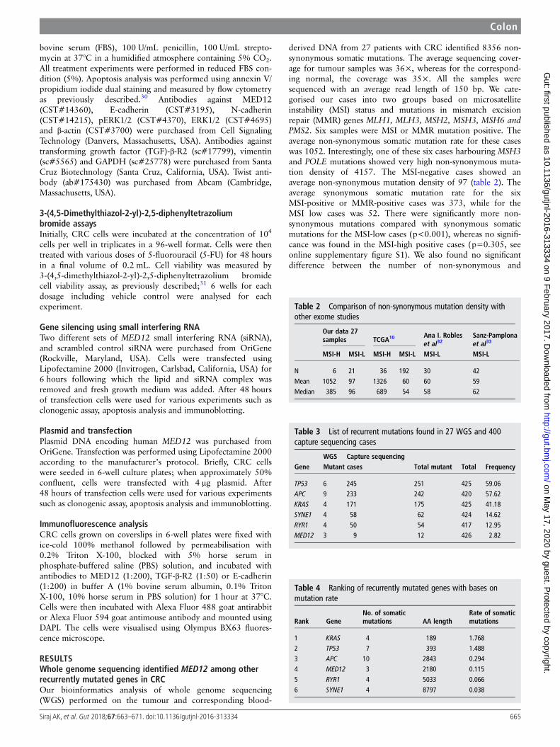
RESULTSWhole genome sequencing identified MED12 among otherrecurrently mutated genes in CRCOur bioinformatics analysis of whole genome sequencing(WGS) performed on the tumour and corresponding blood-
derived DNA from 27 patients with CRC identified 8356 non-synonymous somatic mutations. The average sequencing cover-age for tumour samples was 36×, whereas for the correspond-ing normal, the coverage was 35×. All the samples weresequenced with an average read length of 150 bp. We cate-gorised our cases into two groups based on microsatelliteinstability (MSI) status and mutations in mismatch excisionrepair (MMR) genes MLH1, MLH3, MSH2, MSH3, MSH6 andPMS2. Six samples were MSI or MMR mutation positive. Theaverage non-synonymous somatic mutation rate for these caseswas 1052. Interestingly, one of these six cases harbouring MSH3and POLE mutations showed very high non-synonymous muta-tion density of 4157. The MSI-negative cases showed anaverage non-synonymous mutation density of 97 (table 2). Theaverage synonymous somatic mutation rate for the sixMSI-positive or MMR-positive cases was 373, while for theMSI low cases was 52. There were significantly more non-synonymous mutations compared with synonymous somaticmutations for the MSI-low cases (p<0.001), whereas no signifi-cance was found in the MSI-high positive cases (p=0.305, seeonline supplementary figure S1). We also found no significantdifference between the number of non-synonymous and
Table 2 Comparison of non-synonymous mutation density withother exome studies
Our data 27samples TCGA10 Ana I. Robles
et al32Sanz-Pamplonaet al33
MSI-H MSI-L MSI-H MSI-L MSI-L MSI-L
N 6 21 36 192 30 42Mean 1052 97 1326 60 60 59Median 385 96 689 54 58 62
Table 3 List of recurrent mutations found in 27 WGS and 400capture sequencing cases
WGS Capture sequencingGene Mutant cases Total mutant Total Frequency
TP53 6 245 251 425 59.06APC 9 233 242 420 57.62KRAS 4 171 175 425 41.18SYNE1 4 58 62 424 14.62RYR1 4 50 54 417 12.95MED12 3 9 12 426 2.82
Table 4 Ranking of recurrently mutated genes with bases onmutation rate
Rank GeneNo. of somaticmutations AA length
Rate of somaticmutations
1 KRAS 4 189 1.7682 TP53 7 393 1.4883 APC 10 2843 0.294
4 MED12 3 2180 0.1155 RYR1 4 5033 0.0666 SYNE1 4 8797 0.038
665Siraj AK, et al. Gut 2018;67:663–671. doi:10.1136/gutjnl-2016-313334
Colon on M
ay 17, 2020 by guest. Protected by copyright.
http://gut.bmj.com
/G
ut: first published as 10.1136/gutjnl-2016-313334 on 9 February 2017. D
ownloaded from

synonymous mutations per sample and patient age in both thegroups (p=0.5922).
In this discovery cohort, we prioritised genes that are somatic-ally mutated in at least three patients as likely candidate ‘driver’genes (see online supplementary table S1). Reassuringly, the listincludes the well-established CRC driver genes APC, KRAS andTP53 (table 3). In addition, the list also includes the followingcandidate genes: RYR1, SYNE1 and MED12. Other well-knowndriver genes of CRC like BRAF and SMAD4 were not found inour initial small cohort of 27 samples, however, we have foundPIK3CA mutated in just 1 case. MED12 was particularly interest-ing given its recently established role as a driver gene in anumber of tumours and largely uncharacterised role inCRC.14–17 Furthermore, when recurrently mutated genes in ourdiscovery cohort were ranked based on the relative mutationfrequency by taking the protein size into account,34 35
MED12 ranked fourth to APC, KRAS and TP53 (table 4). Thefollowing likely pathogenic somatic variants were identified inMED12: c.110C>T;p.Thr37Met, c.130G>A;p.Gly44Ser andc.6369_6380dupCCAGCAGCAACA; p.His2123_Gln2126dup(table 5).
To further investigate a potential role of MED12 in CRC, wesequenced MED12 in a replication cohort of 400 patients withCRC using in-solution capture and next-generation sequencing.One sample was not sequenced due to quality control (QC)failure. Using a stringent filtering pipeline, we identified 15likely pathogenic variants in MED12, 11 of which were con-firmed by Sanger sequencing to be true somatic mutations in 9cases (table 5; figure 1 and see online supplementary figure S2).Most of these mutations were pathogenic as predicted by threedifferent computational algorithms.
Therefore, by combining the discovery and replicationcohort, the percentage of patients with somatic MED12 muta-tions is 2.8%, a small but comparable percentage to some of theknown driver genes in CRC, for example, BRAF, PTEN, NRAS,SMAD4 and MUTYH.10 36–38 We found a total of 14 MED12gene mutations in 12 CRC cases in this cohort. Out of 14 muta-tions, 11 were missense, 2 duplication and 1 deletion. Threemutations were seen in exon 1, two mutations were seen inexon 13, 37, 43 each and one mutation was found in exon 9,18, 27, 39 and 42 each. The concurrent presence of nearly allof these MED12 somatic mutations with mutations in well-
Table 5 MED12 mutations with three different pathogenicity scores
ID Chr POS REF ALT Mutation HGVS.c HGVS.p PolyPhen SIFT CADD
112-084 Chr X 70338672 A G Missense c.68A>G p.Asp23Gly 0.904 0 29300-013 Chr X 70342408 G T Missense c.1299G>T p.Gln433His 0.994 0.006 19.6300-074 Chr X 70344018 G A Missense c.1754G>A p.Arg585Gln 0.027 0.585 23300-298 Chr X 70345918 C T Missense c.2455C>T p.Arg819Trp 0.994 0.001 32300-300 Chr X 70349536 C T Missense c.3698C>T p.Ala1233Val 0.982 0.052 25.9300-074 Chr X 70349689 G T Missense c.3851G>T p.Arg1284Leu 0.582 0.005 25.1300-090 Chr X 70356162 C T Missense c.5057C>T p.Ser1686Leu 1 0.022 33112-084 Chr X 70356420 C A Missense c.5315C>A p.Pro1772Gln 0.228 0.05 23.9300-004 Chr X 70357181 G A Missense c.5696G>A p.Arg1899Gln 0.96 0.308 23.1300-211 Chr X 70344030 A T Missense c.1766A>T p.Glu589Val 0.996 0.005 29.5197-019 Chr X 70360698 G GATC Insertion c.6268_6270dupATC p.Ile2090dup NA NA 14.6300-004 Chr X 70361090 AACAGCA A Deletion c.6294_6299delACAGCA p.Gln2098_Gln2099del NA NA 17.1CRC-161 Chr X 70339253 G A Missense c.130G>A p.Gly44Ser 1 0 27.3CRC-181 Chr X 70339233 C T Missense c.110C>T p.Thr37Met 1 0.001 20.5CRC-302 Chr X 70361159 A ACCAGCAGCAACA Insertion c.6369_6380dupCCAGCAGCAACA p.His2123_Gln2126dup NA NA 0.8
Most of the mutations were predicted to be pathogenic by at least two prediction algorithms.A SIFT score of ≤0.05 indicates the amino acid substitution is pathogenic (damaging), whereas a score of ≥0.05 is predicted to be tolerant. PolyPhen predicts results of nsSNPs aspossibly damaging and probably damaging (PSIC >0.5) or benign (PSIC <0.5). According to CADD classification, variants with a C-score of 10 or greater (≥C10) are probable functionalvariants, variants with a C-score of 20 or greater (≥C20) are most deleterious and variants with a C-score of 30 or greater (≥C30) are lethal. Pathogenic mutation scores arehighlighted in bold.ALT, altered; CADD, combined annotation dependent depletion; Chr, chromosome; CRC, colorectal cancer; NA, not applicable; PSIC, position-specific independent counts; POS, position;REF, reference; SIFT, sorting intolerant from tolerant.
Figure 1 Schematic diagram of distribution of 14 MED12 mutations identified in 12 colorectal cancer cases.
666 Siraj AK, et al. Gut 2018;67:663–671. doi:10.1136/gutjnl-2016-313334
Colon on M
ay 17, 2020 by guest. Protected by copyright.
http://gut.bmj.com
/G
ut: first published as 10.1136/gutjnl-2016-313334 on 9 February 2017. D
ownloaded from

established driver genes suggests a potential role in tumour evo-lution past the initiation stage.28 Unexpected for an X linkedgene, only 33% (4/12) patients with CRC with somatic MED12mutations were males. This prompted us to investigate whetherMED12-mutated females harboured their somatic mutations onthe active allele X chromosome, essentially mimicking the effectof hemizygous male mutations. Indeed, in all eight females thepresence of somatic MED12 mutation on the active allele of thechromosome X rather than inactive imprinted allele wasconfirmed (figure 2). Unfortunately, the relatively smallnumber of MED12-mutated patients precluded formal testingfor such associations as disease severity, response tochemotherapy and survival. However, it is worth noting that50% of MED12-mutated patients with CRC who underwent
chemotherapy (4/8) showed poor response to standardchemotherapy.
MED12 is a likely tumour suppressor that modulatesTGF-β-induced epithelial-mesenchymal transition signallingin CRC cell linesIn order to gain insight into the biological plausibility ofMED12 mutations in the pathogenesis of CRC, which would besuggested by our finding above, we set out to test its potentialrole in relevant CRC cell lines. To determine the role ofMED12 expression in vitro, we assessed expression of MED12in a panel of eight CRC cell lines by immunoblotting. As shownin figure 3A, there was differential expression of MED12 inthese cell lines. On the basis of MED12 expression, we
Figure 2 Sanger sequencing electropherogram of inactive/active allele of MED12 gene on chromosome X in eight female cases harbouring MED12mutations. Top panel: sequencing electropherogram of enriched methylated DNA indicating no mutation was identified on the inactive allele causedby methylation. Lower panel: sequencing electropherogram of enriched unmethylated and unbounded methylated DNA. WT, wild type.
667Siraj AK, et al. Gut 2018;67:663–671. doi:10.1136/gutjnl-2016-313334
Colon on M
ay 17, 2020 by guest. Protected by copyright.
http://gut.bmj.com
/G
ut: first published as 10.1136/gutjnl-2016-313334 on 9 February 2017. D
ownloaded from
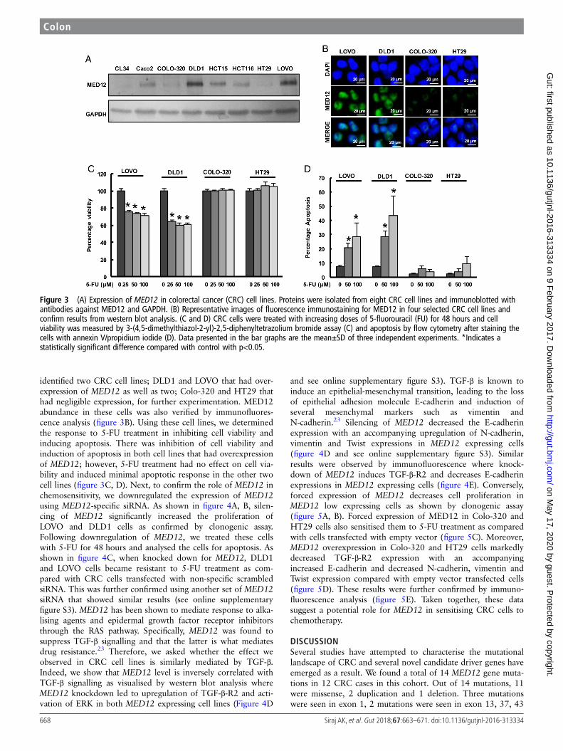
identified two CRC cell lines; DLD1 and LOVO that had over-expression of MED12 as well as two; Colo-320 and HT29 thathad negligible expression, for further experimentation. MED12abundance in these cells was also verified by immunofluores-cence analysis (figure 3B). Using these cell lines, we determinedthe response to 5-FU treatment in inhibiting cell viability andinducing apoptosis. There was inhibition of cell viability andinduction of apoptosis in both cell lines that had overexpressionof MED12; however, 5-FU treatment had no effect on cell via-bility and induced minimal apoptotic response in the other twocell lines (figure 3C, D). Next, to confirm the role of MED12 inchemosensitivity, we downregulated the expression of MED12using MED12-specific siRNA. As shown in figure 4A, B, silen-cing of MED12 significantly increased the proliferation ofLOVO and DLD1 cells as confirmed by clonogenic assay.Following downregulation of MED12, we treated these cellswith 5-FU for 48 hours and analysed the cells for apoptosis. Asshown in figure 4C, when knocked down for MED12, DLD1and LOVO cells became resistant to 5-FU treatment as com-pared with CRC cells transfected with non-specific scrambledsiRNA. This was further confirmed using another set of MED12siRNA that showed similar results (see online supplementaryfigure S3). MED12 has been shown to mediate response to alka-lising agents and epidermal growth factor receptor inhibitorsthrough the RAS pathway. Specifically, MED12 was found tosuppress TGF-β signalling and that the latter is what mediatesdrug resistance.23 Therefore, we asked whether the effect weobserved in CRC cell lines is similarly mediated by TGF-β.Indeed, we show that MED12 level is inversely correlated withTGF-β signalling as visualised by western blot analysis whereMED12 knockdown led to upregulation of TGF-β-R2 and acti-vation of ERK in both MED12 expressing cell lines (Figure 4D
and see online supplementary figure S3). TGF-β is known toinduce an epithelial-mesenchymal transition, leading to the lossof epithelial adhesion molecule E-cadherin and induction ofseveral mesenchymal markers such as vimentin andN-cadherin.23 Silencing of MED12 decreased the E-cadherinexpression with an accompanying upregulation of N-cadherin,vimentin and Twist expressions in MED12 expressing cells(figure 4D and see online supplementary figure S3). Similarresults were observed by immunofluorescence where knock-down of MED12 induces TGF-β-R2 and decreases E-cadherinexpressions in MED12 expressing cells (figure 4E). Conversely,forced expression of MED12 decreases cell proliferation inMED12 low expressing cells as shown by clonogenic assay(figure 5A, B). Forced expression of MED12 in Colo-320 andHT29 cells also sensitised them to 5-FU treatment as comparedwith cells transfected with empty vector (figure 5C). Moreover,MED12 overexpression in Colo-320 and HT29 cells markedlydecreased TGF-β-R2 expression with an accompanyingincreased E-cadherin and decreased N-cadherin, vimentin andTwist expression compared with empty vector transfected cells(figure 5D). These results were further confirmed by immuno-fluorescence analysis (figure 5E). Taken together, these datasuggest a potential role for MED12 in sensitising CRC cells tochemotherapy.
DISCUSSIONSeveral studies have attempted to characterise the mutationallandscape of CRC and several novel candidate driver genes haveemerged as a result. We found a total of 14 MED12 gene muta-tions in 12 CRC cases in this cohort. Out of 14 mutations, 11were missense, 2 duplication and 1 deletion. Three mutationswere seen in exon 1, 2 mutations were seen in exon 13, 37, 43
Figure 3 (A) Expression of MED12 in colorectal cancer (CRC) cell lines. Proteins were isolated from eight CRC cell lines and immunoblotted withantibodies against MED12 and GAPDH. (B) Representative images of fluorescence immunostaining for MED12 in four selected CRC cell lines andconfirm results from western blot analysis. (C and D) CRC cells were treated with increasing doses of 5-fluorouracil (FU) for 48 hours and cellviability was measured by 3-(4,5-dimethylthiazol-2-yl)-2,5-diphenyltetrazolium bromide assay (C) and apoptosis by flow cytometry after staining thecells with annexin V/propidium iodide (D). Data presented in the bar graphs are the mean±SD of three independent experiments. *Indicates astatistically significant difference compared with control with p<0.05.
668 Siraj AK, et al. Gut 2018;67:663–671. doi:10.1136/gutjnl-2016-313334
Colon on M
ay 17, 2020 by guest. Protected by copyright.
http://gut.bmj.com
/G
ut: first published as 10.1136/gutjnl-2016-313334 on 9 February 2017. D
ownloaded from

each and 1 mutation was found in exon 9, 18, 27, 39 and 42each. The Cancer Genome Atlas Network in their analysis on224 CRCs found 15 MED12 mutations in 14 CRC cases.10 Intheir study they found, out of 15 mutations, 10 were missense,4 were silent and 1 mutation was in-frame deletion. Mutationwere seen in exon 36 (2 mutations), exon 42 (2 mutations) and1 mutation each in exon 1, 2, 5, 7, 10, 18, 23, 29, 35, 40 and43. Mutation in exon 2 of MED12 gene is the most commongenetic aberration present in roughly 64% of leiomyomas.39
Although very rare, MED12 mutations in leiomyoma have alsobeen reported in other regions of gene such as intron 140 andexon 1.41 In leiomyoma, codon 44 is the most common hotspotfor MED12 mutation and both missense and in-frame insertion-deletion mutations are seen.42
The large study by the Cancer Genome Atlas Network didnot highlight MED12 as a likely candidate even though 15somatic mutations were identified, most likely because of thesmall percentage of affected samples.10 Indeed, Kämpjärviet al43 specifically sequenced exon 2 MED12 in 392 patientswith cancer and identified only two patients with somaticmutations. This is consistent with our data where we show thatonly 2.8% of patients with CRC have somatic MED12mutations.
Genes in the RAS pathway are known to contribute to thepathogenesis of CRC, most notably KRAS and BRAF in whichsomatic mutations are observed in 30%–40% and 10%, respect-ively, of patients with CRC.44 45 These somatic mutations aretypically activating in nature and are associated with the activa-tion of the Erk1/2 kinase pathway.46 Thus, our finding thatMED12 deficiency is also associated with the activation of theErk1/2 kinase pathway would be consistent with the proposedoncogenic potential of MED12 somatic mutations in CRC.Although the numbers are small, we observed a trend whereMED12-mutated patients with CRC have high likelihood of dis-playing resistance to alkalising chemotherapeutic agents. Thisappears to be consistent with our finding in CRC cell lineswhere we show that MED12 mediates sensitivity to 5-FU, likelyvia suppressing TGF-β signalling, a finding that was alsoobtained by Huang et al23 using different cell lines.
The location of MED12 on the X chromosome provides aunique advantage, where biallelic inactivation can be acquired asa one-step rather than the traditional two-step loss of heterozy-gosity (LOH) process.47 In other words, because females arefunctionally hemizygous for MED12, somatic mutations in theactive X chromosome can result in complete inactivation of thegene. This has been shown in MED12-related breast
Figure 4 Silencing of MED12 increases the chemoresistance in colorectal cancer (CRC) cell lines. (A and B) CRC cells were transfected with eitherscrambled siRNA or MED12-specific small interfering RNA (siRNA) for 48 hours and clonogenic assay were performed. Cells (6×10²) afterpost-transfection were seeded into each of three dishes (60 mm diameter), and grown for an additional 10 days, then stained with crystal violet (A).Colony numbers in the entire dish were counted (B). (C) CRC cells were transfected with either scrambled siRNA or MED12 siRNA and subsequentlytreated with 50 and 100 μM 5-fluorouracil (FU) for 48 hours. Following treatment, cells were analysed for apoptosis by flow cytometry. (D) LOVOand DLD1 cells were transfected with either scrambled siRNA or MED12-specific siRNA for 48 hours. Proteins were isolated and immunoblotted withantibodies against MED12, transforming growth factor (TGF)-β-R2, p-ERK1/2, ERK1/2, E-cadherin, N-cadherin, vimentin, Twist and β-actin for equalloading. (E) Representative images of fluorescence immunostaining for MED12, TGF-β-R2 and E-cadherin in LOVO and DLD1 cells afterpost-transfection with MED12 siRNA. Data presented in the bar graphs are the mean±SD of three independent experiments. *Indicates a statisticallysignificant difference compared with control with p<0.05.
669Siraj AK, et al. Gut 2018;67:663–671. doi:10.1136/gutjnl-2016-313334
Colon on M
ay 17, 2020 by guest. Protected by copyright.
http://gut.bmj.com
/G
ut: first published as 10.1136/gutjnl-2016-313334 on 9 February 2017. D
ownloaded from

fibroadenoma and leiomyoma.17 48 Our finding that all femalepatients with CRC with somatic MED12 mutations harbouredthe mutations on their active X chromosome serves as an add-itional line of evidence in support of these mutations playing arole in driving CRC tumourigenesis. The ‘single-hit’ require-ment for inactivating MED12 in females, likely explains the lackof male predominance as expected for an X linked gene andrepresents a hypothetical opportunity for treatment throughreactivating (demethylating) the normal X chromosome in thesepatients to restore normal level of MED12. We will pursue thisas a future research direction.
In conclusion, we show that MED12 is recurrently mutated inSaudi patients with CRC and propose this corroborates previ-ously proposed tumour suppresser role. Our data also suggestthat such a role may be mediated through antagonising TGF-βsignalling.
Acknowledgements We thank Wael Haqawi, Zeeshan Qadri, Dionne Rae Rala,Ingrid Francesca Victoria, Maha Al Rasheed, Khadija Al Obaisi, Roxanne
Melosantos, Rafia Begum, Sarah Siraj and Saravanan Thangavel for technicalassistance.
Contributors We have added as a coauthor a team member who helped generatethe additional data in functional analyses (PP).
Competing interests None declared.
Patient consent Obtained.
Ethics approval Research ethics committee, King Faisal Specialist Hospital andResearch Centre.
Provenance and peer review Not commissioned; externally peer reviewed.
Open Access This is an Open Access article distributed in accordance with theCreative Commons Attribution Non Commercial (CC BY-NC 4.0) license, whichpermits others to distribute, remix, adapt, build upon this work non-commercially,and license their derivative works on different terms, provided the original work isproperly cited and the use is non-commercial. See: http://creativecommons.org/licenses/by-nc/4.0/
REFERENCES1 Boyle P, Ferlay J. Cancer incidence and mortality in Europe, 2004. Ann Oncol
2005;16:481–8.
Figure 5 Forced expression of MED12 decreases the chemoresistance in colorectal cancer (CRC) cell lines. (A and B) CRC cells were transfectedwith either empty vector or MED12 pcDNA for 48 hours and clonogenic assay were performed. Cells (6×10²) after post-transfection were seededinto each of three dishes (60 mm diameter), and grown for an additional 10 days, then stained with crystal violet (A). Colony numbers in the entiredish were counted (B). (C) CRC cells were transfected with either empty vector or MED12 pcDNA and subsequently treated with 50 and 100 μM5-fluorouracil (FU) for 48 hours. Following treatment, cells were analysed for apoptosis by flow cytometry. (D) COLO-320 and HT29 cells weretransfected with either empty vector or MED12 pcDNA for 48 hours. Proteins were isolated and immunoblotted with antibodies against MED12,transforming growth factor (TGF)-β-R2, p-ERK1/2, ERK1/2, E-cadherin, N-cadherin, vimentin, Twist and GAPDH for equal loading. (E) Representativeimages of fluorescence immunostaining for MED12, TGF-β-R2 and E-cadherin in COLO-320 and HT29 cells after post-transfection with MED12pcDNA. Data presented in the bar graphs are the mean±SD of three independent experiments. *Indicates a statistically significant differencecompared with control with p<0.05.
670 Siraj AK, et al. Gut 2018;67:663–671. doi:10.1136/gutjnl-2016-313334
Colon on M
ay 17, 2020 by guest. Protected by copyright.
http://gut.bmj.com
/G
ut: first published as 10.1136/gutjnl-2016-313334 on 9 February 2017. D
ownloaded from
2 Quinn M, Babb P, Brock A, et al. Cancer trends in England and Wales, 1950–1999. Health Stat Q 2000:5–19.
3 Welch HG, Robertson DJ. Colorectal cancer on the decline—why screening can’texplain it All. N Engl J Med 2016;374:1605–7.
4 Holme Ø, Bretthauer M, Fretheim A, et al. Flexible sigmoidoscopy versus faecaloccult blood testing for colorectal cancer screening in asymptomatic individuals.Cochrane Database Syst Rev 2013;(9):CD009259.
5 Vogelstein B, Fearon ER, Hamilton SR, et al. Genetic alterations duringcolorectal-tumor development. N Engl J Med 1988;319:525–32.
6 Fearon ER, Vogelstein B. A genetic model for colorectal tumorigenesis. Cell1990;61:759–67.
7 Wood LD, Parsons DW, Jones S, et al. The genomic landscapes of human breastand colorectal cancers. Science 2007;318:1108–13.
8 Greenman C, Stephens P, Smith R, et al. Patterns of somatic mutation in humancancer genomes. Nature 2007;446:153–8.
9 Sjöblom T, Jones S, Wood LD, et al. The consensus coding sequences of humanbreast and colorectal cancers. Science 2006;314:268–74.
10 Network CGA. Comprehensive molecular characterization of human colon and rectalcancer. Nature 2012;487:330–7.
11 Taatjes DJ. The human Mediator complex: a versatile, genome-wide regulator oftranscription. Trends Biochem Sci 2010;35:315–22.
12 Graham JM, Schwartz CE. MED12 related disorders. Am J Med Genet A2013;161A:2734–40.
13 Rocha PP, Scholze M, Bleiss W, et al. Med12 is essential for early mousedevelopment and for canonical Wnt and Wnt/PCP signaling. Development2010;137:2723–31.
14 Kämpjärvi K, Mäkinen N, Mehine M, et al. MED12 mutations and FH inactivationare mutually exclusive in uterine leiomyomas. Br J Cancer 2016;114:1405–11.
15 Assié G, Letouzé E, Fassnacht M, et al. Integrated genomic characterization ofadrenocortical carcinoma. Nat Genet 2014;46:607–12.
16 Barbieri CE, Baca SC, Lawrence MS, et al. Exome sequencing identifies recurrentSPOP, FOXA1 and MED12 mutations in prostate cancer. Nat Genet 2012;44:685–9.
17 Lim WK, Ong CK, Tan J, et al. Exome sequencing identifies highly recurrent MED12somatic mutations in breast fibroadenoma. Nat Genet 2014;46:877–80.
18 Halder SK, Laknaur A, Miller J, et al. Novel MED12 gene somatic mutations inwomen from the Southern United States with symptomatic uterine fibroids. MolGenet Genomics 2015;290:505–11.
19 Kim S, Xu X, Hecht A, et al. Mediator is a transducer of Wnt/beta-catenin signaling.J Biol Chem 2006;281:14066–75.
20 Wang Q, Sharma D, Ren Y, et al. A coregulatory role for the TRAP-mediatorcomplex in androgen receptor-mediated gene expression. J Biol Chem2002;277:42852–8.
21 Donner AJ, Szostek S, Hoover JM, et al. CDK8 is a stimulus-specific positivecoregulator of p53 target genes. Mol Cell 2007;27:121–33.
22 Beg S, Siraj AK, Prabhakaran S, et al. Molecular markers and pathway analysis ofcolorectal carcinoma in the Middle East. Cancer 2015;121:3799–808.
23 Huang S, Hölzel M, Knijnenburg T, et al. MED12 controls the response to multiplecancer drugs through regulation of TGF-β receptor signaling. Cell2012;151:937–50.
24 Raczy C, Petrovski R, Saunders CT, et al. Isaac: ultra-fast whole-genome secondaryanalysis on Illumina sequencing platforms. Bioinformatics 2013;29:2041–3.
25 Cingolani P, Platts A, Wang Le L, et al. A program for annotating and predictingthe effects of single nucleotide polymorphisms, SnpEff: SNPs in the genome ofDrosophila melanogaster strain w1118; iso-2; iso-3. Fly (Austin) 2012;6:80–92.
26 Li H, Durbin R. Fast and accurate long-read alignment with Burrows–Wheelertransform. Bioinformatics 2010;26:589–95.
27 McKenna A, Hanna M, Banks E, et al. The Genome Analysis Toolkit: a MapReduceframework for analyzing next-generation DNA sequencing data. Genome Res2010;20:1297–303.
28 Siraj AK, Masoodi T, Bu R, et al. Genomic profiling of thyroid cancer reveals a rolefor thyroglobulin in metastasis. Am J Hum Genet 2016;98:1170–80.
29 De Meyer T, Mampaey E, Vlemmix M, et al. Quality evaluation of methyl bindingdomain based kits for enrichment DNA-methylation sequencing. PLoS ONE 2013;8:e59068.
30 Ahmed M, Hussain AR, Bavi P, et al. High prevalence of mTOR complex activity canbe targeted using Torin2 in papillary thyroid carcinoma. Carcinogenesis2014;35:1564–72.
31 Hussain AR, Ahmed SO, Ahmed M, et al. Cross-talk between NFkB and thePI3-kinase/AKT pathway can be targeted in primary effusion lymphoma (PEL) celllines for efficient apoptosis. PLoS ONE 2012;7:e39945.
32 Robles AI, Traverso G, Zhang M, et al. Whole-exome sequencing analyses of inflammatorybowel disease-associated colorectal cancers. Gastroenterology 2016;150:931–43.
33 Sanz-Pamplona R, Lopez-Doriga A, Paré-Brunet L, et al. Exome sequencing revealsAMER1 as a frequently mutated gene in colorectal cancer. Clin Cancer Res2015;21:4709–18.
34 Schell MJ, Yang M, Teer JK, et al. A multigene mutation classification of468 colorectal cancers reveals a prognostic role for APC. Nat Commun2016;7:11743.
35 Lawrence MS, Stojanov P, Polak P, et al. Mutational heterogeneity in cancer andthe search for new cancer-associated genes. Nat 2013;499:214–18.
36 Malapelle U, Pisapia P, Sgariglia R, et al. Less frequently mutated genes incolorectal cancer: evidences from next-generation sequencing of 653 routine cases.J Clin Pathol 2016;69:767–71.
37 Chang YC, Chang JG, Liu TC, et al. Mutation analysis of 13 driver genes ofcolorectal cancer-related pathways in Taiwanese patients. World J Gastroenterol2016;22:2314.
38 Yu J, Wu WK, Li X, et al. Novel recurrently mutated genes and a prognosticmutation signature in colorectal cancer. Gut 2015;64:636–45.
39 Croce S, Chibon F. MED12 and uterine smooth muscle oncogenesis: state of the artand perspectives. Eur J Cancer 2015;51:1603–10.
40 Wang H, Ye J, Qian H, et al. High-resolution melting analysis of MED12 mutationsin uterine leiomyomas in Chinese patients. Genet Test Mol Biomarkers2015;19:162–6.
41 Kämpjärvi K, Park MJ, Mehine M, et al. Mutations in Exon 1 highlight the role ofMED12 in uterine leiomyomas. Hum Mutat 2014;35:1136–41.
42 Heinonen HR, Sarvilinna NS, Sjöberg J, et al. MED12 mutation frequency inunselected sporadic uterine leiomyomas. Fertil Steril 2014;102:1137–42.
43 Kämpjärvi K, Mäkinen N, Kilpivaara O, et al. Somatic MED12 mutations in uterineleiomyosarcoma and colorectal cancer. Br J Cancer 2012;107:1761–5.
44 Phipps AI, Buchanan DD, Makar KW, et al. KRAS-mutation status in relation tocolorectal cancer survival: the joint impact of correlated tumour markers.Br J Cancer 2013;108:1757–64.
45 Barras D. BRAF mutation in colorectal cancer: an update. Biomark Cancer2015;7:9.
46 Wiener Z, Band AM, Kallio P, et al. Oncogenic mutations in intestinal adenomasregulate Bim-mediated apoptosis induced by TGF-β. Proc Natl Acad Sci U S A2014;111:E2229–36.
47 Liu Y, Wang L, Zheng P. X-linked tumor suppressors: perplexing inheritance, aunique therapeutic opportunity. Trends Genet 2010;26:260–5.
48 Mäkinen N, Mehine M, Tolvanen J, et al. MED12, the mediator complex subunit12 gene, is mutated at high frequency in uterine leiomyomas. Science2011;334:252–5.
671Siraj AK, et al. Gut 2018;67:663–671. doi:10.1136/gutjnl-2016-313334
Colon on M
ay 17, 2020 by guest. Protected by copyright.
http://gut.bmj.com
/G
ut: first published as 10.1136/gutjnl-2016-313334 on 9 February 2017. D
ownloaded from


![Man in the Middle [ESTUDIO]](https://static.fdocuments.ec/doc/165x107/5572029f4979599169a3d7b0/man-in-the-middle-estudio.jpg)