Nanoscale detection of organic signatures in carbonate ...complex NEXAFS spectra at the carbon...
6
Nanoscale detection of organic signatures in carbonate microbialites Karim Benzerara †‡§ , Nicolas Menguy † , Purificacio ´ n Lo ´ pez-Garcı ´a ¶ , Tae-Hyun Yoon ‡ , Jo ´ zef Kazmierczak †† , Tolek Tyliszczak ‡‡ , Franc ¸ois Guyot † , and Gordon E. Brown, Jr. ‡§§ † Institut de Mine ´ ralogie et de Physique des Milieux Condense ´ s, Unite ´ Mixte de Recherche 7590, Centre National de la Recherche Scientifique, Department de Mine ´ ralogie, Institut de Physique du Globe de Paris, University of Paris 6 and 7, 140 Rue de Lourmel, 75015 Paris, France; ‡ Surface and Aqueous Geochemistry Group, Department of Geological and Environmental Sciences, Stanford University, Stanford, CA 94305-2115; ¶ Unite ´ d’Ecologie, Syste ´ matique et Evolution, Unite ´ Mixte de Recherche 8079, Centre National de la Recherche Scientifique, Universite ´ Paris-Sud, 91405 Orsay Cedex, France; Department of Chemistry, Hanyang University, Music 17 Haengdang-dong, Seongdong-gu, Seoul, 133-791, Korea; †† Institute of Paleobiology, Polish Academy of Sciences, Twarda 5155, 00818 Warsaw, Poland; ‡‡ Advanced Light Source, Lawrence Berkeley National Laboratory, Berkeley, CA 94720; and §§ Stanford Synchrotron Radiation Laboratory, Stanford Linear Accelerator Center, MS 69, 2575 Sand Hill Road, Menlo Park, CA 94025 Communicated by W. G. Ernst, Stanford University, Stanford, CA, April 21, 2006 (received for review January 12, 2006) Microbialites are sedimentary deposits associated with microbial mat communities and are thought to be evidence of some of the oldest life on Earth. Despite extensive studies of such deposits, little is known about the role of microorganisms in their formation. In addition, unambiguous criteria proving their biogenicity have yet to be established. In this study, we characterize modern calcareous microbialites from the alkaline Lake Van, Turkey, at the nanometer scale by combining x-ray and electron microscopies. We describe a simple way to locate microorganisms entombed in calcium carbonate precipitates by probing aromatic carbon func- tional groups and peptide bonds. Near-edge x-ray absorption fine structure spectra at the C and N K-edges provide unique signatures for microbes. Aragonite crystals, which range in size from 30 to 100 nm, comprise the largest part of the microbialites. These crystals are surrounded by a 10-nm-thick amorphous calcium carbonate layer containing organic molecules and are embedded in an or- ganic matrix, likely consisting of polysaccharides, which helps explain the unusual sizes and shapes of these crystals. These results provide biosignatures for these deposits and suggest that micro- bial organisms significantly impacted the mineralogy of Lake Van carbonates. aragonite biosignature biomineralization spectromicroscopy L ake Van (eastern Anatolia, Turkey) is the largest soda lake on Earth, with a pH of 9.7–9.8 and a salinity of 21.7‰ (1). It harbors the largest known living microbialites, which are structures resulting from precipitation of aragonite at sites where calcium-rich groundwater seeps into the alkaline lake water (1, 2) and are associated with a wide diversity of microorganisms (3). Lake Van microbialites have a fine-grained micritic texture similar to most carbonate microbialites (4, 5) and consist of 30- to 100-nm-sized aragonite crystals (2, 3), which have morphol- ogies that resemble bacteria-like forms (2). Some authors have suggested the possibility that nanospheres in microbialites could represent very small, entombed bacteria or ‘‘nanobacteria’’ (4, 5). However, the real nature of these carbonates, as well as their relationship to the microorganisms detected in the microbialites, remain unresolved. Because Lake Van is highly oversaturated with aragonite (1), the role of microorganisms in aragonite precipitation can be questioned, and the observed presence of microbes in these structures could simply result from passive trapping during mineral precipitation. This question is not new and has been raised systematically since the earliest studies of microbialites to the most recent ones (e.g., refs. 6–9). Some studies have dem- onstrated that microbes can actively mediate carbonate, in particular dolomite, precipitation (10). However, if passive trapping is operative, features suggesting that discrimination between microbially generated and purely abiotic precipitates is possible may be illusory. For example, the biogenicity of ancient stromatolites (i.e., laminated microbialites), usually considered as evidence of some of the oldest life on Earth, has been thoroughly questioned because the macroscopic structure of stromatolites does not constitute a biosignature, and no unam- biguous evidence for fossilized microbes has been found in most Conflict of interest statement: No conflicts declared. Abbreviations: ACC, amorphous calcium carbonate; EELS, electron energy loss spec- troscopy; EPS, extracellular polymeric substances; NEXAFS, near-edge x-ray absorption fine structure; STXM, scanning transmission x-ray microscopy; TEM, transmission elec- tron microscopy. § To whom correspondence should be addressed. E-mail: [email protected]. © 2006 by The National Academy of Sciences of the USA Fig. 1. STXM images of Lake Van microbialites. (a) Image of microbialites at 288.2 eV. Image is 4 m 40 m. (b) Map of peptide-rich areas absorbing at 288.2 eV showing a number of bright spots of various morphologies. Area 1 and area 2 correspond to bright spots that were further analyzed in Figs. 2 and 4, respectively. 9440 –9445 PNAS June 20, 2006 vol. 103 no. 25 www.pnas.orgcgidoi10.1073pnas.0603255103 Downloaded by guest on March 22, 2020
Transcript of Nanoscale detection of organic signatures in carbonate ...complex NEXAFS spectra at the carbon...

Nanoscale detection of organic signaturesin carbonate microbialitesKarim Benzerara†‡§, Nicolas Menguy†, Purificacion Lopez-Garcıa¶, Tae-Hyun Yoon‡�, Jozef Kazmierczak††,Tolek Tyliszczak‡‡, Francois Guyot†, and Gordon E. Brown, Jr.‡§§
†Institut de Mineralogie et de Physique des Milieux Condenses, Unite Mixte de Recherche 7590, Centre National de la Recherche Scientifique, Departmentde Mineralogie, Institut de Physique du Globe de Paris, University of Paris 6 and 7, 140 Rue de Lourmel, 75015 Paris, France; ‡Surface and AqueousGeochemistry Group, Department of Geological and Environmental Sciences, Stanford University, Stanford, CA 94305-2115; ¶Unite d’Ecologie, Systematiqueet Evolution, Unite Mixte de Recherche 8079, Centre National de la Recherche Scientifique, Universite Paris-Sud, 91405 Orsay Cedex, France; �Department ofChemistry, Hanyang University, Music 17 Haengdang-dong, Seongdong-gu, Seoul, 133-791, Korea; ††Institute of Paleobiology, Polish Academy of Sciences,Twarda 51�55, 00818 Warsaw, Poland; ‡‡Advanced Light Source, Lawrence Berkeley National Laboratory, Berkeley, CA 94720; and §§Stanford SynchrotronRadiation Laboratory, Stanford Linear Accelerator Center, MS 69, 2575 Sand Hill Road, Menlo Park, CA 94025
Communicated by W. G. Ernst, Stanford University, Stanford, CA, April 21, 2006 (received for review January 12, 2006)
Microbialites are sedimentary deposits associated with microbialmat communities and are thought to be evidence of some of theoldest life on Earth. Despite extensive studies of such deposits,little is known about the role of microorganisms in their formation.In addition, unambiguous criteria proving their biogenicity haveyet to be established. In this study, we characterize moderncalcareous microbialites from the alkaline Lake Van, Turkey, at thenanometer scale by combining x-ray and electron microscopies. Wedescribe a simple way to locate microorganisms entombed incalcium carbonate precipitates by probing aromatic carbon func-tional groups and peptide bonds. Near-edge x-ray absorption finestructure spectra at the C and N K-edges provide unique signaturesfor microbes. Aragonite crystals, which range in size from 30 to 100nm, comprise the largest part of the microbialites. These crystalsare surrounded by a 10-nm-thick amorphous calcium carbonatelayer containing organic molecules and are embedded in an or-ganic matrix, likely consisting of polysaccharides, which helpsexplain the unusual sizes and shapes of these crystals. These resultsprovide biosignatures for these deposits and suggest that micro-bial organisms significantly impacted the mineralogy of Lake Vancarbonates.
aragonite � biosignature � biomineralization � spectromicroscopy
Lake Van (eastern Anatolia, Turkey) is the largest soda lakeon Earth, with a pH of 9.7–9.8 and a salinity of 21.7‰ (1).
It harbors the largest known living microbialites, which arestructures resulting from precipitation of aragonite at sites wherecalcium-rich groundwater seeps into the alkaline lake water (1,2) and are associated with a wide diversity of microorganisms (3).Lake Van microbialites have a fine-grained micritic texturesimilar to most carbonate microbialites (4, 5) and consist of 30-to 100-nm-sized aragonite crystals (2, 3), which have morphol-ogies that resemble bacteria-like forms (2). Some authors havesuggested the possibility that nanospheres in microbialites couldrepresent very small, entombed bacteria or ‘‘nanobacteria’’ (4,5). However, the real nature of these carbonates, as well as theirrelationship to the microorganisms detected in the microbialites,remain unresolved.
Because Lake Van is highly oversaturated with aragonite (1),the role of microorganisms in aragonite precipitation can bequestioned, and the observed presence of microbes in thesestructures could simply result from passive trapping duringmineral precipitation. This question is not new and has beenraised systematically since the earliest studies of microbialites tothe most recent ones (e.g., refs. 6–9). Some studies have dem-onstrated that microbes can actively mediate carbonate, inparticular dolomite, precipitation (10). However, if passivetrapping is operative, features suggesting that discriminationbetween microbially generated and purely abiotic precipitates ispossible may be illusory. For example, the biogenicity of ancient
stromatolites (i.e., laminated microbialites), usually consideredas evidence of some of the oldest life on Earth, has beenthoroughly questioned because the macroscopic structure ofstromatolites does not constitute a biosignature, and no unam-biguous evidence for fossilized microbes has been found in most
Conflict of interest statement: No conflicts declared.
Abbreviations: ACC, amorphous calcium carbonate; EELS, electron energy loss spec-troscopy; EPS, extracellular polymeric substances; NEXAFS, near-edge x-ray absorptionfine structure; STXM, scanning transmission x-ray microscopy; TEM, transmission elec-tron microscopy.
§To whom correspondence should be addressed. E-mail: [email protected].
© 2006 by The National Academy of Sciences of the USA
Fig. 1. STXM images of Lake Van microbialites. (a) Image of microbialites at288.2 eV. Image is 4 �m � 40 �m. (b) Map of peptide-rich areas absorbing at288.2 eV showing a number of bright spots of various morphologies. Area 1and area 2 correspond to bright spots that were further analyzed in Figs. 2 and4, respectively.
9440–9445 � PNAS � June 20, 2006 � vol. 103 � no. 25 www.pnas.org�cgi�doi�10.1073�pnas.0603255103
Dow
nloa
ded
by g
uest
on
Mar
ch 2
2, 2
020
of them (11, 12). A major problem hampering this type of studyis recognition of microorganisms in a highly mineralized, fine-grained matrix. Optical and scanning electron microscopies arefrequently used for studying microbialites, but they provide onlymorphological and compositional information, which cannot beused to reliably discriminate between fossilized microbes andcalcium carbonate crystals. Lake Van microbialites are associ-ated with a wide diversity of contemporary cyanobacteria andalkaliphilic and�or heterotrophic bacteria (3). Locating thesemicroorganisms by conventional or epif luorescent optical mi-croscopy is difficult, however, because of the high relief ofaragonite crystals relative to microorganisms, the small size ofthe latter, and the difficulty in distinguishing the fluorescencesignal of aragonite crystals from that of stained cells. Toovercome these problems and to explore the association betweenminerals and organic molecules comprising the Lake Van mi-crobialites, we combined scanning transmission x-ray microscopy(STXM) (13), which allows imaging and acquisition of near-edgex-ray absorption fine structure (NEXAFS) spectra at high spec-tral and spatial resolution, and transmission electron microscopy(TEM) (14), on the same samples. Our observations show theintimate association of polysaccharides with the mineral matrixat the nanoscale and strongly suggest that microbial organismsplayed an important role in the formation of the Lake Vanmicrobialites.
ResultsAs described below, STXM provided strong evidence for thepresence of microbial cells in the Van Lake microbialites in theform of organic functional groups typically associated withmicroorganisms. Those areas identified as microorganisms al-ways contained peptide bonds characterized by a specific ab-sorption feature in the carbon K-edge at 288.2 eV. In contrast,the surrounding carbonates displayed a strong absorption peakat 290.2 eV (13). Taking into account these spectroscopicdifferences, energy-filtered imaging was used to map proteins inthe carbonate microbialites (Fig. 1). Bright spots were observed(Fig. 1b) with filamentous or spherical morphologies and diversesizes in the submicrometer to micrometer size range compatiblewith those of individual microbial cells (15) or remnants ofmicrobial cells. Reference microorganisms [the Gram-negativeCaulobacter crescentus (�-Proteobacteria) and the Gram-positive Bacillus subtilis (see Fig. 6, which is published assupporting information on the PNAS web site), but also the�-Proteobacterium Shewanella oneidensis, the cyanobacteriumSynechococcus leopoliensis, or the �-Proteobacterium Ramli-bacter tataouinensis (data not shown)] all displayed identicalcomplex NEXAFS spectra at the carbon K-edge, dominated bythe specific absorption of proteins at 288.2 eV (13, 16) (Fig. 6).The similarity of NEXAFS spectra at the carbon K-edge fordiverse bacteria basically results from the universal basic chem-istry of living cells, all of which contain proteins, polysaccharides,
Fig. 2. Spectromicroscopy analysis of Lake Van microorganisms. (a) STXM image at 288.2 eV of area 1 (see Fig. 1). The arrow indicates a protein-rich area. (b)Equivalent map of objects absorbing at 288.2 eV. (c) TEM image of the same area. The protein-rich area displays a low contrast compared with the surroundingaragonite. (d) C K-edge NEXAFS spectra from the protein-rich area (Lake Van microbe), reference C. crescentus cells, and albumin as a protein model compound.Dashed lines at 285.2, 286.8, and 288.2 eV highlight the prevalent peaks observed in the Lake Van microbe and C. crescentus. The dashed line at 290.2 eV highlightsthe carbonate peak. (e) Close-up of the C K-edge NEXAFS spectra of the protein-rich area in the Lake Van microbialite, Lake Van aragonite, and C. crescentusin the 287.5- to 292-eV energy range. The arrow indicates the position of a shoulder at 289.5 eV.
Benzerara et al. PNAS � June 20, 2006 � vol. 103 � no. 25 � 9441
GEO
LOG
Y
Dow
nloa
ded
by g
uest
on
Mar
ch 2
2, 2
020
and nucleic acids. Therefore, no major differences are expectedfor the other microbial groups that have not yet been investi-gated. Based on both morphological and spectroscopic consid-erations, we argue that the peptide-bond map of Lake Vanmicrobialite samples shows the locations of living or fossilizedmicrobial cells. In addition to the peptide peak at 288.2 eV, peaksat 285.2 and 286.8 eV were observed that correspond, respec-tively, to aromatic groups (16) and phenolic or ketonic groups,all of which were observed in C K-edge NEXAFS spectra of the
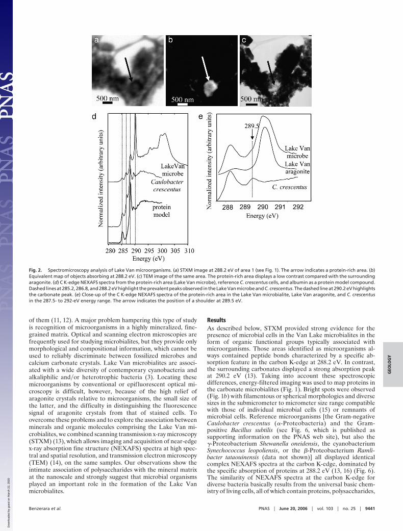
reference bacteria in similar ratios (Fig. 2; and see Figs. 6–8,which are published as supporting information on the PNAS website). The systematic presence of a carbonate peak at 290.2 eVin the C K-edge NEXAFS spectrum of the peptide-, aromatic-,and ketone-rich regions of the Lake Van microorganisms indi-cates that they are closely associated with carbonates (Fig. 2). Inaddition, the carbonate peak overlaps a peak at 289.5 eV, whichwas detected systematically and exclusively in those regions (Fig.2). The energy position of this peak matches the peak observedin reference bacterial cells and in nucleic acids (13, 17). Hence,these regions have a complex spectrum identical to those ofdiverse cultured bacterial strains, which strongly supports theidentification of the observed spots within the Lake Van car-bonates as microbial cells. The detection of microorganisms wasfurther supported by spectroscopy at the N K-edge, whichshowed the presence of nitrogen associated with protein-richareas, with a spectroscopic signature very similar to that ob-served in C. crescentus cells (Fig. 3). Peaks at 399 and 399.9 eVare related to heterocyclic aromatics such as pyridine (18), whichare present in many biological molecules (e.g., nucleic acids,ATP). The peak at 401.2 eV is related to amide groups (19).Therefore, spectromicroscopy allowed the detection of livingand�or fossilized microorganisms in microbialites by character-izing and mapping, within a mineral matrix, organic functionalgroups composing microbial cells. These cell-shaped areas richin peptide bonds, phenolic�ketonic and aromatic groups, andorganic nitrogen were systematically located and imaged byTEM. However, in most of the samples examined, microbial cells
Fig. 3. Nitrogen K-edge NEXAFS spectra of Lake Van calcified microorganisms.The energy positions of the peaks are, respectively, 399, 399.9, and 401.2 eV.
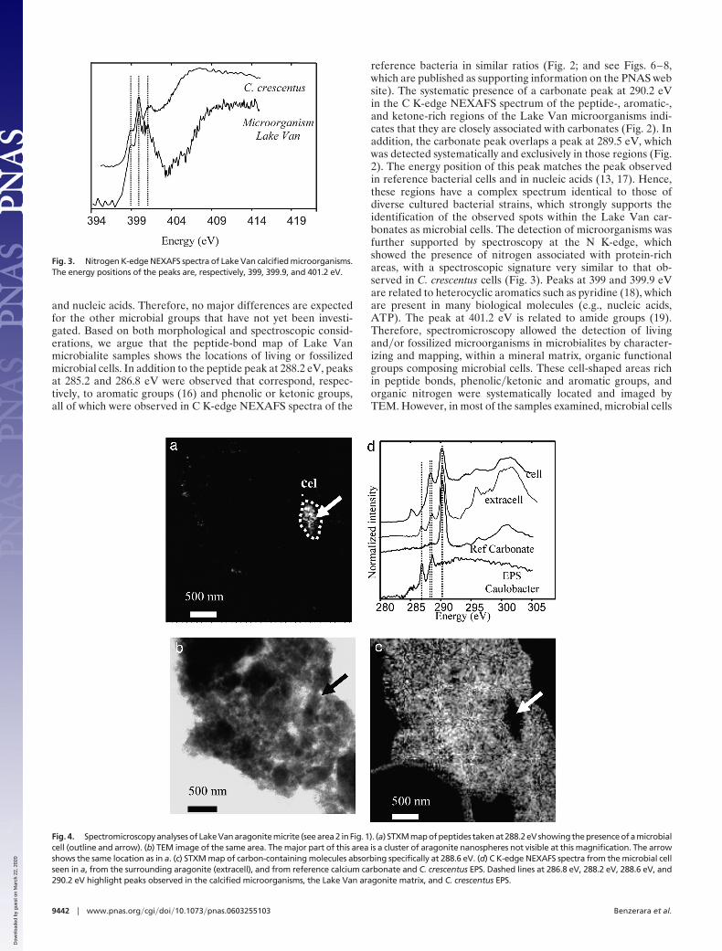
Fig. 4. Spectromicroscopy analyses of Lake Van aragonite micrite (see area 2 in Fig. 1). (a) STXM map of peptides taken at 288.2 eV showing the presence of a microbialcell (outline and arrow). (b) TEM image of the same area. The major part of this area is a cluster of aragonite nanospheres not visible at this magnification. The arrowshows the same location as in a. (c) STXM map of carbon-containing molecules absorbing specifically at 288.6 eV. (d) C K-edge NEXAFS spectra from the microbial cellseen in a, from the surrounding aragonite (extracell), and from reference calcium carbonate and C. crescentus EPS. Dashed lines at 286.8 eV, 288.2 eV, 288.6 eV, and290.2 eV highlight peaks observed in the calcified microorganisms, the Lake Van aragonite matrix, and C. crescentus EPS.
9442 � www.pnas.org�cgi�doi�10.1073�pnas.0603255103 Benzerara et al.
Dow
nloa
ded
by g
uest
on
Mar
ch 2
2, 2
020
were masked in the TEM images by strongly diffracting arago-nite nanoglobules precipitated on them (Figs. 4 and 8).
In addition to microbial cells with highly localized distribution,a second type of organic component showed a much widerdistribution in Lake Van microbialites. Most of the samplesexamined, other than in the peptide-rich regions, consisted ofclusters of aragonite nanospheres visible by TEM (3) (Fig. 4).Although spectromicroscopy on those clusters showed no aro-matic groups (285.2 eV) or peptide bonds (288.2 eV), peaks at286.8 and 288.6 eV were detected together with the maincarbonate peak at 290.2 eV (Fig. 4). The peaks at 286.8 and 288.6eV, which were absent in C K-edge NEXAFS reference spectraof abiotic aragonite crystals, corresponded to ketone and car-boxylic groups, respectively. They are also observed in acidicpolysaccharides (17) and in extracellular polymeric substances(EPS) from C. crescentus (Fig. 4), suggesting that similar poly-saccharides were associated with Lake Van aragonite nanocrys-tals. Such polymers were observed throughout the carbonateclusters at a spatial scale down to a few tens of nanometers (Fig.4). Epifluorescence microscopy observations were performed onLake Van microbialites by using the fluorescently labeled lectinCon A, which has high affinity for �-D-mannose and �-D-glucoseresidues and has been validated and extensively used in previousstudies on carbonate microbialites to probe mannose and�orglucose residues at nonreducing terminals of polysaccharides(17, 20–21). They support our spectromicroscopy observationsby suggesting the presence of these polymers throughout Lake
Van carbonates at a larger scale (see Fig. 9, which is publishedas supporting information on the PNAS web site). The validityof the spectroscopic signatures proposed for calcified cells andEPS in the Lake Van microbialites was checked on 4-year-oldexperimentally calcified bacterial biofilms consisting of the�-Proteobacterium R. tataouinensis. STXM observations showedthe presence, in the experimentally calcified cultures, of R.tataouinensis cells displaying C K-edge and N K-edge NEXAFSspectra identical to those from Lake Van microbial cells andextracellular polymers with C K-edge NEXAFS spectra identicalto Lake Van EPS (see Fig. 10, which is published as supportinginformation on the PNAS web site).
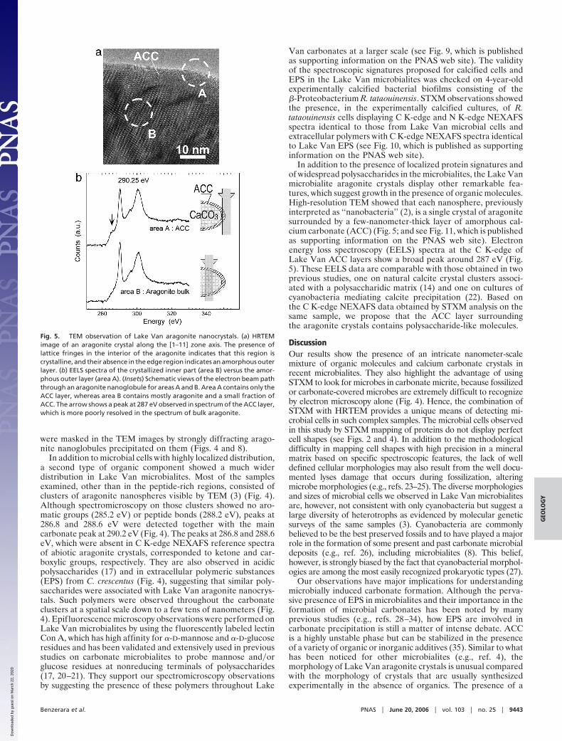
In addition to the presence of localized protein signatures andof widespread polysaccharides in the microbialites, the Lake Vanmicrobialite aragonite crystals display other remarkable fea-tures, which suggest growth in the presence of organic molecules.High-resolution TEM showed that each nanosphere, previouslyinterpreted as ‘‘nanobacteria’’ (2), is a single crystal of aragonitesurrounded by a few-nanometer-thick layer of amorphous cal-cium carbonate (ACC) (Fig. 5; and see Fig. 11, which is publishedas supporting information on the PNAS web site). Electronenergy loss spectroscopy (EELS) spectra at the C K-edge ofLake Van ACC layers show a broad peak around 287 eV (Fig.5). These EELS data are comparable with those obtained in twoprevious studies, one on natural calcite crystal clusters associ-ated with a polysaccharidic matrix (14) and one on cultures ofcyanobacteria mediating calcite precipitation (22). Based onthe C K-edge NEXAFS data obtained by STXM analysis on thesame sample, we propose that the ACC layer surroundingthe aragonite crystals contains polysaccharide-like molecules.
DiscussionOur results show the presence of an intricate nanometer-scalemixture of organic molecules and calcium carbonate crystals inrecent microbialites. They also highlight the advantage of usingSTXM to look for microbes in carbonate micrite, because fossilizedor carbonate-covered microbes are extremely difficult to recognizeby electron microscopy alone (Fig. 4). Hence, the combination ofSTXM with HRTEM provides a unique means of detecting mi-crobial cells in such complex samples. The microbial cells observedin this study by STXM mapping of proteins do not display perfectcell shapes (see Figs. 2 and 4). In addition to the methodologicaldifficulty in mapping cell shapes with high precision in a mineralmatrix based on specific spectroscopic features, the lack of welldefined cellular morphologies may also result from the well docu-mented lyses damage that occurs during fossilization, alteringmicrobe morphologies (e.g., refs. 23–25). The diverse morphologiesand sizes of microbial cells we observed in Lake Van microbialitesare, however, not consistent with only cyanobacteria but suggest alarge diversity of heterotrophs as evidenced by molecular geneticsurveys of the same samples (3). Cyanobacteria are commonlybelieved to be the best preserved fossils and to have played a majorrole in the formation of some present and past carbonate microbialdeposits (e.g., ref. 26), including microbialites (8). This belief,however, is strongly biased by the fact that cyanobacterial morphol-ogies are among the most easily recognized prokaryotic types (27).
Our observations have major implications for understandingmicrobially induced carbonate formation. Although the perva-sive presence of EPS in microbialites and their importance in theformation of microbial carbonates has been noted by manyprevious studies (e.g., refs. 28–34), how EPS are involved incarbonate precipitation is still a matter of intense debate. ACCis a highly unstable phase but can be stabilized in the presenceof a variety of organic or inorganic additives (35). Similar to whathas been noticed for other microbialites (e.g., ref. 4), themorphology of Lake Van aragonite crystals is unusual comparedwith the morphology of crystals that are usually synthesizedexperimentally in the absence of organics. The presence of a
Fig. 5. TEM observation of Lake Van aragonite nanocrystals. (a) HRTEMimage of an aragonite crystal along the [1–11] zone axis. The presence oflattice fringes in the interior of the aragonite indicates that this region iscrystalline, and their absence in the edge region indicates an amorphous outerlayer. (b) EELS spectra of the crystallized inner part (area B) versus the amor-phous outer layer (area A). (Insets) Schematic views of the electron beam paththrough an aragonite nanoglobule for areas A and B. Area A contains only theACC layer, whereas area B contains mostly aragonite and a small fraction ofACC. The arrow shows a peak at 287 eV observed in spectrum of the ACC layer,which is more poorly resolved in the spectrum of bulk aragonite.
Benzerara et al. PNAS � June 20, 2006 � vol. 103 � no. 25 � 9443
GEO
LOG
Y
Dow
nloa
ded
by g
uest
on
Mar
ch 2
2, 2
020

widespread organic-rich matrix may thus explain the stabiliza-tion of the ACC layer as well as the clustering and submicrome-ter-size of the aragonite crystals, because organic molecules canpromote the formation of numerous nucleation sites (36) andinhibit crystal growth by poisoning their surfaces (37). Thewidespread occurrence of EPS down to the nanometer-scaleevidenced in this study may explain the micritic texture of LakeVan microbialites and more generally the micritic texture usuallyobserved in carbonate microbialites (4). Some authors state thatacidic polysaccharides inhibit carbonate precipitation, whichinstead occurs in EPS-poor areas where high concentrations ofCa and Mg have been released by the degradation of polysac-charides by heterotrophic bacteria (38). Others propose thatdecaying EPS transform into a highly organized template struc-ture favoring calcium carbonate nucleation (39). Although poly-saccharide degradation cannot be excluded, the presence ofdetectable amounts of EPS in Lake Van microbialites, includingareas of extensive precipitation, is consistent with a promoterrole for EPS in carbonate precipitation. The questions ofwhether the presence of EPS in Lake Van microbialites indicatesthat degradation by heterotrophic bacteria does not balance EPSproduction or that some molecules are more resistant thanothers to bacterial degradation and�or protected by mineralprecipitation cannot be answered by this study. This pervasiveassociation of organic polymers with aragonite supports a bio-genic origin for microbialite carbonates, but the relatively lowcell density observed shows that carbonates are not generallyassociated with detectable cells. Many ancient carbonates do notpreserve clear evidence of the organisms responsible for theirformation and hence their biogenicity has been questioned (11).Our observations reconcile these considerations with a bioticorigin. Lake Van microorganisms and their pervasive associatedpolymers do impact mineral nucleation and�or growth of car-bonate crystals in the highly oversaturated waters of the lake,producing clusters of nanobacteria-like aragonite crystals andleaving biosignatures in the mineralogy and in the associatedorganic matter of these deposits.
ACC has been observed around nanometer-sized rod-shaped calcite crystals surrounded by polysaccharides (40–41)and has also been recently described in mollusks and arthro-pods (42). Although ACC has not been described in microbialcarbonates previously, its identification in Lake Van micro-bialites should stimulate a systematic search for this feature incarbonates precipitated in the presence of organic compounds.The presence of ACC is not a biosignature per se because ACCcan be stabilized by abiotic impurities (43); however, itsassociation with organic molecules may be an indicator ofbiogenicity. In that regard, the use of cryo-TEM to imagesingle organic molecules in microbialites at very high spatialresolution would be an interesting approach to develop in thefuture. Moreover, the preservation of the organic-ACC layerafter secondary mineralogical processes, such as transforma-tion of aragonite to more stable carbonate phases (i.e., calcite,dolomite) that may affect older microbialites, is yet to beinvestigated.
Summary and ConclusionsLake Van microbialites are composed of living and�or recentlyfossilized microbes and nanobacteria-like aragonite crystals,embedded together in a polysaccharide matrix. Our observa-tions strongly suggest that the presence of this pervasive
organic matrix, intimately associated with the aragonite crys-tals down to the nanometer scale, is responsible for theparticular mineralogical features observed in Lake Van mi-crobialites, i.e., clustered spherical aragonite crystals in the 30-to 100-nm size range, surrounded by a usually highly unstableACC phase. The use of both STXM and TEM to characterizemicrobialites at the submicrometer scale provides a uniquecapability to detect viable or fossilized microbial cells andexopolymers in such deposits in the form of complex mixturesof organic functional groups. Although preservation of organicand mineralogical features of the type reported here hereto-fore has not been documented in ancient microbialites, un-derstanding such signatures in modern microbialites is aprerequisite to their potential detection and use in determin-ing the origin of ancient microbialites.
MethodsSamples were gently crushed in an agate mortar (see Fig. 12, whichis published as supporting information on the PNAS web site).Powders were suspended for a few seconds in Milli-Q grade water,and one drop was deposited on the membrane of a lacey carbon-coated 200 mesh copper grid and dried in air. We did not detecteither by STXM or by TEM any artifactual salt precipitation.STXM observations were performed at Advanced Light Sourcebranch line 11.0.2.2 according to the procedures described in ref. 13.The synchrotron storage ring operated at 1.9 GeV and 200–400 mAstored current. A 150 lines per mm grating and 20-�m exit slit wereused for carbon and nitrogen K-edge imaging and spectroscopy,providing a theoretical energy resolution of 100 meV. Energycalibration was accomplished by using the well resolved 3p Rydbergpeak at 294.96 eV of gaseous CO2 for the C K-edge. Calibration atthe N K-edge was made by using the N 1s 3 �* (401.1 eV)transition of atmospheric N2 (44).
TEM observations were performed on a JEOL 2010F micro-scope operating at 200 kV and equipped with a field emissiongun, a high-resolution UHR pole piece, and a Gatan GIF 200energy filter. EELS analyses were performed according to theprocedures described in ref. 14. EELS spectra were acquired byusing a dispersion of 0.3 eV per channel to record spectra in therange 250–560 eV. The energy resolution was �1.3 eV asmeasured by the full width at half maximum of the zero-losspeak. The dwell time was optimized to acquire sufficient signalintensity and to limit beam damage. Spectra were corrected fromplural scattering by using the Egerton procedure available withthe EL�P program (Gatan).
We thank A. P. Hitchcock (McMaster University, Hamilton, ON,Canada) for providing carbon K-edge spectra for DNA, albumin, andsodium alginate. Bill Burkholder (Stanford University) kindly pro-vided a B. subtilis strain. Microbialites were collected during aGerman–Turkish Geological Expedition to Lake Van (led by StephanKempe, Darmstadt), which was supported by the Deutsche For-schungsgemeinschaft. This work was supported by National ScienceFoundation Grant CHE-0431425 (Stanford EMSI) and by a grant fromthe Woods Institute for the Environment at Stanford University. TheSTXM studies were performed at the Advanced Light Source on theSTXM end station (11.0.2.2) of the Molecular Environmental Sciencebeam line (11.0.2), which is supported by the Division of ChemicalSciences, Geosciences, and Biosciences, Department of Energy Officeof Basic Energy Sciences. The Advanced Light Source is supported bythe Scientific User Facilities Division of the Department of EnergyOffice of Basic Energy Sciences.
1. Kempe, S., Kazmierczak, J., Landmann, G., Konuk, T., Reimer, A. & Lipp, A.(1991) Nature 349, 605–608.
2. Kazmierczak, J. & Kempe, S. (2003) Naturwissenschaften 90, 167–172.3. Lopez-Garcıa, P., Kazmierczak, J., Benzerara, K., Kempe, S., Guyot, F. &
Moreira, D. (2005) Extremophiles 9, 263–274.4. Riding, R. (2000) Sedimentology 47, 179–214.
5. Dupraz, C., Visscher, P. T., Baumgartner, L. K. & Reid, R. P. (2004)Sedimentology 51, 745–765.
6. Kalkowsky, E. (1908) Z. Dtsch. Geol. Ges. 60, 68–125.7. Reid, R. P., Visscher, P. T., Decho, A. W., Stolz, J. F., Beboutk, B. M., Dupraz,
C., Macintyre, I. G., Paerl, H. W., Pinckney, J. L., Prufert-Beboutk, L., et al.(2000) Nature 406, 989–992.
9444 � www.pnas.org�cgi�doi�10.1073�pnas.0603255103 Benzerara et al.
Dow
nloa
ded
by g
uest
on
Mar
ch 2
2, 2
020
8. Arp, G., Reimer, A. & Reitner, J. (2001) Science 292, 1701–1704.9. Bosak, T. & Newman, D. K. (2003) Geology 31, 577–580.
10. Vasconcelos, C., McKenzie, J. A., Bernasconi, S., Grujic, D. & Tien, A. J.(1995) Nature 377, 220–222.
11. Grotzinger, J. P. & Rothman, D. H. (1996) Nature 383, 423–425.12. Grotzinger, J. P. & Knoll, A. H. (1999) Annu. Rev. Earth Planet. Sci. 27, 313–358.13. Benzerara, K., Yoon, T. H., Tyliszczak, T., Constantz, B., Spormann, A. M. &
Brown, G. E., Jr. (2004) Geobiology 2, 249–259.14. Benzerara, K., Menguy, N., Guyot, F., Vanni, C. & Gillet, P. (2005) Geochim.
Cosmochim. Acta 69, 1413–1422.15. Maniloff, J. (1997) Science 276, 1773–1776.16. Boese, J., Osanna, A., Jacobsen, C. & Kirz, J. (1997) J. Electron Spectrosc. Relat.
Phenom. 85, 9–15.17. Lawrence, J. R., Swerhone, G. D. W., Leppard, G. G., Araki, T., Zhang, X.,
West, M. M. & Hitchcock, A. P. (2003) Appl. Environ. Microbiol. 69, 5543–5554.18. Myneni, S. C. B. (2002) Rev. Mineral. Geochem. 49, 485–579.19. Gordon, M. L., Cooper, G., Morin, C., Araki, T., Turci, C. C., Kaznatcheev, K.
& Hitchcock, A. P. (2003) J. Phys. Chem. A 107, 6144–6159.20. Decho, A. W. & Kawaguchi, T. (1999) BioTechniques 27, 1246–1252.21. Kawaguchi, T. & Decho, A. W. (2002) Mar. Biotechnol. 4, 127–131.22. Obst, M., Gasser, P., Mavrocordatos, D. & Dittrich, M. (2005) Am. Mineral. 90,
1270–1277.23. Ferris, F. G., Fyfe, W. S. & Beveridge, T. J. (1988) Geology 16, 149–152.24. Benzerara, K., Menguy, N., Guyot, F., Skouri, F., de Luca, G., Barakat, M. &
Heulin, T. (2004) Earth Planet. Sci. Lett. 228, 439–449.25. Stolz, J. F., Feinstein, T. N., Salsi, J., Visscher, P. T. & Reid, R. P. (2001) Am.
Mineral. 86, 826–833.26. Thompson, J. B., Schultze-Lam, S., Beveridge, T. J. & DesMarais, D. J. (1997)
Limnol. Oceanogr. 42, 133–141.27. Westall, F. (2005) Science 308, 366–367.28. Arp, G., Reimer, A. & Reitner, J. (2003) J. Sediment. Res. 73, 105–127.
29. Gautret, P., Camoin, G., Golubic, S. & Sprachta, S. (2004) J. Sediment. Res. 74,462–478.
30. Kuhl, M., Fenchel, T. & Kazmierczak, J. (2003) in Fossil and Recent Biofilms—ANatural History of Life on Earth, eds. Krumbein, W. E., Paterson, D. M. & Zavarzin,G. A. (Kluwer Academic, Dordrecht, The Netherlands), pp. 77–102.
31. Kazmierczak, J., Kempe, S. & Altermann, W. (2004) in The Precambrian Earth:Tempos and Events, eds. Eriksson, P. G., Altermann, W., Nelson, D. R.,Mueller, W. U. & Catuneanu, O. (Elsevier, Amsterdam), pp. 545–564.
32. Rougeaux, H., Guezennec, M., Che, L. M., Payri, C., Deslandes, E. &Guezennec, J. (2000) Mar. Biotechnol. 3, 181–187.
33. Decho, A. W. (2000) Continental Shelf Res. 20, 1257–1273.34. Kawaguchi, T. & Decho, A. W. (2000) Prep. Biochem. Biotechnol. 30,
321–330.35. Aizenberg, J., Lambert, G., Weiner, S. & Addadi, L. (2002) J. Am. Chem. Soc.
124, 32–39.36. Grassmann, O. & Lobmann, P. (2004) Biomaterials 25, 277–282.37. Manoli, F. & Dalas, E. (2002) J. Mater. Sci. Mater. Med. 13, 155–
158.38. Arp, G., Reimer, A. & Reitner, J. (1999) Eur. J. Phycol. 34, 393–403.39. Defarge C., Trichet, J., Jaunet, A. M., Robert, M., Tribble, J. & Sansone, F. J.
(1996) J. Sediment. Res. 66, 935–947.40. Benzerara, K., Menguy, N., Guyot, F., Dominici, D. & Gillet, P. (2003) Proc.
Natl. Acad. Sci. USA 100, 7438–7442.41. Benzerara, K., Yoon, T.-H., Menguy, N., Tyliszczak, T. & Brown, G. E., Jr.
(2005) Proc. Natl. Acad. Sci. USA 102, 979–982.42. Becker, A., Bismayer, U., Epple, M., Fabritius, H., Hasse, B., Shi, J. M. &
Ziegler, A. (2003) J. Chem. Soc. Dalton Trans. 4, 551–555.43. Raz, S., Weiner, S. & Addadi, L. (2000) Adv. Mater. 12, 38–42.44. Sodhi, R. N. S. & Brion, C. E. (1984) J. Electron Spectrosc. Rel. Phenom. 34,
363–372.
Benzerara et al. PNAS � June 20, 2006 � vol. 103 � no. 25 � 9445
GEO
LOG
Y
Dow
nloa
ded
by g
uest
on
Mar
ch 2
2, 2
020