Muscle Tissue Engineering Using Gingival Mesenchymal Stem ...
13
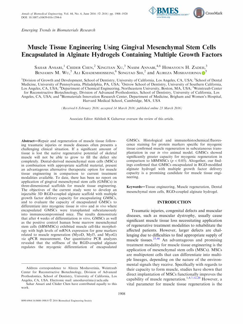
Emerging Trends in Biomaterials Research Muscle Tissue Engineering Using Gingival Mesenchymal Stem Cells Encapsulated in Alginate Hydrogels Containing Multiple Growth Factors SAHAR ANSARI, 1 CHIDER CHEN, 2 XINGTIAN XU, 3 NASIM ANNABI, 4,6 HOMAYOUN H. ZADEH, 3 BENJAMIN M. WU, 5 ALI KHADEMHOSSEINI, 6 SONGTAO SHI, 2 and ALIREZA MOSHAVERINIA 5 1 Division of Growth and Development, School of Dentistry, University of California, Los Angeles, CA, USA; 2 School of Dental Medicine, University of Pennsylvania, Philadelphia, PA, USA; 3 Ostrow School of Dentistry, University of Southern California, Los Angeles, CA, USA; 4 Department of Chemical Engineering, Northeastern University, Boston, MA, USA; 5 Weintraub Center for Reconstructive Biotechnology, Division of Advanced Prothodontics, School of Dentistry, University of California, Los Angeles, CA, USA; and 6 Biomaterials Innovation Research Center, Department of Medicine, Brigham and Women’s Hospital, Harvard Medical School, Cambridge, MA, USA (Received 6 February 2016; accepted 14 March 2016; published online 23 March 2016) Associate Editor Akhilesh K Gaharwar oversaw the review of this article. Abstract—Repair and regeneration of muscle tissue follow- ing traumatic injuries or muscle diseases often presents a challenging clinical situation. If a significant amount of tissue is lost the native regenerative potential of skeletal muscle will not be able to grow to fill the defect site completely. Dental-derived mesenchymal stem cells (MSCs) in combination with appropriate scaffold material, present an advantageous alternative therapeutic option for muscle tissue engineering in comparison to current treatment modalities available. To date, there has been no report on application of gingival mesenchymal stem cells (GMSCs) in three-dimensional scaffolds for muscle tissue engineering. The objectives of the current study were to develop an injectable 3D RGD-coupled alginate scaffold with multiple growth factor delivery capacity for encapsulating GMSCs, and to evaluate the capacity of encapsulated GMSCs to differentiate into myogenic tissue in vitro and in vivo where encapsulated GMSCs were transplanted subcutaneously into immunocompromised mice. The results demonstrate that after 4 weeks of differentiation in vitro, GMSCs as well as the positive control human bone marrow mesenchymal stem cells (hBMMSCs) exhibited muscle cell-like morphol- ogy with high levels of mRNA expression for gene markers related to muscle regeneration (MyoD, Myf5, and MyoG) via qPCR measurement. Our quantitative PCR analyzes revealed that the stiffness of the RGD-coupled alginate regulates the myogenic differentiation of encapsulated GMSCs. Histological and immunohistochemical/fluores- cence staining for protein markers specific for myogenic tissue confirmed muscle regeneration in subcutaneous trans- plantation in our in vivo animal model. GMSCs showed significantly greater capacity for myogenic regeneration in comparison to hBMMSCs (p < 0.05). Altogether, our find- ings confirmed that GMSCs encapsulated in RGD-modified alginate hydrogel with multiple growth factor delivery capacity is a promising candidate for muscle tissue engi- neering. Keywords—Tissue engineering, Muscle regeneration, Dental mesenchymal stem cells, RGD-coupled alginate hydrogel. INTRODUCTION Traumatic injuries, congenital defects and muscular diseases, such as muscular dystrophy, usually cause significant muscle tissue loss necessitating application of regenerative treatment modalities to rehabilitate the affected patients. However, larger defects are chal- lenging due to difficulties to find appropriate supply of muscle tissues. 25,46 An advantageous and promising treatment modality for muscle tissue engineering is the application of mesenchymal stem cells (MSCs). MSCs are multipotent cells that can differentiate into multi- ple lineages, depending on the nature of the environ- mental signals they receive. Specifically with regards to their capacity to form muscle, studies have shown that direct implantation of MSCs functionally improves the capability of muscle regeneration. 1,4,7,12,20 However, a vital parameter for muscle tissue regeneration is the Address correspondence to Alireza Moshaverinia, Weintraub Center for Reconstructive Biotechnology, Division of Advanced Prothodontics, School of Dentistry, University of California, Los Angeles, CA, USA. Electronic mail: [email protected] Sahar Ansari and Chider Chen have contributed equally to this work. Annals of Biomedical Engineering, Vol. 44, No. 6, June 2016 (ȑ 2016) pp. 1908–1920 DOI: 10.1007/s10439-016-1594-6 0090-6964/16/0600-1908/0 ȑ 2016 Biomedical Engineering Society 1908
Transcript of Muscle Tissue Engineering Using Gingival Mesenchymal Stem ...
Emerging Trends in Biomaterials Research
Muscle Tissue Engineering Using Gingival Mesenchymal Stem Cells
Encapsulated in Alginate Hydrogels Containing Multiple Growth Factors
SAHAR ANSARI,1 CHIDER CHEN,2 XINGTIAN XU,3 NASIM ANNABI,4,6 HOMAYOUN H. ZADEH,3
BENJAMIN M. WU,5 ALI KHADEMHOSSEINI,6 SONGTAO SHI,2 and ALIREZA MOSHAVERINIA5
1Division of Growth and Development, School of Dentistry, University of California, Los Angeles, CA, USA; 2School of DentalMedicine, University of Pennsylvania, Philadelphia, PA, USA; 3Ostrow School of Dentistry, University of Southern California,Los Angeles, CA, USA; 4Department of Chemical Engineering, Northeastern University, Boston, MA, USA; 5Weintraub Centerfor Reconstructive Biotechnology, Division of Advanced Prothodontics, School of Dentistry, University of California, Los
Angeles, CA, USA; and 6Biomaterials Innovation Research Center, Department of Medicine, Brigham and Women’s Hospital,Harvard Medical School, Cambridge, MA, USA
(Received 6 February 2016; accepted 14 March 2016; published online 23 March 2016)
Associate Editor Akhilesh K Gaharwar oversaw the review of this article.
Abstract—Repair and regeneration of muscle tissue follow-ing traumatic injuries or muscle diseases often presents achallenging clinical situation. If a significant amount oftissue is lost the native regenerative potential of skeletalmuscle will not be able to grow to fill the defect sitecompletely. Dental-derived mesenchymal stem cells (MSCs)in combination with appropriate scaffold material, presentan advantageous alternative therapeutic option for muscletissue engineering in comparison to current treatmentmodalities available. To date, there has been no report onapplication of gingival mesenchymal stem cells (GMSCs) inthree-dimensional scaffolds for muscle tissue engineering.The objectives of the current study were to develop aninjectable 3D RGD-coupled alginate scaffold with multiplegrowth factor delivery capacity for encapsulating GMSCs,and to evaluate the capacity of encapsulated GMSCs todifferentiate into myogenic tissue in vitro and in vivo whereencapsulated GMSCs were transplanted subcutaneouslyinto immunocompromised mice. The results demonstratethat after 4 weeks of differentiation in vitro, GMSCs as wellas the positive control human bone marrow mesenchymalstem cells (hBMMSCs) exhibited muscle cell-like morphol-ogy with high levels of mRNA expression for gene markersrelated to muscle regeneration (MyoD, Myf5, and MyoG)via qPCR measurement. Our quantitative PCR analyzesrevealed that the stiffness of the RGD-coupled alginateregulates the myogenic differentiation of encapsulated
GMSCs. Histological and immunohistochemical/fluores-cence staining for protein markers specific for myogenictissue confirmed muscle regeneration in subcutaneous trans-plantation in our in vivo animal model. GMSCs showedsignificantly greater capacity for myogenic regeneration incomparison to hBMMSCs (p< 0.05). Altogether, our find-ings confirmed that GMSCs encapsulated in RGD-modifiedalginate hydrogel with multiple growth factor deliverycapacity is a promising candidate for muscle tissue engi-neering.
Keywords—Tissue engineering, Muscle regeneration, Dental
mesenchymal stem cells, RGD-coupled alginate hydrogel.
INTRODUCTION
Traumatic injuries, congenital defects and musculardiseases, such as muscular dystrophy, usually causesignificant muscle tissue loss necessitating applicationof regenerative treatment modalities to rehabilitate theaffected patients. However, larger defects are chal-lenging due to difficulties to find appropriate supply ofmuscle tissues.25,46 An advantageous and promisingtreatment modality for muscle tissue engineering is theapplication of mesenchymal stem cells (MSCs). MSCsare multipotent cells that can differentiate into multi-ple lineages, depending on the nature of the environ-mental signals they receive. Specifically with regards totheir capacity to form muscle, studies have shown thatdirect implantation of MSCs functionally improves thecapability of muscle regeneration.1,4,7,12,20 However, avital parameter for muscle tissue regeneration is the
Address correspondence to Alireza Moshaverinia, Weintraub
Center for Reconstructive Biotechnology, Division of Advanced
Prothodontics, School of Dentistry, University of California, Los
Angeles, CA, USA. Electronic mail: [email protected] Ansari and Chider Chen have contributed equally to this
work.
Annals of Biomedical Engineering, Vol. 44, No. 6, June 2016 (� 2016) pp. 1908–1920
DOI: 10.1007/s10439-016-1594-6
0090-6964/16/0600-1908/0 � 2016 Biomedical Engineering Society
1908

identification of an optimal source of cell with suit-able muscle differentiation capacity. Studies haveshown the presence of satellite cells (SCs) in maturemuscles that are identified to be committed to myo-genic tissue regeneration.22,27 However, the main dis-advantages associated with the application of theseSCs are the invasive isolation procedure and difficultyin the purification process. In addition, the lowexpansion capacity of satellite cells makes them aquestionable cell source for repair of large muscledefects.15,21,22,27 Alternatively, it has been shown thatMSCs can be identified and isolated from a wide rangeof post-natal tissue types, including the craniofacialstructures.10,17,19,28,34,44,45 One of the attractive sourcesof MSCs in the craniofacial region is gingiva.54 Gin-gival mesenchymal stem cells (GMSCs) are of partic-ular interest as they are easily harvested in the oralenvironment. GMSCs are obtained as discarded bio-logical samples in dental clinics for many commondental procedures.31,32 Moreover, the donor site in theoral cavity tends to heal faster than all other humandermal tissues, and completes healing without scarring.Our previous studies and others have confirmed themultilineage differentiation capabilities of these stemcells in vitro and in vivo.30–33,35,54 Therefore, humanGMSC-mediated tissue regeneration can be consideredas a promising cellular-based treatment for muscletissue engineering.
One of the challenges in the stem cell mediated tissueregeneration is development of a suitable microenviron-ment for stem cells to proliferate and differentiate themto the desired lineage/phenotype. It is well known thatscaffolds have important role in tissue engineer-ing.18,40,41,50 It has been reported that stem cell-mediatedtissue regeneration is partially controlled by the recipientlocal microenvironment, including the presence ofgrowth factors, immune cells and cytokines.11,24 To thatend, exogenous growth factors have beendelivered alongwith stem cells to differentiate the stem cells to desiredlineage/phenotype. Several studies have demonstratedthat the mechanical properties (e.g., elasticity) of thescaffold control the survival anddetermine the fate of thestem cells.13,14,41 Therefore, a scaffold with tunablephysiochemical properties, to regulate the fate ofencapsulated stem cells, seems to be very important andpractical in developing stem cell based therapies.
To develop a promising microenvironment formuscle regeneration based on GMSCs, we sought toengineer a microenvironment with the physiochemicalcharacteristics of the extracellular micro-milieu. Weutilized RGD-coupled alginate hydrogel to modify theniche properties and to direct the cell phenotypethrough differentiation. Alginate microspheres havebeen used extensively for controlled delivery of growthfactors and have an excellent track record for
safety.3,5,8,14,39,40 Presence of cell-binding peptides,such as RGD (arginine- glycine-aspartic acid tripep-tide), in the structure of the alginate scaffold could beadvantageous because these peptides mimic the cell-matrix interaction typical of the ECM.26,33 Studieshave shown that alginate hydrogels have multiplegrowth factor delivery capacity, which make the scaf-fold of choice for complex tissue regeneration pur-poses.6,34
Therefore, in the current study, we focus on thepotential of the delivery of multiple growth factors (acocktail of Forskolin (FSK), MeBIO (6-Bromo-1-methylindirubin-3’-oxime), and basic-FGF) to driveGMSCs differentiation into muscle tissue.2,9,16 Studieshave shown that FSK induces cell differentiation viadirect activation of adenylate cyclase and the stimula-tion of cyclic AMP (cAMP) production, promotingmuscle regeneration in animal models.2,9,16,23,43
Moreover, MeBIO (an analog of 6-bromoindirubin-30-oxime) is a specific inhibitor of glycogen synthasekinase-3 (GSK-3).49 It has been shown that this smallmolecule can support self-renewal and pluripotencypotential in human and mouse embryonic stem cells(ESCs) leading to maintenance of stem cell proper-ties.37 Furthermore, it has been demonstrated thatMeBIO has the ability to control the muscle regener-ation and proliferation of mammalian cardiomy-ocytes.48,49 Basic fibroblast growth factor (bFGF)plays an important role in the survival and differenti-ation of MSCs in muscle regeneration and repair.37,44
A literature search failed to reveal any reportsevaluating the application of GMSCs encapsulated inRGD-coupled alginate microspheres, with multiplegrowth factor delivery capacity, in muscle repair andregeneration. Therefore, in the current study, wedeveloped an injectable and 3D RGD-coupled alginatehydrogel cell encapsulation architecture with multiplegrowth factor delivery capability. Considering the factthat GMSCs can be easily accessible from the oralcavity, and given the rapidity of healing without scarformation, they can be considered ideal for stem cellbanking purposes provided they show promise inMSC-based tissue regeneration. This approach wasdesigned to optimize muscle regeneration for potentialapplication in the repair of the skeletal or cardiacmuscle regeneration.
MATERIALS AND METHODS
Animals
Female immunocompromised nude (Beige nu/nuXIDIII) mice were used in this study. All the animalexperiments were performed in accordance with
Muscle Tissue Engineering Using Gingival Mesenchymal Stem Cells Encapsulated 1909

IACUC-approved small animal protocols at theUniversity of Southern California.
Progenitor Cell Isolation, and Culture
Human GMSCs were isolated and culturedaccording to previously published procedures.52,53
Human bone marrow mesenchymal stem cells(BMMSCs), purchased from Lonza (Gaithersburg,MD).
GMSCs, and hBMMSCs (as a positive control)were separately cultured in a regular culture mediacontaining alpha-MEM (Invitrogen) with 15% FBS,2 mM L-glutamine (Invitrogen), 100 nM Dex, 100 lMascorbic acid (Sigma), 2 mM sodium pyruvate (R&DSystems Inc, Minneapolis, MN), 100 U/mL penicillin,and 100 lg/mL streptomycin (Sigma). Passage 4 cellswere used in the experiments and hBMMSCs wereused as the positive control group.
Flow Cytometric Analysis
Approximately 2 9 105 of either GMSCs orhBMMSCs from passages 4 were incubated withspecific phycoerythrin conjugated mouse monoclonalantibodies for human CD34, and CD45 (as negativehematopoietic stem cell marker), CD73, CD105, andCD146 (as positive MSC marker) (BD Biosciences), orisotype-matched control immunoglobulin Gs (IgGs;Southern Biotechnology Associates) and subjected toflow cytometric analysis using a FACSCalibur withCellQuest software (BD Biosciences) according tomethods previously reported.33
Biomaterial Fabrication and Characterization
RGD-coupled alginate (NovaMatrix FMC Biopoly-mer, Norway) was used as the scaffold material.Alginate was purified and partially oxidized (2%) toincrease its degradability according to publishedmethods in the literature.31,32 The prepared alginatewas concentrated, freeze-dried under reduced pressure,and mixed with a cocktail containing 10 lM forskolin(FSK) (Sigma), 10 lM MeBIO (Santa Cruz Biotech-nology), and 10 ng/ml recombinant human bFGF(R&D Systems).
To study the effects of elasticity of the alginatehydrogel on myogenic differentiation capacity ofencapsulated GMSCs the elastic modulus (E) of algi-nate hydrogels in presence of different concentrationsof CaCl2 (10, 25, 50, 75, and 100 mM) was measuredusing an Instron mechanical testing machine (Nor-wood, PA) at a compression rate of 0.5 mm min21
according to the methods already in literature.18
To characterize the release profile of all the com-ponents of the cocktail, the hydrogel microsphereswere loaded at the abovementioned concentrations andat each selected time interval (1, 3, 5, 7, 10, 12, and14 days), the amounts of medium were collected andanalyzed for released of FSK and MeBIO, using a UVspectrophotometer at 320 nm (Beckman, Brea, CA).Moreover, the release profile of bFGF was character-ized using human b-FGF Immunoassay kit (BioSourceInternational Inc. Camerillo, CA).
Cell Encapsulation and Live/Dead Staining
GMSCs and hBMMSCs (as a positive control) wereencapsulated separately in alginate loaded with cock-tail of 10 lM forskolin (FSK), MeBIO (10 lM), and10 ng/ml recombinant human bFGF. Cells wereencapsulated at a density of 2 9 106 cells/mL of algi-nate solution. Microsphere formation was accom-plished by adding the MSC-alginate mixture dropwiseto 100 mM CaCl2 solution. The resulting microsphereswere incubated at 37 �C for 45 min to complete cross-linking and then washed three times in non-supple-mented DMEM. RGD coupled alginate hydrogelwithout cells (and not loaded with cocktail) was usedas the negative control in this study.
Following 14 days of incubation in culture medium,the cell viability of the encapsulated MSCs was mea-sured as described according to methods in the litera-ture30,35 using calcein AM to stain live cells andethidium bromide homodimer-1 to stain dead cells(Invitrogen). The percentage of live cells was measuredusing NIH ImageJ software (NIH, Bethesda, MD).
Moreover, to evaluate stem cell viability in thealginate microspheres loaded with myogenic differen-tiation cocktail, MTT assay was utilized according topreviously published protocols.33 The MTT absor-bance was obtained at different time intervals (1, 7,and 14 days) and normalized to the absorbance ofalginate containing the same type of stem cells mea-sured at day 1. For each experimental group tested fiveindependent specimens were analyzed at each timeinterval.
In Vitro Myogenic Differentiation
To induce myogenic differentiation, encapsulatedGMSCs and hBMMSCs (2x10⁶ cells) in 1 mL alginatemicrospheres containing the growth factors cocktailwere cultured in a medium containing alpha- MEMwith 15% FBS, 2 mM L-glutamine, 100 nM Dex,100 lM ascorbic acid, 2 mM sodium pyruvate (R&DSystems Inc), 100 U/mL penicillin, 100 lg/mL strep-tomycin, and a cocktail of 10 lM forskolin (FSK),MeBIO, and 10 ng/ml recombinant human bFGF.
S. ANSARI et al.1910

Cell free RGD-alginate microspheres without anygrowth factor were used as the control group. Fourweeks after induction, the samples were fixed with 4%PFA, and paraffin sections were made. Sections wereimmunolabeled using antibodies against MF20 (R&DSystems Inc), Myf5, and MyoD (Santa Cruz Biotech-nology Inc, Dallas, TX), at 4 �C overnight, detectedusing Alexa fluor conjugated secondary antibody(1:200 dilution; Invitrogen), and counterstained withDAPI.
RNA Isolation, Reverse Transcription and Real TimePCR
Following 2 weeks of myogenic differentiationRNA was extracted from the encapsulated cells.30,35
Data were analyzed by the 2�DDCt method, with nor-malization to the Ct of the housekeeping geneGAPDH (glyceraldehyde 3-phosphate dehydroge-nase). Primer sequences are described in Table 1.Additionally, to study the effects of the elasticity of thehydrogel biomaterial on the myogenic differentiationcapacity of encapsulated cells; GMSCs were encapsu-lated in alginate hydrogels with different modulus ofelasticity and after 2 weeks of myogenic differentia-tion, the myogenic genes expression levels were ana-lyzed using qPCR.
Western Blot Analysis
After 2 weeks of myogenic differentiation in vitro,Western blot analysis was performed to analyze thedifferentiation of encapsulated MSCs. Primary anti-body (mouse monoclonal anti-MyoD antibody) wasused at 1:500. Western blot analyzes were carried outas previously reported.30
In Vivo Model for Muscle Regeneration
All animals in our in vivo experiments were treatedaccording to the approved Guidelines and Regulationsfor the Use and Care of Animals (IACUC) at the
University of Southern California. 4 9 106 of GMSCsor hBMMSCs (passage 4) were encapsulated in RGD-coupled alginate microspheres loaded with myogenicdifferentiation cocktail and approximately 500 ll (10alginate microspheres) were implanted subcutaneouslyinto the dorsal surface of 5-month-old Beige nude mice(Harlan, Livermore, CA; N = 4 for each group). After8 weeks of implantation the mice were sacrificed, andthe microspheres and the surrounding tissue weresurgically removed and analyzed using histological andimmunohistochemical staining. Cell free RGD-algi-nate microspheres were used as the negative controlgroup.
Histological, Immunohistochemical andImmunofluorescence Staining
Harvested specimens were fixed in 4% PFA solu-tion, dehydrated in an ascending series of ethanol, andembedded in paraffin. 6-lm sections were cut and glassslides were prepared. Five randomly selected cross-sections from each group were stained with Hema-toxylin & Eosin (H&E).
Immunofluorescence staining was utilized andspecimens were treated with 3% H2O2, followed by ablocking buffer (1% BSA and 0.25% Triton X-100 inPBS). Then, sections were incubated with anti-MyoD(Santa Cruz Biotechnology, Inc, 1:100 dilution), anti-Myf5 (Santa Cruz Biotechnology, Inc, 1:100 dilution)and detected using Alexa Fluor conjugated secondaryantibody (1:200 dilution; Invitrogen). The sampleswere then counterstained with DAPI.
Moreover, to study the degree of local angiogenesisimmunohistochemical analysis was used. De-paraf-finized sections were washed, and non-specific endoge-nous peroxidase activity was quenched by immersingin 3% H2O2/methanol for 15 min. Immunohisto-chemistry examination was performed on sectionsusing anti-CD31 (Abcam, 1:200 dilution) and coun-terstaining with hematoxylin.
To identify the origin of the cells immunohisto-chemical staining was utilized. The retrieved specimenswere incubated with antibody directed against human
TABLE 1. Oligonucleotide primers used in RT-PCR analysis.
Gene Sequence Amplification (bp)
MyoD 5¢-GGGAAGAGTGCGGCGGTGTCGAG-3¢ (forward)5¢-TCCGAGAAGGGTGCTGCGTGGAA-3¢ (reverse) 448
MYF5 (Myogenic factor 5) 5¢-TTC TCC CCA TCC CTC TCG CT-3¢ (forward)5¢-AGC CTG GTT GAC CTT CTTCAG-3¢ (reverse) 285
MyoG (Myogenin, MYF4) 5¢-CAG CTC CCT CAA CCA GGA-3¢ (forward)5¢-GCT GTG AGA GCT GCA TTC G-3¢ (reverse) 245
Glyceraldehyde 3-phosphate dehydrogenase (GAPDH) 5¢-AGCCGCATCTTCTTTTGCGTC-3¢ (forward)5¢-TCATATTTGGCAGGTTTTT CT-3¢ (reverse) 418
Muscle Tissue Engineering Using Gingival Mesenchymal Stem Cells Encapsulated 1911

mitochondria (mouse antibody anti-human mito-chondria; Chemicon, Billerica, MA) at 1:200 dilutionand detected using the universal immunoperoxidase(HRP) ABC kit (Vector Laboratories). Subsequently,sections were counterstained with hematoxylin.
Statistical Analysis
The Kruskal–Wallis rank sum test was utilized toanalyze the data at a significance level of a = 0.05.Also, two-tailed Student’s t-test and two-way ANOVAwere utilized for comparisons whenever needed.Quantitative data are expressed as mean ± standarddeviation (SD).
RESULTS
RGD Coupled Alginate Microsphere Fabrication andRelease Profile Characterization
Our results demonstrated that both GMSCs andhBMMSCs after 2 weeks of culturing in myogeniccondition media changed their morphology towardsmyogenic cells (Fig. 1a). However, GMSCs exhibited
morphological changes more rapidly and extensively incomparison to hBMMSCs. In addition, the stem cellswere expanded in vitro until passages 4 for furtherexperiments. Our flow cytometric analysis revealedthat the GMSCs as well as hBMMSCs expressedspecific MSC markers CD73, CD105, and CD146, butnot the hematopoietic lineage markers CD34 andCD45 (Fig. 1b) with similar expression profiles con-firming the stem cell properties of GMSCs. Moreover,the light microscopic imaging confirmed that the fab-ricated alginate microspheres were of uniform size andshowed even cell distribution (Fig. 1c). Approximately,10 microspheres were fabricated from each 1 mL ofalginate/MSC solution with average diameter of650 A lm for each microsphere containing 105 loadedcells in each sphere. Our SEM analysis showed that thealginate hydrogel had a porous morphology withencapsulated MSCs dispersed within the hydrogelmatrix (Fig. 1d).
In addition, live/dead assays exhibited high cellviability of encapsulated MSCs up to 2 weeks ofin vitro culturing (Fig. 2a). Additionally, our MTTassay confirmed that the presence of the myogeniccocktail did not show any significant effects on MSC
FIGURE 1. Development of RGD-coupled alginate hydrogel microenvironment containing myogenic cocktail for encapsulation ofMSCs. (a) Characterization and comparison of the cellular morphology of GMSCs and hBMMSCs before and during myogenicdifferentiation. (b) Evaluation and quantification of the percentage of cells that express stem cell surface markers (passage 4)through flow cytometric analysis. (c) Bright field image of translucent alginate microspheres showing their retained sphericalshape with a uniform cell distribution (average microsphere diameter 650 A lm). (d) SEM image of the alginate hydrogel-MSCconstruct showing encapsulated MSCs within porous alginate hydrogel microspheres after 2 weeks of culturing in regular media.NS not significant.
S. ANSARI et al.1912
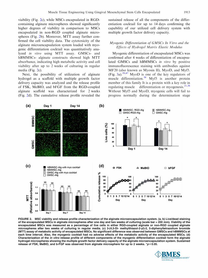
viability (Fig. 2c), while MSCs encapsulated in RGD-containing alginate microspheres showed significantlyhigher degrees of viability in comparison to MSCsencapsulated in non-RGD coupled alginate micro-spheres (Fig. 2b). Moreover, MTT assay further con-firmed the cell viability data. The cytotoxicity of thealginate microencapsulation system loaded with myo-genic differentiation cocktail was quantitatively ana-lyzed in vitro using MTT assay. GMSCs- andhBMMSCs- alginate constructs showed high MTTabsorbance, indicating high metabolic activity and cellviability after up to 2 weeks of culturing in regularmedia (Fig. 2c).
Next, the possibility of utilization of alginatehydrogel as a scaffold with multiple growth factordelivery capacity was analyzed and the release profileof FSK, MeBIO, and bFGF from the RGD-coupledalginate scaffold was characterized for 2 weeks(Fig. 2d). The cumulative release profile revealed the
sustained release of all the components of the differ-entiation cocktail for up to 14 days confirming thecapability of our utilized cell delivery system withmultiple growth factor delivery capacity.
Myogenic Differentiation of GMSCs In Vitro and theEffects of Hydrogel Matrix Elastic Modulus
Myogenic differentiation of encapsulated MSCs wasconfirmed after 4 weeks of differentiation of encapsu-lated GMSCs and hBMMSCs in vitro by positiveimmunofluorescence staining with antibodies againstMF20 (also known as Myosin II), MyoD, and Myf5.(Fig. 3a).29,47 MyoD is one of the key regulators ofmuscle differentiation.38 Myf5 is another proteinmember of this family It is a protein with a key role inregulating muscle differentiation or myogenesis.21,38
Without Myf5 and MyoD, myogenic cells will fail toprogress normally during the determination stage
FIGURE 2. MSC viability and release profile characterization of the alginate microencapsulation system. (a, b) Live/dead stainingof the encapsulated MSCs in alginate microspheres after one day and two weeks of culturing (scale bar = 200 mm). Viability of theencapsulated MSCs was measured as a percentage of live cells in either RGD-coupled alginate or non-RGD coupled alginatemicrospheres after two weeks of culturing in regular media. (c) 3-(4,5-Di- methylthiazol-2-yl)-2, 5-diphenyltetrazolium bromide(MTT) assay of metabolic activity of encapsulated MSCs. No significant difference was observed between GMSCs and hBMMSCs ateach time interval. Also, the myogenic cocktail had no adverse effects of the metabolic activity of the encapsulated MSCs. (d)Characterization of the in vitro release profile of different components of the myogenic differentiation cocktail form the alginatehydrogel microspheres showing the multiple growth factor delivery capacity of the alginate microencapsulation system. Sustainedrelease of FSK, MeBIO, and b-FGF was observed from alginate microsphere for up to 2 weeks. *p< 0.05.
Muscle Tissue Engineering Using Gingival Mesenchymal Stem Cells Encapsulated 1913

of myogenesis.15,47,51,52 Semi-quantitative analysis ofthe specimens confirmed that engrafted GMSCs ex-pressed higher amounts of myogenic related genes(MF20, Myf5, and MyoD) than engrafted hBMMSCs(p value <0.05) (Fig. 3b).
In the next step, the molecular mechanism under-lying the myogenic differentiation was evaluated. Geneexpression analyzes were performed using severalmarkers that are associated with myogenic differenti-ation of MSCs, including MyoG, MyoD, and Myf5(Fig. 4a). MyoG or myogenin is a member of the basichelix-loop-helix (bHLH) family of transcription fac-tors, which also includes MyoD and Myf5.50,51
The expression levels of these myogenic genes wereanalyzed and compared via quantitative PCR. Ourresults revealed that GMSCs as well as hBMMSCsexpressed abundant MyoG, MyoD, and Myf5 aftercultured in myogenic cocktail (Fig. 4a). Specifically,RGD-coupled alginate hydrogel markedly elevatedmyogenic gene expression when compared to non-RGD alginate hydrogel (Fig. 4a). GMSCs showedsignificantly higher expression levels for all the tested
genes compared to hBMMSC (p< 0.05). Moreover,encapsulation of GMSCs in alginate hydrogel signifi-cantly enhanced the expression level of these myogenicgenes (p< 0.05) in comparison to GMSCs culturedwithout any encapsulation (Fig. 4b) suggesting elas-ticity of alginate hydrogel may contribute to myogen-esis of MSCs. Additionally, our immunofluorescencestaining (Figs. 5a and 5b) and PCR (Fig. 5c) analysisconfirmed that the modulus of RGD-coupled alginatehydrogel regulates the myogenic differentiation ofencapsulated MSCs. Our data obtained from quanti-tative PCR analysis showed that MSC encapsulated inalginate hydrogel with intermediate modulus of elas-ticity (10–16 kPa) exhibited the highest capacity formyogenic differentiation in comparison to softer(<5 kPa) or stiffer (>20 kPa) hydrogels (Fig. 5c).Our findings are in good correlation with data reportedin the literature.10,13,36
Furthermore, Western blot analysis showed increasedexpression levels of myogenesis-related gene, MyoD, inspecimens encapsulated in RGD-coupled alginatemicrospheres with middle range elasticity containing
FIGURE 3. In vitro myogenic differentiation of GMSC. (a) Immunofluorescence staining against MF20 (Myosin Heavy Chain), Myf5(Myogenic factor 5), and MyoD (myogenic differentiation protein) antibodies after 4 weeks of in vitro culturing in myogenicdifferentiation. Both GMSCs and hBMMSCs positively immunostained with antibodies against MF20, Myf5, and MyoD. Resultsconfirmed that both hBMMSCs and GMSCs were positively stained for myogenic markers (white arrows), while the negative control(-), cell-free alginate hydrogel microspheres failed to express any of these myogenic markers. (b) Analysis of the percentage ofcells positive for anti- MF20, Myf5, and MyoD antibodies, showing higher expression levels of myogenic markers in GMSCs incomparison to hBMMSCs (positive control) and negative control groups. *p< 0.05.
S. ANSARI et al.1914

myogenic differentiation cocktail (Fig. 5d). The re-sults of Western blot analysis correlated well withthe data from the immunostaining and PCR ana-lyzes.
Ectopic Myogenic Tissue Regeneration In Vivo
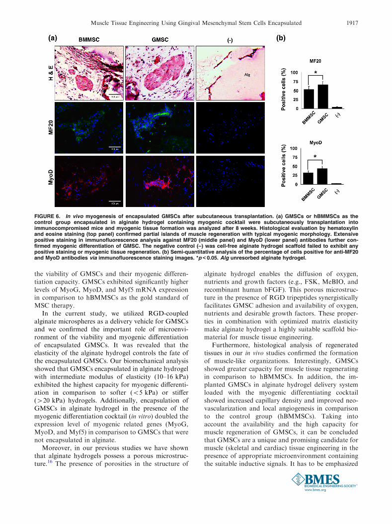
Encapsulated GMSCs in alginate microspheres loa-ded with myogenic differentiation cocktail were sub-cutaneously implanted in immunocompromised mice.H&E staining showed the formation of small islands ofmuscle-like structures, while unresorbled alginatehydrogel was still observed after 8 weeks of implanta-tion (Fig. 6). Immunohistochemical and immunofluo-rescent staining was used to detect newly formed tissueby gene expression analysis. Immunofluorescencestaining for MF20 andMyoD antigens after 8 weeks oftransplantation revealed extensive production and
deposition of these markers within the regeneratedmuscle tissues (Fig. 6a). Interestingly, engraftedGMSCs showed significantly higher MF20 and MyoDexpression levels in comparison to engraftedhBMMSCs (p< 0.05) (Fig. 6b).
In addition, the human origin of cellular compo-nents of the transplants was confirmed throughimmunostaining with specific antibodies againsthuman mitochondria (Fig. 7a). Furthermore, immuno-histochemical staining with CD-31 endothelial cellmarker was utilized to analyze the angiogenesis levelsin the regenerated tissues. Our data showed that im-planted GMSCs in alginate hydrogel delivery systemloaded with the myogenic differentiating cocktail sig-nificantly increased the capillary density based on CD-31 endothelial cell marker staining and H & E stainingin comparison to the control group (Figs. 7a and 7b)confirming the contribution of engrafted GMSCs to
FIGURE 4. Molecular analysis of myogenic differentiation of MSCs in vitro. (a) RT-PCR analysis demonstrate significantly greaterexpression level (in fold changes) of MyoG, Myf5, and MyoD genes for encapsulated GMSCs after 4 weeks of culturing in myogenicdifferentiation media in vitro in comparison to hBMMSCs. The obtained data were normalized by the Ct of the housekeeping geneGAPDH and expressed relative to the expression level for the same gene at day 1. (b) The expression level of MyoG and MyoD forencapsulated hBMMSCs and GMSCs in alginate hydrogel in comparison to scaffold-free MSC cultures after 2 weeks of myogenicdifferentiation in vitro containing the myogenic cocktail. Data confirmed the important role of the microenvironment and thepresence of an encapsulating scaffolds, as the encapsulated MSC expressed greater levels (p< 0.05) of expressions for examinedmyogenic genes in comparison to scaffold free MSC groups. *p< 0.05; **p< 0.01.
Muscle Tissue Engineering Using Gingival Mesenchymal Stem Cells Encapsulated 1915

enhanced local vasculature. To examine the ability ofimplanted MSCs to form vascular networks in vivo,microvessel density was determined, based on the H &E staining, by counting erythrocyte-containing luminalstructures in the retrieved specimens. Our data(Fig. 7b) confirmed that implanted GMSCs formedsignificantly higher number of perfused blood vesselsin comparison to hBMMSCs (p< 0.05).
DISCUSSION
Studies have exploited the potential of cell-basedtherapies to promote muscle regeneration. It has beenshown the possibility of application of stem cells frommuscle tissues, satellite cells or entire myofibres formuscle tissue engineering. However, none of thesestrategies have shown promising results and lowexpansion capacity (low regenerative capacity) of thesestem cell sources have been reported.15,21,22,27 Inaddition, the isolation procedure for these type of stemcells is invasive and they require a difficult purificationprocedure.22,27 It has been confirmed that pluripotentembryonic stem cells (ESCs) have great potential forcell-based therapies. However, their applications are
limited due to ethical issues, heterologous immuno-rejection possibility, and the risk of teratoma forma-tion.42 As an alternative to ESCs, induced pluripotentstem cells (iPSCs) have been successfully utilized formuscle tissue engineering. GMSCs are easily accessibleby harvesting from the oral cavity or cells can often beobtained as discarded biological tissue samples.Therefore, gingiva can be considered as an ideal sourcefor stem cell banking purposes provided they showpromise in MSC-based tissue regeneration.42 A thor-ough review of the literature search revealed no reporton application of gingival mesenchymal stem cells formuscle tissue engineering. Therefore, in the currentstudy we demonstrate that GMSCs encapsulated in aninjectable and biodegradable RGD-modified alginatehydrogel can effectively be differentiated into muscletissue in vitro and in vivo in the presence of appropriateinductive signals. Alginate hydrogel coupled withRGD tripeptide was utilized as the cell delivery systemand loaded with a myogenic differentiation cocktailcontaining FSK, MeBIO, and recombinant humanbFGF to optimize the microenvironment for GMSCsto be differentiated into muscle tissue.
Our in vitro analyzes demonstrate that RGD cou-pled alginate hydrogel encapsulation system supported
FIGURE 5. Fate determination and myogenic differentiation of MSCs encapsulated in RGD-coupled alginate hydrogel micro-spheres. (a) Immunofluorescence detection of MyoD protein localized to the GMSCs encapsulated in alginate microspheres withdifferent modulus of elasticity after 4 weeks of culturing in myogenic differentiation media (counterstained with DAPI). (b) Semi-quantitative analysis of the percentage of cells positive for anti-MyoD antibodies via immunofluorescence staining images in panela. (c) MyoD gene expression evaluation via RT-PCR for GMSCs encapsulated in RGD-coupled alginate hydrogel with differentelastic moduli after 2 weeks of culturing in myogenic differentiation media. (d) Western blot analysis presented changes in theexpression levels of regulators of myogenesis of GMSCs. The expression level of MyoD gene is elevated in the encapsulatedGMSCs in RGD containing alginate microspheres with intermediate modulus of elasticity, while GMSCs encapsulated in alginatehydrogels with higher or lower elastic modulus showed decreased levels of MyoD gene expression conforming the important roleof the mechanical properties of the matrix in fate determination of the encapsulated MSCs. *p< 0.05; **p< 0.01.
S. ANSARI et al.1916
the viability of GMSCs and their myogenic differen-tiation capacity. GMSCs exhibited significantly higherlevels of MyoG, MyoD, and Myf5 mRNA expressionin comparison to hBMMSCs as the gold standard ofMSC therapy.
In the current study, we utilized RGD-coupledalginate microspheres as a delivery vehicle for GMSCsand we confirmed the important role of microenvi-ronment of the viability and myogenic differentiationof encapsulated GMSCs. It was revealed that theelasticity of the alginate hydrogel controls the fate ofthe encapsulated GMSCs. Our biomechanical analysisshowed that GMSCs encapsulated in alginate hydrogelwith intermediate modulus of elasticity (10–16 kPa)exhibited the highest capacity for myogenic differenti-ation in comparison to softer (<5 kPa) or stiffer(>20 kPa) hydrogels. Additionally, encapsulation ofGMSCs in alginate hydrogel in the presence of themyogenic differentiation cocktail (in vitro) doubled theexpression level of myogenic related genes (MyoG,MyoD, and Myf5) in comparison to GMSCs that werenot encapsulated in alginate.
Moreover, in our previous studies we have shownthat alginate hydrogels possess a porous microstruc-ture.16 The presence of porosities in the structure of
alginate hydrogel enables the diffusion of oxygen,nutrients and growth factors (e.g., FSK, MeBIO, andrecombinant human bFGF). This porous microstruc-ture in the presence of RGD tripeptides synergisticallyfacilitates GMSC adhesion and availability of oxygen,nutrients and desirable growth factors. These proper-ties in combination with optimized matrix elasticitymake alginate hydrogel a highly suitable scaffold bio-material for muscle tissue engineering.
Furthermore, histological analysis of regeneratedtissues in our in vivo studies confirmed the formationof muscle-like organizations. Interestingly, GMSCsshowed greater capacity for muscle tissue regeneratingin comparison to hBMMSCs. In addition, the im-planted GMSCs in alginate hydrogel delivery systemloaded with the myogenic differentiating cocktailshowed increased capillary density and improved neo-vascularization and local angiogenesis in comparisonto the control group (hBMMSCs). Taking intoaccount the availability and the high capacity formuscle regeneration of GMSCs, it can be concludedthat GMSCs are a unique and promising candidate formuscle (skeletal and cardiac) tissue engineering in thepresence of appropriate microenvironment containingthe suitable inductive signals. It has to be emphasized
FIGURE 6. In vivo myogenesis of encapsulated GMSCs after subcutaneous transplantation. (a) GMSCs or hBMMSCs as thecontrol group encapsulated in alginate hydrogel containing myogenic cocktail were subcutaneously transplantation intoimmunocompromised mice and myogenic tissue formation was analyzed after 8 weeks. Histological evaluation by hematoxylinand eosine staining (top panel) confirmed partial islands of muscle regeneration with typical myogenic morphology. Extensivepositive staining in immunofluorescence analysis against MF20 (middle panel) and MyoD (lower panel) antibodies further con-firmed myogenic differentiation of GMSC. The negative control (–) was cell-free alginate hydrogel scaffold failed to exhibit anypositive staining or myogenic tissue regeneration. (b) Semi-quantitative analysis of the percentage of cells positive for anti-MF20and MyoD antibodies via immunofluorescence staining images. *p< 0.05. Alg unresorbed alginate hydrogel.
Muscle Tissue Engineering Using Gingival Mesenchymal Stem Cells Encapsulated 1917

that GMSCs were not cultured in myogenic inductionmedia prior to their application in vivo studies. Thesefindings of the current study confirm the importantrole of the microenvironment, as well as the value ofpresenting inductive signals necessary to support theviability and differentiation of MSCs along a desiredphenotype. More importantly, considering the neuralcrest origin of GMSCs they might be utilized as aunique platform for functional regeneration of smoothmuscles (e.g., cardiac regeneration) or tongue muscletissues.
Here, in a proof of concept study, we demonstratethe potential of GMSCs to be utilized in repair/re-generation of muscular tissues when encapsulated in anappropriately designed delivery vehicle loaded withmyogenic growth factors. Additionally, the GMSC/alginate hydrogel construct can be considered apromising candidate for vascularized tissue engineer-ing. Considering the promising results of the currentstudy it can be envisioned that GMSCs might bepromising alternative for cardiac tissue engineering ortongue muscle repair and regeneration.
FIGURE 7. The origin of the implanted MSCs and their contribution to vascularization in vivo. (a) The human origin of theengrafted GMSCs and hBMMSCs was confirmed by immunohistochemical staining with an antibody specific for human mito-chondria (white arrows) (upper panel). Endothelial cell marker was identified by immunohistochemistry using anti- CD31 antibody(middle panel). Histological evaluation by hematoxylin and eosine staining (top panel) (yellow arrows) (lower panel). (b) Semiquantitative analysis of microvessel density based on panel c data. *p<0.05.
S. ANSARI et al.1918

ACKNOWLEDGMENTS
This work was supported by grants from the Na-tional Institute of Dental, Craniofacial Research(K08DE023825 to A.M. and R01 DE017449 to S.S.).The authors declare no potential conflicts of interestwith respect to the authorship and/or publication ofthis article.
REFERENCES
1Abou-Khalil, R., F. Yang, S. Lieu, A. Julien, J. Perry, C.Pereira, F. Relaix, T. Miclau, R. Marcucio, and C. Colnot.Role of muscle stem cells during skeletal regeneration.Stem Cells. 33:1501–1511, 2015.2Adnot, S., M. Desmier, N. Ferry, J. Hanoune, and T. Se-venet. Forskolin (a powerful inhibitor of human plateletaggregation). Biochem. Pharmacol. 31:4071–4074, 1982.3Ansari, S., A. Moshaverinia, S. H. Pi, A. Han, A. I.Abdelhamid, and H. H. Zadeh. Functionalization ofscaffolds with chimeric anti-BMP-2 monoclonal antibodiesfor osseous regeneration. Biomaterials. 34:10191–10198,2013.4Beier, J. P., F. F. Bitto, C. Lange, D. Klumpp, A. Arkudas,O. Bleiziffer, et al. Myogenic differentiation of mesenchy-mal stem cells co-cultured with primary myoblasts. Cell.Biol. Int. 35:397–406, 2011.5Boontheekula, T., H. J. Kongc, and D. J. Mooney. Con-trolling alginate gel degradation utilizing partial oxidationand bimodal molecular weight distribution. Biomaterials.26:2455–2465, 2005.6Borselli, C., C. A. Cezar, D. Shvartsman, H. H. Vanden-burgh, and D. J. Mooney. The role of multifunc-tional delivery scaffold in the ability of cultured myoblaststo promote muscle regeneration. Biomaterials. 32:8905–8914, 2011.7Borselli, C., H. Storrie, F. Benesch-Lee, D. Shvartsman, C.Cezar, J. W. Lichtman, H. H. Vandenburgh, and D. J.Mooney. Functional muscle regeneration with combineddelivery of angiogenesis and myogenesis factors. Proc.Natl. Acad. Sci. USA 107:3287–3292, 2010.8Bouhadir, K. H., K. Y. Lee, E. Alsberg, K. L. Damm, K.W. Anderson, and D. J. Mooney. Degradation of partiallyoxidized alginate and its potential application for tissueengineering. Biotechnol. Prog. 17:945–950, 2001.9Bristow, M. R., R. Ginsburg, A. Strosberg, W. Mont-gomery, and W. Minobe. Pharmacology and inotropicpotential of for- skolin in the human heart. J. Clin. Invest.74:212–223, 1984.
10Cantu, D. A., P. Hematti, and W. J. Kao. Cell encapsu-lating biomaterial regulates mesenchymal stromal/stem celldifferentiation and macrophage immuno- phenotype. Stem.Cell. Transl. Med. 1:740–749, 2012.
11Chen, F. M., J. Zhang, M. Zhang, Y. An, F. Chen, and Z.F. Wu. A review on endogenous regenerative technology inperiodontal regenerative medicine. Biomaterials. 31:7892–7927, 2010.
12Dezawa, M., H. Ishikawa, Y. Itokazu, T. Yoshihara, M.Hoshino, S. Takeda, et al. Bone marrow stromal cellsgenerate muscle cells and repair muscle degeneration. Sci-ence. 309:314–317, 2005.
13Engler, A. J., S. Sen, H. L. Sweeney, and D. E. Discher.Matrix elasticity directs stem cell lineage specification. Cell.126:677–689, 2006.
14Evangelista, M. B., S. X. Hsiong, R. Fernandes, P. Sam-paio, H. Kong, C. C. Barrias, et al. Upregulation of bonecell differentiation through immobilization within a syn-thetic extracellular matrix. Biomaterials. 28:3644–3655,2007.
15Goncalves, M. A., J. Swildens, M. Holkers, A. Narain, G.P. van Nierop, M. J. van de Watering, et al. Geneticcomplementation of human muscle cells via directed stemcell fusion. Mol. Ther. 16:741–748, 2008.
16Gonzalez, A., E. Aranda, D. Mezzano, and J. Garrido.Effects of diterpene forskolin on the release reaction andprotein phosphorylation of human platelets. Cell. Biochem.Funct. 1:179–185, 1983.
17Gronthos, S., M. Mankani, J. Brahim, P. G. Robey, and S.Shi. Postnatal human dental pulp stem cells (DPSCs)in vitro and in vivo. Proc. Natl. Acad. Sci. USA 97:13625–13630, 2000.
18Huebsch, N., P. R. Arany, A. S. Mao, D. Shvartsman, O.A. Ali, S. A. Bencherif, J. Rivera-Feliciano, and D. J.Mooney. Harnessing traction-mediated manipulation ofthe cell/matrix interface to control stem-cell fate. Nat.Mater. 9:518–526, 2010.
19Iwata, T., M. Yamato, Z. Zhang, S. Mukobata, K. Washio,T. Ando, et al. Validation of human periodontal liga-ment-derived cells as a reliable source for cytotherapeuticuse. J. Clin. Periodontol. 37:1088–1099, 2010.
20Koning, M., M. C. Harmsen, M. J. van Luyn, and P. M.Werker. Current opportunities and challenges in skeletalmuscle tissue engineering. J. Tissue Eng. Regen. Med.3:407–415, 2009.
21Krauss, R. S., F. Cole, U. Gaio, G. Takaesu, W. Zhang,and J. S. Kang. Close encounters: regulation of vertebrateskeletal myogenesis by cell-cell contact. J. Cell Sci.118:2355–2362, 2005.
22Kuang, S., M. A. Gillespie, and M. A. Rudnicki. Nicheregulation of muscle satellite cell self-renewal and differ-entiation. Cell Stem Cell. 2:22–31, 2008.
23Litosch, I., T. H. Hudson, I. Mills, S. Y. Li, and J. N. Fain.Forskolin as an activator of cyclic AMP accumulation andlipolysis in rat adipocytes. Mol. Pharmacol. 22:109–115,1982.
24Lu, H. H., J. M. Vo, H. S. Chin, J. Lin, M. Cozin, R. Tsay,et al. Controlled delivery of platelet-rich plasmagrowth factors for bone formation. J. Biomed. Mater. Res.A. 86:1128–1136, 2008.
25Lubeck, D. P. The costs of musculoskeletal disease: healthneeds assessment and health economics. Best. Pract. Res.Clin. Rheumatol. 17:529–539, 2003.
26Markusen, J. F., C. Mason, D. A. Hull, M. A. Town, A. B.Tabor, M. Clements, C. H. Boshoff, and P. Dunnill.Behavior of adult human mesenchymal stem cells en-trapped in alginate-GRGDY beads. Tissue Eng. Part A.12:821–830, 2006.
27Mikos, A. G., S. W. Herring, P. Ochareon, J. Elisseeff, H.H. Lu, R. Kandel, et al. Engineering complex tissues. Tis-sue Eng. 12:3307–3339, 2006.
28Miura, M., S. Gronthos, M. Zhao, B. Lu, L. W. Fisher, P.G. Robey, et al. SHED: stem cells from human exfoliateddeciduous teeth. Proc. Natl. Acad. Sci. USA 100:5807–5812, 2003.
29Morosetti, R., M. Mirabella, C. Gliubizzi, A. Broccolini, L.De Angelis, E. Tagliafico, et al. MyoD expression restores
Muscle Tissue Engineering Using Gingival Mesenchymal Stem Cells Encapsulated 1919
defective myogenic differentiation of human mesoan-gioblasts from inclusion-body myositis muscle. Proc. Natl.Acad. Sci. USA 103:16995–17000, 2006.
30Moshaverinia, A., S. Ansari, C. Chen, X. Xu, K. Akiyama,M. L. Snead, et al. Co-encapsulation of anti-BMP2 mon-oclonal antibody and mesenchymal stem cells in alginatemicrospheres for bone tissue engineering. Biomaterials.34:6572–6579, 2013.
31Moshaverinia, A., C. Chen, K. Akiyama, S. Ansari, X. Xu,W. W. Chee, et al. Alginate hydrogel as a promising scaf-fold for dental-derived stem cells: an in vitro study. J.Mater. Sci: Mater. Med. 23:3041–3051, 2012.
32Moshaverinia, A., C. Chen, K. Akiyama, X. Xu, W. W.Chee, S. R. Schricker, and S. Shi. Encapsulated den-tal-derived stem cells in an injectable and biodegradablescaffold for applications in bone tissue engineering. J.Biomed. Mater. Res. Part. A. 101:3285–3294, 2013.
33Moshaverinia, A., C. Chen, X. Xu, K. Akiyama, S. Ansari,H. H. Zadeh, and S. Shi. Bone regeneration potential ofstem cells derived from periodontal ligament or gingivaltissue sources encapsulated in RGD-modified alginatescaffold. Tissue Eng. Part A. 20:611–621, 2013.
34Moshaverinia, A., C. Chen, X. Xu, S. Ansari, H. H. Zadeh,S. R. Schricker, et al. Regulation of the stem cell-host im-mune system interplay using hydrogel coencapsulationsystem with an anti-inflammatory drug. Adv Funct Mater.15:2296–2307, 2015.
35Moshaverinia, A., X. Xu, C. Chen, S. Ansari, H. H. Zadeh,M. L. Snead, et al. Application of stem cells derived fromthe periodontal ligament or gingival tissue sources fortendon tissue regeneration. Biomaterials. 35:2642–2650,2014.
36Murphy, W. L., T. C. McDevitt, and A. J. Engler. Mate-rials as stem cell regulators. Nat. Mater. 13:547–557, 2014.
37Pirskanen, A., J. C. Kiefer, and S. D. Hauschka. IGFs,insulin, Shh, bFGF, and TGF-beta1 interact synergisticallyto pro- mote somite myogenesis in vitro. Dev. Biol.224:189–203, 2000.
38Puri, P. L., S. Iezzi, P. Stiegler, T. T. Chen, R. L. Schiltz, G.E. Muscat, A. Giordano, L. Kedes, J. Y. Wang, and V.Sartorelli. Class I histone deacetylases sequentially interactwith MyoD and pRb during skeletal myogenesis.Mol. Cell.8:885–897, 2001.
39Re’em, T., O. Tsur-Gang, and S. Cohen. The effect ofimmobilized RGD peptide in macroporous alginate scaf-folds on TGFb1-induced chondrogenesis of human mes-enchymal stem cells. Biomaterials. 31:6746–6755, 2010.
40Rowley, J. A., G. Madlambayan, and D. J. Mooney.Alginate hydrogels as synthetic extracellular matrix mate-rials. Biomaterials. 20:45–51, 1999.
41Sachlos, E., and J. T. Czernuszka. Making tissue engi-neering scaffolds work. Review on the application of solidfreeform fabrication technology to the production of tissueengineering scaffold. Euro. Cell. Mater. 5:29–40, 2003.
42Salani, S., C. Donadoni, F. Rizzo, N. Bresolin, G. P. Comi,and S. Corti. Generation of skeletal muscle cells fromembryonic and induced pluripotent stem cells as an in vitromodel and for therapy of muscular dystrophies. J. CellMol. Med. 16:1353–1364, 2012.
43Seamon, K. B., W. Padgett, and J. W. Daly. Forskolinunique diterpene activator of adenylate cyclase in mem-branes and in intact cells. Proc. Natl. Acad. Sci. USA78:3363–3367, 1981.
44Seed, J., and S. D. Hauschka. Clonal analysis of vertebratemyogenesis. VIII. Fibroblasts growth factor (FGF)-de-pendent and FGF-independent muscle colony types duringchick wing development. Dev. Biol. 128:40–49, 1988.
45Seo, B. M., M. Miura, S. Gronthos, P. M. Bartold, S.Batouli, J. Brahim, et al. Investigation of multipotentpostnatal stem cells from human periodontal ligament.Lancet. 364:149–155, 2004.
46Siebens, H. C. Musculoskeletal problems as comorbidities.Am. J. Phys. Med. Rehabil. 86:69–78, 2007.
47Silva, A. K., M. Juenet, A. Meddahi-Pelle, and D. Le-tourneur. Polysaccharide-based strategies for heart tissueengineering. Carbohydr. Polym. 116:267–277, 2015.
48Tseng, A. S., F. B. Engel, and M. T. Keating. The GSK 3inhibitor BIO promotes proliferation in mammalian car-diomyocytes. Chem Biol. 13:957–963, 2006.
49Tseng, B. S., P. Zhao, J. S. Pattison, S. E. Gordon, J. A.Granchelli, R. W. Madsen, et al. Regenerated mdx mouseskeletal muscle shows differential mRNA expression. J.Appl. Physiol. 93:537–545, 2002.
50Wang, W., N. Ma, K. Kratz, X. Xu, Z. Li, T. Roch, et al.The influence of polymer scaffolds on cellular behaviour ofbone marrow derived human mesenchymal stem cells. Clin.Hemorheol. Microcirc. 52:357–373, 2012.
51Weintraub, H. The MyoD family and myogenesis: redun-dancy, networks, and thresholds. Cell. 75:1241–1244, 1993.
52Wright, W. E., D. A. Sassoon, and V. K. Lin. Myogenin, afactor regulating myogenesis, has a domain homologous toMyoD. Cell. 56:607–617, 1989.
53Xu,X.,C.Chen,K.Akiyama,Y.Chai,A.D.Le,Z.Wang, andS. Shi. Gingivae contain neural-crest and mesoderm-derivedmesenchymal stem cells. J. Dent. Res. 92:825–832, 2013.
54Zhang, Q., S. Shi, Y. Liu, J. Uyanne, Y. Shi, S. Shi, et al.mesenchymal stem cells derived from human gingiva arecapable of immunomodulatory functions and ameliorateinflammation-related tissue destruction in experimentalcolitis. J. Immunol. 183:7787–7798, 2009.
S. ANSARI et al.1920