LÍNEA EDITORIAL: GENERACIÓN DE ILUSIONES … · ureterohydronephrosis grade IV/V and benign...
20
AGOSTO 2018. Volumen 8 LÍNEA EDITORIAL: GENERACIÓN DE ILUSIONES RENOVADAS DIAGNÓSTICO DE ESCLERODERMIA MEDIANTE LA DETECCIÓN DE ANTICUERPOS ANTINUCLEARES POR INMUNOFLUORESCENCIA UTILIZACIÓN DEL DIAGRAMA DE FLUJO DE PROCESOS COMO HERRAMIENTA PARA EL ENTRENAMIENTO DEL PERSONAL TÉCNICO LITIASIS VESICAL CUASI GIGANTE UTILIDAD DE LA ESPECTROFOTOMETRÍA DE ABSORCIÓN EN LA DETECCIÓN DE XANTOCROMÍA EN LA HEMORRAGIA SUBARACNOIDEA
Transcript of LÍNEA EDITORIAL: GENERACIÓN DE ILUSIONES … · ureterohydronephrosis grade IV/V and benign...

AGOSTO 2018. Volumen 8
LÍNEA EDITORIAL:
GENERACIÓN DE ILUSIONES RENOVADAS
DIAGNÓSTICO DE ESCLERODERMIA MEDIANTE LA DETECCIÓN DE ANTICUERPOS
ANTINUCLEARES POR INMUNOFLUORESCENCIA
UTILIZACIÓN DEL DIAGRAMA DE FLUJO DE PROCESOS COMO HERRAMIENTA PARA EL
ENTRENAMIENTO DEL PERSONAL TÉCNICO
LITIASIS VESICAL CUASI GIGANTE
UTILIDAD DE LA ESPECTROFOTOMETRÍA DE ABSORCIÓN EN LA DETECCIÓN DE
XANTOCROMÍA EN LA HEMORRAGIA SUBARACNOIDEA
Laboratory Medicine at a glance
Medicina de Laboratorio de un vistazo
VOL.8 ISSN 2444-8699
LÍNEA EDITORIAL DE NUESTRA REVISTA
GENERACIÓN DE ILUSIONES RENOVADAS
Como norma general la rutina nos invade cuando el proceso de aprendizaje se
convierte en dominio de la profesión que se ejerce, aun cuando ésta sea poco monótona
por su constante y necesaria actualización, como ocurre con la rama sanitaria. No
obstante, la rutina aparece, y es por ella que se tiende a olvidar y a perder la ilusión. A
olvidar todo el esfuerzo, dedicación y tiempo que se ha tenido que invertir hasta llegar a
ser un gran profesional, y a perder la ilusión por ejercer esta honorable profesión. Esa
ilusión que no se podía reprimir a la salida del Ministerio de Sanidad recién elegida la
plaza, esa ilusión que era el gran motor que nos impulsó a continuar avanzando y que
hizo que toda la inversión anterior hubiera valido la pena.
En esta época del año donde sangre de nuevas generaciones invade todos los
rincones de los laboratorios dentro de los hospitales del territorio nacional, es necesario
contagiarse de esa ilusión. Y es necesario por todos los avances que la era digital nos
está aportando de una forma tan trepidante, y que precisamente el cambio generacional
tiene tan ligado a su personalidad y bajo un dominio total. Hoy día la ley del “esto siempre
se ha hecho así” ha llegado a su ocaso.
Es imprescindible subirse al tren de la nueva perspectiva, de la transformación en
la forma de pensar, de relacionarse y de asimilar el mundo que nos rodea, porque cuando
el verano llega, no queda otra alternativa que la de asombrarse con la explosión de
sensaciones que trae con él, y olvidar las sombras del invierno. Vivir en la época donde
florece la Tercera Revolución Industrial implica sin remedio adaptarse a ella, como
generaciones anteriores hicieron para llegar hasta este punto.
Laboratory Medicine at a Glance intenta aportar su semilla ante este necesario
cambio de enfoque en la interacción con el conocimiento actual y su manera de
transmitirse, intentando hacerlo de una forma atractiva, llamativa y útil, tal y como lo
requiere el momento. Es posible exponer el conocimiento, la curiosidad y la ilusión de
forma distinta a las que estamos acostumbrados, y es nuestro empeño el demostrar que
con esta revista podemos dar la oportunidad de expresarlo según nuestra forma de
entenderlo.
"El que tiene imaginación, con qué facilidad saca de la nada un
mundo".
Gustavo Adolfo Bécquer (1836-1970)
Editores Julio Díaz Muñoz
Luis Francisco Sáenz Mateos
Gema María Varo Sánchez
Joaquín Bobillo Lobato
Fecha de publicación 03 agosto 2018
Páginas Página 1

Laboratory Medicine at a glance
Medicina de Laboratorio de un vistazo
VOL.8 ISSN 2444-8699
LITIASIS VESICAL CUASI GIGANTE
Quasi-giant lithiasis
Figura 1. a) Imagen macroscópica de las 57 litiasis vesicales obtenidas tras cistolitotomía. b)
Aspecto interior al corte longitudinal. (**: Cuasi gigante; * Más pequeñas).
Figure 1. a) Macroscopic image of the 57 bladder stones obtained after cystolithotomy. b)
Internal aspect of longitudinal section. (**: Quasi giant; * Smaller).
Autores Adriana Rivero Marcotegui
Blanca Acha Santamaría
Raquel Martínez Serrano
Filiación Servicio de Análisis Clínicos.
Complejo Hospitalario de
Navarra. Pamplona.
Fecha de publicación 03 agosto 2018
Páginas Páginas 2-6

VOLUMEN 8
LITIASIS VESICAL CUASI GIGANTE 03
Laboratory Medicine at a glance
Los 57 cálculos presentados (Figura 1a) fueron
extraídos mediante cistolitotomía y remitidos al
laboratorio para su análisis, el cual se realizó mediante
espectroscopía infrarroja (EIR) (Spectrum Two (Perkin
Elmer S.L., Madrid, España).
De los métodos analíticos existentes, la EIR se
ha revelado como uno de los más fiables por su
capacidad para detectar material inorgánico y
orgánico (cistina/úrico/sales)1. La EIR proporciona los
espectros de los componentes presentes en el
fragmento analizado y su comparación con una
biblioteca espectral de referencia.
Dado que los factores implicados en la
nucleación pueden ser diferentes a los del crecimiento
del cálculo resulta fundamental el examen separado
del núcleo, la sección y la superficie.
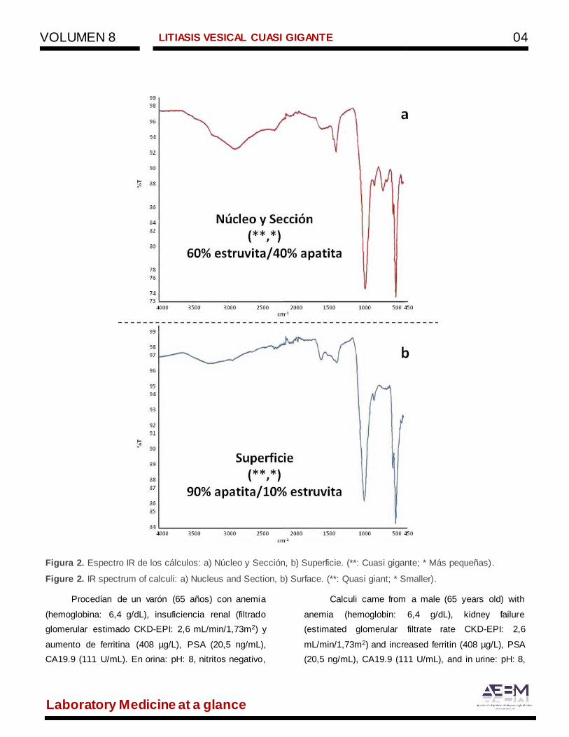
Conforme a la clasificación de Daudon2, la
descripción morfológico/macroscópica y la compo-
sición de los cálculos fue la siguiente (Figuras 1 y 2):
Núcleo y Sección: (**,*): duro, estructura
compacta, ligera y discontinua laminación
concéntrica, color blanquecino; 60%
estruvita/40% apatita (IVc).
Superficie: (**): esferoidal, color beige-marrón,
ligeramente rugoso, mate, 5 cm de diámetro, 96
g de peso; (*): piramidal, color beige-marrón,
liso, brillante, 0,3-3,3 g de peso, 0,7x0,7x0,5 -
2x1,9x1,1 cm de tamaño; (**, *): 90%
apatita/10% estruvita (IVb).
Los cálculos analizados eran mixtos, indicando
que los procesos formativos incluían varios factores
de riesgo. Los cálculos infecciosos son consecuencia
directa de una infección persistente o recurrente por
bacterias ureolíticas y suelen ser exacerbados por
alguna obstrucción, ya que este tipo de cálculos crece
rápidamente3.
The 57 calculi presented (Figure 1a) were
extracted using cystolithotomy and sent to the
laboratory for analysis, which was performed using
infrared spectroscopy (IRS) (Spectrum Two (Perkin
Elmer SL, Madrid, Spain).
Among the existing analytical methods, IRS has
been reported as one of the most reliable methods due
to its ability to detect inorganic and organic material
(cystine/uric/salts)1. IRS provides the spectra of the
components which are present in the fragment
analyzed and their comparison with a spectral
baseline library.
Since factors involved in the nucleation may be
different from those involved in the growth of the
calculus, the separate examination of the nucleus, the
section and the surface is essential.
In accordance with Daudon's classification2, the
morphological/macroscopic description and the
composition of the calculi were the following (Figures
1 and 2):
Nucleus and Section: (**,*): hard, compact
structure, light and discontinuous concentric
lamination, whitish colour; 60% struvite/40%
apatite (IVc).
Surface: (**): spheroidal, beige-brown, slightly
rough, matt, 5 cm in diameter, 96 g in weight ;
(*): pyramidal, beige-brown, smooth, shiny, 0.3-
3.3 g in weight, 0.7x0.7x0.5 - 2x1.9x1.1 cm in
size; (**, *): 90% apatite/10% struvite (IVb).
The calculi analyzed were mixed, thus
indicating that formation processes included several
risk factors. Infectious calculi are a direct consequence
of a persistent or recurrent infection due to ureolytic
bacteria and these are usually exacerbated by some
obstruction, since this type of stones grow rapidly3.
VOLUMEN 8
LITIASIS VESICAL CUASI GIGANTE 04
Laboratory Medicine at a glance
Figura 2. Espectro IR de los cálculos: a) Núcleo y Sección, b) Superficie. (**: Cuasi gigante; * Más pequeñas).
Figure 2. IR spectrum of calculi: a) Nucleus and Section, b) Surface. (**: Quasi giant; * Smaller).
Procedían de un varón (65 años) con anemia
(hemoglobina: 6,4 g/dL), insuficiencia renal (filtrado
glomerular estimado CKD-EPI: 2,6 mL/min/1,73m2) y
aumento de ferritina (408 µg/L), PSA (20,5 ng/mL),
CA19.9 (111 U/mL). En orina: pH: 8, nitritos negativo,
Calculi came from a male (65 years old) with
anemia (hemoglobin: 6,4 g/dL), kidney failure
(estimated glomerular filtrate rate CKD-EPI: 2,6
mL/min/1,73m2) and increased ferritin (408 µg/L), PSA
(20,5 ng/mL), CA19.9 (111 U/mL), and in urine: pH: 8,
VOLUMEN 8
LITIASIS VESICAL CUASI GIGANTE 05
Laboratory Medicine at a glance
albuminuria (174 mg/L), hematuria y leucocituria (30-
60 hematíes y 30-60 leucocitos/campo). El urocultivo
fue informado como orina contaminada por múltiples
organismos, prescribiéndose cefuroxima. Tras
ecografía se identificó: globo vesical, presencia de
numerosas litiasis en su interior y próstata aumentada
que improntaba sobre la vejiga. El diagnóstico fue
ureterohidronefrosis bilateral grado IV/V e hiperplasia
benigna de próstata.
La litiasis vesical gigante (>100 g) representa
tan solo el 1% de las urolitiasis. Es más frecuente en
varones de edad avanzada siendo la principal causa
la obstructiva (75%), y otras la infección crónica o la
presencia de cuerpo extraño4.
Los pacientes suelen referir clínica secundaria
a la inflamación que la litiasis genera en la mucosa
vesical y acuden por síndrome miccional o hematuria.
Los cálculos vesicales pueden causar hidronefros is
uni-bilateral5.
La infección del tracto urinario está asociada
con la patogénesis de la litiasis vesical (22-34% de los
casos). Cabe mencionar gérmenes como Proteus spp,
Pseudomonas spp y algunas cepas de
Staphylococcus que hidrolizan la urea con el
consiguiente aumento de NH3, CO2 y pH (>7,2),
sobresaturación de la orina y nucleación heterogénea
con formación de cálculo de estruvita (fosfato amónico
magnésico)6. La estruvita suele ir acompañada de
apatita (fosfato cálcico)1. Generalmente el cálculo es
único (70 %) y de composición mixta.
Dado que la litiasis se caracteriza por una alta
frecuencia de recidiva conviene conocer su
composición química para instaurar el tratamiento
adecuado e incluso como elemento imprescindible en
el estudio del origen de la enfermedad7.
negative nitrites, albuminuria (174 mg/L), hematuria
and leukocyturia (30-60 erythrocytes and 30-60
leukocytes/field). The urine culture was reported as
urine contaminated by multiple organisms and he was
prescribed cefuroxime. After ultrasound, it was
identified: distended bladder, presence of numerous
stones inside and enlarged prostate that was imprinted
on the bladder. The diagnosis was bilateral
ureterohydronephrosis grade IV/V and benign
prostatic hyperplasia.
Giant bladder stones (> 100 g) represent only
1% of urolithiasis and they are more common in males
and elderly men, being the obstructive uropathy the
main cause (75%) but others are chronic infection or
the presence of a foreign body4.
Patients usually complain about clinical
symptoms which are secondary to the inflammation
generated by the lithiasis in the bladder mucosa; they
suffer from voiding syndrome or hematuria. Bladder
stones may cause uni-bilateral hydronephrosis5.
Urinary tract infection is associated with the
pathogenesis of bladder stones (22-34% of cases). It
is worth mentioning germs such as Proteus spp,
Pseudomonas spp and some strains of
Staphylococcus which hydrolyze urea with the
consequent increase in NH3, CO2 and pH (>7.2), urine
oversaturation and heterogeneous nucleation with the
formation of struvite stones (magnesium ammonium
phosphate)6. Struvite is often accompanied by apatite
(calcium phosphate)1. The calculus is generally unique
(70%) and its composition is mixed.
Given that lithiasis is characterized by a high
frequency of relapse, it is convenient to know its
chemical composition in order to establish proper
treatment and even as an essential element in the
study of the disease origin7.

VOLUMEN 8
LITIASIS VESICAL CUASI GIGANTE 06
Laboratory Medicine at a glance
Bibliografía/References:
1. Guerra López JR, Güida JA, Della CO, Ricardo R. Estudio de cálculos renales por espectroscopía de
Infrarrojo. Acta Bioquim Clin Latinoam 2008; 42(2): 189-93.
2. Daudon M, Bader CA, Jungers P. Urinary calculi: review of classification methods and correlations with
etiology. Scanning Microsc 1993; 7(3): 1081-106.
3. Miller N, Evan A, Lingeman J. Patogenia de los cálculos renales. Urol Clin N Am 2007; 34:295-313.
4. Lázaro Rodríguez T, Camilo Ramírez AF, Bueno Sánchez E, Horroutinel Scull RS. Litiasis Vesical gigante.
Rev Cub Med Mil 2013; 42(3): 411-6.
5. Ciftci H, Savas M. Unilateral hydronephrosis. Secondary to Giant Bladder Stone. Turk J Urol 2008; 34:
261-3.
6. Griffith DP. Urease stones. Urol Res 1979; 7(3):215-21.
7. Tiselius HG, Ackermann D, Alken P, Buck C, Conort O, Gallucci. Guidelines on urolithiasis. Eur Urol 2001;
40(4):362-71.

Laboratory Medicine at a glance
Medicina de Laboratorio de un vistazo
VOL.8 ISSN 2444-8699
UTILIZACIÓN DEL DIAGRAMA DE FLUJO DE PROCESOS
COMO HERRAMIENTA PARA EL ENTRENAMIENTO DEL
PERSONAL TÉCNICO
USING A PROCESS FLOW DIAGRAM AS A TRAINING TOOL FOR
LABORATORY TECHNICIANS
Autores Rosa Mª Lillo Rodríguez
Mª Concepción Donlo Gil
Filiación Servicio de Análisis Clínicos.
Complejo Hospitalario de
Navarra
Fecha de publicación 03 agosto 2018
Páginas Páginas 7-10
Figura 1: Diagrama de flujo en la recepción de
muestra de semen posvasectomía. Figure 1: Postvasectomy
semen analysis process flow diagram.
VOLUMEN 8
UTILIZACIÓN DEL DIAGRAMA DE FLUJO DE PROCESOS COMO HERRAMIENTA PARA EL ENTRENAMIENTO DEL PERSONAL TÉCNICO 08
Laboratory Medicine at a glance
El estudio de las muestras de semen
posvasectomía de acuerdo con las recomendaciones
de la guía clínica de la Asociación Europea de
Urología1 y de la Sociedad Española de Medicina de
Laboratorio (SEQC-ML)2,3, requiere un control estricto
de la fase preanalítica y el cumplimiento por parte del
paciente de los requisitos establecidos para la toma
de la muestra.
En la figura 1 se muestra el diagrama de flujo
elaborado en el Complejo Hospitalario de Navarra
(CHN) con el objetivo de facilitar el entrenamiento del
personal de la sección recepción de muestras. En él
se muestran las acciones correspondientes a cada
fase del proceso. Se establecen incidencias
resolubles por parte del técnico de recepción de
muestras (cajas amarillas) e incidencias irresolubles
(cajas rojas) que obligan al rechazo de la muestra y
solicitud de nueva cita por parte del paciente. De esta
manera, únicamente se enviarán al laboratorio
aquellas muestras que cumplan con los siguientes
requisitos:
1. Pacientes citados que acuden con la solicitud
correspondiente debidamente identificada.
2. Pacientes que han cumplido con los requisitos
para la toma de la muestra.
3. Muestra identificada de manera inequívoca.
El proceso se inicia con la identificación del
paciente y su búsqueda en el listado diario. El trabajo
mediante cita previa permite que la carga de trabajo
no supere la capacidad del laboratorio para el
procesamiento adecuado de las muestras. Por lo
tanto, la falta de cita previa se consideró una
incidencia irresoluble. Asimismo, la ausencia del
volante de solicitud de la prueba se consideró
irresoluble, aunque en este caso por motivos
administrativos. En cambio, la identificación
incompleta de los datos del paciente en la solicitud se
Postvasectomy semen analysis according to
the European Urology Association1 and Sociedad
Española de Medicina de Laboratorio (SEQC-ML)
guidelines2,3, requires an strict control for the
preanalytical phase and also patient´s compliance with
sample collection requirements.
Figure 1 shows the process flow diagram
designed at Complejo Hospitalario de Navarra (CHN)
in order to improve personnel training at the sample
reception area. It shows specific tasks in every phase
of the process. Minor incidents which could be solved
by CHN staff are marked with yellow boxes, while the
serious ones are marked in red, cause sample
rejection and force the obtaining of a new appointment
by the patient. The procedure only allows sample
shipment for laboratory testing if the following
requirements are met:
4. Samples from patients with an appointment and
a laboratory request form.
5. Samples collected following CHN staff
requirements.
6. Complete identified samples.
The procedure starts with patient identification
and the checking of patient identification with the
appointments list. Working under appointment allows
controlling the laboratory workload, so excess of
samples do not compromise the quality of the analysis.
Thus, absence of an appointment is considered as a
serious incident that cannot be solved by the reception
area laboratory technicians. Lack of an acceptable
laboratory request is also considered as a serious
incident due to administrative issues. On the contrary,
incomplete patient identification in the laboratory
request is considered as a mild issue, because it can
be solved after checking his electronic medical records
at CHN.
VOLUMEN 8
UTILIZACIÓN DEL DIAGRAMA DE FLUJO DE PROCESOS COMO HERRAMIENTA PARA EL ENTRENAMIENTO DEL PERSONAL TÉCNICO 09
Laboratory Medicine at a glance
consideró solucionable tras el cotejo de los datos
aportados por el paciente con los registrados en la
historia clínica del CHN.
El cumplimiento de los requisitos establecidos
de manera conjunta por los servicios de Urología y
Análisis Clínicos del CHN para el análisis de las
muestras se comprueba a través de la inspección de
los datos aportados en una encuesta preanalítica.
Compliance with the Urology and Laboratory
Medicine Department requirements for sample
analysis is assessed using a preanalytical sample data
collection form.
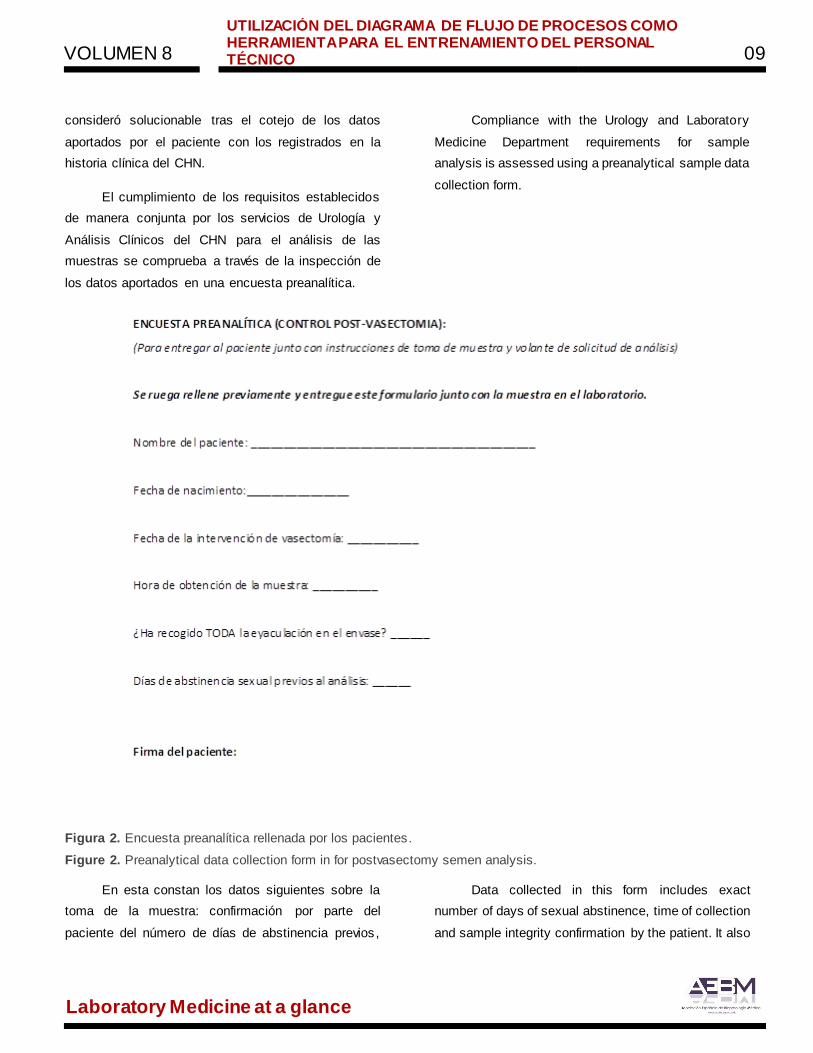
Figura 2. Encuesta preanalítica rellenada por los pacientes.
Figure 2. Preanalytical data collection form in for postvasectomy semen analysis.
En esta constan los datos siguientes sobre la
toma de la muestra: confirmación por parte del
paciente del número de días de abstinencia previos ,
Data collected in this form includes exact
number of days of sexual abstinence, time of collection
and sample integrity confirmation by the patient. It also
VOLUMEN 8
UTILIZACIÓN DEL DIAGRAMA DE FLUJO DE PROCESOS COMO HERRAMIENTA PARA EL ENTRENAMIENTO DEL PERSONAL TÉCNICO 10
Laboratory Medicine at a glance
hora exacta y recogida íntegra de esta. Además
deberán constar los datos personales del paciente y
su firma. El incumplimiento de alguno de los requisitos
se considera criterio directo para el rechazo de la
muestra, ya que podría poner en riesgo la veracidad
de los resultados obtenidos.
Finalmente, la muestra quedará identificada
con los datos demográficos del paciente y un número
de laboratorio en el cual se incluirá el informe de
resultados.
De esta forma, el laboratorio puede garantizar
la recepción, procesamiento y realización del informe
de las muestras de semen posvasectomía antes de
dos horas tras su toma, asegurando la calidad del
resultado, al tiempo que permite que el paciente
pueda acudir el mismo día a la consulta de Urología si
fuera necesario.
records patient´s demographic data and signature.
Failure to fulfill with any of the requirements is a
straight sample rejection criterion due to possible
sample misdiagnosis. Finally, samples are identified
with patient´s demographic data and also laboratory
identification number that will appear on the sample
analysis report.
This procedure, allows sample reception,
laboratory analysis and sample reporting within two
hours after sample collection under strict quality
sample management. Moreover, patients are allowed
to attend to the urology office within the day if needed.
Bibliografía/References:
1. G.R. Dohle, T. Diemer, Z. Kopa, C. Krausz, A. Giwercman, A. Jungwirth para el Grupo de Trabajo de la
Asociación Europea de Urología sobre la Infertilidad Masculina. Guía clínica de la Asociación Europea de
Urología sobre la vasectomía. Actas Urol Esp. 2012;36(5):276-281
2. M.C. Sánchez Pozo, I. Sánchez Prieto, M.I. Jiménez García. Comité Científico Comisión de Seminología
y Técnicas de Reproducción Asistida. Recomendaciones para la estandarización del análisis de semen
posvasectomía Documento Técnico (2012). Documentos de la SEQC- Diciembre 2012.
3. M.C. Sánchez Pozo, S. Izquierdo Álvarez, I. Sánchez Prieto, M.I. Jiménez García. Comité Científico
Comisión de Seminología y Técnicas de Reproducción Asistida. Recomendaciones en el proceso
preanalítico del análisis de semen II. Recomendación 2016.
Laboratory Medicine at a glance
Medicina de Laboratorio de un vistazo
VOL.8 ISSN 2444-8699
DIAGNÓSTICO DE ESCLERODERMIA MEDIANTE LA
DETECCIÓN DE ANTICUERPOS ANTINUCLEARES POR
INMUNOFLUORESCENCIA
DIAGNOSIS OF SCLERODERMA THROUGH DETECTION OF
ANTINUCLEAR ANTIBODIES BY IMMUNOFLUORESCENCE
A
B
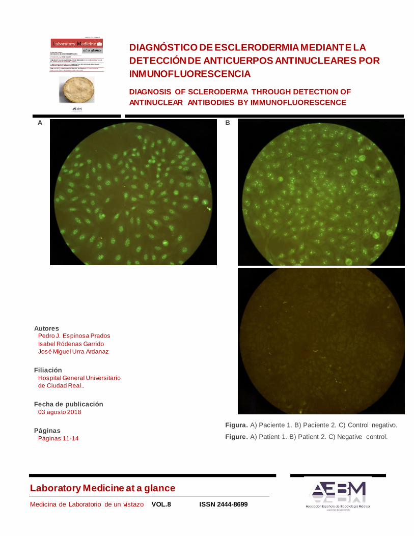
Figura. A) Paciente 1. B) Paciente 2. C) Control negativo.
Figure. A) Patient 1. B) Patient 2. C) Negative control.
Autores Pedro J. Espinosa Prados
Isabel Ródenas Garrido
José Miguel Urra Ardanaz
Filiación Hospital General Universitario
de Ciudad Real..
Fecha de publicación 03 agosto 2018
Páginas Páginas 11-14
VOLUMEN 8 DIAGNÓSTICO DE ESCLERODERMIA MEDIANTE LA DETECCIÓN DE
ANTICUERPOS ANTINUCLEARES POR INMUNOFLUORESCENCIA 12
Laboratory Medicine at a glance
Se presentan dos imágenes, donde se
muestran la detección de anticuerpos antinucleares
(ANA) mediante inmunofluorescencia indirecta (IFI) ,
en suero de dos pacientes de 41 y 45 años, con
sospecha clínica de esclerodermia.
La primera paciente se diagnosticó inicialmente
como fenómeno de Raynaud y se inició tratamiento
con antagonistas del calcio. Debido a la persistencia
del cuadro clínico, se realizó un estudio de ANA, por
screening mediante enzimoinmunoensayo con un
resultado positivo de 10.20 U/mL. En la imagen A se
observa la detección, sobre células Hep-2, de ANA
con un patrón anti-centrómero (ACA) con un título de
1/640, que es un criterio muy positivo para el
diagnóstico de la Esclerodermia Sistémica Limitada
(ESL). El paciente presenta únicamente afectación
cutánea y se descartó la afectación en otros órganos
tras los resultados de radiografía de tórax, TAC
torácico y analítica completa con gasometría.
La segunda paciente acude al Servicio de
Urgencias con un cuadro clínico que se relacionó con
un brote de fenómeno de Raynaud. Se realizó el
estudio de ANA, con un resultado positivo de 2.81
U/mL en el screening, y, con un patrón nucleolar
moteado de título 1/640, compatible con anticuerpos
anti-fibrilarina (Imagen B). En el estudio clínico se le
observan nódulos pulmonares, y se le diagnostica de
Esclerodermia Sistémica Difusa (ESD) con afectación
pulmonar.
El estudio de ANA se realizó mediante
microscopia de fluorescencia con un aumento de 40x,
por IFI sobre la línea celular Hep-2 (KallestadTMHep-2
Cell Line Slide, Bio-Rad; filtros Exc:330-
380/EspDic:400/Bar:420; lámpara LED UV, pE300
Light Source). En ambos casos, la IFI fue suficiente
Two images are presented, where the detection
of antinuclear antibodies (ANA) by indirect
immunofluorescence (IFA), in the serum of two
patients aged 41 and 45 years old, with suggestive
clinical of Scleroderma.
The first patient was diagnosed as Raynaud´s
phenomenon and became a treatment with calcium
channel blockers. Due to the persistence of the
clinical, an ANA study was carried out, by screening
with enzyme immunoassay with a positive result of
10.20 U/mL. In the image, we see the detection of
Hep-2 cells of ANA with an anti-centromere pattern
(ACA) with a title of 1/640, which is diagnostic of
Limited Systemic Scleroderma (LSS). The patient
presented cutaneous affectation, and the affectation in
other organs was ruled out after the results of the chest
x-ray, thoracic TC and complete analytical with arterial
blood gases.
The second patient goes to the emergency with
a clinical picture that was related to an outbreak of
Raynaud´s phenomenon. The ANA study was
performed, with a positive result of 2.81 U/mL in the
screening, and, with a clumpy nucleolar pattern of title
of 1/640, compatible with antibodies anti-fibrillarin
(Image B). In the clinical study, are observed
pulmonary nodules, and he is diagnosed as Diffuse
Systemic Scleroderma (DSS) with pulmonary
involvement.
The ANA study was performed by fluorescence
microscopy with the 40x objective, by IFI on the Hep-
2 cell line (KallestadTMHep-2 Cell Line Slide, Bio-Rad;
filters Exc:330-380/MirDic:400/Bar:420; LED lamp of
UV light, pE300). In both cases, the ANA pattern
establishes the differentiated diagnoses of S.S.
Scleroderma is a rare and serious disease,
inside the group of connective tissue diseases. It

VOLUMEN 8 DIAGNÓSTICO DE ESCLERODERMIA MEDIANTE LA DETECCIÓN DE
ANTICUERPOS ANTINUCLEARES POR INMUNOFLUORESCENCIA 13
Laboratory Medicine at a glance
para establecer los diagnósticos diferenciados de las
dos modalidades de ES.
La esclerodermia es una enfermedad rara, del
grupo de las enfermedades del tejido conectivo.
Afecta preferentemente a mujeres entre la 3ª y 5ª
década de vida1. Es una enfermedad de etiología
desconocida en la que intervienen tanto factores
genéticos como ambientales2. Se caracteriza por una
afectación vascular precoz (el primer síntoma suele
ser el fenómeno de Raynaud), seguida por una fibrosis
tisular sistémica que puede afectar a muchos órganos.
Cursa con cansancio, artralgias, mialgias, hinchazón
de manos, afectación cutánea y de órganos internos
(como son pulmón, tubo digestivo, corazón y riñón),
siendo una enfermedad con alta morbimortalidad1,3.
Tanto las manifestaciones clínicas como el curso de la
enfermedad son muy variadas.
Se clasifica en dos grandes grupos: ESL, con
afectación cutánea y leve en órganos internos, y ESD,
donde se pueden afectar un importante número de
órganos con un curso clínico más grave. La presencia
de patrones IFI característicos diferencia en los ANA
las dos modalidades3.
La ESL se asocia a ACA (80-90% de los
pacientes) que reconocen las proteínas del
centrómero (CENP)-A, B y C4. Al microscopio se
observa como los núcleos se tiñen con puntos finos
distribuidos de manera homogénea en las células de
interfase, en cambio, en las células en mitosis el
punteado de localiza en la placa de la cromatina5. En
nuestro caso se confirmó la presencia de anticuerpos
frente a la proteína recombinante B del centrómero,
mediante inmunoblot (nº Ref: DL 1590-161-3 G.
Anticuerpos contra antígenos nucleares (IgG),
Euroimmun).
Por su parte, la ESD se asocia a un patrón
nucleolar con una tinción intensa de nucleolos. La
preferentially affects women between the 3rd and 5th
decade of life1. It is a disease of unknown etiology in
which both genetic and environmental factors was
present2. It is characterized by early vascular
involvement (the first frequent symptom of Raynaud´s
phenomenon), followed by a systemic tissue fibrosis
that can affect many organs. Clinically presents
fatigue, arthralgias, myalgia, swelling of the hands,
cutaneous involvement and internal organs (such as
lung, digestive tract, heart and kidney), being a
disease with high morbidity and mortality1,3. Both the
clinical manifestations and the course of the disease
are very varied.
It is classified into two large groups: LSS, with
cutaneous involvement and slight in internal organs,
and DSS, where a significant number of organs can be
affected with a more serious clinical course. The
presence of ANA differs by means of its characteristic
IFA patterns the two forms3. The LSS is associated
with ACA (80-90% of patients) that recognizes the
centromere proteins (CENP)-A, B and C4. Under the
microscope, the nuclei are stained with fine points
distributed homogeneously in the interphase cells,
whereas in the cells in mitosis the dotted location in
the chromatin plate5. In our case, the presence of
antibodies against recombinant protein B of the
centromere was confirmed by immunoblot (Ref: DL
1590-161-3G. Antibodies against nuclear antigens
(IgG), Euroimmun).
For its part, the DSS is associated with a
nucleolar pattern with intense staining of nucleoli.
Staining on cells in mitosis is variable depending on its
specific antigen: fibrillarin, NOR90, RNA polymerase
or topoisomerase I (anti-Scl70), although all present
nucleolar staining and are associated with DSS6. In
our hospital, no protocol is followed where other
techniques are used to confirm the presence of a
specific type of anti-nucleolar antibodies. The
VOLUMEN 8 DIAGNÓSTICO DE ESCLERODERMIA MEDIANTE LA DETECCIÓN DE
ANTICUERPOS ANTINUCLEARES POR INMUNOFLUORESCENCIA 14
Laboratory Medicine at a glance
tinción sobre células en mitosis es variable
dependiendo de su antígeno específico: fibrilarina,
NOR90, RNA polimerasa o topoisomerasa I (anti-
Scl70), aunque todas presentan tinción nucleolar y se
asocian con ESD6. En nuestro hospital no se sigue
ningún protocolo donde se empleen otras técnicas
para confirmar la presencia de un tipo específico de
anticuerpos anti-nucleolares. En este contexto, el
patrón nucleolar en IFI tiene valor diagnóstico con
independencia del antígeno específico.
nucleolar pattern in IFI has a diagnostic value
independent of the specific antigen.
.
Bibliografía/References:
1. Elhai M, Avouac J, Kahan A, Allanore Y. Esclerodermia sistémica. EMC - Apar Locomot [Internet].
2015;48(3):1–15. Available from: http://dx.doi.org/10.1016/S1286-935X(15)72882-0
2. Batista Remedios ES, Grass Velázquez A, Avilés del Campo E, Pérez Torres L. Mecanismos
etiopatogénicos en la esclerosis sistémica. Rev Cuba Reumatol. 2014;16(3):304–8.
3. Carreira PE, Martín-López M, Pablos Álvarez JL. Esclerodermia. Med. 2017;12(25):1448–57.
4. Kuwana M. Circulating Anti-Nuclear Antibodies in Systemic Sclerosis: Utility in Diagnosis and Disease
Subsetting. J Nippon Med Sch. 2017;84(2):56–63.
5. Cabiedes J, Núñez-Álvarez CA. Anticuerpos antinucleares. Reumatol Clin. 2010;6(4):224–30.
6. Ho KT, Reveille JD. The clinical relevance of autoantibodies in scleroderma. Arthritis Res Ther [Internet].
2003;5(2):80–93. Available from:
http://www.ncbi.nlm.nih.gov/pubmed/12718748%5Cnhttp://www.pubmedcentral.nih.gov/articlerender. fcgi
?artid=PMC165038.
Laboratory Medicine at a glance
Medicina de Laboratorio de un vistazo
VOL.8 ISSN 2444-8699
UTILIDAD DE LA ESPECTROFOTOMETRÍA DE ABSORCIÓN
EN LA DETECCIÓN DE XANTOCROMÍA EN LA HEMORRAGIA
SUBARACNOIDEA
USAGE OF ABSORPTION SPECTROSCOPY TO IDENTIFY
XANTHOCHROMIA IN SUBARACHNOID HAEMORRAGE
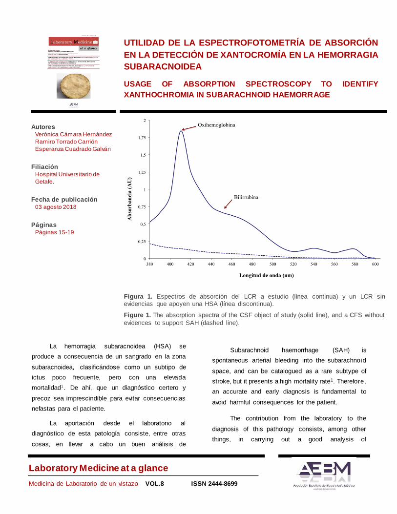
Figura 1. Espectros de absorción del LCR a estudio (línea continua) y un LCR sin evidencias que apoyen una HSA (línea discontinua).
Figure 1. The absorption spectra of the CSF object of study (solid line), and a CFS without
evidences to support SAH (dashed line).
La hemorragia subaracnoidea (HSA) se
produce a consecuencia de un sangrado en la zona
subaracnoidea, clasificándose como un subtipo de
ictus poco frecuente, pero con una elevada
mortalidad1. De ahí, que un diagnóstico certero y
precoz sea imprescindible para evitar consecuencias
nefastas para el paciente.
La aportación desde el laboratorio al
diagnóstico de esta patología consiste, entre otras
cosas, en llevar a cabo un buen análisis de
Subarachnoid haemorrhage (SAH) is
spontaneous arterial bleeding into the subarachnoid
space, and can be catalogued as a rare subtype of
stroke, but it presents a high mortality rate1. Therefore,
an accurate and early diagnosis is fundamental to
avoid harmful consequences for the patient.
The contribution from the laboratory to the
diagnosis of this pathology consists, among other
things, in carrying out a good analysis of
Autores Verónica Cámara Hernández
Ramiro Torrado Carrión
Esperanza Cuadrado Galván
Filiación Hospital Universitario de
Getafe.
Fecha de publicación 03 agosto 2018
Páginas Páginas 15-19

VOLUMEN 8
UTILIDAD DE LA ESPECTROFOTOMETRÍA DE ABSORCIÓN EN LA DETECCIÓN DE XANTOCROMÍA EN LA HEMORRAGIA SUBARACNOIDEA 16
Laboratory Medicine at a glance
xantocromía. A pesar de las múltiples revisiones sobre
el tema, no hay unanimidad a la hora de definir el
método más adecuado para ello2,3. Sin embargo, el
estudio espectrofotométrico proporciona mayor
sensibilidad en los resultados si se sigue un buen
protocolo de actuación4, frente al estudio visual que
puede conducir a falsos positivos, puesto que el
líquido cefalorraquídeo (LCR) puede tener aspecto
xantocrómico a consecuencia de una punción
traumática.
El caso a estudio corresponde a un paciente de
73 años, que acude a urgencias por presentar desde
hace 10-14 días molestias en la parte posterior de
piernas y en ambos muslos dificultándole la
deambulación. Diez días previos al ingreso presenta
cifras tensionales elevadas con tensión arterial
sistólica de 180 mmHg junto con cefalea bifrontal y
occipital pulsátil intensa que le imposibilita el sueño.
En la tomografía computerizada (TC) de cráneo sin
contraste no se observan hallazgos reseñables.
Debido a la sospecha de HSA, entre otras
pruebas el clínico solicita estudio de xantocromía, que
se realizará mediante espectrofotometría de absor-
ción, celularidad y bioquímica de una muestra de LCR.
El LCR obtenido es hemático y no aclara con
sucesivos tubos. Desde el laboratorio de urgencias se
aporta el informe de la figura 2.
El espectro de absorción obtenido en el estudio
de xantocromía (Figura 1) es compatible con HSA. Se
observa un máximo de absorción aproximadamente a
410 nm correspondiente a la oxihemoglobina, junto
con un hombro entre 450-460 nm asociado a la
bilirrubina.
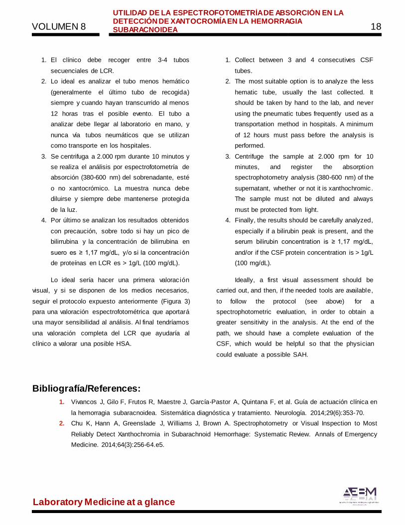
A la hora de llevar a cabo el estudio de
xantocromía nuestro laboratorio sigue el protocolo
mostrado en la Figura 3 basado en guías actualizadas
en el tema5.
xanthochromia. Despite the multiple reviews about the
subject2,3, there is no unanimity defining the most
appropriate method for its determination. However,
when instead of a visual study, a spectrophotometric
study following an accurate protocol is performed, we
can see that higher sensitivity in the results is
obtained4. The visual study could lead to a false-
positive, due to the fact that xanthochromia might be
present in the cerebrospinal fluid (CSF) as a
consequence of a traumatic puncture.
The case to study corresponds with a 73 years
old patient attending to emergencies as a
consequence of a 10-14 days long discomfort in the
back side of his legs and thighs, making him difficult to
walk. Besides, 10 days before admission, he
presented high blood pressure, with a systolic
pressure of 180 mmHg, as well as a bifrontal and
intense pulsatile occipital headache that impeded him
to sleep. The non-contrast computed tomography (CT)
of the cranium showed nothing remarkable.
Due to suspicion of SAH, among other tests, the
physician requested xanthochromia, which it will be
carry out by absorption spectroscopy, cellularity and
biochemistry tests of the CSF.
The CSF obtained was hematic and did not
lighten with successive tubes. The report of the figure
2 was provided by the laboratory.
The absorption spectrum obtained from the
xanthochromia study (Figure 1) was compatible with
SAH. An absorption maximum corresponding to
oxyhemoglobin was observed around 410 nm,
together with a shoulder associated with bilirubin
between 450-460 nm.
Our laboratory follows the protocol shown in
Figure 3, based on updated guides about the subject,
to study the xanthochromia5.
VOLUMEN 8
UTILIDAD DE LA ESPECTROFOTOMETRÍA DE ABSORCIÓN EN LA DETECCIÓN DE XANTOCROMÍA EN LA HEMORRAGIA SUBARACNOIDEA 17
Laboratory Medicine at a glance
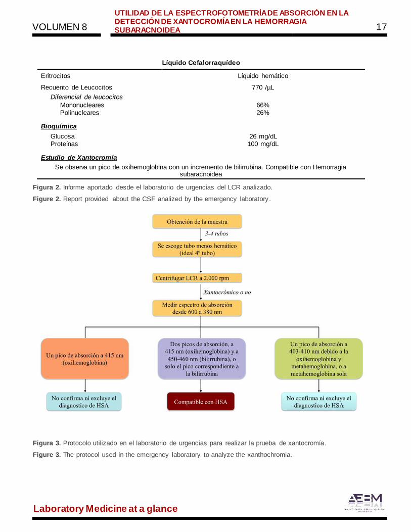
Líquido Cefalorraquídeo
Eritrocitos Líquido hemático
Recuento de Leucocitos 770 /µL
Diferencial de leucocitos
Mononucleares 66% Polinucleares 26%
Bioquímica
Glucosa 26 mg/dL Proteínas 100 mg/dL
Estudio de Xantocromía
Se observa un pico de oxihemoglobina con un incremento de bilirrubina. Compatible con Hemorragia subaracnoidea
Figura 2. Informe aportado desde el laboratorio de urgencias del LCR analizado.
Figure 2. Report provided about the CSF analized by the emergency laboratory .
Figura 3. Protocolo utilizado en el laboratorio de urgencias para realizar la prueba de xantocromía.
Figure 3. The protocol used in the emergency laboratory to analyze the xanthochromia.
VOLUMEN 8
UTILIDAD DE LA ESPECTROFOTOMETRÍA DE ABSORCIÓN EN LA DETECCIÓN DE XANTOCROMÍA EN LA HEMORRAGIA SUBARACNOIDEA 18
Laboratory Medicine at a glance
1. El clínico debe recoger entre 3-4 tubos
secuenciales de LCR.
2. Lo ideal es analizar el tubo menos hemático
(generalmente el último tubo de recogida)
siempre y cuando hayan transcurrido al menos
12 horas tras el posible evento. El tubo a
analizar debe llegar al laboratorio en mano, y
nunca vía tubos neumáticos que se utilizan
como transporte en los hospitales.
3. Se centrifuga a 2.000 rpm durante 10 minutos y
se realiza el análisis por espectrofotometría de
absorción (380-600 nm) del sobrenadante, esté
o no xantocrómico. La muestra nunca debe
diluirse y siempre debe mantenerse protegida
de la luz.
4. Por último se analizan los resultados obtenidos
con precaución, sobre todo si hay un pico de
bilirrubina y la concentración de bilirrubina en
suero es ≥ 1,17 mg/dL, y/o si la concentración
de proteínas en LCR es > 1g/L (100 mg/dL).
Lo ideal sería hacer una primera valorac ión
visual, y si se disponen de los medios necesarios,
seguir el protocolo expuesto anteriormente (Figura 3)
para una valoración espectrofotométrica que aportará
una mayor sensibilidad al análisis. Al final tendríamos
una valoración completa del LCR que ayudaría al
clínico a valorar una posible HSA.
1. Collect between 3 and 4 consecutives CSF
tubes.
2. The most suitable option is to analyze the less
hematic tube, usually the last collected. It
should be taken by hand to the lab, and never
using the pneumatic tubes frequently used as a
transportation method in hospitals. A minimum
of 12 hours must pass before the analysis is
performed.
3. Centrifuge the sample at 2.000 rpm for 10
minutes, and register the absorption
spectrophotometry analysis (380-600 nm) of the
supernatant, whether or not it is xanthochromic .
The sample must not be diluted and always
must be protected from light.
4. Finally, the results should be carefully analyzed,
especially if a bilirubin peak is present, and the
serum bilirubin concentration is ≥ 1,17 mg/dL,
and/or if the CSF protein concentration is > 1g/L
(100 mg/dL).
Ideally, a first visual assessment should be
carried out, and then, if the needed tools are available,
to follow the protocol (see above) for a
spectrophotometric evaluation, in order to obtain a
greater sensitivity in the analysis. At the end of the
path, we should have a complete evaluation of the
CSF, which would be helpful so that the physician
could evaluate a possible SAH.
Bibliografía/References:
1. Vivancos J, Gilo F, Frutos R, Maestre J, García-Pastor A, Quintana F, et al. Guía de actuación clínica en
la hemorragia subaracnoidea. Sistemática diagnóstica y tratamiento. Neurología. 2014;29(6):353-70.
2. Chu K, Hann A, Greenslade J, Williams J, Brown A. Spectrophotometry or Visual Inspection to Most
Reliably Detect Xanthochromia in Subarachnoid Hemorrhage: Systematic Review. Annals of Emergency
Medicine. 2014;64(3):256-64.e5.

VOLUMEN 8
UTILIDAD DE LA ESPECTROFOTOMETRÍA DE ABSORCIÓN EN LA DETECCIÓN DE XANTOCROMÍA EN LA HEMORRAGIA SUBARACNOIDEA 19
Laboratory Medicine at a glance
3. Perry JJ, Sivilotti MLA, Stiell IG, Wells GA, Raymond J, Mortensen M, et al. Should Spectrophotometry
Be Used to Identify Xanthochromia in the Cerebrospinal Fluid of Alert Patients Suspected of Having
Subarachnoid Hemorrhage? Stroke. 2006;37(10):2467-72.
4. Long B, Koyfman A, Runyon MS. Subarachnoid Hemorrhage: Updates in Diagnosis and Management.
Emergency Medicine Clinics of North America. 2017;35(4):803-24.
5. Cruickshank A, Auld P, Beetham R, Burrows G, Egner W, Holbrook I, et al. Revised national guidelines
for analysis of cerebrospinal fluid for bilirubin in suspected subarachnoid haemorrhage. Annals of Clinical
Biochemistry. 2008;45(3):238-44.