Leucemia e Inmunodeficiencia felina: Claves Diagnósticas · Leucemia e Inmunodeficiencia felina:...
22
Leucemia e Inmunodeficiencia felina: Claves Diagnósticas Mª Luisa Palmero. Certificada Medicina Felina ESVPS Acreditada Medicina Felina AVEPA Gattos Centro Clínico Felino www.gattos.net Virus de la Leucemia felina (FeLV) Es un gammaretrovirus responsable de la aparición de enfermedades no neoplásicas como anemia no regenerativa, inmunosupresión y neoplásicas como linfomas, leucemia y trastornos mieloproliferativos. La prevalencia es muy variable dependiendo de la zona o núcleo de población estudiado (albergues, gatos de ciudad…) Patogénesis: 1º Adsorción vírica a la superficie de la célula diana. 2º Fusión de la envuelta vírica con la pared celular y liberación de la nucleocápside con ARN vírico. 3º El ARN vírico se transcribe a ADN por acción de la transcriptasa inversa. El ADN vírico es transportado al núcleo celular donde se integra. Se forma así el PROVIRUS. 4º Durante la mitosis celular, las células hijas heredan el provirus (ADN vírico integrado). Por ello las infecciones por retrovirus son de por vida. 5º Producción de nuevas partículas víricas: El ADN integrado, produce ARNm, proteínas de la cápside (gag y pol) y ARN de nuevas partículas. El virión se ensambla bajo la membrana celular donde se encuentran proteínas de la envuelta necesarias para su salida de la célula por gemación. Este proceso no conlleva muerte celular. Diagnóstico: a. Pruebas diagnósticas: En FeLV las proteínas internas son muy inmunógenas y antigénicamente idénticas para todos los subgrupos de FeLV. La proteína de la cápside vírica gag, p27, se sintetiza en gran cantidad y se encuentra tanto en el citoplasma celular como en el medio extracelular como antígeno libre.
Transcript of Leucemia e Inmunodeficiencia felina: Claves Diagnósticas · Leucemia e Inmunodeficiencia felina:...

Leucemia e Inmunodeficiencia felina: Claves Diagnósticas
Mª Luisa Palmero.
Certificada Medicina Felina ESVPS Acreditada Medicina Felina AVEPA
Gattos Centro Clínico Felino www.gattos.net
Virus de la Leucemia felina (FeLV)
Es un gammaretrovirus responsable de la aparición de enfermedades no neoplásicas como anemia no regenerativa, inmunosupresión y neoplásicas como linfomas, leucemia y trastornos mieloproliferativos. La prevalencia es muy variable dependiendo de la zona o núcleo de población estudiado (albergues, gatos de ciudad…)
Patogénesis: 1º Adsorción vírica a la superficie de la célula diana. 2º Fusión de la envuelta vírica con la pared celular y liberación de la nucleocápside con ARN vírico. 3º El ARN vírico se transcribe a ADN por acción de la transcriptasa inversa. El ADN vírico es transportado al núcleo celular donde se integra. Se forma así el PROVIRUS. 4º Durante la mitosis celular, las células hijas heredan el provirus (ADN vírico integrado). Por ello las infecciones por retrovirus son de por vida. 5º Producción de nuevas partículas víricas: El ADN integrado, produce ARNm, proteínas de la cápside (gag y pol) y ARN de nuevas partículas. El virión se ensambla bajo la membrana celular donde se encuentran proteínas de la envuelta necesarias para su salida de la célula por gemación. Este proceso no conlleva muerte celular. Diagnóstico:
a. Pruebas diagnósticas:
En FeLV las proteínas internas son muy inmunógenas y antigénicamente idénticas para todos los subgrupos de FeLV. La proteína de la cápside vírica gag, p27, se sintetiza en gran cantidad y se encuentra tanto en el citoplasma celular como en el medio extracelular como antígeno libre.
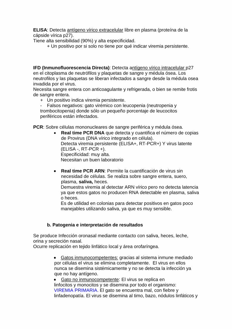
ELISA: Detecta antígeno vírico extracelular libre en plasma (proteína de la cápside vírica p27). Tiene alta sensibilidad (90%) y alta especificidad.
+ Un positivo por si solo no tiene por qué indicar viremia persistente.
IFD (Inmunofluorescencia Directa): Detecta antígeno vírico intracelular p27 en el citoplasma de neutrófilos y plaquetas de sangre y médula ósea. Los neutrofilos y las plaquetas se liberan infectados a sangre desde la médula osea invadida por el virus. Necesita sangre entera con anticoagulante y refrigerada, o bien se remite frotis de sangre entera.
+ Un positivo indica viremia persistente. - Falsos negativos: gato virémico con leucopenia (neutropenia y trombocitopenia) donde sólo un pequeño porcentaje de leucocitos periféricos están infectados.
PCR: Sobre células mononucleares de sangre periférica y médula ósea.
Real time PCR DNA que detecta y cuantifica el número de copias de Provirus (DNA vírico integrado en célula). Detecta viremia persistente (ELISA+, RT-PCR+) Y virus latente (ELISA -, RT-PCR +). Especificidad: muy alta. Necesitan un buen laboratorio
Real time PCR ARN: Permite la cuantificación de virus sin necesidad de células. Se realiza sobre sangre entera, suero, plasma, saliva, heces. Demuestra viremia al detectar ARN vírico pero no detecta latencia ya que estos gatos no producen RNA detectable en plasma, saliva o heces. Es de utilidad en colonias para detectar positivos en gatos poco manejables utilizando saliva, ya que es muy sensible.
b. Patogenia e interpretación de resultados Se produce Infección oronasal mediante contacto con saliva, heces, leche, orina y secreción nasal. Ocurre replicación en tejido linfático local y área orofaríngea.
Gatos inmunocompetentes: gracias al sistema inmune mediado por células el virus se elimina completamente. El virus en ellos nunca se disemina sistémicamente y no se detecta la infección ya que no hay antígeno.
Gato no inmunocompetente: El virus se replica en linfocitos y monocitos y se disemina por todo el organismo: VIREMIA PRIMARIA. El gato se encuentra mal, con fiebre y linfadenopatía. El virus se disemina al timo, bazo, nódulos linfáticos y

glándulas salivares por lo que es infeccioso. Esta fase dura entre 3 y 16 semanas y en algunas ocasiones hasta un año.
Viremia primaria ELISA + IFD – RT-PCR +
Tras la viremia primaria:
VIREMICO TRANSITORIO o REGRESOR: El sistema inmune puede eliminar el virus antes de que éste llegue a Médula Osea. Ocurre en el 30-40% de los gatos infectados. Desarrollan respuesta inmune eficaz por neutralización con anticuerpos que le protege frente a futuras infecciones pero no tiene una duración de por vida. Se deberán vacunar anualmente de leucemia felina para aumentar su inmunidad natural
Virémico transitorio (pruebas en sangre) 1º ELISA +, RT-PCR + y tras meses del primero se hace el 2º ELISA -, RT-PCR –, IFD –
VIRÉMICO PERSISTENTE (viremia secundaria) El sistema inmune NO puede eliminar el virus, y éste llega a Médula Osea (ocurre en un 30-40% de los gatos infectados). Las células hematopoyéticas producen granulocitos y plaquetas infectadas que circulan por el cuerpo. El virus se encuentra integrado en el DNA celular en forma de provirus, por lo que la división celular resulta en células hijas que también contienen virus. Esta es la causa de que se mantengan durante años en el gato tras la invasión de la médula ósea. Para eliminar la infección todas las células deberían ser detectadas y eliminadas, pero eso supondría la eliminación de todo el pool hematológico y del sistema inmune. La viremia es máxima con niveles altos de virus (1 ml. de saliva con 1 millón de virus). Se trata de un
1º ELISA + 2º ELISA +, IFD + (en plaquetas y granulocitos con p27 tras
afectación de médula osea). RT-PCR + (sangre y médula) Tras la invasión de la médula osea:
Desaparece la viremia, pero la médula persiste infectada. PORTADOR LATENTE EN MEDULA OSEA: La viremia desaparece, pero no así el virus del organismo, ya que se encuentra integrado en el DNA de algunas células de la médula ósea en forma de PROVIRUS DNA. En estos gatos no se producen copias víricas libres ya que la información del DNA no se transfiere a producción de proteínas víricas. La división de esas células producirá nuevas células con PROVIRUS pero no se producen copias víricas. Ocurre en un porcentaje bajo de gatos y cuanto más tiempo permanece la viremia 2º menos probable es que esto suceda.

ELISA – (No encuentra antígenos de la cápsula) IFD – (No puede encontrar antígenos de la cápsula) RT-PCR – (sangre) RT-PCR + (médula)
No son infecciosos. Pero la Infección latente se puede reactivar ante inmunosupresión y estrés intenso como el producido durante la preñez y durante lactación donde puede aparecer viremia eliminación de virus por saliva y leche. Pero cuanto más tiempo transcurra entre infección latente y posible inmunosupresión o estrés, menos probable será la reactivación ya que se producen errores en el material genético vírico y no es posible producir proteínas víricas viables. Se cree que la latencia puede ser un método de eliminación del virus.
¿Produce sintomatología un virus latente en médula osea?. Puede originar mielosupresión o malignidad hematopoyética debido a que FeLV se puede integrar en lugares del genoma responsables de la regulación correcta de la división celular. Puede que el provirus integrado altere la función celular y contribuya a la patogénesis de la mielosupresión.
OTROS TIPOS DE LATENCIA: Debido a la realización cada vez más frecuente de RT-PCR, se ha observado que un 10% de las muestras de sangre resultan RT-PCR + pero ELISA -. Son PORTADORES LATENTES y es más frecuente de lo que se creía. La causa está en que un sistema inmune eficaz que consiga eliminar la viremia, no es capaz de eliminar completamente el virus de todas las células del cuerpo. En un estudio reciente (Dr.Hans Lutz, Universidad de Zurich, ESFM Feline Crongress 2008) se ha observado que esto ocurre tanto en gatos no vacunados como vacunados con posterior exposición al virus de leucemia. Ninguna vacuna utilizada en el estudio era capaz de proteger de la integración del virus como provirus en células y de una mínima replicación, pero si de la viremia. Este provirus puede permanecer durante años y la reactivación puede ocurrir pero el riesgo es muy bajo. Muestra de sangre: ELISA – RT-PCR ADN + RT-PCR ARN + (pero muy bajo número de copias) Nota: RT-PCR ARN es capaz de detectar FeLV en un número pequeñisimo de células, al ser más sensible que ELISA. ¿Qué efectos clínicos puede tener?: Se han encontrado provirus en células tumorales de gatos FeLV negativo (ELISA), por lo que puede estar involucrado en la patogénesis del tumor.
GATOS DISCORDANTES: El virus no permanece en médula ósea ni en sangre, sino en otros órganos donde se replica de forma intermitente o permanece latente en vejiga, ojos, tejido mamario. Ocurre en un 5% de los gatos y explica resultados discordantes o alternancia de ELISA

positivos y negativos. Puede haber madres que transmitan infección a sus hijos a través de la leche, pero que ellas resulten negativas.
Pruebas en sangre: ELISA + varible (al eliminarse p27 a la circulación de forma
intermitente). PCR -
Virus de la Inmunodeficiencia felina (FIV)
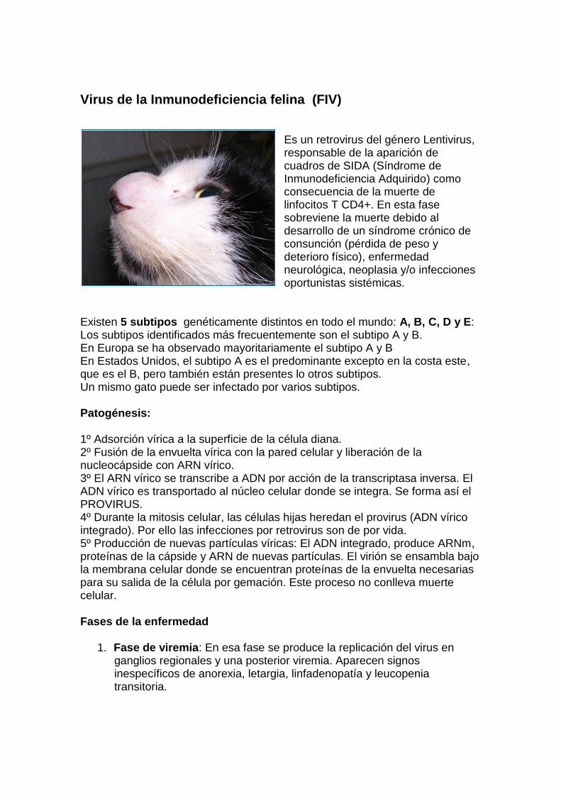
Es un retrovirus del género Lentivirus, responsable de la aparición de cuadros de SIDA (Síndrome de Inmunodeficiencia Adquirido) como consecuencia de la muerte de linfocitos T CD4+. En esta fase sobreviene la muerte debido al desarrollo de un síndrome crónico de consunción (pérdida de peso y deterioro físico), enfermedad neurológica, neoplasia y/o infecciones oportunistas sistémicas.
Existen 5 subtipos genéticamente distintos en todo el mundo: A, B, C, D y E: Los subtipos identificados más frecuentemente son el subtipo A y B. En Europa se ha observado mayoritariamente el subtipo A y B En Estados Unidos, el subtipo A es el predominante excepto en la costa este, que es el B, pero también están presentes lo otros subtipos. Un mismo gato puede ser infectado por varios subtipos. Patogénesis: 1º Adsorción vírica a la superficie de la célula diana. 2º Fusión de la envuelta vírica con la pared celular y liberación de la nucleocápside con ARN vírico. 3º El ARN vírico se transcribe a ADN por acción de la transcriptasa inversa. El ADN vírico es transportado al núcleo celular donde se integra. Se forma así el PROVIRUS. 4º Durante la mitosis celular, las células hijas heredan el provirus (ADN vírico integrado). Por ello las infecciones por retrovirus son de por vida. 5º Producción de nuevas partículas víricas: El ADN integrado, produce ARNm, proteínas de la cápside y ARN de nuevas partículas. El virión se ensambla bajo la membrana celular donde se encuentran proteínas de la envuelta necesarias para su salida de la célula por gemación. Este proceso no conlleva muerte celular. Fases de la enfermedad
1. Fase de viremia: En esa fase se produce la replicación del virus en ganglios regionales y una posterior viremia. Aparecen signos inespecíficos de anorexia, letargia, linfadenopatía y leucopenia transitoria.
2. Fase asintomática o de latencia clínica. En esta fase no hay signos de enfermedad ya que la carga viral se reduce pero la viremia no desaparece. La infección viral progresa durante este periodo sufriendo el sistema inmune una disminución progresiva del cociente T CD4/CD8.
3. Fase de inmunodeficiencia. En esta fase se produce una dismisnución severa del cociente de linfocitos T CD4+/ CD8+ y una alteración de su función dando lugar al desarrollo del Síndrome de Inmunodeficiencia Adquirido (SIDA).
Diagnóstico
b. Pruebas diagnósticas:
ELISA: Detecta anticuerpos anti-FIV contra tres proteínas estructurales del virus: p15 y p24 (proteínas de la nucleocápside) y gp40 (glicoproteína de la envoltura) en suero, plasma o sangre entera. Tiene alta sensibilidad del 99.3% y alta especificidad 99.8%. Falsos positivos si…
Detecta anticuerpos vacunales, por lo que no debe utilizarse en el caso de sospechar que un gato esté vacunado frente a FIV (Confirmar con PCR).
Detecta anticuerpos maternales, por lo que en un gato menor de 8 meses pueden obtenerse falsos positivos (Repetir tras los 8 meses de vida y confirmar con PCR).
Falsos negativos si…
Hay una carga viral escasa debido a una infección temprana (Confirmar con PCR).
Secuestro de complejos inmunes debido a enfermedad inmunomediada (Confirmar con PCR).
Inmunosupresión severa (Confirmar con PCR). Western Blot: Detecta anticuerpos anti-FIV para cada proteína individual del virus, al separar éstas mediante electroforesis. Se realiza en suero, plasma o sangre entera. Es el gold-standard para el diagnóstico de la infección de FIV. Falsos positivos si…
Detecta anticuerpos vacunales, por lo que no debe utilizarse en el caso de sospechar que un gato esté vacunado frente a FIV (Confirmar con PCR).
Detecta anticuerpos maternales, por lo que en un gato menor de 8 meses pueden obtenerse falsos positivos (Repetir tras los 8 meses de vida y confirmar con PCR).

Falsos negativos si…
Hay una carga viral escasa debido a una infección temprana (Confirmar con PCR).
Secuestro de complejos inmunes debido a enfermedad inmunomediada. (Confirmar con PCR).
Inmunosupresión severa (Confirmar con PCR).
PCR: sobre cualquier tejido. Las diferentes técnicas de PCR disponibles (rt-PCR, RTPCR, PCR anidada, PCR convencional) permiten detectar el subtipo A, pero el resto de subtipos son detectados de forma más variable. Falso negativo si…
En el caso de realizarlo en la fase asintomática con una reducida carga viral, puede obtenerse un falso negativo dependiendo de la sensibilidad de la técnica (Buscar laboratorios con una elevada calidad y confirmar con Western Blot siempre tras los 8 meses de vida).
Un transporte inadecuado de la muestra puede dar lugar a falsos negativos al degradarse el ac. Nucléico (Confirmar con Western Blot siempre tras los 8 meses de vida).
El PCR no está detectando el subtipo viral. (Pedir al laboratorio que especifique qué subtipo detecta su PCR y pedir PCR para el resto de subtipos y confirmar con Western Blot siempre tras los 8 meses de vida).
Falso positivo si…
La vacunación de gatos frente a FIV, puede aumentar el número de falsos positivos pero se desconocen las causas ya que la vacuna se supone que no debe provocar replicación vírica.
Contaminación del PCR con pruebas anteriores en laboratorios no cuidadosos (Repetir y confirmar con Western Blot).
Tratamiento FeLV/ FIV Hasta el día de hoy no existe un tratamiento curativo para esta enfermedad, sólo se pueden utilizar tratamientos paliativos que aumenten la calidad y esperanza de vida.
1. Tratamiento precoz, agresivo y más duradero de complicaciones bacterianas, parasitarias… a las que son más susceptibles debido a la inmunosupresión que padecen aunque no sea detectable ya que la respuesta inmune mediada por anticuerpos está afectada en todos los gatos infectados por FeLV.
2. Tratamiento de linfoma con tratamiento quimioterápico específico. Puede dar supervivencias de hasta dos años en algunos gatos.
3. Antiviarles:

AZT: Bloquea la transcriptasa inversa. Sólo en infección temprana (en las tres primeras semanas tras infección) se ha observado en estudios experimentales que evita la llegada del virus a médula ósea. En infecciones naturales sin embargo, no se obtienen tan buenos resultados como en el tratamiento de FIV donde consigue reducciones en el título viral en plasma. Mejorar el estado clínico y la calidad de vida, mejorando la esperanza de vida en algunos gatos.
Dosis: 5-10 mg/kg cada 12 horas oral en ciclos de 6 meses continuado o bien 6 meses alternos (descansos de un mes). Efectos secundarios sobre todo en la dosis mayor: Anemia no regenerativa es un efecto secundario frecuente. Hay que hacer analíticas semanales. Si el Ht llega a un 20% hay que parar el tratamiento. La anemia se recuperará en unas semanas. Si durante las primeras 4 semanas no hay anemia, hacer analíticas una vez al mes. No tratar a gatos con anemia por aplasia medular. Otros efectos como vómitos y anorexia se presentan de forma muy esporádica.
Otros antivirales inhibidores de la transcriptasa (STAMP, PMEA 9, AMD3100), inhibidores de proteasa, Ac. Valproico y Lamiduvide han mostrado eficacia variable, algunos son tóxicos y muchos de los tienen una disponibilidad limitada en medicina veterinaria.
4. Inmunomoduladores.
Interferón omega felino: mejora la sintomatología clínica y aumenta la vida, pero no ayuda a revertir la viremia. El protocolo en cuadros de anemia e Inmunosupresión recomendado por el laboratorio es de:
1 MU/kg/sc durante 5 días. Esperar 14 días y repetir hematología. Si se han recuperado los valores repetir el ciclo 5 días más. Repetir el ciclo en recaídas. No tiene efectos secundarios. .

Manejo de gatos FeLV + y FIV +
De los gatos que conviven con el positivo: - Testar a todos los gatos de casa - Informar del riesgo para los gatos negativos y de que lo mejor
para no infectar es aislarlos. - Esterilizar a todos los gatos. - No introducir a nuevos gatos - El riesgo para los gatos que ya vivían con un FELV + no es
muy alto ya que han sido infectados antes y pueden ser inmunes para una nueva infección. Se estima que el riesgo es de un 10-15% si es FeLV- habiendo vivido con el FeLV+ durante varios meses. De todos modos, la neutralización de virus por anticuerpos no es duradera de por vida, por lo que un gato que inicialmente es inmune, con los años puede llegar a infectarse.
- El virus FIV se transmite básicamente por mordeduras o heridas en peleas, por lo que la probabilidad de transmisión dentro de grupos estables y pacíficos es prácticamente nula.
- Se deberá vacunar anualmente de leucemia felina para aumentar la inmunidad natural y prevenir de que la vacuna no protege al 100%
- Controlar bien a otros gatos enfermos para que no contagien al gato infectado por FIV.
- Evitar corticoides en gatos negativos que conviven con positivos a FeLV ya que hay riesgo de reactivación de infección latente.
Del gato infectado: - No debe salir a la calle para no diseminar la infección. - Mantener una buena nutrición y evitar carnes crudas por
riesgo de contagio de Toxoplasma. - Mantener programa de vacunación para prevenir infecciones.
Se ha comprobado que su sistema inmune no responde tan eficazmente como un gato libre de infección vírica ante la vacunación de rabia.
- Desparasitar interna y externamente - Revisiones cada 6 meses para curar cualquier posible
patología de forma temprana: hacer analítica de sangre, radiografía, ecografía, analítica de orina. Revisar frecuentemente la boca para evitar infecciones crónicas orales.
- Pesar rutinariamente ya que la pérdida de peso es indicativo de enfermedad aunque no se detecte otro signo.

Bibliografía PALMERO, M. CARBALLÉS, V Leucemia Felina. Enfermedades Infecciosas Felinas. (5-99) Servet. 2010. PALMERO, M. CARBALLÉS, V Inmunodeficiena Felina. Enfermedades Infecciosas Felinas. (99-143) Servet. 2010. HORZINEK, M. ADDIE, D. BELAK, S et al. ABCD guidelines on Feline Leukaemia virus. European dvisory Board con Cat Diseases. October 2007 HORZINEK, M. ADDIE, D. BELAK, S et al. ABCD guidelines on Feline Inmunodeficiency virus. European dvisory Board con Cat Diseases, March 2008 LUTZ, H. HOISE, M.. Feline Retrovirus infections. ESFM Feline Congress 2008. Edimburg. LUTZ.H AND HOSIE.M. Feline retrovirus infections: FeLV/FIV. ESFM feline congress 2008.September, Edinburgh. 25-28
STÜTZER.B, MÜLLER.F, MAJZOUB.M et al. Role of latent feline Leukemia Virus Infection in nonregenerative Cytopenias of cats.Journal of Veterinary Internal Medicine, Nov 2009
CATTORI.V, PEPIN A.C, TANDON. R et al. Real-time PCR investigation of feline leukemia virus proviral and viral RNA loads in leukocyte subsets,
Veterinary immunology and immunopathology, pages 124-128, Volume 123, May 2008 TORRES. A.N, O’HALLORAN. K.P, LARSONA. L.J et al. Development and application of a quantitative real-time PCR assay to detect feline leukemia virus RNA, Veterinary immunology and immunopathology, pages 81-89, Volume 123, May 2008 HARTMANN.K, GRIESSMAYR.P, SCHULZ.B et al. Quality of different in-clinic test systems for feline immunodeficiency virus and feline leukaemia virus infection. Journal of feline medicine and surgery, pages 439-445, Volume 9, December 2007 GROAT. R et al. Upgraded IDEXX Diagnostic Products for Simultaneous Detection of Antibodies to Feline Immunodeficiency Virus (FIV) gag and env Proteins in Feline Blood Samples. IDEXX Laboratories, Inc., Research & Development DIEZ.N. Exploración ecográfica del tracto digestivo. XXVII congreso anual de AMVAC. Madrid, 26,27 y 28 de Febrero de 2010 WOLF.A.M. Care of the FeLV/FIV infected cat. Atlantic Coast Veterinary Conference, 2002

DE MARI K. et al. Effects of a recombinant Feline Omega Interferon on the survival and clinical signs of ill FeLV and/or FIV-infected cats. IFRR Symposium, Amelia Island, USA, 2002 DE MARI K. et al. Therapeutic effects of recombinant Feline Omega Interferón on FeLV-infected and FeLV/FIV-coinfected symptomatic cats. Journal of Veterinary Internal Medicine, 18, pages 477-482, 2004 DE MARI K. and SANQUER.A. Effects of a recombinant Feline Omega Interferón on a population of FeLV and/or FIV infected cats suffering from anemia. 7th International IFRR Symposium, Pisa, Italy, 2004 GINGERICH D.A. Lymphocyte T-Cell inmunomodulator (LTCI): Review of the immunopharmacology of a new veterinary biologic. The International Journal of Applied Research of Veterinary Medicine. Vol 6, Nº2, 2008 SCHERK.M. Vaccination and the Immune Status of the Cat. Waltham Feline Medicine Symposium 2002 SPARKES A.H. Feline leukaemia virus and vaccination. Journal of feline medicine and surgery, volume 5, pages 97-100, April 2003 LANGHAMMER.S, HÜBNER.J, KURTH.R et al. Antibodies neutralizing feline leukaemia virus (FeLV) in cats immunized with the transmembrane envelope protein p15E, Immunology, pages 229-237, February 2006 SCHULTZ.R.D. Vaccines, Vaccination Programs and Methods to Determine Their Effectiveness, 21st Forum of American College of veterinary internal medicine, 2003 HOFMANN-LEHMANN.R, TANTDON.R, BORETTI F.S et al . Reasssessment of feline leukaemia virus vaccines with novel sensitive molecular assays. Vaccine. Pages 1087-194, February 2006 HOFMANN-LEHMANN.R, CATTORI.V, TANDON. R et al. How molecular methods change our views of FeLV infection and vaccination. Veterinary immunology and immunopathology, Vol 123, pages 119-123, May 2008 GROAT R. et al. Upgraded IDEXX Diagnostic Products for Simultaneous Detection of Antibodies to Feline Immunodeficiency Virus (FIV) gag and env Proteins in Feline Blood Samples. IDEXX Laboratories, Inc., Research & Development. LEVY , J., CRAWFORD, C., HARTMA NN, K. et al. 2008 American Association of Feline Practitioners’ feline retrovirus management guidelines. Journal of Feline Medicine and Surgery, 2008, pp. 300-316. RICHA RDS, J.R. Feline immunodeficiency virus vaccine: implications for diagnostic testing and disease management. Biologicals, December 2005.

BERLINSKI, P.J., GIBS ON, J.K., FORESTER, J.K. et al. Further Investigation into the Increased Susceptibility of Cats to Feline Immunodeficiency Virus (FIV) After Vaccination with Parenteral Vaccines. 21st Annual Forum of American College of Veterinary Internal Medicine, 2003. LEVY , J.K., CRAWFORD, P.C., SLATER, M.R. Effect of vaccination against feline immunodeficiency virus on results of serologic testing in cats. Journal of American Veterinary Medical Association. November 2004. GROAT, R., CURATO, J., SEYM OUR, C. et al. FELINE Antibody Response to Fort Dodge Fel-O-Vax FIV Vaccine Interferes with FIV Diagnostic Tests. 21st Annual Forum of American College of Veterinary Internal Medicine, 2003.

Aafpp Etiology and Epidemiology. Feline immunodeficiency virus. Feline immunodeficiency virus (FIV) is an exogenous, single-strand
RNA virus in
the family Retroviridae, subfamily Lentivirinae. The virus is morphologically similar to the human
immunodeficiency
virus (HIV) but it is antigenically distinct. Like FeLV, FIV produces reverse transcriptase to catalyze the
insertion of
viral RNA into the host genome. Multiple subtypes of the virus exist, and some isolates have differing
biologic
behavior. For example, immune deficiency is induced much more quickly by some isolates, and clinical
diseases,
such as uveitis, are induced by some but not all isolates.
Aggressive biting behavior is thought to be the primary route of transmission of FIV; older, male, outdoor
cats with
clinical signs of disease are most commonly infected. The prevalence of FIV antibodies in North America
was 2.5%
in a recent study (Levy et al., 2006). FIV is present in semen and can be transmitted by artificial
insemination.
Transplacental and perinatal transmission occurs from infected queens to kittens. Arthropod transmission
appears to
be unlikely. Transmission by routes other than biting is less common because high levels of viremia are
of short
duration.
FIV replicates in several cell types, including T-lymphocytes (CD4+ and CD8+), B-lymphocytes,
macrophages, and
astrocytes. The primary phase of infection occurs as the virus disseminates throughout the body, initially
leading to
low-grade fever, neutropenia, and generalized reactive lymphadenopathy. A subclinical, latent period of
variable
length then develops; the length of this period is related in part to the strain of virus and the age of the cat
when
infected. The median age of healthy, naturally infected cats and clinically ill naturally infected cats is
approximately 3
years and 10 years, respectively, suggesting a latent period of years for most strains of FIV. Chronic
experimental
and naturally occurring infection results in a slow decline in circulating CD4+ lymphocyte numbers,
response to
mitogens, and decreased production of cytokines associated with cell-mediated immunity, such as
interleukin (IL)-2
and IL-10; neutrophil function and natural killer cell function are also affected. Humoral immune
responses are often
intact, and a polyclonal gammopathy develops from nonspecific B-lymphocyte activation. Within months
to years, an
immune deficiency stage similar to acquired immunodeficiency syndrome (AIDS) in human beings
develops.
Coinfection with FeLV potentiates the primary and immune deficiency phases of FIV. However,
coinfection with
Mycoplasma haemofelis, Toxoplasma gondii, feline herpesvirus, and feline calicivirus, as well as
immunization, failed
to potentiate FIV-associated immunodeficiency in research studies.
Feline leukemia virus. Feline leukemia virus (FeLV) is a single-strand RNA virus in the family
Retroviridae,
subfamily Oncovirinae. The virus produces reverse transcriptase, which catalyzes the reaction, resulting
in the
formation of a DNA copy (provirus) of FeLV viral RNA in the cytoplasm of infected cells; the provirus is
inserted into

the host cell genome. On subsequent host cell divisions the provirus serves as a template for new virus
particles
formed in the cytoplasm and is released across the cell membrane by budding. FeLV is composed of
several core
and envelope proteins. Envelope protein p15e induces immunosuppression. Core protein p27 is present in
the
cytoplasm of infected cells, peripheral blood, saliva, and tears of infected cats; detection of p27 is the
basis of most
FeLV tests. The envelope glycoprotein 70 (gp70) contains the subgroup antigens A, B, or C, which are
associated
with the infectivity, virulence, and disease caused by individual strains of the virus. Neutralizing
antibodies are
produced by some cats after exposure to gp70. Antibodies against feline oncornavirus-associated cell
membrane
antigen (FOCMA) are formed by some cats but are generally not used clinically.
The principal route of infection by FeLV is prolonged contact with infected cat saliva and nasal
secretions; grooming
or sharing common water or food sources effectively results in infection. Because the organism does not
survive in
the environment, feces, or urine, fomite and aerosol transmission is unlikely. Transplacental, lactational,
and
venereal transmission is less important than casual contact. FeLV infection has worldwide distribution;
the
seroprevalence of infection varies geographically and by the population of cats tested. Infection is most
common in
outdoor male cats between ages 1 and 6 years. In a recent study (Levy et al., 2006) the prevalence of
FeLV
antigenemia in cats in North America was 2.3%. FeLV can be detected in feces of infected fleas for 2
weeks (Vobis
et al., 2006). However, the prevalence rates for FeLV vary little across regions of the United States with
high and low
prevalence rates of fleas, so this is an unlikely route of infection.
The virus replicates first in the oropharynx, followed by dissemination through the body to the bone
marrow. Ifpersistent bone marrow infection occurs, infected white blood cells and platelets leave the bone
marrow with ultimate
infection of epithelial structures, including salivary and lacrimal glands. Whether infection occurs after
natural
exposure to FeLV is determined by the virus subtype or strain, the virus dose, the age of the cat when
exposed, and
the cat’s immune responses. Using realtime PCR and antigen ELISA results, four classes of FeLV
infection were
defined (Torres et al. 2005; Levy et al. 2008). Some FeLV-exposed cats can eliminate the infection
(abortive)
whereas others progress to clinical illness and persistent viremia (progressive). Other FeLV-exposed cats
will
develop regressive infection characterized by antigen-negative results and lower transiently positive
realtime PCR
results. Latent FeLV infections are transiently antigen positive but have persistently positive realtime
PCR results.
Latent and regressive infections can be potentially activated by the administration of glucocorticoids or
other
immunosuppressive drugs.
The pathogenesis of various syndromes induced by FeLV is complex but includes induction of lymphoma
from
activation of oncogenes by the virus or insertion of a provirus into the genome of lymphoid precursors;
subgroup C
induction of aplastic anemia from increased secretion of tumor necrosis factor-
�������������������������������������-
lymphocyte depletion (both CD4+ and CD8+ lymphocytes) or dysfunction; neutropenia; neutrophil
function disorders;

malignant transformation; and viral induction of bone marrow growth-promoting substances leading to
myeloproliferative diseases.
Clinical Features. Feline immunodeficiency virus. Clinical signs of infection with FIV can arise from direct viral
effects or secondary
infections that ensue after the development of immunodeficiency. Most of the clinical syndromes
diagnosed in FIVseropositive
cats also occur in FIV-naïve cats, which makes proving disease causation difficult during the subclinical
stage of infection. A positive FIV antibody test does not prove immunodeficiency or disease from FIV
and does not
necessarily indicate a poor prognosis. The only way to determine accurately whether an FIV-seropositive
cat with a
concurrent infectious disease has a poor prognosis is to treat the concurrent infection.
Primary (acute) FIV infection is characterized by fever and generalized lymphadenopathy. Owners
commonly present
FIV-infected cats in the immunodeficiency stage for evaluation of nonspecific signs such as anorexia,
weight loss,
and depression or for evaluation of abnormalities associated with specific organ systems. When a clinical
syndrome
is diagnosed in a cat seropositive for FIV, the workup should include diagnostic tests for other potential
causes.
Clinical syndromes reportedly from primary viral effects include chronic small-bowel diarrhea,
nonregenerative
anemia, thrombocytopenia, neutropenia, lymphadenopathy, pars planitis (inflammation in the anterior
vitreous
humor), anterior uveitis, glomerulonephritis, renal insufficiency, and hyperglobulinemia. However, in one
recent
report of naturally infected cats, FIV was associated with proteinuria but not renal azotemia (Baxter et al,
2012).
Behavioral abnormalities, with dementia, hiding, rage, inappropriate elimination, and roaming, are the
most common
neurologic manifestations of FIV infection. Seizures, nystagmus, ataxia, and peripheral nerve
abnormalities may
occasionally be attributable to primary viral effects. Lymphoid malignancies, myeloproliferative diseases,
and several
carcinomas and sarcomas have been detected in FIV-infected, FeLV-naïve cats, suggesting a potential
association
between FIV and malignancy; FIV-infected cats are at higher risk for the development of lymphoma
(Madgen et al,
2011.
Feline leukemia virus. Owners generally present FeLV-infected cats for evaluation of nonspecific
signs such as
anorexia, weight loss, and depression or abnormalities associated with specific organ systems. Of the
FeLV-infected
cats evaluated at necropsy, 23% had evidence of neoplasia (96% lymphoma/leukemia); the remainder
died from
nonneoplastic diseases (Reinacher, 1989). Specific clinical syndromes can result from specific effects of
the virus or
from opportunistic infections caused by immunosuppression. A positive FeLV test result does not prove
disease
induced by FeLV. When a clinical syndrome is diagnosed in a FeLV-seropositive cat, the workup should
include
diagnostic tests for other potential causes. The opportunistic agents discussed for FIV also are common in
FeLVinfected
cats.
Bacterial or calicivirus-induced stomatitis occurs in some FeLV-infected cats as a result of
immunosuppression. FeLV
infection can result in vomiting or diarrhea from a form of enteritis clinically and histopathologically
resembling
panleukopenia, from alimentary lymphoma, or from secondary infections attributable to
immunosuppression. Icterus
in FeLV-infected cats can be prehepatic from immune-mediated destruction of red blood cells induced by
FeLV or
secondary infection by Mycoplasma haemofelis or “Candidatus Mycoplasma haemominutum”; hepatic from hepatic
lymphoma, hepatic lipidosis, or focal liver necrosis; or posthepatic from alimentary lymphoma. Some
FeLV-infected
cats with icterus may be concurrently infected by FIP virus or T. gondii. Clinical signs of rhinitis or pneumonia occur in some FeLV-infected cats as a result of secondary
infections. Dyspneaor dysphagia from mediastinal lymphoma occurs in some cats. These cats are
generally younger than 3 years and
may have decreased cranial chest compliance on palpation as well as muffled heart and lung sounds if
pleural
effusion is present.
Mediastinal, multicentric, and alimentary lymphomas are the most common neoplasms associated with
FeLV;
lymphoid hyperplasia also occurs. Alimentary lymphoma most commonly involves the small intestine,
mesenteric
lymph nodes, kidneys, and liver of older cats; most cats with alimentary lymphoma are FeLV negative.
Renal
lymphoma can involve one or both kidneys, which are usually enlarged and irregularly marginated on
physical
examination. Fibrosarcomas occasionally develop in young cats coinfected with FeLV and feline sarcoma
virus.
Lymphocytic, myelogenous, erythroid, and megakaryocytic leukemia all are reported with FeLV
infection;
erythroleukemia and myelomonocytic leukemia are the most common. The history and physical
examination findings
are nonspecific.
Renal failure occurs in some FeLV-infected cats from renal lymphoma or glomerulonephritis. Affected
cats are
presented for evaluation of polyuria, polydipsia, weight loss, and inappetence during the last stages of
disease.
Urinary incontinence from sphincter incompetence or detrusor hyperactivity occurs in some cats; small-
bladder
nocturnal incontinence is reported most frequently.
Some FeLV-infected cats are presented for miosis, blepharospasm, or cloudy eyes from ocular
lymphoma. Aqueous
flare, mass lesions, keratic precipitates, lens luxations, and glaucoma are often found on ocular
examination. FeLV
does not likely induce uveitis without lymphoma. Neurologic abnormalities associated with FeLV
infection include
anisocoria, ataxia, weakness, tetraparesis, paraparesis, behavioral changes, and urinary incontinence.
Nervous
system disease is likely to develop as a result of polyneuropathy or lymphoma. Intraocular and nervous
system
disease in FeLV-infected cats can occur from infection with other agents, including FIPV, Cryptococcus neoformans, Bartonella spp., or T. gondii. Abortion, stillbirth, or infertility occurs in some FeLV-infected queens. Kittens infected in utero that
survive to birth
generally develop accelerated FeLV syndromes or die as part of the kitten mortality complex.
Some FeLV-seropositive cats present for lameness or weakness from neutrophilic polyarthritis attributed
to immune
complex deposition. Multiple cartilaginous exostoses occur in some cats and may be FeLV related.
Diagnosis. Feline immunodeficiency virus. Neutropenia, thrombocytopenia, and nonregenerative anemia are
common

hematologic abnormalities associated with FIV infection. Monocytosis and lymphocytosis occur in some
cats and
may be caused by the virus or chronic infection with opportunistic pathogens. Cytologic examination of
bone marrow
aspirates may reveal maturation arrest (i.e., myelodysplasia), lymphoma, or leukemia. A progressive
decline in CD4+
lymphocytes, a plateau or progressive increase in CD8+ lymphocytes, and an inversion of the
CD4+/CD8+ ratio
occurs in experimentally infected cats over time. A multitude of serum biochemical abnormalities is
possible
depending on what FIV-associated syndrome is occurring. Polyclonal gammopathy can occur in some
FIV infected
cats. No pathognomonic imaging abnormalities are associated with FIV infection.
Antibodies against FIV are detected in serum in clinical practice most frequently by enzyme-linked
immunosorbent
assay (ELISA). Comparisons between different tests have shown the results of most assays are
comparable
(Hartmann et al., 2007). Clinical signs can occur before seroconversion in some cats and some infected
cats never
seroconvert; thus false-negative reactions can occur. Results of virus isolation or RT-PCR on blood are
positive in
some antibody-negative cats. False-positive reactions can occur with ELISA; therefore positive ELISA
results in
healthy or low-risk cats should be confirmed by Western blot immunoassay or RT-PCR. Kittens can have
detectable,
colostrum-derived antibodies for several months. Kittens younger than 6 months that are FIV seropositive
should be
tested every 60 days until the result is negative. If antibodies persist at 6 months of age, the kitten is likely
infected.
Virus isolation or PCR on blood can also be performed to confirm infection. The biggest problem with
FIV RT-PCR
assays to date is lack of standardization among laboratories and the potential for both false-positive and
falsenegative
results (Crawford et al., 2005). A vaccine against FIV has been licensed in the United States. This vaccine
induces antibodies that cannot be distinguished from those induced by naturally occurring disease with
currently
available tests (see below).
Detection of antibodies against FIV in the serum of cats that have not been vaccinated against FIV
documents
exposure and correlates well with persistent infection but does not correlate with disease induced by the
virus.
Because many clinical syndromes associated with FIV can be caused by opportunistic infections, further
diagnostic
procedures may determine treatable etiologies. For example, some FIV-seropositive cats with uveitis are
coinfected
by T. gondii and often respond to the administration of anti-Toxoplasma drugsFeline leukemia virus. A variety of nonspecific hematologic, biochemical, urinalysis, and radiographic abnormalities
occur in FeLV-infected cats. Nonregenerative anemia alone or in combination with decreases in
lymphocyte,
neutrophil, and platelet counts is common. The presence of increased numbers of circulating nucleated
red blood
cells or macrocytosis in association with severe nonregenerative anemia occurs frequently; examination
of bone
marrow often documents a maturation arrest in the erythroid line (erythrodysplasia). Immune-mediated
destruction of
erythrocytes can be induced by FeLV and occurs in cats coinfected with hemoplasmas; regenerative
anemia,
microagglutination or macroagglutination of erythrocytes, and a positive result on the direct Coombs test
are common
in these cats. Neutropenia and thrombocytopenia occur from bone marrow suppression or immune-
mediated
destruction. In a recent study, 37 cats with non-regenerative cytopenias were evaluated for latent FeLV in
the bone
marrow by RT-PCR assay and 2 cats were positive (Stützer et al, 2010). FeLV-infected cats with the
panleukopenialike
syndrome have gastrointestinal tract signs and neutropenia and are difficult to differentiate from cats with
panleukopenia virus infection or salmonellosis. In addition, cats with FeLV-induced panleukopenia-like
syndrome
usually have anemia and thrombocytopenia, abnormalities rarely associated with panleukopenia virus
infection.
Azotemia, hyperbilirubinemia, bilirubinuria, and increased activity of liver enzymes are common
biochemical
abnormalities. Proteinuria occurs in some FeLV-infected cats with glomerulonephritis. Cats with
lymphoma have
mass lesions radiographically depending on the organ system affected. Mediastinal lymphoma can result
in pleural
effusion; alimentary lymphoma can cause obstructive intestinal patterns.
Lymphoma can be diagnosed by cytologic or histopathologic evaluation of affected tissues. Because
lymphoma can
be diagnosed cytologically and treated with chemotherapy, cats with mediastinal masses,
lymphadenopathy,
renomegaly, hepatomegaly, splenomegaly, or intestinal masses should be evaluated cytologically before
surgical
intervention. Malignant lymphocytes are also occasionally identified in peripheral blood smears,
effusions, and CSF.
Most cats with suspected FeLV infection are screened for FeLV antigens in neutrophils and platelets by
immunofluorescent antibody (IFA) testing or in whole blood, plasma, serum, saliva, or tears by ELISA.
Serum is the
most accurate fluid to assess in ELISA tests. IFA results are not positive until the bone marrow has been
infected.
The results of IFA testing are accurate more than 95% of the time. False-negative reactions may occur
when
leukopenia or thrombocytopenia prevents evaluation of an adequate number of cells. False-positive
reactions can
occur if the blood smears submitted for evaluation are too thick. A positive IFA result indicates that the
cat is viremic
and contagious; approximately 90% of cats with positive IFA results are viremic for life. The rare
combination of IFApositive
and ELISA-negative results suggests technique-related artifact. Negative ELISA results correlate well
with
negative IFA results and an inability to isolate FeLV. Comparisons of different antigen tests have shown
the results of
most assays to be comparable (Hartmann et al., 2007).
The virus can be detected in serum by ELISA before infection of bone marrow and can therefore be
positive in some
cats during early progressive stages of infection or during early latent infection even though IFA results
are negative.
Other possibilities for discordant results (ELISA positive, IFA negative) are false-positive ELISA results
or falsenegative
IFA results. Cats with positive ELISA results and negative IFA results are probably not contagious at that
time but should be isolated until retested 4 to 6 weeks later because progression to persistent viremia and
epithelial
cell infection may be occurring.
ELISA-positive cats that revert to negative have developed latent infections or regressive infection. Virus
isolation,
IFA performed on bone marrow cells, immunohistochemical staining of tissues for FeLV antigen, and
PCR can be
used to confirm latent or regressive infection in some cats. Cats with latent or regressive infection are not
likely

contagious to other cats, but infected queens may pass the virus to kittens during gestation or parturition
or by milk.
Cats with regressive or latent infection can be immunodeficient and may become viremic (IFA and
ELISA positive)
after receiving corticosteroids or after extreme stress.
A delay of 1 to 2 weeks generally occurs after the onset of viremia before ELISA tear and saliva test
results become
positive; therefore these test results can be negative even when results with serum are positive and so are
not
recommended for use. Antibody titers to FeLV envelope antigens (neutralizing antibody) and against
virustransformed
tumor cells (FOCMA antibody) are available in some research laboratories, but the diagnostic and
prognostic significance of results from these tests is unknown. Realtime PCR assays are more sensitive
than
conventional PCR for FeLV infections, but validated and standardized assays are not currently available
in the United
States (Torres et al. 2005).
Treatment. Feline immunodeficiency virus. Because FIV-seropositive cats are not necessarily
immunosuppressed or
diseased from FIV, the cat should be evaluated and treated for other potential causes of the clinical
syndrome. SomeFIV-seropositive cats are immunodeficient; if infectious diseases are identified,
bacteriocidal drugs administered at
the upper end of the dosage should be chosen. Long-term antibiotic therapy or multiple treatment periods
may be
required. The only way to determine if an FIV-seropositive cat with a concurrent infection has a poor
prognosis is to
treat the concurrent infection.
A number of anti-lentiviral drugs may be effective for the treatment if FIV infected cats but controlled
studies are
largely lacking (Mohammad et al, 2012). Administration of interferons has shown clinical benefit in some
studies
(Domenech et al, 2011). Oral administration of 10 IU/kg of human interferon-alpha led to improved
clinical signs and
prolonged survival compared with a placebo-treated control group in one study (Pedretti et al., 2006). In
another
study feline recombinant interferon was administered at 106 U/kg/day SQ for 5 days in three series
(starting on days
0, 14, and 60) and was shown to improve clinical signs early in the study and prolong survival in treated
cats (de Mari
et al., 2004). Administration of antiviral agents such as the reverse transcriptase inhibitor azidothymidine
(AZT) has
had mixed success in the treatment of FIV. Use of AZT at a dosage of 5 mg/kg PO or SQ q12h improved
overall
quality of life and stomatitis in FIV-infected cats and is believed to aid in the treatment of neurologic
signs (Hartmann
et al., 1995a, 1995b). Cats treated with AZT should be monitored for the development of anemia. The
anti-viral
compound plerixafor was used in a study of naturally infected cats and was shown to lessen pro-viral load
but did not
improve clinical outcomes (Hartmann et al, 2012). When combined with 9-(2-phosphonylmethoxyethyl)
adenine
(PMEA), intolerable side-effects occurred. Administration of bovine lactoferrin by mouth was beneficial
in the
treatment of intractable stomatitis in FIV-seropositive cats (Sato et al., 1996). Removal of all premolar
and molar
teeth has also been effective for treatment of intractable stomatitis in some FIV-seropositive cats.
Immunomodulators
have not been shown to have reproducible clinical effect, but owners sometimes report positive responses.
Human

recombinant erythropoietin administration increased red blood cell and white blood cell counts, did not
increase viral
load, and had no measurable adverse clinical effects in FIV-infected cats compared with placebo (Arai et
al., 2000).
In contrast, although administration of human recombinant granulocyte-monocyte colony-stimulating
factor (GMCSF)
to FIV-infected cats increased white blood cell counts in some treated cats, it also induced fever, anti–
GM-CSF
antibodies, and increased viral load. GM-CSF therefore appears to be contraindicated for the treatment of
FIV in
cats.
Feline leukemia virus. Several antiviral agents have been proposed for the treatment of FeLV; the
reverse
transcriptase inhibitor AZT has been studied the most. Unfortunately, administration of AZT to
persistently viremic
cats does not appear to clear viremia in most, and it had minimal benefits for clinically ill cats in a recent
study
(Hartmann et al., 2002). Interferons have an effect against FeLV in vivo and in vitro (Collado et al., 2007;
de Mari et
al., 2004). Immunotherapy with drugs such as Staphylococcus protein A, Propionibacterium acnes, or acemannan
improves clinical signs of disease in some cats, but controlled studies are lacking.
Chemotherapy should be administered to cats with FeLV-associated neoplasia. Opportunistic agents
should be
managed as indicated; the upper dose range and duration of antibiotic therapy are generally required.
Supportive
therapies such as hematinic agents, vitamin B12, folic acid, anabolic steroids, and erythropoietin generally
have been
unsuccessful in the management of nonregenerative anemia. Blood transfusion is required in many cases.
Cats with
autoagglutinating hemolytic anemia require immunosuppressive therapy, but this may activate virus
replication. The
prognosis for persistently viremic cats is guarded; the majority die within 2 to 3 years.
Prevention and Zoonotic Aspects. Feline immunodeficiency virus. Housing cats indoors to avoid fighting and testing new cats before
introduction to
an FIV-seronegative, multiple-cat household will prevent most cases of FIV. Transmission by fomites is
unusual
because the virus is not easily transmitted by casual contact, is susceptible to most routine disinfectants,
and dies
when out of the host after minutes to hours, especially when dried. Cleaning litter boxes and dishes shared
by cats
with scalding water and detergent inactivates the virus. Cats with potential exposure from fighting should
be retested
60 days after the potential exposure (Goldkamp et al, 2008). Cats that are FIV infected should be housed
indoors at
all times to avoid exposing FIV-naïve cats in the environment to the virus and to lessen the affected
animal’s chance
of acquiring opportunistic infections. Kittens queened by FIV-infected cats should not be allowed to nurse
to avoid
transmission by ingestion of milk. Kittens queened by FIV-infected cats should be shown to be
serologically negative
at 6 months of age to document failure of lactogenic or transplacental transmission before being sold. A
killed
vaccine containing immunogens from two FIV isolates is licensed for use in some countries (Boehringer-
Ingleheim).
The American Association of Feline Practitioners considers the vaccine noncore. However, vaccination of
high risk,
FIV seronegative cats should be considered. In addition, the vaccine induces antibodies that cannot be
distinguished
from those induced by natural exposure by antibody assays currently available in the United States. FIV
RT-PCR
assays can be attempted to differentiate FIV infection from vaccination and a positive test result will
document
infection. However, since FIV induces only low level viremia, a negative RT-PCR assay result does not
exclude the
infection. HIV and FIV are morphologically similar but antigenically distinct. Antibodies against FIV
have not been documented
in the serum of human beings, even after accidental exposure to virus-containing material (Butera et al.,
2000;
Dickerson et al, 2012). Cats with FIV infection resulting in immunodeficiency may be more likely to
spread other
zoonotic agents into the human environment; clinically ill, FIV-seropositive cats should therefore undergo
a thorough
diagnostic evaluation.
Feline leukemia virus. Avoiding contact with FeLV by housing cats indoors is the best form of
prevention. Potential
fomites such as water bowls and litter pans should not be shared by seropositive and seronegative cats.
Testing and
removal of seropositive cats can result in virus-free catteries and multiple-cat households.
Because of variations in challenge study methods and the difficulty of assessing the preventable fraction
of a disease
with a relatively low infection rate, long subclinical phase, and multiple field strains, the efficacy of
individual vaccines
continues to be in question. Vaccination of cats not previously exposed to FeLV should be considered in
cats at high
risk (i.e., contact with other cats), but owners should be warned of the potential efficacy of less than
100%.
Vaccination should be considered for kittens because of increased risk for progressive infection and then
whether to
boost the vaccine evaluated 1 year later. Variable recommendations have been given on whether to use a
prolonged
interval (> 1 year) for FeLV vaccination in cats at risk for infection. There is one vaccine available in
some countries
labelled for a 2 year interval (Merck Animal Health). Cats with persistent FeLV viremia do not benefit
from
vaccination and so FeLV serology should be assessed prior to vaccination. Vaccination is related to the
development
of injection site sarcoma in some cats. Cats developing these tumors may be genetically predisposed
(Banerji et al.,
2007).
FeLV-infected cats should be housed indoors to avoid infecting other cats and avoid exposure to
opportunistic
agents. Flea control should be maintained to avoid exposure to hemoplasmas, and Bartonella spp. FeLV-
infected
cats should not be allowed to hunt or be fed undercooked meats to avoid infection by T. gondii, Cryptosporidium parvum, Giardia spp., and other infectious agents carried by transport hosts.
Antigens of FeLV have never been documented in the serum of human beings, suggesting that the
zoonotic risk is
minimal. However, FeLV-infected cats may be more likely than FeLV-naïve cats to pass other zoonotic
agents, such
as C. parvum and Salmonella spp., into the human environment.