Gut microbiota · Pilon G, et al. Gut 2015;64:872–883. ABSTRACT Objective The increasing...
12
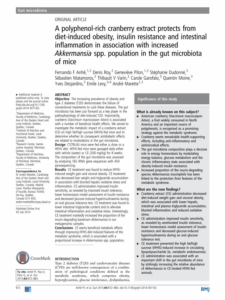
ORIGINAL ARTICLE A polyphenol-rich cranberry extract protects from diet-induced obesity, insulin resistance and intestinal inflammation in association with increased Akkermansia spp. population in the gut microbiota of mice Fernando F Anhê, 1,2 Denis Roy, 2 Geneviève Pilon, 1,2 Stéphanie Dudonné, 2 Sébastien Matamoros, 2 Thibault V Varin, 2 Carole Garofalo, 3 Quentin Moine, 3 Yves Desjardins, 2 Emile Levy, 3,4 André Marette 1,2 ▸ Additional material is published online only. To view please visit the journal online (http://dx.doi.org/10.1136/ gutjnl-2014-307142). 1 Department of Medicine, Faculty of Medicine, Cardiology Axis of the Quebec Heart and Lung Institute, Quebec, Quebec, Canada 2 Institute of Nutrition and Functional Foods, Laval University, Quebec, Quebec, Canada 3 Research Centre, Sainte- Justine Hospital, Montreal, Quebec, Canada 4 Department of Nutrition, Faculty of Medicine, University of Montreal, Montreal, Quebec, Canada Correspondence to Dr André Marette, Cardiology Axis of the Quebec Heart and Lung Institute, Laval University, Quebec, Canada, Hôpital Laval, Pavillon Marguerite d’Youville, Bureau Y4340, Ste-Foy, Québec, Canada G1V 4G5; [email protected] Published Online First 30 July 2014 To cite: Anhê FF, Roy D, Pilon G, et al. Gut 2015;64:872–883. ABSTRACT Objective The increasing prevalence of obesity and type 2 diabetes (T2D) demonstrates the failure of conventional treatments to curb these diseases. The gut microbiota has been put forward as a key player in the pathophysiology of diet-induced T2D. Importantly, cranberry (Vaccinium macrocarpon Aiton) is associated with a number of beneficial health effects. We aimed to investigate the metabolic impact of a cranberry extract (CE) on high fat/high sucrose (HFHS)-fed mice and to determine whether its consequent antidiabetic effects are related to modulations in the gut microbiota. Design C57BL/6J mice were fed either a chow or a HFHS diet. HFHS-fed mice were gavaged daily either with vehicle (water) or CE (200 mg/kg) for 8 weeks. The composition of the gut microbiota was assessed by analysing 16S rRNA gene sequences with 454 pyrosequencing. Results CE treatment was found to reduce HFHS- induced weight gain and visceral obesity. CE treatment also decreased liver weight and triglyceride accumulation in association with blunted hepatic oxidative stress and inflammation. CE administration improved insulin sensitivity, as revealed by improved insulin tolerance, lower homeostasis model assessment of insulin resistance and decreased glucose-induced hyperinsulinaemia during an oral glucose tolerance test. CE treatment was found to lower intestinal triglyceride content and to alleviate intestinal inflammation and oxidative stress. Interestingly, CE treatment markedly increased the proportion of the mucin-degrading bacterium Akkermansia in our metagenomic samples. Conclusions CE exerts beneficial metabolic effects through improving HFHS diet-induced features of the metabolic syndrome, which is associated with a proportional increase in Akkermansia spp. population. INTRODUCTION Type 2 diabetes (T2D) and cardiovascular diseases (CVD) are well-known consequences of a combin- ation of pathological conditions defined as the metabolic syndrome, which comprises obesity, hyperglycaemia, glucose intolerance, dyslipidaemia, Significance of this study What is already known on this subject? ▸ American cranberry (Vaccinium macrocarpon Aiton), a fruit widely consumed in North America and an important source of polyphenols, is recognised as a promising strategy against the metabolic syndrome. ▸ Cranberry exerts remarkable health-supporting effects, including anti-inflammatory and antimicrobial effects. ▸ The gut microbiota composition plays a decisive role in energy homeostasis by modulating energy balance, glucose metabolism and the chronic inflammatory state associated with obesity-induced insulin resistance. ▸ Increased proportion of the mucin-degrading species Akkermansia muciniphila has been linked to the protection from features of the metabolic syndrome. What are the new findings? ▸ Cranberry extract (CE) administration decreased diet-induced weight gain and visceral obesity, which was associated with lower hepatic, intestinal and plasma triglyceride accumulation, blunted inflammation and reduced oxidative stress. ▸ CE administration improved insulin sensitivity, as revealed by ameliorated insulin tolerance, lower homeostasis model assessment of insulin resistance and decreased glucose-induced hyperinsulinaemia during an oral glucose tolerance test. ▸ CE treatment prevented the high fat/high sucrose (HFHS)-induced increase in circulating lipopolysaccharide (ie, metabolic endotoxemia). ▸ CE administration was associated with an important shift in the gut microbiota of mice by strikingly increasing the relative abundance of Akkermansia in CE-treated HFHS-fed animals. Gut microbiota 872 Anhê FF, et al. Gut 2015;64:872–883. doi:10.1136/gutjnl-2014-307142
Transcript of Gut microbiota · Pilon G, et al. Gut 2015;64:872–883. ABSTRACT Objective The increasing...
ORIGINAL ARTICLE
A polyphenol-rich cranberry extract protects fromdiet-induced obesity, insulin resistance and intestinalinflammation in association with increasedAkkermansia spp. population in the gut microbiotaof miceFernando F Anhê,1,2 Denis Roy,2 Geneviève Pilon,1,2 Stéphanie Dudonné,2
Sébastien Matamoros,2 Thibault V Varin,2 Carole Garofalo,3 Quentin Moine,3
Yves Desjardins,2 Emile Levy,3,4 André Marette1,2
▸ Additional material ispublished online only. To viewplease visit the journal online(http://dx.doi.org/10.1136/gutjnl-2014-307142).1Department of Medicine,Faculty of Medicine, CardiologyAxis of the Quebec Heart andLung Institute, Quebec,Quebec, Canada2Institute of Nutrition andFunctional Foods, LavalUniversity, Quebec, Quebec,Canada3Research Centre, Sainte-Justine Hospital, Montreal,Quebec, Canada4Department of Nutrition,Faculty of Medicine, Universityof Montreal, Montreal,Quebec, Canada
Correspondence toDr André Marette, CardiologyAxis of the Quebec Heart andLung Institute, Laval University,Quebec, Canada, HôpitalLaval, Pavillon Marguerited’Youville, Bureau Y4340,Ste-Foy, Québec,Canada G1V 4G5;[email protected]
Published Online First30 July 2014
To cite: Anhê FF, Roy D,Pilon G, et al. Gut2015;64:872–883.
ABSTRACTObjective The increasing prevalence of obesity andtype 2 diabetes (T2D) demonstrates the failure ofconventional treatments to curb these diseases. The gutmicrobiota has been put forward as a key player in thepathophysiology of diet-induced T2D. Importantly,cranberry (Vaccinium macrocarpon Aiton) is associatedwith a number of beneficial health effects. We aimed toinvestigate the metabolic impact of a cranberry extract(CE) on high fat/high sucrose (HFHS)-fed mice and todetermine whether its consequent antidiabetic effectsare related to modulations in the gut microbiota.Design C57BL/6J mice were fed either a chow or aHFHS diet. HFHS-fed mice were gavaged daily eitherwith vehicle (water) or CE (200 mg/kg) for 8 weeks.The composition of the gut microbiota was assessedby analysing 16S rRNA gene sequences with 454pyrosequencing.Results CE treatment was found to reduce HFHS-induced weight gain and visceral obesity. CE treatmentalso decreased liver weight and triglyceride accumulationin association with blunted hepatic oxidative stress andinflammation. CE administration improved insulinsensitivity, as revealed by improved insulin tolerance,lower homeostasis model assessment of insulin resistanceand decreased glucose-induced hyperinsulinaemia duringan oral glucose tolerance test. CE treatment was found tolower intestinal triglyceride content and to alleviateintestinal inflammation and oxidative stress. Interestingly,CE treatment markedly increased the proportion of themucin-degrading bacterium Akkermansia in ourmetagenomic samples.Conclusions CE exerts beneficial metabolic effectsthrough improving HFHS diet-induced features of themetabolic syndrome, which is associated with aproportional increase in Akkermansia spp. population.
INTRODUCTIONType 2 diabetes (T2D) and cardiovascular diseases(CVD) are well-known consequences of a combin-ation of pathological conditions defined as themetabolic syndrome, which comprises obesity,hyperglycaemia, glucose intolerance, dyslipidaemia,
Significance of this study
What is already known on this subject?▸ American cranberry (Vaccinium macrocarpon
Aiton), a fruit widely consumed in NorthAmerica and an important source ofpolyphenols, is recognised as a promisingstrategy against the metabolic syndrome.
▸ Cranberry exerts remarkable health-supportingeffects, including anti-inflammatory andantimicrobial effects.
▸ The gut microbiota composition plays a decisiverole in energy homeostasis by modulatingenergy balance, glucose metabolism and thechronic inflammatory state associated withobesity-induced insulin resistance.
▸ Increased proportion of the mucin-degradingspecies Akkermansia muciniphila has beenlinked to the protection from features of themetabolic syndrome.
What are the new findings?▸ Cranberry extract (CE) administration decreased
diet-induced weight gain and visceral obesity,which was associated with lower hepatic,intestinal and plasma triglyceride accumulation,blunted inflammation and reduced oxidativestress.
▸ CE administration improved insulin sensitivity,as revealed by ameliorated insulin tolerance,lower homeostasis model assessment of insulinresistance and decreased glucose-inducedhyperinsulinaemia during an oral glucosetolerance test.
▸ CE treatment prevented the high fat/highsucrose (HFHS)-induced increase in circulatinglipopolysaccharide (ie, metabolic endotoxemia).
▸ CE administration was associated with animportant shift in the gut microbiota of miceby strikingly increasing the relative abundanceof Akkermansia in CE-treated HFHS-fedanimals.
Gut microbiota
872 Anhê FF, et al. Gut 2015;64:872–883. doi:10.1136/gutjnl-2014-307142
insulin resistance and hypertension.1 T2D is characterised by acomplex interplay between genetic background and environ-mental influences, such as dietary habits, which leads to aninsulin-resistant state.2–4 The molecular mechanisms underlyinginsulin resistance and the onset of associated metabolic altera-tions are related to a low-grade subclinical inflammation, trig-gered by an increased release and action of proinflammatorycytokines, which can impede insulin receptor signalling in skel-etal muscle, liver, intestine and adipose tissue.5–10 Moreover,the intestinal epithelium, along with its colonising bacteria,represents a first site of interactions between diet and the hostimmune system. Such an interaction can impact on the compos-ition of the gut microbiota,11 which in turn directly affects thegut-immune homeostasis and intestinal permeability.12 Theintestine and its microbial flora are therefore a potential sourceof proinflammatory molecules that can affect whole-bodymetabolism, possibly representing an early event that precedesand predisposes to obesity and insulin resistance.13
Growing evidence supports that the gut microbiota plays adecisive role in energy homeostasis through modulating energybalance,14 15 glucose metabolism16–18 and the chronic inflam-matory state associated with obesity.16–21 The gut microbiota-derived lipopolysaccharide (LPS), a potent inducer of inflamma-tion, plays an important role in the onset and progression ofinflammation and related metabolic diseases.17 For instance,high-fat diet intake has been shown to be associated with ele-vated portal and systemic circulating levels of LPS (ie, metabolicendotoxemia).17 22 These findings suggest a link between thegut microbiota-derived endotoxin and the pathogenesis of non-alcoholic fatty liver disease (NAFLD), thereby evidencing a keyrole for the intestinal microbiota as an orchestrator of the gut–liver axis. Metabolic endotoxemia is driven by increased intes-tinal permeability to LPS because of the disruption of the gutbarrier function16 17 23 and/or enhanced LPS transport via chy-lomicrons particles in response to fat feeding.24 25
High-fat diet has been reported to reshape the gut microbiota,particularly by increasing the proportion of Firmicutes in rela-tion to Bacteroidetes,22 26 which is thought to play a key role inthe pathogenesis of obesity-induced metabolic diseases.16 19 27
Furthermore, growing evidence indicates that increased intes-tinal abundance of Akkermansia spp. can protect against obesity-linked metabolic syndrome28 and contributes to the beneficialmetabolic effects of gastric bypass surgery and of the antidia-betic drug metformin.29 30 Akkermansia is a Gram-negative,strict anaerobe and mucin-degrading bacterium that lives in themucus layer of the intestine and represents 1%–3% of the totalgut microbiota.31 Despite steady advances in understanding thecomplex pathophysiology of metabolic disorders, obesity andT2D have grown to worrisome pandemic proportions, thereforeurging the search for new therapeutic approaches.
American cranberry (Vaccinium macrocarpon Aiton) is animportant source of phytochemicals, especially polyphenols,32
and is widely consumed in North America. Its high polyphenolcontent is related to an important antioxidant activity,33 34
which is particularly relevant in the context of the gastrointes-tinal physiology.35 Interestingly, cranberry proanthocyanidins(PAC) have been recently shown to improve the gut mucus layermorphology in mice.36 Moreover, cranberries are also related toanticarcinogenic,37 38 anti-inflammatory39–41 and antimicrobialeffects, the latter being mainly due to changes in the surfacehydrophobicity and biofilm formation of P-fimbriatedEscherichia coli, a species related to urinary tract infection.42 43
Interestingly, cranberry administration has been reported toameliorate dyslipidaemia, hyperglycaemia and oxidative stress inindividuals with the metabolic syndrome.44–46 However, theunderlying mechanisms of the beneficial effects of cranberryconsumption remain largely unknown. The main goal of thepresent study was to define the metabolic influence of a cran-berry extract (CE) on high fat/high sucrose (HFHS)-fed miceand to determine whether its potential health effects are relatedto the modulation of the gut microbiota.
MATERIALS AND METHODSAnimalsEight-week-old C57Bl/6J male mice (n=36, Jackson, USA) werebred, two animals per cage in the animal facility of the QuebecHeart and Lung Institute. Animals were housed in a controlledenvironment (12 h daylight cycle, lights off at 18:00) with foodand water ad libitum. After 2 weeks of acclimation (week 0 andweek 1) on a normal chow diet (Teklad 2018, Harlan), micewere fed either a chow or a HFHS diet containing 65% lipids,15% proteins and 20% carbohydrates. Animals were randomlydivided into three groups of 12 mice, and one group (assignedas CE) received daily doses (200 mg/kg) of CE by gavage,whereas the other two groups (assigned as chow and HFHS)received the vehicle (water). Faeces were collected by the end ofweeks 0, 1, 5 and 9 for subsequent metagenomic analysis (seeonline supplementary figure S1). Body weight gain and foodintake were assessed twice a week. After 8 weeks of HFHSfeeding, animals were anesthetised in chambers saturated withisoflurane and then sacrificed by cardiac puncture. Blood wasdrawn in EDTA-treated tubes and immediately centrifuged inorder to separate plasma from cells. Subcutaneous and visceralfat pads were carefully collected along with gastrocnemiusmuscle, liver and intestine. All procedures were previouslyapproved by the Laval University Animal Ethics Committee.
Cranberry extractCranberry powdered extract was obtained from Nutra Canada(Quebec, Canada). Its phenolic characterisation is shown intable 1. CE was diluted in water at a concentration of 40 mg ofCE powder per mL. The detailed methodology used to performthe phenolic characterisation of the CE is described in theonline supplementary methods.
Glucose homeostasisAt week 7, animals were 6 h fasted and an insulin tolerance test(ITT) was performed after an intraperitoneal injection of insulin(0.75 UI/kg body weight). Blood glucose concentrations weremeasured with an Accu-Check glucometer (Bayer) before(0 min) and after (5, 10, 15, 20, 25, 30 and 60 min) insulininjection. At the end of week 8, mice were fasted overnight andan oral glucose tolerance test (OGTT) was performed aftergavage with glucose (1 g/kg body weight). Blood was collected
How might it impact on clinical practice in theforeseeable future?▸ To date, this is the first report of a fruit extract exerting a
major effect on the presence of Akkermansia in the intestinalmicrobiota of an animal model of diet-induced obesity.
▸ Our findings provide strong evidence that nutritionalmanipulation of the gut microbiota by CE administrationmay improve metabolism in obese and type 2 diabeticpatients.
▸ Akkermansia may become an interesting biomarker for thepositive impact of phytochemicals and other nutritionalinterventions for health.
Gut microbiota
Anhê FF, et al. Gut 2015;64:872–883. doi:10.1136/gutjnl-2014-307142 873
before (0 min) and after (15, 30, 60, 90 and 120 min) glucosechallenge for glycaemia determination. Additionally, bloodsamples (∼30 μL) were collected at each time point duringOGTT for Insulinaemia and C-peptide determination.
Analytical methodsPlasma insulin and C-peptide concentrations were measuredusing an ultrasensitive ELISA kit (Alpco, Salem, USA). Thehomeostasis model assessment of insulin resistance (HOMA-IR)index was calculated based on the following formula: fastinginsulinaemia (μUI/mL)×fasting glycaemia (mM)/22.5. Liver andjejunal triglyceride (TG) as well as cholesterol content wasassessed after chloroform–methanol extraction and enzymaticreactions with commercial kits (Randox Laboratories, Crumlin,UK). Phospholipids were measured by the Bartlett technique asdescribed previously.47 Lipid peroxidation was estimated bymeasuring the production of free malondialdehyde (MDA) injejunal and hepatic tissues using high-performance liquid chro-matography with fluorescence detection as described previ-ously.10 48 Briefly, proteins were first precipitated with a 10%sodium tungstate solution (Sigma). The protein-free superna-tants were then reacted with an equivalent volume of 0.5%(wt/v) thiobarbituric acid solution (Sigma) at 95°C for 60 min.After cooling to room temperature, the pink chromogene thio-barbituric acid 2-MDA was extracted with 1-butanol and driedover a stream of nitrogen at 37°C. The dry extract was thenresuspended in a potassium dihydrogen phosphate-methanolmobile phase (70:30, pH 7.0) before MDA determination. Forthe assessment of the endogenous antioxidant defence, tissuesamples were homogenised in a buffer (50 mM Tris-HCl and0.1 mM EDTA-Na2 pH 7.8), centrifuged at 10 000 g for 5 minat 4°C and the supernatants were collected. The activities ofsuperoxide dismutase (SOD) and glutathione peroxidase (GPx)were determined as described previously.10 Total SOD activitywas determined as described by McCord and Fridovich49 whileSOD2 activity was examined in the presence of sodium diethyl-dithiocarbamate (1 mM) that allows defining the contribution ofMnSOD activity. For GPx activity, hepatic or jejunal tissue homo-genates were added to a phosphate buffered saline buffer (pH 7.0)containing 10 mM GSH, 0.1 U of G-Red and 2 mM NADPHwith 1.5% H2O2 to initiate the reaction. Absorbance was moni-tored every 30 s at 340 nm for 5 min. Plasma LPS concentration
was determined using a kit based on a Limulus amebocyte extract(LAL kit endpoint-QCL1000, Lonza, Switzerland).
Metagenomic analysisThe methods used to analyse the bacterial taxonomic profiles ofthe murine gut microbiome are described in the online supple-mentary methods.
Statistical analysesData are expressed as mean±SEM. Statistical analysis was per-formed using one-way analysis of variance (ANOVA) with a posthoc Bonferroni multiple comparison test (GraphPad, USA).Body weight gain and energy intake curves were statisticallycompared using two-way repeated measures ANOVA with aStudent–Newman–Keuls post hoc test (Sigmaplot, USA). Allresults were considered statistically significant at p<0.05.
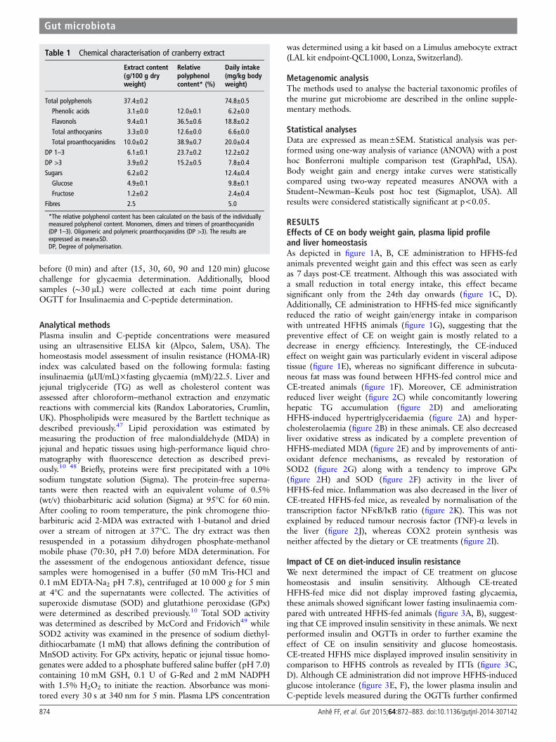
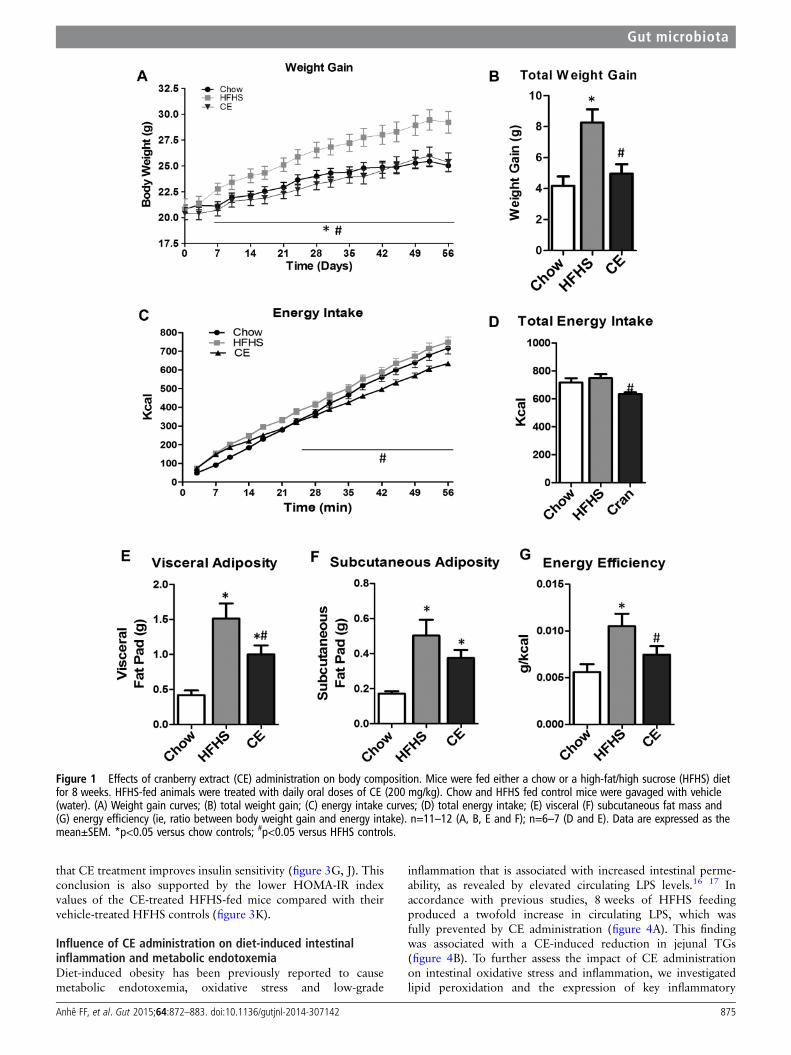
RESULTSEffects of CE on body weight gain, plasma lipid profileand liver homeostasisAs depicted in figure 1A, B, CE administration to HFHS-fedanimals prevented weight gain and this effect was seen as earlyas 7 days post-CE treatment. Although this was associated witha small reduction in total energy intake, this effect becamesignificant only from the 24th day onwards (figure 1C, D).Additionally, CE administration to HFHS-fed mice significantlyreduced the ratio of weight gain/energy intake in comparisonwith untreated HFHS animals (figure 1G), suggesting that thepreventive effect of CE on weight gain is mostly related to adecrease in energy efficiency. Interestingly, the CE-inducedeffect on weight gain was particularly evident in visceral adiposetissue (figure 1E), whereas no significant difference in subcuta-neous fat mass was found between HFHS-fed control mice andCE-treated animals (figure 1F). Moreover, CE administrationreduced liver weight (figure 2C) while concomitantly loweringhepatic TG accumulation (figure 2D) and amelioratingHFHS-induced hypertriglyceridaemia (figure 2A) and hyper-cholesterolaemia (figure 2B) in these animals. CE also decreasedliver oxidative stress as indicated by a complete prevention ofHFHS-mediated MDA (figure 2E) and by improvements of anti-oxidant defence mechanisms, as revealed by restoration ofSOD2 (figure 2G) along with a tendency to improve GPx(figure 2H) and SOD (figure 2F) activity in the liver ofHFHS-fed mice. Inflammation was also decreased in the liver ofCE-treated HFHS-fed mice, as revealed by normalisation of thetranscription factor NFκB/IκB ratio (figure 2K). This was notexplained by reduced tumour necrosis factor (TNF)-α levels inthe liver (figure 2J), whereas COX2 protein synthesis wasneither affected by the dietary or CE treatments (figure 2I).
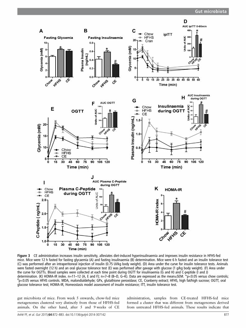
Impact of CE on diet-induced insulin resistanceWe next determined the impact of CE treatment on glucosehomeostasis and insulin sensitivity. Although CE-treatedHFHS-fed mice did not display improved fasting glycaemia,these animals showed significant lower fasting insulinaemia com-pared with untreated HFHS-fed animals (figure 3A, B), suggest-ing that CE improved insulin sensitivity in these animals. We nextperformed insulin and OGTTs in order to further examine theeffect of CE on insulin sensitivity and glucose homeostasis.CE-treated HFHS mice displayed improved insulin sensitivity incomparison to HFHS controls as revealed by ITTs (figure 3C,D). Although CE administration did not improve HFHS-inducedglucose intolerance (figure 3E, F), the lower plasma insulin andC-peptide levels measured during the OGTTs further confirmed
Table 1 Chemical characterisation of cranberry extract
Extract content(g/100 g dryweight)
Relativepolyphenolcontent* (%)
Daily intake(mg/kg bodyweight)
Total polyphenols 37.4±0.2 74.8±0.5Phenolic acids 3.1±0.0 12.0±0.1 6.2±0.0Flavonols 9.4±0.1 36.5±0.6 18.8±0.2Total anthocyanins 3.3±0.0 12.6±0.0 6.6±0.0Total proanthocyanidins 10.0±0.2 38.9±0.7 20.0±0.4
DP 1–3 6.1±0.1 23.7±0.2 12.2±0.2DP >3 3.9±0.2 15.2±0.5 7.8±0.4Sugars 6.2±0.2 12.4±0.4
Glucose 4.9±0.1 9.8±0.1Fructose 1.2±0.2 2.4±0.4
Fibres 2.5 5.0
*The relative polyphenol content has been calculated on the basis of the individuallymeasured polyphenol content. Monomers, dimers and trimers of proanthocyanidin(DP 1–3). Oligomeric and polymeric proanthocyanidins (DP >3). The results areexpressed as mean±SD.DP, Degree of polymerisation.
Gut microbiota
874 Anhê FF, et al. Gut 2015;64:872–883. doi:10.1136/gutjnl-2014-307142
that CE treatment improves insulin sensitivity (figure 3G, J). Thisconclusion is also supported by the lower HOMA-IR indexvalues of the CE-treated HFHS-fed mice compared with theirvehicle-treated HFHS controls (figure 3K).
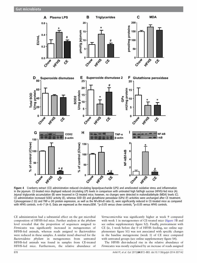
Influence of CE administration on diet-induced intestinalinflammation and metabolic endotoxemiaDiet-induced obesity has been previously reported to causemetabolic endotoxemia, oxidative stress and low-grade
inflammation that is associated with increased intestinal perme-ability, as revealed by elevated circulating LPS levels.16 17 Inaccordance with previous studies, 8 weeks of HFHS feedingproduced a twofold increase in circulating LPS, which wasfully prevented by CE administration (figure 4A). This findingwas associated with a CE-induced reduction in jejunal TGs(figure 4B). To further assess the impact of CE administrationon intestinal oxidative stress and inflammation, we investigatedlipid peroxidation and the expression of key inflammatory
Figure 1 Effects of cranberry extract (CE) administration on body composition. Mice were fed either a chow or a high-fat/high sucrose (HFHS) dietfor 8 weeks. HFHS-fed animals were treated with daily oral doses of CE (200 mg/kg). Chow and HFHS fed control mice were gavaged with vehicle(water). (A) Weight gain curves; (B) total weight gain; (C) energy intake curves; (D) total energy intake; (E) visceral (F) subcutaneous fat mass and(G) energy efficiency (ie, ratio between body weight gain and energy intake). n=11–12 (A, B, E and F); n=6–7 (D and E). Data are expressed as themean±SEM. *p<0.05 versus chow controls; #p<0.05 versus HFHS controls.
Gut microbiota
Anhê FF, et al. Gut 2015;64:872–883. doi:10.1136/gutjnl-2014-307142 875
modulators in the jejunum of chow, HFHS and CE-treatedHFHS animals. No significant changes were noted in MDAand SOD among groups (figure 4C, D). However, CE treat-ment was found to prevent the decrease in SOD2 activity byHFHS (figure 4E) without any effects on GPx (figure 4F).Importantly, CE administration fully prevented HFHSdiet-induced intestinal inflammation, as evidenced by thereduced COX2 and TNF-α protein expression as well as bythe normalisation of NFκB/IκB ratio (figure 4G, E).
Effects of CE administration on the gut microbiotaThe overall composition of the bacterial community in the dif-ferent groups was assessed by analysing the degree of bacterialtaxonomic similarity between metagenomic samples at thegenus level. Bacterial communities were clustered using principalcomponent analysis (PCA), which distinguished microbial com-munities based both on diet/treatment and time of faecal sam-pling (weeks 0, 1, 5 and 9). As shown in figure 5A, PCAdisclosed that distinct diets promoted the main alterations in the
Figure 2 Effect of cranberry extract (CE) administration on plasma and liver lipid profiles, hepatic inflammation and oxidative stress. CEadministration to high fat/high sucrose (HFHS)-fed mice prevented diet-induced increase in plasma triglycerides (A) and total cholesterol (B), whereasliver weight (C) and hepatic triglyceride accumulation (D) were reduced after CE treatment. Moreover, CE decreased liver oxidative stress as indicatedby a complete prevention of HFHS-mediated malondialdehyde (MDA) levels (E) and by improvements of antioxidant defence mechanisms, asrevealed by restoration of superoxide dismutase(SOD2) (G) and a tendency to improve glutathione peroxidase (GPx) (H) and SOD (F) activity in theliver of HFHS-fed mice. A trend towards lessening cyclooxygenase-2 protein expression (I) and a significant reduction in the NFκB/IκB ratio (K) werefound after CE treatment. CE administration did not affect TNF-α protein synthesis ( J) in the liver. n=11–12 (A–D); n=6–7 (E-K). Data are expressedas the mean±SEM. *p<0.05 versus chow controls; #p<0.05 versus HFHS controls.
Gut microbiota
876 Anhê FF, et al. Gut 2015;64:872–883. doi:10.1136/gutjnl-2014-307142
gut microbiota of mice. From week 5 onwards, chow-fed micemetagenomes clustered very distinctly from those of HFHS-fedanimals. On the other hand, after 5 and 9 weeks of CE
administration, samples from CE-treated HFHS-fed miceformed a cluster that was different from metagenomes derivedfrom untreated HFHS-fed animals. These results indicate that
Figure 3 CE administration increases insulin sensitivity, alleviates diet-induced hyperinsulinaemia and improves insulin resistance in HFHS-fedmice. Mice were 12 h fasted for fasting glycaemia (A) and fasting insulinaemia (B) determination. Mice were 6 h fasted and an insulin tolerance test(C) was performed after an intraperitoneal injection of insulin (0.75 UI/kg body weight). (D) Area under the curve for insulin tolerance tests. Animalswere fasted overnight (12 h) and an oral glucose tolerance test (E) was performed after gavage with glucose (1 g/kg body weight). (F) Area underthe curve for OGTTs. Blood samples were collected at each time point during OGTT for insulinaemia (G and H) and C-peptide (I and J)determination. (K) HOMA-IR index. n=11–12 (A, E and F); n=7–8 (B–D, G–K). Data are expressed as the mean±SEM. *p<0.05 versus chow controls;#p<0.05 versus HFHS controls. MDA, malondialdehyde; GPx, glutathione peroxidase; CE, Cranberry extract; HFHS, high fat/high sucrose; OGTT, oralglucose tolerance test; HOMA-IR, Homeostasis model assessment of insulin resistance; ITT, insulin tolerance test.
Gut microbiota
Anhê FF, et al. Gut 2015;64:872–883. doi:10.1136/gutjnl-2014-307142 877
CE administration had a substantial effect on the gut microbialcomposition of HFHS-fed mice. Further analysis at the phylumlevel revealed that the proportion of sequences assigned toFirmicutes was significantly increased in metagenomes ofHFHS-fed animals, whereas reads assigned to Bacteroideteswere reduced in these samples. A similar trend observed for theBacteroidetes phylum in metagenomes from untreatedHFHS-fed animals was found in samples from CE-treatedHFHS-fed mice. Furthermore, the relative abundance of
Verrucomicrobia was significantly higher at week 9 comparedwith week 1 in metagenomes of CE-treated mice (figure 5B andsee online supplementary figure S2). Finally, pretreatment withCE (ie, 1 week before day 0 of HFHS feeding, see online sup-plementary figure S1) was not associated with specific changesin the baseline metagenome (week 1) of CE mice comparedwith untreated groups (see online supplementary figure S4).
The HFHS diet-induced rise in the relative abundance ofFirmicutes was mostly explained by an increase of reads assigned
Figure 4 Cranberry extract (CE) administration reduced circulating lipopolysaccharide (LPS) and ameliorated oxidative stress and inflammationin the jejunum. CE-treated mice displayed reduced circulating LPS levels in comparison with untreated high fat/high sucrose (HFHS)-fed mice (A).Jejunal triglyceride accumulation (B) were lessened in CE-treated mice; however, no changes were detected in malondialdehyde (MDA) levels (C).CE administration increased SOD2 activity (E), whereas SOD (D) and glutathione peroxidase (GPx) (F) activities were unchanged after CE treatment.Cylooxygenase-2 (G) and TNF-α (H) protein expression, as well as the NFκB/IκB ratio (I), were significantly reduced in CE-treated mice as comparedwith HFHS controls. n=6–7 (A–I). Data are expressed as the mean±SEM. *p<0.05 versus chow controls; #p<0.05 versus HFHS controls.
Gut microbiota
878 Anhê FF, et al. Gut 2015;64:872–883. doi:10.1136/gutjnl-2014-307142
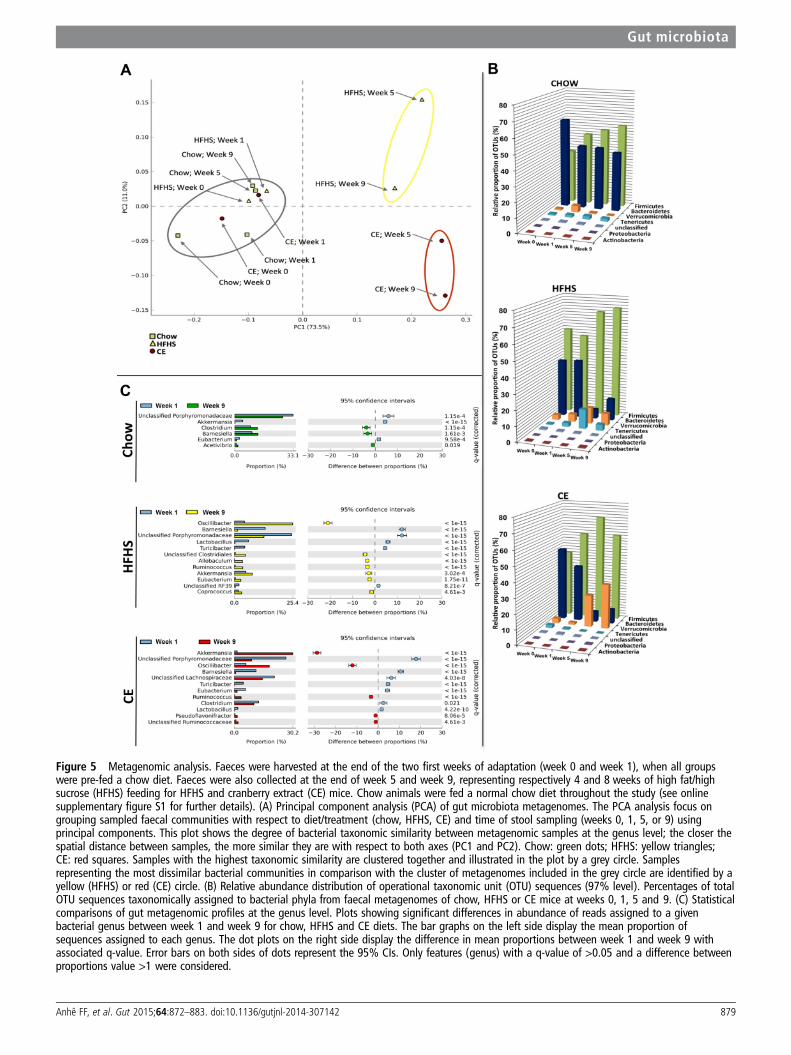
Figure 5 Metagenomic analysis. Faeces were harvested at the end of the two first weeks of adaptation (week 0 and week 1), when all groupswere pre-fed a chow diet. Faeces were also collected at the end of week 5 and week 9, representing respectively 4 and 8 weeks of high fat/highsucrose (HFHS) feeding for HFHS and cranberry extract (CE) mice. Chow animals were fed a normal chow diet throughout the study (see onlinesupplementary figure S1 for further details). (A) Principal component analysis (PCA) of gut microbiota metagenomes. The PCA analysis focus ongrouping sampled faecal communities with respect to diet/treatment (chow, HFHS, CE) and time of stool sampling (weeks 0, 1, 5, or 9) usingprincipal components. This plot shows the degree of bacterial taxonomic similarity between metagenomic samples at the genus level; the closer thespatial distance between samples, the more similar they are with respect to both axes (PC1 and PC2). Chow: green dots; HFHS: yellow triangles;CE: red squares. Samples with the highest taxonomic similarity are clustered together and illustrated in the plot by a grey circle. Samplesrepresenting the most dissimilar bacterial communities in comparison with the cluster of metagenomes included in the grey circle are identified by ayellow (HFHS) or red (CE) circle. (B) Relative abundance distribution of operational taxonomic unit (OTU) sequences (97% level). Percentages of totalOTU sequences taxonomically assigned to bacterial phyla from faecal metagenomes of chow, HFHS or CE mice at weeks 0, 1, 5 and 9. (C) Statisticalcomparisons of gut metagenomic profiles at the genus level. Plots showing significant differences in abundance of reads assigned to a givenbacterial genus between week 1 and week 9 for chow, HFHS and CE diets. The bar graphs on the left side display the mean proportion ofsequences assigned to each genus. The dot plots on the right side display the difference in mean proportions between week 1 and week 9 withassociated q-value. Error bars on both sides of dots represent the 95% CIs. Only features (genus) with a q-value of >0.05 and a difference betweenproportions value >1 were considered.
Gut microbiota
Anhê FF, et al. Gut 2015;64:872–883. doi:10.1136/gutjnl-2014-307142 879
to species from the genus Oscillibacter (figure 5C), whereas thedecreased proportion of Bacteroidetes found at week 9 was asso-ciated with a reduction in sequences assigned to the Barnesiellagenus and other unclassified members of thePorphyromonadaceae family. Importantly, CE administration wasassociated with a striking 30% increase in the relative abun-dance of Akkermansia in the CE-treated mice metagenome atweek 9 (figure 5C).
DISCUSSIONThere is growing evidence that the consumption of fruits andplant-derived foods is inversely correlated with several featuresof the metabolic syndrome, thus reducing the risk of T2D andCVD.50– 53 Despite the positive health effects possibly arisingfrom the presence of vitamins, minerals and dietary fibres infruits, there is a growing body of literature now supporting akey role for polyphenols in the protection against obesity-relateddiseases.54 55 Given that cranberry is one of the fruits with thehighest phenolic content,56 which includes a remarkableamount of PAC,32 57 we have elected to explore its impact onseveral components of the metabolic syndrome. We found thatCE administration prevents HFHS-induced weight gain andreduced visceral adiposity. Moreover, the effect on weight gainwas observed before any difference in caloric intake was noticedand was mostly linked to reduced energy efficiency. CE gavagefully prevented the development of fatty liver disease, asrevealed by reduced TG accumulation and blunted hepaticinflammation, which was associated with restoration of antioxi-dant defence mechanisms. These preventive effects on visceralobesity and liver steatosis were associated with improved insulinsensitivity, as revealed by lower fasting and postglucose insuli-naemia, improved insulin tolerance and a lower HOMA-IRindex in CE-treated HFHS-fed mice. Glucose tolerance per sewas not improved by CE treatment, but this may be explainedby the high glucose challenge during the OGTT, which is quiteimportant as compared with the glucose load generated by acomplex meal, thus possibly overwhelming the ability of thetissues to uptake more glucose, even in the context of animproved insulin sensitivity.
Phenolic phytochemicals are generally poorly absorbed, andthis has been put forward to suggest that these compounds arepossibly acting primarily at the level of intestinal absorption.58 59
Furthermore, several reports have demonstrated that the gutmicrobiota has a causal role in the pathogenesis of obesity andT2D.12 28 30 60 This prompted us to investigate the impact ofCE administration on the gut microbiota in the present study.Our results show that HFHS feeding induced a dramatic shift inthe gut microbiota of mice by increasing the proportion ofFirmicutes and decreasing the proportion of Bacteroidetes. Thisdiet-induced reshape in the microbial community of HFHS-fedmice is a typical characteristic of obesity-driven dysbiosis and isin agreement with previous publications,11 60–62 as was thefinding of an increased presence of the genus Oscillibacter inobese mice.63
The beneficial effects of CE treatment on metabolic pheno-types were associated with a robust modulation in the relativeabundance of Akkermansia spp., as suggested by the higher rep-resentation (30%) of reads assigned to this genus in CE meta-genome at week 9 in comparison with week 1. To the best ofour knowledge, this is the first report of a fruit extract exertingsuch a major effect on the presence of Akkermansia in the intes-tinal microbiota of an animal model of diet-induced obesity.Interestingly, administration of green tea polyphenols tohigh-fat-fed mice has also been recently associated with an
increase in the proportion of Akkermansia,64 whereasKemperman et al65 showed that complex polyphenols fromblack tea and from a grape juice/red wine mixture, in thecontext of a simulator of the human intestinal microbial ecosys-tem (ie, SHIME), increased the relative proportion ofAkkermansia. Moreover, the metabolic benefits of gastric bypasssurgery and of the antidiabetic drug metformin have been alsolinked to an increased intestinal abundance of this bacter-ium.29 30 We found that the CE-mediated increase in the relativeabundance of Akkermansia was associated with the preventionof the HFHS-induced rise in circulating LPS and abrogation ofintestinal inflammation in these animals. Although we have notdirectly established the causal relationship between CE-inducedincrease in the relative proportion of Akkermansia populationand the improved features of the metabolic syndrome inCE-treated HFHS-fed mice, it has been already reported thatoral administration of Akkermansia (ie, as a probiotic) reverseshigh-fat-diet-induced metabolic disorders28 and could alsomimic the antidiabetic effects of metformin in diabetic mice.30
Importantly, our results further suggest that the CE-relatedincrease in Akkermansia population might be sufficient toprevent the negative metabolic phenotype associated withobesity-driven dysbiosis without major modifications in the pro-portions of Firmicutes and Bacteroidetes.
Hepatic TG accumulation is a measure of steatosis, which incombination with oxidative stress and inflammation may lead toNAFLD, the most important cause of liver disease in Westerncountries that usually develops in the setting of insulin resistanceand obesity.66 LPS derived from the gut microbiota reaches theportal circulation and thus can access the liver to affect hostmetabolic physiology in critical ways.67 Elevated levels of circu-lating LPS in obese individuals have been shown to be a conse-quence of nutrient overload and high-fat-induced changes in thegut microbiota.17 Furthermore, metabolic endotoxemia (ie,increased circulating LPS after an obesogenic diet) is explainedby the transport of LPS from the gut lumen by newly synthe-sised chylomicrons from enterocytes in response to fatfeeding24 25 and by a gut microbiota-dependent disruption ofthe gut barrier, thus favouring LPS leakage.16 17 23 Interestingly,besides the fact that Akkermansia uses mucins as a food supply,it seems to be crucial for the mucus layer integrity.28 68
Akkermansia administration as a probiotic was reported toreduce systemic LPS levels in high-fat-fed mice, which is pos-sibly associated with the ability of Akkermansia to preserve themucus layer thickness, therefore reducing gut permeability andLPS leakage.28 Taken together, these observations suggest thatCE, by increasing the presence of Akkermansia, may reduceintestinal permeability and LPS leakage, therefore amelioratinginsulin resistance in diet-induced obese mice. Accordingly, theability of CE to blunt circulating LPS levels may be accountedfor the reduced hepatic TG accumulation and protection fromhepatic oxidative stress and inflammation in HFHS-fed mice.
Intestinal inflammation is growingly recognised to play a majorrole in the early deterioration of glucose and lipid metabolism inmodels of obesity and insulin resistance.13 Remarkably, CEadministration was found to completely suppress NFκB activa-tion in the intestine of HFHS-fed mice. NFκB is a central regula-tor of metabolic inflammation and controls the production ofseveral proinflammatory cytokines, including TNF-α. CE treat-ment reduced the amount of TNF-α in the intestine and alsodecreased COX2 protein expression, an enzyme crucial for thesynthesis of several proinflammatory molecules. Additionally, CEadministration was found to increase the activity of SOD2, sug-gesting that it could exert some of its effects through promoting
Gut microbiota
880 Anhê FF, et al. Gut 2015;64:872–883. doi:10.1136/gutjnl-2014-307142
oxidant defence mechanisms in the intestine. It has been sug-gested that interactions between diet and enteric bacteria arenecessary for inducing inflammatory changes in the intestine andto impact on diet-induced obesity and insulin resistance.13
Therefore, our observations suggest that CE treatment, acting asa prebiotic, may be a novel strategy against intestinal inflamma-tion and the prevention of the metabolic syndrome.
The mechanisms by which phenolic phytochemicals mayexert prebiotic effects and reshape the gut microbiota with bene-fits to the host are still unclear. It has been suggested thatAkkermansia can display a rapid growth rate in order to monop-olise the resources when competition is low following animportant ecological disruption (eg, gastric bypass surgery,caloric restriction, antibiotic therapy).69 CE is very rich in poly-phenols, particularly phenolic acids, flavan-3-ols (eg, catechin,epicatechin) and PAC (table 1). These molecules have beenshown to possess remarkable antibacterial activity43 70 71 andcan potentially reshape the gut microbiota ecology of obesemice. One explanation is that this latter effect could be asso-ciated with a reduction in the abundance of species capable ofholding Akkermansia in check, thus favouring a rise in its pro-portion. In fact, a polyphenol extract of wine and grapes stimu-lated the growth of Akkermansia while exerting profoundantimicrobial activity in the transversal colon of a gut biofer-mentor model.65 Moreover, this hypothesis is supported by thefact that broad-spectrum antibiotic treatment increases the pro-portion of Akkermansia in humans.72 Another explanation isthat CE, through their high PAC content, influences the produc-tion of mucin and thereby provides ample trophic resources forAkkermansia to thrive. Indeed, cranberry PAC have been shownto increase the differentiation of goblet cells and to preserve theproduction of luminal mucin 2 (Muc2) in mice fed throughelemental enteral nutrition, a treatment known to decrease themucosal barrier.36 Conversely, we found that CE administrationis associated with increased Kruppel-like factor 4 (Klf4)—amarker of goblet cells—and Muc2 mRNA expression in theproximal colon (see online supplementary figure S3), which sup-ports the hypothesis that these condensed tannins are able tostimulate mucus production and therefore create an ecologicalniche for the mucus-eating bacterium Akkermansia. Importantly,since it has been reported that Akkermansia administration perse can re-establish the mucus layer integrity in diet-inducedobese mice,28 it is possible that a direct trophic effect of CE onAkkermansia precedes the positive effects found on the mucuslayer integrity. Finally, Akkermansia contributes to the restor-ation of antimicrobial peptides such as regenerating islet-derivedprotein 3 γ (RegIIIγ).28 Although our results did not show a stat-istically significant modulation of Reg3g by CE treatment, wedid observe a trend for modulation of this antimicrobial markerthat would suggest that CE stimulates the induction of anti-microbial defences, which is possibly linked with higherAkkermansia muciniphila abundance in these animals. Furtherinvestigations are definitely warranted in order to better under-stand the prebiotic effects of CE on Akkermansia.
The use of Akkermansia as a probiotic in humans, although apromising strategy, may find some barriers. For instance, besidesthe fact that the safety of Akkermansia administration tohumans is currently unknown, its in vitro culture is technicallycomplex and time-consuming, suggesting that the production ofAkkermansia as a commercial-scale probiotic may be costly.Therefore, finding alternative methods to increase the presenceof Akkermansia spp. in the gut microbiota seems a valid, safeand likely more cost-effective approach. In addition, given thefact that cranberries are already highly consumed (especially in
North America), using CE as a prebiotic might be an interestingstrategy to ease adherence to the treatment compared with theintroduction of a novel probiotic strain. Moreover, the doseused in this study in mice may represent a feasible dose inhumans. By applying the US Food and Drug Administration’sguidelines to establish the human equivalent dose based onbody surface area,73 we found that a 16 mg/kg dose would bethe human equivalent of a 200 mg/kg dose in mice. This is per-fectly achievable by supplementation or by incorporating CE inother food products.
While our study provides evidence for the beneficial meta-bolic effects of CE treatment, some limitations must be acknowl-edged. First, while we have found that the CE treatmentimproves insulin sensitivity, based on ITT analysis, HOMA-IRand fasting and postglucose insulin levels, future studies that usethe euglycemic clamp technique together with isotopic tracerscould be performed to further confirm this effect of CE and todetermine the contribution of the liver and peripheral tissues tothis phenotype. Moreover, we acknowledge that the pooling ofsamples used to analyse the metagenomic bacterial diversity ofthe mice constitutes another limitation of this study, but allowedus to focus on the shifts occurring within the dominant phylo-types due to the different treatments.
In summary, we found that CE treatment protects fromdiet-induced obesity, liver steatosis and insulin resistance inHFHS-fed mice. This effect was associated with alleviation ofmetabolic endotoxemia and intestinal inflammation. Our studyfurther suggests that the ability of CE administration to raise therelative proportion of Akkermansia is playing a key role in thisprotective effect, leading us to propose that fruit polyphenolsmay prevent obesity and the metabolic syndrome through a pre-biotic effect on the gut microbiota.
Acknowledgements We thank André Comeau and Brian Boyle (Plate-formed’Analyses Génomiques, IBIS/Université Laval) for expert guidance in pyrosequencingand subsequent data filtering analysis. We are grateful to Émilie Desfossés-Foucaultfor helping in the early-stage analyses of metagenomic sequences, especially withthe construction of figure plots. We thank Valérie Dumais, Christine Dion, ChristineDallaire and Kim Denault for their expert help with animal experimentations.
Contributors AM, DR, EL, YD, GP and FFA designed the study. FFA, TV, SD, SM,CG and QM performed the experiments. FFA, GP, AM, DR, YD, EL, SM and TVanalysed the data and wrote the manuscript. All authors reviewed and approved thefinal manuscript.
Funding This work was funded by grants from the Ministère du DéveloppementÉconomique, de l’Innovation et de l’Exportation (MDEIE, PSR-SIIRI-444 ) and AgenceUniversitaire de la Francophonie (AUF) to AM and by the Institute of Nutrition andFunctional Foods (INAF, Laval University) to YD, AM, DR and EL.
Competing interests AM was the holder of a Canadian Institutes of HealthResearch (CIHR)/Pfizer research Chair in the pathogenesis of insulin resistance andcardiovascular diseases and EL holds a J.A. deSève Research Chair in nutrition. Theauthors thank Leahy Orchards and AppleActiv for their support to EL. FFA is therecipient of a PhD studentship from the CIHR training programme in obesity.
Ethics approval Laval University Ethics Committee.
Provenance and peer review Not commissioned; externally peer reviewed.
REFERENCES1 Eckel R, Grundy S, Zimmet P. The metabolic syndrome. Lancet 2005;365:1415–28.2 Hossain P, Kawar B, El Nahas M. Obesity and diabetes in the developing world—a
growing challenge. N Engl J Med 2007;356:213–15.3 Doria A, Patti ME, Kahn CR. The emerging genetic architecture of type 2 diabetes.
Cell Metab 2008;8:186–200.4 Walley AJ, Asher JE, Froguel P. The genetic contribution to non-syndromic human
obesity. Nat Rev Genet 2009;10:431–42.5 Marette A. Mediators of cytokine-induced insulin resistance in obesity and other
inflammatory settings. Curr Opin Clin Nutr Metab Care 2002;5:377–83.6 Freedman A, Freeman G, Rhynhart K, et al. Selective induction of B7/BB-1 on
interferon-gamma stimulated monocytes: a potential mechanism for amplification
Gut microbiota
Anhê FF, et al. Gut 2015;64:872–883. doi:10.1136/gutjnl-2014-307142 881
of T cell activation through the CD28 pathway. Cell Immunol 1991;137:429–37.
7 Wellen K, Hotamisligil G. Inflammation, stress, and diabetes. J ClinInvest2005;115:1111–19.
8 Weisberg S, McCann D, Desai M, et al. Obesity is associated with macrophageaccumulation in adipose tissue. J Clin Invest 2003;112:1796–808.
9 Xu H, Barnes G, Yang Q, et al. Chronic inflammation in fat plays a crucial role inthe development of obesity-related insulin resistance. J Clin Invest2003;112:1821–30.
10 Veilleux A, Grenier E, Marceau P, et al. Intestinal lipid handling: evidence andimplication of insulin signaling abnormalities in human obese subjects. ArteriosclerThromb Vasc Biol 2014;34:644–53.
11 Cotillard A, Kennedy SP, Kong LC, et al. Dietary intervention impact on gutmicrobial gene richness. Nature 2013;500:585–8.
12 Arpaia N, Campbell C, Fan X, et al. Metabolites produced by commensal bacteriapromote peripheral regulatory T-cell generation. Nature 2013;504:451–5.
13 Ding S, Chi MM, Scull BP, et al. High-fat diet: bacteria interactions promoteintestinal inflammation which precedes and correlates with obesity and insulinresistance in mouse. PloS One 2010;5:e12191.
14 Backhed F, Ding H, Wang T, et al. The gut microbiota as an environmental factorthat regulates fat storage. Proc Natl Acad Sci U S A 2004;101:15718–23.
15 Turnbaugh PJ, Ley RE, Mahowald MA, et al. An obesity-associated gut microbiomewith increased capacity for energy harvest. Nature 2006;444:1027–31.
16 Cani PD, Bibiloni R, Knauf C, et al. Changes in gut microbiota control metabolicendotoxemia-induced inflammation in high-fat diet-induced obesity and diabetes inmice. Diabetes 2008;57:1470–81.
17 Cani PD, Amar J, Iglesias MA, et al. Metabolic endotoxemia initiates obesity andinsulin resistance. Diabetes 2007;56:1761–72.
18 Cani PD, Neyrinck AM, Maton N, et al. Oligofructose promotes satiety in rats fed ahigh-fat diet: involvement of glucagon-like Peptide-1. Obes Res 2005;13:1000–7.
19 Cani PD, Possemiers S, Van de Wiele T, et al. Changes in gut microbiota controlinflammation in obese mice through a mechanism involving GLP-2-drivenimprovement of gut permeability. Gut 2009;58:1091–103.
20 Rabot S, Membrez M, Bruneau A, et al. Germ-free C57BL/6J mice are resistant tohigh-fat-diet-induced insulin resistance and have altered cholesterol metabolism.FASEB J 2010;24:4948–59.
21 Geurts L, Lazarevic V, Derrien M, et al. Altered gut microbiota and endocannabinoidsystem tone in obese and diabetic leptin-resistant mice: impact on apelin regulationin adipose tissue. Front Microbiol 2011;2:149.
22 Cani PD, Neyrinck AM, Fava F, et al. Selective increases of bifidobacteria in gutmicroflora improve high-fat-diet-induced diabetes in mice through a mechanismassociated with endotoxaemia. Diabetologia 2007;50:2374–83.
23 Muccioli GG, Naslain D, Backhed F, et al. The endocannabinoid system links gutmicrobiota to adipogenesis. Mol Syst Biol 2010;6:392.
24 Vreugdenhil AC, Rousseau CH, Hartung T, et al. Lipopolysaccharide (LPS)-bindingprotein mediates LPS detoxification by chylomicrons. J Immunol2003;170:1399–405.
25 Ghoshal S, Witta J, Zhong J, et al. Chylomicrons promote intestinal absorption oflipopolysaccharides. J Lipid Res 2009;50:90–7.
26 Hildebrandt MA, Hoffmann C, Sherrill-Mix SA, et al. High-fat diet determines thecomposition of the murine gut microbiome independently of obesity.Gastroenterology 2009;137:1716–24.e1–2.
27 Everard A, Lazarevic V, Derrien M, et al. Responses of gut microbiota and glucoseand lipid metabolism to prebiotics in genetic obese and diet-induced leptin-resistantmice. Diabetes 2011;60:2775–86.
28 Everard A, Belzer C, Geurts L, et al. Cross-talk between Akkermansia muciniphilaand intestinal epithelium controls diet-induced obesity. Proc Natl Acad Sci U S A2013;110:9066–71.
29 Liou AP, Paziuk M, Luevano JM Jr, et al. Conserved shifts in the gut microbiota dueto gastric bypass reduce host weight and adiposity. Sci Transl Med 2013;5:178ra41.
30 Shin NR, Lee JC, Lee HY, et al. An increase in the Akkermansia spp. populationinduced by metformin treatment improves glucose homeostasis in diet-inducedobese mice. Gut 2014;63:727–35.
31 Derrien M, Collado MC, Ben-Amor K, et al. The Mucin degrader Akkermansiamuciniphila is an abundant resident of the human intestinal tract. Appl EnvironMicrobiol 2008;74:1646–8.
32 Blumberg JB, Camesano TA, Cassidy A, et al. Cranberries and their bioactiveconstituents in human health. Adv Nutr 2013;4:618–32.
33 Neto CC. Cranberry and blueberry: evidence for protective effects against cancerand vascular diseases. Mol Nutr Food Res 2007;51:652–64.
34 He X, Liu RH. Cranberry phytochemicals: Isolation, structure elucidation, and theirantiproliferative and antioxidant activities. J Agric Food Chem 2006;54:7069–74.
35 Denis MC, Furtos A, Dudonne S, et al. Apple peel polyphenols and their beneficialactions on oxidative stress and inflammation. PloS One 2013;8:e53725.
36 Pierre JF, Heneghan AF, Feliciano RP, et al. Cranberry proanthocyanidins improvethe gut mucous layer morphology and function in mice receiving elemental enteralnutrition. JPEN J Parenter Enteral Nutr 2013;37:401–9.
37 Deziel B, MacPhee J, Patel K, et al. American cranberry (Vaccinium macrocarpon)extract affects human prostate cancer cell growth via cell cycle arrest by modulatingexpression of cell cycle regulators. Food Funct 2012;3:556–64.
38 Neto CC, Amoroso JW, Liberty AM. Anticancer activities of cranberryphytochemicals: an update. Mol Nutr Food Res 2008;52(Suppl 1):S18–27.
39 Madrigal-Carballo S, Rodriguez G, Sibaja M, et al. Chitosomes loaded withcranberry proanthocyanidins attenuate the bacterial lipopolysaccharide-inducedexpression of iNOS and COX-2 in raw 264.7 macrophages. J Liposome Res2009;19:189–96.
40 Kim MJ, Ohn J, Kim JH, et al. Effects of freeze-dried cranberry powder on serumlipids and inflammatory markers in lipopolysaccharide treated rats fed anatherogenic diet. Nutr Res Pract 2011;5:404–11.
41 Kivimäki AS, Ehlers PI, Siltari A, et al. Lingonberry, cranberry and blackcurrant juicesaffect mRNA expressions of inflammatory and atherothrombotic markers of SHR in along-term treatment. J Funct Foods 2012;4:496–503.
42 Foo LY, Lu Y, Howell AB, et al. A-Type proanthocyanidin trimers from cranberry thatinhibit adherence of uropathogenic P-fimbriated Escherichia coli. J Nat Prod2000;63:1225–8.
43 Uberos J, Iswaldi I, Belmonte RR, et al. Cranberry (Vaccinium macrocarpon) changesthe surface hydrophobicity and biofilm formation of E. coli. Microbiol Insights2011;4:21–7.
44 Shidfar F, Heydari I, Hajimiresmaiel SJ, et al. The effects of cranberry juice on serumglucose, apoB, apoA-I, Lp(a), and Paraoxonase-1 activity in type 2 diabetic malepatients. J Res Med Sci 2012;17:355–60.
45 Lee IT, Chan YC, Lin CW, et al. Effect of cranberry extracts on lipid profiles insubjects with Type 2 diabetes. Diabet Med 2008;25:1473–7.
46 Basu A, Lyons TJ. Strawberries, blueberries, and cranberries in the metabolicsyndrome: clinical perspectives. J Agric Food Chem 2012;60:5687–92.
47 Spahis S, Vanasse M, Belanger SA, et al. Lipid profile, fatty acid composition andpro- and anti-oxidant status in pediatric patients with attention-deficit/hyperactivitydisorder. Prostaglandins Leukot Essent Fatty Acids 2008;79:47–53.
48 Levy E, Trudel K, Bendayan M, et al. Biological role, protein expression, subcellularlocalization, and oxidative stress response of paraoxonase 2 in the intestine ofhumans and rats. Am J Physiol Gastrointest Liver Physiol 2007;293:G1252–61.
49 McCord JM, Fridovich I. Superoxide dismutase. An enzymic function forerythrocuprein (hemocuprein). J Biol Chem 1969;244:6049–55.
50 Morimoto A, Ohno Y, Tatsumi Y, et al. Effects of healthy dietary pattern and otherlifestyle factors on incidence of diabetes in a rural Japanese population. Asia Pac JClin Nutr 2012;21:601–8.
51 Bauer F, Beulens JW, van der AD, et al. Dietary patterns and the risk of type 2diabetes in overweight and obese individuals. Eur J Nutr 2013;52:1127–34.
52 Eshak ES, Iso H, Mizoue T, et al. Soft drink, 100% fruit juice, and vegetable juiceintakes and risk of diabetes mellitus. Clin Nutr 2013;32:300–8.
53 Chan HT, Yiu KH, Wong CY, et al. Increased dietary fruit intake was associated withlower burden of carotid atherosclerosis in Chinese patients with Type 2 diabetesmellitus. Diabet Med 2013;30:100–8.
54 Corder R, Mullen W, Khan NQ, et al. Oenology: red wine procyanidins and vascularhealth. Nature 2006;444:566.
55 Anhê FF, Desjardins Y, Pilon G, et al. Polyphenols and type 2 diabetes: aprospective review. PharmaNutrition 2013;1:105–14.
56 Vinson JA, Su X, Zubik L, et al. Phenol antioxidant quantity and quality in foods:fruits. J Agric Food Chem 2001;49:5315–21.
57 Grace MH, Massey AR, Mbeunkui F, et al. Comparison of health-relevant flavonoidsin commonly consumed cranberry products. J Food Sci 2012;77:H176–83.
58 Manach C, Williamson G, Morand C, et al. Bioavailability and bioefficacy ofpolyphenols in humans. I. Review of 97 bioavailability studies. Am J Clin Nutr2005;81:230S–42S.
59 Cardona F, Andres-Lacueva C, Tulipani S, et al. Benefits of polyphenols ongut microbiota and implications in human health. J Nutr Biochem 2013;24:1415–22.
60 Caricilli AM, Picardi PK, de Abreu LL, et al. Gut microbiota is a key modulator ofinsulin resistance in TLR 2 knockout mice. PLoS Biol 2011;9:e1001212.
61 Ley RE, Backhed F, Turnbaugh P, et al. Obesity alters gut microbial ecology. ProcNatl Acad Sci U S A 2005;102:11070–5.
62 Ley RE, Turnbaugh PJ, Klein S, et al. Microbial ecology: human gut microbesassociated with obesity. Nature 2006;444:1022–3.
63 Lam YY, Ha CW, Campbell CR, et al. Increased gut permeability and microbiotachange associate with mesenteric fat inflammation and metabolic dysfunction indiet-induced obese mice. PloS One 2012;7:e34233.
64 Axling U, Olsson C, Xu J, et al. Green tea powder and Lactobacillus plantarumaffect gut microbiota, lipid metabolism and inflammation in high-fat fed C57BL/6Jmice. Nutr Metab (Lond) 2012;9:105.
65 Kemperman RA, Gross G, Mondot S, et al. Impact of polyphenols from black teaand red wine/grape juice on a gut model microbiome. Food Res Int 2013;53:659–69.
66 Gariani K, Philippe J, Jornayvaz FR. Non-alcoholic fatty liver disease andinsulin resistance: From bench to bedside. Diabetes Metab 2013;39:16–26.
Gut microbiota
882 Anhê FF, et al. Gut 2015;64:872–883. doi:10.1136/gutjnl-2014-307142
67 Vajro P, Paolella G, Fasano A. Microbiota and gut-liver axis: their influences onobesity and obesity-related liver disease. J Pediatr Gastroenterol Nutr2013;56:461–8.
68 Belzer C, de Vos WM. Microbes inside—from diversity to function: the case ofAkkermansia. ISME J 2012;6:1449–58.
69 Cox LM, Blaser MJ. Pathways in microbe-induced obesity. Cell Metab2013;17:883–94.
70 Rastmanesh R. High polyphenol, low probiotic diet for weight loss because ofintestinal microbiota interaction. Chem Biolol Interact 2011;189:1–8.
71 Feldman M, Grenier D. Cranberry proanthocyanidins act in synergy withlicochalcone A to reduce Porphyromonas gingivalis growth and virulence properties,and to suppress cytokine secretion by macrophages. J Appl Microbiol2012;113:438–47.
72 Dubourg G, Lagier JC, Armougom F, et al. High-level colonisation of the human gutby Verrucomicrobia following broad-spectrum antibiotic treatment. Int J AntimicrobAgents 2013;41:149–55.
73 Reagan-Shaw S, Nihal M, Ahmad N. Dose translation from animal to human studiesrevisited. FASEB J 2008;22:659–61.
Gut microbiota
Anhê FF, et al. Gut 2015;64:872–883. doi:10.1136/gutjnl-2014-307142 883