eTh ar neGe , cn de Proteus Providencia, and Morganellaottemann/migrated/metx119l/Exercises... ·...
25
CHAPTER 3.3.12 T h e G e n e r a P r o t e u s , P r o v i d e n c i a a n d M o r g a n e l l a The Genera Proteus, Providencia, and Morganella JIM MANOS AND ROBERT BELAS Introduction The three genera Proteus, Morganella and Prov- idencia presently comprise a total of ten species. All are motile, Gram-negative rods with peritri- chous flagella, and are assigned to the Enterobac- teriaceae family mainly on the basis of shared biochemical characteristics. Most significantly, they are characterized by their ability to oxida- tively deaminate phenylalanine and, in most cases (except for some Providencia spp.), to hydrolyze urea (Farmer et al., 1977; Moltke, 1927; Wenner and Retger, 1919). Unusual fea- tures include the ability of Proteus sp. to differ- entiate into swarmer cells upon colonization of solid surfaces. This topic will be covered in more detail in ensuing sections. Interest in the species comprising these genera has occurred mainly from a clinical perspective, as they include a number of significant human pathogens. In human disease, most infections are associated with prolonged hospitalization and in the case of Proteus and Morganella spp., coloni- zation of indwelling catheters and associated uri- nary tract infections (UTIs). Taxonomy and Phylogeny Before the advent of phylogenetically based clas- sification, an array of biochemical tests formed the basis for taxonomic classification of the gen- era Proteus, Providencia, and after its separation into a new genus , Morganella. Tables 1 and 2 list the major biochemical tests used to compare and differentiate between the genera. As shown in Table 1, the most significant shared characteris- tics are the (oxidative) deamination of phenyl- alanine and tryptophan; both are used to distinguish between these three genera and other Enterobacteriaceae that do not produce these deaminases. The tests for production of these deaminases were developed in the 1950s and still are widely used (Henriksen, 1950; Thibault and Le Minor, 1957). Table 2 shows the biochemical tests that are commonly used to distinguish between these genera. The only test that will dis- tinguish Morganella from Proteus and Providen- cia is the lysine iron agar test. On the other hand, several tests will distinguish Proteus from Provi- dencia. Providencia is characterized by the pro- duction of acid from a variety of sugars , whereas Proteus is distinguished from Providencia by the hydrolysis of gelatin and the production of lipase and hydrogen sulfide. The use of molecular phy- logenetic methods of classification has resulted in several species being reassigned to separate genera based on relatedness at the DNA level. These changes include: the new genus Mor- ganella,with transfer of the species Proteus mor- ganii to it (Brenner et al., 1978); the classification of Providencia alcalifaciens biogroup 3 as the separate species (Providencia rustigianii; Higash- itani et al., 1995); and the identification of a sub- group within the latter as a distinct species , Providencia heimbachae (Muller et al., 1986b). Isolated in 1906 (Morgan, 1906), Morganella morganii was originally included in the genus Proteus as Proteus morganii. Brenner et al. (1978) showed that P. morganii had less than 20% homology to Proteus and Providencia spp., neces- sitating relocation into a new genus. The method employed by Brenner et al. in this reclassification compared the amount of single stranded 32 P- labeled DNA reassociating with DNA from the same source (homologous reaction), relative to the amount reassociating with DNA from other species in these genera. The resulting value was expressed as “percent DNA relatedness.” Mor- ganella morganii has subsequently been subdi- vided into two subspecies based on its ability to ferment trehalose and the results of DNA-DNA hybridization studies (Jensen et al., 1992). Mor- ganella morganii subsp. morganii is composed of four biogroups (A, B, C and D), and it does not ferment trehalose, though M. morganii subsp. sibonii contains three biogroups (E, F and G) and does ferment trehalose. Further subdivision of these species is likely with the application of more discriminatory molecular methods of characterization. As an example, the use of molecular typing by 16S ribo- somal RNA (rRNA) gene fingerprints (ribotyp- ing) has recently demonstrated that, though Prokaryotes (2006) 6:245–269 DOI: 10.1007/0-387-30746-x_12
Transcript of eTh ar neGe , cn de Proteus Providencia, and Morganellaottemann/migrated/metx119l/Exercises... ·...
CHAPTER 3.3.12The Genera Proteus, Providencia and Morganella
The Genera Proteus, Providencia, and Morganella
JIM MANOS AND ROBERT BELAS
Introduction
The three genera Proteus, Morganella and Prov-idencia presently comprise a total of ten species.All are motile, Gram-negative rods with peritri-chous flagella, and are assigned to the Enterobac-teriaceae family mainly on the basis of sharedbiochemical characteristics. Most significantly,they are characterized by their ability to oxida-tively deaminate phenylalanine and, in mostcases (except for some Providencia spp.), tohydrolyze urea (Farmer et al., 1977; Moltke,1927; Wenner and Retger, 1919). Unusual fea-tures include the ability of Proteus sp. to differ-entiate into swarmer cells upon colonization ofsolid surfaces. This topic will be covered in moredetail in ensuing sections.
Interest in the species comprising these generahas occurred mainly from a clinical perspective,as they include a number of significant humanpathogens. In human disease, most infections areassociated with prolonged hospitalization and inthe case of Proteus and Morganella spp., coloni-zation of indwelling catheters and associated uri-nary tract infections (UTIs).
Taxonomy and Phylogeny
Before the advent of phylogenetically based clas-sification, an array of biochemical tests formedthe basis for taxonomic classification of the gen-era Proteus, Providencia, and after its separationinto a new genus, Morganella. Tables 1 and 2 listthe major biochemical tests used to compare anddifferentiate between the genera. As shown inTable 1, the most significant shared characteris-tics are the (oxidative) deamination of phenyl-alanine and tryptophan; both are used todistinguish between these three genera and otherEnterobacteriaceae that do not produce thesedeaminases. The tests for production of thesedeaminases were developed in the 1950s and stillare widely used (Henriksen, 1950; Thibault andLe Minor, 1957). Table 2 shows the biochemicaltests that are commonly used to distinguishbetween these genera. The only test that will dis-
tinguish Morganella from Proteus and Providen-cia is the lysine iron agar test. On the other hand,several tests will distinguish Proteus from Provi-dencia. Providencia is characterized by the pro-duction of acid from a variety of sugars, whereasProteus is distinguished from Providencia by thehydrolysis of gelatin and the production of lipaseand hydrogen sulfide. The use of molecular phy-logenetic methods of classification has resultedin several species being reassigned to separategenera based on relatedness at the DNA level.These changes include: the new genus Mor-ganella, with transfer of the species Proteus mor-ganii to it (Brenner et al., 1978); the classificationof Providencia alcalifaciens biogroup 3 as theseparate species (Providencia rustigianii; Higash-itani et al., 1995); and the identification of a sub-group within the latter as a distinct species,Providencia heimbachae (Muller et al., 1986b).
Isolated in 1906 (Morgan, 1906), Morganellamorganii was originally included in the genusProteus as Proteus morganii. Brenner et al.(1978) showed that P. morganii had less than 20%homology to Proteus and Providencia spp., neces-sitating relocation into a new genus. The methodemployed by Brenner et al. in this reclassificationcompared the amount of single stranded 32P-labeled DNA reassociating with DNA from thesame source (homologous reaction), relative tothe amount reassociating with DNA from otherspecies in these genera. The resulting value wasexpressed as “percent DNA relatedness.” Mor-ganella morganii has subsequently been subdi-vided into two subspecies based on its ability toferment trehalose and the results of DNA-DNAhybridization studies (Jensen et al., 1992). Mor-ganella morganii subsp. morganii is composed offour biogroups (A, B, C and D), and it does notferment trehalose, though M. morganii subsp.sibonii contains three biogroups (E, F and G) anddoes ferment trehalose.
Further subdivision of these species is likelywith the application of more discriminatorymolecular methods of characterization. As anexample, the use of molecular typing by 16S ribo-somal RNA (rRNA) gene fingerprints (ribotyp-ing) has recently demonstrated that, though
Prokaryotes (2006) 6:245–269DOI: 10.1007/0-387-30746-x_12
246 J. Manos and R. Belas CHAPTER 3.3.12
clinical isolates of P. mirabilis, P. penneri, M.morganii and P. heimbachae had identicalribotyping patterns to those of their respectivetype strains, those from the remaining species allexhibited heterogeneity, containing from two tofour ribogroups each (Pignato et al., 1999). Thusit is possible that subdivision of members ofthese latter species into one or more subspeciesmay be considered appropriate in the future.
The genus Proteus presently contains fourspecies: P. mirabilis, P. vulgaris, P. penneri andP. myxofaciens. Morganella has one species (M.morganii), whereas Providencia contains fivespecies (Providencia stuartii, Providencia rettgeri,P. rustigianii, P. heimbachae and P. alcalifaciens;(Rozalski, 1997; Rustigian, R., and C. A. Stuart,1945).
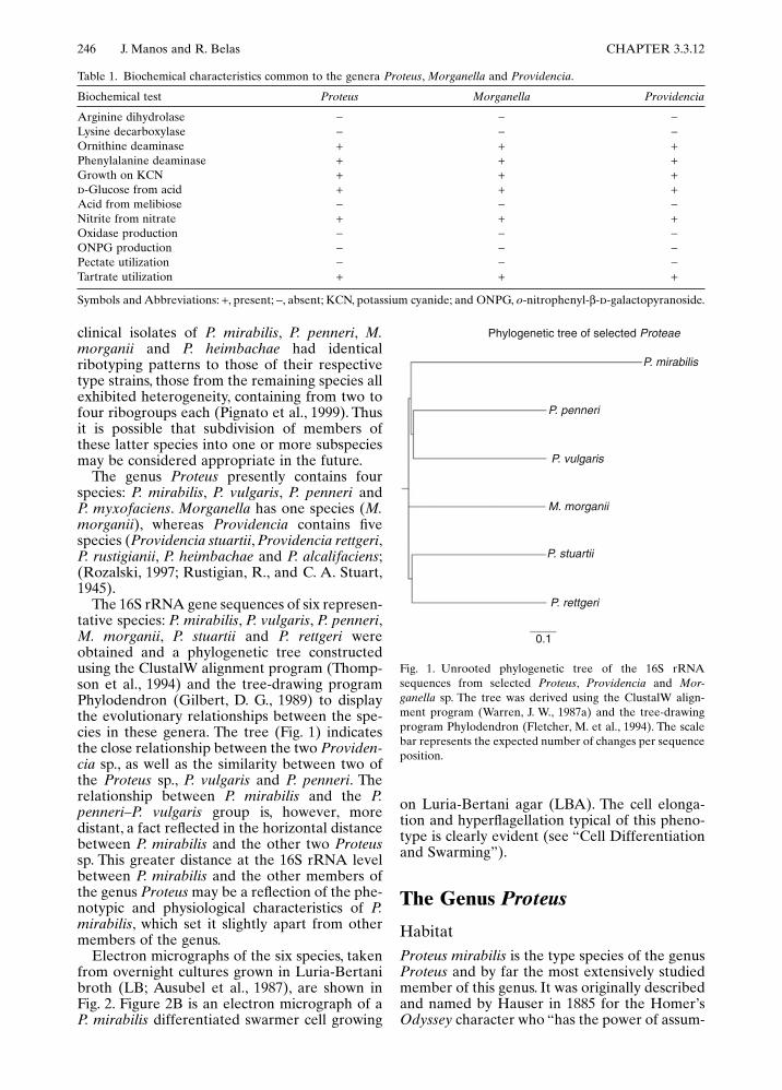
The 16S rRNA gene sequences of six represen-tative species: P. mirabilis, P. vulgaris, P. penneri,M. morganii, P. stuartii and P. rettgeri wereobtained and a phylogenetic tree constructedusing the ClustalW alignment program (Thomp-son et al., 1994) and the tree-drawing programPhylodendron (Gilbert, D. G., 1989) to displaythe evolutionary relationships between the spe-cies in these genera. The tree (Fig. 1) indicatesthe close relationship between the two Providen-cia sp., as well as the similarity between two ofthe Proteus sp., P. vulgaris and P. penneri. Therelationship between P. mirabilis and the P.penneri–P. vulgaris group is, however, moredistant, a fact reflected in the horizontal distancebetween P. mirabilis and the other two Proteussp. This greater distance at the 16S rRNA levelbetween P. mirabilis and the other members ofthe genus Proteus may be a reflection of the phe-notypic and physiological characteristics of P.mirabilis, which set it slightly apart from othermembers of the genus.
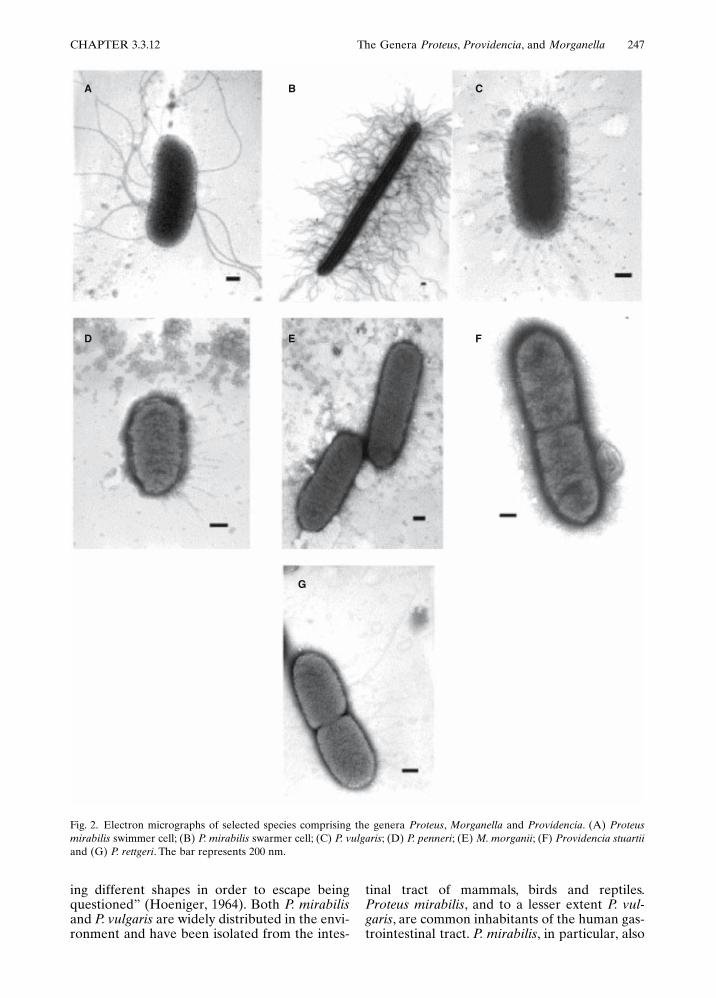
Electron micrographs of the six species, takenfrom overnight cultures grown in Luria-Bertanibroth (LB; Ausubel et al., 1987), are shown inFig. 2. Figure 2B is an electron micrograph of aP. mirabilis differentiated swarmer cell growing
on Luria-Bertani agar (LBA). The cell elonga-tion and hyperflagellation typical of this pheno-type is clearly evident (see “Cell Differentiationand Swarming”).
The Genus Proteus
HabitatProteus mirabilis is the type species of the genusProteus and by far the most extensively studiedmember of this genus. It was originally describedand named by Hauser in 1885 for the Homer’sOdyssey character who “has the power of assum-
Table 1. Biochemical characteristics common to the genera Proteus, Morganella and Providencia.
Symbols and Abbreviations: +, present; !, absent; KCN, potassium cyanide; and ONPG, o-nitrophenyl-"-d-galactopyranoside.
Biochemical test Proteus Morganella Providencia
Arginine dihydrolase ! ! !Lysine decarboxylase ! ! !Ornithine deaminase + + +Phenylalanine deaminase + + +Growth on KCN + + +d-Glucose from acid + + +Acid from melibiose ! ! !Nitrite from nitrate + + +Oxidase production ! ! !ONPG production ! ! !Pectate utilization ! ! !Tartrate utilization + + +
Fig. 1. Unrooted phylogenetic tree of the 16S rRNAsequences from selected Proteus, Providencia and Mor-ganella sp. The tree was derived using the ClustalW align-ment program (Warren, J. W., 1987a) and the tree-drawingprogram Phylodendron (Fletcher, M. et al., 1994). The scalebar represents the expected number of changes per sequenceposition.
Phylogenetic tree of selected Proteae
P. mirabilis
P. penneri
P. vulgaris
P. rettgeri
0.1
P. stuartii
M. morganii
CHAPTER 3.3.12 The Genera Proteus, Providencia, and Morganella 247
ing different shapes in order to escape beingquestioned” (Hoeniger, 1964). Both P. mirabilisand P. vulgaris are widely distributed in the envi-ronment and have been isolated from the intes-
tinal tract of mammals, birds and reptiles.Proteus mirabilis, and to a lesser extent P. vul-garis, are common inhabitants of the human gas-trointestinal tract. P. mirabilis, in particular, also
Fig. 2. Electron micrographs of selected species comprising the genera Proteus, Morganella and Providencia. (A) Proteusmirabilis swimmer cell; (B) P. mirabilis swarmer cell; (C) P. vulgaris; (D) P. penneri; (E) M. morganii; (F) Providencia stuartiiand (G) P. rettgeri. The bar represents 200 nm.
A B C
D E F
G

248 J. Manos and R. Belas CHAPTER 3.3.12
may colonize the urinary tract under certain cir-cumstances, where it is considered an opportu-nistic pathogen and one of the principal causesof UTIs in hospital patients with indwelling uri-nary catheters.
Proteus vulgaris is also a common inhabitantof the human gut and a urinary tract pathogen;however, it is associated much less commonlywith UTIs than P. mirabilis. For example, in astudy of Proteus species found in urine from 217hospital patients, Senior identified 258 strains ofP. mirabilis compared to four strains of P. vul-garis (Senior, B. W., 1979).
Proteus penneri was first described as a speciesdistinct from P. vulgaris in 1982 (Hickman et al.,1982). It has since been isolated from a numberof diverse clinical sites, including abdominalwounds, urine samples, bladder calculi, epiduralulcers and bronchoalveolar lavage fluid (Krajdenet al., 1984; Krajden et al., 1987; Latuszynskiet al., 1998; Li et al., 1992).
The species P. myxofaciens has been isolatedfrom both living and dead gypsy moth larvae(Porthetia dispar; Costas, M., et al., 1993). DNA/DNA hybridization studies and phenotypic sim-ilarity have formed the basis of its inclusion inthe genus Proteus (Brenner et al., 1978). No fur-ther investigations have been reported on itscharacterization or pathogenicity in the host.
Isolation and IdentificationThe species comprising the genus Proteus aredistinguished biochemically from Morganellaand Providencia spp. by their production ofhydrogen sulfide and lipase, hydrolysis of gelatinand a lack of acid production from mannose(Table 2; Penner, J. L., and J. N. Hennessy, 1979b).Optimum growth conditions for these bacterialspecies are obtained at 37°C, which reflects theintestinal niche occupied by many of these bac-teria. When grown in liquid media, Proteus sp.appears as short rods with six to ten peritrichous
flagella (Fig. 2A). Most strains also can differen-tiate into elongated hyperflagellated cells duringgrowth on solid surfaces such as LBA (Fig.2B), leading to the surface translocation eventknown as “swarming” (see “Physiology”).Swarming behavior makes it difficult to isolatesingle colonies for further study; however, colonyisolation on agar can be obtained through anincrease in the agar concentration to 20 g/literand the addition of 5 ml glycerol per liter ofmedium. This has the effect of slowing down orpreventing the initiation of swarming, leading tothe formation of discrete colonies (Belas, 1992).
The spot indole test has been evaluated byBale et al. (1985) as a rapid method of distin-guishing P. mirabilis from P. vulgaris (Bale et al.,1985). In this evaluation, the majority (95.7%) ofP. mirabilis strains gave a negative spot indoleresult. The predictive value was greater than99%, if only isolates representing single strainswere used, whereas P. vulgaris isolates were88.9% positive by this method. Differential cul-ture media also has been developed for pre-sumptive screening of Enterobacteriaceae, whichin turn can distinguish between genera in thisfamily, including Proteus sp. (Hawkey et al.,1986a; Houang, E. T., et al., 1999; Manafi andRotter, 1991).
PhysiologyCELL DIFFERENTIATION AND SWARM-ING. One significant phenotypic characteristicshared by members of the genus Proteus is theability to transform into a distinctive “swarmer”cell when cultured on a solid agar-containingmedium. Differentiation of P. mirabilis to theswarmer stage has been studied most extensively(Allison and Hughes, 1991; Belas, 1992; Williamsand Schwarzhoff, 1978). When grown in liquidmedia, the cells exist as 1.5–2.0 µm rods with 6–10 peritrichous flagella. These so-called “swim-mer” cells exhibit characteristic swimming and
Table 2. Distinguishing biochemical characteristics of the genera Proteus, Morganella and Providencia.
Symbols and Abbreviations: +, present; !, absent; and LIA, lysine iron agar.aNegative for P. stuartii.bNegative for 10–89% of P. stuartii strains.
Biochemical Test Proteus Morganella Providencia
Acid from mannose ! + +Color on LIA Red Colorless RedAcid from inositol ! ! +Acid from d-mannitol ! ! +Acid from d-arabitol ! ! +a
Acid from adonitol ! ! +a
Acid from erythritol ! +b
Gelatin hydrolysis + ! !Lipase production + ! !H2S production + ! !

CHAPTER 3.3.12 The Genera Proteus, Providencia, and Morganella 249
chemotactic behavior, moving away from repel-lents and towards attractants (Allison et al.,1993; Lominski and Lendrum, 1947). Transfer ofswimmer cells onto a solid growth medium, suchas that containing agar, results in a remarkablephysiological and morphological transformationof the bacteria. Shortly after contact with thesurface, the swimmer cells begin to differentiateinto a morphologically and biochemically uniquecell known as “the swarmer cell” (Fig. 2B).
Swarmer cell differentiation and swarmingbehavior may be broken down into discretesteps. The first step in swarmer cell morphogen-esis is cellular elongation, resulting from inhibi-tion of the septation mechanism (Armitage et al.,1974). The molecular basis that underlies theinhibition of proper septum formation is notknown, but may involve SulA (also known as“SfiA”) or other proteins known in Escherichiacoli to adversely affect septum formation(Higashitani et al., 1995; Huisman and D’Ari,1981; Huisman et al., 1984). Belas et al. (1995)analyzed several P. mirabilis mutants defective inswarming and many of these strains had defectsin genes encoding proteins necessary for cell wallstructure. Elongated swarmer cells are typically60–80 µm in length and are polyploid, with thenumber of chromosomes per cell being roughlyproportional to the increase in length. Concur-rent with this, overexpression of the flagellinprotein leads to the synthesis of hundreds tothousands of new flagella required for movementacross the solid surface (Armitage and Smith,1978; Hoeniger, 1965; Hoeniger, 1966; Houwinkand van Iterson, 1950; Leifson et al., 1955). Dia-grammatic representations of typical swimmerand swarmer cells are shown in Fig. 3, togetherwith a summary of the main distinguishing fea-tures of the two cell types. These flagella arecomposed of the same flagellin subunit as theswimmer cell flagella, and in both cases flagellinis transcribed from the flaA gene, indicating thatthe same flagellar species is produced upon sur-face induction (Belas, 1994a; Belas and Flaherty,1994b; Murphy and Belas, 1999).
Studies in P. mirabilis and in the swarmingbacteria Vibrio parahaemolyticus and Serratiamarcescens have shown swarmer-cell-specificgenes are expressed when swimmer cells aretransferred to solid media, suspended in highlyviscous broths or agglutinated with antibody tothe cell surface (Alberti and Harshey, 1990; Alli-son et al., 1993; Belas et al., 1986; McCarter etal., 1988; Stewart et al., 1997). All of these con-ditions result in inhibition of flagellar rotation,leading to the conclusion that the flagella act astactile sensors of the external environment.
Swarmer cell differentiation and swarmingbehavior are inextricably linked, but a differen-tiated swarmer cell by itself is unable to swarm
across a nutrient agar surface. Rather, swarmingbehavior is a cell-cell contact event that requiresintimate contact and interaction between groupsof swarmer cells to coordinate their movements(Bisset, 1973a; Bisset, 1973b; Bisset and Douglas,1976; Brogan et al., 1971; Douglas and Bisset,1976; Douglas, 1979). The arrangement of thecoordinated swarmer cells during migration isillustrated in Fig. 4.
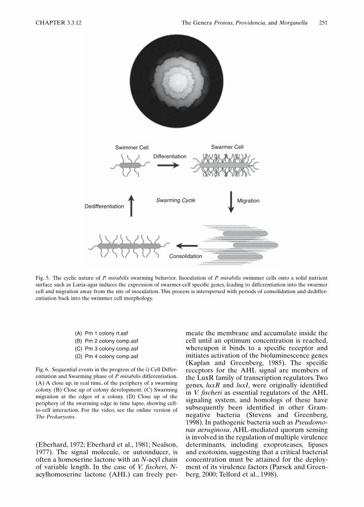
An important aspect of the P. mirabilis swarm-ing colony pattern is its cyclic nature. As shownin Fig. 5, each cycle can be broken down into fourparts, 1) swarmer cell differentiation, 2) the lagperiod prior to active movement, 3) swarmingcolony migration and 4) consolidation (wherethe cells stop moving and dedifferentiate back toswimmer cell morphology). During the migra-tion phase, the fully differentiated swarmer cellsmove outward in unison in all directions from theoriginal site of inoculation for a period of severalhours. Movement then ceases and a processreferred to as consolidation takes place (Bisset,1973a; Bisset, 1973b; Hoeniger, 1964; Hoeniger,1965; Hoeniger, 1966; Hoeniger and Cinits, 1969;Williams and Schwarzhoff, 1978), during whichthe swarmer cells dedifferentiate back into swim-mer cells. After a period in this stage, the swarm-ing phase recommences and proceeds until thenext consolidation phase. The cycle of swarmingand consolidation is then repeated several times,until concentric rings, formed by the swarmingbacteria and delineating the phase changes,cover the agar surface (Fig. 7). The purpose ofthe consolidation phase has yet to be fully eluci-dated; however, recent work by Matsuyama et
Fig. 3. The characteristics of P. mirabilis swarmer cell differ-entiation and swarming motility. Swarmer cell differentiationis controlled through a combination of sensing environmen-tal conditions that reduce wild-type flagellar filament rota-tion and reacting to a specific chemical stimulus, the aminoacid glutamine. The swarmer cell is characterized by an elon-gated polyploid cell that synthesizes numerous flagella inresponse to the aforementioned signals.
EnvironmentalSignals
Flagellar RotationGlutamine
Peptide Signals (?)Others (??)
1.5 to 2.0 mm
4 to 10
1 to 2
Swimming &Chemotaxis
Length 10 to >80 mm
103 to 104
Polyploid
Swarming, Chemotaxis& Coordinated Cell-to-Cell Communication
Flagella
Genomes
Motile Behavior
Swimmer Cell Swarmer CellCharacteristic

250 J. Manos and R. Belas CHAPTER 3.3.12
al. (2000) showed that the time course of thisphase is unaffected by replica plating of theswarm edge. This indicates that consolidation isnot a consequence of nutrient and metabolitechanges in the medium. Figure 6 contains fourvideo segments detailing the events associatedwith swarming at the microscopic level. Figure 7shows swarming behavior during colony devel-opment and bulls-eye ring formation.
THE INFLUENCE OF SIGNAL TRANS-DUCTION IN SWARMING. Swarming is, by
its very nature, a surface-associated and cell-density-dependent phenomenon. Individual P.mirabilis cells rely on their ability to sense thesurrounding environment and use these cues totrigger the development of the swarmer cell, aswell as to coordinate movement of the swarmacross the surface. One way this information isacquired is through monitoring the rotation ofthe flagella to initiate cellular differentiation (see“Cell Differentiation and Swarming”). Othermethods include cell-to-cell contact with neigh-boring bacteria to aid in movement, the chemo-tactic sensing of nutrients and repellents in theexternal medium, and possibly through a den-sity-dependent sensing of cell population densityknown as “quorum sensing.” We will discuss eachof these signal transduction mechanisms in turnin the next paragraphs.
Research into the relationship betweenchemotaxis and swarming in P. mirabilis (Belaset al., 1991a; Williams and Schwarzhoff, 1978)has preceded that into the relationship betweenchemotaxis and other swarming bacteria. Earlywork suggested that chemotaxis did not play amajor role in the differentiation to the swarmingphenotype (Williams et al., 1976). However, laterstudies demonstrated that nonswarming mutantsof P. mirabilis produced by transposon insertionalso exhibit deficiencies in chemotactic response(Belas et al., 1991b), suggesting that chemotacticsignal transduction is important for swarming.Further work by Allison et al. (1993) has shownthat the amino acid glutamine induces differen-tiation to the swarmer cell in P. mirabilis byacting as a chemoattractant. This effect maywork at the level of transcription, because whenglutamine is added to a defined, nonswarmingmedium, expression of the flaA (flagellin) andhpmA (hemolysin) genes in P. mirabilis increases40-fold. The viscosity of the growth medium alsoaffects swarming, with the addition of 3% v/vpolyvinylpyrollidone (PVP) to liquid chemotaxismedium resulting in increased attraction toglutamine. Attempts to repeat these experimentsin other laboratories using different strains of P.mirabilis have been unsuccessful, raising doubtsas to whether all P. mirabilis strains respond toglutamine by swarming. In other swarming mem-bers of the Enterobacteriaceae, Harshey andMatsuyama (1994) reported a link betweenchemotaxis and the swarming of E. coli and Sal-monella typhimurium on specific (Eiken) agar.
Quorum sensing is the ability of a bacterialpopulation to monitor its density throughexpression of small extracellular signaling mole-cules referred to as “autoinducers.” Quorumsensing was first identified and characterized inthe luminescent marine bacterium Vibrio fischeriwherein bioluminescence is controlled by celldensity and autoinducer signal transduction
Fig. 4. The mechanics of swarming in P. mirabilis and therequirement for cell-cell contact. The micrograph (top)shows a finger-like projection of P. mirabilis swarmer cellsmigrating across a solid surface. The cartoon (bottom) dem-onstrates the arrangement of swarmer cells during coordi-nated motion across a solid surface. Individual cells areinterlinked into groups and move en masse away from thepoint of inoculation, with the flagella from all cells in thegroup moving in unison. The arrows point in the direction ofmovement.
CHAPTER 3.3.12 The Genera Proteus, Providencia, and Morganella 251
(Eberhard, 1972; Eberhard et al., 1981; Nealson,1977). The signal molecule, or autoinducer, isoften a homoserine lactone with an N-acyl chainof variable length. In the case of V. fischeri, N-acylhomoserine lactone (AHL) can freely per-
meate the membrane and accumulate inside thecell until an optimum concentration is reached,whereupon it binds to a specific receptor andinitiates activation of the bioluminescence genes(Kaplan and Greenberg, 1985). The specificreceptors for the AHL signal are members ofthe LuxR family of transcription regulators. Twogenes, luxR and luxI, were originally identifiedin V. fischeri as essential regulators of the AHLsignaling system, and homologs of these havesubsequently been identified in other Gram-negative bacteria (Stevens and Greenberg,1998). In pathogenic bacteria such as Pseudomo-nas aeruginosa, AHL-mediated quorum sensingis involved in the regulation of multiple virulencedeterminants, including exoproteases, lipasesand exotoxins, suggesting that a critical bacterialconcentration must be attained for the deploy-ment of its virulence factors (Parsek and Green-berg, 2000; Telford et al., 1998).
Fig. 5. The cyclic nature of P. mirabilis swarming behavior. Inoculation of P. mirabilis swimmer cells onto a solid nutrientsurface such as Luria-agar induces the expression of swarmer-cell specific genes, leading to differentiation into the swarmercell and migration away from the site of inoculation. This process is interspersed with periods of consolidation and dediffer-entiation back into the swimmer cell morphology.
Swimmer Cell Swarmer Cell
Differentiation
DedifferentiationSwarming Cycle Migration
Consolidation
Fig. 6. Sequential events in the progress of the i) Cell Differ-entiation and Swarming phase of P. mirabilis differentiation.(A) A close up, in real time, of the periphery of a swarmingcolony. (B) Close up of colony development. (C) Swarmingmigration at the edges of a colony. (D) Close up of theperiphery of the swarming edge in time lapse, showing cell-to-cell interaction. For the video, see the online version ofThe Prokaryotes.
(A) Pm 1 colony rt.asf(B) Pm 2 colony comp.asf(C)(D)
Pm 3 colony comp.asfPm 4 colony comp.asf

252 J. Manos and R. Belas CHAPTER 3.3.12
In Serratia liquefaciens, another swarmingmember of the Enterobacteriaceae, S. liquefa-ciens, Eberl et al. (1996) have demonstrated thatinitiation of swarmer-cell differentiation involvesdiffusible signal molecules that are released intothe growth medium. In particular, the autoin-ducer N-acylhomoserine lactone (AHL) isrequired for swarming motility in this species
(Givskov et al., 1998; Lindum et al., 1998). TheAHL derivatives N-butanoyl-L-homoserinelactone (BHL) and N-hexanoyl-L-homoserinelactone (HHL) were identified by Eberl et al.(1996) in cell-free S. liquefaciens culturesupernatants.
The swrI (swarmer initiation) gene, whose pre-dicted translation product exhibits substantialhomology to the LuxI family of putative AHLsynthases, is responsible for directing synthesisof both BHL and HHL (Eberl et al., 1996). Fur-ther work by this group since has shown that thecoordinate expression of swrI and the flagellarmaster operon flhDC is required to initiateswarming motility in S. liquefaciens (Eberl et al.,1999; Givskov et al., 1998; Lindum et al., 1998).
Attempts to prove the existence of an AHL-type autoinducer in P. mirabilis have beenunsuccessful; however, evidence exists that thisbacterium may utilize an alternate method ofdensity-dependent quorum sensing. Belas et al.(1998) have characterized a gene that uponmutation decreases the length of the lag phaseprior to swarming. This gene, referred to as“rsbA” (for regulator of swarming behavior)encodes a sensory protein displaying similarity toLuxQ and other bacterial histidine kinases of thetwo-component regulatory superfamily of bacte-rial response regulators that perform functionsrequired for a second density-sensing system(Bassler et al., 1994; Freeman and Bassler, 1999;Lilley and Bassler, 2000). This finding suggeststhat RsbA may function as a sensor of environ-mental conditions required to initiate swarming.
EcologyBoth P. mirabilis and P. vulgaris are members ofthe normal flora of the mammalian intestinaltract and have been isolated from humans, dogs,monkeys, pigs, sheep, cattle, raccoons, cats, ratsand other mammals. They also are distributedwidely in the environment, with reservoirs insoil, water, sewage and feces (Guentzel, 1991).Other species of Proteus are less widely distrib-uted. For example, P. penneri is absent from theintestines of livestock (Hawkey et al., 1986b),whereas P. myxofaciens is confined to the larvaeof the gypsy moth (Cosenza and Podgwaite,1966).
EpidemiologyOwing to their varied habitats, members of thegenus Proteus and related genera have manypossible routes of human infection. The modesof transmission may include nosocomial sources,such as hospital food and equipment, intrave-nous solutions and human contact through con-taminated skin surfaces. Long-term indwellingcatheters are a prime site of colonization and
Fig. 7. Development of the bull’s-eye rings of P. mirabiliscolonies. (A–C) Sequential photographs of L-agar inoculatedin the center with 5 ml of P. mirabilis swimmer cells andincubated at 30°C for 4 h (A), 24 h (B) and 48 h (C). (D)Video recording in time lapse showing swarming of P. mira-bilis across the solid surface of an L-agar plate, taken over24 hours of growth at 30°C and 80% humidity. For the video,see the online version of The Prokaryotes.
A
B
C
CHAPTER 3.3.12 The Genera Proteus, Providencia, and Morganella 253
infection for P. mirabilis and P. vulgaris (Kunin,1989; Stickler and Hughes, 1999; Warren, 1987a).
Serological typing of P. mirabilis and P. vul-garis traditionally has been done using the slideagglutination test and the indirect hemagglutina-tion test (Gmeiner et al., 1977; Schmidt et al.,1970). Previously, the Dienes test, in whichswarming Proteus strains were inoculated onnutrient agar and allowed to swarm into oneanother, was used to distinguish between strainsof these species. If a distinct line of demarcation(a Dienes line) occurred at the junction betweenthe strains, they were considered different(Dienes, 1946). This method has fallen out offavor because of the difficulties associated withinterpreting the results.
Other bacteriological typing methods also havebeen used, including biotyping, bacteriophagetyping, and typing schemes using both bacteriocinproduction and bacteriocin sensitivity (Andersonand Engley, 1978; Hickman and Farmer, 1976;Kusek and Herman, 1980; Schmidt and Jeffries,1974). In a comparative study by Kusek (1981),five bacteriological typing methods were assessedfor their ability to differentiate 100 clinical iso-lates of P. mirabilis. The highest sensitivity andspecificity was obtained using bacteriocin produc-tion typing, which yielded 29 distinct bacteriocintypes among the 80% of strains that were typable(Kusek and Herman, 1981). Bacteriophagetyping sets also have been tested as tools fordifferentiation of Proteus strains; however, amore diverse set of phages is needed toadequately differentiate between as many strainsas possible (Sekaninova et al., 1994; Sekaninovaet al., 1998).
Modern molecular methods employing thepolymerase chain reaction (PCR) to produceDNA fingerprints and other 16S ribosomal RNAgene (ribotyping) methods of strain differentia-tion have been applied to distinguish P. mirabilis,P. vulgaris and P. penneri strains (Costas et al.,1993; Hoffmann et al., 1997; Hoffmann et al.,1998; Serwecinska et al., 1998). Hoffmannet al. (1998) compared four typing methods(including plasmid profiles, outer-membrane-protein profiles, randomly amplified polymorphicDNA PCR [RAPD-PCR], and restriction frag-ment length polymorphism [RFLP] on strains ofP. penneri and found that RAPD-PCR alone, withone of two random primers, revealed 13 repro-ducible typeable patterns (Hoffmann et al., 1998).Thus, the RAPD method, essentially a DNA fin-gerprinting method using arbitrary primers(Akopyanz et al., 1992), revealed a significantDNA diversity among P. penneri strains that wasnot detected by other methods (Hoffmann et al.,1997; Hoffmann et al., 1998). The RAPD-PCRtechnique has the advantage of being quick andeconomical, with high reproducibility and typa-bility; however, the choice of primers is critical to
obtaining discriminating results (Madico et al.,1995).
PathogenicityThe incidence of UTI involving P. mirabilis or P.vulgaris is lower than that for E. coli; however,P. mirabilis infections are more likely to be long-term and persistent, and to lead to greater com-plications threatening the patient’s health thanthose involving E. coli (Mobley and Warren,1987). The majority of Proteus infections areassociated with prolonged hospitalization andthe complications of long-term urinary catheter-ization. The complications that can result fromProteus UTI include catheter and urinary tractobstruction, kidney stone formation (urolithia-sis), pyelonephritis, fevers and bacteremia (Mob-ley and Warren, 1987; Rubin et al., 1986; Senior,1983; Story, 1954; Warren et al., 1987b). In seri-ous cases of Proteus UTI, chronic renal inflam-mation, vasicoureteral reflux and renal failureare frequently observed (Cohen and Preminger,1996; du Toit et al., 1995; Warren, 1987a). Thesites of UTI and the respective complicationsresulting from infection at these sites are shownin Fig. 8, whereas Fig. 9 provides a breakdown ofthe causes of P. mirabilis UTI.
VIRULENCE FACTORS. At least ten viru-lence factors potentially contributing to thepathogenicity of Proteus sp. have been identified.The majority of these virulence factors, withthe exception of the fimbriae, are expressedduring swarmer cell differentiation and swarm-ing behavior. These virulence factors can bedivided into two groups: 1) proteins, enzymesand other secreted products, and 2) surface struc-tures. In terms of their relevance to pathogenesis,the most significant virulence factors (in order ofimportance) are: urease, ZapA (a protease thatspecifically degrades immunoglobulins IgA andIgG), lipopolysaccharide, outer-membrane pro-teins, and hemolysin. The most significant surfacestructures, in order of importance to pathogene-sis, are the flagella and associated swarming phe-notype of P. mirabilis and P. vulgaris, and severaltypes of fimbriae, including; mannose-resistantProteus-like fimbriae (MR/P), mannose-resistantKlebsiella-like fimbriae (MR/K), P. mirabilis fim-briae (PMF), nonagglutinating fimbriae (NAF),and an uncharacterized fimbrial type with a 24-kDa major subunit (also referred to as “F24”).The potential of these virulence factors in Pro-teus to cause disease has been a prime area ofstudy and the major conclusions in each case arepresented here.
Urease. Urea is a by-product of nitrogenmetabolism that is ubiquitous in a wide range ofeukaryotes and prokaryotes. The enzyme ureasebreaks down urea to ammonia and carbon diox-ide. These end products have the effect of

254 J. Manos and R. Belas CHAPTER 3.3.12
increasing the pH, which in turn augments thesurvival of bacteria such as Helicobacter pylori tocreate a less acidic environment that may bemore suited for colonization (Gomez-Duarte etal., 1998; Kuwahara et al., 2000; Lee et al., 1999).In other microbes, ureases play an important rolein utilization of environmental nitrogenous com-pounds and urea-based fertilizers. At the sametime, the production of urease by P. mirabilis andP. vulgaris may contribute to the development ofurinary stones and pyelonephritis, as mentionedearlier. This is one possible reason why Proteusinfections cause more cytological damage thanE. coli infections (Cohen and Preminger, 1996;du Toit et al., 1995; Johnson et al., 1993; Mobleyand Hausinger, 1989).
Early studies using P. mirabilis treated withurease inhibitors established a link between ure-ase and colonization of rat urinary tracts. In
these studies, the renal tissue of control-infectedrats contained a far higher number of bacteriaand had greater tissue damage than did those ofrats infected with inhibitor-treated P. mirabilis(Musher et al., 1975). Further evidence of therole played by urease in P. mirabilis pathogenesishas been demonstrated by comparative histolog-ical examination of renal tissues postinfectionby either the wild type, parental strain, or anisogenic urease-negative mutant using a mousemodel of ascending UTI (Johnson et al., 1993;Jones et al., 1990). Mice challenged with theisogenic urease mutant developed significantlyless bacteriuria and urinary stones compared tothe parent strain. The parent strain also showedgreater persistence in the bladder and kidneythan did the strain lacking urease (for referenceto sites of infection, see Fig. 8).
IgA and IgG Proteases. Secretory immunoglo-bulins of the IgA class are produced by mucoustissue and are particularly resistant to enzymaticbreakdown by proteases. The ability to degradea host’s secretory IgA may provide a microor-ganism with an advantage by evading the hostimmune response, thus gaining valuable time forthe bacterium to establish a foothold for coloni-zation. Many pathogenic bacteria that invademucosal tissues, such as the epithelial lining ofthe intestine and urogenital tract, have potent,extracellular IgA-degrading proteases, whereasnonpathogenic counterpart species in the samegenus often do not. For example, pathogenic spe-cies of Neisseria, such as N. gonorrhoeae and N.meningitidis, possess IgA-degrading proteases,though nonpathogenic species (including N. lac-tamica and N. sica) do not (Kilian et al., 1983).The possession of IgA protease by certain mem-bers of this genus may thus be critical in providingthese bacteria with an advantage to overcome thehost humoral immune response during infection.
A protease capable of degrading two IgA sub-classes (IgA1 and IgA2) as well as IgG has beenidentified in P. mirabilis (Loomes et al., 1990;Milazzo and Delisle, 1984; Senior et al., 1987b).Subsequent work by Wassif et al. (1995) hasresulted in the characterization of this protease(referred to as “ZapA” and encoded by the zapAgene) as a metalloprotease of ca. 50 kDa. Toinvestigate whether zapA expression correlateswith another known virulence-enhancing phe-nomenon, swarmer-cell differentiation (see“Flagella and swarming”), Walker et al. (1999)measured the expression of zapA duringswarmer-cell differentiation. The data obtainedsuggest that zapA expression is tightly coordi-nated not just with swarmer-cell differentiation,but with swarming behavior as well. Also, ZapAproteolysis is not essential for swarming, becauseZapA– strains were shown to produce wild-typeswarmer cells and swarming colonies.
Fig. 8. The human urinary tract, showing the sites of P. mira-bilis infections, hence the diseases resulting from infection,and the virulence factors associated with pathogenesis ofdisease at the respective sites (see “Pathogenicity”).
PyelonephritisFlagella
Kidneys
Ureters
Urethra
Bladder
UreaseHemolysinMR/P FimbriaeIgA Protease
CystitisFlagellaUrease
PMF FimbriaeMR/P Fimbriae
IgA Protease
Fig. 9. The causes of P. mirabilis urinary tract infections(UTI) in humans, compiled using epidemiological data fromhospitalized patients. Adapted from Mobley and Warren(1987).
P. mirabilis UTI
17%
23%
10%
6.3%
5.6%
4.2%2.8%
31%Long-term urethral catheterlleal loopCondom catheterIntermittent catheterizationComplicated UTIRecurrent UTIHospital UTIAcute cystitis
CHAPTER 3.3.12 The Genera Proteus, Providencia, and Morganella 255
Both P. vulgaris and P. penneri also have beenreported to possess IgA proteases. In a surveyof protease production amongst 24 P. vulgarisstrains, Senior et al. (1988) found that half ofthem produced IgA protease. Each of five of theP. penneri strains surveyed in the same study alsoproduced IgA protease. Subsequent purificationand comparative analysis of IgA protease fromP. mirabilis, P. vulgaris and P. penneri showedthat they shared similar electrophoretic patternson sodium-dodecyl-sulfate polyacrylamide gelelectrophoresis (SDS-PAGE) gels, with onlyslight differences in protein band mobility(Loomes et al., 1992).
Lipopolysaccharide and Outer-MembraneProteins. During infection, bacterial lipo-polysaccharide (LPS) is a primary target antigen(O-antigen) for host immunologic responses.Bacterial LPS activates macrophages to producetoxic oxygen radicals, interleukin-1 and tumornecrosis factor, leading to a wide range of effectsin the host, including hypertension, fever andlethal shock (Hamilton and Adams, 1987;Rietschel et al., 1994). By way of example, theeffect of P. mirabilis LPS on pig blood plateletsis akin to that of thrombin (a strong platelet ago-nist), stimulating the enzymatic cascade leadingto platelet aggregation (Wachowicz et al., 1998).
The outer membrane of Gram-negative bacte-ria contains several major proteins that are com-plexed with other membrane components suchas LPS and phospholipid. The polysaccharidechains of the different species of Proteus LPScontain sufficient structural differences to resultin an antigenically heterogeneous genus. Thesedifferences have been used to cluster P. mirabilis,P. vulgaris, and P. penneri strains into serogroupsbased on their agglutination when mixed withantibodies directed against specific species ofLPS molecules (Kotelko, 1986; Penner and Hen-nessy, 1980a; Perch, 1948; Zych and Sidorczyk,1989). Certain LPS epitopes have been investi-gated to determine their role in antigenicspecificity. The particular groups on theoligosaccharides found to play a dominant rolein the specificity of P. mirabilis and P. penneriLPS are the amide of D-galacturonic acid withL-lysine #-D-GalA-(L-Lys) (and the amide of D-galacturonic acid with L-threonine #-D-GalA-[L-Thr]), respectively (Radziejewska-Lebrechtet al., 1995; Sidorczyk et al., 1995).
Specific P. mirabilis outer-membrane proteins(OMPs) have been the targets of study withrespect to their effects on the host immuneresponse. One particular 39-kDa major OMP ofP. mirabilis (subsequently identified as “OmpA”)has been shown to greatly enhance the host IgGresponse for LPS in mice when complexed toLPS (Karch and Nixdorff, 1981; Karch and Nix-dorff, 1983). Also, LPS is an activator of mac-
rophages, another important component of thehost immune response, through their productionof toxic oxygen radicals, interleukin-1 and tumornecrosis factor (Guthrie et al., 1984). Weber etal. have shown that OmpA inhibits the LPS-induced oxidative response and interleukin-1production of murine macrophages when com-plexed to LPS, thus acting as a modulator of theinteraction of LPS with macrophages (Weber etal., 1992; Weber et al., 1993).
Hemolysin. Bacterial hemolysins are fre-quently involved in the destruction of erythro-cytes during infection and have been shown tocontribute to the invasiveness and pathogenicityof several bacterial species (Braun and Focareta,1991; Braun et al., 1993; Goebel et al., 1988;Menestrina et al., 1995). The importance ofhemolysin in the virulence of E. coli was demon-strated through the isolation, cloning and trans-fer of hemolysin genes from virulent strains toavirulent ones (Hacker et al., 1983; Welch et al.,1981).
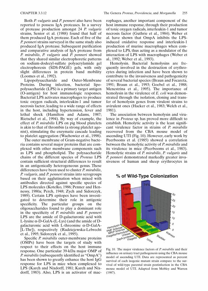
The association between hemolysin and viru-lence in Proteus sp. has proved more difficult toestablish. Hemolytic activity is the least signifi-cant virulence factor in strains of P. mirabilisrecovered from the CBA mouse model ofascending UTI (Fig. 10). However, early work byPeerbooms et al. (1985) showed a correlationbetween the hemolytic activity of P. mirabilis andits virulence in mice (Peerbooms et al., 1983).Hemolytic strains of P. mirabilis, P. vulgaris andP. penneri demonstrated markedly greater inva-siveness of human and sheep erythrocytes in
Fig. 10. The major virulence factors of P. mirabilis and theirinfluence on urinary tract pathogenesis using the CBA mousemodel of ascending UTI. Data are represented as percentsurvival of each isogenic mutant strain compare to the sur-vival of wild-type parental strain postinfection in the CBAmouse model of UTI. Adapted from Mobley and Warren(1987).
% of Wild-Type Colonization
616%
Hemolysin
PMF fimbriae
MR/P fimbriae
Flagella/Hemolysin
Urease
ZapA proteaseFlagella
74%
30
25
20
15
10
5
0KidneysBladderUrine

256 J. Manos and R. Belas CHAPTER 3.3.12
vitro in work by Rozalski and colleagues (Rozal-ski et al., 1986; Rozalski and Kotelko, 1987).
At the molecular level, the hemolysin struc-tural genes (hlyA and hlyC) and the principalsecretion gene (hlyB) of E. coli can be function-ally complemented by the homologous genesfrom P. mirabilis, P. vulgaris and M. morganii,suggesting a functional relatedness between thethree components in the two species. A secondE. coli secretory gene (hlyD) is present in P.vulgaris and M. morganii, but absent from P.mirabilis (Koronakis et al., 1987; Koronakis andHughes, 1988a; Koronakis et al., 1988b). The E.coli hlyD gene is required for secretion of hemol-ysin (Pimenta et al., 1999). This finding mayexplain why the secreted hemolytic activity of P.mirabilis remains cell-associated rather thanbeing released into the surrounding medium.This possibility has been supported by subse-quent investigations showing that the secretedhemolysin (HpmA) of P. mirabilis and P. vulgarisis genetically distinct from that of E. coli (Swi-hart and Welch, 1990b; Welch, 1987). Proteusmirabilis HpmA is cytotoxic for a variety of celllines, including cultured human renal proximaltubular epithelial cells (Mobley et al., 1991; Swi-hart and Welch, 1990a). As mentioned above,colonization by isolates showing hemolysinactivity forms by far the greatest proportion ofP. mirabilis isolates recovered from the urine,kidneys and bladder of CBA mice with UTI (Fig.10; Mohr O’Hara, C., et al., 1999). However,HpmA activity in such clinical isolates does notappear to be crucial to the development ofpathogenesis (Mobley and Chippendale, 1990).Thus, hemolysin is not considered as importanta virulence factor in P. mirabilis pathogenesis asflagella or urease (Mobley and Belas, 1995). Inthe case of P. vulgaris, hemolysin production,measured as hemolytic titer (Peerbooms et al.,1983), was found by Peerbooms et al. (1985) tobe significantly lower than that for P. mirabili,thus reducing the relevance of hemolysin in UTIfor this species.
Structures Involved in the Virulence ofPROTEUS sp. Flagella and Swarming. It is gener-ally accepted that flagella and swarming behav-ior play some role in the pathogenicity of P.mirabilis, and several studies support thishypothesis. For example, in work by Allison etal. (1992) using two different human uroepithe-lial cell lines (EJ/28 and 5637) and P. mirabilismutants lacking flagella (Fla–), the loss of flagellaresulted in a noninvasive bacterium when com-pared to the wild-type parent. In the same study,the ability to invade the EJ/28 and 5637 celllines was closely identified with differentiatedswarmer cells, which were 15-fold more invasivethan undifferentiated swimmer cells. In in vivostudies using a mouse model of UTI, Mobley et
al. (1996) reported a 100-fold lower recovery ofa Fla– mutant defective in flagellar filament syn-thesis when compared to the parent strain. Whilenonflagellated strains are less invasive, flagellado not appear to be absolutely required for vir-ulence. Zunino et al. (1994) have found that anaturally occurring nonmotile strain could infectmice and cause UTI, whereas Legnani-Fajardoet al. (1996) reported similar results with anisogenic mutant lacking flagella.
The differentiated swarmer cell (see “Physiol-ogy”) probably provides P. mirabilis and otherswarming species such as V. parahaemolyticuswith the advantage of rapid colonization duringpathogenesis. Apart from the benefits of motilityand adherence due to hyperflagellation, swarmercell differentiation in P. mirabilis coincides withincreased expression of several virulence factors(see “Virulence Factors”) and has been linkedto the expression of extracellular (capsular)polysaccharide. Evidence for this has been pro-vided in studies by Gygi et al. (1995) utilizingtransposon mutagenesis of a gene coding for acell surface (capsular) polysaccharide in P. mira-bilis. The mutants clearly showed retardation intranslocation velocity across solid media andwere attenuated in their ability to establish UTIin mice (Allison et al., 1994; Gygi et al., 1995).
Fimbriae. Collectively, the different species offimbriae, which can be distinguished from flagel-lae by their shorter “spiked” appearance (Fig.2C) are important in the attachment of the bac-teria to the epithelial cell surfaces and coloniza-tion of the surrounding tissues (Silverblatt, 1974).Fimbriae appear to be differentially expressed atparticular stages of the swarming cycle (Fig. 5)and are absent on hyperflagellated swarmer cells(Hoeniger, 1965; Latta et al., 1999). Proteus mira-bilis produces MR/P, MR/K, NAF and PMF fim-briae (Adegbola et al., 1983; Bahrani et al., 1993a;Bahrani et al., 1993b). Both MR/P and MR/Kfimbriae also have been shown to function ashemagglutinins and are highly immunogenic.
Mutants defective in the synthesis of variousfimbrial species have been used to assess the roleof each fimbrial type during P. mirabilis UTI. Inparticular, MR/P mutants (MrpA–) have beenconstructed by allelic exchange and are between6 and 28–fold less efficient in colonizing mouseurinary tracts than the parental strain of P. mira-bilis (see also Fig. 10). This result is supported byhistopathology results showing less damage tothe uroepithelium and no signs of pyelonephritisduring colonization by Mrp– strains (Bahrani etal., 1994). In studies on PMF fimbriae, isogenicPmfA– mutants were found to colonize the blad-der at lower levels than the wild-type parent, butbacterial colonization of the kidney was notaffected by loss of this fimbrial type (Massad etal., 1994). The involvement of NAF fimbriae inbacterial adherence also has been demonstrated

CHAPTER 3.3.12 The Genera Proteus, Providencia, and Morganella 257
in P. mirabilis strains that express NAF as theironly fimbrial species. These studies have shownthat NAF provide strong adherence to a numberof mammalian cell lines in vitro, including uroep-ithelial cells (Latta et al., 1998; Tolson et al.,1995; Tolson et al., 1997). While it would appearthat the production of fimbriae offers the bacte-rium a distinct advantage for survival in coloniz-ing host tissues, one of the disadvantages offimbriae to the potential colonizer is that theyrender the bacteria more susceptible to phagocy-tosis (Silverblatt and Ofek, 1978). Furthermore,the presence of fimbriae does not equally en-hance adherence to all cell types. For example,the use of MR/P fimbrial mutants (mrpA–) in theCBA mouse model of ascending UTI has shownthat the loss of MR/P fimbrial expression doesnot completely prevent colonization of renal tis-sue in P. mirabilis (Bahrani et al., 1994).
The Genus Morganella
HabitatThe sole species in this genus, Morganella mor-ganii, is a commensal organism that can rapidlycolonize the host gut with an accompanyinghypertrophy of Peyer’s patches and developmentof specific IgA responses in the lamina propriacells (Shroff et al., 1995). Strains of M. morganiialso are known to infect the human urinary tract,respiratory system and blood, though they haveonly been recovered occasionally from thesesources (Braunstein and Tomasulo, 1978).
Isolation and IdentificationIsolation of M. morganii is accomplished usingmedia for the routine isolation of Enterobacteri-aceae. Enrichment media for culturing M. mor-ganii from fecal specimens frequently includetetrathionate and selenite, which are added tonutrient broth (Rustigian and Stuart, 1945). Thebiochemical tests used to identify and distinguishM. morganii are shown in Table 2. It should benoted that although M. morganii is urease andindole positive, it does not swarm and is negativefor most of the biochemical reactions character-istic of the Proteus spp.
The isolation of atypical M. morganii strainswhose characteristics differ from those listed inTable 2 has resulted in the creation of a numberof distinct biogroups that subdivide the species.Hickman et al. (1980) described 19 strains thatwere lysine positive and fermented glycerolwithin 24 h, in contrast to the type strain of M.morganii, which is lysine negative and fermentsglycerol slowly or not at all. Another group of 14M. morganii strains was found to be ornithinenegative, whereas the type strain is ornithinepositive. Because both groups were closely
related to M. morganii by DNA-DNA hybridiza-tion, they were considered distinct biogroups.There are currently seven recognized biogroups,based on ornithine and lysine decarboxylasereactions; however, some strains may carry plas-mid-borne genes that code for these enzymes(Cornelis et al., 1981). Other phenotypes alsohave been used to distinguish the various bio-types. Janda et al. (1996) found resistance to theantibiotic tetracycline to be a useful distinguish-ing characteristic in their classification of 73strains of M. morganii principally recoveredfrom routine clinical specimens. The future sub-division of M. morganii into two or more speciesbased on differences between biotypes remainsa distinct possibility as more characteristics areused to distinguish between the biotypes.
EcologyMorganella morganii occurs in low numbers inthe feces of healthy humans and animals, includ-ing dogs, cattle and chickens (Hawkey et al.,1986b; Phillips, 1955; Prasad and Pandey, 1966;Tanaka et al., 1995). Its habitat may be more far-reaching, as M. morganii strains have beenisolated from snakes, chickens suffering fromrespiratory disease, and ocular lesions of harborseals and elephant seals (Lin et al., 1993; Muller,1972; Thornton et al., 1998). It is unclear whetherM. morganii was the causative agent in thesediseases or an opportunistic colonizer of the pre-viously diseased tissue.
EpidemiologyThe incidence of M. morganii in diarrhea hasbeen studied by Muller (1986a). In this studyof fecal specimens from diarrheal and non-diarrheal patients, Muller isolated P. mirabilisand M. morganii more frequently from humandiarrheal cases than from the stools of healthyindividuals. These results agree with the datafrom earlier studies that showed a similar patternof M. morganii recovery from diarrheal patients(Ahren, 1990; Das, 1996).
Epidemiological typing schemes have beendeveloped for M. morganii based on the bacte-rial somatic and flagellar (O and H, respectively)antigens (Penner and Hennessy, 1979b; 1979d;Rauss and Voros, 1959; Rauss and Voros, 1967a;Rauss and Voros, 1967b; Rauss et al., 1975). Thetyping of M. morganii strains using the lyticactivity of bacteriophages has been investigatedin detail by Schmidt and Jeffries (1974). Sevenphages were isolated from three M. morganiistrains and these phages were successfully usedto differentiate 13 of the 19 M. morganii strainsin the study. Furthermore, lytic patternsremained stable in randomly selected M. morga-nii isolates retested several weeks later. While

258 J. Manos and R. Belas CHAPTER 3.3.12
phage typing is no longer widely used in theUnited States, these methods are still in favor inmany East European countries.
A bacteriocin (referred to as “morganocin”)typing scheme for M. morganii based on produc-tion of and sensitivity to the protein has beendescribed by Senior (Senior, 1987a). A total of160 M. morganii strains were tested for sensitivityto morganocin and classified according to mor-ganocin production and sensitivity. Most strains(97.5%) were sensitive to several different typesof morganocins. Subsequent typing studies foundthat a combination of three distinct methods (bac-teriocin typing, O-antigen serotyping and proteinprofiling) could be used to achieve a much greaterdegree of strain discrimination, especially inas-much as protein profiling appears to be indepen-dent of O-serotype and bacteriocin type (Seniorand Voros, 1989; Senior and Voros, 1990).
PathogenicityWhile the etiological role of M. morganii in diar-rhea has not yet been firmly established, it isconsistently recovered from the feces of diar-rheal patients suggesting an involvement in thedisease. Furthermore, some researchers havefound M. morganii to be the sole potentiallypathogenic bacterial species in the feces of diar-rheal patients, thus strengthening its claim tobeing the cause of the disease in these cases(Rauss, 1936; Senior and Leslie, 1986).
In spite of the involvement of M. morganii indiarrheal disease, this species is less likely to bethe causative agent of human UTI than P. mira-bilis. This is mainly due to the slower growth rateof M. morganii in urine compared to that of P.mirabilis and the noninducible nature of its ure-ase (Senior, 1983). Although not a major con-tributor to human UTI, M. morganii has beenimplicated in outbreaks of septicemia and bacte-remia in humans and animals (Bagel and Gross-man, 1985; Barragan Casas et al., 1999; Heard etal., 1988; McDermott and Mylotte, 1984; Novakand Seigel, 1986; Rowen and Lopez, 1998). Mor-ganella morganii bacteremia most commonlyoccurs in postoperative patients who receive"-lactam antibiotics. McDermott and Mylotte(1984) investigated the case histories of 19 doc-umented episodes of M. morganii bacteremia in18 hospital patients and showed that the majorityof infections were either postoperative or hadassociated wound injuries. They concluded thatM. morganii is an infrequent cause of bacteremiaand its presence in blood cultures may be anindicator of an environment, such as surgery, thatis conducive for an outbreak of nosocomialinfection.
VIRULENCE FACTORS. There is very littleknown about the virulence factors involved in M.
morganii pathogenesis. Despite the paucity ofreports, the most significant of these virulencefactors are described below, though it should benoted that the data supporting the efficacy ofeach virulence factor may be minimal or not assubstantial as those for the virulence factors ofP. mirabilis, for example.
Hemolysin. The synthesis of active intracellu-lar hemolysin (Hly) by M. morganii follows apattern similar to that seen with the hemolysinsfrom E. coli, P. mirabilis and P. vulgaris. Emodyet al. (1980) measured the virulence of M. mor-ganii strains due to hemolysin. Hemolysin (Hly+)wild-type strains and Hly+ transconjugants werefound to be more virulent than Hly– in mice andchick embryos. This enhanced virulence seems tobe connected with the production of a diffusible#-hemolysin. There is an important differencebetween the hemolysins of M. morganii andthose of P. mirabilis and P. vulgaris. Koronakiset al. (1987) found that hemolytic activity in allM. morganii strains tested was cell free (extra-cellular), whereas in all P. mirabilis and 60% ofP. vulgaris strains, hemolysin was only foundassociated with intact cells. The presence of thesecretory gene hlyD in M. morganii, may explainthe difference in secretion patterns observed inthese two genera.
Urease. While both P. mirabilis and M. morga-nii possess urease activity with some similarities,there are also significant differences between theenzymes that may play a role in choice of habitatand pathogenicity. possess ureases, there aredistinct differences between the enzymes in thetwo species that may play a role in habitat andpathogenicity. For example, M. morganii ureasepossesses a higher affinity for urea than P. mira-bilis urease, but the latter hydrolyzes urea at arate 6- to 25-fold faster (Jones and Mobley, 1987).Morganella morganii survives in acidic condi-tions and its ureases have been shown to be acti-vated in vitro by low pH, with an unusually lowactivity optimum of pH 5.5 (Young et al., 1996).In this respect, the urease from M. morganii issimilar to the urease of Y. enterocolitica and H.pylori, both of which can hydrolyze urea at sig-nificantly higher rates under acidic conditionswhen compared with other pathogenic bacteria.A critical assessment of whether the M. morganiiurease is a significant factor in the pathogenesisof this species will ultimately require in vivo stud-ies with urease-negative mutants.
The Genus Providencia
HabitatThe members of the genus Providencia are allfacultative anaerobes and motile by peritrichousflagella (Figs. 2F, G); however, they do not

CHAPTER 3.3.12 The Genera Proteus, Providencia, and Morganella 259
exhibit cellular differentiation and swarmingbehavior. Urease production is not characteristicof all Providencia species, with only P. rettgeristrains producing urease (Brenner et al., 1978).
Providencia stuartii is found most frequently inhospital patients with urinary tract infections.Less frequently, it is found in respiratory andskin infections (Stickler et al., 1985; Warren,1986). Providencia alcalifaciens is generally iso-lated from stool samples along with other entericpathogens. A similar species (P. rustigianii), wasoriginally isolated from human feces as P. alcal-ifaciens biogroup 3 (Ewing et al., 1972) and sub-sequently redesignated as a separate species(Hickman-Brenner et al., 1983). It has since beenisolated from a range of human and animalsources, including (oddly enough) penguin feces(Costas et al., 1987; Muller, 1983). While thisspecies can colonize the human gastrointestinaltract and some of the sources included diarrhealpatients, no direct link with diarrhea has beenestablished.
The third, taxonomically defined species ofProvidencia, Providencia heimbachae, was firstdescribed by Muller 1986 (Muller et al., 1986b)who isolated it from penguin feces and from anaborted bovine fetus. A strain of this speciesrecently has been isolated from humans, specifi-cally, the stool of a patient with idiopathic diar-rhea (Mohr O’Hara et al., 1999). It should benoted, however, that this strain possesses impor-tant biochemical and physiological differencescompared to the type strain of Muller (Muller etal., 1986b). While the type strain is positive forphenylalanine deaminase, gas production fromglucose and fermentation of maltose and D-mannitol, the human isolate is negative for thesetests. Furthermore, motility is only observedafter 6 days for the human isolate, comparedwith 4 days for the type strain.
Isolation and IdentificationIsolation of Providencia sp. is routinely per-formed using bacteriological nutrient media forthe general identification of Enterobacteriaceae,and, together with the biochemical tests shownin Table 2, to descriminate this species fromother enteric forms. Machtiger et al. (1971)found that P. stuartii and P. alcalifaciens requiredfive amino acids (isoleucine, leucine, valine,glutamic acid and cystine), as well as niacin, forgrowth on minimal medium containing glucoseas a carbon source (Machtiger and O’Leary,1971). Hawkey et al. (1982b) used a selective anddifferential enrichment medium to isolate lownumbers of P. stuartii. Thaller et al. (1992) useda modified MacConkey containing methyl greenphosphatase (MGP) to successfully identify100% of P. stuartii isolates from 1,278 seeded
urine samples. By comparison, standard Mac-Conkey medium and the API20E (API System,Bio Merieux Vitec, Inc. Hazelwood, MO.) wereonly able to identify 82.5% of the same isolates.Providencia stuartii and M. morganii are the onlyphosphatase-positive members of the Enterobac-teriaceae, and MGP distinguishes phosphataseproducing colonies on Luria-Bertani-agar bytheir green pigmentation or halo (Pompei et al.,1990; Satta et al., 1979), while on MacConkeymedium the same colonies appear red. Thalleret al. (1992) also added methyl blue to MCPto distinguish lactose-positive colonies (violet)from the phosphatase producers.
A simplified method for the identification ofP. alcalifaciens has been described by Senior(1997). This method relies on the probabilitythat P. alcalifaciens is the only oxidase-negativeorganism likely to be present in fecal culturesenriched in tetrathionate broth, which also isunable to ferment the mannitol, xylose andgalactose present in the medium. When grownon this agar-solidified medium, colonies of P.alcalifaciens appear red, in contrast to the col-orless colonies of non-P. alcalifaciens bacteriathat ferment the three sugars. Extensive testsby Senior showed the medium to be bothhighly specific and sensitive in detecting P.alcalifaciens (Senior, 1997). A number of com-mercial kits have become available for identifi-cation purposes and have been utilized, withvarying success, for identification of P. rustigia-nii (Kitch et al., 1994; Piccolomini et al., 1991).
EcologyThe emergence of P. stuartii as a significant hos-pital pathogen since the 1970s has led to effortsto uncover the natural sources and reservoirs ofthis species. Early work had discounted the gas-trointestinal tract as a potential site owing to thelack of isolates obtained using traditional fecalculture methods; however, Hawkey et al. (1982a)demonstrated fecal colonization by P. stuartiiusing a combination of pre-enrichment andselective media. Moreover, the long-term-catheterized human urinary tract appears tooffer an attractive niche to this species. Resultsfrom studies of patients catheterized for longperiods indicate that P. stuartii can often befound on catheter surfaces as frequently as morefamiliar uropathogens such as E. coli, P. mirabi-lis, Enterococcus sp., and P. aeruginosa (Warren,1986). It also has been isolated from burn andwound infections and bacteremias, which furtheremphasizes the pathogenicity of this species(Penner, 1984).
Providencia rettgeri similarly has emerged inrecent years as a nosocomial pathogen of clinicalimportance. This species has been isolated with

260 J. Manos and R. Belas CHAPTER 3.3.12
regularity from the urinary tract of catheterizedor immunocompromised patients and less fre-quently from human feces, bile and sputum(Bauernfeind and Wiersma, 1977; Cipriani et al.,1988; Gunalp, 1979; Mino et al., 1997). Of theremaining species, P. alcalifaciens is an intestinalcolonizer and a recognized cause of gastroenteri-tis (Janda et al., 1998). Providencia rustigianiialso has been confirmed as a gastrointestinaltract inhabitant and often is found in the intesti-nal tract of mammals, such as humans and pigs,and even in arctic birds, e.g., penguins. The trueecological niche inhabited by P. heimbachae hasyet to be determined, but the evidence at handsuggests that it may inhabit the gastrointestinaltract, as is common for other Providencia species(Costas et al., 1987; Higashitani et al., 1995;Mohr O’Hara et al., 1999).
EpidemiologyBoth the indirect hemagglutination test and theindirect hemagglutination inhibition test havebeen used to type Providencia sp (Levina et al.,1980). These tests are frequently useful in dis-tinguishing between individual Providenciaserogroups (Levina et al., 1980). Antigenicserotyping methods based on the flagellar (H),LPS (O) and capsular (K) antigens also havebeen applied to P. stuartii strains. Such serotyp-ing may be particularly useful in identifying spe-cific strains endemic in different hospitals.Penner and colleagues have used O serotypingsuccessfully (Penner and Hennessy, 1979a; Pen-ner and Hennessy, 1979b; Penner et al., 1979c) totype strains from P. stuartii, P. rettgeri and P.alcalifaciens (Penner and Hennessy, 1979a; Pen-ner and Hennessy, 1979b). In the O serotypingof P. stuartii isolates, Penner and his colleaguesfound that 97% of 829 isolates tested fell intoone of 14 O-antigen serotypes. However, thesomatic (O) antigen serotyping scheme for 54isolates of P. rettgeri based upon a set of 93O-antigens, also developed by Penner andHennessy (1979b), failed to detect a single pre-dominant serotype. While the small size of thestrain pool may have influenced this latter result,it is possible that infection by P. rettgeri is not dueto a few serotypes, as has been found in is muchless strain specific than that by P. stuartii. Simi-larly, in the serotyping of P. alcalifaciens, Penner(1979c) detected 29 serotypes among 82 typeableisolates. Serotyping schemes for the remainingspecies of Providencia have yet to be developedbut will undoubtedly employ similar approachesto those used for other Providencia species.
Bacteriophage and bacteriocin typing meth-ods for Providencia sp. have yet to be developed,though a number of studies have looked for andisolated bacteriophages from both P. stuartii andP. rettgeri (Coetzee, 1967; Gabrilovich et al.,
1998; McHale et al., 1981a). A bacteriocin typingscheme has been tested on >300 Providencia sp.isolates, though no follow-up confirmation of itsreliability has been published (Al-Jumaili andFenwick, 1978).
Molecular methods of epidemiological typing,such as restriction fragment length polymor-phism (RFLP) and ribotyping, have been usedwith P. stuartii and P. alcalifaciens. Owen et al.(1988) noted that the DNA restriction finger-prints for P. stuartii were quite distinct from spe-cies of the allied genera of Providencia andProteus, and provided a more sensitive measureof minor genomic differences than total DNAdigests. Rahav et al. (1994) used RFLP to dem-onstrate that a single strain of P. stuartii persistedin the same patient during a nursing home out-break of P. stuartii bacteriuria, and that severaldifferent strains were responsible for the out-break. In comparison, neither biochemical testsnor antibiotic sensitivity were able to distinguishseparate strains during this outbreak. Guth et al.(1999) used clonal analysis based on ribotypingto show that diarrheal isolates of P. alcalifacienswere clustered into two main groups.
The application of genetic methods to theidentification of bacteria has become routine inthe past decade and this is reflected in the cur-rent methods being applied to identification ofProvidencia sp. Apart from classical ribotyping,other methods that potentially could be used todistinguish strains of Providencia include auto-mated ribotyping, RAPD-PCR (Akopyanz et al.,1992), amplification and restriction analysis ofthe 16S rRNA gene (ARDRA; Andrighetto,1998; Dijkshoorn, 1998) and multilocus sequencetyping (MLST), which exploits the electronicportability of nucleotide sequence data (Maidenet al., 1998).
PathogenicityThe Providencia species that have been clearlyidentified as pathogens are P. stuartii, P. rettgeriand P. alcalifaciens. In human pathogenicity, themost significant member of the genus is P. stuar-tii, whereas the virulence of P. rettgeri and P.alcalifaciens is less clear. Providencia stuartii isparticularly effective in colonizing urinary cath-eters and is a leading risk factor for bacteremia(Muder et al., 1992; Rahav et al., 1994; Rudmanet al., 1988; Woods and Watanakunakorn, 1996).It has been proposed that P. stuartii, as well as P.mirabilis and M. morganii, probably establish aniche within the urinary catheter, thus increasingtheir ability to cause subsequent bladder bacte-riuria (Warren, 1987a). Providencia stuartii isnot particularly invasive; however, certain cir-cumstances tend to increase the probability ofinfection, including prolonged catheterizationand urinary surgery. This species also is resistant

CHAPTER 3.3.12 The Genera Proteus, Providencia, and Morganella 261
to many common antibiotics, including mostpenicillins, aminoglycosides, tetracyclines, oldercephalosporins, sulfamethoxazole and fosfomy-cin (Gomez-Lus et al., 1977; Paradise et al., 1998;Rather et al., 1997; Stock and Wiedemann, 1998;Swiatlo and Kocka, 1987). Such antibiotic resis-tance gives P. stuartii an opportunistic advantagein nosocomial patients. Furthermore, P. stuartiialso has been implicated in septicemia (bactere-mia), with symptoms typical of other septice-mias, except that vascular collapse is not aprominent feature (McHale et al., 1981b; Pren-tice and Robinson, 1979).
While cases of P. stuartii septicemia usuallyprove fatal due to antibiotic resistance, patientsurvival in response to medical therapy has beenreported in individual case studies (Keren andTyrrel, 1987) and more recently, in long-term epi-demiological surveys (Muder et al., 1992; Woodsand Watanakunakorn, 1996). In the latter case,Woods and Watanakunakorn (1996) found amortality rate of 25% in a review of 49 cases ofP. stuartii bacteremia. Considering that 51% ofpatients in this survey were infected by morethan one bacterial species (polymicrobial bacte-remia), the mortality rate for P. stuartii bactere-mia alone is probably lower. The maindifferences between P. stuartii and P. rettgeri areat the biochemical level. Providencia rettgeri canmetabolize the sugars D-arabitol, adonitol anderythritol (Table 2). Otherwise, most P. rettgeristrains exhibit pathogenic properties similar tothose of P. stuartii. Providencia rettgeri UTIsin catheterized and otherwise compromisedpatients are also difficult to treat owing to mul-tiple antibiotic resistance. Overall resistance is,however, less marked in this species than in P.stuartii. By way of example, P. rettgeri is particu-larly susceptible to the aminoglycosides gentam-ycin and tobramycin, whereas P. stuartii is highlyresistant to both (Penner and Preston, 1980b;Penner et al., 1982; Piccolomini et al., 1987).
In contrast to P. stuartii and P. rettgeri, P. alcal-ifaciens is an invasive enteric pathogen andimplicated as a cause of diarrheal disease (Albertet al., 1992; Albert et al., 1998; Guth and Perrella,1996; Haynes and Hawkey, 1989; Sen, 1962). Instudies using pure cultures derived from stoolspecimens that were inoculated into the ilea ofadult rabbits with removable ileal ties (RITARDmodel; Davis, 1991; Spira, 1981), it has beenshown that the development of diarrhea isaccompanied by an intestinal histopathologytypical of other invasive bacterial species, suchas Shigella flexneri (Albert et al., 1992). Mathanet al. (1993) have added more evidence to theP. alcalifaciens virulence model, invasivenessmodel by demonstrating two modes of bacterialentry into epithelial cells. The first mode of entryis by direct endocytosis associated with polymer-ization of cytoskeletal components, and the sec-
ond mode by which the bacteria enter is throughdisruption of tight junctions, with the bacteriaentering into and proliferating in intercellularspaces. The invasive abilities of P. alcalifacienshave been tested in HEp-2 cells (an epithelioidcell line from a human laryngeal carcinoma) bytwo independent scientific groups (Albert et al.,1992; Janda et al., 1998). Both groups were ableto confirm penetration by P. alcalifaciens isolates,whereas no strain of P. stuartii or P. rettgeri testedinvaded the HEp-2 cells.
VIRULENCE FACTORS. Those contributingto the pathogenicity of Providencia sp. that havebeen investigated include cellular adherence, theproduction of fimbriae and of urease. Providen-cia stuartii has been the focus of the majority ofresearch.
Adherence and fimbriae. Urinary tract infec-tions (UTIs) due to P. stuartii persist longer thanthose due to other Gram-negative bacteria. Ithas been suggested that a possible reason for thisincreased persistence may be due to the abilityof P. stuartii to adhere to uroepithelial cells(Mobley et al., 1986). Adherence to uroepithelialcells can be enhanced by the expression of MR/K (hemagglutinin)-type fimbriae (see “Fim-briae”). Mobley et al. (1988) found that a sig-nificant proportion of P. stuartii isolated frompatients experiencing bacteriuria of long dura-tion expressed MR/K fimbriae. These data impli-cate the MR/K hemagglutinin in an importantrole in UTI persistence of P. stuartii.
Urease. While Providencia rettgeri is ureasepositive, as are some strains of P. stuartii. In fact,P. rettgeri and P. stuartii were originally subdi-vided based on urease production (Penner et al.,1976). There is some evidence that urease-producing strains of P. stuartii are more likely tobe involved in long-term colonization of urinarycatheters with subsequent development of block-ages due to stone formation (Kunin, 1989; Stick-ler et al., 1998). Urease-producing P. stuartiistrains also have been found to inhibit the invitro growth of E. coli on catheter surfaces andin urine (Fletcher et al., 1994). This adaptiveadvantage in growth may explain why P. stuartiiare the dominant species isolated in these mixedinfections. The construction of isogenic urease-negative mutants may help to better define therole of urease in P. stuartii pathogenesis.
Conclusion
The genera Proteus, Morganella and Providenciacontain a number of important human pathogensthat often cause serious infections in hospitalizedand immunocompromised patients. Other spe-cies are normal intestinal flora in animals oranimal pathogens. One of the most conspicuousfeatures of members of the genus Proteus is their
262 J. Manos and R. Belas CHAPTER 3.3.12
ability to differentiate into elongated, multinu-cleated swarmer cells upon contact with a solidsurface. Methods of isolation and identificationhave been developed for most species and thesenow include several DNA-based methods ofdetection; however, treatment of infectedpatients is often problematic owing to the devel-opment of antibiotic resistance and the immuno-compromised state of the patient.
The information contained in this chaptergives the reader a broad understanding of thecurrent state of knowledge concerning theserelated genera. There are a number of areaswhere this knowledge base needs strengtheningin the future. For example, our understanding ofthe ecological and/or pathogenic role of severalspecies, including P. myxofaciens, P. rustigianiiand P. heimbachae, has advanced very little sincetheir initial identification, and much workremains to be done in this regard.
Acknowledgments. This work was supported bygrants from the National Institutes of Health(DK49720) and the National Science Foundation(MCB9514803). We wish to express our appre-ciation to Rachel Schneider, of the Center ofMarine Biotechnology, University of MarylandBiotechnology Institute, for the sequencing ofthe 16S rRNA sequences of the tribe Proteeae(Fig. 1), and Dr. Robert K. Naumann of the Elec-tron Microscopy Unit, Dental School, Universityof Maryland, Baltimore, for the electron micro-graphs of selected Proteeae (Fig. 2).
Literature Cited
Adegbola, R. A., D. C. Old, and B. W. Senior. 1983. Theadhesins and fimbriae of Proteus mirabilis strains asso-ciated with high and low affinity for the urinary tract. J.Med. Microbiol. 16:427–431.
Ahren, C. M., M. Jertborn, L. Herclik, B. Kaijser, and A. M.Svennerholm. 1990. Infection with bacterial entero-pathogens in Swedish travellers to South-East Asia—aprospective study. Epidemiol. Infect. 105:325–333.
Akopyanz, N., N. O. Bukanov, T. U. Westblom, S. Kresovich,and D. E. Berg. 1992. DNA diversity among clinicalisolates of Helicobacter pylori detected by PCR-basedRAPD fingerprinting. Nucl. Acids Res. 20:5137–5142.
Albert, M. J., K. Alam, M. Ansaruzzaman, M. M. Islam, A.S. Rahman, K. Haider, N. A. Bhuiyan, S. Nahar, N. Ryan,J. Montanaro, and M. M. Mathan. 1992. Pathogenesisof Providencia alcalifaciens-induced diarrhea. Infect.Immun. 60:5017–5024.
Albert, M. J., A. S. Faruque, and D. Mahalanabis. 1998. Asso-ciation of Providencia alcalifaciens with diarrhea in chil-dren. J. Clin. Microbiol. 36:1433–1435.
Alberti, L., and R. M. Harshey. 1990. Differentiation of Ser-ratia marcescens 274 into swimmer and swarmer cells. J.Bacteriol. 172:4322–4328.
Al-Jumaili, I. J., and G. A. Fenwick. 1978. Bacteriocine typingof Providencia isolates. Zentralbl. Bakteriol. [Orig A]240:202–207.
Allison, C., and C. Hughes. 1991. Bacterial swarming: Anexample of prokaryotic differentiation and multicellularbehaviour. Sci. Prog. 75:403–422.
Allison, C., N. Coleman, P. L. Jones, and C. Hughes. 1992.Ability of Proteus mirabilis to invade human urothelialcells is coupled to motility and swarming differentiation.Infect. Immun. 60:4740–4746.
Allison, C., H. C. Lai, D. Gygi, and C. Hughes. 1993. Celldifferentiation of Proteus mirabilis is initiated byglutamine, a specific chemoattractant for swarming cells.Molec. Microbiol. 8:53–60.
Allison, C., L. Emody, N. Coleman, and C. Hughes. 1994.The role of swarm cell differentiation and multicellularmigration in the uropathogenicity of Proteus mirabilis.J. Infect. Dis. 169:1155–1158.
Anderson, R. L., and F. B. Engley. 1978. Typing methods forProteus rettgeri: Comparison of biotype, antibiograms,serotype, and bacteriocin production. J. Clin. Microbiol.8:715–724.
Andrighetto, C., P. De Dea, A. Lombardi, E. Neviani, L.Rossetti, and G. Giraffa. 1998. Molecular identificationand cluster analysis of homofermentative thermophiliclactobacilli isolated from dairy products. Res. Microbiol.149:631–643.
Armitage, J. P., R. J. Rowbury, and D. G. Smith. 1974. Theeffects of chloramphenicol, nalidixic acid and penicillinon the growth and division of swarming cells of Proteusmirabilis. J. Med. Microbiol. 7:459–464.
Armitage, J. P., and D. G. Smith. 1978. Flagella developmentduring swarmer differentiation in Proteus mirabilis.FEMS Microbiol. Lett. 4:163–165.
Ausubel, F. M., R. Brent, R. E. Kingston, D. D. Moore, J. A.Smith, J. G. Seidman, and K. Struhl. 1987. Escherichiacoli, Plasmids and Bacteriophages. Current Protocols inMolecular Biology. Greene Publishing and John Wiley& Sons. New York, NY. 112.
Bagel, J., and M. E. Grossman. 1985. Hemorrhagic bullaeassociated with Morganella morganii septicemia. J. Am.Acad. Dermatol. 12:575–576.
Bahrani, F. K., S. Cook, R. A. Hull, G. Massad, and H. L.Mobley. 1993a. Proteus mirabilis fimbriae: N-terminalamino acid sequence of a major fimbrial subunit andnucleotide sequences of the genes from two strains.Infect. Immun. 61:884–891.
Bahrani, F. K., G. Massad, and H. L. T. Mobley. 1993b. Iso-lation, N-terminal analysis and expression of a 24 KDafimbrial protein of uropathogenic Proteus mirabilis. In:Domer, J. E., Hall, N. K., Eisenstein, B. I., Maloney, P.,Fung, D. Y. C. and Holmes, K. V. 93rd General meetingof the American Society of Microbiology, Atlanta, GA.The American Society for Microbiology. WashingtonDC, B-167.
Bahrani, F. K., G. Massad, C. V. Lockatell, D. E. Johnson, R.G. Russell, J. W. Warren, and H. L. Mobley. 1994.Construction of an MR/P fimbrial mutant of Proteusmirabilis: Role in virulence in a mouse model ofascending urinary tract infection. Infect. Immun.62:3363–3371.
Bale, M. J., S. M. McLaws, and J. M. Matsen. 1985. The spotindole test for identification of swarming Proteus. Am.J. Clin. Pathol. 83:87–90.
Barragan Casas, J. M., J. M. Hernandez Hernandez, M. A.Garcinuno Jimenez, M. A. Gonzalo Molina, P. Carbon-ero Diaz, R. Ibanez, and R. Serrano-Herranz. 1999. Bac-teremia caused by digestive system endoscopy. Rev. Esp.Enferm. Dig. 91:105–116.

CHAPTER 3.3.12 The Genera Proteus, Providencia, and Morganella 263
Bassler, B. L., M. Wright, and M. R. Silverman. 1994. Multi-ple signalling systems controlling expression of lumines-cence in Vibrio harveyi: Sequence and function of genesencoding a second sensory pathway. Molec. Microbiol.13:273–286.
Bauernfeind, A., and G. Wiersma. 1977. Nosocomial infec-tions of the urinary tract by Proteus rettgeri. Immun.Infekt. 5:138–141.
Belas, R., M. Simon, and M. Silverman. 1986. Regulationof lateral flagella gene transcription in Vibrio para-haemolyticus. J. Bacteriol. 167:210–218.
Belas, R., D. Erskine, and D. Flaherty. 1991. Proteus mirabilismutants defective in swarmer cell differentiation andmulticellular behavior. J. Bacteriol. 173:6279–6288.
Belas, R., D. Erskine, and D. Flaherty. 1991. Transposonmutagenesis in Proteus mirabilis. J. Bacteriol. 173:6289–6333.
Belas, R. 1992. The swarming phenomenon of Proteus mira-bilis. Am. Soc. Microbiol. News 58:15–22.
Belas, R. 1994a. Expression of multiple flagellin-encodinggenes of Proteus mirabilis. J. Bacteriol. 176: 7169–81.
Belas, R., and D. Flaherty. 1994b. Sequence and genetic anal-ysis of multiple flagellin-encoding genes from Proteusmirabilis. Gene 148:33–41.
Belas, R., M. Goldman, and K. Ashliman. 1995. Genetic anal-ysis of Proteus mirabilis mutants defective in swarmercell elongation. J. Bacteriol. 177:823–828.
Belas, R., R. Schneider, and M. Melch. 1998. Characteriza-tion of Proteus mirabilis precocious swarming mutants:Identification of rsbA, encoding a regulator of swarmingbehavior. J. Bacteriol. 180:6126–6139.
Bisset, K. A. 1973a. The motion of the swarm in Proteusmirabilis. J. Med. Microbiol. 6:33–35.
Bisset, K. A. 1973b. The zonation phenomenon and structureof the swarm colony in Proteus mirabilis. J. Med. Micro-biol. 6:429–433.
Bisset, K. A., and C. W. Douglas. 1976. A continuous studyof morphological phase in the swarm of Proteus. J. Med.Microbiol. 9:229–231.
Braun, V., and T. Focareta. 1991. Pore-forming bacterialprotein hemolysins (cytolysins). Crit. Rev. Microbiol.18:115–158.
Braun, V., R. Schonherr, and S. Hobbie. 1993. Enterobacte-rial hemolysins: Activation, secretion and pore forma-tion. Trends Microbiol. 1:211–6.
Braunstein, H., and M. Tomasulo. 1978. Identification of Pro-teus morganii and distinction from other Proteus spe-cies. Am. J. Clin. Pathol. 70:905–908.
Brenner, D. J., J. J. Farmer III, G. R. Fanning, A. G. Steiger-walt, P. Klykken, H. G. Wathen, F. W. Hickman, and W.H. Ewing. 1978. Deoxyribonucleic acid relatedness ofProteus and Providencia species. Int. J. Syst. Bacteriol.28:269–282.
Brogan, T. D., J. Nettleton, and C. Reid. 1971. The swarmingof Proteus on semisynthetic media. J. Med. Microbiol.4:1–11.
Cipriani, S., F. Pannelli, A. R. Picciola, and T. Rocchi. 1988.Urine bacterial flora and chemoantibiotic resistance:observations on ambulatory patients and hospitalizedpatients at the local health district No. 11 of Fabriano(Ancona). Quaderni Sclavo di Diagnostica clinica e dilaboratorio 24:225–237.
Coetzee, J. N. 1967. Bacteriocinogeny in strains of Provi-dence and Proteus morganii. Nature 213:614–616.
Cohen, T. D., and G. M. Preminger. 1996. Struvite calculi.Semin. Nephrol. 16:425–434.
Cornelis, G., M. Van Bouchaute, and G. Wauters. 1981. Plas-mid-encoded lysine decarboxylation in Proteus morga-nii. J. Clin. Microbiol. 14:365–369.
Cosenza, B. J., and J. D. Podgwaite. 1966. A new species ofProteus isolated from larvae of the gypsy moth Porthe-tria dispar (L.). Ant. v. Leeuwenhoek 32:187–191.
Costas, M., B. Holmes, and L. L. Sloss. 1987. Numerical anal-ysis of electrophoretic protein patterns of Providenciarustigianii strains from human diarrhoea and othersources. J. Appl. Bacteriol. 63:319–328.
Costas, M., B. Holmes, K. A. Frith, C. Riddle, and P. M.Hawkey. 1993. Identification and typing of Proteus pen-neri and Proteus vulgaris biogroups 2 and 3, from clinicalsources, by computerized analysis of electrophoreticprotein patterns. J. Appl. Bacteriol. 75:489–498.
Das, A. S., D. N. Mazumder, D. Pal, and U. K. Chattopadhyay.1996. A study of nosocomial diarrhea in Calcutta, IN. J.Gastroenterol. 15:12–13.
Davis, J. A., and R. E. Banks. 1991. Modification to theRITARD surgical model. J. Invest. Surg. 4:499–504.
Dienes, L. 1946. Reproductive processes in Proteus cultures.Proc. Soc. Exp. Biol. Med. 63:265–270.
Dijkshoorn, L., B. Van Harsselaar, I. Tjernberg, P. J. Bouvet,and M. Vaneechoutte. 1998. Evaluation of amplifiedribosomal DNA restriction analysis for identification ofAcinetobacter genomic species. Syst. Appl. Microbiol.21:33–39.
Douglas, C. W., and K. A. Bisset. 1976. Development of con-centric zones in the Proteus swarm colony. J. Med.Microbiol. 9:497–500.
Douglas, C. W. 1979. Measurement of Proteus cell motilityduring swarming. J. Med. Microbiol. 12:195–199.
du Toit, P. J., C. H. van Aswegen, J. A. Nel, P. L. Steyn, A. J.Ligthelm, and D. J. du Plessis. 1995. In vivo effects ofurease-producing bacteria involved with the pathogene-sis of infection-induced urolithiasis on renal urokinaseand sialidase activity. Urol. Res. 23:335–338.
Eberhard, A. 1972. Inhibition and activation of bacterialluciferase synthesis. J. Bacteriol. 109:1101–1105.
Eberhard, A., A. L. Burlingame, C. Eberhard, G. L. Kenyon,K. H. Nealson, and N. J. Oppenheimer. 1981. Structuralidentification of autoinducer of Photobacterium fischeriluciferase. Biochemistry 20:2444–2449.
Eberl, L., M. K. Winson, C. Sternberg, G. S. Stewart, G.Christiansen, S. R. Chhabra, B. Bycroft, P. Williams, S.Molin, and M. Givskov. 1996. Involvement of N-acyl-L-hormoserine lactone autoinducers in controlling themulticellular behaviour of Serratia liquefaciens. Molec.Microbiol. 20:127–136.
Eberl, L., S. Molin, and M. Givskov. 1999. Surface motility ofSerratia liquefaciens MG1. J. Bacteriol. 181:1703–1712.
Emody, L., T. Pal, N. V. Safonova, B. Kuch, and N. K.Golutva. 1980. Alpha-Haemolysin: An additive viru-lence factor in Escherichia coli. Acta Microbiol. Acad.Sci. Hung. 27:333–342.
Ewing, W. H., B. R. Davis, and J. V. Sikes. 1972. Biochemicalcharacterization of Providencia. Pub. Health Lab. 30:25–38.
Farmer, J. J., F. W. Hickman, D. J. Brenner, M. Schreiber, andD. G. Rickenbach. 1977. Unusual Enterobacteriaceae:“Proteus rettgeri”, that “change” into Providencia stu-artii. J. Clin. Microbiol. 6:373–378.
Fletcher, M., S. R. Oppenheimer, and J. W. Warren. 1994.Colonization of urinary catheters by Escherichia coliand Providencia stuartii in a laboratory model system. J.Urol. 152:232–236.
264 J. Manos and R. Belas CHAPTER 3.3.12
Freeman, J. A., and B. L. Bassler. 1999. A genetic analysis ofthe function of LuxO, a two-component response regu-lator involved in quorum sensing in Vibrio harveyi.Molec. Microbiol. 31:665–677.
Gabrilovich, I. M., M. V. Zarochentsev, and S. R. Saimov.1998. Comparative study of Morganella and Providenciabacteriophages. Zh. Mikrobiol. Epidemiol. Immunobiol.5:20–22.
Gilbert, D. G. 1989. Phylodendron, Version 0.8. BiologyDepartment, Indiana University. Bloomington, IN.
Givskov, M., J. Ostling, L. Eberl, P. W. Lindum, A. B. Chris-tensen, G. Christiansen, S. Molin, and S. Kjelleberg.1998. Two separate regulatory systems participate incontrol of swarming motility of Serratia liquefaciensMG1. J. Bacteriol. 180:742–745.
Gmeiner, J., H. Mayer, I. Fromme, K. Kotelko, and K. Zych.1977. Ribitol-containing lipopolysaccharides from Pro-teus mirabilis and their serological relationship. Eur. J.Biochem. 72:35–40.
Goebel, W., T. Chakraborty, and J. Kreft. 1988. Bacterialhemolysins as virulence factors. Ant. v. Leeuwenhoek54:453–463.
Gomez-Duarte, O. G., B. Lucas, Z. X. Yan, K. Panthel, R.Haas, and T. F. Meyer. 1998. Protection of mice againstgastric colonization by Helicobacter pylori by single oraldose immunization with attenuated Salmonella typhi-murium producing urease subunits A and B. Vaccine16:460–471.
Gomez-Lus, R., M. C. Rubio Calvo, and L. L. Mur. 1977.Aminoglycoside inactivating enzymes produced by Rplasmids of Escherichia coli, Citrobacter freundii, Kleb-siella pneumoniae, Proteus vulgaris, Providencia stuartiiand Serratia marcescens. J. Antimicrob. Chemother. 3(Suppl. C):39–41.
Guentzel, M. N. 1991. Escherichia, Klebsiella, Enterobacter,Serratia, Citrobacter and Proteus. In: S. Baron (Ed.)Medical Microbiology, 3rd ed. Churchill Livingstone.New York, NY. 377–387.
Gunalp, A. 1979. The role of enteric bacteria in childhoodurinary tract infections and their in vitro response toantimicrobial agents. Mikrobiyol. Bul. 13:13–25.
Guth, B. E., and E. Perrella. 1996. Prevalence of invasiveability and other virulence-associated characteristics inProvidencia alcalifaciens strains isolated in Sao Paulo,Brazil. J. Med. Microbiol. 45:459–462.
Guth, B. E., K. Irino, and L. R. Trabulsi. 1999. Clonal struc-ture of Providencia alcalifaciens strains isolated fromdiarrhoeal stools in Sao Paulo, Brazil. J. Med. Microbiol.48:205–209.
Guthrie, L. A., L. C. McPhail, P. M. Henson, and R. B.Johnston Jr. 1984. Priming of neutrophils for enhancedrelease of oxygen metabolites by bacterial lipopoly-saccharide. Evidence for increased activity of thesuperoxide-producing enzyme. J. Exp. Med. 160:1656–1671.
Gygi, D., M. M. Rahman, H. C. Lai, R. Carlson, J. Guard-Petter, and C. Hughes. 1995. A cell-surface polysac-charide that facilitates rapid population migration bydifferentiated swarm cells of Proteus mirabilis. Molec.Microbiol. 17:1167–1175.
Hacker, J., C. Hughes, H. Hof, and W. Goebel. 1983. Clonedhemolysin genes from Escherichia coli that cause uri-nary tract infection determine different levels of toxicityin mice. Infect. Immun. 42:57–63.
Hamilton, T. A., and D. O. Adams. 1987. Molecular mecha-nisms of signal transduction in macrophages. Immunol.Today 8:151–158.
Harshey, R., and T. Matsuyama. 1994. Dimorphic transitionin E. coli and S. typhimurium: Surface-induced differen-tiation into hyperflagellate swarmer cells. Proc. Natl.Acad. Sci. USA 91:8631–8635.
Hawkey, P. M., J. L. Penner, M. R. Potten, M. Stephens, L. J.Barton, and D. C. Speller. 1982a. Prospective surveyof fecal, urinary tract, and environmental colonizationby Providencia stuartii in two geriatric wards. J. Clin.Microbiol. 16:422–426.
Hawkey, P. M., M. R. Potten, and M. Stephens. 1982b. Theuse of pre-enrichment for the isolation of small numbersof gentamicin-resistant Providencia stuartii from faeces.J. Hosp. Infect. 3:369–374.
Hawkey, P. M., A. McCormick, and R. A. Simpson. 1986a.Selective and differential medium for the primary isola-tion of members of the Proteeae. J. Clin. Microbiol.23:600–603.
Hawkey, P. M., J. L. Penner, A. H. Linton, C. A. Hawkey,L. J. Crisp, and M. Hinton. 1986b. Speciation, sero-typing, antimicrobial sensitivity and plasmid contentof Proteeae from the environment of calf-rearingunits in South West England. J. Hyg. (London)97:405–417.
Haynes, J., and P. M. Hawkey. 1989. Providencia alcalifaciensand travellers’ diarrhoea. Brit. Med. J. 299:94–95.
Heard, D. J., E. R. Jacobson, R. E. Clemmons, and G. A.Campbell. 1988. Bacteremia and septic arthritis in aWest African dwarf crocodile. J. Am. Vet. Med. Assoc.192:1453–1454.
Henriksen, S. D. 1950. A comparison of the phenylpyruvicacid reaction and the urease testin the differentiation ofProteus from other enteric organisms. J. Bacteriol.60:225–231.
Hickman, F. W., and J. J. I. Farmer. 1976. Differentiation ofProteus mirabilis by bacteriophage typing and theDienes reaction. J. Clin. Microbiol. 10:1–9.
Hickman, F. W., J. J. d. Framer, A. G. Steigerwalt, and D. J.Brenner. 1980. Unusual groups of Morganella (“Pro-teus”) morganii isolated from clinical specimens: Lysine-positive and ornithine-negative biogroups. J. Clin.Microbiol. 12:88–94.
Hickman, F. W., A. G. Steigerwalt, J. J. D. Farmer, and D. J.Brenner. 1982. Identification of Proteus penneri sp. nov.,formerly known as Proteus vulgaris indole negativeor as Proteus vulgaris biogroup 1. J. Clin. Microbiol.15:1097–1102.
Hickman-Brenner, F. W., J. J. d. Farmer, A. G. Steigerwalt,and D. J. Brenner. 1983. Providencia rustigianii: A newspecies in the family Enterobacteriaceae formerlyknown as Providencia alcalifaciens biogroup 3. J. Clin.Microbiol. 17:1057–1060.
Higashitani, A., N. Higashitani, and K. Horiuchi. 1995. A celldivision inhibitor SulA of Escherichia coli directly inter-acts with FtsZ through GTP hydrolysis. Biochem. Bio-phys. Res. Commun. 209:198–204.
Hoeniger, J. F. M. 1964. Cellular changes accompanying theswarming of Proteus mirabilis. I: Observations on livingcultures. Can. J. Microbiol. 10:1–9.
Hoeniger, J. F. M. 1965. Development of flagella by Proteusmirabilis. J. Gen. Microbiol. 40:29–42.
Hoeniger, J. F. M. 1966. Cellular changes accompany-ing the swarming of Proteus mirabilis. II: Observa-tions of stained organisms. Can. J. Microbiol. 12:113–122.
Hoeniger, J. F. M., and E. A. Cinits. 1969. Cell wall growthduring differentiation of Proteus swarmers. J. Bacteriol.148:736–738.
CHAPTER 3.3.12 The Genera Proteus, Providencia, and Morganella 265
Hoffmann, G., G. Gajdos, M. Czako, M. Kerenyi, V. Toth, L.Emody, and T. Tomcsanyi. 1997. Genetic diversity inProteus penneri. Acta Biol. Hung. 48:395–398.
Hoffmann, G., G. Gajdos, M. Czako, M. Kerenyi, V. Toth, L.Emody, and T. Tomcsanyi. 1998. Diversity among clinicalisolates of Proteus penneri detected by random ampli-fied polymorphic DNA analysis. Zentralbl. Bakteriol.288:351–360.
Houang, E. T., P. C. Tam, S. L. Lui, and A. F. Cheng. 1999. Theuse of CHROMagar Orientation as a primary isolationmedium with presumptive identification for the routinescreening of urine specimens. Acta Pathologica Micro-biologica et Immunologica Scandinavica 107:859–862.
Houwink, A. L., and W. van Iterson. 1950. Electron micro-scopical observations on bacterial cytology. II: A studyof flagellation. Biochim. Biophys. Acta 5:10–16.
Huisman, O., and R. D’Ari. 1981. An inducible DNA repli-cation-cell division coupling mechanism in E. coli.Nature 290:797–799.
Huisman, O., R. D’Ari, and S. Gottesman. 1984. Cell-divisioncontrol in Escherichia coli: specific induction of the SOSfunction SfiA protein is sufficient to block septation.Proc. Natl. Acad. Sci. USA 81:4490–4494.
Janda, J. M., S. L. Abbott, S. Khashe, and T. Robin. 1996.Biochemical investigations of biogroups and subspeciesof Morganella morganii. J. Clin. Microbiol. 34:108–113.
Janda, J. M., S. L. Abbott, D. Woodward, and S. Khashe. 1998.Invasion of HEp-2 and other eukaryotic cell lines byProvidenciae: Further evidence supporting the role ofProvidencia alcalifaciens in bacterial gastroenteritis.Curr. Microbiol. 37:159–165.
Jensen, K. T., W. Frederiksen, F. W. Hickman-Brenner, A. G.Steigerwalt, C. F. Riddle, and D. J. Brenner. 1992. Rec-ognition of Morganella subspecies, with proposal ofMorganella morganii subsp. morganii subsp. nov. andMorganella morganii subsp. sibonii subsp. nov. Int. J.Syst. Bacteriol. 42:613–620.
Johnson, D. E., R. G. Russell, C. V. Lockatell, J. C. Zulty, J.W. Warren, and H. L. Mobley. 1993. Contribution ofProteus mirabilis urease to persistence, urolithiasis, andacute pyelonephritis in a mouse model of ascending uri-nary tract infection. Infect. Immun. 61:2748–2754.
Jones, B. D., and H. L. Mobley. 1987. Genetic and biochem-ical diversity of ureases of Proteus, Providencia, andMorganella species isolated from urinary tract infection.Infect. Immun. 55:2198–2203.
Jones, B. D., C. V. Lockatell, D. E. Johnson, J. W. Warren, andH. L. Mobley. 1990. Construction of a urease-negativemutant of Proteus mirabilis: Analysis of virulence in amouse model of ascending urinary tract infection. Infect.Immun. 58:1120–1123.
Kaplan, H. B., and E. P. Greenberg. 1985. Diffusion of auto-inducer is involved in regulation of the Vibrio fischeriluminescence system. J. Bacteriol. 163:1210–1214.
Karch, H., and K. Nixdorff. 1981. Antibody-producing cellresponses to an isolated outer membrane protein and tocomplexes of this antigen with lipopolysaccharide orwith vesicles of phospholipids from Proteus mirabilis.Infect. Immun. 31:862–867.
Karch, H., and K. Nixdorff. 1983. Modulation of the IgGsubclass responses to lipopolysaccharide by bacterialmembrane components: Differential adjuvant effectsproduced by primary and secondary stimulation. J.Immunol. 131:6–8.
Keren, G., and D. L. Tyrrel. 1987. Gram-negative septicemiacaused by Providencia stuartii. Int. J. Pediatr. Nephrol.8:91–94.
Kilian, M., B. Thomsen, T. E. Petersen, and H. S. Bleeg. 1983.Occurrence and nature of bacterial IgA proteases. Ann.NY Acad. Sci. 409:612–624.
Kitch, T. T., M. R. Jacobs, and P. C. Appelbaum. 1994. Eval-uation of RapID onE system for identification of 379strains in the family Enterobacteriaceae and oxidase-negative, gram-negative nonfermenters. J. Clin. Micro-biol. 32:931–934.
Koronakis, V., M. Cross, B. Senior, E. Koronakis, and C.Hughes. 1987. The secreted hemolysins of Proteus mira-bilis, Proteus vulgaris, and Morganella morganii aregenetically related to each other and to the alpha-hemolysin of Escherichia coli. J. Bacteriol. 169:1509–1515.
Koronakis, V., and C. Hughes. 1988a. Identification of thepromoters directing in vivo expression of hemolysingenes in Proteus vulgaris and Escherichia coli. Molec.Gen. Genet. 213:99–104.
Koronakis, V., E. Koronakis, and C. Hughes. 1988b. Compar-ison of the haemolysin secretion protein HlyB fromProteus vulgaris and Escherichia coli; site—directedmutagenesis causing impairment of export function.Molec. Gen. Genet. 213:551–555.
Kotelko, K. 1986. Proteus mirabilis: Taxonomic position,peculiarities of growth, components of the cell envelope.Curr. Top. Microbiol. Immunol. 129:181–215.
Krajden, S., M. Fuksa, W. Lizewski, L. Barton, and A. Lee.1984. Proteus penneri and urinary calculi formation. J.Clin. Microbiol. 19:541–542.
Krajden, S., M. Fuksa, C. Petrea, L. J. Crisp, and J. L. Penner.1987. Expanded clinical spectrum of infections causedby Proteus penneri. J. Clin. Microbiol. 25:578–579.
Kunin, C. M. 1989. Blockage of urinary catheters: role ofmicroorganisms and constituents of the urine on forma-tion of encrustations. J. Clin. Epidemiol. 42:835–842.
Kusek, J. W., and L. G. Herman. 1980. Typing of Proteusmirabilis by bacteriocin production and sensitivity asa possible epidemiological marker. J. Clin. Microbiol.12:112–120.
Kusek, J. W., and L. G. Herman. 1981. Comparison of epide-miological methods for differentiation of Proteus mira-bilis. Am. J. Med. Technol. 47:835–840.
Kuwahara, H., Y. Miyamoto, T. Akaike, T. Kubota, T. Sawa,S. Okamoto, and H. Maeda. 2000. Helicobacter pyloriurease suppresses bactericidal activity of peroxynitritevia carbon dioxide production. Infect. Immun. 68:4378–4383.
Latta, R. K., M. J. Schur, D. L. Tolson, and E. Altman. 1998.The effect of growth conditions on in vitro adherence,invasion, and NAF expression by Proteus mirabilis 7570.Can. J. Microbiol. 44:896–904.
Latta, R. K., A. Grondin, H. C. Jarrell, G. R. Nicholls, and L.R. Berube. 1999. Differential expression of nonaggluti-nating fimbriae and MR/P pili in swarming colonies ofProteus mirabilis. J. Bacteriol. 181:3220–3225.
Latuszynski, D. K., P. Schoch, M. T. Qadir, and B. A. Cunha.1998. Proteus penneri urosepsis in a patient with diabe-tes mellitus. Heart and Lung 27:146–148.
Lee, C. K., K. Soike, J. Hill, K. Georgakopoulos, T. Tibbitts,J. Ingrassia, H. Gray, J. Boden, H. Kleanthous, P. Gian-nasca, T. Ermak, R. Weltzin, J. Blanchard, and T. P.Monath. 1999. Immunization with recombinant Helico-bacter pylori urease decreases colonization levelsfollowing experimental infection of rhesus monkeys.Vaccine 17:1493–1505.
Legnani-Fajardo, C., P. Zunino, C. Piccini, A. Allen, and D.Maskell. 1996. Defined mutants of Proteus mirabilis

266 J. Manos and R. Belas CHAPTER 3.3.12
lacking flagella cause ascending urinary tract infectionin mice. Microb. Pathog. 21:395–405.
Leifson, E., S. R. Carhart, and M. Fulton. 1955. Morpholog-ical characteristics of flagella of Proteus and related bac-teria. J. Bacteriol. 69:73–80.
Levina, L. A., E. V. Kholodkova, V. P. Raginskaia, and L. N.Kuborina. 1980. Serotyping of bacteria of the genusProvidencia using the indirect hemagglutination test andthe indirect hemagglutination inhibition test. Zh. Mik-robiol. Epidemiol. Immunobiol. 5:100–103.
Li, Z., X. Wang, Z. Bian, S. Li, H. Zheng, B. Zhao, and J.Chen. 1992. Proteus penneri isolated from the pus of apatient with epidural abscess. Kansenshogaku Zasshi66:144–148.
Lilley, B. N., and B. L. Bassler. 2000. Regulation of quorumsensing in Vibrio harveyi by LuxO and sigma-54. Molec.Microbiol. 36:940–954.
Lin, M. Y., M. C. Cheng, K. J. Huang, and W. C. Tsai. 1993.Classification, pathogenicity, and drug susceptibility ofhemolytic gram-negative bacteria isolated from sick ordead chickens. Avian Dis. 37:6–9.
Lindum, P. W., U. Anthoni, C. Christophersen, L. Eberl, S.Molin, and M. Givskov. 1998. N-Acyl-L-homoserine lac-tone autoinducers control production of an extracellularlipopeptide biosurfactant required for swarming motilityof Serratia liquefaciens MG1. J. Bacteriol. 180:6384–6388.
Lominski, I., and A. C. Lendrum. 1947. The mechanism ofswarming of Proteus. J. Pathol. Bacteriol. 59:688–691.
Loomes, L. M., B. W. Senior, and M. A. Kerr. 1990. A pro-teolytic enzyme secreted by Proteus mirabilis degradesimmunoglobulins of the immunoglobulin A1 (IgA1),IgA2, and IgG isotypes. Infect. Immun. 58:1979–1985.
Loomes, L. M., B. W. Senior, and M. A. Kerr. 1992. Protein-ases of Proteus spp.: purification, properties, and detec-tion in urine of infected patients. Infect. Immun.60:2267–2273.
Machtiger, N. A., and W. M. O’Leary. 1971. Nutritionalrequirements of Arizona, Citrobacter, and Providencia.J. Bacteriol. 108:948–950.
Madico, G., N. S. Akopyants, and D. E. Berg. 1995. Arbi-trarily primed PCR DNA fingerprinting of Escherichiacoli O157:H7 strains by using templates from boiled cul-tures. J. Clin. Microbiol. 33:1534–1536.
Maiden, M. C., J. A. Bygraves, E. Feil, G. Morelli, J. E. Rus-sell, R. Urwin, Q. Zhang, J. Zhou, K. Zurth, D. A.Caugant, I. M. Feavers, M. Achtman, and B. G. Spratt.1998. Multilocus sequence typing: A portable approachto the identification of clones within populations ofpathogenic microorganisms. Proc. Natl. Acad. Sci. USA95:3140–3145.
Manafi, M., and M. L. Rotter. 1991. A new plate mediumfor rapid presumptive identification and differentiationof Enterobacteriaceae. Int. J. Food Microbiol. 14:127–134.
Massad, G., C. V. Lockatell, D. E. Johnson, and H. L. Mobley.1994. Proteus mirabilis fimbriae: Construction of anisogenic pmfA mutant and analysis of virulence in aCBA mouse model of ascending urinary tract infection.Infect. Immun. 62:536–542.
Mathan, M. M., V. I. Mathan, and M. J. Albert. 1993. Electronmicroscopic study of the attachment and penetration ofrabbit intestinal epithelium by Providencia alcalifaciens.J. Pathol. 171:67–71.
Matsuyama, T., Y. Takagi, Y. Nakagawa, H. Itoh, J. Wakita,and M. Matsushita. 2000. Dynamic aspects of the struc-
tured cell population in a swarming colony of Proteusmirabilis. J. Bacteriol. 182:385–393.
McCarter, L., M. Hilmen, and M. Silverman. 1988. Flagellardynamometer controls swarmer cell differentiation of V.parahaemolyticus. Cell 54:345–351.
McDermott, C., and J. M. Mylotte. 1984. Morganella morga-nii: Epidemiology of bacteremic disease. Infect. Control5:131–137.
McHale, P. J., C. T. Keane, and G. Dougan. 1981. Antibioticresistance in Providencia stuartii isolated in hospitals. J.Clin. Microbiol. 13:1099–1104.
McHale, P. J., F. Walker, B. Scully, L. English, and C. T.Keane. 1981. Providencia stuartii infections: A review of117 cases over an eight year period. J. Hosp. Infect.2:155–165.
Menestrina, G., M. Dalla Serra, C. Pederzolli, M. Bregante,and F. Gambale. 1995. Bacterial hemolysins and leuko-toxins affect target cells by forming large exogenouspores into their plasma membrane: Escherichia colihemolysin A as a case example. Biosci. Rep. 15:543–551.
Milazzo, F. H., and G. J. Delisle. 1984. Immunoglobulin Aproteases in gram-negative bacteria isolated fromhuman urinary tract infections. Infect. Immun. 43:11–13.
Mino, Y., S. Kitano, S. Morimoto, and T. Ogihara. 1997. Uri-nary bacteria in elderly patients with urinary inconti-nence and low levels of daily activity. Nippon RonenIgakkai Zasshi 34:1004–1008.
Mobley, H. L., G. R. Chippendale, J. H. Tenney, and J. W.Warren. 1986. Adherence to uroepithelial cells of Prov-idencia stuartii isolated from the catheterized urinarytract. J. Gen. Microbiol. 132:2863–2872.
Mobley, H. L. T., and J. W. Warren. 1987. Urease-positivebacteriuria and obstruction of long-term urinary cathe-ters. J. Clin. Microbiol. 25:2216–2217.
Mobley, H. L., G. R. Chippendale, J. H. Tenney, A. R.Mayrer, L. J. Crisp, J. L. Penner, and J. W. Warren.1988. MR/K hemagglutination of Providencia stuartiicorrelates with adherence to catheters and with persis-tence in catheter-associated bacteriuria. J. Infect. Dis.157: 264–271.
Mobley, H. L., and R. P. Hausinger. 1989. Microbial ureases:Significance, regulation, and molecular characterization.Microbiol. Rev. 53:85–108.
Mobley, H. L., and G. R. Chippendale. 1990. Hemagglutinin,urease, and hemolysin production by Proteus mirabilisfrom clinical sources. J. Infect. Dis. 161:525–530.
Mobley, H. L., G. R. Chippendale, K. G. Swihart, and R. A.Welch. 1991. Cytotoxicity of the HpmA hemolysin andurease of Proteus mirabilis and Proteus vulgaris againstcultured human renal proximal tubular epithelial cells.Infect. Immun. 59:2036–2042.
Mobley, H. L., and R. Belas. 1995. Swarming and pathoge-nicity of Proteus mirabilis in the urinary tract. TrendsMicrobiol. 3:280–284.
Mobley, H. L., R. Belas, V. Lockatell, G. Chippendale, A. L.Trifillis, D. E. Johnson, and J. W. Warren. 1996. Construc-tion of a flagellum-negative mutant of Proteus mirabilis:Effect on internalization by human renal epithelial cellsand virulence in a mouse model of ascending urinarytract infection. Infect. Immun. 64:5332–5340.
Mohr O’Hara, C., A. G. Steigerwalt, D. Green, M. McDow-ell, B. C. Hill, D. J. Brenner, and J. M. Miller. 1999.Isolation of Providencia heimbachae from human feces.J. Clin. Microbiol. 37:3048–3050.
Moltke, O. 1927. Decomposition of urea by Proteus. Proc.Soc. Exp. Biol. Med. 47:108–112.
CHAPTER 3.3.12 The Genera Proteus, Providencia, and Morganella 267
Morgan, H. D. R. 1906. Upon the bacteriology of the summerdiarrhoea of infants. Brit. Med. J. I:908–912.
Muder, R. R., C. Brennen, M. M. Wagener, and A. M. Goetz.1992. Bacteremia in a long-term-care facility: A five-yearprospective study of 163 consecutive episodes. Clin.Infect. Dis. 14:647–654.
Muller, H. E. 1972. The aerobic flora of reptiles with specialreference to the enterobacteria of snakes. Zentralbl.Bakteriol. Parasitenkd. Infectionskr. Hyg. Abt. 1 Orig.222:487–495.
Muller, H. E. 1983. Providencia friedericiana, a new speciesisolated from penguins. Int. J. Syst. Bacteriol. 33:709–715.
Muller, H. E. 1986a. Occurrence and pathogenic role of Mor-ganella-Proteus-Providencia group bacteria in humanfeces. J. Clin. Microbiol. 23:404–405.
Muller, H. E., C. M. O’Hara, G. R. Fanning, F. W. Hickman-Brenner, J. M. Swensen, and D. J. Brenner. 1986b.Providencia heimbachae, a new species of Enterobacte-riaceae isolated from animals. Int. J. Syst. Bacteriol.36:252–256.
Murphy, C. A., and R. Belas. 1999. Genomic rearrangementsin the flagellin genes of Proteus mirabilis. Molec. Micro-biol. 31:679–690.
Musher, D. M., D. P. Griffith, D. Yawn, and R. D. Rossen.1975. Role of urease in pyelonephritis resulting fromurinary tract infection with Proteus. J. Infect. Dis.131:177–181.
Nealson, K. H. 1977. Autoinduction of bacterial luciferase:Occurrence, mechanism and significance. Arch. Micro-biol. 112:73–79.
Novak, S. S., and R. A. Seigel. 1986. Gram-negative septice-mia in American alligators (Alligator mississippiensis).J. Wildl. Dis. 22:484–487.
Owen, R. J., A. Beck, P. A. Dayal, and C. Dawson. 1988.Detection of genomic variation in Providencia stuartiiclinical isolates by analysis of DNA restriction fragmentlength polymorphisms containing rRNA cistrons. J. Clin.Microbiol. 26:2161–2166.
Paradise, M. R., G. Cook, R. K. Poole, and P. N. Rather. 1998.Mutations in aarE, the ubiA homolog of Providenciastuartii, result in high-level aminoglycoside resistanceand reduced expression of the chromosomal aminogly-coside 2$-N-acetyltransferase. Antimicrob. AgentsChemother. 42:959–962.
Parsek, M. R., and E. P. Greenberg. 2000. Acyl-homoserinelactone quorum sensing in gram-negative bacteria: Asignaling mechanism involved in associations with higherorganisms. Proc. Natl. Acad. Sci. USA 97:8789–8793.
Peerbooms, P. G., A. M. Verweij, and D. M. MacLaren. 1983.Investigation of the haemolytic activity of Proteus mira-bilis strains. Ant. v. Leeuwenhoek 49:1–11.
Peerbooms, P. G., A. M. Verweij, and D. M. MacLaren. 1985.Uropathogenic properties of Proteus mirabilis and Pro-teus vulgaris. J. Med. Microbiol. 19:55–60.
Penner, J. L., N. A. Hinton, G. R. Whiteley, and J. N. Hen-nessy. 1976. Variation in urease activity of endemic hos-pital strains of Proteus rettgeri and Providencia stuartii.J. Infect. Dis. 134:370–376.
Penner, J. L., and J. N. Hennessy. 1979a. Application of O-serotyping in a study of Providencia rettgeri (Proteusrettgeri) isolated from human and nonhuman sources. J.Clin. Microbiol. 10:834–840.
Penner, J. L., and J. N. Hennessy. 1979b. O antigen groupingof Morganella morganii (Proteus morganii) by slideagglutination. J. Clin. Microbiol. 10:8–13.
Penner, J. L., P. C. Fleming, G. R. Whiteley, and J. N. Hen-nessy. 1979c. O-serotyping Providencia alcalifaciens. J.Clin. Microbiol. 10:761–765.
Penner, J. L., N. A. Hinton, I. B. Duncan, J. N. Hennessy, andG. R. Whiteley. 1979d. O serotyping of Providencia stu-artii isolates collected from twelve hospitals. J. Clin.Microbiol. 9:11–14.
Penner, J. L., and J. N. Hennessy. 1980a. Separate O-groupingschemes for serotyping clinical isolates of Proteus vul-garis and Proteus mirabilis. J. Clin. Microbiol. 12:304–309.
Penner, J. L., and M. A. Preston. 1980b. Differences amongProvidencia species in their in vitro susceptibilities tofive antibiotics. Antimicrob. Agents Chemother. 18:868–871.
Penner, J. L., M. A. Preston, J. N. Hennessy, L. J. Barton, andM. M. Goodbody. 1982. Species differences in sus-ceptibilities of Proteeae spp. to six cephalosporins andthree aminoglycosides. Antimicrob. Agents Chemother.22:218–221.
Penner, J. L. 1984. Genus XI Proteus. In: N. R. Kreig andJ. G. Holt (Ed.) Bergey’s Manual of SystematicBacteriology. Williams and Wilkins. Baltimore, MD.1:491–494.
Perch, B. 1948. On the serology of the Proteus group. ActaPathol. Microbiol. Scand. 25:703–714.
Phillips, J. E. 1955. In vitro studies of Proteus organisms ofanimal origin. J. Hyg. (London) 53:26–31.
Piccolomini, R., L. Cellini, N. Allocati, A. Di Girolamo, andG. Ravagnan. 1987. Comparative in vitro activities of 13antimicrobial agents against Morganella-Proteus-Providencia group bacteria from urinary tract infections.Antimicrob. Agents Chemother. 31:1644–1647.
Piccolomini, R., A. Di Girolamo, G. Catamo, L. Cellini, N.Allocati, and G. Ravagnan. 1991. Enterosistem 18-R:Description and comparative evaluation with conven-tional methods for identification of members of the fam-ily Enterobacteriaceae. J. Clin. Microbiol. 29:2300–2304.
Pignato, S., G. M. Giammanco, F. Grimont, P. A. D. Grimont,and G. Giammanco. 1999. Molecular characterisation ofthe genera Proteus, Morganella, and Providencia byribotyping. J. Clin. Microbiol. 37:2480–2487.
Pimenta, A. L., J. Young, I. B. Holland, and M. A. Blight.1999. Antibody analysis of the localisation, expressionand stability of HlyD, the MFP component of the E. colihaemolysin translocator. Molec. Gen. Genet. 261:122–132.
Pompei, R., G. Cornaglia, A. Ingianni, and G. Satta. 1990.Use of a novel phosphatase test for simplified identifica-tion of species of the tribe Proteeae. J. Clin. Microbiol.28:1214–1218.
Prasad, L. B., and S. P. Pandey. 1966. Isolation of Proteusmorganii from an atypical form of fatal gastro-enteritisin cattle. Ind. Vet. J. 43:767–770.
Prentice, B., and B. L. Robinson. 1979. A review of Providen-cia bacteremia in a general hospital, with a comment onpatterns of antimicrobial sensitivity and use. Can. Med.Assoc. J. 121:745–749.
Radziejewska-Lebrecht, J., A. S. Shashkov, E. V. Vinogradov,H. Grosskurth, B. Bartodziejska, A. Rozalski, W. Kaca,L. O. Kononov, A. Y. Chernyak, H. Mayer, Y. A. Knirel,and N. K. Kochetkov. 1995. Structure and epitope char-acterisation of the O-specific polysaccharide of Proteusmirabilis O28 containing amides of D-galacturonic acidwith L-serine and L-lysine. Eur. J. Biochem. 230:705–712.
268 J. Manos and R. Belas CHAPTER 3.3.12
Rahav, G., E. Pinco, F. Silbaq, and H. Bercovier. 1994. Molec-ular epidemiology of catheter-associated bacteriuria innursing home patients. J. Clin. Microbiol. 32:1031–1034.
Rather, P. N., M. M. Parojcic, and M. R. Paradise. 1997. Anextracellular factor regulating expression of the chromo-somal aminoglycoside 2$-N-acetyltransferase of Provi-dencia stuartii. Antimicrob. Agents Chemother. 41:1749–1754.
Rauss, K. F. 1936. The systematic position of Morgan’s Bacil-lus. J. Pathol. Bacteriol. 42:183–192.
Rauss, K., and S. Voros. 1959. The biochemical and serolog-ical properties of Proteus morganii. Acta Microbiol.Acad. Sci. Hung. 14:233–248.
Rauss, K., and S. Voros. 1967a. Antigenic relationshipsbetween Morganella morganii and different genera ofEnterobacteriaceae. Acta Microbiol. Acad. Sci. Hung.14:199–204.
Rauss, K., and S. Voros. 1967b. Five new serotypes of Mor-ganella morganii. Acta Microbiol. Acad. Sci. Hung.14:195–198.
Rauss, K., H. Puzova, L. Dubay, D. Velin, M. Doliak, and S.Voros. 1975. New serotypes of Morganella morganii.Acta Microbiol. Acad. Sci. Hung. 22:315–321.
Rietschel, E. T., T. Kirikae, F. U. Schade, U. Mamat, G.Schmidt, H. Loppnow, A. J. Ulmer, U. Zahringer, M.Schreier, and H. Brade. 1994. Bacterial endotoxin:Molecular relationships of structure to activity and func-tion. FASEB J. 8:217–225.
Rowen, J. L., and S. M. Lopez. 1998. Morganella morganiiearly onset sepsis. Pediatr. Infect. Dis. J. 17:1176–1177.
Rozalski, A., H. Dlugonska, and K. Kotelko. 1986. Cell inva-siveness of Proteus mirabilis and Proteus vulgarisstrains. Arch. Immunolog. Ther. Exp. 34:505–512.
Rozalski, A., and K. Kotelko. 1987. Hemolytic activity andinvasiveness in strains of Proteus penneri. J. Clin. Micro-biol. 25:1094–1096.
Rozalski, A., Z. Sidorczyk, and K. Kotelko. 1997. Potentialvirulence factors of Proteus bacilli. Microbiol. Molec.Biol. Rev. 61:65–89.
Rubin, R. H., N. E. Tolkoff-Rubin, and R. S. Cotran. 1986.Urinary tract infection, pyelonephritis and reflux neph-ropathy. In: B. M. Brenner and F. C. Rector (Eds.) TheKidney. W. B. Saunders. Philadelphia, PA. 1085–1141.
Rudman, D., A. Hontanosas, Z. Cohen, and D. E. Mattson.1988. Clinical correlates of bacteremia in a VeteransAdministration extended care facility. J. Am. Geriatr.Soc. 36:726–732.
Rustigian, R., and C. A. Stuart. 1945. The biochemical andserological relationships of the organisms of the genusProteus. J. Bacteriol. 49:419–436.
Satta, G., G. Grazi, P. E. Varaldo, and R. Fontana. 1979.Detection of bacterial phosphatase activity by means ofan original and simple test. J. Clin. Pathol. 32:391–395.
Schmidt, G., I. Fromme, and H. Mayer. 1970. Immunochem-ical studies on core lipopolysaccharides of Enterobacte-riaceae of different genera. Eur. J. Biochem. 14:357–366.
Schmidt, W. C., and C. D. Jeffries. 1974. Bacteriophage typingof Proteus mirabilis, Proteus vulgaris, and Proteus mor-ganii. Appl. Microbiol. 27:47–53.
Sekaninova, G., M. Hofer, I. Rychlik, J. Pillich, M. Kolarova,V. Zajicova, and D. Kubickova. 1994. A new phage typ-ing scheme for Proteus mirabilis and Proteus vulgarisstrains. 1. Morphological analysis. Folia Microbiol.39:381–386.
Sekaninova, G., I. Rychlik, M. Kolarova, J. Pillich, J.Semenka, and V. Zajicova. 1998. A new bacteriophage
typing scheme for Proteus mirabilis and Proteus vulgarisstrains. 3: Analysis of lytic properties. Folia Microbiol.43:136–140.
Sen, R. 1962. Isolation of strains of Providence group fromcases with diarrhoea in Ibadan, Nigeria, West Africa.Ind. J. Med. Res. 50:622–626.
Senior, B. W. 1979. The special affinity of particular types ofProteus mirabilis for the urinary tract. J. Med. Microbiol.12:1–8.
Senior, B. W. 1983. Proteus morganii is less frequentlyassociated with urinary tract infections than Proteusmirabilis—an explanation. J. Med. Microbiol. 16:317–322.
Senior, B. W., and D. L. Leslie. 1986. Rare occurrence ofProteus vulgaris in faeces: A reason for its rare associa-tion with urinary tract infections. J. Med. Microbiol.21:139–144.
Senior, B. W. 1987a. The typing of Morganella morganii bybacteriocin production and sensitivity. J. Med. Micro-biol. 23:33–39.
Senior, B. W., M. Albrechtsen, and M. A. Kerr. 1987b. Pro-teus mirabilis strains of diverse type have IgA proteaseactivity. J. Med. Microbiol. 24:175–180.
Senior, B. W., M. Albrechtsen, and M. A. Kerr. 1988. Asurvey of IgA protease production among clinical iso-lates of Proteeae. J. Med. Microbiol. 25:27–31.
Senior, B. W., and S. Voros. 1989. Discovery of new morga-nocin types of Morganella morganii in strains of diverseserotype and the apparent independence of bacteriocintype from serotype of strains. J. Med. Microbiol. 29:89–93.
Senior, B. W., and S. Voros. 1990. Protein profile typing—anew method of typing Morganella morganii strains. J.Med. Microbiol. 33:259–264.
Senior, B. W. 1997. Media for the detection and recognitionof the enteropathogen Providencia alcalifaciens infaeces. J. Med. Microbiol. 46:524–527.
Serwecinska, L., T. Cieslikowski, M. Pytlos, A. Jaworski, andW. Kaca. 1998. Genomic fingerprinting of Proteus spe-cies using repetitive sequence based PCR (rep-PCR).Acta Microbiol. Pol. 47:313–319.
Shroff, K. E., K. Meslin, and J. J. Cebra. 1995. Commensalenteric bacteria engender a self-limiting humoralmucosal immune response while permanently colonizingthe gut. Infect. Immun. 63:3904–3913.
Sidorczyk, Z., A. Swierzko, Y. A. Knirel, E. V. Vinogradov,A. Y. Chernyak, L. O. Kononov, M. Cedzynski, A.Rozalski, W. Kaca, A. S. Shashkov, and N. K. Kochetkov.1995. Structure and epitope specificity of the O-specificpolysaccharide of Proteus penneri strain 12 (ATCC33519) containing the amide of D-galacturonic acid withL-threonine. Eur. J. Biochem. 230:713–721.
Silverblatt, F. J. 1974. Host-parasite interaction in the ratrenal pelvis: a possible role for pili in the pathogenesisof pyelonephritis. J. Exp. Med. 140:1696–1711.
Silverblatt, F. J., and I. Ofek. 1978. Effects of pili on suscep-tibility of Proteus mirabilis to phagocytosis and onadherence to bladder cells. In: E. H. Kass and W. Brum-fitt (Eds.) Infections of the Urinary Tract. University ofChicago Press. Chicago, IL. 49–59.
Spira, W. M., R. B. Sack, and J. L. Froehlich. 1981. Simpleadult rabbit model for Vibrio cholerae and enterotoxi-genic Escherichia coli diarrhea. Infect. Immun. 32:739–747.
Stevens, A. M., and E. P. Greenberg. 1998. Transcriptionalactivation by LuxR. In: G. Dunny and S. C. Winans
CHAPTER 3.3.12 The Genera Proteus, Providencia, and Morganella 269
(Eds.) Cell-cell Signalling in Bacteria. American Societyfor Microbiology. Washington DC, 231–242.
Stewart, B. J., J. L. Enos-Berlage, and L. L. McCarter. 1997.The lonS gene regulates swarmer cell differentiation ofVibrio parahaemolyticus. J. Bacteriol. 179:107–114.
Stickler, D. J., C. Fawcett, and J. C. Chawla. 1985. Providenciastuartii: A search for its natural habitat. J. Hosp Infect.6:221–3.
Stickler, D., N. Morris, M. C. Moreno, and N. Sabbuba. 1998.Studies on the formation of crystalline bacterial biofilmson urethral catheters. Eur. J. Clin. Microbiol. Infect. Dis.17:649–652.
Stickler, D., and G. Hughes. 1999. Ability of Proteus mirabilisto swarm over urethral catheters. Eur. J. Clin. Microbiol.Infect. Dis. 18:206–208.
Stock, I., and B. Wiedemann. 1998. Natural antibiotic suscep-tibility of Providencia stuartii, P. rettgeri, P. alcalifaciens,and P. rustigianii strains. J. Med. Microbiol. 47:629–642.
Story, P. 1954. Proteus infections in hospitals. J. Pathol. Bac-teriol. 68:55–62.
Swiatlo, E., and F. E. Kocka. 1987. Inducible expression ofan aminoglycoside-acetylating enzyme in Providenciastuartii. J. Antimicrob. Chemother. 19:27–30.
Swihart, K. G., and R. A. Welch. 1990a. Cytotoxic activity ofthe Proteus hemolysin HpmA. Infect. Immun. 58:1861–1869.
Swihart, K. G., and R. A. Welch. 1990b. The HpmA hemol-ysin is more common than HlyA among Proteus isolates.Infect. Immun. 58:1853–1860.
Tanaka, M., H. Takuma, N. Kokumai, E. Oishi, T. Obi, K.Hiramatsu, and Y. Shimizu. 1995. Turkey rhinotracheitisvirus isolated from broiler chicken with swollen headsyndrome in Japan. J. Vet. Med. Sci. 57:939–941.
Telford, G., D. Wheeler, P. Williams, P. T. Tomkins, P. Appleby,H. Sewell, G. S. Stewart, B. W. Bycroft, and D. I.Pritchard. 1998. The Pseudomonas aeruginosa quorum-sensing signal molecule N-(3-oxododecanoyl)-L-homoserine lactone has immunomodulatory activity.Infect. Immun. 66:36–42.
Thaller, M. C., F. Berlutti, F. Pantanella, R. Pompei, and G.Satta. 1992. Modified MacConkey medium which allowssimple and reliable identification of Providencia stuartii.J. Clin. Microbiol. 30:2054–2057.
Thibault, P., and L. Le Minor. 1957. Méthodes simples derecherche de la lysine-décarboxylase et de la tryp-tophane-déaminase à l’aide des milieux pour differenci-ation rapide des Enterobacteriacées. Ann. Inst. Pasteur92:551–554.
Thompson, J. D., D. G. Higgins, and T. J. Gibson. 1994.CLUSTAL W: Improving the sensitivity of progressivemultiple sequence alignment through sequence weight-ing, position-specific gap penalties and weight matrixchoice. Nucl. Acids Res. 22:4673–4680.
Thornton, S. M., S. Nolan, and F. M. Gulland. 1998. Bacterialisolates from California sea lions (Zalophus califor-nianus), harbor seals (Phoca vitulina), and northern ele-phant seals (Mirounga angustirostris) admitted to arehabilitation center along the central California coast,1994–1995. J. Zoo. Wildl. Med. 29:171–176.
Tolson, D. L., D. L. Barrigar, R. J. McLean, and E. Altman.1995. Expression of a nonagglutinating fimbria by Pro-teus mirabilis. Infect. Immun. 63:1127–1129.
Tolson, D. L., B. A. Harrison, R. K. Latta, K. K. Lee, and E.Altman. 1997. The expression of nonagglutinating fim-
briae and its role in Proteus mirabilis adherence to epi-thelial cells. Can. J. Microbiol. 43:709–717.
Wachowicz, B., J. Saluk, and W. Kaca. 1998. Response ofblood platelets to Proteus mirabilis lipopolysaccharide.Microbiol. Immunol. 42:47–49.
Walker, K. E., S. Moghaddame-Jafari, C. V. Lockatell, D.Johnson, and R. Belas. 1999. ZapA, the IgA-degradingmetalloprotease of Proteus mirabilis, is a virulence fac-tor expressed specifically in swarmer cells. Molec.Microbiol. 32:825–836.
Warren, J. W. 1986. Providencia stuartii: A common causeof antibiotic-resistant bacteriuria in patients withlong-term indwelling catheters. Rev. Infect. Dis. 8:61–67.
Warren, J. W. 1987a. Catheter-associated urinary tract infec-tions. Infect. Dis. Clin. N. Am. 1:823–854.
Warren, J. W., D. Damron, J. H. Tenney, J. M. Hoopes, B.Deforge, and H. L. Muncie Jr. 1987b. Fever, bacteremia,and death as complications of bacteriuria in women withlong-term urethral catheters. J. Infect. Dis. 155:1151–1158.
Wassif, C., D. Cheek, and R. Belas. 1995. Molecular analysisof a metalloprotease from Proteus mirabilis. J. Bacteriol.177:5790–5798.
Weber, G., D. Heck, R. R. Bartlett, and K. Nixdorff. 1992.Modulation of effects of lipopolysaccharide on mac-rophages by a major outer membrane protein of Proteusmirabilis as measured in a chemiluminescence assay.Infect. Immun. 60:1069–1075.
Weber, G., F. Link, E. Ferber, P. G. Munder, D. Zeitter, R.R. Bartlett, and K. Nixdorff. 1993. Differential modula-tion of the effects of lipopolysaccharide on macrophagesby a major outer membrane protein of Proteus mirabilis.J. Immunol. 151:415–424.
Welch, R. A., E. P. Dellinger, B. Minshew, and S. Falkow.1981. Haemolysin contributes to virulence of extra-intestinal E. coli infections. Nature 294:665–667.
Welch, R. A. 1987. Identification of two different hemolysindeterminants in uropathogenic Proteus isolates. Infect.Immun. 55:2183–2190.
Wenner, J. J., and L. F. Retger. 1919. A systematic studyof the Proteus group of bacteria. J. Bacteriol. 4:331–353.
Williams, F. D., D. M. Anderson, P. S. Hoffman, R. H.Schwarzhoff, and S. Leonard. 1976. Evidence against theinvolvement of chemotaxis in swarming of Proteus mira-bilis. J. Bacteriol. 127:237–248.
Williams, F. D., and R. H. Schwarzhoff. 1978. Nature of theswarming phenomenon in Proteus. Ann. Rev. Microbiol.32:101–122.
Woods, T. D., and C. Watanakunakorn. 1996. Bacteremia dueto Providencia stuartii: review of 49 episodes. South.Med. J. 89:221–224.
Young, G. M., D. Amid, and V. L. Miller. 1996. A bifunctionalurease enhances survival of pathogenic Yersinia entero-colitica and Morganella morganii at low pH. J. Bacteriol.178:6487–6495.
Zunino, P., C. Piccini, and C. Legnani-Fajardo. 1994. Flagel-late and non–flagellate Proteus mirabilis in the develop-ment of experimental urinary tract infection. Microb.Pathog. 16:379–385.
Zych, K., and Z. Sidorczyk. 1989. Lipopolysaccharides ofProteus penneri species novum. Carbohydr. Res. 188:105–111.