Eliana P. Velásquez , Neil A. Vásquez , Pablo A. Gutiérrez · 342 Revista Colombiana de...
12
337 Orgánica y Bioquímica Revista Colombiana de Química, Volumen 41, nro. 3 de 2012 Generation of pharmacophores and classification of drugs using protein-ligand complexes Generación de farmacóforos y clasificación de drogas utilizando complejos proteína-ligando Geração de farmacóforos e classificação de fármacos usando-se complexo proteína-ligando Eliana P. Velásquez 1 , Neil A. Vásquez 2 , Pablo A. Gutiérrez 1* . Recibido: 14/08/12 – Aceptado: 27/11/12 ABSTRACT Pharmacophore identification is a very important step in de novo design, lead optimization, chemogenomics, and vir- tual screening of drugs. Unfortunately, the high cost of comercial software for pharmacophore detection is a common limiting factor for researchers with limi- ted funding. This paper presents a set of freely available perl routines that were designed to help in the process of 3D pharmacophore identification and QSAR studies. These routines also allowed the classification of ligands based on their tridimensional similarity and binding mechanism. The family of phosphodies- terases and their inhibitors were used as test model. Key words: pharmacophore, inhibi- tor, protein, enzyme, drug. RESUMEN La identificación de farmacóforos es uno de los pasos más importantes en el diseño de novo, identificación de compuestos líder, quimiogenómica y tamizaje virtual de nuevos medica- mentos. Sin embargo, el alto costo de los paquetes comerciales de software para la detección de farmacóforos es un factor limitante para investigado- res con recursos limitados. En este artículo se presentan un conjuto de rutinas en Perl que se diseñaron para la identificación de farmacóforos en 3 dimensiones y estudios de QSAR. Estas rutinas también permitieron una clasificación de ligandos basada en su similitud tridimensional y mecanismo de unión. La utilidad de estos progra- mas se probó con los inhibidores de las fosfodiesterasas. 1 Group of microbial biotechnology. School of sciences. Universidad Nacional de Colombia. Medellín, Colombia. 2 Group of animal biotechnology. School of sciences. Universidad Nacional de Colombia. Correspondence concerning this paper should be addressed to Pablo A. Gutiérrez, [email protected] Rev. Colomb. Quím., 2012, 41(3): 337-348
Transcript of Eliana P. Velásquez , Neil A. Vásquez , Pablo A. Gutiérrez · 342 Revista Colombiana de...
337
Org
ánic
a y
Bioq
uím
ica
Revista Colombiana de Química, Volumen 41, nro. 3 de 2012
Generation of pharmacophores and classification of drugs using protein-ligand complexes
Generación de farmacóforos y clasificación de drogas utilizando complejos proteína-ligando
Geração de farmacóforos e classificação de fármacos usando-se complexo proteína-ligando
Eliana P. Velásquez1, Neil A. Vásquez2, Pablo A. Gutiérrez1*.
Recibido: 14/08/12 – Aceptado: 27/11/12
AbstrAct
Pharmacophore identification is a very important step in de novo design, lead optimization, chemogenomics, and vir-tual screening of drugs. Unfortunately, the high cost of comercial software for pharmacophore detection is a common limiting factor for researchers with limi-ted funding. This paper presents a set of freely available perl routines that were designed to help in the process of 3D pharmacophore identification and QSAR studies. These routines also allowed the classification of ligands based on their tridimensional similarity and binding mechanism. The family of phosphodies-terases and their inhibitors were used as test model.
Key words: pharmacophore, inhibi-tor, protein, enzyme, drug.
resumen
La identificación de farmacóforos es uno de los pasos más importantes en el diseño de novo, identificación de compuestos líder, quimiogenómica y tamizaje virtual de nuevos medica-mentos. Sin embargo, el alto costo de los paquetes comerciales de software para la detección de farmacóforos es un factor limitante para investigado-res con recursos limitados. En este artículo se presentan un conjuto de rutinas en Perl que se diseñaron para la identificación de farmacóforos en 3 dimensiones y estudios de QSAR. Estas rutinas también permitieron una clasificación de ligandos basada en su similitud tridimensional y mecanismo de unión. La utilidad de estos progra-mas se probó con los inhibidores de las fosfodiesterasas.
1 Group of microbial biotechnology. School of sciences. Universidad Nacional de Colombia. Medellín, Colombia.
2 Group of animal biotechnology. School of sciences. Universidad Nacional de Colombia. Correspondence concerning this paper should be addressed to Pablo A. Gutiérrez, [email protected]
Rev. Colomb. Quím., 2012, 41(3): 337-348

338
Revista Colombiana de Química, Volumen 41, nro. 3 de 2012
Palabras clave: farmacóforo, inhibi-dor, proteína, enzima, droga.
resumo
A identificação de farmacóforos, é um dos passos mais importantes no desen-ho de novo, identificação de compostos líder, quimiogenômica e triagem virtual de novos medicamentos. No entanto, o alto custo dos pacotes comerciais de soft-ware para a detecção de farmacóforos é um fator limitante para os pesquisadores com recursos limitados. Neste artigo apresentamos um conjunto de rotinas em Perl que foram desenhadas para a identi-ficação de farmacóforos em 3 dimensões e estudos de QSAR. Estas rotinas permi-tiram uma classificação de ligandos ba-seada na similitude tridimensional e nos mecanismos de união. A utilidade dos programas foi testada com os inibidores da família das fosfodiesterases.
Palavras-chave: farmacóforo, inibi-dor, proteína, enzima, medicamento.
IntroductIon
The pharmacological effect of drugs is generally the result of their interaction with a specific protein target. Com-pounds with similar activities at the same enzyme or receptor must possess related properties that facilitate their specific binding. A pharmacophore is defined as the 3D arrangement of ligand features responsible for its activity (1, 2). Iden-tification of the pharmacophore is a very important step in de novo design, lead optimization, ADME/TOX studies, che-mogenomics, and virtual screening (3-5). The simplest approach for the identifica-
tion of pharmacophores is based on the alignment of protein-bound ligands and finding their common pharmacophore (1). This method also gives the highest level of resolution as the output consists of a 3D position of an atom associated with its properties (6-8). The performan-ce and applicability of pharmacophore modeling depends on two main factors: the definition and placement of phar-macophoric features and the alignment techniques used to overlay 3D pharma-cophore models and small molecules. Identification of the pharmacophore can be a tedious and cumbersome task when many protein-ligand complexes are avai-lable.
In this case, it is necessary to su-perimpose available structures of the protein-ligand complexes. Unfortuna-tely, compounds of different nature can bind to the same protein pocket and, so-metimes, the same compound can bind in multiple conformations. For these reasons it is difficult to generalize the chemical features required for binding. Additionaly, the high cost of comercial software for pharmacophore identifica-tion can be a limiting factor for resear-chers with limited funding.
This paper presents a set of freely available Perl routines designed to help in the process of 3D pharmacophore identification and QSAR studies. The-se routines also allow the classification of ligands based on their tridimensional similarity and binding mechanism. The family of phosphodiesterases (PDE) and their inhibitors was used as test model, which comprises a complex set of li-gands that can also bind in different con-formations.

339
Org
ánic
a y
Bioq
uím
ica
Revista Colombiana de Química, Volumen 41, nro. 3 de 2012
mAterIAls And methods
scripts
All scripts were written in Perl 5.10 (9). Scripts and instructions on how to use them are freely available upon request to the corresponding author.
data
Comparisons were performed on the fo-llowing set of phosphodiesterase comple-xes obtained from the Protein Data Bank.
• PDE3: 1SO2, 1SOJ.
• PDE4: 1PTW, 2QYK, 3I8V, 1RO6, 1RO9, 1ROR, 1TB5, 1XLX, 1XLZ, 1XM4, 1XM6, 1XMU, 1XMY, 1XN0, 1XOS, 1XOT, 1Y2H, 1Y2J, 2QYL, 1MKD, 1OYN, 1Q9M, 1TB7, 1TBB, 1XOM, 1XON, 1XOQ, 1XOR, 1Y2B, 1Y2C, 1Y2D, 1Y2E, 1Y2K, 1ZKN, 2FM0, 2FM5, 2PW3, 2QYN, 3D3P, 3G45, 3G4G, 3G4I, 3G4K, 3G4L, 3G58, 3IAD, 3IAK, 3K4S, 3KKT.
• PDE5: 1UDT, 1UDU, 1UHO, 1T9S, 1TBF, 1XOZ, 1XP0.
• PDE7: 1ZKL.
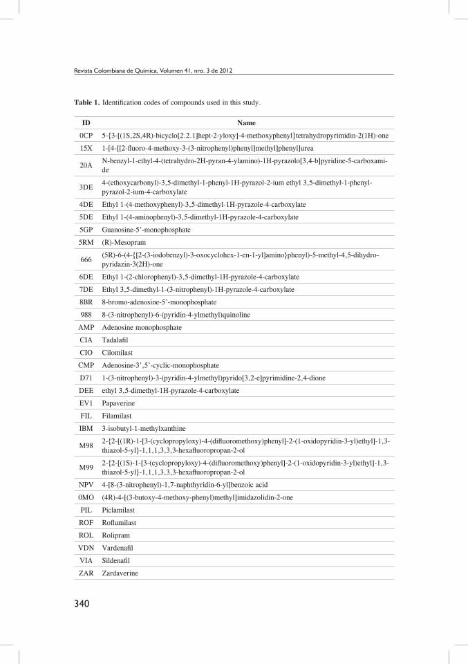
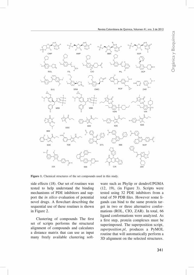
A list of all ligands and their structu-res is shown in table 1 and Figure 1.
cluster analysis
Ligands were superimposed with PyMOL (10). Dissimilarity between compounds was measured using the Jaccard distance defined by equation 1.
J∂ A,B( ) =A∪B − A∩ B
A∩ B Equation (1)
|A∩B| corresponds to the number of atoms closer than 0.7 Å between com-pounds A and B. A∪B is the total atom count for both compounds. Clus-tering was done using the Neighbor-Joining method of Nei and Saitou (11) implemented in PHYLIP (12). The tree was drawn using Dendroscope (13).
Pharmacophore detection and scoring
A total of 27 PDE4D inhibitors were used. The pharmacophore was built by sequentially averaging the position of each pair of atoms closer than a thres-hold distance of 1.2 Å. The total number of atoms used for each average was writ-ten into the temperature factor field of the PDB field. Atom type was assigned to the most frequent atom in the average. Scores were assigned to each inhibitor by adding the temperature field of each mat-ching atom of the pharmacophore.
results And dIscussIon
Phosphodiesterases are a diverse family of enzymes that hydrolyse cyclic nucleo-tides that play a key role in regulating intracellular levels of the second mes-sengers cAMP and cGMP (14). PDEs are clinical targets for a range of biolo-gical disorders, such as congestive heart failure, asthma, chronic obstructive pulmonary disease, depression, retinal degradation, and inflammation (15-17). Currently there is great interest in de-veloping new phosphodiesterase inhi-bitors with higher selectivity and lower
340
Revista Colombiana de Química, Volumen 41, nro. 3 de 2012
Table 1. Identification codes of compounds used in this study.
ID Name
0CP 5-{3-[(1S,2S,4R)-bicyclo[2.2.1]hept-2-yloxy]-4-methoxyphenyl}tetrahydropyrimidin-2(1H)-one
15X 1-[4-[[2-fluoro-4-methoxy-3-(3-nitrophenyl)phenyl]methyl]phenyl]urea
20AN-benzyl-1-ethyl-4-(tetrahydro-2H-pyran-4-ylamino)-1H-pyrazolo[3,4-b]pyridine-5-carboxami-de
3DE4-(ethoxycarbonyl)-3,5-dimethyl-1-phenyl-1H-pyrazol-2-ium ethyl 3,5-dimethyl-1-phenyl-pyrazol-2-ium-4-carboxylate
4DE Ethyl 1-(4-methoxyphenyl)-3,5-dimethyl-1H-pyrazole-4-carboxylate
5DE Ethyl 1-(4-aminophenyl)-3,5-dimethyl-1H-pyrazole-4-carboxylate
5GP Guanosine-5’-monophosphate
5RM (R)-Mesopram
666(5R)-6-(4-{[2-(3-iodobenzyl)-3-oxocyclohex-1-en-1-yl]amino}phenyl)-5-methyl-4,5-dihydro-pyridazin-3(2H)-one
6DE Ethyl 1-(2-chlorophenyl)-3,5-dimethyl-1H-pyrazole-4-carboxylate
7DE Ethyl 3,5-dimethyl-1-(3-nitrophenyl)-1H-pyrazole-4-carboxylate
8BR 8-bromo-adenosine-5’-monophosphate
988 8-(3-nitrophenyl)-6-(pyridin-4-ylmethyl)quinoline
AMP Adenosine monophosphate
CIA Tadalafil
CIO Cilomilast
CMP Adenosine-3’,5’-cyclic-monophosphate
D71 1-(3-nitrophenyl)-3-(pyridin-4-ylmethyl)pyrido[3,2-e]pyrimidine-2,4-dione
DEE ethyl 3,5-dimethyl-1H-pyrazole-4-carboxylate
EV1 Papaverine
FIL Filamilast
IBM 3-isobutyl-1-methylxanthine
M982-{2-[(1R)-1-[3-(cyclopropyloxy)-4-(difluoromethoxy)phenyl]-2-(1-oxidopyridin-3-yl)ethyl]-1,3-thiazol-5-yl}-1,1,1,3,3,3-hexafluoropropan-2-ol
M992-{2-[(1S)-1-[3-(cyclopropyloxy)-4-(difluoromethoxy)phenyl]-2-(1-oxidopyridin-3-yl)ethyl]-1,3-thiazol-5-yl}-1,1,1,3,3,3-hexafluoropropan-2-ol
NPV 4-[8-(3-nitrophenyl)-1,7-naphthyridin-6-yl]benzoic acid
0MO (4R)-4-[(3-butoxy-4-methoxy-phenyl)methyl]imidazolidin-2-one
PIL Piclamilast
ROF Roflumilast
ROL Rolipram
VDN Vardenafil
VIA Sildenafil
ZAR Zardaverine
341
Org
ánic
a y
Bioq
uím
ica
Revista Colombiana de Química, Volumen 41, nro. 3 de 2012
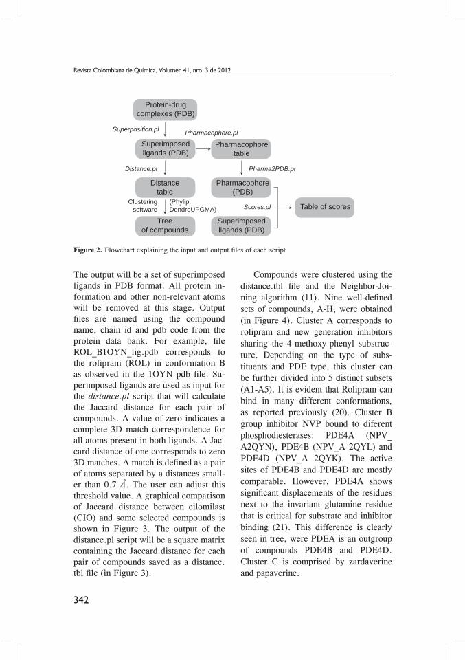
side effects (18). Our set of routines was tested to help understand the binding mechanisms of PDE inhibitors and sup-port the in silico evaluation of potential novel drugs. A flowchart describing the sequential use of these routines is shown in Figure 2.
Clustering of compounds The first set of scripts performs the structural alignment of compounds and calculates a distance matrix that can use as input many freely available clustering soft-
ware such as Phylip or dendroUPGMA (12, 19), (in Figure 3). Scripts were tested using 32 PDE inhibitors from a total of 59 PDB files. However some li-gands can bind to the same protein tar-get in two or three alternative confor-mations (ROL, CIO, ZAR). In total, 66 ligand conformations were analyzed. As a first step, protein complexes must be superimposed. The superposition script, superposition.pl, produces a PyMOL routine that will automatically perform a 3D alignment on the selected structures.
Figure 1. Chemical structures of the set compounds used in this study.
O
O N
N
N
N
N
NN
N
N
N
NN
N
N
N
N
N
NN
N
NN
N
N
NN N
N N
N
N
N
N N
N NN
N N
NN
N
NN
NNNNNN
N N
N+
N
NN
N N
N
N N
N N
N
NN
N
N+ N+
N+
N
NN
NNNN
F
N
N+ N
N N
NN
N
NN
N
N
N
N
N
NN
O O O
OO
OO
O O
O
O
O
O
O
OO
FF F
FOO
O
O
O
O
O
O
O
O
O
O
O
O
O
Cl
Cl
Cl
Cl
Br
O
OO
O
O
O
O
OO
O
O
O
O
O
OO
OO
O
O
O
S
O
O
O
S S
O
O
O
O
O
O
O
O
OO
OO
O
O
O
O
O
O
O
O
O
Cl
O
OO O l
O
O
O O
O
O
O
O
O
O
O
O O
O
O
O-
N+
O-
O-O-
O
O
O
O
O
O
O
O
F
F
F
F FF
F
F
FF
F
FF
FS
FF
O
OO
O
O
OO
CMP AMP 8BR 5GP
EV1
M99
IBM
6DE
15X 666 CIA 20A
7DE D71 988 NVP
DEE3DE 4DE 5DE
M98
VDN VIA
5RM OMO ROF ZAR
FILROL CIO PIL 0CP
P
PP
P
342
Revista Colombiana de Química, Volumen 41, nro. 3 de 2012
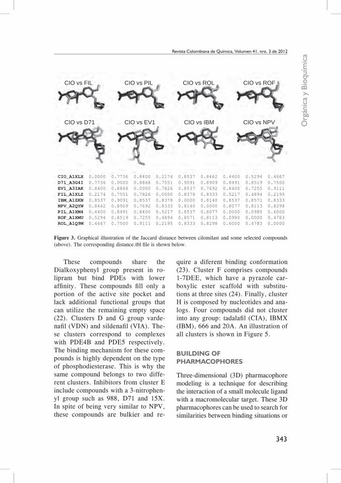
The output will be a set of superimposed ligands in PDB format. All protein in-formation and other non-relevant atoms will be removed at this stage. Output files are named using the compound name, chain id and pdb code from the protein data bank. For example, file ROL_B1OYN_lig.pdb corresponds to the rolipram (ROL) in conformation B as observed in the 1OYN pdb file. Su-perimposed ligands are used as input for the distance.pl script that will calculate the Jaccard distance for each pair of compounds. A value of zero indicates a complete 3D match correspondence for all atoms present in both ligands. A Jac-card distance of one corresponds to zero 3D matches. A match is defined as a pair of atoms separated by a distances small-er than 0.7 Å. The user can adjust this threshold value. A graphical comparison of Jaccard distance between cilomilast (CIO) and some selected compounds is shown in Figure 3. The output of the distance.pl script will be a square matrix containing the Jaccard distance for each pair of compounds saved as a distance.tbl file (in Figure 3).
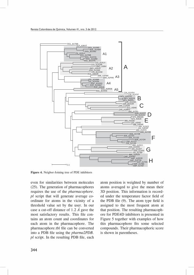
Compounds were clustered using the distance.tbl file and the Neighbor-Joi-ning algorithm (11). Nine well-defined sets of compounds, A-H, were obtained (in Figure 4). Cluster A corresponds to rolipram and new generation inhibitors sharing the 4-methoxy-phenyl substruc-ture. Depending on the type of subs-tituents and PDE type, this cluster can be further divided into 5 distinct subsets (A1-A5). It is evident that Rolipram can bind in many different conformations, as reported previously (20). Cluster B group inhibitor NVP bound to diferent phosphodiesterases: PDE4A (NPV_A2QYN), PDE4B (NPV_A 2QYL) and PDE4D (NPV_A 2QYK). The active sites of PDE4B and PDE4D are mostly comparable. However, PDE4A shows significant displacements of the residues next to the invariant glutamine residue that is critical for substrate and inhibitor binding (21). This difference is clearly seen in tree, were PDEA is an outgroup of compounds PDE4B and PDE4D. Cluster C is comprised by zardaverine and papaverine.
Figure 2. Flowchart explaining the input and output files of each script
Protein-drugcomplexes (PDB)
Superimposedligands (PDB)
Distancetable
Treeof compounds
Pharmacophoretable
Pharmacophore(PDB)
Superimposedligands (PDB)
Table of scores
Superposition.pl
Distance.pl
Scores.plClustering
software(Phylip, DendroUPGMA)
Pharma2PDB.pl
Pharmacophore.pl
343
Org
ánic
a y
Bioq
uím
ica
Revista Colombiana de Química, Volumen 41, nro. 3 de 2012
These compounds share the Dialkoxyphenyl group present in ro-lipram but bind PDEs with lower affinity. These compounds fill only a portion of the active site pocket and lack additional functional groups that can utilize the remaining empty space (22). Clusters D and G group varde-nafil (VDN) and sildenafil (VIA). The-se clusters correspond to complexes with PDE4B and PDE5 respectively. The binding mechanism for these com-pounds is highly dependent on the type of phosphodiesterase. This is why the same compound belongs to two diffe-rent clusters. Inhibitors from cluster E include compounds with a 3-nitrophen-yl group such as 988, D71 and 15X. In spite of being very similar to NPV, these compounds are bulkier and re-
Figure 3. Graphical illustration of the Jaccard distance between cilomilast and some selected compounds (above). The corresponding distance.tbl file is shown below.
quire a diferent binding conformation (23). Cluster F comprises compounds 1-7DEE, which have a pyrazole car-boxylic ester scaffold with substitu-tions at three sites (24). Finally, cluster H is composed by nucleotides and ana-logs. Four compounds did not cluster into any group: tadalafil (CIA), IBMX (IBM), 666 and 20A. An illustration of all clusters is shown in Figure 5.
buIldIng of PhArmAcoPhores
Three-dimensional (3D) pharmacophore modeling is a technique for describing the interaction of a small molecule ligand with a macromolecular target. These 3D pharmacophores can be used to search for similarities between binding situations or
CIO vs FIL
CIO vs D71
CIO vs PIL
CIO vs EV1
CIO vs ROL
CIO vs IBM
CIO vs ROF
CIO vs NPV
CIO_A1XLXD71_A3G4IEV1_A3IAKFIL_A1XLZIBM_A1ZKNNPV_A2QYNPIL_A1XM4ROF_A1XMUROL_A1Q9M
0.00000.77360.84000.21740.85370.84620.44000.52940.4667
0.77360.00000.88680.75510.90910.89090.84910.85190.7500
0.84000.88680.00000.78260.85370.76920.84000.72550.9111
0.21740.75510.78260.00000.83780.83330.52170.48940.2195
0.85370.90910.85370.83780.00000.81400.85370.85710.8333
0.84620.89090.76920.83330.81400.00000.80770.81130.8298
0.44000.84910.84000.52170.85370.80770.00000.09800.6000
0.52940.85190.72550.48940.85710.81130.09800.00000.4783
0.46670.75000.91110.21950.83330.82980.60000.47830.0000
344
Revista Colombiana de Química, Volumen 41, nro. 3 de 2012
even for similarities between molecules (25). The generation of pharmacophores requires the use of the pharmacophore.pl script that will generate average co-ordinate for atoms in the vicinity of a threshold value set by the user. In our case a cut-off distance of 1.2 Å gave the most satisfactory results. This file con-tains an atom count and coordinates for each atom in the pharmacophore. The pharmacophore.tbl file can be converted into a PDB file using the pharma2PDB.pl script. In the resulting PDB file, each
atom position is weighted by number of atoms averaged to give the mean their 3D position. This information is record-ed under the temperature factor field of the PDB file (9). The atom type field is assigned to the most frequent atom at that position. The resulting pharmacoph-ore for PDE4D inhibitors is presented in Figure 5 together with examples of how this pharmacophore fits some selected compounds. Their pharmacophoric score is shown in parentheses.
Figure 4. Neigbor-Joining tree of PDE inhibitors
A
A1
ROL_B1TBB0CP_A3KKT
ROL_A1XN0ROL_A1XMY
ROL_B1XN0FIL_A1XLZ
ROL_A1TBBROL_A1OYN
ROL_A1Q9MROL_B1OYN
CIO_A1XLXCIO_A1XOM
0MO_A3I8V0MO_A3K4S
5RM_A1XM6CIO_B1XOM
ROL_A3G4KM98_A2FM0
M99_A2FM5PIL_A1XM4PIL_A1XON
ROF_A1XOQROF_A1XMU
ROF_A3G4LROL_A1RO6
ROL_B1RO6CIA_A1UDU
CIA_A1XOZNPV_A2QYN
NPV_A2QYKNPV_A2QYL
ZAR_A1XORZAR_B1XOR
ZAR_C1XOREV1_A3IAK
ZAR_A1MKDIBM_A1ZKL
VDN_A1XOTVIA_A1XOS
D71_A3G4GD71_A3G4I
988_A3G5815X_A3IAD
988_A3G45666_A1SO2
20A_A3D3PIBM_A1ZKN
4DE_A1Y2DDEE_A1Y2B
3DE_A1Y2C5DE_A1Y2E
6DE_A1Y2H7DE_A1Y2J
7DE_A1Y2KIBM_A1SOJ
VDN_A1UHOVIA_A1UDT
VDN_A1XP0VIA_A1TBF
8BR_A1RO9CMP_A2PW3
AMP_A1RORAMP_A1PTW
5GP_A1T9SAMP_A1TB5AMP_A1TB70.1
A2
A3
A4
A5
BC
DE
F
G
H

345
Org
ánic
a y
Bioq
uím
ica
Revista Colombiana de Química, Volumen 41, nro. 3 de 2012
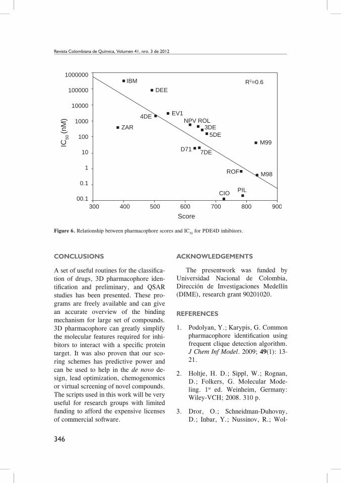
A pharmacophore model has to show predictive power that enables the design of novel chemical structures (26). In or-der to measure how similar a compound is to the pharmacophore, script score.pl was written which will add the score of each atom of the reference compound. A satisfactory model relating scores with IC50 data has been obtained (R=0.6) (in Figure 6). A closer look at the data shows that zardaverin, piclomilast, and cilomilast have an IC50 at least two order of magnitude lower than the prediction from our scoring scheme. These devia-tions suggest molecular features that
Figure 5. Graphical illustration of the clusters from the tree of compounds (A) and the fit between pharma-cophore and some selected compounds (B).
greatly improve the binding. Our scoring scheme depends on the total number of atoms averaging to a given position. It is obvious that in some cases strong inhibi-tors may have some unique substituents that will have low scores in the pharma-cophore. The pharmacophore scores can be corrected empirically, by inclusion of new parameters or adjustement of weights, to give a more acurate predic-tion of inhibition constants. However, these corrections are very case-depen-dent and correspond to a second phase of QSAR studies, which are beyond the scope of the scripts in this study.
A
Cluster E
Pharmacophore
EV1(544) ROL(700) CIO(733) M98(834)
CMP(415) IBM(400) NPV(616)
Cluster G Cluster H
Cluster B Cluster D
B
346
Revista Colombiana de Química, Volumen 41, nro. 3 de 2012
Figure 6. Relationship between pharmacophore scores and IC50 for PDE4D inhibitors.
conclusIons
A set of useful routines for the classifica-tion of drugs, 3D pharmacophore iden-tification and preliminary, and QSAR studies has been presented. These pro-grams are freely available and can give an accurate overview of the binding mechanism for large set of compounds. 3D pharmacophore can greatly simplify the molecular features required for inhi-bitors to interact with a specific protein target. It was also proven that our sco-ring schemes has predictive power and can be used to help in the de novo de-sign, lead optimization, chemogenomics or virtual screening of novel compounds. The scripts used in this work will be very useful for research groups with limited funding to afford the expensive licenses of commercial software.
Acknowledgements
The presentwork was funded by Universidad Nacional de Colombia, Dirección de Investigaciones Medellín (DIME), research grant 90201020.
references
1. Podolyan, Y.; Karypis, G. Common pharmacophore identification using frequent clique detection algorithm. J Chem Inf Model. 2009; 49(1): 13-21.
2. Holtje, H. D.; Sippl, W.; Rognan, D.; Folkers, G. Molecular Mode-ling. 1st ed. Weinheim, Germany: Wiley-VCH; 2008. 310 p.
3. Dror, O.; Schneidman-Duhovny, D.; Inbar, Y.; Nussinov, R.; Wol-
1000000
IC50
(nM
)
100000IBM
DEE
ZAR
4DE EV1NPV ROL
3DE5DE
M997DED71
ROF M98
PILCIO
R2=0.6
10000
1000
100
10
1
0.1
00.1300 400 500 700600 800 900
Score

347
Org
ánic
a y
Bioq
uím
ica
Revista Colombiana de Química, Volumen 41, nro. 3 de 2012
Scientific LLC, San Carlos, CA, USA. 2002. Available from: http: //www.pymol.org.
11. Saitou, N.; Nei, M. The neighbor-joining method: a new method for reconstructing phylogenetic trees. Mol Biol Evol. 1987; 4(4): 406-25.
12. Felsenstein, J. PHYLIP - Phylogeny Inference Package (Version 3.2). Cladistics. 1989. 5: 164-166
13. Huson, D. H.; Richter, D. C.; Rausch, C.; Dezulian, T.; Franz, M.; Rupp, R. Dendroscope: An interactive viewer for large phylo-genetic trees. BMC Bioinformatics. 2007; 8: 460.
14. Boswell-Smith, V., Spina, D., Page CP. Phosphodiesterase inhibitors. Br J Pharmacol. 2006; 147 Suppl 1: S252-7.
15. Conti, M., Jin, S. L.; Monaco, L.; Repaske, D. R.; Swinnen, J. V. Hormonal regulation of cyclic nu-cleotide phosphodiesterases. Endocr Rev. 1991; 12(3): 218-34.
16. Teixeira, M. M.; Gristwood, R. W.; Cooper, N.; Hellewell, P. G. Phosphodiesterase (PDE)4 inhibi-tors: anti-inflammatory drugs of the future? Trends Pharmacol Sci. 1997; 18(5): 164-71.
17. Barnette, M. S.; Underwood, D. C. New phosphodiesterase inhibi-tors as therapeutics for the treatment of chronic lung disease. Curr Opin Pulm Med. 2000; 6(2): 164-9.
18. Spina, D. The potential of PDE4 in-hibitors in asthma or COPD. Curr
fson, H. J. Novel approach for effi-cient pharmacophore-based virtual screening: method and applications. J Chem Inf Model. 2009; 49(10): 2333-43
4. Tripathi, A.; Surface, J. A.; Ke-llogg, G. E. Using active site map-ping and receptor-based pharmaco-phore tools: prelude to docking and de novo/fragment-based ligand de-sign. Methods Mol Biol. 2011; 716: 39-54.
5. Klabunde, T. Chemogenomic Ap-proaches to Drug Discovery: Simi-lar Receptors Bind Similar Ligands. Br J Pharmacol 2007; 152: 5–7.
6. Holliday, J.; Willet, P. Using a Ge-netic Algorithm to Identify Com-mon Structural Features in Sets of Ligands. J Mol Graph Modell 1997; 15: 203–253.
7. Handschuh, S.; Wagener, M.; Gas-teiger, J. The Search for the Spatial and Electronic Requirements of a Drug. J Mol Model 2000; 6: 358–378.
8. Finn, P. W.; Kavraki, L. E.; La-tombe, J. C.; Motwani, R.; Shelton, C.; Venkatasubramanian, S.; Yao, A. RAPID: Randomized Pharmo-cophore Indentification for Drug Design. Comp Geom-Theory Appli. 1998; 10: 263–272.
9. Tidsell, J. Beginning Perl for Bioinformatics. Sebastopol, USA: O´Reilly; 2001, 368 p.
10. DeLano, W. L. The PyMOL Mo-lecular Graphics System.DeLano

348
Revista Colombiana de Química, Volumen 41, nro. 3 de 2012
Opin Investig Drugs. 2000; 1(2): 204-13.
19. Garcia-Vallvé, S.; Palau, J.; Ro-meu, A. Horizontal gene transfer in glycosyl hydrolases inferred from codon usage in Escherichia coli and Bacillus subtilis. Mol Biol Evol. 1999; 16(9): 1125-34.
20. Zhao, Y.; Zhang, H. T.; O’Donnell, J. M. Antidepressant-induced in-crease in high-affinity rolipram bin-ding sites in rat brain: dependence on noradrenergic and serotonergic function. J Pharmacol Exp Ther. 2003; 307(1): 246-53.
21. Wang, H.; Peng, M. S.; Chen, Y.; Geng, J.; Robinson, H.; Houslay, M. D.; Cai, J.; Ke, H. Structures of the four subfamilies of phosphodies-terase-4 provide insight into the se-lectivity of their inhibitors. Biochem J. 2007; 408(2): 193-201.
22. Lee, M. E.; Markowitz, J.; Lee, J. O; Lee, H. Crystal structure of phosphodiesterase 4D and inhibi-tor complex(1). FEBS Lett. 2002; 530(1-3): 53-8.
23. Burgin, A. B.; Magnusson, O. T.; Singh, J.; Witte, P.; Staker, B. L.;
Bjornsson, J. M.; Thorsteinsdottir, M.; Hrafnsdottir, S.; Hagen, T.; Kiselyov, A. S.; Stewart, L. J.; Gurney, M. E. Design of phospho-diesterase 4D (PDE4D) allosteric modulators for enhancing cognition with improved safety. Nat Biotech-nol. 2010; 28(1): 63-70.
24. Card, G. L.; Blasdel, L.; England, B. P.; Zhang, C.; Suzuki, Y.; Gi-llette, S.; Fong, D.; Ibrahim, P. N.; Artis, D. R.; Bollag, G.; Milburn, M. V.; Kim, S. H.; Schlessinger, J.; Zhang, K. Y. A family of phos-phodiesterase inhibitors discovered by cocrystallography and scaffold-based drug design. Nat Biotechnol. 2005 Feb; 23(2): 201-7.
25. Schneider, G.; Neidhart, W.; Gi-ller, T.; Schmid, G. “Scaffold-Hopping” by Topological Pharma-cophore Search: A Contribution to Virtual Screening. Angew Chem Int Ed Engl. 1999; 38(19): 2894-2896.
26. Dror, O.; Schneidman-Duhovny, D.; Inbar, Y.; Nussinov, R.; Wolfson, H. A. A Novel Approach for Effi-cient Pharmacophore-based Virtual Screening: Method and Applications J Chem Inf Model. 2009; 49(10): 2333-2343.