El papel de los sistemas emocionales y las emociones 12-02...
87
1 El papel de los sistemas emocionales Contenido La organización del cerebro: tipos de procesos cerebrales......2 El Modelo de Pankseep de los tres procesos cerebrales........3 Los sistemas emocionales.......................................5 Sístema de búsqueda o exploración de lo desconocido..........5 Sistema de deseo sexual (Lust): Combinación de deseo y placer. ............................................................. 7 Sistema de cuidado o crianza (Care-especialmente en el educador)................................................... 14 Sistema de juego............................................ 19 Sistema de miedo............................................ 25 Sistema de rabia o ira......................................27 Sistema de pánico........................................... 33 Regulación de los sistemas emocionales........................38 El Placer, el gran regulador................................41 Una neurobiología común para el placer y el dolor...........48 Recopilación de textos y traducción: Dr. José Luis Prieto
Transcript of El papel de los sistemas emocionales y las emociones 12-02...

1
El papel de los sistemas emocionales
Contenido
La organización del cerebro: tipos de procesos cerebrales..........................................................2
El Modelo de Pankseep de los tres procesos cerebrales..........................................................3
Los sistemas emocionales............................................................................................................5
Sístema de búsqueda o exploración de lo desconocido...........................................................5
Sistema de deseo sexual (Lust): Combinación de deseo y placer.............................................7
Sistema de cuidado o crianza (Care-especialmente en el educador)....................................14
Sistema de juego....................................................................................................................19
Sistema de miedo...................................................................................................................25
Sistema de rabia o ira.............................................................................................................27
Sistema de pánico..................................................................................................................33
Regulación de los sistemas emocionales....................................................................................38
El Placer, el gran regulador....................................................................................................41
Una neurobiología común para el placer y el dolor................................................................48
Neuroquímica del amor..........................................................................................................49
Diferencias entre el enamoramiento infantil y adulto............................................................50
Referencias y enlaces.................................................................................................................51
Anexos........................................................................................................................................54
Anexo I...................................................................................................................................55
Recopilación de textos y traducción: Dr. José Luis Prieto

2
La organización del cerebro: tipos de procesos cerebrales
Una parte significativa de la comunidad científica actual comparte que los procesos emocionales se organizan en el cerebro en forma de sistemas (Pankseep, Berridge y Ledoux). A partir de esta premisa común y del reconocimiento de un núcleo pequeño de emociones universales básicas, se discute la funcionalidad de cada uno de los sistemas y los niveles cerebrales implicados. Desde la perspectiva de autores como Ledoux o Pankseep, los sistemas emocionales universales serían compartidos por todos los mamíferos y, en consecuencia, el estudio de los circuitos cerebrales y de la neuroquímica asociada, así como de las alteraciones genéticas en animales de fácil reproducción, como las ratas, permitiría extrapolarse a los seres humanos. Tal es el caso del miedo, estudiado por Ledoux, que se puede condicionar sin la participación inicial de la corteza cerebral y de la voluntad. Este planteamiento pretende conocer con precisión la fisiología de los sistemas emocionales e investigar sus relaciones con patologías fundamentales como la depresión o las fobias, y comprender procesos afectivos, como el apego. Desde esta perspectiva, la investigación con animales puede llevarnos a propuestas específicas a nivel neuroquímico o conductual que sirvan para actuar sobre diferentes trastornos o patologías, incidiendo directamente sobre los núcleos o vías neuronales, así como sobre los neurotransmisores, y los mecanismos químicos subyacentes.
A su vez, esta línea de investigación se complementa con los estudios sobre el ADN y las instrucciones genéticas que regulan la producción molecular asociada a la formación y fisiología del sistema nervioso, lo que permite a su vez desarrollar terapias que permiten regular, controlar o modificar la expresión del ADN o actuar químicamente o por otros medios sobre las proteínas sintetizadas.
El enorme desarrollo que ha tenido en los últimos años la investigación genética y cerebral no se podría haber producido sin la ayuda de la bioingenieria y la informática. De esta manera, por ejemplo, disciplinas como la optogenética, que permite manipular a voluntad la activación o inhibición de neuronas y circuitos nerviosos, introduciendo genes específicos sensibles a la luz a nivel neuronal, se han podido desarrollar gracias a los avances señalados. Estas técnicas se pueden aplicar a animales vivos, lo que permite estudiar la estructura y la función cerebral al mismo tiempo, mientras el animal desarrolla una conducta, sin tener que recurrir, como hasta ahora, a los estudios sobre
Recopilación de textos y traducción: Dr. José Luis Prieto

3
los tejidos de los animales muertos. La posibilidad de conectar al cerebro, microdispositivos electrónicos, incluídos microscopios, así como microcánulas y microelecrodos, a los animales vivos, en su mayoría ratas, permite introducir con precisión celular, sustancias en el cerebro, así como estimular eléctricamente y medir al mismo tiempo los cambios químicos y eléctricos, registrándose y analizándose en los ordenadores, mediante el software adecuado, los datos que nos proporcionan los circuitos cerebrales o incluso las neuronas individualmente. De todos estos avances se están beneficiando enormemente los estudios sobre los sistemas emocionales.
El Modelo de Pankseep de los tres procesos cerebrales
Teniendo en cuenta las consideraciones anteriores y que el Cerebro-Mente de los mamíferos es un órgano muy complejo, necesitamos de alguna ayuda conceptual para el estudio de los sistemas emocionales. De este modo, algunos autores, proponen simplificaciones teóricas evolutivas, desde las que poder hacernos una idea global e integrada.
Yo prefiero un concepto tripartito de las complejidades algo diferente, que no nos meta en “problemas” neuroanatómicos (Pankseep,2014).
Siguiendo a Pankseep, que toma a su vez como referencia a MacLean (1970) planteamos 3 tipos de procesos cerebrales que pueden participar en la explicación de los sistemas emocionales:
Procesos-primarios, (Afectos primordiales básicos, sub-neocorticales), que como resultado de la evolución proveen útiles vitales toscos pero eficaces, muchos de las cuales típicamente reflejan “intenciones-en-acciones” intrínsecas —procesos tradicionalmente tratados como incondicionados, “innatos” o “instintivos” y que han engendrado tanto acalorado debate (los sustratos neurales generalmente corresponden a los cerebros reptiliano y paleomamífero de MacLean). Se incluyen entre los procesos primarios:
Recopilación de textos y traducción: Dr. José Luis Prieto

4
Afectos emocionales (Sistemas de acción-emoción. Intenciones en acciones. Ej: El dolor producido por una quemadura produce miedo al fuego; un pinchazo con una aguja produce miedo a la aguja.
Afectos Homeostáticos (Intraceptores cuerpo-cerebro: hambre, sed…)
Afectos sensoriales (Sensores exteroceptivos que disparan las sensaciones y los sentimientos de placer y displacer. Ej el roce del alimento con la lengua).
Procesos-secundarios, (Emociones de proceso secundario, aprendizaje vía ganglios basales) que reflejan las capacidades cerebrales básicas de aprender mediante sensibilización-habituación, condicionamientos clásico y operante, (estos están representados en todos los niveles de organización cerebral). Se incluyen entre los procesos secundarios:
Condicionamiento clásico (Por ej, miedo, vía amígdala central y vasolateral)
Condicionamiento instrumental u operante. (Seeking vía Nucleus Accumbes)
Hábitos emocionales o conductuales (Ampliamente inconscientes- Estriado dorsal)
Procesos-terciarios, (Afectos terciarios y Funciones de la conciencia Neo-cortical), que incluyen todos aquellos procesos “reflexivos” del Cerebro-Mente superior que incluimos en conceptos tales como pensamiento, deliberación, planificación y las formas superiores de la intencionalidad (esto es, intenciones-de-actuar), y de esto no podemos tener mucho sin las funciones de aprendizaje general evolutivamente marcadas de nuestras expansiones neocorticales. Se incluyen entre los procesos terciarios:
Funciones cognitivo ejecutivas. Pensamientos y planificación (Corteza Frontal).
Reflexiones emocionales y regulaciones emocionales (Regiones Medial Frontal)
“Free Will”. (Funciones de la más Alta Memoria de Trabajo-intención para actuar)
Recopilación de textos y traducción: Dr. José Luis Prieto

5
Los sistemas emocionales
Las emociones básicas que Ekman (1972) descubrió como universales a través de estudios transculturales de la percepción del rostro humano no son fenómenos o reacciones simples, sino que están vinculadas a sistemas emocionales. Cada uno de éstos presenta características neurológicas propias, tanto en circuitos neuronales, punto emocionales específicos (por ej, Berridge la existencia de unos puntos calientes para el placer, - hotspot-) como en neurotrasmisores, neuropéptidos o química hormono-cerebral. Además, algunas emociones comparten en gran parte los circuitos y los neuropéptidos, diferenciándose sólo en las concentraciones necesarias del neuropéptido para provocar una reacción emocional u otra. Por ejemplo esto puede suceder entre el placer y el miedo. A su vez otras emociones como la rabia, están muy vinculadas al miedo y al dolor. En consecuencia cada emoción debe ser estudiada dentro de un sistema y dicho sistema puede incluir uno o diferentes procesos. Por ejemplo para Ledoux, el sistema de miedo es fundamentalmente secundario, no consciente y adaptativo para la supervivencia; sus procesos neuroanatómicos residen en la amígdala (Ledoux, 2014)i. A continuación describimos los tipos de sistemas emocionales, empleando el sistema de clasificación de Pankseppii:
Sístema de búsqueda o exploración de lo desconocido
Según Panksepp es el más universal de los sistemas emocionales. El Sistema de búsqueda fue expuesto por primera vez en 1954 por Milner. Él lo consideraba como un sistema de búsqueda de la recompensa. Si estimulamos cerebralmente al animal, lo que se produce es una actividad exploratoria o de búsqueda, sin que necesariamente se satisfaga ninguna necesidad. El animal puede mantener las conductas de exploración hasta que cae exhausto y se duerme, sin que necesariamente haya tenido que obtener satisfacción alguna. Es el sistema fundamental del que están dotadas las mentes creativas, siempre ávidas de acercarse a lo desconocido.
Recopilación de textos y traducción: Dr. José Luis Prieto
6
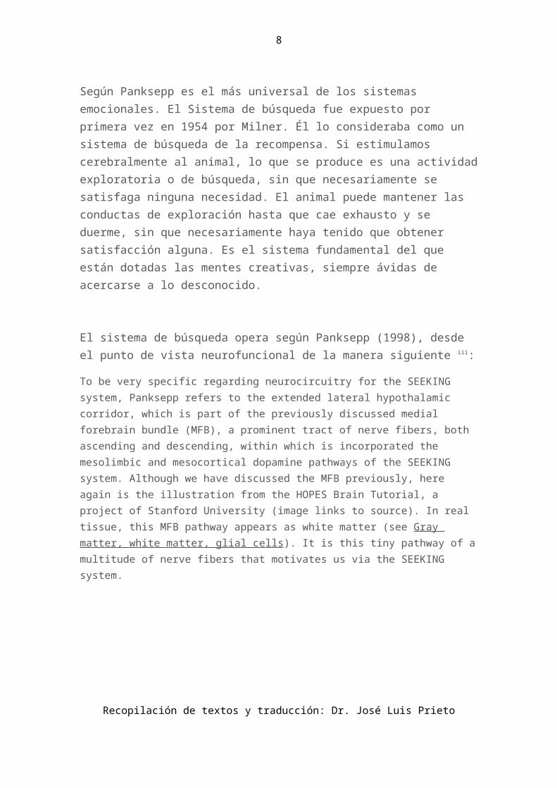
El sistema de búsqueda opera según Panksepp (1998), desde el punto de vista neurofuncional de la manera siguiente iii:
To be very specific regarding neurocircuitry for the SEEKING system, Panksepp refers to the extended lateral hypothalamic corridor, which is part of the previously discussed medial forebrain bundle (MFB), a prominent tract of nerve fibers, both ascending and descending, within which is incorporated the mesolimbic and mesocortical dopamine pathways of the SEEKING system. Although we have discussed the MFB previously, here again is the illustration from the HOPES Brain Tutorial, a project of Stanford University (image links to source). In real tissue, this MFB pathway appears as white matter (see Gray matter, white matter, glial cells). It is this tiny pathway of a multitude of nerve fibers that motivates us via the SEEKING system.
In locating the SEEKING system, Panksepp refers to the nucleus accumbens, which is part of the corpus striata (basal ganglia). The lateral hypothalamic corridor, explains Panksepp, "running from the ventral tegmental area (VTA) to the nucleus accumbens, is the area of the brain where local application of electrical stimulation will promptly evoke the most energized exploratory and search behaviors an animal is capable of exhibiting." The "corridor" to which Panksepp refers is also called the mesolimbic pathway, first discussed in Dopamine action, synthesis, and pathways.
If you are interested in obsessions and compulsions, it is important to remember that the SEEKING system as a whole and the nucleus accumbens in particular play important roles in generating these behaviors.
When the mesolimbic pathway from the dopamine-producing VTA to the nucleus accumbens is stimulated, SEEKING behavior ensues. Panksepp writes: "For instance, stimulated rats move about excitedly, sniffing vigorously, pausing at times to
Recopilación de textos y traducción: Dr. José Luis Prieto

7
investigate various nooks and crannies of their environment. If one presents the animal with a manipulandum, a lever that controls the onset of brain stimulation, it will readily learn to press the lever and will eagerly continue to 'self-stimulate' for extended periods, until physical exhaustion and collapse set in. The outward behavior of the animal commonly appears as if it is trying to get something behind the lever."
Sistema de deseo sexual (Lust): Combinación de deseo y placer.
Los animales tienen la necesidad de traspasar sus genes y dentro de éstos, los mamíferos han desarrollado un poderoso sistema, diferenciado en machos y hembras, que permite la supervivencia y el éxito de la especie. Las conductas sexuales de aproximación, (gestos, miradas, sonidos, conductas de cortejo) modifican e incrementan el deseo sexual, lo que incrementa los niveles hormonales y de determinados neuropéptidos, (tetosterona y dopamina,). El contacto físico y la obtención de orgasmos suponen una enorme descarga de opiáceos naturales y neurotransmisores. Finalmente, después del orgasmo, se desarrollan las conductas que implican intimidad y fidelidad, necesarias para el cuidado de la madre y de la prole, con cambios químicos asociados, como el incremento de la oxitocina. El lector puede profundizar más adelante en el apartado dedicado al placer y a la neuroquímica del amor.
La actividad sexual se apoya en los demás sistemas emocionales: búsqueda, exploración, juego, miedo y pánico.
Si este sistema ha funcionado correctamente precisaremos del sistema de cuidado o crianza.
The MATING System, the Brain, and Gender Determination
From the perspective of sociobiology, natural selection favors genes (including those that code for neurocircuitry and neurochemicals) that increase chances for gene duplication. One could say that the biological aim of living creatures is to organize in
Recopilación de textos y traducción: Dr. José Luis Prieto
8
such a way as to duplicate their genes. In mammalian societies, care of the young is of upmost importance. But mammals, depending on their environment, organize things differently.
Jaak Panksepp points out in Affective Neuroscience: The Foundations of Human and Animal Emotions (1998) that "among our brethren great apes" there is no single plan for family structure." He writes: "Whereas gibbons appear to mate for life with a single partner, gorillas prefer a harem-type family structure, orangutans tend to be social isolates, with the sexes coming together mostly for copulatory purposes, while chimpanzees are quite social and promiscuous, sharing partners rather indiscriminately." The photograph above right is of two gibbons from Highland Farm Gibbons Sanctuary (image links to source).
Regardless of family matters, certain things must get done to duplicate those genes. When my husband was a young lad, his friend told him that there was something inside a man that had to get inside a woman to make a baby. "So how does it get there," my very analytical husband-to-be asked with disbelief, "fly through the air?"
Sex differences in brain anatomy:
From his neuroscientist point of view, Panksepp points out that due to the "branching of control factors for brain and body organization, it is quite possible for a male-type body to contain a female-type brain, and for a female-type body to contain a male-type brain." But before we delve into issues of gender and sexuality, we will first discuss brain anatomy and circuitry related to optimizing reproduction. While they share some neurochemicals such as oxytocin, Panksepp explains that specific brain circuits and chemistries that are distinct for males and females mediate sexual urges that foster reproduction.
When fetal steroids masculinize the XY rat's brain, a specific area of the pre-optic area (POA) is enlarged compared to females. (We will discuss more specifics about how the mammalian brain is masculinized a little later in this section.) Panksepp explains that this enlarged, masculinized area is called the sexually dimorphic nuclei of the preoptic area (SDN-POA). In this case, the term "dimorphic" means a structure that occurs in two different forms—the female form and the male form. Compared to the more robust preoptic area in males, Panksepp explains that in females, "many neurons in this part of the brain die during fetal development for lack of testosterone, or more precisely its product estrogen, which is a powerful growth factor for these neurons."
When fetal steroids masculinize the XY human's brain, a specific area of the brain is also enlarged compared to females, although the size difference is not as great as that found in rats. This enlarged brain area is called the interstitial nuclei of anterior hypothalamus (INAH). In the image below (links to source) from Neuroscience, Purves
Recopilación de textos y traducción: Dr. José Luis Prieto

9
et al., editors, Sineuar Associates, Inc., the large vivid pink area represents the anterior hypothalamus.
The enlarged pre-optic area (POA) of the male rat brain is an important part of what here we will call MATING neurocircuitry. Panksepp writes: "Following lesions of the POA, male rats that have had abundant sexual experience will seek access to receptive females, even though they do not attempt to copulate with them. In other words, their social memories, situated perhaps in the cingulate cortex, amygdala, and nearby areas of the temporal lobes, are still capable of motivating social approach, although sexual engagement is no longer initiated." Later, he adds: "Castrated male rats that have lost their sexual ardor can be reinvigorated simply by placing testosterone directly into the POA."
In Part 1 of MyBrainNotes.com, we discuss Paul MacLean's triune brain concept. MacLean also played a role in delineating MATING neurocircuitry. Panksepp writes: "Paul MacLean mapped out the monkey brain for sites from which genital arousal (erections) could be evoked by localized ESB [electrical brain stimulation]. He discovered a broad swath of tissue, in higher limbic areas, where sexual response could be elicited. They included, prominently, areas such as the septal area, bed nucleus of the stria terminalis [BNST], and preoptic areas, all of which converge through the anterior hypothalamus into the medial forebrain bundle of the lateral hypothalamus."
There are other differences between the female brain and the masculinized male brain. For example, growth in the masculinized corpus callosum, which connects the two cerebral hemispheres, is reduced in males when compared to females. Panksepp points out that androgen (testosterone is the primary androgen) and estrogen receptors are concentrated in certain brain areas, "down to the lower reaches of the spinal cord, where both male and female sexual reflexes are controlled." Panksepp reports that in the lower spinal cord, the nucleus of the bulbocavernosus is distinctly larger in males than in females.
As we discuss above, specialized neurochemicals combine with specialized neurocircuitry and, as Panksepp puts it, "can trigger complex and coordinated sequences of sexual behavior." He writes: "If one places a small, naturally occurring, nine-amino acid peptide called vasotocin into the brains of male frogs and lizards, they begin to exhibit courting sounds and sexual behaviors. Given the opportunity, males treated with vasotocin mount and clasp females and copulate." The picture to the right (links to source) of two green anoles mating naturally is courtesy of [email protected].
Recopilación de textos y traducción: Dr. José Luis Prieto

10
In mammals, Panksepp explains that two neurochemicals very similar to the reptilian vasotocin play a role in sexual behavior. These two mammalian neurochemicals are vasopressin and oxytocin; they "assume key roles in controlling certain aspects of sexual behaviors" and each "differs from vasotocin [the reptilian neurochemical] by only one amino acid."
Panksepp emphasizes that vasopressin and oxytocin are not strictly male and female neurochemicals. Both play a role in the reproductive and parental behavior of both males and females. Vasopressin is more abundant in the male brain and has a primary effect on male sexual and social behavior. Vasopressin mediates "many aspects of male sexual persistence (including courtship, territorial marking, and intermale aggression)." Panksepp elaborates on the male rat sex act:
the general male strategy (facilitated by testosterone) is to exhibit fairly persistent searching for numerous sexual interactions … followed by the emission of vigorous … 50 KHz vocalizations … which, if the female does not object, culminate in … copulatory behavior. … the male mounts the female from the rear, palpating her flanks with his forepaws to arouse an arched-backed, rump-raised receptive posture called lordosis. Whereupon, the male rat exhibits sets of rapid thrusting movements called intromissions, which, if well guided, lead to entry of the penis into the vagina. After a series of intromissions, the male ejaculates, which is accompanied by a "deep thrust," and then he pushes off, often falling over in the process. He then attends to personal matters, with intense grooming of his genital area, with a shift to 22 KHz … vocalizations.
Panksepp points out that, in the female brain, vasopressin energizes "some of the more aggressive aspects of maternal behavior (i.e., protecting the young from harm)."
Oxytocin is more abundant in the female brain. Panksepp writes: "Animal research indicates that both brain opioid and oxytocin circuits are activated by various pleasurable pro-social activities, such as grooming, play, and sexual interchange." Oxytocin mediates "female social and sexual responsivity (especially the tendency of female rodents when mounted to exhibit lordosis…)," writes Panksepp. He explains that sensitization of female sexual eagerness transpires in the ventromedial nucleus of the hypothalamus and that damage to this area impairs responsivity. Panksepp writes:
The sex hormones that prepare the body for fertilization also dramatically change neurochemical sensitivities in this part of the brain. … Hormone priming (just like normal estrus) leads to a proliferation of oxytocin receptors in the medial hypothalamus, as well as an expansion of the dendritic fields, which physically expand, reaching out toward the incoming oxytocinergic nerve terminals arising from more rostral neurons. This completes a circuit that sensitizes the lordosis reflex of the spinal cord (and presumably prepares the female psychologically to interact seductively with males).
Recopilación de textos y traducción: Dr. José Luis Prieto

11
Regarding the female rat's role in the sex act, Panksepp writes: "The most evident behaviors in the rat are repeatedly running toward and away from the male, or past him in a hopping, darting fashion with the head wiggling and many 50 KHz vocalizations."
In the male brain, oxytocin sustains "some of the gentler aspects of male behavior (e.g., the tendency of fathers to be nonaggressive and supportive toward their offspring)." Oxytocin "also appears to help mediate the behavioral inhibition, or 'refractory period,' that follows orgasm in males."
Gender determinants—the role of testosterone:
In Affective Neuroscience, Panksepp writes: "One is typically born either genetically female (with the XX pattern of sex chromosomes) or genetically male (with the XY pattern)." These sets of chromosomes are not, however, the ultimate determination of gender. Panksepp says that "masculinization results from the organizational effects of fetal testosterone, which, in humans, occur during the second trimester of pregnancy."
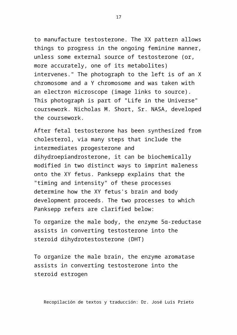
So what prompts fetal testosterone? "What the Y chromosome provides for the male is testis determining factor (TDF)," explains Panksepp, "which ultimately induces the male gonadal system to manufacture testosterone. The XX pattern allows things to progress in the ongoing feminine manner, unless some external source of testosterone (or, more accurately, one of its metabolites) intervenes." The photograph to the left is of an X chromosome and a Y chromosome and was taken with an electron microscope (image links to source). This photograph is part of "Life in the Universe" coursework. Nicholas M. Short, Sr. NASA, developed the coursework.
After fetal testosterone has been synthesized from cholesterol, via many steps that include the intermediates progesterone and dihydroepiandrosterone, it can be biochemically modified in two distinct ways to imprint maleness onto the XY fetus. Panksepp explains that the "timing and intensity" of these processes determine how the XY fetus's brain and body development proceeds. The two processes to which Panksepp refers are clarified below:
To organize the male body, the enzyme 5α-reductase assists in converting testosterone into the steroid dihydrotestosterone (DHT)
Recopilación de textos y traducción: Dr. José Luis Prieto

12
To organize the male brain, the enzyme aromatase assists in converting testosterone into the steroid estrogen
These processes are confusing since most people associate estrogen production strictly with being female when in reality it is estrogen that organizes the male brain. Panksepp explains that "the XX sex chromosome pattern informs the female body to manufacture proteins such as the steroid-binding factor alpha-fetoprotein… ." This chemical "protects the female fetus from being masculinized by the generally high levels of maternal estrogens. If there is not enough of this fail-safe factor, or it the maternal levels of estrogens are so high that they saturate the available alpha-fetoprotein, the female will proceed toward a male pattern of development—sometimes in both body and mind, sometimes in one but not the other, depending on the hormonal details that have transpired."
The effects of maternal stress on gender:
Regarding development of the XY fetus and the two modifications to testosterone that imprint maleness mentioned above, Panksepp notes that the "products of testosterone metabolism are critical ingredients that dictate whether a genetic male will continue along the male path in terms of body and brain development, both before and after puberty." He explains that "homosexuality and bisexuality are promoted if 'errors' occur in the various control points of these biochemical processes … ." Regarding such errors, Panksepp writes: "It has been repeatedly shown in animal models that maternal stress can hinder the normal process of brain masculinization by desynchronizing the underlying physiological processes… ."
In a normal litter from unstressed rat mothers, approximately eighty percent of the male offspring will exhibit male-typical, sex-seeking behavior while twenty percent remain asexual. Panksepp emphasizes that stress changes these ratios. "When a pregnant rat is exposed to any of a variety of stressors during the last trimester (third week) of the three-week gestation period," only about 20 percent of male offspring will exhibit male-typical, sex-seeking behavior while sixty percent are either bisexual or homosexual. The bisexual XY rats exhibit male behavior with a highly receptive female and female behavior in response to a sex-seeking male. The homosexual
Recopilación de textos y traducción: Dr. José Luis Prieto

13
XY rats exhibit lordosis when a sexually aroused male mounts them. As in unstressed litters, the remaining twenty percent of rats born to stressed rat moms are asexual. Panksepp does point out, however, that environment has some effect on sexuality. He writes: "The male offspring of stressed mothers exhibit more 'normal' sexual behavior if they are housed continuously during adulthood with sexually experienced females."
The role of genetics and timing in determining gender:
In addition to the effects of stress on gender development, Panksepp provides an example of how a genetically induced neurochemical deficiency can affect gender, at least during childhood. Some babies born in the Dominican Republic appear female at birth, although close inspection would reveal some enlargement of what seems to be a clitoris. Panksepp explains that in the womb, these XY children do secrete testosterone at the usual time and since they have normal aromatase activity, their testosterone is converted to estrogen. Accordingly, their brains are fully organized as male. Because they are genetically deficient in 5α-reductase, however, testosterone is not converted to DHT so their bodies do not appear male at birth. When such XY children "enter puberty and begin to secrete testosterone," writes Panksepp, "they develop male-typical bodies—with an increase of body hair, deepening of the voice, enlargement of the penis, and finally, the descent of the testes. Male-typical sexual urges also begin to emerge. Thus, the boys' pubescent erotic desires come to be directed toward females, even though they were reared as girls throughout childhood!" These boys are called guevedoces, which means "penis at 12," notes Panksepp.
"The hormones secreted at the onset of puberty," observes Panksepp, activate "the latent male or female sexual proclivities that have remained comparatively dormant within brain circuits since infancy." He writes: "Thus, the brain substrates for sexuality that are organized by these early hormonal experiences help determine what type of gender identities, erotic desires, and sex behaviors individuals will exhibit at puberty, when the elevations in hypothalamic gonadotrophic hormones and gonadal sex steroids begin to 'activate' sexual tendencies."
Recopilación de textos y traducción: Dr. José Luis Prieto

14
Mariateresa Molo et al., in "Characteristics of Brain Activity in Patients With Gender Identity Disorder," provides support for the idea that an XY fetus can end up with a female-type brain and that an XX fetus can end up with a male-type brain. The researchers found bioelectrical similarities in the brains of female controls and male-to-female transsexuals. Likewise, they found similarities in the brains of male controls and female-to-male transsexuals.
Books about gender:
David Bainbridge, The X in Sex: How the X Chromosome Controls our Lives, Harvard University Press, Cambridge, 2003.
Simon LeVay, Gay, Straight, and the Reason Why: The Science of Sexual Orientation, Oxford, 2010.
Sistema de cuidado o crianza (Care-especialmente en el educador)
Según Pankseep, es probablemente una fuente primaria de la empatía.
Participan en él: la corteza cingulada, área septal, nucleos basales, amígdala y algunas áreas del hipotálamo. Está íntimamente relacionado con el nivel de oxitocina. La lactancia, las caricias, el lamido o las palabras amables de alguien querido, incrementan el nivel de oxitocina. En el caso de las caricias o de las lamidas de las madres, caso de las ratas, se ha demostrado que mejoran la resistencia al stress de las crías en la edad adulta (McGowan y cols, 2011) . Las mujeres tienen un nivel medio superior a los hombres.
Se puede activar con los llantos y gritos de la cría.
In Affective Neuroscience: The Foundations of Human and Animal Emotions (1998), Jaak Panksepp proposes that nurturance in mammals probably "arose from neurochemical processes that controlled mating and
Recopilación de textos y traducción: Dr. José Luis Prieto

15
egg laying in reptiles." The precursor of mammalian oxytocin, the neurochemical vasotocin, explains Panksepp, "controls sexual urges in reptiles" and "helps deliver reptilian young into the world." Regarding reptiles, Panksepp writes: "Although a number of species—for instance, crocodiles—do exhibit some parental care, it is meager by mammalian standards." He provides a delightful story to illustrate hands-off reptilian parenting:
When a sea turtle, after thousands of miles of migration, lands on its ancestral beach and begins to dig its nest, an ancient birthing system comes into action. The hormone vasotocin is secreted from the posterior pituitary to facilitate the delivery of the young. Vasotocin levels in the mother turtle's blood begin to increase as she lands on the beach, rise further as she digs a pit large enough to receive scores of eggs, and reach even higher levels as she deposits one egg after the other. With her labors finished, she covers the eggs, while circulating vasotocin diminishes to insignificant levels… . Her maternal responsibilities fulfilled, she departs on another long sea journey. Weeks later, the newly hatched turtles enter the world and scurry independently to the sea without the watchful, caring eyes of mother to guide or protect them.
As mammals evolved, vasotocin evolved into oxytocin and argine-vasopressin (AVP), the neurochemicals so very important to mating behavior. As we discuss in The MATING System, the Brain, and Gender Determination, these two mammalian neurochemicals differ from vasotocin, the reptilian neurochemical, by only one amino acid. Panksepp points out that oxytocin, the same neurochemical which prompts receptivity in female mammals—including the lordosis reflex in the rat—also prepares "the mother's brain for nurturance." He writes: "The initial clue that there is an intrinsic bodily signal to promote maternal behavior was the fact that transfusion of blood from a female rat that had just given birth could instigate maternal behaviors in a virgin female."
Maternal care, hormones, and brain anatomy:
In rats, Panksepp reports that a "heightened maternal desire corresponds to the peak" of hormonal changes, "reaching an apex several hours before birth." Rat mothers begin to build nests for their offspring during this time.
Recopilación de textos y traducción: Dr. José Luis Prieto

16
Panksepp explains that "estrogen, which has remained at modest levels throughout pregnancy, rapidly increases as parturition nears. Progesterone, which has been high throughout pregnancy, begins to plummet. And, of course, there is a precipitous rise in prolactin, which induces the mother's acinar glandular tissues to manufacture milk." Panksepp writes: "Prolactin may be the critical ingredient in sustaining the natural behavior sequence, not only because brain injections of prolactin promote nurturance, but females who are nonmaternal because they have been surgically deprived of their pituitary glands do gradually become maternal when replacement injections of prolactin are provided."
Regarding oxytocin, Panksepp notes that "during the last few days of pregnancy and the first few days of lactation, there are remarkable increases in oxytocin receptors in several brain areas, as well as increases in the number of hypothalamic neurons that begin to manufacture this neuropeptide." He writes: "During lactation, oxytocin cells begin to communicate with each other directly via the development of gap junctions between adjacent oxytocinergic neurons, allowing them to synchronize their neural messages precisely." Regarding specific structures in the brain where this activity takes place, Panksepp points out that "the greatest oxytocin receptor proliferation is observed in the bed nucleus of the stria terminalis (BNST); when that area is damaged, maternal behavior is severely impaired." We discuss the stria terminalis in other sections of MyBrainNotes.com but I will reiterate here the excellent description found in MedlinePlus Dictionary. The stria terminalis is "a bundle of nerve fibers that passes from the amygdala along the demarcation between the thalamus and caudate nucleus mostly to the anterior part of the hypothalamus with a few fibers crossing the anterior commissure to the amygdala on the opposite side."
Panksepp notes that "lesions of the BNST and the nearby POA [preoptic area within the hypothalamus] can eliminate essentially all aspects of maternal behavior." The preoptic area lies in the same general subcortical
Recopilación de textos y traducción: Dr. José Luis Prieto
17
area as the BNST; this area is outlined with a red circle in the MRI image above left (links to source). Oxytocin prompts signaling in neurons that exit "from the POA laterally and descend in the medial forebrain bundle, with key terminals being in the VTA [ventral tegmental area]." The VTA is located in the midbrain, very near to the BNST, as illustrated in the MRI image above left. The illustration to the right shows the position of the preoptic area within the hypothalamus. This image is from Endocrinology, S.S. Nussey and S.A. Whitehead, obtained from the NCBI bookshelf (links to source).
As we discuss in Dopamine action, synthesis, and pathways, neurons in the VTA within the midbrain synthesize dopamine, which is essential to motivated behavior. Panksepp writes: "It has been established that the oxytocinergic synapses that terminate on dopamine cells on the VTA do, in fact, promote maternal behavior." He explains that oxytocin injections into the VTA "can induce maternal behavior… ."
Panksepp points out that "well-established maternal behavior no longer requires brain oxytocin arousal; oxytocin blockade impairs maternal behavior only if administered to mothers during the birth of their first litter of pups. In animals that have been allowed to exhibit maternal behavior for several days, oxytocin antagonists have no outward effect on maternal competence." In other words, even when drugs block oxytocin, such action does not block previous learning. In addition to neurons in the pre-optic nucleus of the hypothalamus, neurons in the paraventricular nucleus (PVN) of the hypothalamus also produce oxytocin. Panksepp writes: "PVN lesions administered prior to parturition weaken subsequent maternal behavior, but those administered after several days of normal maternal functioning do not." His postulates that "a great deal of learning is probably controlled in the higher reaches of CARE circuits such as the anterior cingulate cortex and bed nucleus of the stria terminalis [BNST]."
Unfortunately, the circumstances within which human infants are born are not always supportive of necessary learning curves and attachment. Panksepp illustrates:
For instance, not too long ago in certain arctic aboriginal groups, such as the Netsilik Eskimo of northern Canada, long-term social concerns often
Recopilación de textos y traducción: Dr. José Luis Prieto

18
overrode short-term emotional ones. Female babies who had little hope of finding an appropriate mate, because no male babies of comparable age had been born in the tribe, would be left to die in the snow, with little outward distress or remorse exhibited by the parents.
In addition to adverse cultural pressures in various human cultures, it is my opinion that another anxiety-provoking circumstance—uncertain paternity—can also affect a woman's attachment to her offspring. The fear that the questionable paternity issue will be discovered can only lead to a great deal of anxiety—the focus of which, of course, is the baby. I contend this kind of situation happens much more often than we humans are willing to admit.
Access to sex, male oxytocin, and reduced aggression:
Above, we discuss the neurochemical details of a mother's attachment to offspring. But what about neurochemicals and paternal attachment? Panksepp is clear on this: "Oxytocin administration reduces all forms of aggression that have been studied." In rodents, Panksepp points out that "free access to sexual gratification can lead to an enormous threefold elevation in oxytocin levels in some parts of the male brain. Apparently, sex promotes the synthesis of nurturant and antiaggressive neurochemistries." Male rat behavior bears this out. Panksepp reports that male rats will often "kill the young in a territory they have successfully invaded." However, once they have mated with a female, most often any rats born three weeks later are spared. This makes evolutionary sense since, as Panksepp explains, for rats, "it typically takes three weeks from the time of successful fertilization to the time of birth." In other words, the increased oxytocin in male rats that have had sex makes the male rats more nurturing and less aggressive. But how do researchers know it is the oxytocin that promotes nurturing behaviors? In the laboratory, when a male rat is placed into a new territory where there are young rats, he would be expected to kill the rat babies. But when oxytocin is administered to the male rate in this situation, Panksepp notes that the tendency to commit infanticide "dramatically diminishes."
Recopilación de textos y traducción: Dr. José Luis Prieto

19
Sistema de juego
Es un sistema que permite adquirir habilidades sociales y motrices que no son innatas. Produce placer y está intimamente vinculado a la risa. El juego se produce muy claramente en todos los mamíferos que requieren de complejas habilidades sociales y destrezas motoras.
"To the best of our knowledge," writes Jaak Panksepp in Affective Neuroscience: The Foundations of Human and Animal Emotions (1998), "a basic urge to play exists among the young of most mammalian species…." He recounts Jane Goodall's experience with chimpanzees: "A chimpanzee infant has his first experience of social play from his mother as, very gently, she tickles him with her fingers or with little nibbling, nuzzling movements of her jaws. Initially these bouts are brief, but by the time the infant is six months old and begins to respond to her with play face and laughing, the bouts become longer."
Before initiating play, animals must be comfortable. "Indeed, when placed in new environments, animals typically exhibit strong exploratory activity with little tendency to play until they have familiarized themselves with the new surroundings," writes Panksepp. "In all species that have been studied, playfulness is inhibited by motivations such as hunger and negative emotions, including loneliness, anger, and fear." Panksepp points out that in "most primates, prior social isolation has a devastating effect on the urge to play. After several days of isolation, young monkeys and chimps become despondent and are likely to exhibit relatively little play when reunited. …" He notes that playfulness returns "only when confidence has been restored." Rodents respond differently to isolation. "Laboratory rats show a greater emotional equanimity in coping with social isolation as compared to many other mammals." writes Panksepp. "Prior social isolation systematically increases roughhousing play in juvenile rats, while social satiation systematically reduces it."
While hunger reduces play behavior in young rats, "a single meal brings play right back to normal," reports Panksepp. "It may come as a surprise to
Recopilación de textos y traducción: Dr. José Luis Prieto

20
some, but young rats given no other ludic [from ludare, meaning 'to play'] outlets love to be tickled by and play with a frisky human hand." He points out that juvenile rats exhibit rough-and-tumble play behaviors "even if they have been prevented from having any prior play experiences during earlier phases of development." Panksepp explains that young rats start to play around 17 days of age, and if denied social interaction throughout the early phases of psychosocial development (e.g., from 15 to 25 days of age), "they play vigorously as soon as they are given their very first opportunity." Panksepp concludes that the impulse for rough-and-tumble play "is created not from past experiences but from the spontaneous neural urges within the brain."
The rough-and-tumble PLAY system "is important for learning various emotional and cognitive skills," Panksepp emphasizes, "including aspirations for social dominance and cooperation, which influence behavior with different intensities throughout the life span of each animal." He explains that "play may allow young animals to be effectively assimilated into the structures of their society. This requires knowing who they can bully, and who can bully them. One must also identify individuals with whom one can develop cooperative relationships, and those whom one should avoid." Panksepp points out that "the most vigorous play occurs in the context of preexisting social bonds." In contrast, he says that if "one animal becomes a 'bully' and aspires to end up on top all the time, playful activity gradually diminishes and the less successful animal begins to ignore the winner."
"Play probably allows animals to develop effective courting skills and parenting skills," writes Panksepp, "as well as increasing their effectiveness in various aspects of aggression, including knowledge about how to accept defeat gracefully." He points out that "PLAY circuitry allows other emotional operating systems, especially social ones, to be exercised in the relative safety of one's home environment. Thus, in the midst of play, an animal may gradually reach a point where true anger, fear, separation distress, or sexuality is aroused." Panksepp notes, however, that serious aggressive postures and sexual-type behaviors are rarely seen in play-fighting. During the later stages of juvenile life, because of their larger size and stronger competitive urges, more mature male animals may appear to
Recopilación de textos y traducción: Dr. José Luis Prieto
21
play more vigorously than their smaller companions. This difference, Panksepp explains, may in part reflect the drive to attain male dominance. He writes: "It is certainly possibly that PLAY systems contribute to social dominance urges, which may help explain our love of rough professional sports, where such issues are paramount in the minds of players and spectators alike."
Regarding the nonsocial functions of PLAY neurocircuitry, Panksepp points out that play increases physical fitness, skillful tool use, and the ability to innovate and think creatively. Young predators learn to hunt and prey species learn how to avoid predators. He writes: "Indeed, perhaps play even allows animals to hone deceptive skills, and thus in humans may refine the ability to create false impressions."
The brain's PLAY neurocircuitry:
PLAY neurocircuitry appears to be "intimately linked to somatosensory information processing within the midbrain, thalamus, and cortex," explains Panksepp. John A. Beal, Department of Cellular Biology and Anatomy, Louisiana State University, provides the image below. I have added labeling for these three areas of the brain. We discuss sensory information processing in Part 1 of MyBrainNotes.com in The brain's motor and somatosensory cortical maps.
PLAY neurocircuitry certainly helps young animals learn to interact with their environment since somatosensory information is obtained from the sense organs, such as the eyes and ears. Touch is also a form of somatosensory information. Generally speaking, somatosensory neurosignaling conveys information about the state of the body and immediate environment, such as body position and ambient temperature.
Within the thalami, Panksepp notes that somatosensory information is projected in two directions—up to the parietal cortex that processes bodily sensations and into nonspecific thalamic nuclei that elaborate a playful
Recopilación de textos y traducción: Dr. José Luis Prieto
22
motivational state. He points out that bilateral damage to thalamic areas involved in PLAY circuitry reduces both pinning and dorsal contacts and that "lesioned animals are no longer motivated to play." Panksepp reports that in such lesioned animals, "other relatively complex motivated behaviors, such as food seeking (foraging), are not diminished." John A. Beal, Department of Cellular Biology and Anatomy, Louisiana State University, provides the image above (links to source).
Panksepp emphasizes that PLAY behavior is an "endogenous urge" within the brain and does not necessarily rely on somatosensory input. In rats, "neither vision nor olfactory senses (including vibrissae) are necessary for the generation of normal play." He points out that the "auditory system contributes positively to play to some extent, since deafened animals play slightly less, and rats do emit many 50-KHz laughter-type chirps both during play and in anticipation of play." Panksepp explains that touch is the sensory system that helps most in instigating and sustaining normal play. Regarding experiments to test the importance of touch in generating PLAY behavior, Panksepp writes: "Local anesthetization of the neck and shoulder area is highly effective in reducing the level of playful pinning in young rats even though the motivation for play, as measured by dorsal contacts, is not reduced." Citing laboratory evidence, Panksepp concludes that "rats have specialized skin zones that send play signals into the nervous system when they are touched. In other words, mammals appear to have 'play skin,' or 'tickle skin,' with specialized receptors sending information to specific parts of the brain that communicate playful intentions between animals."
In animals that have had their cortex removed, "play solicitations and overall roughhousing, as monitored by direct activity measures, remain intact," writes Panksepp, although pinning behavior is reduced by about half. "It seems clear that play has powerful effects on the cortex." Panksepp concludes that juvenile play "involves programming various cortical functions." He writes: "In a sense, the cortex may be the playground of the mind, and PLAY circuits may be a major coordinator of activities on the field of play."
Laughter and the brain:
Recopilación de textos y traducción: Dr. José Luis Prieto

23
Panksepp asserts that "the hallmark of PLAY circuitry in action for humans is laughter, a projectile respiratory movement with no apparent function, except perhaps to signal to others one's social mood and sense of carefree camaraderie." (Photo courtesy of David Shankbone.)
"Laughter," Panksepp explains "is not learned by imitation, since blind and deaf children laugh readily." Ethologists consider genuine laughter to be innate and primal. The social smile is more contrived. In Part 1 of MyBrainNotes.com, we discuss the emotional versus the social smile in The anterior cingulate cortex–emotion, attention, and working memory.
Panksepp notes that "an openmouthed display characterizes the most intense forms of human laughter, and similar gestures are used as signals for play readiness in other species such as chimpanzees and dogs." He adds that chimpanzees' reunion rituals, "especially after long separations, are typically characterized by a lot of hooting, howling, and touching."
Other evidence indicating that specific neurocircuitry in the brain generates laughter is that "amylotrophic lateral sclerosis (ALS), a demyelinating disease that affects the brain stem," according to Panksepp, "can release impulsive laughter." He also points to "gelastic epilepsy, which is accompanied by bouts of laughter."
ADHD and PLAY neurocircuitry:
Previously, in Attention, Learning, and Memory: The VIGILANCE System, we learned that in ADHD, due to low levels of norepinephrine and perhaps, dopamine, the prefrontal cortex fails to adequately inhibit inappropriate impulses or distractions. Panksepp postulates that "many children diagnosed with ADHD may, in fact, be exhibiting heightened play tendencies." He writes:
Their hyperactivity, impulsiveness, and rapid shifting from one activity to another may be partly due to their unconstrained and unfocused playful tendencies. Indeed, the medications that are used to treat the disorder—psychostimulants such as methylphenidate (i.e., Ritalin) and amphetamines—are all very effective in reducing playfulness in animals. Moreover, parents of hyperkinetic children often complain that one of the undesirable side effects of such medications is the reduced playfulness of their children.
Recopilación de textos y traducción: Dr. José Luis Prieto
24
Obviously, parents value these childlike characteristics and are typically disturbed when the children's natural playfulness is pharmacologically diminished.
Autism, opioids, and PLAY neurocircuitry:
"Virtually all investigators now agree that autism is a neurobiological disorder," writes Panksepp in Affective Neuroscience. He explains that compared to normal brain development, people with autism have "an undersized cerebellum and brain stem, and a larger than normal cerebrum," along with "too many densely packed small neurons within parts of the limbic system, suggesting that selective cell death, a natural process of the developing brain called apoptosis, has not progressed normally." The result is that in autistic individuals, subcortical or so-called limbic structures do not interconnect with the rest of the brain as well as they normally would.
Panksepp quotes Leo Kanner, who in 1943 proposed that autistic children "have come into the world with an innate inability to form the usual, biologically provided affective contact with people." Panksepp reports that the "current theoretical perspective is that these children do not develop a 'theory of mind,' which refers to the ability of most children past the age of 2 to begin recognizing the types of thoughts and feelings that go on in the minds of others."
The motivation for rough-and-tumble PLAY, Panksepp points out, "is practically the only social desire that autistic kids exhibit at a relatively high level, but not with the reciprocating give and take and fantasy structures of normal childhood play." He notes that rats treated with low doses of opioids, like autistic children, do not exhibit a high desire for social companionship except for rough-and-tumble play. Panksepp proposes that "autistic children may have been exposed to excessive levels of endogenous opioids, or related molecules, during early development." He writes:
Moreover, they may continue to experience excessive opioid activity within certain circuits of their brains as they mature. This could explain their pain insensitivity and consequent tendency to exhibit self-injurious behavior, as well as many other symptoms ranging from stereotypies to
Recopilación de textos y traducción: Dr. José Luis Prieto
25
social aloofness. Because of these considerations, it has been suggested that some benefits may be brought to these children by the administration of opiate receptor blocking agents such as naltrexone.
Naltrexone may improve the lives of those autistic children "who have high circulating levels of opioids in the brain, a condition that has been demonstrated in about half of all autistic children who have been tested," explains Panksepp. "Moderate doses of naltrexone can reduce some of the active symptoms of autism such as overactivity, stereotypies, and self-injurious behaviors, and in low infrequent doses, it can promote social activities."
Sistema de miedo
El miedo según Le Doux es fundamentalmente un sistema de proceso secundario, no consciente que opera como una respuesta condicionada ante las amenazas. Tiene una alto valor adaptativo para la supervivencia y por lo tanto, no precisa ser consciente. Hay que diferenciarlo conceptualmente según Ledoux (2014), del miedo consciente.
Fear conditioning thus became a process that is carried out by
cells, synapses, and molecules in specific circuits of the nervous
system. As such, fear conditioning is explainable solely in terms
of associations created and stored via cellular, synaptic, and
molecular plasticity mechanisms in amygdala circuits. When the
CS later occurs, it activates the association and leads to the expression
of species-typical defensive responses that prepare the
organism to cope with the danger signaled by the CS. There is no need for conscious feelings of fear to intervene.
Recopilación de textos y traducción: Dr. José Luis Prieto

26
Research on patients with brain damage revealed that fear
conditioning creates implicit (nonconscious) memories that are distinct from explicit/declarative (conscious) memory
La amígdala es el órgano más estudiado (Ledoux) del sistema de miedo defensivo pero intervienen también otras zonas mesolímbicas y de la corteza prefrontal. A su vez, la amígdala se subdivide en varias estructuras. Se interrelaciona intensamente con los circuitos del placer (Berridge). A continuación Ledoux se describe la estructura y el funcionamiento de la amígdala en un proceso de condicionamiento clásico defensivo por miedo.
The lateral nucleus of the amygdala (LA) receives sensory inputs about the CS and US. Before training, the CS only weakly activates LA neurons (Le doux, 2014).
The neural circuits and cellular, synaptic, and molecular
mechanisms underlying the acquisition and expression of conditioned
fear responses have been characterized in detail (4, 5,
53, 80–82). (For a different perspective on the circuitry, see refs.
49 and 83.) The lateral nucleus of the amygdala (LA) receives
sensory inputs about the CS and US. Before training, the CS only
weakly activates LA neurons. After the CS is paired with the US,
i
ii
iii http://mybrainnotes.com/brain-ocd-dopamine.html
Recopilación de textos y traducción: Dr. José Luis Prieto

27
the ability of the CS to activate the LA increases. When the CS
later occurs alone, CS activation of the LA leads to neural activity
that propagates through amygdala circuits to the central
nucleus (CeA). Output connections of CeA then result in the
expression of defensive behavior and physiological responses, as
well changes brain arousal. Plasticity also occurs in the central
nucleus of the amygdala (84–86) and in CS sensory processing areas (87). At the cellular and molecular levels, fear conditioning
occurs when LA neurons that process the CS are weakly activated
at the same time that the US strongly depolarizes the
neurons (5, 53, 80, 81, 88, 89). This results in an increase in the
strength of the synapses that process the CS, allowing it to more
effectively activate amygdala circuits. Molecular mechanisms
engaged result in gene expression and protein synthesis, stabilizing
temporary changes in synaptic strength and creating longterm
memories. Many of the molecular findings were pursued
following leads from invertebrate work (14, 77, 90).
Los estudios sobre la amígdala y sus relaciones con la corteza prefrontal, nos ponen de manifiesto (Ledoux) que las conexiones entre el cortex y la amígdala son más débiles que las que se producen entre la amígdala y el resto del cerebro, lo que podría explicar la dificultad de controlar los estados emocionales agresivos, una vez que éstos se han desencadenado. La estimulación de la amígdala a través de estímulos visuales desencadena reacciones de miedo, ansiedad o pánico que deberán ser reguladas por la corteza prefrontal. A su vez, los estados emocionales generados en la amígdala están vinculados neuronalmente con el hipocampo y son muy relevantes en la formación de memorias.
.
Recopilación de textos y traducción: Dr. José Luis Prieto
28
Sistema de rabia o ira
http://mybrainnotes.com/brain-rage-violence.html
Es un sistema que se activa en escenarios naturales en situaciones extremas. Por ejemplo en la lucha por la caza, o contra los enemigos o depredadores. Es un sistema de apariencia negativa pero indispensable para la supervivencia.
When we think of rage, we often think of crime, and then guilt. In doing this, we fail to recognize that rage is an innate emotional system in the human brain that contributes to our survival. Stimulating specific neurocircuitry evokes rage in laboratory animals. Physical brain damage and seizures can certainly affect how this innate neurocircuitry operates and sometimes makes rage neurocircuitry more responsive, more automatic. Moreover, evidence indicates that extremely negative experiences can result in physiological changes in the brain that may predispose one to bouts of rage.
(1998), Dorothy Otnow Lewis describes
The lesions between the cortex of the frontal lobes and the rest of the central nervous system, between the self-reflective portions and the more instinctual portions of the brain, also contributed to Lucky's episodic violence. When the cortex of a cat is separated surgically from the rest of the brain, leaving only the lower centers of the brain intact, the cat may at first glance appear normal. The decorticate animal, when stimulated, becomes ferocious, directing its attack at anything it perceives as threatening or uncomfortable.
Regarding how negative experience affects the brain, Lewis writes:
What fascinates me most is the fact that brain concentrations of substances like serotonin are not immutable. They are not simply genetic givens—experience affects them. Certain kinds of stressors can decrease brain
Recopilación de textos y traducción: Dr. José Luis Prieto

29
serotonin levels and thereby change behavior. For example, if you isolate animals at crucial developmental stages, if you keep them caged all alone, their serotonin drops. What is more, when you then release them and put them in contact with other animals, they are fiercely aggressive. Pain and fear also reduce serotonin levels and promote aggression.
Rage, predatory, and other aggressions defined:
It seems that aggression, in its varied forms, arises from very different neural circuits in the brain. In Affective Neuroscience: The Foundations of Human and Animal Emotions (1998), Jaak Panksepp explains that scientists applying electrical stimulation to "slightly different brain zones" in laboratory animals evoke three distinct kinds of aggression. 1) predatory aggression, 2) rage-like aggression, and 3) inter-male aggression or dominance aggression. Panksepp points out that "prolonged social isolation or hunger may increase all forms of aggression, while high brain serotonin activity may reduce them all."
Circuitry that prompts aggression is quite specific. Panksepp explains that quiet-biting attack is typically evoked during electrical stimulation of the dorsolateral hypothalamus while rage-driven aggression is typically evoked during electrical stimulation of the ventrolateral and medial hypothalamus.
Electrical stimulation to SEEKING system locations in rats and cats prompt different behaviors. Panksepp writes: "The species-typical expressions of this system lead to foraging in some species and predatory stalking in others." Stimulating this system in cats results in predatory stalking and quiet-biting attack. "Obviously, this is a reasonable species-typical SEEKING behavior for a carnivorous animal that subsists at the top of the food chain."
Panksepp points out that when scientists stimulate specific circuits for rage-driven aggression in humans, the subjects report "experiencing a feeling of intense rage." When rats are stimulated in specific RAGE neurocircuits, they will attempt escape. Panksepp explains that most animals have "unpleasant affective experiences" during electrical stimulation to RAGE neurocircuits in their brain. He observes that such animals "exhibit piloerection, autonomic arousal, hissing, and growling," and readily learn
Recopilación de textos y traducción: Dr. José Luis Prieto

30
to turn the stimulation off. He points out that these animals direct their anger towards anything in their environment perceived as a threat, even members of their own species.
In addition to innate circuitry for predation, rage, and dominance, Panksepp discusses how animals develop a kind of "defensive" aggression which "emerges largely from a dynamic intermixture of RAGE and FEAR systems." He also draws attention to innate "appeasement" behaviors." An animal that lies on it's back and exposes vulnerable parts like the belly and neck can often reduce aggression by others of the same species. Sometimes the appeasement signal is vocal. Panksepp writes: "Defeated rats often emit long 22 Khz vocalizations."
Panksepp also categorizes infanticide as a form of aggression although pinpointing specific circuitry for this kind of behavior is not so easy. In the animal world, especially including rats, it seems that males sometimes kill the offspring of another male in order to stop lactation in the female mother, restoring her reproductive abilities. The new male is thus able to more quickly mate and produce his own offspring. "Considering that female rats have a three-week gestational period," Panksepp writes, "it was anticipated that the pup-killing tendencies of males might diminish approximately three weeks after mating, at about the time their own offspring might be born." He explains that research in the laboratory indicates that this is exactly what happens. Panksepp points out two other motivators of infanticide: "A mother may kill and consume some of her own offspring if food is scarce, even though such killing can also occur for more subtle 'political' reasons. Perhaps the most famous perpetrators of such acts were the cruel female chimpanzees, Passion and her daughter Pom, who killed off at least three and probably more of the young infants of other females in the group that Jane Goodal studied for many years."
A very interesting observation that Panksepp makes relates to genetic transmission of aggression. He points out that "genetic selection experiments in both male and female rodents indicate that one can markedly potentiate aggressiveness through selective breeding within a half dozen generations, and that breeding for aggression is as effective in females as in males."
Recopilación de textos y traducción: Dr. José Luis Prieto

31
The brain's RAGE neurocircuitry:
In Affective Neuroscience, Panksepp points to the work of Walter Hess during the 1930s in determining that electrical stimulation to certain brain areas can produce rage behavior in animals. Hess won the Nobel Prize in 1949. Panksepp writes: "It has long been known that one can enrage both animals and humans by stimulating very specific parts of the brain, which parallel the trajectory of the FEAR system." He adds: "Brain tumors that irritate the circuit can cause pathological rage, while damage to the system can promote serenity."
Panksepp writes: "The core of the RAGE system runs from medial amygdaloid areas downward, largely via the stria terminalis [a bundle of nerve fibers] to the medial hypothalamus, and from there to specific locations within the PAG [periaqueductal gray] of the midbrain."
In the illustration to the left (links to source), the amygdala is labeled on the right and the thin string-like stria terminalis, also
labeled on the right, links the amygdala to the hypothalamus, which lies hidden beneath the thalamus in this illustration. Although not labeled, the periaqueductal gray lies in the yellow center area that represents the midbrain.
Regarding the kinds of stimuli that can access RAGE circuitry, Panksepp points to such things as body surface irritation or when one does not receive an expected reward. He explains that the most common triggers of rage "are the irritations and frustrations that arise from events that restrict freedom of action or access to resources." He points out that "a human baby
Recopilación de textos y traducción: Dr. José Luis Prieto

32
typically becomes enraged if its freedom of action is restricted simply by holding its arms to its sides." Activation of RAGE circuits is "accompanied by an invigoration of the musculature, with corresponding increases in autonomic indices such as heart rate, blood pressure, and muscular blood flow." According to Panksepp, the phrase "getting hot under the collar," is accurate in that body temperature also increases during rage.
In the image below left, the medial hypothalamus is labeled in red lettering. This image is from S.S. Nussey and S.A. Whitehead, Endocrinology, NCBI bookshelf (image links to source). For some perspective, the image below right depicts the location and relative size of the hypothalamus as a whole (image links to source).
RAGE circuitry is organized hierarchically. Lesions of higher areas such as the amygdalae do not diminish responses from lower areas, while damage to lower areas such as the medial hypothalami and periaqueductal gray zones dramatically diminishes rage evoked from the amygdalae.
The image below (links to source) illustrates the position of the periaqueductal gray of the midbrain and is taken from professor Robert Lynch's course, "Territoriality and Aggressive Behavior," at the University of Colorado at Boulder.
According to Panksepp, the following areas provide input to the periaqueductal gray (PAG), a sort of primary generator for RAGE circuitry. Panksepp emphasizes that most of these connections are reciprocating two-way circuits.
Areas of the frontal cortex containing reward-relevance neurons influence RAGE circuitry.
Recopilación de textos y traducción: Dr. José Luis Prieto

33
Cortical areas called frontal eye fields, which help direct eye movements to especially prominent objects in the environment, influence RAGE circuitry.
The orbitoinsular cortex, especially the insular area—where a multitude of senses converge including pain and perhaps hearing—may provide specific sounds direct access to RAGE circuitry. In humans, these sounds may include, for example, an angry voice.
The medial hypothalamus provides powerful input related to energy (food) requirements and sexual matters thus influencing RAGE circuitry activated in pursuit of such resources.
A lower area, the vestibular complex, may help enrage animals when their bodily orientation is disturbed.
Cell groups such as the norepinephrine-producing loci coerulei and serotonin-producing raphe nuclei, which exert modulatory control over all behaviors, also influence RAGE circuits.
The nucleus of the solitary tract, which collects information via the vagus nerve that is probably related to processes such as heart rate and blood pressure, inputs to RAGE circuitry.
Sistema de pánico
Se puede desencadenar ante la separación de la figura de apego. Puede producirse pérdida del apetito, sueño, irritabilidad y depresión. El neuromodulador principal es el glutamato. Se ha estudiado a través de las vocalizaciones que realiza la cría cuando la
Recopilación de textos y traducción: Dr. José Luis Prieto

34
madre se separa de ella. También intervienen la noradrenalina, la serotonina y el factor de liberación de la corticotropina (Panksepp, 2006).
Es un sistema diferente al del miedo. Implica una elevada ansiedad permanente y según Pankseep es el fundamento de la depresión.
Although they surely interact in some way, in Affective Neuroscience: The Foundations of Human and Animal Emotions (1998), Jaak Panksepp explains that "as indexed by measures of separation calls in species as diverse as primates, rodents, and birds," PANIC/LOSS neurocircuitry is clearly distinct from FEAR neurocircuitry. Electrical stimulation to very specific brain areas, that we will refer to in this discussion as PANIC/LOSS neurocircuitry, produces the separation calls to which Panksepp refers. Although he considered both "sorrow" and "distress" as labels, he decided to call the neurocircuitry that generates feelings of loneliness, grief, and separation distress—as well as panic attacks in humans—the PANIC system. I have added "LOSS" to Panksepp's "PANIC" to draw attention to some of his observations that I find particularly meaningful.
The brain's PANIC/LOSS system and attachment:
Panksepp emphasizes that the PANIC/LOSS system "is especially important in the elaboration of social emotional processes related to attachment." He cites research that points to early childhood loss as a major risk factor for future depression and panic attacks. He proposes that one may be more vulnerable to depression and panic attacks "because of permanent developmental modification of the emotional substrates of separation distress." Indeed, in "Life Events Preceding the Onset of Panic Disorder" (1985) Faravelli writes that panic patients were more likely to have "underwent a major life event (death or severe illness, either personal or of a cohabiting relative) in the two months preceding the onset of symptoms."
Recopilación de textos y traducción: Dr. José Luis Prieto

35
Panksepp explains that "especially in intense forms such as grief," activation of PANIC/LOSS neurocircuitry "is accompanied by feelings of weakness and depressive lassitude, with autonomic symptoms of a parasympathetic nature, such as strong urges to cry, often accompanied by tightness in the chest and the feeling of having a lump in the throat."
Panksepp explains: "To be a mammal is to be born socially dependent." When "young animals are socially isolated, they typically lose weight even if they have free access to lots of food. When the young are reunited with their kin, and a mood of apparent contentment is reestablished, appetite returns."
"Brain evolution has provided safeguards to assure that parents (usually the mother) take care of the offspring," writes Panksepp, "and the offspring have powerful emotional systems to indicate that they are in need of care (as reflected in crying or, as scientists prefer to say, separation calls)." Regarding such vocalizations, Panksepp points out that "specific locations in the auditory system, in both the inferior colliculi and the medial geniculate nuclei, are highly tuned to receive and process these primal communications." Panksepp provides a vivid example of the mother-infant bond in the animal world:
The life of a young sea otter is completely dependent on the care provided by its mother. After his sexual contribution, the father pays little heed to his young. It is the mother's job to be both caretaker and food provider, as often as not, on the open sea. The pup's life revolves around maternal devotion. When she dives beneath the dark surface of the water for food, being absent from her infant's side for many minutes at a stretch, the young otter begins to cry and swim about in an agitated state. If it were not for those calls of distress among the rising and falling waves, young otters might be lost forever. Their security and future are unequivocally linked to the audiovocal thread of attachment that joins them to their mothers. It is the same for all mammals. At the outset, we are utterly dependent creatures whose survival is founded on the quality of our social bonds—one of the remaining great mysteries, and gifts, of nature.
The PANIC/LOSS neurocircuitry that prompts separation distress "probably evolved from more ancient pain mechanisms of the brain,"
Recopilación de textos y traducción: Dr. José Luis Prieto

36
concludes Panksepp. He proposes that "social attachments emerge, in part, from environmental events activating brain chemistries that can reduce arousal in these [PANIC/LOSS] distress circuits."
The brain's PANIC/LOSS neurocircuitry:
In the laboratory, opioids were the first neurochemical discovered to "powerfully reduce separation distress," notes Panksepp And what in our environment stimulates opioid release naturally in the brain? Panksepp writes: "Love is, in part, the neurochemically based positive feeling that negates those negative feelings." More definitively, he points out that "neural circuits mediating separation distress are under the control of brain opioids… ." The role of opiates in decreasing activity in PANIC/LOSS neurocircuits also helps distinguish PANIC/LOSS circuitry from FEAR circuitry. Panksepp explains that "opiates are very effective in reducing separation distress but not fearful behaviors."
An important component of PANIC/LOSS neurocircuitry is the bed nucleus of the stria terminalis. According to MedlinePlus Dictionary, the stria terminalis is "a bundle of nerve fibers that passes from the amygdala along the demarcation between the thalamus and caudate nucleus mostly to the anterior part of the hypothalamus with a few fibers crossing the anterior commissure to the amygdala on the opposite side." In the illustration below left, the stria terminalis links to the hypothalamus which is hidden beneath the thalamus.
The illustration above right shows the position of the preoptic area within the hypothalamus. This image is from S.S. Nussey and S.A. Whitehead, Endocrinology, from the NCBI bookshelf (links to source). Regarding neural specifics for PANIC/LOSS neurocircuitry, Panksepp notes that there is a high density of active distress-vocalization sites "in the ventral septal area, the preoptic area [within the hypothalamus], and many sites in the bed nucleus of the stria terminalis [BNST] (areas that figure heavily in sexual and maternal behaviors)."
Recopilación de textos y traducción: Dr. José Luis Prieto

37
The red circle on the image to the right (image links to source) indicates the general area within which the ventral septal area, preoptic area of the hypothalamus, and bed nucleus of the stria terminalis are nestled.
From the amygdaloid, hypothalamic, and BNST areas, PANIC/LOSS neurocircuitry runs "down through the dorsomedical thalamus to the vicinity of the PAG [periaqueductal gray in the midbrain]," explains Panksepp. Within the periaqueductal gray area, Panksepp notes that PANIC/LOSS neurocircuitry appears to arise from areas "very close to where one can generate physical pain responses." He writes: "Anatomically, it almost seems that separation has emerged from more basic pain systems during brain evolution… ."
As it does in other emotional neurocircuits, Panksepp points out that glutamate, an excitatory neurotransmitter, is probably the neurochemical that activates PANIC/LOSS neurocircuitry, thus generating distress vocalizations in young animals. In the laboratory, activating receptors for glutamate and corticotrophin releasing hormone (CRH) can dramatically increase distress vocalizations, even in the presence of other animals. In Part 1 of MyBrainNotes.com, we discuss the role of CRH in triggering the fight-or-flight response of the sympathetic nervous system, a component of the autonomic nervous system (see ANS—the autonomic nervous system). Panksepp emphasizes that CRH "arising from the paraventricular nucleus of the hypothalamus ... accompanies virtually all emotions and many psychiatric disturbances, especially depression." He points out that blocking receptors for glutamate and CRH dramatically decreases such vocalizations, even those induced by electrical brain stimulation.
Administration of opioids, oxytocin, and prolactin decreases activity in PANIC/LOSS neurocircuitry. Drugs that block transmission of glutamate and corticotrophin releasing hormone (CRH) also decrease PANIC/LOSS activity.
Depression and PANIC/LOSS neurocircuitry:
Panksepp asserts that "the major life factor in humans that precipitates depression is social loss." He explains that "the cascade of events during the initial protest phase of separation [marked by separation calls or crying]
Recopilación de textos y traducción: Dr. José Luis Prieto

38
appears to establish the brain conditions for the subsequent despair phase [depression]." More specifically, he explains that when the stress response is activated, "a depletion of brain norepinephrine, serotonin, and certain dopamine reserves" follows. Panksepp points out that activity in PANIC/LOSS neurocircuitry is attenuated with tricyclic antidepressants such as imipramine and chlorimipramine. These antidepressants have no clear effect on FEAR-induced anxiety but have been "found to exert clear antipanic effects in humans and to also reduce separation distress in animals." Panksepp notes the interesting fact that although they experience far fewer panic attacks, people whose panic attacks have been attenuated with tricyclic antidepressants often still fear that the attacks will occur.
Regulación de los sistemas emocionales
Como veremos a continuación, las emociones las puedo provocar, inhibir o alterar de muchas maneras, dado que los procesos primarios, secundarios y terciarios (Pankseep) están interconectados. Puedo intervenir de distintas maneras
Modificación cognitiva: Comprensión de la situación, análisis, cambio de actitudes, planificación futura y mejora de las expectativas. Este proceso modifica la fisiología del S. Nervioso. Es fundamentalmente un proceso mental terciario.
Modificación de contexto: Ubico al sujeto en un contexto placentero favorable, (lugar, alimentación, sueño, temperatura, ruido, sexualidad, juego, entorno natural, música...). Estoy regulando el placer y evitando el displacer. Se produce también una modificación del S.N. Proceso mental fundamentalmente primario y secundario.
Modificación de los vínculos afectivos, la interacción social y las relaciones sociales: A este nivel actúo sobre las relaciones de apego, amistad, enamoramiento y otras de naturaleza social (laborales, ocio, vecinales, participación social).
Recopilación de textos y traducción: Dr. José Luis Prieto

39
Modificación conductual: programas de condicionamientos asociados al placer/displacer, aprendizaje por observación. Generación de hábitos sanos. Enseñanza del placer y del displacer. Educación emocional. Estaríamos en el caso de las emociones aprendidas. En este caso y siguiendo los principios del condicionamiento clásico podemos estudiar las situaciones placenteras asociadas a determinados estímulos condicionados e incondicionados. Nos encontramos con emociones generadas en situaciones de aprendizaje observacional, por modelado u otras metodologías. Por ej: Práctica del Yoga, Taichi, meditación, baile, educación emocional.
Modificación neurológica: Estimulación eléctrica o química. Farmacología.
Modificación química sobre el deseo: Introduciendo sustancias químicas en el organismo o generando situaciones carenciales (alimento, sal, agua) puedo provocar un exceso o déficit en la concentración de sustancias químicas que alteran la homeostasis cerebral. Esto conlleva modificaciones en la valencia (saliencia) del deseo, y puede modificar e incluso invertir el deseo (Berridge, 2013: caso del déficit salino en ratas), alterándose los niveles de dopamina, neurotransmisor responsable del mantenimiento del deseo. Estos cambios son independientes de los aprendizajes previos e incluso pueden ir en contra de ellos, como Berridge ha demostrado en estudios con ratas a través del condicionamiento clásico. Según Berridge se producen alteraciones mesolímbicas de los circuitos del placer que son independientes del aprendizaje anterior. Las estructuras principales del circuito neuronal del placer son: Nucleus accumbes, ventral pallidum, parabrachial nucleus y cortex orbitofrontal, situándose los puntos hedónicos “calientes” en los tres primeros. Se considera que la estimulación de estos puntos tiene un papel predominante en los mecanismos del placer. Según la Teoría de Berridge en el placer hay dos procesos distintos y complementarios: El primero es el del deseo, regulado por la dopamina, sus estimulantes, simuladores e inhibidores. Y el segundo es el de la acción de los opiáceos que producen placer directo. De este modo, por ejemplo, los adictos pueden desear mucho consumir una droga aunque ésta no les produzca un efecto placentero. Este fenómeno se produce en todo tipo de ludopatías y dependencias psicológicas.
La inhibición con Gaba del nucleus accumbes produce según el caso, un incremento del apetito y o de las conductas de temor.
Recopilación de textos y traducción: Dr. José Luis Prieto

40
Modificación química sobre el producto: Otra posibilidad más común consiste en introducir opiáceos o sustancias que simulan, potencian o inhiben a los naturales, caso de la cocaína, la morfina, el cannabis o la marihuana.
La acción combinada de la estimulación química, por ejemplo, en puntos calientes (hostpot), con los estímulos condicionados e incondicionados (acción externa), produce un efecto enorme sobre la sensación de placer.
Modificación epigenética:
Debemos partir de la idea previa de que ante un estímulo amenazador o estresor, el hipocampo actúa sobre el hipotálamo generando la descarga del factor de liberación de la corticotropina (CRF) sobre la hipófisis anterior, que a su vez, sintetiza y libera adrenocorticotropina (ACTH) sobre la corteza suprarrenal. Esta libera distintos glucorticoides, como el cortisol que cumplen una función de mantenimiento de la respuesta al stress. El cortisol liberado en sangre será capturado por los receptores del hipocampo hasta un determinado umbral en el número de receptores.
Teniendo en cuenta el mecanismo anterior se ha detectado un gen cuya expresión afecta a la estructura y/o función de un sistema emocional asociado al miedo, pánico y stress en general. Es el caso de las ratas lamidas (Weaver, Meaney y Szyf, 2006) que se ven afectadas positivamente por la expresión genética de receptores glucocorticoides del hipocampo. De esta manera, en las ratas lamidas hay un mayor número de receptores. A mayor número de éstos, mayor capacidad de recepción de glucocorticoides como el cortisol, que cumple una importante función en el mantenimiento de la respuesta al stress, incrementando la glucosa en sangre, la metabolización de grasas e hidratos de carbono. Los receptores intervienen en un mecanismo de retroalimentación negativa, de tal manera que a mayor número de ellos, mayor incremento de la retroalimentación negativa. Cuando el nivel de los receptores de glucocorticoides, llega a un determinado umbral, el hipocampo inhibe la liberación de corticotropina (CRF) sobre la hipófisis anterior, lo que a su vez inhibe la produción de adrenocorticotropina (ACTH) sobre la corteza suprarrenal, lo que a su vez conlleva que no se produzcan glucocorticoides, como el cortisol, disminuyendo la respuesta al estímulo estresante. Ahora bien, si no hay suficiente número de receptores de glucorticoides, como en el caso de las ratas
Recopilación de textos y traducción: Dr. José Luis Prieto

41
no lamidas, no se inhibirá tan rápidamente la producción de corticotropina en la hipófisis y, por lo tanto, se seguirá produciendo cortisol u otros glucocorticoides. En este último caso las consecuencias del stress se mantendrán durante más tiempo, manteniendo al individuo en estado de alerta. El estrés es una reacción normal ante un estímulo o situación amenazante, pero su permanencia en el tiempo, una vez que ha desaparecido o atenuado la causa, tiene indudables consecuencias negativas.
El Placer, el gran regulador.
Tal como planteó Freud, el placer es un gran regulador de la conducta; posiblemente, como planteaba en su teoría, el más primigenio de todos los mecanismos reguladores de la conducta.
En la actualidad los estudios neurocientíficos nos enseñan que podemos descomponerlo, como ya citamos anteriormente, en deseo de obtener placer y en placer en sí mismo (wanting and liking). Ambos mecanismos están regulados por neuropéptidos (dopamina, opioides..) y se producen dentro de circuitos cerebrales específicos (Berridge, 2013).
En los centros hedónicos, uno de los neurotransmisores que se libera ante un estímulo placentero, por ejemplo el olor o la visión de un chocolate, es la encefalina, que coopera, a su vez, para que se pueda liberar otro neurotransmisor en las neuronas postsinápticas vecinas, la anandamida. Al difundirse este nuevo neurotransmisor desde su lugar de liberación, interacciona con receptores ubicados en la primera neurona que había liberado encefalinas aumentando la liberación de éstas. Creándose así un ciclo de retroalimentación positivo que permite intensificar el placer percibido.
Las encefalinas y endorfinas, que son péptidos opioides endógenos ubicados en el cerebro, también se producen en la glándula pituitaria y son liberados como hormonas. Tienen acción analgésica y gran afinidad con los receptores de la morfina; regulan el dolor y la nocicepción corporal.
Circuitos sobre el deseo y el placer
Los esquemas y dibujos que se exponen a continuación explican los circuitos fundamentales que relacionan deseo, placer y aprendizaje (Berridge, 2013),
Recopilación de textos y traducción: Dr. José Luis Prieto

42
especificando las partes específicas de la región mesolímbica del cerebro y los neuropéptidos empleados, entre los cuales se hace especial referencia al incremento del deseo a través del incremento de los niveles de dopamina. Dentro de los circuitos, juega un papel fundamental la activación o inhibición de los puntos calientes (hostspot) que permiten incrementar la sensación de placer cuando se activan sincrónicamente.
Recopilación de textos y traducción: Dr. José Luis Prieto

43
Recopilación de textos y traducción: Dr. José Luis Prieto

44
Recopilación de textos y traducción: Dr. José Luis Prieto

45
Recopilación de textos y traducción: Dr. José Luis Prieto

46
Recopilación de textos y traducción: Dr. José Luis Prieto

47
Recopilación de textos y traducción: Dr. José Luis Prieto

48
Una neurobiología común para el placer y el dolor
Siri Leknes and Irene Tracey
La amplificación del placer y la evitación del displacer
Cuando inyectamos un opiáceo, por ej, un agonista de la morfina, se incrementa el placer que nos produce un rostro que nos agrada (estamos más tiempo viéndolo), bien porque nos guste más o porque deseemos verlo más. Sin embargo, en este mismo caso, tratamos también de evitar más rápidamente un rostro que no nos guste. Estos resultados se encontraron en un experimento donde los hombres podían mantener o eliminar un rostro de mujer atractiva o no atractiva. Conclusión. Si introducimos una sustancia placentera, que incremente los opiáceos naturales del organismo, por ej, el chocolate, podemos incrementar el deseo, (wanting) o el placer sexual (liking).
Los beneficios del dolor (Siri Leknes&Brock Bastian, 2014)
The International Association for the Study of Pain (IASP). Según la IASP se define dolor como:
“an unpleasant sensory and emotional experience associated with actual or
potential tissue damage or described in terms of such damage.”
La definición anterior presenta limitaciones a su validez psicológica, debido a que:Recopilación de textos y traducción: Dr. José Luis Prieto

49
El ser humano puede profundizar en el dolor como por ejemplo sucede en los castigos o en los deportes más extremos o duros.
Hay evidencia de una serie de mecanismos implicados en los beneficios putativos del dolor, como es el caso del incremento de la atención. En otros casos, como en el placer producido por el consumo de especias altamente irritantes en la comida, los sujetos encuentran en ello un efecto placentero. En ocasiones una experiencia aversiva, contrastada con otra que no lo es, se convierte en beneficiosa para el sujeto, como por ejemplo, un masaje después de un ejercicio físico doloroso. A su vez, otros beneficios potenciales derivan de la inhibición de otras experiencias dolorosas que pasan a un segundo plano, como sucede cuando un dolor secundario pasa inadvertido ante el producido por una lesión principal. También se puede obtener un mayor grado de empatía y soporte social.
Neuroquímica del amor
Según Helen Fisher el amor es una combinación de lujuria, amor romántico y apego. Pasemos a esbozar inicialmente, el primer componente: la lujuria.
La lujuria según diferentes autores es el pilar básico para el amor. El sistema emocional sexual tiene dos componentes fundamentales: El deseo sexual (modificado especialmente por los niveles de dopamina) y el placer sexual (desencadena la producción de opiáceos naturales). Ambos son producidos fundamentalmente por los efectos en el organismo de los elementos siguientes: lenguaje erótico (palabras y paralenguaje), comunicación no verbal erótica (gestos, miradas, sonrisas), condicionamientos pavlovianos eróticos (objetos, ropa, semidesnudos, visión de zonas corporales), placer sensorial (vista, olfato, gusto..; incluíria las imágenes, los besos, chupadas, lamidas, caricias, mordiscos, susurros, aromas, olores), orgasmos (placer en sí mismo que se podría considerar como una modalidad muy especializada de placer sensorial, fundamentalmente de carácter incondicionado pero, tal vez también, de carácter condicionado primario).
En resumen, la actividad sexual puede estar destinada a incrementar el deseo sexual (modificando lo niveles de dopamina) o a producir placer en sí mismo. Desde un punto de vista cultural, la estimulación y el control del deseo sexual se ha convertido en un patrón cultural central en la mayoría de las sociedades.
Recopilación de textos y traducción: Dr. José Luis Prieto

50
Tal como indícabamos, para Helen Fisher, el amor es una combinación de lujuria, amor romántico y apego. Es un sistema cerebral que una vez activado desencadena un conjunto de reacciones neuroquímicas con finalidades adaptativas: la reproducción y el mantenimiento de la pareja durante unos pocos años, básicamente los que se necesitan para el cuidado de la cría. Para ella, el amor intenso es una respuesta de estrés que activa la amígdala, lo que provoca una reacción de alerta e incluso activa el sistema de pánico, lo que vuelve ciegos a los sujetos. Se activan también los centros de recompensa, especialmente el Nucleo Accumbes, donde Berridge sitúa los principales puntos calientes del placer que actúan coordinadamente con otros puntos extendidos en la zona mesolímbica y probablemente en la corteza cerebral. Se produce también la activación del hipotálamo lo que produce la acción de endorfinas y un decrecimiento de los niveles de serotonina. El hipotálamo provoca la producción de cortisol lo que reduce el campo visual del sujeto. Además incrementa la producción de tetosterona en las mujeres y lo disminuye en los hombres, lo que hace que nos sintamos muy próximos al otro género. Antes del orgasmo hay una gran producción de dopamina lo que conlleva el incremento del deseo del otro y en el momento del orgasmo se produce un “pico” de noradrenalina, vasopresina y oxitocina, lo que conlleva un elevado placer y el deseo de unión con el otro. La oxitocina ayudaría a mantener el vínculo para el cuidado de las crías. No obstante la oxitocina también se produce con el trato amable, el contacto físico (lamidas, caricias y abrazos) y el diálogo con personas queridas como demuestran varios autores (Pankseep). Además de estos mecanismos neuroquímicos intervienen otros, como la adrenalina, la fenil-etil-amina (FEA) y las feromonas, que regulan el deseo sexual y el amor romántico y que podemos consultar en el anexo I.
Fisher está muy preocupada con el empleo permanente de antidepresivos, porque estos incrementan la producción de la serotonina, que inhibe la producción de dopamina, lo que conlleva la desaparición del deseo sexual y en consecuencia del orgasmo. Sin orgasmos, disminuye la producción de oxitocina lo que debilita el vínculo de las relaciones de pareja. Por lo tanto, desde el punto de vista neuroquímico, los orgasmos facilitarían la construcción y el mantenimiento del apego entre adultos.
Para el análisis del apego, remitimos al lector a la teoría de Bowlby. En cuanto al amor romántico lo consideramos una construcción cultural sujeta a los discursos de cada cultura.
Diferencias entre el enamoramiento infantil y adulto
Recopilación de textos y traducción: Dr. José Luis Prieto

51
Si seguimos el modelo de Fisher, el niño y el adulto comparten gran parte de la lujuria, el amor romántico y, muy especialmente, el apego. Las diferencias en relación a la lujuria estarían en el deseo sexual y la conducta sexual. Si el placer sexual lo descomponemos en los componentes que hemos enunciados anteriormente, sólo el placer ante el coito, sería distinto que en el caso de los adultos. Incluso es posible que éste último lo sea sólo por razones culturales. La madre o el padre en la medida que intuyen el deseo sexual del niño/a van retirándose de la relación “amorosa corporal” y obligan a que el niño renuncie, algo que ya Freud analizó en el Complejo de Edipo. Esto es especialmente relevante en la adolescencia. También podemos encontrar diferencias en el grado de concentración de tetosterona que es mucho más bajo en el niño que en el adolescente o el adulto. El adolescente hace un intento de acercamiento hacia la madre y quizás hacia la hermana, pero éstas le rechazan, lo que le lleva a la búsqueda de una mujer o un hombre externo al núcleo familiar. En resumen, el deseo y el placer sexual se desplaza de las figuras parentales hacia figuras externas a las familiares. Esto tiene un indudable valor adaptativo de cara a la especie.
Cuestiones
Hasta ahora nos hemos situado fundamentalmente en la zona mesolímbica, tal vez el Ello de Freud. Pero qué pasa con el principio de realidad, con el Yo, con la corteza cerebral. La mayor parte de las conductas no están orientadas de forma inmediata al placer. Las funciones de la conciencia y por lo tanto de la corteza cerebral no están nada claras en la actualidad. Es evidente que participan en los procesos emocionales, aunque más tardíamente que las zonas mesolímbicas. No obstante, es preciso que la investigación avance en este sentido, para poder explicar cómo regulamos de forma consciente nuestros sistemas emocionales.
Referencias y enlaces
En la presentación de referencias se ha optado por incluir todas las fuentes utilizadas en cada sistema emocional y en cada autor, incluendo referencias bibliográficas y enlaces. De este modo el lector podrá consultar con mayor facilidad las fuentes de cada materia.
I.Bartal
EmpatiaRecopilación de textos y traducción: Dr. José Luis Prieto

52
Bartal, I(2011). Empathy and Pro-Social Behavior in Rats, Science 334, 1427.
Enlace: http://www.sciencemag.org/content/334/6061/1427.abstract?ijkey=a4cdecd3caaca20c6296b9ba3422aa5c201fb9ff&keytype2=tf_ipsecsha
K.Berridge.
El placer
Affective Neuroscience & Biopsychology Lab . University of Michigan Psychology Department. Biopsychology Program.
Berridge, K.C & Kringelbach, (2013). M.L. Neuroscience of affect: brain mechanisms of pleasure and displeasure. Current Opinion in Neurobiology. 23: 294-303,.
Enlace: http://www-personal.umich.edu/~berridge/
Virginia Campbell Podcast
Brain Science Podcast
Enlace: http://brainsciencepodcast.com/
Hellen Fisher
Neuroquímica del amor
Fisher, H. (2004). ¿Por qué amamos?. Naturaleza y química del amor romántico. Madrid. Santillana
Enlace: http://www.slideshare.net/ZUNAC/fisher-helen-porque-amamos
Vídeo 1: Helen Fisher: ¿Por qué amamos y engañamos? https://www.youtube.com/watch?v=CgVIPVODTXA
Vídeo 2: Neuroquimica: Cerebro enamorado (Helen Fisher) http://www.dailymotion.com/video/xoy8v9_neuroquimica-cerebro-enamorado-helen-fisher_school
Recopilación de textos y traducción: Dr. José Luis Prieto

53
Siri Graff Leknes
Placer y Dolor
UIO, Department of Psychology
Leknes S, Bastian B (2014). The benefits of pain. Review of Philosophy and Psychology (Special issue on Pain and Pleasure): 57-70
Enlace: http://www.sv.uio.no/psi/english/people/aca/sirigra/
Joseph Ledoux. Ledoux Lab. Center
El miedo
Ledoux, J. (2014) Coming to terms with fear. Center for Neural Science and Department of Psychology, New York University, New York.
LeDoux JE. (2012) Rethinking the Emotional Brain. Neuron, Volume 73, Issue 4, Pages 653-676,
Enlace: http://www.cns.nyu.edu/ledoux/
Jaak Panksepp, Ph.D.
Sistemas emocionales
Campbell, G. (Interviewer). & Panksepp, J. (Interviwee). (2014) Retrieve from Brain Science Podcast. Episode #91.
Enlace: http://brainsciencepodcast.com/bsp/the-origin-of-emotions-with-jaak-panksepp-bsp-91.html
Washington State University. Neuroscience Program, Integrative Physiology and neuroscience
Enlaces http://www.sciencemag.org/content/334/6061/1358.full?ijkey=IVBCV.ahM1.s.&keytype=ref&siteid=sci#xref-ref-3-1
http://ipn.vetmed.wsu.edu/people/faculty-ipn/pankseep-j
http://mybrainnotes.com/brain-ocd-dopamine.html
Panksepp, J. (2011). Empathy and the Laws of Affect. Science, 334, 1358-1359.
Recopilación de textos y traducción: Dr. José Luis Prieto

54
Panksepp, J. (2010) http://www.ncbi.nlm.nih.gov/pmc/articles/PMC3181986/
Luiz Pessoa and Ralph Adolphs
Procesamiento emocional
Pessoa, L. & Adolphs, R.(2010). Emotion processing and the amygdala: from a ‘low road’ to ‘many roads’ of evaluating biological significance. Nat Rev Neurosci. 11(11), 773-783
Enlace: http://www.ncbi.nlm.nih.gov/pmc/articles/PMC3025529/
Otros autores
Apego
Barg, G (2011) Bases neurobiológicas del apego. Revisión temática. Cienc. Psicol. vol.5 no.1 Montevideo.
Weaver, I. C. G., Meaney, M. J., & Szyf, M. (2006) in the offspring that are reversible in adulthood. Proceedings of the National Academy of Sciences of the United States of America, 103(9), 3480-5.
Anexos
Recopilación de textos y traducción: Dr. José Luis Prieto

55
Anexo INEUROQUÍMICA DEL AMOR
* Por Virginia GudiñoLas conductas de afiliación y apego social son comportamientos esenciales en las relaciones humanas, pero recién comienzan a conocerse los circuitos cerebrales que regulan su complejidad. La evolución realizó ciertos trucos neurales y utilizó los mismos recursos que usó para mantener unidos a madres e hijos, reciclándolos para lograr el mismo efecto con las parejas.
¨Etapas o Estados¨ del AmorLos mamíferos y aves han desarrollado tres sistemas cerebrales de motivación-emoción que actúan en serie:
¿ Cómo reacciona nuestro cuerpo?Fisiológicamente hay reacciones como: sudoración, taquicardia, indecisión, enfoque exclusivo y distorsión de la naturaleza del estímulo mismo. El iniciador de estos procesos, es un ¨chispazo¨ hormonal. En esta situación nuestro cuerpo produce una inundación de sustancias endógenas, llamadas neurotransmisores (comunican a las células nerviosas entre sí).
Al encontrarnos con una persona deseada, nuestro organismo recibe una señal que no tarda en manifestarse. El hipotálamo, a través del sistema nervioso, emite mensajes que, captados por las glándulas suprarrenales, segregarán un mayor cantidad de adrenalina y noradrenalina (neurotransmisores).
Consecuentemente, el corazón comenzará a latir con una mayor velocidad (unas 130 pulsaciones por minuto), aumenta la presión arterial máxima (sistólica), se liberan grasas y azúcares que aumenta la capacidad muscular y se produce una cantidad de mayor de glóbulos rojos con el objeto de mejorar el transporte de oxígeno a través del torrente sanguíneo.
Enamoramiento Estado alterado,
transitorio, de la conciencia.
Proceso que hace
Enamorarse está entre los comportamientos humanos más irracionales.
Los circuitos
del cerebro quedan intactos
Recopilación de textos y traducción: Dr. José Luis Prieto

56
emerger el amor romántico.
Sume al que lo siente en algo que parece una enfermedad
mental –mezcla de manía, demencia y
obsesión– que podría ser
confundido con una psicosis.
Con el tiempo, hay sentimientos más
¨reales¨, por tanto, puede que ese amor perdure o bien que se
busque a otra persona con la esperanza de
encontrar a la adecuada.
hasta que un nuevo amor
se cruce por el
camino
Se activan 12 áreas en el cerebro cuando nos enamoramosLa Dra Stephanie Ortique, profesora de la Universidad de Syracuse en Nueva York, ha llegado a la conclusión de que enamorarse puede provocar la misma respuesta eufórica que provocan las drogas ilícitas en el cerebro y que, asimismo, se activan varias zonas cerebrales, incluyendo aquellas vinculadas con funciones cognitivas sofisticadas.
En este estudio, que fuera publicado por el Journal of Sexual Medicine, la investigadora y su equipo analizaron otros estudios llevados a cabo en el pasado sobre cómo responde el cerebro al concepto del amor.
El resultado alcanzado fue que cuando una persona se enamora se activan 12 áreas cerebrales para liberar compuestos químicos como la dopamina, la oxitocina y la adrenalina y, es la activación de ciertas zonas cerebrales lo que genera un estímulo en el corazón y el sentimiento de ¨mariposas en el estómago¨, pero también se activan áreas cognitivas más complejas como las encargadas de la representación mental y la autoimagen corporal, así que ¨el amor es un proceso más complicado que la adicción a las drogas. ¨Si reducimos el Amor a sustancias químicas ...La Dra. Helen Fisher, autora del libro The Anatomy of Love, divide en tres las etapas del amor romántico:
1- El Deseo o la Lujuria: Predomina la testosterona: prima el deseo de
sexo, dado que, los niveles altos de esta hormona van de la mano con la pulsión sexual.
Se produce un pico de adrenalina, lo que incrementa la presión sanguínea (provoca los rubores de las primeras etapas del enamoramiento), aceleración del ritmo cardíaco y sudoración en la palma de las manos.
Hay una elevación de la noradrenalina, lo que lleva a la excitación sexual y “elevación” del humor, con sentimientos de seguridad y gusto al compartir momentos con la persona que consideramos especial.
Cuando alguien está en las garras de este amor romántico es irracional, va al gimnasio a las seis de la mañana... ¿Por qué? Porque él, o ella, está ahí. ‘Esta compulsión hacia el amor romántico puede ser más fuerte todavía que las propias ansias de vivir’, dice la doctora Helen Fisher, antropóloga de la Universidad Rutgers
Recopilación de textos y traducción: Dr. José Luis Prieto

57
y coautora del análisis.
2- Atracción
Predominan la feniletilamina (FEA), la dopamina, la norepinefrina y la oxitocina. Baja la Serotina, estando así en la etapa de euforia y de romance.
La FEA (estimulante natural conocido como “molécula del Amor”) inicia una reacción química en cadena en el cerebro. Estimula la secreción de dopamina y luego ésta inducirá a la de oxitocina.
La dopamina afecta procesos cerebrales que controlan el movimiento, la respuesta emocional y la capacidad de experimentar dolor o placer. También Induce un proceso de aprendizaje positivo en el cerebro, transformando el simple deseo con fines sexuales en algo mucho más profundo, dando asi inicio a las relaciones.
La oxitocina, es el “el químico de los mimos”. Cuando el cuerpo entra en contacto con las feromonas (sustancias químicas que nuestro organismo produce y que tienen como única misión afectar nuestro comportamiento sexual y atraer al sexo opuesto) nuestro proceder se altera y nuestras percepciones se tornan totalmente idiosincrásicas e impredecibles. Estamos enamorados (Véase: Sex, Time and Power: How Women’s Sexuality Shaped Human Evolution por L. Shlain).El amor romántico es "creado" por una endorfina, la fenil-etil-amina (FEA), que produce sensaciones de satisfacción y armonía ante el estímulo que la desencadena, y nos produce una sensación de bienestar asociada a una cierta persona.
En efecto, el idilio romántico coincide con estos factores químicos que dan lugar a las sensación tan especial de ¨estar enamorado¨. Esta es la razón por la cual, los enamorados pueden permanecer largas horas conversando, haciendo el amor o simplemente 'estando juntos' sin sentir cansancio uno del otro.
Y en esta misma línea, se ha observado que las personas que vivieron una desilusión amorosa, buscarían compensar la disminución de feniletilamina (FEA) a través del consumo de chocolate (golosina especialmente rica en esta sustancia).
La función de la fenil-etil-amina (FEA) es la de garantizar la armonía y la tolerancia hacia una posible unión reproductora, destinada a persistir como relación amorosa por unos 3 ó 4 años.
3) Vínculo:
Predominan la oxitocina, la vasopresina y la serotonina, evolucionado así hacia una relación apacible, duradera y segura.
La oxitocina es la responsable del nacimiento de lazos afectivos en una pareja. En las mujeres, es liberada principalmente durante el parto (contracción del músculo uterino), el amamantamiento y los orgasmos (contracciones musculares). En los hombres: propicia la erección y acelera el impulso eyaculatorio.
Recopilación de textos y traducción: Dr. José Luis Prieto

58
La vasopresina, tambien llamada “hormona monogámica”, es liberada durante la actividad sexual . Incrementa los niveles de agresión y acompañada de la oxitocina, induce el sueño.
La oxitocina aumenta la confianza entre las personas y ayuda a forjar lazos permanentes entre los amantes, tras la primera oleada de emoción. Reduce la actividad de la parte del cerebro donde se detecta el temor, facilitando el unirse a una persona.
¿Cómo revitalizar una relación?Los científicos creen saber cómo mantener fluidos los circuitos amorosos. Se puede estimular el amor romántico mediante sustancias químicas, a través de gestos como abrazos, besos y otras formas de contacto íntimo. Si uno quiere realmente revigorizar una relación, se deben hacer las cosas que estimulan la producción de estas moléculas y dejarlas que alimenten sus emociones. "Mi esposa me dice que las flores la estimulan"…"No sé si es así, a un científico le cuesta ver cómo pueden estimular los circuitos, pero sé que parecen tener efecto. Y la ausencia de ellas también tiene un efecto". (Larry Young, del centro de investigaciones Yerkes de la Universidad Emory de Atlanta).
Hoy, conocer aspectos científicos relevantes en ese sentido, nos posibilita comprender estos procesos, pero lo más importante es la aplicación práctica que le damos a la ¨nueva¨ información que tenemos, pues de nuestras acciones y del compromiso que ejercitemos dependerá el que podamos consolidar o no, una vida compartida gratificante.
Toda relación podría considerarse un desafío, una construcción a la cual dar forma y en la que intervienen multiplicidad de factores (neurobiológicos, psicológicos, culturales, experiencias de vida, etc)de cada una de las personas involucradas. Un desafío que vale la pena afrontar, si de ello depende el poder crear y fortalecer relaciones duraderas y felices.
Artículos Relacionados:
¿Qué es el Amor?
Recopilación de textos y traducción: Dr. José Luis Prieto