Ejercicio y envejecimiento: cambios transcripcionales y...
213
Ejercicio y envejecimiento: cambios transcripcionales y epigenéticos en un modelo murino de envejecimiento acelerado Maria Jesús Álvarez López Aquesta tesi doctoral està subjecta a la llicència Reconeixement- NoComercial – SenseObraDerivada 3.0. Espanya de Creative Commons . Esta tesis doctoral está sujeta a la licencia Reconocimiento - NoComercial – SinObraDerivada 3.0. España de Creative Commons . This doctoral thesis is licensed under the Creative Commons Attribution-NonCommercial- NoDerivs 3.0. Spain License .
Transcript of Ejercicio y envejecimiento: cambios transcripcionales y...
Ejercicio y envejecimiento: cambios transcripcionales y epigenéticos en un modelo
murino de envejecimiento acelerado
Maria Jesús Álvarez López
Aquesta tesi doctoral està subjecta a la llicència Reconeixement- NoComercial – SenseObraDerivada 3.0. Espanya de Creative Commons .
Esta tesis doctoral está sujeta a la licencia Reconocimiento - NoComercial –
SinObraDerivada 3.0. España de Creative Commons .
This doctoral thesis is licensed under the Creative Commons Attribution-NonCommercial- NoDerivs 3.0. Spain License .
EEJJEERRCCIICCIIOO YY EENNVVEEJJEECCIIMMIIEENNTTOO::
CCAAMMBBIIOOSS TTRRAANNSSCCRRIIPPCCIIOONNAALLEESS YY
EEPPIIGGEENNÉÉTTIICCOOSS EENN UUNN MMOODDEELLOO MMUURRIINNOO DDEE
EENNVVEEJJEECCIIMMIIEENNTTOO AACCEELLEERRAADDOO
FACULTAD DE FARMACIA
MARÍA JESÚS ÁLVAREZ
2015

Es verdad, pues: reprimamos esta fiera condición,
esta furia, esta ambición, por si alguna vez soñamos. Y sí haremos, pues estamos
en mundo tan singular, que el vivir sólo es soñar;
y la experiencia me enseña, que el hombre que vive, sueña
lo que es, hasta despertar.
Sueña el rey que es rey, y vive con este engaño mandando, disponiendo y gobernando; y este aplauso, que recibe
prestado, en el viento escribe y en cenizas le convierte
la muerte (¡desdicha fuerte!): ¡que hay quien intente reinar viendo que ha de despertar en el sueño de la muerte!
Sueña el rico en su riqueza, que más cuidados le ofrece; sueña el pobre que padece
su miseria y su pobreza; sueña el que a medrar empieza, sueña el que afana y pretende, sueña el que agravia y ofende, y en el mundo, en conclusión,
todos sueñan lo que son, aunque ninguno lo entiende.
Yo sueño que estoy aquí, destas prisiones cargado; y soñé que en otro estado
más lisonjero me vi. ¿Qué es la vida? Un frenesí. ¿Qué es la vida? Una ilusión,
una sombra, una ficción, y el mayor bien es pequeño;
que toda la vida es sueño, y los sueños, sueños son.
“La vida es sueño”
Pedro Calderón de la Barca (1600‐1681)

En primer lugar me gustaría agradecerle a mi directora de tesis la Dra. Perla Kaliman
por su confianza en mí desde el primer día. Aún sin tener el expediente más brillante apostó
por mí y me dio la oportunidad de llevar a cabo uno de mis sueños. Gracias a su apoyo, su
seguimiento y sus enseñanzas, no solo a nivel científico sino también en la vida en general, he
crecido como persona y como investigadora bajo su tutela. Ha sido durante este tiempo y será
en un futuro, un ejemplo para mí. Cada vez que recuerdo aquella llamada preguntándome si
seguía interesada en formar parte de su laboratorio la emoción me inunda por dentro, gracias
a ella he vivido una de las etapas más enriquecedoras de mi carrera y de mi vida.
También me gustaría agradecer a mi co‐directora de tesis la Dra. Mercè Pallàs, sin su
confianza y apoyo esta tesis no hubiera sido posible, gracias por sus consejos y sus ideas,
siempre acompañados de una sonrisa y buen humor. También a la Dra. Rosa Mª Escorihuela y
los miembros de su grupo en la UAB: Sandra, Jaume e Igor; por formar parte de esta
colaboración y ser parte fundamental de este proyecto. A la Dra. Coral Sanfeliu por acogernos
en un momento difícil y hacernos sentir parte de su grupo desde el primer día.
Sin duda, quiero hacer mención especial a mis compañeros de laboratorio María
Umpierrez, Marco Castro y Marta Cosín por ser mis camaradas en esa lucha que hemos vivido
entre poyatas. Por hacerme mucho más ameno el trabajo diario y estar conmigo codo con
codo en todo momento. Por las risas y buenos ratos que hemos pasado durante estos años y
ser parte esencial en mi día a día y en mi tesis. En especial a Marta, por aportar ese empuje y
esa chispa de optimismo, por su bondad, buen humor y todo su cariño. Por ser el yin de mi yin‐
yang en el laboratorio y una auténtica amiga fuera de él.
Al Dr. Richard Davidson y Dr. Antoine Lutz por esa fantástica colaboración
internacional en un proyecto tan emocionante e innovador, gracias al cual pude vivir una
increíble y enriquecedora experiencia en Madison (Wisconsin). A la Dra. Marcelina Párrizas y
Dra. Rosa Gasa por su apoyo científico y técnico. A los compañeros del CSIC: Sara, Rubén y
Bego por acogernos con tanto cariño en nuestra reciente invasión y a los compañeros que
tuvimos en IDIBAPS, vecinos de laboratorio y amasadores de masa fuera de él: Bea, Diego,
Juan, Julia, Laura, Lorena, Marcel, María, Mattia, Núria, Patricia, Thomas y todos los demás que
pasaron por allí.
5
A mi familia por su apoyo moral en todo momento. A mi madre por todo su amor y
ánimos, por su dedicación, preocupación constante y por aquella frase: “Estamos aquí para ti y
si no lo haces ahora no lo harás nunca” que me animó a arriesgarme a luchar por mi sueño. A
mi padre por ser un ejemplo de bondad y equilibro y ser el pilar de esta familia, y a mi
hermano por cuidarme desde que llegué a este mundo y por todo su cariño. Sin vosotros y sin
vuestro esfuerzo yo no estaría donde estoy, os lo debo todo y os quiero muchísimo. Aunque no
sea demasiado común, también como parte de mi familia, a mi perro Mojito por su amor y
fidelidad incondicional.
A mis amigos hospitalenses: Ely, Ana, Robert A., Unanue, David, Juan, Marina, Robert
V., Sergi y Arnau; y a los no‐hospitalenses: Gemma y Héctor, Sergi y Andrea; por crecer
conmigo, por ser como una familia para mí, por amenizar mis ratos de ocio y preguntarme
constantemente como llevo mi tesis, aunque alguno de ellos ni siquiera supiera que es eso de
la epigenética. También agradecer a mi buen amigo Alex, quien me ha ayudado en el diseño de
la portada de esta tesis.
Y finalmente a todas aquellas personas que, aunque no he mencionado, han formado
parte de éste viaje y sin las cuales nada habría sido lo mismo.
Muchísimas gracias a todos, de corazón.
6

Este trabajo ha estado financiado por el proyecto SAF2010‐15050 (Perla Kaliman) del
Ministerio de Economía y Competitividad (MINECO) con la colaboración del proyecto
DPS 2008‐06998‐C02 (Merce Pallàs y Rosa M.Escorihuela) (MINECO).
Y por la beca predoctoral de la Generalitat de Catalunya (FI‐DGR2011).
7

ÍNDICE:
ABREVIATURAS 11
INTRODUCCIÓN 13
1. El proceso de envejecimiento 15
1.1. Teorías del envejecimiento 15
1.2. Características del proceso de envejecimiento 17
1.2.1. Estrés oxidativo 17
1.2.2. Inflamación 18
1.2.3. Senescencia celular 19
1.2.4. Acortamiento de telómeros 19
2. Envejecimiento y neurodegeneración 21
2.1. La enfermedad de Alzheimer 21
2.2. Factores de riesgo y procesos implicados en su aparición 23
2.2.1. Factores genéticos 23
2.2.2. Factores ambientales 24
2.2.3. Envejecimiento acelerado 25
2.2.4. Degeneración y desconexión de redes neuronales 26
2.2.5. Trastorno metabólico por disfunción mitocondrial 27
2.2.6. Disfunción en la barrera hemato‐encefálica 27
2.2.7. Disfunción en sistema inmune 27
2.2.8. Agentes infecciosos 28
2.3. El modelo de ratón SAMP8 29
3. Epigenética y envejecimiento 31
3.1. Definición y mecanismos 31
3.1.1. Metilación del ADN 31
3.1.2. Modificaciones de histonas 33
3.1.3. ARNs no codificantes 39
3.2. Modificaciones epigenéticas asociadas a envejecimiento 41
3.2.1. Metilación del ADN 41
3.2.2. Modificaciones de histonas 42
3.2.3. microARNs 43
9
4. Ejercicio físico y envejecimiento 45
4.1. Modelos experimentales de ejercicio físico 45
4.2. Efectos del ejercicio físico en modelos murinos y humanos 47
4.2.1. Sistemas periféricos 47
4.2.2. Sistema nervioso central 48
4.2.3. Ejercicio físico, envejecimiento y Alzheimer 50
4.3. Ejercicio y epigenética 51
OBJETIVOS 53
RESULTADOS 57
Capítulo 1. Disminución de la expresión de Rcor2 en un modelo de senescencia
acelerada y su posible función como regulador de la inflamación asociada al
envejecimiento 61
Capítulo 2. El ejercicio físico a largo plazo modula la expresión génica en
hipocampo de ratones hembra senescentes 85
Capítulo 3. Alteraciones epigenéticas en hipocampo de ratones SAMP8 y su
modulación tras una intervención de ejercicio físico voluntario 107
DISCUSIÓN 137
El modelo SAMP8: ventajas y limitaciones 140
Inflammaging y epigenética, ¿causa o consecuencia del envejecimiento? 141
El papel de la remodelación de cromatina en longevidad y senescencia 143
La matriz extracelular, diana del ejercicio físico en cerebro 145
Mejoras del ejercicio físico y su relación con la vía de BDNF 148
Epigenética y estilo de vida 149
CONCLUSIONES 151
ANEXO‐I 155
ANEXO‐II 159
REFERENCIAS 165
10
Abreviaturas
ABREVIATURAS
Aβ: proteína beta-amieloide
AICD: APP intra cellular domain
ALDH1A2: aldehyde dehydrogenase 1
family, member A2
APP: proteína precursora amieloide
AR: receptor de andrógenos
ARNm: ARN mensajero
ASC: proteína asociada a apoptosis
speck-like
BACE: beta-site APP cleaving enzyme
BDNF: brain-derived neurotrophic
factor
BHE: barrera hemato-encefálica
BMI: índice de masa corporal
CLU: clusterin
COL1A: colágeno tipo 1 alfa
CRP: proteína C-reactiva
CR1: complement receptor 1
DDR: DNA damage response
DMN: Default mode network
EA: enfermedad de Alzheimer
EEG: electroencefalografía
EZH1: enhancer of zeste 1 polycomb
repressive complex 2 subunit
FA: anemia de Fanconi
FAD: flavin adenine dinucleotide
Fmod: fibromodulina
fMRI: resonancia magnética funcional
HAT: histonas acetiltransferasas
HDAC: histonas deacetilasas
HPA: eje hipotálamo-pituitario-adrenal
HSV1: virus del herpes simple tipo 1
IGF1: insulin-like growth factor 1
IL: interleucina
JARID: Jumonji/ARID domain-
containing protein
JMJD: Jumonji Domain-Containing
Protein
LPS: Lipopolysaccharid
LSD1: lysine-specific demethylase 1
LZTFL1: leucine zipper transcription
factor-like protein 1
MAP: proteína asociada a microtúbulos
MCPs: monocyte chemoattractant
proteins
miARNs: microARNs
MIPs: macrophage inflammatory
proteins
NEP: neprilisina
NRN1: neuritina
PADs: Peptidilarginina deiminasas
PBMCs: células mononucleares de
sangre
PDH: deshidrogenasa de piruvato
PET: tomografía de emisión de
positones
PFK: fosfofructoquinasa
PICALM: phosphatidylinositol binding
clathrin assembly protein
PSEN: presenilina
PTGDS: prostaglandin D2 synthase
RCOR: correpresor de REST
11
Abreviaturas
REST: RE1-silencing transcription
factor
ROS: reactive oxygen species
SAMP8: Senescence-Accelerated
Mouse Prone 8
SAMR1: Senescence-Accelerated
Mouse Resistant 1
SOD: superóxido dismutasa
SETD1: SET domain containing 1
SET7: SET domain containing 7
SUMO: small ubiquitin-related
modifier
TNF: tumor necrosis factor
TGF: tumor growth factor
12

INTRODUCCIÓN

Introducción
1. EL PROCESO DE ENVEJECIMIENTO
La mejora en la calidad de vida y los avances científicos y tecnológicos han
permitido el aumento de la esperanza de vida. Este hecho, sumado al descenso en la tasa
de natalidad en algunos países, está originando un crecimiento acelerado en el
porcentaje de personas de edad avanzada con el consecuente envejecimiento de la
población. Según la Organización Mundial de la Salud, el número de personas de más
de 60 años en todo el mundo se ha duplicado desde 1980, y se prevé que se triplique
pasando de 600 millones en el año 2000 a unos 2 billones de personas en el año 2050.
Asociado al proceso de envejecimiento, el número de enfermedades crónicas se ha visto
aumentado en las últimas décadas, lo que genera la necesidad de estudiar los
mecanismos implicados en el proceso de envejecimiento así como estrategias que
reduzcan sus síntomas y promuevan un envejecimiento saludable.
1.1. TEORÍAS DEL ENVEJECIMIENTO
El envejecimiento es un proceso fisiológico complejo, que incluye diferentes
sistemas y afecta al organismo completo. A lo largo de la historia se han ido
proponiendo diferentes teorías que probablemente se complementan entre ellas para dar
como resultado las características asociadas al envejecimiento observadas en personas
de edad avanzada, entre las cuales se destacan problemas cardiovasculares, sarcopenia,
osteoporosis, fallos metabólicos y deterioro cognitivo.
Una de las primeras teorías fue postulada por Hayflick y Moorhead en 1961, cuando
observaron que los fibroblastos en cultivo se dividían un número limitado de veces. Esta
observación se ha relacionado posteriormente con el acortamiento de los telómeros que
se produce en cada división celular y el consecuente bloqueo del ciclo celular para
evitar la inestabilidad cromosómica, denominado senescencia replicativa o celular.
Por otro lado, existe la teoría de la programación genética, en la cual la presencia de
ciertos genes de senescencia expresados en diferentes momentos de la vida, regularían
vías bioquímicas y metabólicas y determinarían la longevidad de una determinada
especie [1, 2].
15
Introducción
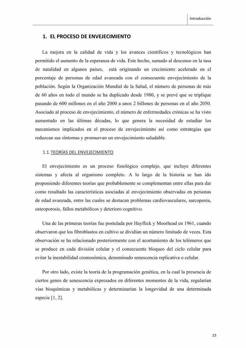
Otra de las posibles teorías, es la afectación del sistema neuroendocrino. Esta teoría
se basa en la desregulación por parte del hipotálamo de la secreción hormonal y la
disminución en la sensibilidad de respuesta del eje HPA (hipotálamo-pituitario-adrenal)
a medida que avanza la edad [3, 4], lo cual se ha relacionado con un incremento en los
niveles de cortisol [5] que desencadenaría una respuesta fisiológica en forma de cascada
(Fig 1).
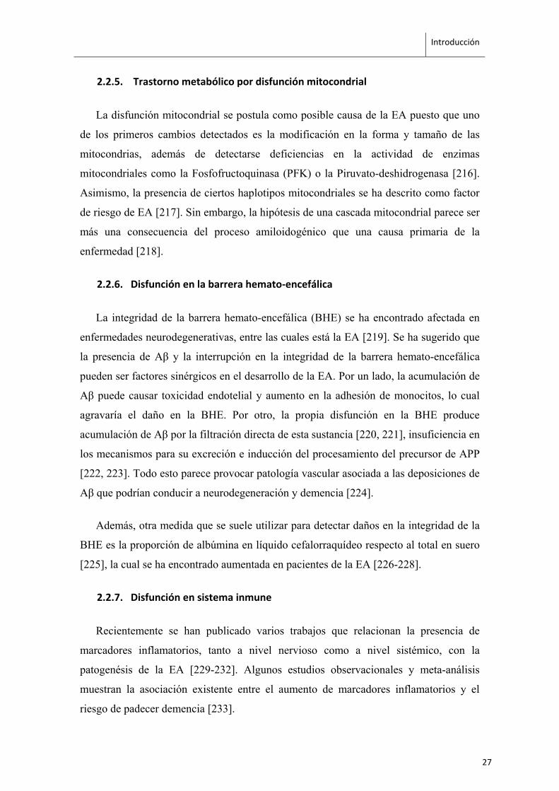
Figura 1. Posibles secuelas clínicas y resultados adversos en diversos sistemas del cuerpo, de la excesiva activación del eje HPA e incremento de cortisol. (Figura traducida de Veldhuis et al. [6])
Una de las teorías más conocidas y estudiadas, es la que identifica la acumulación de
radicales libres, producidos por el metabolismo energético mitocondrial, como
principales causantes de toxicidad y daño celular (stress-induced theory) [7]. Se ha visto
que los radicales libres pueden provocar alteraciones en la secuencia del ADN [8], en la
estructura de membranas celulares por la afectación en lípidos [9], y acumular daños en
el ADN mitocondrial, lo cual afecta a la cadena de transporte de electrones y puede
reducir la eficiencia energética celular [10-12].
Por último, una mayor sensibilización de las membranas a la peroxidación lipídica y
el aumento en el grado de insaturación de ácidos grasos de membrana también se ha
propuesto como mecanismo determinante del proceso de envejecimiento y longevidad
en diferentes especies como C.elegans, D.melanogaster, roedores e incluso en humanos
[13, 14].La composición de membrana de las diferentes especies estudiadas muestra un
16
Introducción
promedio similar en la longitud de la cadena de los ácidos grasos (18 átomos de
carbono), en la proporción de ácidos grasos saturados e insaturados (40:60) y en la
distribución de fosfolípidos [15]. Sin embargo, los cambios en la resistencia a la
peroxidación lipídica hallados en los organismos más longevos parecen estar
determinados por la reducción en la actividad de desaturasas que conlleva a cambios en
el tipo de ácidos grasos insaturados presentes en la membrana [16].
1.2. CARACTERÍSTICAS DEL PROCESO DE ENVEJECIMIENTO
En 1962, B. Strehler [17] propuso cinco criterios para definir las características del
envejecimiento que se siguen utilizando como guía para la identificación de
biomarcadores de senescencia. Dichos criterios implican que para que un proceso sea
considerado parte del envejecimiento, las alteraciones que provoca deben ser
acumulativas, progresivas, intrínsecas, deletéreas para las funciones biológicas y
universales (observadas en todos los miembros de la especie). El proceso de
envejecimiento se desarrolla de manera progresiva durante el ciclo vital del individuo y
la afectación de órganos y sistemas incluye varios fenómenos moleculares y fisiológicos
a la vez influenciados por factores genéticos y ambientales.
1.2.1. Estrés oxidativo
El estrés oxidativo se define como el desequilibrio entre las especies reactivas de
oxígeno (reactive oxygen species, ROS) y las defensas antioxidantes del organismo
(como la catalasa, GSH-peroxidasas y reductasas o la SOD, superóxido dismutasa) en
favor de los factores oxidantes. Las ROS interactúan con biomoléculas celulares como
los fosfolípidos, proteínas y ácidos nucleicos, afectando a su estructura y funcionalidad
y provocando fallos que pueden desencadenar en daños tisulares y celulares.
La producción de ROS se da principalmente durante el proceso de transformación de
NADH o succinato en ATP. A lo largo de la cadena respiratoria mitocondrial, en
concreto en los complejos I y III [18, 19], los electrones derivados de la fosforilación
oxidativa pueden reaccionar con el oxígeno (O2) u otros aceptores de electrones,
generando radicales libres [20], lo cual se ha correlacionado negativamente con la
longevidad en diferentes especies [21]. Las ROS también se producen en otros
compartimentos celulares, como los microsomas, peroxisomas o en las oxidasas
17
Introducción
dependientes de NADPH ligadas a membrana. Sin embargo, las mitocondrias son
especialmente sensibles a las alteraciones asociadas al envejecimiento especialmente
debido a los efectos de las ROS sobre el ADN mitocondrial [22, 23].
Una de las observaciones que apoyan este hecho, es que los errores en ADN
mitocondrial se acumulan con la edad. Esto viene dado tanto por errores de replicación
como por fallos en la reparación. Es en este último mecanismo donde se ha sugerido
que las ROS tienen un papel relevante, ya que pueden producir rotura de simple o doble
cadena en el ADN, dando como resultado mutaciones puntuales e incluso deleciones de
fragmentos de ADN mitocondrial que pueden producir disfunciones, como se ha
observado en cerebro, músculo esquelético y corazón [24].
1.2.2. Inflamación
Una de las principales características del proceso de envejecimiento, es la reducción
global en la respuesta a factores de estrés acompañada de un incremento progresivo en
el estado proinflamatorio, recientemente denominado como inflammaging [25]. La
inmunosenescencia parece estar caracterizada por una reorganización funcional del
sistema immune donde algunos mecanismos disminuyen su eficiencia y otros
permanecen no alterados o incuso más activos [26, 27].
Los niveles de citoquinas proinflamatorias como la interleucina (IL)-6, IL-1β, TNFα
(tumor necrosis factor) y TGFβ (tumor growth factor) [28-30] se ven alterados durante
el envejecimiento. Asimismo, se observa una afectación en la respuesta inmunitaria
caracterizada por una reducción del número de células B maduras, menor afinidad de
los anticuerpos producidos [31], un aumento en la proporción de células B de memoria
[32-34], una reducción del repertorio de células T y CD4+ helper [35, 36] y un
desequilibrio hacia células CD8+, las cuales son productoras de citoquinas
proinflamatorias [37-39]. Un sistema inmunitario poco eficiente a su vez propicia el
desarrollo de estados de inflamación crónica.
18
Introducción
1.2.3. Senescencia celular
La senescencia celular es un proceso de respuesta frente a estímulos estresantes o
daños ocurridos en la célula, que provoca el bloqueo del ciclo y división celular [40-42],
pudiendo afectar al potencial regenerativo del tejido [43, 44]. Los mecanismos
subyacentes de este proceso incluyen la regulación paracrina por parte de las células
senescentes hacia células sanas de su alrededor mediante la secreción de IL-1β o TGFβ
[45, 46], la pérdida de estructura y organización por la acción de las proteasas
secretadas por las células senescentes [47] y el aumento del ambiente inflamatorio por
la secreción de factores de crecimiento, citoquinas y quemocinas proinflamatorias como
IL-1, IL-6, MIPs (macrophage inflammatory proteins) y MCPs (monocyte chemo-
attractant proteins) [41, 48].
Existen varios factores desencadenantes de senescencia celular entre los cuales se
incluyen cambios de expresión de genes supresores de tumores [49, 50], cambios
metabólicos [51, 52] o remodelación de la cromatina inducida por factores epigenéticos
[53, 54], entre otros. La vía de señalización más conocida por la cual las células dejan
de dividirse es mediante la activación de la respuesta a daños en ADN (DDR). En esta
vía, diferentes tipos de quinasas bloquean la progresión del ciclo celular mediante la
estabilización de p53 y la activación transcripcional de la quinasa dependiente de ciclina
p21 [55, 56]. También se ha observado senescencia celular por vías independientes a
DDR, las cuales implican p19Arf y p16Ink4a [49, 57] o la activación de la deshidrogenasa
de piruvato mitocondrial (PDH) [58]. Además, las vías efectoras de p53–p21 y p16Ink4a–
RB, pueden actuar conjuntamente en el mantenimiento de procesos de senescencia [59,
60].
1.2.4. Acortamiento de telómeros
Los telómeros son estructuras de heterocromatina situados en la parte final de los
cromosomas, cuya función es protegerlos frente a mecanismos de reparación y
degradación [61, 62]. Están formados por repeticiones en tándem de la secuencia de
nucleótidos TTAGGG y un complejo compuesto por 6 proteínas llamado telosoma o
shelterina [63-65]. Estas proteínas son esenciales ya que el silenciamiento de la mayoría
de ellas es letal en las primeras etapas embrionarias [66-68]. La enzima responsable de
19
Introducción
la síntesis de telómeros es la telomerasa, formada por un componente de ARN (TERC)
y una subunidad catalítica proteica con actividad de transcriptasa reversa (TERT) [69].
Tras cada división celular, los telómeros se acortan por la incapacidad de las
polimerasas de ADN de replicar completamente los extremos de las moléculas de ADN
linear [70]. El acortamiento telomérico causa pérdida en la funcionalidad de las células
madre de los tejidos, afectando a su renovación y funcionalidad [71-73]. Paralelamente,
también se activa la maquinaria de respuesta a daños en ADN (DDR) que reconoce el
final de los cromosomas como roturas de doble cadena induciendo procesos de
senescencia celular [74, 75]. Se ha asociado el acortamiento de telómeros con
enfermedades como el síndrome de Zinsser-Cole-Engman o disqueratosis congénita, el
síndrome de Werner, síndrome de Bloom, ataxia–telangiectasia, anemia de Fanconi
(FA) o fibrosis pulmonar, entre otros [76, 77]. También se han descrito evidencias de un
acortamiento telómerico en enfermedades crónicas frecuentes como varios tipos de
cancer [78-80], hepatitis crónica [81, 82], declive cognitivo [83] y la enfermedad de
Alzheimer (EA) [84], y en personas con problemas cardiovasculares e hipertensión [85-
89] y diabetes tipo II [90, 91].
Por otro lado también se ha observado que ciertos hábitos de vida pueden afectar a la
longitud telomérica. Factores como el tabaco [92-94], el consumo de carne procesada
[95] y un elevado índice de masa corporal (BMI) [96-98] se han asociado con el
acortamiento en la longitud telomérica. Otros en cambio, como elevados niveles de
vitamina D [99], folato [100] o ácidos grasos como el omega-3 [101] muestran relación
con mayores niveles de longitud telomérica. El ejercicio físico [102, 103], la reducción
de estrés mediante la meditación [104] o cambios en varios patrones de estilo de vida
[105] también provocan un descenso en el ratio de acortamiento telomérico, sugiriendo
que el efecto de este tipo de intervenciones puede ser de gran importancia en la
prevención de los efectos perjudiciales asociados al envejecimiento.
20
Introducción
2. ENVEJECIMIENTO Y NEURODEGENERACIÓN: LA ENFERMEDAD DE
ALZHEIMER
Las células del sistema nervioso, al igual que las del resto del cuerpo, están
afectadas por el proceso de envejecimiento fisiológico. Sin embargo, existe variabilidad
interindividual en este proceso. El equilibrio entre los sistemas neuronales afectados y la
eficacia de los mecanismos de reparación y compensatorios, es lo que determina la
aparición de procesos patológicos que pueden dar como resultado declive cognitivo y
neurodegeneración.
Las células del sistema nervioso, debido a su mofología y fisiología, son
especialmente vulnerables al estrés oxidativo [106, 107], alteraciones en la homeostasis
energética [108], acumulación de daños [109, 110] y lesiones en ácidos nucleicos [111,
112], lo cual provoca que sean más susceptibles al envejecimiento. Además,
modificaciones postranslacionales en proteínas neuronales como la carbonilación, la
nitración, y la unión covalente de productos de peroxidación lipídica [113], se dan
durante el proceso de envejecimiento y de forma más acusada en personas afectadas por
la enfermedad de Alzheimer [114].
2.1. LA ENFERMEDAD DE ALZHEIMER
La enfermedad de Alzheimer (EA) fue descrita por primera vez por Alois Alzheimer
en 1907 [115] y en la sociedad actual es el tipo de demencia más frecuente,
comprendiendo el 60-90% de los casos [116]. Se trata de un desorden multifactorial en
el cual tanto factores genéticos como ambientales interaccionan produciendo declive
cognitivo y neurodegeneración.
Existen dos variantes de la enfermedad: la forma familiar o de aparición temprana, y
la forma esporádica o de aparición tardía. La forma familiar se manifiesta antes de los
65 años, mientras que la esporádica suele aparecer a partir de los 60-65 años [114]. Sin
embargo, ambas formas comparten ciertos rasgos que se han determinado como
características clínico-patológicas que definen la enfermedad [117-123]:
‐ Placas de beta-amieloide (Aβ): El péptido beta-amiloide fue descubierto en 1984
por Glenner y se origina a partir del procesamiento de la proteína precursora
21
Introducción
amiloide (APP) mediante distintas proteasas que actúan de forma secuencial
(alfa-, beta- y gamma-secretasas). La APP es una proteína transmembranal con
un extremo amino-terminal situado hacia el exterior de la célula y un extremo
carboxi-terminal situado hacia el citoplasma. Se localiza principalmente en las
sinapsis entre neuronas y podría actuar como regulador en la formación de las
mismas y en plasticidad sináptica [124, 125]. Se distinguen dos vías principales
de procesamiento de la APP:
a- Vía no-amiloidogénica: en esta vía una alfa-secretasa de la familia de las
metaloprotesasas (ADAM10 de manera constitutiva y ADAM17 de manera
regulada) corta la APP en el aminoácido 687 liberando la parte extracelular
de la APP en forma de péptido soluble.
b- Vía amiloidogénica: en este caso la primera lisis la lleva a cabo una beta-
secretasa (beta-site APP cleaving enzyme, BACE) en el aminoácido 671,
liberando una porción extracelular soluble de menor tamaño.
Seguidamente, y de manera común a ambas vías, actúa una gamma-secretasa
(presenilina, PSEN) que procesa el fragmento anclado a la membrana de la APP
en los aminoácidos 712, 714 o 715. En el caso de la vía no-amiloidogénica se
libera el péptido p83. En cambio, en la vía amiloidogénica el fragmento
generado es un péptido de 40, 42 o 43 aminoácidos, denominado beta-amiloide
(Aβ), el cual es insoluble y forma agregados que componen las placas neuríticas
presentes en la EA. Finalmente también es liberada la región intracelular del
APP (APP intra-cellular domain, AICD).
‐ Ovillos neurofibrilares de proteína Tau: la proteína Tau pertenece a la familia de
proteínas MAP (proteínas asociadas a microtúbulos) y se une a microtúbulos de
citoesqueleto estabilizándolos. Las proteínas Tau se caracterizan por tener
facilidad en fosforilarse, permitiendo así su movilidad dentro de los axones de
las neuronas. En la enfermedad de Alzheimer y otras tauopatías se produce un
fenómeno de hiperfosforilización que es el responsable de la formación
intracelular de ovillos de neurofibrillas helicoidales, los cuales comprometen la
capacidad de la neurona de transmitir mensajes nerviosos y pueden conducir
finalmente a muerte neuronal y neurodegeneración [126, 127].
22
Introducción
‐ Declive cognitivo y conductual progresivo: en un inicio se ve afectada la
memoria a corto plazo, dificultad en la realización de tareas simultáneas o en la
resolución de problemas. A medida que la enfermedad avanza, se pueden
observar cambios en el patrón de sueño, trastornos del lenguaje, irritabilidad y
cambios de humor y afectación en la memoria a largo plazo. La pérdida
neuronal puede llegar al punto en el que no se mantengan las funciones
esenciales y desencadenar en la muerte del individuo [128-130].
2.2. FACTORES DE RIESGO Y PROCESOS IMPLICADOS EN LA APARICIÓN DE LA
ENFERMEDAD DE ALZHEIMER
2.2.1. Factores genéticos
Son los principales causantes de la forma familiar de la EA. Las mutaciones más
descritas y estudiadas afectan a los genes que codifican para el APP y presenilinas 1 y 2
y se transmiten con carácter mendeliano autosómico dominante. Representan
aproximadamente el 5% de los casos de EA [116].
El gen APP contiene 19 exones, la combinación de los cuales puede dar lugar hasta
8 isoformas de la proteína. La isoforma predominante en tejido nervioso es la APP695,
la cual es posteriormente procesada por alfa-, beta- y gamma-secretasas. La lisis
mediante α-secretasas produce un péptido que no está presente en las placas amieloides,
en cambio β- y γ-secretasas dan lugar al péptido β-amieloide [131]. Según la hipótesis
de la cascada amieloide, la deposición de péptidos Aβ42 se da en las fases iniciales de la
enfermedad, dando lugar a la formación de placas seniles y ovillos neurofibrilares, los
cuales atraen proteínas adicionales como la APOE, ubiquitinas, o proteínas del
complemento que promueven la muerte celular y demencia [118, 132].
Se han identificado varias mutaciones en el gen que codifica para APP [133] siendo
la V171 una de las más estudiadas ya que se estima que incrementa de 1.5 a 1.9 veces la
frecuencia de producción de fragmentos Aβ de mayor tamaño [134].
Recientemente, se ha descrito una mutación menos frecuente en el gen de APP
(A673T) que parece ser protectora para la EA. La mutación afecta a la región sobre la
23
Introducción
cual actuan las β-secretasas, reduciendo en un 40% la formación de péptidos
amiloidogénicos in vitro [135].
Por otro lado, las presenilinas (PSEN) 1 y 2 forman parte del complejo responsable
de la actividad γ-secretasa [136]. Todas las mutaciones que afectan al gen de PSEN1
aumentan la actividad γ-secretasa, incrementando también la producción de Aβ42 [137-
139]. Las mutaciones en el gen PSEN2 son menos frecuentes pero actúan al mismo
nivel que las de PSEN1, estimulando la actividad γ-secretasa [140].
En cuanto a la forma espóradica de la EA, la variación alélica en el gen APOE-ε4
fue la primera variante que se estableció como marcador de riesgo [141] ya que aumenta
el riesgo de dos a tres veces cuando se presenta en heterocigosis y hasta 15 veces en
homocigosis [142]. Sin embargo su frecuencia en la población se estima que ronda el
27% [143].
Otros genes recientemente identificados como posibles marcadores asociados a la
EA son las variantes en los genes clusterin (CLU), complement receptor 1 (CR1) y
phosphatidylinositol binding clathrin assembly protein (PICALM) [144].
2.2.2. Factores ambientales
La teoría que relaciona la exposición a metales como el aluminio con la aparición de
la EA ha sido descartada por una gran parte de la comunidad científica debido a la
inconsistencia de los trabajos previos que mostraban una relación con la patología
[145], la ausencia de efectos adversos a nivel cognitivo ante la exposición a aluminio
demostrada en algunos estudios [146-150], las diferencias toxicocinéticas existentes
entre los modelos animales y los humanos, y por último, la teoría de que los efectos de
la exposición al aluminio influyen en la progresión de la patología pero no en la
causalidad de la misma, ya que los factores y procesos determinantes en la aparición de
la EA se dan varios años antes de la detección de los síntomas clínicos.
Sin embargo, algunos autores siguen trabajando en esta hipótesis y han demostrado
la presencia de ciertas características en modelos celulares y animales y mediante
estudios epidemiológicos que sugieren una posible correlación con la EA [151, 152].
Entre ellas cabe destacar la acumulación de agregados amieloides [153-157]; la
24
Introducción
activación de la vía pro-inflamatoria del factor NF-κB [158, 159]; la presencia de
muchos de los déficits observados en la EA tras la exposición a aluminio [160-163]; la
relación entre la cantidad de aluminio ingerido en el agua y la incidencia de EA [159,
164]; y finalmente la utilización de la deferoxamina, antioxidante y quelante de iones
trivalentes de hierro y aluminio, como una de las estrategias terapéuticas más efectivas
para la EA [165, 166].
Otro de los determinantes ambientales que se han relacionado con la EA es la
presencia de traumatismos craneoencefálicos que frecuentemente conllevan la extensión
del daño producido por la liberación de citoquinas inflamatorias y la activación de
microglia [167]. Por ejemplo, se ha asociado la producción de APP como respuesta
cerebral al daño neuronal estableciendo una posible modulación en la progresión de la
EA [168-170].
Por último, se ha visto que déficits nutritivos y metabólicos afectan a procesos de
homeostasis neuronal y sináptica [171, 172], siendo así factores potencialmente
implicados en la aparición y/o progresión de la EA [173-176]. Tanto el índice de masa
corporal [177, 178], el consumo de antioxidantes [179-181], los niveles de vitamina
B12 y homocisteina [182-184], de vitamina D [185, 186], y de ácidos grasos [187-190]
se han asociado con el riesgo de padecer declive cognitivo y EA.
2.2.3. Envejecimiento acelerado
Esta teoría se basa en el hecho de que los cambios patológicos característicos de la
EA se presentan también en el proceso natural de envejecimiento fisiológico, con la
única diferencia del grado de severidad en cada caso [191]. En individuos que no
presentan declive cognitivo, se ha observado una reducción del volumen y peso del
cerebro, así como pérdida de sinapsis y dendritas en áreas específicas durante el proceso
de envejecimiento fisiológico [192]. Por otro lado, el principal componente molecular
de las placas seniles es el péptido β-amieloide [193], producto de la escisión de la
proteína precursora amieloide (APP) [194] que también se encuentra presente en
individuos sanos mayores de 60 años [195]. Sin embargo, la presencia de ovillos
neurofibrilares durante el envejecimiento fisiológico es un tema que aún genera
controversia [196-199]. Otros procesos presentes durante el envejecimiento fisiológico
25
Introducción
que parecen estar implicados en la aparición de la EA son la descomposición de la
mielina [200] y la muerte celular en el locus caeruleus el cual es el encargado de
proporcionar noradrenalina al cortex y estimular la microglia para suprimir la
producción de Aβ [201, 202].
En general, los estudios realizados hasta ahora indican que las diferencias entre un
envejecimiento normal y la EA son de carácter cuantitativo más que cualitativo [203].
No obstante, la distribución de los rasgos patológicos parece diferir entre los individuos
control y los pacientes de EA, siendo más localizada en áreas del lóbulo temporal
durante el envejecimiento fisiológico, y propagándose a zonas de hipocampo y áreas de
asociación cortical en la EA [204, 205].
2.2.4. Degeneración y desconexión de redes neuronales
Se basa en definir la EA como un síndrome de desconexión caracterizado por la
desorganización de las conexiones aferentes y eferentes entre el hipocampo, la corteza
cerebral y el resto del cerebro [204, 206-209]. Esta teoría se fundamenta en el hecho de
que los principales marcadores moleculares de la enfermedad (en especial los ovillos
neurofibrilares) se concentran en zonas cerebrales donde se producen las conexiones
corticocorticales, como son las neuronas piramidales en capas III y V de áreas temporal,
parietal y en lóbulo frontal. Por otro lado, se ha observado mayor vulnerabilidad a los
procesos de desconexión en corteza entorrinal, área CA1 hipocampal y la amígdala,
mientras que las zonas sensoriales primarias parecen ser las menos afectadas [209], y la
función de la denominada Default mode network (DMN) también se ha descrito dañada
en pacientes afectados por la EA [210].
Además, las diferentes técnicas de neuroimagen (electroencefalografía, EEG;
tomografía de emisión de positones, PET; y resonancia magnética funcional, fMRI); así
como estudios neuropsicológicos con tareas que requieren la integridad de las
conexiones entre hemisferios y dentro de cada hemisferio por separado, han apoyado la
hipótesis de desconexión tanto entre zonas anteriores y posteriores, como entre
hemisferios [211-215].
26
Introducción
2.2.5. Trastorno metabólico por disfunción mitocondrial
La disfunción mitocondrial se postula como posible causa de la EA puesto que uno
de los primeros cambios detectados es la modificación en la forma y tamaño de las
mitocondrias, además de detectarse deficiencias en la actividad de enzimas
mitocondriales como la Fosfofructoquinasa (PFK) o la Piruvato-deshidrogenasa [216].
Asimismo, la presencia de ciertos haplotipos mitocondriales se ha descrito como factor
de riesgo de EA [217]. Sin embargo, la hipótesis de una cascada mitocondrial parece ser
más una consecuencia del proceso amiloidogénico que una causa primaria de la
enfermedad [218].
2.2.6. Disfunción en la barrera hemato‐encefálica
La integridad de la barrera hemato-encefálica (BHE) se ha encontrado afectada en
enfermedades neurodegenerativas, entre las cuales está la EA [219]. Se ha sugerido que
la presencia de Aβ y la interrupción en la integridad de la barrera hemato-encefálica
pueden ser factores sinérgicos en el desarrollo de la EA. Por un lado, la acumulación de
Aβ puede causar toxicidad endotelial y aumento en la adhesión de monocitos, lo cual
agravaría el daño en la BHE. Por otro, la propia disfunción en la BHE produce
acumulación de Aβ por la filtración directa de esta sustancia [220, 221], insuficiencia en
los mecanismos para su excreción e inducción del procesamiento del precursor de APP
[222, 223]. Todo esto parece provocar patología vascular asociada a las deposiciones de
Aβ que podrían conducir a neurodegeneración y demencia [224].
Además, otra medida que se suele utilizar para detectar daños en la integridad de la
BHE es la proporción de albúmina en líquido cefalorraquídeo respecto al total en suero
[225], la cual se ha encontrado aumentada en pacientes de la EA [226-228].
2.2.7. Disfunción en sistema inmune
Recientemente se han publicado varios trabajos que relacionan la presencia de
marcadores inflamatorios, tanto a nivel nervioso como a nivel sistémico, con la
patogenésis de la EA [229-232]. Algunos estudios observacionales y meta-análisis
muestran la asociación existente entre el aumento de marcadores inflamatorios y el
riesgo de padecer demencia [233].
27
Introducción
Se ha descrito que la activación de la microglia en cerebros de EA se asocia al
depósito de Aβ [234]. Alrededor de las placas de amiloide en cerebros con EA se han
detectado agrupaciones de microglia y astrocitos activados [235, 236], y una
sobreexpresión de una variedad de citoquinas inflamatorias como la IL-1 [237], IL-6
[238], TNF-α [239] o TGF-β [240].
También se ha relacionado la EA con patologías caracterizadas por procesos de
inflamación crónica y factores de riesgo vascular como la hipertensión, la diabetes
mellitus o la arterioesclerosis [241-244]. Se ha sugerido que la presencia simultánea de
disfunción cerebrovascular y la EA [245-248] puede conllevar efectos sinérgicos en la
progresión de la demencia [249, 250].
2.2.8. Agentes infecciosos
Se ha establecido que la presencia de virus del herpes simple tipo 1 (HSV1) es más
frecuente en ancianos que en personas jóvenes y de mediana edad [251, 252],
probablemente como consecuencia del declive en sistema inmune durante el
envejecimiento. De hecho se ha demostrado que el virus HSV1 puede activarse en el
cerebro de manera periódica y recurrente en periodos de estrés, o de cierta
inmunodeficiencia, causando muerte neuronal [253, 254] y en presencia de APOE
puede facilitar la acumulación de Aβ y Tau-fosforilada [255, 256].
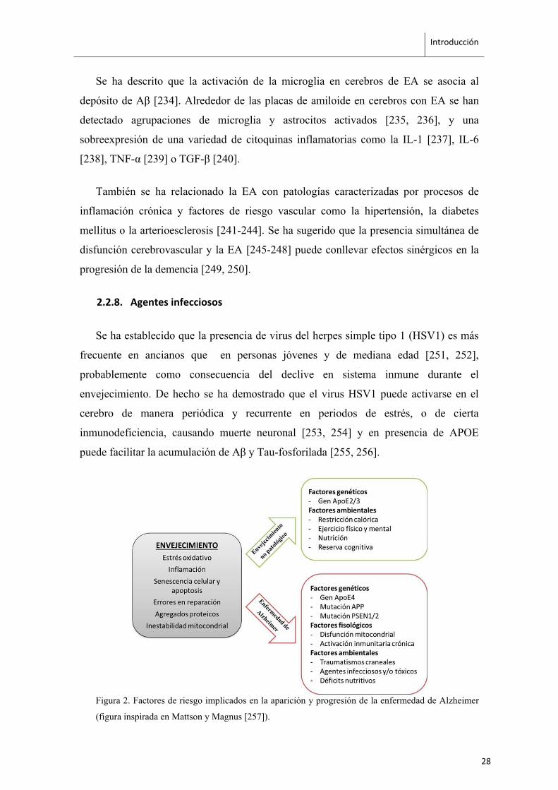
Figura 2. Factores de riesgo implicados en la aparición y progresión de la enfermedad de Alzheimer
(figura inspirada en Mattson y Magnus [257]).
28
Introducción
2.3. LOS RATONES SAMP8: UN MODELO DE ENVEJECIMIENTO ACELERADO Y
ENFERMEDAD DE ALZHEIMER
Los ratones SAMP8 (Senescence-Accelerated Mouse Prone 8) son un modelo
murino de envejecimiento acelerado que fue seleccionado fenotípicamente a partir de la
cepa AKR/J por el Dr. Takeda y sus colaboradores en la Universidad de Kyoto [258].
Durante el mantenimiento de la colonia, observaron que ciertos animales presentaban
signos propios de un proceso de envejecimiento acelerado, como son un descenso en la
actividad motora, pérdida y empeoramiento en el aspecto del pelo, lordosis y un
acortamiento significativo de la longevidad. Mediante cruzamiento entre hermanos
establecieron nuevas series de animales (series P) que mantenían estos rasgos de
generación en generación, mientras que los cruzamientos entre individuos
aparentemente sanos establecieron las series R (resistentes). Basandose en los fenotipos
patológicos que expresaban y su longevidad establecieron diferentes líneas P y R.
Los ratones SAMP8 presentan de manera prematura los rasgos propios de un
proceso de envejecimiento además de un acortamiento significativo de su esperanza de
vida. A nivel cognitivo, los ratones SAMP8 muestran déficits en aprendizaje y memoria
espacial [259-261], deterioro en la adquisición y ejecución de tareas de evitación pasiva
y activa [262, 263], de condicionamiento por miedo (fear conditioning) [264] y de
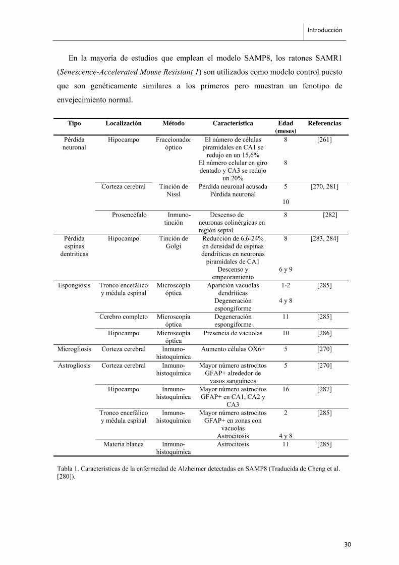
reconocimiento de objetos [265, 266]. A nivel histológico, los ratones SAMP8
presentan pérdida neuronal, reducción en la densidad de espinas dendríticas, patrones de
degeneración espongiforme, microgliosis y astrogliosis en hipocampo y otras regiones
cerebrales (Tabla 1). También se han detectado en cerebro de ratones SAMP8 la
acumulación de Aβ [267-269] y la hiperfosforilación de Tau [270, 271]. Además, los
ratones SAMP8 presentan alteraciones en la barrera hematoencefálica, reducción de la
población de células de Purkinje en cerebelo [272] y aumento de estrés oxidativo por
disfunción mitocondrial [270, 273-276], alteraciones de los ritmos circadianos y
aumento en marcadores inflamatorios [276, 277]. Por todo esto, los ratones SAMP8 se
consideran un buen modelo de envejecimiento acelerado y de enfermedad de Alzheimer
[278-280].
29
Introducción
En la mayoría de estudios que emplean el modelo SAMP8, los ratones SAMR1
(Senescence-Accelerated Mouse Resistant 1) son utilizados como modelo control puesto
que son genéticamente similares a los primeros pero muestran un fenotipo de
envejecimiento normal.
Tipo Localización Método Característica Edad (meses)
Referencias
Pérdida neuronal
Hipocampo
Fraccionador óptico
El número de células piramidales en CA1 se
redujo en un 15,6% El número celular en giro dentado y CA3 se redujo
un 20%
8
8
[261]
Corteza cerebral Tinción de Nissl
Pérdida neuronal acusada Pérdida neuronal
5
10
[270, 281]
Prosencéfalo Inmuno-tinción
Descenso de neuronas colinérgicas en región septal
8 [282]
Pérdida espinas
dentriticas
Hipocampo Tinción de Golgi
Reducción de 6,6-24% en densidad de espinas dendríticas en neuronas
piramidales de CA1 Descenso y
empeoramiento
8
6 y 9
[283, 284]
Espongiosis Tronco encefálico y médula espinal
Microscopía óptica
Aparición vacuolas dendríticas
Degeneración espongiforme
1-2
4 y 8
[285]
Cerebro completo Microscopía óptica
Degeneración espongiforme
11 [285]
Hipocampo Microscopía óptica
Presencia de vacuolas 10 [286]
Microgliosis Corteza cerebral Inmuno-histoquímica
Aumento células OX6+ 5 [270]
Astrogliosis Corteza cerebral Inmuno-histoquímica
Mayor número astrocitos GFAP+ alrededor de
vasos sanguíneos
5 [270]
Hipocampo Inmuno-histoquímica
Mayor número astrocitos GFAP+ en CA1, CA2 y
CA3
16 [287]
Tronco encefálico y médula espinal
Inmuno-histoquímica
Mayor número astrocitos GFAP+ en zonas con
vacuolas Astrocitosis
2
4 y 8
[285]
Materia blanca Inmuno-histoquímica
Astrocitosis 11 [285]
Tabla 1. Características de la enfermedad de Alzheimer detectadas en SAMP8 (Traducida de Cheng et al. [280]).
30
Introducción
3. EPIGENÉTICA Y ENVEJECIMIENTO
3.1. DEFINICIÓN Y MECANISMOS
La epigenética es la ciencia que estudia los mecanismos implicados en la regulación
de la expresión génica, de manera estable pero potencialmente reversible y sin que
conlleve alteración de la secuencia de nucleótidos del ADN [288]. Estos procesos se
pueden dar directamente sobre las moléculas de ADN, como en el caso de la metilación
de ADN; sobre las proteínas implicadas en el empaquetamiento de la cromatina, como
la modificación de histonas; o sobre la funcionalidad del ARN mensajero producido,
mediante los llamados ARNs no codificantes entre los cuales se incluyen los
microARNs.
3.1.1 Metilación del ADN
El ADN de los vertebrados sufre modificaciones covalentes que consisten en la
adición de grupos metilos en los nucleótidos de citosina, principalmente en los pares de
bases con secuencia CpG [289, 290]. En menor medida, se ha descrito también la
metilación del ADN en pares de bases con secuencias CpA y CpT, en plantas [291], en
células madre embrionarias en Drosophila [292] y estadíos iniciales del desarrollo
embrionario de ratón [293].
El radical metilo de la metilcitosina reside y ejerce su función en el surco mayor de
la hélice de ADN, lugar donde la mayoría de las proteínas se unen. Existe una familia de
proteínas que se unen específicamente al ADN metilado, son las Methyl-CpG-binding
proteins y reclutan complejos represores en las regiones promotoras de los genes,
contribuyendo al silenciamiento transcripcional. Ciertos factores de transcripción se
unen a las secuencias de ADN que contienen secuencias CpG no-metiladas, de manera
que la metilación de ADN en esos casos previene la unión de proteínas, inhibiendo
también la transcripción. En algunos casos, como el del gen IGF2 (insulin-like gowth
factor 2) silenciado en el alelo materno durante el desarrollo, la metilación del promotor
activa la transcripción ya que permite la interacción entre el gen y sus potenciadores
(enhancers) [294].
31
Introducción
En efecto, estudios moleculares y genéticos han mostrado que la metilación del
ADN está asociada con silenciamiento génico y ejerce un papel fundamental en
procesos de desarrollo embrionario como la inactivación del cromosoma X en hembras
o los mecanismos de imprinting que definen qué alelo, materno o paterno, será el que
se exprese en el embrión. Además la metilación del ADN también ejerce un papel en el
silenciamiento de retrotransposones endógenos y en el establecimiento de los patrones
de expresión génica específicos de tejido.
Aunque los patrones de metilación del ADN se transmiten en la división celular, no
son permanentes. Se pueden producir cambios en respuesta a factores externos como el
entorno y también como consecuencia de procesos internos oncogénicos o de
envejecimiento celular. La enzima Dnmt1 tiene actividad ADN metiltransferasa y es la
responsable del mantenimiento del patrón de metilación en la división celular ya que se
une preferencialmente al ADN hemimetilado, en el cual sólo una de las cadenas de
ADN está metilada. La DNMT3A y DNMT3B son las metiltransferasas que se encargan
del establecimiento de patrones de metilación de novo. La inactivación de ambos genes,
DNMT3A y DNMT3B, es letal en las primeras etapas embrionarias.
El ADN en mamíferos esta metilado en un 70% de los pares CpG [295],
principalmente en los llamados ADN satélite, elementos repetitivos como transposones,
ADN presente entre genes y exones. Como excepción está el caso de las islas CpG, las
cuales contienen secuencias ricas en pares CpG y se suelen situar en los promotores
génicos. Su estado de metilación obedece a otro tipo de regulación, ya que es su
metilación la que determina la actividad transcripcional del gen.
La represión de la transcripción viene dada, tanto por la imposibilidad de los
factores de transcripción de unirse a la hebra de ADN como por la atracción de
complejos correpresores específicos de zonas metiladas. Existen varios ejemplos de
esto, la proteína MeCP2 reconoce y se une al ADN metilado y atrae a su vez la unión
del complejo corepressor mSin3a, dependiendo así de la deacetilación de histonas para
ello [296, 297]. MBD1 actua conjuntamente con la metiltransferasa SETDB1 durante la
replicación del ADN [298] lo que conlleva también a cambios de metilación de
histonas. MBD2 por su parte forma parte del complejo MeCP1 el cual incluye al
complejo correpresor NuRD que contiene diferentes deacetilasas de histonas y otras
32
Introducción
proteínas remodeladoras. Cada proteína de reconocimiento y unión a zonas metiladas de
ADN parece ser específica de secuencia ya que no se ha encontrado solapamiento ni
competición entre ellas [299]. Además, la metilación del ADN parece estar asociada con
otras marcas epigenéticas como la trimetilación en histona H3 lisina 9 (H3K9me3)
[300-302].
3.1.2 Modificaciones de histonas
El ADN, de aproximadamente unos dos metros de longitud es empaquetado en
apenas unas pocas micras en el núcleo de la célula. Para conseguir este nivel de
compactación, la doble hélice se pliega alrededor de los nucleosomas, unidad básica de
la cromatina [303]. Los nucleosomas están formados por un octámero de proteínas
compuesto por 4 pares de histonas (H2A, H2B, H3 y H4), alrededor de las cuales se
enrollan 147 pares de bases de ADN. Entre cada uno de los nucleosomas existe un
ADN espaciador, de entre 0 y 80 pares de bases que otorga flexibilidad a la fibra de
cromatina. Posteriormente, estos nucleosomas son empaquetados unos sobre otros
mediante la acción de la histona H1 formando lo que se conoce como "fibra de 30nm".
Los extremos N-terminales de las histonas son flexibles y quedan expuestos fuera
del nucleosoma donde pueden sufrir modificaciones covalentes en sus residuos por la
adición de grupos metilo o acetilo, entre otros. Estas modificaciones pueden actuar en
cis, afectando la conformación de la propia cromatina; o en trans, impidiendo la unión
de ciertos factores de transcripción o alterando los sitios de reconocimiento para atraer
otros efectores específicos. Todo ello da como resultado la activación o silenciamiento
de la expresión génica de la región donde se localizan.
3.1.2.1 Metilación
La metilación de histonas es un mecanismo complejo, ya que se puede dar tanto en
lisinas como en argininas y puede tener como resultado la activación o el silenciamiento
de la expresión génica dependiendo del tipo de histona, el aminoácido en que se
encuentre y la cantidad de grupos metilo que contenga. Las lisinas pueden estar mono-
(me1), di- (me2) o tri- (me3) metiladas, mientras que las argininas pueden estar mono-
(me1) o di- (me2) metiladas. Las enzimas implicadas en añadir grupos metilo a los
residuos de histonas son las metiltranfersas y las que los eliminan, las demetilasas, y
33
Introducción
ambos tipos son específicos del residuo que modifican. La metilación en lisinas (K) ha
sido ampliamente caracterizada en los residuos de histona H3 lisinas 4, 9, 27, 36 y 79 e
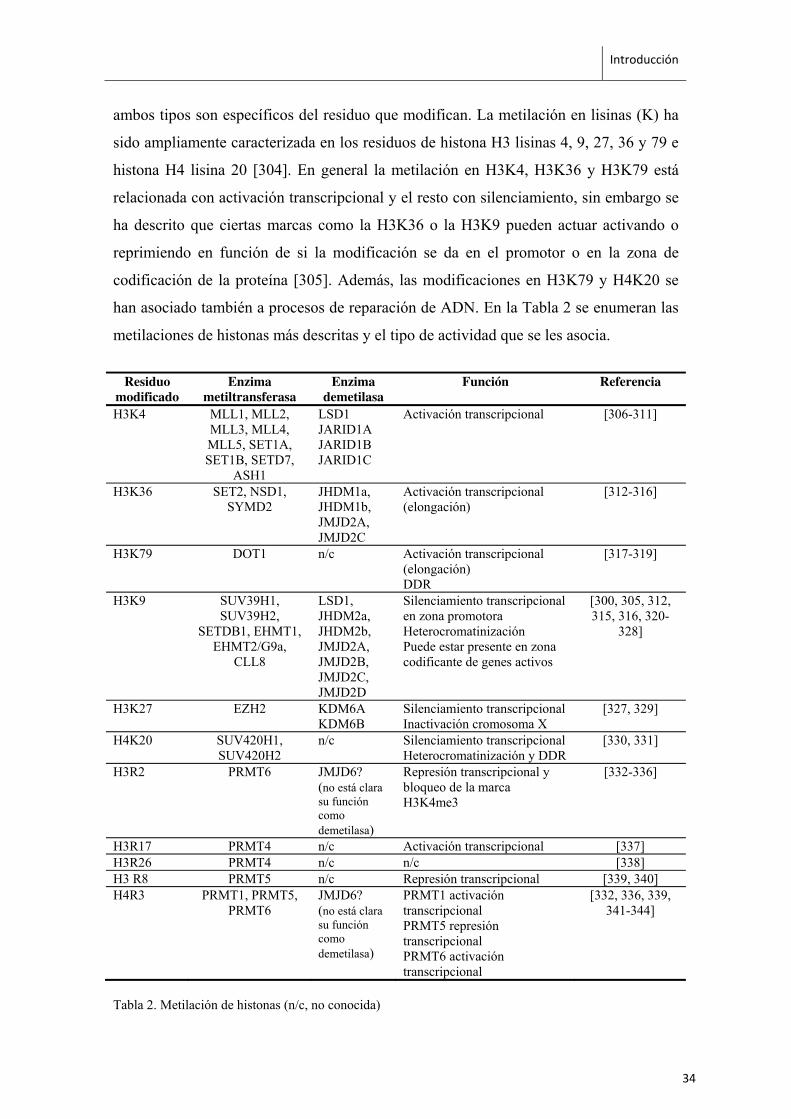
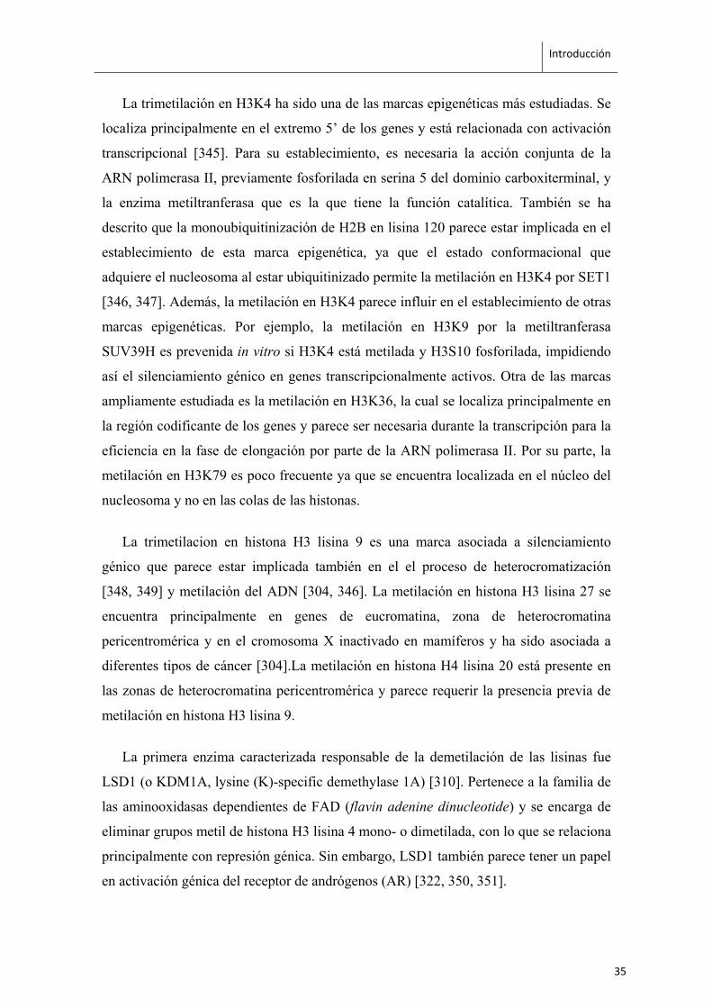
histona H4 lisina 20 [304]. En general la metilación en H3K4, H3K36 y H3K79 está
relacionada con activación transcripcional y el resto con silenciamiento, sin embargo se
ha descrito que ciertas marcas como la H3K36 o la H3K9 pueden actuar activando o
reprimiendo en función de si la modificación se da en el promotor o en la zona de
codificación de la proteína [305]. Además, las modificaciones en H3K79 y H4K20 se
han asociado también a procesos de reparación de ADN. En la Tabla 2 se enumeran las
metilaciones de histonas más descritas y el tipo de actividad que se les asocia.
Residuo modificado
Enzima metiltransferasa
Enzima demetilasa
Función Referencia
H3K4 MLL1, MLL2, MLL3, MLL4, MLL5, SET1A, SET1B, SETD7,
ASH1
LSD1 JARID1A JARID1B JARID1C
Activación transcripcional [306-311]
H3K36 SET2, NSD1, SYMD2
JHDM1a, JHDM1b, JMJD2A, JMJD2C
Activación transcripcional (elongación)
[312-316]
H3K79 DOT1 n/c Activación transcripcional (elongación) DDR
[317-319]
H3K9 SUV39H1, SUV39H2,
SETDB1, EHMT1, EHMT2/G9a,
CLL8
LSD1, JHDM2a, JHDM2b, JMJD2A, JMJD2B, JMJD2C, JMJD2D
Silenciamiento transcripcional en zona promotora Heterocromatinización Puede estar presente en zona codificante de genes activos
[300, 305, 312, 315, 316, 320-
328]
H3K27 EZH2 KDM6A KDM6B
Silenciamiento transcripcional Inactivación cromosoma X
[327, 329]
H4K20 SUV420H1, SUV420H2
n/c Silenciamiento transcripcional Heterocromatinización y DDR
[330, 331]
H3R2 PRMT6 JMJD6? (no está clara su función como demetilasa)
Represión transcripcional y bloqueo de la marca H3K4me3
[332-336]
H3R17 PRMT4 n/c Activación transcripcional [337] H3R26 PRMT4 n/c n/c [338] H3 R8 PRMT5 n/c Represión transcripcional [339, 340] H4R3 PRMT1, PRMT5,
PRMT6 JMJD6? (no está clara su función como demetilasa)
PRMT1 activación transcripcional PRMT5 represión transcripcional PRMT6 activación transcripcional
[332, 336, 339, 341-344]
Tabla 2. Metilación de histonas (n/c, no conocida)
34
Introducción
La trimetilación en H3K4 ha sido una de las marcas epigenéticas más estudiadas. Se
localiza principalmente en el extremo 5’ de los genes y está relacionada con activación
transcripcional [345]. Para su establecimiento, es necesaria la acción conjunta de la
ARN polimerasa II, previamente fosforilada en serina 5 del dominio carboxiterminal, y
la enzima metiltranferasa que es la que tiene la función catalítica. También se ha
descrito que la monoubiquitinización de H2B en lisina 120 parece estar implicada en el
establecimiento de esta marca epigenética, ya que el estado conformacional que
adquiere el nucleosoma al estar ubiquitinizado permite la metilación en H3K4 por SET1
[346, 347]. Además, la metilación en H3K4 parece influir en el establecimiento de otras
marcas epigenéticas. Por ejemplo, la metilación en H3K9 por la metiltranferasa
SUV39H es prevenida in vitro si H3K4 está metilada y H3S10 fosforilada, impidiendo
así el silenciamiento génico en genes transcripcionalmente activos. Otra de las marcas
ampliamente estudiada es la metilación en H3K36, la cual se localiza principalmente en
la región codificante de los genes y parece ser necesaria durante la transcripción para la
eficiencia en la fase de elongación por parte de la ARN polimerasa II. Por su parte, la
metilación en H3K79 es poco frecuente ya que se encuentra localizada en el núcleo del
nucleosoma y no en las colas de las histonas.
La trimetilacion en histona H3 lisina 9 es una marca asociada a silenciamiento
génico que parece estar implicada también en el el proceso de heterocromatización
[348, 349] y metilación del ADN [304, 346]. La metilación en histona H3 lisina 27 se
encuentra principalmente en genes de eucromatina, zona de heterocromatina
pericentromérica y en el cromosoma X inactivado en mamíferos y ha sido asociada a
diferentes tipos de cáncer [304].La metilación en histona H4 lisina 20 está presente en
las zonas de heterocromatina pericentromérica y parece requerir la presencia previa de
metilación en histona H3 lisina 9.
La primera enzima caracterizada responsable de la demetilación de las lisinas fue
LSD1 (o KDM1A, lysine (K)-specific demethylase 1A) [310]. Pertenece a la familia de
las aminooxidasas dependientes de FAD (flavin adenine dinucleotide) y se encarga de
eliminar grupos metil de histona H3 lisina 4 mono- o dimetilada, con lo que se relaciona
principalmente con represión génica. Sin embargo, LSD1 también parece tener un papel
en activación génica del receptor de andrógenos (AR) [322, 350, 351].
35
Introducción
Otro grupo de demetilasas comparten una estructura catalítica común denominada
JmjC-domain. A diferencia de LSD1, el mecanismo de acción de estas enzimas no
requiere de un grupo ε-amino susceptible de ser protonado. Por esto, pueden actuar en
todos los grados de metilación existentes (mono-, di- y trimetilación).
Por último, la metilación en argininas se ha relacionado con la regulación positiva y
negativa de la trascripción. Los residuos H4R3, H3R2, H3R17 y H3R26 [352, 353] se
han relacionado con la activación transcripcional y son captados por factores de
transcripción como p53 o NFκB. En cambio, H3R8 y H4R3 se asocian con represión
génica [339, 342].
3.1.2.2 Acetilación
La adición de grupos acetilos a los extremos expuestos de las histonas se suele
asociar con una activación transcripcional. Se considera que la neutralización de la
carga positiva de la lisina por la acetilación, reduce la fuerza de la unión entre las
histonas y el ADN, lo cual crea una relajación de la cromatina haciéndola más accesible
a los factores de transcripción [354]. Por otro lado, la acetilación en residuos de lisina es
una marca reconocida por los bromodominios presentes en proteínas de unión a la
cromatina [355] que a su vez, pueden formar parte de otros complejos remodeladores de
cromatina [356].
En general, los factores activadores de expresión génica reclutan histonas
acetiltransferasas (HAT) que incorporan grupos acetilos (Tabla 4), y los factores
represores atraen la unión de histonas deacetilasas (HDAC) que eliminan grupos
acetilos (Tabla 5).
36
Introducción
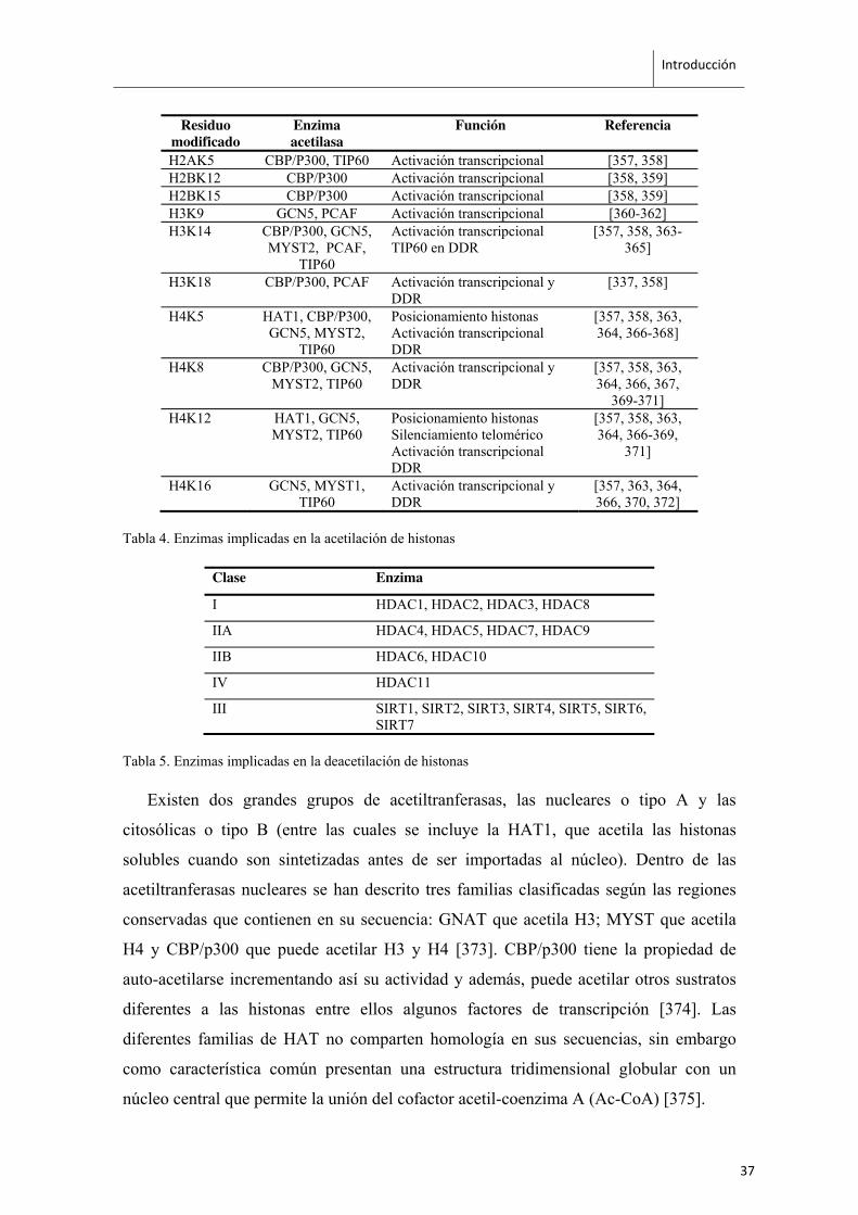
Residuo modificado
Enzima acetilasa
Función Referencia
H2AK5 CBP/P300, TIP60 Activación transcripcional [357, 358] H2BK12 CBP/P300 Activación transcripcional [358, 359] H2BK15 CBP/P300 Activación transcripcional [358, 359] H3K9 GCN5, PCAF Activación transcripcional [360-362] H3K14 CBP/P300, GCN5,
MYST2, PCAF, TIP60
Activación transcripcional TIP60 en DDR
[357, 358, 363-365]
H3K18 CBP/P300, PCAF Activación transcripcional y DDR
[337, 358]
H4K5 HAT1, CBP/P300, GCN5, MYST2,
TIP60
Posicionamiento histonas Activación transcripcional DDR
[357, 358, 363, 364, 366-368]
H4K8 CBP/P300, GCN5, MYST2, TIP60
Activación transcripcional y DDR
[357, 358, 363, 364, 366, 367,
369-371] H4K12 HAT1, GCN5,
MYST2, TIP60 Posicionamiento histonas Silenciamiento telomérico Activación transcripcional DDR
[357, 358, 363, 364, 366-369,
371]
H4K16 GCN5, MYST1, TIP60
Activación transcripcional y DDR
[357, 363, 364, 366, 370, 372]
Tabla 4. Enzimas implicadas en la acetilación de histonas
Clase Enzima
I HDAC1, HDAC2, HDAC3, HDAC8
IIA HDAC4, HDAC5, HDAC7, HDAC9
IIB HDAC6, HDAC10
IV HDAC11
III SIRT1, SIRT2, SIRT3, SIRT4, SIRT5, SIRT6, SIRT7
Tabla 5. Enzimas implicadas en la deacetilación de histonas
Existen dos grandes grupos de acetiltranferasas, las nucleares o tipo A y las
citosólicas o tipo B (entre las cuales se incluye la HAT1, que acetila las histonas
solubles cuando son sintetizadas antes de ser importadas al núcleo). Dentro de las
acetiltranferasas nucleares se han descrito tres familias clasificadas según las regiones
conservadas que contienen en su secuencia: GNAT que acetila H3; MYST que acetila
H4 y CBP/p300 que puede acetilar H3 y H4 [373]. CBP/p300 tiene la propiedad de
auto-acetilarse incrementando así su actividad y además, puede acetilar otros sustratos
diferentes a las histonas entre ellos algunos factores de transcripción [374]. Las
diferentes familias de HAT no comparten homología en sus secuencias, sin embargo
como característica común presentan una estructura tridimensional globular con un
núcleo central que permite la unión del cofactor acetil-coenzima A (Ac-CoA) [375].
37
Introducción
Existen cuatro familias de HDACs. Las de tipo I, II y IV no requieren cofactor para
deacetilar, mientras que la de tipo III requieren del cofactor NAD+ como parte de su
mecanismo de catalización. En general no presentan mucha especificidad en cuanto al
residuo acetilo que eliminan y están presentes en numerosos complejos represores.
Regulan procesos de proliferación celular, diferenciación, senescencia, apoptosis,
cáncer, metabolismo y funciones cognitivas como el aprendizaje y la memoria [376,
377].
3.1.2.3 Fosforilación
La fosforilación de proteínas juega un papel clave en vías de señalización celular. En
1991 el Dr. Mahadevan y sus colaboradores observaron que la estimulación del ciclo
celular y activación de la transcripción de los genes “immediate-early” correlacionaba
con la fosforilación de histona H3 [378]. Se han identificado varias quinasas que actúan
en este residuo, como la MSK1/2 y la RSK2 [379-381]. El residuo de serina 10 en
histona H3 es una marca mantenida entre especies desde levaduras a humanos y ha sido
especialmente caracterizada en Drosophila [382], donde se ha descrito que su
fosforilación tiene un papel importante en la regulación de la transcripción y en
procesos de condensación cromosómica desplazando a la proteína de unión HP1 [383].
3.1.2.4 Otras modificaciones
La ubiquitinación y la sumoilación son modificaciones post-traduccionales que, a
diferencia de la metilación, acetilación y fosforilación, incrementan en
aproximadamente dos tercios el tamaño de las histonas. La ubiquitinación consiste en
la adición de una molécula de ubiquitina formada por 76 aminoácidos y una glicina, y
puede ser una marca represora o activadora, en función del lugar en el que se posicione.
Por ejemplo, la monoubiquitinación en H2B es activadora transcripcional [384-386] y
está relacionada con el establecimiento de metilación en H3K4 [387, 388]. La
monoubiquitinación en H2A, en cambio, es una marca asociada al silenciamiento
génico [389]. La sumoilación es la adición de una proteína llamada SUMO (small
ubiquitin-related modifier) de 11kDa, es una marca represiva que actua bloqueando los
residuos de lisina que no pueden ser metilados o ubiquitinados, de este modo las
38
Introducción
enzimas HDACs son atraídas y se encargan de eliminar los grupos acetilo presentes en
esa región de cromatina [390].
La desiminación es la conversión del aminoácido arginina en citrulina por acción de
las Peptidilarginina deiminasas (PADs). En concreto se ha demostrado que la PADI4
puede llevar a cabo esta reacción en las argininas presentes en histonas [391, 392]. Por
otro lado, la aparición de citrulina en histona H3 y H4 correlaciona con la desaparición
de la metilación en argininas in vivo, y podría actuar como un antagonista del efecto
activador de la transcripción dependiente de la metilación de argininas.
3.1.3 ARNs no codificantes
Los ARN no codificantes se dividen en función de su tamaño en lncARNs (long
non-coding ARNs) y sncARNs (short non-coding ARNs) de más y menos de 200pb,
respectivamente. Dentro de los sncARNs se incluyen los snoARNs (de 70 a 100
nucleótidos que actúan modificando otros ARN como el ribosomal y el de
transferencia), los piARNs (de 26 a 31 nucleótidos que forman complejos de ARN y
proteínas piwi y actúan en el silenciamiento de retrotransposones y otros genes de
células germinales), los siARNs (de 20 a 25 nucleótidos que actúan como pequeñas
moléculas de ARN de interefencia silenciando la expresión génica), y los microARNs
(miARNs).
Los microARNs se identificaron por primera vez en C.elegans [393] y corresponden
a fragmentos de ARN no codificante de entre 18 y 25 nucleótidos que regulan la
expresión génica a nivel post-transcripcional [394]. Los microARNs son inicialmente
transcritos en forma de miARN primario (pri-miARN) el cual forma una estructura de
bucle. Seguidamente, este precursor es procesado en el núcleo por la enzima DROSHA
para formar el pre-miARN de unos 70 nucleótidos de tamaño que será exportado al
citoplasma por la enzima Exportina 5 y procesado por la enzima DICER para formar
ARN de doble cadena (miARN-miARN*). La hebra de ARN que llevará a cabo la
regulación génica es incluida dentro del complejo miRISC. Por su parte, la hebra
complementaria (marcada con un asterisco en la nomenclatura clásica) se creía que era
simplemente degradada pero nuevos trabajos apuntan hacia un posible papel como
modulador de la función celular [395, 396].
39
Introducción
Los miARNs reconocen un ARN mensajero (ARNm) en base a la homología de
secuencias [397]. En función del nivel de complementaridad, el silenciamiento de
expresión se producirá mediante la degradación del ARNm (en el caso de que la
complementariedad de bases sea total) [398-400], o por bloqueo del acceso de la
maquinaria de la traducción en los ribosomas [401-403].
El análisis de expresión y funcionalidad de los microARNs mediante plataformas
basadas en microarrays de expresión, secuenciación y PCR a tiempo real, ha revelado el
papel de los miARNs como factores reguladores de vías de señalización imprescindibles
para la homeostasis y diferenciación celular, regulación epigenética y respuesta frente a
daños en el ADN, entre otros [404-406]. Actualmente existen varias bases de datos de
miARNs, entre las cuales destaca miRBase (http://www.mirbase.org/) por ser una de las
más actualizadas y con mayor cantidad de información sobre las secuencias de miARNs
maduros, precursores, localización, predicción de genes diana y bibliografía. También
hay disponibles otras páginas como GOmir (http://www.bioacademy.gr/bioinformatics-
/GOmir/bioinformatics_home.htm) especializadas en predicción de genes dianas y
agrupación mediante categorías ontológicas.
La mayoría de miARNs son específicos de tejido [394, 407]. En sistema nervioso
central los microARNs modulan procesos de gran relevancia funcional [408-411] como
la neurogénesis [412, 413], crecimiento de dendritas [414, 415] y formación de espinas
dendríticas [416, 417]. La especificidad de expresión en cerebro se da a diferentes
niveles. Se ha observado que el hipocampo y el córtex son dos áreas del cerebro
especialmente ricas en expresión de miARNs, y se ha sugerido su implicación en el
establecimiento de la memoria y en procesos de plasticidad neuronal [418]. El tipo
célular también es uno de los determinantes en la expresión de ciertos miARNs, ya que
los circuitos glutamatérgicos y las neuronas GABAergicas tienen perfiles de expresión
diferenciales de otros tipos neuronales [419, 420].
Asimismo, la localización dentro de la neurona también es un rasgo específico de cada
miARN. Por ejemplo miR-26a se encuentra enriquecido en dendritas [421], mientras
que otros como miR-200c, miR-339, miR-7 o miR-137 se localizan principalmente en
la región sináptica; o miR-15bm, miR-16 y miR-221 que se encuentran en el axón
neuronal [422].
40
Introducción
3.2. MODIFICACIONES EPIGENÉTICAS ASOCIADAS AL ENVEJECIMIENTO
3.2.1 Metilación ADN
Con el proceso de envejecimiento se produce una reducción de los niveles globales
de metilación en el ADN así como un aumento en zonas específicas. Estudios
comparativos entre grupos de diferentes edades de sangre humana [423-427] y otros
tejidos [428-430] corroboran estos resultados.
Sorprendentemente, la hipometilación asociada a envejecimiento se produce
principalmente en los denominados transposones (transposable repetitive elements). La
propiedad principal de los transposones es insertarse en diferentes partes del genoma de
manera autónoma, pudiendo provocar cambios en la expresión del gen donde se inserta
o incluso afectando de manera irreversible su funcionalidad si rompe su secuencia
durante la transposición. Alguno de ellos, como por ejemplo Alu y L1, suelen
encontrarse reprimidos, de manera que la eliminación de metilación en el ADN asociada
a envejecimiento en estos elementos supone un incremento en la inestabilidad
genómica. Otras regiones sensibles a la hipometilación durante el envejecimiento
incluyen regiones promotoras ricas en CpG de genes inflamatorios como IGF2 [431],
TNF [432], iNOS y TLR2 [426].
A su vez, durante el envejecimiento también se observa una hipermetilación en
ciertas regiones del genoma. Por ejemplo, se han descrito alteraciones en el patrón de
metilación de islas CGs en genes reguladores de la transcripción [427], específicos de
tejido [433], implicados en desarrollo y diferenciación [434], apoptosis [435],
senescencia [436] o asociados con cáncer [437]. En un estudio llevado a cabo por
Bocklandt y colaboradores [438] se identificaron 3 genes (EDARDD, TOM1L1 y
NPTX2) de un total de 88 relacionados con el proceso de envejecimiento con una
relación entre el patrón de metilación y la edad que se ajustaba a un modelo de regresión
lineal, pudiéndose considerar candidatos para predecir la edad fisiológica con una
precisión de 5.2 años.
Por otra parte, también se ha descrito un descenso de los niveles de las enzimas
DNMT1 y DNMT3a y un incremento de DNMT3b en células somáticas durante el
envejecimiento [439, 440]. En ratones se ha visto que la insuficiencia de DNMT1 [424]
41
Introducción
y DNMT3a [441] provoca alteraciones en el aprendizaje y la memoria de manera
dependiente a la edad. En Drosophila, la sobreexpresión de DNMT2 (la única
metiltransferasa de ADN presente en esta especie) provoca un incremento en la
esperanza de vida de hasta el 58% [442]. Sin embargo, el enzima DNMT2 de mamíferos
no actua como metiltransferasa de ADN sino de ARN [443]. Aún así, estos resultados
indican que la metilación en ADN puede ser importante para determinar la esperanza de
vida. Sin embargo, algunos autores sugieren una regulación más compleja para explicar
los cambios observados en el estado de metilación en el ADN durante el
envejecimiento, en la cual se incluyen otros factores como el efecto del entorno y los
hábitos de vida o la localización del gen estudiado [444].
3.2.2 Modificaciones de histonas
Algunas de las modificaciones de histonas que afectan el envejecimiento y la
longevidad incluyen enzimas y cofactores responsables de acetilación y metilación de
histonas. Caenorhabditis elegans es una especie de nematodo muy utilizado en genética
del desarrollo y longevidad ya que posee ciertas características que lo hacen un buen
modelo de estudio. Presenta un aspecto transparente a contraluz, facilitando su
observación bajo el microscopio; posee muchos de los órganos y sistemas de otros
organismos; tiene un mantenimiento muy fácil a nivel de alimentación y reproducción y
tiene una esperanza de vida de entre dos o tres semanas permitiendo estudiar los
procesos de envejecimiento y longevidad en un plazo de tiempo muy corto. Por
ejemplo, la sobreexpresión de la histona deacetilasa SIR-2, responsable de la acetilación
en H4K16, se ha asociado con un aumento en la longevidad de C.elegans [445-447].
El estado de metilación de histonas parece ser un determinante de la longevidad en
C.elegans. Por ejemplo la inhibición de las metiltransferasas ASH-2 y SET-2 se ha
asociado con una reducción global en los niveles de H3K4me3 que extiende la
esperanza de vida. En cambio, la inhibición de la demetilasa RBR-2 reduce la
longevidad [448, 449]. Además, la inhibición de UTX-1, enzima responsable de
demetilar la marca de silenciamiento H3K27me3, aumentó la esperanza de vida [450,
451].
42
Introducción
En mamíferos la alteración del homólogo a SIR-2, SIRT1, produce mejoras en la
respuesta a estrés, la supervivencia celular mediante la regulación de p53 [452] y el
metabolismo [453] pero tiene muy poco efecto en términos de longevidad [454, 455].
De manera similar, la inhibición de las histonas deacetilasas HDAC de clase I y II en
ratón no produce cambios en la longevidad, pero revierte el declive en aprendizaje y
memoria asociado a envejecimiento, relacionados con cambios en los niveles de
H4K12ac en regiones codificantes y de inicio de la transcripción [456].
La deacetilasa SIRT6 que modifica los residuos de H3K9 y H3K56, es un factor
necesario en la reparación de ADN dañado, y se ha observado que su deficiencia afecta
a la longevidad y causa envejecimiento prematuro [457]. Por último, en primates y
líneas celulares humanas, también se han encontrado asociaciones entre envejecimiento
y aumento global en los niveles de H3K4me2 [458], H4K20me2 y H3K79me2 [459], y
reducción global en H3K9me3 y H3K27me3 [460, 461] y aumento en acetilación global
de histonas [462, 463].
Otro de los procesos alterados durante el envejecimiento es el sistema de reparación
de daños del ADN, el cual requiere la presencia de ciertas modificaciones en histonas en
particular la fosforilación en H2AX [464]. Esta modificación esta aumentada con la
edad en roedores y modelos humanos con envejecimiento prematuro [465-467].
3.2.3 microARNs
Recientemente se ha comenzado a describir la implicación de los microARNs en la
longevidad y los procesos de envejecimiento [468]. En general, la expresión global de
los miARNs va disminuyendo con la edad mientras que algunos específicos asociados a
patologías propias del envejecimiento, aumentan su expresión [469].
En C.elegans se han identificado unos 200miARNs de los cuales más del 50 %
contienen secuencias conservadas en humanos y más del 25 % son sensibles al
envejecimiento [470, 471]. Entre estos últimos, los niveles de expresión de mir-71, mir-
246 y mir-239 pueden provocar cambios de hasta un 47% en la esperanza de vida [472],
algunos aumentándola (mir-239) y otros disminuyéndola (lin-4, mir-71 y -238) [473,
474]. Otros microARNs como let-7 o miR-1, conservados en organismos superiores,
también se relacionan con procesos de envejecimiento [475].
43
Introducción
Una de las vías de señalización modulada por algunos de estos microARNs, es la vía
de la insulina/IGF, ampliamente asociada a la regulación de la senescencia [472, 474].
Algunas mutaciones en genes implicados en la vía de señalización de la insulina/IGF
modifican la esperanza de vida en C.elegans [476], Drosophila [477] y ratón [478].
Entre los microARNs que regulan esta vía se encuentran lin-4 que modula la expresión
de DAF-16 (inhibidor transcripcional que se activa por IGF-1) [479], miR-216a, miR-
217 y miR21 que regulan PTEN (inhibidor de la vía de Akt mediante defosforilación de
PIP3) [480, 481] y miR-1, miR-320 y miR-206 que inhiben la expresión de IGF-1 [482,
483].
Otra vía de gran importancia en la modulación de la longevidad afectada por la
expresión de microARNs es la vía de TOR [484]. Esta vía estimula el crecimiento
celular y bloquea la autofagia en respuesta al contexto nutricional y la presencia de
factores de crecimiento [485]. La inhibición de la vía de TOR aumenta la longevidad en
levadura [486], C.elegans [487, 488], Drosophila [489] y ratón [490].
También es importante destacar la acción de los microARNs sobre SIRT1, factor
con un papel importante en longevidad y envejecimiento. Entre ellos se encuentran
miR-217 cuya expresión aumenta con la edad [491], miR-34a regulado a su vez por p53
(diana de SIRT1) [492], miR-199a [493] y miR-132 [494].
Por último, diversos microARNs se han relacionado con la manifestación de algunas
patologías crónicas asociadas al envejecimiento. Por ejemplo, miR-1, miR-122 y miR-
375 se han relacionado con hipertrofia e insuficiencia cardíaca [495-497], miR-1 y miR-
206 con hipertrofia muscular y sarcopenia [498-500] y miR-21, con regulación del
perfil lipídico [501, 502]. A nivel de sistema nervioso central, varios estudios han
mostrado variaciones en el patrón de expresión de miARNs en modelos de EA [503-
505]. Por ejemplo, se ha descrito la disminución de miR-124 en cerebros de pacientes
de Alzheimer [506], y alteraciones de miR-101 que modula la proteína percursora
amieloide (APP) [507], y de miR-195, miR-107, miR-298 o miR-29a/b-1, implicados en
el control traduccional de la enzima beta-secretasa 1 (BACE) [508-511].
44
Introducción
4. EJERCICIO FÍSICO Y ENVEJECIMIENTO
Una de las principales recomendaciones de la Organización Mundial de la Salud es
mantener un estilo de vida físicamente activo a lo largo de toda la vida. En jóvenes,
hasta los 17 años se recomienda invertir un mínimo de 60 minutos diarios en
actividades físicas de intesidad moderada que incluyen juegos, deportes y actividades
recreativas y educación física. En adultos de 18 a 64 años, se recomiendan 150 minutos
semanales de actividad física aeróbica moderada o bien 75 minutos de intensidad
elevada, con el fin de mejorar las funciones cardiorrespiratorias y musculares y la salud
ósea. También se recomienda la incorporación de actividades de fortalecimiento
muscular un mínimo de dos veces por semana. En el caso de adultos de más de 65 años,
se recomienda mantener una actividad física activa coherente con el estado de salud la
persona, mediante actividades recreativas o de ocio, desplazamientos y paseos, y otras
actividades realizadas en el contexto familiar y comunitario. De igual forma que en
individuos adultos, las recomendaciones mínimas en personas de edad avanzada son de
150 minutos semanales, teniendo especial importancia en este grupo de edad las
actividades de refuerzo muscular y la incorporación de actividades de mejora de
equilibrio. Numerosos estudios muestran los beneficios psicoemocionales y
neurofisiológicos del ejercicio físico [512] con particular interés en los efectos
saludables durante el envejecimiento [513-515].
4.1. MODELOS EXPERIMENTALES DE EJERCICIO FÍSICO
Existen diferentes métodos para valorar la actividad física en humanos, siendo los
más empleados la valoración personal mediante cuestionarios, el uso de sensores de
movimiento, medidas de la tasa metabólica y de otras características cardio-
respiratorias. Uno de los métodos utilizados es la evaluación del gasto energético
durante las actividades cotidianas de la persona en periodos prologados de tiempo
mediante la administración de agua que contiene isótopos de oxígeno e hidrógeno
marcados [516]. Aunque este método proporciona ciertas ventajas en la valoración
durante la vida normal de los sujetos de estudio en periodos prolongados de tiempo, no
aporta información del tipo, frecuencia, duración e intensidad de la actividad física
realizada. Otro método utilizado es la observación directa o a través de grabaciones de
las principales actividades cotidianas, aunque este método se suele utilizar sólo en
45
Introducción
grupos pequeños y actividades específicas. El uso de sensores de movimiento, como
podómetros y acelerómetros, también es uno de los métodos escogidos en el estudio de
la actividad física en humanos. Mediante este tipo de herramientas, se puede obtener
información sobre la frecuencia e intensidad de la actividad física realizada [517]. Sin
embargo, el uso de estos dispositivos requiere el análisis de una gran cantidad de datos
y no indican el tipo específico de actividad realizada. Existen también medidas
indirectas para evaluar la actividad física, como la frecuencia cardíaca o la capacidad
aeróbica del individuo (VO2) [518]. Sin embargo, estas medidas están influencias por el
nivel de entrenamiento previo del sujeto y requieren del estudio previo de las
características de cada individuo [519]. Además, hay que tener en cuenta que factores
de tipo emocional, temperatura y humedad, nivel de deshidratación e incluso posturales
durante la realización de la actividad física, pueden afectar a estos parámetros [520].
Dadas las limitaciones metodológicas de los estudios en humanos, existen dos
estrategias principales para el estudio de los efectos del ejercicio físico utilizando
roedores como modelo experimental: ejercicio forzado utilizando una cinta ergométrica
y ejercicio voluntario basado en ruedas de actividad [521].
El ejercicio físico forzado implica extraer al animal de la jaula y colocarlo sobre una
cinta ergométrica variando la velocidad y el tiempo de exposición según el diseño
experimental. Las ventajas de este tipo de ejercicio son la posibilidad de controlar la
duración y la intensidad del ejercicio realizado por cada animal, además de la
posibilidad de mantener a los animales estabulados en grupos o aislados en cajas
individuales. Por el contrario, hay que tener en cuenta que los resultados de este tipo de
ejercicio pueden verse influidos por la manipulación realizada sobre el animal al
colocarlo en la cinta [522] y por la aparición de respuestas de estrés si se utilizan
métodos de motivación con descargas eléctricas [523, 524].
En los modelos de ejercicio físico voluntario, los animales tienen acceso libre a
ruedas de actividad que permiten controlar el nivel de ejercicio realizado. Para ello, las
ruedas de actividad poseen un imán conectado a un interruptor electrónico colocado en
el eje de la rueda, la señal que emite es enviada a un concentrador que transmite los
datos a un ordenador por un sistema de transmisión sin cables, y mediante un software
específico se contabiliza la actividad y tiempo de cada giro. Las ventajas de este tipo de
46
Introducción
ejercicio son que no es necesario aplicar descargas eléctricas ni motivación de ningún
tipo a los animales y no genera estrés per se ya que la actividad depende de la voluntad
del propio animal [525]. Sin embargo, este tipo de intervención conlleva algunos
inconvenientes ya que se suele observar heterogeneidad en la cantidad de ejercicio
realizado por cada animal [526] y además los animales deben mantenerse en jaulas
individuales para poder cuantificar correctamente el ejercicio realizado por cada uno.
Este aislamiento puede incrementar los niveles de estrés y ansiedad de los roedores
[527-529], así como conductas depresivas [530], llegando a afectar las capacidades
cognitivas y fisiológicas de los animales [531-533].
4.2. EFECTOS DEL EJERCICIO FÍSICO EN MODELOS MURINOS Y EN HUMANOS
4.2.1. Sistemas periféricos
Gran cantidad de estudios epidemiológicos han mostrado una fuerte correlación
negativa entre el ejercicio físico aeróbico y el riesgo cardiovascular [534-541] tanto en
individuos sanos [542-548] como en individuos de edad avanzada o con enfermedades
cardíacas [549-551]. Además, los efectos del ejercicio físico no parecen estar
influenciados por la presencia de marcadores genéticos asociados con déficits
cardiovasculares [549, 552-554] mostrando el potencial terapéutico de este tipo de
práctica sobre la salud cardiovascular. Sorprendentemente, algunos estudios muestran
una mayor protección del ejercicio físico en mujeres que en hombres [555, 556]. La
actividad física moderada es la que mayor impacto tiene sobre el riesgo de
enfermedades coronarias [554] y por ello es la que se recomienda en la mayor parte de
guías públicas de salud [512, 557]. La hipertensión es uno de los principales factores de
riesgo en la aparición de enfermedades cardiovasculares [558] y se ha demostrado que
hábitos de vida saludables como la pérdida de peso y una actividad física habitual
contribuyen positivamente a la disminución y control de la presión sanguínea [559-
564].
Por otro lado, la incidencia de diabetes mellitus tipo 2 está inversamente relacionada
con el ejercicio físico [565-567] y la mortalidad debida a esta enfermedad llega a ser el
doble en personas sedentarias independientemente del peso corporal que tengan [568-
570]. No sólo se ha visto esta relación a nivel epidemiológico, dos estudios
intervencionales mostraron una reducción de hasta el 58% en marcadores de riesgo de
47
Introducción
padecer diabetes [571, 572]. Todo ello puede estar relacionado con el hecho de que,
tanto el ejercicio aeróbico como el anaeróbico [573, 574], aumentan la captación de
glucosa mediante el transportador GLUT-4, presente en las células musculares, de
manera independiente a la insulina [575].
El ejercicio aeróbico también da lugar a cambios en los niveles de lípidos y
lipoproteínas en circulación [576-578]. El resultado más consistente entre los estudios
realizados en humanos es la reducción de los niveles de lipoproteínas de baja densidad
(low-density lipoprotein, LDL) [579-583], otro de los principales marcadores de riesgo
de fallo cardíaco.
En cuanto al sistema inmune y estado inflamatorio, el ejercicio físico agudo e
intenso induce una respuesta inflamatoria transitoria debido a la estimulación muscular
tras el esfuerzo realizado. Sin embargo, una actividad regular y moderada reduce los
niveles de marcadores inflamatorios como la proteína C-reactiva (CRP) [584-588],
comunmente asociada a un elevado riesgo de infarto cardíaco y cerebrovascular [589] y
otras citoquinas proinflamatorias como el TNF-α y IL-6 [590]. El ejercicio físico
también parece tener un efecto inhibitorio sobre factores trombogénicos y estimula el
potencial fibrinolítico, lo cual contribuye a reducir el riesgo de trombosis [591-593].
4.2.2 Sistema nervioso central
Numerosas evidencias indican que el ejercicio físico aeróbico tiene efectos positivos
a nivel cognitivo en individuos adultos de más de 55 años [594], produciendo mejoras
en atención y memoria [595]. También se ha observado un aumento de materia gris en
la zona prefrontal y temporal y de materia blanca en la zona anterior del cuerpo calloso
[596-599], ambas áreas importantes para la ejecución de tareas de atención selectiva que
son especialmente sensibles al envejecimiento [600-602].
Estudios de resonancia magnética funcional (fMRI) han mostrado un aumento en el
volumen hipocampal de individuos sanos de entre 59 y 81 años de edad [603], así como
mejoras en memoria espacial [604] en respuesta a la actividad física. Por ejemplo, en un
estudio randomizado reciente se observó un aumento de un 2% en el volumen
hipocampal de individuos sanos de entre 55 y 80 años de edad tras un entrenamiento
aeróbico [605] asociado a un aumento en los niveles del factor neurotrófico BDNF en
48
Introducción
suero y a mejoras en funciones de aprendizaje y memoria. En un estudio realizado por
Pereira y colaboradores [606] se analizaron los efectos tras 2 semanas de ejercicio físico
voluntario en ratón y 12 semanas de entrenamiento aeróbico en humanos. Mediante
resonancia magnética (MRI) describieron un aumento en la irrigación sanguínea en el
giro dentado hipocampal en ambos modelos experimentales, que en ratón pudieron
correlacionar con aumento en procesos de neurogenésis y en humanos con mejoras en
aprendizaje y memoria. Los efectos del ejercicio sobre la memoria y el volumen del
hipocampo [607, 608] también se han observado en niños de 10 años de edad.
Los modelos animales permiten el estudio de las vías moleculares y los cambios a
nivel celular producidos en el cerebro en respuesta al ejercicio físico. El aumento en la
proliferación celular y la supervivencia neuronal en giro dentado es uno de los efectos
obervados más consistentes tras intervenciones de ejercicio físico [609-614] tanto en
animales de edad media [611] como de edad avanzada [615]. Incluso animales recién
nacidos de madres que han practicado ejercicio físico durante la gestación, muestran
mayor supervivencia celular en hipocampo [616, 617]. También se ha descrito el
aumento de células progenitoras neuronales en otras regiones como el área
paraventricular de los ventrículos laterales [613, 618, 619]. Los efectos del ejercicio
sobre la neurogenésis se ha asociado a una mayor vascularización en corteza cerebral
[620], estriado [621] e hipocampo [622], y al aumento en los niveles de IGF1 en
hipocampo [623] y de IGF1 [610] y VEGF en suero [624], reforzando la idea de que las
respuestas centrales y periféricas al ejercicio físico actúan de forma coordinada.
El factor neurotrófico derivado del cerebro (brain-derived neurotrophic factor,
BDNF) es una neurotrofina implicada en la excitabilidad neuronal, la plasticidad
sináptica y el mantenimiento de funciones cognitivas como la memoria y el aprendizaje
[625-628]. El aumento de BDNF es uno de los marcadores más validado a nivel
experimental en modelos de ejercicio físico y que tanto en expresión génica como en
niveles de proteína de BDNF muestran un aumento en sus niveles en hipocampo tras
intervenciones de ejercicio físico [629, 630]. La práctica de ejercicio físico de tan sólo
una semana ya provoca mejoras en las funciones cognitivas de ratones jóvenes y de
edad avanzada, y esto se ha asociado al aumento en la expresión de los genes Bdnf,
sinapsina, Mapk y Crfr1, implicados en procesos de memoria y plasticidad sináptica
[626]. Además, se ha observado que la práctica de ejercicio físico facilita el aumento de
49
Introducción
expresión del factor Bdnf en intervenciones posteriores y produce una respuesta más
eficiente [631].
A nivel psicoemocional, el ejercicio voluntario facilita la asimilación de situaciones
nuevas reduciendo los comportamientos relacionados con ansiedad [632-634] y parece
ejercer un efecto antidepresivo, descrito tanto en modelos de roedores [635] como en
humanos [636-638], el cual se asocia al aumento de BDNF [639] y de VGF [640].
4.2.3 Ejercicio físico, envejecimiento y Alzheimer
El ejercicio físico produce una serie de beneficios en sistema nervioso central que
están implicados en el mantenimiento y mejora cognitiva. Estas observaciones han
suscitado interés en el contexto de la prevención y tratamiento de la EA [641-643].
En humanos, diferentes meta-análisis realizados han demostrado que el ejercicio
físico tiene un efecto positivo sobre la cognición y que existe una relación entre la
actividad física y las mejoras cognitivas tanto en individuos sanos como en pacientes
con signos de demencia propios de la EA [594, 644, 645]. Estudios realizados en
personas en un estadío temprano de la EA han mostrado un aumento en el volumen del
hipocampo [605, 646, 647] y corteza cerebral [596, 600, 648], así como una mejora en
las habilidades cognitivas que puede llegar a perdurar hasta 6 meses después de finalizar
la intervención de ejercicio [649]. También se ha observado una mejora en la
conectividad entre corteza cerebral e hipocampo [650-652] y en la vascularización en
hipocampo [606]. Además, se ha estimado que la práctica regular de ejercicio físico
podría llegar a reducir el riesgo de padecer la EA hasta en un 45% [653] y que el inicio
de una actividad física desde edades tempranas tendría un mayor impacto [654]. Incluso
se ha comprobado que la practica de ejercicio puede asociarse con menores niveles de
Aβ en humanos [655], especialmente en individuos con predisposición genética a
padecer la enfermedad [656].
En modelos animales, se han observado mejoras de memoria y aprendizaje y una
diminución en el depósito de placas de Aβ en corteza cerebral e hipocampo tras 5 meses
de ejercicio voluntario [657]. También se han observado mejoras en memoria (fear
conditioning test) y un aumento en la expresión del gen Lztfl1 (leucine zipper
transcription factor-like protein 1) asociado a crecimiento de neuritas en el modelo
50
Introducción
SAMP8 de envejecimiento y EA tras 4 meses de ejercicio voluntario [658]. Estos
ratones también presentan mayores niveles de SIRT1 en hipocampo y corteza cerebral
de ratones después de 6 meses de ejercicio voluntario moderado que provocaron una
activación de la vía de AMPK y mejoras en la función mitocondrial en hipocampo
[659]. Por último, en este mismo modelo se observaron mejoras en la actividad
locomotora y la función sensorimotora, así como mejoras en factores implicados en
revascularización (VEGFα), función mitocondrial (Sirt1-Ampk-Pgc1α) y metabolismo
energético (Glut4 y Cd36) en músculo esquelético [660].
4.3. EJERCICIO Y EPIGENÉTICA
Algunas de las mejoras observadas en metabolismo, inflamación y sistema nervioso
en respuesta a la práctica de ejercicio físico se han relacionado recientemente con
cambios a nivel epigenético implicados en la metilación del ADN, metilación y
acetilación de histonas y la expresión de diferentes microARNs.
A nivel global, se ha descrito un descenso en los niveles de metilación del ADN en
regiones promotoras de genes relacionados con vías metabólicas en respuesta al
ejercicio físico en músculo esquelético [661, 662], como por ejemplo el receptor gamma
activador de peroxisomas (PPAR-γ), el coactivador 1 alfa (PGC-1α) o el factor de
transcripción mitocondrial A (TFAM) [661]. También se ha observado que durante la
práctica de ejercicio se produce un cambio en la localización intracelular de las
desacetilasas de histonas HDAC4 y HDAC5, posiblemente mediado por la fosforilación
mediante AMPK. Esto produciría una disociación del factor MEF2, permitiendo su
unión a histonas acetiltransferasas y el posterior aumento de expresión de Glut4
observado en músculo esquelético en respuesta al ejercicio [663, 664]. En cuanto a la
expresión de miARNs en músculo esquelético, se ha observado una reducción de varios
miARNs implicados en metabolismo y procesos de fosforilación oxidativa [665].
Por otro lado, el ejercicio moderado se ha asociado con un retraso en la
hipometilación durante el proceso de envejecimiento en genes implicados en respuesta
inflamatoria como la proteína asociada a apoptosis speck-like (ASC) [666], implicada en
la secreción de citoquinas proinflamatorias como IL-1β e IL-18. También se han
descrito cambios en la expresión de 34 microARNs en células mononucleares de sangre
periférica (PBMCs) implicados en la regulación de vías inflamatorias [667].
51
Introducción
A nivel de sistema nervioso central, se han descrito cambios epigenéticos asociados
a la expresión de BDNF. Tras una intervención de una semana de ejercicio se detectó
una demetilación la región promotora del gen de Bdnf y una correlación entre el
aumento de expresión de dicho factor y los niveles de acetilación en histona H3 en
hipocampo [668]. El ejercicio también produjo una disminución en los niveles de
HDAC5, enzima implicada en la modulación del gen de Bndf en un modelo murino de
depresión [669]. La acetilación de histonas es uno de los mecanismos principales en
sistema nervioso central para el establecimiento de la memoria y plasticidad sináptica
[376, 670, 671] y se ha asociado a declive cognitivo y neurodegeneración [672-674] y la
EA [675]. La proporción HAT/HDAC en hipocampo se ha visto alterada en respuesta al
ejercicio físico, hacia un estado de hiperacetilación [676], lo cual se relaciona con un
efecto neuroprotector en memoria y cognición [677]. De hecho, en ratas se observaron
mejoras en el rendimiento en tareas que producen cierto estrés (como la natación
forzada o la exposición a un entorno nuevo) tras una intervención de 4 semanas de
ejercicio físico voluntario, que correlacionaron con la fosfoacetilación en histona H3 y
la inducción del gen c-Fos en neuronas de la región hipocampal del giro dentado [678].
Por último, mientras que el envejecimiento reduce los niveles de SIRT1 [679, 680],
se ha descrito un aumento de los mismos en corazón y tejido adiposo de ratas de edad
avanzada tras 8 semanas de ejercicio forzado [681] y en músculo esquelético tanto en
ratas jóvenes como de edad avanzada después de 6 semanas de ejercicio forzado
[682].En humanos, también se han detectado mayores niveles de SIRT1 en músculo
esquelético [683, 684] y sangre periférica [685] tras la realización de actividades físicas
de elevada intensidad.
Las evidencias obtenidas hasta el momento ponen de manifiesto el potencial
terapéutico del ejercicio físico como estrategia de prevención y tratamiento de
patologías asociadas al envejecimiento. Los efectos beneficiosos del ejercicio sobre los
diferentes sistemas periféricos y el sistema nervioso central lo convierten en un modelo
adecuado para comprender mejor los efectos protectores del estilo de vida activo sobre
el proceso global de envejecimiento y la neurodegeneración.
52

OBJETIVOS

Objetivos
El actual envejecimiento de la población y el consecuente aumento de
enfermedades crónicas asociadas al mismo, establecen como prioritario el estudio de
los mecanismos presentes durante este proceso, así como la búsqueda de acciones
terapéuticas que favorezcan un envejecimiento saludable, como el ejercicio físico. La
epigenética, por su implicación funcional y su modulación ambiental podría ser el
mediador de los efectos beneficiosos de la práctica de ejercicio físico.
En este contexto, el objetivo principal de este proyecto fue identificar marcadores
de envejecimiento en el modelo murino de senescencia acelerada SAMP8 y su control
SAMR1, y su posible regulación mediante la práctica de ejercicio físico.
Dentro de este eje temático, los objetivos específicos planteados fueron:
1. Estudiar los mecanismos epigenéticos asociados a neuroinflamación y
senescencia.
2. Analizar las alteraciones transcripcionales en hipocampo sensibles a
ejercicio físico moderado en un contexto de envejecimiento y
neurodegeneración.
3. Identificar factores de regulación epigenética asociados a envejecimiento y
su modulación por la práctica de ejercicio físico moderado.
55


RESULTADOS

Resultados
Los resultados de esta tesis se agrupan en 3 capítulos. Todos ellos se corresponden a
artículos ya publicados. Cada capítulo contiene el artículo original precedido de un
resumen de los resultados y conclusiones principales. Las figuras, tablas y referencias se
encuentran en el artículo original correspondiente adjuntado al final de cada capítulo.
59

Resultados
I
Disminución de la expresión de Rcor2 en un modelo de
senescencia acelerada (ratones SAMP8) y su posible función como
regulador de la inflamación asociada al envejecimiento
61

Resultados
Los resultados presentados en este capítulo se incluyen en el siguiente artículo
publicado:
Rcor2 underexpression in senescent mice: a target for inflammaging?
Journal of Neuroinflammation, 11:126 (2014)
María J Alvarez-López, Patricia Molina-Martínez, Marco Castro-Freire, Marta
Cosín-Tomás, Rosa Cristòfol, Marcelina Párrizas, Rosa María Escorihuela, Merce
Pallàs, Coral Sanfeliu y Perla Kaliman.
Contribución: Purificación de ARN y estudios de expresión génica por Real Time
PCR, ensayos con células C6 y supervisión de los resultados de transfección, así como
análisis estadístico de todos los resultados presentados, participación en la
interpretación de resultados y en la elaboración del manuscrito.
63
Resultados
RESUMEN
La presencia de procesos inflamatorios a nivel sistémico durante el envejecimiento
es uno de los factores que contribuyen al desarrollo de patologías crónicas, entre ellas la
enfermedad de Alzheimer. Sin embargo, los mecanismos moleculares subyacentes a la
presencia de neuroinflamación crónica están poco estudiados. En este contexto,
trabajos previos han descrito la implicación de reguladores epigenéticos en inflamación
y longevidad, entre los cuales se incluye el correpresor de REST, Rcor. La isoforma
Rcor1 participa en la modulación de la neuroinflamación en respuesta a la inducción por
LPS (Lipopolysaccharid) en microglia y astrocitos [686], por su parte Rcor2 ha
mostrado tener un papel relevante en la expresión de genes inflamatorios como Il-6,
Ccl2 o Mip1a entre otros, en preadipocitos [687].
En este trabajo analizamos el impacto del factor Rcor, in vivo e in vitro, en procesos
neuroinflamatorios asociados a envejecimiento. Los datos que se presentan muestran
una interacción entre la reducción de Rcor2 y la presencia de mayores niveles de
inflamación, sugiriendo que Rcor2 podría ser un importante regulador de estados
proinflamatorios crónicos característicos del envejecimiento o el cada vez más utilizado
término, inflammaging.
PRINCIPALES RESULTADOS
4.3.1. 1.1. El modelo murino de envejecimiento acelerado SAMP8 presenta
niveles exacerbados de IL‐6 en suero en respuesta a LPS
Tres horas después de la inyección intraperitoneal del lipopolisacárido LPS se
analizó la respuesta inflamatoria en ratones SAMR1 y SAMP8 de 12 meses de edad. Se
encontraron niveles elevados de IL-1β, TNF-α e IL-6 en suero de ambas cepas en
respuesta al estímulo inflamatorio por LPS. Sin embargo, sólo se observaron efectos de
cepa y tras la estimulación con LPS para IL-6, así como tendencia estadística casi
significativa para la interacción entre ambos factores (p=0.068). Además, IL-6 presentó
mayores niveles a nivel basal en los ratones SAMP8 (Figura 1C del Artículo 1). Estos
datos sugieren que los niveles en plasma de IL-6 pueden ser un biomarcador
inflamatorio asociado al fenotipo de senescencia acelerada presente en los ratones
SAMP8.
64
Resultados
4.3.2. 1.2. Los ratones SAMP8 presentan niveles reducidos de expresión de
Rcor2 en linfocitos, corteza cerebral, hipocampo y astrocitos
Con el objetivo de describir un posible mecanismo responsable del fenotipo
inflamatorio observado en los ratones SAMP8, se estudió la expresión génica de Rcor1
y Rcor2 en estos animales y en sus controles SAMR1. Se hallaron niveles mínimos de
expresión génica de la isoforma del correpresor de REST, Rcor2, en linfocitos de los
ratones SAMP8 desde edades tempranas (Fig 2B del Artículo 1), en astrocitos
provenientes de animales de 2 días de edad, y en hipocampo y corteza cerebral de
animales adultos (Fig 3 del Artículo 1). Además, los niveles reducidos de Rcor2 en
astrocitos SAMP8 estaban acompañados de un aumento en los niveles basales de Il-6
(Fig 3G del Artículo 1). Asimismo, confirmamos una correlación inversa entre la
expresión génica de Rcor2 y de Il-6 tras la administración de LPS en hipocampo de
ratones SAMR1 (Fig 4 del Artículo 1) y en la línea celular C6 (Fig 5 del Artículo 1).
4.3.3. 1.3. El silenciamiento de Rcor2 en astrocitos provoca un aumento de
expresión de Il‐6
Para determinar la relación causal entre la disminución de RCOR2 y el aumento de
IL6, se silenció la expresión de Rcor2 en astrocitos de SAMR1 y SAMP8. En primer
lugar se observó que el silenciamiento de Rcor2 produjo un incremento en la expresión
de Il-6 de manera basal, reforzando la hipótesis de que RCOR2 actúa reprimiendo
procesos inflamatorios (Fig 6 del Artículo 1). En segundo lugar, la activación de estas
células mediante LPS e interferón gamma produjo una reducción de la expresión de
Rcor2 y un aumento de Il-6; siendo el aumento de Il-6 en respuesta a LPS mayor en las
células en las que se silenció Rcor2 (Fig 7 del Artículo 1). Todo ello indica que tanto el
estímulo inflamatorio por LPS como la reducción de Rcor2 son factores que
incrementan la expresión de la citoquina proinflamatoria Il-6.
4.3.4. 1.4. Alteración en factores de regulación de metilación de histonas en el
modelo SAMP8 (resultados no publicados relacionados con el artículo 1)
Recientemente se ha descrito la implicación de la metilación en histona H3 y de los
genes que la regulan en la longevidad de C. elegans y D.melanogaster [448, 449, 688].
65
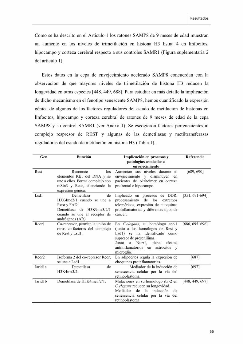
Resultados
Como se ha descrito en el Artículo 1 los ratones SAMP8 de 9 meses de edad muestran
un aumento en los niveles de trimetilación en histona H3 lisina 4 en linfocitos,
hipocampo y corteza cerebral respecto a sus controles SAMR1 (Figura suplementaria 2
del artículo 1).
Estos datos en la cepa de envejecimiento acelerado SAMP8 concuerdan con la
observación de que mayores niveles de trimetilación de histona H3 reducen la
longevidad en otras especies [448, 449, 688]. Para estudiar en más detalle la implicación
de dicho mecanismo en el fenotipo senescente SAMP8, hemos cuantificado la expresión
génica de algunos de los factores reguladores del estado de metilación de histonas en
linfocitos, hipocampo y corteza cerebral de ratones de 9 meses de edad de la cepa
SAMP8 y su control SAMR1 (ver Anexo 1). Se escogieron factores pertenecientes al
complejo respresor de REST y algunas de las demetilasas y metiltransferasas
reguladoras del estado de metilación en histona H3 (Tabla 1).
Gen Función Implicación en procesos y patologías asociadas a
envejecimiento
Referencia
Rest Reconoce los elementos RE1 del DNA y se une a ellos. Forma complejo con mSin3 y Rcor, silenciando la expresión génica.
Aumentan sus niveles durante el envejecimiento y disminuyen en pacientes de Alzheimer en corteza prefrontal e hipocampo.
[689, 690]
Lsd1 Demetilasa de H3K4me2/1 cuando se une a Rcor y FAD. Demetilasa de H3K9me3/2/1 cuando se une al receptor de andrógenos (AR).
Implicado en procesos de DDR, procesamiento de los extremos teloméricos, expresión de citoquinas proinflamatorias y diferentes tipos de cáncer.
[351, 691-694]
Rcor1 Co-represor, permite la unión de otros co-factores del complejo de Rest y Lsd1.
En C.elegans, su homólogo spr-1 (junto a los homólogos de Rest y Lsd1) se ha identificado como supresor de presenilinas. Junto a Nurr1, tiene efectos antiinflamatorios en astrocitos y microglia.
[686, 695, 696]
Rcor2 Isoforma 2 del co-represor Rcor, se une a Lsd1.
En adipocitos regula la expresión de citoquinas proinflamatorias.
[687]
Jarid1a Demetilasa de H3K4me3/2.
Mediador de la inducción de senescencia celular por la vía del retinoblastoma.
[697]
Jarid1b Demetilasa de H3K4me3/2/1. Mutaciones en su homólogo rbr-2 en C.elegans reducen su longevidad. Mediador de la inducción de senescencia celular por la vía del retinoblastoma.
[448, 449, 697]
66
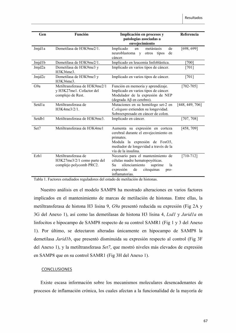
Resultados
Gen Función Implicación en procesos y patologías asociadas a
envejecimiento
Referencia
Jmjd1a Demetilasa de H3K9me2/1. Implicado en metástasis de neuroblastoma y otros tipos de cáncer.
[698, 699]
Jmjd1b Demetilasa de H3K9me2/1. Implicado en leucemia linfoblástica. [700] Jmjd2a Demetilasa de H3K9me3 y
H3K36me3. Implicado en varios tipos de cáncer. [701]
Jmjd2c Demetilasa de H3K9me3 y H3K36me3.
Implicado en varios tipos de cáncer. [701]
G9a Metiltransferasa de H3K9me2/1 y H3K27me1. Cofactor del complejo de Rest.
Función en memoria y aprendizaje. Implicado en varios tipos de cáncer. Modulador de la expresión de NEP (degrada Aβ en cerebro).
[702-705]
Setd1a Metiltransferasa de H3K4me3/2/1.
Mutaciones en su homólogo set-2 en C.elegans extienden su longevidad. Sobreexpresado en cáncer de colon.
[448, 449, 706]
Setdb1 Metiltransferasa de H3K9me3. Implicado en cáncer.
[707, 708]
Set7 Metiltransferasa de H3K4me1 Aumenta su expresión en corteza cerebral durante el envejecimiento en primates. Modula la expresión de FoxO3, mediador de longevidad a través de la vía de la insulina.
[458, 709]
Ezh1 Metiltransferasa de H3K27me3/2/1 como parte del complejo polycomb PRC2.
Necesario para el mantenimiento de células madre hematopoyéticas. Su silenciamiento suprime la expresión de citoquinas pro-inflamatorias.
[710-712]
Tabla 1. Factores estudiados reguladores del estado de metilación de histonas.
Nuestro análisis en el modelo SAMP8 ha mostrado alteraciones en varios factores
implicados en el mantenimiento de marcas de metilación de histonas. Entre ellas, la
metiltransferasa de histona H3 lisina 9, G9a presentó reducida su expresión (Fig 2A y
3G del Anexo 1), así como las demetilasas de histona H3 lisina 4, Lsd1 y Jarid1a en
linfocitos e hipocampo de SAMP8 respecto de su control SAMR1 (Fig 1 y 3 del Anexo
1). Por último, se detectaron alteradas únicamente en hipocampo de SAMP8 la
demetilasa Jarid1b, que presentó disminuida su expresión respecto al control (Fig 3F
del Anexo 1), y la metiltransferasa Set7, que mostró niveles más elevados de expresión
en SAMP8 que en su control SAMR1 (Fig 3H del Anexo 1).
CONCLUSIONES
Existe escasa información sobre los mecanismos moleculares desencadenantes de
procesos de inflamación crónica, los cuales afectan a la funcionalidad de la mayoría de
67
Resultados
órganos y tejidos, y que dan lugar a patologías asociadas a envejecimiento y
neurodegeneración [713, 714].
Nuestro estudio muestra una reducción de Rcor2 en diversos tejidos en la cepa
SAMP8, alteración presente desde los primeros días de vida en linfocitos y astrocitos.
RCOR forma parte del complejo de REST, cuyo papel en la preservación de cognición,
el silenciamiento de genes proapoptóticos y la promoción de factores de transcripción
de resistencia a estrés oxidativo se ha identificado recientemente en modelos animales y
humanos de edad avanzada [690]. Estos datos sugieren que la reducción de Rcor2 en
SAMP8 podría estar ligada a su fenotipo senescente. Además, hemos demostrado que la
reducción de Rcor2 correlaciona con el aumento de la citoquina proinflamatoria Il6 en
astrocitos. Por ello, Rcor2 debería ser considerado como una posible diana para la
prevención y/o tratamiento de la inflamación crónica asociada a envejecimiento.
Paralelamente, y teniendo en cuenta el resultado mostrado en el artículo 1, donde se
detectaron mayores niveles de trimetilación en histona H3 lisina 4, y que esta marca
epigénetica parece tener un papel importante en longevidad y envejecimiento [448,
449]; estamos estudiando las posibles alteraciones presentes en factores de regulación
del estado de metilación de histonas. Hemos detectado una disminución de expresión en
linfocitos e hipocampo en ratones SAMP8 de la metiltransferasa G9a. Esta
metiltransferasa está implicada en silenciamiento génico y actúa conjuntamente en el
complejo correpresor de REST con la demetilasa LSD1, la cual también hemos
detectado reducida en linfocitos e hipocampo de SAMP8. Estos datos podrían indicar la
existencia de una alteración en la formación y actividad de este complejo lo que daría
como resultado la desregulación en funciones tan importantes como la diferenciación
neuronal y plasticidad [715-718]. Además se ha identificado a G9a como uno de los
factores de regulación de la enzima neprilisina [705], responsable de la eliminación de
Aβ en cerebro y una de las principales dianas terapéuticas estudiadas para la EA [719].
Asimismo, encontramos menores niveles de expresión de las demetilasas Jarid1a y
Jarid1b, las cuales son homólogas a rbr-2. Greer y sus colaboradores mostraron en
trabajos previos [448] que el silenciamiento de expresión de rbr-2 va acompañado de un
aumento en los niveles de trimetilación en H3 lisina 4 y de una reducción significativa
68

Resultados
de la esperanza de vida en C.elegans, demostrando ser un factor crucial en la regulación
de la longevidad. En conjunto, el aumento detectado en los ratones SAMP8 en la
trimetilación de histona H3 y la reducción de expresión de la metiltransferasa G9a y de
las demetilasas Jarid1a y Jarid1b nos sugieren la importante implicación del estado de
metilación de histona H3 en la reducida longevidad y déficits cognitivos que muestran
los ratones SAMP8. (*Esta parte del trabajo se está completando actualmente para su
futura publicación).
69
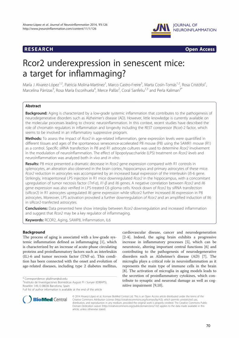
RESEARCH Open Access
Rcor2 underexpression in senescent mice:a target for inflammaging?María J Alvarez-López1,2, Patricia Molina-Martínez1, Marco Castro-Freire3, Marta Cosín-Tomás1,2, Rosa Cristòfol1,Marcelina Párrizas3, Rosa María Escorihuela4, Merce Pallàs2, Coral Sanfeliu1,3 and Perla Kaliman3*
Abstract
Background: Aging is characterized by a low-grade systemic inflammation that contributes to the pathogenesis ofneurodegenerative disorders such as Alzheimer’s disease (AD). However, little knowledge is currently available onthe molecular processes leading to chronic neuroinflammation. In this context, recent studies have described therole of chromatin regulators in inflammation and longevity including the REST corepressor (Rcor)-2 factor, whichseems to be involved in an inflammatory suppressive program.
Methods: To assess the impact of Rcor2 in age-related inflammation, gene expression levels were quantified indifferent tissues and ages of the spontaneous senescence-accelerated P8 mouse (P8) using the SAMR1 mouse (R1)as a control. Specific siRNA transfection in P8 and R1 astrocyte cultures was used to determine Rcor2 involvementin the modulation of neuroinflammation. The effect of lipopolysaccharide (LPS) treatment on Rcor2 levels andneuroinflammation was analyzed both in vivo and in vitro.
Results: P8 mice presented a dramatic decrease in Rcor2 gene expression compared with R1 controls insplenocytes, an alteration also observed in the brain cortex, hippocampus and primary astrocytes of these mice.Rcor2 reduction in astrocytes was accompanied by an increased basal expression of the interleukin (Il)-6 gene.Strikingly, intraperitoneal LPS injection in R1 mice downregulated Rcor2 in the hippocampus, with a concomitantupregulation of tumor necrosis factor (Tnf-α), Il1-β and Il6 genes. A negative correlation between Rcor2 and Il6gene expression was also verified in LPS-treated C6 glioma cells. Knock down of Rcor2 by siRNA transfection(siRcor2) in R1 astrocytes upregulated Il6 gene expression while siRcor2 further increased Il6 expression in P8astrocytes. Moreover, LPS activation provoked a further downregulation of Rcor2 and an amplified induction of Il6in siRcor2-tranfected astrocytes.
Conclusions: Data presented here show interplay between Rcor2 downregulation and increased inflammationand suggest that Rcor2 may be a key regulator of inflammaging.
Keywords: RCOR2, Aging, SAMP8, Inflammation, IL6
BackgroundThe process of aging is associated with a low-grade sys-temic inflammation defined as inflammaging [1], whichis characterized by an increase of acute-phase circulatingproteins and proinflammatory factors such as interleukin(IL)-6 and tumor necrosis factor (TNF-α). This condi-tion has been connected with the onset and evolution ofage-related diseases, including type 2 diabetes mellitus,
cardiovascular disease, cancer and neurodegeneration[2-4]. Indeed, the aging brain exhibits a progressiveincrease in inflammatory processes [5], which can beneurotoxic, altering important central functions [6] andcontributing to the pathogenesis of neurodegenerativedisorders such as Alzheimer’s disease (AD) [7]. Themicroglia plays a critical role in neuroinflammation as itrepresents the main type of immune cells in the brain[8]. The activation of microglia in aging models leads tothe secretion of proinflammatory cytokines, which con-tribute to synaptic and neuronal damage as well as cog-nitive impairment [9,10].
* Correspondence: [email protected] de Investigaciones Biomédicas August Pi i Sunyer (IDIBAPS),Rosellón 149, E-08036 Barcelona, SpainFull list of author information is available at the end of the article
JOURNAL OF NEUROINFLAMMATION
© 2014 Alvarez-López et al.; licensee BioMed Central Ltd. This is an Open Access article distributed under the terms of theCreative Commons Attribution License (http://creativecommons.org/licenses/by/4.0), which permits unrestricted use,distribution, and reproduction in any medium, provided the original work is properly credited. The Creative Commons PublicDomain Dedication waiver (http://creativecommons.org/publicdomain/zero/1.0/) applies to the data made available in thisarticle, unless otherwise stated.
Alvarez-López et al. Journal of Neuroinflammation 2014, 11:126http://www.jneuroinflammation.com/content/11/1/126
70
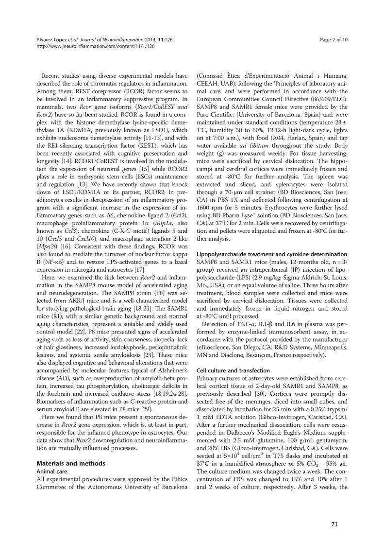
Recent studies using diverse experimental models havedescribed the role of chromatin regulators in inflammation.Among them, REST corepressor (RCOR) factor seems tobe involved in an inflammatory suppressive program. Inmammals, two Rcor gene isoforms (Rcor1/CoREST andRcor2) have so far been studied. RCOR is found in a com-plex with the histone demethylase lysine-specific deme-thylase 1A (KDM1A, previously known as LSD1), whichexhibits nucleosome demethylase activity [11-13], and withthe RE1-silencing transcription factor (REST), which hasbeen recently associated with cognitive preservation andlongevity [14]. RCOR1/CoREST is involved in the modula-tion the expression of neuronal genes [15] while RCOR2plays a role in embryonic stem cells (ESCs) maintenanceand regulation [13]. We have recently shown that knockdown of LSD1/KDM1A or its partner, RCOR2, in pre-adipocytes results in derepression of an inflammatory pro-gram with a significant increase in the expression of in-flammatory genes such as Il6, chemokine ligand 2 (Ccl2),macrophage proinflammatory protein 1α (Mip1a, alsoknown as Ccl3), chemokine (C-X-C motif) ligands 5 and10 (Cxcl5 and Cxcl10), and macrophage activation 2-like(Mpa2l) [16]. Consistent with these findings, RCOR wasalso found to mediate the turnover of nuclear factor kappaB (NF-κB) and to restore LPS-activated genes to a basalexpression in microglia and astrocytes [17].
Here, we examined the link between Rcor2 and inflam-mation in the SAMP8 mouse model of accelerated agingand neurodegeneration. The SAMP8 strain (P8) was se-lected from AKR/J mice and is a well-characterized modelfor studying pathological brain aging [18-21]. The SAMR1mice (R1), with a similar genetic background and normalaging characteristics, represent a suitable and widely usedcontrol model [22]. P8 mice presented signs of acceleratedaging such as loss of activity, skin coarseness, alopecia, lackof hair glossiness, increased lordokyphosis, periophthalmiclesions, and systemic senile amyloidosis [23]. These micealso displayed cognitive and behavioral alterations that wereaccompanied by molecular features typical of Alzheimer’sdisease (AD), such as overproduction of amyloid-beta pro-tein, increased tau phosphorylation, cholinergic deficits inthe forebrain and increased oxidative stress [18,19,24-28].Biomarkers of inflammation such as C-reactive protein andserum amyloid P are elevated in P8 mice [29].
Here we found that P8 mice present a spontaneous de-crease in Rcor2 gene expression, which is, at least in part,responsible for the inflamed phenotype in astrocytes. Ourdata show that Rcor2 downregulation and neuroinflamma-tion are mutually influenced processes.
Materials and methodsAnimal careAll experimental procedures were approved by the EthicsCommittee of the Autonomous University of Barcelona
(Comissió Ètica d’Experimentació Animal i Humana,CEEAH, UAB), following the ‘Principles of laboratory ani-mal care’, and were performed in accordance with theEuropean Communities Council Directive (86/609/EEC).SAMP8 and SAMR1 female mice were provided by theParc Cientific, (University of Barcelona, Spain) and weremaintained under standard conditions (temperature 23 ±1°C, humidity 50 to 60%, 12:12-h light-dark cycle, lightson at 7:00 a.m.), with food (A04, Harlan, Spain) and tapwater available ad libitum throughout the study. Bodyweight (g) was measured weekly. For tissue harvesting,mice were sacrificed by cervical dislocation. The hippo-campi and cerebral cortices were immediately frozen andstored at -80°C for further analysis. The spleen wasextracted and sliced, and splenocytes were isolatedthrough a 70-μm cell strainer (BD Biosciences, San Jose,CA) in PBS 1X and collected following centrifugation at1600 rpm for 5 minutes. Erythrocytes were further lysedusing BD Pharm Lyse™ solution (BD Biosciences, San Jose,CA) at 37°C for 2 min. Cells were recovered by centrifuga-tion and pellets were aliquoted and frozen at -80°C for fur-ther analysis.
Lipopolysaccharide treatment and cytokine determinationSAMP8 and SAMR1 mice (males, 12-months old, n = 3/group) received an intraperitoneal (IP) injection of lipo-polysaccharide (LPS) (2.9 mg/kg; Sigma-Aldrich, St. Louis,Mo., USA), or an equal volume of saline. Three hours aftertreatment, blood samples were collected and mice weresacrificed by cervical dislocation. Tissues were collectedand immediately frozen in liquid nitrogen and storedat -80°C until processed.
Detection of TNF-α, IL1-β and IL6 in plasma was per-formed by enzyme-linked immunosorbent assay, in ac-cordance with the protocol provided by the manufacturer(eBioscience, San Diego, CA; R&D Systems, Minneapolis,MN and Diaclone, Besançon, France respectively).
Cell culture and transfectionPrimary cultures of astrocytes were established from cere-bral cortical tissue of 2-day-old SAMR1 and SAMP8, aspreviously described [30]. Cortices were promptly dis-sected free of the meninges, diced into small cubes, anddissociated by incubation for 25 min with a 0.25% trypsin/1 mM EDTA solution (Gibco-Invitrogen, Carlsbad, CA).After a further mechanical dissociation, cells were resus-pended in Dulbecco’s Modified Eagle’s Medium supple-mented with 2.5 mM glutamine, 100 g/mL gentamycin,and 20% FBS (Gibco-Invitrogen, Carlsbad, CA). Cells wereseeded at 5×10⁴ cell/cm2 in T75 flasks and incubated at37°C in a humidified atmosphere of 5% CO2 - 95% air.The culture medium was changed twice a week. The con-centration of FBS was changed to 15% and 10% after 1and 2 weeks of culture, respectively. After 3 weeks, the
Alvarez-López et al. Journal of Neuroinflammation 2014, 11:126 Page 2 of 10http://www.jneuroinflammation.com/content/11/1/126
71
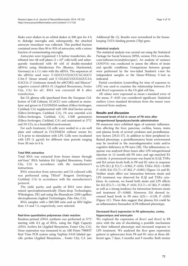
flasks were shaken in an orbital shaker at 200 rpm for 4 hto dislodge microglia and, subsequently, the attachedastrocyte monolayer was collected. This purified fractioncontained more than 90 to 95% of astrocytes, with a minorfraction of contaminating microglia.
Astrocytes were trypsinized 1 h before transfection, dis-tributed into 48-well plates (1 × 10⁵ cells/well) and subse-quently transfected with 60 nM of double-strandedsiRNAs using Metafectene Pro (Biontex, Martinsried,Germany) at a 1:1 ratio with the siRNA. The sequences ofthe siRNAs used were: 5’-GGUCUUGACUCACAGCUCAtt-3’ (Sense strand) and 5’-UGAGCUGUGAGUCAAGACCtc-3’ (Antisense strand) for siRCOR2; and Silencer™negative control siRNA #1 (Applied Biosystems, FosterCity, CA) for siC. RNA was extracted 48 h aftertransfection.
Rat C6 glioma cells (purchased from the European Col-lection of Cell Cultures, ECACC) were cultured as mono-layer and grown in F12/DMEM medium (Gibco-Invitrogen,Carlsbad, CA) supplemented with 10% fetal bovine serum(Gibco-Invitrogen, Carlsbad, CA), 1% non essential a/a’s(Gibco-Invitrogen, Carlsbad, CA), 1/500 gentamicin(Gibco-Invitrogen, Carlsbad, CA) and maintained at 37°Cand 5% CO2 in a humidified environment.
For each treatment, C6 cells were seeded in a six-wellplate and cultured in F12/DMEM without serum for12 h prior to stimulation with LPS. Cells were incubatedwith LPS (1 μg/ml) for different time periods rangingfrom 30 min to 6 h.
Total RNA extractionTotal RNA was extracted from frozen tissues throughmirVana™ RNA Isolation Kit (Applied Biosystems, FosterCity, CA) in accordance with the manufacturer’sinstructions.
RNA extraction from astrocytes and C6 cultured cellswas performed using TRIzol™ Reagent (Invitrogen,Carlsbad, CA) in accordance with the manufacturer’sprotocol.
The yield, purity, and quality of RNA were deter-mined spectrophotometrically (Nano-drop Technologies,Wilmington, DE) and using the Bioanalyzer 2100 capillaryelectrophoresis (Agilent Technologies, Palo Alto, CA).
RNA samples with a 260/280 ratio and an RIN higherthan 1.9 and 7.5, respectively, were selected.
Real-time quantitative polymerase chain reactionRandom-primed cDNA synthesis was performed at 37°Cstarting with 0.3 μg of RNA, using the High CapacitycDNA Archive kit (Applied Biosystems, Foster City, CA).Gene expression was measured in an ABI Prism 7900HTReal Time PCR system using TaqMan FAM labeled spe-cific probes (Applied Biosystems, , Foster City, CA [see
Additional file 1]). Results were normalized to the house-keeping TATA-binding protein (Tbp) gene.
Statistical analysisThe statistical analysis was carried out using the StatisticalPackage for Social Sciences (SPSS, version 19.0, www.ibm.com/software/es/analytics/spss/). An analysis of variance(ANOVA) was conducted to assess the effects of strainand specific conditions. Comparisons between groupswere performed by the two-tailed Student’s t-test forindependent samples or the Mann-Whitney U-test asindicated.
Partial correlation (controlling for time of exposure toLPS) was used to examine the relationship between Il-6and Rcor2 expression in the C6 glial cell line.
All values were expressed as mean ± standard error ofthe mean. P <0.05 was considered significant. Statisticaloutliers (≥two standard deviations from the mean) wereremoved from analyses.
Results and discussionIncreased levels of IL6 in serum of P8 mice afterintraperitoneal lipopolysaccharide administrationP8 senescent mice exhibited signs of systemic inflamma-tion affecting the liver, pancreas, cochlea, bones, testesand plasma levels of several cytokines and proinflamma-tory factors [29,31-37]. In addition to their peripheral in-flamed phenotype, a proinflammatory brain environmentmay be involved in the neurodegenerative traits and/orcognitive deficiency in P8 mice [38]. The inflammatory re-sponse was analyzed three hours after LPS intraperitonealinjection in 12 month-old P8 mice and age-matched R1controls. A pronounced increase was found in IL1β, TNFαand IL6 serum levels both in P8 and R1 mice in responseto LPS (IL1 β: F(1,7) = 0.962, P <0.05; TNFα: H(3) = 8.389,P <0.05; IL6: F(1,7) = 67.362, P <0.001) (Figure 1A and B).Neither strain effect nor interaction between strain andLPS treatment was observed for IL1β and TNFα cyto-kines. In contrast, we found both strain and LPS effectsfor IL6 (F(1,7) = 12.706, P <0.01; F(1,7) = 67.362, P <0.001)as well as a strong tendency for interaction between strainand treatment (P = 0.068). Moreover, IL6 showed in-creased basal levels in P8 mice (t(3) = −4.139; P <0.05)(Figure 1C). These data suggest that plasma IL6 could bean inflammatory biomarker of P8 inflamed phenotype.
Decreased Rcor2 expression in P8 splenocytes, cortex,hippocampus and astrocytesWe explored the expression of Rcor1 and Rcor2 in P8mice with the aim of elucidating a possible mechanismfor their inflamed phenotype and increased response toLPS treatment. We analyzed the Rcor gene expressionpattern in splenocytes from P8 and R1 mice at three dif-ferent ages: 7 days, 3 months and 9 months. Both strains
Alvarez-López et al. Journal of Neuroinflammation 2014, 11:126 Page 3 of 10http://www.jneuroinflammation.com/content/11/1/126
72
presented similar age-dependent gene expression patternsfor Rcor1 with higher levels at 7 days and 9 monthscompared with 3 months (F(2,22) = 32.306, P <0.001)(Figure 2A). In contrast, R1 mice showed an age-dependent increase in Rcor2 gene expression which wasnot observed in P8 mice (interaction between strain andage: F(2,29) = 4.279, P <0.05) (Figure 2B). Intriguingly, P8mice exhibited low expression levels of Rcor2 from thefirst week of age and throughout the adult life. These data
indicate that Rcor2 underexpression in splenocytes pre-cedes most of the accelerated-aging characteristics of thismodel. Indeed, we found a marked effect for strain andage in Rcor2 gene expression (F(1,29) = 31.620, P <0.001;F(2,29) = 3.363, P <0.001; respectively).
Chronic inflammation is associated with early develop-ment and acceleration of neurodegenerative diseases suchas Alzheimer’s disease [39,40]. Therefore, we analyzed theexpression of Rcor1 and Rcor2 in P8 cortices and hippo-campi. Rcor1 presented age effect in both cortex and hippo-campus tissues (F(2,27) = 49.140, P <0.001; F(2,32) = 12.597,P <0.001; respectively) with a similar age-dependent ex-pression pattern in both strains (Figure 3A and B). How-ever, 9-month-old P8 mice, unlike R1 mice, presentedslight but significant alterations in Rcor1 levels in the cor-tex and in the hippocampus. On the other hand, Rcor2was expressed similarly in R1 and P8 brain cortices andhippocampi at 7 days and 3 months of age (Figure 3Cand D).
Remarkably, a consistent characteristic among spleno-cytes, cortex and hippocampus was that Rcor2 gene ex-pression in 9-month-old P8 mice decreased significantlyin comparison with R1 controls (cortex: P <0.001; hippo-campus: P <0.01).
Figure 1 Inflammatory response in serum from R1 and P8 miceafter lipopolysaccharide (LPS) administration. (A) Interleukin 1beta, (B) TNF-α serum levels, (C) Interleukin 6. Cytokine were measuredby ELISA (n = 3/group). Mean ± standard error are represented; Two-wayANOVA results are indicated as *P <0.05; **P <0.01; ***P <0.001.
Figure 2 Rcor gene expression profile at different ages insplenocytes from R1 and P8 mice. (A) Rcor1 and (B) Rcor2 geneexpression at the ages of 7 days, 3 months and 9 months (n = 3 to10/group). Gene expression levels were determined by real time PCRusing TaqMan FAM-labeled specific probes and expressed relative toTbp. Mean ± standard error are represented; Two-way ANOVA resultsare indicated as *P <0.05; ***P <0.001.
Alvarez-López et al. Journal of Neuroinflammation 2014, 11:126 Page 4 of 10http://www.jneuroinflammation.com/content/11/1/126
73
We also explored the gene expression of Rcor1 andRcor2 in P8 and R1 astrocytes obtained from cerebralcortical tissue of 2-day-old mice. P8 cells showed similarRcor1 but decreased Rcor2 gene expression in com-parison with R1 cells (t(11) = 5,837; P <0.001) (Figure 3Eand F). Notably, the reduced levels of Rcor2 in P8 com-pared with R1 astrocytes were accompanied by in-creased basal levels of Il6 gene expression (t(3) = −4.734;P = 0.016) (Figure 3G).
These data show that Rcor2 is underexpressed in dif-ferent tissues and organs in P8 mice and, in some cases(namely, in splenocytes and astrocytes), this alteration isdetected from early ages. Interestingly, Yankner andcolleagues have recently shown that the transcriptionalrepressor REST (RE1-silencing transcription factor) is cru-cial for neuroprotection during aging [14]. It is known thatREST recruitment of RCOR corepressor is required for itsgene silencing activity [15]. The downregulation of Rcor2in immune and brain cells of the P8 senescent mice
strongly suggests that the accelerated aging in these mice,as well as their neurodegenerative phenotype, may be re-lated to Rcor2 deficiency.
On the other hand, RCOR is part of a chromatin remo-deling complex containing the lysine specific demethylase(KDM1A), which catalyzes H3 histone demethylation as away to repress transcription [12]. In the absence of RCOR,KDM1A demethylase activity is impaired with a concomi-tant increase in H3 histone methylation levels. In this con-text, preliminary data from our group indicate that thealteration in histone methylation levels is a candidatemechanism through which Rcor2 could also influence theaging process in P8 mice. Indeed, P8 mice presentincreased levels of trimethylated histone H3 lysine 4(H3K4me3) in splenocytes, cortex, and hippocampus(supplementary information, [see Additional file 2A-C]).Moreover, consistent with a role of Rcor in methylationsignaling and in neuroinflammation, we have found thatIl6 upregulation in response to LPS is significantly
Figure 3 Rcor gene expression profile in brain tissues from R1 and P8 mice. Rcor1 and Rcor2 gene expression was quantified in the cortex(A and C), in the hippocampus (B and D) at the ages 7 days, 3 months and 9 months (n = 4 to 8/group), and in primary cultured astrocytes(n = 6 to 9/group) (E and F) from 2 days-old mice. (G) Interleukin 6 basal gene expression levels in primary cultured astrocytes obtained from2-day-old mice (n = 4/group). Gene expression levels were measured by real time PCR. Mean ± standard error are represented; Two-way ANOVAresults are indicated as *P <0.05; **P <0.01; ***P <0.001.
Alvarez-López et al. Journal of Neuroinflammation 2014, 11:126 Page 5 of 10http://www.jneuroinflammation.com/content/11/1/126
74
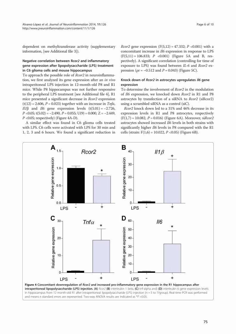
dependent on methyltransferase activity (supplementaryinformation, [see Additional file 3]).
Negative correlation between Rcor2 and inflammatorygene expression after lipopolysaccharide (LPS) treatmentin C6 glioma cells and mouse hippocampusTo approach the possible role of Rcor2 in neuroinflamma-tion, we first analyzed its gene expression after an in vivointraperitoneal LPS injection in 12-month-old P8 and R1mice. While P8 hippocampus was not further responsiveto the peripheral LPS treatment [see Additional file 4], R1mice presented a significant decrease in Rcor2 expression(t(12) = 2.606, P = 0.023) together with an increase in Tnfα,Il1β and Il6 gene expression levels (t(5.01) = −2.726,P <0.05; t(5.02) = −2.490, P = 0.055; U(9) = 0.000, Z = −2.449,P <0.05; respectively) (Figure 4A-D).
A similar effect was found in C6 glioma cells treatedwith LPS. C6 cells were activated with LPS for 30 min and1, 2, 3 and 6 hours. We found a significant reduction in
Rcor2 gene expression (F(5,12) = 47.332; P <0.001) with aconcomitant increase in Il6 expression in response to LPS(F(5,11) = 106.833; P <0.001) (Figure 5A and B, res-pectively). A significant correlation (controlling for time ofexposure to LPS) was found between IL-6 and Rcor2 ex-pression (ρr = −0.512 and P = 0.043) (Figure 5C).
Knock down of Rcor2 in astrocytes upregulates Il6 geneexpressionTo determine the involvement of Rcor2 in the modulationof Il6 expression, we knocked down Rcor2 in R1 and P8astrocytes by transfection of a siRNA to Rcor2 (siRcor2)using a scrambled siRNA as a control (siC).
Rcor2 knock down led to a 31% and 46% decrease in itsexpression levels in R1 and P8 astrocytes, respectively(F(1,7) = 10.082, P = 0.016) (Figure 6A). Moreover, siRcor2astrocytes showed increased Il6 levels in both strains withsignificantly higher Il6 levels in P8 compared with the R1cells (strain: F(1,6) = 10.022, P <0.05) (Figure 6B).
Figure 4 Concomitant downregulation of Rcor2 and increased pro-inflammatory gene expression in the R1 hippocampus afterintraperitoneal lipopolysaccharide (LPS) injection. (A) Rcor2 (B) interleukin 1 beta, (C) tnf-alpha and) (D) interleukin 6 gene expression levelsin hippocampus from 12 month-old R1 after intraperitoneal lipopolysaccharide (LPS) injection (n = 5 to 7/group). Real-time PCR was performedand means ± standard errors are represented. Two-way ANOVA results are indicated as *P <0.05.
Alvarez-López et al. Journal of Neuroinflammation 2014, 11:126 Page 6 of 10http://www.jneuroinflammation.com/content/11/1/126
75
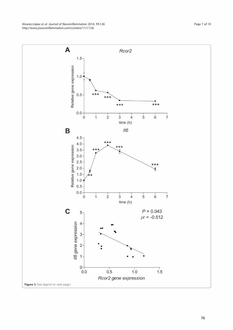
Figure 5 (See legend on next page.)
Alvarez-López et al. Journal of Neuroinflammation 2014, 11:126 Page 7 of 10http://www.jneuroinflammation.com/content/11/1/126
76
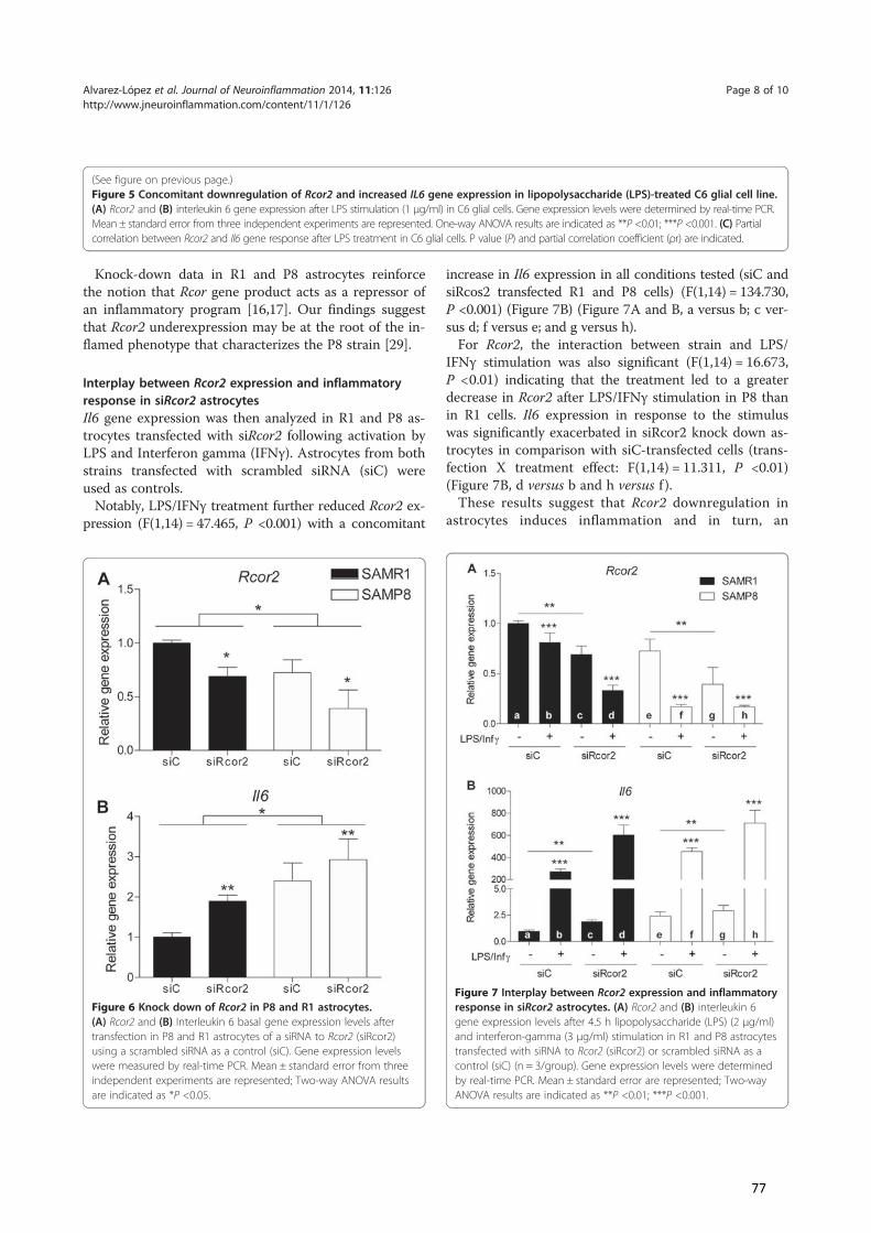
Knock-down data in R1 and P8 astrocytes reinforcethe notion that Rcor gene product acts as a repressor ofan inflammatory program [16,17]. Our findings suggestthat Rcor2 underexpression may be at the root of the in-flamed phenotype that characterizes the P8 strain [29].
Interplay between Rcor2 expression and inflammatoryresponse in siRcor2 astrocytesIl6 gene expression was then analyzed in R1 and P8 as-trocytes transfected with siRcor2 following activation byLPS and Interferon gamma (IFNγ). Astrocytes from bothstrains transfected with scrambled siRNA (siC) wereused as controls.
Notably, LPS/IFNγ treatment further reduced Rcor2 ex-pression (F(1,14) = 47.465, P <0.001) with a concomitant
increase in Il6 expression in all conditions tested (siC andsiRcos2 transfected R1 and P8 cells) (F(1,14) = 134.730,P <0.001) (Figure 7B) (Figure 7A and B, a versus b; c ver-sus d; f versus e; and g versus h).
For Rcor2, the interaction between strain and LPS/IFNγ stimulation was also significant (F(1,14) = 16.673,P <0.01) indicating that the treatment led to a greaterdecrease in Rcor2 after LPS/IFNγ stimulation in P8 thanin R1 cells. Il6 expression in response to the stimuluswas significantly exacerbated in siRcor2 knock down as-trocytes in comparison with siC-transfected cells (trans-fection X treatment effect: F(1,14) = 11.311, P <0.01)(Figure 7B, d versus b and h versus f ).
These results suggest that Rcor2 downregulation inastrocytes induces inflammation and in turn, an
Figure 7 Interplay between Rcor2 expression and inflammatoryresponse in siRcor2 astrocytes. (A) Rcor2 and (B) interleukin 6gene expression levels after 4.5 h lipopolysaccharide (LPS) (2 μg/ml)and interferon-gamma (3 μg/ml) stimulation in R1 and P8 astrocytestransfected with siRNA to Rcor2 (siRcor2) or scrambled siRNA as acontrol (siC) (n = 3/group). Gene expression levels were determinedby real-time PCR. Mean ± standard error are represented; Two-wayANOVA results are indicated as **P <0.01; ***P <0.001.
Figure 6 Knock down of Rcor2 in P8 and R1 astrocytes.(A) Rcor2 and (B) Interleukin 6 basal gene expression levels aftertransfection in P8 and R1 astrocytes of a siRNA to Rcor2 (siRcor2)using a scrambled siRNA as a control (siC). Gene expression levelswere measured by real-time PCR. Mean ± standard error from threeindependent experiments are represented; Two-way ANOVA resultsare indicated as *P <0.05.
(See figure on previous page.)Figure 5 Concomitant downregulation of Rcor2 and increased IL6 gene expression in lipopolysaccharide (LPS)-treated C6 glial cell line.(A) Rcor2 and (B) interleukin 6 gene expression after LPS stimulation (1 μg/ml) in C6 glial cells. Gene expression levels were determined by real-time PCR.Mean ± standard error from three independent experiments are represented. One-way ANOVA results are indicated as **P <0.01; ***P <0.001. (C) Partialcorrelation between Rcor2 and Il6 gene response after LPS treatment in C6 glial cells. P value (P) and partial correlation coefficient (ρr) are indicated.
Alvarez-López et al. Journal of Neuroinflammation 2014, 11:126 Page 8 of 10http://www.jneuroinflammation.com/content/11/1/126
77
inflammatory environment further downregulates Rcor2.We can conclude from our data on transfected astrocytesthat the LPS/INFγ stimulus and Rcor2 knock down repre-sent independent modulators of Il6 expression and show asynergic effect when combined.
ConclusionsLittle information is currently available on the moleculartriggers of chronic low-grade inflammation, a processthat affects the function of most organs and tissues in-cluding the central nervous system and is involved inaging and neurodegeneration [41,42].
RCOR is found in a complex with RE1-silencing tran-scription factor (REST), which plays a role in protectingthe brain from age-related insults in animal models anda role in preservation of cognitive function and longevityin aging humans [14]. Taking into account the earlydownregulation of RCOR2 in the P8 mouse model of ac-celerated aging and neurodegeneration, further studiesare warranted so as to examine whether the REST/RCOR complex is involved in the shorter lifespan of P8mice as well as the role of RCOR2 in human cognitionand longevity.
Our study indicates that neuroinflammation is a pa-thophysiological outcome of Rcor2 downregulation inthe SAMP8 mice. The fact that Rcor2 and Il6 presentedan opposite response after an inflammatory stimulus inhippocampus and in glial cells, suggests a possible inter-play between the pathways involved in the regulation ofboth genes. This notion is reinforced by our data sho-wing that Il6 gene is upregulated by Rcor2 knock downin astrocytes. Therefore, RCOR2 may represent a poten-tial target for the prevention or treatment of age-relatedchronic inflammation and neurodegeneration.
Additional files
Additional file 1: List of primers and probe sets used for real timeRT-PCR analysis.
Additional file 2: Histone H3 methylation pattern in R1 and P8tissues.
Additional file 3: Methyltranferase inhibitor MTA reduceslipopolysaccharide (LPS)-induced Il6 gene expression in C6 gliomacells.
Additional file 4: Pro‐inflammatory gene expression response inP8 hippocampus after intraperitoneal lipopolysaccharide (LPS)injection.
AbbreviationsRcor: REST corepressor; P8: senescence-accelerated prone 8 mouse;R1: senescence-accelerated resistant 1; LPS: lipopolysaccharide; IL: interleukin;Tnf-α: tumor necrosis factor; AD: Alzheimer’s disease; KDM1A: histonedemethylase lysine-specific demethylase 1A; REST: RE1-silencing transcriptionfactor; ESCs: embryonic stem cells; Ccl2: chemokine ligand 2;Mip1a: macrophage proinflammatory protein 1α; Cxcl: chemokine (C-X-Cmotif) ligands; NF-κB: nuclear factor kappa B; IFNγ: Interferon gamma;Rcor: REST corepressor; P8: senescence-accelerated prone 8 mouse;
R1: senescence-accelerated resistant 1; LPS: lipopolysaccharide; IL: interleukin;Tnf-α: tumor necrosis factor; AD: Alzheimer’s disease; KDM1A: histonedemethylase lysine-specific demethylase 1A; REST: RE1-silencing transcriptionfactor; ESCs: embryonic stem cells; Ccl2: chemokine ligand 2;Mip1a: macrophage proinflammatory protein 1α; Cxcl: chemokine (C-X-Cmotif) ligands; NF-κB: nuclear factor kappa B; IFNγ: Interferon gamma.
Competing interestsThe authors declare that they have no competing interests.
Authors’ contributionsDesigned research: MJAL and PK; performed research: MJAL, PMM, MCF,MCT, MPar, RC, RME, MP and PK; contributed new reagents/analytic tools:MPar, RME, MP, CS and PK; analyzed data MJAL, MCF, MCT, MPar, RC, CS, andPK; wrote the manuscript: MJAL and PK. All authors read and approved thefinal manuscript.
Author details1Institut d’Investigacions Biomèdiques de Barcelona (IIBB), CSIC, Rosselló 161,E-08036 Barcelona, Spain. 2Unidad de Farmacología y Farmacognósia. Institutde Biomedicina (IBUB), Universidad de Barcelona y CIBERNED. Facultad deFarmacia, Av Diagonal, 643, E-08028 Barcelona, Spain. 3Instituto deInvestigaciones Biomédicas August Pi i Sunyer (IDIBAPS), Rosellón 149,E-08036 Barcelona, Spain. 4Instituto de Neurociencias, Departamento dePsiquiatría y Medicina Legal. Facultad de Medicina, Universitat Autònoma deBarcelona, Campus de Bellaterra, Bellaterra (Cerdanyola del Vallès), E-08193Barcelona, Spain.
Received: 15 April 2014 Accepted: 7 July 2014Published: 23 July 2014
References1. Franceschi C, Bonafe M, Valensin S, Olivieri F, De Luca M, Ottaviani E,
De Benedictis G: Inflamm-aging: an evolutionary perspective onimmunosenescence. Ann N Y Acad Sci 2000, 908:244–254.
2. Cevenini E, Monti D, Franceschi C: Inflamm-ageing. Curr Opin Clin NutrMetab Care 2013, 16:14–20.
3. Vasto S, Candore G, Balistreri CR, Caruso M, Colonna-Romano G, GrimaldiMP, Listi F, Nuzzo D, Lio D, Caruso C: Inflammatory networks in ageing,age-related diseases and longevity. Mech Ageing Dev 2007, 128:83–91.
4. Kaliman P, Parrizas M: Obesity and systemic inflammation: insight onepigenetic mechanisms. In Role of the Adipocyte in the Development of Type2 Diabetes, Volume 1st. Edited by Rijeka CC. Croatia: InTech; 2011:65–88.
5. Pizza V, Agresta A, D’Acunto CW, Festa M, Capasso A: Neuroinflammationand ageing: current theories and an overview of the data. Rev Recent ClinTrials 2011, 6:189–203.
6. Liu Y, Zhang M, Hao W, Mihaljevic I, Liu X, Xie K, Walter S, Fassbender K:Matrix metalloproteinase-12 contributes to neuroinflammation in theaged brain. Neurobiol Aging 2013, 34:1231–1239.
7. McGeer PL, McGeer EG: Inflammation and the degenerative diseases ofaging. Ann N Y Acad Sci 2004, 1035:104–116.
8. Lee M: Neurotransmitters and microglial-mediated neuroinflammation.Curr Protein Pept Sci 2013, 14:21–32.
9. Bardou I, Brothers HM, Kaercher RM, Hopp SC, Wenk GL: Differential effectsof duration and age on the consequences of neuroinflammation in thehippocampus. Neurobiol Aging 2013, 34:2293–2301.
10. Farso M, Menard C, Colby-Milley J, Quirion R: Immune marker CD68correlates with cognitive impairment in normally aged rats. NeurobiolAging 2013, 34:1971–1976.
11. Lee MG, Wynder C, Cooch N, Shiekhattar R: An essential role for CoRESTin nucleosomal histone 3 lysine 4 demethylation. Nature 2005,437:432–435.
12. Shi Y, Lan F, Matson C, Mulligan P, Whetstine JR, Cole PA, Casero RA, Shi Y:Histone demethylation mediated by the nuclear amine oxidase homologLSD1. Cell 2004, 119:941–953.
13. Yang P, Wang Y, Chen J, Li H, Kang L, Zhang Y, Chen S, Zhu B, Gao S:RCOR2 is a subunit of the LSD1 complex that regulates ESC propertyand substitutes for SOX2 in reprogramming somatic cells topluripotency. Stem Cells 2011, 29:791–801.
Alvarez-López et al. Journal of Neuroinflammation 2014, 11:126 Page 9 of 10http://www.jneuroinflammation.com/content/11/1/126
78
14. Lu T, Aron L, Zullo J, Pan Y, Kim H, Chen Y, Yang TH, Kim HM, Drake D,Liu XS, Bennett DA, Colaiácovo MP, Yankner BA: REST and stress resistancein ageing and Alzheimer’s disease. Nature 2014, 507:448–454.
15. Andres ME, Burger C, Peral-Rubio MJ, Battaglioli E, Anderson ME, Grimes J,Dallman J, Ballas N, Mandel G: CoREST: a functional corepressor requiredfor regulation of neural-specific gene expression. Proc Natl Acad Sci U S A1999, 96:9873–9878.
16. Hanzu FA, Musri MM, Sanchez-Herrero A, Claret M, Esteban Y, Kaliman P, GomisR, Parrizas M: Histone demethylase KDM1A represses inflammatory geneexpression in preadipocytes. Obesity (Silver Spring) 2013, 21:E616–E625.
17. Saijo K, Winner B, Carson CT, Collier JG, Boyer L, Rosenfeld MG, Gage FH,Glass CK: A Nurr1/CoREST pathway in microglia and astrocytes protectsdopaminergic neurons from inflammation-induced death. Cell 2009,137:47–59.
18. Cheng XR, Zhou WX, Zhang YX: The behavioral, pathological andtherapeutic features of the senescence-accelerated mouse prone8 strain as an Alzheimer’s disease animal model. Ageing Res Rev 2014,13:13–37.
19. Morley JE, Farr SA, Kumar VB, Armbrecht HJ: The SAMP8 mouse: a modelto develop therapeutic interventions for Alzheimer’s disease. Curr PharmDes 2012, 18:1123–1130.
20. Pallas M, Camins A, Smith MA, Perry G, Lee HG, Casadesus G: From agingto Alzheimer’s disease: unveiling “the switch” with the senescence-accelerated mouse model (SAMP8). J Alzheimers Dis 2008, 15:615–624.
21. Wang H, Lian K, Han B, Wang Y, Kuo SH, Geng Y, Qiang J, Sun M, Wang M:Age-related alterations in the metabolic profile in the hippocampus ofthe senescence-accelerated mouse prone 8: a spontaneous Alzheimer’sdisease mouse model. J Alzheimers Dis 2014, 39:841–848.
22. Takeda T, Hosokawa M, Higuchi K: Senescence-accelerated mouse (SAM):a novel murine model of accelerated senescence. J Am Geriatr Soc 1991,39:911–919.
23. Nomura Y, Okuma Y: Age-related defects in lifespan and learning abilityin SAMP8 mice. Neurobiol Aging 1999, 20:111–115.
24. Morley JE, Armbrecht HJ, Farr SA, Kumar VB: The senescence acceleratedmouse (SAMP8) as a model for oxidative stress and Alzheimer’s disease.Biochim Biophys Acta 2012, 1822:650–656.
25. del Valle J, Duran-Vilaregut J, Manich G, Pallas M, Camins A, Vilaplana J,Pelegri C: Cerebral amyloid angiopathy, blood–brain barrier disruptionand amyloid accumulation in SAMP8 mice. Neurodegener Dis 2011,8:421–429.
26. Takeda T: Senescence-accelerated mouse (SAM) with special referencesto neurodegeneration models, SAMP8 and SAMP10 mice. Neurochem Res2009, 34:639–659.
27. Canudas AM, Gutierrez-Cuesta J, Rodriguez MI, Acuna-Castroviejo D, Sureda FX,Camins A, Pallas M: Hyperphosphorylation of microtubule-associated proteintau in senescence-accelerated mouse (SAM). Mech Ageing Dev 2005,126:1300–1304.
28. Sureda FX, Gutierrez-Cuesta J, Romeu M, Mulero M, Canudas AM, Camins A,Mallol J, Pallas M: Changes in oxidative stress parameters and neurode-generation markers in the brain of the senescence-accelerated miceSAMP-8. Exp Gerontol 2006, 41:360–367.
29. Bayram B, Nikolai S, Huebbe P, Ozcelik B, Grimm S, Grune T, Frank J,Rimbach G: Biomarkers of oxidative stress, antioxidant defence andinflammation are altered in the senescence-accelerated mouse prone 8.Age 2013, 35:1205–1217.
30. Garcia-Matas S, Gutierrez-Cuesta J, Coto-Montes A, Rubio-Acero R, Diez-Vives C,Camins A, Pallas M, Sanfeliu C, Cristofol R: Dysfunction of astrocytes insenescence-accelerated mice SAMP8 reduces their neuroprotectivecapacity. Aging Cell 2008, 7:630–640.
31. Cuesta S, Kireev R, Forman K, Garcia C, Escames G, Ariznavarreta C, Vara E,Tresguerres JA: Melatonin improves inflammation processes in liver ofsenescence-accelerated prone male mice (SAMP8). Exp Gerontol 2010,45:950–956.
32. Cuesta S, Kireev R, Garcia C, Forman K, Escames G, Vara E, Tresguerres JA:Beneficial effect of melatonin treatment on inflammation, apoptosis andoxidative stress on pancreas of a senescence accelerated mice model.Mech Ageing Dev 2011, 132:573–582.
33. Cuesta S, Kireev R, Garcia C, Forman K, Vara E, Tresguerres JA: Effect ofgrowth hormone treatment on pancreatic inflammation, oxidative stress,and apoptosis related to aging in SAMP8 mice. Rejuvenation Res 2011,14:501–512.
34. Menardo J, Tang Y, Ladrech S, Lenoir M, Casas F, Michel C, Bourien J, Ruel J,Rebillard G, Maurice T, Puel JL, Wang J: Oxidative stress, inflammation,and autophagic stress as the key mechanisms of premature age-relatedhearing loss in SAMP8 mouse cochlea. Antioxid Redox Signal 2012,16:263–274.
35. Rodriguez MI, Escames G, Lopez LC, Lopez A, Garcia JA, Ortiz F, Acuna-CastroviejoD: Chronic melatonin treatment reduces the age-dependent inflammatoryprocess in senescence-accelerated mice. J Pineal Res 2007, 42:272–279.
36. Wauquier F, Barquissau V, Leotoing L, Davicco MJ, Lebecque P, Mercier S,Philippe C, Miot-Noirault E, Chardigny JM, Morio B, Wittrant Y, Coxam V:Borage and fish oils lifelong supplementation decreases inflammationand improves bone health in a murine model of senile osteoporosis.Bone 2012, 50:553–561.
37. Zhao X, Bian Y, Sun Y, Li L, Wang L, Zhao C, Shen Y, Song Q, Qu Y, Niu S,Wu W, Gao F: Effects of moderate exercise over different phases onage-related physiological dysfunction in testes of SAMP8 mice.Exp Gerontol 2013, 48:869–880.
38. Tha KK, Okuma Y, Miyazaki H, Murayama T, Uehara T, Hatakeyama R,Hayashi Y, Nomura Y: Changes in expressions of proinflammatorycytokines IL-1beta, TNF-alpha and IL-6 in the brain of senescenceaccelerated mouse (SAM) P8. Brain Res 2000, 885:25–31.
39. Lyman M, Lloyd DG, Ji X, Vizcaychipi MP, Ma D: Neuroinflammation:the role and consequences. Neurosci Res 2014, 79C:1–12.
40. Jana M, Palencia CA, Pahan K: Fibrillar amyloid-beta peptides activatemicroglia via TLR2: implications for Alzheimer’s disease. J Immunol 2008,181:7254–7262.
41. Yaffe K, Kanaya A, Lindquist K, Simonsick EM, Harris T, Shorr RI, Tylavsky FA,Newman AB: The metabolic syndrome, inflammation, and risk ofcognitive decline. JAMA 2004, 292:2237–2242.
42. von Bernhardi R: Glial cell dysregulation: a new perspective on Alzheimerdisease. Neurotox Res 2007, 12:215–232.
doi:10.1186/1742-2094-11-126Cite this article as: Alvarez-López et al.: Rcor2 underexpression insenescent mice: a target for inflammaging? Journal of Neuroinflammation2014 11:126.
Submit your next manuscript to BioMed Centraland take full advantage of:
• Convenient online submission
• Thorough peer review
• No space constraints or color figure charges
• Immediate publication on acceptance
• Inclusion in PubMed, CAS, Scopus and Google Scholar
• Research which is freely available for redistribution
Submit your manuscript at www.biomedcentral.com/submit
Alvarez-López et al. Journal of Neuroinflammation 2014, 11:126 Page 10 of 10http://www.jneuroinflammation.com/content/11/1/126
79
Additional file 1. List of primers and probe sets used for real time RT-PCR analysis.
Gene Symbol Specie Reference (ABI) Reference Sequence ID
Il1β Mouse Mm00434228_m1 NM_008361.3
Il6 Mouse Mm00446191_m1 NM_031168.1
Rcor1 Mouse Mm01336704_m1 NM_198023.2
Rcor2 Mouse Mm00499756_m1 NM_054048.3
Tnfα Mouse Mm00443258_m1 NM_013693.2
Tbp Mouse Mm00446971_m1 NM_013684.3
Il6 Rat Rn01410330_m1 NM_012589.1
Rcor2 Rat Rn01454655_g1 NM_001013994.1
Tbp Rat Rn01455646_m1 NM_001004198.1
80
Supp(A) Msplentrimeand hdensihistoindica
Mate
Histonand qK4 QnormaAcidic(2007NaCl, centriwith 0histonHistontransfincubwasheImmuquantSciencexpre
plementary SMono‐, di‐ annocytes fromethylated hishippocampuitometry (n=ne H3 and dated as * p<0
rials and Met
ne proteins wuantification
Quantification alized to SAMc histone frac7). Briefly, tiss2 mM EDTA,ifuged 10 min0.2 M HCl andne fractions wne fractions ferred to a 0.ated overnighed and incubunoreactive btified by scannce Research, ession.
S2. Histonend tri‐methym 9 month‐stone H3 lyss (C) from 9=3/group anata was repr0.05.
thods
were extractedof methylatedKit (Epigent
MR1 levels. ctions from csues for immu, protease inhn at 400 x g. Td incubated owere immediat(5μg) were e.45μm PVDF ht at 4°C withbated with pebands were dning densitomHercules, C
H3 methylatiylation in lys‐old P8 andine 4 (H3K4month‐old nd n=6/grouresented as
d from splenod H3K4 was ptek), in acco
cortices and hunoblotting whibitor cocktaThe remaininn ice for 30 mtely frozen at electrophoretmembrane. Mh the specificeroxidase‐labedetected by ametry using QuA). Histone
ion pattern isine 4 histond R1 mice wme3) was deSAMP8 and up, respectivmean ± SE; t
ocytes using Epperformed usiordance with
hippocampi wwere lysed in il (Pierce) andg pellets wermin. After a ce‐80°C. tically analyzeMembranes wc primary antieled secondaautoradiograpuantity One® modifications
in R1 and P8ne H3 (H3K4were determetermined bSAMR1 micvely). H3K4mt‐test for ind
piQuik Total Hng the EpiQuih the manuf
were obtaineda buffer contd 1% Triton‐Xe dissolved inentrifugation,
ed on 12% bwere blocked ibodies (1:100ry antibodiesphy. Specific 1‐D analysis 4s levels were
8 tissues. 4me, H3K4mmined by ELby Western be and quantme3 was codependent sa
Histone Extrack Global Pan‐facturer’s pro
d as describetaining 50 mMX100 at 4°C fon the same buthe supernat
bis‐Tris polyacwith 5% BSA 00, Millipore)s for 1 h at rbands from W4.6.3 softwaree corrected b
me2, H3K4mLISA (n=3/gblots in corttified by scaorrected byamples resul
ction kit (Epig‐Methyl Histootocol. Data
ed by FischerM Tris HCl, 15or 15 min anduffer supplemtants containi
crylamide gein PBS for 1 ). Membranesroom temperWestern blote (Bio‐Rad USby total hist
e3) in roup); ex (B) anning total
lts are
gentek) ne H3‐were
r et al. 50 mM d then mented ng the
ls and h and s were rature. t were SA, Life tone 3
81
Suppin C6 C6 glmethreal adjus
Mate
Rat C6monoserummaintFor eafor 12expos
plementary S glioma cellsioma cells w
hylthioadenotime PCR. stment from
rials and Met
6 glioma cellsolayer and grom (Invitrogen),tained at 37°Cach treatmen2 h prior to stised to MTA (1
S3. Methyltrs were treatedosine (MTA) Mean ± statwo‐way AN
thods
s (purchased fown in F12/D, 1% non esseC and 5% CO2 t, C6 cells weimulation wit1mM) or vehic
anferase inh
d for 1 h wit(1mM) (n=6andard erroNOVA are ind
from the EuroMEM mediumential a/a’s (Gin a humidifieere seeded in h LPS (1µg/mcle during LPS
hibitor MTA
th LPS (1µg/6/group). Ger are repredicated as **
opean Collectm (Gibco‐Invitibco‐Invitrogeed environmea six‐well plal) for differentreatment wh
reduces LPS
/ml) in C6 inne expressiosented. Pai* p<0.01; ***
tion of Cell Cutrogen) suppleen), 1/500 gennt. ate and culturt time periodhen indicated
S–induced Il6
n the absencon levels werwise comp* p<0.001.
ultures, ECACCemented withntamicine (Gi
ed in F12/DMs ranging from.
6 gene expre
ce or presenere determinparisons with
C) were cultuh 10% foetal bbco‐Invitroge
MEM without m 30 min to 6
ession
nce of ned by h LSD
ured as bovine en) and
serum 6 h and
82
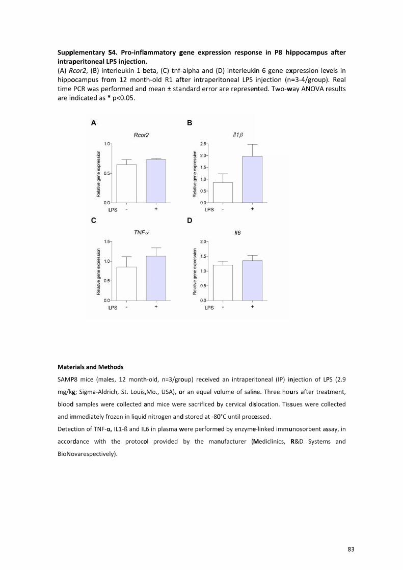
Suppintrap(A) Rhippotime are in
Mate
SAMP
mg/kg
blood
and im
Detec
accord
BioNo
plementary Speritoneal Lcor2, (B) intocampus froPCR was perndicated as *
rials and Met
P8 mice (male
g; Sigma‐Aldr
d samples wer
mmediately fr
ction of TNF‐α
dance with
ovarespective
S4. Pro‐inflaPS injectionterleukin 1 bom 12 montrformed and* p<0.05.
thods
es, 12 month
ich, St. Louis,
re collected a
rozen in liquid
α, IL1‐ß and IL
the protoco
ly).
ammatory g. beta, (C) tnf‐th‐old R1 aftd mean ± sta
h‐old, n=3/gro
,Mo., USA), o
and mice wer
d nitrogen and
6 in plasma w
ol provided
gene expres
‐alpha and (ter intraperndard error
oup) received
or an equal vo
re sacrificed b
d stored at ‐80
were performe
by the man
sion respon
D) interleukiitoneal LPS are represen
d an intraperi
olume of salin
by cervical dis
0°C until proce
ed by enzyme
nufacturer (M
se in P8 hi
in 6 gene exinjection (n=nted. Two‐w
toneal (IP) in
ne. Three hou
slocation. Tiss
essed.
e‐linked immu
Mediclinics, R
ippocampus
xpression lev=3‐4/group)
way ANOVA r
njection of LP
urs after treat
sues were co
unosorbent as
R&D System
after
vels in . Real results
PS (2.9
tment,
llected
ssay, in
s and
83

Resultados
II
El ejercicio físico a largo plazo modula la expresión génica en
hipocampo de ratones hembra senescentes
85


Resultados
Los resultados presentados en este capítulo se incluyen en el siguiente artículo
publicado:
Long-Term Exercise Modulates Hippocampal Gene Expression in Senescent
Female Mice
Journal of Alzheimer’s Disease, 33: 1177–1190 (2013)
María J Alvarez-López, Marco Castro-Freire, Marta Cosín-Tomás, Sandra
Sanchez-Roig, Jaume F. Lalanza, Jaume del Valle, Marcelina Párrizas, Antonio
Camins, Merce Pallàs, Rosa María Escorihuela y Perla Kaliman.
Contribución: Purificación de ARN y estudios de expresión génica por Real Time
PCR, análisis de los resultados obtenidos por microarrays de expresión, participación
en la interpretación de resultados y en la elaboración del manuscrito.
87
Resultados
RESUMEN
El ejercicio físico moderado es una práctica recomendada debido a los numerosos
beneficios que produce en la salud cardiovascular y metabólica, así como a nivel
cognitivo y cerebral [720]. Numerosos son los trabajos que muestran en modelos
transgénicos de Alzheimer los efectos preventivos y protectores de un estilo de vida
activo sobre la neurodegeneración. Puesto que los ratones SAMP8 son un modelo que
por sus características fenotípicas y fisiológicas está ampliamente utilizado para el
estudio del deterioro cognitivo y la enfermedad de Alzheimer espontánea [273, 721],
nos planteamos estudiar las implicaciones de una intervención de 6 meses de ejercicio
voluntario y moderado en ratones adultos SAMP8 y sus controles SAMR1.
Nuestros datos muestran que una intervención de ejercicio voluntario y moderado a
largo plazo mejora en los ratones SAMP8 rasgos fenotípicos externos asociados a
envejecimiento, por ejemplo el color de la piel o el temblor físico, y promueve la
vascularización y la expresión de genes neurotróficos en hipocampo. Además, el estudio
de genoma murino completo por medio de microarrays de expresión permitió la
identificación de posibles biomarcadores de envejecimiento sensibles al ejercicio físico.
PRINCIPALES RESULTADOS
4.3.5. 2.1. El ejercicio a largo plazo induce mejoras fenotípicas en ratones
SAMP8
Con el objetivo de evaluar los efectos de la actividad física, se analizaron los rasgos
fenotípicos que presentaban ambas cepas, SAMP8 y su control SAMR1, sedentarios o
después de 6 meses de ejercicio moderado y voluntario. Al finalizar la intervención los
ratones de todos los grupos tenían 10 meses de edad, lo cual en el caso de los SAMP8
representa un estadio muy envejecido ya que su esperanza de vida promedio es de 12
meses [721]. El fenotipo senescente propio del modelo SAMP8 afectó los niveles de
ejercicio registrados en esta cepa, que fueron menores que los de su control SAMR1
(Fig 1 del Artículo 2). La evaluación de las características fenotípicas mediante el test
SHIRPA, como el temblor o el aspecto de la piel, mostraron un claro deterioro en los
88
Resultados
animales SAMP8 respecto a su control SAMR1 que fue mejorado significativamente
tras la intervención de ejercicio en la cepa SAMP8 (Fig 2 del Artículo 2).
Por otro lado, se analizaron niveles de vascularización cerebral mediante
inmunohistoquímica utilizando laminina como marcador y se observó una reducción en
el área vascular en hipocampos de ratones SAMP8 respecto a sus controles SAMR1.
Tras la intervención de ejercicio físico, el área vascular se incrementó
significativamente en ambas cepas (Fig 3 del Artículo 2).
4.3.6. 2.2. Aumento de expresión génica de Bdnf en hipocampo de ratones
SAMP8 tras una intervención de ejercicio físico a largo plazo
La disminución de expresión de BDNF y de otros componentes de su vía de
señalización se ha asociado con procesos de envejecimiento [722-724] y con fenotipos
característicos de patologías neurodegenerativas como la enfermedad de Alzheimer
[725, 726]. El aumento de expresión de componentes de dicha vía tras intervenciones de
ejercicio físico y las mejoras cognitivas que ello conlleva ha sido descrito en numerosos
trabajos previos [658, 720]. En condiciones basales (sedentarias) los ratones SAMP8
presentaron niveles reducidos de expresión de los genes Bdnf y de su receptor Trkb
respecto de su control SAMR1 (Fig 4 del Artículo 2). El ejercicio físico incrementó la
expresión en el gen Bdnf en la cepa SAMP8 pero no en SAMR1, confirmando así la
eficacia de la intervención aplicada. Por el contrario, su receptor Trkb no mostró
diferencias asociadas a la práctica de ejercicio físico a largo plazo.
4.3.7. 2.3. Ánalisis por microarrays de expresión génica en hipocampo de
ratones SAMP8 y SAMR1 tras una intervención de ejercicio físico a largo
plazo
Con el objetivo de identificar genes asociados al proceso de envejecimiento,
modulables por la práctica de ejercicio físico, se llevaron a cabo estudios por
microarrays de expresión de genoma murino completo en hipocampo de animales
SAMP8 sedentarios y activos, utilizando ratones SAMR1 sedentarios como control de
envejecimiento normal. Todos los animales analizados tenían 10 meses de edad. Los
resultados obtenidos se representaron mediante diagramas de Venn que permitieron
identificar 34 genes como posibles genes marcadores de envejecimiento modulables por
89
Resultados
el ejercicio físico (Fig 5, Tabla 2 del Artículo 2). Utilizando la base de datos Gene
Ontology (GO) database, se estableció que dichos genes se asociaban principalmente a
vías relacionadas con el mantenimiento de matriz extracelular (Tabla 3 del Artículo 2).
Por último, validamos algunos de los genes candidatos que presentaban especial
relevancia por su funcionalidad en sistema nervioso y en la patofisiología de la EA
mediante Real Time PCR. Así determinamos la especicificidad del colágeno tipo 1 alfa
1 (Col1a1) y 2 (Col1a2), fibromodulina (Fmod), la sintasa Ptgds y la aldehído
deshidrogenasa Aldh1a2 como posibles marcadores de envejecimiento en hipocampo
que revierten sus alteraciones en respuesta al ejercicio físico moderado y mantenido
(Fig 6 del Artículo 2).
4.3.8. 2.4. Microarrays en corteza cerebral (resultados relacionados no
publicados en el artículo)
Es importante comentar que, aunque los siguientes datos no se incluyeron en el
artículo presentado, se realizaron estudios por microarrays de expresión también en
tejido de corteza cerebral, donde se hallaron 153 genes asociados a envejecimiento y
modulados por ejercicio que revelaron, de igual forma que en hipocampo, vías
relacionadas con matriz y espacio extracelular así como adhesión celular y diferentes
tipos de uniones entre células (Anexo 2). Sin embargo, decidimos no incluir estos datos
en el artículo puesto que el tejido analizado mostraba gran heterogeneidad, ya que se
trataba de corteza cerebral completa; y la validación de algunos de estos genes por real
time PCR no dio resultados significativos, posiblemente por las diferencias en la
sensibilidad de ambas técnicas.
90
Resultados
CONCLUSIONES
En este trabajo se muestran mejoras en respuesta a una práctica de ejercicio físico
moderado y voluntario a nivel fenotípico y nervioso en ratones senescentes SAMP8 de
edad avanzada. Aspectos como la vascularización de hipocampo, la expresión génica de
factores neurotróficos concretos como el Bdnf y alteraciones en el patrón global de
expresión de varios genes mediante el estudio por microarrays, ponen de manifiesto la
influencia positiva de la actividad física en el estado fisiológico del individuo durante el
envejecimiento.
Además, el estudio de las vías en las que se encuentran implicados los genes
alterados tras el ejercicio físico, ha identificado a la matriz extracelular como elemento
común de respuesta a la intervención en cerebro. Éste es un dato de gran importancia
puesto que la matriz extracelular es el soporte físico para la función, homeostasis y
supervivencia neuronal, y se han descrito alteraciones en los elementos que la
componen en modelos de neurodegeneración y enfermedad de Alzheimer [727, 728],
apuntando a que el ejercicio puede contrarrestar en parte estos procesos en sus primeras
etapas.
En conclusión, nuestro estudio ha identificado nuevos candidatos de genes
marcadores de envejecimiento sensibles a ejercicio físico y ha demostrado mejoras
fenotípicas y moleculares que sugieren la práctica de ejercicio físico moderado como un
estilo de vida saludable que puede prevenir o retardar rasgos asociados a deterioro
cognitivo durante el envejecimiento.
91
Journal of Alzheimer’s Disease 33 (2013) 1177–1190DOI 10.3233/JAD-121264IOS Press
1177
Long-Term Exercise ModulatesHippocampal Gene Expression in SenescentFemale Mice
Marıa Jesus Alvarez-Lopeza, Marco Castro-Freirea, Marta Cosın-Tomasa, Sandra Sanchez-Roigeb,Jaume F. Lalanzab, Jaume del Vallec, Marcelina Parrizasd, Antonio Caminsc, Merce Pallasc, Rosa MarıaEscorihuelab and Perla Kalimana,∗aInstituto de Investigaciones Biomedicas August Pi i Sunyer (IDIBAPS), Barcelona, SpainbFacultad de Medicina, Departamento de Psiquiatrıa y Medicina Legal, Instituto de Neurociencias, UniversitatAutonoma de Barcelona, Campus de Bellaterra, Bellaterra (Cerdanyola del Valles) Barcelona, SpaincFacultad de Farmacia, Unidad de Farmacologıa y Farmacognosia. Institut de Biomedicina (IBUB), Universidadde Barcelona y CIBERNED, Barcelona, SpaindCentro de Investigacion Biomedica en Red de Diabetes y Enfermedades Metabolicas Asociadas (CIBERDEM),Spain
Accepted 10 October 2012
Abstract. The senescence-accelerated SAMP8 mouse is considered a useful non-transgenic model for studying aspects ofprogressive cognitive decline and Alzheimer’s disease (AD). Using SAMR1 mice as controls, here we explored the effects of6 months of voluntary wheel running in 10-month-old female SAMP8 mice. Exercise in SAMP8 mice improved phenotypicfeatures associated with premature aging (i.e., skin color and body tremor) and enhanced vascularization and BDNF geneexpression in the hippocampus compared with controls. With the aim of identifying genes involved in brain aging responsive tolong-term exercise, we performed whole genome microarray studies in hippocampus from sedentary SAMP8 (P8sed), SAMR1(R1sed), and exercised SAMP8 (P8run) mice. The genes differentially expressed in P8sed versus R1sed were considered asputative aging markers (i) and those differentially expressed in P8run versus P8sed were considered as genes modulated byexercise (ii). Genes differentially expressed in both comparisons (i and ii) were considered as putative aging genes responsive tophysical exercise. We identified 34 genes which met both criteria. Gene ontology analysis revealed that they are mainly involvedin functions related to extracellular matrix maintenance. Selected genes were validated by real-time quantitative PCR assays, i.e.,collagen type 1 alpha 1 (col1a1), collagen type 1 alpha 2 (col1a2), fibromodulin (fmod), prostaglandin D(2) synthase (ptgds),and aldehyde dehydrogenase (Aldh1a2). As a whole, our study suggests that exercise training during adulthood may prevent ordelay gene expression alterations and processes associated with hippocampal aging in at-risk subjects.
Keywords: Aging, Alzheimer’s disease, brain, exercise, gene, long-term, mice, microarrays, SAMP8, voluntary
INTRODUCTION
It is now well accepted that sedentarism is a riskfactor for cardiovascular and metabolic diseases andfor premature mortality. Indeed, the World Health
∗Correspondence to: Perla Kaliman, Instituto de InvestigacionesBiomedicas August Pi i Sunyer (IDIBAPS), Villarroel 170, E-08036Barcelona, Spain. Tel.: +34 934024531 Ext14; Fax: +34 934035982;E-mail: [email protected].
Organization has stressed the relevance of an activelifestyle to prevent chronic diseases and promotehealthy aging [1]. The regular practice of moder-ate physical exercise enhances cardiovascular health[2–5], decreases the impact of risk factors affectingmetabolic and brain health [6–11], and protects cog-nitive and psychological functions [12–15]. There isincreasing evidence of the beneficial effects of aer-obic physical exercise on synapse and general brain
ISSN 1387-2877/13/$27.50 © 2013 – IOS Press and the authors. All rights reserved
This article is published online with Open Access and distributed under the terms of the Creative Commons Attribution Non-Commercial
License.
92
1178 M.J. Alvarez-Lopez et al. / Exercise Modulates Brain Aging in SAMP8 Mice
function, at both young and old ages and in both healthyand pathological states [15, 16]. In particular, severalstudies using transgenic mouse models of Alzheimer’sdisease (AD) have demonstrated the protective effectsof this healthy lifestyle against neurodegeneration[17]. The rodent cortex and hippocampus are particu-larly sensitive to exercise-inducedmodulation of genesinvolved in neuronal plasticity and activity [18–21].Although transcriptional responsiveness varies overthe lifetime, many brain genes still seem to be respon-sive to exercise in old animals and in animal modelsfor neurodegenerative diseases [22–24]. The precisemolecular pathways that sustain these processes areunknown but recent data have described that epigeneticmodifications seem to be involved in the mechanismsof cognitive and stress resistance improvement associ-ated with physical exercise [17].The senescence-accelerated P8 (SAMP8) mouse
model of aging displays many features known tooccur early in the pathogenesis of chronic diseases.The SAMP8 strain was initially selected from AKR/Jmice for its spontaneous differential phenotypic char-acteristics indicative of an accelerated aging process.SAM resistant mice (SAMR1), with a similar geneticbackground to SAMP8, show normal aging char-acteristics and have been extensively used as anappropriate control model [25]. SAMP8 mice sufferincreased oxidative stress, vascular dysfunction, over-production of amyloid-? protein precursor (A?PP) andamyloid-? (A?) protein, cholinergic deficits in theforebrain, increased tau phosphorylation, and cogni-tive and behavioral alterations [26–28]. Therefore, theSAMP8 mouse is considered a useful model to studycertain aspects of cognitive decline and AD.Here we show that a long-term voluntary exercise
intervention in adult SAMP8 mice improved exter-nal signs of phenotypic decline (i.e., skin color, coatappearance, body tremor) and enhanced vasculariza-tion and BDNF gene expression in the hippocampus.Through whole genome microarray analyses, we iden-tified hippocampal genes that were altered in sedentarySAMP8 and reached control levels in response toexercise, therefore representing putative aging genesresponsive to chronic exercise practice.
MATERIALS AND METHODS
Animal care
All experimental procedures were approved by theEthics Committee of the University Autonomous ofBarcelona (Comissio Etica d’Experimentacio Animal
i Humana, CEEAH, UAB), following the ‘Principlesof laboratory animal care’, and were carried out inaccordance with the European Communities CouncilDirective (86/609/EEC).SAMP8 and SAMR1 female mice were provided
by El Parc Tecnologic (Barcelona, Spain). Theywere maintained under standard conditions (tempera-ture 23± 1◦C, humidity 50–60%, 12 : 12-h light-darkcycle, lights on at 7 : 00 a.m.), with food (A04, Harlan,Spain) and tap water available ad libitum throughoutthe study. Body weight (g) was measured weekly. Sen-tinels from the facility were tested regularly to ensurethe facility was virus- and pathogen-free.
Voluntary exercise paradigm
The running wheels (ENV-044 Mouse Low-ProfileWireless Running Wheel, Med Associates Inc.;15.5 cm circumference; 25◦ from horizontal plane)were located in the animal colony room inside cagesof 19 cm high× 27 cm wide× 40 cm deep. Wheel-running activity was monitored through a wirelesstransmitter systemby using aHub (13.70× 15.25 cm2)located in the same animal colony room. Revolu-tions were monitored continuously although voluntaryactivity occurred primarily during the dark phase.Four-month-old mice were randomly divided into
four groups (R1 sedentary, R1 running, P8 seden-tary, and P8 running (n = 14/group) and housed2–3 mice per cage in plastic Macrolon colonyboxes (15 cm high× 27 cm wide× 27 cm deep)with a sawdust floor. For 6 months, three alter-nate days a week (Monday–Wednesday–Friday orTuesday–Thursday–Saturday), mice were individuallyaccommodated in large cages containing only sawdust(sedentary groups) or sawdust with a running wheelavailable ad libitum for voluntary exercise (exercisegroups)
Modified SHIRPA test
The day before sacrifice, the degree of senescencewas evaluated according to a modified SHIRPA Test[29]. The SHIRPA is a comprehensive assessment dur-ing which a total of nine separate measurements arerecorded for each animal (n = 14/group). The micewere first weighed, and evaluated for body posi-tion (0, active; 1, inactive or hyperactive), skin color(0, pink; 1, bleached or deep red), coat appearance(0, tidy; 1, irregularities), body tremor (0, absent;1, present), eyes closure (0, open; 1, one closed; 2,both closed), lordokyphosis of the spine (0, absent;
93
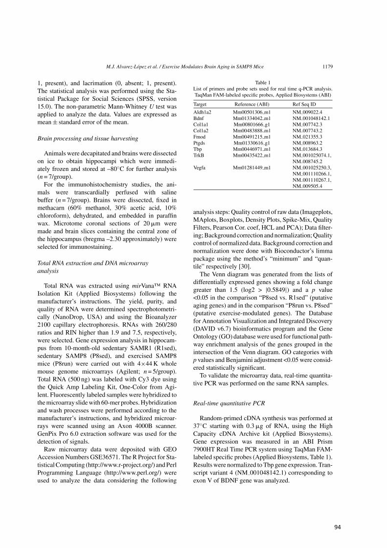
M.J. Alvarez-Lopez et al. / Exercise Modulates Brain Aging in SAMP8 Mice 1179
1, present), and lacrimation (0, absent; 1, present).The statistical analysis was performed using the Sta-tistical Package for Social Sciences (SPSS, version15.0). The non-parametric Mann-Whitney U test wasapplied to analyze the data. Values are expressed asmean± standard error of the mean.
Brain processing and tissue harvesting
Animals were decapitated and brains were dissectedon ice to obtain hippocampi which were immedi-ately frozen and stored at –80◦C for further analysis(n = 7/group).For the immunohistochemistry studies, the ani-
mals were transcardially perfused with salinebuffer (n = 7/group). Brains were dissected, fixed inmethacarn (60% methanol, 30% acetic acid, 10%chloroform), dehydrated, and embedded in paraffinwax. Microtome coronal sections of 20?m weremade and brain slices containing the central zone ofthe hippocampus (bregma –2.30 approximately) wereselected for immunostaining.
Total RNA extraction and DNA microarrayanalysis
Total RNA was extracted using mirVana™ RNAIsolation Kit (Applied Biosystems) following themanufacturer’s instructions. The yield, purity, andquality of RNA were determined spectrophotometri-cally (NanoDrop, USA) and using the Bioanalyzer2100 capillary electrophoresis. RNAs with 260/280ratios and RIN higher than 1.9 and 7.5, respectively,were selected. Gene expression analysis in hippocam-pus from 10-month-old sedentary SAMR1 (R1sed),sedentary SAMP8 (P8sed), and exercised SAMP8mice (P8run) were carried out with 4×44K wholemouse genome microarrays (Agilent; n = 5/group).Total RNA (500 ng) was labeled with Cy3 dye usingthe Quick Amp Labeling Kit, One-Color from Agi-lent. Fluorescently labeled samples were hybridized tothemicroarray slidewith 60-mer probes.Hybridizationand wash processes were performed according to themanufacturer’s instructions, and hybridized microar-rays were scanned using an Axon 4000B scanner.GenPix Pro 6.0 extraction software was used for thedetection of signals.Raw microarray data were deposited with GEO
Accession Numbers GSE36571. The R Project for Sta-tisticalComputing (http://www.r-project.org/) andPerlProgramming Language (http://www.perl.org/) wereused to analyze the data considering the following
Table 1List of primers and probe sets used for real time q-PCR analysis.TaqMan FAM-labeled specific probes, Applied Biosystems (ABI)
Target Reference (ABI) Ref Seq ID
Aldh1a2 Mm00501306 m1 NM 009022.4Bdnf Mm01334042 m1 NM 001048142.1Col1a1 Mm00801666 g1 NM 007742.3Col1a2 Mm00483888 m1 NM 007743.2Fmod Mm00491215 m1 NM 021355.3Ptgds Mm01330616 g1 NM 008963.2Tbp Mm00446971 m1 NM 013684.3TrkB Mm00435422 m1 NM 001025074.1,
NM 008745.2Vegfa Mm01281449 m1 NM 001025250.3,
NM 001110266.1,NM 001110267.1,NM 009505.4
analysis steps: Quality control of raw data (Imageplots,MAplots, Boxplots, Density Plots, Spike-Mix, QualityFilters, Pearson Cor. coef, HCL and PCA); Data filter-ing;Background correction andnormalization;Qualitycontrol of normalized data. Background correction andnormalization were done with Bioconductor’s limmapackage using the method’s “minimum” and “quan-tile” respectively [30].The Venn diagram was generated from the lists of
differentially expressed genes showing a fold changegreater than 1.5 (log2 > |0.5849|) and a p value<0.05 in the comparison “P8sed vs. R1sed” (putativeaging genes) and in the comparison “P8run vs. P8sed”(putative exercise-modulated genes). The Databasefor Annotation Visualization and Integrated Discovery(DAVID v6.7) bioinformatics program and the GeneOntology (GO) databasewere used for functional path-way enrichment analysis of the genes grouped in theintersection of the Venn diagram. GO categories withp values and Benjamini adjustment <0.05 were consid-ered statistically significant.To validate the microarray data, real-time quantita-
tive PCR was performed on the same RNA samples.
Real-time quantitative PCR
Random-primed cDNA synthesis was performed at37◦C starting with 0.3?g of RNA, using the HighCapacity cDNA Archive kit (Applied Biosystems).Gene expression was measured in an ABI Prism7900HT Real Time PCR system using TaqMan FAM-labeled specific probes (Applied Biosystems, Table 1).Results were normalized to Tbp gene expression. Tran-script variant 4 (NM 001048142.1) corresponding toexon V of BDNF gene was analyzed.
94
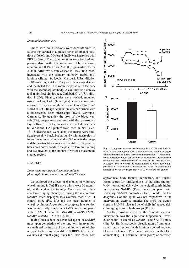
1180 M.J. Alvarez-Lopez et al. / Exercise Modulates Brain Aging in SAMP8 Mice
Immunohistochemistry
Slides with brain sections were deparaffinized inxylene, rehydrated in a graded series of ethanol solu-tions (100, 90, and 70%) and finally washed twice withPBS for 5min. Then, brain sections were blocked andpermeabilized with PBS containing 1% bovine serumalbumin and 0.1% Triton-X-100 (Sigma-Aldrich) for20min. After two 5-min washes in PBS, slides wereincubated with the primary antibody, rabbit anti-laminin (Sigma, St. Louis, Missouri, USA; dilution1 : 100) overnight at 4◦C. Theywere thenwashed againand incubated for 1 h at room temperature in the darkwith the secondary antibody, AlexaFluor 546 donkeyanti-rabbit IgG (Invitrogen, Carlsbad, CA, USA; dilu-tion 1 : 250). Finally, slides were washed, mountedusing Prolong Gold (Invitrogen) anti-fade medium,allowed to dry overnight at room temperature andstored at 4◦C. Image acquisition was performed witha fluorescence laser microscope (BX41, Olympus,Germany). To quantify the area of the blood ves-sels (VA), images were analyzed with the open-sourceFiji software. Briefly, in order to exclude inciden-tal variations, CA1 picture from each animal (n = 4,13–15 slices/group) were taken, the images were bina-rized (vessels = black, background =white), a region ofinterest was set to include all the CA1 seen in the imageand the positive black area was quantified. The positiveblack area corresponds to the positive laminin stainingand is equivalent to the amount of vessel surface in thestudied region.
RESULTS
Long-term exercise performance inducesphenotypic improvements in old SAMP8 mice
We explored the effects of 6 months of voluntarywheel running in SAMP8 mice which were 10-month-old at the end of the training. Consistent with theiraccelerated aging phenotype, during the interventionSAMP8 mice displayed less exercise than SAMR1control mice (Fig. 1A) and the mean number ofwheel revolutions/week for the complete interventionwas significantly lower in SAMP8 mice comparedwith SAMR1 controls (SAMR1= 74296± 5590;SAMP8 = 56964± 5186; Fig. 1B).Taking into account the advanced age of the SAMP8
mice upon completion of the long-term intervention,we analyzed the impact of the training on a set of phe-notypic traits using a modified SHIRPA test, whichevaluates different aging traits (i.e., skin color, coat
Fig. 1. Long-term exercise performance in SAMP8 and SAMR1mice.Wheel-running activity was continuously monitored through awireless transmitter during the 6-month intervention. A)Mean num-ber of wheel revolutions per session was calculated as the total wheelrevolutions per week/numbers of sessions of the week (ANOVA:F(1,26) = 7.868 *p < 0.01). B) Mean number of wheel revolutionsper week was calculated as the mean total wheel revolutions/totalnumber of weeks (n = 14/group; *p < 0.05 versus R1 run group).
appearance, body tremor, lacrimation, and others).Mean scores for lordokyphosis of the spine (hump),body tremor, and skin color were significantly higherin sedentary SAMP8 (P8sed) mice compared withsedentary SAMR1 controls (R1sed). While the lor-dokyphosis of the spine was not responsive to theintervention, exercise practice abolished the tremorsigns in SAMP8 mice and beneficially influenced skincolor aging signs in both groups (Fig. 2).Another positive effect of the 6-month exercise
intervention was the significant hippocampal revas-cularization in exercised SAMR1 and SAMP8 mice(Fig. 3A–E). Microscopic visualization of immunos-tained brain sections with laminin showed reducedblood vessel area in P8sed mice compared with R1sedanimals (Fig. 3 C versus A). Both groups of exercised
95
M.J. Alvarez-Lopez et al. / Exercise Modulates Brain Aging in SAMP8 Mice 1181
Fig. 2. Exercise delays aging traits in old SAMP8 mice. Mice wereevaluated for aging-sensitive traits using the modified SHIRPA test.Higher scores correspond to more advanced age phenotypes. Traitsshowing significant basal differences between groups included: lor-dokyphosis of the spine (0, absent; 1, present), body tremor (0,absent; 1, present), and skin color (0, pink; 1, bleached or deep red).Total score was recorded as the sum of the scores for each of thecategories (*p < 0.05, n = 14/group; non-parametric Mann-WhitneyU test).
mice exhibited enhanced vascularization comparedwith their respective age-matched sedentary controls(Fig. 3B versus A, and D versus C). Remarkably, thearea of blood vessels in P8run mice was significantlyhigher than in R1sed mice and reached levels undistin-guishable from those observed in R1run mice (Fig. 3Dversus A and B, respectively) The effects of exerciseon revascularization seem to be unrelated to VEGFexpression which was significantly decreased in theSAMP8 mice but was not modified by the interventionin any group (Fig. 3F).
Upregulation of Bdnf gene expression inhippocampus from old SAMP8 mice afterlong-term exercise training
Bdnf upregulation is emerging as one of the mostreproducible molecular outcomes of physical exercisein the rodent hippocampus [17]. Before performing themicroarray studies, we further confirmed the efficacyof the long-term exercise intervention in old SAMP8mice by examining Bdnf gene expression as well as theexpression of its receptor TrkB. Both genes were foundunderexpressed in P8sed compared with R1sed mice(Fig. 4). After the exercise intervention, Bdnf geneexpressionwas significantly upregulated in P8runmice(Fig. 4A). In contrast, TrkB gene expression showedno improvement after the exercise training (Fig. 4B).No effect of exercise was observed on Bdnf or TrkBgenes in SAMR1 mice.
Whole genome microarray analysis inhippocampus of old SAMP8 mice after long-termexercise training
With the aim of identifying genes involved in brainaging and modulated by long-term voluntary exer-cise, we performed whole genome microarray studiesin the hippocampus from sedentary SAMP8 (P8sed),sedentary SAMR1 (R1sed). and exercised SAMP8(P8run)mice.We compared “P8sed vs.R1sed” in orderto detect putative aging markers (A) and “P8run vs.P8sed” to detect genes modulated by exercise (B). Forboth comparisons, only genes differentially expressedwith a fold change≥1.5 and a p value <0.05 wereincluded. Genes differentially expressed in A and Bwere integrated in a Venn diagram which yielded 34genes in the intersection (Fig. 5). The overlap betweenthe lists A and Bwas significant as revealed by Fisher’sexact test (p-value <3 E-16). These genes thereforemet the criteria for putative aging genes modulated byexercise (Table 2).
96
1182 M.J. Alvarez-Lopez et al. / Exercise Modulates Brain Aging in SAMP8 Mice
Fig. 3. Effects of 6 months of voluntary exercise on hippocampal vascularization in old SAMP8 mice. Hippocampal sections were incubatedwith the primary antibody rabbit anti-laminin (red) in sedentary SAMR1 (A, R1sed), SAMR1 (B, R1run), sedentary SAMP8 (C, P8sed), andexercised SAMP8 (D, P8run). Representative images are shown, scale bar: 100?m in all panels. E) Area of the blood vessels (VA) was quantifiedwith the open-source Fiji software. F) Vegf gene expression was measured by real time quantitative PCR. *p < 0.05 2 tail t-test; ** p < 0.005 2tail t-test (n = 4, 13–15 slices/group).
Pathway enrichment analysis of the genes listed inTable 2 was performed using the DAVID program andthe Gene Ontology (GO) database. GO categories withp values and Benjamini adjustment <0.05 were con-sidered statistically significant (Table 3A). We foundthat these genes were mainly clustered in functionsassociated with extracellular matrix maintenance. The
reported functions in the central nervous system ofthe genes included in the GO categories are listed inTable 3B [31–43].Moreover, in accordancewith the literature (seeDis-
cussion), functionally relevant genes identified throughthe microarray analysis were validated by real-timequantitative PCR assays, i.e., collagen type 1 alpha
97
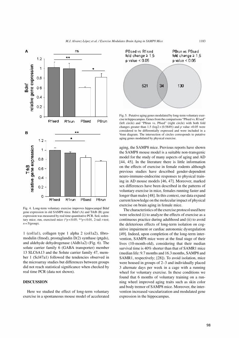
M.J. Alvarez-Lopez et al. / Exercise Modulates Brain Aging in SAMP8 Mice 1183
Fig. 4. Long-term voluntary exercise improves hippocampal Bdnfgene expression in old SAMP8 mice. Bdnf (A) and TrkB (B) geneexpression was measured by real time quantitative PCR. Sed, seden-tary mice; run, exercised mice (*p < 0.05; **p < 0.01, 2-tail t-test;n = 5/group).
1 (col1a1), collagen type 1 alpha 2 (col1a2), fibro-modulin (fmod), prostaglandin D(2) synthase (ptgds),and aldehyde dehydrogenase (Aldh1a2) (Fig. 6). Thesolute carrier family 6 (GABA transporter) member13 SLC6A13 and the Solute carrier family 47, mem-ber 1 (Scl47a1) followed the tendencies observed inthe microarray studies but differences between groupsdid not reach statistical significance when checked byreal time PCR (data not shown).
DISCUSSION
Here we studied the effect of long-term voluntaryexercise in a spontaneous mouse model of accelerated
Fig. 5. Putative aging genesmodulated by long-term voluntary exer-cise in hippocampus.Genes from the comparisons “P8sed vs.R1sed”(left circle) and “P8run vs. P8sed” (right circle) with both foldchanges greater than 1.5 (log2 > |0.5849|) and p value <0.05 wereconsidered to be differentially expressed and were included in aVenn diagram. The intersection of circles corresponds to putativeaging genes modulated by physical exercise.
aging, the SAMP8 mice. Previous reports have shownthe SAMP8 mouse model is a suitable non-transgenicmodel for the study of many aspects of aging and AD[44, 45]. In the literature there is little informationon the effects of exercise in female rodents althoughprevious studies have described gender-dependentneuro-immuno-endocrine responses to physical train-ing in AD mouse models [46, 47]. Moreover, markedsex differences have been described in the patterns ofvoluntary exercise in mice, females running faster andlonger thanmales [48]. In this context, our data expandcurrent knowledge on themolecular impact of physicalexercise on brain aging in female mice.The characteristics of the exercise protocol used here
were selected (i) to analyze the effects of exercise as acontinuous practice during adulthood and (ii) to avoidthe deleterious effects of long-term isolation on cog-nitive impairment or cardiac autonomic dysregulation[49]. Indeed, upon completion of the long-term inter-vention, SAMP8 mice were at the final stage of theirlives (10-month-old), considering that their mediansurvival time is 40% shorter than that of SAMR1 mice(median life: 9.7months and 16.3months, SAMP8 andSAMR1, respectively; [28]). To avoid isolation, micewere housed in groups of 2–3 and individually placed3 alternate days per week in a cage with a runningwheel for voluntary exercise. In these conditions wefound that 6 months of voluntary training on a run-ning wheel improved aging traits such as skin colorand body tremor of SAMP8 mice. Moreover, the inter-vention increased vascularization and modulated geneexpression in the hippocampus.
98
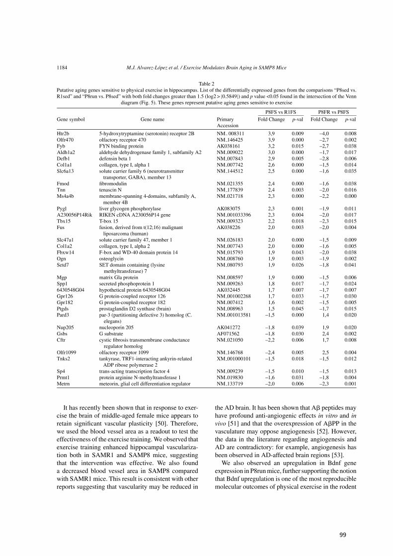
1184 M.J. Alvarez-Lopez et al. / Exercise Modulates Brain Aging in SAMP8 Mice
Table 2Putative aging genes sensitive to physical exercise in hippocampus. List of the differentially expressed genes from the comparisons “P8sed vs.R1sed” and “P8run vs. P8sed” with both fold changes greater than 1.5 (log2 > |0.5849|) and p value <0.05 found in the intersection of the Venn
diagram (Fig. 5). These genes represent putative aging genes sensitive to exercise
P8FS vs R1FS P8FR vs P8FSGene symbol Gene name Primary Fold Change p-val Fold Change p-val
Accession
Htr2b 5-hydroxytryptamine (serotonin) receptor 2B NM 008311 3,9 0.009 –4,0 0.008Olfr470 olfactory receptor 470 NM 146425 3,9 0.000 –2,7 0.002Fyb FYN binding protein AK038161 3,2 0.015 –2,7 0.038Aldh1a2 aldehyde dehydrogenase family 1, subfamily A2 NM 009022 3,0 0.000 –1,7 0.017Defb1 defensin beta 1 NM 007843 2,9 0.005 –2,8 0.006Col1a1 collagen, type I, alpha 1 NM 007742 2,6 0.000 –1,5 0.014Slc6a13 solute carrier family 6 (neurotransmitter NM 144512 2,5 0.000 –1,6 0.035
transporter, GABA), member 13Fmod fibromodulin NM 021355 2,4 0.000 –1,6 0.038Tnn tenascin N NM 177839 2,4 0.003 –2,0 0.016Ms4a4b membrane-spanning 4-domains, subfamily A, NM 021718 2,3 0.000 –2,2 0.000
member 4BPygl liver glycogen phosphorylase AK083075 2,3 0.001 –1,9 0.011A230056P14Rik RIKEN cDNA A230056P14 gene NM 001033396 2,3 0.004 –2,0 0.017Tbx15 T-box 15 NM 009323 2,2 0.018 –2,3 0.015Fus fusion, derived from t(12;16) malignant AK038226 2,0 0.003 –2,0 0.004
liposarcoma (human)Slc47a1 solute carrier family 47, member 1 NM 026183 2,0 0.000 –1,5 0.009Col1a2 collagen, type I, alpha 2 NM 007743 2,0 0.000 –1,6 0.005Fbxw14 F-box and WD-40 domain protein 14 NM 015793 1,9 0.043 –2,0 0.038Ogn osteoglycin NM 008760 1,9 0.003 –1,9 0.002Setd7 SET domain containing (lysine
methyltransferase) 7NM 080793 1,9 0.026 –1,8 0.041
Mgp matrix Gla protein NM 008597 1,9 0.000 –1,5 0.006Spp1 secreted phosphoprotein 1 NM 009263 1,8 0.017 –1,7 0.0246430548G04 hypothetical protein 6430548G04 AK032445 1,7 0.007 –1,7 0.007Gpr126 G protein-coupled receptor 126 NM 001002268 1,7 0.033 –1,7 0.030Gpr182 G protein-coupled receptor 182 NM 007412 1,6 0.002 –1,5 0.005Ptgds prostaglandin D2 synthase (brain) NM 008963 1,5 0.045 –1,7 0.015Pard3 par-3 (partitioning defective 3) homolog (C.
elegans)NM 001013581 –1,5 0.000 1,4 0.020
Nup205 nucleoporin 205 AK041272 –1,8 0.039 1,9 0.020Gsbs G substrate AF071562 –1,8 0.030 2,4 0.002Cftr cystic fibrosis transmembrane conductance
regulator homologNM 021050 –2,2 0.006 1,7 0.008
Olfr1099 olfactory receptor 1099 NM 146768 –2,4 0.005 2,5 0.004Tnks2 tankyrase, TRF1-interacting ankyrin-related XM 001000101 –1,5 0.018 –1,5 0.012
ADP ribose polymerase 2Sp4 trans-acting transcription factor 4 NM 009239 –1,5 0.010 –1,5 0.013Prmt1 protein arginine N-methyltransferase 1 NM 019830 –1,6 0.031 –1,8 0.004Metrn meteorin, glial cell differentiation regulator NM 133719 –2,0 0.006 –2,3 0.001
It has recently been shown that in response to exer-cise the brain of middle-aged female mice appears toretain significant vascular plasticity [50]. Therefore,we used the blood vessel area as a readout to test theeffectiveness of the exercise training.We observed thatexercise training enhanced hippocampal vasculariza-tion both in SAMR1 and SAMP8 mice, suggestingthat the intervention was effective. We also founda decreased blood vessel area in SAMP8 comparedwith SAMR1 mice. This result is consistent with otherreports suggesting that vascularity may be reduced in
the AD brain. It has been shown that A? peptides mayhave profound anti-angiogenic effects in vitro and invivo [51] and that the overexpression of A?PP in thevasculature may oppose angiogenesis [52]. However,the data in the literature regarding angiogenesis andAD are contradictory: for example, angiogenesis hasbeen observed in AD-affected brain regions [53].We also observed an upregulation in Bdnf gene
expression in P8runmice, further supporting the notionthat Bdnf upregulation is one of the most reproduciblemolecular outcomes of physical exercise in the rodent
99
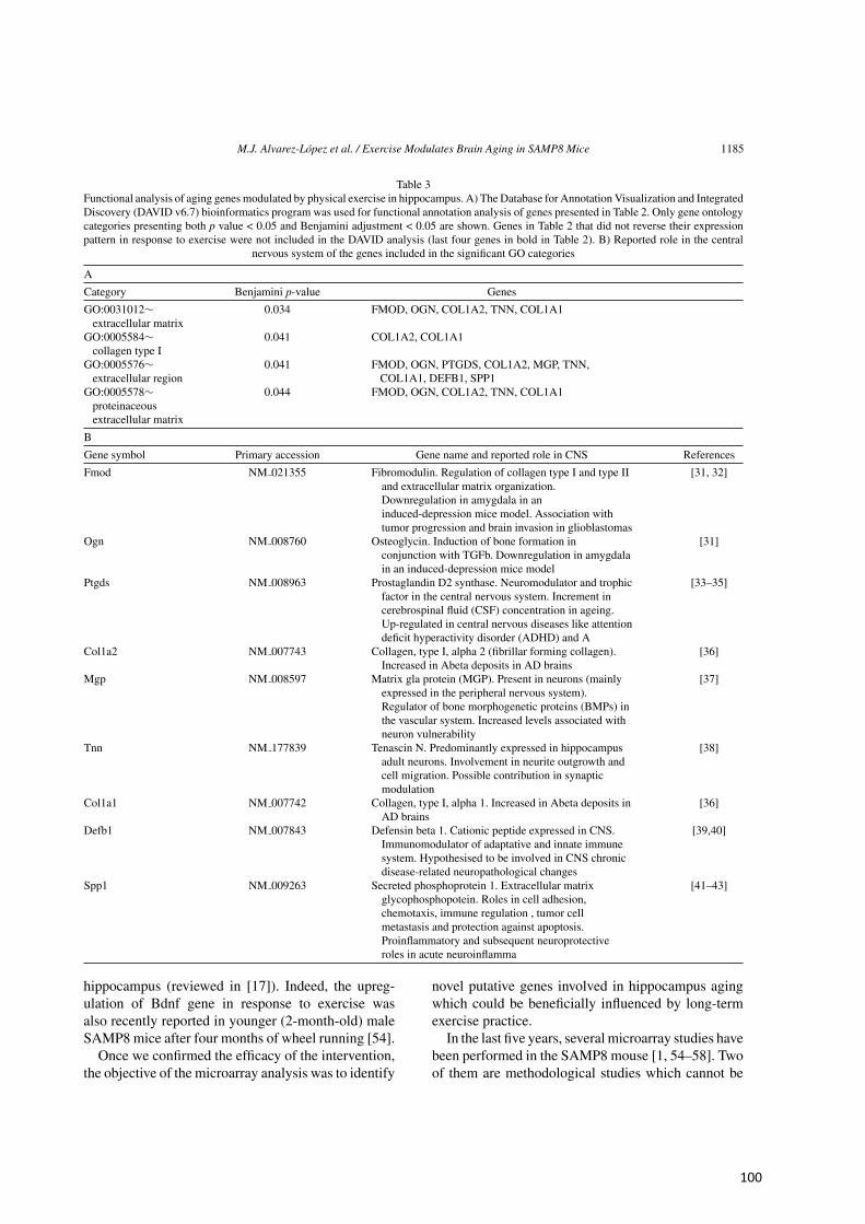
M.J. Alvarez-Lopez et al. / Exercise Modulates Brain Aging in SAMP8 Mice 1185
Table 3Functional analysis of aging genes modulated by physical exercise in hippocampus. A) The Database for Annotation Visualization and IntegratedDiscovery (DAVID v6.7) bioinformatics program was used for functional annotation analysis of genes presented in Table 2. Only gene ontologycategories presenting both p value < 0.05 and Benjamini adjustment < 0.05 are shown. Genes in Table 2 that did not reverse their expressionpattern in response to exercise were not included in the DAVID analysis (last four genes in bold in Table 2). B) Reported role in the central
nervous system of the genes included in the significant GO categories
A
Category Benjamini p-value Genes
GO:0031012∼ 0.034 FMOD, OGN, COL1A2, TNN, COL1A1extracellular matrix
GO:0005584∼ 0.041 COL1A2, COL1A1collagen type I
GO:0005576∼ 0.041 FMOD, OGN, PTGDS, COL1A2, MGP, TNN,extracellular region COL1A1, DEFB1, SPP1
GO:0005578∼ 0.044 FMOD, OGN, COL1A2, TNN, COL1A1proteinaceousextracellular matrix
B
Gene symbol Primary accession Gene name and reported role in CNS References
Fmod NM 021355 Fibromodulin. Regulation of collagen type I and type IIand extracellular matrix organization.Downregulation in amygdala in aninduced-depression mice model. Association withtumor progression and brain invasion in glioblastomas
[31, 32]
Ogn NM 008760 Osteoglycin. Induction of bone formation inconjunction with TGFb. Downregulation in amygdalain an induced-depression mice model
[31]
Ptgds NM 008963 Prostaglandin D2 synthase. Neuromodulator and trophicfactor in the central nervous system. Increment incerebrospinal fluid (CSF) concentration in ageing.Up-regulated in central nervous diseases like attentiondeficit hyperactivity disorder (ADHD) and A
[33–35]
Col1a2 NM 007743 Collagen, type I, alpha 2 (fibrillar forming collagen).Increased in Abeta deposits in AD brains
[36]
Mgp NM 008597 Matrix gla protein (MGP). Present in neurons (mainlyexpressed in the peripheral nervous system).Regulator of bone morphogenetic proteins (BMPs) inthe vascular system. Increased levels associated withneuron vulnerability
[37]
Tnn NM 177839 Tenascin N. Predominantly expressed in hippocampusadult neurons. Involvement in neurite outgrowth andcell migration. Possible contribution in synapticmodulation
[38]
Col1a1 NM 007742 Collagen, type I, alpha 1. Increased in Abeta deposits inAD brains
[36]
Defb1 NM 007843 Defensin beta 1. Cationic peptide expressed in CNS.Immunomodulator of adaptative and innate immunesystem. Hypothesised to be involved in CNS chronicdisease-related neuropathological changes
[39,40]
Spp1 NM 009263 Secreted phosphoprotein 1. Extracellular matrixglycophosphopotein. Roles in cell adhesion,chemotaxis, immune regulation , tumor cellmetastasis and protection against apoptosis.Proinflammatory and subsequent neuroprotectiveroles in acute neuroinflamma
[41–43]
hippocampus (reviewed in [17]). Indeed, the upreg-ulation of Bdnf gene in response to exercise wasalso recently reported in younger (2-month-old) maleSAMP8 mice after four months of wheel running [54].Once we confirmed the efficacy of the intervention,
the objective of the microarray analysis was to identify
novel putative genes involved in hippocampus agingwhich could be beneficially influenced by long-termexercise practice.In the last five years, several microarray studies have
been performed in the SAMP8 mouse [1, 54–58]. Twoof them are methodological studies which cannot be
100
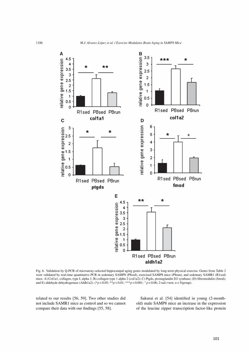
1186 M.J. Alvarez-Lopez et al. / Exercise Modulates Brain Aging in SAMP8 Mice
Fig. 6. Validation by Q-PCR of microarray-selected hippocampal aging genes modulated by long-term physical exercise. Genes from Table 2were validated by real-time quantitative PCR in sedentary SAMP8 (P8sed), exercised SAMP8 mice (P8run), and sedentary SAMR1 (R1sed)mice. A) Col1a1, collagen, type I, alpha 1; B) collagen type 1 alpha 2 (col1a2); C) Ptgds, prostaglandin D2 synthase; (D) fibromodulin (fmod);and E) aldehyde dehydrogenase (Aldh1a2). (*p < 0.05; **p < 0.01; ***p < 0.001; +p = 0.06; 2-tail t-test; n = 5/group).
related to our results [56, 59]. Two other studies didnot include SAMR1 mice as control and so we cannotcompare their data with our findings [55, 58].
Sakurai et al. [54] identified in young (2-month-old) male SAMP8 mice an increase in the expressionof the leucine zipper transcription factor-like protein
101
M.J. Alvarez-Lopez et al. / Exercise Modulates Brain Aging in SAMP8 Mice 1187
1 (Lztfl1) levels after four months of wheel running.However, in 12-old-month mice, we did not observeany effect of exercise in the expression of Lztfl1 inSAMP8 mice. The study by Cheng et al. [57] analyzedmicroarray expression profiles in the hippocampusof 12-month-old male SAMP8 and SAMR1 mice. Inagreement with our study in females, they detected thedown-regulation of Dusp12 (Dual specificity proteinphosphatase 12) in the SAMP8 hippocampus. Recentevidence suggests that this enzyme plays a role incell survival and ribosome biogenesis [60]. We didnot detect any change in Dusp12 gene expression inresponse to exercise. Finally, using microarray anal-ysis, Casadesus et al. [61] compared the expressionof 62 genes involved in different signaling pathwayssuch as oxidative stress, mitochondrial-related apop-tosis, and cell cycle, in the hippocampus of 3-, 6-, and9-months-old male SAMP8 and SAMR1 mice, andfound no differences between strains at any of the agesanalyzed. Of these 62 genes, we found that, comparedwith SAMR1 mice, female SAMP8 mice presented adown-regulation in Park2 gene (Park2 is an E3 ubiqui-tin ligase that regulates systemic lipid metabolism andis defective in early onset Parkinson’s disease patients[62]) and an upregulation of the Gfra1 gene (Gfra1gene encodes the glial cell line-derived neurotrophicfactor family receptor ?-1, GFR ?-1, which has beenreported to be beneficial for the survival of hippocam-pal neurons [63]). However, the expression levels ofthese genes were not modulated by exercise.Regarding proteomic studies using SAMP8 mice,
Zhu et al. [64] analyzed hippocampus and cortexof 5- and 15-month-old male SAMP8 and SAMR1mice. In agreement with their findings, we found adown-regulation Uchl3 (Ubiquitin carboxyl-terminalhydrolase isozyme L3) and a down-regulation ofHebp1 in female SAMP8 hippocampus compared withSAMR1 mice, although exercise did not modifiedtheir expression levels. Interestingly, another pro-teomic study reported a down regulation of Uchl3 inthe SAMP8 hippocampus at six months [65], stronglysuggesting that lower expression of Uchl3 is a distinc-tive feature of the SAMP8 hippocampus and may beassociated with the pathological phenotype of thesemice.Differences in the profiling platforms used, in the
animal sex and age, and/or in the intervention designsare some of the factors that could explain the lowagreements between our study and the cited reports.Moreover, the limitations of the microarray techniqueare highlighted by the observation that in response toexercise we have found an upregulation of BDNF gene
expression by real time PCR but not by microarrayanalysis.In this study, we found 34 genes in hippocam-
pus that met our criteria for aging genes sensitiveto physical exercise. Gene ontology analysis revealedthat long-term physical exercise modulated pathwaysmainly involved in extracellular matrix (ECM)mainte-nance in hippocampus. Interestingly, the ECMnot onlyprovides physical support for neuronal function andsurvival but also plays a crucial homeostatic role [66]and changes in ECM components have been describedas pathological features of AD [66, 67]. In this context,our data suggest that physical exercise may counteractat least in part such processes in early-stage AD.Although the relevance of microarray findings will
require further characterization, based on the previ-ous literature some inferences about their functionalsignificance can be made. Our microarray studies inhippocampus showed increased expression of the col-lagen type 1 alpha 1 (col1a1) and alpha 2 (col1a2)genes in SAMP8 mice compared with controls anda decrease in their expression levels in response toexercise. These effects were confirmed by quantitativereal-time PCR. Interestingly, the 3xTg-AD transgenicmouse model presents a 20% increase in brain colla-gen content compared with controls and a significantvascular volume reduction in the hippocampus, but notin the frontal cortex or cerebellum [36]. These resultsare of particular interest since A?PP can physicallyinteract with many different collagen and extracellu-lar matrix components and because other forms ofcollagen (e.g., XXV and XVIII) have been foundin amyloid plaques [68, 69]. Another gene benefi-cially modulated by exercise in SAMP8 hippocampuswas the prostaglandin D(2) synthase (Pgds). Pgds isexpressed in astrocytes and oligodendrocytes [70] andhas been proposed as a biomarker of aging in cere-brospinal fluid [33]. Using real-time PCR, we alsovalidated the beneficial effect of exercise on fibromod-ulin gene expression (Fmod) in SAMP8 mice. Fmodis a proteoglycan component involved in the regula-tion and assembly of extracellular matrix organization.It has been associated with brain tumor progressionin glioblastomas [32] and there is growing evidencethat keratan sulfate proteoglycans enhance A? aggre-gation and senile plaques formation in the pathogenesisof AD [71]. Finally, exercise also restored geneexpression of the aldehyde dehydrogenase Aldh1a2in the SAMP8 hippocampus. Aldh1a2 is the rate-limiting retinoic acid-synthesizing enzyme and it hasbeen reported that both decreased [72] and increased[73] retinoic acid levels in the hippocampus lead to
102
1188 M.J. Alvarez-Lopez et al. / Exercise Modulates Brain Aging in SAMP8 Mice
impaired neurogenesis and performance in cognitivetasks.In summary, our study identifies new candidate
aging genes in the hippocampus which are sensitive tophysical exercise, and suggests that an active lifestyleduring adulthood may promote a healthier aging in at-risk populations by preventing or reversing senescentsigns in the brain.
ACKNOWLEDGMENTS
This study was supported by grant SAF2010-15050(PK) from the Spanish Ministry of Science and Inno-vation (MICINN); DPS2008-06998-C01,02 (MPallasand RME) and by grants PI080400 from the Fondode Investigacion Sanitaria and the Instituto de SaludCarlos III (AC), BFU2009-09988/BMC (MParrizas),SAF-2009-13093 (MPallas) from the Spanish Min-istry of Science and Innovation (MICINN). We thankthe Catalan Government (Generalitat de Catalunya)for their support (2009/SGR00853) (PK). J.F.L. andM.J.A.L received a predoctoral fellowship from theGeneralitat de Catalunya (FI-DGR 2011). We thankSergi Beltran Agullo from the Bioinformatics Anal-ysis Group (CNAG – National Center for GenomicAnalysis) for statistical analysis of microarray data.Authors’ disclosures available online (http://www.j-
alz.com/disclosures/view.php?id=1548).
REFERENCES
[1] (2011) Global status report on noncommunicable diseases2010, World Health Organisation, http://www.who.int/nmh/publications/ncd report2010/en/index.html, 2011.
[2] BassukSS,Manson JE (2010) Physical activity and cardiovas-cular disease prevention in women: A review of the epidemi-ologic evidence. Nutr Metab Cardiovasc Dis 20, 467-473.
[3] Kokkinos P, Myers J (2010) Exercise and physical activity:Clinical outcomes and applications. Circulation 122,1637-1648.
[4] Metkus TS, Baughman KL, Thompson PD (2010) Exerciseprescription and primary prevention of cardiovascular dis-ease. Circulation 121, 2601-2604.
[5] Mobius-Winkler S, Linke A, Adams V, Schuler G, Erbs S(2010) How to improve endothelial repair mechanisms: Thelifestyle approach. Expert Rev Cardiovasc Ther 8, 573-580.
[6] Bianchi G, Rossi V, Muscari A, Magalotti D, Zoli M (2008)Physical activity is negatively associated with the metabolicsyndrome in the elderly. QJM 101, 713-721.
[7] Colberg SR (2007) Physical activity, insulin action, and dia-betes prevention and control. Curr Diabetes Rev 3, 176-184.
[8] Cotman CW, Berchtold NC, Christie LA (2007) Exercisebuilds brain health: Key roles of growth factor cascades andinflammation. Trends Neurosci 30, 464-472.
[9] Horton ES (2009) Effects of lifestyle changes to reduce risksof diabetes and associated cardiovascular risks: Results from
large scale efficacy trials. Obesity (Silver Spring) 17(Suppl3), S43-S48.
[10] Saraceni C, Broderick TL (2007) Cardiac and metabolicconsequences of aerobic exercise training in experimentaldiabetes. Curr Diabetes Rev 3, 75-84.
[11] Zanuso S, Jimenez A, Pugliese G, Corigliano G, Balducci S(2010) Exercise for the management of type 2 diabetes: Areview of the evidence. Acta Diabetol 47, 15-22.
[12] Deslandes A, Moraes H, Ferreira C, Veiga H, Silveira H,Mouta R, Pompeu FA, Coutinho ES, Laks J (2009) Exerciseand mental health: Many reasons to move. Neuropsychobiol-ogy 59, 191-198.
[13] Erickson KI, Kramer AF (2009) Aerobic exercise effects oncognitive and neural plasticity in older adults.Br J SportsMed43, 22-24.
[14] Rolland Y, Abellan van Kan G, Vellas B (2010) Healthy brainaging: Role of exercise and physical activity. Clin GeriatrMed 26, 75-87.
[15] van PraagH (2009) Exercise and the brain: Something to chewon. Trends Neurosci 32, 283-290.
[16] Archer T (2011) Physical exercise alleviates debilities of nor-mal aging and Alzheimer’s disease. Acta Neurol Scand 123,221-238.
[17] Kaliman P, Parrizas M, Lalanza JF, Camins A, EscorihuelaRM, Pallas M (2011) Neurophysiological and epigeneticeffects of physical exercise on the aging process. Ageing ResRev 10, 475-486.
[18] Bekinschtein P, Oomen CA, Saksida LM, Bussey TJ (2011)Effects of environmental enrichment and voluntary exerciseon neurogenesis, learning and memory, and pattern separa-tion: BDNF as a critical variable? Semin Cell Dev Biol 22,536-542.
[19] Christie BR, Eadie BD, Kannangara TS, Robillard JM, ShinJ, Titterness AK (2008) Exercising our brains: How physi-cal activity impacts synaptic plasticity in the dentate gyrus.Neuromolecular Med 10, 47-58.
[20] van Praag H (2008) Neurogenesis and exercise: Past andfuture directions. Neuromolecular Med 10, 128-140.
[21] Yau SY, Lau BW, So KF (2011) Adult hippocampal neuro-genesis: A possible way how physical exercise counteractsstress. Cell Transplant 20, 99-111.
[22] Garcia-Mesa Y, Lopez-Ramos JC, Gimenez-Llort L, RevillaS, Guerra R, Gruart A, Laferla FM, Cristofol R, Delgado-Garcia JM, Sanfeliu C (2011) Physical exercise protectsagainst Alzheimer’s disease in 3xTg-AD mice. J AlzheimersDis 24, 421-454.
[23] Kohman RA, Rodriguez-Zas SL, Southey BR, Kelley KW,Dantzer R, Rhodes JS (2011) Voluntary wheel runningreverses age-induced changes in hippocampal gene expres-sion. PLoS One 6, e22654.
[24] Parachikova A, Nichol KE, Cotman CW (2008) Short-termexercise in aged Tg2576 mice alters neuroinflammation andimproves cognition. Neurobiol Dis 30, 121-129.
[25] Takeda T, Hosokawa M, Higuchi K (1991) Senescence-accelerated mouse (SAM): A novel murine model ofaccelerated senescence. J Am Geriatr Soc 39, 911-919.
[26] del Valle J, Duran-Vilaregut J, Manich G, Pallas M, CaminsA,Vilaplana J, Pelegri C (2011) Cerebral amyloid angiopathy,blood-brain barrier disruption and amyloid accumulation inSAMP8 mice. Neurodegener Dis 8, 421-429.
[27] Morley JE, Armbrecht HJ, Farr SA, Kumar VB (2012) Thesenescence acceleratedmouse (SAMP8) as amodel for oxida-tive stress and Alzheimer’s disease. Biochim Biophys Acta1822, 650-656.
103
M.J. Alvarez-Lopez et al. / Exercise Modulates Brain Aging in SAMP8 Mice 1189
[28] Takeda T (2009) Senescence-accelerated mouse (SAM)with special references to neurodegenerationmodels, SAMP8and SAMP10 mice. Neurochem Res 34, 639-659.
[29] Rogers DC, Peters J, Martin JE, Ball S, Nicholson SJ,Witherden AS, Hafezparast M, Latcham J, Robinson TL,Quilter CA, Fisher EM (2001) SHIRPA, a protocol forbehavioral assessment: Validation for longitudinal studyof neurological dysfunction inmice.Neurosci Lett 306, 89-92.
[30] Smyth GK (2004) Linear models and empirical bayesmethods for assessing differential expression in microarrayexperiments. Stat Appl Genet Mol Biol 3, Article3.
[31] Jung S, Lee Y, Kim G, Son H, Lee DH, Roh GS, Kang SS,Cho GJ, Choi WS, Kim HJ (2012) Decreased expression ofextracellular matrix proteins and trophic factors in the amyg-dala complex of depressed mice after chronic immobilizationstress. BMC Neurosc 13, 58.
[32] Colin C, Baeza N, Bartoli C, Fina F, Eudes N, Nanni I, MartinPM, Ouafik L, Figarella-Branger D (2006) Identification ofgenes differentially expressed in glioblastomaversus pilocyticastrocytoma using Suppression Subtractive Hybridization.Oncogene 25, 2818-2826.
[33] Chen CP, Chen RL, Preston JE (2009) Age-related increaseof prostaglandin D(2) synthase concentration and glycationin ovine cerebrospinal fluid. Exp Gerontol 44, 639-645.
[34] Marin-Mendez JJ, Patino-Garcia A, Segura V, Ortuno F,Galvez MD, Soutullo CA (2012) Differential expression ofprostaglandinD2 synthase (PTGDS) in patientswith attentiondeficit-hyperactivity disorder and bipolar disorder. J AffectDisord 138, 479-484.
[35] Lovell MA, Lynn BC, Xiong S, Quinn JF, Kaye J, Markes-bery WR (2008) An aberrant protein complex in CSF as abiomarker of Alzheimer disease. Neurology 70, 2212-2128.
[36] Bourasset F, Ouellet M, Tremblay C, Julien C, Do TM, OddoS, LaFerla F, Calon F (2009) Reduction of the cerebrovascularvolume in a transgenic mouse model of Alzheimer’s disease.Neuropharmacology 56, 808-813.
[37] Santa-Maria I, Avila J, Rabano A (2010) Differential geneexpression analysis of human entorhinal cortex support a pos-sible role of some extracellular matrix proteins in the onset ofAlzheimer disease. Neurosci Lett 468, 225-228.
[38] Neidhardt J, Fehr S, Kutsche M, Lohler J, Schachner M(2003) Tenascin-N: Characterization of a novel member ofthe tenascin family that mediates neurite repulsion from hip-pocampal explants. Mol Cell Neurosci 23, 193-209.
[39] Hao HN, Zhao J, Lotoczky G, GreverWE, LymanWD (2001)Induction of human beta-defensin-2 expression in humanastrocytes by lipopolysaccharide and cytokines. J Neurochem77, 1027-1035.
[40] Williams WM, Castellani RJ, Weinberg A, Perry G, SmithMA (2012) Do beta-defensins and other antimicrobialpeptides play a role in neuroimmune function and neurode-generation? ScientificWorldJournal 2012, 905785.
[41] Shin T (2012) Osteopontin as a two-sided mediator inacute neuroinflammation in rat models. Acta Histochem 114,749-754.
[42] BraitchM, Constantinescu CS (2010) The role of osteopontinin experimental autoimmune encephalomyelitis (EAE) andmultiple sclerosis (MS). Inflamm Allergy Drug Targets 9,249-256.
[43] Tsai WC, Lee HS, Lin CK, Chen A, Nieh S, Ma HI(2012) The association of osteopontin and LMX1A expres-sion with World Health Organization grade in meningiomasand gliomas. Histopathology 61, 844-856.
[44] Morley JE, Farr SA, Kumar VB, Armbrecht HJ (2012) TheSAMP8 mouse: A model to develop therapeutic interven-
tions for Alzheimer’s disease. Curr Pharm Des 18, 1123-1130.
[45] Pallas M, Camins A, Smith MA, Perry G, Lee HG, CasadesusG (2008) From aging to Alzheimer’s disease: Unveiling“the switch” with the senescence-accelerated mouse model(SAMP8). J Alzheimers Dis 15, 615-624.
[46] Gimenez-Llort L, Arranz L, Mate I, De la Fuente M (2008)Gender-specific neuroimmunoendocrine aging in a triple-transgenic 3xTg-AD mouse model for Alzheimer’s diseaseand its relation with longevity. Neuroimmunomodulation 15,331-343.
[47] Gimenez-Llort L, Garcia Y, Buccieri K, Revilla S, Sunol C,Cristofol R, Sanfeliu C (2010) Gender-specific neuroimmu-noendocrine response to treadmill exercise in 3xTg-ADMice.Int J Alzheimers Dis 2010, 128354.
[48] De Bono JP, Adlam D, Paterson DJ, Channon KM (2006)Novel quantitative phenotypes of exercise training in mousemodels. Am J Physiol Regul Integr Comp Physiol 290, R926-R934.
[49] Norman GJ, Zhang N, Morris JS, Karelina K, Berntson GG,DeVries AC (2010) Social interaction modulates autonomic,inflammatory, and depressive-like responses to cardiac arrestand cardiopulmonary resuscitation. Proc Natl Acad Sci U S A107, 16342-16347.
[50] Latimer CS, Searcy JL, Bridges MT, Brewer LD, Popovic J,Blalock EM, Landfield PW, Thibault O, Porter NM (2011)Reversal of glial and neurovascular markers of unhealthybrain aging by exercise in middle-aged female mice. PLoSOne 6, e26812.
[51] Paris D, Townsend K, Quadros A, Humphrey J, Sun J, BremS,Wotoczek-ObadiaM, DelleDonneA, Patel N, ObregonDF,CrescentiniR,AbdullahL,CoppolaD,RojianiAM,CrawfordF, Sebti SM, Mullan M (2004) Inhibition of angiogenesis byAbeta peptides. Angiogenesis 7, 75-85.
[52] Paris D, Patel N,DelleDonneA,QuadrosA, SmeedR,MullanM (2004) Impaired angiogenesis in a transgenic mouse modelof cerebral amyloidosis. Neurosci Lett 366, 80-85.
[53] Desai BS, Schneider JA, Li JL, Carvey PM, Hendey B (2009)Evidence of angiogenic vessels inAlzheimer’s disease. JNeu-ral Transm 116, 587-597.
[54] Sakurai T, Ogasawara J, Kizaki T, Ishibashi Y, Fujiwara T,Akagawa K, Izawa T, Oh-ishi S, Haga S, Ohno H (2011)Involvement of leucine zipper transcription factor-like pro-tein 1 (Lztfl1) in the attenuation of cognitive impairment byexercise training. Biochem Biophys Res Commun 416, 125-129.
[55] Chen SC, Lu G, Chan CY, Chen Y, Wang H, Yew DT, FengZT, KungHF (2010)Microarray profile of brain aging-relatedgenes in the frontal cortex of SAMP8. J Mol Neurosci 41,12-16.
[56] Cheng XR, Zhou WX, Zhang YX (2006) [Preparation ofthe cDNA microarray on the differential expressed cDNAof senescence-accelerated mouse’s hippocampus]. Sheng WuGong Cheng Xue Bao 22, 457-464.
[57] Cheng XR, Zhou WX, Zhang YX, Zhou DS, Yang RF, ChenLF (2007) Differential gene expression profiles in the hip-pocampus of senescence-acceleratedmouse.Neurobiol Aging28, 497-506.
[58] Zheng Y, Cheng XR, Zhou WX, Zhang YX (2008) Geneexpression patterns of hippocampus and cerebral cortex ofsenescence-accelerated mouse treated with Huang-Lian-Jie-Du decoction. Neurosci Lett 439, 119-124.
[59] Greenhall JA, Zapala MA, Caceres M, Libiger O, Barlow C,Schork NJ, Lockhart DJ (2007) Detecting genetic variation inmicroarray expression data. Genome Res 17, 1228-1235.
104
1190 M.J. Alvarez-Lopez et al. / Exercise Modulates Brain Aging in SAMP8 Mice
[60] Kozarova A, Hudson JW, Vacratsis PO (2011) The dual-specificity phosphatase hYVH1 (DUSP12) is a novelmodulator of cellularDNAcontent.CellCycle 10, 1669-1678.
[61] Casadesus G, Gutierrez-Cuesta J, Lee HG, Jimenez A, TajesM,Ortuno-SahagunD,CaminsA, SmithMA,PallasM (2012)Neuronal cell cycle re-entry markers are altered in the senes-cence accelerated mouse P8 (SAMP8). J Alzheimers Dis 30,573-583.
[62] KimKY, StevensMV, Akter MH, Rusk SE, Huang RJ, CohenA, Noguchi A, Springer D, BocharovAV, Eggerman TL, SuenDF, Youle RJ, Amar M, Remaley AT, SackMN (2011) Parkinis a lipid-responsive regulator of fat uptake inmice andmutanthuman cells. J Clin Invest 121, 3701-3712.
[63] WangW, Redecker C, Bidmon HJ,Witte OW (2004) Delayedneuronal death and damage of GDNF family receptors in CA1following focal cerebral ischemia. Brain Res 1023, 92-101.
[64] Zhu L, Yu J, Shi Q, Lu W, Liu B, Xu S, Wang L, Han J,Wang X (2011) Strain- and age-related alteration of proteinsin the brain of SAMP8 and SAMR1 mice. J Alzheimers Dis23, 641-654.
[65] Wang Q, Liu Y, Zou X, An M, Guan X, He J, Tong Y, Ji J(2008) The hippocampal proteomic analysis of senescence-accelerated mouse: Implications of Uchl3 and mitofilin incognitive disorder and mitochondria dysfunction in SAMP8.Neurochem Res 33, 1776-1782.
[66] Bonneh-Barkay D, Wiley CA (2009) Brain extracellularmatrix in neurodegeneration. Brain Pathol 19, 573-585.
[67] Caltagarone J, Jing Z, Bowser R (2007) Focal adhesions reg-ulate Abeta signaling and cell death in Alzheimer’s disease.Biochim Biophys Acta 1772, 438-445.
[68] Hashimoto T, Wakabayashi T, Watanabe A, Kowa H, HosodaR, Nakamura A, Kanazawa I, Arai T, Takio K, Mann DM,Iwatsubo T (2002) CLAC:A novel Alzheimer amyloid plaquecomponent derived from a transmembrane precursor, CLAC-P/collagen type XXV. EMBO J 21, 1524-1534.
[69] van Horssen J, Wilhelmus MM, Heljasvaara R, PihlajaniemiT, Wesseling P, de Waal RM, Verbeek MM (2002) Colla-gen XVIII: A novel heparan sulfate proteoglycan associatedwith vascular amyloid depositions and senile plaques inAlzheimer’s disease brains. Brain Pathol 12, 456-462.
[70] Urade Y, Hayaishi O (2000) Biochemical, structural, genetic,physiological, and pathophysiological features of lipocalin-type prostaglandin D synthase. Biochim Biophys Acta 1482,259-271.
[71] Ariga T,Miyatake T, YuRK (2010) Role of proteoglycans andglycosaminoglycans in the pathogenesis of Alzheimer’s dis-ease and related disorders: Amyloidogenesis and therapeuticstrategies–a review. J Neurosci Res 88, 2303-2315.
[72] Jacobs S, Lie DC, DeCicco KL, Shi Y, DeLuca LM, GageFH, Evans RM (2006) Retinoic acid is required early duringadult neurogenesis in the dentate gyrus. Proc Natl Acad SciU S A 103, 3902-3907.
[73] Hsiao YH, Kuo JR, Chen SH, Gean PW (2012) Ameliora-tion of social isolation-triggered onset of early Alzheimer’sdisease-related cognitive deficit by N-acetylcysteine in atransgenic mouse model. Neurobiol Dis 45, 1111-1120.
105

Resultados
III
Alteraciones epigenéticas en hipocampo de ratones SAMP8 y su
modulación tras una intervención de ejercicio físico voluntario
107

Resultados
Los resultados presentados en este capítulo se incluyen en siguiente artículo
publicado:
Epigenetic alterations in hippocampus of SAMP8 senescent mice and
modulation by voluntary physical exercise
Frontiers in Aging Neuroscience, 6 (2014)
Marta Cosín-Tomás*, María J Alvarez-López*, Sandra Sanchez-Roig, Jaume F.
Lalanza, Sergi Bayod, Coral Sanfeliu, Merce Pallàs, Rosa María Escorihuela y Perla
Kaliman.
*Co-primeras autoras.
Contribución (MJA-L): Purificación de ARN y estudios de expresión génica por
Real Time PCR, análisis estadístico, participación en la interpretación de resultados y
en la elaboración del manuscrito.
109
Resultados
RESUMEN
Durante el proceso de envejecimiento y la aparición de patologías asociadas al
mismo se ha detectado la presencia de alteraciones y modificaciones epigenéticas [42,
729]. Entre ellas destacan los cambios de expresión de ARNs no codificantes
denominados microARNs [730-732]. Por otro lado, las modificaciones de histonas, y en
concreto la alteración de patrones de acetilación de histonas, también parece tener un
papel relevante en patologías asociadas al envejecimiento [445, 733-735]. Por ello, nos
propusimos identificar marcadores epigenéticos potenciales implicados en el proceso de
envejecimiento y EA, y su posible modulación tras dos meses de ejercicio físico
moderado utilizando en modelo de ratones SAMP8 y su control SAMR1.
Nuestros datos identificaron varios factores epigenéticos alterados en hipocampo de
animales SAMP8 algunos de los cuales respondieron a la intervención de ejercicio
físico, incluyendo microARNs e histonas deacetilasas alterados en la enfermedad de
Alzheimer.
PRINCIPALES RESULTADOS
4.3.9. 3.1. Efectos positivos de una intervención de dos meses de ejercicio físico
moderado en el modelo de envejecimiento acelerado SAMP8
Con el objetivo de confirmar la eficacia de la intervención de ejercicio físico,
analizamos varios factores que han sido previamente identificados como modulables al
ejercicio físico en roedores (por ejemplo niveles en plasma de IGF1 o expresión génica
de Bdnf en hipocampo) [720].
En concordancia con dichos estudios, tras 2 meses de ejercicio observamos un
incremento en los niveles de IGF1 en plasma en ambas cepas, SAMR1 y SAMP8 (Fig
1E del artículo 3). Por su parte, como demostramos en nuestro estudio previo (ver
Artículo 2) Bdnf presentó niveles reducidos en los ratones SAMP8 sedentarios que
aumentaron significativamente tras la intervención de ejercicio físico. Observamos un
incremento en la expresión de Bdnf y Trkb en respuesta al ejercicio en ambas cepas,
confirmando la eficiencia de la intervención (Fig 1F-G del artículo 3).
110
Resultados
Neuritina (Nrn1), un gen diana de la vía de Bdnf implicado en supervivencia
neuronal y neurogénesis [736], también aumentó su expresión tras 8 semanas de
ejercicio voluntario en ambas cepas, SAMR1 y SAMP8.
3.2. Alteraciones de expresión en microARNs y modificaciones de histonas (esta
parte del trabajo fue realizada por MC-T como parte de su tesis doctoral, con la participación de MJA-L
en la obtención de tejidos, purificación de RNAs, colaboración en los ensayos de arrays de expresión
génica de miRNAs y participación en interpretación de resultados)
Identificamos 21 microARNs, asociados a enfermedades neurológicas o implicados
en desarrollo neuronal, alterados en la cepa senescente SAMP8 respecto a su control
SAMR1 en condiciones sedentarias. Entre ellos, tres resultaron sensibles al ejercicio
físico. También identificamos 4 microARNs expresados de manera similar entre
SAMP8 y SAMR1 que respondieron al ejercicio físico en ambas cepas (Tabla 2 del
artículo 3). Es interesante remarcar que la mayoría de estos microARNs se han descrito
alterados en personas afectadas por la enfermedad de Alzheimer.
Por último, en este trabajo se observó una reducción en la expresión de las
deacetilasas Sirt1, Hdac5 y Hdac6 en la cepa senescente respecto a su control SAMR1.
Las deacetilasas Hdac5 y Hdac3 se vieron moduladas por el ejercicio, correlacionando
inversamente ésta última con el aumento de acetilación asociado al ejercicio hallado en
histonas H3 de los ratones SAMP8 (Fig 3 del artículo 3).
111
Resultados
CONCLUSIONES
En este trabajo hemos explorado los cambios epigenéticos presentes en hipocampo
de ratones SAMP8 y su modulación mediante una intervención de 2 meses de ejercicio
físico moderado y voluntario. Se han descrito alteraciones en la expresión de factores
neurotróficos, microARNs, deacetilasas de histonas y los niveles de acetilación global
en histona 3.
Algunos de los cambios detectados en respuesta al ejercicio (aumento de Trkb y
Nrn1 en hipocampo) no se observaron en nuestro trabajo previo (ver Artículo 2),
sugiriendo que la modulación por ejercicio físico de estos genes, depende en gran
medida del tipo y duración de la intervención. Por otro lado, nuestros resultados
confirman que el factor neurotrófico Bdnf es un excelente marcador de los efectos
positivos del ejercicio físico sobre el cerebro, como mejoras en aprendizaje y memoria,
en procesos de neurogénesis y plasticidad sináptica.
Nuestro trabajo muestra algunas alteraciones epigenéticas en hipocampo de ratones
SAMP8 que son comunes a las halladas en personas humanas que sufren enfermedad de
Alzheimer, apoyando el uso de este modelo en futuros estudios sobre vías epigenéticas
relacionadas con la aparición y progresión de esta enfermedad. Estos datos reflejan el
impacto positivo del ejercicio físico en la reversión de ciertas alteraciones epigenéticas
y transcripcionales asociadas a envejecimiento.
Actualmente se acepta que la actividad física moderada es una estrategia de
promoción del envejecimiento saludable. En este contexto, nuestro estudio contribuye a
entender los mecanismos subyacentes e indica que el ejercicio físico también podría
representar un complemento terapéutico en patologías neurodegenerativas como la
enfermedad de Alzheimer.
112
ORIGINAL RESEARCH ARTICLEpublished: 20 March 2014
doi: 10.3389/fnagi.2014.00051
Epigenetic alterations in hippocampus of SAMP8 senescentmice and modulation by voluntary physical exerciseMarta Cosín-Tomás 1,2 †, María J. Alvarez-López 1,2 †, Sandra Sanchez-Roige 3, Jaume F. Lalanza 3,Sergi Bayod 1, Coral Sanfeliu 2, Merce Pallàs 1, Rosa M. Escorihuela 3 and Perla Kaliman 2*1 Unitat de Farmacologia, Facultat de Farmàcia Institut de Biomedicina Universitat de Barcelona (IBUB), Nucli Universitari de Pedralbes, Barcelona, Spain2 Department of Brain Ischemia and Neurodegeneration, Institut d’Investigacions Biomèdiques de Barcelona (IIBB)-Consejo Superior de Investigaciones
Científicas (CSIC), Barcelona, Spain3 Departamento de Psiquiatría y Medicina Legal, Facultad de Medicina, Instituto de Neurociencias, Universitat Autònoma de Barcelona, Barcelona, Spain
Edited by:Rodrigo Orlando Kuljiš, Zdrav MozakLimitada, Chile
Reviewed by:Ángel M. Carrión, Universidad Pablode Olavide de Sevilla, SpainDiego Ruano, University of Sevilla,Spain
*Correspondence:Perla Kaliman, Department of BrainIschemia and Neurodegeneration,Institut d’InvestigacionsBiomèdiques de Barcelona, ConsejoSuperior de InvestigacionesCientíficas, Rosselló 161, E-08036Barcelona, Spaine-mail: [email protected]
†These authors have contributedequally to this work.
The senescence-accelerated SAMP8 mouse model displays features of cognitive declineand Alzheimer’s disease. With the purpose of identifying potential epigenetic markersinvolved in aging and neurodegeneration, here we analyzed the expression of 84 maturemiRNAs, the expression of histone-acetylation regulatory genes and the global histoneacetylation in the hippocampus of 8-month-old SAMP8 mice, using SAMR1 mice ascontrol. We also examined the modulation of these parameters by 8 weeks of voluntaryexercise. Twenty-one miRNAs were differentially expressed between sedentary SAMP8and SAMR1 mice and seven miRNAs were responsive to exercise in both strains. SAMP8mice showed alterations in genes involved in protein acetylation homeostasis such asSirt1 and Hdac6 and modulation of Hdac3 and Hdac5 gene expression by exercise. Globalhistone H3 acetylation levels were reduced in SAMP8 compared with SAMR1 mice andreached control levels in response to exercise. In sum, data presented here provide newcandidate epigenetic markers for aging and neurodegeneration and suggest that exercisetraining may prevent or delay some epigenetic alterations associated with acceleratedaging.
Keywords: exercise, aging, Alzheimer, SAMP8, microRNAs
INTRODUCTIONEpigenetic changes are currently recognized as part of the agingprocess and have been implicated in many age-related chronicdiseases (Jakovcevski and Akbarian, 2012; Akbarian et al., 2013;Lopez-Otin et al., 2013). The term epigenetics includes a vari-ety of processes known to regulate gene expression in a sta-ble and potentially reversible way, without altering the primaryDNA sequence (Jaenisch and Bird, 2003). The molecular mech-anisms that mediate epigenetic regulation are principally DNAmethylation, post-translational modifications of the histones, andregulation by non-coding RNAs. Among the latter, microRNAs(miRNAs) are small molecules (22 nucleotides approximately)that regulate gene expression by binding to its target messengerRNA (mRNA) inhibiting its translation, or, less frequently, pro-moting its degradation (Bartel, 2009). To date, around 700–800miRNAs have been identified in the human genome (Bentwichet al., 2005). Altered expression of miRNAs has been describedin different chronic pathologies and they are currently consid-ered to be critical in the aging process (Jung and Suh, 2012).miRNAs seem to play an important role in the developing ner-vous system, in the physiology of high-order brain functions suchas learning, memory, and emotion regulation, and in the man-ifestation of neurological disorders such as amyotrophic lateralsclerosis, Tourette’s syndrome, Alzheimer’s disease (AD) and oth-ers (Yang et al., 2007; Mastroeni et al., 2011; Goldie and Cairns,2012; Van Den Hove et al., 2014). On the other hand, histone
covalent modifications have also been implicated in the aging pro-cess (Dang et al., 2009; Greer et al., 2010; Siebold et al., 2010;Di Bernardo et al., 2012; Huidobro et al., 2013; Tammen et al.,2013). Histone acetyltransferases (HATs) and histone deacetylases(HDACs) are among the best characterized histone modifyingenzymes in neurons (Crepaldi and Riccio, 2009). HATs transferan acetyl group to the amino groups of histone lysine residuesand generally increase DNA transcription. For their part, HDACsdecrease DNA accessibility by deacetylation of histone lysines(Legube and Trouche, 2003). An adequate balance between HATand HDAC levels and activity is crucial for neuronal homeosta-sis and for brain functions such as learning and memory (Sahaand Pahan, 2006). Notably, alterations in histone acetylation lev-els have been observed in several models of neurodegenerativediseases, including AD (Scheff et al., 2007; Arendt, 2009).
It has been widely reported that the regular practice of phys-ical exercise improves brain health and provides cognitive andpsychological benefits (Kaliman et al., 2011). Some of the neu-rophysiological effects of physical exercise have been attributedto changes in the transcriptional profiles of growth and neu-rotrophic factors such as IGF1 and BDNF (Dishman et al.,2006; Gomez-Pinilla et al., 2008; Trejo et al., 2008; Alvarez-Lopez et al., 2013). Recent data have described the positiveimpact of physical exercise on epigenetic alterations in the rodentbrain (Chandramohan et al., 2008; Collins et al., 2009; Abel andRissman, 2013; Lovatel et al., 2013).
Frontiers in Aging Neuroscience www.frontiersin.org March 2014 | Volume 6 | Article 51 | 1
AGING NEUROSCIENCE
113
Cosín-Tomás et al. Exercise and epigenetics in SAMP8
The spontaneous senescence-accelerated P8 mouse model(SAMP8) is currently considered a model of AD (Pallas et al.,2008a; Morley et al., 2012b; Cheng et al., 2013a,b; Wang et al.,2013). Indeed, SAMP8 mice display cognitive and behavioralalterations which are accompanied by molecular features typicalof AD such as overproduction of amyloid-beta protein, increasedtau phosphorylation, cholinergic deficits in the forebrain andincreased oxidative stress (Takeda, 2009; Del Valle et al., 2011;Morley et al., 2012a). SAMP8 mice were phenotypically selectedfrom AKR/J, and SAM resistant mice (SAMR1), which have a sim-ilar genetic background, have been extensively used as a controlmodel because they show normal aging characteristics (Takedaet al., 1991). With the purpose of identifying potential epige-netic markers involved in aging and neurodegeneration, here westudied the expression levels of a set of 84 mature miRNAs withreported effects on neurological development and disease, theexpression of several genes involved in maintenance of the histoneacetylation balance (HATs and HDACs) and the levels of histoneglobal acetylation (H3ac, H4ac) in the hippocampus of 8-month-old SAMP8 and SAMR1 mice. We also explored the impactof 8 weeks of voluntary wheel running intervention on theseparameters. Through these analyses, we identified hippocampalepigenetic factors that are altered in the senescent SAMP8 mice,some of which were modulated by physical exercise.
MATERIALS AND METHODSANIMAL CARE AND VOLUNTARY EXERCISE PARADIGMAll experimental procedures were approved by the EthicsCommittee of the University of Barcelona (Comissió Èticad’Experimentació Animal UB), following the “Principles of lab-oratory animal care” and were carried out in accordance to theEuropean Communities Council Directive (86/609/EEC).
SAMP8 and SAMR1 female mice were provided by El ParcTecnològic (Barcelona, Spain) and were maintained under stan-dard conditions (temperature 23 ± 1◦C, humidity 50–60%,12:12-h light-dark cycle, lights on at 7:00 a.m.), with food (A04,Harlan, Spain) and tap water available ad libitum throughoutthe study. Body weight (g) was measured weekly. This study wasperformed in female mice as sex differences in the patterns of vol-untary exercise in mice have been reported (Alvarez-Lopez et al.,2013).
The running wheels (ENV-044 Mouse Low-Profile WirelessRunning Wheel, Med Associates Inc.; 15.5 cm circumference; 25◦from horizontal plane) were located in the animal colony roominside cages 19 cm high × 27 cm wide × 40 cm deep. Wheel-running activity was monitored through a wireless transmittersystem by using a Hub located in the same animal colony room.Wireless Running Wheel Manager Data Acquisition Software(SOF-860; Med Associates Inc.) recorded the activity and timeof each wheel revolution, which occurred whenever a magnetattached to the wheel’s axis made contact with an electronicswitch sending a signal to the Hub. Although revolutions weremonitored continuously, voluntary activity occurred primarilyduring the dark phase. The running mice were placed individuallyin the large cages and had unlimited access to a running wheel 7days a week for 8 weeks. Control mice were placed individually incages of equal size without a running wheel.
At the end of the intervention all mice were 8-month-old.Animals were sacrificed by decapitation and the brains weredissected on ice to obtain the hippocampus. Tissues were imme-diately frozen and stored at −80◦C for further analysis.
PLASMA ANALYSISBlood samples were collected in 5% EDTA-tubes at the timeof death for IGF1, cholesterol, and triglycerides determination.Plasma was obtained by centrifugation (3500 rpm, 10 min, roomtemperature) and stored at −80◦C.
IGF1 was determined by the ELISA kit Mouse/Rat Insulin-like Growth Factor-I; (Mediagnost, IGF-I EIA E25, Reutlingen,Germany) following the manufacturer’s recommendations.
Plasma triglyceride and cholesterol concentrations were mea-sured by using the colorimetric tests (Triglyceride L-type andCholesterol kit, respectively), from Wako Chemicals GmbH(Neuss, Germany).
TOTAL RNA EXTRACTIONTotal RNA was extracted using mirVana™ RNA Isolation Kit(Applied Biosystems) according to the instructions of the man-ufacturer. The yield, purity and quality of RNA were deter-mined spectrophotometrically (NanoDrop, USA) and using theBioanalyzer 2100 capillary electrophoresis. RNAs with 260/280ratios and RIN higher than 1.9 and 7.5, respectively, were selected.
REAL-TIME QUANTITATIVE PCRRandom-primed cDNA synthesis was performed at 37◦C start-ing with 0.3 μg of RNA, using the High Capacity cDNA Archivekit (Applied Biosystems). Gene expression was measured in anABI Prism 7900HT Real Time PCR system using TaqMan FAM-labeled specific probes (Applied Biosystems). A list of the probesused is presented in Supplementary Table 1 (S1). Results werenormalized to TATA-binding protein (Tbp) expression.
WESTERN BLOTSHistone fractions (5 μg) were electrophoretically analyzed on12% bis-Tris polyacrylamide gels and transferred to a 0.45 μmPVDF membrane. Membranes were blocked for 1 h with 5% BSAin PBS and incubated overnight at 4◦C with the specific primaryantibodies (1:1000, Millipore). Membranes were washed andincubated with peroxidase-labeled secondary antibodies at roomtemperature for 1 h. Immunoreactive bands were detected byautoradiography. Specific bands from Western blot were quanti-fied by scanning densitometry using Quantity One® 1-D analysis4.6.3. software (Bio-Rad USA, Life Science Research, Hercules,CA). Histone modifications levels were corrected by total histoneexpression.
microRNA EXPRESSION ARRAYRNA samples from 16 female individuals (four from each group:sedentary SAMR1, runner SAMR1, sedentary SAMP8, runnerSAMP8) were converted to cDNA through a reverse transcrip-tion reaction using miScript II RT Kit (Qiagen, Hilden Germany)according to the manufacturer’s instructions. The expression of84 mature miRNAs was then analyzed using the miScript® miRNAPCR Array-Neurological Development and Disease miRNA PCRArray (Qiagen). miRNAs expression was measured in an ABI
Frontiers in Aging Neuroscience www.frontiersin.org March 2014 | Volume 6 | Article 51 | 2
114
Cosín-Tomás et al. Exercise and epigenetics in SAMP8
Prism 7900HT through SYBR-green-based real time PCR. Thedata obtained were processed with the online software “Web-based miScript miRNA PCR Array data analysis tool.” The meanof the relative gene expression of the small non-coding RNAsSNORD61, SNORD68, SNORD72, SNORD95, SNORD96A wasused to normalize results since they presented similar expressionlevels across the individuals and groups and the lowest standarddeviations among all the housekeeping miRNAs proposed.
STATISTICAL ANALYSISThe statistical analysis was performed using the Statistical Packagefor Social Sciences (SPSS, version 19.0). The Two-Way ANOVAanalysis of variance [2 strains (R1,P8) × 2 conditions (seden-tary, runner)] was conducted to assess strain and exercise inter-vention effects. Comparisons between groups were performedby two-tailed Student’s t-test for independent samples; p-valuesbelow 0.05 were considered statistically significant. Statistical out-liers (≥two standard deviations from the mean) were removedfrom the analyses. Functional prediction analysis (Supplementaryinformation S3–S6) was only performed for those miRNAssignificantly altered with a p < 0.05 and a magnitude ofeffect ≥1.4.
RESULTSPOSITIVE EFFECTS OF 8 WEEKS OF VOLUNTARY WHEEL RUNNING INHIPPOCAMPAL GENE EXPRESSION AND IGF1 PLASMA LEVELSWe analyzed the effects of 8 weeks of voluntary wheel running in6-month-old SAMP8 and SAMR1 mice. Both strains displayed asimilar and stable number of wheel revolutions/week over timethroughout the intervention (SAMR1, 17762 ± 2221.9 averagewheel revolutions/week; SAMP8, 18018 ± 3035.7 average wheelrevolutions/week, Figure 1A). No changes were found in bodyweight, plasma triglycerides, or plasma cholesterol between theexperimental groups (Figures 1B–D; Table 1).
To confirm the efficacy of the exercise intervention, we ana-lyzed several factors previously reported to be modulated inrodents in response to physical training (e.g., plasma IGF1and hippocampal Bdnf gene expression) (Kaliman et al., 2011).Both exercised SAMR1 and SAMP8 mice showed significantlyincreased IGF1 plasma levels compared with their correspondingsedentary groups (Figure 1E; Table 1).
In the hippocampus, Bdnf gene was underexpressed in seden-tary SAMP8 compared with SAMR1 mice and both Bdnf andits receptor TrkB were significantly upregulated in response tothe exercise intervention in both strains (Figures 1F,G; Table 1).Notably, after the exercise intervention Bdnf levels in SAMP8mice were undistinguishable from those found in sedentarySAMR1 controls [t(12) = 0.279, p = 0.785] (Figure 1F). Neuritingene, a well characterized target of BDNF, was upregulated in bothstrains by exercise training (Figure 1H; Table 1).
These results confirmed that the wheel running interventionwas effective and therefore represents a good model to exploreepigenetic effects of exercise in the SAMP8 mice.
microRNA EXPRESSION PROFILE IS ALTERED IN SAMP8HIPPOCAMPUS AND MODULATED BY PHYSICAL EXERCISEWe compared the microRNA expression profiles between 8-month-old SAMP8 and SAMR1 mice in order to identify putative
senescence markers in the hippocampus. We used a miRNAPCR array which analyzes 84 different mouse miRNAs knownto be altered in neurological diseases or involved in neuronaldevelopment. We found 18 miRNAs altered in sedentary SAMP8compared with SAMR1 mice which were unresponsive to exer-cise, three miRNAs altered in SAMP8 and modulated by exerciseand four miRNAs that were similarly expressed in SAMP8 andSAMR1 mice and modulated by exercise in both strains. Two-Way ANOVA analysis of this set of miRNAs are shown in Table 2.Statistical analysis of miRNAs similarly expressed in SAMP8 andSAMR1 mice and unresponsive to the exercise intervention areshown in Supplementary Table 2 (S2).
Among the miRNAs that were significantly upregulated inSAMP8 compared with SAMR1 mice, miR-30e-5p, miR-125b-5p,and miR-128-3p have also been reported to be upregulated inpost-mortem human AD hippocampus (Lukiw, 2007; Cogswellet al., 2008). Similarly, we found an increased expression of let-7i-5p, miR-29a-3p, miR-29c-3p, miR-30a-5p, miR-98-5p, miR-138-5p, miR-139-5p, miR-140-5p, miR-146b-5p, miR-148b-3p,miR-181a-1-3p, miR-181a-5p, miR-194-5p, and miR-342-3p, allof which have been reported to be altered in different AD tis-sues (Cogswell et al., 2008; Hebert et al., 2008; Maes et al., 2009;Wang et al., 2011, 2012; Lau et al., 2013). The rest of the dif-ferentially expressed miRNAs between strains have been foundto be altered in different neurodegenerative models (28a-5p,miR-337-3p, miR-431-5p, miR-455-5p). The functional informa-tion available in the literature for these miRNAs in the cen-tral nervous system (CNS) is summarized in SupplementaryTable 3 (S3).
Interestingly, miR28a-5p, miR-98-5p, and miR-148b-3pexpression was significantly higher in sedentary SAMP8 com-pared with sedentary SAMR1 mice and this difference wasfurther accentuated by exercise (Figures 2A–C). In addition, wefound that miR-7a-5p, miR-15b-5p, miR-105, and miR-133-3pexhibited similar expression levels in sedentary strains butwere similarly modulated by exercise in SAMP8 and SAMR1mice (Figures 2D–G). Functional information available in theliterature for the role in the CNS of the miRNAs regulated byexercise is summarized in Supplementary Table 4 (S4).
ALTERATIONS IN THE EXPRESSION OF HAT AND HDAC GENES AND INGLOBAL HISTONE MODIFICATIONS IN HIPPOCAMPUS OF SEDENTARYSAMP8: EFFECTS OF EXERCISEAlterations in histone acetylation levels have been observed inseveral models of neurodegenerative diseases. Therefore, we ana-lyzed in sedentary and exercised SAMP8 and SAMR1 mice, thehippocampal expression of the HAT P300 and the NAD+ depen-dent HDAC Sirt1, both of which have been implicated in ADpathogenesis (Pallas et al., 2008b; Min et al., 2010) as well asa group of NAD+ independent HDACs (Hdac1, Hdac2, Hdac3,Hdac5, Hdac6).
We did not find any differences between strains or any modu-lation with exercise in histone acetyltransferase P300 gene expres-sion (Table 3, Figure 3A). We found lower expression levels of thehistone deacetylases Sirt1, Hdac5, and Hdac6 in sedentary SAMP8compared with SAMR1 mice (Table 3, Figures 3B,F,G) while nosignificant differences between strains were detected for Hdac1,Hdac2 and Hdac3 (Table 3, Figures 3C–E).
Frontiers in Aging Neuroscience www.frontiersin.org March 2014 | Volume 6 | Article 51 | 3
115
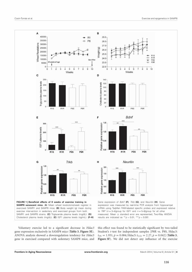
Cosín-Tomás et al. Exercise and epigenetics in SAMP8
FIGURE 1 | Beneficial effects of 8 weeks of exercise training inSAMP8 senescent mice. (A) Mean wheel revolutions/week register inexercised SAMR1 and SAMP8 mice; (B) Body weight (g) mean duringexercise intervention in sedentary and exercised groups from bothSAMR1 and SAMP8 strains. (C) Triglyceride plasma levels (mg/dL). (D)Cholesterol plasma levels (mg/dL). (E) IGF1 plasma levels (ng/mL). (F–H)
Gene expression of Bdnf (F), Trkb (G), and Neuritin (H). Geneexpression was measured by real-time PCR analysis from hippocampalmRNA using TaqMan FAM-labeled specific probes and expressed relativeto TBP (n = 3–4/group for IGF1 and n = 5–8/group for all othermeasures). Mean ± standard error are represented; Two-Way ANOVAresults are indicated as ∗∗p < 0.01; ∗∗∗p < 0.001.
Voluntary exercise led to a significant decrease in Hdac3gene expression exclusively in SAMP8 mice (Table 3, Figure 3E).ANOVA analysis showed a downregulation tendency for Hdac5gene in exercised compared with sedentary SAMP8 mice, and
this effect was found to be statistically significant by two-tailedStudent’s t-test for independent samples [P8R vs. P8S, Hdac3:t(9) = 1.931, p = 0.084; Hdac5: t(12) = 2.27, p = 0.042] (Table 3,Figure 3F). We did not detect any influence of the exercise
Frontiers in Aging Neuroscience www.frontiersin.org March 2014 | Volume 6 | Article 51 | 4
116
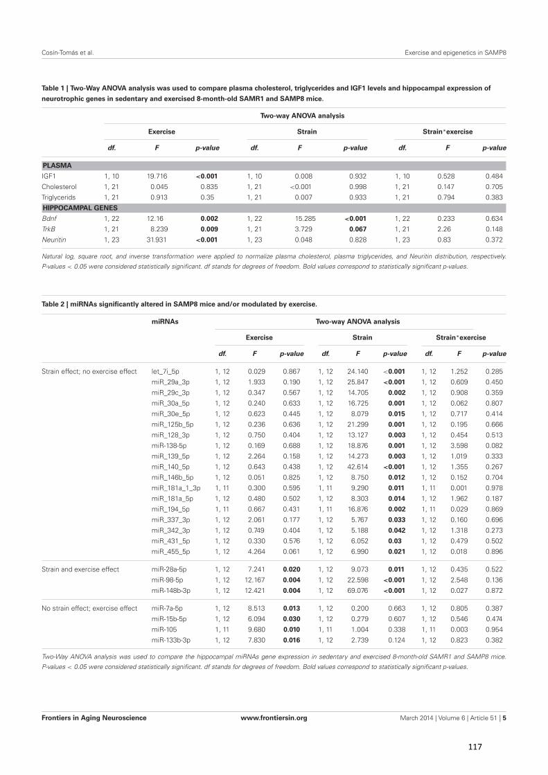
Cosín-Tomás et al. Exercise and epigenetics in SAMP8
Table 1 | Two-Way ANOVA analysis was used to compare plasma cholesterol, triglycerides and IGF1 levels and hippocampal expression of
neurotrophic genes in sedentary and exercised 8-month-old SAMR1 and SAMP8 mice.
Two-way ANOVA analysis
Exercise Strain Strain*exercise
df. F p-value df. F p-value df. F p-value
PLASMA
IGF1 1, 10 19.716 <0.001 1, 10 0.008 0.932 1, 10 0.528 0.484
Cholesterol 1, 21 0.045 0.835 1, 21 <0.001 0.998 1, 21 0.147 0.705
Triglycerids 1, 21 0.913 0.35 1, 21 0.007 0.933 1, 21 0.794 0.383
HIPPOCAMPAL GENES
Bdnf 1, 22 12.16 0.002 1, 22 15.285 <0.001 1, 22 0.233 0.634
TrkB 1, 21 8.239 0.009 1, 21 3.729 0.067 1, 21 2.26 0.148
Neuritin 1, 23 31.931 <0.001 1, 23 0.048 0.828 1, 23 0.83 0.372
Natural log, square root, and inverse transformation were applied to normalize plasma cholesterol, plasma triglycerides, and Neuritin distribution, respectively.
P-values < 0.05 were considered statistically significant. df stands for degrees of freedom. Bold values correspond to statistically significant p-values.
Table 2 | miRNAs significantly altered in SAMP8 mice and/or modulated by exercise.
miRNAs Two-way ANOVA analysis
Exercise Strain Strain*exercise
df. F p-value df. F p-value df. F p-value
Strain effect; no exercise effect let_7i_5p 1, 12 0.029 0.867 1, 12 24.140 <0.001 1, 12 1.252 0.285
miR_29a_3p 1, 12 1.933 0.190 1, 12 25.847 <0.001 1, 12 0.609 0.450
miR_29c_3p 1, 12 0.347 0.567 1, 12 14.705 0.002 1, 12 0.908 0.359
miR_30a_5p 1, 12 0.240 0.633 1, 12 16.725 0.001 1, 12 0.062 0.807
miR_30e_5p 1, 12 0.623 0.445 1, 12 8.079 0.015 1, 12 0.717 0.414
miR_125b_5p 1, 12 0.236 0.636 1, 12 21.299 0.001 1, 12 0.195 0.666
miR_128_3p 1, 12 0.750 0.404 1, 12 13.127 0.003 1, 12 0.454 0.513
miR-138-5p 1, 12 0.169 0.688 1, 12 18.876 0.001 1, 12 3.598 0.082
miR_139_5p 1, 12 2.264 0.158 1, 12 14.273 0.003 1, 12 1.019 0.333
miR_140_5p 1, 12 0.643 0.438 1, 12 42.614 <0.001 1, 12 1.355 0.267
miR_146b_5p 1, 12 0.051 0.825 1, 12 8.750 0.012 1, 12 0.152 0.704
miR_181a_1_3p 1, 11 0.300 0.595 1, 11 9.290 0.011 1, 11 0.001 0.978
miR_181a_5p 1, 12 0.480 0.502 1, 12 8.303 0.014 1, 12 1.962 0.187
miR_194_5p 1, 11 0.667 0.431 1, 11 16.876 0.002 1, 11 0.029 0.869
miR_337_3p 1, 12 2.061 0.177 1, 12 5.767 0.033 1, 12 0.160 0.696
miR_342_3p 1, 12 0.749 0.404 1, 12 5.188 0.042 1, 12 1.318 0.273
miR_431_5p 1, 12 0.330 0.576 1, 12 6.052 0.03 1, 12 0.479 0.502
miR_455_5p 1, 12 4.264 0.061 1, 12 6.990 0.021 1, 12 0.018 0.896
Strain and exercise effect miR-28a-5p 1, 12 7.241 0.020 1, 12 9.073 0.011 1, 12 0.435 0.522
miR-98-5p 1, 12 12.167 0.004 1, 12 22.598 <0.001 1, 12 2.548 0.136
miR-148b-3p 1, 12 12.421 0.004 1, 12 69.076 <0.001 1, 12 0.027 0.872
No strain effect; exercise effect miR-7a-5p 1, 12 8.513 0.013 1, 12 0.200 0.663 1, 12 0.805 0.387
miR-15b-5p 1, 12 6.094 0.030 1, 12 0.279 0.607 1, 12 0.546 0.474
miR-105 1, 11 9.680 0.010 1, 11 1.004 0.338 1, 11 0.003 0.954
miR-133b-3p 1, 12 7.830 0.016 1, 12 2.739 0.124 1, 12 0.823 0.382
Two-Way ANOVA analysis was used to compare the hippocampal miRNAs gene expression in sedentary and exercised 8-month-old SAMR1 and SAMP8 mice.
P-values < 0.05 were considered statistically significant. df stands for degrees of freedom. Bold values correspond to statistically significant p-values.
Frontiers in Aging Neuroscience www.frontiersin.org March 2014 | Volume 6 | Article 51 | 5
117
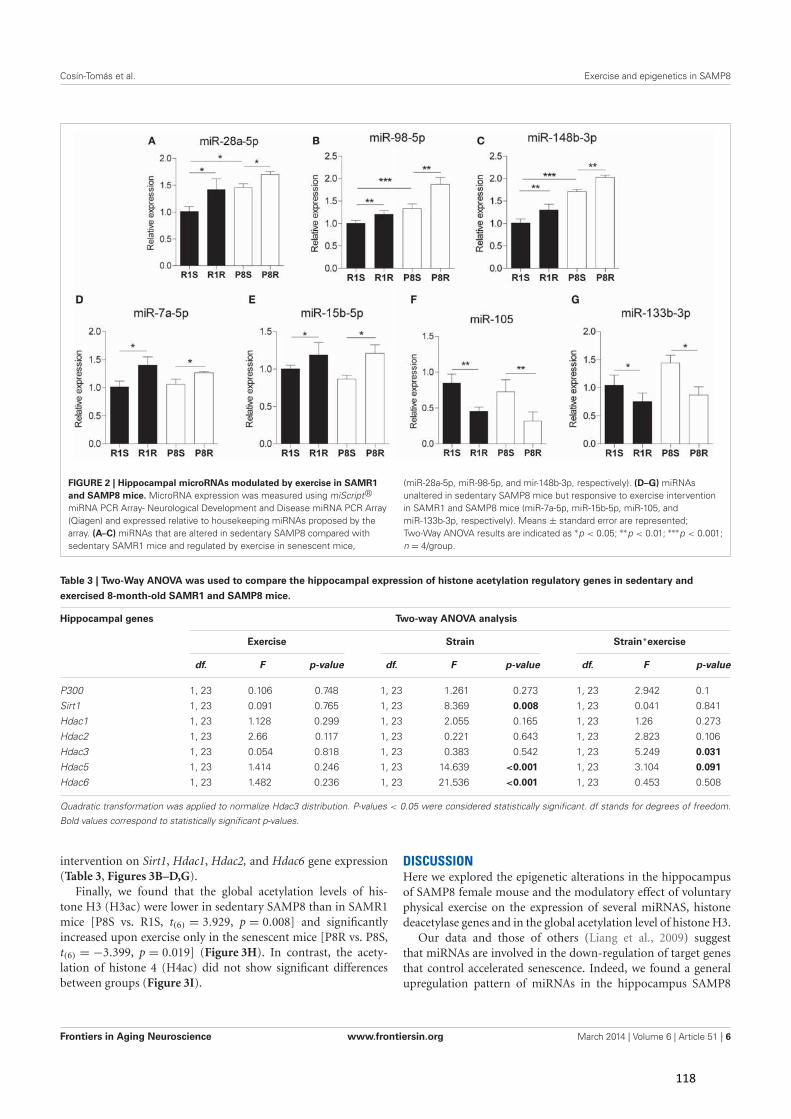
Cosín-Tomás et al. Exercise and epigenetics in SAMP8
FIGURE 2 | Hippocampal microRNAs modulated by exercise in SAMR1and SAMP8 mice. MicroRNA expression was measured using miScript?
miRNA PCR Array- Neurological Development and Disease miRNA PCR Array(Qiagen) and expressed relative to housekeeping miRNAs proposed by thearray. (A–C) miRNAs that are altered in sedentary SAMP8 compared withsedentary SAMR1 mice and regulated by exercise in senescent mice,
(miR-28a-5p, miR-98-5p, and mir-148b-3p, respectively). (D–G) miRNAsunaltered in sedentary SAMP8 mice but responsive to exercise interventionin SAMR1 and SAMP8 mice (miR-7a-5p, miR-15b-5p, miR-105, andmiR-133b-3p, respectively). Means ± standard error are represented;Two-Way ANOVA results are indicated as ∗p < 0.05; ∗∗p < 0.01; ∗∗∗p < 0.001;n = 4/group.
Table 3 | Two-Way ANOVA was used to compare the hippocampal expression of histone acetylation regulatory genes in sedentary and
exercised 8-month-old SAMR1 and SAMP8 mice.
Hippocampal genes Two-way ANOVA analysis
Exercise Strain Strain*exercise
df. F p-value df. F p-value df. F p-value
P300 1, 23 0.106 0.748 1, 23 1.261 0.273 1, 23 2.942 0.1
Sirt1 1, 23 0.091 0.765 1, 23 8.369 0.008 1, 23 0.041 0.841
Hdac1 1, 23 1.128 0.299 1, 23 2.055 0.165 1, 23 1.26 0.273
Hdac2 1, 23 2.66 0.117 1, 23 0.221 0.643 1, 23 2.823 0.106
Hdac3 1, 23 0.054 0.818 1, 23 0.383 0.542 1, 23 5.249 0.031
Hdac5 1, 23 1.414 0.246 1, 23 14.639 <0.001 1, 23 3.104 0.091
Hdac6 1, 23 1.482 0.236 1, 23 21.536 <0.001 1, 23 0.453 0.508
Quadratic transformation was applied to normalize Hdac3 distribution. P-values < 0.05 were considered statistically significant. df stands for degrees of freedom.
Bold values correspond to statistically significant p-values.
intervention on Sirt1, Hdac1, Hdac2, and Hdac6 gene expression(Table 3, Figures 3B–D,G).
Finally, we found that the global acetylation levels of his-tone H3 (H3ac) were lower in sedentary SAMP8 than in SAMR1mice [P8S vs. R1S, t(6) = 3.929, p = 0.008] and significantlyincreased upon exercise only in the senescent mice [P8R vs. P8S,t(6) = −3.399, p = 0.019] (Figure 3H). In contrast, the acety-lation of histone 4 (H4ac) did not show significant differencesbetween groups (Figure 3I).
DISCUSSIONHere we explored the epigenetic alterations in the hippocampusof SAMP8 female mouse and the modulatory effect of voluntaryphysical exercise on the expression of several miRNAS, histonedeacetylase genes and in the global acetylation level of histone H3.
Our data and those of others (Liang et al., 2009) suggestthat miRNAs are involved in the down-regulation of target genesthat control accelerated senescence. Indeed, we found a generalupregulation pattern of miRNAs in the hippocampus SAMP8
Frontiers in Aging Neuroscience www.frontiersin.org March 2014 | Volume 6 | Article 51 | 6
118
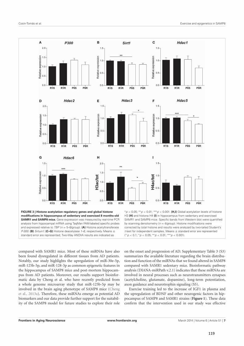
Cosín-Tomás et al. Exercise and epigenetics in SAMP8
FIGURE 3 | Histone acetylation regulatory genes and global histonemodifications in hippocampus of sedentary and exercised 8 months-oldSAMR1 and SAMP8 mice. Gene expression was measured by real-time PCRanalysis from hippocampal mRNA using TaqMan FAM-labeled specific probesand expressed relative to TBP (n = 5–8/group). (A) Histone acetyltransferaseP-300. (B) Sirtuin1. (C–G) Histone deacetylase 1–6, respectively. Means ±standard error are represented; Two-Way ANOVA results are indicated as
∗p < 0.05; ∗∗p < 0.01; ∗∗∗p < 0.001. (H,I) Global acetylation levels of histoneH3 (H) and histone H4 (I) in hippocampus from sedentary and exercisedSAMR1 and SAMP8 mice. Specific bands from Western blot were quantifiedby scanning densitometry (n = 4/group). Histone modifications werecorrected by total histone and results were analyzed by two-tailed Student’st-test for independent samples. Means ± standard error are represented(+p < 0.1; ∗p < 0.05; ∗∗p < 0.01; ∗∗∗p < 0.001).
compared with SAMR1 mice. Most of these miRNAs have alsobeen found dysregulated in different tissues from AD patients.Notably, our study highlights the upregulation of miR-30e-5p,miR-125b-5p, and miR-128-3p as common epigenetic features inthe hippocampus of SAMP8 mice and post-mortem hippocam-pus from AD patients. Moreover, our results support bioinfor-matic data by Cheng et al. who have recently predicted froma whole genome microarray study that miR-125b-5p may beinvolved in the brain aging phenotype of SAMP8 mice (Chenget al., 2013a). Therefore, these miRNAs emerge as potential ADbiomarkers and our data provide further support for the suitabil-ity of the SAMP8 model for future studies to explore their role
on the onset and progression of AD. Supplementary Table 3 (S3)summarizes the available literature regarding the brain distribu-tion and function of the miRNAs that we found altered in SAMP8compared with SAMR1 sedentary mice. Bioinformatic pathwayanalysis (DIANA-miRPath v.2.1) indicates that these miRNAs areinvolved in neural processes such as neurotransmitters synapses(acetylcholine, glutamate, dopamine), long-term potentiation,axon guidance and neurotrophin signaling (S5).
Exercise training led to the increase of IGF1 in plasma andthe upregulation of BDNF and other neurogenic factors in hip-pocampus of SAMP8 and SAMR1 strains (Figure 1). These dataconfirm that the intervention used in our study was effective
Frontiers in Aging Neuroscience www.frontiersin.org March 2014 | Volume 6 | Article 51 | 7
119
Cosín-Tomás et al. Exercise and epigenetics in SAMP8
as such effects have previously been reported in a variety ofrodent models in response to exercise (Saltiel and Kahn, 2001;Llorens-Martin et al., 2010; Chang et al., 2011; Kaliman et al.,2011; Sakurai et al., 2011; Higashi et al., 2012; Alvarez-Lopezet al., 2013). We found that miR-28a-5p, miR-98a-5p, miR-148b-3p were altered in sedentary SAMP8 compared with SAMR1mice and changed their expression levels in response to exer-cise (putative aging markers responsive to exercise). On the otherhand, miR-7a-5p, miR-15b-5p, miR-105, miR-133b-3p, whichwere similarly expressed in SAMP8 and SAMR1 mice, were mod-ulated by exercise in both strains (putative markers of exerciseunrelated to aging). The data available on the function andexpression of these miRNAs in the CNS are summarized inSupplementary Table 4 (S4). Further study is warranted to explorethe precise mechanistic links between these miRNAs and theprotective central effects of physical exercise. In this context, aprediction through bioinformatic pathway analysis for multiplemiRNA effect indicates that these exercise-responsive miRNAsare involved in the regulation of PI-3-kinase-Akt, focal adhesion,insulin, mTOR and MapK signaling pathways, all of which aremodulated in the brain by exercise (Shen et al., 2001; Tong et al.,2001; Bruel-Jungerman et al., 2009; Muller et al., 2011; Elfvinget al., 2013) (S6).
Both the sedentary and exercised SAMP8 mice showed alteredexpression patterns of protein deacetylases with reported func-tions in the aging brain and AD such as Hdac6, Sirt1, Hdac3,and Hdac5. We found a downregulation of histone deacetylaseHdac6 in the hippocampus of sedentary SAMP8 mice. HDAC6specific inhibitors have been described as potential therapeuticapproaches to rescue the neurodegeneration, however an induc-tion of HDAC6 was reported to facilitate the autophagy of mis-folded proteins and aggregates of Aβ42 and p-tau (Simoes-Pireset al., 2013). Further research is required to better understand thestill controversial role of HDAC6 and our data indicate that theSAMP8 mice may represent a suitable model for this purpose.We also found a downregulation of the protein deacetylase Sirt1in the hippocampus of SAMP8 mice, supporting the notion thatdecreased Sirt1 expression is a feature of the accelerated brainaging and neurodegeneration (Pallas et al., 2008b; Duan, 2013).However, we did not find a modulation of Sirt1 mRNA levelsin response to the running intervention in contrast to previousfindings using other experimental models (Ferrara et al., 2008;Dumke et al., 2009; Koltai et al., 2010).
Our data suggest that exercise may exert some of its reportedbeneficial effects on SAMP8 cognitive performance throughHdac3 downregulation, a mechanism involved in long-termmemory enhancement (Mcquown et al., 2011) and in the rever-sion of contextual memory deficits in a mouse model of AD(Fischer et al., 2007; Kilgore et al., 2010).
It has previously been reported that exercise activates Bdnftranscription through Hdac5 downregulation (Gomez-Pinillaet al., 2011). Therefore, the downregulation of Hdac5 expressionin the exercised SAMP8 mice may be at least in part responsi-ble for the observed Bdnf upregulation (Figure 2). Finally, globalacetylation levels of histone H3 (H3ac) were increased after theexercise intervention in the SAMP8 mice suggesting that HDACgene downregulation had some impact on chromatin remodeling.
Notably, partial correlation analyses revealed a negative associa-tion between the modification H3ac and Hdac3 gene expression[ρ(11) = −0.721, p < 0.01].
As a whole, our study highlights some common epigeneticfeatures in hippocampus of SAMP8 mice and human AD, andprovides further support for the suitability of this experimen-tal model for future epigenetic studies regarding the onset andprogression of AD. Among them, miRNAs emerge as poten-tially valuable biomarkers for the development of new therapeuticstrategies for senescence and neurodegeneration. Moreover, ourdata suggest a positive impact of voluntary exercise in revers-ing some epigenetic and transcriptional alterations associatedwith the aging brain, and reinforces the prevailing concept thatphysical training is a promising therapeutic strategy for neurode-generative diseases such as AD.
ACKNOWLEDGMENTSThis study was supported by grant SAF2010-15050 (PerlaKaliman) from the Spanish Ministry of Economy andCompetitiveness (MINECO) with the collaboration of grantsDPS2008-06998-C02 (Merce Pallàs and Rosa M. Escorihuela);CSD2010-45 (Coral Sanfeliu) and SAF2012-39852 (Merce Pallàsand Coral Sanfeliu) from MINECO. Marta Cosín-Tomás is sup-ported by a predoctoral fellowship from MINECO (FPU 2013);Jaume F. Lalanza and María J. Alvarez-López were supportedby a predoctoral fellowship from the Generalitat de Catalunya(FI-DGR 2011).
SUPPLEMENTARY MATERIALThe Supplementary Material for this article can be found onlineat: http://www.frontiersin.org/journal/10.3389/fnagi.2014.
00051/abstract
REFERENCESAbel, J. L., and Rissman, E. F. (2013). Running-induced epigenetic and gene expres-
sion changes in the adolescent brain. Int. J. Dev. Neurosci. 31, 382–390. doi:10.1016/j.ijdevneu.2012.11.002
Akbarian, S., Beeri, M. S., and Haroutunian, V. (2013). Epigenetic determinants ofhealthy and diseased brain aging and cognition. JAMA Neurol. 70, 711–718. doi:10.1001/jamaneurol.2013.1459
Alvarez-Lopez, M. J., Castro-Freire, M., Cosin-Tomas, M., Sanchez-Roige, S.,Lalanza, J. F., Del Valle, J., et al. (2013). Long-term exercise modulates hip-pocampal gene expression in senescent female mice. J. Alzheimers Dis. 33,1177–1190. doi: 10.3233/JAD-121264
Arendt, T. (2009). Synaptic degeneration in Alzheimer’s disease. Acta Neuropathol.118, 167–179. doi: 10.1007/s00401-009-0536-x
Bartel, D. P. (2009). MicroRNAs: target recognition and regulatory functions. Cell136, 215–233. doi: 10.1016/j.cell.2009.01.002
Bentwich, I., Avniel, A., Karov, Y., Aharonov, R., Gilad, S., Barad, O., et al. (2005).Identification of hundreds of conserved and nonconserved human microRNAs.Nat. Genet. 37, 766–770. doi: 10.1038/ng1590
Bruel-Jungerman, E., Veyrac, A., Dufour, F., Horwood, J., Laroche, S., and Davis,S. (2009). Inhibition of PI3K-Akt signaling blocks exercise-mediated enhance-ment of adult neurogenesis and synaptic plasticity in the dentate gyrus. PLoSONE 4:e7901. doi: 10.1371/journal.pone.0007901
Chandramohan, Y., Droste, S. K., Arthur, J. S., and Reul, J. M. (2008). Theforced swimming-induced behavioural immobility response involves histoneH3 phospho-acetylation and c-Fos induction in dentate gyrus granule neu-rons via activation of the N-methyl-D-aspartate/extracellular signal-regulatedkinase/mitogen- and stress-activated kinase signalling pathway. Eur. J. Neurosci.27, 2701–2713. doi: 10.1111/j.1460-9568.2008.06230.x
Frontiers in Aging Neuroscience www.frontiersin.org March 2014 | Volume 6 | Article 51 | 8
120
Cosín-Tomás et al. Exercise and epigenetics in SAMP8
Chang, H. C., Yang, Y. R., Wang, P. S., Kuo, C. H., and Wang, R. Y. (2011).Insulin-like growth factor I signaling for brain recovery and exercise abil-ity in brain ischemic rats. Med. Sci. Sports Exerc. 43, 2274–2280. doi:10.1249/MSS.0b013e318223b5d9
Cheng, X. R., Cui, X. L., Zheng, Y., Zhang, G. R., Li, P., Huang, H., et al. (2013a).Nodes and biological processes identified on the basis of network analysis inthe brain of the senescence accelerated mice as an Alzheimer’s disease animalmodel. Front. Aging Neurosci. 5:65. doi: 10.3389/fnagi.2013.00065
Cheng, X. R., Zhou, W. X., and Zhang, Y. X. (2013b). The behavioral, patho-logical and therapeutic features of the senescence-accelerated mouse prone 8strain as an Alzheimer’s disease animal model. Ageing Res. Rev. 13C, 13–37. doi:10.1016/j.arr.2013.10.002
Cogswell, J. P., Ward, J., Taylor, I. A., Waters, M., Shi, Y., Cannon, B., et al. (2008).Identification of miRNA changes in Alzheimer’s disease brain and CSF yieldsputative biomarkers and insights into disease pathways. J. Alzheimers Dis. 14,27–41.
Collins, A., Hill, L. E., Chandramohan, Y., Whitcomb, D., Droste, S. K., andReul, J. M. (2009). Exercise improves cognitive responses to psychological stressthrough enhancement of epigenetic mechanisms and gene expression in thedentate gyrus. PLoS ONE 4:e4330. doi: 10.1371/journal.pone.0004330
Crepaldi, L., and Riccio, A. (2009). Chromatin learns to behave. Epigenetics 4,23–26. doi: 10.4161/epi.4.1.7604
Dang, W., Steffen, K. K., Perry, R., Dorsey, J. A., Johnson, F. B., Shilatifard, A., et al.(2009). Histone H4 lysine 16 acetylation regulates cellular lifespan. Nature 459,802–807. doi: 10.1038/nature08085
Del Valle, J., Duran-Vilaregut, J., Manich, G., Pallas, M., Camins, A., Vilaplana,J., et al. (2011). Cerebral amyloid angiopathy, blood-brain barrier disruptionand amyloid accumulation in SAMP8 mice. Neurodegener. Dis. 8, 421–429. doi:10.1159/000324757
Di Bernardo, G., Cipollaro, M., and Galderisi, U. (2012). Chromatinmodification and senescence. Curr. Pharm. Des. 18, 1686–1693. doi:10.2174/138161212799859693
Dishman, R. K., Berthoud, H. R., Booth, F. W., Cotman, C. W., Edgerton, V. R.,Fleshner, M. R., et al. (2006). Neurobiology of exercise. Obesity (Silver Spring)14, 345–356. doi: 10.1038/oby.2006.46
Duan, W. (2013). Sirtuins: from metabolic regulation to brain aging. Front. AgingNeurosci. 5:36. doi: 10.3389/fnagi.2013.00036
Dumke, C. L., Mark Davis, J., Angela Murphy, E., Nieman, D. C., Carmichael, M.D., Quindry, J. C., et al. (2009). Successive bouts of cycling stimulates genesassociated with mitochondrial biogenesis. Eur. J. Appl. Physiol. 107, 419–427.doi: 10.1007/s00421-009-1143-1
Elfving, B., Christensen, T., Ratner, C., Wienecke, J., and Klein, A. B. (2013).Transient activation of mTOR following forced treadmill exercise in rats.Synapse 67, 620–625. doi: 10.1002/syn.21668
Ferrara, N., Rinaldi, B., Corbi, G., Conti, V., Stiuso, P., Boccuti, S., et al. (2008).Exercise training promotes SIRT1 activity in aged rats. Rejuvenation Res. 11,139–150. doi: 10.1089/rej.2007.0576
Fischer, A., Sananbenesi, F., Wang, X., Dobbin, M., and Tsai, L. H. (2007). Recoveryof learning and memory is associated with chromatin remodelling. Nature 447,178–182. doi: 10.1038/nature05772
Goldie, B. J., and Cairns, M. J. (2012). Post-transcriptional trafficking andregulation of neuronal gene expression. Mol. Neurobiol. 45, 99–108. doi:10.1007/s12035-011-8222-0
Gomez-Pinilla, F., Vaynman, S., and Ying, Z. (2008). Brain-derived neurotrophicfactor functions as a metabotrophin to mediate the effects of exercise on cogni-tion. Eur. J. Neurosci. 28, 2278–2287. doi: 10.1111/j.1460-9568.2008.06524.x
Gomez-Pinilla, F., Zhuang, Y., Feng, J., Ying, Z., and Fan, G. (2011). Exerciseimpacts brain-derived neurotrophic factor plasticity by engaging mechanismsof epigenetic regulation. Eur. J. Neurosci. 33, 383–390. doi: 10.1111/j.1460-9568.2010.07508.x
Greer, E. L., Maures, T. J., Hauswirth, A. G., Green, E. M., Leeman, D. S., Maro, G.S., et al. (2010). Members of the H3K4 trimethylation complex regulate lifes-pan in a germline-dependent manner in C. elegans. Nature 466, 383–387. doi:10.1038/nature09195
Hebert, S. S., Horre, K., Nicolai, L., Papadopoulou, A. S., Mandemakers, W.,Silahtaroglu, A. N., et al. (2008). Loss of microRNA cluster miR-29a/b-1 in sporadic Alzheimer’s disease correlates with increased BACE1/beta-secretase expression. Proc. Natl. Acad. Sci. U.S.A. 105, 6415–6420. doi:10.1073/pnas.0710263105
Higashi, Y., Sukhanov, S., Anwar, A., Shai, S. Y., and Delafontaine, P. (2012). Aging,atherosclerosis, and IGF-1. J. Gerontol. A Biol. Sci. Med. Sci. 67, 626–639. doi:10.1093/gerona/gls102
Huidobro, C., Fernandez, A. F., and Fraga, M. F. (2013). Aging epige-netics: causes and consequences. Mol. Aspects Med. 34, 765–781. doi:10.1016/j.mam.2012.06.006
Jaenisch, R., and Bird, A. (2003). Epigenetic regulation of gene expression: how thegenome integrates intrinsic and environmental signals. Nat. Genet. 33(Suppl.),245–254. doi: 10.1038/ng1089
Jakovcevski, M., and Akbarian, S. (2012). Epigenetic mechanisms in neurologicaldisease. Nat. Med. 18, 1194–1204. doi: 10.1038/nm.2828
Jung, H. J., and Suh, Y. (2012). microRNA in aging: from discovery to biology. Curr.Genomics 13, 548–557. doi: 10.2174/138920212803251436
Kaliman, P., Parrizas, M., Lalanza, J. F., Camins, A., Escorihuela, R. M., and Pallas,M. (2011). Neurophysiological and epigenetic effects of physical exercise on theaging process. Ageing Res. Rev. 10, 475–486. doi: 10.1016/j.arr.2011.05.002
Kilgore, M., Miller, C. A., Fass, D. M., Hennig, K. M., Haggarty, S. J., Sweatt, J. D.,et al. (2010). Inhibitors of class 1 histone deacetylases reverse contextual mem-ory deficits in a mouse model of Alzheimer’s disease. Neuropsychopharmacology35, 870–880. doi: 10.1038/npp.2009.197
Koltai, E., Szabo, Z., Atalay, M., Boldogh, I., Naito, H., Goto, S., et al. (2010).Exercise alters SIRT1, SIRT6, NAD and NAMPT levels in skeletal muscle of agedrats. Mech. Ageing Dev. 131, 21–28. doi: 10.1016/j.mad.2009.11.002
Lau, P., Bossers, K., Janky, R., Salta, E., Frigerio, C. S., Barbash, S., et al. (2013).Alteration of the microRNA network during the progression of Alzheimer’sdisease. EMBO Mol. Med. 5, 1613–1634. doi: 10.1002/emmm.201201974
Legube, G., and Trouche, D. (2003). Regulating histone acetyltransferases anddeacetylases. EMBO Rep. 4, 944–947. doi: 10.1038/sj.embor.embor941
Liang, R., Bates, D. J., and Wang, E. (2009). Epigenetic control of microRNA expres-sion and aging. Curr. Genomics 10, 184–193. doi: 10.2174/138920209788185225
Llorens-Martin, M. V., Rueda, N., Tejeda, G. S., Florez, J., Trejo, J. L., and Martinez-Cue, C. (2010). Effects of voluntary physical exercise on adult hippocampalneurogenesis and behavior of Ts65Dn mice, a model of Down syndrome.Neuroscience 171, 1228–1240. doi: 10.1016/j.neuroscience.2010.09.043
Lopez-Otin, C., Blasco, M. A., Partridge, L., Serrano, M., and Kroemer, G. (2013).The hallmarks of aging. Cell 153, 1194–1217. doi: 10.1016/j.cell.2013.05.039
Lovatel, G. A., Elsner, V. R., Bertoldi, K., Vanzella, C., Moyses Fdos, S., Vizuete, A.,et al. (2013). Treadmill exercise induces age-related changes in aversive memory,neuroinflammatory and epigenetic processes in the rat hippocampus. Neurobiol.Learn. Mem. 101, 94–102. doi: 10.1016/j.nlm.2013.01.007
Lukiw, W. J. (2007). Micro-RNA speciation in fetal, adult and Alzheimer’s diseasehippocampus. Neuroreport 18, 297–300. doi: 10.1097/WNR.0b013e3280148e8b
Maes, O. C., Chertkow, H. M., Wang, E., and Schipper, H. M. (2009). microRNA:implications for Alzheimer disease and other human CNS disorders. Curr.Genomics 10, 154–168. doi: 10.2174/138920209788185252
Mastroeni, D., Grover, A., Delvaux, E., Whiteside, C., Coleman, P. D., and Rogers,J. (2011). Epigenetic mechanisms in Alzheimer’s disease. Neurobiol. Aging 32,1161–1180. doi: 10.1016/j.neurobiolaging.2010.08.017
Mcquown, S. C., Barrett, R. M., Matheos, D. P., Post, R. J., Rogge, G. A., Alenghat,T., et al. (2011). HDAC3 is a critical negative regulator of long-term memoryformation. J. Neurosci. 31, 764–774. doi: 10.1523/JNEUROSCI.5052-10.2011
Min, S. W., Cho, S. H., Zhou, Y., Schroeder, S., Haroutunian, V., Seeley, W. W., et al.(2010). Acetylation of tau inhibits its degradation and contributes to tauopathy.Neuron 67, 953–966. doi: 10.1016/j.neuron.2010.08.044
Morley, J. E., Armbrecht, H. J., Farr, S. A., and Kumar, V. B. (2012a). The senescenceaccelerated mouse (SAMP8) as a model for oxidative stress and Alzheimer’s dis-ease. Biochim. Biophys. Acta 1822, 650–656. doi: 10.1016/j.bbadis.2011.11.015
Morley, J. E., Farr, S. A., Kumar, V. B., and Armbrecht, H. J. (2012b). The SAMP8mouse: a model to develop therapeutic interventions for Alzheimer’s disease.Curr. Pharm. Des. 18, 1123–1130. doi: 10.2174/138161212799315795
Muller, A. P., Gnoatto, J., Moreira, J. D., Zimmer, E. R., Haas, C. B., Lulhier, F.,et al. (2011). Exercise increases insulin signaling in the hippocampus: physi-ological effects and pharmacological impact of intracerebroventricular insulinadministration in mice. Hippocampus 21, 1082–1092. doi: 10.1002/hipo.20822
Pallas, M., Camins, A., Smith, M. A., Perry, G., Lee, H. G., and Casadesus, G.(2008a). From aging to Alzheimer’s disease: unveiling “the switch” with thesenescence-accelerated mouse model (SAMP8). J. Alzheimers Dis. 15, 615–624.
Pallas, M., Pizarro, J. G., Gutierrez-Cuesta, J., Crespo-Biel, N., Alvira, D.,Tajes, M., et al. (2008b). Modulation of SIRT1 expression in different
Frontiers in Aging Neuroscience www.frontiersin.org March 2014 | Volume 6 | Article 51 | 9
121
Cosín-Tomás et al. Exercise and epigenetics in SAMP8
neurodegenerative models and human pathologies. Neuroscience 154,1388–1397. doi: 10.1016/j.neuroscience.2008.04.065
Saha, R. N., and Pahan, K. (2006). HATs and HDACs in neurodegeneration: a taleof disconcerted acetylation homeostasis. Cell Death Differ. 13, 539–550. doi:10.1038/sj.cdd.4401769
Sakurai, T., Ogasawara, J., Kizaki, T., Ishibashi, Y., Fujiwara, T., Akagawa, K.,et al. (2011). Involvement of leucine zipper transcription factor-like protein1 (Lztfl1) in the attenuation of cognitive impairment by exercise training.Biochem. Biophys. Res. Commun. 416, 125–129. doi: 10.1016/j.bbrc.2011.11.008
Saltiel, A. R., and Kahn, C. R. (2001). Insulin signalling and the regulation ofglucose and lipid metabolism. Nature 414, 799–806. doi: 10.1038/414799a
Scheff, S. W., Price, D. A., Schmitt, F. A., Dekosky, S. T., and Mufson,E. J. (2007). Synaptic alterations in CA1 in mild Alzheimer dis-ease and mild cognitive impairment. Neurology 68, 1501–1508. doi:10.1212/01.wnl.0000260698.46517.8f
Shen, H., Tong, L., Balazs, R., and Cotman, C. W. (2001). Physical activity elic-its sustained activation of the cyclic AMP response element-binding proteinand mitogen-activated protein kinase in the rat hippocampus. Neuroscience 107,219–229. doi: 10.1016/S0306-4522(01)00315-3
Siebold, A. P., Banerjee, R., Tie, F., Kiss, D. L., Moskowitz, J., and Harte, P. J.(2010). Polycomb repressive complex 2 and trithorax modulate drosophilalongevity and stress resistance. Proc. Natl. Acad. Sci. U.S.A. 107, 169–174. doi:10.1073/pnas.0907739107
Simoes-Pires, C., Zwick, V., Nurisso, A., Schenker, E., Carrupt, P. A., and Cuendet,M. (2013). HDAC6 as a target for neurodegenerative diseases: what makes itdifferent from the other HDACs? Mol. Neurodegener. 8:7. doi: 10.1186/1750-1326-8-7
Takeda, T. (2009). Senescence-accelerated mouse (SAM) with special referencesto neurodegeneration models, SAMP8 and SAMP10 mice. Neurochem. Res. 34,639–659. doi: 10.1007/s11064-009-9922-y
Takeda, T., Hosokawa, M., and Higuchi, K. (1991). Senescence-accelerated mouse(SAM): a novel murine model of accelerated senescence. J. Am. Geriatr. Soc. 39,911–919.
Tammen, S. A., Friso, S., and Choi, S. W. (2013). Epigenetics: the linkbetween nature and nurture. Mol. Aspects Med. 34, 753–764. doi:10.1016/j.mam.2012.07.018
Tong, L., Shen, H., Perreau, V. M., Balazs, R., and Cotman, C. W. (2001). Effectsof exercise on gene-expression profile in the rat hippocampus. Neurobiol. Dis. 8,1046–1056. doi: 10.1006/nbdi.2001.0427
Trejo, J. L., Llorens-Martin, M. V., and Torres-Aleman, I. (2008). The effectsof exercise on spatial learning and anxiety-like behavior are mediated by anIGF-I-dependent mechanism related to hippocampal neurogenesis. Mol. Cell.Neurosci. 37, 402–411. doi: 10.1016/j.mcn.2007.10.016
Van Den Hove, D. L., Kompotis, K., Lardenoije, R., Kenis, G., Mill, J., Steinbusch,H. W., et al. (2014). Epigenetically regulated microRNAs in Alzheimer’s disease.Neurobiol. Aging 35, 731–745. doi: 10.1016/j.neurobiolaging.2013.10.082
Wang, H., Lian, K., Han, B., Wang, Y., Kuo, S. H., Geng, Y., et al. (2013). Age-relatedalterations in the metabolic profile in the hippocampus of the senescence-accelerated mouse prone 8: a spontaneous Alzheimer’s disease mouse model.J. Alzheimers Dis. 39, 841–848. doi: 10.3233/JAD-131463
Wang, W., Kwon, E. J., and Tsai, L. H. (2012). MicroRNAs in learning, memory, andneurological diseases. Learn. Mem. 19, 359–368. doi: 10.1101/lm.026492.112
Wang, W. X., Huang, Q., Hu, Y., Stromberg, A. J., and Nelson, P. T. (2011). Patternsof microRNA expression in normal and early Alzheimer’s disease human tem-poral cortex: white matter versus gray matter. Acta Neuropathol. 121, 193–205.doi: 10.1007/s00401-010-0756-0
Yang, Z., Vilkaitis, G., Yu, B., Klimasauskas, S., and Chen, X., (2007). Approachesfor studying microRNA and small interfering RNA methylation in vitro and invivo. Methods Enzymol. 427, 139–154. doi: 10.1016/S0076-6879(07)27008-9
Conflict of Interest Statement: The authors declare that the research was con-ducted in the absence of any commercial or financial relationships that could beconstrued as a potential conflict of interest.
Received: 20 January 2014; paper pending published: 09 February 2014; accepted: 03March 2014; published online: 20 March 2014.Citation: Cosín-Tomás M, Alvarez-López MJ, Sanchez-Roige S, Lalanza JF, Bayod S,Sanfeliu C, Pallàs M, Escorihuela RM and Kaliman P (2014) Epigenetic alterations inhippocampus of SAMP8 senescent mice and modulation by voluntary physical exercise.Front. Aging Neurosci. 6:51. doi: 10.3389/fnagi.2014.00051This article was submitted to the journal Frontiers in Aging Neuroscience.Copyright © 2014 Cosín-Tomás, Alvarez-López, Sanchez-Roige, Lalanza, Bayod,Sanfeliu, Pallàs, Escorihuela and Kaliman. This is an open-access article distributedunder the terms of the Creative Commons Attribution License (CC BY). The use, dis-tribution or reproduction in other forums is permitted, provided the original author(s)or licensor are credited and that the original publication in this journal is cited, inaccordance with accepted academic practice. No use, distribution or reproduction ispermitted which does not comply with these terms.
Frontiers in Aging Neuroscience www.frontiersin.org March 2014 | Volume 6 | Article 51 | 10
122
Supplementary information S1. List of primers and probe sets used for real time RT-PCR analysis.
Gene Symbol Reference (ABI) Reference Sequence ID Bdnf Mm01334042_m1 NM_001048142.1
Hdac1 Mm02391771_g1 NM_008228.2 Hdac2 Mm00515108_m1 NM_008229.2 Hdac3 Mm00515916_m1 NM_010411.2 Hdac5 Mm01246076_m1 NM_001077696.1; NM_010412.3 Hdac6 Mm01341125_m1 NM_001130416.1; NM_010413.3 Neuritin Mm00467844_m1 NM_153529.1 P-300 Mm00625535_m1 NM_177821.6 Sirt1 Mm00490758_m1 NM_001159589.1 Tbp Mm00446971_m1 NM_013684.3 TrkB Mm00435422_m1 NM_001025074.1, NM_008745.2
123
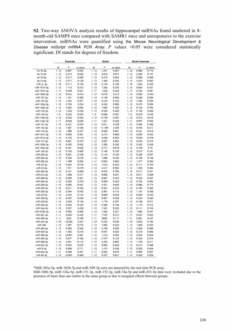
S2. Two-way ANOVA analysis results of hippocampal miRNAs found unaltered in 8-month-old SAMP8 mice compared with SAMR1 mice and unresponsive to the exercise intervention. miRNAs were quantified using the Mouse Neurological Development & Disease miScript miRNA PCR Array. P values <0.05 were considered statistically significant. Df stands for degrees of freedom.
Exercise Strain Strain*exercise
df. F p-value df. F p-value df. F p-value
let-7b-5p 1, 12 0.007 0.935 1, 12 1.241 0.287 1, 12 0.086 0.774 let-7c-5p 1, 12 0.213 0.653 1, 12 3.818 0.074 1, 12 2.695 0.127 let-7d-5p 1, 12 0.217 0.650 1, 12 0.475 0.504 1, 12 0.906 0.360 let-7e-5p 1, 12 2.517 0.139 1, 12 1.369 0.265 1, 12 4.235 0.062
miR_9_3p 1, 10 0,117 0,739 1, 10 3,149 0,106 1, 10 1,034 0,333 miR-101a-3p 1, 12 1.113 0.312 1, 12 1.302 0.276 1, 12 0.664 0.431 miR-101b-3p 1, 11 0.440 0.521 1, 11 1.632 0.228 1, 11 0.762 0.401 miR-106b-5p 1, 12 0.012 0.915 1, 12 8.510 0.013 1, 12 5.092 0.043 miR-107-3p 1, 12 1.161 0.302 1, 12 4.130 0.065 1, 12 0.298 0.595 miR-124-3p 1, 12 1.455 0.251 1, 12 2.219 0.162 1, 12 1.565 0.235
miR-126a-5p 1, 12 5.759 0.034 1, 12 3.529 0.085 1, 12 0.475 0.504 miR-130a-3p 1, 12 4.584 0.053 1, 12 1.004 0.336 1, 12 0.153 0.702 miR-132-3p 1, 12 1.533 0.239 1, 12 0.559 0.469 1, 12 0.192 0.669 miR-134-5p 1, 9 0.243 0.634 1, 9 0.826 0.387 1, 9 0.678 0.432
miR-135b-5p 1, 12 0.952 0.349 1, 12 0.740 0.407 1, 12 0.274 0.610 miR-146a-5p 1, 11 0.048 0.830 1, 11 1.421 0.258 1, 11 0.007 0.935 miR-151-3p 1, 12 0.431 0.524 1, 12 5.011 0.045 1, 12 0.566 0.466 miR-152-3p 1, 12 1.967 0.186 1, 12 11.180 0.006 1, 12 0.446 0.517 miR-15a-5p 1, 12 1.000 0.337 1, 12 0.020 0.891 1, 12 0.441 0.519 miR-181c-5p 1, 12 0.903 0.361 1, 12 3.413 0.089 1, 12 0.659 0.433 miR-181d-5p 1, 12 0.135 0.719 1, 12 3.558 0.084 1, 12 0.535 0.478 miR-191-5p 1, 12 0.861 0.372 1, 12 3.607 0.082 1, 12 0.543 0.475
miR-193b-3p 1, 12 0.303 0.592 1, 12 1.005 0.336 1, 12 0.420 0.529 miR-195a-5p 1, 12 0.421 0.529 1, 12 3.717 0.078 1, 12 0.106 0.75 miR-19b-3p 1, 12 0.195 0.666 1, 12 2.186 0.165 1, 12 2.013 0.181 miR-203-3p 1, 12 0.091 0.768 1, 12 2.144 0.169 1, 12 0.365 0.557 miR-20a-5p 1, 12 0.326 0.578 1, 12 1.008 0.335 1, 12 0.108 0.748 miR-20b-5p 1, 11 1.400 0.262 1, 11 0.023 0.882 1, 11 1.377 0.265 miR-22-3p 1, 12 0.444 0.518 1, 12 1.515 0.242 1, 12 0.111 0.745 miR-24-3p 1, 12 1757 0.210 1, 12 4.517 0.055 1, 12 1.259 0.284 miR-26b-5p 1, 12 0.161 0.696 1, 12 0.076 0.788 1, 12 0.417 0.531 miR-27a-3p 1, 12 1.089 0.317 1, 12 0.008 0.931 1, 12 0.011 0.920 miR-298-5p 1, 12 0.902 0.361 1, 12 0.687 0.423 1, 12 0.424 0.527 miR-29b-3p 1, 12 0.836 0.379 1, 12 0.600 0.453 1, 12 0.756 0.402 miR-30d-5p 1, 12 0.005 0.947 1, 12 4.341 0.059 1, 12 0.086 0.774 miR-320-3p 1, 12 0.911 0.359 1, 12 0.391 0.543 1, 12 0.784 0.393 miR-328-3p 1, 12 0.394 0.542 1, 12 2.901 0.114 1, 12 1.181 0.298 miR-33-5p 1, 12 2.444 0.144 1, 12 0.600 0.453 1, 12 0.629 0.443 miR-338-3p 1, 12 0.407 0.536 1, 12 0.997 0.338 1, 12 0.524 0.483 miR-339-5p 1, 12 2.529 0.138 1, 12 1.778 0.207 1, 12 0.189 0.671 miR-346-5p 1, 12 0.659 0.433 1, 12 2.386 0.148 1, 12 1.115 0.312 miR-34a-5p 1, 12 5.557 0.036 1, 12 1.561 0.235 1, 12 0.111 0.745
miR-376b-3p 1, 12 0.002 0.969 1, 12 1.593 0.231 1, 12 1.002 0.337 miR-381-3p 1, 11 0.629 0.445 1, 11 1120 0.313 1, 11 0.437 0.522 miR-409-3p 1, 11 3233 0.100 1, 11 2890 0.117 1, 11 0.681 0.427 miR-433-3p 1, 10 18.826 0.001 1, 10 0.423 0.530 1, 10 2.022 0.185
miR-484 1, 12 1.307 0.275 1, 12 1.062 0.323 1, 12 1.058 0.324 miR-485-5p 1, 12 0.303 0.592 1, 12 4.768 0.050 1, 12 3.265 0.096 miR-485-3p 1, 12 1.683 0.219 1, 12 0.047 0.832 1, 12 0.276 0.609 miR-488-3p 1, 12 <0.001 0.991 1, 12 1.212 0.293 1, 12 0.232 0.639 miR-489-3p 1, 12 0.077 0.786 1, 12 2.737 0.124 1, 12 0.334 0.574 miR-598-3p 1, 12 2.951 0.112 1, 12 4.253 0.062 1, 12 1.745 0.211 miR-652-3p 1, 12 0.025 0.878 1, 12 0.005 0.946 1, 12 0.512 0.488 miR-9-5p 1, 12 0.089 0.771 1, 12 1.474 0.248 1, 12 0.205 0.659
miR-92a-3p 1, 11 0.568 0.467 1, 11 0.025 0.876 1, 11 0.003 0.957 miR-93-5p 1, 12 <0.001 0.998 1, 12 0.257 0.621 1, 12 0.362 0.558
*MiR-302a-5p, miR-302b-5p and miR-509-3p were not detected by the real time PCR array. MiR-106b-5p, miR-126a-5p, miR-151-3p, miR-152-3p, miR-34a-5p and miR-433-3p data were excluded due to the presence of more than one outlier in the same group or due to marginal effects between groups.
124
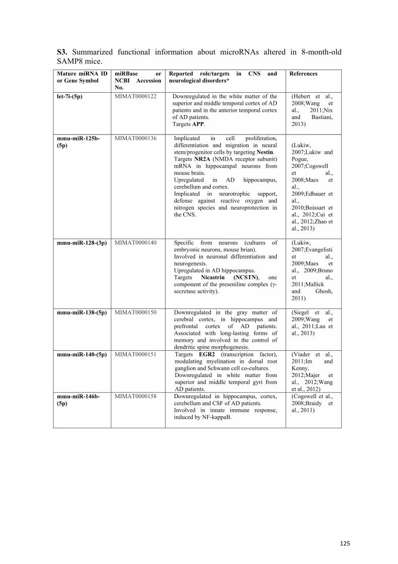
S3. Summarized functional information about microRNAs altered in 8-month-old SAMP8 mice. Mature miRNA ID or Gene Symbol
miRBase or NCBI Accession No.
Reported role/targets in CNS and neurological disorders*
References
let-7i-(5p) MIMAT0000122 Downregulated in the white matter of the superior and middle temporal cortex of AD patients and in the anterior temporal cortex of AD patients. Targets APP.
(Hebert et al., 2008;Wang et al., 2011;Nix and Bastiani, 2013)
mmu-miR-125b-(5p)
MIMAT0000136 Implicated in cell proliferation, differentiation and migration in neural stem/progenitor cells by targeting Nestin. Targets NR2A (NMDA receptor subunit) mRNA in hippocampal neurons from mouse brain. Upregulated in AD hippocampus, cerebellum and cortex. Implicated in neurotrophic support, defense against reactive oxygen and nitrogen species and neuroprotection in the CNS.
(Lukiw, 2007;Lukiw and Pogue, 2007;Cogswell et al., 2008;Maes et al., 2009;Edbauer et al., 2010;Boissart et al., 2012;Cui et al., 2012;Zhao et al., 2013)
mmu-miR-128-(3p) MIMAT0000140 Specific from neurons (cultures of embryonic neurons, mouse brian). Involved in neuronal differentiation and neurogenesis. Upregulated in AD hippocampus. Targets Nicastrin (NCSTN), one component of the preseniline complex (γ-secretase activity).
(Lukiw, 2007;Evangelisti et al., 2009;Maes et al., 2009;Bruno et al., 2011;Mallick and Ghosh, 2011)
mmu-miR-138-(5p) MIMAT0000150 Downregulated in the gray matter of cerebral cortex, in hippocampus and prefrontal cortex of AD patients. Associated with long-lasting forms of memory and involved in the control of dendritic spine morphogenesis.
(Siegel et al., 2009;Wang et al., 2011;Lau et al., 2013)
mmu-miR-140-(5p) MIMAT0000151 Targets EGR2 (transcription factor), modulating myelination in dorsal root ganglion and Schwann cell co-cultures. Downregulated in white matter from superior and middle temporal gyri from AD patients.
(Viader et al., 2011;Im and Kenny, 2012;Majer et al., 2012;Wang et al., 2012)
mmu-miR-146b-(5p)
MIMAT0000158 Downregulated in hippocampus, cortex, cerebellum and CSF of AD patients. Involved in innate immune response, induced by NF-kappaB.
(Cogswell et al., 2008;Braidy et al., 2011)
125
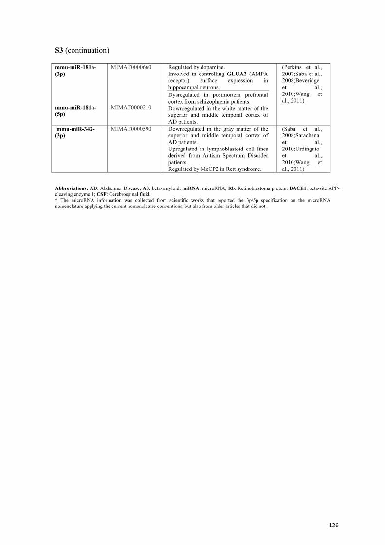
S3 (continuation) mmu-miR-181a-(3p) mmu-miR-181a-(5p)
MIMAT0000660 MIMAT0000210
Regulated by dopamine. Involved in controlling GLUA2 (AMPA receptor) surface expression in hippocampal neurons. Dysregulated in postmortem prefrontal cortex from schizophrenia patients. Downregulated in the white matter of the superior and middle temporal cortex of AD patients.
(Perkins et al., 2007;Saba et al., 2008;Beveridge et al., 2010;Wang et al., 2011)
mmu-miR-342-(3p)
MIMAT0000590 Downregulated in the gray matter of the superior and middle temporal cortex of AD patients. Upregulated in lymphoblastoid cell lines derived from Autism Spectrum Disorder patients. Regulated by MeCP2 in Rett syndrome.
(Saba et al., 2008;Sarachana et al., 2010;Urdinguio et al., 2010;Wang et al., 2011)
Abbreviations: AD: Alzheimer Disease; Aβ: beta-amyloid; miRNA: microRNA; Rb: Retinoblastoma protein; BACE1: beta-site APP-cleaving enzyme 1; CSF: Cerebrospinal fluid. * The microRNA information was collected from scientific works that reported the 3p/5p specification on the microRNA nomenclature applying the current nomenclature conventions, but also from older articles that did not.
126
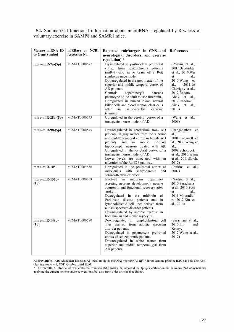
S4. Summarized functional information about microRNAs regulated by 8 weeks of voluntary exercise in SAMP8 and SAMR1 mice.
Abbreviations: AD: Alzheimer Disease; Aβ: beta-amyloid; miRNA: microRNA; Rb: Retinoblastoma protein; BACE1: beta-site APP-cleaving enzyme 1; CSF: Cerebrospinal fluid. * The microRNA information was collected from scientific works that reported the 3p/5p specification on the microRNA nomenclature applying the current nomenclature conventions, but also from older articles that did not.
Mature miRNA ID or Gene Symbol
miRBase or NCBI Accession No.
Reported role/targets in CNS and neurological disorders, and exercise regulation) *
References
mmu-miR-7a-(5p) MIMAT0000677 Dysregulated in postmortem prefrontal cortex from schizophrenia patients (miR-7) and in the brain of a Rett syndrome mice model. Downregulated in the grey matter of the superior and middle temporal cortex of AD patients. Controls dopaminergic neurons phenotype of the adult mouse forebrain. Upregulated in human blood natural killer cells and blood mononuclear cells after an acute-aerobic exercise (running).
(Perkins et al., 2007;Beveridge et al., 2010;Wu et al., 2010;Wang et al., 2011;de Chevigny et al., 2012;Radom-Aizik et al., 2012;Radom-Aizik et al., 2013)
mmu-miR-28a-(5p) MIMAT0000653 Upregulated in the cerebral cortex of a transgenic mouse model of AD.
(Wang et al., 2009)
mmu-miR-98-(5p) MIMAT0000545 Downregulated in cerebellum from AD patients, in gray matter from the superior and middle temporal cortex in female AD patients and in mouse primary hippocampal neurons treated with Aβ. Upregulated in the cerebral cortex of a transgenic mouse model of AD. Lower levels are associated with an alteration of the Rb/E2F pathway.
(Ranganathan et al., 2001;Cogswell et al., 2008;Wang et al., 2009;Schonrock et al., 2010;Wang et al., 2011;Satoh, 2012)
mmu-miR-105 MIMAT0004856 Upregulated in the prefrontal cortex of individuals with schizophrenia and schizoaffective disorder.
(Perkins et al., 2007)
mmu-miR-133b-(3p)
MIMAT0000769 Involved in midbrain dopamine-secreting neurons development, neurite outgrowth and functional recovery after stroke. Dysregulated in the midbrain of Parkinson disease patients and in lymphoblastoid cell lines derived from autism spectrum disorder patients. Downregulated by aerobic exercise in both human and mouse myocytes.
(Nielsen et al., 2010;Sarachana et al., 2010;Soci et al., 2011;Mouradian, 2012;Xin et al., 2013)
mmu-miR-148b-(3p)
MIMAT0000580 Downregulated in lymphoblastoid cell lines derived from autistic spectrum disorder patients. Dysregulated in postmortem prefrontal cortex of schizophrenic patients. Downregulated in white matter from superior and middle temporal gyri from AD patients.
(Sarachana et al., 2010;Im and Kenny, 2012;Wang et al., 2012)
127
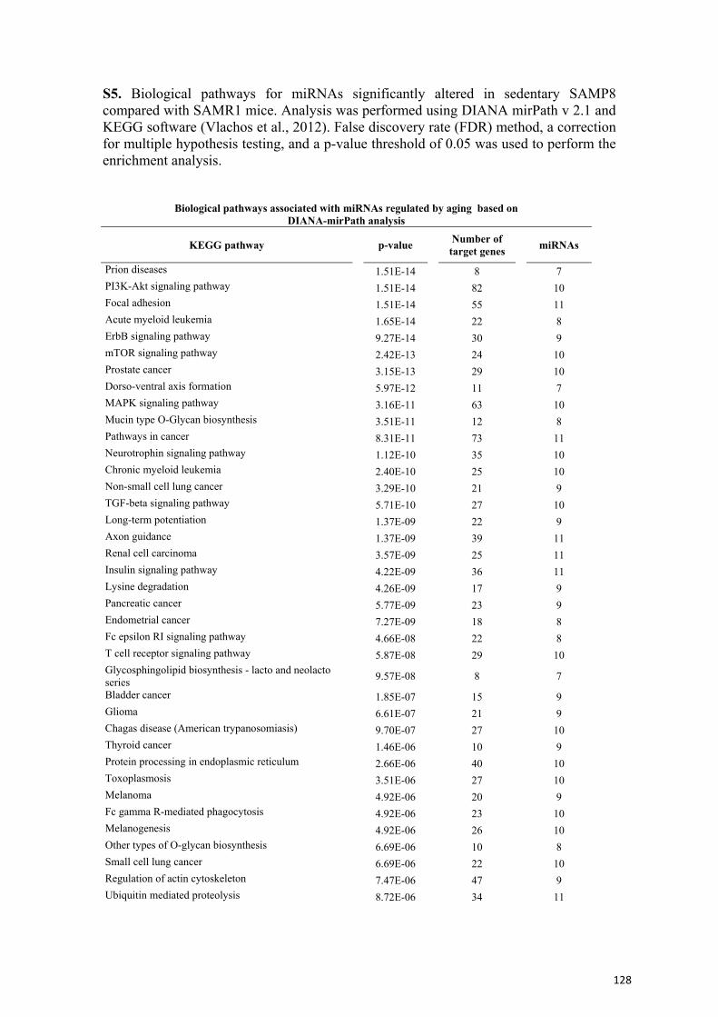
S5. Biological pathways for miRNAs significantly altered in sedentary SAMP8 compared with SAMR1 mice. Analysis was performed using DIANA mirPath v 2.1 and KEGG software (Vlachos et al., 2012). False discovery rate (FDR) method, a correction for multiple hypothesis testing, and a p-value threshold of 0.05 was used to perform the enrichment analysis.
Biological pathways associated with miRNAs regulated by aging based on DIANA-mirPath analysis
KEGG pathway p-value Number of target genes miRNAs
Prion diseases 1.51E-14 8 7 PI3K-Akt signaling pathway 1.51E-14 82 10 Focal adhesion 1.51E-14 55 11 Acute myeloid leukemia 1.65E-14 22 8 ErbB signaling pathway 9.27E-14 30 9 mTOR signaling pathway 2.42E-13 24 10 Prostate cancer 3.15E-13 29 10 Dorso-ventral axis formation 5.97E-12 11 7 MAPK signaling pathway 3.16E-11 63 10 Mucin type O-Glycan biosynthesis 3.51E-11 12 8 Pathways in cancer 8.31E-11 73 11 Neurotrophin signaling pathway 1.12E-10 35 10 Chronic myeloid leukemia 2.40E-10 25 10 Non-small cell lung cancer 3.29E-10 21 9 TGF-beta signaling pathway 5.71E-10 27 10 Long-term potentiation 1.37E-09 22 9 Axon guidance 1.37E-09 39 11 Renal cell carcinoma 3.57E-09 25 11 Insulin signaling pathway 4.22E-09 36 11 Lysine degradation 4.26E-09 17 9 Pancreatic cancer 5.77E-09 23 9 Endometrial cancer 7.27E-09 18 8 Fc epsilon RI signaling pathway 4.66E-08 22 8 T cell receptor signaling pathway 5.87E-08 29 10 Glycosphingolipid biosynthesis - lacto and neolacto series 9.57E-08 8 7
Bladder cancer 1.85E-07 15 9 Glioma 6.61E-07 21 9 Chagas disease (American trypanosomiasis) 9.70E-07 27 10 Thyroid cancer 1.46E-06 10 9 Protein processing in endoplasmic reticulum 2.66E-06 40 10 Toxoplasmosis 3.51E-06 27 10 Melanoma 4.92E-06 20 9 Fc gamma R-mediated phagocytosis 4.92E-06 23 10 Melanogenesis 4.92E-06 26 10 Other types of O-glycan biosynthesis 6.69E-06 10 8 Small cell lung cancer 6.69E-06 22 10 Regulation of actin cytoskeleton 7.47E-06 47 9 Ubiquitin mediated proteolysis 8.72E-06 34 11
128
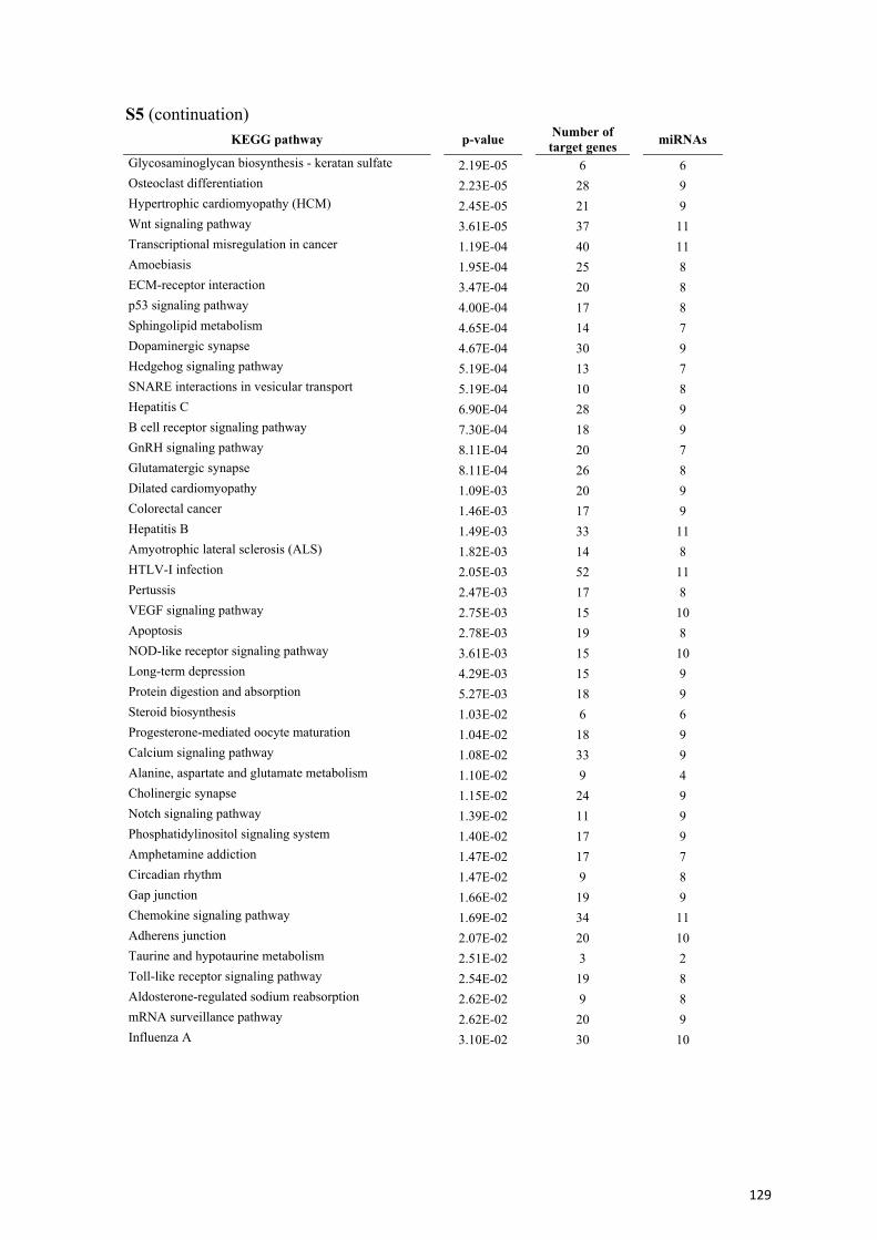
S5 (continuation) KEGG pathway p-value Number of
target genes miRNAs
Glycosaminoglycan biosynthesis - keratan sulfate 2.19E-05 6 6 Osteoclast differentiation 2.23E-05 28 9 Hypertrophic cardiomyopathy (HCM) 2.45E-05 21 9 Wnt signaling pathway 3.61E-05 37 11 Transcriptional misregulation in cancer 1.19E-04 40 11 Amoebiasis 1.95E-04 25 8 ECM-receptor interaction 3.47E-04 20 8 p53 signaling pathway 4.00E-04 17 8 Sphingolipid metabolism 4.65E-04 14 7 Dopaminergic synapse 4.67E-04 30 9 Hedgehog signaling pathway 5.19E-04 13 7 SNARE interactions in vesicular transport 5.19E-04 10 8 Hepatitis C 6.90E-04 28 9 B cell receptor signaling pathway 7.30E-04 18 9 GnRH signaling pathway 8.11E-04 20 7 Glutamatergic synapse 8.11E-04 26 8 Dilated cardiomyopathy 1.09E-03 20 9 Colorectal cancer 1.46E-03 17 9 Hepatitis B 1.49E-03 33 11 Amyotrophic lateral sclerosis (ALS) 1.82E-03 14 8 HTLV-I infection 2.05E-03 52 11 Pertussis 2.47E-03 17 8 VEGF signaling pathway 2.75E-03 15 10 Apoptosis 2.78E-03 19 8 NOD-like receptor signaling pathway 3.61E-03 15 10 Long-term depression 4.29E-03 15 9 Protein digestion and absorption 5.27E-03 18 9 Steroid biosynthesis 1.03E-02 6 6 Progesterone-mediated oocyte maturation 1.04E-02 18 9 Calcium signaling pathway 1.08E-02 33 9 Alanine, aspartate and glutamate metabolism 1.10E-02 9 4 Cholinergic synapse 1.15E-02 24 9 Notch signaling pathway 1.39E-02 11 9 Phosphatidylinositol signaling system 1.40E-02 17 9 Amphetamine addiction 1.47E-02 17 7 Circadian rhythm 1.47E-02 9 8 Gap junction 1.66E-02 19 9 Chemokine signaling pathway 1.69E-02 34 11 Adherens junction 2.07E-02 20 10 Taurine and hypotaurine metabolism 2.51E-02 3 2 Toll-like receptor signaling pathway 2.54E-02 19 8 Aldosterone-regulated sodium reabsorption 2.62E-02 9 8 mRNA surveillance pathway 2.62E-02 20 9 Influenza A 3.10E-02 30 10
129
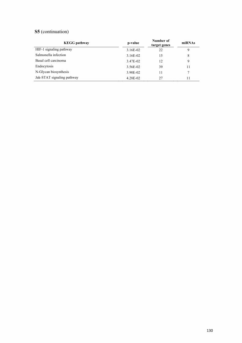
S5 (continuation)
KEGG pathway p-value Number of target genes miRNAs
HIF-1 signaling pathway 3.16E-02 22 9 Salmonella infection 3.16E-02 15 8 Basal cell carcinoma 3.47E-02 12 9 Endocytosis 3.56E-02 39 11 N-Glycan biosynthesis 3.90E-02 11 7 Jak-STAT signaling pathway 4.28E-02 27 11
130
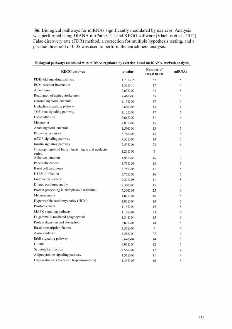
S6. Biological pathways for miRNAs significantly modulated by exercise. Analysis was performed using DIANA mirPath v 2.1 and KEGG software (Vlachos et al., 2012). False discovery rate (FDR) method, a correction for multiple hypothesis testing, and a p-value threshold of 0.05 was used to perform the enrichment analysis.
Biological pathways associated with miRNAs regulated by exercise based on DIANA-mirPath analysis
KEGG pathway p-value Number of target genes miRNAs
PI3K-Akt signaling pathway 1.73E-15 57 5 ECM-receptor interaction 1.52E-10 17 4 Amoebiasis 2.07E-09 22 5 Regulation of actin cytoskeleton 7.46E-09 35 5 Chronic myeloid leukemia 9.15E-09 17 6 Hedgehog signaling pathway 2.60E-08 13 4 TGF-beta signaling pathway 1.12E-07 17 6 Focal adhesion 2.66E-07 31 6 Melanoma 7.87E-07 15 5 Acute myeloid leukemia 1.50E-06 13 5 Pathways in cancer 3.76E-06 45 6 mTOR signaling pathway 7.53E-06 13 5 Insulin signaling pathway 7.53E-06 22 6 Glycosphingolipid biosynthesis - lacto and neolacto series 1.21E-05 5 4
Adherens junction 1.95E-05 16 5 Pancreatic cancer 5.75E-05 13 5 Basal cell carcinoma 5.75E-05 11 5 HTLV-I infection 5.75E-05 36 6 Endometrial cancer 7.21E-05 11 5 Dilated cardiomyopathy 7.30E-05 15 5 Protein processing in endoplasmic reticulum 7.30E-05 25 6 Melanogenesis 1.02E-04 16 5 Hypertrophic cardiomyopathy (HCM) 1.03E-04 14 5 Prostate cancer 1.15E-04 15 5 MAPK signaling pathway 1.18E-04 33 6 Fc gamma R-mediated phagocytosis 1.34E-04 15 6 Protein digestion and absorption 2.02E-04 14 5 Basal transcription factors 3.58E-04 9 4 Axon guidance 4.28E-04 22 6 ErbB signaling pathway 6.64E-04 14 5 Glioma 6.91E-04 12 5 Salmonella infection 9.56E-04 12 4 Adipocytokine signaling pathway 1.31E-03 11 4 Chagas disease (American trypanosomiasis) 1.35E-03 16 5
131

S6 (continuation)
KEGG pathway p-value Number of target genes miRNAs
Non-small cell lung cancer 1.52E-03 10 5 Long-term potentiation 1.52E-03 11 6 Dorso-ventral axis formation 3.34E-03 5 4 Bacterial invasion of epithelial cells 5.82E-03 10 4 Small cell lung cancer 6.59E-03 12 5 Salivary secretion 8.69E-03 11 4 Colorectal cancer 1.18E-02 10 5 Regulation of autophagy 2.15E-02 6 4 Chemokine signaling pathway 2.48E-02 21 6 Valine, leucine and isoleucine biosynthesis 2.74E-02 1 1 Calcium signaling pathway 2.74E-02 20 5 Lysine degradation 2.88E-02 7 5 Hepatitis C 2.88E-02 16 5 Transcriptional misregulation in cancer 2.88E-02 21 6 Gastric acid secretion 3.14E-02 10 5 Proximal tubule bicarbonate reclamation 3.29E-02 4 3 Aldosterone-regulated sodium reabsorption 4.41E-02 6 4 GnRH signaling pathway 4.41E-02 11 5 Jak-STAT signaling pathway 4.66E-02 17 5 Vasopressin-regulated water reabsorption 4.66E-02 7 5
132
References S3 and S4 Beveridge, N.J., Gardiner, E., Carroll, A.P., Tooney, P.A., and Cairns, M.J. (2010).
Schizophrenia is associated with an increase in cortical microRNA biogenesis. Mol Psychiatry 15, 1176-1189. doi: 10.1038/mp.2009.84.
Boissart, C., Nissan, X., Giraud-Triboult, K., Peschanski, M., and Benchoua, A. (2012). miR-125 potentiates early neural specification of human embryonic stem cells. Development 139, 1247-1257. doi: 10.1242/dev.073627.
Braidy, N., Guillemin, G.J., Mansour, H., Chan-Ling, T., Poljak, A., and Grant, R. (2011). Age related changes in NAD+ metabolism oxidative stress and Sirt1 activity in wistar rats. PLoS One 6, e19194. doi: 10.1371/journal.pone.0019194.
Bruno, I.G., Karam, R., Huang, L., Bhardwaj, A., Lou, C.H., Shum, E.Y., Song, H.W., Corbett, M.A., Gifford, W.D., Gecz, J., Pfaff, S.L., and Wilkinson, M.F. (2011). Identification of a microRNA that activates gene expression by repressing nonsense-mediated RNA decay. Mol Cell 42, 500-510. doi: 10.1016/j.molcel.2011.04.018.
Cogswell, J.P., Ward, J., Taylor, I.A., Waters, M., Shi, Y., Cannon, B., Kelnar, K., Kemppainen, J., Brown, D., Chen, C., Prinjha, R.K., Richardson, J.C., Saunders, A.M., Roses, A.D., and Richards, C.A. (2008). Identification of miRNA changes in Alzheimer's disease brain and CSF yields putative biomarkers and insights into disease pathways. J Alzheimers Dis 14, 27-41.
Cui, Y., Xiao, Z., Han, J., Sun, J., Ding, W., Zhao, Y., Chen, B., Li, X., and Dai, J. (2012). MiR-125b orchestrates cell proliferation, differentiation and migration in neural stem/progenitor cells by targeting Nestin. BMC Neurosci 13, 116. doi: 10.1186/1471-2202-13-116.
De Chevigny, A., Core, N., Follert, P., Gaudin, M., Barbry, P., Beclin, C., and Cremer, H. (2012). miR-7a regulation of Pax6 controls spatial origin of forebrain dopaminergic neurons. Nat Neurosci 15, 1120-1126. doi: 10.1038/nn.3142.
Edbauer, D., Neilson, J.R., Foster, K.A., Wang, C.F., Seeburg, D.P., Batterton, M.N., Tada, T., Dolan, B.M., Sharp, P.A., and Sheng, M. (2010). Regulation of synaptic structure and function by FMRP-associated microRNAs miR-125b and miR-132. Neuron 65, 373-384. doi: 10.1016/j.neuron.2010.01.005.
Evangelisti, C., Florian, M.C., Massimi, I., Dominici, C., Giannini, G., Galardi, S., Bue, M.C., Massalini, S., Mcdowell, H.P., Messi, E., Gulino, A., Farace, M.G., and Ciafre, S.A. (2009). MiR-128 up-regulation inhibits Reelin and DCX expression and reduces neuroblastoma cell motility and invasiveness. FASEB J 23, 4276-4287. doi: 10.1096/fj.09-134965.
Hebert, S.S., Horre, K., Nicolai, L., Papadopoulou, A.S., Mandemakers, W., Silahtaroglu, A.N., Kauppinen, S., Delacourte, A., and De Strooper, B. (2008). Loss of microRNA cluster miR-29a/b-1 in sporadic Alzheimer's disease correlates with increased BACE1/beta-secretase expression. Proc Natl Acad Sci U S A 105, 6415-6420. doi: 10.1073/pnas.0710263105.
Im, H.I., and Kenny, P.J. (2012). MicroRNAs in neuronal function and dysfunction. Trends Neurosci 35, 325-334. doi: 10.1016/j.tins.2012.01.004.
Lau, P., Bossers, K., Janky, R., Salta, E., Frigerio, C.S., Barbash, S., Rothman, R., Sierksma, A.S., Thathiah, A., Greenberg, D., Papadopoulou, A.S., Achsel, T., Ayoubi, T., Soreq, H., Verhaagen, J., Swaab, D.F., Aerts, S., and De Strooper, B. (2013). Alteration of the microRNA network during the progression of Alzheimer's disease. EMBO Mol Med 5, 1613-1634. doi: 10.1002/emmm.201201974.
133
Lukiw, W.J. (2007). Micro-RNA speciation in fetal, adult and Alzheimer's disease hippocampus. Neuroreport 18, 297-300. doi: 10.1097/WNR.0b013e3280148e8b.
Lukiw, W.J., and Pogue, A.I. (2007). Induction of specific micro RNA (miRNA) species by ROS-generating metal sulfates in primary human brain cells. J Inorg Biochem 101, 1265-1269. doi: 10.1016/j.jinorgbio.2007.06.004.
Maes, O.C., Chertkow, H.M., Wang, E., and Schipper, H.M. (2009). MicroRNA: Implications for Alzheimer Disease and other Human CNS Disorders. Curr Genomics 10, 154-168. doi: 10.2174/138920209788185252.
Majer, A., Medina, S.J., Niu, Y., Abrenica, B., Manguiat, K.J., Frost, K.L., Philipson, C.S., Sorensen, D.L., and Booth, S.A. (2012). Early mechanisms of pathobiology are revealed by transcriptional temporal dynamics in hippocampal CA1 neurons of prion infected mice. PLoS Pathog 8, e1003002. doi: 10.1371/journal.ppat.1003002.
Mallick, B., and Ghosh, Z. (2011). A complex crosstalk between polymorphic microRNA target sites and AD prognosis. RNA Biol 8, 665-673. doi: 10.4161/rna.8.4.15584.
Mouradian, M.M. (2012). MicroRNAs in Parkinson's disease. Neurobiol Dis 46, 279-284. doi: 10.1016/j.nbd.2011.12.046.
Nielsen, S., Scheele, C., Yfanti, C., Akerstrom, T., Nielsen, A.R., Pedersen, B.K., and Laye, M.J. (2010). Muscle specific microRNAs are regulated by endurance exercise in human skeletal muscle. J Physiol 588, 4029-4037. doi: 10.1113/jphysiol.2010.189860.
Nix, P., and Bastiani, M. (2013). Neuroscience. Heterochronic genes turn back the clock in old neurons. Science 340, 282-283. doi: 10.1126/science.1237921.
Perkins, D.O., Jeffries, C.D., Jarskog, L.F., Thomson, J.M., Woods, K., Newman, M.A., Parker, J.S., Jin, J., and Hammond, S.M. (2007). microRNA expression in the prefrontal cortex of individuals with schizophrenia and schizoaffective disorder. Genome Biol 8, R27. doi: 10.1186/gb-2007-8-2-r27.
Radom-Aizik, S., Zaldivar, F., Haddad, F., and Cooper, D.M. (2013). Impact of brief exercise on peripheral blood NK cell gene and microRNA expression in young adults. J Appl Physiol 114, 628-636. doi: 10.1152/japplphysiol.01341.2012.
Radom-Aizik, S., Zaldivar, F., Jr., Leu, S.Y., Adams, G.R., Oliver, S., and Cooper, D.M. (2012). Effects of exercise on microRNA expression in young males peripheral blood mononuclear cells. Clin Transl Sci 5, 32-38. doi: 10.1111/j.1752-8062.2011.00384.x.
Ranganathan, S., Scudiere, S., and Bowser, R. (2001). Hyperphosphorylation of the retinoblastoma gene product and altered subcellular distribution of E2F-1 during Alzheimer's disease and amyotrophic lateral sclerosis. J Alzheimers Dis 3, 377-385.
Saba, R., Goodman, C.D., Huzarewich, R.L., Robertson, C., and Booth, S.A. (2008). A miRNA signature of prion induced neurodegeneration. PLoS One 3, e3652. doi: 10.1371/journal.pone.0003652.
Sarachana, T., Zhou, R., Chen, G., Manji, H.K., and Hu, V.W. (2010). Investigation of post-transcriptional gene regulatory networks associated with autism spectrum disorders by microRNA expression profiling of lymphoblastoid cell lines. Genome Med 2, 23. doi: 10.1186/gm144.
Satoh, J. (2012). Molecular network of microRNA targets in Alzheimer's disease brains. Exp Neurol 235, 436-446. doi: 10.1016/j.expneurol.2011.09.003.
134
Schonrock, N., Ke, Y.D., Humphreys, D., Staufenbiel, M., Ittner, L.M., Preiss, T., and Gotz, J. (2010). Neuronal microRNA deregulation in response to Alzheimer's disease amyloid-beta. PLoS One 5, e11070. doi: 10.1371/journal.pone.0011070.
Siegel, G., Obernosterer, G., Fiore, R., Oehmen, M., Bicker, S., Christensen, M., Khudayberdiev, S., Leuschner, P.F., Busch, C.J., Kane, C., Hubel, K., Dekker, F., Hedberg, C., Rengarajan, B., Drepper, C., Waldmann, H., Kauppinen, S., Greenberg, M.E., Draguhn, A., Rehmsmeier, M., Martinez, J., and Schratt, G.M. (2009). A functional screen implicates microRNA-138-dependent regulation of the depalmitoylation enzyme APT1 in dendritic spine morphogenesis. Nat Cell Biol 11, 705-716. doi: 10.1038/ncb1876.
Soci, U.P., Fernandes, T., Hashimoto, N.Y., Mota, G.F., Amadeu, M.A., Rosa, K.T., Irigoyen, M.C., Phillips, M.I., and Oliveira, E.M. (2011). MicroRNAs 29 are involved in the improvement of ventricular compliance promoted by aerobic exercise training in rats. Physiol Genomics 43, 665-673. doi: 10.1152/physiolgenomics.00145.2010.
Urdinguio, R.G., Fernandez, A.F., Lopez-Nieva, P., Rossi, S., Huertas, D., Kulis, M., Liu, C.G., Croce, C.M., Calin, G.A., and Esteller, M. (2010). Disrupted microRNA expression caused by Mecp2 loss in a mouse model of Rett syndrome. Epigenetics 5, 656-663. doi: 10.4161/epi.5.7.13055.
Viader, A., Chang, L.W., Fahrner, T., Nagarajan, R., and Milbrandt, J. (2011). MicroRNAs modulate Schwann cell response to nerve injury by reinforcing transcriptional silencing of dedifferentiation-related genes. J Neurosci 31, 17358-17369. doi: 10.1523/JNEUROSCI.3931-11.2011.
Vlachos, I.S., Kostoulas, N., Vergoulis, T., Georgakilas, G., Reczko, M., Maragkakis, M., Paraskevopoulou, M.D., Prionidis, K., Dalamagas, T., and Hatzigeorgiou, A.G. (2012). DIANA miRPath v.2.0: investigating the combinatorial effect of microRNAs in pathways. Nucleic Acids Res 40, W498-504. doi: 10.1093/nar/gks494.
Wang, W., Kwon, E.J., and Tsai, L.H. (2012). MicroRNAs in learning, memory, and neurological diseases. Learn Mem 19, 359-368. doi: 10.1101/lm.026492.112.
Wang, W.X., Huang, Q., Hu, Y., Stromberg, A.J., and Nelson, P.T. (2011). Patterns of microRNA expression in normal and early Alzheimer's disease human temporal cortex: white matter versus gray matter. Acta Neuropathol 121, 193-205. doi: 10.1007/s00401-010-0756-0.
Wang, X., Liu, P., Zhu, H., Xu, Y., Ma, C., Dai, X., Huang, L., Liu, Y., Zhang, L., and Qin, C. (2009). miR-34a, a microRNA up-regulated in a double transgenic mouse model of Alzheimer's disease, inhibits bcl2 translation. Brain Res Bull 80, 268-273. doi: 10.1016/j.brainresbull.2009.08.006.
Wu, H., Tao, J., Chen, P.J., Shahab, A., Ge, W., Hart, R.P., Ruan, X., Ruan, Y., and Sun, Y.E. (2010). Genome-wide analysis reveals methyl-CpG-binding protein 2-dependent regulation of microRNAs in a mouse model of Rett syndrome. Proc Natl Acad Sci U S A 107, 18161-18166. doi: 10.1073/pnas.1005595107.
Xin, H., Li, Y., Liu, Z., Wang, X., Shang, X., Cui, Y., Gang Zhang, Z., and Chopp, M. (2013). Mir-133b Promotes Neural Plasticity and Functional Recovery after Treatment of Stroke with Multipotent Mesenchymal Stromal Cells in Rats Via Transfer of Exosome-Enriched Extracellular Particles. Stem Cells. doi: 10.1002/stem.1409.
Zhao, Y., Bhattacharjee, S., Jones, B.M., Hill, J., Dua, P., and Lukiw, W.J. (2013). Regulation of Neurotropic Signaling by the Inducible, NF-kB-Sensitive miRNA-
135

125b in Alzheimer's Disease (AD) and in Primary Human Neuronal-Glial (HNG) Cells. Mol Neurobiol. doi: 10.1007/s12035-013-8595-3.
136

DISCUSIÓN

Discusión
Este trabajo de tesis se centró en el que sin duda es uno de los temas más complejos
desde el punto de visto social, económico y biomédico de nuestro siglo: el
envejecimiento. En este contexto, los diferentes objetivos planteados han abarcado
temáticas como la enfermedad de Alzheimer, la epigenética y el estilo de vida
saludable.
El envejecimiento es uno de los principales ejes de la investigación científica actual.
Los datos poblacionales más recientes muestran un patrón de pirámide poblacional cada
vez más envejecido y como consecuencia la proporción de patologías asociadas a este
proceso ha aumentado significativamente. Este hecho provoca un alto porcentaje de
situaciones de dependencia o semi-dependencia en la población envejecida y mayores
costes económicos a nivel sanitario. Todo ello conduce a la necesidad de buscar
estrategias terapéuticas que permitan prevenir o retrasar algunos de los síntomas y/o
patologías asociadas al envejecimiento, mejorando así la calidad de vida del individuo.
Entre estas estrategias destacan los denominados hábitos de vida saludables, que
incluyen dieta, actividad física, estimulación cognitiva y estrategias de reducción del
estrés, entre otros.
El ejercicio físico fue una de las primeras actividades recomendadas y la más
aceptada e incorporada actualmente en nuestra sociedad. Hoy en día es habitual ver
personas corriendo por los parques de nuestra ciudad, utilizando la bicicleta como
entrenamiento o incluso medio de transporte habitual, entrenando de manera regular en
gimnasios, piscinas y otros centros deportivos. Esto es en parte el resultado de la
divulgación a través de los medios de comunicación y a las recomendaciones de las
autoridades de salud pública sobre los beneficios asociados a la práctica de ejercicio
regular, derivados en gran medida de los esfuerzos científicos realizados al respecto.
Por otro lado, desde que en 1942 Conrad Waddington definió por primera vez la
epigenética, el interés en este fenómeno ha ido aumentando exponencialmente en la
comunidad científica. Se ha visto que la influencia de este tipo de regulación se extiende
a todos los procesos celulares y funciones biológicas, y el hecho de que el entorno y los
hábitos de estilo de vida ejerzan una influencia en la modulación epigenética abre un
nuevo campo en la investigación relacionada con la prevención y el retraso en la
aparición de enfermedades crónicas.
139
Discusión
El modelo SAMP8: ventajas y limitaciones
Para estudiar los efectos del ejercicio y su posible modulación epigenética durante el
proceso de envejecimiento, en esta tesis escogimos como modelo los ratones SAMP8.
Esta cepa de ratón presenta rasgos de envejecimiento espontáneos y prematuros,
incluyendo déficits cognitivos, algunos de los cuales son similares a los observados en
patologías como la EA.
A diferencia de los modelos transgénicos de experimentación donde las causas del
fenotipo se asocian a alteraciones en genes específicos, las causas y alteraciones
concretas que producen el fenotipo observado en los ratones SAMP8 no están
completamente descritas y probablemente sean multifactoriales, como ocurre durante el
proceso de envejecimiento en humanos. Desde este punto de vista, el modelo SAMP8
podría considerarse más integrador que los modelos transgénicos. Sin embargo, la
utilización de este modelo espontáneo de envejecimiento también supone una limitación
en el momento de explicar los resultados obtenidos, ya que frente a multitud de
variables que desconocemos, resulta complejo discernir entre las causas y las
consecuencias de un determinado fenotipo.
Otra de las limitaciones del modelo escogido es el control utilizado. Se acepta el uso
de los ratones SAMR1 como cepa control ya que ambos surgieron de la misma colonia
original y por tanto son similares genéticamente. Sin embargo, el establecimiento de
cada una de estas cepas (P8 y R1) mediante cruzamiento entre hermanos puede dar una
deriva genética en las mismas, que tan solo es posible identificar por el fenotipo
observado o durante el análisis de los resultados obtenidos tras el estudio.
140
Discusión
Inflammaging y epigenética, ¿causa o consecuencia del
envejecimiento?
El concepto de inflammaging explica los cambios moleculares y fisiológicos en el
sistema inmune que se producen a medida que avanza el ciclo vital de un individuo. En
nuestro estudio hemos observado un entorno proinflamatorio en astrocitos de ratones
SAMP8, en concreto un aumento en la expresión de Il-6 en ratones neonatos,
comparados con los ratones control SAMR1. Esta alteración está probablemente
relacionada con el fenotipo de envejecimiento acelerado y déficits cognitivos de los
ratones SAMP8, aunque nuestros resultados no permiten dilucidar si el aumento de este
marcador inflamatorio está en el origen del fenotipo de este modelo o es consecuencia
de otros mecanismos subyacentes. Asimismo, hemos observado mayores niveles de IL-
6 en plasma de ratones SAMP8 adultos de 12 meses de edad en los cuales también
observamos una respuesta inflamatoria exacerbada frente a LPS. Este conjunto de datos
nos permitió identificar IL-6 como un biomarcador inflamatorio persistente durante el
ciclo vital de los ratones SAMP8. Un hallazgo particularmente interesante fue que la
causa del aumento de IL-6 se encontró directamente asociada a los niveles de expresión
de un factor de remodelación de cromatina, el correpresor de REST, RCOR2. Estos
resultados nos permitieron integrar los conceptos inflammaging y epigenética en nuestro
estudio. De hecho, observamos una sorprendente y marcada reducción en la expresión
de la isoforma Rcor2 en el modelo SAMP8 respecto a su control SAMR1 en varios
tipos celulares y tejidos analizados, entre ellos linfocitos y astrocitos, hipocampo y
corteza cerebral. Además, en linfocitos observamos la reducción de Rcor2 desde edades
muy tempranas, mucho antes de que se produzca la manifestación del fenotipo
senescente. Asimismo, en cultivos de astrocitos provenientes de ratones SAMP8
neonatos, los niveles de expresión de Rcor2 también fueron menores. Estos resultados
sugieren la posible implicación de RCOR2 en el fenotipo senescente de los ratones
SAMP8.
El hecho de hallar una correlación negativa entre Il-6 y Rcor2, tanto in vivo como in
vitro, nos hizo plantearnos la existencia de una posible asociación entre ambos factores
ya que, por un lado la inflamación crónica se ha asociado al desarrollo prematuro y la
aceleración de enfermedades neurodegenerativas como la EA [737, 738], y por el otro el
141
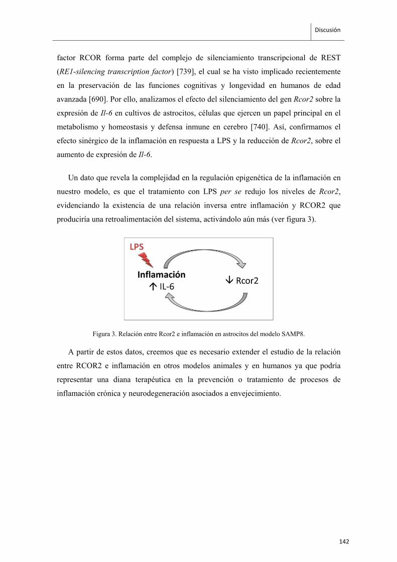
Discusión
factor RCOR forma parte del complejo de silenciamiento transcripcional de REST
(RE1-silencing transcription factor) [739], el cual se ha visto implicado recientemente
en la preservación de las funciones cognitivas y longevidad en humanos de edad
avanzada [690]. Por ello, analizamos el efecto del silenciamiento del gen Rcor2 sobre la
expresión de Il-6 en cultivos de astrocitos, células que ejercen un papel principal en el
metabolismo y homeostasis y defensa inmune en cerebro [740]. Así, confirmamos el
efecto sinérgico de la inflamación en respuesta a LPS y la reducción de Rcor2, sobre el
aumento de expresión de Il-6.
Un dato que revela la complejidad en la regulación epigenética de la inflamación en
nuestro modelo, es que el tratamiento con LPS per se redujo los niveles de Rcor2,
evidenciando la existencia de una relación inversa entre inflamación y RCOR2 que
produciría una retroalimentación del sistema, activándolo aún más (ver figura 3).
Figura 3. Relación entre Rcor2 e inflamación en astrocitos del modelo SAMP8.
A partir de estos datos, creemos que es necesario extender el estudio de la relación
entre RCOR2 e inflamación en otros modelos animales y en humanos ya que podría
representar una diana terapéutica en la prevención o tratamiento de procesos de
inflamación crónica y neurodegeneración asociados a envejecimiento.
142
Discusión
El papel de la remodelación de cromatina en longevidad y
senescencia
Nuestros resultados también nos han permitido relacionar RCOR2,
neuroinflamación y envejecimiento prematuro con el estado de metilación de la
cromatina. Basamos esta hipótesis en varias observaciones. El complejo de REST, del
cual forma parte RCOR2, ejerce un papel remodelador del estado de la cromatina e
incluye enzimas demetilasas de histonas como LSD1 [310]. La disminución de Rcor2
en ratones SAMP8 podría provocar una alteración en la actividad del complejo REST y
por lo tanto un aumento en la metilación de la lisina 4 de histona H3. Esta hipótesis es
consistente con el aumento en los niveles de trimetilación en lisina 4 de histona H3
global que detectamos en linfocitos, hipocampo y corteza cerebral de ratones SAMP8
de edad avanzada. Este posible mecanismo implicando factores de remodelación de
cromatina, como el complejo REST, también se ve indirectamente apoyado por los
resultados que obtuvimos en células gliales que muestran que la sobreexpresión de Il-6
en respuesta a un estímulo inflamatorio es dependiente a la vez de RCOR2 y de la
presencia de actividad metiltransferasa. Estas observaciones nos motivaron a comenzar
a caracterizar los niveles de expresión de las principales enzimas y cofactores asociados
al complejo de REST y otros factores remodeladores de cromatina en el modelo
senescente SAMP8 y su control SAMR1, trabajo del que se muestran resultados
preliminares aún no publicados en el capítulo 1 del apartado de Resultados. Ente los
datos a destacar hasta el momento, encontramos reducidos los niveles de enzimas
demetilasas Lsd1 y G9a presentes en el complejo de REST. Esta reducción, unida a la
hallada para Rcor2, sugiere que la funcionalidad del complejo podría estar afectada lo
cual podría dar como resultado alteraciones en diferenciación neuronal y plasticidad
[715-718]. Además, se ha descrito la implicación de G9a en la regulación de la enzima
neprilisina [705], una de las principales responsables de la eliminación de los depósitos
de Aβ en cerebro y diana terapéutica muy prometedora en el tratamiento de la EA [719],
indicando que éste podría ser otro de los mecanismos moleculares subyacentes en el
fenotipo neurodegenerativo observado en los ratones SAMP8.
143
Discusión
También se vieron reducidas demetilasas Jarid1a y Jarid1b, homólogas a la
demetilasa rbr-2 de C. elegans, la cual Greer y sus colaboradores han identificado como
determinante en la regulación de la longevidad y del aumento en los niveles de
trimetilación en H3 lisina 4 en este organismo [448, 449]. Todos estos resultados nos
indican la necesidad de seguir investigando el papel del complejo REST, del estado de
metilación de histona H3 y del efecto de los factores y cofactores reguladores del
mismo, así como de otros implicados en remodelación de cromatina sobre los procesos
de envejecimiento y longevidad, y su posible implicación en inflammaging.
144
Discusión
La matriz extracelular, diana del ejercicio físico en cerebro
Los beneficios del ejercicio físico sobre la cognición son un tema de creciente
interés debido al número alarmante de personas que sufren deterioro cognitivo asociado
al envejecimiento y demencias. Durante este trabajo de tesis nos interesamos en los
efectos transcripcionales y epigenéticos del ejercicio físico en el cerebro utilizando el
modelo SAMP8 de envejecimiento y EA. Tras una intervención de seis meses de
ejercicio voluntario y moderado realizamos microarrays de expresión de hipocampo
que nos permitieron identificar genes marcadores de envejecimiento y sensibles a la
intervención de ejercicio físico. Dicho grupo de genes mostraba niveles alterados en
SAMP8 comparados con SAMR1 (control) pero recuperaban los niveles de expresión
del grupo control en respuesta al ejercicio físico. El análisis bioinformático de vías de
señalización de este conjunto de genes reveló que uno de los procesos biológicos más
afectado en el hipocampo de los ratones SAMP8 es el mantenimiento de la matriz
extracelular y numerosos genes de esta vía demostraron ser sensibles al ejercicio físico.
Este es un resultado de gran interés puesto que la matriz extracelular representa el
soporte físico para la función, homeostasis y supervivencia neuronal y en modelos de
neurodegeneración y EA se han descrito alteraciones en algunos de los genes que la
mantienen [727, 728]. De hecho, las vías relacionadas con matriz extracelular también
aparecieron alteradas en los análisis que llevamos a cabo en corteza cerebral de estos
mismos animales (resultados no publicados), sugiriendo que se trata de una vía
particularmente sensible al envejecimiento y en cierto grado modulable por ejercicio
físico en cerebro.
Nuestro estudio no reprodujo algunos de los resultados de estudios previos de
expresión génica en cerebro también realizados en el modelo SAMP8. Por ejemplo el
realizado por Sakurai y sus colaboradores [658] en el cual identificaron el gen Lztfl1
(leucine zipper transcription factor-like protein 1) como sensible al ejercicio en SAMP8
de 2 meses de edad, que en nuestro trabajo no se ha visto modificado. En el estudio de
Cheng y colaboradores [741] se identificó una reducción en los niveles de Dusp12
(Dual specificity protein phosphatase, 12) en hembras de 12 meses de edad, enzima
implicada en supervivencia celular y biogénesis ribosomal [742], que en nuestro caso
tampoco se vio alterada. Casadesus y colaboradores [743] no hallaron diferencias entre
SAMP8 y SAMR1 en el patrón de expresión de 62 genes. Dos de entre ellos mostraron
145
Discusión
diferencias entre cepas en nuestro estudio, una reducción en la expresión de Park2 (E3
ubiquitin ligasa, implicada en metabolismo lipídico, alterada en pacientes de Parkinson
[744]) y un aumento de Gfra1 (glial cell line-derived neurotrophic factor family
receptor α-1, que promueve la supervivencia celular en neuronas hipocampales [745]),
aunque ninguno de estos dos genes se vio modificado por el ejercicio. Estas diferencias
entre nuestros resultados y algunos trabajos previos podrían explicarse por la
variabilidad en edad y sexo de los animales y en las condiciones experimentales
utilizadas, además de las propias limitaciones de sensibilidad y reproducibilidad que
presenta la técnica de microarrays.
Entre los genes que identificamos mediante microarrays y validamos por Real Time
PCR destacamos Col1a1 y Col1a2 (collagen type 1 alpha 1, y 2), aumentados en la
cepa senescente. La alteración de estos genes podría tener una papel importante el
deterioro cognitivo de nuestro modelo, ya que se ha visto que un aumento en el
contenido de colágeno en cerebro se asocia a una reducción en el volumen hipocampal
en el modelo transgénico 3xTg para la EA [746]. Además, se sabe que el péptido AβPP
puede interaccionar directamente con diferentes tipos de colágeno y otros componentes
de la matriz extracelular, y algunos tipos de colágeno (XXV y XVIII) se han encontrado
presentes en las propias placas amiloideas [747, 748]. Otro de los genes alterados, Fmod
(fibromodulin) es un proteoglicano implicado en la regulación y mantenimiento de la
matriz extracelular, y se ha asociado con la progresión de glioblastomas [749]. Además,
este tipo de proteoglicanos parecen fomentar la formación de agregados de Aβ y placas
seniles en la EA [750]. Pgds (prostaglandin D(2) synthase) es un gen que se expresa en
astrocitos y oligodendrocitos [751] y es marcador de envejecimiento en líquido
cefalorraquídeo [752], y también aparece alterado en la cepa SAMP8 en nuestro estudio.
Finalmente, el gen Aldh1a2 (aldehyde dehydrogenase), necesario para la síntesis del
ácido retinoico, se ha descrito implicado en las alteraciones en neurogénesis y la
realización de tareas cognitivas tanto cuando su expresión es reducida [753] como
aumentada [754]. La intervención de ejercicio físico moderado revirtió los cambios
observados en todos estos genes en la cepa senescente SAMP8 alcanzando los niveles
de expresión detectados en los controles SAMR1.
146

Discusión
Estos resultados nos muestran los beneficios de una intervención de ejercicio físico
moderado sobre alteraciones presentes en cerebro de un modelo senescente, y destacan
la posible utilidad del ejercicio físico moderado como complemento en el tratamiento de
declive cognitivo y neurodegeneración.
147
Discusión
Mejoras del ejercicio físico y su relación con la vía de BDNF
La intervención de ejercicio físico en ratones SAMP8 provocó mejoras a nivel
fenotípico (como el color de la piel y el temblor físico) y, en hipocampo, un aumento en
el grado de vascularización y en la expresión de factores neurotróficos. Algunos
trabajos describen una reducción en los procesos angiogénicos en relación a los
depósitos de péptido Aβ en cerebros afectados por la EA [755, 756], aunque existe algo
de controversia ya que también se ha descrito mayor angiogénesis en ciertas regiones
cerebrales de pacientes de la EA [757]. Recientemente se ha descrito que el ejercicio
parece fomentar cierta plasticidad vascular en cerebro de ratones hembra de edad media
[758]. Nuestros resultados corroboran estos datos, se hallaron menores niveles de
vascularización en el modelo senescente, los cuales fueron aumentados en respuesta a la
intervención de ejercicio físico moderado.
Por otro lado, estudios previos han descrito que durante el envejecimiento y en
modelos de patologías neurodegenerativas como la EA, se produce una disminución de
la neurotrofina BDNF y de otros componentes de su vía de señalización como el
receptor TrkB [722-726, 759]. La expresión de BDNF es particularmente sensible a la
práctica de ejercicio físico que según numerosos estudios, provoca aumentos en su
expresión génica y proteica [658, 720, 760]. Nuestros resultados demuestran que los
ratones SAMP8 presentan niveles reducidos de expresión de componentes de la vía de
BDNF, algunos de los cuales (BDNF, TrkB y neuritina) recuperan valores de expresión
normales en respuesta a intervenciones de ejercicio físico voluntario cortas (8 semanas)
o de mayor duración (6 meses) y en animales mantenidos en grupo o aislados. En
particular, neuritina, actúa en procesos de supervivencia neuronal, plasticidad sináptica
y neurogénesis [736] y es una diana prometedora en el tratamiento de patologías
asociadas a declive cognitivo y la EA [761].
Estos datos confirman que una de las vías a través de las cuales se producen los
efectos positivos del ejercicio físico sobre el cerebro en un contexto de envejecimiento
prematuro y declive cognitivo es la vía de BNDF e identifican a este factor neurotrófico
como un excelente marcador de la eficacia de intervenciones basadas en ejercicio.
148
Discusión
Epigenética y estilo de vida
Muchos de los cambios observados durante el proceso de envejecimiento [42, 729] y
en respuesta al ejercicio físico [668, 676, 720, 762, 763] se han asociado con la
aparición de modificaciones epigenéticas. En este trabajo de tesis, caracterizamos la
expresión de un grupo de factores remodeladores de cromatina en nuestro modelo
animal de envejecimiento y su posible modulación por el ejercicio físico. Identificamos
cambios en los niveles de expresión de varios microARNs en la cepa senescente
respecto a su control, los cuales se han asociado previamente a enfermedades
neurológicas y desarrollo neuronal, como por ejemplo miR-30e-5p [408, 764], miR-
125b-5p [161, 765] o miR-128-3p [765, 766]. Identificamos tres microARNs alterados
en SAMP8 y modificados por la intervención de ejercicio físico, y otros cuatro que no
mostraban diferencias entre las cepas SAMP8 y SAMR1 y que resultaron igualmente
modulables en ambos grupos en respuesta al ejercicio. También observamos
alteraciones en factores implicados en la regulación de la acetilación en histonas y en
los niveles globales de acetilación de histona H3. Estos cambios tienen gran importancia
ya que la acetilación de histonas es una marca epigenética muy importante a nivel de
establecimiento de memoria y cognición [377, 670, 671] y alteraciones en los niveles de
acetilación se han asociado con declive cognitivo y neurodegeneración [673, 674].
El interés en la influencia del entorno, las experiencias y los hábitos sobre la
expresión génica ha crecido exponencialmente por su potencial regulador y posible
aplicación terapéutica. Además de la actividad física, se ha observado que otros hábitos
de estilo de vida como la dieta, ejercen una gran influencia sobre salud y
envejecimiento. Intervenciones basadas en restricción calórica [767-770], o la
caracterización de ciertos componentes, como polifenoles [771-774], isocianatos [775,
776] o ácidos grasos esenciales [777, 778], han demostrado el potencial terapéutico que
tiene la dieta sobre el envejecimiento fisiológico, procesos cancerígenos y la esperanza
de vida.
Asimismo, se ha descrito que la estimulación sensorial, cognitiva y motora mediante
intervenciones de enriquecimiento ambiental produce cambios en niveles de expresión
génica de factores neurotróficos, plasticidad neuronal, deposiciones de Aβ y mejoras
cognitivas en respuesta al enriquecimiento ambiental [779] así como cambios
149
Discusión
epigenéticos en acetilación y metilación de histonas [670]. A nivel social, las
experiencias durante los primeros años de vida [780, 781], así como situaciones de
estrés y estrategias basadas en su reducción [782-786] se han asociado a cambios a nivel
psicológico, neuronal y endocrino que puede estar mediados por alteraciones
epigenéticas. En este contexto, durante el periodo de tesis realizamos un estudio en
humanos donde mostramos una reducción en los niveles de expresión de genes
proinflamatorios y cambios epigenéticos como alteraciones en acetilación y metilación
de histonas, así como en la expresión de varias histonas deacetilasas, tras una
intervención de 6 horas de meditación [787].
Este tipo de estudios y el conjunto de los resultados derivados de los mismos, están
permitiendo un cambio en la manera de concebir la salud y los factores determinantes
de la aparición y progresión de las enfermedades. Los modelos biomédicos más clásicos
y simplificados comienzan a ser sustituidos por nuevos modelos biopsicosociales que
aportan una visión más holística, en la cual se acepta una comunicación estrecha entre
el ambiente, el individuo y sus genes, que determina finalmente la inclinación del
delicado equilibrio entre la salud y la enfermedad. Así, los avances en el campo de la
epigenética del estilo de vida serán fundamentales en la creación de nuevas estrategias
basadas en hábitos saludables para la prevención y la complementación de los
tratamientos de los síntomas y las patologías asociadas al envejecimiento.
150

CONCLUSIONES

Conclusiones
1. Los ratones senescentes SAMP8 presentan mayores niveles de IL6 en
plasma y tienen aumentada su expresión en astrocitos, de manera basal y
en respuesta a un estímulo inflamatorio (LPS).
2. El correpresor Rcor2 presenta niveles mínimos de expresión en linfocitos,
hipocampo y corteza cerebral de ratones SAMP8.
3. Existe una correlación negativa entre Rcor2 e Il6 en astrocitos, de manera
basal y en respuesta a un estímulo inflamatorio (LPS + interferón γ).
4. La inflamación también produce una reducción en la expresión de Rcor2 en
astrocitos.
5. Los ratones SAMP8 muestran un aumento global de H3K4me3 y
alteraciones en la expresión de demetilasas (Lsd1, Jarid1a y Jarid1b) y
metiltransferasas (G9a) reguladoras de esta marca epigenética.
6. El ejercicio físico produce mejoras fenotípicas y aumento de la
vascularización en hipocampo de ratones senescentes.
7. El ejercicio físico revierte alteraciones transcripcionales asociadas a
envejecimiento, en genes implicados en ensamblaje y mantenimiento de la
matriz extracelular en cerebro.
8. El ejercicio físico aumenta la expresión génica de Bdnf independientemente
de la duración y tipo de intervención, y a corto plazo modula Trkb y Nrn1,
receptor y gen diana de BDNF respectivamente.
9. El ejercicio físico afecta a los niveles de varios miARNs asociados a
neurodegeneración.
10. La acetilación global de histona H3 y varias deacetilasas son sensibles a la
práctica de ejercicio físico.
153


ANEXO‐I

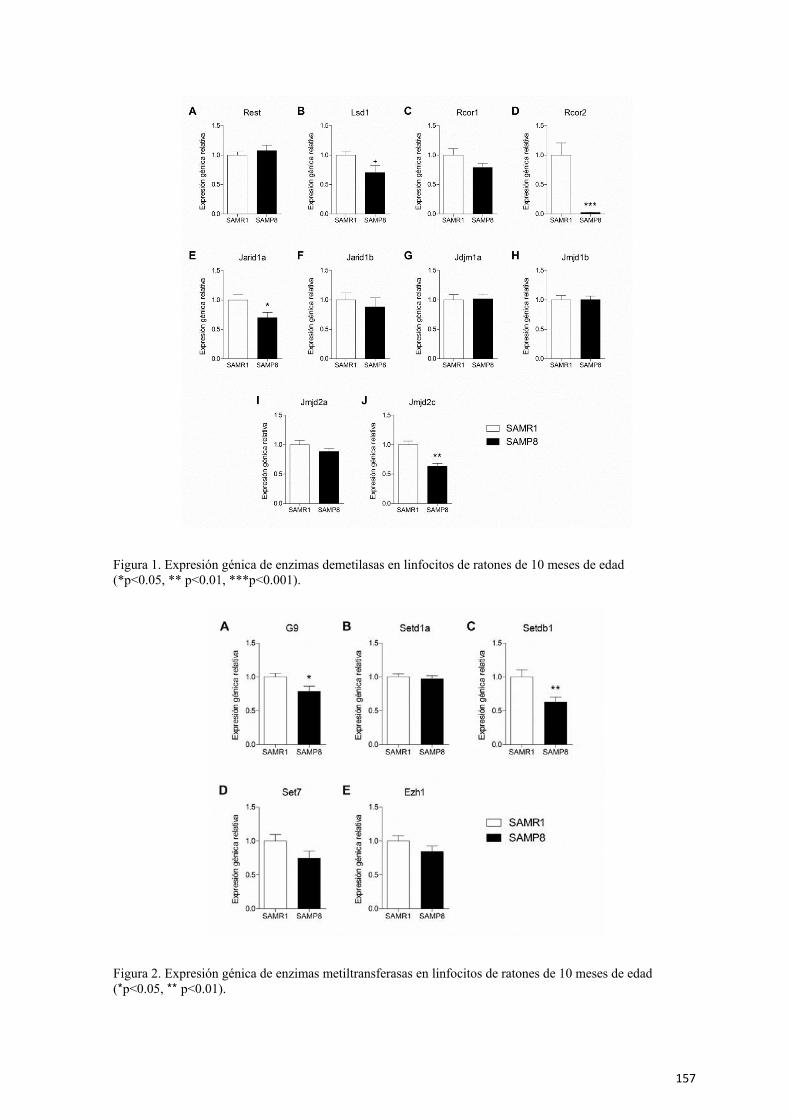
Figura 1. Expresión génica de enzimas demetilasas en linfocitos de ratones de 10 meses de edad (*p<0.05, ** p<0.01, ***p<0.001).
Figura 2. Expresión génica de enzimas metiltransferasas en linfocitos de ratones de 10 meses de edad (*p<0.05, ** p<0.01).
157
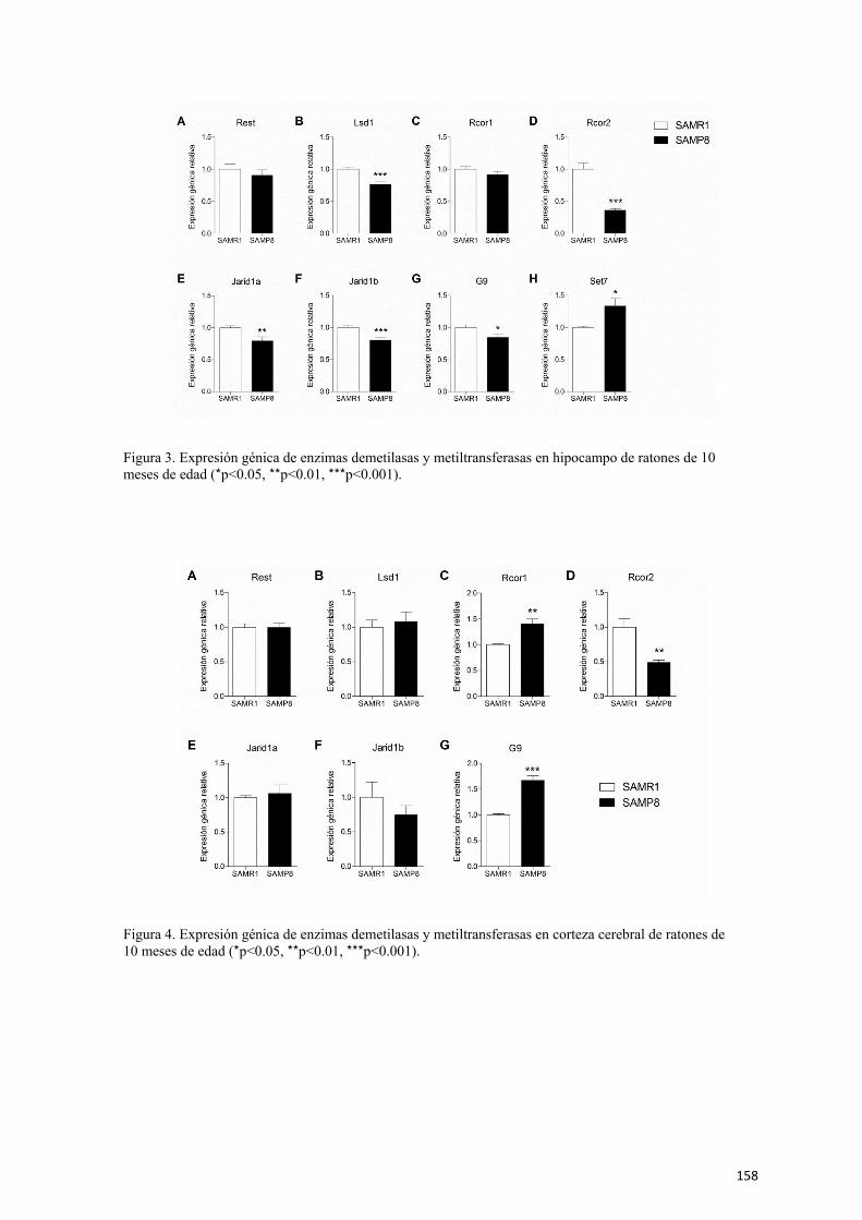
Figura 3. Expresión génica de enzimas demetilasas y metiltransferasas en hipocampo de ratones de 10 meses de edad (*p<0.05, **p<0.01, ***p<0.001).
Figura 4. Expresión génica de enzimas demetilasas y metiltransferasas en corteza cerebral de ratones de 10 meses de edad (*p<0.05, **p<0.01, ***p<0.001).
158

ANEXO‐II

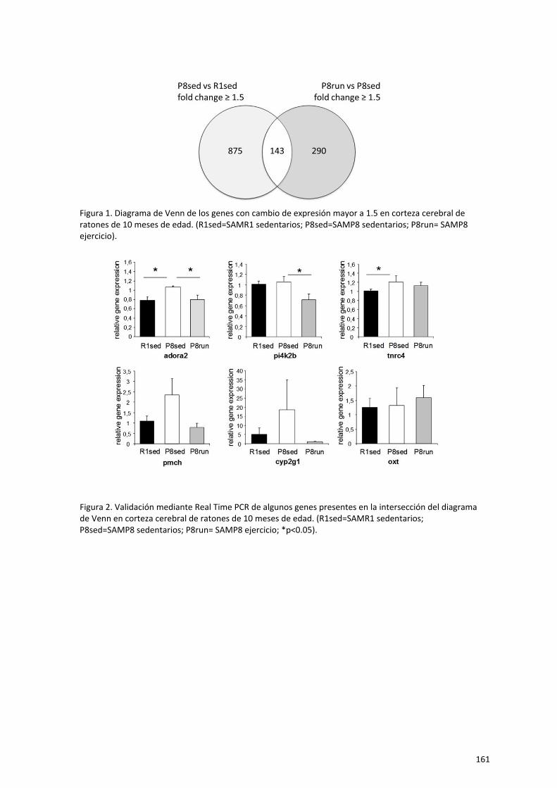
Figura 1. Diagrama de Venn de los genes con cambio de expresión mayor a 1.5 en corteza cerebral de ratones de 10 meses de edad. (R1sed=SAMR1 sedentarios; P8sed=SAMP8 sedentarios; P8run= SAMP8 ejercicio).
Figura 2. Validación mediante Real Time PCR de algunos genes presentes en la intersección del diagrama de Venn en corteza cerebral de ratones de 10 meses de edad. (R1sed=SAMR1 sedentarios; P8sed=SAMP8 sedentarios; P8run= SAMP8 ejercicio; *p<0.05).
161
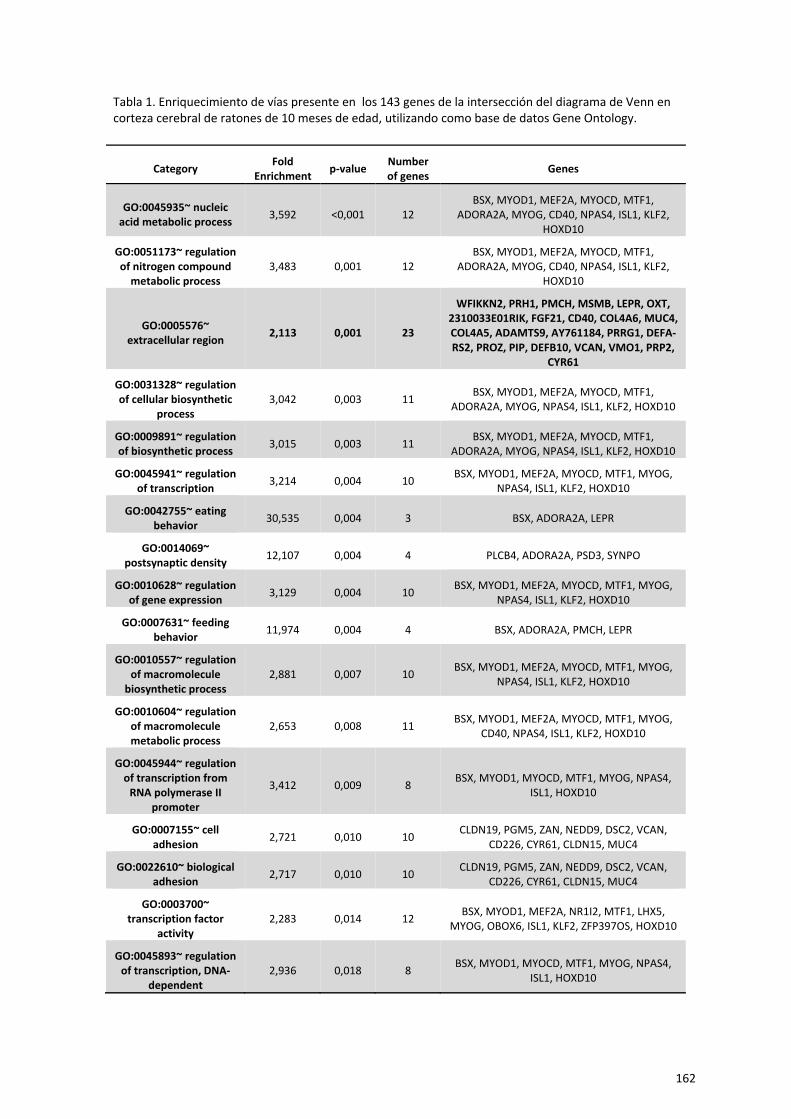
Tabla 1. Enriquecimiento de vías presente en los 143 genes de la intersección del diagrama de Venn en corteza cerebral de ratones de 10 meses de edad, utilizando como base de datos Gene Ontology.
Category Fold Enrichment p‐value Number
of genes Genes
GO:0045935~ nucleic acid metabolic process 3,592 <0,001 12
BSX, MYOD1, MEF2A, MYOCD, MTF1, ADORA2A, MYOG, CD40, NPAS4, ISL1, KLF2,
HOXD10
GO:0051173~ regulation of nitrogen compound
metabolic process 3,483 0,001 12
BSX, MYOD1, MEF2A, MYOCD, MTF1, ADORA2A, MYOG, CD40, NPAS4, ISL1, KLF2,
HOXD10
GO:0005576~ extracellular region 2,113 0,001 23
WFIKKN2, PRH1, PMCH, MSMB, LEPR, OXT, 2310033E01RIK, FGF21, CD40, COL4A6, MUC4, COL4A5, ADAMTS9, AY761184, PRRG1, DEFA‐RS2, PROZ, PIP, DEFB10, VCAN, VMO1, PRP2,
CYR61
GO:0031328~ regulation of cellular biosynthetic
process 3,042 0,003 11 BSX, MYOD1, MEF2A, MYOCD, MTF1,
ADORA2A, MYOG, NPAS4, ISL1, KLF2, HOXD10
GO:0009891~ regulation of biosynthetic process 3,015 0,003 11 BSX, MYOD1, MEF2A, MYOCD, MTF1,
ADORA2A, MYOG, NPAS4, ISL1, KLF2, HOXD10
GO:0045941~ regulation of transcription 3,214 0,004 10 BSX, MYOD1, MEF2A, MYOCD, MTF1, MYOG,
NPAS4, ISL1, KLF2, HOXD10
GO:0042755~ eating behavior 30,535 0,004 3 BSX, ADORA2A, LEPR
GO:0014069~ postsynaptic density 12,107 0,004 4 PLCB4, ADORA2A, PSD3, SYNPO
GO:0010628~ regulation of gene expression 3,129 0,004 10 BSX, MYOD1, MEF2A, MYOCD, MTF1, MYOG,
NPAS4, ISL1, KLF2, HOXD10
GO:0007631~ feeding behavior 11,974 0,004 4 BSX, ADORA2A, PMCH, LEPR
GO:0010557~ regulation of macromolecule
biosynthetic process 2,881 0,007 10 BSX, MYOD1, MEF2A, MYOCD, MTF1, MYOG,
NPAS4, ISL1, KLF2, HOXD10
GO:0010604~ regulation of macromolecule metabolic process
2,653 0,008 11 BSX, MYOD1, MEF2A, MYOCD, MTF1, MYOG, CD40, NPAS4, ISL1, KLF2, HOXD10
GO:0045944~ regulation of transcription from RNA polymerase II
promoter
3,412 0,009 8 BSX, MYOD1, MYOCD, MTF1, MYOG, NPAS4, ISL1, HOXD10
GO:0007155~ cell adhesion 2,721 0,010 10 CLDN19, PGM5, ZAN, NEDD9, DSC2, VCAN,
CD226, CYR61, CLDN15, MUC4
GO:0022610~ biological adhesion 2,717 0,010 10 CLDN19, PGM5, ZAN, NEDD9, DSC2, VCAN,
CD226, CYR61, CLDN15, MUC4
GO:0003700~ transcription factor
activity 2,283 0,014 12 BSX, MYOD1, MEF2A, NR1I2, MTF1, LHX5,
MYOG, OBOX6, ISL1, KLF2, ZFP397OS, HOXD10
GO:0045893~ regulation of transcription, DNA‐
dependent 2,936 0,018 8 BSX, MYOD1, MYOCD, MTF1, MYOG, NPAS4,
ISL1, HOXD10
162
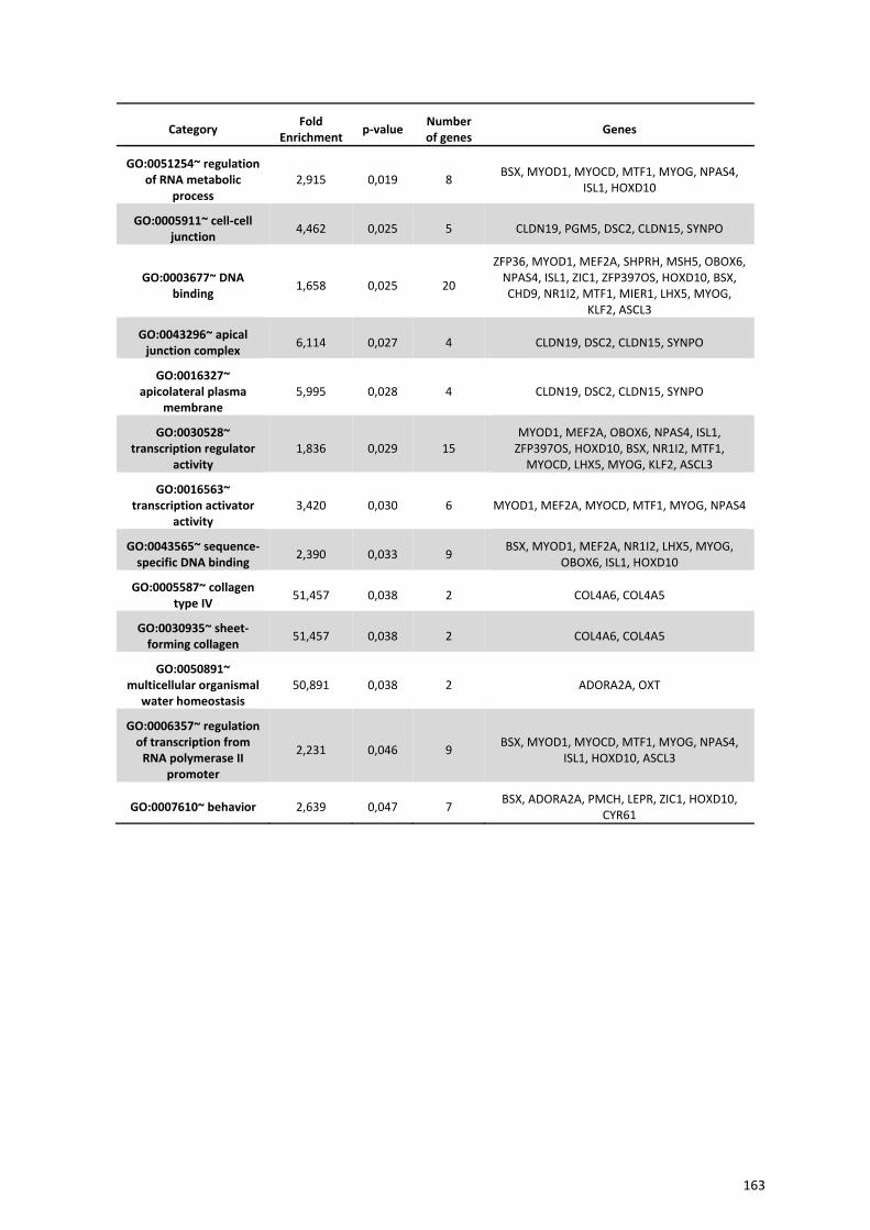
Category Fold Enrichment p‐value Number
of genes Genes
GO:0051254~ regulation of RNA metabolic
process 2,915 0,019 8 BSX, MYOD1, MYOCD, MTF1, MYOG, NPAS4,
ISL1, HOXD10
GO:0005911~ cell‐cell junction 4,462 0,025 5 CLDN19, PGM5, DSC2, CLDN15, SYNPO
GO:0003677~ DNA binding 1,658 0,025 20
ZFP36, MYOD1, MEF2A, SHPRH, MSH5, OBOX6, NPAS4, ISL1, ZIC1, ZFP397OS, HOXD10, BSX, CHD9, NR1I2, MTF1, MIER1, LHX5, MYOG,
KLF2, ASCL3
GO:0043296~ apical junction complex 6,114 0,027 4 CLDN19, DSC2, CLDN15, SYNPO
GO:0016327~ apicolateral plasma
membrane 5,995 0,028 4 CLDN19, DSC2, CLDN15, SYNPO
GO:0030528~ transcription regulator
activity 1,836 0,029 15
MYOD1, MEF2A, OBOX6, NPAS4, ISL1, ZFP397OS, HOXD10, BSX, NR1I2, MTF1, MYOCD, LHX5, MYOG, KLF2, ASCL3
GO:0016563~ transcription activator
activity 3,420 0,030 6 MYOD1, MEF2A, MYOCD, MTF1, MYOG, NPAS4
GO:0043565~ sequence‐specific DNA binding 2,390 0,033 9 BSX, MYOD1, MEF2A, NR1I2, LHX5, MYOG,
OBOX6, ISL1, HOXD10
GO:0005587~ collagen type IV 51,457 0,038 2 COL4A6, COL4A5
GO:0030935~ sheet‐forming collagen 51,457 0,038 2 COL4A6, COL4A5
GO:0050891~ multicellular organismal
water homeostasis 50,891 0,038 2 ADORA2A, OXT
GO:0006357~ regulation of transcription from RNA polymerase II
promoter
2,231 0,046 9 BSX, MYOD1, MYOCD, MTF1, MYOG, NPAS4, ISL1, HOXD10, ASCL3
GO:0007610~ behavior 2,639 0,047 7 BSX, ADORA2A, PMCH, LEPR, ZIC1, HOXD10, CYR61
163


REFERENCIAS

1. Hasty, P. and J. Vijg, Aging. Genomic priorities in aging. Science, 2002. 296(5571): p. 1250‐1.
2. Troen, B.R., The biology of aging. Mt Sinai J Med, 2003. 70(1): p. 3‐22.
3. Dilman, V.M., S.Y. Revskoy, and A.G. Golubev, Neuroendocrine‐ontogenetic mechanism of aging: toward an integrated theory of aging. Int Rev Neurobiol, 1986. 28: p. 89‐156.
4. Ferrari, E., et al., Age‐related changes of the adrenal secretory pattern: possible role in pathological brain aging. Brain Res Brain Res Rev, 2001. 37(1‐3): p. 294‐300.
5. Ferrari, E. and F. Magri, Role of neuroendocrine pathways in cognitive decline during aging. Ageing Res Rev, 2008. 7(3): p. 225‐33.
6. Veldhuis, J.D., A. Sharma, and F. Roelfsema, Age‐dependent and gender‐dependent regulation of hypothalamic‐adrenocorticotropic‐adrenal axis. Endocrinol Metab Clin North Am, 2013. 42(2): p. 201‐25.
7. Harman, D., Aging: a theory based on free radical and radiation chemistry. J Gerontol, 1956. 11(3): p. 298‐300.
8. Bohr, V.A. and R.M. Anson, DNA damage, mutation and fine structure DNA repair in aging. Mutat Res, 1995. 338(1‐6): p. 25‐34.
9. Yeo, E.J. and S.C. Park, Age‐dependent agonist‐specific dysregulation of membrane‐mediated signal transduction: emergence of the gate theory of aging. Mech Ageing Dev, 2002. 123(12): p. 1563‐78.
10. Balaban, R.S., S. Nemoto, and T. Finkel, Mitochondria, oxidants, and aging. Cell, 2005. 120(4): p. 483‐95.
11. Linnane, A.W., S. Kovalenko, and E.B. Gingold, The universality of bioenergetic disease. Age‐associated cellular bioenergetic degradation and amelioration therapy. Ann N Y Acad Sci, 1998. 854: p. 202‐13.
12. Wei, Y.H., et al., Respiratory function decline and DNA mutation in mitochondria, oxidative stress and altered gene expression during aging. Chang Gung Med J, 2009. 32(2): p. 113‐32.
13. Naudí, A., et al., Regulation of Membrane Unsaturation as Antioxidant Adaptive Mechanism in Long‐lived Animal Species. Free Radicals and Antioxidants, 2011. 1(3).
14. Pamplona, R., G. Barja, and M. Portero‐Otin, Membrane fatty acid unsaturation, protection against oxidative stress, and maximum life span: a homeoviscous‐longevity adaptation? Ann N Y Acad Sci, 2002. 959: p. 475‐90.
15. Pamplona, R., Membrane phospholipids, lipoxidative damage and molecular integrity: a causal role in aging and longevity. Biochim Biophys Acta, 2008. 1777(10): p. 1249‐62.
16. Hulbert, A.J., et al., Life and death: metabolic rate, membrane composition, and life span of animals. Physiol Rev, 2007. 87(4): p. 1175‐213.
17. Strehler, B.L., Time, cells and aging. 1st ed1962, New York Academic Press.
18. Kushnareva, Y., A.N. Murphy, and A. Andreyev, Complex I‐mediated reactive oxygen species generation: modulation by cytochrome c and NAD(P)+ oxidation‐reduction state. Biochem J, 2002. 368(Pt 2): p. 545‐53.
167
19. Rigoulet, M., E.D. Yoboue, and A. Devin, Mitochondrial ROS generation and its regulation: mechanisms involved in H(2)O(2) signaling. Antioxid Redox Signal, 2011. 14(3): p. 459‐68.
20. Murphy, M.P., How mitochondria produce reactive oxygen species. Biochem J, 2009. 417(1): p. 1‐13.
21. Lambert, A.J., et al., Low rates of hydrogen peroxide production by isolated heart mitochondria associate with long maximum lifespan in vertebrate homeotherms. Aging Cell, 2007. 6(5): p. 607‐18.
22. Barja, G., et al., A decrease of free radical production near critical targets as a cause of maximum longevity in animals. Comp Biochem Physiol Biochem Mol Biol, 1994. 108(4): p. 501‐12.
23. Barja, G. and A. Herrero, Oxidative damage to mitochondrial DNA is inversely related to maximum life span in the heart and brain of mammals. FASEB J, 2000. 14(2): p. 312‐8.
24. Wallace, D.C., Mitochondrial diseases in man and mouse. Science, 1999. 283(5407): p. 1482‐8.
25. Franceschi, C., et al., Inflamm‐aging. An evolutionary perspective on immunosenescence. Ann N Y Acad Sci, 2000. 908: p. 244‐54.
26. Ponnappan, S. and U. Ponnappan, Aging and immune function: molecular mechanisms to interventions. Antioxid Redox Signal, 2011. 14(8): p. 1551‐85.
27. Sansoni, P., et al., The immune system in extreme longevity. Exp Gerontol, 2008. 43(2): p. 61‐5.
28. Bruunsgaard, H., et al., Elevated levels of tumor necrosis factor alpha and mortality in centenarians. Am J Med, 2003. 115(4): p. 278‐83.
29. Ershler, W.B. and E.T. Keller, Age‐associated increased interleukin‐6 gene expression, late‐life diseases, and frailty. Annu Rev Med, 2000. 51: p. 245‐70.
30. O'Mahony, L., et al., Quantitative intracellular cytokine measurement: age‐related changes in proinflammatory cytokine production. Clin Exp Immunol, 1998. 113(2): p. 213‐9.
31. Gibson, K.L., et al., B‐cell diversity decreases in old age and is correlated with poor health status. Aging Cell, 2009. 8(1): p. 18‐25.
32. Kline, G.H., T.A. Hayden, and N.R. Klinman, B cell maintenance in aged mice reflects both increased B cell longevity and decreased B cell generation. J Immunol, 1999. 162(6): p. 3342‐9.
33. Pinke, K.H., et al., Proinflammatory profile of in vitro monocytes in the ageing is affected by lymphocytes presence. Immun Ageing, 2013. 10(1): p. 22.
34. Stephan, R.P., V.M. Sanders, and P.L. Witte, Stage‐specific alterations in murine B lymphopoiesis with age. Int Immunol, 1996. 8(4): p. 509‐18.
35. Aberle, J.H., et al., Mechanistic insights into the impairment of memory B cells and antibody production in the elderly. Age (Dordr), 2013. 35(2): p. 371‐81.
36. Pawelec, G., Hallmarks of human "immunosenescence": adaptation or dysregulation? Immun Ageing, 2012. 9(1): p. 15.
37. Fagiolo, U., et al., Increased cytokine production in mononuclear cells of healthy elderly people. Eur J Immunol, 1993. 23(9): p. 2375‐8.
168
38. Vescovini, R., et al., Massive load of functional effector CD4+ and CD8+ T cells against cytomegalovirus in very old subjects. J Immunol, 2007. 179(6): p. 4283‐91.
39. Zanni, F., et al., Marked increase with age of type 1 cytokines within memory and effector/cytotoxic CD8+ T cells in humans: a contribution to understand the relationship between inflammation and immunosenescence. Exp Gerontol, 2003. 38(9): p. 981‐7.
40. Adams, P.D., Healing and hurting: molecular mechanisms, functions, and pathologies of cellular senescence. Mol Cell, 2009. 36(1): p. 2‐14.
41. Campisi, J., Aging, cellular senescence, and cancer. Annu Rev Physiol, 2013. 75: p. 685‐705.
42. Lopez‐Otin, C., et al., The hallmarks of aging. Cell, 2013. 153(6): p. 1194‐217.
43. Brack, A.S., et al., Increased Wnt signaling during aging alters muscle stem cell fate and increases fibrosis. Science, 2007. 317(5839): p. 807‐10.
44. Krtolica, A., et al., GROalpha regulates human embryonic stem cell self‐renewal or adoption of a neuronal fate. Differentiation, 2011. 81(4): p. 222‐32.
45. Acosta, J.C., et al., A complex secretory program orchestrated by the inflammasome controls paracrine senescence. Nat Cell Biol, 2013. 15(8): p. 978‐90.
46. Nelson, G., et al., A senescent cell bystander effect: senescence‐induced senescence. Aging Cell, 2012. 11(2): p. 345‐9.
47. Parrinello, S., et al., Stromal‐epithelial interactions in aging and cancer: senescent fibroblasts alter epithelial cell differentiation. J Cell Sci, 2005. 118(Pt 3): p. 485‐96.
48. Freund, A., et al., Inflammatory networks during cellular senescence: causes and consequences. Trends Mol Med, 2010. 16(5): p. 238‐46.
49. Nardella, C., et al., Pro‐senescence therapy for cancer treatment. Nat Rev Cancer, 2011. 11(7): p. 503‐11.
50. Shamma, A., et al., Rb Regulates DNA damage response and cellular senescence through E2F‐dependent suppression of N‐ras isoprenylation. Cancer Cell, 2009. 15(4): p. 255‐69.
51. Dorr, J.R., et al., Synthetic lethal metabolic targeting of cellular senescence in cancer therapy. Nature, 2013. 501(7467): p. 421‐5.
52. Kondoh, H., et al., Glycolytic enzymes can modulate cellular life span. Cancer Res, 2005. 65(1): p. 177‐85.
53. LaPak, K.M. and C.E. Burd, The molecular balancing act of p16(INK4a) in cancer and aging. Mol Cancer Res, 2014. 12(2): p. 167‐83.
54. Romanov, V.S., et al., p21(Waf1) is required for cellular senescence but not for cell cycle arrest induced by the HDAC inhibitor sodium butyrate. Cell Cycle, 2010. 9(19): p. 3945‐55.
55. Aird, K.M., et al., Suppression of nucleotide metabolism underlies the establishment and maintenance of oncogene‐induced senescence. Cell Rep, 2013. 3(4): p. 1252‐65.
169
56. Di Micco, R., et al., Oncogene‐induced senescence is a DNA damage response triggered by DNA hyper‐replication. Nature, 2006. 444(7119): p. 638‐42.
57. Lazzerini Denchi, E., et al., Deregulated E2F activity induces hyperplasia and senescence‐like features in the mouse pituitary gland. Mol Cell Biol, 2005. 25(7): p. 2660‐72.
58. Kaplon, J., et al., A key role for mitochondrial gatekeeper pyruvate dehydrogenase in oncogene‐induced senescence. Nature, 2013. 498(7452): p. 109‐12.
59. Passos, J.F., et al., Feedback between p21 and reactive oxygen production is necessary for cell senescence. Mol Syst Biol, 2010. 6: p. 347.
60. Freund, A., C.K. Patil, and J. Campisi, p38MAPK is a novel DNA damage response‐independent regulator of the senescence‐associated secretory phenotype. EMBO J, 2011. 30(8): p. 1536‐48.
61. Blasco, M.A., The epigenetic regulation of mammalian telomeres. Nat Rev Genet, 2007. 8(4): p. 299‐309.
62. Martinez, P. and M.A. Blasco, Telomeric and extra‐telomeric roles for telomerase and the telomere‐binding proteins. Nat Rev Cancer, 2011. 11(3): p. 161‐76.
63. de Lange, T., Shelterin: the protein complex that shapes and safeguards human telomeres. Genes Dev, 2005. 19(18): p. 2100‐10.
64. Oeseburg, H., et al., Telomere biology in healthy aging and disease. Pflugers Arch, 2010. 459(2): p. 259‐68.
65. Smogorzewska, A. and T. de Lange, Regulation of telomerase by telomeric proteins. Annu Rev Biochem, 2004. 73: p. 177‐208.
66. Chiang, Y.J., et al., Telomere‐associated protein TIN2 is essential for early embryonic development through a telomerase‐independent pathway. Mol Cell Biol, 2004. 24(15): p. 6631‐4.
67. Karlseder, J., et al., Targeted deletion reveals an essential function for the telomere length regulator Trf1. Mol Cell Biol, 2003. 23(18): p. 6533‐41.
68. Wu, L., et al., Pot1 deficiency initiates DNA damage checkpoint activation and aberrant homologous recombination at telomeres. Cell, 2006. 126(1): p. 49‐62.
69. Blackburn, E.H., Switching and signaling at the telomere. Cell, 2001. 106(6): p. 661‐73.
70. Olovnikov, A.M., A theory of marginotomy. The incomplete copying of template margin in enzymic synthesis of polynucleotides and biological significance of the phenomenon. J Theor Biol, 1973. 41(1): p. 181‐90.
71. Flores, I., M.L. Cayuela, and M.A. Blasco, Effects of telomerase and telomere length on epidermal stem cell behavior. Science, 2005. 309(5738): p. 1253‐6.
72. Hemann, M.T., et al., The shortest telomere, not average telomere length, is critical for cell viability and chromosome stability. Cell, 2001. 107(1): p. 67‐77.
73. Hao, L.Y., et al., Short telomeres, even in the presence of telomerase, limit tissue renewal capacity. Cell, 2005. 123(6): p. 1121‐31.
170
74. d'Adda di Fagagna, F., et al., A DNA damage checkpoint response in telomere‐initiated senescence. Nature, 2003. 426(6963): p. 194‐8.
75. Takai, H., A. Smogorzewska, and T. de Lange, DNA damage foci at dysfunctional telomeres. Curr Biol, 2003. 13(17): p. 1549‐56.
76. Armanios, M., Syndromes of telomere shortening. Annu Rev Genomics Hum Genet, 2009. 10: p. 45‐61.
77. Kong, C.M., X.W. Lee, and X. Wang, Telomere shortening in human diseases. FEBS J, 2013. 280(14): p. 3180‐93.
78. Artandi, S.E., et al., Telomere dysfunction promotes non‐reciprocal translocations and epithelial cancers in mice. Nature, 2000. 406(6796): p. 641‐5.
79. Ju, Z. and K.L. Rudolph, Telomeres and telomerase in stem cells during aging and disease. Genome Dyn, 2006. 1: p. 84‐103.
80. Lechel, A., et al., Telomerase deletion limits progression of p53‐mutant hepatocellular carcinoma with short telomeres in chronic liver disease. Gastroenterology, 2007. 132(4): p. 1465‐75.
81. Aikata, H., et al., Telomere reduction in human liver tissues with age and chronic inflammation. Exp Cell Res, 2000. 256(2): p. 578‐82.
82. Wiemann, S.U., et al., Hepatocyte telomere shortening and senescence are general markers of human liver cirrhosis. FASEB J, 2002. 16(9): p. 935‐42.
83. Martin‐Ruiz, C., et al., Telomere length predicts poststroke mortality, dementia, and cognitive decline. Ann Neurol, 2006. 60(2): p. 174‐80.
84. Panossian, L.A., et al., Telomere shortening in T cells correlates with Alzheimer's disease status. Neurobiol Aging, 2003. 24(1): p. 77‐84.
85. Balasubramanyam, M., et al., Telomere shortening & metabolic/vascular diseases. Indian J Med Res, 2007. 125(3): p. 441‐50.
86. Brouilette, S.W., et al., Telomere length, risk of coronary heart disease, and statin treatment in the West of Scotland Primary Prevention Study: a nested case‐control study. Lancet, 2007. 369(9556): p. 107‐14.
87. Fitzpatrick, A.L., et al., Leukocyte telomere length and cardiovascular disease in the cardiovascular health study. Am J Epidemiol, 2007. 165(1): p. 14‐21.
88. Matsubara, Y., et al., Coronary artery disease and a functional polymorphism of hTERT. Biochem Biophys Res Commun, 2006. 348(2): p. 669‐72.
89. van der Harst, P., et al., Telomere length of circulating leukocytes is decreased in patients with chronic heart failure. J Am Coll Cardiol, 2007. 49(13): p. 1459‐64.
90. Adaikalakoteswari, A., M. Balasubramanyam, and V. Mohan, Telomere shortening occurs in Asian Indian Type 2 diabetic patients. Diabet Med, 2005. 22(9): p. 1151‐6.
91. Sampson, M.J., et al., Monocyte telomere shortening and oxidative DNA damage in type 2 diabetes. Diabetes Care, 2006. 29(2): p. 283‐9.
171

92. Babizhayev, M.A. and Y.E. Yegorov, Smoking and health: association between telomere length and factors impacting on human disease, quality of life and life span in a large population‐based cohort under the effect of smoking duration. Fundam Clin Pharmacol, 2011. 25(4): p. 425‐42.
93. McGrath, M., et al., Telomere length, cigarette smoking, and bladder cancer risk in men and women. Cancer Epidemiol Biomarkers Prev, 2007. 16(4): p. 815‐9.
94. Mirabello, L., et al., The association between leukocyte telomere length and cigarette smoking, dietary and physical variables, and risk of prostate cancer. Aging Cell, 2009. 8(4): p. 405‐13.
95. Nettleton, J.A., et al., Associations between microalbuminuria and animal foods, plant foods, and dietary patterns in the Multiethnic Study of Atherosclerosis. Am J Clin Nutr, 2008. 87(6): p. 1825‐36.
96. Lee, M., et al., Inverse association between adiposity and telomere length: The Fels Longitudinal Study. Am J Hum Biol, 2011. 23(1): p. 100‐6.
97. Nordfjall, K., et al., Telomere length is associated with obesity parameters but with a gender difference. Obesity (Silver Spring), 2008. 16(12): p. 2682‐9.
98. O'Callaghan, N.J., et al., Weight loss in obese men is associated with increased telomere length and decreased abasic sites in rectal mucosa. Rejuvenation Res, 2009. 12(3): p. 169‐76.
99. Richards, J.B., et al., Higher serum vitamin D concentrations are associated with longer leukocyte telomere length in women. Am J Clin Nutr, 2007. 86(5): p. 1420‐5.
100. Paul, L., et al., Telomere length in peripheral blood mononuclear cells is associated with folate status in men. J Nutr, 2009. 139(7): p. 1273‐8.
101. Farzaneh‐Far, R., et al., Association of marine omega‐3 fatty acid levels with telomeric aging in patients with coronary heart disease. JAMA, 2010. 303(3): p. 250‐7.
102. Cherkas, L.F., et al., The association between physical activity in leisure time and leukocyte telomere length. Arch Intern Med, 2008. 168(2): p. 154‐8.
103. Puterman, E., et al., The power of exercise: buffering the effect of chronic stress on telomere length. PLoS One, 2010. 5(5): p. e10837.
104. Jacobs, T.L., et al., Intensive meditation training, immune cell telomerase activity, and psychological mediators. Psychoneuroendocrinology, 2011. 36(5): p. 664‐81.
105. Ornish, D., et al., Increased telomerase activity and comprehensive lifestyle changes: a pilot study. Lancet Oncol, 2008. 9(11): p. 1048‐57.
106. Serrano, F. and E. Klann, Reactive oxygen species and synaptic plasticity in the aging hippocampus. Ageing Res Rev, 2004. 3(4): p. 431‐43.
107. Zecca, L., et al., Iron, brain ageing and neurodegenerative disorders. Nat Rev Neurosci, 2004. 5(11): p. 863‐73.
108. Ames, B.N. and J. Liu, Delaying the mitochondrial decay of aging with acetylcarnitine. Ann N Y Acad Sci, 2004. 1033: p. 108‐16.
109. Gray, D.A., M. Tsirigotis, and J. Woulfe, Ubiquitin, proteasomes, and the aging brain. Sci Aging Knowledge Environ, 2003. 2003(34): p. RE6.
172
110. Trojanowski, J.Q. and M.P. Mattson, Overview of protein aggregation in single, double, and triple neurodegenerative brain amyloidoses. Neuromolecular Med, 2003. 4(1‐2): p. 1‐6.
111. Kyng, K.J. and V.A. Bohr, Gene expression and DNA repair in progeroid syndromes and human aging. Ageing Res Rev, 2005. 4(4): p. 579‐602.
112. Lu, T., et al., Gene regulation and DNA damage in the ageing human brain. Nature, 2004. 429(6994): p. 883‐91.
113. Floyd, R.A. and K. Hensley, Oxidative stress in brain aging. Implications for therapeutics of neurodegenerative diseases. Neurobiol Aging, 2002. 23(5): p. 795‐807.
114. Mattson, M.P., Pathways towards and away from Alzheimer's disease. Nature, 2004. 430(7000): p. 631‐9.
115. About a peculiar disease of the cerebral cortex. By Alois Alzheimer, 1907 (Translated by L. Jarvik and H. Greenson). Alzheimer Dis Assoc Disord, 1987. 1(1): p. 3‐8.
116. 2013 Alzheimer's disease facts and figures. Alzheimers Dement, 2013. 9(2): p. 208‐45.
117. Armstrong, R.A., Plaques and tangles and the pathogenesis of Alzheimer's disease. Folia Neuropathol, 2006. 44(1): p. 1‐11.
118. Armstrong, R.A., The molecular biology of senile plaques and neurofibrillary tangles in Alzheimer's disease. Folia Neuropathol, 2009. 47(4): p. 289‐99.
119. Frost, B., J. Gotz, and M.B. Feany, Connecting the dots between tau dysfunction and neurodegeneration. Trends Cell Biol, 2015. 25(1): p. 46‐53.
120. Hardy, J. and D.J. Selkoe, The amyloid hypothesis of Alzheimer's disease: progress and problems on the road to therapeutics. Science, 2002. 297(5580): p. 353‐6.
121. Huang, Y. and L. Mucke, Alzheimer mechanisms and therapeutic strategies. Cell, 2012. 148(6): p. 1204‐22.
122. Iqbal, K. and I. Grundke‐Iqbal, Discoveries of tau, abnormally hyperphosphorylated tau and others of neurofibrillary degeneration: a personal historical perspective. J Alzheimers Dis, 2006. 9(3 Suppl): p. 219‐42.
123. Mirra, S.S., et al., The Consortium to Establish a Registry for Alzheimer's Disease (CERAD). Part II. Standardization of the neuropathologic assessment of Alzheimer's disease. Neurology, 1991. 41(4): p. 479‐86.
124. Priller, C., et al., Synapse formation and function is modulated by the amyloid precursor protein. J Neurosci, 2006. 26(27): p. 7212‐21.
125. Turner, P.R., et al., Roles of amyloid precursor protein and its fragments in regulating neural activity, plasticity and memory. Prog Neurobiol, 2003. 70(1): p. 1‐32.
126. Mandelkow, E.M., et al., Clogging of axons by tau, inhibition of axonal traffic and starvation of synapses. Neurobiol Aging, 2003. 24(8): p. 1079‐85.
127. Trojanowski, J.Q. and V.M. Lee, The role of tau in Alzheimer's disease. Med Clin North Am, 2002. 86(3): p. 615‐27.
173
128. Arnaiz, E. and O. Almkvist, Neuropsychological features of mild cognitive impairment and preclinical Alzheimer's disease. Acta Neurol Scand Suppl, 2003. 179: p. 34‐41.
129. Palmer, K., et al., Predictors of progression from mild cognitive impairment to Alzheimer disease. Neurology, 2007. 68(19): p. 1596‐602.
130. Small, B.J., E. Gagnon, and B. Robinson, Early identification of cognitive deficits: preclinical Alzheimer's disease and mild cognitive impairment. Geriatrics, 2007. 62(4): p. 19‐23.
131. Barber, R.C., The genetics of Alzheimer's disease. Scientifica (Cairo), 2012. 2012: p. 246210.
132. Hardy, J.A. and G.A. Higgins, Alzheimer's disease: the amyloid cascade hypothesis. Science, 1992. 256(5054): p. 184‐5.
133. Raux, G., et al., Molecular diagnosis of autosomal dominant early onset Alzheimer's disease: an update. J Med Genet, 2005. 42(10): p. 793‐5.
134. Suzuki, N., et al., An increased percentage of long amyloid beta protein secreted by familial amyloid beta protein precursor (beta APP717) mutants. Science, 1994. 264(5163): p. 1336‐40.
135. Jonsson, T., et al., A mutation in APP protects against Alzheimer's disease and age‐related cognitive decline. Nature, 2012. 488(7409): p. 96‐9.
136. Lee, S.F., et al., Mammalian APH‐1 interacts with presenilin and nicastrin and is required for intramembrane proteolysis of amyloid‐beta precursor protein and Notch. J Biol Chem, 2002. 277(47): p. 45013‐9.
137. Citron, M., et al., Mutant presenilins of Alzheimer's disease increase production of 42‐residue amyloid beta‐protein in both transfected cells and transgenic mice. Nat Med, 1997. 3(1): p. 67‐72.
138. Duff, K., et al., Increased amyloid‐beta42(43) in brains of mice expressing mutant presenilin 1. Nature, 1996. 383(6602): p. 710‐3.
139. St George‐Hyslop, P.H. and A. Petit, Molecular biology and genetics of Alzheimer's disease. C R Biol, 2005. 328(2): p. 119‐30.
140. Sherrington, R., et al., Alzheimer's disease associated with mutations in presenilin 2 is rare and variably penetrant. Hum Mol Genet, 1996. 5(7): p. 985‐8.
141. Roses, A.D. and A.M. Saunders, APOE is a major susceptibility gene for Alzheimer's disease. Curr Opin Biotechnol, 1994. 5(6): p. 663‐7.
142. Farrer, L.A., et al., Effects of age, sex, and ethnicity on the association between apolipoprotein E genotype and Alzheimer disease. A meta‐analysis. APOE and Alzheimer Disease Meta Analysis Consortium. JAMA, 1997. 278(16): p. 1349‐56.
143. Lambert, J.C., et al., Meta‐analysis of 74,046 individuals identifies 11 new susceptibility loci for Alzheimer's disease. Nat Genet, 2013. 45(12): p. 1452‐8.
144. Seshadri, S., et al., Genome‐wide analysis of genetic loci associated with Alzheimer disease. JAMA, 2010. 303(18): p. 1832‐40.
145. Lidsky, T.I., Is the Aluminum Hypothesis dead? J Occup Environ Med, 2014. 56(5 Suppl): p. S73‐9.
174
146. Akiyama, H., et al., Long‐term oral intake of aluminium or zinc does not accelerate Alzheimer pathology in AbetaPP and AbetaPP/tau transgenic mice. Neuropathology, 2012. 32(4): p. 390‐7.
147. Reusche, E., et al., Alzheimer morphology is not increased in dialysis‐associated encephalopathy and long‐term hemodialysis. Acta Neuropathol, 2001. 101(3): p. 211‐6.
148. Ribes, D., et al., Recognition memory and beta‐amyloid plaques in adult Tg2576 mice are not modified after oral exposure to aluminum. Alzheimer Dis Assoc Disord, 2012. 26(2): p. 179‐85.
149. Letzel, S., et al., Longitudinal study of neurotoxicity with occupational exposure to aluminum dust. Neurology, 2000. 54(4): p. 997‐1000.
150. Mizoroki, T., et al., Aluminum induces tau aggregation in vitro but not in vivo. J Alzheimers Dis, 2007. 11(4): p. 419‐27.
151. Walton, J.R., Chronic aluminum intake causes Alzheimer's disease: applying Sir Austin Bradford Hill's causality criteria. J Alzheimers Dis, 2014. 40(4): p. 765‐838.
152. Yokel, R.A., The toxicology of aluminum in the brain: a review. Neurotoxicology, 2000. 21(5): p. 813‐28.
153. Exley, C., The aluminium‐amyloid cascade hypothesis and Alzheimer's disease. Subcell Biochem, 2005. 38: p. 225‐34.
154. Rodella, L.F., et al., Aluminium exposure induces Alzheimer's disease‐like histopathological alterations in mouse brain. Histol Histopathol, 2008. 23(4): p. 433‐9.
155. Walton, J.R. and M.X. Wang, APP expression, distribution and accumulation are altered by aluminum in a rodent model for Alzheimer's disease. J Inorg Biochem, 2009. 103(11): p. 1548‐54.
156. Yumoto, S., et al., Demonstration of aluminum in amyloid fibers in the cores of senile plaques in the brains of patients with Alzheimer's disease. J Inorg Biochem, 2009. 103(11): p. 1579‐84.
157. Wang, L., et al., Effects of aluminium on beta‐amyloid (1‐42) and secretases (APP‐cleaving enzymes) in rat brain. Neurochem Res, 2014. 39(7): p. 1338‐45.
158. Bondy, S.C., Prolonged exposure to low levels of aluminum leads to changes associated with brain aging and neurodegeneration. Toxicology, 2014. 315: p. 1‐7.
159. Walton, J.R., Aluminum involvement in the progression of Alzheimer's disease. J Alzheimers Dis, 2013. 35(1): p. 7‐43.
160. Alexandrov, P.N., et al., Synergistic effects of iron and aluminum on stress‐related gene expression in primary human neural cells. J Alzheimers Dis, 2005. 8(2): p. 117‐27; discussion 209‐15.
161. Lukiw, W.J. and A.I. Pogue, Induction of specific micro RNA (miRNA) species by ROS‐generating metal sulfates in primary human brain cells. J Inorg Biochem, 2007. 101(9): p. 1265‐9.
162. Pogue, A.I., et al., Metal‐Sulfate Induced Generation of ROS in Human Brain Cells: Detection Using an Isomeric Mixture of 5‐ and 6‐Carboxy‐2',7'‐Dichlorofluorescein Diacetate (Carboxy‐DCFDA) as a Cell Permeant Tracer. Int J Mol Sci, 2012. 13(8): p. 9615‐26.
163. Bhattacharjee, S., et al., Selective accumulation of aluminum in cerebral arteries in Alzheimer's disease (AD). J Inorg Biochem, 2013. 126: p. 35‐7.
175
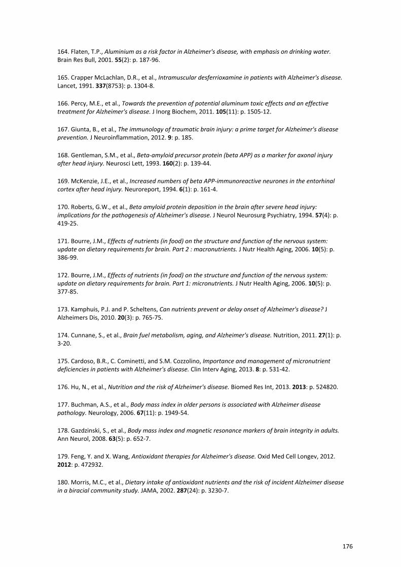
164. Flaten, T.P., Aluminium as a risk factor in Alzheimer's disease, with emphasis on drinking water. Brain Res Bull, 2001. 55(2): p. 187‐96.
165. Crapper McLachlan, D.R., et al., Intramuscular desferrioxamine in patients with Alzheimer's disease. Lancet, 1991. 337(8753): p. 1304‐8.
166. Percy, M.E., et al., Towards the prevention of potential aluminum toxic effects and an effective treatment for Alzheimer's disease. J Inorg Biochem, 2011. 105(11): p. 1505‐12.
167. Giunta, B., et al., The immunology of traumatic brain injury: a prime target for Alzheimer's disease prevention. J Neuroinflammation, 2012. 9: p. 185.
168. Gentleman, S.M., et al., Beta‐amyloid precursor protein (beta APP) as a marker for axonal injury after head injury. Neurosci Lett, 1993. 160(2): p. 139‐44.
169. McKenzie, J.E., et al., Increased numbers of beta APP‐immunoreactive neurones in the entorhinal cortex after head injury. Neuroreport, 1994. 6(1): p. 161‐4.
170. Roberts, G.W., et al., Beta amyloid protein deposition in the brain after severe head injury: implications for the pathogenesis of Alzheimer's disease. J Neurol Neurosurg Psychiatry, 1994. 57(4): p. 419‐25.
171. Bourre, J.M., Effects of nutrients (in food) on the structure and function of the nervous system: update on dietary requirements for brain. Part 2 : macronutrients. J Nutr Health Aging, 2006. 10(5): p. 386‐99.
172. Bourre, J.M., Effects of nutrients (in food) on the structure and function of the nervous system: update on dietary requirements for brain. Part 1: micronutrients. J Nutr Health Aging, 2006. 10(5): p. 377‐85.
173. Kamphuis, P.J. and P. Scheltens, Can nutrients prevent or delay onset of Alzheimer's disease? J Alzheimers Dis, 2010. 20(3): p. 765‐75.
174. Cunnane, S., et al., Brain fuel metabolism, aging, and Alzheimer's disease. Nutrition, 2011. 27(1): p. 3‐20.
175. Cardoso, B.R., C. Cominetti, and S.M. Cozzolino, Importance and management of micronutrient deficiencies in patients with Alzheimer's disease. Clin Interv Aging, 2013. 8: p. 531‐42.
176. Hu, N., et al., Nutrition and the risk of Alzheimer's disease. Biomed Res Int, 2013. 2013: p. 524820.
177. Buchman, A.S., et al., Body mass index in older persons is associated with Alzheimer disease pathology. Neurology, 2006. 67(11): p. 1949‐54.
178. Gazdzinski, S., et al., Body mass index and magnetic resonance markers of brain integrity in adults. Ann Neurol, 2008. 63(5): p. 652‐7.
179. Feng, Y. and X. Wang, Antioxidant therapies for Alzheimer's disease. Oxid Med Cell Longev, 2012. 2012: p. 472932.
180. Morris, M.C., et al., Dietary intake of antioxidant nutrients and the risk of incident Alzheimer disease in a biracial community study. JAMA, 2002. 287(24): p. 3230‐7.
176
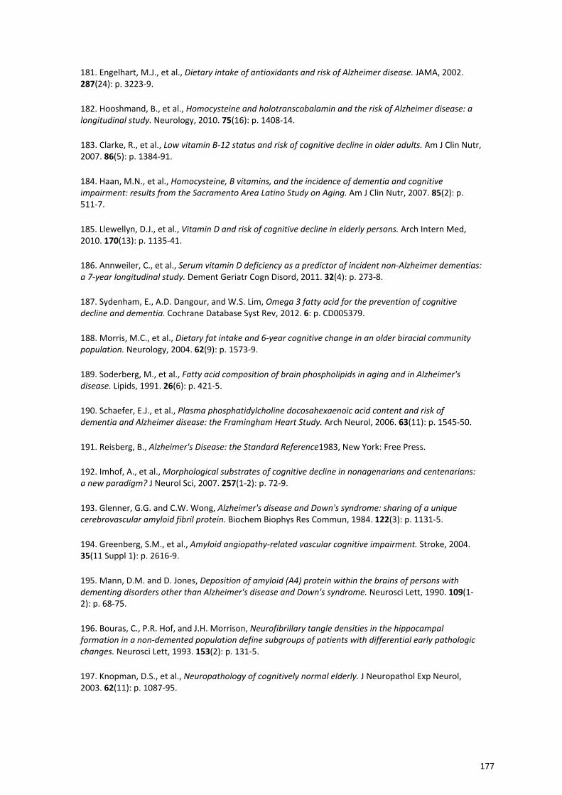
181. Engelhart, M.J., et al., Dietary intake of antioxidants and risk of Alzheimer disease. JAMA, 2002. 287(24): p. 3223‐9.
182. Hooshmand, B., et al., Homocysteine and holotranscobalamin and the risk of Alzheimer disease: a longitudinal study. Neurology, 2010. 75(16): p. 1408‐14.
183. Clarke, R., et al., Low vitamin B‐12 status and risk of cognitive decline in older adults. Am J Clin Nutr, 2007. 86(5): p. 1384‐91.
184. Haan, M.N., et al., Homocysteine, B vitamins, and the incidence of dementia and cognitive impairment: results from the Sacramento Area Latino Study on Aging. Am J Clin Nutr, 2007. 85(2): p. 511‐7.
185. Llewellyn, D.J., et al., Vitamin D and risk of cognitive decline in elderly persons. Arch Intern Med, 2010. 170(13): p. 1135‐41.
186. Annweiler, C., et al., Serum vitamin D deficiency as a predictor of incident non‐Alzheimer dementias: a 7‐year longitudinal study. Dement Geriatr Cogn Disord, 2011. 32(4): p. 273‐8.
187. Sydenham, E., A.D. Dangour, and W.S. Lim, Omega 3 fatty acid for the prevention of cognitive decline and dementia. Cochrane Database Syst Rev, 2012. 6: p. CD005379.
188. Morris, M.C., et al., Dietary fat intake and 6‐year cognitive change in an older biracial community population. Neurology, 2004. 62(9): p. 1573‐9.
189. Soderberg, M., et al., Fatty acid composition of brain phospholipids in aging and in Alzheimer's disease. Lipids, 1991. 26(6): p. 421‐5.
190. Schaefer, E.J., et al., Plasma phosphatidylcholine docosahexaenoic acid content and risk of dementia and Alzheimer disease: the Framingham Heart Study. Arch Neurol, 2006. 63(11): p. 1545‐50.
191. Reisberg, B., Alzheimer's Disease: the Standard Reference1983, New York: Free Press.
192. Imhof, A., et al., Morphological substrates of cognitive decline in nonagenarians and centenarians: a new paradigm? J Neurol Sci, 2007. 257(1‐2): p. 72‐9.
193. Glenner, G.G. and C.W. Wong, Alzheimer's disease and Down's syndrome: sharing of a unique cerebrovascular amyloid fibril protein. Biochem Biophys Res Commun, 1984. 122(3): p. 1131‐5.
194. Greenberg, S.M., et al., Amyloid angiopathy‐related vascular cognitive impairment. Stroke, 2004. 35(11 Suppl 1): p. 2616‐9.
195. Mann, D.M. and D. Jones, Deposition of amyloid (A4) protein within the brains of persons with dementing disorders other than Alzheimer's disease and Down's syndrome. Neurosci Lett, 1990. 109(1‐2): p. 68‐75.
196. Bouras, C., P.R. Hof, and J.H. Morrison, Neurofibrillary tangle densities in the hippocampal formation in a non‐demented population define subgroups of patients with differential early pathologic changes. Neurosci Lett, 1993. 153(2): p. 131‐5.
197. Knopman, D.S., et al., Neuropathology of cognitively normal elderly. J Neuropathol Exp Neurol, 2003. 62(11): p. 1087‐95.
177
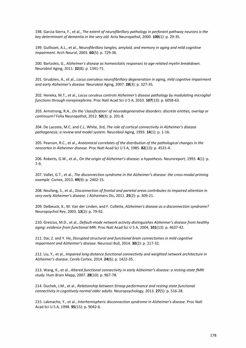
198. Garcia‐Sierra, F., et al., The extent of neurofibrillary pathology in perforant pathway neurons is the key determinant of dementia in the very old. Acta Neuropathol, 2000. 100(1): p. 29‐35.
199. Guillozet, A.L., et al., Neurofibrillary tangles, amyloid, and memory in aging and mild cognitive impairment. Arch Neurol, 2003. 60(5): p. 729‐36.
200. Bartzokis, G., Alzheimer's disease as homeostatic responses to age‐related myelin breakdown. Neurobiol Aging, 2011. 32(8): p. 1341‐71.
201. Grudzien, A., et al., Locus coeruleus neurofibrillary degeneration in aging, mild cognitive impairment and early Alzheimer's disease. Neurobiol Aging, 2007. 28(3): p. 327‐35.
202. Heneka, M.T., et al., Locus ceruleus controls Alzheimer's disease pathology by modulating microglial functions through norepinephrine. Proc Natl Acad Sci U S A, 2010. 107(13): p. 6058‐63.
203. Armstrong, R.A., On the 'classification' of neurodegenerative disorders: discrete entities, overlap or continuum? Folia Neuropathol, 2012. 50(3): p. 201‐8.
204. De Lacoste, M.C. and C.L. White, 3rd, The role of cortical connectivity in Alzheimer's disease pathogenesis: a review and model system. Neurobiol Aging, 1993. 14(1): p. 1‐16.
205. Pearson, R.C., et al., Anatomical correlates of the distribution of the pathological changes in the neocortex in Alzheimer disease. Proc Natl Acad Sci U S A, 1985. 82(13): p. 4531‐4.
206. Roberts, G.W., et al., On the origin of Alzheimer's disease: a hypothesis. Neuroreport, 1993. 4(1): p. 7‐9.
207. Vallet, G.T., et al., The disconnection syndrome in the Alzheimer's disease: the cross‐modal priming example. Cortex, 2013. 49(9): p. 2402‐15.
208. Neufang, S., et al., Disconnection of frontal and parietal areas contributes to impaired attention in very early Alzheimer's disease. J Alzheimers Dis, 2011. 25(2): p. 309‐21.
209. Delbeuck, X., M. Van der Linden, and F. Collette, Alzheimer's disease as a disconnection syndrome? Neuropsychol Rev, 2003. 13(2): p. 79‐92.
210. Greicius, M.D., et al., Default‐mode network activity distinguishes Alzheimer's disease from healthy aging: evidence from functional MRI. Proc Natl Acad Sci U S A, 2004. 101(13): p. 4637‐42.
211. Dai, Z. and Y. He, Disrupted structural and functional brain connectomes in mild cognitive impairment and Alzheimer's disease. Neurosci Bull, 2014. 30(2): p. 217‐32.
212. Liu, Y., et al., Impaired long distance functional connectivity and weighted network architecture in Alzheimer's disease. Cereb Cortex, 2014. 24(6): p. 1422‐35.
213. Wang, K., et al., Altered functional connectivity in early Alzheimer's disease: a resting‐state fMRI study. Hum Brain Mapp, 2007. 28(10): p. 967‐78.
214. Duchek, J.M., et al., Relationship between Stroop performance and resting state functional connectivity in cognitively normal older adults. Neuropsychology, 2013. 27(5): p. 516‐28.
215. Lakmache, Y., et al., Interhemispheric disconnection syndrome in Alzheimer's disease. Proc Natl Acad Sci U S A, 1998. 95(15): p. 9042‐6.
178
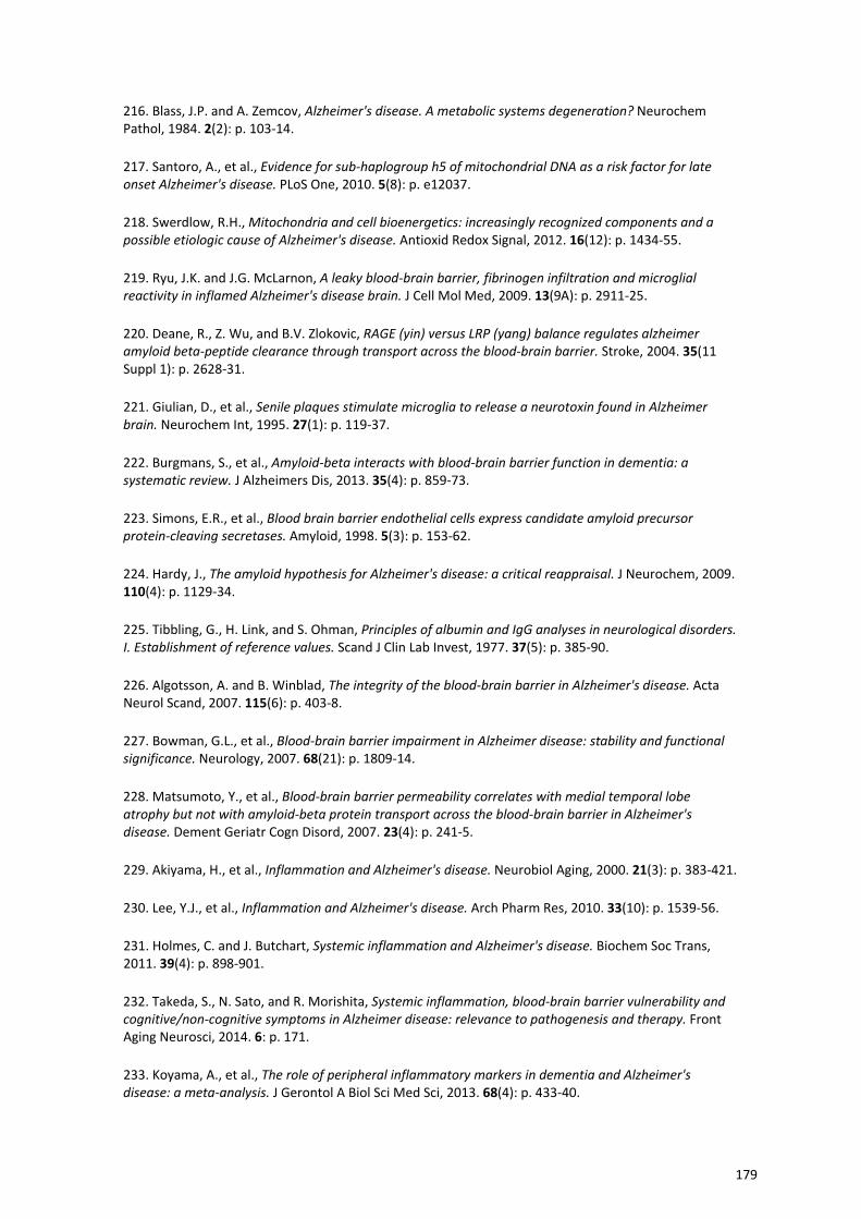
216. Blass, J.P. and A. Zemcov, Alzheimer's disease. A metabolic systems degeneration? Neurochem Pathol, 1984. 2(2): p. 103‐14.
217. Santoro, A., et al., Evidence for sub‐haplogroup h5 of mitochondrial DNA as a risk factor for late onset Alzheimer's disease. PLoS One, 2010. 5(8): p. e12037.
218. Swerdlow, R.H., Mitochondria and cell bioenergetics: increasingly recognized components and a possible etiologic cause of Alzheimer's disease. Antioxid Redox Signal, 2012. 16(12): p. 1434‐55.
219. Ryu, J.K. and J.G. McLarnon, A leaky blood‐brain barrier, fibrinogen infiltration and microglial reactivity in inflamed Alzheimer's disease brain. J Cell Mol Med, 2009. 13(9A): p. 2911‐25.
220. Deane, R., Z. Wu, and B.V. Zlokovic, RAGE (yin) versus LRP (yang) balance regulates alzheimer amyloid beta‐peptide clearance through transport across the blood‐brain barrier. Stroke, 2004. 35(11 Suppl 1): p. 2628‐31.
221. Giulian, D., et al., Senile plaques stimulate microglia to release a neurotoxin found in Alzheimer brain. Neurochem Int, 1995. 27(1): p. 119‐37.
222. Burgmans, S., et al., Amyloid‐beta interacts with blood‐brain barrier function in dementia: a systematic review. J Alzheimers Dis, 2013. 35(4): p. 859‐73.
223. Simons, E.R., et al., Blood brain barrier endothelial cells express candidate amyloid precursor protein‐cleaving secretases. Amyloid, 1998. 5(3): p. 153‐62.
224. Hardy, J., The amyloid hypothesis for Alzheimer's disease: a critical reappraisal. J Neurochem, 2009. 110(4): p. 1129‐34.
225. Tibbling, G., H. Link, and S. Ohman, Principles of albumin and IgG analyses in neurological disorders. I. Establishment of reference values. Scand J Clin Lab Invest, 1977. 37(5): p. 385‐90.
226. Algotsson, A. and B. Winblad, The integrity of the blood‐brain barrier in Alzheimer's disease. Acta Neurol Scand, 2007. 115(6): p. 403‐8.
227. Bowman, G.L., et al., Blood‐brain barrier impairment in Alzheimer disease: stability and functional significance. Neurology, 2007. 68(21): p. 1809‐14.
228. Matsumoto, Y., et al., Blood‐brain barrier permeability correlates with medial temporal lobe atrophy but not with amyloid‐beta protein transport across the blood‐brain barrier in Alzheimer's disease. Dement Geriatr Cogn Disord, 2007. 23(4): p. 241‐5.
229. Akiyama, H., et al., Inflammation and Alzheimer's disease. Neurobiol Aging, 2000. 21(3): p. 383‐421.
230. Lee, Y.J., et al., Inflammation and Alzheimer's disease. Arch Pharm Res, 2010. 33(10): p. 1539‐56.
231. Holmes, C. and J. Butchart, Systemic inflammation and Alzheimer's disease. Biochem Soc Trans, 2011. 39(4): p. 898‐901.
232. Takeda, S., N. Sato, and R. Morishita, Systemic inflammation, blood‐brain barrier vulnerability and cognitive/non‐cognitive symptoms in Alzheimer disease: relevance to pathogenesis and therapy. Front Aging Neurosci, 2014. 6: p. 171.
233. Koyama, A., et al., The role of peripheral inflammatory markers in dementia and Alzheimer's disease: a meta‐analysis. J Gerontol A Biol Sci Med Sci, 2013. 68(4): p. 433‐40.
179
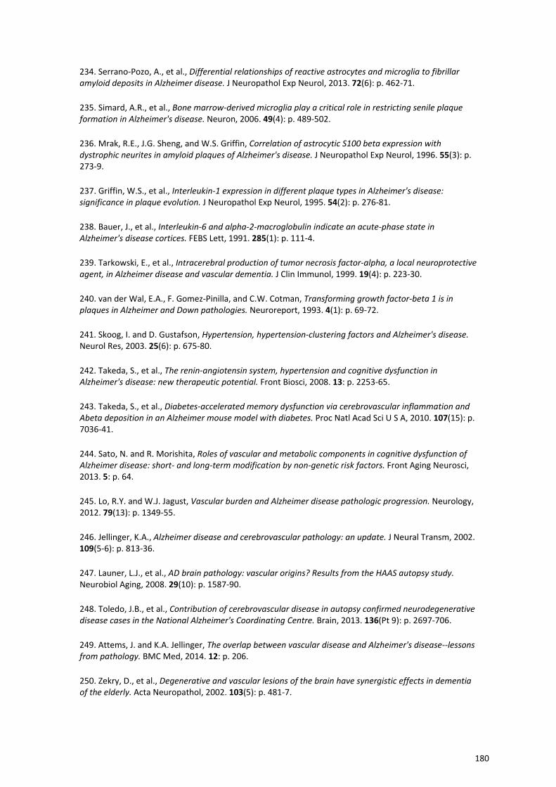
234. Serrano‐Pozo, A., et al., Differential relationships of reactive astrocytes and microglia to fibrillar amyloid deposits in Alzheimer disease. J Neuropathol Exp Neurol, 2013. 72(6): p. 462‐71.
235. Simard, A.R., et al., Bone marrow‐derived microglia play a critical role in restricting senile plaque formation in Alzheimer's disease. Neuron, 2006. 49(4): p. 489‐502.
236. Mrak, R.E., J.G. Sheng, and W.S. Griffin, Correlation of astrocytic S100 beta expression with dystrophic neurites in amyloid plaques of Alzheimer's disease. J Neuropathol Exp Neurol, 1996. 55(3): p. 273‐9.
237. Griffin, W.S., et al., Interleukin‐1 expression in different plaque types in Alzheimer's disease: significance in plaque evolution. J Neuropathol Exp Neurol, 1995. 54(2): p. 276‐81.
238. Bauer, J., et al., Interleukin‐6 and alpha‐2‐macroglobulin indicate an acute‐phase state in Alzheimer's disease cortices. FEBS Lett, 1991. 285(1): p. 111‐4.
239. Tarkowski, E., et al., Intracerebral production of tumor necrosis factor‐alpha, a local neuroprotective agent, in Alzheimer disease and vascular dementia. J Clin Immunol, 1999. 19(4): p. 223‐30.
240. van der Wal, E.A., F. Gomez‐Pinilla, and C.W. Cotman, Transforming growth factor‐beta 1 is in plaques in Alzheimer and Down pathologies. Neuroreport, 1993. 4(1): p. 69‐72.
241. Skoog, I. and D. Gustafson, Hypertension, hypertension‐clustering factors and Alzheimer's disease. Neurol Res, 2003. 25(6): p. 675‐80.
242. Takeda, S., et al., The renin‐angiotensin system, hypertension and cognitive dysfunction in Alzheimer's disease: new therapeutic potential. Front Biosci, 2008. 13: p. 2253‐65.
243. Takeda, S., et al., Diabetes‐accelerated memory dysfunction via cerebrovascular inflammation and Abeta deposition in an Alzheimer mouse model with diabetes. Proc Natl Acad Sci U S A, 2010. 107(15): p. 7036‐41.
244. Sato, N. and R. Morishita, Roles of vascular and metabolic components in cognitive dysfunction of Alzheimer disease: short‐ and long‐term modification by non‐genetic risk factors. Front Aging Neurosci, 2013. 5: p. 64.
245. Lo, R.Y. and W.J. Jagust, Vascular burden and Alzheimer disease pathologic progression. Neurology, 2012. 79(13): p. 1349‐55.
246. Jellinger, K.A., Alzheimer disease and cerebrovascular pathology: an update. J Neural Transm, 2002. 109(5‐6): p. 813‐36.
247. Launer, L.J., et al., AD brain pathology: vascular origins? Results from the HAAS autopsy study. Neurobiol Aging, 2008. 29(10): p. 1587‐90.
248. Toledo, J.B., et al., Contribution of cerebrovascular disease in autopsy confirmed neurodegenerative disease cases in the National Alzheimer's Coordinating Centre. Brain, 2013. 136(Pt 9): p. 2697‐706.
249. Attems, J. and K.A. Jellinger, The overlap between vascular disease and Alzheimer's disease‐‐lessons from pathology. BMC Med, 2014. 12: p. 206.
250. Zekry, D., et al., Degenerative and vascular lesions of the brain have synergistic effects in dementia of the elderly. Acta Neuropathol, 2002. 103(5): p. 481‐7.
180
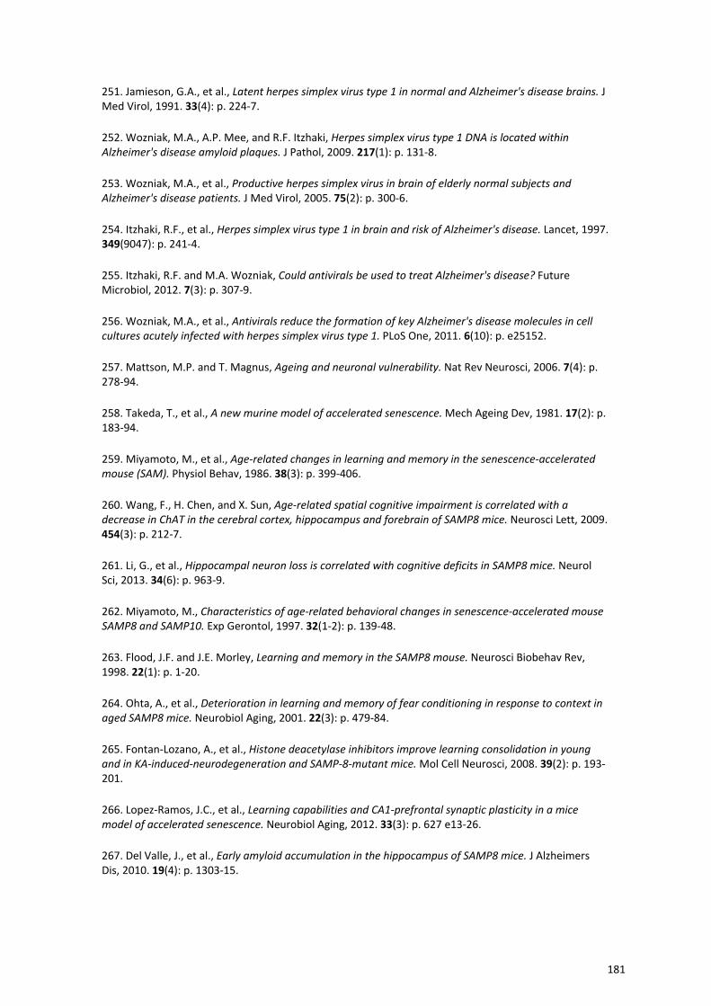
251. Jamieson, G.A., et al., Latent herpes simplex virus type 1 in normal and Alzheimer's disease brains. J Med Virol, 1991. 33(4): p. 224‐7.
252. Wozniak, M.A., A.P. Mee, and R.F. Itzhaki, Herpes simplex virus type 1 DNA is located within Alzheimer's disease amyloid plaques. J Pathol, 2009. 217(1): p. 131‐8.
253. Wozniak, M.A., et al., Productive herpes simplex virus in brain of elderly normal subjects and Alzheimer's disease patients. J Med Virol, 2005. 75(2): p. 300‐6.
254. Itzhaki, R.F., et al., Herpes simplex virus type 1 in brain and risk of Alzheimer's disease. Lancet, 1997. 349(9047): p. 241‐4.
255. Itzhaki, R.F. and M.A. Wozniak, Could antivirals be used to treat Alzheimer's disease? Future Microbiol, 2012. 7(3): p. 307‐9.
256. Wozniak, M.A., et al., Antivirals reduce the formation of key Alzheimer's disease molecules in cell cultures acutely infected with herpes simplex virus type 1. PLoS One, 2011. 6(10): p. e25152.
257. Mattson, M.P. and T. Magnus, Ageing and neuronal vulnerability. Nat Rev Neurosci, 2006. 7(4): p. 278‐94.
258. Takeda, T., et al., A new murine model of accelerated senescence. Mech Ageing Dev, 1981. 17(2): p. 183‐94.
259. Miyamoto, M., et al., Age‐related changes in learning and memory in the senescence‐accelerated mouse (SAM). Physiol Behav, 1986. 38(3): p. 399‐406.
260. Wang, F., H. Chen, and X. Sun, Age‐related spatial cognitive impairment is correlated with a decrease in ChAT in the cerebral cortex, hippocampus and forebrain of SAMP8 mice. Neurosci Lett, 2009. 454(3): p. 212‐7.
261. Li, G., et al., Hippocampal neuron loss is correlated with cognitive deficits in SAMP8 mice. Neurol Sci, 2013. 34(6): p. 963‐9.
262. Miyamoto, M., Characteristics of age‐related behavioral changes in senescence‐accelerated mouse SAMP8 and SAMP10. Exp Gerontol, 1997. 32(1‐2): p. 139‐48.
263. Flood, J.F. and J.E. Morley, Learning and memory in the SAMP8 mouse. Neurosci Biobehav Rev, 1998. 22(1): p. 1‐20.
264. Ohta, A., et al., Deterioration in learning and memory of fear conditioning in response to context in aged SAMP8 mice. Neurobiol Aging, 2001. 22(3): p. 479‐84.
265. Fontan‐Lozano, A., et al., Histone deacetylase inhibitors improve learning consolidation in young and in KA‐induced‐neurodegeneration and SAMP‐8‐mutant mice. Mol Cell Neurosci, 2008. 39(2): p. 193‐201.
266. Lopez‐Ramos, J.C., et al., Learning capabilities and CA1‐prefrontal synaptic plasticity in a mice model of accelerated senescence. Neurobiol Aging, 2012. 33(3): p. 627 e13‐26.
267. Del Valle, J., et al., Early amyloid accumulation in the hippocampus of SAMP8 mice. J Alzheimers Dis, 2010. 19(4): p. 1303‐15.
181
268. del Valle, J., et al., Cerebral amyloid angiopathy, blood‐brain barrier disruption and amyloid accumulation in SAMP8 mice. Neurodegener Dis, 2011. 8(6): p. 421‐9.
269. Morley, J.E., et al., Beta‐amyloid precursor polypeptide in SAMP8 mice affects learning and memory. Peptides, 2000. 21(12): p. 1761‐7.
270. Sureda, F.X., et al., Changes in oxidative stress parameters and neurodegeneration markers in the brain of the senescence‐accelerated mice SAMP‐8. Exp Gerontol, 2006. 41(4): p. 360‐7.
271. Canudas, A.M., et al., Hyperphosphorylation of microtubule‐associated protein tau in senescence‐accelerated mouse (SAM). Mech Ageing Dev, 2005. 126(12): p. 1300‐4.
272. Zhu, Y., et al., Cell death in the Purkinje cells of the cerebellum of senescence accelerated mouse (SAMP(8)). Biogerontology, 2007. 8(5): p. 537‐44.
273. Morley, J.E., et al., The senescence accelerated mouse (SAMP8) as a model for oxidative stress and Alzheimer's disease. Biochim Biophys Acta, 2012. 1822(5): p. 650‐6.
274. Alvarez‐Garcia, O., et al., Elevated oxidative stress in the brain of senescence‐accelerated mice at 5 months of age. Biogerontology, 2006. 7(1): p. 43‐52.
275. Chiba, Y., et al., The senescence‐accelerated mouse (SAM): a higher oxidative stress and age‐dependent degenerative diseases model. Neurochem Res, 2009. 34(4): p. 679‐87.
276. Bayram, B., et al., Biomarkers of oxidative stress, antioxidant defence and inflammation are altered in the senescence‐accelerated mouse prone 8. Age (Dordr), 2013. 35(4): p. 1205‐17.
277. Tha, K.K., et al., Changes in expressions of proinflammatory cytokines IL‐1beta, TNF‐alpha and IL‐6 in the brain of senescence accelerated mouse (SAM) P8. Brain Res, 2000. 885(1): p. 25‐31.
278. Butterfield, D.A. and H.F. Poon, The senescence‐accelerated prone mouse (SAMP8): a model of age‐related cognitive decline with relevance to alterations of the gene expression and protein abnormalities in Alzheimer's disease. Exp Gerontol, 2005. 40(10): p. 774‐83.
279. Pallas, M., et al., From aging to Alzheimer's disease: unveiling "the switch" with the senescence‐accelerated mouse model (SAMP8). J Alzheimers Dis, 2008. 15(4): p. 615‐24.
280. Cheng, X.R., W.X. Zhou, and Y.X. Zhang, The behavioral, pathological and therapeutic features of the senescence‐accelerated mouse prone 8 strain as an Alzheimer's disease animal model. Ageing Res Rev, 2014. 13: p. 13‐37.
281. Gutierrez‐Cuesta, J., et al., Chronic administration of melatonin reduces cerebral injury biomarkers in SAMP8. J Pineal Res, 2007. 42(4): p. 394‐402.
282. Wang, C., et al., DNA damage response and cellular senescence in tissues of aging mice. Aging Cell, 2009. 8(3): p. 311‐23.
283. Kawamata, T., et al., Neuropathological studies on strains of senescence‐accelerated mice (SAM) with age‐related deficits in learning and memory. Exp Gerontol, 1997. 32(1‐2): p. 161‐9.
284. del Valle, J., et al., Dendritic spine abnormalities in hippocampal CA1 pyramidal neurons underlying memory deficits in the SAMP8 mouse model of Alzheimer's disease. J Alzheimers Dis, 2012. 32(1): p. 233‐40.
182
285. Yagi, H., et al., Spontaneous spongy degeneration of the brain stem in SAM‐P/8 mice, a newly developed memory‐deficient strain. J Neuropathol Exp Neurol, 1989. 48(5): p. 577‐90.
286. Chan, Y.C., et al., Favorable effects of tea on reducing the cognitive deficits and brain morphological changes in senescence‐accelerated mice. J Nutr Sci Vitaminol (Tokyo), 2006. 52(4): p. 266‐73.
287. Wu, Y., A.Q. Zhang, and D.T. Yew, Age related changes of various markers of astrocytes in senescence‐accelerated mice hippocampus. Neurochem Int, 2005. 46(7): p. 565‐74.
288. Jaenisch, R. and A. Bird, Epigenetic regulation of gene expression: how the genome integrates intrinsic and environmental signals. Nat Genet, 2003. 33 Suppl: p. 245‐54.
289. Bird, A., DNA methylation patterns and epigenetic memory. Genes Dev, 2002. 16(1): p. 6‐21.
290. Goll, M.G. and T.H. Bestor, Eukaryotic cytosine methyltransferases. Annu Rev Biochem, 2005. 74: p. 481‐514.
291. Chan, S.W., I.R. Henderson, and S.E. Jacobsen, Gardening the genome: DNA methylation in Arabidopsis thaliana. Nat Rev Genet, 2005. 6(5): p. 351‐60.
292. Ramsahoye, B.H., et al., Non‐CpG methylation is prevalent in embryonic stem cells and may be mediated by DNA methyltransferase 3a. Proc Natl Acad Sci U S A, 2000. 97(10): p. 5237‐42.
293. Haines, T.R., D.I. Rodenhiser, and P.J. Ainsworth, Allele‐specific non‐CpG methylation of the Nf1 gene during early mouse development. Dev Biol, 2001. 240(2): p. 585‐98.
294. Hark, A.T., et al., CTCF mediates methylation‐sensitive enhancer‐blocking activity at the H19/Igf2 locus. Nature, 2000. 405(6785): p. 486‐9.
295. Ehrlich, M., et al., Amount and distribution of 5‐methylcytosine in human DNA from different types of tissues of cells. Nucleic Acids Res, 1982. 10(8): p. 2709‐21.
296. Jones, P.L., et al., Methylated DNA and MeCP2 recruit histone deacetylase to repress transcription. Nat Genet, 1998. 19(2): p. 187‐91.
297. Nan, X., et al., Transcriptional repression by the methyl‐CpG‐binding protein MeCP2 involves a histone deacetylase complex. Nature, 1998. 393(6683): p. 386‐9.
298. Sarraf, S.A. and I. Stancheva, Methyl‐CpG binding protein MBD1 couples histone H3 methylation at lysine 9 by SETDB1 to DNA replication and chromatin assembly. Mol Cell, 2004. 15(4): p. 595‐605.
299. Klose, R.J., et al., DNA binding selectivity of MeCP2 due to a requirement for A/T sequences adjacent to methyl‐CpG. Mol Cell, 2005. 19(5): p. 667‐78.
300. Tamaru, H. and E.U. Selker, A histone H3 methyltransferase controls DNA methylation in Neurospora crassa. Nature, 2001. 414(6861): p. 277‐83.
301. Jackson, J.P., et al., Control of CpNpG DNA methylation by the KRYPTONITE histone H3 methyltransferase. Nature, 2002. 416(6880): p. 556‐60.
302. Lehnertz, B., et al., Suv39h‐mediated histone H3 lysine 9 methylation directs DNA methylation to major satellite repeats at pericentric heterochromatin. Curr Biol, 2003. 13(14): p. 1192‐200.
183
303. Kornberg, R.D., Chromatin structure: a repeating unit of histones and DNA. Science, 1974. 184(4139): p. 868‐71.
304. Martin, C. and Y. Zhang, The diverse functions of histone lysine methylation. Nat Rev Mol Cell Biol, 2005. 6(11): p. 838‐49.
305. Vakoc, C.R., et al., Histone H3 lysine 9 methylation and HP1gamma are associated with transcription elongation through mammalian chromatin. Mol Cell, 2005. 19(3): p. 381‐91.
306. Beisel, C., et al., Histone methylation by the Drosophila epigenetic transcriptional regulator Ash1. Nature, 2002. 419(6909): p. 857‐62.
307. Briggs, S.D., et al., Histone H3 lysine 4 methylation is mediated by Set1 and required for cell growth and rDNA silencing in Saccharomyces cerevisiae. Genes Dev, 2001. 15(24): p. 3286‐95.
308. Nakamura, T., et al., ALL‐1 is a histone methyltransferase that assembles a supercomplex of proteins involved in transcriptional regulation. Mol Cell, 2002. 10(5): p. 1119‐28.
309. Sedkov, Y., et al., Methylation at lysine 4 of histone H3 in ecdysone‐dependent development of Drosophila. Nature, 2003. 426(6962): p. 78‐83.
310. Shi, Y., et al., Histone demethylation mediated by the nuclear amine oxidase homolog LSD1. Cell, 2004. 119(7): p. 941‐53.
311. Wang, H., et al., Purification and functional characterization of a histone H3‐lysine 4‐specific methyltransferase. Mol Cell, 2001. 8(6): p. 1207‐17.
312. Cloos, P.A., et al., The putative oncogene GASC1 demethylates tri‐ and dimethylated lysine 9 on histone H3. Nature, 2006. 442(7100): p. 307‐11.
313. Klose, R.J., et al., The transcriptional repressor JHDM3A demethylates trimethyl histone H3 lysine 9 and lysine 36. Nature, 2006. 442(7100): p. 312‐6.
314. Krogan, N.J., et al., Methylation of histone H3 by Set2 in Saccharomyces cerevisiae is linked to transcriptional elongation by RNA polymerase II. Mol Cell Biol, 2003. 23(12): p. 4207‐18.
315. Tsukada, Y., et al., Histone demethylation by a family of JmjC domain‐containing proteins. Nature, 2006. 439(7078): p. 811‐6.
316. Whetstine, J.R., et al., Reversal of histone lysine trimethylation by the JMJD2 family of histone demethylases. Cell, 2006. 125(3): p. 467‐81.
317. Feng, Q., et al., Methylation of H3‐lysine 79 is mediated by a new family of HMTases without a SET domain. Curr Biol, 2002. 12(12): p. 1052‐8.
318. Huyen, Y., et al., Methylated lysine 79 of histone H3 targets 53BP1 to DNA double‐strand breaks. Nature, 2004. 432(7015): p. 406‐11.
319. Krogan, N.J., et al., The Paf1 complex is required for histone H3 methylation by COMPASS and Dot1p: linking transcriptional elongation to histone methylation. Mol Cell, 2003. 11(3): p. 721‐9.
320. Fodor, B.D., et al., Jmjd2b antagonizes H3K9 trimethylation at pericentric heterochromatin in mammalian cells. Genes Dev, 2006. 20(12): p. 1557‐62.
184
321. Johnson, L., X. Cao, and S. Jacobsen, Interplay between two epigenetic marks. DNA methylation and histone H3 lysine 9 methylation. Curr Biol, 2002. 12(16): p. 1360‐7.
322. Metzger, E., et al., LSD1 demethylates repressive histone marks to promote androgen‐receptor‐dependent transcription. Nature, 2005. 437(7057): p. 436‐9.
323. Nakayama, J., et al., Role of histone H3 lysine 9 methylation in epigenetic control of heterochromatin assembly. Science, 2001. 292(5514): p. 110‐3.
324. Rea, S., et al., Regulation of chromatin structure by site‐specific histone H3 methyltransferases. Nature, 2000. 406(6796): p. 593‐9.
325. Schultz, D.C., et al., SETDB1: a novel KAP‐1‐associated histone H3, lysine 9‐specific methyltransferase that contributes to HP1‐mediated silencing of euchromatic genes by KRAB zinc‐finger proteins. Genes Dev, 2002. 16(8): p. 919‐32.
326. Shin, S. and R. Janknecht, Diversity within the JMJD2 histone demethylase family. Biochem Biophys Res Commun, 2007. 353(4): p. 973‐7.
327. Tachibana, M., et al., Set domain‐containing protein, G9a, is a novel lysine‐preferring mammalian histone methyltransferase with hyperactivity and specific selectivity to lysines 9 and 27 of histone H3. J Biol Chem, 2001. 276(27): p. 25309‐17.
328. Yamane, K., et al., JHDM2A, a JmjC‐containing H3K9 demethylase, facilitates transcription activation by androgen receptor. Cell, 2006. 125(3): p. 483‐95.
329. Cao, R., et al., Role of histone H3 lysine 27 methylation in Polycomb‐group silencing. Science, 2002. 298(5595): p. 1039‐43.
330. Nishioka, K., et al., PR‐Set7 is a nucleosome‐specific methyltransferase that modifies lysine 20 of histone H4 and is associated with silent chromatin. Mol Cell, 2002. 9(6): p. 1201‐13.
331. Schotta, G., et al., A silencing pathway to induce H3‐K9 and H4‐K20 trimethylation at constitutive heterochromatin. Genes Dev, 2004. 18(11): p. 1251‐62.
332. Chang, B., et al., JMJD6 is a histone arginine demethylase. Science, 2007. 318(5849): p. 444‐7.
333. Guccione, E., et al., Methylation of histone H3R2 by PRMT6 and H3K4 by an MLL complex are mutually exclusive. Nature, 2007. 449(7164): p. 933‐7.
334. Hyllus, D., et al., PRMT6‐mediated methylation of R2 in histone H3 antagonizes H3 K4 trimethylation. Genes Dev, 2007. 21(24): p. 3369‐80.
335. Iberg, A.N., et al., Arginine methylation of the histone H3 tail impedes effector binding. J Biol Chem, 2008. 283(6): p. 3006‐10.
336. Webby, C.J., et al., Jmjd6 catalyses lysyl‐hydroxylation of U2AF65, a protein associated with RNA splicing. Science, 2009. 325(5936): p. 90‐3.
337. Daujat, S., et al., Crosstalk between CARM1 methylation and CBP acetylation on histone H3. Curr Biol, 2002. 12(24): p. 2090‐7.
338. Di Lorenzo, A. and M.T. Bedford, Histone arginine methylation. FEBS Lett, 2011. 585(13): p. 2024‐31.
185
339. Fabbrizio, E., et al., Negative regulation of transcription by the type II arginine methyltransferase PRMT5. EMBO Rep, 2002. 3(7): p. 641‐5.
340. Pal, S., et al., Human SWI/SNF‐associated PRMT5 methylates histone H3 arginine 8 and negatively regulates expression of ST7 and NM23 tumor suppressor genes. Mol Cell Biol, 2004. 24(21): p. 9630‐45.
341. Harrison, M.J., Y.H. Tang, and D.H. Dowhan, Protein arginine methyltransferase 6 regulates multiple aspects of gene expression. Nucleic Acids Res, 2010. 38(7): p. 2201‐16.
342. Pal, S., et al., mSin3A/histone deacetylase 2‐ and PRMT5‐containing Brg1 complex is involved in transcriptional repression of the Myc target gene cad. Mol Cell Biol, 2003. 23(21): p. 7475‐87.
343. Strahl, B.D., et al., Methylation of histone H4 at arginine 3 occurs in vivo and is mediated by the nuclear receptor coactivator PRMT1. Curr Biol, 2001. 11(12): p. 996‐1000.
344. Wang, H., et al., Methylation of histone H4 at arginine 3 facilitating transcriptional activation by nuclear hormone receptor. Science, 2001. 293(5531): p. 853‐7.
345. Santos‐Rosa, H., et al., Active genes are tri‐methylated at K4 of histone H3. Nature, 2002. 419(6905): p. 407‐11.
346. Zhang, Y. and D. Reinberg, Transcription regulation by histone methylation: interplay between different covalent modifications of the core histone tails. Genes Dev, 2001. 15(18): p. 2343‐60.
347. Lee, D.Y., et al., Role of protein methylation in regulation of transcription. Endocr Rev, 2005. 26(2): p. 147‐70.
348. Bannister, A.J., et al., Selective recognition of methylated lysine 9 on histone H3 by the HP1 chromo domain. Nature, 2001. 410(6824): p. 120‐4.
349. Lachner, M., et al., Methylation of histone H3 lysine 9 creates a binding site for HP1 proteins. Nature, 2001. 410(6824): p. 116‐20.
350. Garcia‐Bassets, I., et al., Histone methylation‐dependent mechanisms impose ligand dependency for gene activation by nuclear receptors. Cell, 2007. 128(3): p. 505‐18.
351. Wissmann, M., et al., Cooperative demethylation by JMJD2C and LSD1 promotes androgen receptor‐dependent gene expression. Nat Cell Biol, 2007. 9(3): p. 347‐53.
352. Schurter, B.T., et al., Methylation of histone H3 by coactivator‐associated arginine methyltransferase 1. Biochemistry, 2001. 40(19): p. 5747‐56.
353. Bauer, U.M., et al., Methylation at arginine 17 of histone H3 is linked to gene activation. EMBO Rep, 2002. 3(1): p. 39‐44.
354. Vettese‐Dadey, M., et al., Acetylation of histone H4 plays a primary role in enhancing transcription factor binding to nucleosomal DNA in vitro. EMBO J, 1996. 15(10): p. 2508‐18.
355. Dhalluin, C., et al., Structure and ligand of a histone acetyltransferase bromodomain. Nature, 1999. 399(6735): p. 491‐6.
356. Hassan, A.H., et al., Function and selectivity of bromodomains in anchoring chromatin‐modifying complexes to promoter nucleosomes. Cell, 2002. 111(3): p. 369‐79.
186
357. Kimura, A. and M. Horikoshi, Tip60 acetylates six lysines of a specific class in core histones in vitro. Genes Cells, 1998. 3(12): p. 789‐800.
358. Schiltz, R.L., et al., Overlapping but distinct patterns of histone acetylation by the human coactivators p300 and PCAF within nucleosomal substrates. J Biol Chem, 1999. 274(3): p. 1189‐92.
359. Kawasaki, H., et al., ATF‐2 has intrinsic histone acetyltransferase activity which is modulated by phosphorylation. Nature, 2000. 405(6783): p. 195‐200.
360. Grant, P.A., et al., Expanded lysine acetylation specificity of Gcn5 in native complexes. J Biol Chem, 1999. 274(9): p. 5895‐900.
361. Sobel, R.E., et al., Conservation of deposition‐related acetylation sites in newly synthesized histones H3 and H4. Proc Natl Acad Sci U S A, 1995. 92(4): p. 1237‐41.
362. Spencer, T.E., et al., Steroid receptor coactivator‐1 is a histone acetyltransferase. Nature, 1997. 389(6647): p. 194‐8.
363. Bird, A.W., et al., Acetylation of histone H4 by Esa1 is required for DNA double‐strand break repair. Nature, 2002. 419(6905): p. 411‐5.
364. Clarke, A.S., et al., Esa1p is an essential histone acetyltransferase required for cell cycle progression. Mol Cell Biol, 1999. 19(4): p. 2515‐26.
365. Kueh, A.J., et al., HBO1 is required for H3K14 acetylation and normal transcriptional activity during embryonic development. Mol Cell Biol, 2011. 31(4): p. 845‐60.
366. Ikura, T., et al., Involvement of the TIP60 histone acetylase complex in DNA repair and apoptosis. Cell, 2000. 102(4): p. 463‐73.
367. Miotto, B. and K. Struhl, HBO1 histone acetylase activity is essential for DNA replication licensing and inhibited by Geminin. Mol Cell, 2010. 37(1): p. 57‐66.
368. Parthun, M.R., J. Widom, and D.E. Gottschling, The major cytoplasmic histone acetyltransferase in yeast: links to chromatin replication and histone metabolism. Cell, 1996. 87(1): p. 85‐94.
369. Iizuka, M., et al., Histone acetyltransferase Hbo1: catalytic activity, cellular abundance, and links to primary cancers. Gene, 2009. 436(1‐2): p. 108‐14.
370. Kuo, M.H., et al., Transcription‐linked acetylation by Gcn5p of histones H3 and H4 at specific lysines. Nature, 1996. 383(6597): p. 269‐72.
371. Kelly, T.J., et al., Type B histone acetyltransferase Hat1p participates in telomeric silencing. Mol Cell Biol, 2000. 20(19): p. 7051‐8.
372. Yang, Y., et al., Regulation and function of histone acetyltransferase MOF. Front Med, 2014. 8(1): p. 79‐83.
373. Sterner, D.E. and S.L. Berger, Acetylation of histones and transcription‐related factors. Microbiol Mol Biol Rev, 2000. 64(2): p. 435‐59.
374. Glozak, M.A., et al., Acetylation and deacetylation of non‐histone proteins. Gene, 2005. 363: p. 15‐23.
187
375. Marmorstein, R. and R.C. Trievel, Histone modifying enzymes: structures, mechanisms, and specificities. Biochim Biophys Acta, 2009. 1789(1): p. 58‐68.
376. Graff, J. and L.H. Tsai, Histone acetylation: molecular mnemonics on the chromatin. Nat Rev Neurosci, 2013. 14(2): p. 97‐111.
377. Graff, J. and L.H. Tsai, The potential of HDAC inhibitors as cognitive enhancers. Annu Rev Pharmacol Toxicol, 2013. 53: p. 311‐30.
378. Mahadevan, L.C., A.C. Willis, and M.J. Barratt, Rapid histone H3 phosphorylation in response to growth factors, phorbol esters, okadaic acid, and protein synthesis inhibitors. Cell, 1991. 65(5): p. 775‐83.
379. Sassone‐Corsi, P., et al., Requirement of Rsk‐2 for epidermal growth factor‐activated phosphorylation of histone H3. Science, 1999. 285(5429): p. 886‐91.
380. Lo, W.S., et al., Snf1‐‐a histone kinase that works in concert with the histone acetyltransferase Gcn5 to regulate transcription. Science, 2001. 293(5532): p. 1142‐6.
381. Soloaga, A., et al., MSK2 and MSK1 mediate the mitogen‐ and stress‐induced phosphorylation of histone H3 and HMG‐14. EMBO J, 2003. 22(11): p. 2788‐97.
382. Nowak, S.J. and V.G. Corces, Phosphorylation of histone H3: a balancing act between chromosome condensation and transcriptional activation. Trends Genet, 2004. 20(4): p. 214‐20.
383. Fischle, W., et al., Regulation of HP1‐chromatin binding by histone H3 methylation and phosphorylation. Nature, 2005. 438(7071): p. 1116‐22.
384. Wood, A., et al., The Paf1 complex is essential for histone monoubiquitination by the Rad6‐Bre1 complex, which signals for histone methylation by COMPASS and Dot1p. J Biol Chem, 2003. 278(37): p. 34739‐42.
385. Kim, J., S.B. Hake, and R.G. Roeder, The human homolog of yeast BRE1 functions as a transcriptional coactivator through direct activator interactions. Mol Cell, 2005. 20(5): p. 759‐70.
386. Zhu, B., et al., Monoubiquitination of human histone H2B: the factors involved and their roles in HOX gene regulation. Mol Cell, 2005. 20(4): p. 601‐11.
387. Henry, K.W., et al., Transcriptional activation via sequential histone H2B ubiquitylation and deubiquitylation, mediated by SAGA‐associated Ubp8. Genes Dev, 2003. 17(21): p. 2648‐63.
388. Kao, C.F., et al., Rad6 plays a role in transcriptional activation through ubiquitylation of histone H2B. Genes Dev, 2004. 18(2): p. 184‐95.
389. Wang, H., et al., Role of histone H2A ubiquitination in Polycomb silencing. Nature, 2004. 431(7010): p. 873‐8.
390. Shiio, Y. and R.N. Eisenman, Histone sumoylation is associated with transcriptional repression. Proc Natl Acad Sci U S A, 2003. 100(23): p. 13225‐30.
391. Cuthbert, G.L., et al., Histone deimination antagonizes arginine methylation. Cell, 2004. 118(5): p. 545‐53.
188
392. Wang, Y., et al., Human PAD4 regulates histone arginine methylation levels via demethylimination. Science, 2004. 306(5694): p. 279‐83.
393. Lee, R.C., R.L. Feinbaum, and V. Ambros, The C. elegans heterochronic gene lin‐4 encodes small RNAs with antisense complementarity to lin‐14. Cell, 1993. 75(5): p. 843‐54.
394. Bartel, D.P., MicroRNAs: genomics, biogenesis, mechanism, and function. Cell, 2004. 116(2): p. 281‐97.
395. Okamura, K., et al., The regulatory activity of microRNA* species has substantial influence on microRNA and 3' UTR evolution. Nat Struct Mol Biol, 2008. 15(4): p. 354‐63.
396. Ro, S., et al., Tissue‐dependent paired expression of miRNAs. Nucleic Acids Res, 2007. 35(17): p. 5944‐53.
397. Bartel, D.P., MicroRNAs: target recognition and regulatory functions. Cell, 2009. 136(2): p. 215‐33.
398. Llave, C., et al., Cleavage of Scarecrow‐like mRNA targets directed by a class of Arabidopsis miRNA. Science, 2002. 297(5589): p. 2053‐6.
399. Yekta, S., I.H. Shih, and D.P. Bartel, MicroRNA‐directed cleavage of HOXB8 mRNA. Science, 2004. 304(5670): p. 594‐6.
400. Wu, L., J. Fan, and J.G. Belasco, MicroRNAs direct rapid deadenylation of mRNA. Proc Natl Acad Sci U S A, 2006. 103(11): p. 4034‐9.
401. Petersen, C.P., et al., Short RNAs repress translation after initiation in mammalian cells. Mol Cell, 2006. 21(4): p. 533‐42.
402. Guo, H., et al., Mammalian microRNAs predominantly act to decrease target mRNA levels. Nature, 2010. 466(7308): p. 835‐40.
403. Huntzinger, E. and E. Izaurralde, Gene silencing by microRNAs: contributions of translational repression and mRNA decay. Nat Rev Genet, 2011. 12(2): p. 99‐110.
404. Ichimura, A., et al., miRNAs and regulation of cell signaling. FEBS J, 2011. 278(10): p. 1610‐8.
405. Inui, M., G. Martello, and S. Piccolo, MicroRNA control of signal transduction. Nat Rev Mol Cell Biol, 2010. 11(4): p. 252‐63.
406. He, L. and G.J. Hannon, MicroRNAs: small RNAs with a big role in gene regulation. Nat Rev Genet, 2004. 5(7): p. 522‐31.
407. Landgraf, P., et al., A mammalian microRNA expression atlas based on small RNA library sequencing. Cell, 2007. 129(7): p. 1401‐14.
408. Wang, W., E.J. Kwon, and L.H. Tsai, MicroRNAs in learning, memory, and neurological diseases. Learn Mem, 2012. 19(9): p. 359‐68.
409. Kosik, K.S. and A.M. Krichevsky, The Elegance of the MicroRNAs: A Neuronal Perspective. Neuron, 2005. 47(6): p. 779‐82.
410. Kim, J., et al., Identification of many microRNAs that copurify with polyribosomes in mammalian neurons. Proc Natl Acad Sci U S A, 2004. 101(1): p. 360‐5.
189
411. Sempere, L.F., et al., Expression profiling of mammalian microRNAs uncovers a subset of brain‐expressed microRNAs with possible roles in murine and human neuronal differentiation. Genome Biol, 2004. 5(3): p. R13.
412. De Pietri Tonelli, D., et al., miRNAs are essential for survival and differentiation of newborn neurons but not for expansion of neural progenitors during early neurogenesis in the mouse embryonic neocortex. Development, 2008. 135(23): p. 3911‐21.
413. Cheng, L.C., et al., miR‐124 regulates adult neurogenesis in the subventricular zone stem cell niche. Nat Neurosci, 2009. 12(4): p. 399‐408.
414. Fiore, R., et al., Mef2‐mediated transcription of the miR379‐410 cluster regulates activity‐dependent dendritogenesis by fine‐tuning Pumilio2 protein levels. EMBO J, 2009. 28(6): p. 697‐710.
415. Vo, N., et al., A cAMP‐response element binding protein‐induced microRNA regulates neuronal morphogenesis. Proc Natl Acad Sci U S A, 2005. 102(45): p. 16426‐31.
416. Siegel, G., et al., A functional screen implicates microRNA‐138‐dependent regulation of the depalmitoylation enzyme APT1 in dendritic spine morphogenesis. Nat Cell Biol, 2009. 11(6): p. 705‐16.
417. Schratt, G.M., et al., A brain‐specific microRNA regulates dendritic spine development. Nature, 2006. 439(7074): p. 283‐9.
418. Bak, M., et al., MicroRNA expression in the adult mouse central nervous system. RNA, 2008. 14(3): p. 432‐44.
419. He, M., et al., Cell‐type‐based analysis of microRNA profiles in the mouse brain. Neuron, 2012. 73(1): p. 35‐48.
420. Obernosterer, G., et al., Post‐transcriptional regulation of microRNA expression. RNA, 2006. 12(7): p. 1161‐7.
421. Kye, M.J., et al., Somatodendritic microRNAs identified by laser capture and multiplex RT‐PCR. RNA, 2007. 13(8): p. 1224‐34.
422. Natera‐Naranjo, O., et al., Identification and quantitative analyses of microRNAs located in the distal axons of sympathetic neurons. RNA, 2010. 16(8): p. 1516‐29.
423. Liu, L., et al., Aging, cancer and nutrition: the DNA methylation connection. Mech Ageing Dev, 2003. 124(10‐12): p. 989‐98.
424. Liu, L., et al., Insufficient DNA methylation affects healthy aging and promotes age‐related health problems. Clin Epigenetics, 2011. 2(2): p. 349‐60.
425. Heyn, H., et al., Distinct DNA methylomes of newborns and centenarians. Proc Natl Acad Sci U S A, 2012. 109(26): p. 10522‐7.
426. Bjornsson, H.T., et al., Intra‐individual change over time in DNA methylation with familial clustering. JAMA, 2008. 299(24): p. 2877‐83.
427. Gentilini, D., et al., Role of epigenetics in human aging and longevity: genome‐wide DNA methylation profile in centenarians and centenarians' offspring. Age (Dordr), 2013. 35(5): p. 1961‐73.
190
428. Bollati, V., et al., Decline in genomic DNA methylation through aging in a cohort of elderly subjects. Mech Ageing Dev, 2009. 130(4): p. 234‐9.
429. Kim, M., et al., DNA methylation as a biomarker for cardiovascular disease risk. PLoS One, 2010. 5(3): p. e9692.
430. Moore, L.E., et al., Genomic DNA hypomethylation as a biomarker for bladder cancer susceptibility in the Spanish Bladder Cancer Study: a case‐control study. Lancet Oncol, 2008. 9(4): p. 359‐66.
431. Bennett‐Baker, P.E., J. Wilkowski, and D.T. Burke, Age‐associated activation of epigenetically repressed genes in the mouse. Genetics, 2003. 165(4): p. 2055‐62.
432. Madrigano, J., et al., Aging and epigenetics: longitudinal changes in gene‐specific DNA methylation. Epigenetics, 2012. 7(1): p. 63‐70.
433. Christensen, B.C., et al., Aging and environmental exposures alter tissue‐specific DNA methylation dependent upon CpG island context. PLoS Genet, 2009. 5(8): p. e1000602.
434. Salpea, P., et al., Postnatal development‐ and age‐related changes in DNA‐methylation patterns in the human genome. Nucleic Acids Res, 2012. 40(14): p. 6477‐94.
435. Murphy, T.M., A.S. Perry, and M. Lawler, The emergence of DNA methylation as a key modulator of aberrant cell death in prostate cancer. Endocr Relat Cancer, 2008. 15(1): p. 11‐25.
436. Funayama, R. and F. Ishikawa, Cellular senescence and chromatin structure. Chromosoma, 2007. 116(5): p. 431‐40.
437. Gronniger, E., et al., Aging and chronic sun exposure cause distinct epigenetic changes in human skin. PLoS Genet, 2010. 6(5): p. e1000971.
438. Bocklandt, S., et al., Epigenetic predictor of age. PLoS One, 2011. 6(6): p. e14821.
439. Casillas, M.A., Jr., et al., Transcriptional control of the DNA methyltransferases is altered in aging and neoplastically‐transformed human fibroblasts. Mol Cell Biochem, 2003. 252(1‐2): p. 33‐43.
440. Li, Y., et al., Age‐dependent decreases in DNA methyltransferase levels and low transmethylation micronutrient levels synergize to promote overexpression of genes implicated in autoimmunity and acute coronary syndromes. Exp Gerontol, 2010. 45(4): p. 312‐22.
441. Oliveira, A.M., T.J. Hemstedt, and H. Bading, Rescue of aging‐associated decline in Dnmt3a2 expression restores cognitive abilities. Nat Neurosci, 2012. 15(8): p. 1111‐3.
442. Lin, M.J., et al., DNA methyltransferase gene dDnmt2 and longevity of Drosophila. J Biol Chem, 2005. 280(2): p. 861‐4.
443. Goll, M.G., et al., Methylation of tRNAAsp by the DNA methyltransferase homolog Dnmt2. Science, 2006. 311(5759): p. 395‐8.
444. Bell, J.T., et al., Epigenome‐wide scans identify differentially methylated regions for age and age‐related phenotypes in a healthy ageing population. PLoS Genet, 2012. 8(4): p. e1002629.
445. Dang, W., et al., Histone H4 lysine 16 acetylation regulates cellular lifespan. Nature, 2009. 459(7248): p. 802‐7.
191
446. Kaeberlein, M., M. McVey, and L. Guarente, The SIR2/3/4 complex and SIR2 alone promote longevity in Saccharomyces cerevisiae by two different mechanisms. Genes Dev, 1999. 13(19): p. 2570‐80.
447. Viswanathan, M. and L. Guarente, Regulation of Caenorhabditis elegans lifespan by sir‐2.1 transgenes. Nature, 2011. 477(7365): p. E1‐2.
448. Greer, E.L., et al., Members of the H3K4 trimethylation complex regulate lifespan in a germline‐dependent manner in C. elegans. Nature, 2010. 466(7304): p. 383‐7.
449. Greer, E.L., et al., Transgenerational epigenetic inheritance of longevity in Caenorhabditis elegans. Nature, 2011. 479(7373): p. 365‐71.
450. Jin, C., et al., Histone demethylase UTX‐1 regulates C. elegans life span by targeting the insulin/IGF‐1 signaling pathway. Cell Metab, 2011. 14(2): p. 161‐72.
451. Maures, T.J., et al., The H3K27 demethylase UTX‐1 regulates C. elegans lifespan in a germline‐independent, insulin‐dependent manner. Aging Cell, 2011. 10(6): p. 980‐90.
452. Cheng, H.L., et al., Developmental defects and p53 hyperacetylation in Sir2 homolog (SIRT1)‐deficient mice. Proc Natl Acad Sci U S A, 2003. 100(19): p. 10794‐9.
453. Lagouge, M., et al., Resveratrol improves mitochondrial function and protects against metabolic disease by activating SIRT1 and PGC‐1alpha. Cell, 2006. 127(6): p. 1109‐22.
454. Oberdoerffer, P., et al., SIRT1 redistribution on chromatin promotes genomic stability but alters gene expression during aging. Cell, 2008. 135(5): p. 907‐18.
455. Pearson, K.J., et al., Resveratrol delays age‐related deterioration and mimics transcriptional aspects of dietary restriction without extending life span. Cell Metab, 2008. 8(2): p. 157‐68.
456. Peleg, S., et al., Altered histone acetylation is associated with age‐dependent memory impairment in mice. Science, 2010. 328(5979): p. 753‐6.
457. Mostoslavsky, R., et al., Genomic instability and aging‐like phenotype in the absence of mammalian SIRT6. Cell, 2006. 124(2): p. 315‐29.
458. Han, Y., et al., Stress‐associated H3K4 methylation accumulates during postnatal development and aging of rhesus macaque brain. Aging Cell, 2012. 11(6): p. 1055‐64.
459. O'Sullivan, R.J., et al., Reduced histone biosynthesis and chromatin changes arising from a damage signal at telomeres. Nat Struct Mol Biol, 2010. 17(10): p. 1218‐25.
460. Scaffidi, P. and T. Misteli, Lamin A‐dependent nuclear defects in human aging. Science, 2006. 312(5776): p. 1059‐63.
461. Shumaker, D.K., et al., Mutant nuclear lamin A leads to progressive alterations of epigenetic control in premature aging. Proc Natl Acad Sci U S A, 2006. 103(23): p. 8703‐8.
462. Narita, M., et al., Rb‐mediated heterochromatin formation and silencing of E2F target genes during cellular senescence. Cell, 2003. 113(6): p. 703‐16.
463. Zhang, R., et al., Formation of MacroH2A‐containing senescence‐associated heterochromatin foci and senescence driven by ASF1a and HIRA. Dev Cell, 2005. 8(1): p. 19‐30.
192
464. Polo, S.E. and S.P. Jackson, Dynamics of DNA damage response proteins at DNA breaks: a focus on protein modifications. Genes Dev, 2011. 25(5): p. 409‐33.
465. Sedelnikova, O.A., et al., Senescing human cells and ageing mice accumulate DNA lesions with unrepairable double‐strand breaks. Nat Cell Biol, 2004. 6(2): p. 168‐70.
466. Sedelnikova, O.A., et al., Delayed kinetics of DNA double‐strand break processing in normal and pathological aging. Aging Cell, 2008. 7(1): p. 89‐100.
467. Herbig, U., et al., Cellular senescence in aging primates. Science, 2006. 311(5765): p. 1257.
468. Lanceta, J., et al., MicroRNA group disorganization in aging. Exp Gerontol, 2010. 45(4): p. 269‐78.
469. Zhang, J., et al., Comparative profiling of genes and miRNAs expressed in the newborn, young adult, and aged human epididymides. Acta Biochim Biophys Sin (Shanghai), 2010. 42(2): p. 145‐53.
470. Ibanez‐Ventoso, C. and M. Driscoll, MicroRNAs in C. elegans Aging: Molecular Insurance for Robustness? Curr Genomics, 2009. 10(3): p. 144‐53.
471. Kozomara, A. and S. Griffiths‐Jones, miRBase: integrating microRNA annotation and deep‐sequencing data. Nucleic Acids Res, 2011. 39(Database issue): p. D152‐7.
472. Pincus, Z., T. Smith‐Vikos, and F.J. Slack, MicroRNA predictors of longevity in Caenorhabditis elegans. PLoS Genet, 2011. 7(9): p. e1002306.
473. Kato, M., et al., Age‐associated changes in expression of small, noncoding RNAs, including microRNAs, in C. elegans. RNA, 2011. 17(10): p. 1804‐20.
474. de Lencastre, A., et al., MicroRNAs both promote and antagonize longevity in C. elegans. Curr Biol, 2010. 20(24): p. 2159‐68.
475. Ibanez‐Ventoso, C., et al., Modulated microRNA expression during adult lifespan in Caenorhabditis elegans. Aging Cell, 2006. 5(3): p. 235‐46.
476. Kimura, K.D., et al., daf‐2, an insulin receptor‐like gene that regulates longevity and diapause in Caenorhabditis elegans. Science, 1997. 277(5328): p. 942‐6.
477. Tatar, M., et al., A mutant Drosophila insulin receptor homolog that extends life‐span and impairs neuroendocrine function. Science, 2001. 292(5514): p. 107‐10.
478. Bluher, M., B.B. Kahn, and C.R. Kahn, Extended longevity in mice lacking the insulin receptor in adipose tissue. Science, 2003. 299(5606): p. 572‐4.
479. Boehm, M. and F. Slack, A developmental timing microRNA and its target regulate life span in C. elegans. Science, 2005. 310(5756): p. 1954‐7.
480. Kato, M., et al., TGF‐beta activates Akt kinase through a microRNA‐dependent amplifying circuit targeting PTEN. Nat Cell Biol, 2009. 11(7): p. 881‐9.
481. Sayed, D. and M. Abdellatif, AKT‐ing via microRNA. Cell Cycle, 2010. 9(16): p. 3213‐7.
482. Wang, X.H., et al., MicroRNA‐320 expression in myocardial microvascular endothelial cells and its relationship with insulin‐like growth factor‐1 in type 2 diabetic rats. Clin Exp Pharmacol Physiol, 2009. 36(2): p. 181‐8.
193
483. Shan, Z.X., et al., Upregulated expression of miR‐1/miR‐206 in a rat model of myocardial infarction. Biochem Biophys Res Commun, 2009. 381(4): p. 597‐601.
484. Wei, Y., Y.J. Zhang, and Y. Cai, Growth or longevity: the TOR's decision on lifespan regulation. Biogerontology, 2013. 14(4): p. 353‐63.
485. Laplante, M. and D.M. Sabatini, mTOR signaling in growth control and disease. Cell, 2012. 149(2): p. 274‐93.
486. Kaeberlein, M., et al., Regulation of yeast replicative life span by TOR and Sch9 in response to nutrients. Science, 2005. 310(5751): p. 1193‐6.
487. Jia, K., D. Chen, and D.L. Riddle, The TOR pathway interacts with the insulin signaling pathway to regulate C. elegans larval development, metabolism and life span. Development, 2004. 131(16): p. 3897‐906.
488. Vellai, T., et al., Genetics: influence of TOR kinase on lifespan in C. elegans. Nature, 2003. 426(6967): p. 620.
489. Kapahi, P., et al., Regulation of lifespan in Drosophila by modulation of genes in the TOR signaling pathway. Curr Biol, 2004. 14(10): p. 885‐90.
490. Harrison, D.E., et al., Rapamycin fed late in life extends lifespan in genetically heterogeneous mice. Nature, 2009. 460(7253): p. 392‐5.
491. Menghini, R., et al., MicroRNA 217 modulates endothelial cell senescence via silent information regulator 1. Circulation, 2009. 120(15): p. 1524‐32.
492. Lee, J., et al., A pathway involving farnesoid X receptor and small heterodimer partner positively regulates hepatic sirtuin 1 levels via microRNA‐34a inhibition. J Biol Chem, 2010. 285(17): p. 12604‐11.
493. Strum, J.C., et al., MicroRNA 132 regulates nutritional stress‐induced chemokine production through repression of SirT1. Mol Endocrinol, 2009. 23(11): p. 1876‐84.
494. Rane, S., et al., Downregulation of miR‐199a derepresses hypoxia‐inducible factor‐1alpha and Sirtuin 1 and recapitulates hypoxia preconditioning in cardiac myocytes. Circ Res, 2009. 104(7): p. 879‐86.
495. Han, M., J. Toli, and M. Abdellatif, MicroRNAs in the cardiovascular system. Curr Opin Cardiol, 2011. 26(3): p. 181‐9.
496. D'Alessandra, Y., et al., Circulating microRNAs are new and sensitive biomarkers of myocardial infarction. Eur Heart J, 2010. 31(22): p. 2765‐73.
497. Sayed, D., et al., MicroRNAs play an essential role in the development of cardiac hypertrophy. Circ Res, 2007. 100(3): p. 416‐24.
498. Hamrick, M.W., et al., The adipokine leptin increases skeletal muscle mass and significantly alters skeletal muscle miRNA expression profile in aged mice. Biochem Biophys Res Commun, 2010. 400(3): p. 379‐83.
499. Guller, I. and A.P. Russell, MicroRNAs in skeletal muscle: their role and regulation in development, disease and function. J Physiol, 2010. 588(Pt 21): p. 4075‐87.
194
500. Chen, J.F., T.E. Callis, and D.Z. Wang, microRNAs and muscle disorders. J Cell Sci, 2009. 122(Pt 1): p. 13‐20.
501. Rottiers, V. and A.M. Naar, MicroRNAs in metabolism and metabolic disorders. Nat Rev Mol Cell Biol, 2012. 13(4): p. 239‐50.
502. Vinciguerra, M., et al., Unsaturated fatty acids inhibit the expression of tumor suppressor phosphatase and tensin homolog (PTEN) via microRNA‐21 up‐regulation in hepatocytes. Hepatology, 2009. 49(4): p. 1176‐84.
503. Junn, E. and M.M. Mouradian, MicroRNAs in neurodegenerative diseases and their therapeutic potential. Pharmacol Ther, 2012. 133(2): p. 142‐50.
504. Provost, P., MicroRNAs as a molecular basis for mental retardation, Alzheimer's and prion diseases. Brain Res, 2010. 1338: p. 58‐66.
505. Eacker, S.M., T.M. Dawson, and V.L. Dawson, Understanding microRNAs in neurodegeneration. Nat Rev Neurosci, 2009. 10(12): p. 837‐41.
506. Smith, P., et al., In vivo regulation of amyloid precursor protein neuronal splicing by microRNAs. J Neurochem, 2011. 116(2): p. 240‐7.
507. Vilardo, E., et al., MicroRNA‐101 regulates amyloid precursor protein expression in hippocampal neurons. J Biol Chem, 2010. 285(24): p. 18344‐51.
508. Zhu, H.C., et al., MicroRNA‐195 downregulates Alzheimer's disease amyloid‐beta production by targeting BACE1. Brain Res Bull, 2012. 88(6): p. 596‐601.
509. Hebert, S.S., et al., Loss of microRNA cluster miR‐29a/b‐1 in sporadic Alzheimer's disease correlates with increased BACE1/beta‐secretase expression. Proc Natl Acad Sci U S A, 2008. 105(17): p. 6415‐20.
510. Wang, W.X., et al., The expression of microRNA miR‐107 decreases early in Alzheimer's disease and may accelerate disease progression through regulation of beta‐site amyloid precursor protein‐cleaving enzyme 1. J Neurosci, 2008. 28(5): p. 1213‐23.
511. Boissonneault, V., et al., MicroRNA‐298 and microRNA‐328 regulate expression of mouse beta‐amyloid precursor protein‐converting enzyme 1. J Biol Chem, 2009. 284(4): p. 1971‐81.
512. Haskell, W.L., et al., Physical activity and public health: updated recommendation for adults from the American College of Sports Medicine and the American Heart Association. Med Sci Sports Exerc, 2007. 39(8): p. 1423‐34.
513. Nelson, M.E., et al., Physical activity and public health in older adults: recommendation from the American College of Sports Medicine and the American Heart Association. Med Sci Sports Exerc, 2007. 39(8): p. 1435‐45.
514. Chodzko‐Zajko, W.J., et al., American College of Sports Medicine position stand. Exercise and physical activity for older adults. Med Sci Sports Exerc, 2009. 41(7): p. 1510‐30.
515. Gremeaux, V., et al., Long‐term lifestyle intervention with optimized high‐intensity interval training improves body composition, cardiometabolic risk, and exercise parameters in patients with abdominal obesity. Am J Phys Med Rehabil, 2012. 91(11): p. 941‐50.
516. Schoeller, D.A., Measurement of energy expenditure in free‐living humans by using doubly labeled water. J Nutr, 1988. 118(11): p. 1278‐89.
195
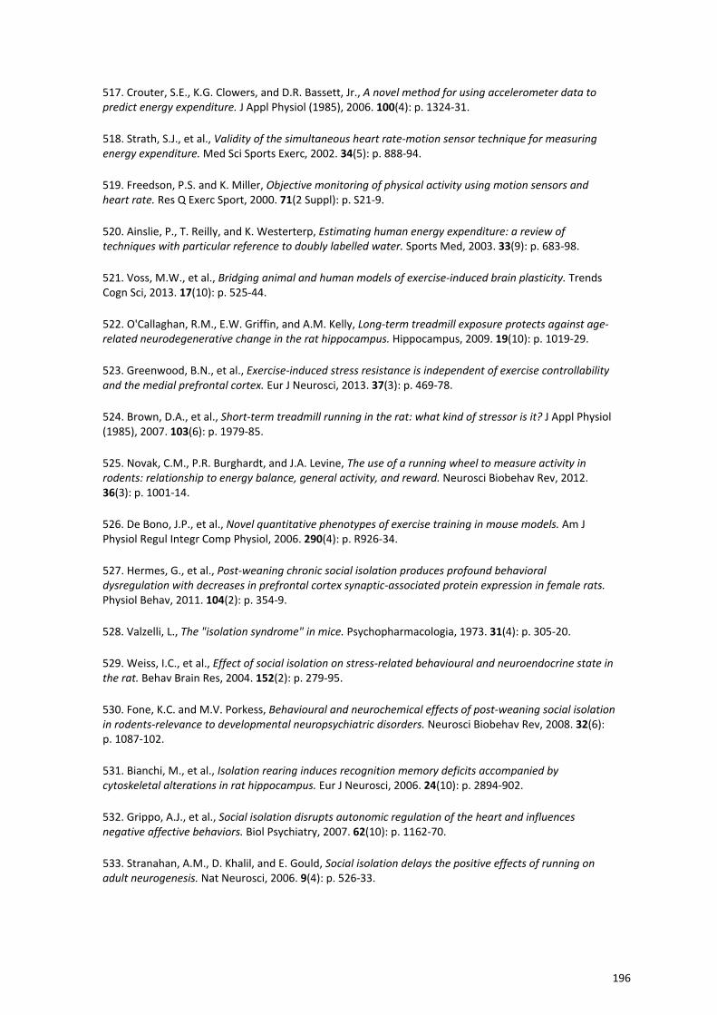
517. Crouter, S.E., K.G. Clowers, and D.R. Bassett, Jr., A novel method for using accelerometer data to predict energy expenditure. J Appl Physiol (1985), 2006. 100(4): p. 1324‐31.
518. Strath, S.J., et al., Validity of the simultaneous heart rate‐motion sensor technique for measuring energy expenditure. Med Sci Sports Exerc, 2002. 34(5): p. 888‐94.
519. Freedson, P.S. and K. Miller, Objective monitoring of physical activity using motion sensors and heart rate. Res Q Exerc Sport, 2000. 71(2 Suppl): p. S21‐9.
520. Ainslie, P., T. Reilly, and K. Westerterp, Estimating human energy expenditure: a review of techniques with particular reference to doubly labelled water. Sports Med, 2003. 33(9): p. 683‐98.
521. Voss, M.W., et al., Bridging animal and human models of exercise‐induced brain plasticity. Trends Cogn Sci, 2013. 17(10): p. 525‐44.
522. O'Callaghan, R.M., E.W. Griffin, and A.M. Kelly, Long‐term treadmill exposure protects against age‐related neurodegenerative change in the rat hippocampus. Hippocampus, 2009. 19(10): p. 1019‐29.
523. Greenwood, B.N., et al., Exercise‐induced stress resistance is independent of exercise controllability and the medial prefrontal cortex. Eur J Neurosci, 2013. 37(3): p. 469‐78.
524. Brown, D.A., et al., Short‐term treadmill running in the rat: what kind of stressor is it? J Appl Physiol (1985), 2007. 103(6): p. 1979‐85.
525. Novak, C.M., P.R. Burghardt, and J.A. Levine, The use of a running wheel to measure activity in rodents: relationship to energy balance, general activity, and reward. Neurosci Biobehav Rev, 2012. 36(3): p. 1001‐14.
526. De Bono, J.P., et al., Novel quantitative phenotypes of exercise training in mouse models. Am J Physiol Regul Integr Comp Physiol, 2006. 290(4): p. R926‐34.
527. Hermes, G., et al., Post‐weaning chronic social isolation produces profound behavioral dysregulation with decreases in prefrontal cortex synaptic‐associated protein expression in female rats. Physiol Behav, 2011. 104(2): p. 354‐9.
528. Valzelli, L., The "isolation syndrome" in mice. Psychopharmacologia, 1973. 31(4): p. 305‐20.
529. Weiss, I.C., et al., Effect of social isolation on stress‐related behavioural and neuroendocrine state in the rat. Behav Brain Res, 2004. 152(2): p. 279‐95.
530. Fone, K.C. and M.V. Porkess, Behavioural and neurochemical effects of post‐weaning social isolation in rodents‐relevance to developmental neuropsychiatric disorders. Neurosci Biobehav Rev, 2008. 32(6): p. 1087‐102.
531. Bianchi, M., et al., Isolation rearing induces recognition memory deficits accompanied by cytoskeletal alterations in rat hippocampus. Eur J Neurosci, 2006. 24(10): p. 2894‐902.
532. Grippo, A.J., et al., Social isolation disrupts autonomic regulation of the heart and influences negative affective behaviors. Biol Psychiatry, 2007. 62(10): p. 1162‐70.
533. Stranahan, A.M., D. Khalil, and E. Gould, Social isolation delays the positive effects of running on adult neurogenesis. Nat Neurosci, 2006. 9(4): p. 526‐33.
196
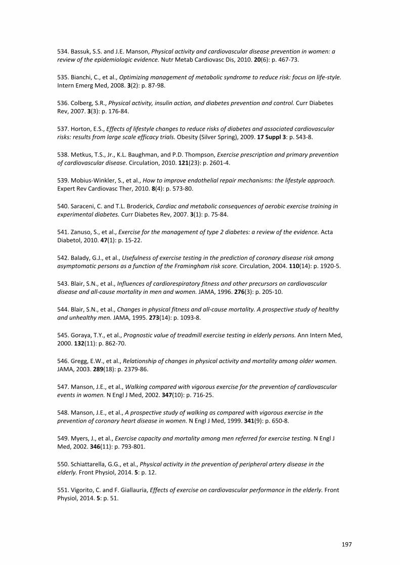
534. Bassuk, S.S. and J.E. Manson, Physical activity and cardiovascular disease prevention in women: a review of the epidemiologic evidence. Nutr Metab Cardiovasc Dis, 2010. 20(6): p. 467‐73.
535. Bianchi, C., et al., Optimizing management of metabolic syndrome to reduce risk: focus on life‐style. Intern Emerg Med, 2008. 3(2): p. 87‐98.
536. Colberg, S.R., Physical activity, insulin action, and diabetes prevention and control. Curr Diabetes Rev, 2007. 3(3): p. 176‐84.
537. Horton, E.S., Effects of lifestyle changes to reduce risks of diabetes and associated cardiovascular risks: results from large scale efficacy trials. Obesity (Silver Spring), 2009. 17 Suppl 3: p. S43‐8.
538. Metkus, T.S., Jr., K.L. Baughman, and P.D. Thompson, Exercise prescription and primary prevention of cardiovascular disease. Circulation, 2010. 121(23): p. 2601‐4.
539. Mobius‐Winkler, S., et al., How to improve endothelial repair mechanisms: the lifestyle approach. Expert Rev Cardiovasc Ther, 2010. 8(4): p. 573‐80.
540. Saraceni, C. and T.L. Broderick, Cardiac and metabolic consequences of aerobic exercise training in experimental diabetes. Curr Diabetes Rev, 2007. 3(1): p. 75‐84.
541. Zanuso, S., et al., Exercise for the management of type 2 diabetes: a review of the evidence. Acta Diabetol, 2010. 47(1): p. 15‐22.
542. Balady, G.J., et al., Usefulness of exercise testing in the prediction of coronary disease risk among asymptomatic persons as a function of the Framingham risk score. Circulation, 2004. 110(14): p. 1920‐5.
543. Blair, S.N., et al., Influences of cardiorespiratory fitness and other precursors on cardiovascular disease and all‐cause mortality in men and women. JAMA, 1996. 276(3): p. 205‐10.
544. Blair, S.N., et al., Changes in physical fitness and all‐cause mortality. A prospective study of healthy and unhealthy men. JAMA, 1995. 273(14): p. 1093‐8.
545. Goraya, T.Y., et al., Prognostic value of treadmill exercise testing in elderly persons. Ann Intern Med, 2000. 132(11): p. 862‐70.
546. Gregg, E.W., et al., Relationship of changes in physical activity and mortality among older women. JAMA, 2003. 289(18): p. 2379‐86.
547. Manson, J.E., et al., Walking compared with vigorous exercise for the prevention of cardiovascular events in women. N Engl J Med, 2002. 347(10): p. 716‐25.
548. Manson, J.E., et al., A prospective study of walking as compared with vigorous exercise in the prevention of coronary heart disease in women. N Engl J Med, 1999. 341(9): p. 650‐8.
549. Myers, J., et al., Exercise capacity and mortality among men referred for exercise testing. N Engl J Med, 2002. 346(11): p. 793‐801.
550. Schiattarella, G.G., et al., Physical activity in the prevention of peripheral artery disease in the elderly. Front Physiol, 2014. 5: p. 12.
551. Vigorito, C. and F. Giallauria, Effects of exercise on cardiovascular performance in the elderly. Front Physiol, 2014. 5: p. 51.
197
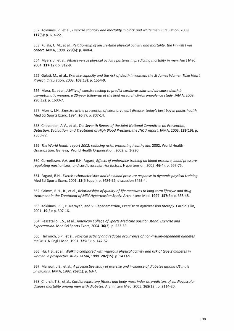
552. Kokkinos, P., et al., Exercise capacity and mortality in black and white men. Circulation, 2008. 117(5): p. 614‐22.
553. Kujala, U.M., et al., Relationship of leisure‐time physical activity and mortality: the Finnish twin cohort. JAMA, 1998. 279(6): p. 440‐4.
554. Myers, J., et al., Fitness versus physical activity patterns in predicting mortality in men. Am J Med, 2004. 117(12): p. 912‐8.
555. Gulati, M., et al., Exercise capacity and the risk of death in women: the St James Women Take Heart Project. Circulation, 2003. 108(13): p. 1554‐9.
556. Mora, S., et al., Ability of exercise testing to predict cardiovascular and all‐cause death in asymptomatic women: a 20‐year follow‐up of the lipid research clinics prevalence study. JAMA, 2003. 290(12): p. 1600‐7.
557. Morris, J.N., Exercise in the prevention of coronary heart disease: today's best buy in public health. Med Sci Sports Exerc, 1994. 26(7): p. 807‐14.
558. Chobanian, A.V., et al., The Seventh Report of the Joint National Committee on Prevention, Detection, Evaluation, and Treatment of High Blood Pressure: the JNC 7 report. JAMA, 2003. 289(19): p. 2560‐72.
559. The World Health report 2002: reducing risks, promoting healthy life, 2002, World Health Organization: Geneva, World Health Organization, 2002. p. 1‐230.
560. Cornelissen, V.A. and R.H. Fagard, Effects of endurance training on blood pressure, blood pressure‐regulating mechanisms, and cardiovascular risk factors. Hypertension, 2005. 46(4): p. 667‐75.
561. Fagard, R.H., Exercise characteristics and the blood pressure response to dynamic physical training. Med Sci Sports Exerc, 2001. 33(6 Suppl): p. S484‐92; discussion S493‐4.
562. Grimm, R.H., Jr., et al., Relationships of quality‐of‐life measures to long‐term lifestyle and drug treatment in the Treatment of Mild Hypertension Study. Arch Intern Med, 1997. 157(6): p. 638‐48.
563. Kokkinos, P.F., P. Narayan, and V. Papademetriou, Exercise as hypertension therapy. Cardiol Clin, 2001. 19(3): p. 507‐16.
564. Pescatello, L.S., et al., American College of Sports Medicine position stand. Exercise and hypertension. Med Sci Sports Exerc, 2004. 36(3): p. 533‐53.
565. Helmrich, S.P., et al., Physical activity and reduced occurrence of non‐insulin‐dependent diabetes mellitus. N Engl J Med, 1991. 325(3): p. 147‐52.
566. Hu, F.B., et al., Walking compared with vigorous physical activity and risk of type 2 diabetes in women: a prospective study. JAMA, 1999. 282(15): p. 1433‐9.
567. Manson, J.E., et al., A prospective study of exercise and incidence of diabetes among US male physicians. JAMA, 1992. 268(1): p. 63‐7.
568. Church, T.S., et al., Cardiorespiratory fitness and body mass index as predictors of cardiovascular disease mortality among men with diabetes. Arch Intern Med, 2005. 165(18): p. 2114‐20.
198
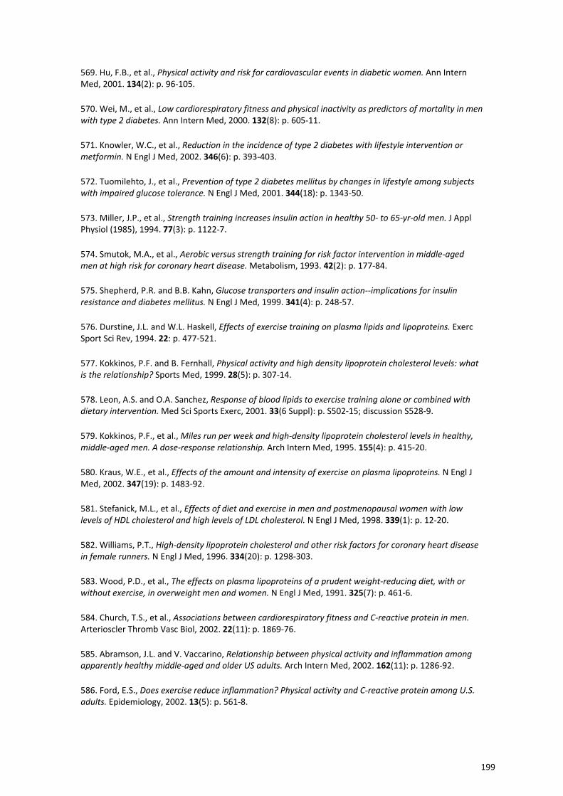
569. Hu, F.B., et al., Physical activity and risk for cardiovascular events in diabetic women. Ann Intern Med, 2001. 134(2): p. 96‐105.
570. Wei, M., et al., Low cardiorespiratory fitness and physical inactivity as predictors of mortality in men with type 2 diabetes. Ann Intern Med, 2000. 132(8): p. 605‐11.
571. Knowler, W.C., et al., Reduction in the incidence of type 2 diabetes with lifestyle intervention or metformin. N Engl J Med, 2002. 346(6): p. 393‐403.
572. Tuomilehto, J., et al., Prevention of type 2 diabetes mellitus by changes in lifestyle among subjects with impaired glucose tolerance. N Engl J Med, 2001. 344(18): p. 1343‐50.
573. Miller, J.P., et al., Strength training increases insulin action in healthy 50‐ to 65‐yr‐old men. J Appl Physiol (1985), 1994. 77(3): p. 1122‐7.
574. Smutok, M.A., et al., Aerobic versus strength training for risk factor intervention in middle‐aged men at high risk for coronary heart disease. Metabolism, 1993. 42(2): p. 177‐84.
575. Shepherd, P.R. and B.B. Kahn, Glucose transporters and insulin action‐‐implications for insulin resistance and diabetes mellitus. N Engl J Med, 1999. 341(4): p. 248‐57.
576. Durstine, J.L. and W.L. Haskell, Effects of exercise training on plasma lipids and lipoproteins. Exerc Sport Sci Rev, 1994. 22: p. 477‐521.
577. Kokkinos, P.F. and B. Fernhall, Physical activity and high density lipoprotein cholesterol levels: what is the relationship? Sports Med, 1999. 28(5): p. 307‐14.
578. Leon, A.S. and O.A. Sanchez, Response of blood lipids to exercise training alone or combined with dietary intervention. Med Sci Sports Exerc, 2001. 33(6 Suppl): p. S502‐15; discussion S528‐9.
579. Kokkinos, P.F., et al., Miles run per week and high‐density lipoprotein cholesterol levels in healthy, middle‐aged men. A dose‐response relationship. Arch Intern Med, 1995. 155(4): p. 415‐20.
580. Kraus, W.E., et al., Effects of the amount and intensity of exercise on plasma lipoproteins. N Engl J Med, 2002. 347(19): p. 1483‐92.
581. Stefanick, M.L., et al., Effects of diet and exercise in men and postmenopausal women with low levels of HDL cholesterol and high levels of LDL cholesterol. N Engl J Med, 1998. 339(1): p. 12‐20.
582. Williams, P.T., High‐density lipoprotein cholesterol and other risk factors for coronary heart disease in female runners. N Engl J Med, 1996. 334(20): p. 1298‐303.
583. Wood, P.D., et al., The effects on plasma lipoproteins of a prudent weight‐reducing diet, with or without exercise, in overweight men and women. N Engl J Med, 1991. 325(7): p. 461‐6.
584. Church, T.S., et al., Associations between cardiorespiratory fitness and C‐reactive protein in men. Arterioscler Thromb Vasc Biol, 2002. 22(11): p. 1869‐76.
585. Abramson, J.L. and V. Vaccarino, Relationship between physical activity and inflammation among apparently healthy middle‐aged and older US adults. Arch Intern Med, 2002. 162(11): p. 1286‐92.
586. Ford, E.S., Does exercise reduce inflammation? Physical activity and C‐reactive protein among U.S. adults. Epidemiology, 2002. 13(5): p. 561‐8.
199
587. Mattusch, F., et al., Reduction of the plasma concentration of C‐reactive protein following nine months of endurance training. Int J Sports Med, 2000. 21(1): p. 21‐4.
588. Yeh, E.T. and J.T. Willerson, Coming of age of C‐reactive protein: using inflammation markers in cardiology. Circulation, 2003. 107(3): p. 370‐1.
589. Ridker, P.M., R.J. Glynn, and C.H. Hennekens, C‐reactive protein adds to the predictive value of total and HDL cholesterol in determining risk of first myocardial infarction. Circulation, 1998. 97(20): p. 2007‐11.
590. Golbidi, S. and I. Laher, Exercise induced adipokine changes and the metabolic syndrome. J Diabetes Res, 2014. 2014: p. 726861.
591. Lockard, M.M., et al., Exercise training‐induced changes in coagulation factors in older adults. Med Sci Sports Exerc, 2007. 39(4): p. 587‐92.
592. Wang, J.S., et al., Effects of exercise training and deconditioning on platelet aggregation induced by alternating shear stress in men. Arterioscler Thromb Vasc Biol, 2005. 25(2): p. 454‐60.
593. Womack, C.J., P.R. Nagelkirk, and A.M. Coughlin, Exercise‐induced changes in coagulation and fibrinolysis in healthy populations and patients with cardiovascular disease. Sports Med, 2003. 33(11): p. 795‐807.
594. Colcombe, S. and A.F. Kramer, Fitness effects on the cognitive function of older adults: a meta‐analytic study. Psychol Sci, 2003. 14(2): p. 125‐30.
595. Smith, P.J., et al., Aerobic exercise and neurocognitive performance: a meta‐analytic review of randomized controlled trials. Psychosom Med, 2010. 72(3): p. 239‐52.
596. Colcombe, S.J., et al., Aerobic exercise training increases brain volume in aging humans. J Gerontol A Biol Sci Med Sci, 2006. 61(11): p. 1166‐70.
597. Colcombe, S.J., et al., Cardiovascular fitness, cortical plasticity, and aging. Proc Natl Acad Sci U S A, 2004. 101(9): p. 3316‐21.
598. Gordon, B.A., et al., Neuroanatomical correlates of aging, cardiopulmonary fitness level, and education. Psychophysiology, 2008. 45(5): p. 825‐38.
599. Marks, B.L., et al., Role of aerobic fitness and aging on cerebral white matter integrity. Ann N Y Acad Sci, 2007. 1097: p. 171‐4.
600. Colcombe, S.J., et al., Aerobic fitness reduces brain tissue loss in aging humans. J Gerontol A Biol Sci Med Sci, 2003. 58(2): p. 176‐80.
601. Davatzikos, C. and S.M. Resnick, Degenerative age changes in white matter connectivity visualized in vivo using magnetic resonance imaging. Cereb Cortex, 2002. 12(7): p. 767‐71.
602. O'Sullivan, M., et al., Evidence for cortical "disconnection" as a mechanism of age‐related cognitive decline. Neurology, 2001. 57(4): p. 632‐8.
603. Erickson, K.I., et al., Physical activity predicts gray matter volume in late adulthood: the Cardiovascular Health Study. Neurology, 2010. 75(16): p. 1415‐22.
200
604. Erickson, K.I., et al., Aerobic fitness is associated with hippocampal volume in elderly humans. Hippocampus, 2009. 19(10): p. 1030‐9.
605. Erickson, K.I., et al., Exercise training increases size of hippocampus and improves memory. Proc Natl Acad Sci U S A, 2011. 108(7): p. 3017‐22.
606. Pereira, A.C., et al., An in vivo correlate of exercise‐induced neurogenesis in the adult dentate gyrus. Proc Natl Acad Sci U S A, 2007. 104(13): p. 5638‐43.
607. Chaddock, L., et al., A neuroimaging investigation of the association between aerobic fitness, hippocampal volume, and memory performance in preadolescent children. Brain Res, 2010. 1358: p. 172‐83.
608. Chaddock, L., et al., Aerobic fitness and executive control of relational memory in preadolescent children. Med Sci Sports Exerc, 2011. 43(2): p. 344‐9.
609. Brown, J., et al., Enriched environment and physical activity stimulate hippocampal but not olfactory bulb neurogenesis. Eur J Neurosci, 2003. 17(10): p. 2042‐6.
610. Trejo, J.L., E. Carro, and I. Torres‐Aleman, Circulating insulin‐like growth factor I mediates exercise‐induced increases in the number of new neurons in the adult hippocampus. J Neurosci, 2001. 21(5): p. 1628‐34.
611. van Praag, H., et al., Running enhances neurogenesis, learning, and long‐term potentiation in mice. Proc Natl Acad Sci U S A, 1999. 96(23): p. 13427‐31.
612. van Praag, H., G. Kempermann, and F.H. Gage, Running increases cell proliferation and neurogenesis in the adult mouse dentate gyrus. Nat Neurosci, 1999. 2(3): p. 266‐70.
613. Cotman, C.W. and N.C. Berchtold, Physical activity and the maintenance of cognition: learning from animal models. Alzheimers Dement, 2007. 3(2 Suppl): p. S30‐7.
614. Eadie, B.D., V.A. Redila, and B.R. Christie, Voluntary exercise alters the cytoarchitecture of the adult dentate gyrus by increasing cellular proliferation, dendritic complexity, and spine density. J Comp Neurol, 2005. 486(1): p. 39‐47.
615. van Praag, H., et al., Exercise enhances learning and hippocampal neurogenesis in aged mice. J Neurosci, 2005. 25(38): p. 8680‐5.
616. Kim, H., et al., The influence of maternal treadmill running during pregnancy on short‐term memory and hippocampal cell survival in rat pups. Int J Dev Neurosci, 2007. 25(4): p. 243‐9.
617. Lee, H.H., et al., Maternal swimming during pregnancy enhances short‐term memory and neurogenesis in the hippocampus of rat pups. Brain Dev, 2006. 28(3): p. 147‐54.
618. Blackmore, D.G., et al., Exercise increases neural stem cell number in a growth hormone‐dependent manner, augmenting the regenerative response in aged mice. Stem Cells, 2009. 27(8): p. 2044‐52.
619. Kannangara, T.S., et al., Running reduces stress and enhances cell genesis in aged mice. Neurobiol Aging, 2011. 32(12): p. 2279‐86.
620. Kleim, J.A., N.R. Cooper, and P.M. VandenBerg, Exercise induces angiogenesis but does not alter movement representations within rat motor cortex. Brain Res, 2002. 934(1): p. 1‐6.
201
621. Ding, Y., et al., Exercise pre‐conditioning reduces brain damage in ischemic rats that may be associated with regional angiogenesis and cellular overexpression of neurotrophin. Neuroscience, 2004. 124(3): p. 583‐91.
622. Lopez‐Lopez, C., D. LeRoith, and I. Torres‐Aleman, Insulin‐like growth factor I is required for vessel remodeling in the adult brain. Proc Natl Acad Sci U S A, 2004. 101(26): p. 9833‐8.
623. Ding, Q., et al., Insulin‐like growth factor I interfaces with brain‐derived neurotrophic factor‐mediated synaptic plasticity to modulate aspects of exercise‐induced cognitive function. Neuroscience, 2006. 140(3): p. 823‐33.
624. Fabel, K., et al., VEGF is necessary for exercise‐induced adult hippocampal neurogenesis. Eur J Neurosci, 2003. 18(10): p. 2803‐12.
625. Martinowich, K., H. Manji, and B. Lu, New insights into BDNF function in depression and anxiety. Nat Neurosci, 2007. 10(9): p. 1089‐93.
626. Adlard, P.A. and C.W. Cotman, Voluntary exercise protects against stress‐induced decreases in brain‐derived neurotrophic factor protein expression. Neuroscience, 2004. 124(4): p. 985‐92.
627. Cotman, C.W. and N.C. Berchtold, Exercise: a behavioral intervention to enhance brain health and plasticity. Trends Neurosci, 2002. 25(6): p. 295‐301.
628. Yamada, K., M. Mizuno, and T. Nabeshima, Role for brain‐derived neurotrophic factor in learning and memory. Life Sci, 2002. 70(7): p. 735‐44.
629. Vaynman, S., Z. Ying, and F. Gomez‐Pinilla, Exercise induces BDNF and synapsin I to specific hippocampal subfields. J Neurosci Res, 2004. 76(3): p. 356‐62.
630. Vaynman, S., Z. Ying, and F. Gomez‐Pinilla, Hippocampal BDNF mediates the efficacy of exercise on synaptic plasticity and cognition. Eur J Neurosci, 2004. 20(10): p. 2580‐90.
631. Berchtold, N.C., et al., Exercise primes a molecular memory for brain‐derived neurotrophic factor protein induction in the rat hippocampus. Neuroscience, 2005. 133(3): p. 853‐61.
632. Binder, E., et al., Regular voluntary exercise reduces anxiety‐related behaviour and impulsiveness in mice. Behav Brain Res, 2004. 155(2): p. 197‐206.
633. Droste, S.K., et al., Voluntary exercise impacts on the rat hypothalamic‐pituitary‐adrenocortical axis mainly at the adrenal level. Neuroendocrinology, 2007. 86(1): p. 26‐37.
634. Droste, S.K., et al., Effects of long‐term voluntary exercise on the mouse hypothalamic‐pituitary‐adrenocortical axis. Endocrinology, 2003. 144(7): p. 3012‐23.
635. Greenwood, B.N., et al., Freewheel running prevents learned helplessness/behavioral depression: role of dorsal raphe serotonergic neurons. J Neurosci, 2003. 23(7): p. 2889‐98.
636. Dimeo, F., et al., Benefits from aerobic exercise in patients with major depression: a pilot study. Br J Sports Med, 2001. 35(2): p. 114‐7.
637. Lai, S.M., et al., Therapeutic exercise and depressive symptoms after stroke. J Am Geriatr Soc, 2006. 54(2): p. 240‐7.
202
638. Singh, N.A., et al., A randomized controlled trial of high versus low intensity weight training versus general practitioner care for clinical depression in older adults. J Gerontol A Biol Sci Med Sci, 2005. 60(6): p. 768‐76.
639. Nibuya, M., S. Morinobu, and R.S. Duman, Regulation of BDNF and trkB mRNA in rat brain by chronic electroconvulsive seizure and antidepressant drug treatments. J Neurosci, 1995. 15(11): p. 7539‐47.
640. Hunsberger, J.G., et al., Antidepressant actions of the exercise‐regulated gene VGF. Nat Med, 2007. 13(12): p. 1476‐82.
641. Karp, A., et al., Mental, physical and social components in leisure activities equally contribute to decrease dementia risk. Dement Geriatr Cogn Disord, 2006. 21(2): p. 65‐73.
642. Vaynman, S. and F. Gomez‐Pinilla, Revenge of the "sit": how lifestyle impacts neuronal and cognitive health through molecular systems that interface energy metabolism with neuronal plasticity. J Neurosci Res, 2006. 84(4): p. 699‐715.
643. Wilson, R.S., et al., Participation in cognitively stimulating activities and risk of incident Alzheimer disease. JAMA, 2002. 287(6): p. 742‐8.
644. Etnier, J.L., et al., A meta‐regression to examine the relationship between aerobic fitness and cognitive performance. Brain Res Rev, 2006. 52(1): p. 119‐30.
645. Heyn, P., B.C. Abreu, and K.J. Ottenbacher, The effects of exercise training on elderly persons with cognitive impairment and dementia: a meta‐analysis. Arch Phys Med Rehabil, 2004. 85(10): p. 1694‐704.
646. Burns, J.M., et al., Cardiorespiratory fitness in early‐stage Alzheimer disease. Alzheimer Dis Assoc Disord, 2008. 22(1): p. 39‐46.
647. Honea, R.A., et al., Cardiorespiratory fitness and preserved medial temporal lobe volume in Alzheimer disease. Alzheimer Dis Assoc Disord, 2009. 23(3): p. 188‐97.
648. Weinstein, A.M., et al., The association between aerobic fitness and executive function is mediated by prefrontal cortex volume. Brain Behav Immun, 2012. 26(5): p. 811‐9.
649. Lautenschlager, N.T., et al., Effect of physical activity on cognitive function in older adults at risk for Alzheimer disease: a randomized trial. JAMA, 2008. 300(9): p. 1027‐37.
650. Burdette, J.H., et al., Using network science to evaluate exercise‐associated brain changes in older adults. Front Aging Neurosci, 2010. 2: p. 23.
651. Voss, M.W., et al., Functional connectivity: a source of variance in the association between cardiorespiratory fitness and cognition? Neuropsychologia, 2010. 48(5): p. 1394‐406.
652. Voss, M.W., et al., Plasticity of brain networks in a randomized intervention trial of exercise training in older adults. Front Aging Neurosci, 2010. 2.
653. Hamer, M. and Y. Chida, Physical activity and risk of neurodegenerative disease: a systematic review of prospective evidence. Psychol Med, 2009. 39(1): p. 3‐11.
654. Middleton, L.E., et al., Physical activity over the life course and its association with cognitive performance and impairment in old age. J Am Geriatr Soc, 2010. 58(7): p. 1322‐6.
203
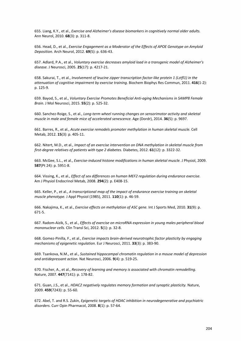
655. Liang, K.Y., et al., Exercise and Alzheimer's disease biomarkers in cognitively normal older adults. Ann Neurol, 2010. 68(3): p. 311‐8.
656. Head, D., et al., Exercise Engagement as a Moderator of the Effects of APOE Genotype on Amyloid Deposition. Arch Neurol, 2012. 69(5): p. 636‐43.
657. Adlard, P.A., et al., Voluntary exercise decreases amyloid load in a transgenic model of Alzheimer's disease. J Neurosci, 2005. 25(17): p. 4217‐21.
658. Sakurai, T., et al., Involvement of leucine zipper transcription factor‐like protein 1 (Lztfl1) in the attenuation of cognitive impairment by exercise training. Biochem Biophys Res Commun, 2011. 416(1‐2): p. 125‐9.
659. Bayod, S., et al., Voluntary Exercise Promotes Beneficial Anti‐aging Mechanisms in SAMP8 Female Brain. J Mol Neurosci, 2015. 55(2): p. 525‐32.
660. Sanchez‐Roige, S., et al., Long‐term wheel running changes on sensorimotor activity and skeletal muscle in male and female mice of accelerated senescence. Age (Dordr), 2014. 36(5): p. 9697.
661. Barres, R., et al., Acute exercise remodels promoter methylation in human skeletal muscle. Cell Metab, 2012. 15(3): p. 405‐11.
662. Nitert, M.D., et al., Impact of an exercise intervention on DNA methylation in skeletal muscle from first‐degree relatives of patients with type 2 diabetes. Diabetes, 2012. 61(12): p. 3322‐32.
663. McGee, S.L., et al., Exercise‐induced histone modifications in human skeletal muscle. J Physiol, 2009. 587(Pt 24): p. 5951‐8.
664. Vissing, K., et al., Effect of sex differences on human MEF2 regulation during endurance exercise. Am J Physiol Endocrinol Metab, 2008. 294(2): p. E408‐15.
665. Keller, P., et al., A transcriptional map of the impact of endurance exercise training on skeletal muscle phenotype. J Appl Physiol (1985), 2011. 110(1): p. 46‐59.
666. Nakajima, K., et al., Exercise effects on methylation of ASC gene. Int J Sports Med, 2010. 31(9): p. 671‐5.
667. Radom‐Aizik, S., et al., Effects of exercise on microRNA expression in young males peripheral blood mononuclear cells. Clin Transl Sci, 2012. 5(1): p. 32‐8.
668. Gomez‐Pinilla, F., et al., Exercise impacts brain‐derived neurotrophic factor plasticity by engaging mechanisms of epigenetic regulation. Eur J Neurosci, 2011. 33(3): p. 383‐90.
669. Tsankova, N.M., et al., Sustained hippocampal chromatin regulation in a mouse model of depression and antidepressant action. Nat Neurosci, 2006. 9(4): p. 519‐25.
670. Fischer, A., et al., Recovery of learning and memory is associated with chromatin remodelling. Nature, 2007. 447(7141): p. 178‐82.
671. Guan, J.S., et al., HDAC2 negatively regulates memory formation and synaptic plasticity. Nature, 2009. 459(7243): p. 55‐60.
672. Abel, T. and R.S. Zukin, Epigenetic targets of HDAC inhibition in neurodegenerative and psychiatric disorders. Curr Opin Pharmacol, 2008. 8(1): p. 57‐64.
204
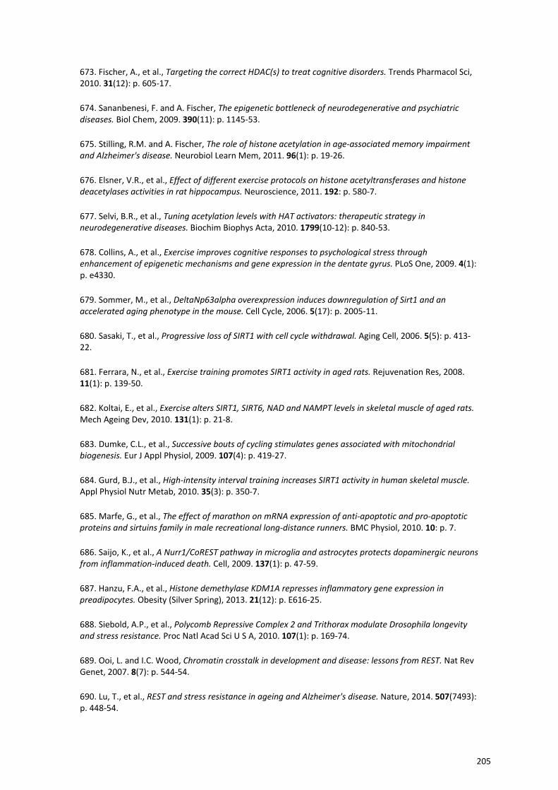
673. Fischer, A., et al., Targeting the correct HDAC(s) to treat cognitive disorders. Trends Pharmacol Sci, 2010. 31(12): p. 605‐17.
674. Sananbenesi, F. and A. Fischer, The epigenetic bottleneck of neurodegenerative and psychiatric diseases. Biol Chem, 2009. 390(11): p. 1145‐53.
675. Stilling, R.M. and A. Fischer, The role of histone acetylation in age‐associated memory impairment and Alzheimer's disease. Neurobiol Learn Mem, 2011. 96(1): p. 19‐26.
676. Elsner, V.R., et al., Effect of different exercise protocols on histone acetyltransferases and histone deacetylases activities in rat hippocampus. Neuroscience, 2011. 192: p. 580‐7.
677. Selvi, B.R., et al., Tuning acetylation levels with HAT activators: therapeutic strategy in neurodegenerative diseases. Biochim Biophys Acta, 2010. 1799(10‐12): p. 840‐53.
678. Collins, A., et al., Exercise improves cognitive responses to psychological stress through enhancement of epigenetic mechanisms and gene expression in the dentate gyrus. PLoS One, 2009. 4(1): p. e4330.
679. Sommer, M., et al., DeltaNp63alpha overexpression induces downregulation of Sirt1 and an accelerated aging phenotype in the mouse. Cell Cycle, 2006. 5(17): p. 2005‐11.
680. Sasaki, T., et al., Progressive loss of SIRT1 with cell cycle withdrawal. Aging Cell, 2006. 5(5): p. 413‐22.
681. Ferrara, N., et al., Exercise training promotes SIRT1 activity in aged rats. Rejuvenation Res, 2008. 11(1): p. 139‐50.
682. Koltai, E., et al., Exercise alters SIRT1, SIRT6, NAD and NAMPT levels in skeletal muscle of aged rats. Mech Ageing Dev, 2010. 131(1): p. 21‐8.
683. Dumke, C.L., et al., Successive bouts of cycling stimulates genes associated with mitochondrial biogenesis. Eur J Appl Physiol, 2009. 107(4): p. 419‐27.
684. Gurd, B.J., et al., High‐intensity interval training increases SIRT1 activity in human skeletal muscle. Appl Physiol Nutr Metab, 2010. 35(3): p. 350‐7.
685. Marfe, G., et al., The effect of marathon on mRNA expression of anti‐apoptotic and pro‐apoptotic proteins and sirtuins family in male recreational long‐distance runners. BMC Physiol, 2010. 10: p. 7.
686. Saijo, K., et al., A Nurr1/CoREST pathway in microglia and astrocytes protects dopaminergic neurons from inflammation‐induced death. Cell, 2009. 137(1): p. 47‐59.
687. Hanzu, F.A., et al., Histone demethylase KDM1A represses inflammatory gene expression in preadipocytes. Obesity (Silver Spring), 2013. 21(12): p. E616‐25.
688. Siebold, A.P., et al., Polycomb Repressive Complex 2 and Trithorax modulate Drosophila longevity and stress resistance. Proc Natl Acad Sci U S A, 2010. 107(1): p. 169‐74.
689. Ooi, L. and I.C. Wood, Chromatin crosstalk in development and disease: lessons from REST. Nat Rev Genet, 2007. 8(7): p. 544‐54.
690. Lu, T., et al., REST and stress resistance in ageing and Alzheimer's disease. Nature, 2014. 507(7493): p. 448‐54.
205
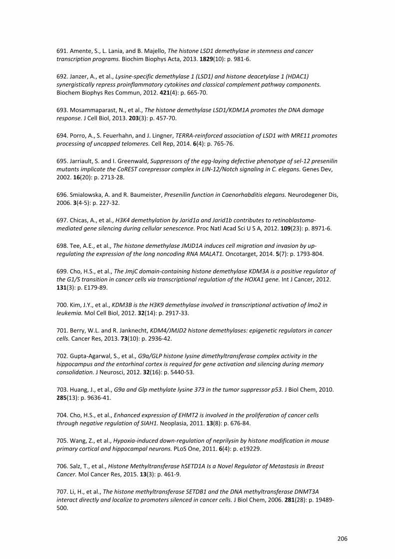
691. Amente, S., L. Lania, and B. Majello, The histone LSD1 demethylase in stemness and cancer transcription programs. Biochim Biophys Acta, 2013. 1829(10): p. 981‐6.
692. Janzer, A., et al., Lysine‐specific demethylase 1 (LSD1) and histone deacetylase 1 (HDAC1) synergistically repress proinflammatory cytokines and classical complement pathway components. Biochem Biophys Res Commun, 2012. 421(4): p. 665‐70.
693. Mosammaparast, N., et al., The histone demethylase LSD1/KDM1A promotes the DNA damage response. J Cell Biol, 2013. 203(3): p. 457‐70.
694. Porro, A., S. Feuerhahn, and J. Lingner, TERRA‐reinforced association of LSD1 with MRE11 promotes processing of uncapped telomeres. Cell Rep, 2014. 6(4): p. 765‐76.
695. Jarriault, S. and I. Greenwald, Suppressors of the egg‐laying defective phenotype of sel‐12 presenilin mutants implicate the CoREST corepressor complex in LIN‐12/Notch signaling in C. elegans. Genes Dev, 2002. 16(20): p. 2713‐28.
696. Smialowska, A. and R. Baumeister, Presenilin function in Caenorhabditis elegans. Neurodegener Dis, 2006. 3(4‐5): p. 227‐32.
697. Chicas, A., et al., H3K4 demethylation by Jarid1a and Jarid1b contributes to retinoblastoma‐mediated gene silencing during cellular senescence. Proc Natl Acad Sci U S A, 2012. 109(23): p. 8971‐6.
698. Tee, A.E., et al., The histone demethylase JMJD1A induces cell migration and invasion by up‐regulating the expression of the long noncoding RNA MALAT1. Oncotarget, 2014. 5(7): p. 1793‐804.
699. Cho, H.S., et al., The JmjC domain‐containing histone demethylase KDM3A is a positive regulator of the G1/S transition in cancer cells via transcriptional regulation of the HOXA1 gene. Int J Cancer, 2012. 131(3): p. E179‐89.
700. Kim, J.Y., et al., KDM3B is the H3K9 demethylase involved in transcriptional activation of lmo2 in leukemia. Mol Cell Biol, 2012. 32(14): p. 2917‐33.
701. Berry, W.L. and R. Janknecht, KDM4/JMJD2 histone demethylases: epigenetic regulators in cancer cells. Cancer Res, 2013. 73(10): p. 2936‐42.
702. Gupta‐Agarwal, S., et al., G9a/GLP histone lysine dimethyltransferase complex activity in the hippocampus and the entorhinal cortex is required for gene activation and silencing during memory consolidation. J Neurosci, 2012. 32(16): p. 5440‐53.
703. Huang, J., et al., G9a and Glp methylate lysine 373 in the tumor suppressor p53. J Biol Chem, 2010. 285(13): p. 9636‐41.
704. Cho, H.S., et al., Enhanced expression of EHMT2 is involved in the proliferation of cancer cells through negative regulation of SIAH1. Neoplasia, 2011. 13(8): p. 676‐84.
705. Wang, Z., et al., Hypoxia‐induced down‐regulation of neprilysin by histone modification in mouse primary cortical and hippocampal neurons. PLoS One, 2011. 6(4): p. e19229.
706. Salz, T., et al., Histone Methyltransferase hSETD1A Is a Novel Regulator of Metastasis in Breast Cancer. Mol Cancer Res, 2015. 13(3): p. 461‐9.
707. Li, H., et al., The histone methyltransferase SETDB1 and the DNA methyltransferase DNMT3A interact directly and localize to promoters silenced in cancer cells. J Biol Chem, 2006. 281(28): p. 19489‐500.
206
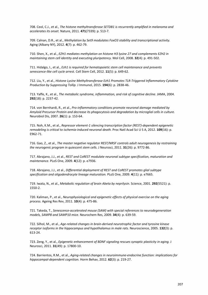
708. Ceol, C.J., et al., The histone methyltransferase SETDB1 is recurrently amplified in melanoma and accelerates its onset. Nature, 2011. 471(7339): p. 513‐7.
709. Calnan, D.R., et al., Methylation by Set9 modulates FoxO3 stability and transcriptional activity. Aging (Albany NY), 2012. 4(7): p. 462‐79.
710. Shen, X., et al., EZH1 mediates methylation on histone H3 lysine 27 and complements EZH2 in maintaining stem cell identity and executing pluripotency. Mol Cell, 2008. 32(4): p. 491‐502.
711. Hidalgo, I., et al., Ezh1 is required for hematopoietic stem cell maintenance and prevents senescence‐like cell cycle arrest. Cell Stem Cell, 2012. 11(5): p. 649‐62.
712. Liu, Y., et al., Histone Lysine Methyltransferase Ezh1 Promotes TLR‐Triggered Inflammatory Cytokine Production by Suppressing Tollip. J Immunol, 2015. 194(6): p. 2838‐46.
713. Yaffe, K., et al., The metabolic syndrome, inflammation, and risk of cognitive decline. JAMA, 2004. 292(18): p. 2237‐42.
714. von Bernhardi, R., et al., Pro‐inflammatory conditions promote neuronal damage mediated by Amyloid Precursor Protein and decrease its phagocytosis and degradation by microglial cells in culture. Neurobiol Dis, 2007. 26(1): p. 153‐64.
715. Noh, K.M., et al., Repressor element‐1 silencing transcription factor (REST)‐dependent epigenetic remodeling is critical to ischemia‐induced neuronal death. Proc Natl Acad Sci U S A, 2012. 109(16): p. E962‐71.
716. Gao, Z., et al., The master negative regulator REST/NRSF controls adult neurogenesis by restraining the neurogenic program in quiescent stem cells. J Neurosci, 2011. 31(26): p. 9772‐86.
717. Abrajano, J.J., et al., REST and CoREST modulate neuronal subtype specification, maturation and maintenance. PLoS One, 2009. 4(12): p. e7936.
718. Abrajano, J.J., et al., Differential deployment of REST and CoREST promotes glial subtype specification and oligodendrocyte lineage maturation. PLoS One, 2009. 4(11): p. e7665.
719. Iwata, N., et al., Metabolic regulation of brain Abeta by neprilysin. Science, 2001. 292(5521): p. 1550‐2.
720. Kaliman, P., et al., Neurophysiological and epigenetic effects of physical exercise on the aging process. Ageing Res Rev, 2011. 10(4): p. 475‐86.
721. Takeda, T., Senescence‐accelerated mouse (SAM) with special references to neurodegeneration models, SAMP8 and SAMP10 mice. Neurochem Res, 2009. 34(4): p. 639‐59.
722. Silhol, M., et al., Age‐related changes in brain‐derived neurotrophic factor and tyrosine kinase receptor isoforms in the hippocampus and hypothalamus in male rats. Neuroscience, 2005. 132(3): p. 613‐24.
723. Zeng, Y., et al., Epigenetic enhancement of BDNF signaling rescues synaptic plasticity in aging. J Neurosci, 2011. 31(49): p. 17800‐10.
724. Barrientos, R.M., et al., Aging‐related changes in neuroimmune‐endocrine function: implications for hippocampal‐dependent cognition. Horm Behav, 2012. 62(3): p. 219‐27.
207
725. Burnouf, S., et al., Hippocampal BDNF expression in a tau transgenic mouse model. Curr Alzheimer Res, 2012. 9(4): p. 406‐10.
726. Nagahara, A.H. and M.H. Tuszynski, Potential therapeutic uses of BDNF in neurological and psychiatric disorders. Nat Rev Drug Discov, 2011. 10(3): p. 209‐19.
727. Bonneh‐Barkay, D. and C.A. Wiley, Brain extracellular matrix in neurodegeneration. Brain Pathol, 2009. 19(4): p. 573‐85.
728. Caltagarone, J., Z. Jing, and R. Bowser, Focal adhesions regulate Abeta signaling and cell death in Alzheimer's disease. Biochim Biophys Acta, 2007. 1772(4): p. 438‐45.
729. Akbarian, S., M.S. Beeri, and V. Haroutunian, Epigenetic determinants of healthy and diseased brain aging and cognition. JAMA Neurol, 2013. 70(6): p. 711‐8.
730. Van den Hove, D.L., et al., Epigenetically regulated microRNAs in Alzheimer's disease. Neurobiol Aging, 2014. 35(4): p. 731‐45.
731. Jung, H.J. and Y. Suh, MicroRNA in Aging: From Discovery to Biology. Curr Genomics, 2012. 13(7): p. 548‐57.
732. Mastroeni, D., et al., Epigenetic mechanisms in Alzheimer's disease. Neurobiol Aging, 2011. 32(7): p. 1161‐80.
733. Huidobro, C., A.F. Fernandez, and M.F. Fraga, Aging epigenetics: causes and consequences. Mol Aspects Med, 2013. 34(4): p. 765‐81.
734. Tammen, S.A., S. Friso, and S.W. Choi, Epigenetics: the link between nature and nurture. Mol Aspects Med, 2013. 34(4): p. 753‐64.
735. Di Bernardo, G., M. Cipollaro, and U. Galderisi, Chromatin modification and senescence. Curr Pharm Des, 2012. 18(13): p. 1686‐93.
736. Zhou, S. and J. Zhou, Neuritin, a neurotrophic factor in nervous system physiology. Curr Med Chem, 2014. 21(10): p. 1212‐9.
737. Lyman, M., et al., Neuroinflammation: the role and consequences. Neurosci Res, 2014. 79: p. 1‐12.
738. Jana, M., C.A. Palencia, and K. Pahan, Fibrillar amyloid‐beta peptides activate microglia via TLR2: implications for Alzheimer's disease. J Immunol, 2008. 181(10): p. 7254‐62.
739. Andres, M.E., et al., CoREST: a functional corepressor required for regulation of neural‐specific gene expression. Proc Natl Acad Sci U S A, 1999. 96(17): p. 9873‐8.
740. Gonzalez, H., et al., Neuroimmune regulation of microglial activity involved in neuroinflammation and neurodegenerative diseases. J Neuroimmunol, 2014. 274(1‐2): p. 1‐13.
741. Cheng, X.R., et al., Differential gene expression profiles in the hippocampus of senescence‐accelerated mouse. Neurobiol Aging, 2007. 28(4): p. 497‐506.
742. Kozarova, A., J.W. Hudson, and P.O. Vacratsis, The dual‐specificity phosphatase hYVH1 (DUSP12) is a novel modulator of cellular DNA content. Cell Cycle, 2011. 10(10): p. 1669‐78.
208
743. Casadesus, G., et al., Neuronal cell cycle re‐entry markers are altered in the senescence accelerated mouse P8 (SAMP8). J Alzheimers Dis, 2012. 30(3): p. 573‐83.
744. Kim, K.Y., et al., Parkin is a lipid‐responsive regulator of fat uptake in mice and mutant human cells. J Clin Invest, 2011. 121(9): p. 3701‐12.
745. Wang, W., et al., Delayed neuronal death and damage of GDNF family receptors in CA1 following focal cerebral ischemia. Brain Res, 2004. 1023(1): p. 92‐101.
746. Bourasset, F., et al., Reduction of the cerebrovascular volume in a transgenic mouse model of Alzheimer's disease. Neuropharmacology, 2009. 56(4): p. 808‐13.
747. Hashimoto, T., et al., CLAC: a novel Alzheimer amyloid plaque component derived from a transmembrane precursor, CLAC‐P/collagen type XXV. EMBO J, 2002. 21(7): p. 1524‐34.
748. van Horssen, J., et al., Collagen XVIII: a novel heparan sulfate proteoglycan associated with vascular amyloid depositions and senile plaques in Alzheimer's disease brains. Brain Pathol, 2002. 12(4): p. 456‐62.
749. Colin, C., et al., Identification of genes differentially expressed in glioblastoma versus pilocytic astrocytoma using Suppression Subtractive Hybridization. Oncogene, 2006. 25(19): p. 2818‐26.
750. Ariga, T., T. Miyatake, and R.K. Yu, Role of proteoglycans and glycosaminoglycans in the pathogenesis of Alzheimer's disease and related disorders: amyloidogenesis and therapeutic strategies‐‐a review. J Neurosci Res, 2010. 88(11): p. 2303‐15.
751. Urade, Y. and O. Hayaishi, Biochemical, structural, genetic, physiological, and pathophysiological features of lipocalin‐type prostaglandin D synthase. Biochim Biophys Acta, 2000. 1482(1‐2): p. 259‐71.
752. Chen, C.P., R.L. Chen, and J.E. Preston, Age‐related increase of prostaglandin D(2) synthase concentration and glycation in ovine cerebrospinal fluid. Exp Gerontol, 2009. 44(10): p. 639‐45.
753. Jacobs, S., et al., Retinoic acid is required early during adult neurogenesis in the dentate gyrus. Proc Natl Acad Sci U S A, 2006. 103(10): p. 3902‐7.
754. Hsiao, Y.H., et al., Amelioration of social isolation‐triggered onset of early Alzheimer's disease‐related cognitive deficit by N‐acetylcysteine in a transgenic mouse model. Neurobiol Dis, 2012. 45(3): p. 1111‐20.
755. Paris, D., et al., Impaired angiogenesis in a transgenic mouse model of cerebral amyloidosis. Neurosci Lett, 2004. 366(1): p. 80‐5.
756. Paris, D., et al., Inhibition of angiogenesis by Abeta peptides. Angiogenesis, 2004. 7(1): p. 75‐85.
757. Desai, B.S., et al., Evidence of angiogenic vessels in Alzheimer's disease. J Neural Transm, 2009. 116(5): p. 587‐97.
758. Latimer, C.S., et al., Reversal of glial and neurovascular markers of unhealthy brain aging by exercise in middle‐aged female mice. PLoS One, 2011. 6(10): p. e26812.
759. Zhang, F., et al., Roles of brain‐derived neurotrophic factor/tropomyosin‐related kinase B (BDNF/TrkB) signalling in Alzheimer's disease. J Clin Neurosci, 2012. 19(7): p. 946‐9.
209
760. Cassilhas, R.C., et al., Spatial memory is improved by aerobic and resistance exercise through divergent molecular mechanisms. Neuroscience, 2012. 202: p. 309‐17.
761. An, K., et al., Neuritin can normalize neural deficits of Alzheimer's disease. Cell Death Dis, 2014. 5: p. e1523.
762. Abel, J.L. and E.F. Rissman, Running‐induced epigenetic and gene expression changes in the adolescent brain. Int J Dev Neurosci, 2013. 31(6): p. 382‐90.
763. Lovatel, G.A., et al., Treadmill exercise induces age‐related changes in aversive memory, neuroinflammatory and epigenetic processes in the rat hippocampus. Neurobiol Learn Mem, 2013. 101: p. 94‐102.
764. Cogswell, J.P., et al., Identification of miRNA changes in Alzheimer's disease brain and CSF yields putative biomarkers and insights into disease pathways. J Alzheimers Dis, 2008. 14(1): p. 27‐41.
765. Maes, O.C., et al., MicroRNA: Implications for Alzheimer Disease and other Human CNS Disorders. Curr Genomics, 2009. 10(3): p. 154‐68.
766. Mallick, B. and Z. Ghosh, A complex crosstalk between polymorphic microRNA target sites and AD prognosis. RNA Biol, 2011. 8(4): p. 665‐73.
767. Li, Y., M. Daniel, and T.O. Tollefsbol, Epigenetic regulation of caloric restriction in aging. BMC Med, 2011. 9: p. 98.
768. Park, S., R. Mori, and I. Shimokawa, Do sirtuins promote mammalian longevity? A critical review on its relevance to the longevity effect induced by calorie restriction. Mol Cells, 2013. 35(6): p. 474‐80.
769. Rizza, W., N. Veronese, and L. Fontana, What are the roles of calorie restriction and diet quality in promoting healthy longevity? Ageing Res Rev, 2014. 13: p. 38‐45.
770. Weiss, E.P. and L. Fontana, Caloric restriction: powerful protection for the aging heart and vasculature. Am J Physiol Heart Circ Physiol, 2011. 301(4): p. H1205‐19.
771. Goel, A. and B.B. Aggarwal, Curcumin, the golden spice from Indian saffron, is a chemosensitizer and radiosensitizer for tumors and chemoprotector and radioprotector for normal organs. Nutr Cancer, 2010. 62(7): p. 919‐30.
772. Ravindran, J., S. Prasad, and B.B. Aggarwal, Curcumin and cancer cells: how many ways can curry kill tumor cells selectively? AAPS J, 2009. 11(3): p. 495‐510.
773. Shanmugam, M.K., R. Kannaiyan, and G. Sethi, Targeting cell signaling and apoptotic pathways by dietary agents: role in the prevention and treatment of cancer. Nutr Cancer, 2011. 63(2): p. 161‐73.
774. Shukla, Y. and R. Singh, Resveratrol and cellular mechanisms of cancer prevention. Ann N Y Acad Sci, 2011. 1215: p. 1‐8.
775. Cheung, K.L. and A.N. Kong, Molecular targets of dietary phenethyl isothiocyanate and sulforaphane for cancer chemoprevention. AAPS J, 2010. 12(1): p. 87‐97.
776. Keum, Y.S., et al., Pharmacokinetics and pharmacodynamics of broccoli sprouts on the suppression of prostate cancer in transgenic adenocarcinoma of mouse prostate (TRAMP) mice: implication of induction of Nrf2, HO‐1 and apoptosis and the suppression of Akt‐dependent kinase pathway. Pharm Res, 2009. 26(10): p. 2324‐31.
210
777. Uauy, R. and A.D. Dangour, Nutrition in brain development and aging: role of essential fatty acids. Nutr Rev, 2006. 64(5 Pt 2): p. S24‐33; discussion S72‐91.
778. Bouwens, M., et al., Fish‐oil supplementation induces antiinflammatory gene expression profiles in human blood mononuclear cells. Am J Clin Nutr, 2009. 90(2): p. 415‐24.
779. Nithianantharajah, J. and A.J. Hannan, Enriched environments, experience‐dependent plasticity and disorders of the nervous system. Nat Rev Neurosci, 2006. 7(9): p. 697‐709.
780. Weaver, I.C., et al., Epigenetic programming by maternal behavior. Nat Neurosci, 2004. 7(8): p. 847‐54.
781. Kundakovic, M. and F.A. Champagne, Early‐life experience, epigenetics, and the developing brain. Neuropsychopharmacology, 2015. 40(1): p. 141‐53.
782. Crews, D., et al., Nature, nurture and epigenetics. Mol Cell Endocrinol, 2014. 398(1‐2): p. 42‐52.
783. Farb, N.A., A.K. Anderson, and Z.V. Segal, The mindful brain and emotion regulation in mood disorders. Can J Psychiatry, 2012. 57(2): p. 70‐7.
784. Ludwig, D.S. and J. Kabat‐Zinn, Mindfulness in medicine. JAMA, 2008. 300(11): p. 1350‐2.
785. Rosenkranz, M.A., et al., A comparison of mindfulness‐based stress reduction and an active control in modulation of neurogenic inflammation. Brain Behav Immun, 2013. 27(1): p. 174‐84.
786. Schmidt, S., et al., Treating fibromyalgia with mindfulness‐based stress reduction: results from a 3‐armed randomized controlled trial. Pain, 2011. 152(2): p. 361‐9.
787. Kaliman, P., et al., Rapid changes in histone deacetylases and inflammatory gene expression in expert meditators. Psychoneuroendocrinology, 2014. 40: p. 96‐107.
211