Cor Triatriatum: Preoperative Diagnosis and Successful ...
3
The Journal of Tehran University Heart Center 153 TEHRAN HEART CENTER Cor Triatriatum: Preoperative Diagnosis and Successful Surgical Correction in a Four-Year-Old Girl * Corresponding Author: Afsaneh Sadeghian, Shahrood University of Medical Sciences, Fatemieh Hospital, Shahrood, Iran. 3615741151. Tel: +98 273 2224091. Fax: +98 273 2227607. E-mail: [email protected]. Case Report Kiyomars Abbasi, MD 1 , Hakimeh Sadeghian, MD 1 , Neda Ghafari-Marandi, MD 1 , Afsaneh Sadeghian, MD 2* 1 Tehran Heart Center, Tehran University of Medical Sciences, Tehran, Iran. 2 Fatemieh Hospital, Shahrood University of Medical Sciences, Shahrood, Iran. Abstract Cor triatriatum is defined as a membrane within the left atrium, which might lead to restricted pulmonary venous return. Diagnosis is usually achieved by echocardiography in early infancy. Therapy of choice is the excision of the membrane. Herein, successful correction of cor triatriatum in a 4-year-old girl is presented, and the clinical features, echocardiographic findings, and the surgical treatment are discussed. Introduction Cor triatriatum is one of the most sparse congenital cardiac anomalies, accounting for approximately 0.1% of all congenital heart diseases. 1, 2 A classic form of cor triatriatum may be defined as the left atrium subdivided into dorsal and ventral chambers by a fibromuscular diaphragm; the dorsal chamber received the pulmonary veins and the ventral chamber was attached to the left atrial appendage and led to the mitral valve (Figure 1). The communication between the dorsal and ventral chambers is variable or absent and depends on the size of the opening in the fibromuscular “membrane”. 3, 4 Approximately in 75% of individuals, there exists an atrial septal defect (ASD) between the right atrium and the proximal venous chamber (subtype A1 according to the Lam classification) or the true distal atrium, as was the case in our patient. 5, 6 Figure 1. Schematic diagram of intraoperative finding of nearly atretic cor triatriatum and atrial septal defect RA, Right atrium; ASD, Atrial septal defect; DC, Distal chamber; PV, Pul- monary veins entrance; PC, Proximal chamber; FM, Fibromuscular mem- brane; MV, Mitral valve; LV, Left ventricle; RV, Right ventricle Keywords: Heart defects, congenital • Cor triatriatum • Echocardiography Received 16 May 2009; Accepted 23 February 2010 J Teh Univ Heart Ctr 3 (2010) 153-155
Transcript of Cor Triatriatum: Preoperative Diagnosis and Successful ...
The Journal of Tehran University Heart Center153
TEHRAN HEART CENTER
Cor Triatriatum: Preoperative Diagnosis and SuccessfulSurgical Correction in a Four-Year-Old Girl
*Corresponding Author: Afsaneh Sadeghian, Shahrood University of Medical Sciences, Fatemieh Hospital, Shahrood, Iran. 3615741151. Tel: +98 273 2224091. Fax: +98 273 2227607. E-mail: [email protected].
Case Report
Kiyomars Abbasi, MD1, Hakimeh Sadeghian, MD1, Neda Ghafari-Marandi, MD1, Afsaneh Sadeghian, MD2*
1Tehran Heart Center, Tehran University of Medical Sciences, Tehran, Iran.2Fatemieh Hospital, Shahrood University of Medical Sciences, Shahrood, Iran.
Abstract
Cor triatriatum is defined as a membrane within the left atrium, which might lead to restricted pulmonary venous return. Diagnosis is usually achieved by echocardiography in early infancy. Therapy of choice is the excision of the membrane. Herein, successful correction of cor triatriatum in a 4-year-old girl is presented, and the clinical features, echocardiographic findings, and the surgical treatment are discussed.
Introduction
Cor triatriatum is one of the most sparse congenital cardiac anomalies, accounting for approximately 0.1% of all congenital heart diseases.1, 2 A classic form of cor triatriatum may be defined as the left atrium subdivided into dorsal and ventral chambers by a fibromuscular diaphragm; the dorsal chamber received the pulmonary veins and the ventral chamber was attached to the left atrial appendage and led to the mitral valve (Figure 1). The communication between the dorsal and ventral chambers is variable or absent and depends on the size of the opening in the fibromuscular “membrane”.3, 4
Approximately in 75% of individuals, there exists an atrial septal defect (ASD) between the right atrium and the proximal venous chamber (subtype A1 according to the Lam classification) or the true distal atrium, as was the case in our patient.5, 6
Figure 1. Schematic diagram of intraoperative finding of nearly atretic cor triatriatum and atrial septal defectRA, Right atrium; ASD, Atrial septal defect; DC, Distal chamber; PV, Pul-monary veins entrance; PC, Proximal chamber; FM, Fibromuscular mem-brane; MV, Mitral valve; LV, Left ventricle; RV, Right ventricle
Keywords: Heart defects, congenital • Cor triatriatum • Echocardiography
Received 16 May 2009; Accepted 23 February 2010
J Teh Univ Heart Ctr 3 (2010) 153-155
154
The Journal of Tehran University Heart Center
The clinical symptoms depend on the size of the orifice (the classic case mimicking mitral stenosis) and the morphology of the defect, including the presence and location of the ASD and associated congenital heart disease. The diagnosis of cor triatriatum should be considered in patients with pulmonary congestion, pulmonary arterial hypertension, and pulmonary venous obstruction. Symptoms are usually not related to a specific anatomic subtype and comprise failure to thrive, tachypnea, dyspnea (as was the case in our patient), and occasionally, cyanosis. The latter can be caused by either the restricted pulmonary venous return (then associated with severe pulmonary symptoms like tachydyspnea) or the presence of severe stenosis and ASD; a right-to-left shunt might be present at atrial level.7, 8
Previous studies have reported that cor triatriatum is usually diagnosed and surgically treated before 24 months old. Herein, we present a case of cor triatriatum diagnosed in a 4-year-old girl.
Case report
The patient was a 4-year-old girl, who was the fifth child of her family. Pregnancy and delivery were normal, birth weight was eleven pounds, the immediate postnatal period was event-free, and she was breastfed for two years. The patient was first hospitalized for one week at 2 months old due to severe respiratory tract infection. A diagnosis of congenital heart disease was made after a thorough examination, and treatment was offered for later.
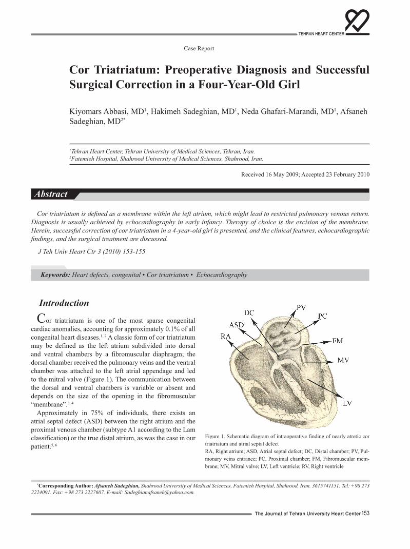
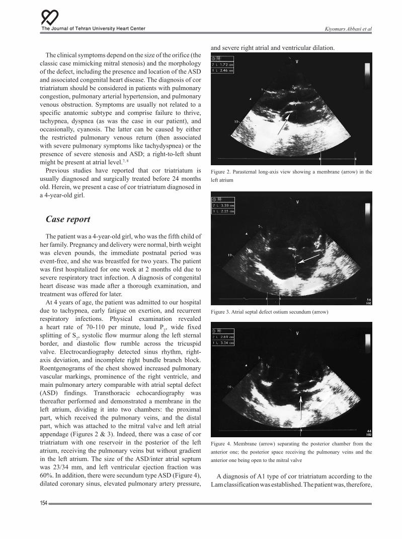
At 4 years of age, the patient was admitted to our hospital due to tachypnea, early fatigue on exertion, and recurrent respiratory infections. Physical examination revealed a heart rate of 70-110 per minute, loud P2, wide fixed splitting of S2, systolic flow murmur along the left sternal border, and diastolic flow rumble across the tricuspid valve. Electrocardiography detected sinus rhythm, right-axis deviation, and incomplete right bundle branch block. Roentgenograms of the chest showed increased pulmonary vascular markings, prominence of the right ventricle, and main pulmonary artery comparable with atrial septal defect (ASD) findings. Transthoracic echocardiography was thereafter performed and demonstrated a membrane in the left atrium, dividing it into two chambers: the proximal part, which received the pulmonary veins, and the distal part, which was attached to the mitral valve and left atrial appendage (Figures 2 & 3). Indeed, there was a case of cor triatriatum with one reservoir in the posterior of the left atrium, receiving the pulmonary veins but without gradient in the left atrium. The size of the ASD/inter atrial septum was 23/34 mm, and left ventricular ejection fraction was 60%. In addition, there were secundum type ASD (Figure 4), dilated coronary sinus, elevated pulmonary artery pressure,
and severe right atrial and ventricular dilation.
Figure 2. Parasternal long-axis view showing a membrane (arrow) in the left atrium
Figure 3. Atrial septal defect ostium secundum (arrow)
Figure 4. Membrane (arrow) separating the posterior chamber from the anterior one; the posterior space receiving the pulmonary veins and the anterior one being open to the mitral valve
A diagnosis of A1 type of cor triatriatum according to the Lam classification was established. The patient was, therefore,
Kiyomars Abbasi et al
TEHRAN HEART CENTER
The Journal of Tehran University Heart Center155
transferred to the operating room. The heart was approached through a median sternotomy incision. Cardiopulmonary bypass (CPB) was initiated, and the aortic crass-clamp was performed under mild systemic hypothermia and cold blood cardioplegia. The cor triatriatum membrane was thick with one orifice measuring about 2 mm; it was resected via a transseptal approach. The ASD (8 mm in diameter) was closed using a pericardial patch. The postoperative cardiac echocardiogram showed the status/post (S/P) ASD closure, the repair/removal of the cor triatriatum, normal left ventricle size and function, severe right ventricle enlargement with mild right ventricular hypertrophy and good function, mild tricuspid regurgitation, mild pulmonary insufficiency, and trivial pericardial effusion Additionally, there was no residual ASD or pulmonary stenosis or left atrium gradient. The postoperative course was event-free, and the patient was discharged on the 4th postoperative day.
Discussion
The youngest cor triatriatum case was an 18-day-old patient who also had infracardiac total anomalous pulmonary venous drainage in the Baek MJ et al. study1 and the oldest case was a 93-year-old patient in the Arciniegas E et al. study.9
In the Alphonso study 61% of the patients had ASD (Lam type A1/A2).5 Arciniegas and Richardson et al. reported 83% and 95% of ASD with cor triatriatum.9, 10 In the Krasemann et al. study, and there were two adult patients with an additional ASD, which was thermodynamically insignificant. In one of them, the closure of the defect was favored after a stroke to prevent possible right-to-left embolic events, and the membrane was resected during ASD repair. The other one had non-specific symptoms (decreased exercise tolerance), leading to diagnostic and, subsequently, surgical procedures.7
The treatment of cor triatriatum is primarily surgical. There are only two reports of successful balloon catheter dilation of the communication between the proximal and distal chambers, and the follow-up periods for balloon dilatation are generally 3 and 12 months. The long-term outcome of balloon dilatation remains to be determined.5
According to the Alphonso study, the most current complications after surgery were supraventricular tachycardia and peritoneal dialysis.5 Other complications were seizure, pleural effusion requiring drainage, urinary tract infection, pyrexia of unknown origin, ventricular tachycardia, cerebrovascular accident, pneumothorax requiring drainage, acute renal failure, superficial wound infection requiring debridement, and pericardial effusion requiring drainage, which were seen in 10 (40%) patients. Seventeen (60%) patients made an uncomplicated recovery.5
Conclusion
In brief, cor triatriatum is a rare congenital cardiac anom-aly. Surgical correction offers good early and long-term re-sults for both classic and atypical cases of cor triatriatum.5
References
Baek MJ, Kim W, Young Na C, Se Oh S, Cheol Kim S, Young 1. Lee J, Bin Jeon Y, Ki lee S, Lee Ch, Woong Lee J, Sung kim W, Tak Lee Y, Park Y, Kim Ch. Cor triatriatum with infracardiac total anomalous pulmonary venous drainage. Korean Thorac Cardiovasc Surg 2002;35:52-55.Lim CW, Yip WC, Quek SC. Cor triatriatum and partial 2. atrioventricular septal defect. Pediatr Cardiol 2007;28:72-73.Arrants JE, Riopel DA, Catalano PW. Cor triatriatum: preoperative 3. diagnosis and successful surgical correction in a ten-week-old infant. Chest 1973;63:1027-1028.Takeuchi Y, Kurogane K, Nishimura Y, Kajiura T, Nakata H. 4. Asymptomatic Cor Triatrium in an elderly patient-observation by biplanar transesophageal Doppler echocardiography. Jpn Circ J 1997;61:189-191.Alphonso N, Nørgaard MA, Newcomb A, d’Udekem Y, Brizard 5. CP, Cochrane A. Cor Triatrium: presentation, diagnosis and long-term surgical results. Ann Thorac Surg 2005;80:1666-1671.Lâcis A, Zîdere V,Lubaua, Straume Z, Auziòð J,Đmits L, Lâce 6. I. Cor Triatrium associated with complex heart defects. ACTA Medica Lituanica 2004;11:59-63.Krasemann Z, Scheld HH, Tjan TD, Krasemann T. Cor triatriatum: 7. short review of the literature upon ten new cases. Herz 2007;32:506-510.Huang YK, Chu JJ, Chang JP, Lu MS, Tseng CN, Chang YS, 8. Tsai FC, Lin PJ. Cor triatriatum sinistrum: surgical experience in Taiwan. Surg Today 2007;37:449-454.Arciniegas E, Farooki ZQ, Hakimi M, Perry BL, Green EW. Surgical 9. treatment of cor triatriatum. Ann Thorac Surg 1981;32:571-577.Richardson JV, Doty DB, Siewers RD, Zuberbuhler JR. Cor 10. triatriatum (subdivided left atrium). J Thorac Cardiovasc Surg 1981;81:232-238.
Cor Triatriatum: Preoperative Diagnosis and Successful Surgical Correction ...


![Mapping with an Autonomous Carstachnis/pdf/lamon06iros.pdfIn the context of autonomous cars, a series of successful systems [45, 3, 48] have been developed for the DARPA Gran Challenge](https://static.fdocuments.ec/doc/165x107/5ec924ecc063fe57f13877a9/mapping-with-an-autonomous-stachnispdflamon06irospdf-in-the-context-of-autonomous.jpg)