Congruent microbiome signatures in fibrosis-prone ...
14
RESEARCH Open Access Congruent microbiome signatures in fibrosis-prone autoimmune diseases: IgG4- related disease and systemic sclerosis Damian R. Plichta 1 , Juhi Somani 2 , Matthieu Pichaud 3 , Zachary S. Wallace 4,5 , Ana D. Fernandes 4 , Cory A. Perugino 4,6 , Harri Lähdesmäki 2 , John H. Stone 4 , Hera Vlamakis 1 , Daniel C. Chung 7,8 , Dinesh Khanna 9 , Shiv Pillai 6 and Ramnik J. Xavier 1,10,11,12* Abstract Background: Immunoglobulin G4-related disease (IgG4-RD) and systemic sclerosis (SSc) are rare autoimmune diseases characterized by the presence of CD4+ cytotoxic T cells in the blood as well as inflammation and fibrosis in various organs, but they have no established etiologies. Similar to other autoimmune diseases, the gut microbiome might encode disease-triggering or disease-sustaining factors. Methods: The gut microbiomes from IgG4-RD and SSc patients as well as healthy individuals with no recent antibiotic treatment were studied by metagenomic sequencing of stool DNA. De novo assembly-based taxonomic and functional characterization, followed by association and accessory gene set enrichment analysis, were applied to describe microbiome changes associated with both diseases. Results: Microbiomes of IgG4-RD and SSc patients distinctly separated from those of healthy controls: numerous opportunistic pathogenic Clostridium and typically oral Streptococcus species were significantly overabundant, while Alistipes, Bacteroides, and butyrate-producing species were depleted in the two diseases compared to healthy controls. Accessory gene content analysis in these species revealed an enrichment of Th17-activating Eggerthella lenta strains in IgG4-RD and SSc and a preferential colonization of a homocysteine-producing strain of Clostridium bolteae in SSc. Overabundance of the classical mevalonate pathway, hydroxyproline dehydratase, and fibronectin-binding protein in disease microbiomes reflects potential functional differences in host immune recognition and extracellular matrix utilization associated with fibrosis. Strikingly, the majority of species that were differentially abundant in IgG4-RD and SSc compared to controls showed the same directionality in both diseases. Compared with multiple sclerosis and rheumatoid arthritis, the gut microbiomes of IgG4-RD and SSc showed similar signatures; in contrast, the most differentially abundant taxa were not the facultative anaerobes consistently identified in inflammatory bowel diseases, suggesting the microbial signatures of IgG4-RD and SSc do not result from mucosal inflammation and decreased anaerobism. Conclusions: These results provide an initial characterization of gut microbiome ecology in fibrosis-prone IgG4-RD and SSc and reveal microbial functions that offer insights into the pathophysiology of these rare diseases. Keywords: Gut microbiome, IgG4-RD, Systemic sclerosis, Autoimmunity © The Author(s). 2021 Open Access This article is licensed under a Creative Commons Attribution 4.0 International License, which permits use, sharing, adaptation, distribution and reproduction in any medium or format, as long as you give appropriate credit to the original author(s) and the source, provide a link to the Creative Commons licence, and indicate if changes were made. The images or other third party material in this article are included in the article's Creative Commons licence, unless indicated otherwise in a credit line to the material. If material is not included in the article's Creative Commons licence and your intended use is not permitted by statutory regulation or exceeds the permitted use, you will need to obtain permission directly from the copyright holder. To view a copy of this licence, visit http://creativecommons.org/licenses/by/4.0/. The Creative Commons Public Domain Dedication waiver (http://creativecommons.org/publicdomain/zero/1.0/) applies to the data made available in this article, unless otherwise stated in a credit line to the data. * Correspondence: [email protected] 1 Broad Institute of MIT and Harvard, Cambridge, MA, USA 10 Center for Computational and Integrative Biology, Massachusetts General Hospital and Harvard Medical School, Boston, MA, USA Full list of author information is available at the end of the article Plichta et al. Genome Medicine (2021) 13:35 https://doi.org/10.1186/s13073-021-00853-7
Transcript of Congruent microbiome signatures in fibrosis-prone ...
RESEARCH Open Access
Congruent microbiome signatures infibrosis-prone autoimmune diseases: IgG4-related disease and systemic sclerosisDamian R. Plichta1, Juhi Somani2, Matthieu Pichaud3, Zachary S. Wallace4,5, Ana D. Fernandes4, Cory A. Perugino4,6,Harri Lähdesmäki2, John H. Stone4, Hera Vlamakis1, Daniel C. Chung7,8, Dinesh Khanna9, Shiv Pillai6 andRamnik J. Xavier1,10,11,12*
Abstract
Background: Immunoglobulin G4-related disease (IgG4-RD) and systemic sclerosis (SSc) are rare autoimmunediseases characterized by the presence of CD4+ cytotoxic T cells in the blood as well as inflammation and fibrosisin various organs, but they have no established etiologies. Similar to other autoimmune diseases, the gutmicrobiome might encode disease-triggering or disease-sustaining factors.
Methods: The gut microbiomes from IgG4-RD and SSc patients as well as healthy individuals with no recentantibiotic treatment were studied by metagenomic sequencing of stool DNA. De novo assembly-based taxonomicand functional characterization, followed by association and accessory gene set enrichment analysis, were appliedto describe microbiome changes associated with both diseases.
Results: Microbiomes of IgG4-RD and SSc patients distinctly separated from those of healthy controls: numerousopportunistic pathogenic Clostridium and typically oral Streptococcus species were significantly overabundant, whileAlistipes, Bacteroides, and butyrate-producing species were depleted in the two diseases compared to healthy controls.Accessory gene content analysis in these species revealed an enrichment of Th17-activating Eggerthella lenta strains inIgG4-RD and SSc and a preferential colonization of a homocysteine-producing strain of Clostridium bolteae in SSc.Overabundance of the classical mevalonate pathway, hydroxyproline dehydratase, and fibronectin-binding protein indisease microbiomes reflects potential functional differences in host immune recognition and extracellular matrixutilization associated with fibrosis. Strikingly, the majority of species that were differentially abundant in IgG4-RD and SSccompared to controls showed the same directionality in both diseases. Compared with multiple sclerosis and rheumatoidarthritis, the gut microbiomes of IgG4-RD and SSc showed similar signatures; in contrast, the most differentially abundanttaxa were not the facultative anaerobes consistently identified in inflammatory bowel diseases, suggesting the microbialsignatures of IgG4-RD and SSc do not result from mucosal inflammation and decreased anaerobism.
Conclusions: These results provide an initial characterization of gut microbiome ecology in fibrosis-prone IgG4-RD andSSc and reveal microbial functions that offer insights into the pathophysiology of these rare diseases.
Keywords: Gut microbiome, IgG4-RD, Systemic sclerosis, Autoimmunity
© The Author(s). 2021 Open Access This article is licensed under a Creative Commons Attribution 4.0 International License,which permits use, sharing, adaptation, distribution and reproduction in any medium or format, as long as you giveappropriate credit to the original author(s) and the source, provide a link to the Creative Commons licence, and indicate ifchanges were made. The images or other third party material in this article are included in the article's Creative Commonslicence, unless indicated otherwise in a credit line to the material. If material is not included in the article's Creative Commonslicence and your intended use is not permitted by statutory regulation or exceeds the permitted use, you will need to obtainpermission directly from the copyright holder. To view a copy of this licence, visit http://creativecommons.org/licenses/by/4.0/.The Creative Commons Public Domain Dedication waiver (http://creativecommons.org/publicdomain/zero/1.0/) applies to thedata made available in this article, unless otherwise stated in a credit line to the data.
* Correspondence: [email protected] Institute of MIT and Harvard, Cambridge, MA, USA10Center for Computational and Integrative Biology, Massachusetts GeneralHospital and Harvard Medical School, Boston, MA, USAFull list of author information is available at the end of the article
Plichta et al. Genome Medicine (2021) 13:35 https://doi.org/10.1186/s13073-021-00853-7
BackgroundThe gut microbiome exists in an essential symbiosis withits human host by serving as a source of nutrients andsmall molecules, informing the development and activityof the immune system and providing colonization resist-ance against pathogens. Strains of gut bacteria stimulatethe expansion of various immune cell populations [1] andprovide signals for orchestrated anti- and pro-inflammatoryresponses locally and systemically [2]. Dysregulation of thissymbiosis through expansion or depletion of specific taxaand their associated proteins and metabolic capabilities hasbeen correlated with multiple diseases. In inflammatorybowel diseases (IBD) and rheumatoid arthritis (RA), micro-biome studies yielded potential clues to the identity ofspecific microbes (e.g., Ruminococcus gnavus, Prevotellacopri) that may act as sources of disease-triggering ordisease-sustaining molecules and antigens.Microbial antigens are recognized by the immune
system via presentation on major histocompatibilitycomplex (MHC) class II molecules to CD4+ T cells.Recently, an unusual subset of cytotoxic CD4+ T cellshas been described in patients with two rare, fibrosis-proneautoimmune disorders: immunoglobulin G4-related disease(IgG4-RD) and systemic sclerosis (SSc) [3–5]. IgG4-RD andSSc are complex diseases characterized by chronic inflam-mation and generalized fibrosis in multiple organs as wellas dysregulation of adaptive and innate immunity. IgG4-RDhas been reported in almost every organ [6–8], with similarhistopathological and serological features regardless of thedisease site [6, 9, 10]. SSc, on the other hand, is a rareconnective tissue disease that can be classified into fourmain subgroups: limited cutaneous SSc, diffuse cutaneousSSc, sine scleroderma, and overlap scleroderma [11]. Thesesubgroups are determined based on the localization of thefibrosis, extent of skin involvement, circulating autoanti-bodies, and manifestation of other connective tissue dis-eases [11]. While both conditions can lead to failure ofaffected organs, IgG4-RD typically responds to therapy[10], while SSc has limited therapeutic options and is asso-ciated with high morbidity and mortality [12–14]. To date,the etiology and pathogenesis of each disease remain elusiveand poorly understood.The immunological characteristics of IgG4-RD and
SSc overlap, where CD4+ T cells—including the unusualsubpopulation of IFN-γ, IL-1β, and TGF-β secretingcytotoxic CD4+ T cells (CTLs)—play a key role indisease pathogenesis [4, 15]. Accordingly, they are themajor constituents of the lymphoplasmacytic infiltrate inIgG4-RD and SSc lesions [3, 7, 16]. B cells have been im-plicated in pathogenesis by acting as antigen-presentingcells to CD4+ CTLs and producing different autoanti-bodies [17, 18]. Genetic studies in SSc have identifiedmultiple single-nucleotide polymorphisms in the humanleukocyte antigen (HLA) locus and in non-coding
regions of the genome that strongly associate with thedisease phenotype [15, 19]. In IgG4-RD, certain HLAhaplotypes have been associated with autoimmune pan-creatitis in Japanese and Korean populations [6, 7, 20].Since the HLA genes encode MHC proteins that presentantigens to CD4+ T cells, individuals with a certain gen-etic architecture could be predisposed to react todisease-triggering antigens. Antigens that are not exclu-sive to IgG4-RD and SSc have been implicated in thepathogenesis of each disease, including Annexin A11 [9]and galectin-3 [21]. These may stem from abnormal ex-pression of intracellular proteins in damaged tissue afterlong-term exposure to toxic industrial chemicals [9, 11]or an encounter with specific microbes.Given the pivotal role of the immune system in the patho-
genesis of IgG4-RD and SSc, it is important to identifytriggers of inflammatory responses and the potential contri-bution from the human gut microbiome. To our knowledge,there have been no reports on the microbiome in IgG4-RD,and only a few 16S studies in SSc described microbiomechanges [22]. Here we evaluated stool microbiomes of pa-tients with IgG4-RD and SSc, characterized their compos-ition by metagenomics, and pinpointed strain differencesand functional capabilities that distinguish them from ahealthy control population. We detected consistent micro-biome signatures in the two disorders that extend beyondknown microbial species and include clades of unknownFirmicutes. These changes resemble microbiome signaturesfrom other autoimmune diseases (e.g., Eggerthella lentaenrichment in RA) but not IBD. We also identified over-abundance of microbiome pathways related to nutrition(ethanolamine utilization) and fibrosis (hydroxyprolineutilization and fibronectin binding). Finally, strain-levelanalysis showed preferential colonization of autoimmunepatients by Clostridium bolteae encoding a strain-specificcystine uptake and metabolism locus as well as by a proin-flammatory strain of E. lenta.
MethodsCohort and approval for human subject researchPatients with immunoglobulin G4-related disease (IgG4-RD, N = 58) and systemic sclerosis (SSc, N = 90) wererecruited at Massachusetts General Hospital (MGH) andthe University of Michigan, respectively. The IgG4-RDcohort included patients in remission (N = 45) and withactive disease (N = 13) assessed using the IgG4-RD re-sponder index [23]. SSc patients met the 2013 AmericanCollege of Rheumatology/EULAR classification and werefurther subclassified as limited cutaneous SSc (lcSSc,N = 39), diffuse cutaneous SSc (dcSSc, N = 39), sinescleroderma (ssSSc, N = 7), and overlap scleroderma(osSSc, N = 5). Available patient information includedage, sex, and current medication. As controls, healthy,non-medicated individuals (N = 165) were recruited
Plichta et al. Genome Medicine (2021) 13:35 Page 2 of 14

during screening visits at MGH. For the purpose of thisstudy, we only collected stool samples from the studyparticipants. Human patient research in the IgG4-RDcohort was reviewed and approved by the PartnersHuman Research Committee (2008P002154). Humanpatient research in the SSc cohort, who meet the 2013classification criteria, was approved by the InstitutionalReview Board of the University of Michigan MedicalSchool (HUM00101836). Human patient research in thehealthy control cohort was reviewed and approved bythe Partners Human Research Committee (2015P000275).The study was approved by the Office of Research SubjectProtection at the Broad Institute of MIT and Harvard. Allexperiments adhered to the regulations of these reviewboards. Study procedures were performed in compliancewith all relevant ethical and federal regulations. Thisresearch conformed to the principles of the HelsinkiDeclaration. All study participants gave their writteninformed consent for sample collection and to participatein the study.
Sample handling and sequencingStool samples were collected by study participants athome using self-collection kits in 100% ethanol andstored at room temperature for less than 48 h prior todissection and long-term storage at − 80 °C, as previouslydescribed [24]. To extract nucleic acid from stoolsamples, we used the AllPrep 96 PowerFecal DNA/RNAkit from QIAGEN (custom product # 1114341). Thismethod pairs bead-beating on a Tissuelyser II (QIAGEN) with a 96 well AllPrep protocol and is availablethrough QIAGEN. Purified DNA was stored at − 20 °C.For metagenomic library construction, DNA sampleswere first quantified by Quant-iT PicoGreen dsDNAAssay (Life Technologies) and normalized to a concen-tration of 50 pg/μL. Illumina sequencing libraries wereprepared from 100 to 250 pg of DNA using the NexteraXT DNA Library Preparation kit (Illumina) according tothe manufacturer’s recommended protocol, with reac-tion volumes scaled accordingly. Prior to sequencing,libraries were pooled by collecting equal volumes (200nl) of each library from batches of 96 samples. Insertsizes and concentrations for each pooled library weredetermined using an Agilent Bioanalyzer DNA 1000 kit(Agilent Technologies). Libraries were sequenced onHiSeq 2500 2 × 101 to yield ~ 10 million paired-endreads per sample. De-multiplexing and BAM and FASTQ file generation were performed using the Picard suite(https://broadinstitute.github.io/picard).
Processing of sequencing dataThe quality control for the metagenomic data wasconducted using Trim Galore! to detect and removesequencing adapters (minimum overlap of 5 bp) and
kneadData v0.7.2 to remove human DNA contaminationand trim low-quality sequences (HEADCROP:15, SLIDINGWINDOW:1:20), retaining reads that were at least50 bp long. Metagenomic reads were assembled indi-vidually for each sample into contigs using MEGAHIT[25], followed by an open reading frame prediction withProdigal [26] and retaining predicted genes that hadboth a start and a stop codon. A non-redundant genecatalog was constructed by clustering predicted genesbased on sequence similarity at 95% identity and 90%coverage of the shorter sequence using CD-HIT [27, 28].Reads were mapped to the gene catalog with theBurrows-Wheeler Aligner (BWA) requiring a unique,strong mapping with at least 95% sequence identity overthe length of the read [29], counted (count matrix) andnormalized to transcript-per-million (TPM matrix) usingin-house scripts. Count matrix served as an input forbinning genes into metagenomic species pan-genomes(core and accessory genes) using MSPminer with defaultsettings [30]. We represented the abundance of everymetagenomic species (MSP) in a sample as a medianTPM for 30 top representative core genes reported byMSPminer. Assembled genes were annotated withKEGG KO genes [31] using eggNOG-Mapper [32] andat species, genus, and phylum levels with NCBI RefSeq(version May 2018) as described previously [33]. To an-notate phylogenetically MSPs that had no match to anyspecies from NCBI RefSeq, we used Phylophlan with de-fault settings [34]. For reference-based microbiome ana-lysis, we used MetaPhlAn v2.7.730 to determine relativeabundance at species and phylum levels and HUMAnN2v0.11.2 31 to functionally profile with MetaCycpathways.
Alpha and beta diversity calculationsAlpha diversities were calculated using Shannon andbeta diversity was calculated using Bray-Curtis dissimi-larity based on relative abundances at species and MSPlevels (R package vegan). The significance of differencesin alpha diversity between disease and control cohortswas determined using linear fixed effects modeling withcovariates (age, sex, treatment information). Alpha diver-sity differences in IgG4-RD patients due to the diseaseactivity and stratified by SSc subgroups were also evalu-ated using linear fixed effects modeling with age, sex, andtreatment information as covariates.
PERMANOVA analysisThe permutational multivariate analysis of variance(PERMANOVA) analysis was performed on the species-level data to identify correlation between the fixed effectcovariates (age, sex, cohort information, and treatmentinformation) and the composition of the gut microbiomeas a whole. The PERMANOVA implementation in the
Plichta et al. Genome Medicine (2021) 13:35 Page 3 of 14

adonis() function from the vegan-package in R was utilizedin this study. A model selection step, determining theorder of the linear predictors (fixed effect covariates) inthe adonis function’s model formula, was performed priorto PERMANOVA analysis. Each covariate was individuallyanalyzed for its association to the microbial dataset andthen ordered based on the effect size (i.e., partial R-squared) from the most significant to the least. The pair-wise distances between the species were determined usingthe Bray-Curtis dissimilarity measure and 1000 permuta-tions were performed per analysis. Other parameters ofthe analysis were kept as default.
Accessory genes analysisWe used USEARCH to identify, in the gene catalog,homologs of the 7 genes specific to E. lenta’s cgr locus[35], and detected strong hits for 5 of them at 95%sequence identity and 90% coverage (cgr1, cac1, cac3,cac4, cac5). An additional search for the missing hits tocgr2 and cac6 revealed that they assembled on the endsof the contigs as partial genes and hence were missingfrom the gene catalog (data not shown). We used asummed count of reads mapping to all 5 cgr locus genehomologs as a signal for abundance of the cgr locus. It isoften difficult to disentangle the absence of a gene oroperon in a bacterial genome from a missing observationdue to an insufficient depth of sequencing. Yet the ab-sence of a gene can readily be assessed when the countsof the core genes are proportional to the counts of theaccessory genes, as identified by MSPminer [30].Accordingly, the cgr genes can be reliably detected onlyin samples where the coverage of the E. lenta genome issufficient. We used the proportionality between the cgrlocus counts and the counts of the top 30 top represen-tative core genes reported by MSPminer for E. lenta toderive the minimal number of reads mapping to E. lentacore genes that would allow the observation of at least 1read mapping to the cgr locus. In that way, the metage-nomic samples with less than 16 reads mapping to E.lenta core genes were discarded from the analysis. Oddsratios and significance of enrichment of the cgr locus indisease compared to controls were determined using aone-sided Fisher’s test. We note here that in the readmapping procedure, after the BWA step, we filter align-ments to only retain uniquely mapped reads at 95% orgreater level of nucleotide identity along the read length.This alleviates the risk of recruiting reads originating fromidentical or highly homologous regions in other genes.For the microbiome-wide search for enriched accessory
modules, we used a similar approach. A threshold of atleast 1 mapped read was used to analyze the presence andabsence of accessory modules reported by MSPminer andassociated with specific MSPs; a minimal number of readsmapping to the top 30 representative core genes reported
by MSPminer for a given MSP was similarly derived basedon the proportionality rule to determine samples withenough signal for that MSP to be included in the analysis[30]. Additionally, we only considered MSPs detected inmore than 20 healthy control, 20 IgG4-RD, and 20 SScsamples. Odds ratios and significance of enrichment ofthe accessory modules in disease compared to controlswere determined using a two-sided Fisher’s test and nom-inal P values were adjusted for multiple hypothesis testingusing Benjamini-Hochberg correction.
Differential abundance analysisLinear fixed effects modeling, as implemented in thelm() function from stats-package in R, was performed toidentify differentially abundant features (metagenomicspecies, phyla, and various functional categories) betweencohorts, SSc subgroups, and individuals with active and in-active IgG4-RD status. Prior to linear modeling, featurespresent in less than 20% of the samples were filtered out.In analyses involving multiple cohorts, the less prevalentspecies (< 20% samples) that are specifically absent in ei-ther the control or disease cohorts (identified using Fisher’sexact test, FDR < 0.05) were included in downstreamanalyses. Furthermore, the zeros were replaced by half ofthe smallest non-zero measurement on a per-feature basisand log10 transformation was applied on the relativeabundances for normality. In analyses studying differencesbetween the cohorts (controls vs diseased, IgG4-RD vsSSc), linear modeling included fixed effect covariates: age,sex, cohort information and treatment information.Variable “treatment information” represented 6 differenttreatment categories: no treatment, rituximab (RTX),prednisone, other medication, RTX with prednisone andprednisone with other medication. Moreover, in analyseswithin a particular cohort, such as between individualswith the active and inactive IgG4-RD status and the SScsubgroups, covariates related to the IgG4-RD status andSSc subgroups were added to the respective models, whilethe cohort information was excluded. Comparison betweenthe SSc subgroups and the control cohort was performedby expanding the cohort information with the four SScsubgroup classifications (i.e. classifying the samples belong-ing to the SSc category in the cohort-covariate into theirrespective subgroups). Nominal P values from the lm()output were adjusted for multiple testing using Benjamini-Hochberg correction and associations at FDR < 0.05(unless stated differently) were considered as significant.
ResultsThe autoimmune diseases IgG4-RD and SSc share afibrotic phenotype and characteristic skew in immunecell populations. Both are associated with polymorphismsin the HLA locus and characterized by the presence of anunusual subset of cytotoxic CD4+ T cells [5], suggesting
Plichta et al. Genome Medicine (2021) 13:35 Page 4 of 14
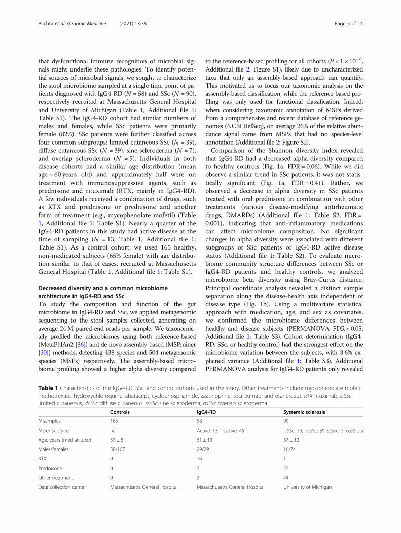
that dysfunctional immune recognition of microbial sig-nals might underlie these pathologies. To identify poten-tial sources of microbial signals, we sought to characterizethe stool microbiome sampled at a single time point of pa-tients diagnosed with IgG4-RD (N = 58) and SSc (N = 90),respectively recruited at Massachusetts General Hospitaland University of Michigan (Table 1, Additional file 1:Table S1). The IgG4-RD cohort had similar numbers ofmales and females, while SSc patients were primarilyfemale (82%). SSc patients were further classified acrossfour common subgroups: limited cutaneous SSc (N = 39),diffuse cutaneous SSc (N = 39), sine scleroderma (N = 7),and overlap scleroderma (N = 5). Individuals in bothdisease cohorts had a similar age distribution (meanage ~ 60 years old) and approximately half were ontreatment with immunosuppressive agents, such asprednisone and rituximab (RTX, mainly in IgG4-RD).A few individuals received a combination of drugs, suchas RTX and prednisone or prednisone and anotherform of treatment (e.g., mycophenolate mofetil) (Table1, Additional file 1: Table S1). Nearly a quarter of theIgG4-RD patients in this study had active disease at thetime of sampling (N = 13, Table 1, Additional file 1:Table S1). As a control cohort, we used 165 healthy,non-medicated subjects (65% female) with age distribu-tion similar to that of cases, recruited at MassachusettsGeneral Hospital (Table 1, Additional file 1: Table S1).
Decreased diversity and a common microbiomearchitecture in IgG4-RD and SScTo study the composition and function of the gutmicrobiome in IgG4-RD and SSc, we applied metagenomicsequencing to the stool samples collected, generating onaverage 24M paired-end reads per sample. We taxonomic-ally profiled the microbiomes using both reference-based(MetaPhlAn2 [36]) and de novo assembly-based (MSPminer[30]) methods, detecting 438 species and 504 metagenomicspecies (MSPs) respectively. The assembly-based micro-biome profiling showed a higher alpha diversity compared
to the reference-based profiling for all cohorts (P < 1 × 10− 8,Additional file 2: Figure S1), likely due to uncharacterizedtaxa that only an assembly-based approach can quantify.This motivated us to focus our taxonomic analysis on theassembly-based classification, while the reference-based pro-filing was only used for functional classification. Indeed,when considering taxonomic annotation of MSPs derivedfrom a comprehensive and recent database of reference ge-nomes (NCBI RefSeq), on average 26% of the relative abun-dance signal came from MSPs that had no species-levelannotation (Additional file 2: Figure S2).Comparison of the Shannon diversity index revealed
that IgG4-RD had a decreased alpha diversity comparedto healthy controls (Fig. 1a, FDR = 0.06). While we didobserve a similar trend in SSc patients, it was not statis-tically significant (Fig. 1a, FDR = 0.41). Rather, weobserved a decrease in alpha diversity in SSc patientstreated with oral prednisone in combination with othertreatments (various disease-modifying antirheumaticdrugs, DMARDs) (Additional file 1: Table S2, FDR =0.001), indicating that anti-inflammatory medicationscan affect microbiome composition. No significantchanges in alpha diversity were associated with differentsubgroups of SSc patients or IgG4-RD active diseasestatus (Additional file 1: Table S2). To evaluate micro-biome community structure differences between SSc orIgG4-RD patients and healthy controls, we analyzedmicrobiome beta diversity using Bray-Curtis distance.Principal coordinate analysis revealed a distinct sampleseparation along the disease-health axis independent ofdisease type (Fig. 1b). Using a multivariate statisticalapproach with medication, age, and sex as covariates,we confirmed the microbiome differences betweenhealthy and disease subjects (PERMANOVA FDR < 0.05,Additional file 1: Table S3). Cohort determination (IgG4-RD, SSc, or healthy control) had the strongest effect on themicrobiome variation between the subjects, with 3.6% ex-plained variance (Additional file 1: Table S3). AdditionalPERMANOVA analysis for IgG4-RD patients only revealed
Table 1 Characteristics of the IgG4-RD, SSc, and control cohorts used in the study. Other treatments include mycophenolate mofetil,methotrexate, hydroxychloroquine, abatacept, cyclophosphamide, azathioprine, tocilizumab, and etanercept. RTX rituximab, lcSSclimited cutaneous, dcSSc diffuse cutaneous, ssSSc sine scleroderma, osSSc overlap scleroderma
Controls IgG4-RD Systemic sclerosis
N samples 165 58 90
N per subtype na Active: 13, inactive: 45 lcSSc: 39, dcSSc: 39, ssSSc: 7, osSSc: 5
Age, years (median ± sd) 57 ± 8 61 ± 13 57 ± 12
Males/females 58/107 29/29 16/74
RTX 0 16 1
Prednisone 0 7 27
Other treatment 0 3 44
Data collection center Massachusetts General Hospital Massachusetts General Hospital University of Michigan
Plichta et al. Genome Medicine (2021) 13:35 Page 5 of 14

no significant differences in the microbiomes of patientswith active disease relative to those of patients in remission(Additional file 1: Table S3). Altogether, our diversity ana-lysis revealed a significantly altered microbiome architec-ture in IgG4-RD and SSc relative to healthy controls. In thecase of SSc, this confirms earlier gut microbiome studiesthat employed 16S rDNA sequencing [22, 37]. Importantly,the clustering of samples from IgG4-RD and SSc based ontheir microbiome diversity strongly suggests that they sharecommon microbial signatures.
Depletion of Bacteroidetes and overabundance ofFirmicutes in disease microbiomesTo identify specific taxonomic groups that contribute tothe reconfigured gut microbiome architecture in thesediseases, we tested the association of their relative abun-dances to disease status using a linear model with age,sex, and medication as covariates. As expected, IgG4-RD, SSc, and control microbiomes were dominated byBacteroidetes and Firmicutes species (Fig. 2a); however,we saw a consistent depletion of Bacteroidetes in bothdiseases (FDR ≤ 1 × 10⁻³) with a concordant increase ofFirmicutes in SSc (FDR = 0.06) and Actinobacteria inIgG4-RD (FDR = 0.15) relative to controls (Fig. 2b). Atthe species level, we detected 38 MSPs concordantlyoverabundant or depleted in both diseases compared tocontrols (FDR < 0.05, Additional file 1: Table S4, Fig. 2c,d). All 11 differentially abundant Bacteroidetes MSPswere depleted in one or both diseases (Fig. 2d), consist-ent with our phylum-level observations, and includedphylogenetically related taxa (e.g., four Alistipes and four
Bacteroides species). Among the Firmicutes, numerousopportunistic pathogenic Clostridium species were over-abundant (e.g., C. innocuum, C. clostridioforme, C.bolteae, and C. symbiosum). These Clostridia are well-adapted to invade host tissue by encoding pathogenicityfactors, such as antibiotic resistance genes and flagellins,and have been identified as extraintestinal infectiousagents [38–40]. Additionally, multiple commensalstypical of the oral cavity were overabundant in disease,including three Veillonella and five Streptococcusspecies. Colonization of the lower parts of the gastro-intestinal (GI) tract by oral microbes is a recognizedphenomenon in systemic diseases such as RA [41] andhas been reported in other immune-mediated diseasessuch as IBD [42, 43]. In contrast, Faecalibacteriumspecies from group IV Clostridium (two MSPs annotatedas Faecalibacterium prausnitzii) that are known for theirbeneficial butyrate production were depleted in the twodiseases. Lastly, an additional 67 MSPs that were differ-entially abundant in only one disease showed a consist-ent trend of overabundance or depletion in the otherdisease (Additional file 2: Figure S3, Additional file 1:Table S4).The overabundance of Actinobacteria was attributed
to one of the top differentially abundant species,Eggerthella lenta (SSc FDR = 5.4 × 10⁻⁷ and IgG4-RDFDR = 1.8 × 10⁻⁴). E. lenta is also overabundant in thegut microbiome of RA and multiple sclerosis patients[44, 45], suggesting that E. lenta might be playing animportant role in multiple autoimmune diseases. Apartfrom the species with known taxonomy, we observed 36
Fig. 1 Microbiome community structure in IgG4-RD and SSc. a Alpha diversity boxplots indicate significantly lower alpha diversity in IgG4-RDcompared to controls (FDR = 0.06). Boxplots show median and lower/upper quartiles; whiskers show inner fences. b PCoA plot of beta diversityusing Bray-Curtis dissimilarity measure. Beta diversity correlates significantly with sample annotation according to IgG4-RD, SSc, or control status(PERMANOVA FDR = 0.004). Marginal figures present distributions of samples along each axis. Controls N = 165, IgG4-RD N = 58, SSc N = 90
Plichta et al. Genome Medicine (2021) 13:35 Page 6 of 14

differentially abundant MSPs with no species-level infor-mation (FDR < 0.05, Additional file 1: Table S4). Phylogen-etic analysis [34] revealed that most of them representedpreviously uncharacterized Firmicutes (Additional file 2:Figure S4). Among those depleted in disease (33/36MSPs), we observed that MSP 182, MSP 234, MSP 326,and MSP 378 clustered closely with F. prausnitzii, indicat-ing an additional decreased abundance of potentialbutyrate-producing species. Among those overabundantin either IgG4-RD or SSc, we observed that MSP 038,MSP 122, and MSP 179 formed a closely phylogeneticallyrelated clade and as such might encode similar functional-ities that allow them to bloom in these diseases or arerelevant to the disease phenotype. Finally, we want tohighlight that no significant differences in known or un-known MSPs were observed comparing IgG4-RD and SScpatients, reinforcing the view that these diseases share acommon microbiome signature.We next asked if SSc subgroups are characterized by
overabundance or depletion of specific MSPs in order toidentify potential pathobionts unique to each subgroup.We compared microbiomes in limited cutaneous SSc(lcSSc), diffuse cutaneous SSc (dcSSc), sine scleroderma(ssSSc), and overlap scleroderma (osSSc) to healthy
controls. We observed 16 MSPs that were commonlydifferentially abundant between at least two SScsubgroups; most notably, overabundance of threeStreptococcus species, S. parasanguinis, S. vestibularis,and S. salivarius was common to lcSSc, dcSSc, andosSSc. Each subgroup also showed unique microbialchanges relative to healthy controls (FDR < 0.05,Additional file 1: Table S5, Additional file 2: Figure S5).Depletion of F. prausnitzii characterized lcSSc, whileoverabundance of two taxa typical of the oral cavity,Veillonella parvula and Klebsiella pneumoniae, was ob-served in dcSSc. We also tested the pairwise differencesbetween the four SSc subgroups and observed four dif-ferentially abundant species in osSSc compared to eitherlcSSc or dcSSc (FDR < 0.05, Additional file 1: Table S5).Finally, we turned to IgG4-RD patients and tested forspecies-level differences between patients with activedisease or in remission, and observed no significantassociation.
Strain-specific gene enrichment in diseaseA species-level focus in human microbiome studies canlack the specificity necessary for attributing functionalroles, as different strains of the same species can harbor
Fig. 2 Overabundant and depleted gut microbiome taxa in IgG4-RD and SSc. a Relative abundances of the five most abundant phyla in thedisease and control cohorts. b Differentially abundant phyla in at least one of the disease cohorts when compared to the controls (FDR < 0.2).Boxplots show median and lower/upper quartiles; whiskers show inner fences. c Top 30 differentially abundant species in IgG4-RD and/or SScwhen compared to the controls (*FDR < 0.05). See also Additional file 1: Table S4. d The number of overabundant or depleted species from eachphylum in the disease cohorts. In total, 19 overabundant and 19 depleted species were common to both IgG4-RD and SSc; no discordant species(i.e., overabundant in one disease and depleted in the other) were identified. Panels a, c, and d use the same color scheme to represent phyla
Plichta et al. Genome Medicine (2021) 13:35 Page 7 of 14

vastly different accessory genomes. These genomicdifferences can be particularly relevant in the context ofimmune system activation. For instance, different strainsof K. pneumoniae show variable potential in activatingtype 1 T helper (Th) cells, highlighting the importanceof accessory genes on modulating immune function [46].With that in mind, we evaluated the genetic compositionof E. lenta, which was one of the top differentiallyabundant species in our comparison (Fig. 2c). E. lenta iswell-known for its potential to inactivate plant toxins,including the cardiac drug digoxin, in the human gut viaa strain-specific cardiac glycoside reductase (cgr) operon[47]. The cgr locus has been attributed immunomodula-tory function: cgr locus positive (cgr+) E. lenta drive acti-vation of Th17 cells and production of pro-inflammatorycytokines, while cgr− strains do not show this phenotype[48]. To determine if there is a colonization bias of cgr+E. lenta strains in SSc and IgG4-RD patients, we identi-fied five genes in the assembled gene catalog that com-prise the cgr locus and evaluated their abundances ineach sample. Using that signal as the marker for pres-ence or absence of the cgr locus, in samples with suffi-cient coverage of E. lenta (see the “Methods” section),
we observed that approximately half of the healthy con-trols were cgr+ E. lenta carriers, consistent with previousreports on the distribution of that function in a generalhuman population [35]. E. lenta in patients affected byIgG4-RD and SSc, however, were more likely to be cgr+(~ 75% patients; Fisher’s test, IgG4-RD P = 0.04, SSc P =0.01, Fig. 3a). Additionally, E. lenta tended to reachhigher relative abundance in cgr+ samples compared tocgr- samples (Wilcoxon, controls P = 2 × 10⁻⁵, IgG4-RDP = 0.02, SSc P = 0.04). These observations implicate E.lenta as a potential microbiome factor with strain-dependent enzymatic activity that might lead to thebreakdown of immune homeostasis and expansion of itspopulation in colonized subjects.Our assembly-based microbiome characterization
through MSPs grouped genes into clusters based ontheir presence and co-abundance pattern such that eachspecies is represented by its core genes and oftenmultiple clusters of different accessory genes. In order toperform a microbiome-wide screen for disease-relatedaccessory modules, we evaluated whether any specificaccessory module was differentially distributed in IgG4-RD and SSc compared to healthy controls. We detected
Fig. 3 Strain-level signals in IgG4-RD and SSc. a Top, Strains of E. lenta are more likely to harbor a cgr operon (cgr+) in disease samples thanhealthy control samples (Fisher’s test, comparison with controls: IgG4-RD P = 0.04; SSc P = 0.01; N controls = 107, N IgG4-RD = 51, N SSc = 81).Bottom, Cgr+ E. lenta achieve a higher relative abundance than cgr− E. lenta in control, IgG4-RD, and SSc samples. Boxplots show median andlower/upper quartiles; whiskers show inner fences. b Species-specific accessory modules (acc) that are enriched or depleted in SSc or IgG4-RDcompared to healthy controls (FDR < 0.05). Effect size is shown as log2 odds ratio. The number of genes contained in each accessory module isindicated by the size of a dot. c Accessory module acc 058 in C. bolteae. Left, Gene composition of the locus encoding cystine uptake andhomocysteine metabolism genes. Right, NCBI’s phylogenetic tree of 15 C. bolteae reference genomes. Stars indicate strains encoding the locus
Plichta et al. Genome Medicine (2021) 13:35 Page 8 of 14
53 accessory modules (acc) that were enriched ordepleted in SSc (FDR < 0.05, Fig. 3b). These accessorymodules showed largely similar enrichment or depletiontrends in IgG4-RD, however only one was significant inthis lower-powered cohort (FDR < 0.05, Fig. 3b). Theidentified accessory modules contained between 4 and158 genes that encoded for various, often related func-tions (Additional file 1: Table S6). The only accessorygene set enriched in both SSc and IgG4-RD, acc 002from Clostridium scindens, contained multiple proteinsinvolved in nutrient transport across membranes (ABCtransporters, efflux pumps, and antiporters). In SSc-enriched acc 058 from C. bolteae, five of its six genesco-assembled next to each other on the same contig inour samples; two additional genes were predicted in thecontig using Glimmer [49]. These similarly co-assemblein a subset of C. bolteae reference genomes and repre-sent a variation present only in some strains of this spe-cies (Fig. 3c, Additional file 2: Figure S6). Interestingly,this accessory module likely expands access of C. bolteaeto the sulfur-containing amino acid cysteine as itencodes three cystine transporters (homologs to TCYA,TCYB, and TCYC transport system), which is consistentwith the ability of C. bolteae to bloom with cysteine as asole carbon source [50]. An additional gene found in thismodule, cystathionine beta-lyase (CBL), catalyzes thebreakdown of the cysteine-related metabolite cystathio-nine to homocysteine and pyruvate. Increased homocyst-eine concentration in circulation could contribute toSSc-related vasculopathy, according to a model in whichhomocysteine inhibits hydrogen sulfide signaling inblood vessels [51].
Changes to immune signaling and ECM-related microbialfunctionsIn addition to strain-specific functional potential, multiplespecies can contribute to similar functional capabilities.To investigate this in an untargeted manner, we summa-rized the relative abundance of microbial pathways usingHUMAnN2 [52]. Consistent with the overall reconfigur-ation of the microbiome composition in IgG4-RD andSSc, a large number of MetaCyc database pathways weredifferentially abundant (FDR < 0.05, Fig. 4a, Additional file1: Table S7). Among the top pathways overabundant inIgG4-RD and SSc were those involved in lipid biosyn-thesis, including the classical mevalonate pathway (IgG4-RD FDR = 0.003, SSc FDR = 8.6 × 10⁻¹⁰) that is encodedby specific microbial families [53]. This overabundancesignal was consistent when we investigated the eightenzymes that constitute this pathway (Additional file 1:Table S8). The classical mevalonate pathway leads to theproduction of isopentenyl pyrophosphate (IPP), a buildingblock for membrane lipids and an important signalingmolecule to immune cells [54].
Many biologically important genes are not annotatedas pathways; hence, we also evaluated abundance ofgenes from the assembled gene catalog that were anno-tated using eggNOG-Mapper [32]. Intriguingly, 12 genesbelonging to the ethanolamine utilization compartmentwere overabundant in IgG4-RD and SSc compared tohealthy controls (FDR < 0.05, Fig. 4b). Ethanolamine isparticularly prevalent in the GI tract where it is releasedas a phosphatidylethanolamine from the renewingepithelium, and its abundance increases during inflam-mation, as observed in IBD [55, 56]. Importantly, etha-nolamine can be used as a carbon and nitrogen sourceto give a growth advantage to the microbes capable ofmetabolizing it, providing a plausible explanation for theoverabundance of potential ethanolamine metabolizersobserved in this study, such as R. gnavus, Blautia pro-ducta, or C. clostridioforme (Additional file 2: Figure S7).Finally, we specifically focused on relative abundance
changes in functions related to extracellular matrix(ECM) binding and fibrosis. Using eggNOG-Mapper[32], we detected the fibronectin-binding gene sfb1(K13734) to be overabundant in SSc and IgG4-RD com-pared to healthy controls (FDR < 0.1, Fig. 4c). Fibronec-tins, among others, bind to ECM that is excessivelyproduced during fibrosis, and many pathogens includingstreptococci use fibronectins as anchors to the epithe-lium [57, 58]. We also evaluated changes to the recentlydescribed 4-hydroxyproline dehydratase (HypD) that de-grades the major constituent of ECM, hydroxyproline,and provides access to this nutrient as an energy sourcefor the encoding bacteria [59]. As a relatively novel func-tional category, it was missing from the annotationsobtained using eggNOG-Mapper. We directly searchedfor homologs of HypD from Clostridioides difficile(A0A031WDE4) in the assembled gene catalog and de-tected 17 homologs with at least 62% amino acid identity[60]. The combined relative abundance signal of theidentified HypD homologs was increased in SSc com-pared to healthy controls (FDR = 0.01, Fig. 4d), whichmay be reflective of overabundant matrix components inthis fibrosis-prone autoimmune disease.
DiscussionHere we characterized a strikingly similar gut micro-biome architecture between two fibrotic autoimmunediseases: IgG4-RD and SSc. Consistent sampling, datageneration, and processing allowed us to avoid technicalbiases and identify biologically relevant features commonbetween the disorders. The gut microbiomes in bothdiseases were significantly different from healthy con-trols and showed a depletion of typical, health-associatedcommensals and expansion of potentially pathogenicand pro-inflammatory species.
Plichta et al. Genome Medicine (2021) 13:35 Page 9 of 14

Our analysis greatly expanded the number of samplesanalyzed and added species-level and functional depth toa previous 16S rRNA-based report on the SSc micro-biome [37]. Consistent with this study, we observed adepletion of numerous Bacteroides and Faecalibacteriumspecies as well as an overabundance of Bifidobacteriumdentium and several Lactobacillus species. We addition-ally identified different Clostridium and Streptococcusspecies to be overabundant in SSc compared to healthycontrols, a signature that indicates either fewer GI symp-toms in our cohort or a cohort-specific signal [14, 37].In contrast to these previous studies, we directly modeledthe contribution of potential confounders, including non-antibiotic medications, when identifying significant associa-tions, which is important for disentangling disease-specificeffects [61]. We detected a negative effect on alpha diversity
from combinatorial treatment with prednisone and DMARDs, mainly mycophenolate mofetil, methotrexate, or hydro-xychloroquine. This observation supports previous reportsof the gut microbiome being affected by immunosuppres-sive medications, such as methotrexate [62] and glucocorti-coids [63]. Given a lack of dietary information in thiscohort, we could not similarly evaluate the expected effectof diet on the gut microbiome.By employing a de novo assembly approach to study
IgG4-RD and SSc patient microbiomes, we pinpointedchanges to the as-yet uncultured constituents of thehuman gut and performed strain analysis focused on thevariation in accessory gene content. This revealed anenrichment in IgG4-RD and SSc of cgr+ E. lenta strains,a clade that increasingly is being attributed pro-inflammatoryfunctions [48]. We further observed preferential colonization
Fig. 4 Functional microbiome signatures common to IgG4-RD and SSc. a Top 20 significantly up- and downregulated MetaCyc pathwayscommon to IgG4-RD and SSc compared to healthy controls (FDR < 0.05). b Numerous genes belonging to the ethanolamine utilization pathwaywere increased in abundance in disease. Asterisks (*, **) indicate differential abundance at FDR < 0.2 and FDR < 0.05 levels. Differentialabundances (transcripts per million, TPM) of c Sfb1 (SSc FDR = 0.008, IgG4-RD FDR = 0.07) and d HypD (SSc FDR = 0.01) in IgG4-RD and SSccompared to controls. Boxplots show median and lower/upper quartiles; whiskers show inner fences. Full list of differentially abundant MetaCycpathways and KEGG KO genes are in Additional file 1: Tables S7 and S9
Plichta et al. Genome Medicine (2021) 13:35 Page 10 of 14
of SSc patients with a strain of C. bolteae encoding ahomocysteine metabolism locus. SSc is associated withvasculopathy that may be promoted by elevated levelsof homocysteine in circulation [51], which have beenreported in SSc and atherosclerosis. Disease-associatedmicrobiomes also showed changes in relative abun-dances of lipid metabolism and ECM-modifyingenzymes. We detected an increase in the relative abun-dances of genes encoding an ECM binding protein(sfb1) and an enzyme that potentially allows for energyextraction from ECM (HypD homologs). These enrich-ments might enhance the availability of ECM to specificmicrobial species for attachment and additional nutri-ent sources [57, 59].Several functional signatures link the microbiomes of
IgG4-RD and SSc patients to inflammation. The classicalmevalonate pathway that leads to IPP synthesis was in-creased in both diseases. IPP and other metabolites fromthis pathway are recognized by ɣδ T cells as a part ofsurveillance against microbial infections [64]; differencesin the signaling molecule repertoire may alter immuneresponses in IgG4-RD and SSc patients. Another unify-ing functional characteristic that we observed was theexpansion of pro-inflammatory, Th17 cell-activatingcgr+ E. lenta [48]. Th17 cell expansion is often related tobreakdown of gut microbiome homeostasis and impairedT regulatory cell activity and has been studied experimen-tally in IBD [65]. Consistent with an increased abundanceof cgr+ E. lenta, the levels of circulating Th17 cells are ele-vated in patients with IgG4-RD and SSc [66, 67]. Whilethere is not yet consensus in the field, mouse models ofSSc and cellular assays connect the level of IL-17 signalingwith the overproduction of collagen and fibrosis [68–70].Such disease-relevant taxonomical and functional
characteristics encourage further study into the identifica-tion of specific microbiome-derived molecules that candrive autoimmune pathologies [71], such as microbiomeproteins that molecularly mimic disease-associatedautoantigens as reported in RA [72]. To date, no specificautoantigens for IgG4-RD or SSc have been identified tofacilitate such a discovery, and this remains a challenge forautoimmune diseases in general. Identification of autoan-tigens and other immune-centered efforts will be neededto link microbiome changes reported here and elsewherewith immunophenotypes in order to further understandthe role of the microbiome in IgG4-RD and SSc.The shared microbiome signature in IgG4-RD and SSc
raises a question about the existence of a more universalmicrobiome architecture in immune-mediated disorders.We observed expansions of E. lenta and taxa typical ofthe oral cavity that are similar to autoimmune diseasesincluding the neuroimmune disorder multiple sclerosisand RA, a disease with multiple joint pathologies [42, 44,73]. Comparing the differentially abundant species in
IgG4-RD and SSc with those from ulcerative colitis andCrohn’s disease, we observed enrichment in four speciestypically overabundant in IBD (S. parasanguinis, B.producta, Lactobacillus gasseri, and R. gnavus) as well ascommon signals with 11 out of 42 species depleted inIBD [74]. We observed Escherichia coli to be overabun-dant, albeit weakly, in SSs but not IgG4-RD. This patho-biont has been associated with mucosal inflammationand decreased gut anaerobism in IBD [75]. Gut fibrosisin Crohn’s disease leads to thickening of the intestinalwall and strictures; therefore, we searched for the HypDoverabundance signal in this disease [24, 56] butdetected no significant associations. Future integrativestudies will be needed to present a coherent view of gutmicrobiome signals broadly implicated in immune-mediated disorders.
ConclusionsOur characterization of the gut microbiome in fibrosis-prone IgG4-RD and SSc revealed taxonomic and functionalgut microbiome features that are common in these twodiseases, including reduction of health-associated commen-sals and expansion of potentially pathogenic and pro-inflammatory species. IgG4-RD and SSc patients showedexpansion of a Th17-inducing strain of E. lenta that encodesa cgr locus, indicating a potential microbiome-driven skew-ing of the immune cell population in the context of theserare autoimmune diseases. We also found that microbiomechanges in IgG4-RD and SSc partially recount observationsfrom other autoimmune diseases and that IgG4-RD- andSSc-specific effects likely further shape the landscape of theassociated gut microbiomes.
Supplementary InformationThe online version contains supplementary material available at https://doi.org/10.1186/s13073-021-00853-7.
Additional file 1. Supplementary Tables.
Additional file 2. Supplementary Figures.
AbbreviationsIgG4-RD: Immunoglobulin G4-related disease; SSc: Systemic sclerosis;IBD: Inflammatory bowel diseases; RA: Rheumatoid arthritis; MHC: Majorhistocompatibility complex; CTL: Cytotoxic T cell; HLA: Human leukocyteantigen; lcSSc: Limited cutaneous SSc; dcSSc: Diffuse cutaneous SSc;ssSSc: Sine scleroderma; osSSc: Overlap scleroderma; MSP: Metagenomicspecies; DMARD: Disease-modifying antirheumatic drug; GI: Gastrointestinal;Th: T helper; cgr: Cardiac glycoside reductase; acc: Accessory modules;CBL: Cystathionine beta-lyase; IPP: Isopentenyl pyrophosphate;ECM: Extracellular matrix; HypD: 4-hydroxyproline dehydratase
AcknowledgementsWe thank Tiffany Poon and Luke Besse for project management and theBroad Institute Microbial ‘Omics Core and Genomics Platform for sampleprocessing and sequencing data generation. We are grateful to TheresaReimels for editorial assistance for the manuscript and figures. We alsoacknowledge the dedication of the study participants without whom thisresearch would not be possible.
Plichta et al. Genome Medicine (2021) 13:35 Page 11 of 14

Authors’ contributionsSP and RJX coordinated and helped conceive the study. DRP, HV, SP, andRJX designed the research. ZSW, ADF, CAP, JHS, DC, DK, and SP participatedin data collection. HV and RJX generated data. DRP and JS performed theresearch and analyzed data. MP, HL, DK, HV, SP, and RJX provided criticalfeedback. DRP, JS, HV, and RJX wrote the paper. All authors reviewed thepaper. All authors read and approved the final manuscript.
FundingSP and RJX were supported by National Institute of Allergy and InfectiousDiseases (NIAID) grant U19 AI110495. JHS, CAP, ZSW, and ADF weresupported by NIAID grant UM1 AI144295. DK was supported by NIAID grantUM1 AI110557 to the University of Michigan and National Institute ofArthritis and Musculoskeletal and Skin Diseases grant K24 AR063120.
Availability of data and materialsThe sequencing dataset generated and analyzed during the current study isavailable in the NCBI BioProject under PRJNA615162 (https://www.ncbi.nlm.nih.gov/bioproject/ PRJNA615162) [76]. Correspondence and requests formaterials should be addressed to R.J.X.
Ethics approval and consent to participateHuman patient research in the IgG4-RD cohort was reviewed andapproved by the Partners Human Research Committee (2008P002154).Human patient research in the SSc cohort was approved by theInstitutional Review Board of the University of Michigan Medical School(HUM00101836). Human patient research in the healthy control cohortwas reviewed and approved by the Partners Human ResearchCommittee (2015P000275). The study was approved by the Office ofResearch Subject Protection at the Broad Institute of MIT and Harvard.All experiments adhered to the regulations of these review boards.Study procedures were performed in compliance with all relevantethical and federal regulations. This research conformed to theprinciples of the Helsinki Declaration. All study participants gave theirwritten informed consent for sample collection and to participate inthe study.
Consent for publicationNot applicable.
Competing interestsRJX is a consultant to Novartis and Nestle. DK is a consultant to Acceleron,Actelion, Amgen, Bayer, Blade Therapeutics, Boehringer Ingelheim, CSLBehring, Corbus, Cytori, Galapagos, Genentech/Roche, GSK, Horizon, Merck,Mitsubishi Tanabe Pharma, Regeneron, Sanofi-Aventis, and United Therapeu-tics; CME programs: Impact PH; Stocks: Eicos Sciences, Inc.; Leadership/Equityposition -Medical lead, Scleroderma Development, CiviBioPharma/Eicos Sci-ences, Inc. The remaining authors declare that they have no competinginterests.
Author details1Broad Institute of MIT and Harvard, Cambridge, MA, USA. 2Department ofComputer Science, Aalto University, 02150 Espoo, Finland. 3Novartis Institutefor Biomedical Research, Cambridge, MA, USA. 4Division of Rheumatology,Allergy, and Immunology, Massachusetts General Hospital, Boston, MA, USA.5Clinical Epidemiology Program and Rheumatology Unit, MassachusettsGeneral Hospital and Harvard Medical School, Boston, MA, USA. 6RagonInstitute of MGH, MIT and Harvard, Cambridge, MA, USA. 7Division ofGastroenterology, Massachusetts General Hospital and Harvard MedicalSchool, Boston, MA, USA. 8Center for Cancer Risk Assessment, MassachusettsGeneral Hospital and Harvard Medical School, Boston, MA, USA. 9Universityof Michigan Scleroderma Program, Ann Arbor, MI, USA. 10Center forComputational and Integrative Biology, Massachusetts General Hospital andHarvard Medical School, Boston, MA, USA. 11Department of MolecularBiology, Massachusetts General Hospital and Harvard Medical School, Boston,MA, USA. 12Center for Microbiome Informatics and Therapeutics, MIT,Cambridge, MA, USA.
Received: 24 July 2020 Accepted: 11 February 2021
References1. Skelly AN, Sato Y, Kearney S, Honda K. Mining the microbiota for microbial
and metabolite-based immunotherapies. Nat Rev Immunol. 2019; Availablefrom: https://doi.org/10.1038/s41577-019-0144-5.
2. Tanoue T, Morita S, Plichta DR, Skelly AN, Suda W, Sugiura Y, et al. A definedcommensal consortium elicits CD8 T cells and anti-cancer immunity. Nature.2019;565(7741):600–5.
3. Mattoo H, Mahajan VS, Maehara T, Deshpande V, Della-Torre E, Wallace ZS,et al. Clonal expansion of CD4(+) cytotoxic T lymphocytes in patients withIgG4-related disease. J Allergy Clin Immunol. 2016;138(3):825–38.
4. Pillai S. T and B lymphocytes in fibrosis and systemic sclerosis. Curr OpinRheumatol. 2019;31(6):576–81.
5. Maehara T, Kaneko N, Perugino CA, Mattoo H, Kers J, Allard-Chamard H,et al. Cytotoxic CD4+ T lymphocytes may induce endothelial cell apoptosisin systemic sclerosis. J Clin Invest. 2020; Available from: https://doi.org/10.1172/JCI131700.
6. Stone JH, Zen Y, Deshpande V. IgG4-related disease. N Engl J Med. 2012;366(6):539–51.
7. Mahajan VS, Mattoo H, Deshpande V, Pillai SS, Stone JH. IgG4-relateddisease. Annu Rev Pathol. 2014;9:315–47.
8. Brito-Zerón P, Ramos-Casals M, Bosch X, Stone JH. The clinical spectrum ofIgG4-related disease. Autoimmun Rev. 2014;13(12):1203–10.
9. Hubers LM, Vos H, Schuurman AR, Erken R, Oude Elferink RP, Burgering B,et al. Annexin A11 is targeted by IgG4 and IgG1 autoantibodies in IgG4-related disease. Gut. 2018;67(4):728–35.
10. Della-Torre E, Lanzillotta M, Doglioni C. Immunology of IgG4-related disease.Clin Exp Immunol. 2015;181(2):191–206.
11. Denton CP, Khanna D. Systemic sclerosis. Lancet. 2017;390(10103):1685–99.12. Desbois AC, Cacoub P. Systemic sclerosis: an update in 2016. Autoimmun
Rev. 2016;15(5):417–26.13. Steen VD. Autoantibodies in systemic sclerosis. Semin Arthritis Rheum. 2005;
35(1):35–42.14. Patrone V, Puglisi E, Cardinali M, Schnitzler TS, Svegliati S, Festa A, et al. Gut
microbiota profile in systemic sclerosis patients with and without clinicalevidence of gastrointestinal involvement. Sci Rep. 2017;7(1):14874.
15. Allanore Y, Simms R, Distler O, Trojanowska M, Pope J, Denton CP, et al.Systemic sclerosis. Nat Rev Dis Primers. 2015;1:15002.
16. Laurent P, Sisirak V, Lazaro E, Richez C, Duffau P, Blanco P, et al. Innateimmunity in systemic sclerosis fibrosis: recent advances. Front Immunol.2018;9:1702.
17. Sakkas LI, Bogdanos DP. Systemic sclerosis: new evidence re-enforces therole of B cells. Autoimmun Rev. 2016;15(2):155–61.
18. Haldar D, Hirschfield GM. Deciphering the biology of IgG4-related disease:specific antigens and disease? Gut. 2018;67(4):602–5.
19. Burbelo PD, Gordon SM, Waldman M, Edison JD, Little DJ, Stitt RS, et al.Autoantibodies are present before the clinical diagnosis of systemicsclerosis. PLoS One. 2019;14(3):e0214202.
20. Yamamoto M, Takahashi H, Shinomura Y. Mechanisms and assessment ofIgG4-related disease: lessons for the rheumatologist. Nat Rev Rheumatol.2014;10(3):148–59.
21. Perugino CA, AlSalem SB, Mattoo H, Della-Torre E, Mahajan V, Ganesh G,et al. Identification of galectin-3 as an autoantigen in patients with IgG4-related disease. J Allergy Clin Immunol. 2019;143(2):736–45.e6.
22. Bellocchi C, Volkmann ER. Update on the gastrointestinal microbiome insystemic sclerosis. Curr Rheumatol Rep. 2018;20(8):49.
23. Wallace ZS, Khosroshahi A, Carruthers MD, Perugino CA, Choi H,Campochiaro C, et al. An international multispecialty validation study of theIgG4-related disease responder index. Arthritis Care Res. 2018;70(11):1671–8.
24. Lloyd-Price J, Arze C, Ananthakrishnan AN, Schirmer M, Avila-Pacheco J,Poon TW, et al. Multi-omics of the gut microbial ecosystem in inflammatorybowel diseases. Nature. 2019;569(7758):655–62.
25. Li D, Liu C-M, Luo R, Sadakane K, Lam T-W. MEGAHIT: an ultra-fast single-node solution for large and complex metagenomics assembly via succinctde Bruijn graph. Bioinformatics. 2015;31(10):1674–6.
26. Hyatt D, Chen G-L, Locascio PF, Land ML, Larimer FW, Hauser LJ. Prodigal:prokaryotic gene recognition and translation initiation site identification.BMC Bioinformatics. 2010;11:119.
Plichta et al. Genome Medicine (2021) 13:35 Page 12 of 14

27. Fu L, Niu B, Zhu Z, Wu S, Li W. CD-HIT: accelerated for clustering the next-generation sequencing data. Bioinformatics. 2012;28(23):3150–2.
28. Qin J, Li R, Raes J, Arumugam M, Burgdorf KS, Manichanh C, et al. A humangut microbial gene catalogue established by metagenomic sequencing.Nature. 2010;464(7285):59–65.
29. Li H, Durbin R. Fast and accurate short read alignment with Burrows-Wheeler transform. Bioinformatics. 2009;25(14):1754–60.
30. Plaza Oñate F, Le Chatelier E, Almeida M, Cervino ACL, Gauthier F, MagoulèsF, et al. MSPminer: abundance-based reconstitution of microbial pan-genomes from shotgun metagenomic data. Bioinformatics. 2018; Availablefrom: https://doi.org/10.1093/bioinformatics/bty830.
31. Kanehisa M, Goto S, Sato Y, Furumichi M, Tanabe M. KEGG for integrationand interpretation of large-scale molecular data sets. Nucleic Acids Res.2012;40(Database issue):D109–14.
32. Huerta-Cepas J, Forslund K, Coelho LP, Szklarczyk D, Jensen LJ, vonMering C, et al. Fast genome-wide functional annotation throughorthology assignment by eggNOG-mapper. Mol Biol Evol. 2017;34(8):2115–22.
33. Li J, Jia H, Cai X, Zhong H, Feng Q, Sunagawa S, et al. An integrated catalogof reference genes in the human gut microbiome. Nat Biotechnol. 2014;32(8):834–41.
34. Segata N, Börnigen D, Morgan XC, Huttenhower C. PhyloPhlAn is a newmethod for improved phylogenetic and taxonomic placement of microbes.Nat Commun. 2013;4:2304.
35. Koppel N, Bisanz JE, Pandelia ME, Turnbaugh PJ, Balskus EP. Discovery andcharacterization of a prevalent human gut bacterial enzyme sufficient forthe inactivation of a family of plant toxins. Elife. 2018;7:1–32.
36. Truong DT, Franzosa EA, Tickle TL, Scholz M, Weingart G, Pasolli E, et al.MetaPhlAn2 for enhanced metagenomic taxonomic profiling. Nat Methods.2015;12(10) Available from: https://doi.org/10.1038/nmeth.3589.
37. Volkmann ER, Hoffmann-Vold A-M, Chang Y-L, Jacobs JP, Tillisch K, MayerEA, et al. Systemic sclerosis is associated with specific alterations ingastrointestinal microbiota in two independent cohorts. BMJ OpenGastroenterol. 2017;4(1):e000134.
38. Elsayed S, Zhang K. Bacteremia caused by Clostridium symbiosum. J ClinMicrobiol. 2004;42(9):4390–2.
39. Chia J-H, Feng Y, Su L-H, Wu T-L, Chen C-L, Liang Y-H, et al. Clostridiuminnocuum is a significant vancomycin-resistant pathogen for extraintestinalclostridial infection. Clin Microbiol Infect. 2017;23(8):560–6.
40. Dehoux P, Marvaud JC, Abouelleil A, Earl AM, Lambert T, Dauga C.Comparative genomics of Clostridium bolteae and Clostridiumclostridioforme reveals species-specific genomic properties andnumerous putative antibiotic resistance determinants. BMC Genomics.2016;17(1):819.
41. Olsen I, Yamazaki K. Can oral bacteria affect the microbiome of the gut? JOral Microbiol. 2019;11(1):1586422.
42. Brusca SB, Abramson SB, Scher JU. Microbiome and mucosal inflammationas extra-articular triggers for rheumatoid arthritis and autoimmunity. CurrOpin Rheumatol. 2014;26(1):101–7.
43. Opazo MC, Ortega-Rocha EM, Coronado-Arrázola I, Bonifaz LC, Boudin H,Neunlist M, et al. Intestinal microbiota influences non-intestinal relatedautoimmune diseases. Front Microbiol. 2018;9(MAR):1–20.
44. Miyake S, Kim S, Suda W, Oshima K, Nakamura M, Matsuoka T, et al.Dysbiosis in the gut microbiota of patients with multiple sclerosis, with astriking depletion of species belonging to clostridia XIVa and IV Clusters.Wilson BA, editor. PLoS One. 2015;10(9):e0137429.
45. Zhang X, Zhang D, Jia H, Feng Q, Wang D, Liang D, et al. The oral and gutmicrobiomes are perturbed in rheumatoid arthritis and partly normalizedafter treatment. Nat Med. 2015;21(8):895–905.
46. Atarashi K, Suda W, Luo C, Kawaguchi T, Motoo I, Narushima S, et al. Ectopiccolonization of oral bacteria in the intestine drives TH1 cell induction andinflammation. Science. 2017;358(6361):359–65.
47. Haiser HJ, Gootenberg DB, Chatman K, Sirasani G, Balskus EP, Turnbaugh PJ.Predicting and manipulating cardiac drug inactivation by the human gutbacterium Eggerthella lenta. Science. 2013;341(6143):295–8.
48. Alexander M, Ang QY, Turnbaugh PJ. A diet-dependent enzyme from thehuman gut microbiome promotes Th17 accumulation and colitis. bioRxiv.2019:766899 [cited 2019 Sep 27]. Available from: https://www.biorxiv.org/content/10.1101/766899v1.full.
49. Delcher AL, Harmon D, Kasif S, White O, Salzberg SL. Improved microbialgene identification with GLIMMER. Nucleic Acids Res. 1999;27(23):4636–41.
50. Feng Y, Stams AJM, de Vos WM, Sánchez-Andrea I. Enrichment ofsulfidogenic bacteria from the human intestinal tract. FEMS Microbiol Lett.2017;364(4) Available from: https://doi.org/10.1093/femsle/fnx028.
51. Abdulle AE, van Goor H, Mulder DJ. Hydrogen sulfide: a therapeutic optionin systemic sclerosis. Int J Mol Sci. 2018;19(12) Available from: https://doi.org/10.3390/ijms19124121.
52. Franzosa EA, McIver LJ, Rahnavard G, Thompson LR, Schirmer M, WeingartG, et al. Species-level functional profiling of metagenomes andmetatranscriptomes. Nat Methods. 2018;15(11):962–8.
53. Heuston S, Begley M, Gahan CGM, Hill C. Isoprenoid biosynthesis in bacterialpathogens. Microbiology. 2012;158(Pt 6):1389–401.
54. Gruenbacher G, Thurnher M. Mevalonate metabolism in immuno-oncology.Front Immunol. 2017;8:1714.
55. Kaval KG, Garsin DA. Ethanolamine utilization in bacteria. MBio. 2018;9(1):e00066–18.
56. Franzosa EA, Sirota-Madi A, Avila-Pacheco J, Fornelos N, Haiser HJ, Reinker S,et al. Gut microbiome structure and metabolic activity in inflammatorybowel disease. Nat Microbiol. 2019;4(2):293–305.
57. Towers RJ, Fagan PK, Talay SR, Currie BJ, Sriprakash KS, Walker MJ, et al.Evolution of sfbI encoding streptococcal fibronectin-binding protein I:horizontal genetic transfer and gene mosaic structure. J Clin Microbiol.2003;41(12):5398–406.
58. Roche FM, Downer R, Keane F, Speziale P, Park PW, Foster TJ. The N-terminal A domain of fibronectin-binding proteins a and B promotesadhesion of Staphylococcus aureus to elastin. J Biol Chem. 2004;279(37):38433–40.
59. Huang YY, Martínez-del Campo A, Balskus EP. Anaerobic 4-hydroxyprolineutilization: discovery of a new glycyl radical enzyme in the human gutmicrobiome uncovers a widespread microbial metabolic activity. GutMicrobes. 2018;6:1–16.
60. Levin BJ, Huang YY, Peck SC, Wei Y, Campo AM, Marks JA, et al. Aprominent glycyl radical enzyme in human gut microbiomes metabolizestrans-4-hydroxy-l-proline. Science. 2017;355(6325):eaai8386.
61. Forslund K, Hildebrand F, Nielsen T, Falony G, Le Chatelier E, Sunagawa S,et al. Disentangling type 2 diabetes and metformin treatment signatures inthe human gut microbiota. Nature. 2015;528(7581):262–6.
62. Nayak RR, Alexander M, Stapleton-Grey K, Ubeda C, Scher JU, Turnbaugh PJ.Perturbation of the human gut microbiome by a non-antibiotic drugcontributes to the resolution of autoimmune disease. bioRxiv. 2019:600155[cited 2019 Sep 27]. Available from: https://www.biorxiv.org/content/10.1101/600155v1.
63. Huang EY, Inoue T, Leone VA, Dalal S, Touw K, Wang Y, et al. Usingcorticosteroids to reshape the gut microbiome: implications forinflammatory bowel diseases. Inflamm Bowel Dis. 2015;21(5):963–72.
64. Singhal A, Mori L, De Libero G. T cell recognition of non-peptidic antigensin infectious diseases. Indian J Med Res. 2013;138(5):620–31.
65. Britton GJ, Contijoch EJ, Mogno I, Vennaro OH, Llewellyn SR, Ng R, et al.Microbiotas from humans with inflammatory bowel disease alter thebalance of gut Th17 and RORγt+ regulatory T cells and exacerbate colitis inmice. Immunity. 2019;50(1):212–24.e4.
66. Radstake TRD, van Bon L, Broen J, Hussiani A, Hesselstrand R, Wuttge DM,et al. The pronounced Th17 profile in systemic sclerosis (SSc) together withintracellular expression of TGFβ and IFNγ distinguishes SSc phenotypes.PLoS One. 2009;4(6):e5903.
67. Grados A, Ebbo M, Piperoglou C, Groh M, Regent A, Samson M, et al. T cellpolarization toward TH2/TFH2 and TH17/TFH17 in patients with IgG4-related disease. Front Immunol. 2017;8:235.
68. Park M-J, Moon S-J, Lee E-J, Jung K-A, Kim E-K, Kim D-S, et al. IL-1-IL-17signaling axis contributes to fibrosis and inflammation in two differentmurine models of systemic sclerosis. Front Immunol. 2018;9:1611.
69. Yang X, Yang J, Xing X, Wan L, Li M. Increased frequency of Th17 cells insystemic sclerosis is related to disease activity and collagen overproduction.Arthritis Res Ther. 2014;16(1):R4.
70. Chizzolini C, Dufour AM, Brembilla NC. Is there a role for IL-17 in thepathogenesis of systemic sclerosis? Immunol Lett. 2018;195:61–7.
71. Trivedi PJ, Bruns T, Ward S, Mai M, Schmidt C, Hirschfield GM, et al. IntestinalCCL25 expression is increased in colitis and correlates with inflammatoryactivity. J Autoimmun. 2016;68:98–104.
72. Pianta A, Arvikar SL, Strle K, Drouin EE, Wang Q, Costello CE, et al. Tworheumatoid arthritis-specific autoantigens correlate microbial immunity withautoimmune responses in joints. J Clin Invest. 2017;127(8):2946–56.
Plichta et al. Genome Medicine (2021) 13:35 Page 13 of 14

73. Chen J, Wright K, Davis JM, Jeraldo P, Marietta EV, Murray J, et al. Anexpansion of rare lineage intestinal microbes characterizes rheumatoidarthritis. Genome Med. 2016;8(1):43.
74. Schirmer M, Garner A, Vlamakis H, Xavier RJ. Microbial genes and pathwaysin inflammatory bowel disease. Nat Rev Microbiol. 2019; Available from:https://doi.org/10.1038/s41579-019-0213-6.
75. Byndloss MX, Litvak Y, Bäumler AJ. Microbiota-nourishing immunity and itsrelevance for ulcerative colitis. Inflamm Bowel Dis. 2019;25(5):811–5.
76. Plichta DR, Somani J, Pichaud M, Wallace ZS, Fernandes AD, Perugino CA,Lähdesmäki H, Stone JH, Vlamakis H, Chung D, Khanna D, Pillai S, Xavier RJ.Gut microbiome characterization of IgG4 patients and systemic sclerosispatients. NCBI BioProject. 2020; https://www.ncbi.nlm.nih.gov/bioproject/%20PRJNA615162.
Publisher’s NoteSpringer Nature remains neutral with regard to jurisdictional claims inpublished maps and institutional affiliations.
Plichta et al. Genome Medicine (2021) 13:35 Page 14 of 14