Ch 06_lecture_presentation
90
PowerPoint ® Lecture Presentations prepared by Bradley W. Christian, McLennan Community College C H A P T E R © 2016 Pearson Education, Inc. Microbial Growth 6
-
Upload
kevperrino -
Category
Education
-
view
873 -
download
1
Transcript of Ch 06_lecture_presentation
PowerPoint® Lecture Presentations prepared byBradley W. Christian, McLennan Community College
C H A P T E R
© 2016 Pearson Education, Inc.
Microbial Growth
6
© 2016 Pearson Education, Inc.
© 2016 Pearson Education, Inc.
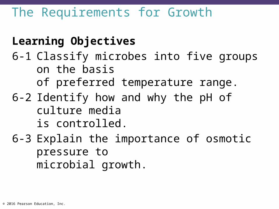
The Requirements for Growth
Learning Objectives6-1 Classify microbes into five groups on the basis
of preferred temperature range.6-2 Identify how and why the pH of culture media
is controlled.6-3 Explain the importance of osmotic pressure to
microbial growth.
© 2016 Pearson Education, Inc.
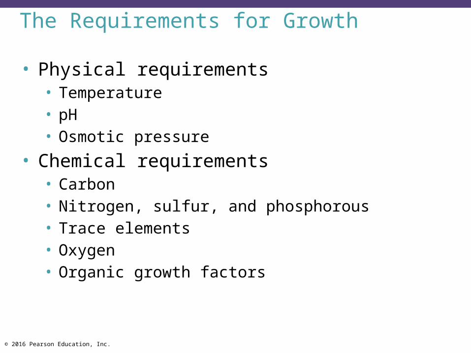
The Requirements for Growth
• Physical requirements• Temperature• pH• Osmotic pressure
• Chemical requirements• Carbon• Nitrogen, sulfur, and phosphorous• Trace elements• Oxygen• Organic growth factors

© 2016 Pearson Education, Inc.
Physical Requirements
• Temperature• Minimum growth temperature• Optimum growth temperature• Maximum growth temperature

© 2016 Pearson Education, Inc.
Physical Requirements
• Temperature (cont'd)• Psychrophiles—cold-loving• Mesophiles—moderate-temperature-loving• Thermophiles—heat-loving

© 2016 Pearson Education, Inc.
Figure 6.1 Typical growth rates of different types of microorganisms in response to temperature.

© 2016 Pearson Education, Inc.
Temperature
• Psychrotrophs • Grow between 0C and 20 to 30C• Cause food spoilage

© 2016 Pearson Education, Inc.
Figure 6.2 Food preservation temperatures.
Temperatures in this range destroy mostmicrobes, although lower temperaturestake more time.
Very slow bacterial growth.
Rapid growth of bacteria; some mayproduce toxins.
Many bacteria survive; some may grow.Refrigerator temperatures; may allow slowgrowth of spoilage bacteria, very few pathogens.No significant growth below freezing.
Dangerzone
1301201101009080706050
40302010
0
260240
220200
180160
140120100
80
6040
20
0–20
–20–10
–30
CF
© 2016 Pearson Education, Inc.
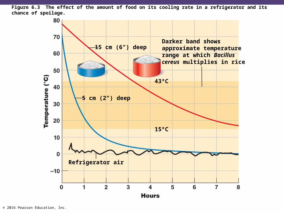
Figure 6.3 The effect of the amount of food on its cooling rate in a refrigerator and its chance of spoilage.
Darker band showsapproximate temperaturerange at which Bacilluscereus multiplies in rice
43ºC
15ºC
Refrigerator air
5 cm (2") deep
15 cm (6") deep
© 2016 Pearson Education, Inc.
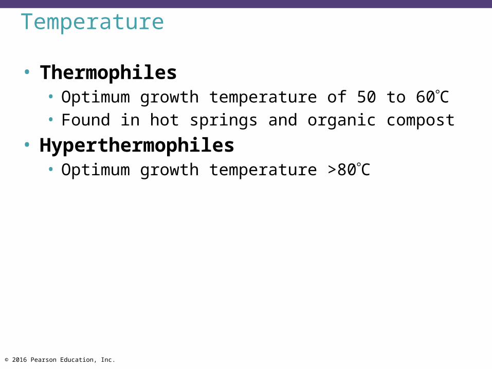
Temperature
• Thermophiles• Optimum growth temperature of 50 to 60C• Found in hot springs and organic compost
• Hyperthermophiles • Optimum growth temperature >80C
© 2016 Pearson Education, Inc.
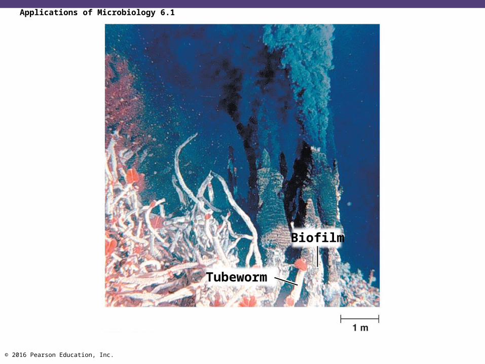
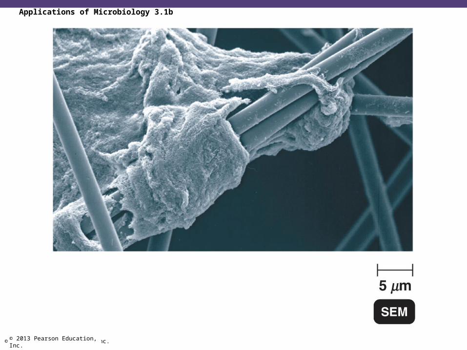
Applications of Microbiology 6.1
Biofilm
Tubeworm
© 2016 Pearson Education, Inc.
pH
• Most bacteria grow between pH 6.5 and 7.5• Molds and yeasts grow between pH 5 and 6• Acidophiles grow in acidic environments
© 2016 Pearson Education, Inc.
Osmotic Pressure
• Hypertonic environments (higher in solutes than inside the cell) cause plasmolysis due to high osmotic pressure
• Extreme or obligate halophiles require high osmotic pressure (high salt)
• Facultative halophiles tolerate high osmotic pressure
© 2016 Pearson Education, Inc.
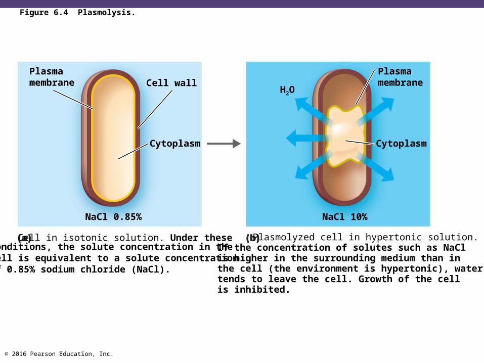
Figure 6.4 Plasmolysis.
Cell wall
Cytoplasm
Plasmamembrane
NaCl 0.85% NaCl 10%
H2O
Plasmamembrane
Cytoplasm
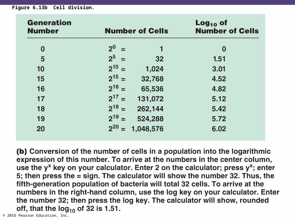
conditions, the solute concentration in thecell is equivalent to a solute concentrationof 0.85% sodium chloride (NaCl).
Cell in isotonic solution. Under these Plasmolyzed cell in hypertonic solution.If the concentration of solutes such as NaClis higher in the surrounding medium than inthe cell (the environment is hypertonic), watertends to leave the cell. Growth of the cellis inhibited.

© 2016 Pearson Education, Inc.
Check Your Understanding Why are hyperthermophiles that grow at temperatures
above 100C seemingly limited to oceanic depths?6-1
Other than controlling acidity, what is an advantage of using phosphate salts as buffers in growth media?6-2
Why might primitive civilizations have used food preservation techniques that rely on osmotic pressure?6-3
© 2016 Pearson Education, Inc.
The Requirements for Growth
Learning Objectives6-4 Name a use for each of the four elements
(carbon, nitrogen, sulfur, and phosphorus) needed in large amounts for microbial growth.
6-5 Explain how microbes are classified on the basis of oxygen requirements.
6-6 Identify ways in which aerobes avoid damage by toxic forms of oxygen.

© 2016 Pearson Education, Inc.
Chemical Requirements
• Carbon• Structural backbone of organic molecules• Chemoheterotrophs use organic molecules as energy• Autotrophs use CO2

© 2016 Pearson Education, Inc.
Chemical Requirements
• Nitrogen• Component of proteins, DNA, and ATP• Most bacteria decompose protein material for the
nitrogen source• Some bacteria use NH4
+ or NO3– from organic material
• A few bacteria use N2 in nitrogen fixation
© 2016 Pearson Education, Inc.
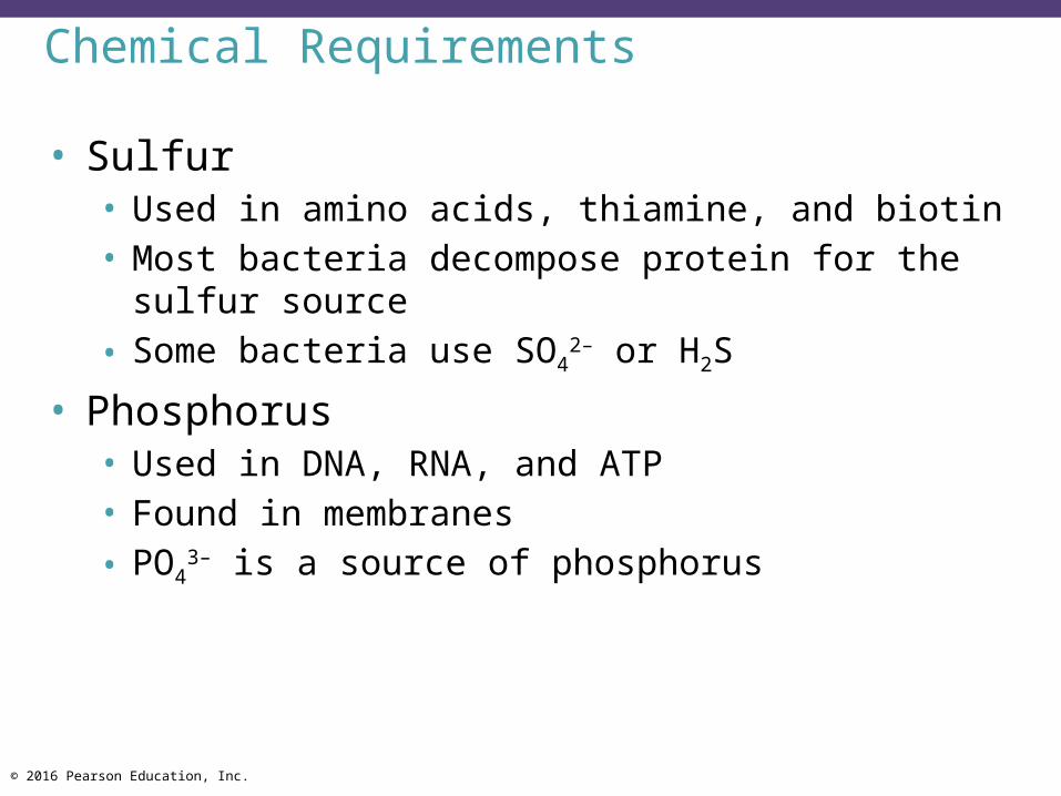
Chemical Requirements
• Sulfur• Used in amino acids, thiamine, and biotin• Most bacteria decompose protein for the sulfur source• Some bacteria use SO4
2– or H2S
• Phosphorus • Used in DNA, RNA, and ATP• Found in membranes• PO4
3– is a source of phosphorus

© 2016 Pearson Education, Inc.
Trace Elements
• Inorganic elements required in small amounts• Usually as enzyme cofactors• Include iron, copper, molybdenum, and zinc
© 2016 Pearson Education, Inc.
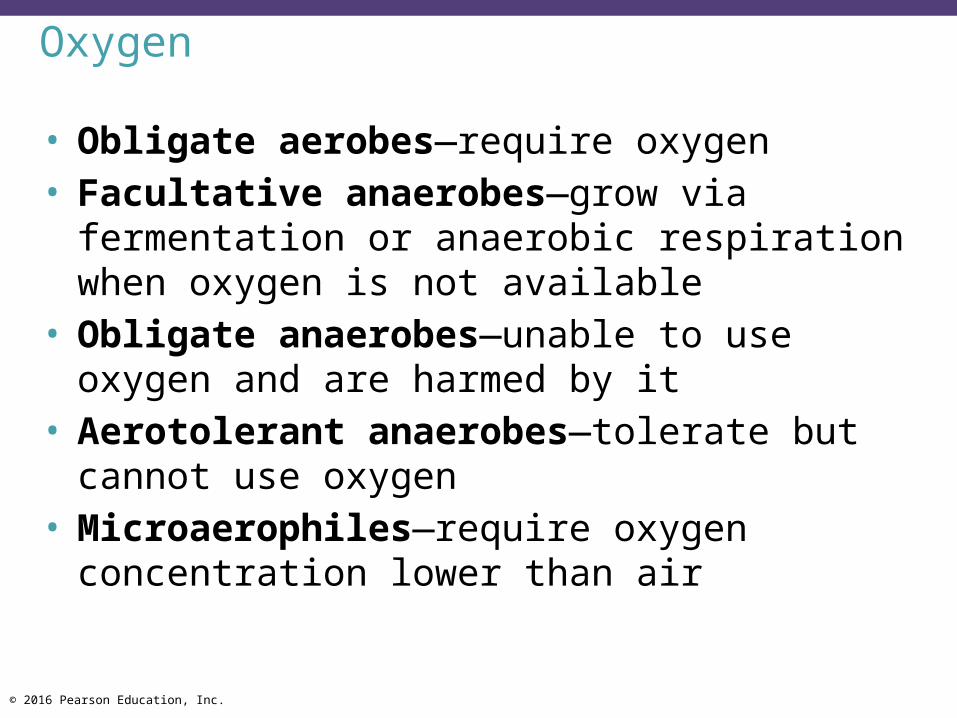
Oxygen
• Obligate aerobes—require oxygen• Facultative anaerobes—grow via fermentation or
anaerobic respiration when oxygen is not available• Obligate anaerobes—unable to use oxygen and
are harmed by it• Aerotolerant anaerobes—tolerate but cannot use
oxygen• Microaerophiles—require oxygen concentration
lower than air
© 2016 Pearson Education, Inc.
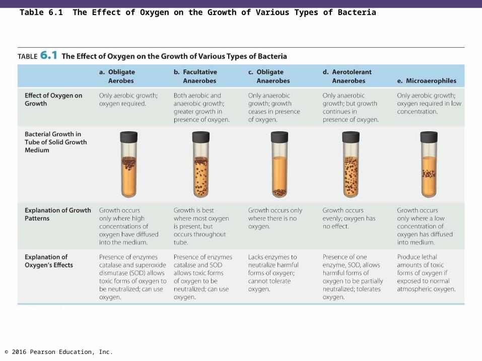
Table 6.1 The Effect of Oxygen on the Growth of Various Types of Bacteria

© 2016 Pearson Education, Inc.
• Singlet oxygen: (1O2−) boosted to a higher-energy
state and is reactive• Superoxide radicals: O2
• Peroxide anion: O22–
• Hydroxyl radical (OH•)
Oxygen
© 2016 Pearson Education, Inc.
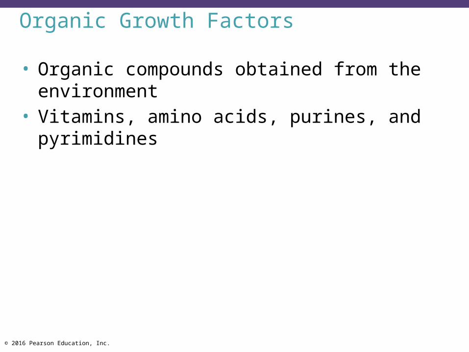
Organic Growth Factors
• Organic compounds obtained from the environment
• Vitamins, amino acids, purines, and pyrimidines
© 2016 Pearson Education, Inc.
Check Your Understanding If bacterial cells were given a sulfur source containing
radioactive sulfur (35S) in their culture media, in what molecules would the 35S be found in the cells?6-4
How would one determine whether a microbe is a strict anaerobe?6-5
Oxygen is so pervasive in the environment that it would be very difficult for a microbe to always avoid physical contact with it. What, therefore, is the most obvious way for a microbe to avoid damage?6-6
© 2016 Pearson Education, Inc.
Biofilms
Learning Objective6-7 Describe the formation of biofilms and their
potential for causing infection.
© 2016 Pearson Education, Inc.
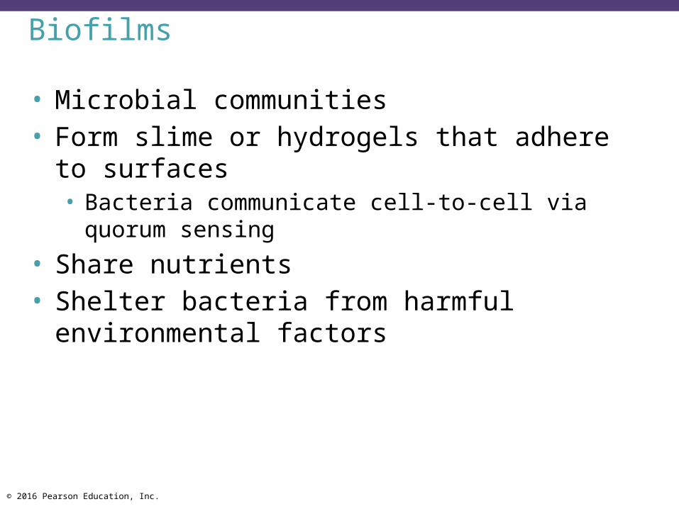
Biofilms
• Microbial communities• Form slime or hydrogels that adhere to surfaces• Bacteria communicate cell-to-cell via quorum sensing
• Share nutrients• Shelter bacteria from harmful environmental
factors
© 2016 Pearson Education, Inc.
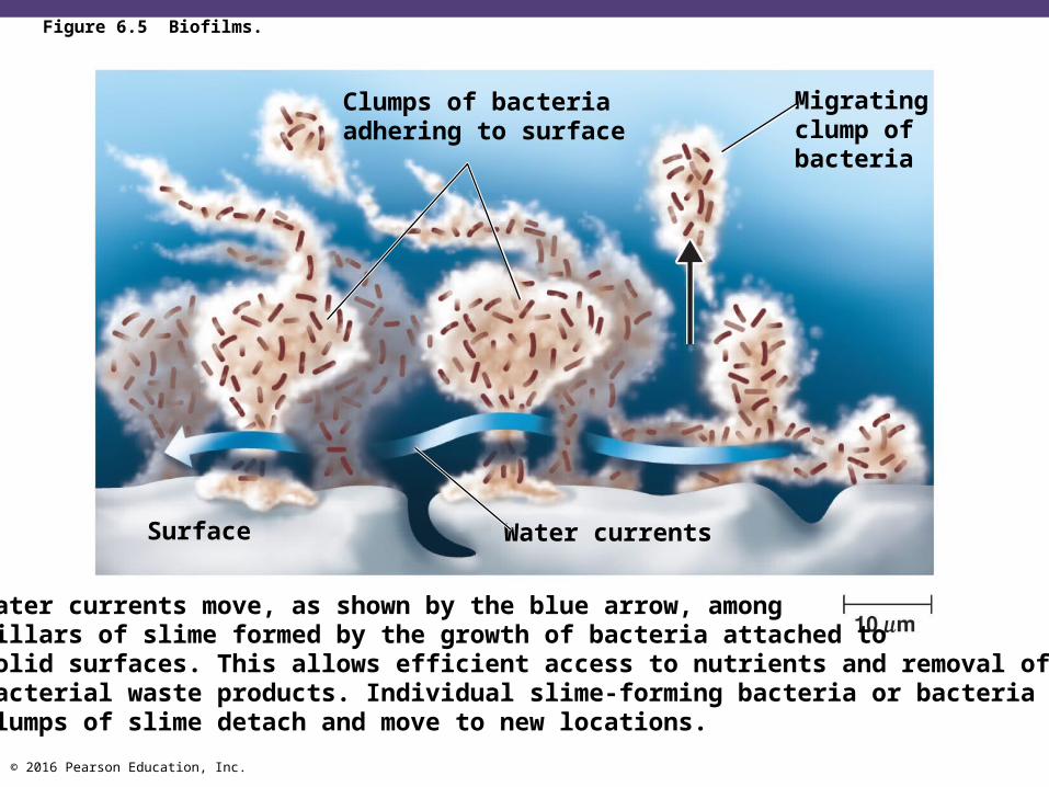
Figure 6.5 Biofilms.
Clumps of bacteriaadhering to surface
Migratingclump ofbacteria
Water currentsSurface
Water currents move, as shown by the blue arrow, amongpillars of slime formed by the growth of bacteria attached tosolid surfaces. This allows efficient access to nutrients and removal ofbacterial waste products. Individual slime-forming bacteria or bacteria inclumps of slime detach and move to new locations.
© 2016 Pearson Education, Inc.
Biofilms
• Found in digestive system and sewage treatment systems; can clog pipes
• 1000x resistant to microbicides• Involved in 70% of infections• Catheters, heart valves, contact lenses, dental caries
© 2016 Pearson Education, Inc.© 2013 Pearson Education, Inc.
Applications of Microbiology 3.1b

© 2016 Pearson Education, Inc.
Check Your Understanding Identify a way in which pathogens find it
advantageous to form biofilms.6-7

© 2016 Pearson Education, Inc.
Culture Media
Learning Objectives6-8 Distinguish chemically defined and complex
media.6-9 Justify the use of each of the following:
anaerobic techniques, living host cells, candle jars, selective and differential media, enrichment medium.
6-10 Differentiate biosafety levels 1, 2, 3, and 4.
© 2016 Pearson Education, Inc.
Culture Media
• Culture medium: nutrients prepared for microbial growth
• Sterile: no living microbes• Inoculum: introduction of microbes into a medium• Culture: microbes growing in or on a culture
medium
© 2016 Pearson Education, Inc.
Culture Media
• Agar • Complex polysaccharide • Used as a solidifying agent for culture media in Petri
plates, slants, and deeps• Generally not metabolized by microbes• Liquefies at 100C• Solidifies at ~40C
© 2016 Pearson Education, Inc.
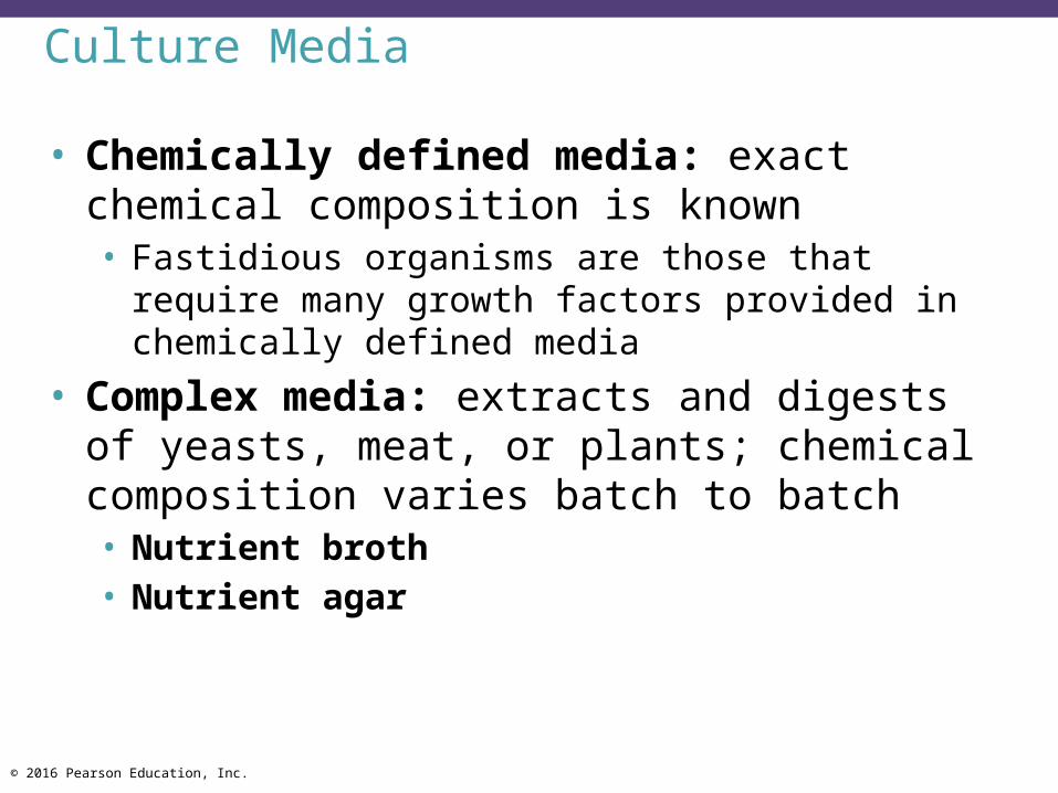
Culture Media
• Chemically defined media: exact chemical composition is known• Fastidious organisms are those that require many
growth factors provided in chemically defined media• Complex media: extracts and digests of yeasts,
meat, or plants; chemical composition varies batch to batch• Nutrient broth• Nutrient agar
© 2016 Pearson Education, Inc.
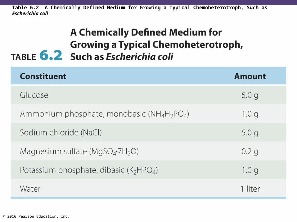
Table 6.2 A Chemically Defined Medium for Growing a Typical Chemoheterotroph, Such as Escherichia coli

© 2016 Pearson Education, Inc.
Table 6.3 Defined Culture Medium for Leuconostoc mesenteroides

© 2016 Pearson Education, Inc.
Table 6.4 Composition of Nutrient Agar, a Complex Medium for the Growth of Heterotrophic Bacteria
© 2016 Pearson Education, Inc.
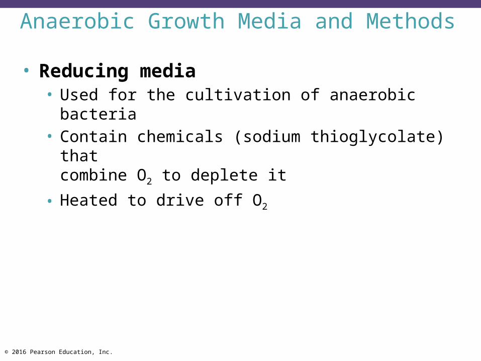
Anaerobic Growth Media and Methods
• Reducing media• Used for the cultivation of anaerobic bacteria• Contain chemicals (sodium thioglycolate) that
combine O2 to deplete it• Heated to drive off O2
© 2016 Pearson Education, Inc.
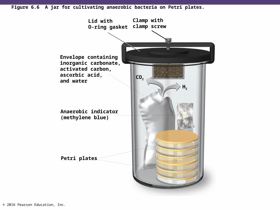
Figure 6.6 A jar for cultivating anaerobic bacteria on Petri plates.
Clamp withclamp screw
Lid withO-ring gasket
Envelope containinginorganic carbonate,activated carbon,ascorbic acid,and water
Anaerobic indicator(methylene blue)
Petri plates
CO2
H2
© 2016 Pearson Education, Inc.
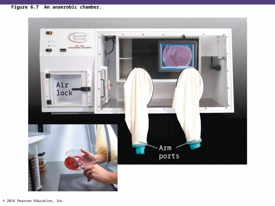
Figure 6.7 An anaerobic chamber.
Airlock
Armports

© 2016 Pearson Education, Inc.
Special Culture Techniques
• Capnophiles • Microbes that require high CO2 conditions• CO2 packet• Candle jar
© 2016 Pearson Education, Inc.
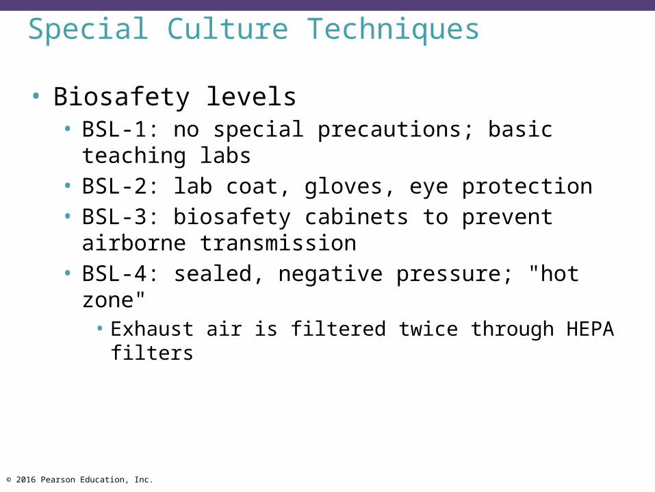
Special Culture Techniques
• Biosafety levels• BSL-1: no special precautions; basic teaching labs• BSL-2: lab coat, gloves, eye protection• BSL-3: biosafety cabinets to prevent airborne
transmission• BSL-4: sealed, negative pressure; "hot zone"
• Exhaust air is filtered twice through HEPA filters

© 2016 Pearson Education, Inc.
Figure 6.8 Technicians in a biosafety level 4 (BSL-4) laboratory.

© 2016 Pearson Education, Inc.
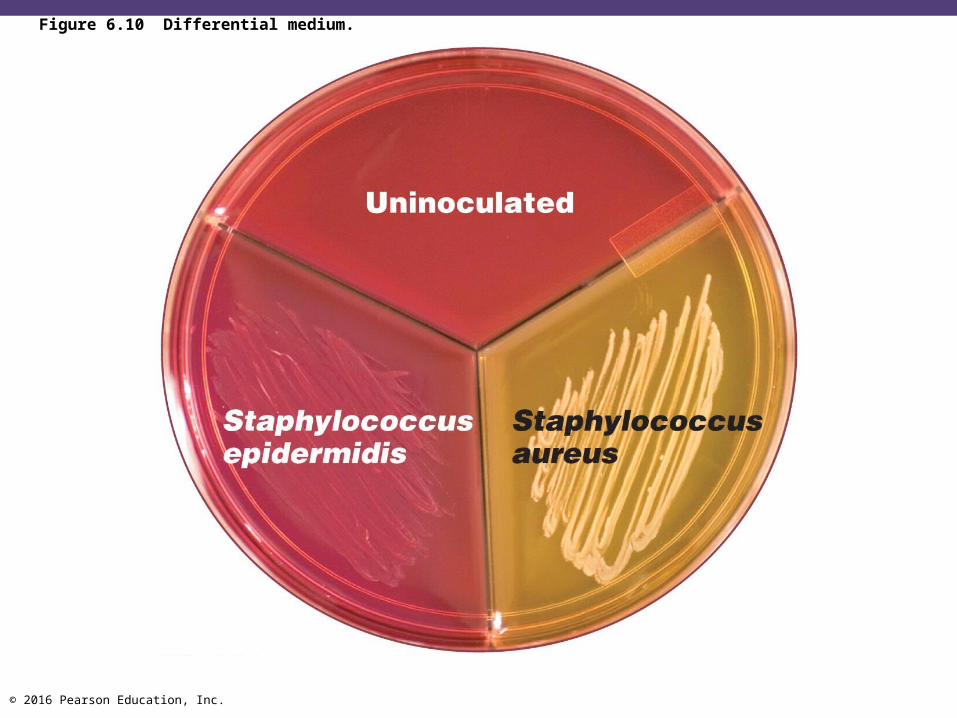
Selective and Differential Media
• Selective media• Suppress unwanted microbes and encourage desired
microbes• Contain inhibitors to suppress growth
© 2016 Pearson Education, Inc.
Selective and Differential Media
• Differential media • Allow distinguishing of colonies of different microbes on
the same plate• Some media have both selective and differential
characteristics
© 2016 Pearson Education, Inc.
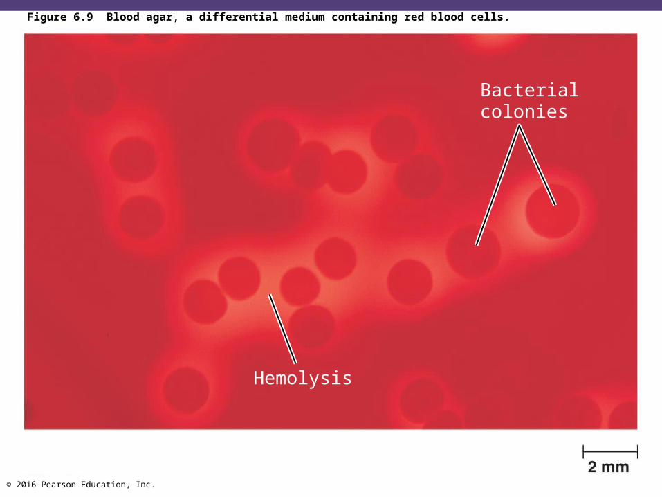
Figure 6.9 Blood agar, a differential medium containing red blood cells.
Bacterialcolonies
Hemolysis
© 2016 Pearson Education, Inc.
Figure 6.10 Differential medium.

© 2016 Pearson Education, Inc.
Enrichment Culture
• Encourages the growth of a desired microbe by increasing very small numbers of a desired organism to detectable levels
• Usually a liquid

© 2016 Pearson Education, Inc.
Table 6.5 Culture Media
© 2016 Pearson Education, Inc.
Check Your Understanding Could humans exist on chemically defined media,
at least under laboratory conditions?6-8
Could Louis Pasteur, in the 1800s, have grown rabies viruses in cell culture instead of in living animals?6-9
What BSL is your laboratory?6-10

© 2016 Pearson Education, Inc.
Obtaining Pure Cultures
Learning Objectives6-11 Define colony.6-12 Describe how pure cultures can be isolated by
using the streak plate method.
© 2016 Pearson Education, Inc.
Obtaining Pure Cultures
• A pure culture contains only one species or strain• A colony is a population of cells arising from a
single cell or spore or from a group of attached cells
• A colony is often called a colony-forming unit (CFU)
• The streak plate method is used to isolate pure cultures

© 2016 Pearson Education, Inc.
Figure 6.11 The streak plate method for isolating pure bacterial cultures.
Colonies

© 2016 Pearson Education, Inc.
Check Your Understanding Can you think of any reason why a colony does
not grow to an infinite size, or at least fill the confines of the Petri plate?6-11
Could a pure culture of bacteria be obtained by the streak plate method if there were only one desired microbe in a bacterial suspension of billions?6-12
© 2016 Pearson Education, Inc.
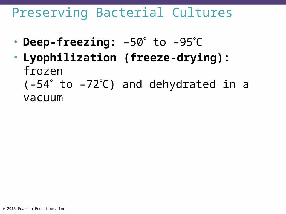
Preserving Bacterial Cultures
Learning Objective6-13 Explain how microorganisms are preserved by
deep-freezing and lyophilization(freeze-drying).
© 2016 Pearson Education, Inc.
Preserving Bacterial Cultures
• Deep-freezing: –50 to –95C• Lyophilization (freeze-drying): frozen
(–54 to –72C) and dehydrated in a vacuum

© 2016 Pearson Education, Inc.
Check Your Understanding If the Space Station in Earth orbit suddenly
ruptured, the humans on board would die instantly from cold and the vacuum of space. Would all the bacteria in the capsule also be killed?6-13
© 2016 Pearson Education, Inc.
The Growth of Bacterial Cultures
Learning Objectives6-14 Define bacterial growth, including binary
fission.6-15 Compare the phases of microbial growth, and
describe their relation to generation time.

© 2016 Pearson Education, Inc.
Bacterial Division
• Increase in number of cells, not cell size• Binary fission• Budding• Conidiospores (actinomycetes)• Fragmentation of filaments

© 2016 Pearson Education, Inc.
Bacterial Growth: Overview
Animation: Bacterial Growth: OverviewPLAY

© 2016 Pearson Education, Inc.
Figure 6.12a Binary fission in bacteria.
Plasma membraneCell wall
DNA (nucleoid)
Cell elongates andDNA is replicated.
Cell wall andplasma membranebegin to constrict.
Cross-wall forms,completelyseparating thetwo DNA copies.
Cellsseparate.
A diagram of the sequence of cell division
© 2016 Pearson Education, Inc.
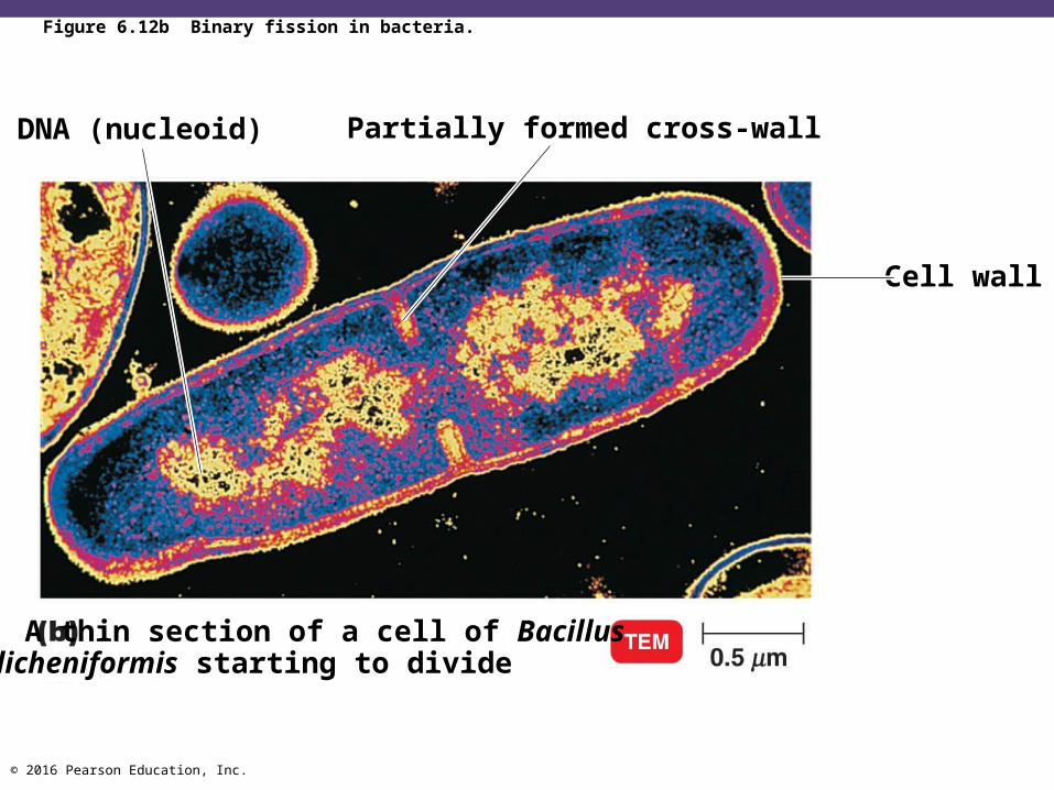
Figure 6.12b Binary fission in bacteria.
Partially formed cross-wall
Cell wall
DNA (nucleoid)
A thin section of a cell of Bacilluslicheniformis starting to divide

© 2016 Pearson Education, Inc.
Generation Time
• Time required for a cell to divide• 20 minutes to 24 hours
• Binary fission doubles the number of cells each generation
• Total number of cells = 2number of generations • Growth curves are represented logarithmically
© 2016 Pearson Education, Inc.
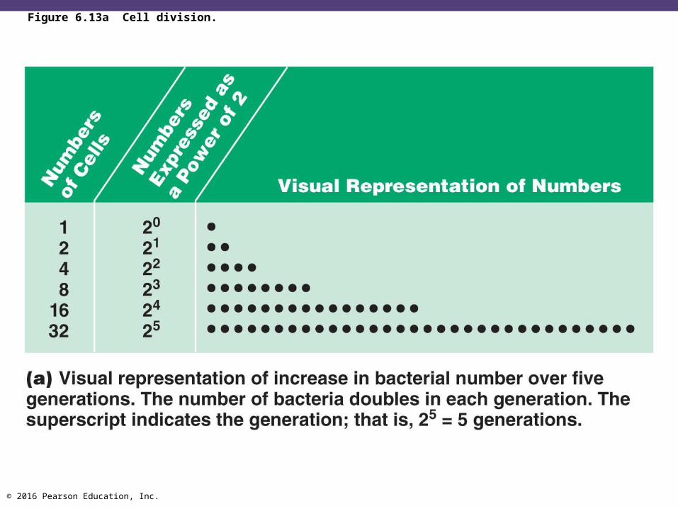
Figure 6.13a Cell division.
© 2016 Pearson Education, Inc.
Figure 6.13b Cell division.
© 2016 Pearson Education, Inc.
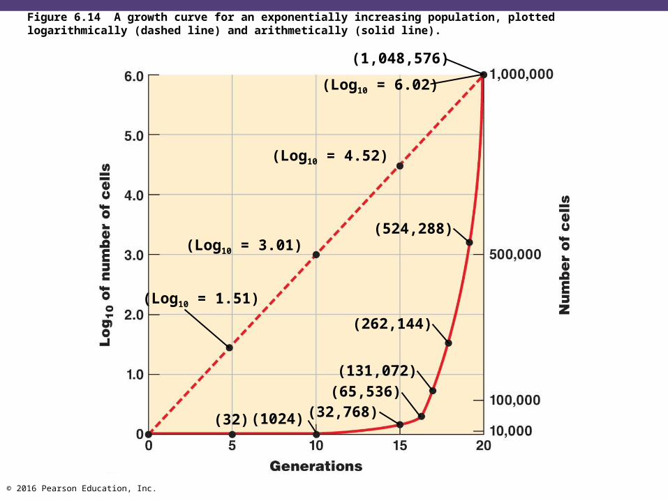
Figure 6.14 A growth curve for an exponentially increasing population, plotted logarithmically (dashed line) and arithmetically (solid line).
(1,048,576)
(Log10 = 6.02)
(Log10 = 4.52)
(Log10 = 3.01)
(Log10 = 1.51)
(524,288)
(262,144)
(131,072)(65,536)
(32,768)(1024)(32)

© 2016 Pearson Education, Inc.
Binary Fission
Animation: Binary FissionPLAY

© 2016 Pearson Education, Inc.
Check Your Understanding Can a complex organism, such as a beetle, divide
by binary fission?6-14

© 2016 Pearson Education, Inc.
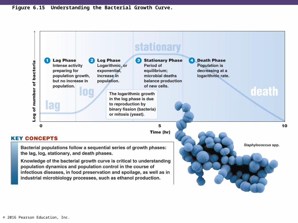
Phases of Growth
• Lag phase• Log phase• Stationary phase• Death phase
© 2016 Pearson Education, Inc.
Figure 6.15 Understanding the Bacterial Growth Curve.

© 2016 Pearson Education, Inc.
Bacterial Growth Curve
Animation: Bacterial Growth CurvePLAY

© 2016 Pearson Education, Inc.
Check Your Understanding If two mice started a family within a fixed
enclosure, with a fixed food supply, would the population curve be the same as a bacterial growth curve?6-15
© 2016 Pearson Education, Inc.
The Growth of Bacterial Cultures
Learning Objectives6-16 Explain four direct methods of measuring cell
growth.6-17 Differentiate direct and indirect methods of
measuring cell growth.6-18 Explain three indirect methods of measuring
cell growth.
© 2016 Pearson Education, Inc.
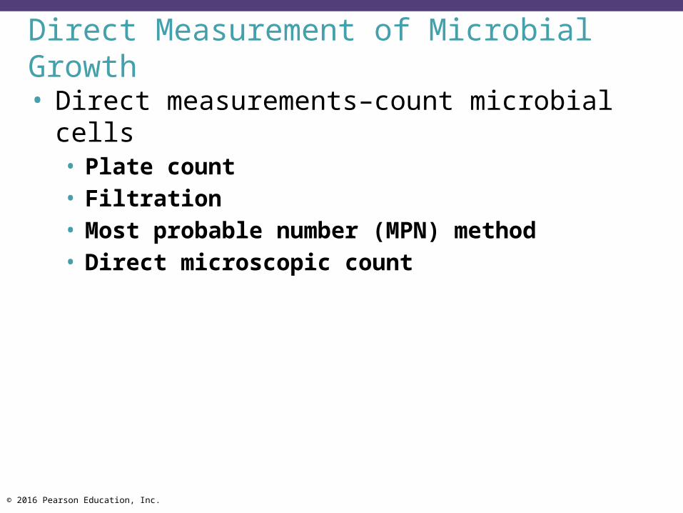
Direct Measurement of Microbial Growth
• Direct measurements–count microbial cells• Plate count• Filtration• Most probable number (MPN) method• Direct microscopic count

© 2016 Pearson Education, Inc.
Plate Counts
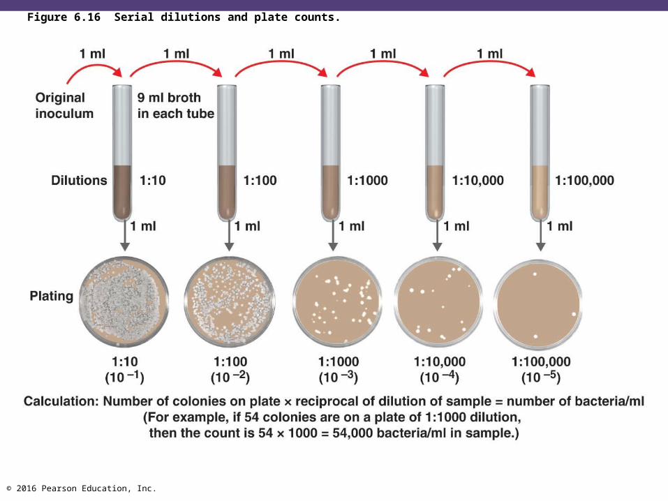
• Count colonies on plates that have 30 to 300 colonies (CFUs)
• To ensure the right number of colonies, the original inoculum must be diluted via serial dilution
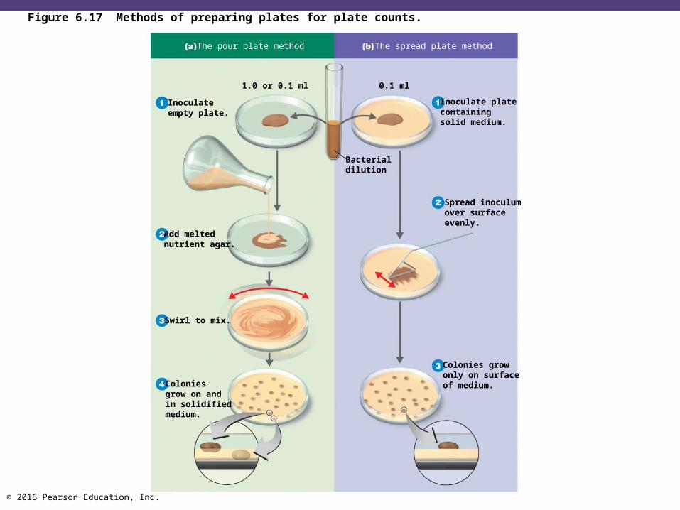
• Counts are performed on bacteria mixed into a dish with agar (pour plate method) or spread on the surface of a plate (spread plate method)
© 2016 Pearson Education, Inc.
Figure 6.16 Serial dilutions and plate counts.
© 2016 Pearson Education, Inc.
The pour plate method The spread plate method
Figure 6.17 Methods of preparing plates for plate counts.
The pour plate method The spread plate method
0.1 ml
Inoculate platecontainingsolid medium.
Spread inoculumover surfaceevenly.
Colonies growonly on surfaceof medium.
1.0 or 0.1 ml
Inoculateempty plate.
Add meltednutrient agar.
Swirl to mix.
Coloniesgrow on andin solidifiedmedium.
Bacterialdilution
© 2016 Pearson Education, Inc.
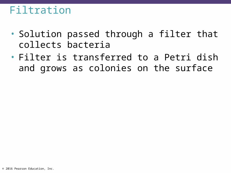
Filtration
• Solution passed through a filter that collects bacteria
• Filter is transferred to a Petri dish and grows as colonies on the surface

© 2016 Pearson Education, Inc.
Figure 6.18 Counting bacteria by filtration.

© 2016 Pearson Education, Inc.
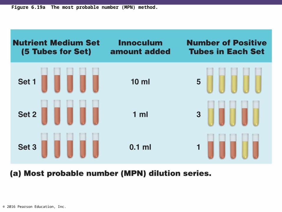
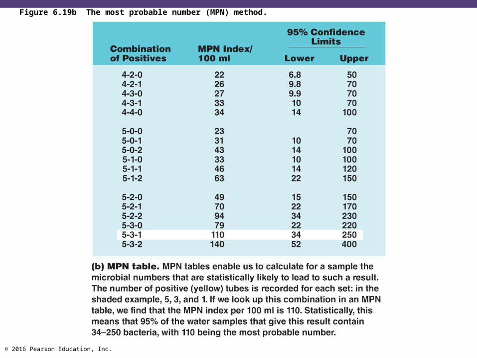
The Most Probable Number (MPN) Method
• Multiple tube test• Count positive tubes• Compare with a statistical table
© 2016 Pearson Education, Inc.
Figure 6.19a The most probable number (MPN) method.
© 2016 Pearson Education, Inc.
Figure 6.19b The most probable number (MPN) method.

© 2016 Pearson Education, Inc.
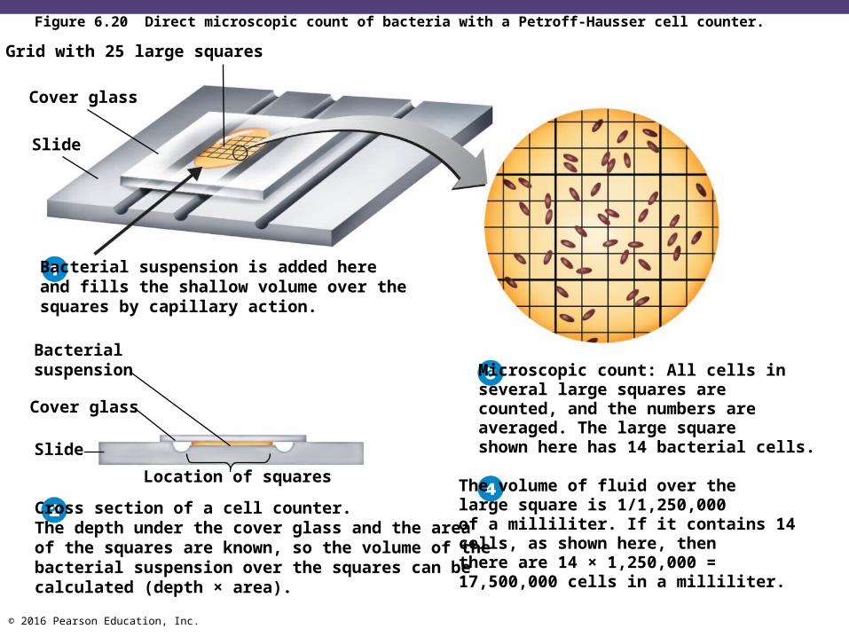
• Volume of a bacterial suspension placed on a slide• Average number of bacteria per viewing field is
calculated• Uses a special Petroff-Hausser cell counter
Direct Microscopic Count
Number of bacteria/ml =Number of cells countedVolume of area counted
© 2016 Pearson Education, Inc.
Figure 6.20 Direct microscopic count of bacteria with a Petroff-Hausser cell counter.
Grid with 25 large squares
Cover glass
Slide
Bacterial suspension is added hereand fills the shallow volume over thesquares by capillary action.
Bacterialsuspension
Cover glass
Slide
Cross section of a cell counter.The depth under the cover glass and the areaof the squares are known, so the volume of thebacterial suspension over the squares can becalculated (depth × area).
Location of squares
Microscopic count: All cells inseveral large squares arecounted, and the numbers areaveraged. The large squareshown here has 14 bacterial cells.
The volume of fluid over thelarge square is 1/1,250,000of a milliliter. If it contains 14cells, as shown here, thenthere are 14 × 1,250,000 =17,500,000 cells in a milliliter.
© 2016 Pearson Education, Inc.
Check Your Understanding Why is it difficult to measure realistically the
growth of a filamentous mold isolate by the plate count method?6-16

© 2016 Pearson Education, Inc.
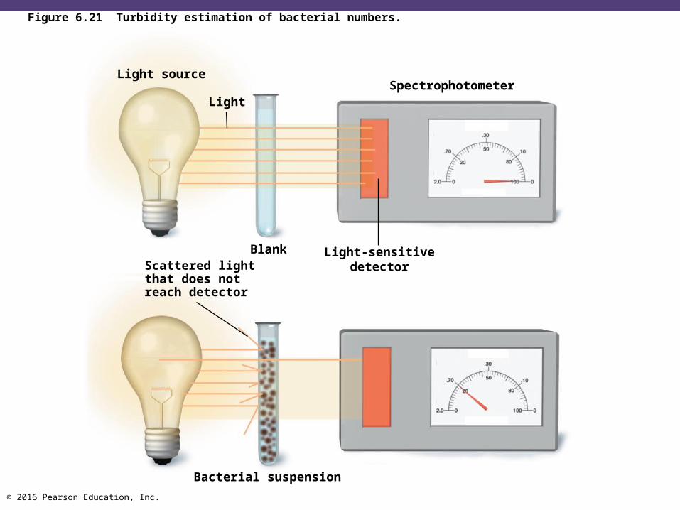
Estimating Bacterial Numbers by Indirect Methods
• Turbidity—measurement of cloudiness with a spectrophotometer
• Metabolic activity—amount of metabolic product is proportional to the number of bacteria
• Dry weight—bacteria are filtered, dried, and weighed; used for filamentous organisms
© 2016 Pearson Education, Inc.
Figure 6.21 Turbidity estimation of bacterial numbers.
Light source
LightSpectrophotometer
BlankScattered lightthat does notreach detector
Light-sensitivedetector
Bacterial suspension

© 2016 Pearson Education, Inc.
Check Your Understanding Direct methods usually require an incubation time
for a colony. Why is this not always feasible for analyzing foods?6-17
If there is no good method for analyzing a product for its vitamin content, what is a feasible method of determining the vitamin content?6-18